Gender differences in stimulated cytokine production following acute psychological stress
17
Gender differences in stimulated cytokine production following acute psychological stress Aric A. Prather, M.S., Judith E. Carroll, M.S., Jacqueline M. Fury, B.S., Kevin K. McDade, M.S., Diana Ross, R.N., BSN, and Anna L. Marsland, Ph.D., R.N. Abstract Emerging evidence suggests that acute psychological stress modulates inflammatory competence; however, not all findings are consistent. Gender is one factor that may impact magnitude of response. To explore this possibility, we examined the effects of acute mental stress on lipopolysaccharide- induced production of pro-inflammatory cytokines interleukin (IL)- 1 β, IL-6, and tumor necrosis factor (TNF)- α among a relatively healthy sample of midlife men (n=28) and women (n=34). Blood samples for the assessment of cytokine production were drawn before, immediately after and 30 minutes following subjects’ performance of an evaluative speech task. Relative to baseline evaluations, the speech stressor elicited a significant increase in stimulated production of all 3 pro- inflammatory cytokines, as measured 30 minutes following the end of the task. There were no gender differences in the magnitude of this effect. However, men showed a significant decrease in cytokine production from before to immediately following the stressor, whereas women showed no change across this period. Menopausal status partially accounted for these gender differences, with postmenopausal women displaying greater increases in IL-6 and TNF-α production from baseline- to-post task when compared to men. These data provide further evidence that acute psychological stress primes the immune system to mount larger inflammatory responses and initial support for gender differences in the patterning of stress-related cytokine activity. In addition, this study presents novel evidence that post-menopausal women may be particularly susceptible to stress-related inflammatory responses. The possibility that this contributes to the increased risk of inflammatory disease observed among older women warrants investigation. Introduction Consistent evidence shows that brief psychological stress modulates components of the immune system. Early studies focused on stress-induced changes in cellular immune parameters (for review, see Segerstrom and Miller, 2004); however, more recent interest has shifted to the effects of acute laboratory stress on immune processes involved in the inflammatory response. In this regard, evidence shows reliable, stress-related increases in circulating levels of inflammatory mediators, including interleukin (IL)-6, IL-1β, and C- reactive protein (Steptoe et al., 2007). These circulating signaling proteins are assumed to reflect systemic inflammation, with higher levels predicting increased risk for inflammatory disease (Libby and Ridker, 1999; Ridker et al., 2000a; Ridker et al., 2000b). However, the interpretation of circulating levels of these proteins is complicated by the multiple cell types All correspondence concerning this manuscript should be addressed to Aric A. Prather, Behavioral Immunology Laboratory, Department of Psychology, 3943 O’Hara Street, Pittsburgh, PA 15260; Telephone: (412) 624-2466; FAX: (412) 648-3720; E-mail: E-mail: [email protected]. Publisher's Disclaimer: This is a PDF file of an unedited manuscript that has been accepted for publication. As a service to our customers we are providing this early version of the manuscript. The manuscript will undergo copyediting, typesetting, and review of the resulting proof before it is published in its final citable form. Please note that during the production process errors may be discovered which could affect the content, and all legal disclaimers that apply to the journal pertain. NIH Public Access Author Manuscript Brain Behav Immun. Author manuscript; available in PMC 2010 July 1. Published in final edited form as: Brain Behav Immun. 2009 July ; 23(5): 622–628. doi:10.1016/j.bbi.2008.11.004. NIH-PA Author Manuscript NIH-PA Author Manuscript NIH-PA Author Manuscript
Transcript of Gender differences in stimulated cytokine production following acute psychological stress

Gender differences in stimulated cytokine production followingacute psychological stress
Aric A. Prather, M.S., Judith E. Carroll, M.S., Jacqueline M. Fury, B.S., Kevin K. McDade,M.S., Diana Ross, R.N., BSN, and Anna L. Marsland, Ph.D., R.N.
AbstractEmerging evidence suggests that acute psychological stress modulates inflammatory competence;however, not all findings are consistent. Gender is one factor that may impact magnitude of response.To explore this possibility, we examined the effects of acute mental stress on lipopolysaccharide-induced production of pro-inflammatory cytokines interleukin (IL)- 1 β, IL-6, and tumor necrosisfactor (TNF)- α among a relatively healthy sample of midlife men (n=28) and women (n=34). Bloodsamples for the assessment of cytokine production were drawn before, immediately after and 30minutes following subjects’ performance of an evaluative speech task. Relative to baselineevaluations, the speech stressor elicited a significant increase in stimulated production of all 3 pro-inflammatory cytokines, as measured 30 minutes following the end of the task. There were no genderdifferences in the magnitude of this effect. However, men showed a significant decrease in cytokineproduction from before to immediately following the stressor, whereas women showed no changeacross this period. Menopausal status partially accounted for these gender differences, withpostmenopausal women displaying greater increases in IL-6 and TNF-α production from baseline-to-post task when compared to men. These data provide further evidence that acute psychologicalstress primes the immune system to mount larger inflammatory responses and initial support forgender differences in the patterning of stress-related cytokine activity. In addition, this study presentsnovel evidence that post-menopausal women may be particularly susceptible to stress-relatedinflammatory responses. The possibility that this contributes to the increased risk of inflammatorydisease observed among older women warrants investigation.
IntroductionConsistent evidence shows that brief psychological stress modulates components of theimmune system. Early studies focused on stress-induced changes in cellular immuneparameters (for review, see Segerstrom and Miller, 2004); however, more recent interest hasshifted to the effects of acute laboratory stress on immune processes involved in theinflammatory response. In this regard, evidence shows reliable, stress-related increases incirculating levels of inflammatory mediators, including interleukin (IL)-6, IL-1β, and C-reactive protein (Steptoe et al., 2007). These circulating signaling proteins are assumed toreflect systemic inflammation, with higher levels predicting increased risk for inflammatorydisease (Libby and Ridker, 1999; Ridker et al., 2000a; Ridker et al., 2000b). However, theinterpretation of circulating levels of these proteins is complicated by the multiple cell types
All correspondence concerning this manuscript should be addressed to Aric A. Prather, Behavioral Immunology Laboratory, Departmentof Psychology, 3943 O’Hara Street, Pittsburgh, PA 15260; Telephone: (412) 624-2466; FAX: (412) 648-3720; E-mail: E-mail:[email protected]'s Disclaimer: This is a PDF file of an unedited manuscript that has been accepted for publication. As a service to our customerswe are providing this early version of the manuscript. The manuscript will undergo copyediting, typesetting, and review of the resultingproof before it is published in its final citable form. Please note that during the production process errors may be discovered which couldaffect the content, and all legal disclaimers that apply to the journal pertain.
NIH Public AccessAuthor ManuscriptBrain Behav Immun. Author manuscript; available in PMC 2010 July 1.
Published in final edited form as:Brain Behav Immun. 2009 July ; 23(5): 622–628. doi:10.1016/j.bbi.2008.11.004.
NIH
-PA Author Manuscript
NIH
-PA Author Manuscript
NIH
-PA Author Manuscript

that produce them, including adipocytes and endothelial cells (Mohamed-Ali et al., 1997;Papanicolaou et al., 1998), raising the possibility that they may not reflect a primaryinflammatory response. In order to more directly assess inflammatory competence, recentstudies have begun to examine the effects of acute stress on the functional ability of immunecells to produce pro-inflammatory mediators when stimulated in vitro.
The inflammatory response begins when monocytes/macrophages are activated by pathogensor tissue damage to release pro-inflammatory cytokines (e.g. IL-6, IL-1β, TNF-α), which, inturn, recruit leukocytes to the area of injury, up-regulate the expression of cellular adhesionmolecules on the endothelium, and promote the systemic release of acute phase proteins (e.g.C-reactive protein) (Maier and Watkins, 1998). The magnitude of the pro-inflammatorycytokine response to immune activation is critical; insufficient response may leave theorganism vulnerable to infection, whereas excessive response can increase risk forinflammatory diseases (Nathan, 2002; Pavlov and Tracey, 2004). Convergent evidence fromthe animal and in vitro literatures shows that the autonomic nervous system plays a key rolein regulating the magnitude of this response. Signaling by the sympathetic nervous system(SNS) can up- and down-regulate production of pro-inflammatory cytokines by activatedmonocytes/macrophages and thus modulate inflammatory potential (Elenkov et al., 2000;Hasko and Szabo, 1998; van der Poll et al., 1994; van der Poll and Lowry, 1997). Theparasympathetic nervous system also plays a role in the down-regulation of the inflammatoryresponse (Pavlov and Tracey, 2005; Tracey, 2002). Although there are wide and stableindividual differences in the magnitude of autonomic responses to acute laboratory stress, ingeneral, the ratio of sympathetic to parasympathetic activation increases, which may primeimmune cells to mount larger pro-inflammatory responses to inflammatory stimuli.
To date, findings from experimental studies that examine whether acute stress alters stimulatedcytokine production have been inconsistent. In a recent review of the literature, Steptoe andcolleagues (2007) identified 8 studies that examined endotoxin-stimulated inflammatoryresponses to acute laboratory stress in healthy adults (Steptoe et al., 2007). Of the studies thatmeasured stress-induced changes in IL-6 production, 4 showed an increase (Gaab et al.,2005; Goebel et al., 2000; Peters et al., 1999; Rohleder et al., 2003), 2 showed a decrease(Jacobs et al., 2001; Rohleder et al., 2001), and 2 showed no effect (Miller et al., 2005; Suarezet al., 2006). Similarly variable results were observed on examination of stimulated productionof TNF-α. As noted by Steptoe et al (2007), reasons for these inconsistencies are unclear dueto small samples sizes, variation in sample characteristics, timing of measures, and the typesof challenges employed. One characteristic of participants that may contribute to differencesacross studies is gender. A fairly robust literature exists on gender differences in cardiovascularresponses to acute psychological stress, with men exhibiting greater increases in indices ofsympathetic activation than women (Allen et al., 1993; Matthews and Stoney, 1988). To theextent that autonomic activation modulates inflammatory competence, one might expectgender differences in stimulated cytokine production following an acute stressor. To date, onlyone study has directly examined this possibility, comparing endotoxin-stimulated productionof IL-6 and TNF-α in response to the Trier Social Stress Test among 18 women and 27 men(Rohleder et al., 2001). Results show a pre- to 60 minute post-task decrease in stimulatedproduction of IL-6 among males, but not females. These findings are inconsistent withpredictions based on the autonomic regulation of inflammatory competence; however, it ispossible that measures taken closer to the end of the challenge would reveal a different patternof findings. To explore this possibility, the present study examines the capacity of immunecells to produce IL-6, IL-1β, and TNF-α following in vitro stimulation with the bacterialproduct LPS, as measured before, immediately after, and 30 minutes following a speech taskamong males and females. Based on evidence that males show larger autonomic responses toacute psychological stress than females, it was hypothesized that men would show greaterstress-induced increases in the production of these pro-inflammatory cytokines than women.
Prather et al. Page 2
Brain Behav Immun. Author manuscript; available in PMC 2010 July 1.
NIH
-PA Author Manuscript
NIH
-PA Author Manuscript
NIH
-PA Author Manuscript

In secondary analyses, the possibility that menopausal status moderates the magnitude ofinflammatory responses in women was also explored. To date, no studies have examined thispossibility despite evidence that (1) immune cells have receptors for estrogen and progesterone(Turgeon et al., 2006), (2) in vitro treatment of immune cells with estrogen and progesteronemodulates pro-inflammatory cytokine production (Rogers & Eastell, 2001; Turgeon et al.,2006; Zhang et al., 2001), and (3) risk for inflammatory disease increases following menopausewhen levels of these hormones are reduced (Moxley et al., 2002; Turgeon et al., 2006).
MethodsParticipants
Study participants were 28 men and 34 women (92% Caucasian) between the ages of 40 and59 enrolled in the Vaccination Immunity Project (VIP), a longitudinal study investigatingpsychological, behavioral, and physiological predictors of antibody response to hepatitis Bvaccination. Participants were recruited via mass mail solicitation in Western Pennsylvania(principally Allegheny County). Eligible participants were non-smokers, reported being ingood general health (including no history or symptoms of myocardial infarction, asthma, cancertreatment in the past year, psychotic illness, or other systemic diseases known to affect theimmune system), and free from medications known to affect the nervous, endocrine, or immunesystems in the past 3 months (not including oral contraceptives). Women who were pregnantor lactating were also ineligible. No women in the sub-study endorsed taking hormonereplacement therapy. Appointments were scheduled so that all participants were free of signsand symptoms of infection for 2 weeks prior to the acute laboratory session. Participantsincluded in this sub-study were a random subset of participants in the parent project on whomwe drew blood samples for the measurement of endotoxin-stimulated pro-inflammatorycytokine production. Informed consent was acquired in compliance with guidelines of theUniversity of Pittsburgh Institutional Review Board.
Laboratory Stress TestingData used in the present analyses were collected at a single laboratory session that lastedapproximately 2 hours and started between 7:00 AM and 9:00 AM. Prior to the session, subjectsabstained from food, beverages (except water), and caffeine for 12 hours, strenuous physicalactivity and non-prescription medications for 24 hours, and alcohol consumption for 48 hours.On arrival, subjects completed a battery of paper and pencil questionnaires, includingassessment of current symptoms of acute illness, medical history, and menopausal status. Aftercompletion of questionnaires, subjects’ height and weight was recorded for determination ofbody mass index (BMI; kg/m2) and they were escorted to a testing chamber. Here, anintravenous catheter was inserted into the antecubital fossa of the non-dominant arm for thecollection of blood samples, and an occluding cuff was placed on the other arm for automatedmeasurement of heart rate (HR) and systolic blood pressure (SBP) and diastolic BP (DBP)(Critikon Dinamap 8100 Vital Signs Monitor, Tampa, FL). Following instrumentation,subjects rested for a 30-minute adaptation period. During the last 6 minutes of this period, BPand HR were recorded 4 times (every 90 seconds) and a 20ml blood sample was drawn. Subjectswere then asked to perform a simulated public speaking task, consisting of 2 minutes ofpreparation for a speech defending themselves against an alleged transgression, followed by 3minutes of videotaped speech delivery. HR and BP were measured every 90 seconds duringspeech preparation and delivery, and a second 20ml blood sample was collected immediatelyafter task performance. Following the task, subjects rested for a 30-minute recovery period.HR and BP were recorded 4 times (every 90 seconds) during the last 6 minutes of this periodand a final 20ml blood sample was drawn at the end of this period.
Prather et al. Page 3
Brain Behav Immun. Author manuscript; available in PMC 2010 July 1.
NIH
-PA Author Manuscript
NIH
-PA Author Manuscript
NIH
-PA Author Manuscript

Stimulated Cytokine ProductionWhole blood was collected in heparin-treated vacutainer tubes and stimulated withlipopolysaccharide (LPS, serotype 026:B6, Sigma) at a final concentration of 2.5 ug/ml withoutantibiotics in polypropylene tubes under sterile conditions (stimulated sample). Controlsamples, containing whole blood without LPS, were set up in parallel to measure spontaneouslevels of cytokine production (unstimulated sample). The samples were incubated at 37°C with5% CO2 humidified atmosphere for 24 hours. Following incubation, the tubes were centrifugedat 1000g for 10 minutes. Supernatants were collected and frozen at −80°C until the study wascomplete.
At the end of the study, LPS-stimulated and unstimulated supernatants were assayed in onebatch using a multiplex analysis system, as previously described (Marsland et al., 2007b).Briefly, multiplex bead kits, based on the principle of solid phase sandwich immunoassays,were employed. All reagents, working standards, and samples were prepared as per themanufacturer’s specifications and were run in duplicates. Samples were read within 24 hoursusing Bio-Plex reader (Luminex 100™). Stimulated levels of IL-6, IL-1β, and TNF-α weredetermined using the Bio-Plex Manager Software (Bio-rad Corporation, Hercules, CA),interpolating from the standard curve (Logistic-5PL curve fit). Pooled plasma controls wereincluded on all plates. Inter- and intra- assay coefficients of variability were less than or equalto 10%. Stimulated cytokine production was quantified by subtracting unstimulated fromstimulated cytokine levels.
Menopausal StatusMenopausal status was defined on the basis of reported menstrual history over the prior 12months, with premenopausal being regular, normal cycles; perimenopausal being irregularcycles and other related symptoms; and postmenopausal being no cycles.
Statistical AnalysesAll analyses were performed using SPSS for windows (version 14.0). Stimulated pro-inflammatory cytokines were natural log transformed prior to analyses to better approximatenormal distributions. Comparisons of demographic characteristics and baseline cytokineconcentrations between male and female participants were carried out using independent t testsor χ2 statistics. To evaluate the effects of the speech task on cardiovascular and immuneparameters, repeated-measures analyses of covariance (ANCOVAs) were conducted on eachdependent variable. For these analyses, HR and BP data were reduced by calculating meanvalues for baseline, task, and recovery periods. These values, and the cytokine production data,were then subjected to 2 (Gender women, men) × 3 (Period baseline, task, recovery) repeated-measuresANCOVAs. In light of significant correlations between baseline cytokine production andmagnitude of stress-related changes in cytokine production (r’s= −.30–−.49, p<.05), baselinecytokine production was included as a covariate in immune analyses. Greenhouse-Geissercorrections for repeated measures were calculated where appropriate. Significant effects werefollowed by Bonferroni post-hoc comparisons. Sex differences were further explored bystratifying women by menopausal status (premenopausal and postmenopausal). All α’s forstatistical analyses were set to .05 and two-tailed tests of significance were used.
ResultsPreliminary Analyses
Mean values of IL-6, IL-1β, and TNF-α with and without stimulation at each study time point(baseline, task, recovery) are presented in Table 1. As expected, whole blood stimulation withLPS induced a marked increase in the levels of IL-1β, IL-6, and TNF-α and baseline stimulated
Prather et al. Page 4
Brain Behav Immun. Author manuscript; available in PMC 2010 July 1.
NIH
-PA Author Manuscript
NIH
-PA Author Manuscript
NIH
-PA Author Manuscript
levels of the 3 pro-inflammatory cytokines were significantly correlated with one another(r’s= .67–.79). Demographic characteristics of the study sample are displayed in Table 2. Theonly significant gender difference was in resting BP, with men showing higher DBP thanwomen [t (1,60) = −2.46, p<.02]. There were no significant associations between baseline levelsof cytokine production and any of the demographic characteristics.
Effect of the speech task on HR, BP, and stimulated production of pro-inflammatorycytokines
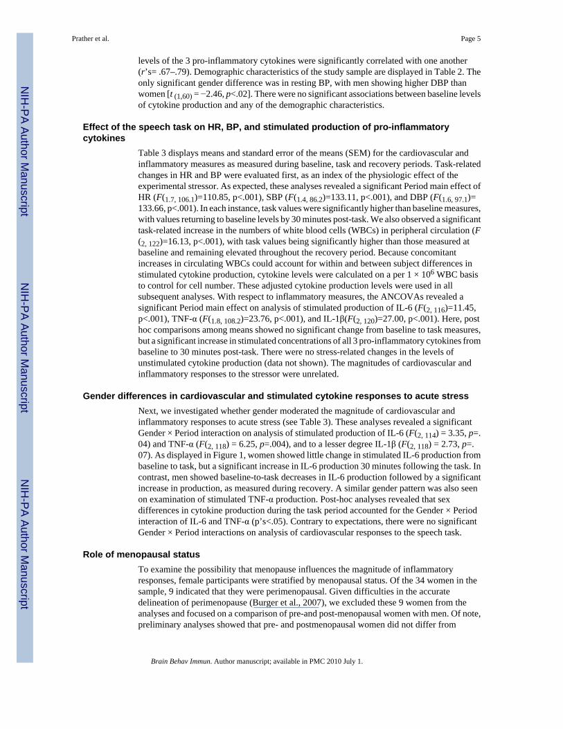
Table 3 displays means and standard error of the means (SEM) for the cardiovascular andinflammatory measures as measured during baseline, task and recovery periods. Task-relatedchanges in HR and BP were evaluated first, as an index of the physiologic effect of theexperimental stressor. As expected, these analyses revealed a significant Period main effect ofHR (F(1.7, 106.1)=110.85, p<.001), SBP (F(1.4, 86.2)=133.11, p<.001), and DBP (F(1.6, 97.1)=133.66, p<.001). In each instance, task values were significantly higher than baseline measures,with values returning to baseline levels by 30 minutes post-task. We also observed a significanttask-related increase in the numbers of white blood cells (WBCs) in peripheral circulation (F(2, 122)=16.13, p<.001), with task values being significantly higher than those measured atbaseline and remaining elevated throughout the recovery period. Because concomitantincreases in circulating WBCs could account for within and between subject differences instimulated cytokine production, cytokine levels were calculated on a per 1 × 10⁶ WBC basisto control for cell number. These adjusted cytokine production levels were used in allsubsequent analyses. With respect to inflammatory measures, the ANCOVAs revealed asignificant Period main effect on analysis of stimulated production of IL-6 (F(2, 116)=11.45,p<.001), TNF-α (F(1.8, 108.2)=23.76, p<.001), and IL-1β(F(2, 120)=27.00, p<.001). Here, posthoc comparisons among means showed no significant change from baseline to task measures,but a significant increase in stimulated concentrations of all 3 pro-inflammatory cytokines frombaseline to 30 minutes post-task. There were no stress-related changes in the levels ofunstimulated cytokine production (data not shown). The magnitudes of cardiovascular andinflammatory responses to the stressor were unrelated.
Gender differences in cardiovascular and stimulated cytokine responses to acute stressNext, we investigated whether gender moderated the magnitude of cardiovascular andinflammatory responses to acute stress (see Table 3). These analyses revealed a significantGender × Period interaction on analysis of stimulated production of IL-6 (F(2, 114) = 3.35, p=.04) and TNF-α (F(2, 118) = 6.25, p=.004), and to a lesser degree IL-1β (F(2, 118) = 2.73, p=.07). As displayed in Figure 1, women showed little change in stimulated IL-6 production frombaseline to task, but a significant increase in IL-6 production 30 minutes following the task. Incontrast, men showed baseline-to-task decreases in IL-6 production followed by a significantincrease in production, as measured during recovery. A similar gender pattern was also seenon examination of stimulated TNF-α production. Post-hoc analyses revealed that sexdifferences in cytokine production during the task period accounted for the Gender × Periodinteraction of IL-6 and TNF-α (p’s<.05). Contrary to expectations, there were no significantGender × Period interactions on analysis of cardiovascular responses to the speech task.
Role of menopausal statusTo examine the possibility that menopause influences the magnitude of inflammatoryresponses, female participants were stratified by menopausal status. Of the 34 women in thesample, 9 indicated that they were perimenopausal. Given difficulties in the accuratedelineation of perimenopause (Burger et al., 2007), we excluded these 9 women from theanalyses and focused on a comparison of pre-and post-menopausal women with men. Of note,preliminary analyses showed that pre- and postmenopausal women did not differ from
Prather et al. Page 5
Brain Behav Immun. Author manuscript; available in PMC 2010 July 1.
NIH
-PA Author Manuscript
NIH
-PA Author Manuscript
NIH
-PA Author Manuscript

perimenopausal women on demographic characteristics or baseline stimulated production ofcytokines (p’s>.05).
Table 2 displays demographic and baseline inflammatory measures for men (n=28),premenopausal women (n= 10), and postmenopausal women (n= 15). As expected, thepostmenopausal group was significantly older than the premenopausal women and the men.Premenopausal women were also more racially diverse than postmenopausal women, with30% of premenopausal group being non-Caucasian compared to the postmenopausal groupwhich was comprised of solely Caucasian women. Men continued to show higher resting DBPthan pre- and postmenopausal women; otherwise, there were no significant group differences.
Stimulated production of pro-inflammatory cytokine data were subjected to 3(Group premenopausal women, postmenopausal women, and men) × 3 (Period baseline, task, recovery)repeated measures ANCOVAs to evaluate whether the groups differed in responses to theexperimental stressor. These analyses revealed significant Group × Period interactions onanalysis of IL-6 (F(4, 94) = 2.75, p=.03) and TNF-α production (F(4, 98) = 2.73, p=.04), but notIL-1β (F(4, 98) = .62, p=.65). As shown in Figure 2A, the pattern of stimulated production ofIL-6 was similar across the 3 groups; however, post hoc comparisons reveal thatpostmenopausal women showed significantly higher levels of stimulated IL-6 production whencompared to men during both the task and recovery periods (p’s<.02). Similarly, post hoccomparisons (see Figure 2B) indicated that postmenopausal women showed greater levels ofTNF-α production than men, but only during the task period (p<.05). Taken together, thesefindings suggest that observed gender differences in stress-induced cytokine production arelargely attributable to differences between men and postmenopausal women. To explorewhether age contributed to the observed differences in cytokine reactivity between men, pre-menopausal, and post-menopausal women, we conducted additional correlational analyses.We found no significant correlations between age and stress-related changes in cytokineproduction for any of the cytokines (r’s= .05–.08), suggesting that age does not account for thedifferences observed in this sample.
DiscussionThe present study provides further evidence that brief laboratory stress primes peripheralimmune cells to produce higher levels of pro-inflammatory cytokines in response to endotoxin,here among a community sample of relatively healthy midlife men and women. Consistentwith a growing literature suggesting that acute stress is associated with activation of innateinflammatory pathways (Steptoe et al., 2007), we found that a public speaking task resulted insignificant increases in cellular production of IL-6, TNF-α, and to a lesser extent, IL-1β, asmeasured 30 minutes after task completion. Although there were no gender differences in themagnitude of the increase in cytokine production from baseline to 30 minutes post-task, wedid observe differences in stimulated production of IL-6, TNF-α, and IL-1β from baseline toimmediately post-task, with men showing a significant decrease and women no change. Thus,there were notable gender differences in the pattern of cytokine production responses to acutestress, with women showing no change from baseline to post-task, but a significant increaseduring the 30 minute recovery period and men showing a more biphasic response, with asignificant decrease from baseline to post-task and an increase during recovery. Interestingly,this pattern of gender differences is inconsistent with the only other study that we know of thathas examined gender differences in cytokine production following acute stress. In this study,Rohleder (2001) showed a significant decrease in stimulated production of IL-6 from beforeto 60 minutes after the Trier Social Stress Test for males, but no change for females. Onepossible explanation for the inconsistent findings across these studies is differences in thetiming of measures, with stress-related increases in cytokine production having returned tobaseline levels by 60 minutes post-task. In this regard, evidence suggests that many immune
Prather et al. Page 6
Brain Behav Immun. Author manuscript; available in PMC 2010 July 1.
NIH
-PA Author Manuscript
NIH
-PA Author Manuscript
NIH
-PA Author Manuscript

responses to acute stress return to baseline levels precipitously following the end of the stressor(Segerstrom and Miller, 2004). Thus, further research is warranted to better delineate the rolegender plays in modulating inflammatory response trajectories following acute challenge.
The speech stressor used in the current study was associated with significant increases in HRand BP, yet we did not observe gender differences in the magnitude of these effects. This raisesquestions regarding the mechanism of gender differences in cytokine production in responseto the task. Convincing animal and in vitro evidence suggests that the autonomic nervoussystem can up- and down-regulate production of pro-inflammatory cytokines (Nance andSanders, 2007). For example, experimental evidence shows that stress-induced increases incatecholamines upregulate the NF-kB signaling pathways responsible for pro-inflammatorycytokine production (Bierhaus et al., 2003). Conversely, LPS-stimulated cells treated withcatecholamines or β-agonists show decreased pro-inflammatory cytokine production (Izeboudet al., 1999; van der Poll et al., 1994). Recent evidence also suggests that the parasympatheticdivision of the ANS can regulate inflammatory competence in real time via the vagus nerve(Pavlov and Tracey, 2005). Indeed, cross-sectional evidence shows that heart rate variability,an index of sympathovagal balance, covaries inversely with stimulated pro-inflammatorycytokine production (Marsland et al., 2007a; Sloan et al., 2007), and that this relationshipappears to be moderated by gender (O’Connor et al., 2007). This raises the possibility thatgender differences in sympathovagal activation in response to acute stress may contribute todifferences in the inflammatory response.
Our findings suggest that menopausal status partially accounts for gender differences in stress-induced cytokine production, with post-menopausal women showing greater increases incytokine production from baseline to immediately following the stressor than pre-menopausalwomen or men. Though preliminary, given small sample sizes, this is the first study todocument menopausal differences in stress-induced cytokine reactivity. Hormonal differencesbetween pre and postmenopausal women may help explain these findings. Plasmaconcentrations of estradiol are lower in postmenopausal women (< 15 pg/ml) thanpremenopausal women (50–250 pg/ml) or men (50 pg/ml) (Walsh and Shiff, 2001; Winters,2001). Estradiol acts to inhibit pro-inflammatory cytokine gene expression, NF-kB binding,and production of pro-inflammatory cytokines (Deshpande et al., 1997; Liu et al., 2005; Rayet al., 1997). Thus, it is possible that the increased inflammatory reactivity we observed amongpost-menopausal women is related to lower circulating levels of reproductive hormones.Further research on the effects of stress on cytokine production in immune cells treated in vitrowith reproductive hormones is warranted. Moreover, future studies are needed to explorewhether this increased inflammatory propensity contributes to the heightened susceptibility toinflammatory disease that is observed among older women.
The present findings should be interpreted in context of a number of study limitations. First,this study did not include a control condition that would allow us to rule out alternativeexplanations for stress-related changes in cytokine production, such as prolonged IVcatheterization and repeated blood draws. Levels of circulating pro-inflammatory mediatorsare largely unaffected by these factors (Steptoe et al., 2001; von Kanel et al., 2006); however,it remains unclear whether this is the case for the cellular production of cytokines (Steptoe etal., 2007). Second, information regarding menstrual phase was not collected in our sample.Given that reproductive hormones, including progesterone and estrogen, regulateinflammatory activity, future work should consider menstrual phase in interpreting gender andmenopausal status effects. In this regard, Rohleder and colleagues (2003) reported a modestincrease in IL-6 production following a laboratory stressor among women taking oralcontraceptives whereas levels among women in their luteal phase decreased. In the currentstudy, only one premenopausal woman reported taking oral contraceptives; however, thereported findings remained unchanged when this person was dropped from the analyses.
Prather et al. Page 7
Brain Behav Immun. Author manuscript; available in PMC 2010 July 1.
NIH
-PA Author Manuscript
NIH
-PA Author Manuscript
NIH
-PA Author Manuscript

Finally, it should be noted that demonstrating gender differences in the magnitude of responsesto a speech task offers only limited evidence for gender differences in stress-induced cytokinereactivity. To conclude that there are generalized gender differences will require that the currentfindings be replicated and reproduced with diverse behavioral stressors.
The current study employed a whole blood assay designed to permit the quantification ofcytokines produced by the full array of immune cells acting in concert within their normalmilieu and is thought to simulate the in vivo environment (De Groote et al., 1992). In contrast,others have measured cytokine production by activated monocytes or mononuclear cells (e.g.,Suarez et al., 2003, 2004), which provides greater specificity, but a less accurate picture of thetotal immune response. Evidence suggests that these differing methods can influence results.For example, in samples taken from the same healthy individuals, the stimulation of isolatedmononuclear cells resulted in higher levels of IL-1 than the stimulation of whole blood culturesthat contained identical concentrations of mononuclear cells, suggesting a downregulation ofthe cytokine response by elements in whole blood (DeGroote et al., 1992). Given the goals ofthe current study, we selected to use a whole blood assay that offers a closer approximation ofin vivo immune processes. Future work may benefit from the isolation of cells to permit anexamination of the specific immune cells contributing to the observed inflammatory response(e.g. intracellular cytokine staining for flow cytometry).
It is well documented that women are more susceptible than men to a number of inflammatoryconditions (e.g., autoimmune diseases) (Gleicher and Barad, 2007; Lockshin, 2001). However,the mechanisms of these differences are poorly elucidated. Although gender differences incytokine production may contribute to this differential risk, it is important to note that theclinical significance of individual differences in pro-inflammatory cytokine production remainunclear. That said, initial evidence shows a positive association of stimulated cytokineproduction with health risk. For example, higher levels of LPS-stimulated inflammatoryactivity have been observed in patients with rheumatoid arthritis relative to healthy controls(Scuderi et al., 2003) and among post myocardial infarction patients who go on to experienceheart failure when compared with those who do not (Satoh et al., 2006). Thus, furtherprospective investigation of gender differences in stress induced cytokine reactivity is justifiedto determine whether individual response patterns contribute to susceptibility to inflammatorydisease.
AcknowledgmentsThis study was supported by a grant from the National Institute of Nursing Research (NR008237; ALM) and a NationalInstitute of Health fellowship (HL007560; AAP). The expert technical assistance of Adele Marrangoni at theUniversity of Pittsburgh Cancer Institute Luminex Core Facility is gratefully acknowledged.
ReferencesAllen MT, Stoney CM, Owens JF, Matthews KA. Hemodynamic adjustments to laboratory stress: the
influence of gender and personality. Psychosom Med 1993;55:505–517. [PubMed: 8310111]Bierhaus A, Wolf J, Andrassy M, Rohleder N, Humpert PM, Petrov D, Ferstl R, von Eynatten M, Wendt
T, Rudofsky G, Joswig M, Morcos M, Schwaninger M, McEwen B, Kirschbaum C, Nawroth PP. Amechanism converting psychosocial stress into mononuclear cell activation. Proc Natl Acad Sci U SA 2003;100:1920–1925. [PubMed: 12578963]
Burger H, Woods NF, Dennerstein L, Alexander JL, Kotz K, Richardson G. Nomenclature andendocrinology of menopause and perimenopause. Expert Rev Neurother 2007;7:S35–43. [PubMed:18039067]
De Groote D, Zangerle PF, Gevaert Y, Fassotte MF, Beguin Y, Noizat-Pirenne F, Pirenne J, Gathy R,Lopez M, Dehart I, Igot D, Baudrihaye M, Delacroix D, Franchimont P. Direct stimulation of cytokines
Prather et al. Page 8
Brain Behav Immun. Author manuscript; available in PMC 2010 July 1.
NIH
-PA Author Manuscript
NIH
-PA Author Manuscript
NIH
-PA Author Manuscript

(IL-1β, TNF-α, IL-6, IL-2, IFN-γ and GM-CSF) in whole blood. Cytokine 1992;4:239–248. [PubMed:1498259]
Deshpande R, Khalili H, Pergolizzi RG, Michael SD, Chang MD. Estradiol down-regulates LPS-inducedcytokine production and NFkB activation in murine macrophages. Am J Reprod Immunol 1997;38:46–54. [PubMed: 9266010]
Elenkov IJ, Wilder RL, Chrousos GP, Vizi ES. The sympathetic nerve--an integrative interface betweentwo supersystems: the brain and the immune system. Pharmacol Rev 2000;52:595–638. [PubMed:11121511]
Gaab J, Rohleder N, Heitz V, Engert V, Schad T, Schurmeyer TH, Ehlert U. Stress-induced changes inLPS-induced pro-inflammatory cytokine production in chronic fatigue syndrome.Psychoneuroendocrinology 2005;30:188–198. [PubMed: 15471616]
Gleicher N, Barad DH. Gender as risk factor for autoimmune diseases. J Autoimmun 2007;28:1–6.[PubMed: 17261360]
Goebel MU, Mills PJ, Irwin MR, Ziegler MG. Interleukin-6 and tumor necrosis factor-alpha productionafter acute psychological stress, exercise, and infused isoproterenol: differential effects and pathways.Psychosom Med 2000;62:591–598. [PubMed: 10949106]
Hasko G, Szabo C. Regulation of cytokine and chemokine production by transmitters and co-transmittersof the autonomic nervous system. Biochem Pharmacol 1998;56:1079–1087. [PubMed: 9802316]
Izeboud CA, Monshouwer M, van Miert AS, Witkamp RF. The beta-adrenoceptor agonist clenbuterol isa potent inhibitor of the LPS-induced production of TNF-alpha and IL-6 in vitro and in vivo. InflammRes 1999;48:497–502. [PubMed: 10522805]
Jacobs R, Pawlak CR, Mikeska E, Meyer-Olson D, Martin M, Heijnen CJ, Schedlowski M, Schmidt RE.Systemic lupus erythematosus and rheumatoid arthritis patients differ from healthy controls in theircytokine pattern after stress exposure. Rheumatology (Oxford) 2001;40:868–875. [PubMed:11511755]
Libby P, Ridker PM. Novel inflammatory markers of coronary risk: theory versus practice. Circulation1999;100:1148–1150. [PubMed: 10484532]
Liu H, Liu K, Bodenner DL. Estrogen receptor inhibits interleukin-6 gene expression by disruption ofnuclear factor kappaB transactivation. Cytokine 2005;31:251–257. [PubMed: 16043358]
Lockshin MD. Invited review: sex ratio and rheumatic disease. J Appl Physiol 2001;91:2366–2373.[PubMed: 11641382]
Maier SF, Watkins LR. Cytokines for psychologists: implications of bidirectional immune-to-braincommunication for understanding behavior, mood, and cognition. Psychol Rev 1998;105:83–107.[PubMed: 9450372]
Marsland AL, Gianaros PJ, Prather AA, Jennings JR, Neumann SA, Manuck SB. Stimulated productionof pro-inflammatory cytokines covaries inversely with heart rate variability. Psychosom Med 2007a;69:709–716. [PubMed: 17942840]
Marsland AL, Sathanoori R, Muldoon MF, Manuck SB. Stimulated production of interleukin-8 covarieswith psychosocial risk factors for inflammatory disease among middle-aged community volunteers.Brain Behav Immun 2007b;21:218–228. [PubMed: 16996240]
Matthews KA, Stoney CM. Influences of sex and age on cardiovascular responses during stress.Psychosom Med 1988;50:46–56. [PubMed: 3344302]
Miller GE, Rohleder N, Stetler C, Kirschbaum C. Clinical depression and regulation of the inflammatoryresponse during acute stress. Psychosom Med 2005;67:679–687. [PubMed: 16204423]
Mohamed-Ali V, Goodrick S, Rawesh A, Katz DR, Miles JM, Yudkin JS, Klein S, Coppack SW.Subcutaneous adipose tissue releases interleukin-6, but not tumor necrosis factor-alpha, in vivo. JClin Endocrinol Metab 1997;82:4196–4200. [PubMed: 9398739]
Moxley G, Posthuma D, Carlson P, Estrada E, Han J, Benson LL, Neale MC. Sexual dimorphism ininnate immunity. Arthritis Rheum 2002;46:250–258. [PubMed: 11817599]
Nance DM, Sanders VM. Autonomic innervation and regulation of the immune system (1987–2007).Brain Behav Immun 2007;21:736–745. [PubMed: 17467231]
Nathan C. Points of control in inflammation. Nature 2002;420:846–852. [PubMed: 12490957]
Prather et al. Page 9
Brain Behav Immun. Author manuscript; available in PMC 2010 July 1.
NIH
-PA Author Manuscript
NIH
-PA Author Manuscript
NIH
-PA Author Manuscript

O’Connor MF, Motivala SJ, Valladares EM, Olmstead R, Irwin MR. Sex differences in monocyteexpression of IL-6: role of autonomic mechanisms. Am J Physiol Regul Integr Comp Physiol2007;293:R145–151. [PubMed: 17428894]
Papanicolaou DA, Wilder RL, Manolagas SC, Chrousos GP. The pathophysiologic roles of interleukin-6in human disease. Ann Intern Med 1998;128:127–137. [PubMed: 9441573]
Pavlov VA, Tracey KJ. Neural regulators of innate immune responses and inflammation. Cell Mol LifeSci 2004;61:2322–2331. [PubMed: 15378203]
Pavlov VA, Tracey KJ. The cholinergic anti-inflammatory pathway. Brain Behav Immun 2005;19:493–499. [PubMed: 15922555]
Peters ML, Godaert GL, Ballieux RE, Brosschot JF, Sweep FC, Swinkels LM, van Vliet M, Heijnen CJ.Immune responses to experimental stress: effects of mental effort and uncontrollability. PsychosomMed 1999;61:513–524. [PubMed: 10443760]
Ray P, Ghosh SK, Zhang DH, Ray A. Repression of interleukin-6 gene expression by 17 beta-estradiol:inhibition of the DNA-binding activity of the transcription factors NF-IL6 and NF-kappa B by theestrogen receptor. FEBS Lett 1997;409:79–85. [PubMed: 9199508]
Ridker PM, Rifai N, Pfeffer M, Sacks F, Lepage S, Braunwald E. Elevation of tumor necrosis factor-alpha and increased risk of recurrent coronary events after myocardial infarction. Circulation 2000a;101:2149–2153. [PubMed: 10801754]
Ridker PM, Rifai N, Stampfer MJ, Hennekens CH. Plasma concentration of interleukin-6 and the risk offuture myocardial infarction among apparently healthy men. Circulation 2000b;101:1767–1772.[PubMed: 10769275]
Rogers A, Eastell R. The effects of 17beta-estradiol on production of cytokines in cultures of peripheralblood. Bone 2001;29:30–34. [PubMed: 11472888]
Rohleder N, Schommer NC, Hellhammer DH, Engel R, Kirschbaum C. Sex differences in glucocorticoidsensitivity of pro-inflammatory cytokine production after psychosocial stress. Psychosom Med2001;63:966–972. [PubMed: 11719636]
Rohleder N, Wolf JM, Piel M, Kirschbaum C. Impact of oral contraceptive use on glucocorticoidsensitivity of pro-inflammatory cytokine production after psychosocial stress.Psychoneuroendocrinology 2003;28:261–273. [PubMed: 12573295]
Satoh M, Shimoda Y, Maesawa C, Akatsu T, Ishikawa Y, Minami Y, Hiramori K, Nakamura M. Activatedtoll-like receptor 4 in monocytes is associated with heart failure after acute myocardial infarction.Int J Cardiol 2006;109:226–234. [PubMed: 16051384]
Saurez EC, Krishnan RR, Lewis JG. The relation of the severity of depressive symptoms to monocyte-associated proinflammatory cytokines and chemokines in apparently healthy men. Psychsom Med2003;65:362–368.
Suarez EC, Lewis JG, Krishnan RR, Young KH. Enhanced expression of cytokines and chemokines byblood monocytes to in vitro lipopolysaccharide stimulation are associated with hostility and severityof depressive symptoms in healthy women. Psychoneuroendocrinology 2004;29:1119–1128.[PubMed: 15219635]
Scuderi F, Convertino R, Molino N, Provenzano C, Marino M, Zoli A, Bartoccioni E. Effect of pro-inflammatory/anti-inflammatory agents on cytokine secretion by peripheral blood mononuclear cellsin rheumatoid arthritis and systemic lupus erythematosus. Autoimmunity 2003;36:71–77. [PubMed:12820688]
Segerstrom SC, Miller GE. Psychological stress and the human immune system: a meta-analytic studyof 30 years of inquiry. Psychol Bull 2004;130:601–630. [PubMed: 15250815]
Sloan RP, McCreath H, Tracey KJ, Sidney S, Liu K, Seeman T. RR interval variability is inversely relatedto inflammatory markers: the CARDIA study. Mol Med 2007;13:178–184. [PubMed: 17592552]
Steptoe A, Hamer M, Chida Y. The effects of acute psychological stress on circulating inflammatoryfactors in humans: a review and meta-analysis. Brain Behav Immun 2007;21:901–912. [PubMed:17475444]
Steptoe A, Willemsen G, Owen N, Flower L, Mohamed-Ali V. Acute mental stress elicits delayedincreases in circulating inflammatory cytokine levels. Clin Sci (Lond) 2001;101:185–192. [PubMed:11473494]
Prather et al. Page 10
Brain Behav Immun. Author manuscript; available in PMC 2010 July 1.
NIH
-PA Author Manuscript
NIH
-PA Author Manuscript
NIH
-PA Author Manuscript

Suarez EC, Boyle SH, Lewis JG, Hall RP, Young KH. Increases in stimulated secretion of pro-inflammatory cytokines by blood monocytes following arousal of negative affect: the role of insulinresistance as moderator. Brain Behav Immun 2006;20:331–338. [PubMed: 16288846]
Tracey KJ. The inflammatory reflex. Nature 2002;420:853–859. [PubMed: 12490958]Turgeon JL, Carr MC, Maki PM, Mendelsohn ME, Wise PM. Complex actions of sex steroids in adipose
tissue, the cardiovascular system, and brain: Insights from basic science and clinical studies. EndocrRev 2006;27:575–605. [PubMed: 16763155]
van der Poll T, Jansen J, Endert E, Sauerwein HP, van Deventer SJ. Noradrenaline inhibitslipopolysaccharide-induced tumor necrosis factor and interleukin 6 production in human wholeblood. Infect Immun 1994;62:2046–2050. [PubMed: 8168970]
Van der Poll T, Lowry SF. Epinephrine inhibits endotoxin-induced IL-1 beta production: roles of tumornecrosis factor-alpha and IL-10. Am J Physiol 1997;273:R1885–1890. [PubMed: 9435641]
von Kanel R, Kudielka BM, Preckel D, Hanebuth D, Fischer JE. Delayed response and lack of habituationin plasma interleukin-6 to acute mental stress in men. Brain Behav Immun 2006;20:40–48. [PubMed:15890495]
Walsh, B.; Shiff, I. Menopause. In: Becker, editor. Principles and Practice of Endocrinology andMetabolism. Lippincott Williams & Wilkins; Philadelphia, PA: 2001. p. 982-991.
Winters, SJ. Evaluation of Testicular Function. In: Becker, editor. Principles and Practice ofEndocrinology and Metabolism. Lippincott, Williams & Wilkins; Philadelphia, PA: 2001. p.1115-1125.
Zhang X, Wang L, Zhang H, Guo D, Qiao Z, Qiao J. Estrogen inhibits lipopolysaccharide-induced tumornecrosis factor-alpha release from murine macrophages. Methods Find Exp Clin Pharmacol2001;23:169–173. [PubMed: 11676224]
Prather et al. Page 11
Brain Behav Immun. Author manuscript; available in PMC 2010 July 1.
NIH
-PA Author Manuscript
NIH
-PA Author Manuscript
NIH
-PA Author Manuscript

Figure 1.Natural log transformed IL-6 production at baseline, task, and recovery among males (n=28)and females (n=34). Vertical lines indicate standard errors of the means. All values are adjustedfor baseline levels.
Prather et al. Page 12
Brain Behav Immun. Author manuscript; available in PMC 2010 July 1.
NIH
-PA Author Manuscript
NIH
-PA Author Manuscript
NIH
-PA Author Manuscript

Prather et al. Page 13
Brain Behav Immun. Author manuscript; available in PMC 2010 July 1.
NIH
-PA Author Manuscript
NIH
-PA Author Manuscript
NIH
-PA Author Manuscript

Figure 2.Figure 2A. Natural log transformed IL-6 production at baseline, task, and recovery among men(n=28) and pre- (n=10) and postmenopausal women (n=15). All values are adjusted for baselinelevels.Figure 2B. Natural log transformed TNF-α production at baseline, task, and recovery amongmen (n=28) and pre- (n=10) and postmenopausal women (n=15). All values are adjusted forbaseline levels.
Prather et al. Page 14
Brain Behav Immun. Author manuscript; available in PMC 2010 July 1.
NIH
-PA Author Manuscript
NIH
-PA Author Manuscript
NIH
-PA Author Manuscript

NIH
-PA Author Manuscript
NIH
-PA Author Manuscript
NIH
-PA Author Manuscript
Prather et al. Page 15Ta
ble
1M
ean
(sta
ndar
d de
viat
ion)
stim
ulat
ed a
nd u
nstim
ulat
ed c
ytok
ine
leve
ls a
cros
s the
who
le sa
mpl
e (n
=62)
.
Bas
elin
eT
ask
Rec
over
y
Stim
ulat
edU
nstim
ulat
edSt
imul
ated
Uns
timul
ated
Stim
ulat
edU
nstim
ulat
ed
IL-6
(pg/
ml)
65,6
16 (3
7,05
4)1,
008
(1,9
57)
67,9
37 (4
3,44
7)1,
278
(2,3
63)
85,8
43 (5
5,07
7)1,
582
(3,7
43)
IL-1β
(pg/
ml)
6,07
2 (3
,154
)20
9 (2
67)
6,39
8 (3
,527
)19
7 (2
50)
8,23
0 (3
,810
)30
8 (3
93)
TNF-α
(pg/
ml)
7,65
1 (8
,881
)77
(267
)7,
651
(8,9
50)
69 (1
92)
12,8
38 (1
3,90
7)44
9 (1
,788
)
Brain Behav Immun. Author manuscript; available in PMC 2010 July 1.

NIH
-PA Author Manuscript
NIH
-PA Author Manuscript
NIH
-PA Author Manuscript
Prather et al. Page 16Ta
ble
2M
ean
(sta
ndar
d de
viat
ion)
bas
elin
e ch
arac
teris
tics a
mon
g m
en a
nd w
omen
(fur
ther
stra
tifie
d by
men
opau
sal s
tatu
s)
Men
(n=2
8)Fe
mal
es (n
=34)
Prem
enop
ausa
l (n=
10)
Post
men
opau
sal (
n=15
)
Age
(yea
rs)
49.5
(4.7
)±51
.2 (5
.6)
44.8
(2.6
) ±55
.9 (2
.8)
Rac
e (%
Cau
casi
an)
93%
Cau
casi
an, 7
% O
ther
91%
Cau
casi
an, 9
% O
ther
70%
Cau
casi
an, 3
0% o
ther
±10
0% C
auca
sian
BM
I (kg
/m2 )
27.3
(3.5
)26
.0 (4
.0)
25.1
(4.3
)27
.7 (3
.8)
Hea
rt ra
te (b
eats
/min
)58
.7 (7
.4)
61.2
(6.6
)63
.6 (8
.0)
59.6
(4.1
)
Syst
olic
blo
od p
ress
ure
(mm
Hg)
115.
5 (1
4.1)
111.
4 (1
2.2)
114.
7 (1
2.2)
111.
8 (1
3.6)
Dia
stol
ic b
lood
pre
ssur
e (m
mH
g)75
.2 (7
.8)*±
69.8
(9.3
)74
.4 (1
0.7)
68.0
(6.2
)
IL-6
pro
duct
ion
(pg/
ml)a
62,5
18 (2
4,01
0)66
,509
(45,
602)
48,9
23 (2
2,77
9)79
,650
(61,
802)
IL-1β
prod
uctio
n (p
g/m
l)a6,
718
(3,0
27)
5,36
7 (3
,180
)4,
965
(2,7
61)
5,48
5 (3
,890
)
TNF-α
prod
uctio
n (p
g/m
l)a7,
682
(6,7
80)
7,56
3 (1
0,36
7)5,
122
(6,1
37)
10,7
11 (1
4,28
2)
a corr
ecte
d fo
r uns
timul
ated
val
ues;
* p<.0
5, w
hen
com
pare
d to
wom
en;
± p<.0
5, w
hen
com
pare
d to
pos
tmen
opau
sal w
omen
Brain Behav Immun. Author manuscript; available in PMC 2010 July 1.

NIH
-PA Author Manuscript
NIH
-PA Author Manuscript
NIH
-PA Author Manuscript
Prather et al. Page 17Ta
ble
3M
ean
(SEM
) lev
els o
f stim
ulat
ed c
ytok
ines
and
car
diov
ascu
lar p
aram
eter
s dur
ing
base
line,
task
, and
reco
very
Who
le sa
mpl
e (n
=62)
Men
(n=2
8)W
omen
(n=3
4)
Bas
elin
eT
ask
Rec
over
yB
asel
ine
Tas
kR
ecov
ery
Bas
elin
eT
ask
Rec
over
y
Hea
rt R
ate
(bea
ts/m
in)
60 (.
90)
68 (1
.2)
60 (.
99)
59 (1
.3)
65 (1
.7)
58 (1
.4)
61 (1
.2)
70 (1
.5)
61 (1
.3)
Syst
olic
BP
(mm
Hg)
113
(1.7
)13
2 (2
.2)
116
(1.8
)11
5 (2
.5)
135
(3.2
)11
9 (2
.6)
111
(2.2
)13
0 (2
.9)
113
(2.4
)
Dia
stol
ic B
P (m
mH
g)72
(1.1
)82
(1.3
)73
(1.0
)75
(1.6
)85
(1.9
)76
(1.5
)70
(1.5
)79
(1.7
)70
(1.4
)
IL-6
pro
duct
ion
(pg/
ml)a
64,7
06 (4
,725
)66
,804
(5,5
12)
84,4
35 (7
,026
)62
,518
(4,5
37)
60,8
54 (6
,600
)76
,555
(5,9
51)
66,5
09 (7
,821
)71
,703
(8,4
53)
90,4
61 (1
1,51
9)
IL-1β
prod
uctio
n (p
g/m
l)a5,
977
(401
)6,
303
(448
)8,
071
(480
)6,
718
(572
)6,
514
(639
)8,
892
(677
)5,
367
(545
)6,
129
(631
)7,
395
(660
)
TNF-α
prod
uctio
n (p
g/m
l)a7,
617
(1,1
25)
7,62
2 (1
,113
)12
,671
(1,6
12)
7,68
2 (1
,281
)7,
189
(1,3
19)
14,8
64 (2
,224
)7,
563
(1,7
78)
7,97
8 (1
,774
)10
,864
(2,2
81)
a corr
ecte
d fo
r ust
imul
ated
leve
ls
Brain Behav Immun. Author manuscript; available in PMC 2010 July 1.