Galvanic replacement of As(0) nanoparticles by Au(III) for nanogold fabrication and SERS application
9
This journal is © The Royal Society of Chemistry and the Centre National de la Recherche Scientifique 2014 New J. Chem. Cite this: DOI: 10.1039/c3nj01489d Galvanic replacement of As(0) nanoparticles by Au(III) for nanogold fabrication and SERS application† Anjali Pal,* a Sandip Saha, a Sanjoy Kumar Maji, a Ramkrishna Sahoo, a Mainak Kundu b and Arpan Kundu b A galvanic replacement reaction between As(0) nanoparticles and Au(III) ions has been reported for the first time. Initially the stable yellow-brown As(0) nanoparticles were prepared by the borohydride reduction of an arsenite solution. The characterization of the As(0) particles was discussed in a recent report. In the present work these As(0) nanoparticles were exploited to fabricate gold nanoparticles (AuNPs). The as-obtained red colored gold sol showed a l max at 540 nm and the size of the AuNPs were 62 Æ 7 nm as observed from TEM analyses. It was interesting to note that the size of the AuNPs was comparable to that of the As(0) nanoparticles, which could be a sign of galvanic replacement in the absence of any stabilizer. The particles were spherical with a hollow core. The AuNPs were characterized by SEM, TEM, XRD, DLS and UV-visible spectroscopy. FTIR and Raman analysis indicated that during the galvanic replacement reaction As(0) was oxidized to arsenate, which stabilized the AuNPs through adsorption and H-bonding. Thus a stable assembly of AuNPs was obtained in the absence of any external stabilizer. The potential of such an assembly was further exploited for SERS detection of Rhodamine 6G, 4-mercaptopyridine and 4-aminothiophenol. Introduction The synthesis of gold nanoparticles 1–4 with controllable size and shape is of great interest due to their wide range of potential applications in optoelectronic, chemical or fluores- cence sensing devices, and the biological sciences. It is well documented in the literature that nanoparticles in various states of aggregation, shape and size influence plasma resonance. 5,6 Gold sols have played a significant role in the study of colloidal systems due to their narrow size distribution, small size, and optical properties. Faraday investigated the stability of a gold sol 7 while Mie developed his theory of light scattering from colloidal suspensions to describe color change with particle size. 8 Many recipes are available for the formation of uniform gold particles using various reductants (such as citrate, borohydride, ascorbic acid, hydrazine, hydroxylamine, amines, phenols, etc.) and stabilizers (micelles, polymers, dendrimers, alginates, etc.). Very recently we reported the synthesis of monodispersed spherical As(0) nanoparticles using a novel wet-chemical procedure. 9 In the present work we have exploited the reducing property of the as-prepared As(0) nanoparticles to synthesize spherical gold nanocrystals (AuNCs) in aqueous solution. This method has a resemblance to a phosphorus reduction technique first used by Sir Michael Faraday in 1856 to prepare the ruby-red colored gold sol still kept in the Royal Society in London. Phosphorus and arsenic belong to the same group in the periodic table and show similar behavior in many respects; for example both have strong reducing properties. This was the first time, however, that the reducing properties of As(0) were exploited for reducing HAuCl 4 to Au(0). To the best of our knowledge, this is the first time when the possibility of As(0) acting as a reductant to form gold nanoparticles (AuNPs) has been explored successfully. This is a unique galvanic replacement reaction between the metalloid As(0) and Au(III) ion. A similar type of galvanic exchange reaction has been reported by many groups of scientists where Ag(0) and Au(III), 10 Cu(0) and Au(III), 11 Pd and Au(III) 12 are used. Galvanic replacement reactions in metal oxide nanocrystals such as Mn 3 O 4 /Fe 2 O 3 , Co 3 O 4 /SnO 2 , and Mn 3 O 4 /SnO 2 are also well documented. 13 Generally the factors which are responsible for the formation of the final products are the shape, size and surface defects of the template materials. In the present work no additional stabilizer is used for stabilizing the gold nanoparticles. a Department of Civil Engineering, Indian Institute of Technology Kharagpur, Kharagpur, India. E-mail: [email protected]; Fax: +91-3222-282254; Tel: +91-3222-281920 b Summer Student, Indian Institute of Technology Kharagpur, Kharagpur, India † Electronic supplementary information (ESI) available: Time dependant UV-visible spectra of AuNP formation and the SEM images; TEM images of the AuNPs with [As] : [Au] = 1 : 2.26 and 1 : 2.83 (M/M). See DOI: 10.1039/c3nj01489d Received (in Montpellier, France) 27th November 2013, Accepted 8th January 2014 DOI: 10.1039/c3nj01489d www.rsc.org/njc NJC PAPER Published on 09 January 2014. Downloaded by Humboldt-Universität zu Berlin on 24/02/2014 12:44:30. View Article Online View Journal
Transcript of Galvanic replacement of As(0) nanoparticles by Au(III) for nanogold fabrication and SERS application

This journal is©The Royal Society of Chemistry and the Centre National de la Recherche Scientifique 2014 New J. Chem.
Cite this:DOI: 10.1039/c3nj01489d
Galvanic replacement of As(0) nanoparticlesby Au(III) for nanogold fabrication and SERSapplication†
Anjali Pal,*a Sandip Saha,a Sanjoy Kumar Maji,a Ramkrishna Sahoo,a Mainak Kundub
and Arpan Kundub
A galvanic replacement reaction between As(0) nanoparticles and Au(III) ions has been reported for the
first time. Initially the stable yellow-brown As(0) nanoparticles were prepared by the borohydride
reduction of an arsenite solution. The characterization of the As(0) particles was discussed in a recent
report. In the present work these As(0) nanoparticles were exploited to fabricate gold nanoparticles
(AuNPs). The as-obtained red colored gold sol showed a lmax at 540 nm and the size of the AuNPs
were 62 � 7 nm as observed from TEM analyses. It was interesting to note that the size of the AuNPs
was comparable to that of the As(0) nanoparticles, which could be a sign of galvanic replacement in
the absence of any stabilizer. The particles were spherical with a hollow core. The AuNPs were
characterized by SEM, TEM, XRD, DLS and UV-visible spectroscopy. FTIR and Raman analysis indicated
that during the galvanic replacement reaction As(0) was oxidized to arsenate, which stabilized the AuNPs
through adsorption and H-bonding. Thus a stable assembly of AuNPs was obtained in the absence of
any external stabilizer. The potential of such an assembly was further exploited for SERS detection of
Rhodamine 6G, 4-mercaptopyridine and 4-aminothiophenol.
Introduction
The synthesis of gold nanoparticles1–4 with controllable sizeand shape is of great interest due to their wide range ofpotential applications in optoelectronic, chemical or fluores-cence sensing devices, and the biological sciences. It is welldocumented in the literature that nanoparticles in variousstates of aggregation, shape and size influence plasmaresonance.5,6 Gold sols have played a significant role in thestudy of colloidal systems due to their narrow size distribution,small size, and optical properties. Faraday investigated thestability of a gold sol7 while Mie developed his theory of lightscattering from colloidal suspensions to describe color changewith particle size.8 Many recipes are available for the formationof uniform gold particles using various reductants (such ascitrate, borohydride, ascorbic acid, hydrazine, hydroxylamine,amines, phenols, etc.) and stabilizers (micelles, polymers,dendrimers, alginates, etc.).
Very recently we reported the synthesis of monodispersedspherical As(0) nanoparticles using a novel wet-chemicalprocedure.9 In the present work we have exploited the reducingproperty of the as-prepared As(0) nanoparticles to synthesizespherical gold nanocrystals (AuNCs) in aqueous solution. Thismethod has a resemblance to a phosphorus reduction techniquefirst used by Sir Michael Faraday in 1856 to prepare the ruby-redcolored gold sol still kept in the Royal Society in London.Phosphorus and arsenic belong to the same group in the periodictable and show similar behavior in many respects; for exampleboth have strong reducing properties. This was the first time,however, that the reducing properties of As(0) were exploited forreducing HAuCl4 to Au(0). To the best of our knowledge, this isthe first time when the possibility of As(0) acting as a reductant toform gold nanoparticles (AuNPs) has been explored successfully.This is a unique galvanic replacement reaction between themetalloid As(0) and Au(III) ion. A similar type of galvanic exchangereaction has been reported by many groups of scientists whereAg(0) and Au(III),10 Cu(0) and Au(III),11 Pd and Au(III)12 are used.Galvanic replacement reactions in metal oxide nanocrystalssuch as Mn3O4/Fe2O3, Co3O4/SnO2, and Mn3O4/SnO2 are alsowell documented.13 Generally the factors which are responsiblefor the formation of the final products are the shape, size andsurface defects of the template materials. In the present work noadditional stabilizer is used for stabilizing the gold nanoparticles.
a Department of Civil Engineering, Indian Institute of Technology Kharagpur,
Kharagpur, India. E-mail: [email protected]; Fax: +91-3222-282254;
Tel: +91-3222-281920b Summer Student, Indian Institute of Technology Kharagpur, Kharagpur, India
† Electronic supplementary information (ESI) available: Time dependantUV-visible spectra of AuNP formation and the SEM images; TEM images of theAuNPs with [As] : [Au] = 1 : 2.26 and 1 : 2.83 (M/M). See DOI: 10.1039/c3nj01489d
Received (in Montpellier, France)27th November 2013,Accepted 8th January 2014
DOI: 10.1039/c3nj01489d
www.rsc.org/njc
NJC
PAPER
Publ
ishe
d on
09
Janu
ary
2014
. Dow
nloa
ded
by H
umbo
ldt-
Uni
vers
ität z
u B
erlin
on
24/0
2/20
14 1
2:44
:30.
View Article OnlineView Journal

New J. Chem. This journal is©The Royal Society of Chemistry and the Centre National de la Recherche Scientifique 2014
The arsenate which is produced in situ from the oxidation ofAs(0) by HAuCl4 served the purpose of a stabilizer. Moreover inthe present system no additional reducing agents are used. Thiscuts down the complexity of competitive reduction. The reac-tion was carried out at room temperature (B25 1C) in aqueoussolution under controlled pH conditions.
Arsenite and arsenate can exist in various protonated andunprotonated forms depending on the pH of the medium andtheir chemistry is very unique. It is known that the pKa ofarseneous acid (HAsO2) is 9.23. Therefore it exists in its ionizedform only at pH 4 10. In the case of H3AsO4, the first ionizationoccurs at pH = 2.24, when it exists as AsO2(OH)2
� (i.e. H2AsO4�),
and the second ionization occurs at pH = 6.76, when it exists asHAsO4
2�. Also, whether the As(0) will be converted to AsO33� or
AsO43� depends on the redox potential of the As(0)/AsO3
3� andAs(0)/AsO4
3� systems. It is also possible that AsO33� is converted
to AsO43� under experimental conditions of longer periods of
time.14 In our present work FTIR studies reveal the existence of anarsenate ion in solution. The characterization of the As(0) nano-particles was done by XPS.9 The evolved Au(0) nanoparticles fromthe galvanic replacement reaction were characterized by XRD,TEM, SEM/EDX, DLS, and UV-visible spectroscopic analyses.
Since its first observation by Fleischmann, Hendra andMcQuillan15 the surface enhanced Raman scattering (SERS)technique has evolved as one of the most sensitive and selectivespectroscopic techniques due to the very narrow and highlyresolved bands associated with Raman scattering.16–25 SERSenhancement to a certain extent depends on external conditions,e.g. the nature and morphology of the metallic nanoparticles,as well as on the probe molecules. Development of practicalsubstrates to investigate the SERS effect still remains a challenge.
In the present work we report a new route to AuNP formationvia the galvanic replacement of As(0) nanoparticles, and theirsubsequent application to SERS studies. The unique feature ofthe work is the size similarity of the As(0) nanoparticles to theAuNPs, which confirm that the reaction happens via galvanicreplacement reaction. The interesting observation is the pre-sence of a hollow core within the spherical AuNPs.
Experimental detailsReagents
Hydrogen tetrachloroaurate(III) (HAuCl4), Rhodamine 6G (R6G),4-mercaptopyridine (4-MP), 4-aminothiophenol (4-ATP) and1-dodecanethiol were purchased from Aldrich, and used withoutfurther purification. NaAsO2 (Loba Chemicals, GR) was used forthe preparation of a 1 � 10�2 M As(III) stock solution. Furtherdilutions of the As(III) solution were made from the stock solutionas and when required. Na2HAsO4�7H2O was from Nice Chemicals.NaBH4 was from BDH. The water used was double distilled.
Synthesis of As(0)
An aqueous freshly prepared solution of NaBH4 stored at B5 1Cwas used for reducing As(III) to As(0) under ambient conditions.In a typical procedure an aliquot of 250 mL of As(III) solution
(1 � 10�2 M) was mixed with 3.0 mL of distilled water, the pHwas maintained at 7–9 and finally 150 mL of 1 � 10�1 M ice-coldNaBH4 solution was added. On addition of NaBH4 the pH of themedium increased slightly. The final concentration of arsenicwas 7.35 � 10�4 M. The mixture was allowed to stand at roomtemperature for 2 h, when a yellow-brown colored As(0) solappeared.9 The solution was first heated to B60 1C in a hotwater bath (90 1C) for 15 min for complete decomposition ofNaBH4, and then cooled to room temperature. The as-synthesizedAs(0) solution was used to reduce HAuCl4 to Au(0).
Synthesis of gold nanoparticles using the as-prepared As(0) sol
The synthesis of gold nanoparticles was done in the following way:first, an aliquot of 1.2 mL of the as-prepared As(0) sol was mixedwith 0.3 mL of HAuCl4 (5� 10�3 M). Then the mixture was allowedto stand for 10 min when a red colored gold sol appeared. The solwas stable for months under ambient conditions (see Fig. S1a,ESI†). The final relative proportion was [As] : [Au] = 1 : 1.66 (M/M).
Instrumentation
A digital pH meter (Orion, London) was used for all pH measure-ments. A high precision electrical balance (Sartorious GmbH)was used for weighing. Absorption spectra were recorded on aSpectrascan UV 2600 spectrophotometer (Chemito) equippedwith a 1 cm quartz cell. A Gilson micropipette with disposabletips was used to add samples. High resolution SEM images wereaccumulated by placing the samples on carbon tape in a CarlZeiss Supra-40 scanning electron microscope. The samples wereprepared by drop casting 10 mL of solution on glass slides, anddried overnight under ambient conditions. TEM images wereobtained in a FEI TECHNICAL G2 20S TWIN instrument operat-ing at 200 kV. The samples for TEM analysis were prepared bymounting a drop of the solution containing the gold nano-particles on a carbon coated Cu grid and allowing it to dry in adesiccator. To get the XRD pattern, the gold nanoparticles werecentrifuged, thoroughly washed with distilled water, dried in ahot air oven at 60 1C and finally dried in a desiccator. The XRDpattern was recorded in a Philips X’pert pro diffractometer withCu (Ka = 1.54056) radiation. The EDX data were recorded using aJEOL JSM-5800 scanning electron microscope. The FTIR spectraof arsenite (NaAsO2), arsenate (NaH2AsO4) and the gold sol wereobtained on a FTIR instrument (Thermo Nicolet Coporation,Model: NEXUS-870). In the case of the gold nanoparticles, 100 mLof gold sol was evaporated, dried in a desiccator and the solidwas collected. The solid samples were made into a pellet withKBr, and the FTIR spectra were recorded. Dynamic light scatter-ing (DLS) experiments for the Au(0) sol were carried out on aMalvern Nano ZS instrument equipped with a 4 mW He–Ne laser(l = 632.8 nm). The SERS spectra of the analytes were recordedon a Ranishaw Raman confocal system spectrophotometerequipped with an integral microscope. A He–Ne (633 nm) laserexcitation source with a Peltier-cooled (�70 1C) charge-coupledevice (CCD) camera was used. Laser power for sample analysis was20 mW, and the spectra were collected at 30 s for single accumula-tion. In a typical set 90 mL of the sol was mixed with 10 mL ofthe analyte solution viz., Rhodamine 6G (R6G; conc. 10�6 M),
Paper NJC
Publ
ishe
d on
09
Janu
ary
2014
. Dow
nloa
ded
by H
umbo
ldt-
Uni
vers
ität z
u B
erlin
on
24/0
2/20
14 1
2:44
:30.
View Article Online
This journal is©The Royal Society of Chemistry and the Centre National de la Recherche Scientifique 2014 New J. Chem.
4-mercaptopyridine (4-MP; conc. 10�5 M) and 4-aminothio-phenol (4-ATP; conc. 10�4 M), so that the final concentrationsremained within 10�7–10�5 M. This was kept standing forB12 h. Then 15 mL of the solution was spotted on a glass slidewrapped with aluminum foil. After a wait of B1 h for drying, theSERS spectrum was recorded. The Raman spectra of NaAsO2,Na2HAsO4 (each at 1 M concentration) and the as-prepared goldsol (blank) were recorded by spotting 15 mL of the solutions onthe same substrate as described above, and after 1 h of drying.
Results and discussionSynthesis of As(0)
The reduction of As(III) to As(0) by NaBH4 in an aqueous mediumunder properly controlled pH produced a yellow-brown coloredstable As(0) sol, which did not show any distinct peak but absorbedin the range of 250–350 nm, and the intensity was proportional toarsenic concentration. This was elaborated in our earlier report.9
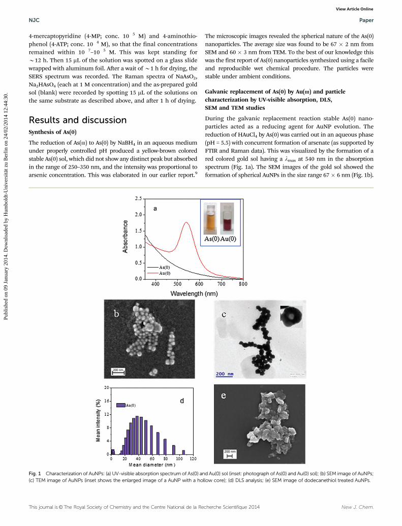
The microscopic images revealed the spherical nature of the As(0)nanoparticles. The average size was found to be 67 � 2 nm fromSEM and 60 � 3 nm from TEM. To the best of our knowledge thiswas the first report of As(0) nanoparticles synthesized using a facileand reproducible wet chemical procedure. The particles werestable under ambient conditions.
Galvanic replacement of As(0) by Au(III) and particlecharacterization by UV-visible absorption, DLS,SEM and TEM studies
During the galvanic replacement reaction stable As(0) nano-particles acted as a reducing agent for AuNP evolution. Thereduction of HAuCl4 by As(0) was carried out in an aqueous phase(pH = 5.5) with concurrent formation of arsenate (as supported byFTIR and Raman data). This was visualized by the formation of ared colored gold sol having a lmax at 540 nm in the absorptionspectrum (Fig. 1a). The SEM images of the gold sol showed theformation of spherical AuNPs in the size range 67� 6 nm (Fig. 1b).
Fig. 1 Characterization of AuNPs: (a) UV-visible absorption spectrum of As(0) and Au(0) sol (inset: photograph of As(0) and Au(0) sol); (b) SEM image of AuNPs;(c) TEM image of AuNPs (inset shows the enlarged image of a AuNP with a hollow core); (d) DLS analysis; (e) SEM image of dodecanethiol treated AuNPs.
NJC Paper
Publ
ishe
d on
09
Janu
ary
2014
. Dow
nloa
ded
by H
umbo
ldt-
Uni
vers
ität z
u B
erlin
on
24/0
2/20
14 1
2:44
:30.
View Article Online

New J. Chem. This journal is©The Royal Society of Chemistry and the Centre National de la Recherche Scientifique 2014
The TEM images (Fig. 1c) also authenticated the formation ofspherical AuNPs (size 62 � 7 nm). A more careful investigationinto the TEM images of the AuNPs indicated some void spaces(inset of Fig. 1c), which was also characteristic of the galvanicreplacement reaction.10 DLS studies on the as-prepared Au(0)sol indicated that the average particle size was B44 nm(Fig. 1d). The most interesting point was that the average sizeobtained from DLS studies for As(0) is B45 nm, which wascomparable to that of the AuNPs. The DLS studies indicatedthat Au(0) formation occurred through the galvanic replace-ment reaction between As(0) and the Au(III) ion, where As(0)behaved as a reducing agent. The same interesting feature wasalso observed when SEM and TEM images of As(0) and Au(0)were compared. The evolution of AuNPs of comparable sizefrom As(0) nanoparticles might be regarded as a perfect visualdepiction of the galvanic replacement reaction, and in theabsence of any external stabilizer. After the addition of HAuCl4
to the As(0) solution, Au(III) first diffused from the bulk phase tothe As(0) surface and adsorbed. As the reaction proceeded,Au(III) ions continued to etch the As(0) nanoparticle surfacesforming AuNPs. As a result As(0) was oxidized to AsO4
3� andleached out into the medium. This induced the hollow natureof the as-synthesized AuNPs. TEM revealed the formation ofAu(0) nanoparticles with hollow cores. The as-formed AsO4
3�
ions finally acted as a stabilizer for the AuNPs. The wholeprocess was fast and took less than 10 min for particle stabi-lization. The time dependant AuNP evolution was monitoredthrough UV-visible absorption spectra (see Fig. S1a, ESI†), andSEM images were observed at o1 min and at B5 min afterinitiation. At the initiation stage the appearance of a peak at570 nm and its gradual blue shift with time was observed in theabsorption spectra. Finally the peak stabilized at 540 nm atB9 min. No definite shape of the particles was observed in theSEM image at the initial stage (see Fig. S1b, ESI†). However,within 5 min the particles evolved (see Fig. S1c, ESI†). The insetshows a highly magnified SEM image indicating that the shapesare spherical. Though there was no surfactant or organicmoiety, the sol was stable for months. This was supported byUV-visible spectra of the particles after long periods of timeunder ambient conditions. The stability of the materials mightbe due to the anionic surface charge caused by the adsorptionof arsenate ions onto the Au(0) nanoparticles. Although it wasassumed that the oxidation product of As(0) was mostlyarsenate as evidenced from the FTIR and Raman spectralanalyses, the possibility of the formation of arsenite in theinitial stage might not be ruled out. The standard redoxpotential of the As(III)/As(V) system is +0.56 V and that of theO2–H2O system in acidic conditions is +1.23 V. So it is quitepossible that arsenite, if present in the solution, on furtherkeeping for a longer time under aerobic conditions, is con-verted to arsenate.26 During the formation of the gold sol, thepH was B5.5, and at this pH the existent arsenate should be inits ionized form i.e. as H2AsO4
�, which has the potential to actas a good stabilizer. The stability of the gold sol under variouspH conditions and during the addition of 1-dodecanethiol(0.17 M) was examined. It was noted that at lower pH (B2.0)
the sol turned blue and precipitated over time. However, athigher pH (up to B10.0) it was stable. This also confirms theformation of arsenate stabilized AuNPs. At low pH arsenate waspresent as protonated arsenic acid, and the sol becameunstable. On the other hand, dodecanethiol, having a higheraffinity towards Au(0), attached itself to the AuNPs makingthem hydrophobic and destabilized. At this stage the sphericalshape of the AuNPs dissapeared (Fig. 1e).
The formation and the nature of the AuNPs were investi-gated employing two other stoichiometric molar ratios of As(0)and Au(III). Spherical AuNPs with a hollow core are seen in theTEM images (see Fig. S2a and b, ESI;† inset shows the highresolution TEM) formed with [As] : [Au] = 1 : 2.26 and 1 : 2.83 (M/M).This indicates that the hollow nature is not restricted to aparticular stoichiometric ratio of the reactants. Also, the increasein stoichiometry did not affect the size of AuNPs.
X-Ray diffraction studies for particle characterization
XRD analysis was carried out on the AuNPs after thoroughwashing and drying in order to confirm their purity and to findout the crystal phase. Fig. 2a shows the XRD pattern of theas-prepared AuNPs. The diffraction pattern shows peaks at2y = 38.06, 44.51, 64.87, 77.72, 81.91 corresponding to the(111), (200), (220), (311), (222) planes of the standard cubicphase of Au.27
EDX, FTIR and Raman spectra
EDX studies were conducted on the Au(0) nanoparticles tounderstand their purity. After formation, the particles werecentrifuged and thoroughly washed and dried. The EDX studiesshowed no evidence of arsenic present on the AuNPs (Fig. 2b).This suggested that upon thorough washing, all arsenic con-taining compounds were washed away and the particles werefree from any adhered stabilizer. The possibility of the forma-tion of bimetallic As–Au was thus ruled out for this study.On the other hand, the FTIR studies done using the gold sol(Fig. 2c, curve 1) after evaporation, and a direct comparisonwith the FTIR spectra of arsenite (Fig. 2c, curve 2) and arsenate(Fig. 2c, curve 3) showed that the gold sol contained mostlyarsenate. The FTIR spectrum of arsenate showed absorptionpeaks at 3461, 2354, 1637, 1276, 1178, 831, and 677 cm�1, mostof which are in close agreement with the reported spectrumof Na2HAsO4�7H2O.28 The main absorption bands appeared at1637 and 831 cm�1. The characteristic band (designated as n3)of the free arsenate ion is known to appear somewhere in therange of 800–880 cm�1. A shift of the absorption band mightoccur if the symmetry of the ion is lowered. The as-preparedgold sol showed, in its FTIR spectrum, bands at 3420 (broadband), 1620, 1380, 1170, 1078, 854 and 667 cm�1 which closelyresembled those of arsenate indicating that arsenate wasmostly formed in the sol as an outcome of HAuCl4 reductionby As(0). The appearance of band at 43400 cm�1 was due to thepresence of an O–H group in both the arsenate ion and arsenatestabilized gold sol.
The other convincing proof of arsenate formation during thegalvanic replacement reaction between As(0) and Au(III) was the
Paper NJC
Publ
ishe
d on
09
Janu
ary
2014
. Dow
nloa
ded
by H
umbo
ldt-
Uni
vers
ität z
u B
erlin
on
24/0
2/20
14 1
2:44
:30.
View Article Online

This journal is©The Royal Society of Chemistry and the Centre National de la Recherche Scientifique 2014 New J. Chem.
appearance of characteristic SERS bands in the gold sol. Veryrecently the characterization and analytical determination ofAs(III) and As(V) were done by SERS studies,29,30 where theappearance of Raman peaks at 750 cm�1 and 800 cm�1 forAs(III) and As(V), due to the n1 (A1) symmetric As–O stretchingmode, and their shift in SERS to 721 and 780 cm�1, respectively,was documented. In our case the Raman bands of As(III)(appearing at 726 cm�1) (Fig. 2d, curve 1) and As(V) (appearingat 851 cm�1) (Fig. 2d, curve 2) were compared with the peaksappearing in the SERS spectrum of the as-prepared gold sol(Fig. 2d, curve 3). The major peaks appearing at 821, 386 and329 cm�1 in the SERS spectrum (Fig. 2d, curve 3) support theformation of H2AsO4
� during the reduction of Au(III) to Au(0) byAs(0). The band at 821 cm�1 appeared due to As–O stretchingvibration. The other two bands at 386 and 329 cm�1 are primarilyassigned to the bending modes of the arsenate group31,32 arisingowing to its symmetry reduction upon binding to a metal surface(here it is the AuNP surface). The adsorption of arsenate onAuNPs is thus authenticated.
The strong adsorption phenomenon of H2AsO4� on AuNPs
was also substantiated by a simple experiment. An anionexchange resin (Amberlite IRA-400 designated as R+Cl�) wasadded to the as-prepared Au sol, and kept overnight. The resinbead, which was originally bright yellow in color, was found toturn pinkish upon adsorption of the AuNPs, and the solutionbecame colorless. If H2AsO4
� is weakly adsorbed then anexchange of H2AsO4
� with Cl� with the concomitant precipi-tation of Au(0) particles would be expected. In our case noprecipitation was noticed. This also confirms the formation of
an arsenate stabilized gold sol. The close proximity of theAuNPs is due to the intermolecular H-bonding among theadsorbed H2AsO4
� ions giving extra stabilization.
Mechanism of arsenate-stabilized AuNP formation
Based on the above discussion, a mechanistic model for theformation and stabilization of spherical AuNPs was proposed(Fig. 3). The formation of AuNPs is a fast process. The stabilityof the spherical gold nanoparticles and their close assembly,
Fig. 2 Characterization of AuNPs: (a) XRD pattern; (b) EDX analysis; (c) FTIR spectra of the as-prepared AuNPs (curve 1), arsenite (curve 2), and arsenate(curve 3); (d) Raman spectra of arsenite (curve 1), arsenate (curve 2), and the as-prepared AuNPs (curve 3).
Fig. 3 A mechanistic model for the formation and stabilization ofspherical AuNPs.
NJC Paper
Publ
ishe
d on
09
Janu
ary
2014
. Dow
nloa
ded
by H
umbo
ldt-
Uni
vers
ität z
u B
erlin
on
24/0
2/20
14 1
2:44
:30.
View Article Online
New J. Chem. This journal is©The Royal Society of Chemistry and the Centre National de la Recherche Scientifique 2014
in the absence of any external stabilizer, is a unique featureobserved in the present study. It was presumed that thestabilization was due to intermolecular H-bonding of arsenateions adsorbed onto the gold nanoparticles. The H2AsO4
� ionhad two effects: first, as a capping agent to stabilize the goldparticles and to keep them in close proximity due to the smallsize of the H2AsO4
� ion to form AuNCs; secondly, it couldimpart an electrolytic effect and further stabilize the AuNPsthrough intermolecular H-bonding causing the sphericalparticles to remain in close proximity, as observed in bothTEM and SEM images. Recent studies have demonstrated thatmany uniform systems can be formed from agglomerates ofsmaller particles and their assemblies.33–40 These types ofaggregates or assemblies of gold or other nanoparticles arevery important and can be used for a variety of electronic,catalytic and sensing purposes. The aggregation may beinduced by either the analytes concerned or by external electro-lytes such as NaCl, MgCl2, etc. Many reports have addressed theaggregation of nanoparticles into linear chains,41–43 templateassisted organization,44 and spontaneous organization throughdipole–dipole interactions.45 Very recently biologically pro-grammed nanoparticle assembly has also been achieved.46,47
These facts prompted the present investigators to explore thepossibility of the as-prepared AuNPs to be used as a substratefor surface enhanced Raman spectroscopy (SERS).
Application of the as-prepared AuNPs as a SERS substrate
Colloidal solutions of gold and silver have been studied exten-sively for SERS applications. It is very important to have anenhancement of the optical field in the vicinity of the nano-particles constituting the SERS-active system.17 Aggregates andassemblies of nanoparticles, in this respect, are thus veryimportant to observe a good SERS signal of a molecule. It hasbeen suggested by Moskovits et al.16 that a large enhancementmay result from electromagnetic effects in aggregatescombined with either intermolecular or metal-to-molecule(or molecule to metal) resonances. Another important criterionto get a good SERS signal is to obtain resonance between theplasma bands of metal nanoparticles and the incoming light.Linear assemblies of Au and Ag nanoparticles have alreadybeen proved to be good SERS substrates due to the presence of‘hot-spots’ within them.17 For detecting SERS active molecules,usually the aggregation of metal nanoparticles into theirlinear assemblies, is done under forced conditions either byusing electrolytes or other molecules (like DNA bases, thiolsetc.). Sometimes the assembly is obtained in situ during nano-particle preparation. This enables avoidance of additionalreagents for aggregating the nanoparticles. In many cases astrong ligand or polymer can also cause the particles to remainclose to each other. However, this may hinder the diffusion ofthe probe molecules to get attached to the metal surface (whichis a prerequisite for SERS observation). This may cause thesubstrate to become inappropriate for SERS analysis. Our aimwas to investigate the behavior of R6G, 4-MP and 4-ATPmolecules towards SERS detection using the as-prepared goldsol with an expectation that it could be a potential SERS
substrate due to the absence of a stabilizing organic ligand orpolymer etc., and also due to the close proximity of the AuNPs.A 633 nm He–Ne laser was used in our study to investigate thepotentiality of the as-prepared gold sol as a SERS substrate. Thesize range of the as-obtained gold particles was appropriatefor such studies.48,49 The SERS spectra of all three probemolecules in the concentration range of 10�5–10�7 M, andthe Raman spectrum of the as-prepared gold sol are shown inFig. 4a–d. A significant SERS enhancement was observed in allcases. R6G is a well studied SERS active dye molecule.49,50 Thecharacteristic Raman peaks of R6G are shown in Fig. 4a andTable 1. The obtained peaks are in agreement with the earlierliterature.51,52 We chose 4-MP as another probe moleculebecause it has a large Raman scattering cross section. TheSERS of 4-MP has been studied on various surfaces such as Ag,Au, Pt, Cu, ZnS, CdS and Rh.53–59 4-MP has two pKa values:1.43 and 8.83 indicating that the molecule can exist in differentionic forms at different solution pH. The characteristic SERSpeaks of 4-MP obtained (Fig. 4b) using the prepared gold solwere compared with literature values obtained on Ag colloids(Table 2).53 In the present study the SERS of 4-MP on Aunanoparticles matched quite well with the literature values.To extend this further, the AuNPs were used as a SERS substratefor the detection of 4-ATP. Various substrates60–63 like 2DAu networks, Au nanochains, and AuNPs have already beenexploited for SERS detection of 4-ATP. The characteristic bandsobtained for the 4-ATP molecule (Fig. 4c) using the as-preparedsol are in good agreement with those reported earlier e.g. ongold film (Table 3).64 At this point it is important to mentionthat upon mixing the probe solution with the red colored goldsol, the red color changed to blue, which might have a positiveeffect on SERS detection. Fig. 4d shows the Raman spectrum ofthe as-prepared gold sol.
To estimate the detection limits of the probe molecules onthe as-prepared gold sol, it was assumed that the moleculeswere homogeneously distributed in the solution phase anduniformly adsorbed on the AuNPs. In our study 15 mL of thesolution (R6G, 4-MP and 4-ATP at concentration levels of10�7 M, 10�6 M and 10�5 M, respectively, already mixed withthe gold sol) was spotted on a glass slide wrapped with aluminiumfoil. After a wait of B1 h for drying, the SERS spectrum wasrecorded. The area covered by the sol was determined. The laserilluminated only 1/106 part of the described area.65,66 Thedetection limits were estimated to be in the femto- and sub-femto gram level.
Calculation of the Analytical Enhancement Factor (AEF)65,67
for the probe molecules with the as-prepared gold substrate wasdone by comparing the SERS intensity with the normal Ramanintensity (keeping other experimental conditions the same) andusing the following relation
AEF = (ISERSCRS)/(IRSCSERS)
where, ISERS and IRS are the respective SERS intensity andnormal Raman intensity, respectively, of the analyte. Theanalyte concentration CRS that produces the Raman signal IRS
under non-SERS conditions was 0.1 M in all cases. The AEFs
Paper NJC
Publ
ishe
d on
09
Janu
ary
2014
. Dow
nloa
ded
by H
umbo
ldt-
Uni
vers
ität z
u B
erlin
on
24/0
2/20
14 1
2:44
:30.
View Article Online

This journal is©The Royal Society of Chemistry and the Centre National de la Recherche Scientifique 2014 New J. Chem.
obtained were 1.7 � 105 for 4-MP (calculated at the peak1498 cm�1) and 8.6 � 103 for 4-ATP (calculated at the peak1590 cm�1). These values are comparable to the enhancementfactor reported earlier for 4-ATP on a gold surface64 and for 4-MPon TiO2 supported Ag.68
Conclusions
A yellow-brown As(0) sol was produced from arsenite uponits reduction with NaBH4 under carefully controlled pH asdescribed in ref. 9. SEM images showed the presence ofspherical As(0) nanoparticles in the size range 67 � 2 nm.In the present work the reducing properties of the as-preparedAs(0) were exploited to fabricate stable gold nanoparticles(AuNPs) of comparable size. This is the first report of AuNPpreparation by As(0) nanoparticles. The formation of Au(0) tookplace via a galvanic replacement reaction where AuCl4
�migratedfrom the bulk phase to the As(0) surface, and was reducedinstantaneously. The reaction is very fast, thermodynamicallyallowed and the resultant gold sol was very stable. The AuNPswere characterized by different physical methods. The feature ofthe AuNPs was its comparable size with the reductant, As(0)nanoparticles, with an associated hollow core. The close proxi-mity of the AuNPs was due to intermolecular hydrogen bondingbetween the arsenate ions produced in situ as a result of As(0)
Table 1 Assignment of major SERS peaks of R6G on the as-preparedAuNPs and on a Ag surface
Assignments
Ramanpeak(cm�1)51
SERS onAg surface(cm�1)52
SERS onas-preparedAuNPs (cm�1)
n(C–H) out of plane bending 776 774 773n(C–H) in plane bending 1131 1129 1127n(C–C) stretching 1312 1380 1310
1365 1363 13611509 1509 15081575 — 15731652 1650 1647
Table 2 Assignment of major SERS peaks of 4-MP on the as-preparedAuNPs and on a Ag colloid
Assignments
Ramanpeak(cm�1)53
SERS onAg colloid(cm�1)53
SERS onas-preparedAuNPs (cm�1)
1a1, n(ring breathing) 1004 1007 100818b2, b(C–H) — 1065 104412a1, n(ring breathing)/(C–S) 1119 1098 10969a1, b(C–H) — 1222 122019a1, n(CQC/CQN) 1487 1470 14988b2, n(C–C) — 1573 1590
Table 3 Assignment of major SERS peaks of 4-ATP on the as-preparedAuNPs and on a Au film
Assignments
Ramanpeak(cm�1)63
SERS onAu film(cm�1)64
SERS onas-preparedAuNPs (cm�1)
n(C–S), 7a(a1) 1080 1078 1080n(C–C) + d(C–H), 19a(a1) 1484 — —n(C–C) + d(C–H), 19b(b2) — 1438 1444n(C–C), 8a(a1) 1585 1583 1590
Fig. 4 SERS spectrum of (a) R6G (10�7 M), (b) 4-MP (10�6 M), (c) 4-ATP (10�5 M), and (d) the Raman spectrum of the as-prepared AuNPs.
NJC Paper
Publ
ishe
d on
09
Janu
ary
2014
. Dow
nloa
ded
by H
umbo
ldt-
Uni
vers
ität z
u B
erlin
on
24/0
2/20
14 1
2:44
:30.
View Article Online

New J. Chem. This journal is©The Royal Society of Chemistry and the Centre National de la Recherche Scientifique 2014
nanoparticle oxidation. The arsenate ions, authenticated fromFTIR and Raman studies, served the function of a stabilizer andhence no extra stabilizer was needed for stabilizing the AuNPs insolution. The as-prepared AuNPs exhibited the potential to beused as a substrate for SERS detection of R6G, 4-MP and 4-ATP ata sub-femtogram level.
Acknowledgements
The authors are thankful to the DST, UGC, New Delhi, and theIndian Institute of Technology, Kharagpur, India for financialassistance. The authors are also thankful to Prof. T. Pal andProf. N. Sarkar, Department of Chemistry, IIT Kharagpur for theRaman/SERS and DLS measurements, respectively.
References
1 M.-C. Daniel and D. Astruc, Chem. Rev., 2004, 104, 293.2 S. K. Ghosh and T. Pal, Chem. Rev., 2007, 107, 4797.3 K. G. Thomas and P. V. Kamat, Acc. Chem. Res., 2003,
36, 888.4 K. C. Graber, P. C. Smith, M. D. Musick, J. A. Davis, D. G.
Walter, M. A. Jackson and A. P. Guthrie, J. Am. Chem. Soc.,1996, 118, 1148.
5 U. Krebig and M. Vollmer, Optical Properties of MetalClusters, Springer, Berlin, 1995.
6 S. Link and M. A. El-Sayed, J. Phys. Chem. B, 1999, 103, 8410.7 M. Faraday, Philos. Trans. R. Soc. London, 1857, 147, 145.8 G. Mie, Ann. Phys., 1908, 25, 377.9 A. Pal, S. Saha, S. K. Maji, M. Kundu and A. Kundu, Adv.
Mater. Lett., 2012, 3, 177.10 X. Lu, H.-Y. Tuan, J. Chen, Z.-Y. Li, B. A. Korgel and Y. Xia,
J. Am. Chem. Soc., 2007, 129, 1733.11 M. Pradhan, J. Chowdhury, S. Sarkar, A. K. Sinha and T. Pal,
J. Phys. Chem. C, 2012, 116, 24301.12 X. Teng, Q. Wang, P. Liu, W. Han, A. I. Frenkel, W. Wen,
N. Marinkovic, J. C. Hanson and J. A. Rodriguez, J. Am.Chem. Soc., 2008, 130, 1093.
13 M. H. Oh, T. Yu, S.-H. Yu, B. Lim, K.-T. Ko, M.-G. Willinger,D.-H. Seo, B. H. Kim, M. G. Cho, J.-H. Park, K. Kang,Y.-E. Sung, N. Pinna and T. Hyeon, Science, 2013, 340, 964.
14 J. F. Ferguson and J. Gavis, Water Res., 1972, 6, 1259.15 M. Fleischmann, P. J. Hendra and A. J. McQuillan,
Chem. Phys. Lett., 1974, 26, 163.16 M. Moskovits, L. L. Tay, J. Yang and T. Haslett, Top. Appl.
Phys., 2002, 82, 215.17 S. Nie and S. R. Emory, Science, 1997, 275, 1102.18 D. L. Jeanmaire and R. P. Van Duyne, J. Electroanal. Chem.,
1977, 84, 1.19 J. A. Creighton, C. G. Blatchford and M. G. Albrecht, J. Chem.
Soc., Faraday Trans. 2, 1979, 75, 790.20 K. Kneipp, H. Kneipp and J. Kneipp, Acc. Chem. Res., 2006,
39, 443.21 T. Vo-Dinh, M. Y. K. Hiromoto, G. M. Begun and R. L.
Moody, Anal. Chem., 1984, 56, 1667.
22 B. Nikoobakht and M. A. El-Sayed, J. Phys. Chem. A, 2003,107, 3372.
23 C. J. Orendorff, A. Gole, T. K. Sau and C. J. Murphy,Anal. Chem., 2005, 77, 3261.
24 S. Panigrahi, S. Praharaj, S. Basu, S. K. Ghosh, S. Jana,S. Pande, T. Vo-Dinh, H. Jiang and T. Pal, J. Phys. Chem. B,2006, 110, 13436.
25 A. Pal and T. Pal, J. Raman Spectrosc., 1999, 30, 199.26 W. Drieham, R. Seith and M. Jekel, Water Res., 1995, 29, 297.27 X. Li, Y. Li, Y. Tan, C. Yang and Y. Li, J. Phys. Chem. B, 2004,
108, 5192.28 T.-H. Hsia, S.-L. Lo, C.-F. Lin and D.-Y. Lee, Colloids Surf., A,
1994, 85, 1.29 Z. Xu, J. Hao, F. Li and X. Meng, J. Colloid Interface Sci.,
2010, 347, 90.30 M. Mulvihill, A. Tao, K. Benjauthrit, J. Arnold and P. Yang,
Angew. Chem., Int. Ed., 2008, 47, 6456.31 W. Martens, R. L. Frost and P. A. Williams, J. Raman
Spectrosc., 2003, 34, 104.32 S. J. Greaves and W. P. Griffith, J. Raman Spectrosc., 1988,
19, 503.33 M. Brust, C. J. Kiely, D. Bethell and D. J. Schiffrin, J. Am.
Chem. Soc., 1998, 120, 12367.34 N. Cheng, S. Dong and E. Wang, J. Phys. Chem. B, 2005,
109, 19213.35 J. Liao, Y. Zhang, W. Yu, L. Xu, C. Ge, J. Lie and N. Gu,
Colloids Surf., A, 2003, 223, 177.36 A. Pal, S. K. Ghosh, K. Esumi and T. Pal, Langmuir, 2004,
20, 575.37 M. K. Chow and C. F. Zukoski, J. Colloid Interface Sci., 1994,
165, 97.38 J.-Y. Chang, H. Wu, H. Chen, Y.-C. Ling and W. Tan,
Chem. Commun., 2005, 1092.39 M. M. Maye, J. Luo, I.-I. S. Lim, L. Han, N. N. Kariuki,
D. Rabinovich, T. Liu and C.-J. Zhong, J. Am. Chem. Soc.,2003, 125, 9906.
40 M. M. Maye, S. C. Chun, L. Han, D. Rabinovich and C.-J.Zhong, J. Am. Chem. Soc., 2002, 124, 4958.
41 Y. Sun and Y. Xia, Adv. Mater., 2002, 14, 833.42 Q. Lu, F. Gao and D. Zhao, Nano Lett., 2002, 2, 725.43 T. Pal, T. Thundat and A. Pal, J. Mater. Sci. Lett., 1999,
18, 1391.44 S. Hoeppener, R. Maoz, S. R. Cohen, L. F. Chi, H. Fuchs and
J. Sagiv, Adv. Mater., 2002, 14, 1036.45 Z. Tang, N. A. Kotov and M. Giersig, Science, 2002, 297, 237.46 S. Mann, W. Shenton, M. Li, S. Connolly and D. Fitzmaurice,
Adv. Mater., 2000, 12, 147.47 C. A. Mirkin, R. L. Letsinger, R. C. Mucic and J. J. Storhoff,
Nature, 1996, 382, 607.48 M. Mandal, S. Kundu, S. K. Ghosh, N. R. Jana, S. Panigrahi
and T. Pal, Curr. Sci., 2004, 86, 556.49 A. Pal, D. L. Stokes and T. Vo-Dinh, Curr. Sci., 2004, 87, 486.50 A. M. Michaels, M. Nirmal and L. E. Brus, J. Am. Chem. Soc.,
1999, 121, 9932.51 P. Hildebrandt and M. Stockburger, J. Phys. Chem., 1984,
88, 5935.
Paper NJC
Publ
ishe
d on
09
Janu
ary
2014
. Dow
nloa
ded
by H
umbo
ldt-
Uni
vers
ität z
u B
erlin
on
24/0
2/20
14 1
2:44
:30.
View Article Online

This journal is©The Royal Society of Chemistry and the Centre National de la Recherche Scientifique 2014 New J. Chem.
52 Y. Lu, G. L. Liu and L. P. Lee, Nano Lett., 2005, 5, 5.53 J. A. Baldwin, B. Vlckova, M. P. Andrews and I. S. Butler,
Langmuir, 1997, 13, 3744.54 Y. Wang, Z. Sun, H. Hu, S. Jing, B. Zhao, W. Xu, C. Zhao and
J. R. Lombardi, J. Raman Spectrosc., 2007, 38, 34.55 Y. Wang, Z. Sun, Y. Wang, H. Hu, B. Zhao, W. Xu and
J. R. Lombardi, Spectrochim. Acta, Part A, 2007, 66, 1199.56 Y. Wang and T. Asefa, Langmuir, 2010, 26, 7469.57 H.-Z. Yu, N. Xia and Z.-F. Liu, Anal. Chem., 1999, 71,
1354.58 M. A. Bryant, S. L. Joa and J. E. Pemberton, Langmuir, 1992,
8, 753.59 H. Zhang, X. Xia, W. Li, J. Zeng, Y. Dai, D. Yang and Y. Xia,
Angew. Chem., Int. Ed., 2010, 49, 5296.60 H. Chen, Y. Wang, H. Jiang, B. Liu and S. Dong, Cryst.
Growth Des., 2007, 7, 1771.
61 D.-F. Zhang, L.-Y. Niu, L. Jiang, P.-G. Yin, L.-D. Sun,H. Zhang, R. Zhang, L. Guo and C.-H. Yan, J. Phys.Chem. C, 2008, 112, 16011.
62 J. H. Yoon, J. S. Park and S. Yoon, Langmuir, 2009, 25, 12475.63 T. Zhu, X. Fu, T. Mu, J. Wang and Z. Liu, Langmuir, 1999,
15, 5197.64 K. Uetsuki, P. Verma, T. Yano, Y. Saito, T. Ichimura and
S. Kawata, J. Phys. Chem. C, 2010, 114, 7515.65 S. Saha, A. Pal, S. Pande, S. Sarkar, S. Panigrahi and T. Pal,
J. Phys. Chem. C, 2009, 113, 7553.66 A. Pal, N. R. Isola, J. P. Alarie, D. Stokes and T. Vo-Dinh,
Faraday Discuss., 2006, 132, 293.67 E. C. Le Ru, E. Blackie, M. Meyer and P. G. Etchegoin,
J. Phys. Chem. C, 2007, 111, 13794.68 W. Song, Y. Wang and B. Zhao, J. Phys. Chem. C, 2007,
111, 12786.
NJC Paper
Publ
ishe
d on
09
Janu
ary
2014
. Dow
nloa
ded
by H
umbo
ldt-
Uni
vers
ität z
u B
erlin
on
24/0
2/20
14 1
2:44
:30.
View Article Online