Prefrontal Cortical GABA Modulation of Spatial Reference and Working Memory

THE JOURNAL OF COMPARATIVE NEUROLOGY 291:281-304 (1990)
GABA-ergic and Glycinergic Pathways in the Inner Plexiform Layer of the
Goldfish Retina
JAY F. MULLER AND ROBERT E. MARC Department of Physiology, University of Utah School of Medicine, Salt Lake City, Utah 84108
(J.E.M.); Sensory Sciences Center, University of Texas Graduate School of Biomedical Sciences, Houston, Texas 77030 (R.E.M.)
ABSTRACT GABA-ergic and glycinergic circuitry in the inner plexiform layer of the
goldfish retina was evaluated by electron microscopic autoradiography of "H- GABA and 3H-glycine uptake, combined with retrograde horseradish peroxi- dase (HRP) labeling of ganglion cells. GABA-ergic and glycinergic synapses were found on labeled ganglion cells throughout the inner plexiform layer. This reinforces the idea that physiological evidence of GABA-ergic and plycin- ergic influence on a variety of ganglion cells in goldfish and carp often reflects direct inputs. Double-labeled synapses are presented as evidence of direct type Ab amacrine cell input to on-center ganglion cells. At least one popula- tion of type Aa sustained-off GABA-ergic amacrine cell is proposed, on the basis of profuse GABA-ergic inputs onto bipolar cells in sublamina a. Similar GABA-labeled profiles are shown to synapse onto HRP-labeled probable off- center ganglion cells. Thus GABA-ergic amacrine cells not only provide the predominant feedback to depolarizing (on-center) and hyperpolarizing (off- center) bipolar cells but also provide feed-forward inputs to on- and off-center ganglion cells. Large-caliber GABA-ergic dendrites present in both sublami- nae a and b resemble those expected of a previously described bistratified, transient amacrine cell. These processes synapse onto HKP-labeled ganglion cell profiles in both sublaminae. Two morphologies of glycinergic amacrine cell are proposed on the basis of light microscopic autoradiography, 1) the pre- viously described small pyriform cell and 2) a multipolar cell. The differential connectivity of the glycinergic neurons described, however, remains indistin- guishable. Whereas abundant glycinergic inputs to ganglion cells occur throughout the inner plexiform layer, contacts between glycinergic profiles and bipolar cells are extremely rare. Therefore, interpreting the meaning of glycinergic input to ganglion cells will require further study of amacrine cell circuitry.
Key words: amacrine cells, ganglion cells, autoradiography, horseradish peroxidase, neuronal circuitry
The specific connectivity of retinal neurons underlies image coding by the visual system. All vertebrate retinas share a neuronal architecture composed of photoreceptors, horizontal cells, bipolar cells, amacrine cells, and ganglion cells; the projections of these cells within the retina form the outer and inner plexiform layers. I t is the variety of cell types within each class that gives rise to the diversity of reti- nal information coding. The present work concerns the com- ponents that interact in the inner plexiform layer of the gol- fish retina. Accepted August 23,1989.
In vertebrates, as is well known, the inner plexiform layer can be divided into a distal region, closer to the amacrine cell layer (sublamina a), and a proximal region, near the ganglion cell layer (sublamina b). In general, components of off-center pathways synapse in sublamina a and those of the on-center pathway in sublamina b (Famiglietti and Kolb,
0 1990 WILEY-LISS, INC.
282
'76; Famiglietti et al., '77; Nelson et al., '78). Although there are differences among vertebrates regarding to what extent the various subtypes of bipolar cells and amacrine cells fit the sublamina a/sublamina b model, ganglion cells appear to fit in all species studied (Kolb, '82; Kolb and Nelson, '84). In fishes, the terminals of hyperpolarizing mixed rod-cone hi- polar cells are in sublamina a, and depolarizing mixed rod- cone bipolar cells clearly terminate in sublamina b. Accord- ingly, they are abbreviated as Ma and Mb bipolar cells (Famiglietti et al., '77; Ishida et al., '80; Kaneko et al., '79, '80, '81; Saito et al., '83, '84, '85). Furthermore, in fishes, sus- tained-hyperpolarizing amacrine cells generally stratify in sublamina a and sustained-depolarizing amacrine cells gen- erally stratify in sublamina b; we refer to them as Aa and Ab amacrine cells, respectively (Famglietti et al., '77; Kaneko et al., '79; Teranishi et al., '84, '85, '87; Djamgoz et al., '85; Djamgoz, '86; Djamgoz and Wagner, '87). Transient, on/off amacrine cells and ganglion cells, which respond briefly at stimulus onset and offset, stratify either along the sublam- ina a/b border or, more often, in both sublaminae a and b (Famiglietti et al., '77; Teranishi et al., '84, '85, '87; Djamgoz et al., '85; Djamgoz, '86; Djamgoz and Wagner, '87).
In teleostean fishes (Famiglietti et al., '77; Naka, '77) and mudpuppy (Frumkies et al., '81; Miller and Dacheux, '76), the basic design of parallel pathways through the retina has been well established. Intracellular current injection com- bined with extracellular recording showed that depolarizing bipolar cells in catfish drive on-center ganglion cells through a sign-conserving path; off-center ganglion cells were like- wise driven by hyperpolarizing bipolar cells (Naka, '77). In mudpuppy, the on- and off-center pathways were found to be separable by low-chloride blockade of the on-center pathway (Miller and Dacheux, '76), and pharmacological blockade, by glutamate analogues, of either on or off path- ways (Slaughter and Miller, '81, '83).
In fishes, ganglion cells are GABA and glycine-receptive (Cohen and Fain, '88; Ishida and Cohen. '88) and, as in other vertebrates, GABA and glycine have been found to in- fluence on-center, off-center, and on/off ganglion cells (Ne- gishi et al., '78; Djamgoz et al., '81; Glickman et al., '82). This is reflected anatomically; the sum of putatively GABA-ergic and glycinergic neurons makes up the majority of amacrine cells (Marc et al., '88; Miller, '88; Marc, '89). In cyprinid fishes, the two primary strategies for marking GABA-ergic and glycinergic neurons have been 1) high affinity uptake of radiolabeled GABA, GABA analogs, and glycine, followed by autoradiographic analysis (Lam and Steinman, '71; Marc et al., '78, '88; Marc and Lam, '81; Marc, '82, '89; Marc and Liu, '84; Ayoub and Lam, '84; Ball and Brandon, '86; Yazulla et al., '84; Yazulla, '86; Ball, '87; Muller and Marc, '88), and 2) immunocytochemical localization of GABA-ergic probes: GABA-like and glutamic acid decarboxylase (GAD)-like im- munoreactivity and glycine-like immunoreactivity (Lam et al., '79, '85; Brandon, '85; Ball, '87; Studholme and Yazulla, '87, '88; Marc et al., '88). Correlations of autoradiographic and immunocytochemical data have been important recent strategies for ascertaining the neurochemical identities of retinal neurons. With certain notable exceptions to be dis- cussed later, there has been good correspondence between probes. I t has also become apparent that both GARA-ergic and glycinergic amacrine cells are heterogeneous popula- tions, composed of distinct subtypes.
We have employed retrograde transport of horseradish peroxidase (HRP) as a structural label for ganglion cells and high-affinity uptake of radiolabeled GABA and glycine as
J.F. MULLER AND R.E. MARC
neurochemical markers for amacrine cells. Combining easily distinguishable ultrastructural markers for arnacrine cells and ganglion cells is advantageous. Otherwise, delineating amacrine cell from ganglion cell profiles without extensive reconstruction can be risky, particularly with recent evi- dence from catfish that some ganglion cells may be presyn- aptic in the inner plexiform layer (Sakai et al., '86). In the present study, HRP-labeled ganglion cells were selected for their dendritic stratification patterns; they correlate to on- center, off-center, and on/off' physiological types (Fami- glietti et al., '77). By adding autoradiographic markers for GABA and glycine, we have sought further insight into how GABA-ergic and glycinergic interneurons can directly in- fluence on-center, off-center, and on/off signal pathways.
MATERIALS AND METHODS The double-label employed herein has allowed two gen-
eral alternatives for analysis: 1) serial sectioning and elec- tron microscopic (EM) autoradiography of well isolated HRP-labeled ganglion cells whose dendritic arbors are sol- idly filled and 2) sectioning retinal regions with dense fields of solidly labeled ganglion cells and processing them for EM autoradiography. Double-labeled synapses can then be sur- veyed throughout the inner plexiform layer for both an over- all distribution of inputs onto ganglion cells and selected patterns of input to individual cell types. We express the location of a feature in the inner plexiform layer as a percent distance from the amacrine cell layer border: level 0 (LO) to the ganglion cell layer border: Level 100 (LIOO). Sublayers 1-5 are equal subdivisions (each 20% intervals); sublayer 1 begins a t LO, and sublayer 5 ends at LlOO (Marc, '86).
Anesthesia and HRP injection Goldfish (Carassius auratus) , 8-15 cm in standard
length, were secured dorsal side up with sponge in an ice bath, and anesthetized by respiration with 0.01-0.@2% MS222 (tricaine methanesulfonate) or 4 % benzocaine in acetone diluted 1500 with water. Optic nerves were exposed by cutling a notch in the dorsal cartilage, rostromedial to the orbit (avoiding the major blood vessels leading from the gills). The optic nerves were injected with a 10 yl Hamilton syringe containing 25-30 % Sigma VI or Boehringer-Mann- heim grade I HRP in 2 90 aqueous dimethyl sulfoxide, within 2-3 mm of the eye. The injection site was plugged with HRP-soaked Gelfoam, and the surgical area was filled with Aquaphor gel for protection. Fishes were revived by respira- tion with oxygenated water, keeping the gills free and moist until normal head and tail movements were seen, for a sur- vival time of 20-48 hours.
Isolated retina preparation: Incubation in 3H-neurotransmitter and fixation
As the last part of the survival time, fishes were dark- adapted, usually overnight, to facilitate retinal isolation. Fishes were cervically transected and pithed, and the eyes were removed and hemisected. The eyecup was quartered, and the retinal portions were freed in cold, oxygenated tele- ost saline: 150 mM NaCl, 5 mM Hepes, 3 mM KC1,2.1 mM CaCl,, 1.4 mM MgSO,, 1.4 mM NaHC03, 0.7 mM NaH,PO,, 10 mM glucose. In certain preparations the saline was nutrient-fortified according to Ames and Nesbett ('81). Each quarter retina was incubated for 10 minutes in a 25 or 50 p1 droplet of 2-6 yM concentrations of 3H-GABA, (specific activity [S.A.] 25-50 Ci/mMole) or 3H-glycine (S.A.
GARA AND GLYCINE PATHWAYS IN THE GOLDFISH IPL 283
16-20 Ci/mMole) in teleost saline, in a moist, oxygenated environment. Isotopes were obtained from New England Nuclear (Boston, MA). In several preparations 1 mM nipe- cotic acid (Sigma), a competitive inhibitor of GABA uptake, was added to the 3H-GABA incubation medium. Nipecotic acid counteracts the spatial buffering effect caused by the high density of GABA uptake sites in the inner and outer plexiform layers (Marc, '86, '89; Marc et al., '88). Similar concentrations of unlabeled GABA added to the incubation medium would be equally effective in improving 3H-GABA's access to the entire inner plexiform layer (Marc et al., '88; Marc, '89). Nipecotic acid was chosen for the present study becausp it is not metabolized after uptake (Madtes and Red- burn, '85), nor does it specifically hind to GABA receptors (Redburn et al., '83). After a 30 second saline rinse, retinas were fixed in a freshly prepared fixative: 1% paraformalde- hyde, 2.5c0 glutaraldehyde, 0.012'0 CaC12, and 3Y0 sucrose in 80 mM sodium cacodylate buffer, at pH 7.4, after which they were rinsed several times in 0.16 M sodium cacodylate (pH 7.4) and left overnight in 0.16 M sodium cacodylate with sCc sucrose. Unless otherwise specified, all prepara- tions were done a t room temperature, and the buffer used was 0.16 M sodium cacodylate.
HRP development and histology After a brief buffer rinse, the fixed samples were devel-
oped for HRP content with diaminobenzidine tetrachloride (DAB). All samples were presoaked, with rotation. for 30-45 minutes in a filtered solution of 0.08-0.1('0 DAB, 0.02- 0.03'~ cobalt chloride, and nickel ammonium sulfate in buffer (Adams, '81). Hydrogen peroxide was added to a final concentration of 0.02"o, and the samples were incubated for an additional 30-45 minutes. Some samples, after a short buffer rinse, were then incubated for 30 minutes in a 0.15 Hanker-Yates reagent (Polysciences) in buffer with 0.02 0
hydrogen peroxide (Muller and Marc, '84). The samples were buffer rinsed and postfixed in buffered
1 ' C osmium tetroxide. In some experiments 0.1-0.1570 po- tassium ferricyanide was added to the osmium tetroxide for postfixation at 4°C in the dark. Later we found that lr% osmium tetroxide for 40 minutes followed by 1";) osmium tetroxide with potassium ferricyanide for 30 minutes may be preferable (Wong-Reilly and Kageyama, '85). After a rinse in buffer or deionized water, the retinal portions were dehy- drated in a cold graded methanol series and acetone, embed- ded in a soft Polybed 812 (Polysciences) mixture (21 DDSA:Polybed 812, 2 O C DMP-30 by volume), and polymer- ized at 60-65"C 6 hours to overnight.
Serial 40 or 60 pm sections were cut on an American Optical sliding microtome, dipped in unpolymerized Poly- bed mixture, serially slide mounted, and polymerized as above. This section thickness range has been useful for cam- era lucida drawing, light microscopy with Nomarski optics, and remounting for thin sectioning. Most thick sections were vertical, but some samples were chosen for horizontal or near-horizontal sections. Sections were examined for gan- glion cells whose cell bodies and dendrites were solidly filled with reaction product and well isolated from the labeled dendrites of other ganglion cells. From these isolated gan- glion cells, examples were chosen primarily on the basis of their dendritic stratification. Camera lucida drawings were made of those individual HRP-labeled ganglion cells chosen for subsequent autoradiographic analysis. Several ganglion cells, viewed in slightly oblique sections, were drawn ab- stracted to the vertical by using landmarks such as the ama-
crine cell layer border and the ganglion cell-optic fiber layer border to sketch the dendrites a t different planes of focus. The areas of the thick sections selected were remounted onto plastic blanks, either with rapid setting epoxy cement or unpolymerized Polyhed, and resectioned on a Sorvall M'l'2B microtome for EM autoradiography.
Autoradiography Retinal areas selected for EM autoradiographic analysis
were screened by sectioning the same region of a neighbor- ing section for light microscopic autoradiography. Semithin (0.5-1.0 Fm) sections were deplasticized with sodium me- thoxide and coated with a 50"0 solution of Kodak NTB-2 photographic emulsion. After 1-4 weeks of exposure at 4"C, the slides were developed with Dektol or D-19 (Kodak) for 2 minutes and examined for silver grain density and localiza- tion. The sections were counterstained with dilute toluidine blue in sodium borate. On the basis of these results, final selections of labeled ganglion cells were made, and exposure times for EM autoradiography were set.
Silver-gold thin sections (70-80 nm) were cut with glass knives on a Sorvall MT2-B ultramicrotome and collected on slot grids coated with Parlodion film, stained with uranyl acetate and Reynolds' or Luft's lead citrate, and carbon- coated. A fresh stock of Parlodion solution, 0.5'Tc in n-pentyl acetate, and a moderately heavy carbon coat are recom- mended to protect against film hydration and weakening during long exposures and from the rigors of development. The grids were then coated with a 1:2 dilution of Ilford L-4 photographic emulsion by using wire loops (Bouteille, '76). The emulsion-coated grids were mounted on slides with double-stick tape or Grid Sticks (Ted Pella, Inc.) and stored a t 4°C in slide boxes containing desiccant packets for the desired exposure times.
In some experiments, a number of grids were first devel- oped at 2-3 weeks with D-I9 or Microdol-X (Kodak) for early assessment. However, physical development with phe- nidon ( I minute a t 10-15°C). though requiring longer expo- sure times (5-10 weeks), produced more discrete silver grains, preferable for analysis of small labeled profiles. All electron microscope autoradiographic data presented in this study were drawn from phenidon-developed grids (Bou- teillc, '76).
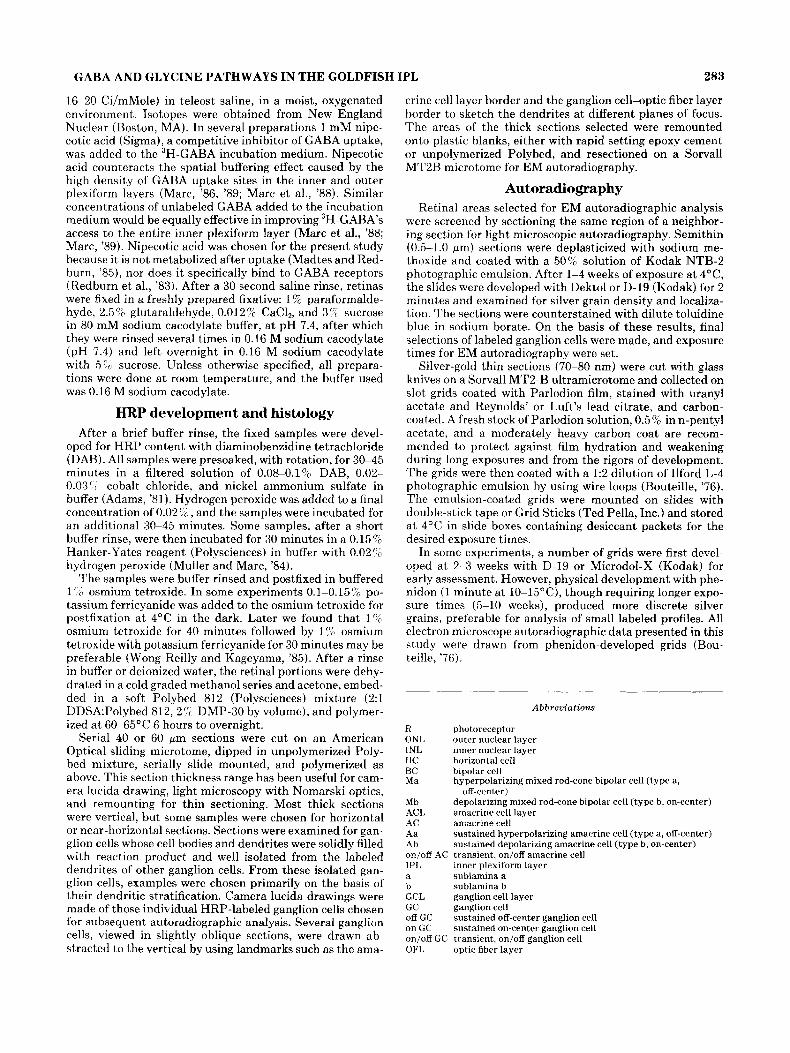
Abbreviations
R photoreceptor ONL outer nuclear layer INL inner nuclear layer HC horizontal cell BC bipolar cell M a
Mb ACL amacrine cell layer AC amacrine cell Aa sustained hyperpolarizing amacrine cell (type a, off-center) Ab sustained depolarizing amacrine cell (type b, on-center) on/off AC transient, on/off amacrine cell IPL inner plexiform layer a sublamina a b sublamina b GCL ganglion cell layer GC ganglion cell off GC on GC on/& GC transient, on/off ganglion cell OFL optic fiber layer
hyperpolarizing mixed rod-cone bipolar cell (type a,
depolarizing mixed rod-cone bipolar cell (type b, on-center) off-center)
sustained off-center ganglion cell sustained on-center ganglion cell
284 J.F. MULLER AND R.E. MARC
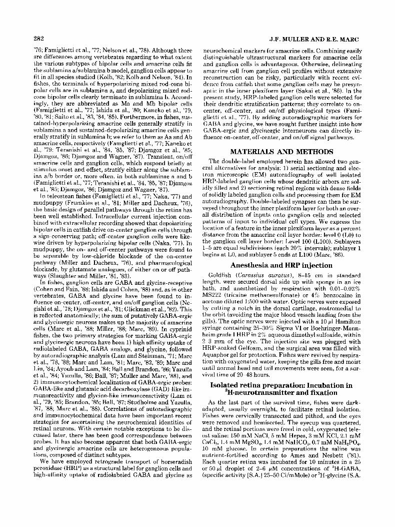
Fig. 1. 3H-GABA uptake, light microscopic autoradiography. Com- parison between a standard 3H-GARA preparation (A) and ’H-GABA + nipecotic acid preparations (B,C). All are 0.5 pm sections; A,C exposed for 4 weeks, B for 2 weeks. The arrow in A indicates a type Ab pyriform amacrine cell. A possible GABA-ergic transient amacrine cell and two of its dendrites are pointed out in B (large arrow and two dark arrowheads,
respectively). Asterisks mark horizontal cell axon terminals. In C, a long view of a 3H-GABA + nipecotic acid incubated sample, two cell bodies not obscured by the densely labeled band of the distal inner plexiform layer are indicated by arrows. Large terminals from type Mb, depolariz- ing mixed rod-cone bipolar cells are seen surrounded by grains in the proximal inner plexiform layer (white arrowhead).
Double-label data analysis
Standard sample analysis entailed following labeled pro- files through serial sections within each grid, over consecu- tive grids where possible. Parlodion-coated slot grids al- lowed unobstructed viewing of numerous structures from section to section. In most cases, vertical or near-vertical views were selected for resectioning and double-label analy- sis. Vertical sections are advantageous in that they allow 1) clear mapping of the levels in the inner plexiform layer a t which the individual ganglion cells ramify (see Figs. 3, 10) and 2) more serial sections per slot grid. The disadvantages of’ vertical sections stem from the relatively small portion of the HRP-labeled ganglion cell’s dendritic arbor that each 40 or 60 fim section includes, approximately 5-10% for most of the individual ganglion cells sampled. In this study, depend- ing on the individual ganglion cell or ganglion cell field sam-
pled, the number of grids processed for EM autoradiogra- phy ranged from 30 to over 100 from each remounted thick section, with an average of seven or eight sections per grid.
The main object of this study has been to find unequivo- cal evidence of labeled amacrine cell input onto a given type of ganglion cell. To make a confident judgement, certain requirements had to be satisfied for each double-labeled synapse: 1) good overall tissue preservation; 2) clearly recog- nizable, well-oriented synapses; 3) pervasive postsynaptic HRP reaction product; and 4) significant presynaptic silver grain localization. Conventional synapses are recognized by a widened gap or “cleft” between the associated profiles, with vesicles aggregated along the presynaptic side and fila- mentous densifications on both sides. Even if the synaptic cleft is slightly oblique, these features are readily apparent. I t will he assumed that such synapses represent sites of a physiological interaction between pre- and postsynaptic en-

GABA AND GLYCINE PATHWAYS IN THE GOLDFISH IPL 285
Fig. 2. 'H-GABA + nipecotic acid, EM autoradiography. Signal to noise ratio. A detail from one of the inner nuclear layer regions analyzed. The grain density ratio of labeled (arrow) to unlabeled cell bodies, in
these preparations, ranged from 17:l to 191. The inner plexiform layer border (IPL, arrowhead) is indicated for orientation.
tities. Only solidly HRP-labeled ganglion cell processes have been included in the double-label analysis. The dense cyto- plasmic flocculence, excluded from the mitochondria and most cisternae (Muller and Marc, '84), is quite characteris- tic and easily distinguishable under most circumstances. For characterizing significant presynaptic label, we relied on established statistical criteria for ultrastructural grain local- ization (Kelly and Weitsch-Dick, '78), where thresholds of three or four grains/wm2 were calculated for central nervous tissue with a grain density ratio of 5:l between labeled and unlabeled profiles. Data presented herein derive from speci- mens with grain density ratios of 17:l or better for labeled vs. unlabeled areas. Our double-label analysis adopts a stringent standard of four grains per terminal in each of a t least two serial sections, increasing the confidence level con- siderably. An important consideration in distinguishing au- toradiographically labeled structures is the amount of "grain spread" expected for a particular preparation. Lat- eral grain displacement is primarily dependent on the thick- nesses of the section and emulsion. For 70-80 nm sections and emulsion thickness of 70-100 nm, the predicted half- distance is 150-200 nm (Salpeter et al., '78); thus the dis- tance from the terminal predicted to encompass about 90 % of the localized grains would be about 500 nm (0.6 Km). In the labeled terminals presented here, silver grains are dis- cretely localized well within those boundaries.
RESULTS GABA-ergic inputs
High-a.nitg uptake, autoradiography. With rela- tively short incubations. moderate exposure times and concentrations in the micromolar range, GABA uptake is selective for H1 horizontal cells and their axon terminals, distally. and a population of large, pyriform amacrine cells (type Ab), with their labeled dendrites confined to the prox- imal 20':o (L80-100) of the inner plexiform layer (Marc e t al., '78, '88; Marc, '80, '82, '86, '89; Fig. 1A). Thus a major advantage of the standard incubation is selectivity for the type Ab pyriform amacrine cells and their monostratified arbor, allowing circuitry analysis of an individual cell type. However, immunocytochemical work (Ball '87; Ball and Brandon, '86; Marc, '86; Marc et al., '88; Yazulla et al., '861, labeling for GABA-like and GAD-like immunoreactivity, and 3H-muscimol binding studies (Yazulla, '81), have given evidence that there are additional types of GABA-ergic amacrine cell in goldfish. I t became apparent that there was likely to be a high density of GABA-ergic terminals through- out the inner plexiform layer, not only the proximal 20%. I t has been suggested that in fishes (Marc, '86; '89; Marc et al., '88) and turtles (Tachibana and Kaneko, '84) the concentra- tions of high-affinity uptake sites, in both the outer plexi- form and the inner plexiform layers, constitute spatial buff-
286
GABA J.F. MULLER AND R.E. MARC
INL
50 prn
B ;. . . . . . . :.:_. ,,. I- ......... >,. f ;,'..;<>,\ 'i: .....: ..::.I .... .::
. .: . . . . .
INL . .
50 prn
C
. . .
L .
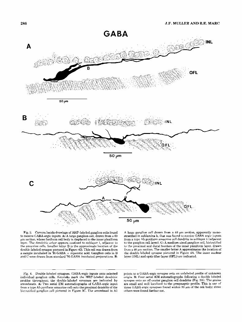
. . .
Fig. 3. Camera lucida drawings of HRP-labeled ganglion cells found to receive GABA-ergic inputs. A: A large ganglion cell, drawn from a 60 pm section, whose fusiform cell body is displaced to the inner plexiform layer. The dendritic arbor appears confined to sublayer 1 , adjacent to the amacrine cells. Smaller letter B is the approximate location of the double-labeled synapse pictured in Figure 4B. This cell was drawn from a sample incubated in 3H-GABA + nipecotic acid. Ganglion cells in B and C were drawn from standard 3H-GABA-incubated preparations. B:
Fig. 4. Double-labeled synapses. GABA-ergic inputs onto selected individual ganglion cells. Asterisks mark the HRP-labeled dendritic profiles throughout; the double-labeled synapses are indicated by arrowheads. A Two serial EM autoradiographs of GABA-ergic input from a type Ab pyriform amacrine cell onto the proximal dendrite of the bistratified ganglion cell pictured in Figure 3C. The arrowhead in A1
50 pm
A large ganglion cell drawn from a 40 Mm section, apparently mono- stratified in sublamina b, that was found to receive GABA-ergic inputs from a type Ab pyriform amacrine cell dendrite in sublayer 5 (adjacent to the ganglion cell layer). C: A medium-sized ganglion cell, bistratified to the proximal and distal borders of the inner plexiform layer, drawn from a 40 pm section. The smaller letter A approximates the location of the double-labeled synapse pictured in Figure 4A. The inner nuclear layer (INL) and optic fiber layer (OFL) are indicated.
points to a GABA-ergic synapse onto an unlabeled profile of unknown origin. B: Four serial EM autoradiographs following a double-laheled synapse onto an off-center ganglion cell dendrite (Fig. 3A). The grains are small and well localized to the presynaptic profile. This is one of three GABA-ergic synapses found within 30 wn of the cell body; three others were found farther out.

GABA AND GLYCINE PATHWAYS IN THE GOLDFISH IPL 287
288
ers for GABA diffusion. During standard incubations this creates a sharp concentration gradient of 3H-GABA, favor- ing the proximal border of the inner plexiform layer and the horizontal cell layer (Marc et al., '88; Marc, '89). The addi- tion of millimolar concentrations of unlabeled GABA or nipecotic acid, as competitive inhibitors of GABA uptake, should reversibly occupy the uptake sites, allowing more 3H- GABA to diffuse through the entire thickness of the retina. This modification allows a more comprehensive view of GABA-ergic stratification and the opportunity to label the full complement of GABA-ergic amacrine cells.
There is a dramatic difference between the label distribu- tions after a standard 3H-GABA incubation (Fig. 1A) and an incubation in 3H-GABA with nipecotic acid added (Fig. lB,C). Immediately noticeable is the dense stratum of label covering the distal 2500 (LO-25) of the inner plexiform layer; in Figure 1C the band is nearly confluent, obscuring many of the labeled amacrine cells that lie adjacent to it. There also appear to be two discontinuous bands of label near T,40 and L70 of the inner plexiform layer. Given the more extensive labeling of the 'H-GABA + nipecotic acid preparations, it was necessary to gauge their overall specif- ity. Were the "newly" labeled cells clearly labeled? Was a large proportion of amacrine cells still clearly unlabeled? The grain density ratios of labeled to unlabeled cell bodies (i.e., signal to noise ratio) in the inner nuclear layer for sev- eral separate 3H-GABA + nipecotic acid preparations (see Fig. 2) ranged from 17:l to 19:l. This is well above the 5:l ratio standard used by previous investigators (Kelly and Weitsch-Dick, '78) as a basis for their three or four grains/ pm'terminal criterion for statistically significant label (see Materials and Methods).
All double- labeled synapses were followed in serial sections. Some indi- vidual ganglion cells receiving GABA-ergic input are shown in Figure 3, representative double-labeled synapses are pre- sented in Figure 4, and the remaining synapses onto these cells are represented on the GABA summary histogram in Figure 14. From standard 3H-GABA preparations, we se- lected isolated, solidly labeled ganglion cells whose arbors were exclusively confined to, or well represented in, the proximal inner plexiform layer. Figure 3B and C are camera lucida drawings of ganglion cells that received inputs from GABA-ergic type Ab pyriform amacrine cells. The ganglion cell in Figure 3 8 is monostratified in the proximal region of the inner plexiform layer (sublamina b) and is probably an on-center ganglion cell. The cell in Figure 3C, stratified along the proximal and distal borders of the inner plexiform layer, is likely to be an on/off ganglion cell. Figure 4A pres- ents two serial EM autoradiographs of a double-labeled synapse, one of the GABA-ergic inputs onto the proximal branch of the ganglion cell illustrated in Figure 3C. 3H- GABA incubation with 1 mM nipecotic acid added to the medium allowed investigation of GABA-ergic inputs onto ganglion cells that arborize distally in the inner plexiform layer. A probable off-center ganglion cell with its dendritic arbor in sublayer 1 (LO-20; Fig. 3A), received GABA-ergic synapses at six loci along the dendrites pictured in Figure 3A. An example is shown in Figure 4B.
Double label survey through the inner plexiform layer. To survey GABA-ergic synapses onto ganglion cell dendrites through the inner plexiform layer, regions were selected for EM autoradiographic analysis from 3H- GABA + nipecotic acid incubations, where 30-40% of the ganglion cells were solidly filled. Double-labeled synapses
Double label: Individual ganglion cells.
J.F. MULLER AND R.E. MARC
from different levels of the inner plexiform layer are shown in Figure 5A-D; the distribution of these inputs is summa- rized in Figure 14. Figure 5E, a detail from Figure lC , serves as a key to the location of the double-labeled synapses. Fig- ure 5B and C are two examples within LO-20 of the inner plexiform layer, thus included in the GABA-ergic system's broad distal stratum. Figure 5A shows a double-labeled synapse near L70, and Figure 5D shows one at about L90, near the ganglion cell layer border. We also found GABA- ergic synapses from large-caliber (2.5 pm and 2.0 pm diame- ters, respectively) microtubule-filled dendrites onto labeled ganglion cell cross sections (Fig. 6A, two serial sections near L70 and 6B within L15-20). These distally and proximally located amacrine cell processes are indistinguishable and frequently observed. We believe they are from the same cell type. Based on correlative evidence from this and other studies (see Discussion) we propose that both GABA-ergic profiles are from a type of transient on/off amacrine cell, bistratified in sublayers 1 and 4. Figure 5C exemplifies com- mon double-labeled interactions, found near L10-15, in- volving small, rounded GABA-ergic endings.
The 3H-GABA + nipecotic acid protocol revealed GABA-ergic inputs onto a variety of bipolar cell profiles in sublayer 1 (LO-20), several of which were probably type Ma mixed rod-cone bipolar cells (Figs. 6, 7). With standard 3H-GABA incubations, it had been estab- lished that type Mb bipolar cells receive profuse inputs from the GABA-ergic type Ab pyriform amacrine cell and that type Mb bipolar cells also provide ribbon inputs (Marc et al., '78). However, the neurochemical vector for feedback to the off-center pathway had not been defined. Based on pres- ent evidence, it appears that the predominant feedback inputs to both on- and off-center pathways (through mixed rod-cone bipolar cells) are GABA-ergic. In Figure 6, a large, scalloped, type Ma bipolar cell terminal near LO5 receives five GABA-ergic inputs. I t appears as if all amacrine cell profiles surrounding this view of the bipolar cell terminal accumulated GABA. Figure 7 shows a bipolar cell terminal near TA1O (probably type Ma) receiving input from three GABA-ergic profiles. There is also a synapse between two of the profiles synapsing onto the bipolar cell terminal. If they are from two different types of amacrine cell then, based on these circuits, two subclasses of GABA-ergic sustained type Aa amacrine cells are predicted. Other pairs of GABA-ergic profiles synapsing onto one another were observed near LIO of the inner plexiform layer.
Glycinergic inputs Our knowledge of the glycine
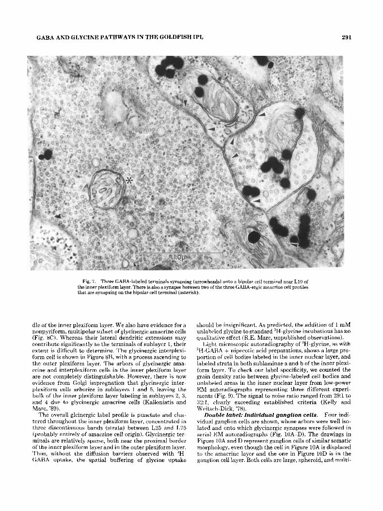
system in the goldfish is largely based on high-affinity uptake and autoradiography (Marc and Lam, '81). The pre- viously known classes of glycinergic neurons were 1) a heterogeneous population of small, predominantly pyriform arnacrine cells and 2) a population of interplexiform cells, pre- and postsynaptic to horizontal cells in the outer plexi- form layer, with descending processes that terminate in the inner plexiform layer. Recent immunocytochemical work (Studholme and Yazulla, '88) has begun to further distin- guish morphological classes of glycinergic amacrine cell at the light microscopic level. The most common morphologi- cal type encountered in the goldfish appears to be a small, pyriform amacrine cell (Fig. 8A). The single descending pro- cess appears to begin ramifying near L10-20, with the pri- mary dendrite sometimes extending further. Studholme and Yazulla ('88) described a similar cell arborizing in the mid-
Inputs onto bipolar cells.
General rnorphologg.

GABA AND GLYCINE PATHWAYS IN THE GOLDFISH IPL 289
Pig. 5. Double-label survey through t,he inner plexiform layer. GABA-ergic inputs onto HRP-labeled ganglion cell dendrites. Retinal samples were incubated in 3H-GABA + nipecotic acid, and regions were selected with 30-40% of the ganglion cells solidly labeled with HRP. A: Two serial sections of GABA-ergic input (arrowhead) onto a ganglion cell dendrite (asterisk) near L70 of the inner plexiform layer. B: GABA- ergic input onto a ganglion cell dendrite between L15 and L20. The
GABA-ergic profile is similar to that pictured in A (see text). C: GABA- ergic input (arrowheads) onto a labeled ganglion cell profile near L10. D Ganglion cell profile receiving GABAergic input (arrowheads) near L90. E A detail of the light microscope autoradiograph in Figure 2C, as a key for the location of double-labeled synapses pictured in A-D. The inner plexiform layer borders are approximated by arrowheads.

290 J.F. MULLER AND R.E. MARC
Fig. 6. GABA-ergic inputs onto a bipolar cell. A large, scalloped bipolar cell terminal (asterisk) near LO5 of the inner plexiform layer, probably from a hyperpolarizing mixed rod-cone bipolar cell, receiving
synaptic input (arrowheads) from five GABA-ergic profiles. Inset: at same magnification, a ribbon synapse (arrow) from approximately 2 pm to the right, out of the field of view.
GABA AND GLYCINE PATHWAYS IN THE GOLDFISH IPL 291
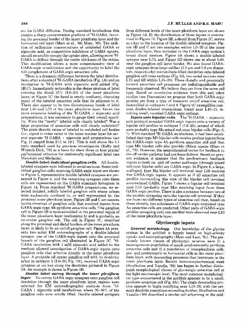
Fig. 7. Three GABA-labeled terminals synapsing (arrowheads) onto a bipolar cell terminal near L10 of the inner plexiform layer. There is also a synapse between two of the three CABA-ergic amacrine cell profiles that are synapsing on the bipolar cell terminal (asterisk).
dle of the inner plexiform layer. We also have evidence for a nonpyriform, multipolar subset of glycinergic amacrine cells (Fig. 8C). Whereas their lateral dendritic extensions may contribute significantly to the terminals of sublayer 1, their extent is difficult to determine. The glycinergic interplexi- form cell is shown in Figure 8B, with a process ascending to the outer plexiform layer. The arbors of glycinergic ama- crine and interplexiform cells in the inner plexiform layer are not completely distinguishable. However, there is now evidence from Golgi impregnation that glycinergic inter- plexiform cells arborize in sublayers 1 and 5, leaving the bulk of the inner plexiform layer labeling in sublayers 2, 3, and 4 due to glycinergic amacrine cells (Kalloniatis and Marc, '89).
The overall glcinergic label profile is punctate and clus- tered throughout the inner plexiform layer, concentrated in three discontinuous bands (strata) between L25 and L75 (probably entirely of amacrine cell origin). Glycinergic ter- minals are relatively sparse, both near the proximal border of the inner plexiform layer and in the outer plexiform layer. Thus, without the diffusion barriers observed with 3H- GABA uptake, the spatial buffering of glycine uptake
should be insignificant. As predicted, the addition of 1 mM unlabeled glycine to standard 3H-glycine incubations has no qualitative effect (RE. Marc, unpublished observations).
Light microscopic autoradiography of 3H-glycine, as with JH-GARA + nipecotic acid preparations, shows a large pro- portion of cell bodies labeled in the inner nuclear layer, and labeled strata in both sublaminae a and b of the inner plexi- form layer. To check our label specificity, we counted the grain density ratio between glycine-labeled cell bodies and unlabeled areas in the inner nuclear layer from low-power EM autoradiographs representing three different experi- ments (Fig. 9). The signal to noise ratio ranged from 29:l to X : I , clearly exceeding established criteria (Kelly and Weitsch-Dick, '78).
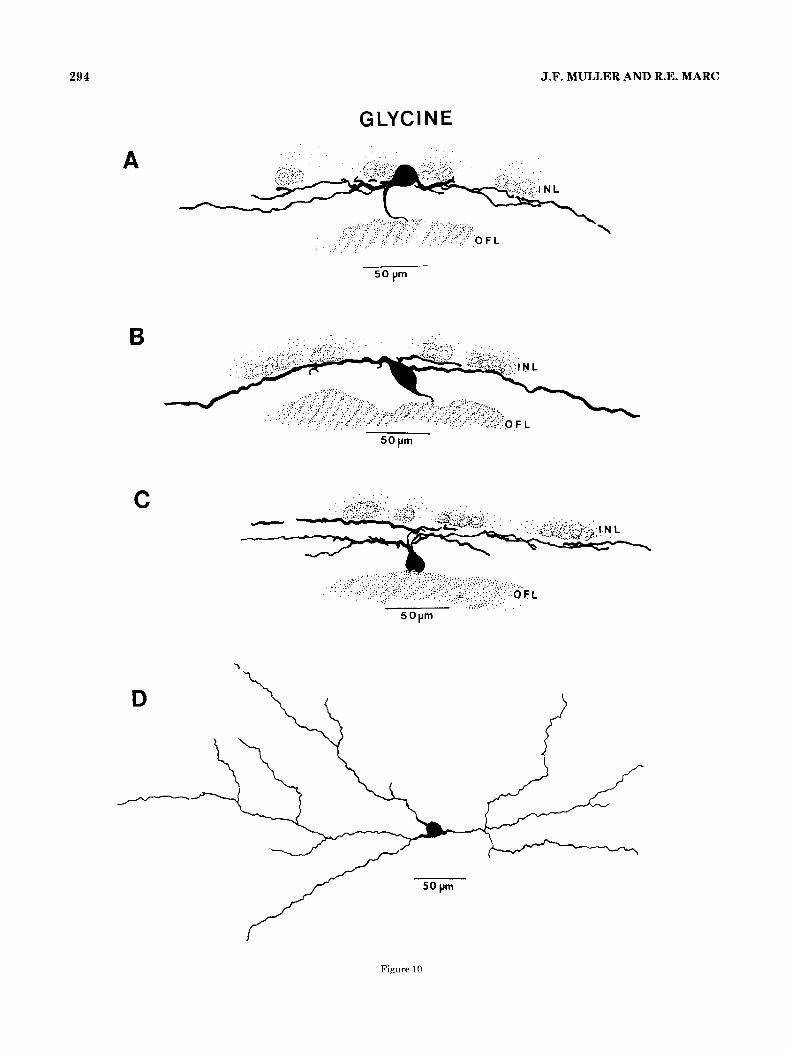
Four indi- vidual ganglion cells are shown, whose arbors were well iso- lated and onto which glycinergic synapses were followed in serial EM autoradiographs (Fig. 10A-D). The drawings in Figure 10A and D represent ganglion cells of similar somatic morphology, even though the cell in Figure 10A is displaced to the amacrine layer and the one in Figure 10D is in the ganglion cell layer. Both cells are large, spheroid, and multi-
Double label: Individual ganglion cells.
292 J.F. MULLER AND R.E. MARC
Fig. 8. Glycine uptake, light microscopic autoradiography. A Pyri- form glycinergic amacrine cell, arrowhead pointing to the descending primary dendrite. A detail from this view serves as a key to stratification in Figure 12. B: A glycinergic interplexiform cell with portions of two ascending processes indicated (arrows). Nearby amacrine cells are
pointed out for comparison (arrowheads). C: Four serial sections depict- ing what may be one of a population of multipolar glycinergic amacrine cells (arrowheads). Dendrites appear to project from multiple sites on the cell body in different directions (brackets).
polar and have their dendritic arbors confined to LO-25 of the inner plexiform layer. Figure 11A presents two views of dendrites from the cell drawn in Figure 10A. Three serial sections (Fig. 11B) show glycinergic input onto a dendrite from one of the labeled profiles in Figure 1 l A . The silver grains are plentiful and well localized, and the pre- and post-
synaptic densities and aggregated vesicles are clearly visible in each section. Also, as is often seen in large ganglion cell profiles, the HRP reaction product decorates the densely packed microtubules. Figure 1OC is a camera lucida drawing from two consecutive 40 wm sections of a fairly large, pyri- form ganglion cell stratified in the distal and middle levels

GABA AND GLYCINE PATHWAYS IN THE GOLDFISH IPL 293
Fig. 9. 3H-glycine uptake, EM autoradiography. Signal to noise ratio. A detail of an inner nuclear layer region, counted for grain density ratio between labeled cell bodies and unlabeled areas. Preparations ana- lyzed had signal to noise ratios ranging from 29:l to 32:l. For orientation, the location of the inner plexiform layer is indicated.
of the inner plexiform layer. Glycinergic synapses were found onto the labeled dendritic profiles in each stratum. The ganglion cell drawn in Figure 10B is large, fusiform, and displaced to the inner plexiform layer, with its broad, sparsely branched dendrites confined to sublayer 1. Figure 11C offers two serial views of a glycinergic input onto a labeled dendritic cross section from this probable off-center ganglion cell. I t presents the same morphology as the gan- glion cell pictured in Figure 3A, which was found to receive CABA-ergic inputs. Therefore, this appears to be one type of ganglion cell that receives both GABA-ergic and glyciner- gic input.
Double label survey through the inner plexiform layer. Glycinergic inputs were found onto labeled gan- glion cell dendrites in each of the five sublayers. The most common regions for these synaptic contacts were near L30 and L90 ofthe inner plexiform layer (Fig. 12A-C). The dou- ble-labeled synapse in Figure 12A is at L85, one of three serial sections of a glycinergic input onto a slender ganglion cell dendrite. Figure 12B is one of two serial sections of gly- cinergic input from a glycogen-rich profile onto a small HRP-labeled ganglion cell cross section near L25. Although overall glycogen levels can fluctuate between preparations, certain glycine-labeled cells and profiles such as this contain a markedly high density. This is one instance of a labeled ganglion cell profile of 0.2 pm or less in diameter that
receives input from a larger radiolabeled t.erminal. It is a good indicator of the resolution achievable with this tech- nique. Only one glycinergic input was observed onto a bipo- lar cell in this study (Fig. 13). Given that mixed rod-cone bipolar cells in the proximal inner plexiform layer tend to be large and glycogen-rich, this may be a cone bipolar cell pro- file.
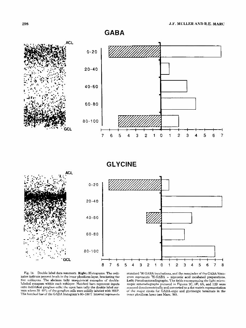
Double label: Data summary The histograms in Figure 14 are a laminar account of 60
double-labeled synapses that clearly met our stringent label criteria. The open bars represent double-labeled synapses counted from retinal regions with 30-40% of the ganglion cells solidly filled, and the hatched bars represent synapses involving individual labeled ganglion cells, as illustrated in Figures 3,4,10, and 11. The hatched bar in the GABA histo- gram for the 80-100% interval represents data collected from standard %-GABA incubated preparations (Figs. lA, 3B,C, 4A). The remainder of the histogram bars in the GABA portion of Figure 16 represent double label data col- lected from preparations incubated in 3H-GABA + nipe- cotic acid (Fig. 2B,C, 3,5B, 6). The open bars in each histo- gram offer a preliminary survey of the overall distribution of GABA-ergic and glycinergic synapses onto ganglion cells. The pseudoautoradiographs (Marc, '86) allow comparison of the distributions of synapses in the inner plexiform layer
294 J.F. MULLER AND R.E. MARC
GLYCINE
A
B
C
. . .
. . .
5 0 pm
50 pm
D
Figure 10

GABA AND GLYCINE PATHWAYS IN THE GOLDFISH IPL 296
Fig. 11. Glycinergic inputs onto individual HRP-labeled ganglion cells. A Light micrographs taken a t two focal planes through the 40 pm section selected for EM autoradiographic analysis, from the cell drawn in Figure 10A. Opposing arrowheads trace the HRP-labeled dendrites, and an arrow points out the axon. B Three serial EM autoradiographs following a synapse (arrowheads) onto an oblique cross section of one of
the dendrites pictured in A (asterisk). C: Two serial views of a glyciner- gic synapse (arrowheads) onto a dendritic cross section from the cell illustrated in Figure 10B (asterisk). Although the synaptic cleft is some- what oblique, the pre- and postsynaptic densities and aggregated synap- tic vesicles are clearly visible.
~~ ~~ ~~
Fig. 10. Camera lucida drawings of HRP-labeled ganglion cells found to receive glycinergic input. A, B, and D Large cells, monostrati- fied in the distal 25% of the inner plexiform layer. D, drawn horizontally from a 60 wm section, has its cell body in the ganglion cell layer. A, of similar morphology (drawn from three consecutive 40 pm sections), has its cell body displaced to the amacrine cell layer. B has a fusiform mor-
phology, similar to the cell pictured in Figure 3A, with its cell body, dis- placed to the inner plexiform layer. c, drawn from two serial 40 pm sec- lions, is apparently bistratified, ramifying in sublayers 1 and 3, receiving glycinergic input in both strata. All four cells are probably off-center ganglion cells.
296 J.F. MULLER A N D R.E. MARC
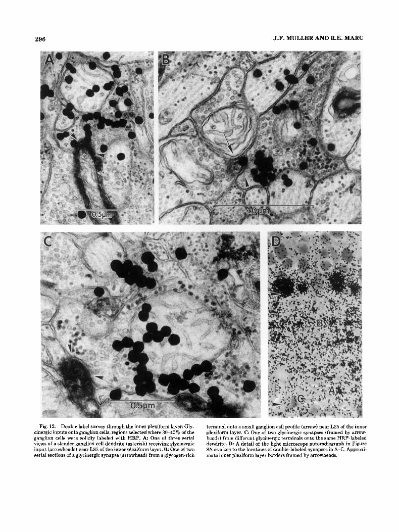
Fig. 12. Double label survey through the inner plexiform layer: Gly- cinergic inputs onto ganglion cells, regions selected where 3040% of the ganglion cells were solidly labeled with HRP. A One of three serial views of a slender ganglion cell dendrite (asterisk) receiving glycinergic input (arrowheads) near L85 of the inner plexiform layer. B. One of two serial sections of a glycinergic synapse (arrowhead) from a glycogen-rich
terminal onto a small ganglion cell profile (arrow) near L25 of the inner plexiform layer. C One of two glycinergic synapses (framed by arrow- heads) from different glycinergic terminals onto the same HRP-labeled dendrite. D: A detail of the light microscope autoradiograph in Figure XA as a key to the locations of double-labeled synapses in A-C. Approxi- mate inner plexiform layer borders framed by arrowheads.

GABA AND GLYCINE PATHWAYS IN THE GOLDFISH IPL 297
Fig. 13. A rare glycinergic input (arrowheads) onto a bipolar cell profile (BC). One of three serial sections, near L85. Note the synaptic ribbon (arrow).
with the major strata of GABA-ergic and glycinergic label. GABA-ergic and glycinergic synapses onto ganglion cells occur in every sublayer. The sample is not large enough to provide statistical significance about relative frequencies in particular sublayers, but there are trends that deserve fur- ther scrutiny. In the 'H-GABA + nipecotic acid prepara- tions, the first and fourth sublayers had the largest numbers of double-labeled synapses. In the 3H-glycine preparations, the second and fifth sublayers registered the highest fre- quencies. 3H-GABA + nipecotic acid experiments uncov- ered a dense band of label along the distal 25% of the inner plexiform layer (L0-25), and we have found that GABA- ergic inputs onto ganglion cells are prevalent where 3H- GABA label is densest. For 3H-glycine experiments, proba- ble off-center ganglion cells were sampled, and glycinergic inputs onto individual ganglion cells did not allign with either of the two sublayers with apparent high frequencies of glycinergic inputs onto ganglion cells (sample bias). Sub- layers two and five, specifically L25-35 and L85-90, in- cluded 65% of the glycinergic inputs onto ganglion cells sur- veyed through the inner plexiform layer. In fact, glycinergic inputs onto ganglion cells in sublayer five {LBO-100) seem a t least as frequent as GABA-ergic inputs. This is surprising, given the relatively low density of glycine label a t that level. There may be an important segregation of targets for glycin- ergic neurons (see Discussion). Although GABA-ergic and glycinergic systems have a considerable number of outputs onto ganglion cells in the two distal sublayers, the GABA system dominates in sublayer 1 and the glycine system in sublayer 2.
DISCUSSION Retrograde HRP transport
Injection of HRP into the optic nerve labels the retina heterogeneously. Some retinal regions have high densities of solidly labeled ganglion cells; other areas are more sparsely labeled. The regions with up to 40% of the ganglion cells sol- idly filled showed a broad morphological variety. Areas of the retinas where solidly labeled ganglion cells were rela- tively sparse proved useful in that the dendritic arbor of an HRP-labeled ganglion cell was more likely to be isolated. These regions appeared biased toward larger to medium- sized labeled cells. Individual labeled ganglion cells illus- trated herein reflect that bias (Figs. 4, 12). Although we have noted the sizes, shapes, and locations of their cell bod- ies, individual labeled ganglion cells represented have been classified according to the apparent stratification of their dendrites in the inner plexiform layer.
Dye and current injection studies in teleosts (Naka, '77; Famiglietti et al., '77) indicate that on-center and off-center pathways in fishes are separate and parallel. Since we define on- and off-center regions of the inner plexiform layer by the locations of mixed rod-cone bipolar cell terminals (Ma [off-center] in sublayers 1 and 2, Mb [on-center] in sub- layers 4 and 5), we predict that GABA-ergic and glycinergic synapses onto labeled ganglion cell profiles in sublayers 1 and 2 directly contribute to either off-center or onloff path- ways, whereas similar synapses in sublayers 4 and 5 are directly influencing on or onloff pathways.
High-affinity uptake Standard and modified approaches to high-affinity up-
take and autoradiography have been employed in this study. Standard protocols for high-affinity uptake of 3H-GABA and 3H-glycine, as previously used in goldfish, showed char- acteristic labeling patterns, specific for unique populations of neurons (Marc et al., '78, '88; Marc, '80, '82, '85, '86, '89; Marc and Lam, '81; Marc e t al., '88; this report). An alterna- tive 3H-GABA incubation protocol has been used to extend our understanding of GABA-ergic amacrine cell populations (Marc et al., '88; Marc, '89). Previous work had already established ultrastructurally that GABA-ergic type Ab amacrine cells reciprocally feedback onto depolarizing (on- center) bipolar cell terminals. This was based on the clear selectivity for type Ab pyriform amacrine cells by using standard 3H-GABA preparations, with short incubations (Marc et al., '78, '88; Marc, '80, '82, '86). Incubations in 3H- nipecotic acid, a competitive inhibitor of GABA uptake with similar affinity (Larsson et al., 'go), show comparable selec- tivity (Ayoub and Lam, '84). Apparently, type Ab pyriform amacrine cells are the only GABA-ergic neurons, in fishes, that project to the proximal border of the inner plexiform layer. Using the modified 3H-GABA uptake protocol, adding 1 mM nipecotic acid (Marc et al., '88; Marc, '89), a number of other questions could be explored. This approach labels nearly all GABA-ergic amacrine cells, as supported by the observations that 1) There is a 95-9792 concordance be- tween anti-GABA immunocytochemistry and 3H-GABA + nipecotic acid labeling and 2) each method labels about 50- 5 2 O 0 of all amacrine cells (Marc et al., '88; Marc, '89).
High-affinity uptake of 3H-glycine labels two general classes of retinal neurons, amacrine cells and interplexiform cells, and shows evidence of labeled terminals throughout the inner plexiform layer (Marc and Lam, '81; this report) concentrated in three discontinuous bands between L25 and
298 J.F. MULLER AND R.E. MARC
GABA
ACL
. ... a .. . . . . . .. - . * . .. . * 'GCL
0-20
20-40
40-60
60-80
80- 100
1
I
P
0 - 2 0
20-40
4 0 - 6 0
6 0 - 8 0
80-1 00
I 1 1 1 I 1 I 1 I
7 6 5 4 3 2 1 0 1 2 3 4 5 6 7
GLYCINE
I , , , 1 1 1 1 - 1 , 1 1 1 1 1 1 ,
8 7 6 5 4 3 2 1 0 1 2 3 4 5 6 7 8
Fig. 14. Double label data summary. Right: Histograms. The ordi- nates indicate percent levels in the inner plexiform layer, bracketing the five sublayers. The abcissas tally unequivocal examples of double- labeled synapses within each suhlayer. Hatched bars represent inputs onto individual ganglion cells; the open bars tally the double label snr- veys where 3040% of the ganglion cells were solidly labeled with HRP. The hatched bar of the GABA histogram's 80-100% interval represents
standard 'H-GABA incubations, and the remainder of the GABA histo- gram represents 'H-GABA + nipecotic acid incubated preparations. Left: Pseudoautoradiographs. The fields encompassing the light micro- scopic autoradiographs pictured in Figures lC, 5E, BA, and 12D were scanned densitometrically and converted to a dot matrix representation of the major strata for GABA-ergic and glycinergic terminals in the inner plexiform layer (see Marc, '86).

GABA AND GLYCINE PATHWAYS IN THE GOLDFISH IPL 299
L75, in sublayers 2-4. Based on light microscopic autoradi- ography of serial sections, we propose at least two morpho- logical types of glycinergic amacrine cell, one general popu- lation of small pyriform cells (Fig. 8A) and one type of apparently multipolar amacrine cell, with several dendrites projecting laterally from the cell body (Fig. 8C). Recently, Kalloniatis and Marc ('89) have obtained Golgi impregna- tions of glycinergic interplexiform cells that reveal that they arborize in sublayers 1 and 5 of the innerplexiform layer and not in sublayers 2, 3, or 4. In fact, the sublayer 5 dendrites appear to run just over the distal surfaces of the ganglion cells and may significantly contribute to the glycinergic synapses seen in that stratum.
EM autoradiographic analysis of GABA and glycine up- take in combination with retrograde HRP label of ganglion cells has led to some general conclusions. 1) Both GABA- ergic and glycinergic profiles are presynaptic to ganglion cell dendrites in each of the five sublayers. 2) There are ganglion cells that may receive direct synaptic input from both GABA-ergic and glycinergic neurons. 3) GABA-ergic ama- crine cells contribute the predominant feedback input to both depolarizing and hyperpolarizing mixed rod-cone bipo- lar cells (i.e., on- and off-center pathways). Glycinergic synapses onto bipolar cells have been observed (Marc and Lam, '81; Studholme and Yazulla, '88; Fig. 13), but they are extremely rare.
The specificities of GABA-ergic and glycinergic markers for the neurons that use those neurotransmitters had been sources of some controversy (Marc, '86; Yazulla, '86; Marc et al., '88; Massey and Redburn, '87; Studholme and Yazulla, '88). It is now clear that the correspondence is excellent for GABA-ergic markers (Ball and Brandon, '86; Ball, '87; Marc et al., '88; Marc, '89). Furthermore, glycine uptake has a 0% overlap with GABA immunocytochemistry (Marc, '891, demonstrating complete separation of these systems (see also Marc, '85, '86).
DATA SUMMARY GABA-ergic inputs
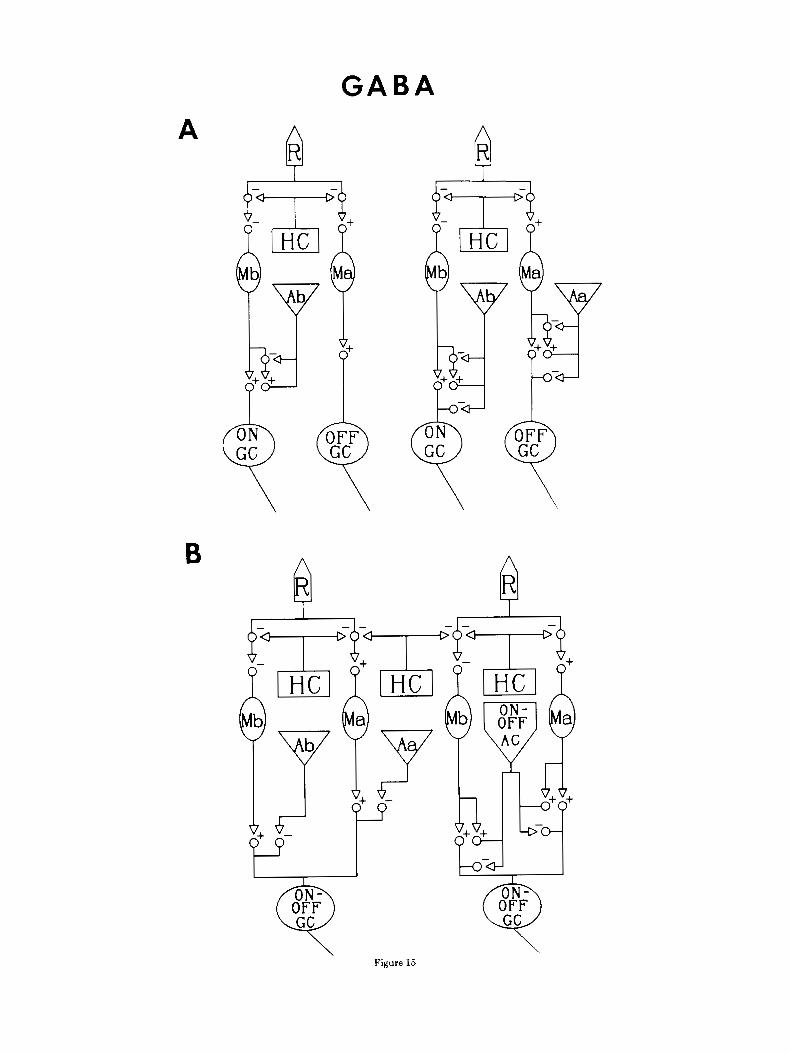
Figure 15A and B are schematic diagrams illustrating pathways to on, off, and on/off ganglion cells, with estab- lished and proposed interneurons GABA-ergic surround pathways. The H1 HC and the type Ab pyriform amacrine cell have been previously established as GABA-ergic (Marc et al., '78, '88; Yazulla et al., '87), a particular transient, on/ off amacrine cell and certain type Aa amacrine cells are now proposed to be GABA-ergic.
In teleosts, large, fusiform transient amacrine cells, bi- stratified to sublayers 1 and 4, have been intracellularly recorded and dye injected by a number of investigators (Djamgoz et al., '85; Murakami and Shimoda, '77; Fami- glietti et al., '77; Teranishi et al., '84, '85, '87) and found to be dye-coupled (Teranishi et al., '84). Ultrastructural evi- dence is provided herein that similarly bistratified GABA- ergic amacrine cells synapse onto ganglion cells in both their dendritic strata. Large caliber, nontapering dendrites near L10 and L70 were found in these double-label preparations to label with GABA, and we provide evidence for their inputs onto labeled ganglion cells in both strata (Fig. 5A,B). We encountered gap junctions on some of these profiles, but not unequivocally contacting homologous dendrites. GAD and GABA immunocytochemistry (Ball and Brandon, '86; Ball, '87; Marc et a]., '88; Marc, '89) and both light and elec- tron microscopic 3H-GABA + nipecotic acid autoradiogra-
phy reveal large, fairly common amacrine cells with distinct, 2-3 pm diameter, nontapering or mildly tapering dendrites arborizing in sublayers 1 and 4 (Marc et al., '88; Figs. 5A,B, 14). It seems just a matter of time before the above- described transient amacrine cell will be definitively estab- lished as GABA-ergic.
As Figure 15 illustrates, GABA-ergic type Aa amacrine cells synapse onto hyperpolarizing bipolar cells and off-cen- ter ganglion cells based on profuse GABA-ergic inputs onto type Ma bipolar cells in sublayer 1 (Marc, '89; Figs. 6, 7). Bipolar cells respond in a sustained manner to light, so it is assumed that amacrine cells synapsing onto bipolar cells are sustained amacrine cells. Several sustained-off amacrine cells have been recorded and dye injected (Teranishi e t al., '85). Each was monostratified in sublamina a, but their mor- phologies varied from pyriform to fusiform. Finding profuse GABA-ergic type Aa feedback onto Ma bipolar cells is exciting because it means that GABA-ergic amacrine cells provide the predominant feedback for both on- and off-cen- ter pathways in goldfish. GABA-ergic inputs were also found onto the HRP-labeled probable off-center ganglion cell pictured in Figure 3A (see Fig. 4B), and the GABA- labeled amacrine cell profiles closely resembled the small rounded swellings seen synapsing onto Ma bipolar cells. GABA-ergic Aa amacrine cells have also been found to synapse onto other GABA-labeled profiles in sublayer 1 (Fig. 7). This may indicate multiple classes of GABA-ergic Aa amacrine cells. Synapses between putatively GABA- ergic amacrine cell dendrites have also been described in mammals (Vaughan et al., '81; Kolb and Nelson, '85; Mar- iani and Caserta, '86).
Figure 15 specifies the synaptic transfers of on- and off- center pathways, indicating whether the polarity of the postsynaptic response is conserved (+) or inverted (-). The on- and off-center visual pathways in vertebrates are distin- guished from one another by a single sign inversion between the receptors and depolarizing bipolar cells (Slaughter and Miller, '81, '83; Miller and Slaughter, '85; Nawi and Copen- hagen, '87). For the purposes of this discussion, all bipolar- ganglion cell interactions will be considered excitatory and sign-conserving (Naka, '77; Slaughter and Miller, '83; Mas- sey and Redburn, '87).
The left diagram in Figure 15A represents the previously established (Marc et al., '78) and the right the newly pro- posed GABA-ergic surround inputs to on- and off-center pathways. Figure 15B outlines proposed sustained (left) and transient (right) GABA-ergic amacrine cell inputs onto (transient) d o f f ganglion cells. Standard 3H-GABA incu- bations select type Ab pyriform amacrine cells (Marc et al., '78) that reciprocally synapse with type Mb bipolar cells. In the right diagram in Figure 15A, an additional circuit con- tributes to the on pathway: GABA-ergic feed-forward on- center ganglion cells. Thus there are a minimum of three GARA-ergic surround inputs that converge on a single on- center ganglion cell: 1) H1 HC-+cone feedback, 2) Ab AC-Mb BC feedback, and 3) Ab AC-on GC feed-forward. Samples incubated in 3H-GABA + nipecotic acid allow a nearly complete assessment of GABA-ergic inputs to the o fken te r pathway. Direct inputs onto probable off-center ganglion cells arise from proposed GABA-ergic Aa amacrine cells GABA-ergic Aa amacrine cells also synapse on Ma bipolar cells (see Figs. 6, 7), and many of these inputs are reciprocal (Marc et al., '88; Marc, '89). The path from GABA-ergic HI horizontal cells to off-center ganglion cells is net sign-inverting (one - synapse in the chain). Thus
G A B A A
B
Figure 15

GABA AND GLYCINE PATHWAYS IN THE GOLDFISH IPL 301
I-
+-+
Fig. 16. Schematic diagram of glycinergic pathways in the goldfish retina. The two major categories of glycinergic neurons in the goldfish are a heterogeneous population of small amacrine cells and an interplex- iform cell that receives input from horizontal cells in the outer plexiform layer and sends terminals to the inner plexiform layer. They are collec- tively abbreviated AC for this diagram. Further study has begun to sub- divide the amacrine cells (see also Studholme and Yazulla, '88). Left: Direct evidence for glycinergic inputs to off-center ganglion cells is illus- hated in Figures 10 and 11. Glycinergic inputs to on-center ganglion
~~
Fig. 15. GABA-ergic inputs to on-center, off-center, and on/off pathways. A t indicates a sign-conserving and a - indicates a sign- inverting synapse. A Left: Previously established circuits in the gold- fish inner plexiform layer. Employing standard high-affinity uptake of 3H-GARA, it had been found that GABA-ergic Ah pyriform amacrine cells and type Mb bipolar cells interact reciprocally in a feedback circuit (Marc et al., '78). Right: New circuits in the goldfish inner plexiform layer. Double label with standard %GABA uptake revealed GABA- ergic inputs from Ah pyriform amacrine cells onto probable on-center ganglion cells: A feed-forward circuit. Data from 3H-GABA + nipecotic acid preparations reveal GABA-ergic feed-forward to probable off-cen-
Glycine
++ 99 L
cells is suggested by data in Figure 12A,C and summarized in Figure 14 (see text). The double arrowheads with the glycinergic amacrine cells symbolize that the predominant inputs to glycinergic amacrine cells are from other amacrine cells. Right: Glycinergic inputs onto on/off gan- glion cells are suggested by frequent glycinergic synapses onto HRP- labeled ganglion cells in both sublaminae a and b. Recent data suggest that many glycinergic synapses in sublayer 5 come from glycinergic interplexiform cells (Kalloniatis and Marc, '89).
ter ganglion cells by type Aa amacrine cells and reciprocal GABA-ergic feedback onto probable hyperpolarizing mixed rod-cone bipolar cells (see also Marc, '89). B. Pathways to onioff ganglion cells. Left: Inputs from probable sustained GABA-ergic amacrine cells. Figures 3C and 4A illustrate type Ab pyriform, sustained on-center amacrine cell synapses onto the proximal branches of bistratified probable on/off ganglion cells. The direct influence of sustained off-center, GABA-ergic Aa amacrine cells on on/off ganglion cells is speculative, based on the large proportion of GABA-labeled terminals that synapsed onto HRP-labeled ganglion cells in sublayer 1. Right: Proposed direct GABA-ergic transient, on/off amacrine cell inputs onto on/off ganglion cells in both sublaminae.

302
hyperpolarization of H1 horizontal cells by light should effert a depolarization in the off-center ganglion cells, con- sistent with the electrophysiological results of Naka ('77) for catfish. Since the net paths between GABA-ergic Aa ama- crine cells and both Ma bipolar cells and off-center ganglion cells is sign-inverting (one - synapse), the off pathway has three ultimately sign-inverting surround fields converging on ganglion cells. As we learn more about the combinations of inputs onto the various GABA-ergic neurons, we can bet- ter predict their spatial, temporal, and chromatic contribu- tions to the opponent-surround.
Transient on/off ganglion cells respond both to light onset and to light offset with a short burst of action potentials and often multistratified, in both sublaminae a and b (Fami- glietti e t al., "77; Murakami and Shimoda, '77; Teranishi et al., '84, '85, '87; Djamgoz et al., '85; Djamgoz, '86; Djamgoz and Wagner, '87). They presumedly receive inputs from transient amacrine cells, which, in turn, are believed to be driven by depolarizing and hyperpolarizing bipolar cells (Toyoda et al., '73; Famiglietti et al., '77; Frumkies et al., '81; Toyoda and Fujimoto, '84; Miller and Slaughter, '85; Djam- goz, '86; Kujiraoka et al., '88). Recent evidence, including data presented here, indicates that sustained amacrine cells influence transient ganglion cells as well (Belgum et al., '83). Figure 15R is a set of circuit diagrams outlining the pro- posed influences on the transient on/& ganglion cells by sustained GABA-ergic amacrine cells (left diagram) and transient GABA-ergic amacrine cells (right diagram). The left diagram is simplified, in that the amacrine cell recipro- cal feedback onto Ma and Mb bipolar cells, as illustrated in Figure 15A, is implied. I t is expected that the three GABA- ergic surround inputs to on- and off-center pathways are present for on/& pathways, as well. Figures 3C and 4A depict synaptic input from GABA-ergic Ab pyriform ama- crine cells (sustained on-center) to the proximal branch of a bistratified ganglion cell, likely to be an on/off ganglion cell. The proposed Aa (sustained off-center) influence on the on/ off ganglion cell is based merely on the large proportion of GABA-ergic terminals surveyed that synapsed onto HRP- labeled ganglion cells of a variety of dendritic morphologies in sublayer 1 (LO-20) of the inner plexiform layer. Evidence is presented in Figures 1B and 5A,R that a bistratified (probable transient) amacrine cell is GABA-ergic and syn- apses onto ganglion cells in both sublayers 1 and 4. Thus a fourth GABA-ergic opponent-surround pathway is pro- posed. The hyperpolarizing and depolarizing bipolar cells presumedly converge on GABA-ergic transient amacrine cells, which subsequently synapse on transient on/off gan- glion cells.
Glycinergic inputs The combined arbor of the glycinergic amacrine cells and
interplexiform cells reaches all levels of the inner plexiform layer, although three bands in sublayers 2-4 are often apparent (Marc, '86; Fig. 9, 12D). Ultrastructurally, glycin- ergic terminals are often seen as clusters of profiles, fre- quently running along side each other, making no apparent contacts with one another. Figure 12C shows a ganglion cell dendrite with input from one of two adjacent glycinergic endings. Although adjacent glycinergic profiles could be dis- tinguished by relative glycogen content or slight differences in electron density or vesicle concentration, we could not claim different cells of origin.
Figure 16 schematizes the mixed rod-red cone pathways in the goldfish retina and describes our limited knowledge of
J.F. MULLER A N D R.E. MARC
how surrounds are affected by the glycine system. The pre- dominant inputs to the glycinergic system of the inner plexi- form layer are from other, nonglycinergic amacrine cells. Direct glycinergic input occurs onto a variety of probable off-center ganglion cells (Figs. 10, 11, and 14), whereas direct glycinergic inputs to on-center ganglion cells are sug- gested by data presented in Figure 12A,B and the glycine summary presented in Figure 14. With about 30% of the double-labeled synapses occurring in sublayer 5 (L80-100), it is likely that some involve on-center ganglion cells (al- though we cannot rule out that they may all be on/off gan- glion cells). However, sublayer 5 is a major terminator site for glycinergic interplexiform cells (Kalloniatis and Marc, '89). The discordance between the frequency of glycinergic inputs to ganglion cells (high in sublayer 5; see Fig. 14) and the density of glycinergic terminals (low in sublayer 5; see Marc and Lam, '81; Marc, '86; this report) suggests that a significant proportion of ganglion cell inputs from 3H-glycine-labeled profiles could be from glycinergic inter- plexiform cells (see also Rayborn et al., '81). Given that the glycinergic system synapses onto ganglion cells in every sub- layer, one or a variety of glycinergic neurons could also synapse onto transient on/off ganglion cells. Furthermore, there are no clear clues to whether glycinergic inputs are from sustained or transient cells; both possibilities exist (see Marc and Lam, '81). Although there is evidence of glyciner- gic inhibitory effects on on-center, off-center, and on/off ganglion cells (Negishi et al., '78), we still lack correlative physiology for any morphologically defined glycinergic neu- ron in fishes. With these uncertainties, and our general lack of understanding of inputs to glycinergic neurons in the inner plexiform layer, we cannot at this point speculate on their light-evoked effects on on-center and off-center path- ways. Further morphological work (in progress) and more detailed physiological study should help resolve these is- sues.
ACKNOWLEDGMENTS This research was supported by NIH grant EY 02576.
LITERATURE CITED Adams, J.C. (1981) Heavy metal intensification of DAB-based HRP reaction
product. J. Histochem. Cytochem. 29775. Ames, A. 111, and F.B. Nesbett (1981) In vitro retina as an experimental
model of the central nervous system. J. Neurochem. 372367471, Ayouh, G.S., and D.M.K. Lam (1984) The release of y-aminobutyric acid
from horizontal cells of the goldfish (Carassius aurutus) retina. J. Physiol. 355:191-214.
Ball, A.K. (1987) Immunocytochemical and autoradiographic localization of GABAergic neurons in the goldfish retina. J. Comp. Neurol. 255:317- 325.
Ball, A.K., and C. Brandon (1986) Localization of 3H-GABA, -muscimol and -glycine in goldfish retinas stained for glutamate decarboxylase. J. Neu- rosci. 6:1621-1627.
Belgum, J.H., D.R. Dvorak, and J.S. McReynolds (1983) Sustained and t r m - sient synaptic inputs to on-off ganglion cells in the mudpuppy retina. J. Physiol. 340599-610.
Bouteille, M. (1976) The "LIGOP" method for routine ultrastructural auto- radiography: A combination of single grid coating, gold latenvification and phenidon development. J. Microsc. Biol. Cell 27t121-128.
Brandon, C. (1985) Retinal GABA neurons: Localization in vertebrate species using an anti-serum to rabbit brain brain glutamate decarboxylase. Brain Res. 344t286-295.
Cohen, B., and G.L. Fain (1988) GABA and glycine channels in isolated gan- glion cells from the goldfish retina. Invest. Ophthalmol. Vis. Sci. SUPPI. 29:104.

GABA AND GLYCINE PATHWAYS IN THE GOLDFISH IPL 303
Djamgoz, M.B.A. (1986) Common features of light-evoked amacrine cell responses in vertebrate retina. Neurosci. Lett. 71:187-191.
Djamgoz, M.B.A., J.E.G. Downing, E. Wagner, H.-J. Wagner, and I. Zeutzius (1985) Functional organization of amacrine cells in the teleost retina. In A. Gallego (ed): Neurocircuitry of the Retina, A Cajal Memorial. New York Elsevier, pp. 188-204.
Djamgoz, M.B.A., W.K. Stell, C.-A. Chin, and D.M.K. Lam (1981) An opiate system in the goldfish retina. Nature 292.62CL-623.
Djamgoz, M.B.A., and H.-,J. Wagner (1987) Intracellular staining of retinal neurons: Applications to studies of functional organization. In N. Osborne led): Progress in Retinal Research Vol6. Oxford: Pergamon, pp. 85-150.
Famiglietti, E.V. Jr., A. Kaneko, and M. Tachibana (1977) Neuronal architec- ture of on and off pathways to ganglion cells in the carp retina. Science 198:1268-1269.
Famiglietti, E.V. Jr., and H. Kolb (1976) Structural hasis for on and off-cen- ter responses in retinal ganglion cells. Science 194r193-195.
Frumkies, T.E., R.F. Miller, M. Slaughter, and R.F. Dacheux (1981) Physio- logical and pharmacological basis of GABA and glycine adion on neurons of the mudpuppy retina. 111. Amacrine-mediated inhibitory influences on ganglion cell receptive field organization: A model. J. Neurophysiol. 45783-805.
Glickman, R.D., A.R. Adolph, and J.E. Dowling (1982) Innerplexiform cir- cuits in the carp retina: Effects of cholinergic agonists, GABA, and sub- stance P on the ganglion cells. Brain Res. 2343-99.
Ishida, A.T., and B. Cohen (1988) GABA-activated whole cell currents in iso- lated retinal ganglion cells. J. Neurophysiol. 60.381-396.
Ishida, A.T., W.K. Stell, and D.O. Lightfoot (1980) Rod and cone inputs to bipolar cells in goldfish retina. d. Comp. Neurol. 191t315-335.
Kalloniatis, M., and R.E. Marc (1989) Golgi impregnated interplexiform cells in the goldfish retina. Invest. Ophthalmol. Vis. Sci. Suppl. (in press).
Kaneko, A,, E.V. Famiglietti Jr., and Tachibana (1979) Physiological and morphological identification of signal pathways in the carp retina. In M. Otsula (ed): Neurobiology of Chemical Transmission. New York Wiley, pp. 235-253.
Kaneko, A., Y. Nishimura, N. Tachibma, M. Tauchi, and K. Shamai (1981) Physiological and morphological studies of signal pathways in the carp retina. Vision Res. 2I:1519-1526.
Kaneko, A,, Y. Nishimura, M. Tauchi, and K. Shamai (1980) Distribution of afferent synapses along on-center bipolar cells axons in the carp retina. Biomed. Res. It345-348.
Kelly, J.S. and F. Weitsch-Dick (1978) Critical evaluation of the use of radioautography as a tool in the localization of amino acids in the mam- malian nervous system. In F. Fonnum (ed): Amino Acids as Chemical Transmitters. New York Plenum, pp. 102-121.
Kolb, H. (1982) The morphology of the bipolar cells, amacrine cells and gan- glion cells in the retina of the turtle Pseudemys scripta eleguns. Phil. Trans. R. Soc. London [Biol.] 298:355-393.
Kolb, H., and R. Nelson (1984) Neuronal architecture of the cat retina. In N. Osborne (ed): Progress in retinal Research Vol. 3. Oxford: Pergamon, pp. 22-60.
Kolb, H., and R. Nelson (1985) Functional neurocircuitry of amacrine cells in the cat retina. In A. Gallego (ed): Neurocircuitry of the Retina, a Cajal Memorial. New York Elsevier, pp. 21S232.
Kujiraoka, T., T. Saito, and J.-I. Toyoda (1988) Analysis of synaptic inputs to on-off amacrine cells of the carp retina. J. Gen. Physiol. 92:475-487.
Lam, D.M.K., and B.-B. Li, Y.-Y.T. Su, and C.B. Watt (1985) The signature hypothesis: Co-localizations of neuroactive substances as anatomical probes for circuitry analysis. Vision Res. 251353-1364.
Lam, D.M.K., and L. Steinman (1971) The uptake of [y-3H]aminohutyric acid in the goldfish retina. Proc. Nat. Acad. Sci. 68:2777-2781.
Lam, D.M.K., Y.Y.T. Su, L. Swain, R.E. Marc, C. Brandon, and J.-Y. Wu i 1979) Immunocytochemical localization of L-glutamic acid decarboxy- lase in the goldfish retina. Nature 278:565-567.
Larsson, O.M., P. Krogsgaard-Larson, and A. Shoesboe (1980) High affinity uptake of (RS)-nipecotic acid in astrocytes cultured from mouse brain. Comparison with GABA transport. J. Neurochem. 34:970-977.
Madtes, P. Jr., and D. Redburn (1985) Metabolism of [3H]nipecotic acid in the rabbit retina. J. Neurochem. 44t1520-1523.
Marc, R.E. (1980) Retinal colour channels and their neurotransmitters. In G. Verriest (ed): Colour Deficiencies V. London: Adam Hilger, pp. 1529.
Marc, R.E. (1982) Spatial organization of neurochemically classified inter- neurons of the goldfish retina: I. Local patterns. Vision Res. 22:589-608.
Marc, R.E. (1985) The role of glycine in retinal circuitry. In W.W. Morgan (ed): Retinal Transmitters and Modulators: Models for the Brain. Boca Raton, FL: CRC Press, Val. 1, pp. 119-158.
Marc, R.E. (1986) Neurochemical stratification of the inner plexiform layer of the vertebrate retina. Vision Res. 26t223-238.
Marc, R.E. (1989) The anatomy of multiple GABAergic and glycinergic path- ways in the inner plexiform layer of the goldfish retina. In R. Weiler and N. Osborne (eds): The Neurobiology of the Inner Retina: Nato/AS Series. Berlin: Springer-Verlag, pp. 53-64.
Marc, R.E., and D.M.K. Lam (1981) Glycinergic pathways in the goldfish retina. J. Neurosci. 1:152-165.
Marc, R.E., and W.-L.S. Liu (1984) Horizontal cells synapses onto glycine- accumulating interplexiform cells. Nature 311:266-269.
Marc, R.E., W.-L.S. Liu, and J.F. Muller (1988) Multiple GABA-mediated surround channels in goldfish retina. Invest. Ophthalmol. Vis. Sci. Suppl. 29:272.
Marc, R.E., W.K. Stell, D. Bok, and D.M.K. Lam (1978) GABAergic path- ways in the goldfish retina. J. Comp. Neurol. 182t221-246.
Mariani, A.P., and M.T. Caserta (1986) Electron microscopy of glutamate decarboxylase (GAD) immunoreactivity in the inner plexiform layer of the rhesus monkey retina. J. Neurocytol. 15645-655.
Massey, S.C., and D.A. Redburn (1987) Transmitter circuits in the vertebrate retina. Prog. Neurohiol. 28:55-96.
Miller, R.F. (1988) Are single neurons both excitatory and inhibitory. Nature 336:517-518.
Miller, R.F., and R.F. Dacheux (1976) Synaptic organization and ionic basis of on and off channels in mudpuppy retina. 111. A model of ganglion cell receptive field organization based on chloride free experiments. J. Gen. Physiol. 67579 -690.
Muller, J.F., and R.E. Marc (1984) Three distinct morphological classes of receptors in fish olfactory organs. J. Conip. Neurol. 222:482-495.
Muller, J.F., and R.E. Marc (1988) GABAergic and glycinergic pathways in the inner plexiform layer of the goldfish retina. Invest. Ophthalmol. Vis Sci. Suppl. 29t197.
Miller, R.F., and M.M. Slaughter (1985) Excitatory amino acid receptors in the vertebrate retina. In W.W. Morgan (ed): Retinal Transmitters and Modulators: Models for the Brain. Boca Raton, FL: CRC Press, Vol. 2, pp. 123-160.
Murakami, M., and Y. Shimoda (1977) Identification of amacrine and gan- glion cells in the carp retina. J. Physiol. (London) 264:801-818.
Naka, K.-I. (1977) Functional organization of catfish retina. J. Neurophysiol. 40t26-43.
Nawi, S., and D.R. Copenhagen (1987) Multiple clnases of glutamate receptor on depolarizing bipolar cells in retina. Nature ,325.56-58.
Negishi, K., S. Kato, T. Teranishi, and M. Laufer (1978) Dual actions of some amino acids on spike discharges in the carp retina. Brain Res. 148:67-84.
Nelson. R., E.V. Famiglietti Jr., and H. Kolh (1978) Intracellulsr staining reveals different levels of stratification for on- and off-center ganglion cells in the cat retina. J. Neurophysiol. 41t472-483.
Rayborn, M.E., P.V. Sarthy, D.M.K. Lam. and J.G. Hollyfield (1981) The emergence, localization and maturation of neurotransmitter systems dur- ing development of the retina in Xenopus laeuist 11. Glycine. J. Comp. Keurol. 195t585-593.
Redburn, D.A., S.C. Massey, and P. Madtes (1983) The GABA uptake system in rabbit retina. In L. Hertz, E. Kvamme, E.G. McGeer, A. Schousboe (eds) : Glutamine, Glutamate, and GABA in the Central Nervous System. New York Alan R. Liss, Inc., pp. 273-286.
Saito, T., T. Kujiraoka, and J.-I. Toyoda (1984) Electrical and morphological properties of off-center bipolar cells in the carp retina. d. Comp. Neurol. 222:200-208.
Saito, T., T. Kujiraoka, and T. Yanaha (1983) Connections between photore- ceptors and horseradish peroxidase-injected bipolar cells in the carp retina. Vision Res. 23:353-362.
Saito, T., T. Kujiraoka, T. Yanaha, and Y. Chino (1985) Reexamination of photoreceptor-bipolar connectivity patterns in carp retina: HRP-EM and golgi-EM studies. J. Comp. Neurol. 236.141-160.
Sakai, H.M., K:I. Naka, and J.E. Dowling (1986) Ganglion cell dendrites are presynaptic in catfish retina. Nature 319t495-497.
Salpeter, M.M., F.A. McHenry, and E.E. Salpeter (1978) Resolution in elec- tron microscope autoradiography IV. Application to analysis of autora- diographs. J. Cell Biol. 76t127-145.
Slaughter, M.M., and R.F. Miller (1981) 2-Amino-4-phosphonobutyric acid A new pharmacological tool for retina research. Science 211t182-184.

304 J.F. MULLER AND R.E. MARC
Slaughter, M.M., and R.F. Miller (1983) An excitatory amino acid antagonist blocks cone input to sign-conserving second-order retinal neurons. Science 219:1230-1232.
Studholme, K.M., and S. Yazulla (1987) Localization of endogenous GABA and glycine in goldfish retina by postembed EM imrnunocytochemistry. Invest. Ophthalmol. Vis. Sci. Suppl. 28:349.
Studholme, K.M., and S. Yazulla (1988) Localization of glycine immunoreac- tivity in the goldfish retina. Invest. Ophthalmol. Vis. Sci. Suppl. 29:273.
Tachihana, M., and Kaneko (1984) y-Aminobutyric acid acts a t axon termi- nals of turtle photoreceptors: Difference in sensitivity among cell types. Proc. Nat. Acad. Sci. USA 8Ir7961-7964.
Teranishi, T., K. Negishi, and S. Kato (1984) Dye coupling between amacrine cells in the carp retina. Neurosci. Lett. 51:73-78.
Teranishi, T., K. Negishi, and S. Kato (1985) Correlations between photore- sponse and morphology of amacrine cells in the carp retina. Neurosci. Res. 525211-5226.
Teranishi, T., K. Negishi, and S. Kato (1987) Functional and morphological correlates of amacrine cells in the carp retina. Neuroscience 20:935-950.
Toyoda, J.-I., and M. Fujimoto (1984) Application of transretinal current stimulation for the study of bipolar-amacrine transmission. J. Gen. Physiol. 84:915-925.
Toyoda, J.-I., and H. Haahimoto, and K. Ohtsu (1973) Application of trans- retinal current stimulation for the study of bipolar-amacrine transmis-
sion. Vision Res. 13:295-307. Vaughn, J.E., E.V. Famiglietti Jr., R.P. Barber, K. Saito, E. Roberts,and C.E.
Ribak (1981) GABAergic amacrine cells in rat retina: Immunocytochemi- cal identification of synaptic connectivity. J. Comp. Neurol. 197:113- 127.
Wong-Reilly, M., and G. Kageyarna (1985) Cytochrome oxidase cytochemis- try: Electron microscope analysis of serial post-fixation variables. Anat. Rec. 211:217A.
Yazulla, S. (1981) GABAergic synapses in the goldfish retina: An autoradio- graphic study of 'H-muscimol and 3H-GABA binding. J. Comp. Neurol. 200:83-93.
Yazulla, S. (1986) GABAergic mechanisms in the retina. In N. Osborne (ed): Progress in Retinal Research, Vol. 5. Oxford Pergamon, pp. 1-52.
Yazulla, S., J. Mosinger, and C. Zucker (1984) Two types of pyriform Ab ama- crine cells in the goldfish retina: An EM analysis of 3H-GABA uptake and somatostatin-like imniunoreactivity. Brain Res. 32352-356.
Yazulla, S., K. Studholme, and J.-Y. Wu (1986) Comparative distribution of 3H-GABA uptake and GAD immunoreactivity in goldfish retinal ama- crine cells: A double label analysis. J. Comp. Neurol. 244:149-162.
Yazulla, S., K. Studholme, and J.-Y. Wu (1987) GABAergic input to the synaptic terminals of rnhl bipolar cells in the goldfish retina. Brain Res. 411:400-405.
Copyright © 2022 FDOKUMEN