Detection and transmission of Fusarium verticillioides in corn ...

Fusarium
Introduction to the Genus
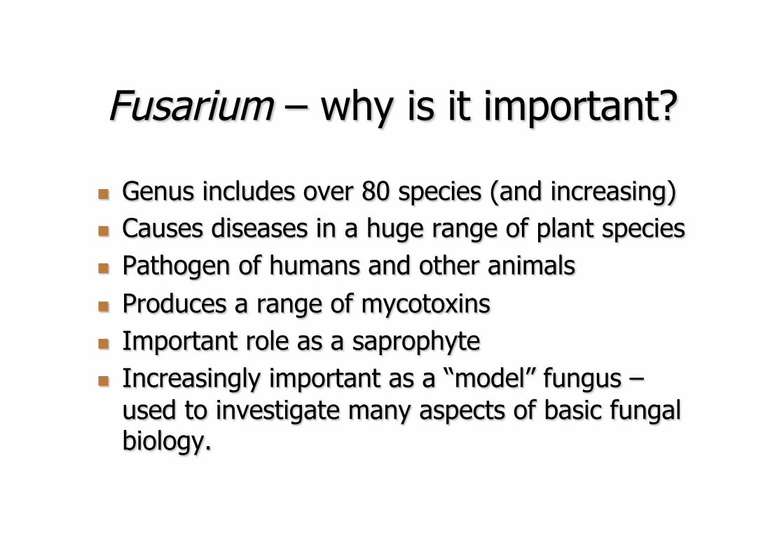
Fusarium – why is it important?
n Genus includes over 80 species (and increasing) n Causes diseases in a huge range of plant species n Pathogen of humans and other animals n Produces a range of mycotoxins n Important role as a saprophyte n Increasingly important as a “model” fungus –
used to investigate many aspects of basic fungal biology.

Plant diseases

Fusarium head blight and crown rot

Wilt diseases
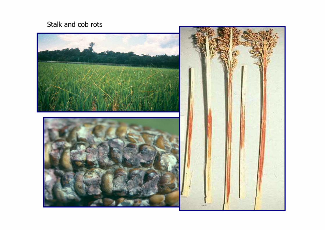
Stalk and cob rots

Mycotoxins


Hemorrhagic Syndromes Alimentary Toxic Aleukia in Humans
Moldy Corn Toxicosis in Animals

Fumonisins • Leukoencephalomalacia in Horses • Pulmonary Edema in Pigs • Liver Cancer in Rats • Oesophageal Cancer in Humans? • Neural Tube Defects in Humans?
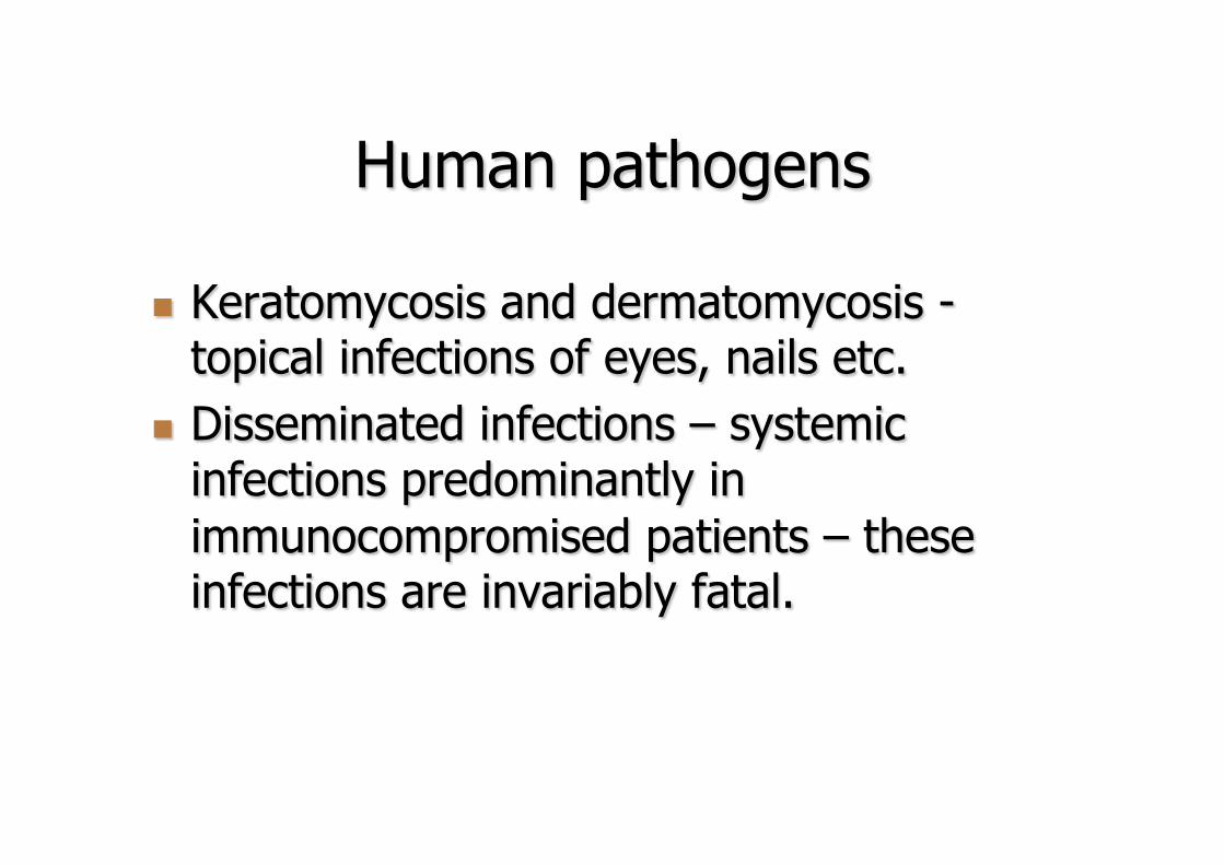
Human pathogens
n Keratomycosis and dermatomycosis - topical infections of eyes, nails etc.
n Disseminated infections – systemic infections predominantly in immunocompromised patients – these infections are invariably fatal.
Fusarium - Link 1809 n Defining characteristic
n Fusoid macroconidia with a foot shaped or notched base to basal cell
n Other genera may have this characteristic n e.g. Coelomycetes such as Botryocrea,
Heteropatella, Zelosatchmopsis, Libartania, Pycnofusarium and many others.

Fusarium – Basic Taxonomy
n Most traditional taxonomic systems arranged Fusarium species in sections
n These sections were groupings of species with some similar morphological characters
n The sections are not monophyletic n Sections are sometimes a useful and practical
way for sorting Fusarium species when identifying them (and for running workshops!)

Fusarium – The Main Sections and Key Species
n Discolor n F. graminearum
n Roseum n F. avenaceum
n Elegans n F. oxysporum
n Martiella-Ventricosum n F. solani
n Liseola n F. verticillioides
n Gibbosum n F. equiseti
n Sporotrichiella n F. poae
n Arthrosporiella n F. semitectum

Fusarium – Minor Sections n Eupionnotes
n F. merismoides, F. dimerum n Lateritium
n F. lateritium n Spicarioides
n F. decemcellulare
n There are others but we won’t cover them in this course

Fusarium – Beginning the Identification Process
n The majority of isolates are not difficult to characterize morphologically if you use: n Standard media n Controlled temperature and light
conditions n Cultures initiated from single spores
Single Spore Isolation
n There are a number of techniques used. n Technique developed by Snyder and
Hansen. n Can be used for macroconidia,
microconidia and ascospores. n Do not try to identify cultures before single
sporing.

Fusarium
n Standard media for morphological examination n Carnation leaf-piece agar (CLA) n Spezieller Nährstoffarmer Agar (SNA) n Potato dextrose agar (PDA) n Soil agar (SA)

CLA PDA
Growth Conditions n Temperature – generally either 25 C or
fluctuating 25/20 C day/night cycle. n Light
n good light conditions will help to stimulate conidia formation
n Black light can also be beneficial and may be essential for development of sexual structures.
n Remember, try to use the conditions outlined in the identification guide you are following.


Fusarium
n Primary criteria for morphological identification
n Morphology of macroconidium formed in sporodochium on carnation leaf pieces in CLA
n Try not to use macroconidia from other sources as they are variable and inconsistent

Sporodochia on carnation leaf-piece

Fusarium compactum
Fusarium
n Criteria for identification (CLA medium) n Shape of macroconidia n Presence/absence of microconidia n Shape and formation of microconidia n Nature of the conidiogenous cells n Presence/absence of chlamydospores
Fusarium
n Criteria for identification (SNA medium) n Presence/absence of microconidia n Shape and formation of the microconidia n Nature of the conidiogenous cell n Presence/absence of the chlamydospores

Fusarium
n Secondary criteria for identification (PDA medium)
n Colony morphology n Pigmentation - hyphae and in agar n Growth rates, especially at 25oC and 30oC.
n Either in agar plates or in race tubes


Growth rates at 25 and 30C

Fusarium
n Physiological criteria to aid identification n Mycotoxins n Secondary metabolite profiles n Odors n Growth rates n Responses to temperature, pH, moisture and
chemicals

Fusarium
n Molecular and genetic criteria n Vegetative compatibility grouping n Mating type and mating populations n DNA analysis based on molecular markers
n RFLP n AFLP n Sequencing – most commonly nuclear
genes – e.g. TEF, β-tubulin, histone etc.
Names associated with Fusarium spp.
n With adoption of single nomenclature for Fungi it has been recommended that the name Fusarium be used for the genus.
n Teleomorph names you might see in literature
n Gibberella - the majority of Fusarium species n Haematonectria – some members of Fusarium solani
complex n Neocosmospora - some members of Fusarium solani
complex n Albonectria - Fusarium decemcellulare

“Gibberella”
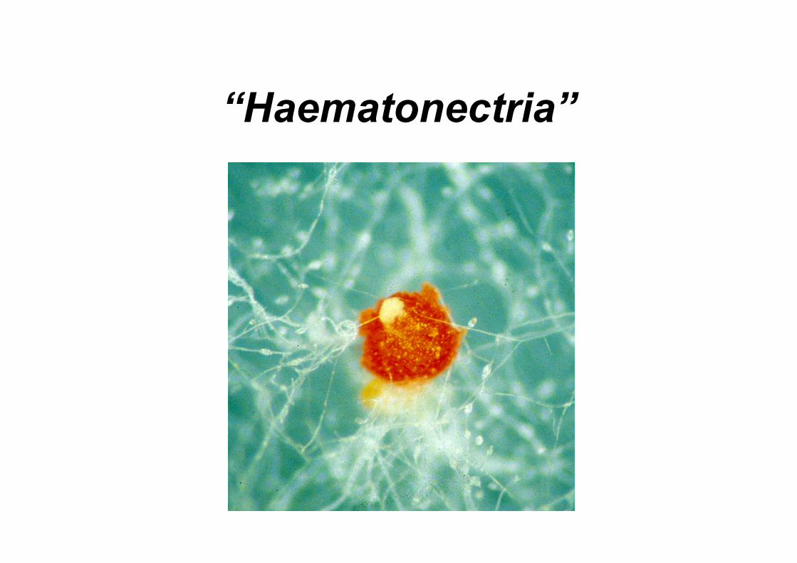
“Haematonectria”

Key characters for identifying Fusarium species
n Macroconidia n shape and size
n Microconidia n shape, size, false heads, chains
n Conidiogenous cells, especially microconidia n monophialides vs polyphialides
n Chlamydospores n Perithecia of the teleomorph

Macroconidia
n Size n large vs small; only look for gross differences
in size of the macroconidia n Shape
n elongate versus squat n degree of curvature

Macroconidia

Macroconidia Size and Shape
Fusarium longipes
Fusarium culmorum

Macroconidia – Basal Cells

Macroconidia – Apical Cells

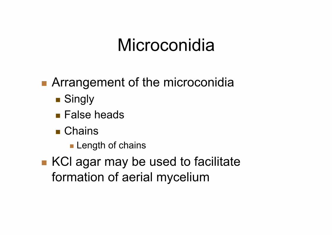
Microconidia
n Shape n number of cells n variation in shape
n Size n look for large variations, and don’t be
distracted by small ones n May need to use SNA to see all shapes and
sizes

Microconidia Shape and Size
Fusarium anthophilum Fusarium solani
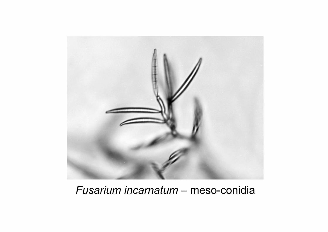
Fusarium incarnatum – meso-conidia
Microconidia
n Arrangement of the microconidia n Singly n False heads n Chains
n Length of chains
n KCl agar may be used to facilitate formation of aerial mycelium
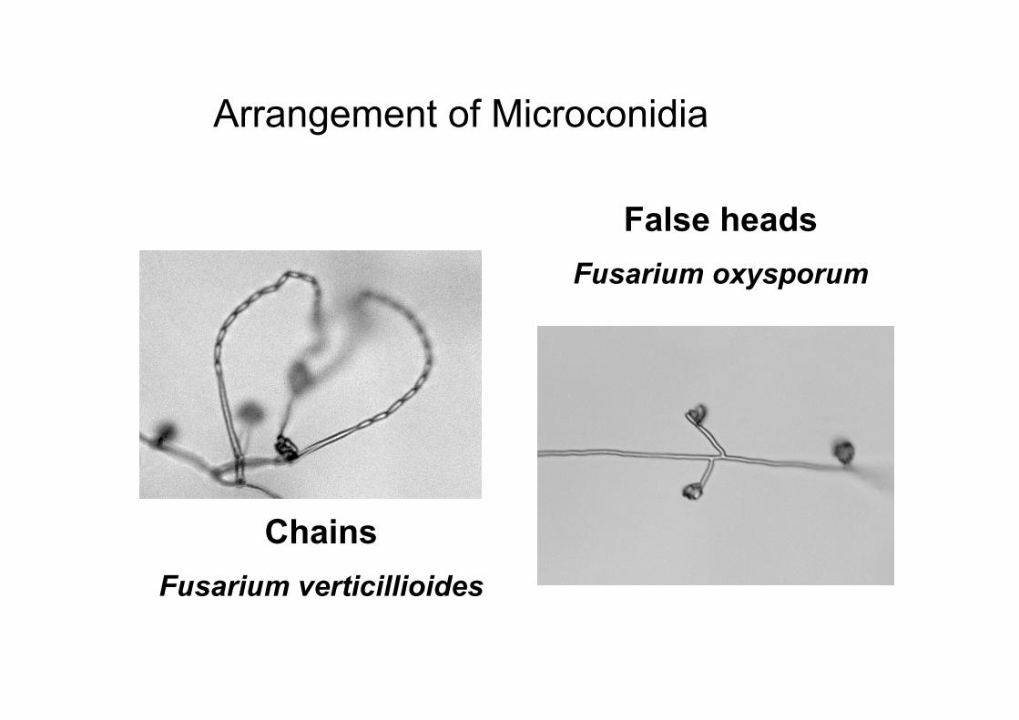
Arrangement of Microconidia
Chains Fusarium verticillioides
False heads Fusarium oxysporum

Microconidia
n Nature of the conidiogenous cell n Monophialides - having only one phialide per
cell n Polyphialides - having more than one phialide
per cell (but not necessarily for all conidiogenous cells)
n length of the condiogenous cell

Monophialide - one opening per cell
False head of microconidia
Fusarium oxysporum

Polyphialide - more than one opening per cell
Fusarium subglutinans

Polyphialide
Fusarium proliferatum
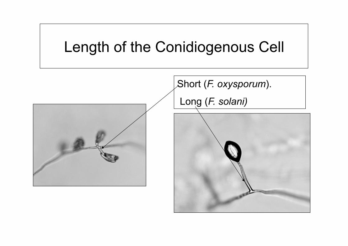
Length of the Conidiogenous Cell
Short (F. oxysporum).
Long (F. solani)

Chlamydospores
n Many Fusarium taxonomists considered chlamydospores to be an important character.
n They can take long periods of time to form (up to 6 weeks)
n Formed singly, doubles or in chains n Can be embedded in agar or in aerial mycelium n For some isolates you may need to use soil agar
or SNA
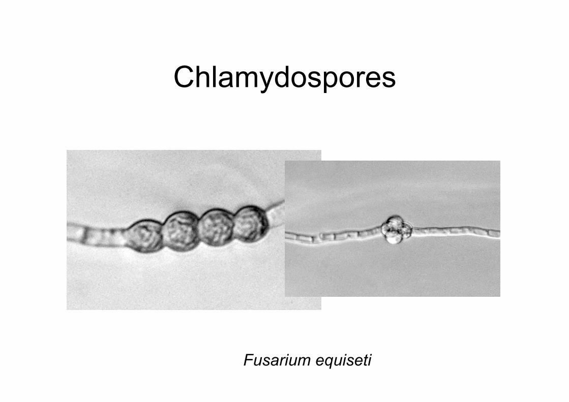
Chlamydospores
Fusarium equiseti
Other morphological features
n Swellings in hyphae n Pseudochlamydospores n Coils in hyphae n Sclerotial-like structures n Crystalline structures in agar

Coils in Hyphae
Fusarium circinatum

Hints for Identifying Fusarium
n Always identify using single-spored cultures
n Look for the most common features – don’t get confused by minor differences
n Your first impressions often are correct n If in doubt send in cultures to a recognized
expert – make sure they are clean and single spored!

When Do You Need to Go Further?
n Many species cannot be accurately identified by this stage
n Precision will be needed if:- n You need accurate information on mycotoxins n Accurate identification for quarantine n Other policy issues n For publication

Beyond Morphology
n There are two general techniques that are used to proceed further in the identification process n Sexual cross fertility n Analysis of DNA sequences Both require specific expertise – we will talk
about them a lot over the next 5 days!
History of the Taxonomy of Fusarium

Wollenweber and Reinking
n “Die Fusarien” - 1935 n Consolidated approximately 1000 taxa
names into: n 65 species; 55 varieties; 22 forms n arranged in 16 sections
n All current taxonomic systems based on their work


Snyder and Hansen
n Most important papers - AJB (1940) 27, 64-67, AJB (1942) 28, 738-742.
n Pioneered use of single spore technique n Reduced sections Elegans and Martiella to
two species, F. oxysporum and F. solani n Eventually reduced whole taxonomic
system to nine species
Snyder and Hansen
n Nine species concept was a gross oversimplification of the taxonomy
n Eventually lost favor because the system did not convey enough information
n As a consequence much of the information based on this system cannot be used unless the cultures are re-assessed
n This system, especially “F. roseum” must not be used

Colin Booth
n “The Genus Fusarium” (1971)
n Major achievements n expanded information on perfect states n introduced information on condiophores and
conidiogenous cells


W. L. Gordon
n Series of journal papers detailing taxonomic system, esp. CJB (1952) 30;209-251
n Looked at Fusarium species from variety of substrates and environments.
n Used aspects of W & R and S & H systems

Gerlach and Nirenberg
n The Genus Fusarium - A Pictorial Atlas (1982)
n Based in Wollenweber’s lab and used similar techniques and philosophies
n Tendency to emphasise differences rather than similarities
n Molecular-based techniques are indicating that many of these species are valid.

Nelson, Toussoun and Marasas
n Fusarium species: An Illustrated Manual for Identification (1983)
n A compromise system of all of the previous systems taking best component of each taxonomic system


Copyright © 2022 FDOKUMEN