Fungal Disease in Britain and the United States 1850–2000
240
-
Upload
khangminh22 -
Category
Documents
-
view
2 -
download
0
Transcript of Fungal Disease in Britain and the United States 1850–2000
Science, Technology and Medicine in Modern History
General Editor: John V. Pickstone, Centre for the History of Science, Technology and Medicine, Universityof Manchester, England (http://chstm.manchester.ac.uk)
One purpose of historical writing is to illuminate the present. At the start of the third millennium, science,technology and medicine are enormously important, yet their development is little studied.
The reasons for this failure are as obvious as they are regrettable. Education in many countries, not least inBritain, draws deep divisions between the sciences and the humanities. Men and women who have beentrained in science have too often been trained away from history, or from any sustained reflection on howsocieties work. Those educated in historical or social studies have usually learned so little of science thatthey remain thereafter suspicious, overawed or both.
Such a diagnosis is by no means novel, nor is it particularly original to suggest that good historical studiesof science may be peculiarly important for understanding our present. Indeed this series could be seen asextending research undertaken over the last half-century. But much of that work has treated science, tech-nology and medicine separately; this series aims to draw them together, partly because the three activitieshave become ever more intertwined. This breadth of focus and the stress on the relationships of knowledgeand practice are particularly appropriate in a series which will concentrate on modern history and on indus-trial societies. Furthermore, while much of the existing historical scholarship is on American topics, thisseries aims to be international, encouraging studies on European material. The intention is to present sci-ence, technology and medicine as aspects of modern culture, analysing their economic, social and politicalaspects, but not neglecting the expert content which tends to distance them from other aspects of history.The books will investigate the uses and consequences of technical knowledge, and how it was shaped withinparticular economic, social and political structures.
Such analyses should contribute to discussions of present dilemmas and to assessments of policy. ‘Sci-ence’ no longer appears to us as a triumphant agent of Enlightenment, breaking the shackles of tradition,enabling command over nature. But neither is it to be seen as merely oppressive and dangerous. Judgementrequires information and careful analysis, just as intelligent policy-making requires a community of dis-course between men and women trained in technical specialities and those who are not.
This series is intended to supply analysis and to stimulate debate. Opinions will vary between authors; weclaim only that the books are based on searching historical study of topics which are important, not leastbecause they cut across conventional academic boundaries. They should appeal not just to historians, norjust to scientists, engineers and doctors, but to all who share the view that science, technology and medicineare far too important to be left out of history.
Titles include:
Julie Anderson, Francis Neary and John V. PickstoneSURGEONS, MANUFACTURERS AND PATIENTSA Transatlantic History of Total Hip Replacement
Roberta E. BivinsACUPUNCTURE, EXPERTISE AND CROSS-CULTURAL MEDICINE
Linda BryderWOMEN’S BODIES AND MEDICAL SCIENCEAn Inquiry into Cervical Cancer
Roger CooterSURGERY AND SOCIETY IN PEACE AND WAROrthopaedics and the Organization of Modern Medicine, 1880–1948
Catherine Cox and Hilary MarlandMIGRATION, HEALTH AND ETHNICITY IN THE MODERN WORLD
Jean-Paul Gaudillière and Ilana Löwy (editors)THE INVISIBLE INDUSTRIALISTManufacture and the Construction of Scientific Knowledge
Jean-Paul Gaudillière and Volker Hess (editors)WAYS OF REGULATING DRUGS IN THE 19TH AND 20TH CENTURIES
Christoph Gradmann and Jonathan Simon (editors)EVALUATING AND STANDARDIZING THERAPEUTIC AGENTS, 1890–1950
Aya Homei and Michael WorboysFUNGAL DISEASE IN BRITAIN AND THE UNITED STATES 1850–2000Mycoses and Modernity
Sarah G. MarsTHE POLITICS OF ADDICTIONMedical Conflict and Drug Dependence in England since the 1960s
Alex Mold and Virginia BerridgeVOLUNTARY ACTION AND ILLEGAL DRUGSHealth and Society in Britain since the 1960s
Ayesha NathooHEARTS EXPOSEDTransplants and the Media in 1960s Britain
Cay-Rüdiger Prüll, Andreas-Holger Maehle and Robert Francis HalliwellA SHORT HISTORY OF THE DRUG RECEPTOR CONCEPT
Thomas SchlichSURGERY, SCIENCE AND INDUSTRYA Revolution in Fracture Care, 1950s–1990s
Eve Seguin (editor)INFECTIOUS PROCESSESKnowledge, Discourse and the Politics of Prions
Crosbie Smith and Jon Agar (editors)MAKING SPACE FOR SCIENCETerritorial Themes in the Shaping of Knowledge
Stephanie J. SnowOPERATIONS WITHOUT PAINThe Practice and Science of Anaesthesia in Victorian Britain
Carsten TimmermannA HISTORY OF LUNG CANCERThe Recalcitrant Disease
Carsten Timmermann and Julie Anderson (editors)DEVICES AND DESIGNSMedical Technologies in Historical Perspective
Carsten Timmermann and Elizabeth Toon (editors)CANCER PATIENTS, CANCER PATHWAYSHistorical and Sociological Perspectives
Jonathan TomsMENTAL HYGIENE AND PSYCHIATRY IN MODERN BRITAIN
Duncan WilsonTISSUE CULTURE IN SCIENCE AND SOCIETYThe Public Life of a Biological Technique in Twentieth Century Britain
Science, Technology and Medicine in Modern HistorySeries Standing Order ISBN 978–0–333–71492–8 hardcoverSeries Standing Order ISBN 978–0–333–80340–0 paperback(outside North America only)
You can receive future titles in this series as they are published by placing a standing order. Please contactyour bookseller or, in case of difficulty, write to us at the address below with your name and address, thetitle of the series and one of the ISBNs quoted above.
Customer Services Department, Macmillan Distribution Ltd, Houndmills, Basingstoke, Hampshire RG216XS, England
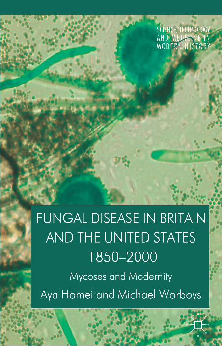
Fungal Disease in Britain andthe United States 1850–2000Mycoses and Modernity
Aya HomeiWellcome Trust Fellow, University of Manchester
and
Michael WorboysProfessor of the History of Science, Technology and Medicine, Centre for the History ofScience, Technology and Medicine, University of Manchester
Except where otherwise noted, this work is licensed under aCreative Commons Attribution 3.0 Unported License. To view
a copy of this license, visit http://creativecommons.org/licenses/by/3.0/

© Aya Homei and Michael Worboys 2013
The authors have asserted their rights to be identified as the authors of thiswork in accordance with the Copyright, Designs and Patents Act 1988.
Open access:Except where otherwise noted, this work is licensed undera Creative Commons Attribution 3.0 Unported License. To
view a copy of this license, visit http://creativecommons.org/licenses/by/3.0/
First published 2013 byPALGRAVE MACMILLAN
Palgrave Macmillan in the UK is an imprint of Macmillan Publishers Limited,registered in England, company number 785998, of Houndmills, Basingstoke,Hampshire RG21 6XS.
Palgrave Macmillan in the US is a division of St Martin’s Press LLC,175 Fifth Avenue, New York, NY 10010.
Palgrave Macmillan is the global academic imprint of the above companiesand has companies and representatives throughout the world.
Palgrave® and Macmillan® are registered trademarks in the United States,the United Kingdom, Europe and other countries.
DOI 10.1057/9781137377029E-PDF ISBN 9781137377029E-PUB ISBN 9781137377036Hardback ISBN 9781137377012Paperback ISBN 9781137392633
A catalogue record for this book is available from the British Library.
A catalog record for this book is available from the Library of Congress.

For Carsten and Carole

This page intentionally left blank
Contents
List of Figures and Tables viii
Acknowledgements x
List of Abbreviations xii
Introduction 1
1 Ringworm: A Disease of Schools and Mass Schooling 17
2 Athlete’s Foot: A Disease of Fitness and Hygiene 43
3 Candida: A Disease of Antibiotics 67
4 Endemic Mycoses and Allergies: Diseases of Social Change 98
5 Aspergillosis: A Disease of Modern Technology 118
Conclusion 137
Notes 142
Bibliography 183
Index 216
vii

Figures and Tables
Figures
1.1 and 1.2 Photographs of X-ray depilation treatment ofringworm of the scalp. British Medical Journal,1905, ii: 14 33
1.3 X-ray apparatus. Suitable for treatment ofringworm and other cutaneous affections 35
1.4 Radiotherapy room for ringworm. 1905 362.1 ‘ABSORBINE JR.’ Athlete’s foot advertisement,
Life, 3(7)16 August 1937, 81 504.1 Distribution of histoplasmosis, blastomycosis and
coccidioidomycosis in North America. Thehistoplasmosis areas are shown in black, theblastomycosis as circles and thecoccidioidomycosis areas as triangles. Smith,D. T., ‘The diagnosis and therapy of mycoticinfections’, Bull NY Acad Med, 1953, 29(10): 778 102
4.2 The geographic distribution ofcoccidioidomycosis. Cross-hatching indicates theheavily disease-endemic area, single hatching andthe moderately disease-endemic area. Kirkland,T. N. and Fierer, J., ‘Coccidioidomycosis: Areemerging infectious disease’, Emerg Infect Dis[serial on the Internet]. 1996, Sep 106
5.1 Incidence of fungal infections (includingaspergillosis) found at autopsy at the JohnsHopkins Hospital, 1941–1963. Asper, S. P. andHeffernan, A. G. A., ‘Insidious fungal disease’,Trans Am Clin Climatol Assoc, 1965, 76: 101 128
viii

List of Figures and Tables ix
Tables
1.1 Cases of ringworm in England and Wales treated by X-rayor other methods, 1933 41
3.1 Treatment of chronic candidiasis 915.1 Incidence of invasive aspergillosis according to
underlying condition 135
Acknowledgements
This book has its origins in the passion for the history of aspergillosisand other fungal diseases of Professor David Denning, Director, NationalAspergillosis Centre, University Hospital of South Manchester. David’senthusiasm infected staff at the University’s Centre for the History ofScience, Technology and Medicine (CHSTM) and led to a number of ini-tiatives. Dr Emm Johnstone, now at Royal Holloway College London,contributed to the history pages of the Aspergillus website (www.aspergillus.org.uk) and then, as interest in fungal infections (mycoses)grew, Aya Homei joined Michael Worboys on a Wellcome Trust-fundedproject grant that is the basis for this volume. We believe this to bethe first book-length study of this class of infectious microorganisms,and we hope it will lead to greater recognition of diseases that becameincreasingly important over the twentieth century, in terms of both thenumber of people affected and the severity of the illnesses caused.
We would like first and foremost to thank the Wellcome Trust forfunding this project (Grant number 074971) and for their overall sup-port of the history of medicine at the University of Manchester, whichhas made CHSTM such a congenial and supportive location for thiswork. Our research was facilitated by the assistance of librarians andarchivists at many sites and we thank them all. We would like to givespecial mention to staff at The University of Manchester Library, theWellcome Library, Wellcome Archives, the National Archives and theBritish Library. Jeff Karr at the Center for the History of Microbiol-ogy/ASM Archives (CHOMA) at the University of Maryland BaltimoreCounty provided access to the papers of the mycology sections of theAmerican Society for Microbiology. We thank the following for permis-sion to use images: the British Medical Journal, the Wellcome Library,New York Academy of Medicine (Arlene Shaner), and the AmericanClinical and Climatological Association (Rick Lange). We have includedpublic domain illustrations from the US Centers for Disease Control andPrevention. We are grateful to Professor Malcolm Richardson, Directorof the Regional Mycology Laboratory at the University Hospital of SouthManchester, for the cover image of an Aspergillus flavus.
x
Acknowledgements xi
Our colleagues at CHSTM have provided a sounding board for theideas developed in the book and we are very grateful for the commentson drafts that we have received from the following: Michael Bresalier,Vladimir Jankovic, Robert Kirk and Neil Pemberton, and especially IanBurney, Elizabeth Toon and Duncan Wilson. Christoph Gradmann readthe whole manuscript and made valuable suggestions about the place offungal infections in the wider history of infections. David Denning keptus on the ball mycologically. John Pickstone, the series editor of ‘Science,Technology and Medicine in Modern History’ read a first draft and sug-gested a new framing of the narrative that we adopted. We would alsolike to thank Francis Arumugam, who oversaw production, and JennyMcCall, Clare Mence and Holly Tyler at Palgrave Macmillan who havebeen a pleasure to work with and helped us on so many fronts in thepreparation and publication of this book.
Finally, we would like to thank our partners Carsten and Carole fortheir forbearance in the long incubation period of this book, and ourchildren for many welcome distractions.
Aya Homei and Michael Worboys
Abbreviations
AAAAI American Academy of Allergy, Asthma andImmunology
ABPA allergic bronchopulmonary aspergillosisAFB Air Force BaseASM American Society for MicrobiologyBDH British Drug HousesBMJ British Medical JournalBOCM British Oil and Cake MillsBPP British Parliamentary PapersBSMM British Society for Medical MycologyCCPA chronic cavitary pulmonary aspergillosisCCSG Veterans Administration-Armed Forces
Coccidioidomycosis Cooperative Study GroupCDC Centers for Disease Control and PreventionCFPA chronic fibrosing pulmonary aspergillosisCIE Committee on Industrial EpidermophytosisCOPD chronic obstructive pulmonary diseaseCPA chronic pulmonary aspergillosisC-PMC Columbia-Presbyterian Medical CenterFDA Food and Drugs AdministrationICI Imperial Chemical IndustriesIHRB Industrial Health Research BoardIPA invasive pulmonary aspergillosisISHAM International Society for Human and Animal
MycologyMAB Metropolitan Asylums BoardMDR-TB multi-drug-resistant tuberculosisME myalgic encephalomyelitisMRC Medical Research CouncilMRSA methicillin resistant Staphylococcus aureusMSG Mycoses Study GroupNAS National Academy of SciencesNAS Naval Air StationNGU non-gonococcal urethritis
xii

List of Abbreviations xiii
NIAID NIAID and National Institute of Allergy andInfectious Diseases
NIH National Institutes of HealthNYAS New York Academy of SciencePAS para-aminosalicylic acidPCP Pneumocystis carinii pneumoniaPVFS post-viral fatigue syndromeRCSA Research Corporation for Scientific AdvancementSAFS severe asthma associated with fungal sensitivityTV Trichomonas vaginitisUCD University of California DavisUCLA University of California Los Angeles

This page intentionally left blank
OPEN
Introduction
Fungal infections or mycoses are the great neglected diseases of medicalhistory.1 There are numerous histories of viral, bacterial and protozoaninfections, for all times and all places, but very few studies of thosecaused by fungi. Why? It cannot be because of prevalence. Histori-cal sources and contemporary epidemiological investigations show thatfungal infections were and are ubiquitous in human and animal popula-tions. Everyone in Britain and the United States in the last half a centurywould have heard of, if not suffered from, athlete’s foot or thrush. In thefirst half of the twentieth century, children feared the school nurse find-ing ringworm on their scalp and having to endure, not only the pains ofX-ray depilation or having their shaven head painted with gentian vio-let, but also exclusion from school and the shame of being stigmatisedas ‘unclean’.2
It seems that medical historians have followed the agenda of the med-ical profession in showing relatively little interest in conditions, suchas the majority of cases of mycoses, that do not lead to ‘illness’ assuch, but cause inflammation, irritation and discomfort. Medical historyremains dominated by studies of diseases that had, or continue to have,a high profile within medicine, or have attracted government interestand investment because they cause significant morbidity or mortality.Yet, the majority experience of ill health was, and is, of self-limiting andself-treated conditions, where sufferers did not, and do not, consult adoctor and become ‘patients’. In their efforts to recover ‘the patient’sview’, medical historians have ignored the minor illnesses, injuries andinfections that were, and remain, outside of the medical gaze.3
But medical historians have also largely ignored the ailments broughton by medical advances, and here too the history of fungal infections
1

2 Fungal Disease in Britain and the United States 1850–2000
can be instructive. The grand narrative of Western medicine in thetwentieth century was one of ‘progress’, evidenced by greater, scientifi-cally based knowledge of the aetiology and pathology of disease, moreaccurate diagnostics, improved management of symptoms and pain,more effective treatments, innovations in surgery, improved health care,falling mortality rates and greater longevity.4 Those telling this storyrecognised that progress was not unalloyed, yet amongst doctors suchwas the step change in their effectiveness and efficiency that problems,like the development of antibiotic resistance, were discounted or seen assomething that would be solved by further scientific and technologicaladvances.5 However, medical professionals soon realised that therapeu-tic and technological advances often led to intractable problems; forexample, the practice of managing the adverse effects of one drug withanother could lead to patients taking more medicines to manage sideeffects than for their primary illness. Such practices were criticised inthe 1960s, but for our narrative of fungal infections Ivan Illich’s bookMedical Nemesis, first published in 1975, is most relevant.6 Illich madeiatrogenesis – doctor induced disease – central to his critique of mod-ern medicine, claiming that around 10% of all clinical encounters werefor such conditions. He argued that the cures of modern medicine wereoften worse than the disease – if indeed there was a disease in the firstplace, as Illich also attacked the medicalisation of everyday life, antic-ipating the burgeoning of risk-defined conditions that emerged in thelast quarter of the twentieth century.7
Thrush, the most prevalent opportunistic mycosis of the twentiethcentury, exemplifies these trends. In the 1940s and 1950s, the emer-gence of resistant bacteria was only one side effect of the new drugs.More important then was the development of so-called ‘superinfec-tions’, also caused by antibiotics as they removed not just disease-causing bacteria but many others, and altered the normal microbial floraof the body. These changes opened the body to opportunistic infectionby other bacteria, such as Staphylococcus aureus, and by fungi, especiallyCandida. This fungus had previously only affected the ‘external’ mucusmembranes in the mouth and genitalia, but emerged in the 1950s as arare, but serious, internal and systemic infection, where fungi grew onmajor organs, such as the heart. It was not just patients on antibioticswho were vulnerable. There were a growing number of patients whoseimmune systems were weakened or immunocompromised. Initially, thissituation developed as a side effect of steroids and other similar treat-ments, but then such states were deliberately produced by doctors toaid the acceptance of transplanted organs, or as a by-product of new
Introduction 3
cancer therapies. In 1987, John W. Rippon, a leading American medicalmycologist, reflected on the situation.
The mycology of human infections in the 1980s is the mycology ofthe soil, rotting vegetables, shower curtains, toilet bowls, leaf piles,wilted flowers and dung heaps. Organisms literally come out of thewalls to infect immunosuppressed patients. Technical medical andsurgical expertise is such that we can pass around hearts, lungs, andlivers only to be thwarted by a Fusarium from a rotting plum.8
Rippon was pointing to a larger truth about human fungal infections,namely, that their prevalence has been linked to specific ecological con-ditions and interactions, not only within the body, but also within thewider social and physical environment. At the time Rippon wrote, theUnited States, and soon the Western world, was gripped by a popularhealth panic about fungal disease. Some fringe doctors promoted theview that Candida infection was responsible for all manner of ‘modern’ailments, including chronic fatigue syndrome (CFS) and inflammatorybowel disease (IBD), in what they styled as ‘the yeast connection’.9
In this book, we discuss the changing medical and public profileof fungal infections in the period 1850–2000. We consider four setsof diseases: ringworm and athlete’s foot (dermatophytosis); thrush orcandidiasis (infection with Candida albicans); endemic, geographicallyspecific infections in North America (coccidioidomycosis, blastomycosisand histoplasmosis) and mycotoxins; and aspergillosis (infection withAspergillus fumigatus). We discuss each disease in relation to developingmedical knowledge and practices, and to social changes associated with‘modernity’. Thus, mass schooling provided ideal conditions for thespread of ringworm of the scalp in children, and the rise of college sportsand improvement of personal hygiene led to the spread of athlete’s foot.Antibiotics seemed to open the body to more serious Candida infections,as did new methods to treat cancers and the development of transplan-tation. Regional fungal infections in North America came to the foredue to the economic development of certain regions, where popula-tion movement brought in non-immune groups who were vulnerable toendemic mycoses. Fungal toxins or mycotoxins were discovered as by-products of modern food storage and distribution technologies. Lastly,the rapid development and deployment of new medical technologies,such as intensive care and immunosuppression in the last quarter ofthe twentieth century, increased the incidence of aspergillosis and othersystemic mycoses.
4 Fungal Disease in Britain and the United States 1850–2000
In understanding and managing infectious diseases, scientists anddoctors have long argued for thinking about them in terms of themetaphor of ‘seed and soil’, where the ‘seed’ is the infectious organismor pathogen: that is, virus, bacteria, fungi, protozoa (single cell) or meta-zoan (multicellular); and the ‘soil’ is the human body and its environs.10
Thus, for someone with the common cold, the notion of ‘seed and soil’ensures that we go beyond focusing only on infection by the virus (theseed) and consider the sufferer (the soil). This means looking at the con-ditions in which the person was exposed to the virus, the quantity andquality of the virus reaching the body, the nature of the body’s specificimmune response and the overall health of the individual. We all knowthat we do not ‘catch a cold’ every time we are exposed to the virusand that some people suffer longer and more serious illness than oth-ers do. Some variations are individual, but epidemiological studies havealways shown patterns of exposure, susceptibility, sickness and recoveryby age, gender, class, occupation, ethnicity and other socio-cultural vari-ables. For example, in their history of pulmonary tuberculosis, René andJean Dubos systematically use the notion of ‘seed and soil’ to discuss thedisease at all levels, from biological factors influencing the susceptibilityof cells and tissues, through to the socio-economic and technologicalvariables that have shaped global trends in morbidity and mortality.11
In this book, we frame our history of fungal infections in terms of‘seed and soil’; hence, our ‘seeds’ are specific fungal pathogens and weinterpret ‘soils’ widely to include the human body, social relations andstructures, and the medical, material and technological environment.
Fungi
Fungi and how they cause diseases are not well known, so it will beuseful here to give a brief introduction to the nature of the ‘seeds’ ofmycoses. Our account is part historical and part current.
Mycology is the branch of science that studies fungi and until the1960s, it was a part of botany, at which time its subject matter wasmoved to the animal kingdom. Since then, fungi have been placed intheir own kingdom, with the other four being plants, animals, proto-zoa and monera (bacteria).12 Current estimates are that there are wellover 100,000 species of fungi and many more are still to be classified,let alone discovered. Some fungi are large and multicellular, like toad-stools. However, most species are microscopic, single cell organisms andare best known as industrial agents (yeast fungi in the production ofbread and beer) and as medical agents (Penicillia spp. remain the source
Introduction 5
of the world’s mostly widely used antibiotic). The larger fungi develop asmicroscopic filaments called hyphae, which branch and grow into net-works or colonies called mycelia, whereas smaller fungi, such as yeasts,are single cell microorganisms.
Many writers divide fungi into ‘good’ and ‘bad’, judged by theirimpact on human existence; fungi themselves, of course, are just fill-ing niches that allow them to multiply and survive. In popular writing,the ‘good’ fungi are those used in industrial processes or medicine, suchas yeasts and penicillins mentioned above, plus those that can be eaten,break down waste or work in plant roots to fix nitrogen. The ‘bad’ fungiare those that produce diseases in plants, animals and humans. In termsof impact on humanity, fungi do most harm as causes of crop diseasesand amongst farm animals, but they are also a threat to homes, wheretheir ability to breakdown organic matter is seen most strikingly in thedry rot fungus which can destroy wooden structures very rapidly. Mostfungi are saprophytic, that is, they obtain their nutrients from breakingdown organic matter, normally dead tissues, and absorbing the productsto ‘feed’ their metabolism. They mostly live on or within the material onwhich they are feeding. A small number of fungi, and of course the onesthat concern medical mycologists, derive their nutrients from infectingliving tissue, either by destroying it, or through establishing a symbioticrelationship that affects human tissues and their functioning.
Following long-established Linnaean principles, the classification offungi was mainly by their reproductive and sexual characteristics. Thus,the 1911 Encyclopaedia Britannica divided fungi into three groups: theBasidiomycota, which produce club-like fruit bodies that spread spores(e.g. mushrooms); the Ascomycota, which produce fruit bodies on specialpods or sac structures (e.g. baker’s yeast, penicillin and most human fun-gal pathogens); and the Phycomycetes that reproduce sexually by sporesjoining (e.g. black bread mould). These classifications held for most ofthe twentieth century, though with many refinements and revisionswith individual groups, genera and species. Certain fungi proved verydifficult to classify as they had different forms in different stages of theirlife cycle. In the final decades of the century, the whole basis of orderingfungi changed as the new types of analysis of their DNA (their genomeor genotype) revealed different relationships from those of their formand function (phenotype). The fluidity of understanding of the natureand classifications of fungi was evident with the microorganism knowncurrently as Pneumocystis jiroveci. Through the 1980s, this organism wasregarded as a protozoan and named Pneumocystis carinii, when it wasthe subject of extensive research as it was a major cause of pneumonia
6 Fungal Disease in Britain and the United States 1850–2000
and death in HIV/AIDS sufferers.13 Indeed, Pneumocystis carinii pneumo-nia (PCP) was an early marker of the epidemic and allegedly responsiblefor the deaths of celebrities such as Freddie Mercury. The redesignationof the organism as a fungus was first made in 1988, based on workusing the new techniques of DNA sequencing, though this remainedcontroversial until the late 1990s when the reclassification was finallyaccepted.14
Fungal diseases
Geoffrey Ainsworth, who has written most extensively on the historyof fungal diseases, argues that fungi are amongst the oldest recog-nised causes of infection in humans.15 Hippocrates seemingly wroteon ‘aphthae’ (sores in the mouth) in 500 BC, which modern mycolo-gists have identified as thrush. Two millennia later, ringworm infectionwas present on the skin and in the hair of the subjects of Old Masters’paintings. In the modern medical era, the first systematic writings onfungi as a source of human disease were by the Hungarian born, Paris-based physician and microscopist David Gruby in 1842–1844. At thetime, fungi were understood to be the sources of a number of dis-eases and attracted considerable scientific interest. In the 1830s, theItalian entomologist Agostino Bassi published claims that the devastat-ing muscardine disease of silkworms was due to a microscopic fungusTritirachium shiotae, which was eventually renamed in his honour asBeauveria bassiana.16 Bassi was a major influence on Louis Pasteur, bothin his work on the silkworm diseases of pébrine and flacherie in the1860s and on the idea that living microorganisms might cause infectiousdiseases. The work of Bassi and Pasteur showed that fungal infectionswere, and in fact still are, the cause of economic problems in agri-culture and related industries.17 Ainsworth goes on to make the pointthat most ‘mycologists’ in Britain and the United States work as plantpathologists, with a disciplinary allegiance to botany, and that medi-cal mycologists were and remain quite a small minority, with a quitedifferent orientation.
In medicine in the 1830s, and in keeping with the then fashion-able focus on pathological anatomy and lesions, distinctive and specificfungal infections of the skin, such as favus and ringworm, were wellrecognised. Classifications or nosologies of skin diseases were producedin the early nineteenth century, most influentially in Thomas Bateman’sA Practical Synopsis of Cutaneous Diseases According to the Arrangementof Dr Willan (1813) and an atlas The Delineations of Cutaneous Disease
Introduction 7
in 1817.18 Many authors followed the French physician Jean LouisAlibert in using extensive colour illustrations and some copied the waxmodels (les moulages) that he collected at the Hôpital Saint-Louis inParis.19 The use of colour illustrations continued with photography,as in Charles-Philippe Lallier’s Leçons cliniques sur les teignes, publishedin 1878.20
The contagious and infectious aspects of fungal disease meantthat, from the 1860s, doctors and scientists regarded them as ‘germdiseases’.21 Early historians of germ theories of disease certainly tracedthe familiar lineage from van Leeuwenhoek through Bassi to Pasteur,and the natural philosophers and medical men who used microscopyand culturing to study fungi. David Gruby first linked specific fungi tofavus, sycosis and ringworm infections of the human scalp in the 1840s.For the latter, he first described the clinical condition of tinea tonsurans(scalp ringworm), though the terms ‘herpes tonsurans’ and ‘teignetondante’ also enjoyed currency.22 In the 1850s, botanists and dermatol-ogists agreed on Trichophyton – literally hair-fungus due to its shape seenthrough microscopes – as the main ringworm germ and, in line withthe wider switch to naming diseases by their causes rather than theirsigns and symptoms, in France tinea tonsurans became ‘trichophytie’.As we discuss in Chapter 1, these developments were followed by lead-ing dermatologists, such as William Tilbury Fox and Thomas M’CallAnderson, but most doctors and dermatologists remained focused onmorbid anatomy and nosologies based on signs and lesions.
Fungus theories of infectious disease were popular in the 1840s andthe best known was the ‘cholera fungus’.23 In a paper read to theMicroscopical Committee of the Bristol Literary and Philosophical Insti-tution in 1849, ‘fungoid’ bodies were reported in the faeces of cholerasufferers.24 The authors emphasised analogies between the growth anddecay of fungi, and the rise and fall of zymotic diseases in individualsand in populations over epidemic periods. However, given that con-temporaries thought that fungi were the ‘appointed executioners andnimble scavengers of nature’, any such organisms were understood bycontemporary doctors to be the consequences rather than the causes ofcholera. Medical views on the causal role of living organisms in diseasewaxed and waned from the 1840s to the 1880s, until bacterial germswere accepted as major pathogens.25 At this time, bacteria were termedas the ‘Schizomycetes’, literally the splitting fungi, so named becausethey reproduced by the division of cells, and were believed to be a typeof fungi because of their microscopic form and physiological functionas saprophytes.
8 Fungal Disease in Britain and the United States 1850–2000
One of the first British textbooks on the new science of germs wasGerman Sims Woodhead and Arthur Hare’s Pathological Mycology pub-lished in 1885.26 However, this was the only time ‘mycology’ was usedin this context; the German term Bacteriologie soon took over. In thenew manuals and textbooks on ‘bacteriology’ and ‘microbiology’, fungias causes of infection were, at best, described briefly and typically ina final chapter or appendix. For example, Muir’s and Ritchie’s influen-tial Manual of Bacteriology, published in 1899, had a chapter entitled‘Non-Pathogenic Micro-organisms – Fungi’, and presented them as likelylaboratory contaminants rather than pathogens. The authors discussedMucor spp., Oidium spp., Aspergillus niger, Penicillium glaucum, plus yeasts,and ended with the comment, ‘Certain fungi closely related to the aboveare pathogenic agents.’ Readers were referred to Anton De Bary’s Com-parative Morphology and Biology of the Fungi, Mycetozoa and Bacteria, firstpublished in 1886, for further details.27
In the twentieth century, fungi were recognised as causing three typesof disease in humans and animals. First, there were infections wherefungi develop parasitically in the tissues of the host, at (literally) threelevels: superficial mycoses, like athlete’s foot, where infection is lim-ited to the outermost layers of the skin, nails and hair; subcutaneousmycoses, like the tropical disease of Madura foot (mycetoma), wherethe growth extends to the underlying layers of the skin and perhaps intobone; and systemic mycoses, like aspergillosis, where infection spreadsthrough internal organs and tissues.
Second, there were fungal poisons, either toxins in the fungi them-selves, as with poisonous toadstools, or toxins produced by the growthof fungi on foodstuffs, as with aflatoxins (produced by Aspergillus flavus).Third, there were allergic reactions to fungal spores and moulds, whichrange from mild to acute, depending on the dose and susceptibility ofthe host; thus, fungi are a common cause of asthma. There was a fourthtype of disease that was ‘discovered’ in the 1980s and remains highlycontested – ‘fungal overgrowth’. As we show in Chapter 3, this condi-tion has been widely dismissed by the medical profession as a fiction,yet it had wide currency with the public and was linked to CFS andother ‘diseases of modernity’. In the cultural climate in North Americaand Europe, where lifestyle was increasingly regarded as a cause, as wellas a solution, to ill health, books such as William G. Crook’s The YeastConnection (1983), which attributed various chronic conditions to theovergrowth of C. albicans, became a best seller and spawned many imi-tators. Crook also had the cure: dietary and lifestyle changes, plus a
Introduction 9
course of antifungal antibiotics, which was surprising given his pedigreein ‘alternative medicine’.
The history of medical mycology
The multi-faceted career of medical mycology’s leading historian Geof-frey Ainsworth exemplifies the diverse and changing character of thefield in the twentieth century. He studied pharmacy at University Col-lege, Nottingham, and then pursued a dual career in plant pathologyand medical mycology.28 He first worked on the virus diseases of plantsat Britain’s two leading botanical institutions, the Rothamsted Experi-mental Station and the Experimental and Research Station in Cheshunt.He spent the Second World War at the Imperial Mycological Insti-tute at Kew, developing abstracting services on all aspects of mycology.After the war, he moved to the pharmaceutical industry, as head ofthe mycological department of the Wellcome Research Laboratories atBeckenham, Kent. There he led work on the antibiotics produced byfungi, such as streptomycin and penicillin. He then moved, first, to theLondon School of Hygiene and Tropical Medicine and later to the Uni-versity of the South West (later the University of Exeter), before return-ing to the now Commonwealth Mycological Institute, where he stayeduntil his retirement in 1968. Ainsworth published widely on all aspectsof fungi. His major works were Dictionary of the Fungi (1943), BritishSmut Fungi (1950) with Kathleen Sampson, Medical Mycology (1952),and the multi-volume The Fungi: An Advanced Treatise (1965–1973) withA. S. Sussman and F. K. Sparrow.
Towards the end of his career, Ainsworth developed an interest inthe history of mycology and published three books that have beenimmensely valuable in the research and writing of this book: Introduc-tion to the History of Mycology (1976), Introduction to the History of PlantPathology (1981) and Introduction to the History of Medical and VeterinaryMycology (1987).29 In his preface to the latter volume, he sets out hisapproach and the scope of the topic.
Although possessing deep, if slender roots that can be traced backto ancient times, medical and veterinary mycology is essentially adevelopment of the twentieth century, especially the last fifty yearsduring which time several mycoses at first considered to be raritieshave been shown to affect millions of men, women, and children andtheir domesticated animals . . . . Here the attempt made to sketch in

10 Fungal Disease in Britain and the United States 1850–2000
the historical background, by illustrating the approaches to a series ofbasic problems, is limited to what might be described as the ‘naturalhistory’ of human and animal mycoses.30
While we agree with Ainsworth on the point that the development ofmedical mycology was a phenomenon of the twentieth century, ourwork differs in two ways. First, we do not take the specialism of medicalmycology as given, or historically constant, rather as a social institutionthat had to be created and sustained. Second, we do not set out a lineageof ideas, but rather discuss changing knowledges in specific institutionaland social settings, and also explore practices and meanings.31
The history of medical mycology in the United States in the twen-tieth century has been described in great detail in a monograph byAna Victoria Espinel-Ingroff published in 2006.32 Her narrative is com-prehensive and wonderfully rich in characters and institutional detail.It focuses on training and mapping the professional networks that haveshaped medical mycology across the country. At the same time, theauthor tells the story of discoveries in the understanding and manage-ment of the main fungal infections that affect Americans. It is historyinformed by disciplinary politics, as Espinel-Ingroff’s reference point iswhat she sees as a crisis in medical mycology in the United States. On theone hand, the importance of mycoses has grown with their increasedprevalence and the arrival of effective antifungal drugs. Yet, on the otherhand, the field seems to be fragmenting, being drawn at one end tomolecular approaches and basic biology, and at the other to appliedclinical research, leading to the neglect of the old, middle ground oftaxonomy, aetiology, physiology and pathogenesis.
Woven into Espinel-Ingroff’s history narrative is a narrative of devel-opments in the field in the twentieth century, with five periods definingher chapters. The discussion of the ‘Era of Discovery (1894–1919)’explores how work on fungi followed that in bacteriology in seeking thecausal organisms of specific infections and the understanding of basicfungal biology. The ‘Formative Years (1920–1949)’ are characterised bythe establishment of training programmes, laboratory services and epi-demiological studies of common diseases, such as athlete’s foot andthrush, or the then very rare systemic mycoses. The period 1950–1969,the ‘Advent of Antifungal and Immunosuppressive Therapies’, was dom-inated by drug discoveries (nystatin, amphotericin B, griseofulvin) andthe increased incidence of severe opportunistic systemic fungal infec-tions that were linked to antibiotics and immunosuppressive therapies.The ‘Years of Expansion (1970–1979)’ are portrayed as the apogee of
Introduction 11
medical mycology, seen in the establishment of services to deal withthe increased incidence of infections, basic research to underpin clinicalinnovations and the recognition of the specialty by the American Soci-ety for Microbiology (ASM). Finally, the ‘Era of Transition (1980–1996)’saw continued increase in the incidence of opportunistic infections incancer and transplant patients, and amongst AIDS patients, but also thefragmentation and relative neglect of the specialism.
What few histories there are of fungal infections are largely embeddedin accounts of the development of the specialty of medical mycology,but there are a number of books and journal articles on specific infec-tions. There is only one monograph on a disease discussed in this book,Thomas Daniel and Gerald L. Baum’s Drama and Discovery: The Story ofHistoplasmosis.33 Their narrative follows the emergence of the diseasefrom social changes in its endemic areas and the research networks inwhich new understandings of its epidemiology, aetiology, pathologyand treatment developed. It is typical of much work on the historyof mycoses, as with Ainsworth and Espinel-Ingroff, in being writtenby medical mycologists, but is quite different and richer as it exploresthe social as well as medical history of histoplasmosis.34 There are nobook length histories of coccidioidomycosis and blastomycosis com-parable to Drama and Discovery, but there are very useful practitionerhistories, for example, Jan Hirschmann’s account of the early history ofcoccidioidomycosis in America.35
Yet, as we have indicated, ‘biographies’ of mycoses written by med-ical historians are rare. Aspergillosis has no thoroughgoing histories.36
Ringworm has few historians in Britain and the United States, and evenreflections by practitioners are rare.37 It has only excited attention inIsrael, in relation to the controversy of the long-term effects on chil-dren of X-ray treatment of the scalp and popular representations of thepractice as the ‘Ringworm Holocaust’.38 It is also surprising that histori-ans of medicine in the United States, who have thoroughly investigatedpopular medications and health activism, have missed athlete’s foot, acondition that plagued not only the athletes but the country’s youth,soldiers and miners.
Mycoses and medical history
In this book, we aim to do more than provide a narrative of a groupof neglected infections. Our study also gives new perspectives on thehistory of twentieth-century medicine on a number of fronts: speciali-sation; minor illnesses and self-treatment; and ‘orphan diseases’. Firstly,
12 Fungal Disease in Britain and the United States 1850–2000
we present an account of an area of medicine – medical mycology –that for most of the twentieth century was small and marginal, andwhere practitioners struggled to establish an area of specialist work. Thedevelopment of specialisms and specialisation has long interested his-torians of medicine.39 George Rosen’s study of ophthalmology was pathbreaking and work since then has linked the division of labour to manyfactors within medicine and outside. George Weisz, in the most recentand comprehensive study on the topic, finds that ‘divide and conquer’best explains the overall process in medicine, as these terms ‘[express] afundamental intellectual strategy’, whereby medical professionals were,in a matter of a century, divided into ‘smaller and more manageablegroups based on common attributes’ and conquered by ‘organizationbased on a novel kind of expertise’.40
Most histories of specialisation and specialisms are of successful enter-prises and can be teleological, charting the seemingly inevitable journeyto the present division of labour in medicine. Our narrative of medicalmycology runs against this grain, though it does not present medicalmycology as a failed specialism, rather one, as Espinel-Ingroff’s workmakes clear, the position and status of which was always problematic.For most of the twentieth century, it was small, institutionally frag-mented and dispersed geographically. Its practitioners tried to ‘divide’themselves off from other specialisms but were relatively unsuccess-ful because their services were never in sufficient demand to forma critical mass either numerically or politically. Thus, we challengethe accepted, though often implicit, view that specialisation was aninevitable path in twentieth-century medicine, where it becomes evermore populated with full-time ‘mono-specialists’; that is, cliniciansand scientists who worked on a single disease or group of diseases, aparticular organ or organ system, specific technologies or a restrictedpatient group, say, by age or sex. Our research on the doctors andresearchers who treated and studied fungal infections shows a differ-ent, and perhaps equally common, pattern of work: clinicians andscientists making a living as working in and combining a number ofspecialisms.41
We suggest that it is useful to think about twentieth-century medicinegenerally in terms of the doctors, and other health workers for thatmatter, developing careers in a number of ‘specialist practices’. His-torians of medicine often overlook the fact that doctors and medicalscientists had to ‘make a living’, and that in less wealthy times, whenhealth was a lower priority in private and state budgets, this was doneby earning where they could and what they could.42 In this con-text, ‘medical mycology’ was an area of ‘specialist practice’ for certain
Introduction 13
botanists, dermatologists, bacteriologists, hospital physicians and sur-geons, infectious disease doctors, microbiologists, general practitionersor, of course, combinations of these. Typically, ‘specialist practice’ wasin cognate areas; hence, the first ‘medical mycologists’ were mostlybotanists, or those who created the specialism of dermatology. Never-theless, in the late nineteenth century few doctors were able to workfull-time on skin diseases, so dermatologists were often general practi-tioners, who functioned as part-time specialists, part-time in hospitaloutpatient clinics.
Secondly, and as noted already, fungal infections represent the over-whelming experience of illness, then and now, like the commoncolds, sickness and diarrhoea, and sore throats that are self-limiting,self-treated or treated after one short consultation with a generalpractitioner.43 Research in the 1980s revealed that on average only onein 20 ‘symptom episodes’ led to a medical consultation, a pattern thatwas termed the ‘iceberg of illness’.44 If that was the position in a countrywith a National Health Service, offering care that was ‘free at the pointof delivery’, the proportion would almost certainly be lower in pay-for-service medical and healthcare systems, then and now. There are fewstudies, except for the era of ‘bedside medicine’, of the everyday expe-rience of illness, and of decisions on when and how to self-treat, andwhen and how to seek medical consultation and become a patient.45
That said, our focus is on the medical history of mycoses – a suf-ferer’s history would be quite different and, in fact, very difficult toresearch. However, we do try to capture sufferers’ agency, for example, inour discussion of the proliferation of proprietary remedies for athlete’sfoot and thrush.
Thirdly, and at the other end of the scale of prevalence, systemic fun-gal infections have been classified as ‘orphan diseases’; that is, those toorare to attract the attention of research agencies or the interest of manyclinicians and researchers.46 The term originated in the United Statesand the Orphan Drug Act, 1983, promoted by the National Organizationfor Rare Disorders and the Federal Drugs Agency (FDA). In the UnitedStates ‘orphan diseases’ are those with a prevalence of less than 2,000cases per year. By the end of the twentieth century, the rise in the inci-dence of mycoses meant that this designation only applied to the geo-graphically localised infections and the rarer types of hospital acquiredor nosocomial infections. Yet, for most of the twentieth century, oppor-tunistic, invasive mycoses were rare and medical mycologists and otherinterested parties bemoaned their neglect. In part, this was because suchinfections were seen as ‘diseases of the diseased’ and affected patientswho were seriously ill and close to death. In fact, doctors spoke of these
14 Fungal Disease in Britain and the United States 1850–2000
patients receiving ‘salvage therapies’, where ethical standards were dif-ferent and there was scope of experiment and the non-standard use ofstandard drugs. Interestingly, when invasive mycoses ceased to be ‘rare’,they attracted the attention of many surgical and medical specialists,and researchers in pharmaceutical companies, who sought to transfertheir successes with mass market, external antifungals to invasive, sys-temic disease. Indeed, the story of medical mycology in the second halfof the twentieth century is dominated by the development of new anti-fungal antibiotics, principally polyenes (e.g. nystatin and amphotericinB), azoles (e.g. clotrimazole and ketoconazole), triazoles (e.g. fluconazoleand itraconazole) and echinocandins (e.g. caspofungin), targeted at the‘seeds’ of infection.
The book
We discuss our four sets of infection in five chapters: two on ringworm(dermatophytosis), and one each on thrush, the geographically spe-cific mycoses and mycotoxins, and aspergillosis. We present historiesof each disease group and while our approach is essentially thematic,there is an overall movement through time. Thus, the first chapteron ringworm begins in the mid-nineteenth century and ends around1910, while the final chapter on aspergillosis is mainly about changes inthe last quarter of the twentieth century. Our narrative moves betweenBritain and the United States following the changing locations wheremedical and social interest and activity was greatest. We are neithercomprehensive nor comparative in our discussion of medical mycol-ogy in these two national contexts. However, we use the fact that workon fungal infections in the twentieth century, as demonstrated by thework of the International Society for Human and Animal Mycology(ISHAM), was dominated by an Anglo-American axis, though this is notto diminish in any way activities in other countries, which we discuss asappropriate.
Our first chapter frames ringworm as a disease of schools andschoolchildren. The disease had been reported previously in orphan-ages and similar institutions, but its incidence and profile increased withthe arrival of mass schooling, which provided ideal conditions for itsspread, both through increased opportunities for contagion (seeding)and the exposure of poor children (weakened soil). We look at responsesto the problem, one of which was special schools for the isolation andtreatment of sufferers, and which became sites for the use of the newX-ray technologies, not to kill the seeds of infection, but to alter the
Introduction 15
soil by removing hair, the locus of infection. In the second chapter, wemove from head to toe, from Britain to the United States, and focus onathlete’s foot. Concern over ringworm infection of the feet, along withinfection of the crotch, armpit and similar areas of the body, began inthe 1920s, principally amongst sportsmen and women. Athlete’s footwas described as a perverse consequence of the nation’s attempt toimprove the health and fitness of its youth, especially with the bur-geoning of college sports and improved hygiene facilities. The infectionwas met with the tools of modern public health propaganda, being pre-sented in some instances as equivalent to a sexually transmitted disease,and by new methods of treatment produced by the pharmaceuticalindustry, first in a rash of proprietary medicines and then antifungalantibiotics.
Thrush, the subject of our third chapter, was regarded at the start ofthe twentieth century as a disease of weak children, but moved in themedical and public view to a genital infection, principally of womenand was linked mainly to alterations in the body due to pregnancyand lifestyle changes.47 We then discuss how, in the second half of thetwentieth century, thrush was linked in different ways to the develop-ment of antibiotics. It was soon recognised as a side effect of penicillintherapy, while the search for new and better bacterial antibiotics ledto the discovery of nystatin – the first modern antifungal antibiotic,which soon became a specific treatment for thrush. Systemic C. albi-cans infection, known as invasive candidiasis, became, paradoxically,more prevalent in patients taking bacterial antibiotics, but also in thosewith cancers, transplants and inflammatory conditions. This problemwas met by a search for new antifungal drugs, with successes improvingthe institutional position of medical mycology. We end the chapter witha discussion of ‘The Yeast Connection’ phenomenon.
In Chapter 4, we discuss the regionally specific fungal infections inthe United States that came to the fore as a consequence of the eco-nomic development of certain regions in the South and Midwest, wherepopulation movement brought in non-immune groups who were vul-nerable to endemic mycoses. The forms of economic development werealso important, as new methods of production and types of industrialand domestic construction created new environmental conditions, andin some cases literally transformed and transported fungi-laden soildust. In the same vein, we show how new technologies of food pro-duction, transportation and storage produced a new class of hazardouscompounds – mycotoxins. In our final chapter, we discuss aspergillo-sis, the most serious of the invasive mycoses that have emerged from
16 Fungal Disease in Britain and the United States 1850–2000
new medical technologies, such as intensive care and immunosuppres-sion. An important theme here is iatrogenesis, as attempts to controlaspergillosis exemplified the now routine issue in modern medicine ofbalancing the benefits and adverse effects of primary treatment, withsecondary and tertiary interventions.
Except where otherwise noted, this work is licensed under aCreative Commons Attribution 3.0 Unported License. To view
a copy of this license, visit http://creativecommons.org/licenses/by/3.0/
OPEN
1Ringworm: A Disease of Schoolsand Mass Schooling
Education is a near universally recognised ‘good’ across histories of themodern world, with more and better quality schooling seen as a progres-sive social reform and a marker of a modern, civilised society. However,the introduction of mass schooling in Britain and America was the prod-uct of a social and political struggle which was not easily won.1 Fewdisagreed that education improved the minds of pupils, but many peo-ple argued that it was not always good for their bodies; indeed, schoolsbecame great centres of contagion. Epidemics of major childhood infec-tions such as measles, diphtheria and chickenpox periodically affectedinstitutions and in some cases led to school closures.2 Less recognisedthen, as now, was that schools were sites of exchange of endemic, socialdiseases, from serious, typically fatal infections, such as tuberculosis,through to endemic conditions, such as ringworm, which had mildsymptoms but carried severe social stigma. The term ‘ringworm’ is veryold and comes from the circular patches of peeled, inflamed skin thatcharacterises the infection. In medicine at least, no one understood it tobe associated with worms of any description.
In the early part of the nineteenth century, ringworm was well recog-nised by doctors and the public as an inflammation of the scalp,associated with reddening of the skin, itching, circles of peeling skin andhair loss. In children it was also popularly known as ‘scald-head’, a termderived from ‘scaled’ and ‘scabby’ rather than burns, and in medicine asa form of porrigo – skin complaints associated with the production ofpustules. The naming and classification of skin diseases had been hugelycontested from the 1790s until the publication of a system proposed bythe English physician Robert Willans, who worked at the Carey StreetPublic Dispensary in London.3 However, by the 1830s, when seriousmedical attention first focused on ringworm, the debate had settled to
17
18 Fungal Disease in Britain and the United States 1850–2000
become one between those who saw the condition as localised in theskin and those who also looked to constitutional, internal factors. Bothsides agreed that it was contagious and prevalent in children, especiallythe poor, who lived in crowded conditions and in orphanages, board-ing schools and other institutions. The exciting cause was mostly talkedabout as a ‘fungus’, but susceptibility was explained in terms of the childhaving immature skin, a weak general constitution, dirty skin and poorhygiene, or all of these.
The role of ‘seed and soil’ in the causes, pathology, treatment andprevention of ringworm was debated throughout the nineteenth cen-tury and beyond. In this chapter, we tell the story of how and why theunderstanding of doctors and the public about the nature of ringwormchanged in the period 1830–1910, focusing on the disease in school chil-dren. We first set the story of ringworm in the context of the emergenceof dermatology, a specialism that grew largely in outpatient and dis-pensary settings. At this time, fungal diseases generally were understoodmostly to affect the skin and outer membranes of the body, which wasthe domain of surgeons and later the new specialists in dermatology.We discuss the role of dermatologists in the development and spread ofgerm theories of skin diseases, showing that they were pioneers amongstclinicians in working with these ideas and changing to antiseptic prac-tices. Our narrative then turns to the problem of ringworm in schoolchildren and attempts to manage the disease for sufferers and theirfamilies, and we show that the social consequences and stigma of theinfection were far worse than the disease itself. Finally, we analyse newtreatments, especially the use of X-rays, and school medical inspections,where children worried about the nurse finding both nits and ringworm.
‘Scald-head’
Robert Willans, London’s leading skin specialist in the late eighteenthand early nineteenth centuries, reported that in his career he had seenchildren from over 200 schools and colleges in London affected byringworm. While its effects on the physical body were localised andrelatively mild, on personal development they were serious, as SamuelPlumbe, Willan’s successor, explained in 1835.4
In the earlier periods of the lives of children there is no disease, nospecies of deviation from sound health, if we except scrofula, whichoperates so perniciously on the future prospects of the individual, asring-worm, if of long continuance. The moment an unfortunate child
Ringworm: A Disease of Schools and Mass Schooling 19
is found by the schoolmaster or the schoolmistress with a spot on thehead, the latter, very properly (not merely for interest’s sake, but as aduty to the parents of all the other children), sends the child home,refuses to readmit until thoroughly cured. The consequence of thisis, to the unfortunate child, a loss of time at that period of life whenit can be least afforded, the period of early education.5
It was not only children who suffered, their teachers did too. Plumbeobserved that the disease was ‘destructive of the best instructors of chil-dren, for the conductors of establishments of previously high characterand reputation found their pupils drop off in large numbers, and manygood schools have been utterly ruined by it’.6
There are no figures for the incidence of ringworm in the nine-teenth century, but every indication is that it was very prevalent.7
There were, for instance, a huge number of proprietary ointments,lotions and potions sold by local chemists and self-treatment advicewas proffered in popular health manuals and advertisements. The 1790edition of William Buchan’s Domestic Medicine recommended ‘keepingthe head very clean, cutting off the hair, combing and brushing awaythe scabs, & c.’, plus the use of ointments.8 Mrs Beeton offered severaltreatment regimes in her Book of Household Management, including theapplication of sulphur and treacle, creosote, or calomel.9 There werenumerous reports of cases and treatments in national and regionalmedical journals, for all types of infection.10 At many sites on thebody, the characteristic rings were hidden by clothing and hard to see,which meant that sufferers and doctors found it difficult to distinguishringworm from other inflammatory afflictions, such as favus, eczema,psoriasis and impetigo. Surgeons considered therapy relatively straight-forward on any part of the body except the scalp, where ringworm wastypically persistent. Although the disease affected all ages, medical dis-cussion focused on children and on their scalps.11 It was the most visibleform of the disease, both medically and socially, as infected childrenwere stigmatised as unclean and their parents regarded as uncaring.
In Britain, ringworm first attracted national medical and public atten-tion in 1835, following reports of its high prevalence at Christ’s Hospi-tal School, one of London’s foremost public schools, which includedamongst its old boys Charles Lamb and Samuel Taylor Coleridge.12
In this outbreak there were two issues: firstly, the infection was oftensaid to be an indicator of poor management by the governors and staff,as well as damaging to the reputation of the school; and secondly, if chil-dren were excluded for weeks on end, their education was suffering and

20 Fungal Disease in Britain and the United States 1850–2000
the school was losing income.13 An editorial in the Lancet complainedthat the governors had been negligent in not drawing upon the expertiseof doctors, especially those who had dealt successfully with other seriousoutbreaks at the London Orphan Asylum and the Royal Naval School.14
A committee of Christ’s governors was appointed to look into the prob-lem and they invited Plumbe to advise them. His report nicely illustratesmedical thinking on the affliction at the time in terms of exciting causes(contagion) and predisposing causes (general health and cleanliness).As was typical of the fractious character of skin specialists at this time,he was dismissive of Robert Willans – who he saw as no better than anostrum monger – and of the French dermatologists. His view of thenature of ringworm was that it was both constitutional and contagious:
The simple circular contagious ringworm is not, as has been supposedby many, produced only by infection or contagion. It arises in a verylarge portion of cases from the same sources as other diseases of theskin, such as improper diet, producing constipation of the bowels;restraint of the due and healthy exercise of children; repletion fromover feeding, or from merely a single indulgence of sweet-meats orcakes, producing acidity. Yet thus originating it is quite as contagiousas that which has spread directly in a family, from child to child,by contact, where no derangement of the stomach or system can betraced or suspected.15
Plumbe advised surveillance to control the spread of the disease byexamining boys on entry, washing bedding regularly and isolating thoseinfected. This might involve moving those suffering to separate rooms,or simply making them wear protective caps or headwear. He alsowanted pupils to have improved diets, both in quantity and in quality.He linked this to the danger of scurvy, writing that ‘the almost entireprivation of vegetables tends to produce, if it be not the sole cause ofthe eruptive diseases’.16 Plumbe was a ‘skin doctor’ before the era ofspecialisation, so it would be anachronistic to characterise him as a der-matologist; indeed, that term did not gain currency until the 1880s,but he does represent the common situation in the nineteenth centurywhere surgeons had known areas of specialist expertise.17
Dermatology and fungus theories of skin diseases
Historians of nineteenth century British clinical medicine have high-lighted that key national characteristic of resistance to specialism in
Ringworm: A Disease of Schools and Mass Schooling 21
hospital practice amongst elite physicians and surgeons and the cel-ebration of the virtues of the generalist.18 ‘The narrow specialism ofdermatology’, as it was termed in 1874, was one of a number of organ-or technique-based specialist areas that drew the wrath of critics.19 Forexample, a reviewer of Mapother’s Diseases of the Skin, published in1875, was severe on the author’s expertise and his claims to specialcompetence.
It is, indeed, but too true that the great body of specialists is com-posed largely of those who are intellectually quite incapable ofcomprehending all the departments for the healing arts. They suc-ceed only by limiting their sphere of action; they triumphantlypaddle in pools who would not live a moment in the stream. Withthe exception of ophthalmologists, specialists cannot, as a rule, besaid to be amongst the best educated of the profession; and worsethan all, the exclusive practice of some small speciality tends to per-petuate and increase ignorance, if it do not also deprave professionalmorals.20
However, Edward Dillon Mapother was no exclusive practitioner.21 Hehad been Medical Officer of Health for Dublin in the 1860s, wrote exten-sively on medical education, and was appointed Professor of Anatomyand Physiology at the Royal College of Surgeons of Ireland, eventuallybecoming its president. He had special interests in syphilis and gout, aswell as in skin diseases.
Why was so much scorn poured on specialists? One explanationwas the rivalry between surgeons and physicians, though this wascomplicated by the emergence of another divide between general prac-titioners and consultants.22 Both consultant surgeons and physiciansattacked specialisation, but many practitioners had niches with partic-ular diseases, and combined general and specialist work. The case ofthe emergent specialism of dermatology is instructive.23 It grew fromsurgical practice after the mid-nineteenth century, with specialist jour-nals being published from the 1870s. The diagnosis and treatment ofskin diseases had been a large and important part of surgeons’ work andhence income. The future of general surgery seemed to lie in two direc-tions: on the one hand extending the number and range of operations,while on the other hand becoming more ‘medical’. For example, in thetreatment of syphilis, the cauterisation or excision of primary lesions onthe skin was regarded as ineffectual and surgeons relied more upon con-stitutional treatment with mercury.24 Treating syphilis may have been a
22 Fungal Disease in Britain and the United States 1850–2000
good source of income for surgeons, but sufferers were stigmatised andthis rubbed off on surgeons. In fact, the term ‘quack’, widely applied toso-called specialists, was a contraction of ‘quacksalver’, or quicksilver,one of the most widely used specific treatments for syphilis.
Specialist practice in skin diseases was largely in hospital outpatientdepartments and dispensaries, the first of which, the Royal London andWestminster Infirmary for the Treatment of Cutaneous Diseases, wasopened in 1819.25 In the capital, a Hospital for Diseases of the Skin(later the Blackfriars Skin Hospital) followed in 1841, with satellite dis-pensaries opening in 1843, 1844, 1850, 1851 and 1857.26 A new era inskin hospitals began in 1863 with the opening of the St John’s Hospi-tal for Disease of the Skin, followed by many more such institutions.27
John Laws Milton founded St John’s initially with the support of lead-ing figures on diseases of the skin, such as Erasmus Wilson, WilliamTilbury Fox and J. Mill Frodsham.28 The new skin hospitals had fewbeds and their dispensary work directly challenged the businesses oflocal general practitioners and elite consultants. In response, many vol-untary hospitals set up ‘skin departments’, promising the best of allworlds: specialist, accessible care without hospitalisation, available ingeneral hospitals where other specialist and general consultants wereavailable.
Erasmus Wilson was Britain’s leading authority on diseases of theskin and he founded the short-lived Journal of Cutaneous Medicine in1867.29 He was a polymath and populariser, who published books onthe skin, food and Egyptology, and is best known for funding the trans-portation of Cleopatra’s Needle to London in 1878. Wilson popularisedthe term ‘dermatology’, first lecturing on the subject in 1840, and pub-lishing On Diseases of the Skin: Practical and Theoretical Treatise in 1842.His private practice and investments were so successful that in 1869 hedonated monies to the Royal College of Surgeons to establish a profes-sorship of dermatology, which he held from 1869 to 1878, giving anannual series of lectures. In his own clinical practice, Wilson saw noconflict between generalism and specialism, but he was opposed to theexclusive specialist practice of others. Although trained as a surgeon, heclaimed that almost all skin diseases were internal and constitutional inorigin, which required medical as much as external surgical or topicaltreatments. Thus, skin diseases needed to be diagnosed and treated bysomeone who understood the workings of the whole body, not just itsouter layer. He was an opponent of contagious germ or fungal explana-tions of skin conditions, believing that any such matter present was a‘secondary or adventitious product’ rather an exciting cause.30
Ringworm: A Disease of Schools and Mass Schooling 23
In the 1860s, two teaching hospitals, University College Hospital andthe Glasgow Western Infirmary, established dermatology departments,and appointed two men who made ringworm a model for germ the-ories of skin disease: Thomas M’Call (sometimes McCall) Andersonand Tilbury Fox.31 M’Call Anderson published On the Parasitic Affec-tions of the Skin in 1861 and Tilbury Fox published his Skin Diseases ofParasitic Origin two years later.32 Like Wilson, Tilbury Fox opposed spe-cialisms, whereas M’Call Anderson argued that this was how progresswas being made in medicine in France and Germany and that Britainshould follow.33 Yet M’Call Anderson was another example of someonewho combined general and specialist practice. He became Professor ofClinical Medicine at the Glasgow Western Infirmary and then RegiusProfessor in 1904, and his obituary celebrated how he maintained spe-cialist work and writing on skin diseases, along with clinical teachingand running a large private practice. Tilbury Fox and M’Call Ander-son united against Wilson’s claim that fungi had no causal role in skindiseases. Given his dominant position, it is unsurprising that Wilsonrepresented what was termed the ‘British school of dermatology’ thatsaw most skin diseases to be of internal, constitutional origin – mostlyforms of eczema – which required internal remedies.
Fungus germs
From the 1850s, ringworm was regarded as a fungus disease. This madeit an early candidate to be a germ disease when debates about the causesof infectious and contagious diseases turned to microorganisms in the1870s.34 Some histories of germ theories of disease, anticipating the clo-sure on bacterial causes in the 1880s, have ignored the many types ofentity – animal, vegetable and mineral – that were candidates to be dis-ease germs in 1860s and 1870s. Good examples of such openness werethe views of Samuel Wilks, the leading London physician. In his Addressin Medicine at the British Medical Association (BMA) in June 1872, hespoke variously of disease being caused by ‘vegetable germs’, ‘a fungus’,‘specific organic particles’ and ‘a virus’.35 Wilks also made the point thatthe ‘seeds’ of disease, its germs, needed to find suitable ‘soil’. Ringwormwas one of his examples and he placed it, no doubt surprisingly for mod-ern readers, alongside cancer as a disease that grew and spread withinthe body.
A ringworm grows and grows wherever the soil is propitious; the itchinsect spreads over the body and the hydatid often swells until its
24 Fungal Disease in Britain and the United States 1850–2000
host is destroyed. Cancer-cells divide and propagate until they havekilled their victim which has supplied them with nourishment; andthe germs of small-pox will do the same.36
Another key issue with fungi (the collective botanical name at the timewas the Mycetes) was whether they were made up of fixed species, orwere they so simple that their biology was shaped by the conditions inwhich they grew. Moreover, if there were fixed species, how could thesebe differentiated when their forms and modes of reproduction were sovariable.
The same question was important in germ theories of diseases, notleast with bacterial versions. The scientific name for bacteria at this timewas the Schizomycetes, literally, ‘fission fungi’.37 Being surgeons by train-ing, dermatologists were early adopters of antiseptics, if not converts togerm theories of putrefaction and inflammation, and through the pro-motional activities of Joseph Lister had early and consistent exposureto new ideas on germs. The standard chemical antiseptic, carbolic acid,was tried as a fungicide with ringworm and other skin infections, alongwith sulphurous acid, acetic acid, iodine and mercuric chloride.38 How-ever, the lengthy applications of such caustic substances meant that thetreatment was often worse than the cure.
The books of Tilbury Fox and M’Call Anderson, which many read assuggesting that almost all skin diseases were of fungal origin, prompteddebates that anticipated many of the issues that divided opinion overbacterial germ theories of disease in the last quarter of the nineteenthcentury.39 First, there was the question of whether any fungi found indiseased skin were necessary causes of disease or just concomitants.40
Second, doctors asked whether fungi, when present, could only developon dead tissue, acting as saprophytes; or whether they could actuallyinvade and colonise living tissue, as infective agents or contagium viva.It was in this vein that the cholera fungus controversy in the late 1840sand 1850s had been framed.41 Third, if fungi were agents of disease, wasthere one pathogenic fungus that produced different diseases because itseffects and form depended on the tissue on which it grew: that is, it waspleomorphic (pleo – many + morphic − form). Or, did distinct species ofpathogenic fungi produce different diseases? In his volume, Tilbury Foxargued that all pathogenic fungi were forms of Tinea – the ringwormfungus – which he made ‘the generic term for parasitic affections of thesurface’, echoing the views of the Ernst Hallier in Germany on the pleo-morphic character of fungi.42 Against this, M’Call Anderson maintainedthat different fungi caused distinct and specific diseases, and that they
Ringworm: A Disease of Schools and Mass Schooling 25
could do so in both dead and living tissue. He classed fungal infectionsas ‘vegetable parasitic affections’, placing them alongside animal par-asitic ones, such as scabies, and those caused by ‘poisons’ or ‘viruses’,such as syphilis.
The impact of bacteriology on the management of skin diseases wasto shift treatments to be anti-germ.43 As noted above, doctors recom-mended germ-killing antiseptics, but also tried to break the passage ofgerms by ‘isolating’ the infected area, by covering it with a dressing orgrease of some type. The ringworm caps worn by children combined allof these. The exclusion of infected children from school became morecommon and there were some suggestions of isolating families in theirhomes. At the same time, most doctors continued to recommend mea-sures that aimed to strengthen the bodily ‘soil’ against the ‘seeds’ ofdisease. Although it would be wrong to make too much of the conjunc-tion, the Dermatological Society of London was founded in 1882, thevery same year in which Koch announced his discovery of the Tuberclebacillus, which could also infect the skin and was associated with leprosyand lupus.44 From this time, leading dermatologists associated particulargerms with specific skin diseases.45
Ringworm in schools – ringworm schools
Outbreaks of ringworm in schools, workhouse and other institutionswere reported throughout the mid-Victorian period, but they attractedlittle medical or public attention. However, things changed after theintroduction of mass schooling following the 1870 Education Act andTilbury Fox was called upon in 1875 for advice on control and pre-vention by the government.46 School attendance had revealed boththe ‘verminous condition’ of many children and created ‘nurseriesof ringworm’ as classroom and playgrounds were ideal for spreadinginfection.47 Ringworm was one of a number of health problems thatwere taken up by medical officers of health, and later school medicalofficers.48 The Lancet established a Commission on the Sanitary Condi-tion of Our Public Schools, which released a report in 1875, calling forimprovements in buildings, dietary and welfare, plus measures to con-trol infectious diseases, especially scabies, scarlet fever and ringworm.49
There was broad medical agreement that children with ringworm shouldbe excluded from school, though there was disagreement on remedialaction: some doctors recommended shaving the head and wearing a cap,others preferred the vigorous application of disinfectant ointments andlotions. When children who had been excluded could return was, in
26 Fungal Disease in Britain and the United States 1850–2000
fact, more of an issue than when to exclude them.50 Capped and shavedringworm children represented popular fears of contagion, though doc-tors often played down the link with dirt and insanitary environments,claiming ringworm was simply a ‘catching’, germ disease. Indeed, RobertLiveing, a leading authority on dermatology, noted in 1879 that ‘gutterchildren’ tended to be exempt from infection, despite being filthy andunkempt. Why? Because they did not attend school, nor did they everbrush their hair, so they were never exposed to the germs.51
The leading medical authority on ringworm in the latter part ofthe nineteenth century was Herbert Alder Smith, who spent his wholecareer as a medical officer at Christ’s Hospital School at Newgate inLondon.52 His book, Ringworm: its diagnosis and treatment, went throughfour editions between 1880 and 1897.53 Alder Smith took the view thatringworm was a local infection that had no impact on general health;hence, it should be treated locally, with general remedies only used as anadjunct. He only saw the bodily ‘soil’ in terms of age and diet, makingthe familiar point that the disease was rarely present after puberty andthat children who disliked fat, along with those who were ill-nourished,seemed more vulnerable. He gained a readership in part because of hisexperience and in part because he offered a novel treatment. He claimedthat he had identified ‘nature’s method of effecting a cure’, a type ofinflammation he termed ‘kerion’ which led to hair loss.54 To produce alocalised ‘kerion’ reaction artificially, he applied drops of croton oil, awidely used counter-irritant, to individual hair follicles to make them‘tender, swollen, red and infiltrated’; the aim was to produce ‘a speedyand certain cure’ by depilation.55
However, this was one was amongst hundreds, possibly thousands,of formulae that doctors prescribed for ringworm, with new treatmentsbeing regularly reported in medical journals.56 On hairless parts of thebody, such as the hands and face, ringworm was readily treatable, withschool children finding ordinary writing ink very effective, probablybecause it contained, ‘gallic acid and tannin (derived from vegetablegalls), ferrous sulphate, mucilage, and haematoxylin (derived from log-wood)’. However, ringworm on the scalp was often unmoveable, hencethe attraction of shaving and chemical depilation. In addition to med-ical remedies, ringworm was included in the conditions cured by thehuge number of proprietary or popular remedies sold by chemists andavailable from many sources. For example, advertised in the Manchesterpress in 1889 was the ‘Health Restorer Ointment’, which was said tobe the ‘Best, Safest and Speediest Cure in the World for Burns, Scalds,Ulcer, Chilblains, Itch, Ringworm, Scabbed Heads, Eczema, and all Skin
Ringworm: A Disease of Schools and Mass Schooling 27
Diseases’, whilst ‘Old Doctor Townsend’s blood purifying ‘Old Ameri-can Sarsaparilla’ offered cleansing from within.57 Londoners could try‘Cook’s Antiseptic Soap’, which had been endorsed in the Lancet in May1888, and ‘Grasshopper Ointment’, which also cured ‘Bad Legs, House-Maids Knee, Ulcerated Joints, Carbuncles, Poisoned Hands, Tumours,Cancers and Abscesses’.58
The main impact of germ ideas and practices was in public health,with a switch to policies that focused on individuals as carriers ofpathogens and practices of disinfection, isolation and notification.59
With regard to infectious diseases overall, this change particularlyaffected children, who were by far the majority of patients in thenew isolation hospitals and whose health was targeted by school med-ical inspections.60 A prominent example of the new concerns andapproaches was in 1891, when the Poor Law North Surrey Board Schoolin Anerley called in a top London dermatologist to advise on dealingwith the large number of children with persistent ringworm.61 JosephPayne found 23 out of 45 children had been in isolation for over a yearand five had suffered for over four years. He found no fault in the ‘thor-ough, scientific and conscientious’ response of the teachers, the medicalofficer or the managers.62 He made recommendations, but the problempersisted. Two years later, in May 1893, the school turned to anotherLondon specialist, Dr Alfred Eddowes. He found 47 cases and, whileagreeing that the medical officer was highly competent, he neverthelessrecommended that he took overall control, as with ringworm ‘detail’was all important.63 He visited once a fortnight over four months,after which he claimed to have cured 25 children and improved theremainder; eventual eradication seemed inevitable.64
Policies for ringworm were developed along similar lines to diphtheriaand scarlet fever, although it was much less serious, because of its impacton sufferers and their families. It became, quite literally, a social dis-ease. Infected children were given special status and treatment becausethey seemed manifestly ‘unclean’ and stigmatised by other children andtheir families, and by neighbours. In addition, teachers and doctorsexpressed concerns about the consequences of exclusion for the individ-ual, their family and the future mental fitness of the nation. Abrahamand Eddowes explained the issues in 1894.
Now that school attendance is compulsory and that the well-cared-for children of poor but respectable families often have to associate atschool with those of the dirtiest and most careless classes of the com-munity it is a moral duty that all reasonable precautions should be
28 Fungal Disease in Britain and the United States 1850–2000
insisted on by the authorities in order to minimise the risk of infec-tion from the diseased to the healthy. A skin disease also, contractedat school, may be taken home to the brothers and sisters.65
Malcolm Morris, a leading dermatologist and syphilologist, while unwa-vering on the need for the strict exclusion of affected children, calledfor a survey to determine the extent of the problem, suggesting thatthere should be special ringworm schools where excluded children couldcontinue their education.66
Ringworm was targeted by London’s Metropolitan Asylums Board(MAB) when, in 1897, it included specific measures in its plans for avariety of special institutions ‘to eradicate the physical taints of pau-perism and to place them on a fairer level of health for the race oflife’.67 Ringworm was included alongside contagious diseases of the eye,convalescence and open air treatment, mental defectives, the physicallydisabled, and ‘young offenders’.68 The first, and as it turned out, tempo-rary special institution for ringworm was the Bridge School in Witham,Essex, started in 1901. It was replaced by the Downs Ringworm School(also known as Banstead Road School) in Sutton, Surrey, in February1903. Here children were housed in blocks of 70 beds, attended lessonswithin the institution, and were treated by the daily bathing of theirscalp, intensive applications of lotions and the extraction of diseasedhairs.69 In the first ten months, 618 children were admitted, of whom208 were discharged, 153 ‘cured’ and the remainder recalled by localPoor Law Guardians.70
Children sent to special schools were the exception; most childrenwith ringworm were excluded from school and treated at home. Somedoctors thought exclusion unnecessary and unproductive, as very fewparents were able to keep infected children away from their siblings, orfrom playing with other children after school. Phineas Abraham, sur-geon at the Hospital of the Skin at Blackfriars, London, argued in 1900that when a child’s head was ‘kept greasy with germicidal ointmentsand always covered with a closely fitted cap’, they should be allowedto attend school.71 Everyone who wrote on the subject agreed that theringworm caused more social than physical suffering. Infected childrenhad no pain (other than from itching and the caustic lotions), no gen-eral illness, and there were no permanent effects on the skin or hair.Their suffering was ‘exclusion from school and, to a great extent, ban-ishment from society’.72 Parents endured some degree of stigma and hadto manage their child’s isolation.73 Also, while doctors accepted that all
Ringworm: A Disease of Schools and Mass Schooling 29
social classes were vulnerable and that ‘dirt’ as such was not a factor,ringworm was far less common amongst the well-to-do, because theywere allegedly ‘less ignorant and gave greater care to their offspring’.
Doctors’ confidence in their ability to prevent and treat the condi-tion grew as they increasingly believed that they knew their enemy.74
The French dermatologist, Raimond Sabouraud, who had trained at theInstitut Pasteur, was a leading doctor at the famous Hôpital Saint-Louisin Paris, and published major works on the biology of ringworm organ-isms. In 1886, the Saint-Louis had opened its ‘L’ecole des teigneux’, or‘ringworm school’, colloquially known as a school for the scabby chil-dren. A decade later it opened ‘le laboratoire municipal des teignes dela Ville de Paris’.75 Sabouraud was the first director and his institutionbecame famous for adapting bacteriological methods to working withfungi in the laboratory and for work on les teignes – ‘ringworm’.76 Heidentified three groups of causal organisms, promising closure to theuncertainty over whether there was one ringworm fungus or many, andthe degree to which species were pleomorphic.77 His publications werewell received, but it was above all his demonstrations and displays atthe 1896 International Congress of Dermatology in London that weredecisive in enrolling others to his standpoints.78 In 1897 Herbert Alder-smith (he changed his name from Alder Smith in the 1890s) wrotethat Sabouraud’s ‘new views have completely revolutionised all olderones, and necessitated the separate description of the different forms ofringworm, and their microscopic appearances’.79
A key finding was distinguishing between ectothrix infections thataffected the outside of the hair (e.g. Microsporon spp.) and endothrixones that invaded the hair shaft (e.g. Trichophyton spp.) There was somedissent in Britain, notably from two leading London dermatologists,Thomas Colcott Fox and Frank Blaxall, of the Westminster Hospital,who maintained that Trichophyton and Microsporon were not in sepa-rate families, and from Leslie Roberts who emphasised physiologicalover morphological differences.80 Nonetheless, Sabouraud’s classifica-tion framed medical work on ringworm for the next decade, not leastin epidemiological surveys of the incidence of the different organisms.81
For example, a survey in 1903 found that over 90% of ringworm casesin London hospitals were due to Microsporon audouinii and Microsporoncanis, the latter found in dogs, which compared with 60% in Metropoli-tan Asylums Board school children.82 In Paris the main species wereM. audouinii and T. mentagrophytes, the latter having a reservoir in dogs,cats and other animals.
30 Fungal Disease in Britain and the United States 1850–2000
Medical interest in ringworm in the United States was much less pro-nounced than in Britain.83 The schooling system was more fragmented,being organised at state and local level across a vast area. While educa-tion was regarded as very important and widely available, compulsoryschooling in all states arrived around 1900, three decades after Britain.84
There was no dedicated American medical publication on ringwormuntil 1921, when John P. Turner’s booklet Ringworm and its successfultreatment was published.85 Turner was a medical inspector of publicschools in Philadelphia, though he wrote as a general practitioner rec-ommending the application of simple chemicals and cleanliness. Therewere few articles in American medical journals on ringworm, thoughcases were discussed at dermatological meetings, along with scabies,pediculosis and impetigo, but as problems of individual hygiene ratherthan being associated with age or class. The main problem was withM. canis, perhaps reflecting the closeness of humans to pets and otheranimals, even in urban settings in America at this time.
However, medical and public responses to the related fungal diseaseof favus were quite different. By the turn of the century, favus had beenlinked to the fungus Achorion schoenleinii and had been found to bethe most common skin infection amongst immigrants from Europe.Favus was characterised as a ‘loathsome disease’ and, after trachoma,a contagious eye infection, was the second largest cause of immigrantsbeing rejected, or sent to isolation for treatment after inspections at EllisIsland.86 Howard Markel has discussed why trachoma attracted so muchattention given its low incidence and the same argument applies tofavus; namely, that it was an easily recognised condition that was madea marker of the person being ‘unclean’ and hence ‘unfit’ for acceptanceinto the United States.87 In American cities, school children with ring-worm were sometimes excluded, but there were no special institutionsas there were in Paris and London.88
‘The X-ray Revolution’
In the 1900s, Raimond Sabouraud’s reputation as the world’s leadingauthority on ringworm was taken to a new level when he pioneeredthe X-ray treatment of infected scalps.89 At this time, X-rays wereone of the technological wonders of the age as ‘skiagraphs’ revealedthe body’s internal structure. They promised not just the transfor-mation of medicine but also wider social and cultural progress.90
Sabouraud’s innovation, first reported in 1904, used X-rays not to killfungi, but to produce depilation. The rationale was to remove the
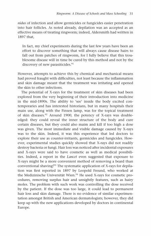
Ringworm: A Disease of Schools and Mass Schooling 31
nidus of infection and allow germicides or fungicides easier penetrationinto hair follicles. As noted already, depilation was an accepted as aneffective means of treating ringworm; indeed, Aldersmith had written in1897 that,
In fact, my chief experiments during the last few years have been aneffort to discover something that will always cause disease hairs tofall out from patches of ringworm, for I fully believe that this trou-blesome disease will in time be cured by this method and not by thediscovery of new parasiticides.91
However, attempts to achieve this by chemical and mechanical meanshad proved fraught with difficulties, not least because the inflammationand skin damage meant that the treatment was irritating and openedthe skin to other infections.
The potential of X-rays for the treatment of skin diseases had beenexplored from the very beginning of their introduction into medicinein the mid-1890s. The ability to ‘see’ inside the body excited con-temporaries and has interested historians, but in many hospitals theirmain use, along with the Finsen lamp, was for the topical treatmentof skin diseases.92 Around 1900, the potency of X-rays was double-edged: they could reveal the inner structure of the body and curecertain diseases, but they could also maim and kill if too high a dosewas given. The most immediate and visible damage caused by X-rayswas to the skin. Indeed, it was this experience that led doctors toexplore their use as counter-irritants, germicides and fungicides. How-ever, experimental studies quickly showed that X-rays did not readilydestroy bacteria or fungi. Hair loss was noticed after incidental exposuresand X-rays were said to have cosmetic as well as medical possibili-ties. Indeed, a report in the Lancet even suggested that exposure toX-rays might be a more convenient method of removing a beard thanconventional shaving!93 The systematic application of X-rays for depila-tion was first reported in 1897 by Leopold Freund, who worked atthe Medizinische Universität Wien.94 He used X-rays for cosmetic pro-cedures, removing surplus hair and unsightly features, such as hairymoles. The problem with such work was controlling the dose receivedby the patient. If the dose was too large, it could lead to permanenthair loss and skin damage. There is no evidence of similar experimen-tation amongst British and American dermatologists; however, they didkeep up with the new applications developed by doctors in continentalEurope.
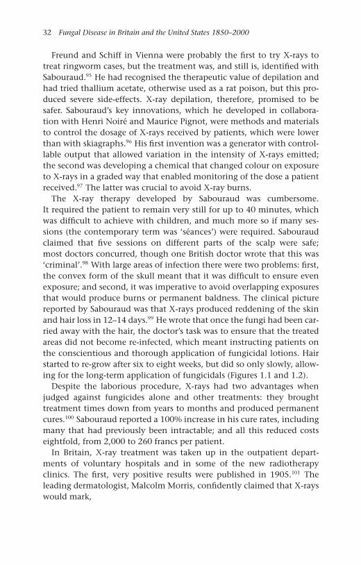
32 Fungal Disease in Britain and the United States 1850–2000
Freund and Schiff in Vienna were probably the first to try X-rays totreat ringworm cases, but the treatment was, and still is, identified withSabouraud.95 He had recognised the therapeutic value of depilation andhad tried thallium acetate, otherwise used as a rat poison, but this pro-duced severe side-effects. X-ray depilation, therefore, promised to besafer. Sabouraud’s key innovations, which he developed in collabora-tion with Henri Noiré and Maurice Pignot, were methods and materialsto control the dosage of X-rays received by patients, which were lowerthan with skiagraphs.96 His first invention was a generator with control-lable output that allowed variation in the intensity of X-rays emitted;the second was developing a chemical that changed colour on exposureto X-rays in a graded way that enabled monitoring of the dose a patientreceived.97 The latter was crucial to avoid X-ray burns.
The X-ray therapy developed by Sabouraud was cumbersome.It required the patient to remain very still for up to 40 minutes, whichwas difficult to achieve with children, and much more so if many ses-sions (the contemporary term was ‘séances’) were required. Sabouraudclaimed that five sessions on different parts of the scalp were safe;most doctors concurred, though one British doctor wrote that this was‘criminal’.98 With large areas of infection there were two problems: first,the convex form of the skull meant that it was difficult to ensure evenexposure; and second, it was imperative to avoid overlapping exposuresthat would produce burns or permanent baldness. The clinical picturereported by Sabouraud was that X-rays produced reddening of the skinand hair loss in 12–14 days.99 He wrote that once the fungi had been car-ried away with the hair, the doctor’s task was to ensure that the treatedareas did not become re-infected, which meant instructing patients onthe conscientious and thorough application of fungicidal lotions. Hairstarted to re-grow after six to eight weeks, but did so only slowly, allow-ing for the long-term application of fungicidals (Figures 1.1 and 1.2).
Despite the laborious procedure, X-rays had two advantages whenjudged against fungicides alone and other treatments: they broughttreatment times down from years to months and produced permanentcures.100 Sabouraud reported a 100% increase in his cure rates, includingmany that had previously been intractable; and all this reduced costseightfold, from 2,000 to 260 francs per patient.
In Britain, X-ray treatment was taken up in the outpatient depart-ments of voluntary hospitals and in some of the new radiotherapyclinics. The first, very positive results were published in 1905.101 Theleading dermatologist, Malcolm Morris, confidently claimed that X-rayswould mark,
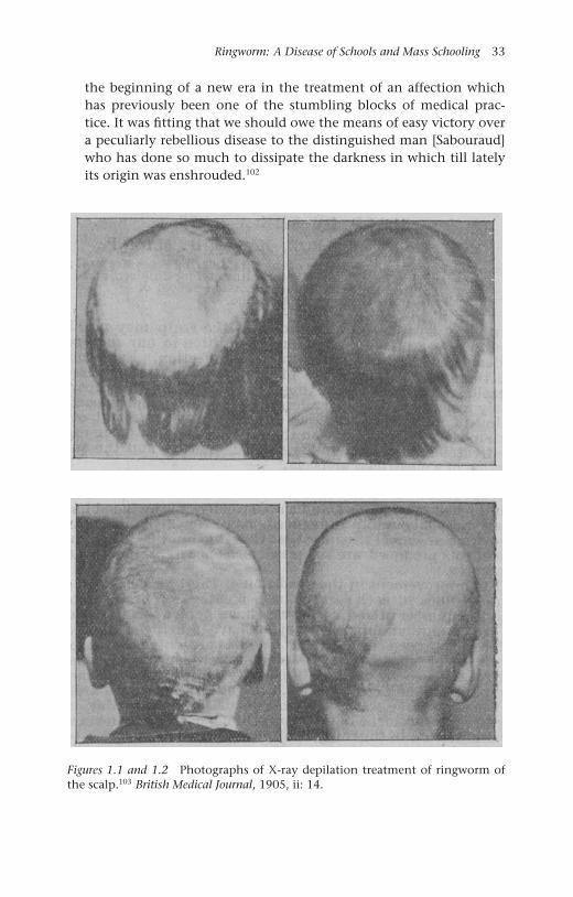
Ringworm: A Disease of Schools and Mass Schooling 33
the beginning of a new era in the treatment of an affection whichhas previously been one of the stumbling blocks of medical prac-tice. It was fitting that we should owe the means of easy victory overa peculiarly rebellious disease to the distinguished man [Sabouraud]who has done so much to dissipate the darkness in which till latelyits origin was enshrouded.102
Figures 1.1 and 1.2 Photographs of X-ray depilation treatment of ringworm ofthe scalp.103 British Medical Journal, 1905, ii: 14.

34 Fungal Disease in Britain and the United States 1850–2000
The number of published reports of success grew. These were typically ofa small number of cases, with doctors cautioning that time was neededto assess whether the cures were permanent. John MacLeod, physicianat Charing Cross Hospital and the Victoria Hospital for Children, didnot regard X-rays as a panacea.
It is a treatment, however, which is by no means easy; first there arethe difficulties of the technique, second there is the all-importantlocal treatment with the parasiticide remedies, and, third, there isthe care which is requisite to avoid mishaps . . . . The immediate dan-gers of the treatment . . . can, as a rule, be avoided, but with regardto the ultimate dangers, if there be any, sufficient time has not yetelapsed to disclose them. It has been suggested that the exposure ofthe scalp to the rays might have some harmful effect on the underly-ing brain. Certainly in an infant or a child under 3 years of age, wherethe scalp is thin and the fontanelles have not closed, one would betimid about submitting the scalp to the X-rays, but with regard toolder children no misfortune of that nature has, as far as we are aware,been recorded.104
In fact, British dermatologists struggled to obtain results as good as thosereported by Sabouraud; yet, even a 50% cure rate was regarded as out-standing compared to other methods.105 Better results were anticipatedonce doctors developed mastery of the equipment and pastilles, andwhen patient compliance could be improved106 (see Figure 1.3).
The first systematic use of X-ray treatment in Britain was at the ring-worm schools of the MAB; indeed, their success reportedly improvedturnover so much that the Bridge School at Witham closed in 1908,saving £500 per year, when the remaining children were transferredto the Downs School.107 Treatment there was directed by Thomas Col-cott Fox, with day-to-day matters in the hands of the school’s medicalofficer Dr Sale. Within a year they reported 400 cures.108 The doctorsenjoyed access to a large number of cases and developed facilities fortreating many children at once (see Figure 1.4). They were treating pau-per children, who were in triple isolation: in a special institution, withinthe Poor Law, and away from parents, hence, there were no problemswith consent, and compliance with young children was largely a mat-ter of discipline. Colcott Fox and Sale conducted a large ‘trial’, but aswas typical for the time there were no controls. Unsurprisingly, whenthey published their results there was no discussion of the ethics ofthis ‘trial’, only wonder at its success.109 Indeed, the London County
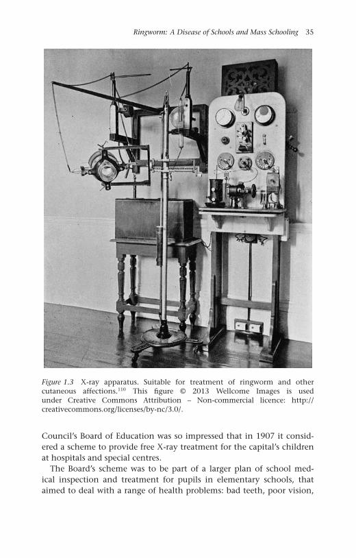
Ringworm: A Disease of Schools and Mass Schooling 35
Figure 1.3 X-ray apparatus. Suitable for treatment of ringworm and othercutaneous affections.110 This figure © 2013 Wellcome Images is usedunder Creative Commons Attribution – Non-commercial licence: http://creativecommons.org/licenses/by-nc/3.0/.
Council’s Board of Education was so impressed that in 1907 it consid-ered a scheme to provide free X-ray treatment for the capital’s childrenat hospitals and special centres.
The Board’s scheme was to be part of a larger plan of school med-ical inspection and treatment for pupils in elementary schools, thataimed to deal with a range of health problems: bad teeth, poor vision,
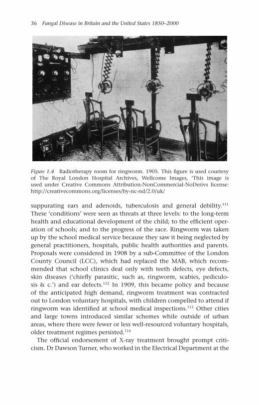
36 Fungal Disease in Britain and the United States 1850–2000
Figure 1.4 Radiotherapy room for ringworm. 1905. This figure is used courtesyof The Royal London Hospital Archives, Wellcome Images, ‘This image isused under Creative Commons Attribution-NonCommercial-NoDerivs license:http://creativecommons.org/licenses/by-nc-nd/2.0/uk/
suppurating ears and adenoids, tuberculosis and general debility.111
These ‘conditions’ were seen as threats at three levels: to the long-termhealth and educational development of the child; to the efficient oper-ation of schools; and to the progress of the race. Ringworm was takenup by the school medical service because they saw it being neglected bygeneral practitioners, hospitals, public health authorities and parents.Proposals were considered in 1908 by a sub-Committee of the LondonCounty Council (LCC), which had replaced the MAB, which recom-mended that school clinics deal only with teeth defects, eye defects,skin diseases (‘chiefly parasitic, such as, ringworm, scabies, pediculo-sis & c.’) and ear defects.112 In 1909, this became policy and becauseof the anticipated high demand, ringworm treatment was contractedout to London voluntary hospitals, with children compelled to attend ifringworm was identified at school medical inspections.113 Other citiesand large towns introduced similar schemes while outside of urbanareas, where there were fewer or less well-resourced voluntary hospitals,older treatment regimes persisted.114
The official endorsement of X-ray treatment brought prompt criti-cism. Dr Dawson Turner, who worked in the Electrical Department at the
Ringworm: A Disease of Schools and Mass Schooling 37
Edinburgh Royal Infirmary and described himself as an ‘old worker withX-rays’, who had suffered permanent injury from exposures, wrote tothe Times in March 1909, with what turned out to be a prescient caution.
The deleterious effects of continuous exposures to X-rays in the caseof adults are only too well known to X-ray operators and it is probablethat delicate cells of the growing brain of a child may be injuriouslyaffected by much short exposures, though the evidence of impair-ment of function may not become noticeable until development iscomplete. No helpless child should have the chief centre of its ner-vous system exposed to the X-rays without the express consent of itsparent, obtained after the possible risks of the treatment have beenfully explained.115
His plea was answered in a report by two directors of London hospitalelectro-therapeutic departments. They stated that ordinary precautionshad ensured no ill effects in their patients, nor did they expect anyfrom other controlled uses of X-rays.116 However, the use of X-rays wasresisted by some parents, though this was as much about distrust ofhospitals and dislike of compulsion, as it was about worries over radi-ation. Mr Harris, a jeweller from Rotherhithe, on being instructed totake his daughter to Guy’s Hospital, wrote back to the LCC’s Child CareBranch stating he did not have ‘much faith in those places’ and that hiswife, who was a trained nurse, was treating the child.117 Walter Longleyasserted his independence in similar vein, saying that his boys werealready being treated with sassafras oil and that his family would nottrouble the LCC, nor the London ratepayer.118 Henry Carter wrote thatthe instruction to take his children to the Evelina Hospital was ‘insultingto my wife and self’.119
Armed with X-rays and with the backing of the LCC administration,dermatologists and school medical offices were optimistic about thefuture control of ringworm.120 Nonetheless, in 1909, the Lancet set upan enquiry to address ‘the grave prevalence’ and ‘the disastrous influ-ence’ ringworm was having on the education of children.121 The LancetCommission on Ringworm, consisting of ‘two thoroughly competentdermatologists’ (who remained anonymous), reported on 1 January1910. They dealt almost exclusively with the situation in London.122 Theauthors opened in eugenic terms, stating that ringworm was more preva-lent in the ‘less educated classes’ and that those affected were ‘reallyrepresentatives of lower grades of civilisation’, where infestation with
38 Fungal Disease in Britain and the United States 1850–2000
internal and external parasites was a marker of being left behind bysocial progress. The authors endorsed X-ray treatment administered bydermatologists and radiotherapists, along with a positive assessment ofthe capacity of existing facilities to cope with the scheme of mass treat-ment that the LCC was contemplating. However, they were ambivalentabout whether to use voluntary and local authority hospitals, or to rec-ommend the creation of special treatment centres, but whatever wasdecided they were certain it would be cost-effective.
The Commission’s report took seriously public concerns about thesafety of X-rays, noting that in early years there had been accidents lead-ing to permanent baldness and ulcers. However, burns were said to bea thing of the past as exposures were now well managed. With regardto brain damage, the authors wrote that the experience of thousandsof cases, over many years, showed no evidence of any effects and that‘It is incumbent now on those who imagine that harm does follow theapplication of X-rays to produce the grounds for the view.’123 Againstthis backdrop, many parents allowed their children to be treated withX-rays but, as mentioned above, others refused. The manufacturers ofpopular alternatives, especially antiseptic creams like ‘Germolene’ and‘Zambuk’ – ‘The Balm that Benefits the Bairns’, also offered their prod-ucts as direct alternatives to X-rays.124 However, some medical officersraised the stakes. For example, Dr Bostock Hill, the Medical Officer ofHealth for Warwickshire, claimed in 1911 that he instructed parentsthat ‘they would be dealt with under the Children’s Act for cruelty . . . orthe case would be referred to the N.S.P.C.C’ [National Society for theProtection of Children], if they refused to allow their children to betreated.125
Ernest Dore, a dermatologist at the Evelina Hospital for Sick Chil-dren, made a telling observation in his review of X-ray treatment in1911, a year after the publication of the Lancet Commission report.126
He returned to the issue of stigma, arguing that before X-ray treatmentsa diagnosis of ringworm was far worse than any physical suffering.
A trivial complaint as regards the health of the child, tinea ton-surans brings in its train so long a category of ills that I have morethan once heard long-suffering mothers say that they dreaded scar-let fever or pneumonia less. The disorganisation of the home thatensues from the isolation of the sufferers; the anxiety of the parentslest other children in the family should become infected; the com-plications with medical men and schoolmasters; the social ostracism;the loss of schooling; the wearisome process of constantly rubbing on
Ringworm: A Disease of Schools and Mass Schooling 39
ointments with little apparent result except the production of soreheads in the children and sore hearts in the parents, these are someof the difficulties which have to be faced under the old régime.127
Given the reactions of children, family, friends, neighbours, teachersand doctors to ringworm, and its position as a marker of ‘low civili-sation’ and social danger, it is clear why a disease that never killed orcaused permanent injury attracted such high-profile medical and publicattention. Indeed, Dore wanted to up the stakes further, hinting at thepossibility of stamping out the disease if compulsion was used: eitherin prevention, ‘such as the wearing of some kind of head gear, like themuzzle in the prophylaxis of rabies’, or with X-ray treatment.
A national picture of ringworm in school children was represented inthe Reports of the Medical Officer to the Board of Education, Dr GeorgeNewman; the first of which was for 1908.128 The prevalence of ring-worm was around 1% amongst children inspected in school, muchlower than other ‘defects’, which were: vision (10%), hearing (3–5%),adenoids and enlarged tonsils (6–8%), tooth decay (40%) and uncleanbodies or heads (30–40%).129 The main issue with ringworm was exclu-sion and its effects on a child’s education; plus, from an administrativeperspective, the impact of long absences on a school’s grant income.Although prevalence was low, it still meant that, on average, 3,000 chil-dren were absent every day, with a typical absence duration of nineweeks.130 Nationally, the longest average exclusion reported of 29 weekswas in Somerset. This finding was seen as surprising for a rural countywith few large towns and low population density, and was attributedto poor inspection regimes causing early cases to be missed. Althoughimpetigo, by this time associated with Staphylococcus aureus infection,was the most prevalent skin disease found in inspections, ringworm wastaken much more seriously.131 Dr Ritchie, the School Medical Officerfor Manchester, reported that inspections in 1913 had revealed the fol-lowing: impetigo – 353, ringworm – 187, scabies – 39 and other skindiseases – 110.132 However, cases reported by doctors and parents led to2,003 notifications of ringworm in the city, with up to 1,500 childrenunder supervision at any time. The Manchester containment regime wasstrict, ‘. . . no cases of ringworm of the scalp are allowed to attend schoolunless the hair over and around the patches is cut and a washable capworn . . . . Children affected with ringworm of the body are not allowedto attend school.’133 In the same year, a ringworm school was establishedin Edinburgh for long-term absentees, including one boy who allegedlyhad been excluded for four years.134
40 Fungal Disease in Britain and the United States 1850–2000
In his annual reports, Newman began to report improvements, par-ticularly in areas where X-ray treatment was available. In London,new cases fell from 5,573 in 1913 to 4,449 a year later, while inBeckenham in Kent, new cases had fallen from 133 in 1911 to just48 in 1914.135 However, nationally, the provision of special serviceswas patchy. Only one third of education authorities had made spe-cial provision for ringworm treatment and in many areas, especiallyoutside of cities and large towns, there was still no access to X-raytreatment at all. In addition, many general practitioners chose to con-tinue to recommend topical fungicides and left treatment to ‘unreliable’parents.136
The decline of ringworm
In Britain, doctors reported that the incidence of ringworm of thescalp in school children fell during the First World War, but increasedafterwards because of the shortage of school nurses, many of whom con-tinued to work with casualties and invalids.137 However, this was a minorpeak as the incidence fell steadily over the inter-war period. In London,the number of new cases had reduced from 6,214 in 1911, to 3,983 in1920. The number dropped further, to 513 in 1930 and by 1936 theywas just 89.138 As early as 1925, the district medical officer for Becken-ham reported no new cases, while in Ilford, ringworm was also said tohave been ‘abolished’.139 In his 26th and final report, for the year 1933,George Newman observed with satisfaction that ‘Ringworm is steadilydisappearing.’140 This situation was reflected in treatment facilities, thenumber of which was reduced from 150 clinics in 1923 to 80 in 1938.The London ringworm school, which had moved to the Goldie LeighCottage Children’s Homes, Woolwich, in 1914, took fewer and fewerresidential cases, and became instead a centre for day treatment withX-rays.141
Doctors attributed the decline in the reported incidence of ringworm,in the words of Norman Walker in 1929, not so much to the characterof the infection, but to ‘the value of cooperation between the scien-tist, the clinician, and the organiser’.142 Success was said to have comefrom school inspection spotting early cases, which were followed upby effective treatments such as X-rays. The provision and use of X-rayswas variable across the country. In the early 1930s only 20% of diag-nosed cases in England were receiving X-rays. The rates of use varied:London was the highest and rural counties were several times lower143
(Table 1.1).

Ringworm: A Disease of Schools and Mass Schooling 41
Table 1.1 Cases of ringworm in England and Wales treated by X-ray or othermethods, 1933
England By X-rays Otherwise X-ray treatment aspercentage of total
Counties 149 2058 6.8County boroughs 540 2040 20.9Boroughs 120 597 16.7Urban districts 22 90 19.6London 160 18 89.9
WalesCounties 20 88 18.5County boroughs 52 19 73.2Boroughs 0 24 0.0Urban districts 7 29 19.4
Chemical and mechanical methods of depilation continued to be usedand there was particular interest again in the 1930s in giving thalliumacetate.144 Some doctors, particularly in the United States, argued thatthallium treatment was safer than X-rays; however, critics termed it ‘ADangerous Drug’ because the margin between achieving effective epi-lation and poisoning was very small.145 During the inter-war period,dermatologists on both sides of the Atlantic showed less interest inringworm of the scalp, reflecting lower incidence and relatively stabletherapeutic regimes.146 Their new areas of interest were ringworm inathletes, college students, soldiers and miners.
Ringworm, although no doubt a common human infection for cen-turies, only gained serious medical and public attention in the secondhalf of the nineteenth century, and then in a specific social group andsetting: school children and schooling. The aggregation of children incrowded classrooms for hours at a time seemed to provide ideal condi-tions for contagion. None the less, it was as a social rather than physicaldisease that ringworm gained medical and public attention. Ringwormepidemics were one of the unintended consequences of the progres-sive reform of mass schooling, which revealed changing social attitudesto markers of disease and the growing stigmatisation of the palpably‘unclean’. While historians such as Nancy Tomes have detailed pub-lic responses to the threat of invisible germs, we have revealed thereactions, some similar and others unique, to conditions where thegerms were highly visible. Perhaps, the ‘gospel of germs’ won convertsmore readily for diseases such as ringworm, favus and trachoma, where
42 Fungal Disease in Britain and the United States 1850–2000
the physical and social manifestations of infection were obvious andreinforcing.
From 1905, ringworm was also seen as a pathology that could be reme-died by medical progress, and not just any new technology, but by themedical icon of the age, X-rays. The use of X-ray depilation was an inno-vation that was taken up rapidly, in large measure because it promisedso much, but also because the necessary equipment was becoming morereadily available and there were opportunities for clinical and organi-sational innovations. In Britain, major public bodies such as the LCC,having been persuaded to create special ringworm institutions, subse-quently invested in the new technologies of treatment. This all seemedto pay off, as the reported incidence of ringworm of the scalp in chil-dren declined rapidly in the inter-war period.147 There was debate aboutthe causes of the fall. Was it due to medical inspection regimes andnew treatments, or to social factors, such as more bathrooms, bettermedicated shampoos, the fashion for shorter hair and grooming withhair creams? ‘Brylcreem’ was introduced in 1928 and marketed for bet-ter ‘bounce’ in styling and control of dandruff, then said to be causedby a yeast fungus Pityrosporon. Whatever the specific reasons, all factorsresponsible for the decline were seen by contemporary commentators tobe due to medical and social progress.
Except where otherwise noted, this work is licensed under aCreative Commons Attribution 3.0 Unported License. To view
a copy of this license, visit http://creativecommons.org/licenses/by/3.0/
OPEN
2Athlete’s Foot: A Disease of Fitnessand Hygiene
In May 1939, a review of the American yearbook of dermatology andsyphilology observed:
As usual they make a prominent feature of an introductory articleon some branch of therapeutics, and this year they deal with thetreatment of the deeper fungous infections of the skin, includingringworm of the scalp and bearded regions, and the comparativelyrare fungous affections of the subcutaneous tissues. As a matter of factthis subject is not of great practical importance in the British Isles,especially in England, where the incidence of ringworm of the scalphas been reduced to quite a trivial number of cases per annum, andringworm of the beard has become an actual rarity. No doubt the stateof affairs is otherwise in the United States, where the standard of liv-ing, both among the large negro population and also to a lesser extentamong the more recent immigrants from Central Europe, is such thatthese infections are much commoner; moreover, a considerable areaof the U.S.A. boasts a subtropical climate in which parasitic fungi arefar more active than in the temperate zones.1
While this statement rightly reflects on improvements in Britain, its per-ception of ringworm in the United States was wrong. Across the Atlantic,it was not a problem amongst immigrants and African Americans; ratherit had been framed as a public health menace in the affluent classes,especially amongst those who frequented swimming pools, and collegestudents and, of course, athletes. While its prevalence had seeminglyshifted up the social scale and from children to adults, its principal siteof infection had moved too, from head to toe, with ‘athlete’s foot’ beingone of the most common and most talked about diseases in America inthe 1930s.2
43
44 Fungal Disease in Britain and the United States 1850–2000
Manufacturers of popular remedies gave ringworm of the foot thename ‘athlete’s foot’ and this was adopted by the public, but in medicinethe infection was known as tinea pedis. It was seen as new disease, or inmodern parlance an emerging infection, having been first described byArthur Whitfield, a dermatologist at King’s College Hospital, London in1908.3 Nonetheless, it was in the United States that tinea pedis becamean epidemic, seemingly spread by modern lifestyles and hygiene prac-tices, and encouraged by modern socks and shoes, which made infectionliable to chronicity by keeping the feet moist and warm. It is perhapssurprising, given the unhygienic conditions of previous centuries andthe ubiquity of ringworm of the scalp, that the feet of Europeans andAmericans had been seemingly free of ringworm infection until themid-twentieth century. Nonetheless, there was a clear understandingin the inter-war years that tinea pedis was new disease and one linkedto modernity. One consequence of the growth of medical attention totinea pedis was the stimulus it gave to the specialism of medical mycol-ogy, with investment in diagnostic services, research on the conditionsin which mycoses spread and their treatment. In turn, this led to thecreation of a cadre of medical mycologists, who identified more fungalinfections in specific social groups and the general population.
We begin this chapter with a discussion of the development of med-ical mycology, especially in the United States, which led the world inthe creation of centres of expertise, training and research. We then dis-cuss the movement of medical and public concern about ringworm fromchildren’s heads to athlete’s toes. In the 1940s, the condition was alsorecognised as a problem amongst soldiers and miners, and was seen interms of greater exposure to new pathogenic species of fungi and theincreased vulnerability of the human soil in tropical and specific workconditions. While orthodox medicine approached tinea pedis as some-thing that was difficult to treat and needed to be prevented, proprietarymedicine companies peddled remedies that filled the therapeutic vac-uum with numerous ‘certain cures’. We follow the controversies aroundpopular and medical treatments, and the emergence in the late 1950sof medically approved, clinically trialled antifungal drugs, modelled onthe antibiotics developed for bacterial infections. The most importantof these was griseofulvin, which promised to be effective and safe withtopical and oral administration. This was followed in the 1970s with arange of azole drugs. These antifungals accelerated the disappearance ofathlete’s foot as a medical, if not public term, thereby divorcing ring-worm from its locus of infection or site of contagion, making it a typeof dermatophytosis, literally, skin fungal infection.
Athlete’s Foot: A Disease of Fitness and Hygiene 45
Medical mycology: ‘An Orphan Science’4
Glenn S. Bulmer has recently characterised the manner in which workon human fungal diseases developed in the inter-war period as theresult of the movement and coalescence of specialists: ‘botany typescame forth and usually joined Departments of Microbiology &/orImmunology – two subjects that most had never taken in school’.5
He may have had in mind the founders of the first specialist medicalmycology laboratory in the United States, which opened in 1926 at theColumbia-Presbyterian Medical Center (C-PMC). One leading light wasthe botanist was Bernard O. Dodge, who then worked at the New YorkBotanical Gardens.6 He was known for his work on genetics and forintroducing T. H. Morgan to Neurospora, an organism that became awidely used experimental model in genetics.7 Dodge was interested infungal diseases in humans and animals, and between 1928 and 1939he was a consultant in mycology at the C-PMC, while at the same timelecturing on dermatology at the College of Physicians and Surgeons.The other founder as a clinician, the dermatologist J. Gardner Hopkins,who worked at the C-PMC’s Vanderbilt Clinic; previously he had workedon a cure for the plague with Hans Zinsser in Serbia during the FirstWorld War.8 Indeed, his research spanned infectious diseases, especiallysyphilis and moniliasis. In the 1920s and 1930s it was mainly ‘botanytypes’, with the ability to identify types of fungi, who dominated thenew field, which was principally concerned with diagnosis, requiringmicroscopy and culturing skills, combined with taxonomy. In 1926,Hopkins hired Rhoda W. Benham – a botanist by training – to undertakemycological diagnoses, and went on to develop a research laboratoryfrom this base.9 His initiative was aided by a grant of $50,000 from theRockefeller Foundation in 1929, which eventually enabled Columbia todevelop the first specialist training in human fungal diseases in 1935.
Hopkins’s laboratory became an influential research centre, producingkey figures such as Chester W. Emmons (1900–1985), the first medi-cal mycologist employed by the National Institutes of Health (NIH) inBethesda.10 Emmons took his PhD with Robert A. Harper in the BotanyDepartment at Columbia on the subject of mildew, before workingwith Dodge and publishing two articles on dermatophytes in Archivesof Dermatology and Syphilis. Neither author was medically qualified.Emmons stated later that when he sought a reference to move to Hop-kins’s laboratory, Harper had asked, ‘Why do you want to study thoseabortive and uninteresting medical fungi?’11 Indeed, Emmons referredto medical mycology as ‘an orphan science’ as it was never taught by
46 Fungal Disease in Britain and the United States 1850–2000
mycologists, whose main interest, outside of taxonomy, reproductionand other purely biological matters, was in plant pathology. In his firstmedical publications, Emmons proposed redefining the genera of themain fungal causes of skin infection: Microsporum, Trichophyton and Epi-dermophyton. His approach was to use their morphologies, rather thantheir pathological effects, pioneering what became the preferred methodof standardising diagnoses and avoiding the variability and inconsis-tency of clinical criteria.12 Emmons then worked with Arturo Carriónin Puerto Rica on chromoblastomycosis, publishing in the Puerto RicoJournal of Public Health and Tropical Medicine. In the event, the move tofungal diseases turned out well, as his appointment at NIH was as prin-cipal mycologist. He stayed for 30 years, becoming recognised as one of‘founding fathers’ of medical mycology in the United States.
The first monograph in the field was Harry Jacobson’s FungousDiseases: A Clinico-Mycology Text published in 1932. The author wasa dermatologist in Los Angeles and he presented ten chapters on‘dermomycoses, moniliasis, maduromycoses, sporotrichoses, blasto-mycoses, actinomycoses, coccidioides, toruloses and aspergilloses’.13
A review in Archives of Dermatology concluded that the book was ‘use-ful’ and, perhaps surprisingly, observed that ‘the mycologic aspects ofthe subject are better handled than the clinical aspects’. The secondmonograph was published from Harvard, where there had been instruc-tion since 1924, and it was this course that was written up in 1935 byCarroll W. Dodge, a botanist who was no relation of Bernard O. Dodge.14
The book was compiled at the suggestion of Roland Thaxter, who wasmedically trained, but had switched to botany, eventually becomingprofessor of cryptogamic botany at Harvard. Carroll Dodge’s MedicalMycology: Fungous Diseases of Man and Other Animals was the first use of‘medical mycology’ in a book title. Here, and in later works, the primaryfocus was on fungi and their identification, with little on the pathol-ogy of the infections they caused. Before publication, Dodge had movedto become professor of botany at Washington University and contin-ued to research fungal diseases in Central America, before moving toLichenology after 1950.
Writing the New England Journal of Medicine in August 1936, JacobH. Swartz, of the Department of Cryptogamic Botany at Harvard Uni-versity, maintained that,
Mycology is no longer a mysterious subject known only to a few.It has been proved to be a part of medicine just as bacteriology, phys-iology and so forth. Some knowledge of mycology is necessary for abetter understanding of disease.15
Athlete’s Foot: A Disease of Fitness and Hygiene 47
He was contributing to a discussion on fungi and internal medicine,where the focus was not on skin diseases, but on systemic infectionsand allergy, with an emphasis on the importance of predisposition – thehuman soil – as a factor with infection. Many contributors stressed therole of the laboratory in confirming clinical diagnoses; though Swartzworried that since the ‘mystery’ of fungi had been largely solved, clin-icians were no longer turning to the laboratory and were relying solelyon clinical signs. The ‘orphan science’ had been adopted by doctors, butit was not being nurtured. Swartz closed the discussion demanding thecreation of a mycological department in every medical school and hos-pital, stating that ‘Mycology at present is in chaos’ and calling for thefield to be ‘simplified and made useful to all branches of medicine’.16
Arthur Greenwood, a dermatologist at Massachusetts General Hospital,suggested the way forward was to create more people trained to workbetween clinicians and mycologists.17 One factor fuelling the demandfor more medical mycologists, reflected in the discussion prompted bySwartz, was recognition of geographically localised foci of fungal disease,which we examine in Chapter 4. Meanwhile, athlete’s foot or tinea pediswas emerging as a dominate concern for dermatologists and nascentmedical mycologists.
Athlete’s foot
Infection in areas other than the scalp came to the fore in the FirstWorld War, when ringworm of the groin (‘dhobi itch’) was found to beprevalent amongst American and British troops in France.18 Some doc-tors thought that damp and crowded conditions of the Western Frontincreased susceptibility and were ideal for contagion; others believed anew fungus had been brought to Europe and the Americas by troopsreturning from the tropics.19 The bacteriological facilities available tothe military medical services meant that specific pathogens were iden-tified, using the methods developed by Sabouraud in the 1900s. Mostcases were of Trichophyton interdigitalis, but there were increasing reportsof Trichophyton rubrum, though at this time there was little interest inthe epidemiology of types of infection.20 Rather, discussion focused onthe observation that, paradoxically, tinea pedis was a disease of hygiene.In the British military, the highest rates of infection were found amongstofficers, who through bathing regularly opened themselves to infectionthrough contact and softening of the skin. Also, tightly laced, ankle-high army boots, worn in hot, damp environments, for long periodswithout changes of socks, were understood to provide ideal conditionsfor tinea pedis to flourish.21

48 Fungal Disease in Britain and the United States 1850–2000
In the United States too, ringworm of the feet was ‘discovered’ as amalady of soldiers in the First World War. Two West Coast Americandoctors, Oliver Ormsby and James Mitchell, put ringworm of the feeton the medical map in the United States in 1916, in an article on infec-tion of the hands and feet.22 The alleged first use of the term ‘athlete’sfoot’ was in December 1928, in an article in the Literary Digest, promptedby reports from Dr Charles F. Pabst, of Greenpoint Hospital Brooklyn.23
Pabst claimed that the condition was already well-known in the UnitedStates, with an estimated ten million sufferers, three quarters of whomwere unaware of the infection. The article stated that the problem was ofrecent origin, but that ‘at least half the adult population suffer from thismalady at some time’. However, the term athlete’s foot took hold, notas a disease of the masses, but one of the America’s affluent classes, whocould now afford to enjoy leisure activities and modern hygiene facil-ities. Pabst, perhaps revealing his limited social circle and awareness,claimed that ‘almost everyone who uses a swimming-pool, golf club,athletic club, or any place where there is a common dressing room, hasthe infection on his feet’.24 Further medical recognition came in May1929, when three doctors, working at the University of California stu-dent health service, published data that showed tinea pedis was endemicamongst students.25 They wrote that many students arrived at collegewith the infection: 53% of males and 15% of females, and the incidencerose to 85% in those final year students who took gymnasium classes.In a follow up study, the doctors showed at the end of their first year,the incidence amongst freshmen had risen from 53% to 78%, but wasonly 2% higher in female students.26 The sex differences suggested tocontemporaries four factors in the emergence of tinea pedis: the newmale enthusiasm for using gymnasia, swimming baths and other sportsfacilities; the poor facilities for changing and showering at these loca-tions; male indifference to personal hygiene; and the types of socks andshoes worn for sport and after, where the lighter, open shoes of womenmilitated against infection taking hold. Athlete’s foot was also possi-bly a consequence of public sanitary facilities, where the sexes, socialclasses, races and different ages mingled, and where anyone might be a‘carrier’.
Awareness of the problem grew quickly. The link between tinea pedisand athletes was evident in 1931 when the Los Angeles Times termedit the ‘gymnasium malady’.27 In the same year, W. L. Gould, a physi-cian from Albany, New York State, reported to the United States PublicHealth Service that possibly 50% of all adults suffered from tinea pedisand that its incidence was particularly high in the states bordering the
Athlete’s Foot: A Disease of Fitness and Hygiene 49
Gulf of Mexico and in California, due to heat and humidity.28 At the1932 Olympic Games in Los Angeles, special antiseptic footbaths wereprovided to prevent infection.29 By the mid-1930s there was recogni-tion that it was not just students and athletes who were vulnerable.J. H. Swartz wrote of ‘the addiction of our generation to the frequent-ing of gymnasiums (sic), baths and locker rooms . . . and the tendency toexercise violently and perspiring in unsterilizable socks and body cloth-ing’. He went on to wonder if the Romans were similarly affected?30
Dermatologists also saw tinea pedis as threat to the family, throughcross-infection in the home, where baths and showers were becom-ing more common.31 However, Ayu Majima has shown that ringwormamongst the poor – even tinea pedis – was never described with theneologism of athlete’s foot and sufferers remained stigmatised.32
Doctors prescribed a variety of remedies for tinea pedis, but because ofmixed results, their advice was mainly that prevention was better thancure. Thus, anyone visiting a swimming baths and showering in publicfacilities was told to disinfect their feet where possible, to avoid walkingon floors in bare feet, to dry their feet and toes after bathing, never toshare towels or clothing, and to wash socks at high temperatures. Themain remedies were topical antifungal creams. The standbys were Whit-field’s ointment (the active ingredients were 5% salicylic acid and 3%benzoic acid in petroleum jelly) and Castellani’s carbol-fuchsin paint.However, the variable presentation of the condition and its tendency tochronicity encouraged innovations, both in combinations of remediesand new chemicals.33 There were many complaints of overtreatment.34
Proprietary medical companies, which already had a significance marketwith topical antiseptic remedies, seized on the new epidemic, pro-ducing new products and rebranding older ones as having antifungalas well as antibacterial properties.35 The most prominently advertisedremedy in newspapers and magazines was a derivative of a veterinaryliniment – ‘Absorbine Jr.’, marketed by W. F. Young, Inc.36 The companyclaimed to have coined the term athlete’s foot and in the late 1930s, theadvertisements for Absorbine Jr. echoed public posters against venerealdisease and referred to earlier fears, from the time of Typhoid Mary, tounrecognised carriers (see Figure 2.1).
Older products for ‘skin disorders of the feet’, such as Dr Scholl’s‘Solvex’, were rebranded to target the new consumers, thus, its adver-tisement claimed it was effective against ‘gym or athlete’s foot’, ‘footitch’ and ‘golfer’s itch’.37 While doctors promoted the possibilities ofprevention and commercial drug companies sold ‘sure-cure remedies’,there was also a sense that the high prevalence of tinea pedis meant
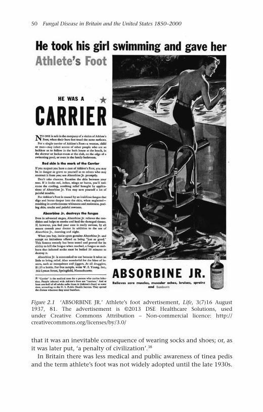
50 Fungal Disease in Britain and the United States 1850–2000
Figure 2.1 ‘ABSORBINE JR.’ Athlete’s foot advertisement, Life, 3(7)16 August1937, 81. The advertisement is ©2013 DSE Healthcare Solutions, usedunder Creative Commons Attribution – Non-commercial licence: http://creativecommons.org/licenses/by/3.0/
that it was an inevitable consequence of wearing socks and shoes; or, asit was later put, ‘a penalty of civilization’.38
In Britain there was less medical and public awareness of tinea pedisand the term athlete’s foot was not widely adopted until the late 1930s.
Athlete’s Foot: A Disease of Fitness and Hygiene 51
Medical writings concentrated on diagnosis and treatment in individualpatients rather than any social group. However, doctors noted that itwas a disease with ‘something of the nature of a social qualification,being commonly met among the upper and middle classes’ and, beingmostly spread through bathing; it was, then ‘paradoxically . . . a penaltyof cleanliness’.39 This changed in the 1940s, when Britain found itsequivalent of American athletes – coal miners. Tinea pedis was iden-tified as the commonest cause of miner’s dermatitis, a new conditionresulting, again paradoxically, from the provision of pithead baths forcommunal bathing at the end of underground shifts. The Sankey Com-mission, which had reported on the future of the coal industry in 1919had recommended measures to improve the health and welfare facili-ties for miners and the largest proportion of its expenditure (35%) wenton pithead baths.40 Baths were introduced only slowly and the preva-lence of tinea pedis only gained a national profile during the SecondWorld War, when absenteeism due to illness and injuries was a threat toproduction in a pivotal industry.
Soldiers and miners
Concern about the incidence of tinea pedis amongst military personnelhad been expressed between the wars. A study for the United States Navypublished as early as 1924, showed 13% of all ranks were affected, butlevels were as high as 91% amongst officers recruited from college. Thesame scenario was reported in the late 1930s in the Royal Navy, withthe condition being prevalent amongst all ranks in tropical stations.Surgeon-Commander J. C. Souter expressed the opinion that tinea pediswas a submerged problem, where itching and discomfort was toleratedby men, ‘yet every sufferer is a potential casualty’ should the condi-tion worsen, as it might on active duty when changes of clothing weredifficult, or in the tropics.41
During the Second World War, skin diseases were a major cause ofinvalidism due to poor skin hygiene in combat locations, communalwashing facilities and exposure to new pathogens.42 In tropical theatresthere were reports that skin conditions were responsible for three quar-ters of sick bay attendances in the Pacific.43 One response at home wasthat in 1943, the Walter Reed Army Medical Centre in Washingtonestablished a mycology laboratory headed by Norman Conant whohad started his career in Botany at Harvard, training with RaymondSabouraud in Paris before taking a post in the Department of Micro-biology at Duke University, specialising in mycology. He had worked onallergies and tinea, but gained international recognition in 1944 with

52 Fungal Disease in Britain and the United States 1850–2000
the publication of a Manual of Clinical Mycology, co-authored with D. S.Martin, D. T. Smith, R. D. Baker and J. L. Callaway, all from Duke’s Med-ical faculty. There were also problems at home amongst soldiers beingexposed to geographically specific fungi at training bases in Western andMidwestern states, which we discuss in the Chapter 4.
However, the highest profile incident with tinea pedis during the warconcerned a treatment on the home front. A mixture of phenol andcamphor was very popular, but there were reports of overtreatment andhigh levels of exposure, leading to deaths.44 The remedy was champi-oned by Paul de Kruif in an article, entitled ‘A Working Cure for Athlete’sFoot’ in The Reader’s Digest in May 1942.45 De Kruif is best known todayfor his book Microbes Hunters and for assisting Sinclair Lewis in the writ-ing of Arrowsmith, but in the 1920s and 1930s he was a significant figurein American medicine.46 He had worked for the Rockefeller Instituteand become as publicist for medical science, serving as secretary to thePresident’s Commission for Infantile Paralysis in 1934. However, by the1940s he had become a controversial personality, mainly through publi-cising various medical innovations, of which his athlete’s foot cure wasseen as another questionable example.47 Indeed, such was de Kruif’s rep-utation that the FDA issued a public warning against the use of phenolcamphor mixture later in 1942.48
Amongst British forces in South East Asia, the prevalence of all formsof ringworm was so serious that the Royal Army Medical Corps setup a research unit there on soldier’s dermatitis. Surveys by unit staffrevealed that amongst soldiers in Malaya and Hong Kong, 79.5% hadtinea pedis and 33.5% had tinea corporis, tinea cruris or all three.49
However, ringworm received most attention in Britain, not because ofits military toll, but due to its incidence in workers at home, along withthe investment in fungus research prompted by the discovery of theantibiotic properties of penicillin and emerging problem of systemicmycoses. In Britain, the Medical Research Council (MRC) appointed aMedical Mycology Committee in 1943.50 One goal was to rationalisetaxonomies and tinea pedis was a particular problem. At the timethe following terms were used by doctors across the Empire: ‘athlete’sfoot’; ‘Hong Kong’, ‘Shanghai’ and ‘Singapore foot’; ‘gym’, ‘golfers’ and‘swimmers’ itch; ‘toe-rot’; ‘ringworm of the feet’; ‘Cantlie’s foot tetter’,‘eczematoid ringworm of the extremities’; ‘dermatomycosis’; ‘epider-momycosis’; and ‘epidermophytosis’.51 Perhaps this variety was furtherevidence of the novelty of the infection, or even the diversity of spe-cific pathogens producing different types of lesion, but most likely itreflected the multiplicity of practitioners, locations and presentations.
Athlete’s Foot: A Disease of Fitness and Hygiene 53
The Committee’s work during the war focused on coal miners. Thefirst detailed study was made in 1943 by R. B. Knowles in the coal-fields of the north Midlands and South Yorkshire. He confirmed thewidely held view that the introduction of pithead baths in the 1920sand 1930s had created the problem.52 After the nationalisation of theindustry in 1946 and the creation of the National Coal Board (NCB),surveys and reporting on the welfare of miners increased.53 One study ofminers in Warwickshire in 1946 found that 52% of the men had ‘intra-digital disease’, 15% had ‘foot lesions other than fungus infection’, andonly 33% had ‘healthy feet’.54 It was against this background that inNovember 1951, a Committee on Industrial Epidermophytosis (CIE) wasestablished within the MRC’s Industrial Health Research Board (IHRB).The Committee’s membership indicates how tinea pedis had becomea multi-specialist problem. The CIE was chaired by John T. Ingram,a dermatologist from Leeds, who had considerable experience in thearmy and was joined by two other dermatologists, George H. Percival(Edinburgh) and H. Renwick Vickers (Sheffield).55 Geoffrey C. Ainsworthprovided mycological expertise, D. D. Reid from the London School ofHygiene and Tropical Medicine dealt with medical statistics, while R. E.Lane from the University of Manchester provided wider occupationalhealth expertise.56 Another member was Archibald L. Cochrane, whowas then at the MRC’s Pneumoconiosis Research Unit in Penarth, andlater became a champion of randomised clinical trials, where his legacyhas been institutionalised in the network of Cochrane collaborations.57
Also on the Committee were T. E. Howell, the Principal Medical Inspec-tor of Mines at the Ministry of Fuel and Power, and J. M. Rogan, PrincipalMedical Officer at NCB.58
The initial brief was quite wide, but Ingram argued that the CIE should‘deal in the first place especially with epidermophytosis of the feet incoal miners and to leave all side issues . . . until later’.59 Rogan stressedthe NCB’s economic objectives of reducing absenteeism and other costs,wanting the CIE to focus on practical measures, such as prevention andmethods for mass treatment.60 However, Ingram had other ideas and fos-tered work across specialisms in epidemiology, the natural history of thedisease, clinical and mycological diagnosis, research on fungal growth,the chemistry of the skin and the histopathology of the condition.61
Finding out the nature and scale of the problem in miners was thepriority. Epidemiological studies were commissioned from J. G. Holmes,a dermatologist in Cardiff, and Jimmy Gentles, a mycologist who, dur-ing his term of appointment, moved to the University of Glasgow. Earlyresults came from a pilot that Rogan arranged for Holmes to conduct

54 Fungal Disease in Britain and the United States 1850–2000
at a colliery in Allerton Bywater, near Leeds.62 There were issues withcompliance and Holmes complained that miners showed a ‘lack of foot-consciousness, thoughtlessness and in a few cases, selfishness’.63 A largerstudy of 2,101 men working in mines across the country was publishedin 1956.64 Clinical examinations found that 1,900 men (90%) had someabnormality of the skin of the foot, yet in only 438 cases (21%) wasthere laboratory-confirmed fungal infection. However, rates varied, from50% in one East Midlands pit to 3.5% in the Yorkshire coalfield over-all. The problems of reconciling clinical and laboratory diagnoses wereshown by the fact that 75 patients ‘showed so-called “diagnostic” lesionswithout any evidence of fungus being found’, while 4% of those with‘clinically normal feet’ tested positive in the laboratory.65
The Medical Mycology Committee’s post-war survey on the incidenceof mycoses in Britain, published in 1948, confirmed a view widelyheld within the medical profession, that the incidence of tinea capi-tis, the new term for ringworm of the scalp, in children had risen duringthe war. The rise was attributed to the suspension of X-ray treatment, therelaxation in school hygiene, and to the evacuation of children, with aconsequent decline in hygiene and greater exposure to cats, dogs andcattle.66 An increase in tinea pedis in adults was explained by contin-uing improvements in hygiene, the exposure of men to infection inmilitary settings, and the circulation of ringworm species around theworld with troop movements.67 A particular concern was the impor-tation of T. rubrum to Europe from the Far East, as it was one of themost difficult ringworm species to treat. However, the reported inci-dence of tinea pedis in Britain was nowhere near that in the UnitedStates. In 1950, Jacqueline Walker reported on a survey of 1,010 armyrecruits, 857 of whom were free of infection and of the 123 suspectedcases, only 39 (4%) were confirmed in the laboratory.68 One reason forthe low incidence was the relative paucity of sports and college facilities,and there were certainly fewer homes with bathrooms where infectioncould spread within families. The place where the spread of tinea pediswas most common was in elite public schools, which had the best sportsfacilities.69
One problem in surveys was the discrepancy between clinical andlaboratory diagnoses. Doctors accepted that microscopy and culturingwere more reliable than clinical methods; however, there were fewlaboratories available to provide the tests and a reluctance amongst der-matologists to use them.70 For some, the reliability of laboratory resultswas a moot point. They depended on many factors: from the quality ofthe skin sample taken, through to the competence of staff in particular
Athlete’s Foot: A Disease of Fitness and Hygiene 55
laboratories. Accuracy mattered more in epidemiological studies than itdid in clinical practice. False positives in a low incidence populationwould skew the result significantly, whereas dermatologists and gen-eral practitioners were prescribing broad spectrum, topical fungicidesfor all presentations of ‘inter-digital dermatitis’ without laboratory con-firmation. Better-targeted treatment would have prevented what onedoctor later termed ‘a chemical assault’ on the feet of the nation in thepost-war era.71
The uncertainties over diagnosis, especially when fungi were foundwithout clinical disease in ‘symptomless carriers’, reopened the ques-tion: were the fungi causing tinea pedis external contagious agentscaught from other people, or were they saprophytic parasites of theskin that only caused disease in certain conditions?72 While patternsof infection in particular groups and the identification of infective fungion floors in baths and showers pointed to the overriding importanceof contagion, some doctors argued that infection was more compli-cated. The seed and soil analogy was used to suggest both that priorphysico-chemical changes had to make the skin open to infection, asin pre-pubescent children, or that a ‘factor X’ was involved.73 Suchviews were important because doctors were only too aware of the lim-itations of topical remedies and, hence, were keen to promote specificand general preventative measures.
Chemical abuse of the nation’s feet
In the late 1940s, the American market was flooded with topical treat-ments for athlete’s foot. Writing in JAMA in 1946, G. B. Underwoodand colleagues wrote on the ‘unbelievable chemical abuse’ of the feet ofAmericans.74 In their practice they reported:
Feet are seen daily, painted all the colors of the rainbow or daubedso thick with salves that removal with a tongue blade is necessaryto view the underlying dermatitis. The shoes smell of solution offormaldehyde and are caked on the inside with fungicidal powders.The patients, when questioned about the number of remedies used,shrug their shoulders and exclaim ‘I couldn’t begin to recall. I’ve usedeverything. You are the sixth or seventh doctor I have seen. I’ve hadthis stuff between my toes for years. Just when I think it is well, it’sback again. Each time it comes back I try something else. I’ve spent asmall fortune for remedies, and look at my poor feet.’ These patientsare sure of the cause of their dermatitis. It is the ‘athlete’s foot’ or the‘fungus.’75

56 Fungal Disease in Britain and the United States 1850–2000
They reproduced examples of the ways that companies selling topicalremedies portrayed athlete’s foot as dire threats to individual and publichealth. Potions were said to ‘Kill Athlete’s Foot Germs on Contact’, tocure ‘Factory Feet’ and warned ‘YOU PROBABLY HAVE ATHLETE’S FOOTor will get it’. Brands had names like ‘Soretone’, ‘Korium’, ‘Octofen’and ‘Desitin’. Some remedies were cure-alls, such as ‘3XB’, which alsohelped with ‘minor wounds, blisters resulting from ivy poisoning, orsimilar conditions, corns, calluses, tired feet, chafing, prickly heat orsimilar skin conditions’. Underwood and his colleagues reported anal-yses of 106 popular remedies, finding the most common ingredientswere phenol, ethyl aminobenzoate and, most worryingly, mercurialcompounds. The Mennem Company’s leading brand ‘Quinsana’, whoseadvertising regularly featured the threat of catching athlete’s foot on thebeach, contained Hydroxy-Quinolene, Magnesium stearate and boricacid. Underwood and his colleagues ended with a plea to dermatologiststo ‘take steps to prevent the commercial commandeering of scientificreports and formulas’, and called for the regulation of popular skintreatments.
Whether the rash of new products was due to a real increase in theincidence of fungal skin infections is unclear, but the post-war increasein tinea capitis in American schools suggests that there was more ring-worm in communities.76 Infected children were treated with X-rays, aswell as topical remedies and there were calls for public health agen-cies to take up the matter.77 Laboratory reports showed that the maincause was no longer Microsporon canis caught from pet and farm animals,but Microsporon audouini, the main European ringworm species.78 Stateand county authorities started campaigns, which were claimed to beeffective. The reported incidence of tinea capitis fell in the 1950s, eventhough X-rays, the former treatment of choice, was dropped because ofconcern about the long term health effects of exposure to radiation.79
The most notorious example of the enthusiasm for developing and tri-alling treatments for athlete’s foot were the investigations on prisonersundertaken by Arthur Kligman in Pennsylvania and discussed in ArthurM. Hornblum’s book Acres of Skin.80 Kligman worked in the Departmentof Dermatology at the University of Pennsylvania Medical School, thenheaded by David Pillsbury. Prior to his work with prisoners, Kligman hadmade experimental studies of children in mental defective homes to testthe effects of X-rays used to treat tinea capitis.81 He wrote in 1952 that,
The work was carried out at a state institution for congenital mentaldefectives . . . The experimental circumstances were ideal in that alarge number of individuals living under confined circumstances
Athlete’s Foot: A Disease of Fitness and Hygiene 57
could be inoculated at will and the course of the disease minutelystudied from its very onset. Biopsy material was freely available.By contrast, Sabouraud’s researches were largely limited to the clin-ical opportunities presented by ringworm patients appearing at theParis clinic.82
This work was part of wider studies of treatments for ringworm, espe-cially the new topical creams versus depilation, either mechanically orby X-rays.83
In 1957, Kligman published an article with John Strauss that openedwith the following statement.
So much has been written about the subject of athlete’s foot that onecan hardly add still another paper to an already mountainous pilewithout some justification. We thought we could gain some freshappreciation of this disease by studying it experimentally in a prisonpopulation. With this group it was possible to do a number of thingswhich would otherwise have been rather difficult. Rigid control overthe subjects, adult males in the age range of 20–50 years, offeredmany experimental advantages.84
Kligman’s research at the prison, which ran from 1951 to 1974, grewfrom initial studies of athlete’s foot treatments, to medications for a widerange of other skin conditions and cosmetics. He worked closely withpharmaceutical companies and prisoners were paid to be human guineapigs. Kligman became infamous because of the ethical status of his trials,however, in American dermatology he remained a hero, with his deathmarked by an article with the by-line ‘Albert the Magnificent’ and nomention of the criticisms of his work.85 Controversy was fed by the factthat Kligman was unapologetic; he considered retrospective judgementsof the ethics of his work unfair and that he defended it, arguing thatmedicine had benefited and ‘no prisoner suffered long-term harm, as faras he knew’.86 Nonetheless, the furore caused by criticisms of his work inthe 1970s, including the development of the anti-wrinkle cream Retin-A, prompted stricter Federal regulations on medical experiments withprisoners and human subjects more widely.87
In both the United States and Britain, the reported prevalence oftinea capitis waxed and waned in the 1960s. One clear trend in theUnited States was the growing importance of infection with T. tonsurans,seemingly imported from Central America and the Caribbean, withM. audouinii seemingly in decline across the northern hemisphere.88
Some dermatologists speculated that the fall in incidence in the late

58 Fungal Disease in Britain and the United States 1850–2000
1960s was due to the fashion for long hair in both men and women, andfor ‘Afros’ amongst African-Americans, both being protective againsthair root infection.89
Griseofulvin: ‘Epoch making’ antifungal treatment
From the early 1940s, state and pharmaceutical company laboratoriessought novel approaches to fungal infections following the model ofantibiotics with bacteria; that is, chemicals that could be injected ortaken orally, that would act as ‘magic bullets’, affecting the pathogenicmicroorganism and not the host’s cells. There had been hopes in theearly 1940s that penicillin, or similar fungally derived products, wouldbe antagonist to pathogenic fungi, but these were unfulfilled. Nonethe-less, the treatment of fungal infections did benefit from the increasein the scale and intensity of biomedical and pharmaceutical research.90
Nystatin, which we discuss in Chapter 3, introduced in the early 1950s,was the only success in the search for antifungal antibiotics for a decade;but it was ineffective against ringworm and could not be taken orally.Researchers turned to other approaches. One was to build upon theobservation that vulnerability to tinea capitis seemed to end at puberty,which pointed to changes to the chemistry of hair follicles and the iden-tification of heavy fatty acids that seemed to have antifungal properties.The most important was undecylenic acid and its salts.91 The effective-ness of this fatty acid was similar to compounds already in use, but it hadthe advantage, allegedly, of being less irritant because it was ‘natural’.In Britain, the following proprietary preparations that were widely usedfrom the late 1940s, all contained zinc undecylenate: ‘Tineax’ from Bur-roughs Wellcome and Co.; ‘Mycota’ from Boots and ‘Desenex’ fromWallace and Tierman. The salts of two other fatty acids, proprionic andcaprylic, were also used in the same way. The market leader in Britainwas ‘Mycil’, produced by British Drug Houses (BDH). Its active substancewas p-chlorophenyl-a-glycerol ether, marketed as ‘chlorphenesin’ anddiscovered in 1947 by Frank Hartley, then Research Director at BDH, in1947.92 In the United States, ‘Desenex’ ointment and foot powder, pro-duced by Wallace and Tiernan, led the way, and were used in the KoreanWar and later enjoyed endorsement from celebrities from the NationalFootball League, such as Johnny Unitas.
An editorial in the Lancet in July 1946 had wryly observed thatmost doctors ‘take a personal interest in tinea pedis, for – like piles,toothache, and sore throats – if we manage to escape it ourselves, itwill not be long before some member of our family is clamouring forattention’.93 The editorial bemoaned the fact that, despite the flood of
Athlete’s Foot: A Disease of Fitness and Hygiene 59
new remedies, most treatments had limited effectiveness, particularly inthe longer term. Over a decade later, reviewing treatments for generalpractitioners in May 1958, Grant Peterkin, head of the Skin Departmentat the Edinburgh Royal Infirmary, reflected that old favourites, such asWhitfield’s ointment and Lassar’s paste, were still second to none.94 Henoted the recent impact of nystatin on the treatment of Candida infec-tions, both in topical applications and when taken orally for intestinalinfection, and regretted that there had been no similar advance withtinea pedis. However, he was hopeful: ‘Yet it seems possible that in thefuture fungus infections of the skin may be eradicated by some antibi-otic given parenterally [orally].’95 By the end of the year his hope hadbeen fulfilled with the announcement of the oral antifungal drug –griseofulvin.96
Griseofulvin had been first isolated from the fungus Penicillium griseo-fulvum by Harold Raistrick at the London School of Hygiene and TropicalMedicine (LSHTM) in 1939.97 Raistrick was Britain’s leading figure in thebiochemistry of fungi, having worked on industrial fermentation forthe government in the First World War and then for Imperial Chemi-cal Industries (ICI), before his appointment to the LSHTM in 1929. Hehad worked on penicillin in the early 1930s, following up AlexanderFleming’s early publications, and his laboratory had continued to usePenicillium spp. as experimental models.98 Surprisingly perhaps, griseo-fulvin was not screened for antibiotic properties in the early 1940s andits antifungal potential was only recognised at the end of the decade,and then in an agricultural rather than medical context.
Researchers at the Butterwick Research Laboratories of ICI found thatit produced ‘curling’ in the hyphae of certain fungal species, inhibit-ing cell wall formation and cell division.99 Further work showed it to bean effective, broad spectrum fungicide, though it had no great advan-tage over existing and cheaper commercial compounds. Mycologistsremained interested in the compound, as did researchers at Glaxo’sSefton Park and ICI’s Alderley Park Laboratories. Parallel, but separate,investigations showed that griseofulvin had a low toxicity when highdoses were given to experimental animals. It also proved to be valuableas a laboratory agent for inhibiting the growth of hyphae-forming fungi,even at quite low concentrations.100 However, its potential as an oralantifungal seemed limited because it was largely insoluble in water and,hence, could not be made available for absorption through the gut.101
Griseofulvin’s promise as a fungicide in agriculture led Glaxoresearchers to test its toxicity to humans to determine safe exposures forfarm workers.102 These trials showed few, if any, toxic effects. However,
60 Fungal Disease in Britain and the United States 1850–2000
commercial development was not fast-tracked because griseofulvin wasexpensive to produce. In the mid-1950s, Glaxo researchers learnt thattwo other groups were interested in griseofulvin: Gentles through hiswork for the NCB, and workers at ICI who were exploring veterinaryand human uses. Both Glaxo and ICI had taken out provisional patentson different aspects of the production and use of griseofulvin, and in anunusual move, signed a joint agreement in the spring of 1957 on theirrespective rights in all areas, from patents through to licensing. Glaxocontinued to work with Gentles on animal studies, which led to a pub-lication in Nature in August 1958.103 However, work at ICI showed thatgriseofulvin could affect mammalian cell division and this promptedfurther collaboration between researchers in both companies.104 In com-paring data, it seemed that the different results were due to the particlesize and that the coarser Glaxo compound was safer. Three dermatolo-gists approached Glaxo for samples to test in patients with ringworm:Gustav Riehl in Vienna, Harvey Blank in Miami and David Williams atKing’s College London. The first results from these clinics were presentedin late 1958.105
David Williams and colleagues published a report of nine patientswith T. rubrum infection who had been successfully treated with orallyadministered griseofulvin, supplied by Glaxo.106 There was great excite-ment about the work. Williams concluded his article with the claim thatgriseofulvin ‘represents a fundamentally new therapeutic approach’.107
One Cambridge dermatologist, hearing of the development, had writtento the MRC claiming that the work was ‘epoch making’.108 The Londontrial had followed on from a report in August 1958, by Gentles, of suc-cessful oral treatment of ringworm in experimentally infected guineapigs.109 Gentles had rushed to publish and was similarly excited; thoughhe went for understatement, ending with the remark that this work ‘maybe of some important for future progress in this hitherto unrewardingfield of investigation’.110
Gentles and Williams spoke on griseofulvin at the annual meetingof the British Dermatological Society in July 1959.111 Gentles reviewedthe clinical literature and the growing consensus that it was effectivefor two reasons: first, through its deposition in keratin (the structuralprotein of hair and nails), and second in being fungistatic, that is,inhibiting the growth of the fungus. This meant that it was likely tobe effective in deep-rooted infections of the hair follicles and in toe-nails. David Williams began his talk by reflecting on the reputation ofdermatology within the medical profession and how this might have tochange.
Athlete’s Foot: A Disease of Fitness and Hygiene 61
Once upon a time – and thus all good stories begin – there was anold retired general practitioner who said that he could treat all skindiseases although he could diagnose none. Those were the slap happydays when local treatment was a magnificent pseudo-science – noteven now do we have much understanding of how local applicationswork – and internal treatment was a desperate matter of letting justiceseem to be done, not too critically . . . . But in the last fifteen yearsthere have been remarkable advances in the chemical and antibioticfield. Treatment is becoming so specific that there is much to be saidfor a proper diagnosis before starting it. Penicillin has made syphilisso rare that it is easy to forget its existence. Anti-tuberculous drugshave ruined for us a fascinating, a lovely group of dermatologicalconditions. And now griseofulvin.112
Williams stated that in his clinic the ‘experience so far has been sogratifying that it is difficult to be restrained about what seems to behappening’. He gave the drug a ringing endorsement, stating ‘griseoful-vin is a remarkable drug with minimal toxic effects and that it has cometo stay, we have no doubt’.113 Questions remained about dosage, resis-tance, re-infection and toxicity, yet in a call-to-arms, he concluded, ‘AsMontgomery must have said, the break-through has been achieved andour forces can now pour through the gap to consolidate our gains.’114
Harvey Blank, who worked at the University of Miami School ofMedicine, had also been prompted by Gentles’s article to obtain griseo-fulvin from Glaxo.115 He first tried it on ‘a desperate and unique case’ ofT. rubrum infection, with some success, before a more organised trial on31 patients with various forms of ringworm. The results were ‘uniformlyfavorable’, though he cautioned that toxicity still needed to be testedwith prolonged use, and that it was too early to say anything aboutthe likelihood of relapses. Nonetheless, the drug was cleared for use inthe United States in July 1959, less than a year after Gentles’s paperhad been published.116 Blank organised a symposium on griseofulvinin Miami in October 1959, funded by McNeil Laboratories, a subsidiaryof Johnson and Johnson, at which 37 papers were given by speakersfrom 11 countries, including Gentles and Williams. The rapid spreadand trialling of the drug indicates the intense medical interest, across somany countries, that there was in finding oral antifungal drugs. Intro-ducing the proceedings in Archives of Dermatology in May 1960, Blankreflected that the development of griseofulvin ‘appears to be assumingthe proportions of an historical nature’.117 A key factor in the enthu-siastic response in the United States was to explore the potential of
62 Fungal Disease in Britain and the United States 1850–2000
the drug to treat persistent infection, such as fungal nail infection –onychomycosis.118
The St John’s Hospital Dermatological Society organised a Britishmeeting on griseofulvin in May 1960, attended by 189 doctors andscientists, with 24 papers published in a special issue of the Society’sTransactions.119 It was already clear that the drug was being widely usedin general practice, as well as in dermatological clinics, and this despitethe fact that it was expensive.120 The introduction to the volume drewparallels between the ten-year lag in recognising the therapeutic poten-tial of penicillin, with the 20 years taken from Raistrick’s isolation ofgriseofulvin to its first clinical use.121 The meeting heard a report ofthe first controlled clinical trial, led by Brian Russell at St John’s Hos-pital, which was about to be published in the Lancet.122 The trial showedgriseofulvin was ‘a striking effective remedy’ and that ‘In retrospect, itis questionable whether a double-blind trial was necessary.’ There were64 patients in the study: just one person of the 31 receiving the drugshowed no clinical improvement, whereas 30 out 34 patients given theplacebo were ‘unimproved’. However, the study showed that the drugwas no cure-all. There was considerable variation in the responses ofindividuals and even the toes of the same person! In addition, when lab-oratory rather than clinical assessments of cures were used, things wereless positive still. Thus, after many weeks and months of treatment, overhalf of patients continued to harbour the fungus in the skin betweentheir toes, and 26 of 32 patients had some abnormality in their nails. Theredeeming feature was that no side-effects were reported; hence, the verylong-term treatments that seemed to be necessary were felt to be safe.
Summing up at the St John’s Symposium, Brian Russell stated thatgriseofulvin should be the treatment of choice for all forms of tinea capi-tis, except that due to M. canis, and, disappointingly, tinea pedis due toT. rubrum.123 The drug was also recommended for other types of ring-worm and favus, but was said to be only moderately effective againstanimal ringworm species. Interestingly, its value was ‘doubtful or occa-sional’ against the species that had been the main cause tinea pedis inearlier decades: T. interdigitale, T. mentagophytes and E. floccosum. Russellemphasised again that griseofulvin was fungistatic and not fungicidal,hence, ‘clinical clearance is not synonymous with cure’, while ‘myco-logical clearance’, if it could be achieved at all, took much longer. Healso pointed to issues with re-infection, carriers, immunological effects,and its impact on the ecology of the body, responding to some reportsthat griseofulvin opened the body to Candida infections.
Griseofulvin became available as a prescription drug in Britain in April1959; marketed as ‘Grisovin’ by Glaxo and ‘Fulcin’ by ICI.124 There was
Athlete’s Foot: A Disease of Fitness and Hygiene 63
no expectation within Glaxo that griseofulvin would be another peni-cillin in terms of sales and profit. Hector Walker, the head of researchand development, observed, ‘Dermatologists – at least some of them –seem a little bit disturbed that a specific treatment is now available thatrepresents a not unsizeable part of their total practice, and there are reac-tionary dermatologists just as there are physicians when new treatmentsappear.’125 The expectation was that topical treatments for ringwormwould continue to be preferred, with griseofulvin used for persistentinfection and onychomycosis. Soon, even these qualified hopes werebeing moderated. An editorial in Lancet mocked the recent meetings onthe drug.
Massed choirs met at international symposia in Miami last Octoberand in London under the wing of the St. John’s Hospital Dermato-logical Society on May 13 and 14 to add their paeans of praise. Theysang, for doctors, in surprisingly close harmony. The main theme hasbeen the remarkable success of griseofulvin, with pitch according toskill and experience. More recent variations have wandered a littleinto the more pensive, minor keys as certain problems and failureshave become evident.126
Two years later, there was another sceptical editorial, this time respond-ing to an epidemiological study of tinea pedis by Mary English thatshowed that only a small proportion of lesions of the toe-webs werefungal in origin and that there very few healthy carriers.127
Griseofulvin has not lived up to expectations, and often does noteliminate fungus from the feet. In acute cases, topical fungicidesoften do more harm than good . . . For chronic cases, Whitfield’s oint-ment is still the most usual remedy, and some difficult cases are keptsymptom free by wearing sandals.128
However, Glaxo had been working on the drug with its Americanlicensees and in 1962 developed fine particle form – GRISOVIN FP – forclinical trials. This was better absorbed through the gut, giving moreeven blood levels of the drug, even at half of the previously recom-mended dose. Nonetheless, results were still mixed and worries abouttoxicity remained.129
Despite the problems, in the 1960s griseofulvin became a stan-dard treatment for susceptible forms of ringworm and joined nystatinin the new armoury of antifungal antibiotics.130 Research on thisclass of drugs burgeoned in the decade, as mycological researchers in
64 Fungal Disease in Britain and the United States 1850–2000
universities, government laboratories and pharmaceutical companiesjoined the search for natural and synthesised compounds with similarproperties.131 In the wake of the thalidomide scandal and the intro-duction of stricter safety regulations, much of the further work ongriseofulvin was on its toxicity.132 However, while many side effectswere identified, they were all either relatively minor (headaches, gastro-intestinal, photosensitivity, liver function, allergic reaction), or clearedup after treatment ended. Griseofulvin was given prophylactically toAmerican troops in Vietnam, though this did not stop ringworm beinga major cause of disability.133 Only reduced exposure, in shorter combatrotations, affected the overall incidence of ringworm and relapses wereblamed on poor compliance in prolonged treatment.134
Griseofulvin, as a treatment for most forms of ringworm, united ath-lete’s foot with other sites of infection, such that tinea pedis becamedistanced from hygiene and fitness. This shift exemplified a trend inmedicine from the late nineteenth century of moving definitions ofinfections based on symptoms to specific causes. Ringworm was not uni-fied by a specific cause, because there were many fungal pathogens, butrather by a treatment – griseofulvin. The drug was a major contributor toathlete’s foot and other forms of ringworm, becoming defined as typesof ‘dermatophytosis’, a term which grew in popularity from the 1960s.It was in fact a quite general, hybrid causal-symptomatic definition,literally, skin infection with fungi.
Azoles: ‘A major advance in medical mycology’135
The success of griseofulvin, more than the earlier nystatin for can-didiasis, changed the prospects of antifungal therapy and further newdrugs were anticipated.136 In the event, a widely adopted, oral antifun-gal alternative to griseofulvin for dermatophytosis did not emerge fornearly 20 years, until in 1977 the Belgian company Janssen announcedketoconazole.137 Branded as ‘Nizoral’, it was one of the group of syn-thetic compounds called ‘imidazoles’, or more generally ‘azoles’, thatthe company had been screening since the late 1960s. The first twowidely used drugs from this work were announced in 1969: clotrima-zole from Bayer and miconazole, also from Bayer, which were targeted atCandida infection and deep seated systemic mycoses. Ketoconazole wasdifferent. It was promoted as a broad spectrum antifungal that could beused to treat dermatophytosis as well as candidiasis, histoplasmosis andcryptococcosis.138
Following the precedent of griseofulvin, Janssen sponsored a meet-ing to review progress and spread the word. The first international

Athlete’s Foot: A Disease of Fitness and Hygiene 65
symposium on ketoconazole was held in Medellin in Columbia inNovember 1979, linked to the Ninth Ibero-Latin American Dermatol-ogy Congress.139 The participants concluded that they were at ‘thethreshold of an important new advance’ and that ketoconazole wasthe orally administered drug that was effective for a range of condi-tions, from acute systemic mycosis through to the growing problem ofonychomycosis, that clinicians had been looking for.140 The drug hadbeen developed by researchers at Janssen Laboratories from the modifi-cation of miconazole, which they made less toxic and more suitable fororal administration.141 The new drug was effective against many of theregionally specific fungal infections discussed in the next chapter andwith immunocompromised patients. The concluding address was givenby William Dismukes, who worked at the University of Alabama Schoolof Medicine in Birmingham and was a founding member of the newlyformed, NIH funded, Mycoses Study Group. He hoped that ketocona-zole would be ‘the first “total” antifungal agent with a broad spectrumof activity’ and that very promising results in vitro and early clinical trialsnow needed to followed by longer term studies.142
The question with ringworm was this, was ketoconazole more effec-tive than griseofulvin? Two reports by clinicians were presented atMedellin, one from Oregon in the United States and other Mexico. Thegroup from Oregon reflected that,
During more than 20 years of clinical experience with griseofulvin,the subject of failure of therapy has received scant notice. Only rarelydo patients fail to respond to this drug because of either resistance ofthe organism or inadequate tissue levels of griseofulvin. Much morecommonly, dermatophytoses respond to the drug but then either failto clear or recur soon after discontinuance of therapy.143
The conclusion of the Oregon study was that ketoconazole was effec-tive in cases that did not respond to griseofulvin, but whether it shouldbe the first choice was left open. The second report was similarly posi-tive. Ketoconazole was approved for clinical use and became available in1982. The results of comparative trials with griseofulvin were publishedin 1985, which found they were equally effective for hair, skin and nailinfection.144 On balance, griseofulvin remained the first choice therapybecause of concerns about liver toxicity of ketoconazole, which wasrecommended for patients who were griseofulvin-intolerant.145 In the1990s, two new, broad spectrum remedies that could be used topi-cally and given orally became available for ringworm: itraconazole,
66 Fungal Disease in Britain and the United States 1850–2000
another triazole developed by Janssen and marketed as Sporanox; andterbinafine developed by Novartis and marketed as Lamisil. Thus, theoptions available to doctors for treatment at all sites, and with all typesof infection increased. However, there was some evidence of the devel-opment of drug resistance and tinea pedis increasingly presented alongwith the more difficult to treat, onychomycosis. Many of the azole com-pounds, when patent protection lapsed, became available for topical usein over-the-counter creams, competing with every other post-war anti-fungal back to nystatin. The development of azole drugs consolidatedthe remaking of athlete’s foot as another type of dermatophytosis.
In this chapter, we have charted the rise and fall of athlete’s foot asa disease of fitness and hygiene. It is not clear if the reported rise inincidence in the 1920s was due to the greater awareness, or presenceof new pathogens in Western populations, or new conditions for ring-worm fungi to spread and flourish. At the time, the majority of doctorsmaintained the latter and, specifically, that ringworm of the feet wasa ‘penalty of civilisation’. In all contexts, medical and public concernwas linked to new lifestyles, new clothing, new military conditions ornew working environments, the latter especially so in Britain, wherecoal miners rather than athletes put the condition on the map. Whilemedical advice initially stressed prevention over treatment, proprietarymedicine manufacturers turned the full weight of product developmentand promotion on the condition, typically selling their wares as prod-ucts of medical progress. Athlete’s foot was also at the forefront of theantibiotic revolution with fungal infections, through the developmentof griseofulvin, coincidentally a compound derived from a species of thePenicillia. The arrival of griseofulvin and then in the 1970s of the azoles,accelerated the redesignation of athlete’s foot and other ringworm infec-tions as dermatophytoses. They were no longer framed as ‘diseases ofmodernity’, but as fungal infections that were conquerable, if not yetfully conquered, by medical progress.
Except where otherwise noted, this work is licensed under aCreative Commons Attribution 3.0 Unported License. To view
a copy of this license, visit http://creativecommons.org/licenses/by/3.0/
OPEN
3Candida: A Disease of Antibiotics
Initial reporting of penicillin as a wonder drug emphasised the fact thatit was derived from a fungus, and a common one at that. Fungi of thegenus Penicillia are ubiquitous in the soil and rotting matter across theworld. They are most commonly seen in the bluish mould growingon old fruits and bread, and there are specific species associated withtypes of cheese: P. camemberti and P. roqueforti. Indeed, the species thatAlexander Fleming derived his pioneering antibacterial from agent wasthe common P. chrysogenum (formerly P. notatum), that was commonenough in London to blow in through the window of his laboratory.1
The main antibiotics that followed penicillin were also derived fromfungi: streptomycin from Streptomyces griseus, tetracycline from Strepto-myces rimosus, cephalosporin from Cephalosporium acremonium and, asdiscussed in the previous chapter, griseofulvin from Penicillium griseoful-vum. These discoveries changed the profile of fungi in popular culture,from agents of contamination and decay to those of medical progressand human improvement, and there was renewed recognition of theirrole in food and drink production.2 Antibiotics affected fungal infec-tions in medicine in two main ways: first, they prompted a search forantifungal as well as antibacterial agents and second, antibiotics seemedto open the body to new types of invasive fungal infection, the mostserious of which was with Candida albicans (C. albicans), which waswell known as the cause of thrush or yeast infections.3 Thrush wascommonly seen as an oral infection, especially in babies, and a genitalinfection in adults, particularly women.
In medicine, the success of antibiotics in treating bacterial infectionsdefined what many historians have termed the ‘Therapeutic Revolution’of the mid-twentieth century.4 The better control of bacterial infectionsallowed more ambitious surgical procedures and the pharmaceutical
67
68 Fungal Disease in Britain and the United States 1850–2000
industry produced drugs for the cure or better management of a seem-ingly ever growing range of diseases. However, assessments of the impactof antibiotics nowadays balance the optimism of effective cures for bac-terial infections, with the increase in the number and seriousness ofantibiotic resistant bacteria.5 In fact, antibiotic resistance was recognisedin the early 1940s and by the early 1950s, streptomycin, which hadradically altered the prospects of tuberculosis sufferers, had to be takenwith two other drugs, isoniazid and para-aminosalicylic acid (PAS), inpart, to overcome antibiotic resistance.6 Less well recognised, and animportant theme of this chapter, is of how antibiotics opened the bodyto new types opportunistic infections, with systemic mycoses amongstthe most difficult to manage. Writing in 1955, Ernest Jawetz, a micro-biologist at the University of California Medical Center, San Francisco,wrote that that the ‘ “rise of the yeasts” during antibiotic administra-tion has been noted quite generally’ and that the ‘pathogenic potentialof these fungi has caused concern’.7 At this time the causal organismwas known as Monilia albicans (M. albicans) and the infection monilia-sis, but this changed to candidiasis or candidosis with the renaming ofthe pathogen.8 In this chapter we keep to the terms used by doctors andothers in context; but be warned there were no sudden changes, thus,old and new terms coexisted for many years.
We begin the chapter with a discussion of thrush in the nineteenthand early twentieth centuries and its transition from an oral infectionof weak children to a genital infection of women. In both cases, doctorsframed the disease in terms of the metaphor of ‘seed and soil’; namely,that to spread and develop pathogenic fungi required vulnerable humantissue, weakened by poor nutrition or other diseases. We then discussthe ‘Antibiotic Era’ and the inter-connected development of fungi assources of antibiotics, including antifungals, and the claims that the useof antibiotics precipitated a general increase in fungal infections andnew types of systemic fungal disease. The iatrogenic consequences ofantibiotics have been discussed by doctors and historians in relation tothe development of bacterial resistance, but hardly at all with regard tofungal infections.9 The most prevalent of the new infections was sys-temic or invasive candidiasis, which was present in new patient groups;firstly, patients with leukaemia and those being treated for other can-cers with steroids; later, transplant patients, and finally in the 1980s,people with HIV/AIDS. The common factor was that all were immuno-compromised or -suppressed, showing once again the importance of therelationship between bodily ‘soil’ and fungal ‘seeds’. We end the chap-ter with a discussion of one of the great popular health crazes of the last
Candida: A Disease of Antibiotics 69
quarter of the twentieth century – ‘The Yeast Connection’ – whose advo-cates argued that many of the new chronic and debilitating ailments ofmodernity were due to C. albicans overgrowth in the body.
Thrush: Weak children
In the mid-nineteenth century, oral thrush was regarded by doctors asa form of stomatitis, the symptomatic name for inflammation of themouth, which also included ulcers, bleeding gums and, most seriously,cranum oris or noma, a gangrenous infection of gums or cheek with tis-sue destruction. Typically, a thin white membrane covered the palate,with white spots on the tongue, but in serious cases the tongue, cheeksand lips were covered, with possible spread to the throat and oesopha-gus. The condition was most prevalent amongst premature babies andthen at weaning, when food matter stuck to gums and the mouth lining,acting as both an irritant and medium for infection.10 Local epidemicswere reported in lying-in hospitals, mostly alleged to be spread by poorhygiene amongst breast feeding mothers. While the disease was typicallyshort-lived, disappearing as the baby gained weight, in a minority ofcases it spread to the gut or lungs, and death usually followed. Motherswould say that thrush had ‘gone through’ their children.11
With children, it was an important skill for doctors to be able to diag-nose differentially thrush from diphtheria; indeed, before the notion ofspecific infections was accepted, doctors believed that the white growthof thrush often transformed into the membrane of diphtheria as thechild’s health deteriorated.12 The only treatment was to clean the mouthafter meals, irrigate the mouth with glycerine borax and improve thegeneral diet. Public health doctors saw thrush as a marker of poverty; itwas most common in children with poor dietary and digestive troubles,which had progressed to general debility and fatigue. Although said tobe common, thrush was rarely discussed in the medical press becauseit was either readily treated or self-limiting. However, it was occasion-ally reported in adult patients in the terminal stages of consumptionand cancer, which resonated with the common observation that fungiflourished on dying or dead matter.
Thrush: Women and the ‘Whites’
With hindsight, medical mycologists have identified the first publica-tion on vaginal thrush as being that of Stuart Wilkinson in the Lancet in1849.13 This article appeared in the context of the contemporary interest
70 Fungal Disease in Britain and the United States 1850–2000
in fungal theories of disease and was published in the same issue as adiscussion of the alleged cholera fungus.14 Wilkinson wrote that he hadobserved filamentous fungi in discharges from a woman that he tracedto her uterus, but noted the ‘healthiness of the vaginal wall’. Interest-ingly, today the vaginal wall understood to be the main site of infection,so it is debatable if this was really the first ever case.15 The report standsalone in nineteenth-century medical literature and there was little orno direct discussion of fungal infection of the vagina again until thetwentieth century. So, what happened to Wilkinson’s thrush? This is, ofcourse, the wrong question. What Wilkinson described was not vaginalthrush in the modern sense of specific infection, but an instance of ‘leu-corrhoea’ or ‘the whites’, discharges that doctors defined against ‘red’menstrual conditions.
Leucorrhoea was difficult terrain for many doctors because it involvedintimate examination of women and was associated with venereal dis-eases, which might mean difficult questions for patients.16 Speaking in1862, Grailly Hewitt, one of London’s leading gynaecologists, observed,
[L]et it be remembered that it is impossible for the practitioner toexercise too great caution in pronouncing an opinion for or againstthe specific nature of a discharge from the female generative organs.In the words of the late Dr Ashwell, ‘it is always his duty to curethe disease, but rarely to venture upon an exposition of its nature.If he can positively affirm that it is of simple origin, let him do so,if suspicion has been aroused; if not, it is better to avoid any distinctallusion to the matter.’17
Nineteenth-century medical books on the ‘diseases of women’ dis-cussed leucorrhoea as a symptom rather than a disease condition inits own right. White discharges pointed either to constitutional disease,anything from tuberculosis to hysteria, or to local problems with theovaries, fallopian tubes, uterus, cervix or vagina, any of which might berelated to gonorrhoea, syphilis or venereal disease. The doctor’s primetask was differential diagnosis, prognosis and treatment. If local treat-ment was recommended, it tended to be the use of anti-inflammatoriesor ‘milder’ antiseptics, such as mercury, boric acid, permanganate ofpotash or silver nitrate.
The direct association of leucorrhoea and specific fungal infection wasfirst made in 1931 by Everett Plass, Henry Hesseltine and Irving Borts,obstetricians and gynaecologists from Iowa, who identified a conditionthey termed ‘Monilial vulvovaginitis’.18 Their finding emerged from a
Candida: A Disease of Antibiotics 71
study of vaginal discharge in two pregnant women, where gonorrhoeawas first suspected as the cause, but all tests had proved negative.19
The broader context for this work was the venereal disease servicesthat were developed after the First World War, through which venere-ologists became more interested in conditions other than syphilis andgonorrhoea, especially non-gonococcal urethritis (NGU).20 NGU was aninteresting condition, its diagnosis combined clinical and laboratorymethods and it was essentially defined by what it was not: persistentgenital discharge from which gonococci were absent.21 NGU was almostexclusively reported in men, with very few women acknowledged suf-fering similar symptoms due to inflammation of the urethra, vaginaor cervix.22 However, in women the principal infective agent found incases of leucorrhoea and vaginitis was Trichmonas vaginalis, a protozoanthat seemed to be more prevalent in the United States than Europe, andC. albicans.23
The other important context was Rhoda Benham’s work on Moniliafungi and disease.24 Benham worked at the Columbia-Presbyterian Med-ical Center, where she and her colleagues became leaders in the fieldof medical mycology in the United States.25 In a paper in the Journal ofInfectious Diseases in September 1931, she argued that M. albicans was a‘well defined species which can be recognized and differentiated fromrelated forms by its morphologic and cultural characteristics’ and thatmany other organisms, previously regarded as distinct, were in fact thesame species.26 She stated the case directly:
The evidence brought out by the different methods of study of thisgroup of organisms gave remarkably concordant results. The strainsisolated from thrush, whether called Monilia or Endomyces, the strainscalled M. psilosis, isolated from sprue, and the strains from erosioninter-digitalis, mycotic paronychia, mycotic intertrigo, perleche andsuperficial glossitis all showed essentially the same morphology, thesame fermentations, essentially the same antigenic properties, bothon direct agglutination and on absorption of agglutinins, and thesame pathogenicity for rabbits. If one were ignorant of the sourceof these cultures, one would be unable to distinguish, for example,M. albicans isolated from thrush from M. psilosis isolated from sprue,and it would seem necessary for the present to regard such forms asmerely strains of one species.27
In the following year, she published a short paper in the American Jour-nal of Public Health, again emphasising that M. albicans was the main
72 Fungal Disease in Britain and the United States 1850–2000
pathogenic ‘yeast’ found in humans.28 She showed that the Moniliainfecting plants and animals were distinct, and suggested keeping theterm Monilia that for the plant pathogens and adopting Berkhout’s term,Candida spp., for human pathogens.29 Many mycologists thought theterm Candida albicans unsatisfactory because it literally meant ‘whiten-ing, white’. Writing in 1940 from Duke University School of Medicine,Donald Martin and Claudius Jones quoted a French study that hadidentified 102 synonyms for C. albicans, while an Italian review hadlisted 121, with only 51 overlapping!30 In 1935, Benham wrote whatturned out to be a forward-looking chapter on ‘Monilia and moniliasis’in Frederick Gay’s encyclopaedic Agents of Disease and Host Resistance.31
She stressed the role of the Monilia spp. in the following: occasionalepidemics of oral thrush and in association with gingivitis; some skinlesions and allergic reactions; infections of the vaginal mucous mem-brane and of the penis; infection of the eyes of the newborn; somebronchial and pulmonary infections; and generalised disease, oftenaffecting the brain.
Thrush: Mothers and babies
Gynaecologists and obstetricians also took more interest in fungalinfections in the 1930s, especially in pregnant women, in whom hor-monal changes were reported to increase susceptibility.32 In 1937 BrookeBland and Abraham Rakoff of Jefferson Medical College, Philadelphiadescribed a study in which 12 pregnant women and 12 non-pregnantwomen were infected with C. albicans. As we might expect for the time,there is no evidence that informed consent was sought or given; thoughas a minor, mostly self-limiting infection, doctors would have judgedany danger to patients as negligible and justifiable for the progress ofmedicine. They found that ten out of 12 pregnant women acquired theinfection, against four who were not pregnant.33 This experiment wasfollowed up by infecting a further 38 pregnant women, 25 of whomdeveloped disease. Thrush was also reported to be common in diabetics,who had the new status of being maintained with insulin injections.34
One idea was that high glucose levels in the blood could precipitateinfection, another was that poor peripheral circulation and changes inpH predisposed diabetics.35 Thrush was one of the many infections thatmade the new diabetes ‘a disease of complications’.36
In the late 1930s, doctors noted that thrush in newborn babies(neonates) was likely caught from mothers during parturition, and therewas cross-infection across sites in the body.37 In 1940 Glen Liston and
Candida: A Disease of Antibiotics 73
Lewis Cruickshank published two studies of leucorrhoea in 200 pregnantwomen in Edinburgh that showed 49 (25%) had C. albicans infec-tion, as against 75 with Trichomonas and four with gonorrhoea.38 Theirwork was discussed in the Lancet, in an editorial on ‘Vaginal Discharge’in September 1940 that pointed to personal and social issues for thepatient.39
One of the most distressing complaints that the gynaecologist andgeneral practitioner are called on to treat is vaginal discharge. Tothe patient it is demoralising, because of its intractability, and in asensitive woman it may cause considerable mental trauma. To thelayman, moreover, a vaginal discharge carries a sinister innuendo –many an innocent woman has suffered unmerited blame from hus-band or family for a non-venereal infection, and a discharge has evenbeen the starting point of an action for divorce.40
The importance of differential diagnosis, of what was also termed ‘vagi-nal mycosis’, was emphasised, along with the new possibilities fortreatment.
The Lancet editorial was followed up by three letters. Dr Mary Michael-Shaw of the Royal Free Hospital and Salvation Army Mothers’ Hospitalin London recommended using specialist laboratories for diagnosis.Along with the other correspondents, she discussed treatment and rec-ommended Stovarsol (branded as Spirocid and Arsetosone), an arsenicaloriginally produced in the Ehrlich’s series that gave the world Salvarsan.Stovarsol was No. 594 and sometimes recommended for syphilis.41
In his letter, Lewis Cruickshank recommended ‘bi-weekly painting ofthe whole vagina, external genitalia, thighs and pubic region with 2%aqueous gentian violet’, while other doctors described their successfultreatments with antibacterial douches, using products such as Eli Lilly’sNegatan (also called Negatol) and Monsol.42 Drug companies, increas-ingly aware of the new market created by thrush infections, developednew formulations and carriers for topical antiseptics, such as gentianviolet, marketed as ‘gentia-jel’.
The accepted ‘reference’ study of neonatal oral thrush in Britain as anemerging problem was published in 1942 by two bacteriologists fromEdinburgh, G. B. Ludlam and J. L. Henderson.43 It was based on a surveyof babies born at the Royal Infirmary in the city in 1940. The inci-dence of the condition diagnosed clinically was 6.4% (163 cases) in thatyear, down from 7.2% (168 cases) in the previous year, but the figurewas believed to be higher, as many babies only showed symptoms after
74 Fungal Disease in Britain and the United States 1850–2000
discharge. A group of ‘60 unselected infants’ was tested by swabbing andlaboratory testing, which revealed the fungus in 18.3% (11 cases), almostthree times that diagnosed symptomatically. The authors suggested thatthe difference pointed to a significant level of latent disease, or benignpresence of the fungus. Amongst babies with symptoms, the incidencewas highest in premature babies, then in those partly or wholly bottle-fed, and lowest in those breast-fed. There was seemingly no discussionover whether thrush was increasing because of the rise in the numberof hospital births, or the switch from breast to bottle feeding that wasbeing reported in the 1940s.44
Paediatricians showed more interest in Candida infection as a poten-tially serious condition and warned that it could rapidly change fromtrivial to life threatening. If it spread to the oesophagus, stomach andintestines, symptoms were diffuse and often missed, with Candida infec-tion often only recognised at post mortem.45 Such concerns added tothe uncertainties about the nature and management of thrush. On theone hand, it appeared to be very common and in the great majority ofcases cleared up quickly, but on the other hand it might be a sign ofpoor general health or a warning of very serious underlying disease.46
In succeeding years the clinical picture worsened further with claimsthat Candida infection could also spread to the lungs and even developas systemic disease, similar to septicaemia.47
In 1952, Ian Donald, later a pioneer of ultrasound in obstetrics, then aReader in obstetrics at the University of London, published on the infec-tions seen at the Institute of Obstetrics and Gynaecology’s ‘D’ Clinicin London over the previous five years.48 The breakdown of the causeof infections in women, after gonorrhoea had been excluded, was Tri-chomonas vaginitis (TV) – 37.4%; Monilia – 16.2%, TV and Monilia –7.7%; – miscellaneous 33.5%, and – ‘insufficient information’ 5.1%.The following year, in a series of ‘Refresher Courses for General Prac-titioners’, Scott Russell, Professor of Obstetrics and Gynaecology at theUniversity of Sheffield, recommended rigorous cleansing and disinfec-tion of the vagina before childbirth to flush out Candida and otherpathogens.49 There were critics who maintained that such measuresmade infection more likely, causing irritation and inflammation. Theyalso argued that it was better to encourage the normal micro-flora of thebody, which helped make the bodily soil less vulnerable to infection.Doctors also speculated that new clothing fashions and materials, suchas tight-fitting nylon underwear that kept the skin warm and moist,had contributed to the increase in the incidence of thrush in women.50
It was not without irony then that the most talked about underwear
Candida: A Disease of Antibiotics 75
of the 1950s, though only seen in newspaper photographs and not bymovie theatregoers, were the panties worn by Marilyn Monroe whenshe stepped into the updraft from the subway grate in the movie ‘TheSeven Year Itch’.51
Yeasts and ‘the antibiotic era’
In June 1951, the Council on Pharmacy and Chemistry of the AmericanMedical Association (AMA) agreed that a statement should be printed onbottles of three leading antibiotics (aureomycin, chloramphenicol andterramycin) to warn ‘that patients receiving these drugs may be moresusceptible to ‘Monilial or other yeast-like organisms’.52 This initiativewas made in the context of patients showing all manner of adverse reac-tions to the new antibiotics. From the first use of penicillin, there hadbeen many, many celebratory assessments of lives saved and improvedby the new ‘wonder drugs’, but by the early 1950s these celebrationshad been tempered. Concerns were expressed by doctors and the pub-lic about antibiotic use on several fronts: resistance in certain bacteria;allergic reactions in patients, including anaphylactic shock; and a grow-ing incidence of superficial and invasive fungal infections.53 In 1951, acollection of essays was published entitled Penicillin Decade 1941–1951:Sensitizations and toxicities.54 Some of the most prominent side effectswere noticed on the skin, in the form of rashes, and in the mouth,with inflammation and infection of various types, including C. albi-cans growth.55 What attracted most attention was the development ofso-called ‘superinfections’, as when Staphylococcus aureus colonised tis-sues from which other bacteria had been cleared by broad-spectrumantibiotics. Previously, doctors had used the term to refer to a secondaryinfection of the same pathogen, especially in cases of syphilis and tuber-culosis, but in the 1950s the ‘super’ came largely to refer to secondaryinfections of a different pathogen and, in the case of secondary mycoticinfections, the term ‘fungal overgrowth’ was coined.56
It is often forgotten that until the mid-1950s penicillin and otherantibiotics were largely administered by injection or used topically,because the formulations available were poorly absorbed by the gut.57
For external infections, penicillin was administered in creams and othercarriers, including mouthwashes and pessaries, while aerosols weredeveloped for throat and bronchial infections.58 For most serious infec-tions, penicillin was given by injection into muscles or via saline drip,which meant that it was most readily given to hospital patients. Gen-eral practitioners were required to make three or four home visits each
76 Fungal Disease in Britain and the United States 1850–2000
day to give injections to keep up the levels of the antibiotic in thesystem.
The awareness of the adverse effects of antibiotics grew with thearrival in the late 1940s of tetracycline, which was both broad spec-trum and taken orally, and could cause the yellowing of teeth in infantsand photosensitivity. Initially, fungal overgrowth was well down the listof concerns, top of which were allergic and toxic reactions, vitamindeficiency, the development of resistance and bacterial overgrowth.59
Indeed, many reviewers implied that the extent and seriousness of fun-gal overgrowth had been overstated by medical mycologists talking upthe importance of their specialism. The first clinical discussion of fungalovergrowth was in June 1949, when Harold Harris spoke at the New YorkAcademy of Medicine on treating patients suffering from brucellosiswith aureomycin and chloramphenicol.60 He suggested that overgrowthwas due to a combination of C. albicans gaining virulence in the absenceof bacterial competition, the destruction of intestinal bacterial flora, andthe lowering of the vitality of gut tissues. He worried too about the per-manence of the changes and the development of more virulent strains ofthe fungus. In June 1951, James Woods and colleagues, from the Wattsand McPherson Hospitals in Durham, North Carolina, published a studyof 25 patients who had developed C. albicans infection after treatmentwith various antibiotics.61 The study found no evidence that antibioticshad a stimulating effect of the fungus, but confirmed the view that theremoval of competing bacteria cleared the gut for fungal colonisation.The report also suggested that treatment with vitamin B complex offeredsome amelioration, but could give no reason why, other than perhapsit improved the general nutritional status of the body. However, theimmediate reason for the intervention of the Council on Pharmacy andChemistry in June 1951 was because of reports that aureomycin, chlo-ramphenicol and terramycin could precipitate fungal infection of thelungs, whereas the bowel infections noted previously had been ‘of littleconsequence’.
Cases of broncho-pulmonary moniliasis had been reported in medi-cal journals for decades.62 The increased attention given to tuberculosisafter the Second World War, because of mass X-ray screening and effec-tive antibiotic treatment, revealed a greater prevalence of broncho andpulmonary mycotic disease.63 A study by Robert Oblath and colleaguesin California, published in July 1951, argued that C. albicans shouldbe added to the list of mycotic pulmonary organisms alongside Coccid-ioides immitis and Histoplasma capsulatum, which we discuss in the nextChapter.64 There were also reports of C. albicans infection of the heart
Candida: A Disease of Antibiotics 77
(endocarditis) and kidneys. This gave wider recognition to the possibil-ity that moniliasis was changing from an irritating, though relativelymild disease of mucous membranes in the mouth and genitalia, to aserious, often fatal disease of major internal organs. An editorial in theBritish Medical Journal in June 1951 noted the decision of the Councilof Pharmacy and Chemistry, but was sceptical of the need for a similarwarning about tetracycline in Britain. 65 The writer suggested that ‘therewas much more extensive use of these drugs generally in America’ andthat it had brought to light complications which were unfamiliar todoctors in Britain.
A year after the call for warnings on tetracycline packaging, an edito-rial in JAMA reaffirmed the action and concluded that ‘The occurrenceof moniliasis as a complication of antibiotic therapy has been definitelyestablished.’66 This claim was contested by Albert Kligman, whose workwas discussed in Chapter 2.67 Kligman argued that, with respect to theimpact of wide-spectrum antibiotics, the ‘incrimination of moniliasis asthe cause of numerous side-reactions requires critical reappraisal’. Headvanced four points. Firstly, much of the evidence for the enhance-ment of fungal growth came only from in vitro experiments.68 Secondly,he suggested that ‘reported instances of localized moniliasis are not actu-ally cases of this disease’, but rather instances of inflammation due tomany causes, where C. albicans, a common non-pathogenic presencein many part of the body, might be expected to be found.69 Thirdly,he argued that diagnoses had been made on insufficient evidence and,fourthly, that mycotic diseases had complex aetiologies, where a singlefactor, such as the presence of an antibiotic, was unlikely to be sufficientto produce disease. Kligman ended by warning that the developmentof antibiotic resistance in staphylococcal and streptococcal bacteria‘is likely to be of far greater significance than the problem of super-infections with fungi’.70 Ernest Jawetz complained that Kligman wasminimising the dangers of moniliasis, saying that ‘the overgrowth ofyeasts was mainly a saprophytic surface phenomenon’.71
However, Kligman’s views were supported by clinical assessmentsin the mid-1950s. Louis Weinstein and Lois Finland, writing in theNew England Journal of Medicine in February 1953 on ‘Complica-tions induced by antimicrobial agents’, mentioned fungal infectionvery briefly and focused on hypersensitivity and superinfections fromantibiotic-resistant bacteria.72 In a paper the following year, Weinsteinannounced his findings on 3015 patients treated with antibiotics, where52 or 1.74% developed superinfections, of which only seven were dueto C. albicans.73 In a study published in the Lancet in 1954, Jessie Sharp
78 Fungal Disease in Britain and the United States 1850–2000
reported that the incidence of C. albicans in the throat, sputum and rec-tum of patients had doubled during oxytetracycline therapy. However,presence of the fungi was not necessarily associated with disease and theonly concern expressed was that these patients would spread C. albicansat home when discharged.74
Despite the relatively low case incidence, antibiotic induced mon-iliasis (or as it was increasing referred to candidosis or candidiasis)attracted interest, not least because doctors linked it to the new phe-nomenon of systemic Candida infection in patients who were severelydebilitated or immunocompromised from other diseases, or receivingtoxic treatments for leukaemia, such as nitrogen mustard therapy.75 Thegeneral point made by medical mycologists was that recent innovationswere changing the internal milieu of the body to achieve radical ther-apeutic advances, but that this led to C. albicans emerging as a seriouspathogen because it was already present in the healthy body, usuallyharmless or perhaps even in a symbiotic relationship.76
Nystatin – The first antifungal antibiotic
The narrative of the antifungal drugs in the antibiotic era is dominatedby the discovery of nystatin by Elizabeth L. Hazen and Rachel F. Brownat the Albany Laboratory of the New York State Department of Health.Their story has been told in Richard Baldwin’s book The Fungus Fight-ers: Two Women Scientists and Their Discovery.77 Hazen had worked as abacteriologist since 1931 and took the special course in medical mycol-ogy at the College of Physicians and Surgeons of New York, befriendingRhoda Benham. Brown was an organic chemist who had joined theAlbany Laboratory in 1926 and worked on serum diagnoses, includingthe Wassermann Reaction for syphilis. They began to work together totry to find antifungal agents against Coccidioides and Candida, and inthe fashion of the time turned to the soil and the chemicals producedby fungi.78 Within two years, in a soil sample from a friend’s garden,they found that the fungus Streptomyces noursei had yielded an antifun-gal compound, which they called fungicidin. It was both fungistatic –preventing the multiplication of organisms – and fungicidal – actuallykilling organisms.79 The discovery was announced at a regional meetingof the National Academy of Science in October 1950.80
Two years later, Selman Waksman, who was then Professor of Microbi-ology at Rutgers University, New York and soon to accept the 1952 NobelPrize in Physiology and Medicine for the development of streptomycin,bemoaned the fact that screening of new chemotherapeutic agentshad been mostly for antibacterial, rather than antifungal activity.81 He
Candida: A Disease of Antibiotics 79
argued that there was no a priori reason why fungi had not developedantagonistic reactions to other fungi as well as bacteria. Indeed, bothpenicillin and tetracycline had proved effective in the treatment of acti-nomycosis, then classified as a fungal disease.82 Waksman suggestedthat, as there were many effective topical antifungals, the research ‘prize’would go to anyone finding an antifungal that could be injected, ortaken orally to attack topical infections from within and combat theemerging problem of systemic infections. He pointed out that suchchemicals would also be very useful in veterinary medicine, where fun-gal diseases were found to be endemic and often epidemic. Waksmanidentified the actinomycetes as the most promising group for antifun-gals and particularly Streptomyces spp., the potential of which had beendemonstrated by Hazen and Brown. However, he was only able to reportpromisingly fungistatic and fungicidal results in laboratory studies.
Nystatin was introduced as ‘Mycostatin’ in 1954. Finance for its devel-opment came from a private foundation, the Research Corporation forScientific Advancement (RCSA). This organisation, which had been cre-ated in 1912, received and distributed funds for what would now betermed near-market research and with nystatin the RCSA dealt withpatents, licences and development. The drug was produced under anagreement, between E. R. Squibb and Sons, the RCSA, and Hazen andBrown, which saw part of the income from sales and royalties reinvestedin research by the RCSA and in the newly created Brown-Hazen Fund.An indication of the success of nystatin was that by 1960, income to thefund had risen to $200,000, which was used mainly to support trainingprogrammes in medical mycology.83
Squibb issued Mycostatin in powder form, which doctors and phar-macists made up into ointments, lotions, pessaries and sprays withappropriate carriers.84 However, it was soon available in tablets for oraladministration to treat intestinal moniliasis where non-absorption wasa boon as the compound remained at high levels in the gut.85 It wasmarketed for the treatment of three conditions: oral thrush, vaginalthrush and ‘monilial overgrowth’ in the intestines. Doctors reportedgood results, and in topical applications patients welcomed not havingto suffer the indignity of having their mouths and other parts paintedwith gentian violet.86 There were no reported side effects from the top-ical application of nystatin, but when doctors tried injecting the drugthere were problems: pain at the site of injection; then shaking, chills,fever and general malaise, and some long-term effects, such as sclerosingof the veins. Nystatin prompted the first international symposium onfungal therapy in Los Angeles in June 1955, where one question, perhaps
80 Fungal Disease in Britain and the United States 1850–2000
surprisingly given the profile of nystatin, was: Why is topical therapyfor the superficial mycoses so ineffective?87 In all, there were 56 paperson every possible aspect of the topic, as the contents page revealed:‘therapy, epidemiology, biology, ecology, reservoir pathogenicity, andimmunization in fungus diseases, a number of factors bearing indirectlyon therapy, such as laboratory controls and hormonal influences’.88 Nys-tatin was more effective than previously available compounds, but it wasnot a cure-all in the clinic.
The first British clinical report of the use of nystatin for vaginal thrushwas in March 1955. Two women who had suffered for many monthsand endured the irritation, inconvenience and often the embarrassmentof using gentian violet, enjoyed rapid symptomatic relief with nys-tatin pessaries.89 The following March, two general articles on nystatinwere published in the British Medical Journal, which prompted letters onlocal experience in Oxford and London.90 In September 1956, details oflarger clinical trials began to appear. Harry Pace and Samuel Schantz,from Brooklyn, presented details of 59 patients with laboratory con-firmed C. albicans vaginitis that were treated simply by the insertionof nystatin tablets into the vagina. The average success rate was 98.3%:100% amongst the 31 women who were pregnant and 96.3% in thenon-pregnant.91 A similar study by Warren Lang and colleagues at theJefferson Hospital, Philadelphia, with 70 patients, again showed promptsymptomatic relief and near total success.92 However, other reports weremore mixed; for example, one study from Los Angeles published in 1957showed ‘excellent’ results in 43% of patients, ‘good’ results in 53% andfair results in 4%.93
Trials in Britain were similar. In January 1957, Roy Jennison andJ. D. Llywelyn-Jones at St Mary’s Maternity Hospital, Manchester,reported 88% success with nystatin in cases of thrush, compared to 47%with gentian violet. Later that year, William Barr, at the Western Infir-mary in Glasgow, published his trials with 64 women: 55 (86%) were‘completely cured’ (mycologically clear); 62 (97%) were cured symp-tomatically; and only 10 (16%) relapsed.94 He also gave the outcomesof 12 diabetic women with infection, where results were less positive:nine (75%) were cured symptomatically, but two of these had relapsed.Barr linked this to raised levels of sugar in the urine that provided asubstrate for the fungi to develop.
In the 1950s, the most controversial use of nystatin was for intestinalCandida overgrowth in patients taking tetracyclines. In fact, the initialpromotion of ‘Mycostatin’ had suggested its use in the ‘prevention andtreatment of intestinal moniliasis, or candidiasis, especially for patients

Candida: A Disease of Antibiotics 81
taking oral antibacterial antibiotics for prolonged periods’.95 Many stud-ies had shown that after taking oral antibiotics, particularly for longperiods, the number of patients with C. albicans in their faeces rosedramatically.96 There were contrary views about what this meant. Somedoctors argued that it caused diarrhoea and intestinal conditions; otherssuggested that most patients with positive rectal swabs had ‘no com-plaints of diarrhoea, burning sensation on defecation, or soreness of theanus and surrounding skin’.97
One solution to the alleged problem of Candida overgrowth in thegut was to give patients on antibiotic regimes nystatin as a prophylac-tic. Andrew Childs at Ruchill Hospital, Glasgow, trialled this protocolin 1954 and in 1955 Squibb introduced ‘Mysteclin’, a combination oftetracycline and nystatin.98 Squibb’s advertising claimed that ‘Myste-clin’ was valuable for ‘many common infections’, including bronchitis,meningitis, pneumonia and tonsillitis, and by halting the overgrowth ofC. albicans, it would also protect against ‘gastrointestinal distress, analpruritis, vaginitis, and thrush’, any of which on occasion ‘may haveserious and even fatal consequences’. Such drug combinations worriedthose doctors concerned about the development of bacterial resistanceand other complications of antibiotic therapy, and they were unhappythat the drug tacitly accepted the theory of antibiotic-induced fungalovergrowth.99
In the 1960s ‘Mysteclin’ became controversial in the new contextof drug regulation. It was one of the antibiotic combinations thatprompted an investigation, sponsored by the National Academy of Sci-ences and National Research Council, into fixed drug combinations in1969.100 Such drugs were seen by many physicians as ‘irrational’ andtypical of the ‘avaricious marketing’ of pharmaceutical companies, butothers worried at the impact of regulations.101 In the event, ‘Myste-clin’ was banned by the FDA.102 Squibb started a counter offensive.This gained notoriety when it emerged that the company had facili-tated the writing of letters from physicians asking for the ban to belifted and enrolled the heads of Harvard and Yale Medical Schools,who were also paid consultants to the company, to give evidence.103
Squibb came up with a new combination, ‘Mysteclin-F’, in which nys-tatin was replaced by amphotericin B; the original formulation became‘Mysteclin-V’.104
By this time amphotericin B was a well known and widely used forsystemic fungal infections. It had been isolated, like nystatin from aStreptomyces species (S. nodosus), in an antibiotic screening programmeat the Squibb Institute for Medical Research in 1953.105 Purification
82 Fungal Disease in Britain and the United States 1850–2000
produced two compounds: amphotericin A and amphotericin B, andthe latter was shown to counter systemic mycoses in experimentallyinfected mice and rats, and to do so through oral administration.Amphotericin B was licensed in 1955.106 For a while, amphotericin Bpromised to be the penicillin of internal fungal infections, but its clini-cal use proved problematic. The compound was not readily absorbed bythe gut, though Squibb overcame this setback by producing a suspen-sion that could be given intravenously. It was tried with some successagainst localised and systemic cryptococcosis, blastomycosis, histoplas-mosis and coccidioidomycosis, but the side effects were many, severeand potentially fatal.107 Reactions included fever, and nausea and vomit-ing, and serious kidney damage. However, the drug was used in patientswith life-threatening systemic fungal infections in what was sometimescalled salvage therapy, with doctors and families calculating that thechance of a cure was worth the risks.
By the 1960s the two most common types of Candida infection, oraland vaginal thrush, were well understood by doctors, not least becausethe availability and success of nystatin had prompted greater medicalinterest. Oral thrush was readily diagnosed by the characteristic whitepatches and, if necessary, samples for microscopy and culturing wereeasily obtained. In neonates doctors found that infection was mostlycaught from nurses and mothers; in Britain the incidence of Candidainfection in pregnant women was around 15%.108 However, diagnoseswere a problem because of the problematic position of medical mycol-ogy. Rosalinde Hurley, who then held a joint clinical and microbiologypost at Queen Charlotte’s Maternity Hospital, London, pointed to a ten-sion between laboratory-based, ‘botany types’ and clinic-based, ‘medicaltypes’ in the specialism.
A ridiculous situation had in the past been reached in clinical micro-biology in which the microbiologist believed Candidal vaginitis tobe a clinical diagnosis and the clinician believed it to be a mycolog-ical diagnosis. The two groups rarely seemed to have discussed theproblem. The situation had now improved, if only to the point ofadmitting that a problem existed.109
It seems that the arrival of nystatin, with its broad-spectrum activity,meant that medical interest in the actual fungi producing infection,which had never been high, remained cursory.
The success of nystatin also led pharmaceutical and disinfectant com-panies to introduce products with, allegedly, similar properties, such as
Candida: A Disease of Antibiotics 83
‘Sporotacin’, candidicin, pimaracine and hamycin.110 There is no doubtthat self-treatment with the new topicals was widely practised. Newprescription antifungals continued to be launched by pharmaceuticalcompanies, including topical amphotericin B, with claims of 85–95%cure rates, though often several courses of treatment were necessary.111
The market leader from the 1970s was Bayer’s ‘Canesten’, the activeprinciple in which was clotrimazole, developed in its laboratories byProf Karl Heinz Büchel and marketed in cream, spray and tablet forms.It was mainly used for vaginal infection, where it offered excellent symp-tomatic relief, but it was no cure-all, as the recurrence of infection wascommon.112 Initially, ‘Canesten’ was a prescription product, but in the1990s it became available over the counter. In pessary form, it remainedthe market leader for vaginal infection in 2000 and sold well in creamform for topical infections, including tinea pedis.113
Systemic candidiasis: ‘A disease of the diseased’
The first book devoted solely to Candida albicans was published in1964.114 Its authors were Howard Winner and Rosalinde Hurley, bothof whom worked in clinical pathology at the Charing Cross Hospital,London.115 Hurley, who qualified in both medicine and law, went onto a distinguished career in medical microbiology, always championingmycology, and eventually working in medical regulation. The authorssaw their book as a response to the increased incidence of the diseaseand the burgeoning literature on the topic, yet they were puzzled by thelack of agreement on many issues.116 One key point of contention was,had there been a ‘real’ increase in the incidence of C. albicans infec-tion, or was the increase only apparent and due to greater awarenessand improved diagnostic methods? Winner and Hurley suggested it wasthe latter. A key piece of evidence was that reported mortality fromsystemic candidiasis (moniliasis had gone out of fashion) showed noincrease at all in recent decades.117 If there had been more infectionsin the general population, they argued, there should have been moredeaths in special groups, as there would have been a greater likelihoodof the development of systemic disease. They thought it unlikely thatthe availability of nystatin and amphotericin B had changed therapeuticoutcomes in terminal cases. The only change in mortality from fungaldisease since 1940 was the decline in deaths from actinomycosis, whichwas susceptible to penicillin.118
A second question was, to what extent was systemic candidiasis a pri-mary rather than secondary disease? Winner and Hurley went with thelatter, endorsing the old adage that Candida infection was primarily the
84 Fungal Disease in Britain and the United States 1850–2000
‘local expression of a very bad state of the whole system’, or was ‘adisease of the diseased’. External infections were associated with pre-disposing conditions, so it seemed logical that the same applied tointernal disease. Winner and Hurley were quite sceptical of the nearorthodoxy that antibacterial antibiotics were an important predispos-ing factor to candidiasis and concluded, ‘One is left unable to advancea precise explanation of the nature of the imbalance between host andparasite which changes a harmless symbiotic relationship into a diseasewhich may have lethal consequences.’119 The mortality rate with sys-temic candidiasis was nearly 90%, which was perhaps unsurprising asmost sufferers had prior serious illnesses.120
The first international symposium on Candida infection was heldin London in 1966, supported by the pharmaceutical companyE. R. Squibb & Sons. The proceedings were edited by Winner and Hurley,and covered all aspects of the infection, but most attention was givento systemic disease, for which Squibb’s amphotericin B remained thetreatment of choice.121 In the same year, Mildred Seelig, of New YorkMedical College, published on ‘The role of antibiotics in Candida infec-tion.’ She noted that a review of mycotic disease in 1945 by Downingand Conant had observed that systemic or disseminated infectionswith C. albicans were rare.122 Two decades later it was clear thingshad changed, for over half of Seelig’s paper was devoted to systemicdisease. The increased incidence was said to be hard to quantify, butSeelig was in no doubt that there had been a major change. Sheargued that this was due firstly to normally saprophytic organismsbecoming pathogenic; and secondly, to the creation of new groups ofvulnerable patients with altered internal bacterial flora and depressedimmune systems. The former related to the increased use of antibi-otics, especially combined and broad-spectrum formulations, while thelatter was due to more invasive surgery and new therapeutics, suchas with cortisone.123 One example of the change was candidal endo-carditis, which was rare in the 1940s, yet by 1961 it was ‘an emergingperil in cardiovascular surgery’.124 The predisposing factors were: the useof multiple antibiotics and adrenal corticosteroids; catheterisation andintravenous fluids; and general poor health of patients.125 The num-ber of cases associated with cortisone and adrenocorticotropic hormone(ACTH) was small, but they pointed to a new situation where deep-seated fungal infections developed as the result of the body’s immuno-logical and physiological functions deliberately altered by therapeuticregimes.126 The novelty of such complications in the 1950s meant thatmany were written up for publication as rare or atypical cases, giving
Candida: A Disease of Antibiotics 85
systemic fungal infections a profile that was greater than their clinicalincidence.
The most controversial site for medical debates about the pathogenic-ity of C. albicans was the lungs, and this went back to tea taster’s coughin Sri Lanka in the 1920s. Doctors had debated whether C. albicans wasa harmless saprophyte as it was found widely in the sputum of childrenand adults, which acted as a reservoir for lung and tracheal infection.127
Bronchopulmonary candidiasis was investigated in the laboratory andthe clinic, with some studies suggesting that fungal infection worsenedasthma and tubercular infection by altering lung tissue and function.128
The number of cases was small, but they were challenging to diagnoseand treat, with suspicions that broncho-mycotic disease was greatlyunderreported. Despite there being very few cases, chest physiciansinvested some effort in devising criteria to determine whether pri-mary infection was due to C. albicans. These standards were very tight,requiring the fulfilment of Koch’s postulates to confirm C. albicans andexclusion of other infections, such as tuberculosis.129
Winner and Hurley’s view of bronchopulmonary candidiasis in 1964was that nothing had been resolved ‘due to the chronic nature of thedisease, to the fact that histopathological studies are made later inthe course of the illness . . . and that there is no clear-cut association ofa particular clinical and a particular pathological feature at all stagesof the disease’.130 One question was, did it matter whether C. albicanswas the primary or secondary infection? A second was, does this mat-ter as the treatment would be the same? For many doctors it did matterand not only to help resolve aetiological uncertainties. They complainedagain that there had in fact been an ‘overgrowth’ of laboratory-basedmedical mycologists, which had led to fungal infections being overdiagnosed and their clinical significance overstated.
Systemic candidiasis gained a higher medical profile in the 1960s and1970s from its association with immunocompromised patients, eitheramongst those with diseases affecting the immune system, principallyleukaemia in the 1960s, and in the growing number of patients onimmunosuppressant therapies, principally anti-inflammatory drugs oranti-rejection drugs in transplant patients in the 1970s. In fact, themost important anti-rejection drug cyclosporine had been isolated froma fungus (Tolypocladium inflatum) by researchers at the Sandoz Companyin Basel, Switzerland and initially viewed as an antifungal antibiotic.131
However, in the 1980s the numbers of immunocompromised patientsexpanded greatly in profile and number with the arrival of HIV/AIDS.Very early in the epidemic, oral and oesophageal candidiasis were
86 Fungal Disease in Britain and the United States 1850–2000
reported as opportunistic infections in AIDS sufferers; indeed, it wasconsidered, along with Kaposi’s sarcoma and pneumocystis pneumo-nia, as a marker of the disease.132 By the mid-1980s, some estimateswere that 75% of AIDS patients had oral candidiasis and doctors wererecommending that any patient presenting with oral Candida infectionin a high risk group should be screened for the infection.133 From theearly 1990s, when doctors differentiated between those who had AIDSrelated complex (ARC), – an early phase of the infection, and those withAIDS, the respective figures for Candida infection were 33% and 90%,respectively.134
From the early 1980s, doctors used nystatin and amphotericin B fororal thrush in AIDS patients, but the new azoles seemed to hold morepromise.135 They proved effective for the oral and oesophageal forms ofcandidiasis common in AIDS patients, though results for systemic dis-ease were mixed.136 However, another azole, fluconazole, came along inthe mid-1980s. This drug, developed by Pfizer as ‘UK-49,858’ in theirlaboratories at Sandwich in Kent, was trialled as a superior alternative toketoconazole, especially for all forms of candidiasis.137 In 1989, de Witand colleagues at the St Pierre University Hospital, Brussels, publishedthe first trial comparing the new drug with ketoconazole in the treat-ment of oropharyngeal candidiasis in patients with AIDS and ARC.138
They reported that fluconazole was not only more effective, but wasless toxic and better tolerated.139 However, it was unavailable in theUnited States and when the ‘People with AIDS Health Group’ heard ofthe potential of the drug, it acted as a buyer’s club for patients. TheGroup announced that it would import the drug pending US approval,which was on an accelerated track, though not finally sanctioned by theFDA until January 1990.140 Doctors added fluconazole to the range ofdrugs used, but treatment regimes varied greatly depending on the typeof infection, likely patient compliance and cost. In addition, drugs werechosen in relation to the other fungal infections affecting AIDS patients,such as cryptococcosis, histoplasmosis and coccidioidomycosis.141
Candidiasis in AIDS patients, though common, was reasonably wellcontrolled with azoles, along with better-tolerated forms of ampho-tericin B. Reported mortality from candidiasis peaked in HIV/AIDSsufferers in the mid-1990s, having done so in all patients in 1989.142
What these trends meant was disputed. Frank Odds argued that thereported mortality for candidiasis was likely to be quite unreliablebecause it was not notifiable and diagnosis was variable. From close anal-ysis of the available data for the United States, England and Wales, heconcluded that while it was likely that there had been a ‘real’ increase in
Candida: A Disease of Antibiotics 87
candidiasis mortality over the 1970s and 1980s, this had probably been‘exaggerated by a rise in enthusiasm for the study of candidosis [Oddspreferred this term] and improved methods of diagnosis’.143 However,he was in no doubt that the clinical incidence of the disease was higherbecause of the continuing rise in the numbers of immunocompromisedpatients and greater awareness of Candida infection.144
The fact that patients treated for systemic candidiasis were relativelysmall in number and typically had multiple disease problems meantthat clinical trials with antifungals had not been of the same rigour asin other fields. In 1977, the NIH and National Institute of Allergy andInfectious Diseases (NIAID) had sought to develop better clinical trialswith systemic fungal infections and convened a group to explore thematter. They met at Atlanta Airport and submitted a bid, led by WilliamDismukes, at University of Alabama School of Medicine in Birminghamfor NIH funding.145 The other members of the group were John Ben-nett (NIH), Gerald Medoff (St. Louis), Richard Duma (Virginia), MerleSande (Virginia) and Harry Gallis (Charlotte) and they became knownas the Mycoses Study Group (MSG). The MSG was awarded their firstcontract by NIAID in the following year and others followed for 27years. This support allowed the establishment of ‘a Central Adminis-trative Core Unit based at the University of Alabama School of Medicineat Birmingham, a Central Biostatistics Unit, distinctly focused disease orpopulation at-risk study groups with designated principal investigators,an annual meeting, and partial funding for various types of clinical tri-als or epidemiologic studies’.146 The first trial, comparing amphotericinB alone and combined with flucytosine in the treatment of cryptococcalmeningitis, was funded by NIAID and the John A. Hartford Foundation,and published in the New England Journal of Medicine in July 1979.147
A year later they published guidelines for clinical trials with antifungaldrugs and many other studies followed.148
A new problem in the final decades of the twentieth century wascandidaemia – C. albicans infection of the blood that was mostly foundas nosocomial infections, that is, those acquired in hospital. The 1979edition of Frank Odds’s Candida and Candidosis had no chapter onthe condition, but the second edition in 1988 did, driven by thegrowing medical and public concern about hospital-acquired Staphylo-coccus aureus and in particular Methicillin-Resistant Staphylococcus aureus(MRSA).149 Most nosocomial infection was bacterial, but up to 10% andrising was due to fungi, with C. albicans the most prevalent; indeed,mycoses were ranked third or fourth overall. Intensive care units wereimportant places of infection because of the proliferation of sites where
88 Fungal Disease in Britain and the United States 1850–2000
C. albicans could either enter the body (e.g. catheters) or grow (mon-itoring sensors). In some cases, suspected septicaemia, the great dreadof those managing high-dependency patients, was found to be candi-daemia, which was soon placed amongst a number of systemic bloodinfections, termed ‘fungaemia’. A review in 1995 claimed that overthe 1980s the incidence of blood-stream infection due to Candida spp.increased by almost 500%, though again the question had to be asked,how much of this was due to greater awareness and better laboratorytesting?150
The requirement for laboratory tests to confirm candidaemia and thenew methods of identifying pathogens revealed that the dominanceof C. albicans as the major cause of candidiasis was under threat fromother species.151 Whereas previously, C. albicans infection had been thedefault, the new molecular technologies of identification enabled fasterand more accurate differentiation of species. These methods were usedbecause clinicians needed to monitor the type and number of fungidue to the emergence of resistance to antifungal drugs. The develop-ment of resistance had been feared in the 1950s from the overuse ofnystatin and amphotericin B, but this proved less of a problem infungi than bacteria because resistance is not readily transmitted betweenstrains. However, resistance did emerge in the late 1980s, followingfrom the extensive and intensive use of fluconazole with AIDS patients.Initially, resistance was partial and overcome by increasing the dose,though in time other drugs became available, notably posaconazole andvoriconazole. The pattern of drug use also affected the epidemiologyof infective species; for example, use of fluconazole reduced the inci-dence of C. albicans, but facilitated the increase in C. krusei, whichwas resistant to the drug.152 These epidemiological discoveries weremade from case reviews and surveys of the usual suspects: patients withleukaemia; cancer sufferers and other patients on immunosuppressanttherapies; those in intensive care or high-maintenance therapy, andthose with HIV/AIDS. Moreover, it was of course around this group thatthe notion that candidiasis was ‘the disease of the diseased’ gained useand acceptance.
‘The Yeast Connection’153
Writing in 1988 in the second edition of his book on Candida, FrankOdds was clear that there had been a ‘public revolution in Candidaconsciousness’ in the 1980s.154 However, this was not due to greaterawareness of systemic candidiasis, candidaemia, or infection in those
Candida: A Disease of Antibiotics 89
with HIV/AIDS, but to two popular books: William Crook’s The YeastConnection: A Medical Breakthrough (1982) and Orian Truss’s The Miss-ing Diagnosis (1983).155 Crook was a paediatrician, who had founded aChildren’s Clinic in the 1950s and served on the staff of the Jackson-Madison County General Hospital in West Tennessee. He developed aninterest in chronic conditions in children, such as bedwetting, colic,migraine, fatigue and hyperactivity, coming to favour the idea thatmany of these were due to food allergies. He was a populariser, publish-ing in 1963 a general parenting advice book, Answering parent’s questions,in the vein of Benjamin Spock, before three books on food allergiesin the 1970s: Your allergic child: a pediatrician’s guide to normal living forallergic adults and children (1973); Can your child read? Is he hyperactive?A pediatrician’s suggestions for helping the child with hyperactivity, behaviorand learning problems (1975); and Are you allergic? A guide to normal liv-ing for allergic adults and children (1978).156 In 1979, Crook claimed thathis life changed – he came across an article by Orian Truss on Candidainfection and chronic diseases in adults in the Journal of OrthomolecularPsychiatry.157
Orian Truss had a private practice in Birmingham, Alabama and aninterest in allergy and infection.158 He was influenced by Linus Paul-ing’s ideas on orthomolecular medicine.159 Pauling had coined the termin 1968 to refer to ‘the maintenance of health and cure of disease byregulating the concentration in the body of substances naturally foundthere’; this meant, literally, striving to have the ‘right’ chemicals at the‘right’ levels in the body.160 Pauling pursued this, most famously, in hissupport for megavitamin treatments, particularly vitamin C to managethe common cold, but initially his focus was on psychiatry. The subjectwas debated extensively in the early 1970s as dietary management wasan attractive alternative to many of the new neuroleptic drugs and theirside effects, but a report for the American Psychiatric Association in 1973was highly critical.161 However, orthomolecular medicine enjoyed pop-ularity as an ‘alternative’ therapy, and, very unusually, one endorsed bya Nobel Prize winner for Physiology and Medicine.162 Orthomolecularmedicine was one of a number of alternative or fringe medical move-ments in the 1970s and 1980s that challenged orthodox medicine atevery level and over the nature and treatment of most diseases.163
Orian Truss first aired his views on the health effects of yeast aller-gies and infections at the eighth Scientific Symposium of Academy ofOrthomolecular Psychiatry in Toronto in May 1977. His talk was pub-lished in 1979. Truss argued that the persistence of a chronic infectionin the body required ‘the absence of an effective immunologic response

90 Fungal Disease in Britain and the United States 1850–2000
to the pathogen’ and that in chronic candidiasis, as in leprosy andtuberculosis, disseminated disease can be due to an ‘antigenic load’ over-whelming the immune defences.164 In turn, a weakened immune systemwould predispose patients to local and general pathological conditions.He painted a picture of the patient with chronic candidiasis that wouldbecome very familiar in succeeding years; hence, it is worth quoting atlength.
A careful history that traces the illness from its onset suggests thediagnosis. It invariably includes a story of futile efforts by many com-petent specialists to establish an organic basis for the chronic illness,and of the almost irresistible recommendation of psychiatric ther-apy. Attention in the history should be directed to the influence ofrepeated pregnancies, birth-control pills, antibiotics, and cortisoneand other immunosuppressants. The onset of local symptoms of yeastinfection in relation to the use of these drugs is especially signifi-cant and usually precedes the systemic response. Repeated coursesof antibiotics and birth-control pills, often punctuated with multiplepregnancies, lead to ever-increasing symptoms of mucosal infectionsin the vagina and gastrointestinal tract. Accompanying these aremanifestations of tissue injury based on immunologic and possiblytoxic responses to yeast products released into the systemic circu-lation. Many infections are secondary to allergic responses of themucous membranes of the respiratory tract, urethra, and bladder,necessitating increasingly frequent antibiotic therapy that simultane-ously aggravates and perpetuates the underlying cause of the allergicmembrane that allowed the infection. Depression is common, oftenassociated with difficulty in memory, reasoning and concentration.These symptoms are especially severe in women, who in additionhave great difficulty with the explosive irritability, crying, and lossof self-confidence that are so characteristic of abnormal function ofthe ovarian hormones. Poor end-organ response to these sex hor-mones is confirmed by the common association of acne, impairmentor total loss of libido, and the whole range of abnormalities of men-strual bleeding and cramps, as well as a very high incidence ofendometriosis in those who have undergone hysterectomy. Manyof these patients also start developing multiple intolerances to foodsand chemicals, making it increasingly difficult for them to live in anormal environment. Many or all of these intolerances disappear asthe yeast problem is brought under control.165
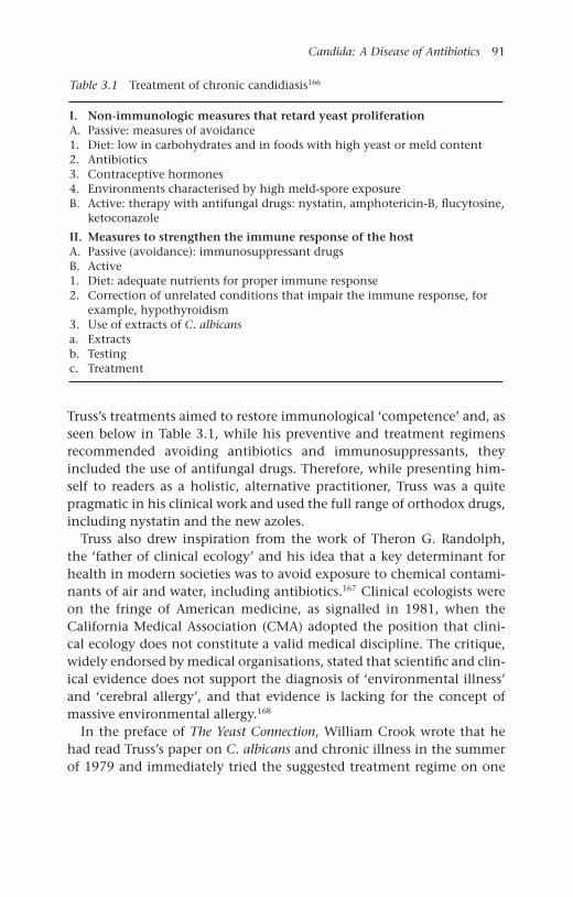
Candida: A Disease of Antibiotics 91
Table 3.1 Treatment of chronic candidiasis166
I. Non-immunologic measures that retard yeast proliferationA. Passive: measures of avoidance1. Diet: low in carbohydrates and in foods with high yeast or meld content2. Antibiotics3. Contraceptive hormones4. Environments characterised by high meld-spore exposureB. Active: therapy with antifungal drugs: nystatin, amphotericin-B, flucytosine,
ketoconazole
II. Measures to strengthen the immune response of the hostA. Passive (avoidance): immunosuppressant drugsB. Active1. Diet: adequate nutrients for proper immune response2. Correction of unrelated conditions that impair the immune response, for
example, hypothyroidism3. Use of extracts of C. albicansa. Extractsb. Testingc. Treatment
Truss’s treatments aimed to restore immunological ‘competence’ and, asseen below in Table 3.1, while his preventive and treatment regimensrecommended avoiding antibiotics and immunosuppressants, theyincluded the use of antifungal drugs. Therefore, while presenting him-self to readers as a holistic, alternative practitioner, Truss was a quitepragmatic in his clinical work and used the full range of orthodox drugs,including nystatin and the new azoles.
Truss also drew inspiration from the work of Theron G. Randolph,the ‘father of clinical ecology’ and his idea that a key determinant forhealth in modern societies was to avoid exposure to chemical contami-nants of air and water, including antibiotics.167 Clinical ecologists wereon the fringe of American medicine, as signalled in 1981, when theCalifornia Medical Association (CMA) adopted the position that clini-cal ecology does not constitute a valid medical discipline. The critique,widely endorsed by medical organisations, stated that scientific and clin-ical evidence does not support the diagnosis of ‘environmental illness’and ‘cerebral allergy’, and that evidence is lacking for the concept ofmassive environmental allergy.168
In the preface of The Yeast Connection, William Crook wrote that hehad read Truss’s paper on C. albicans and chronic illness in the summerof 1979 and immediately tried the suggested treatment regime on one
92 Fungal Disease in Britain and the United States 1850–2000
of his difficult patients, ‘a 41-year old woman (I’ll call her Nancy Jones)with severe chronic hives [urticaria], accompanied by mental confusion,fatigue and depression’.169 He started her on nystatin and a yeast-free,low carbohydrate diet. Within six days her hives had improved, inweeks they disappeared and after almost a year all her symptoms hadimproved. Crook reported trying the regimen with another 20 patients.
Nearly all were adults with complex health problems, includingheadache, fatigue, depression, recurrent vaginal infection, joint painand sensitivity to chemical odours and additives. Almost withoutexception, they improved. And some improved dramatically.170
He continued ad hoc variations in his treatments, extending the rangeof conditions and ages, eventually to include his paediatric patients. Inthe meantime, Truss had been featured in the ‘Dan Freeman Report’ onCNN in September 1981, an appearance that allegedly brought moreresponses than any previous programme.
In was not long before Truss and Crook joined forces and they didso first at an ‘informal’ conference they called on ‘C. albicans and therelationship to human disease’ in Dallas, Texas, in July 1982.171 This wasattended by 20 physicians and an equal number of patients. Crook madehis television debut on the subject in Cincinnati in January 1983, in abroadcast that led to 7,300 requests for more information and his deci-sion to write The Yeast Connection. In the meantime, Truss self-publishedThe Missing Diagnosis; but it was Crook and his book that gained thepublic’s attention, not least because he was accessible to the media andan effective communicator. The first print run of The Yeast Connection in1983 quickly sold out. He claimed that 270,000 copies were purchasedin the first two years. Crook wrote in the preface that, already, ‘my recog-nition of “the yeast connection” has changed my life and my practiceand had enabled me to help many, many patients conquer previouslydisabling illnesses’. The book was in its fourth edition in 1986.
Crook soon had wider ambitions, hoping to forge what he saw as ‘TheComing Revolution in Medicine’.172 He had written The Yeast Connectionas a self-help manual, with checklists, diagrams, illustrations and clearpreventive and therapeutic advice on necessary changes in lifestyle anddiet, including recipes, and special measures for different patient groups.One of the most controversial features of the book was its 10-pointself-diagnosis schedule, where three or four ‘yes’ answers suggested that‘yeasts played a role in your symptoms’.173 The explanation of the causesof yeast overgrowth was presented in words and graphics. Crook’s advice
Candida: A Disease of Antibiotics 93
was threefold: first, ‘avoid foods which promote yeast growth’; second,seek a prescription from your doctor for ‘medication which helps ridyour body of yeast germs’ (nystatin or ketoconazole); and, third, makechanges to your lifestyle and behaviour. In the early 1980s, taking pre-scription antifungal drugs was an integral part of the treatment and themerits of nystatin and ketoconazole were discussed in some detail.174
However, later and in the hands of other advocates, the self-help and‘alternative’ features took over, as the regime moved to a natural therapy,not least because many doctors refused patients antifungal drugs as theydid not accept that ‘fungal overgrowth’ was a disease or syndrome at all.
The popular success of Truss and Crook brought imitators who linkedCandida overgrowth directly to other, so-called, ‘twentieth centurydiseases’.175 In the hands of Truss and Crook, ‘fungal overgrowth’ hadalways been linked to allergies and infection, and to chemical sensi-tivities, hyperactivity and mental disorders.176 Soon the illnesses theyhad identified were medicalised by other doctors, with such names asthe Candida syndrome, Candida allergy syndrome, the yeast syndrome,polysystemic chronic candidiasis, chronic candidiasis syndrome and,most commonly, candidiasis hypersensitivity syndrome (CHS). In June1984, Crook branched out from popular writing and appearances toadvance ‘The Yeast Connection’ to the American medical profession.His chosen subject was depression and he wrote a letter to the Journalof the American Medical Association suggesting that the condition was‘commonly related to prolonged or repeated courses of broad-spectrumantibiotics or to birth control pills, which promote the overgrowthof C. albicans on mucous membranes’.177 He acknowledged that the‘mechanisms involved still have not been clearly elucidated’, but wrotethat he had good evidence ‘from clinical history, followed by a ther-apeutic response to oral nystatin and a yeast-free, low-carbohydratediet’. His views were rounded upon by several correspondents, whodismissed his claims as lacking evidence and being based on multiplemisconceptions.178
The following year, several medical organisations attacked Crook,Truss and their followers. The American Academy of Allergy andImmunology was worried by the attention being given to CHS andin August 1986 published a position statement in its journal.179 ThePractice Standards Committee found ‘multiple problems with the can-didiasis hypersensitivity syndrome’; principally that ‘the concept isspeculative and unproven’ and that ‘elements of the proposed treat-ment program are potentially dangerous’. The Committee stated that‘basic elements of the syndrome would apply to almost all sick patients
94 Fungal Disease in Britain and the United States 1850–2000
at some time’ and that ‘the broad treatment program would produceremission in most illnesses regardless of cause.’ Moreover, there was ‘nopublished proof that C. albicans is responsible for the syndrome’ or that‘treatment . . . with specific antifungal agents . . . benefits the syndrome.’The dangers in the treatment regimes were that the promiscuous use ofdrugs would produce resistant strains of C. albicans and of that therecould be long-term effects with patients on systemic antifungals formany years. In November 1987, at a meeting on Controversies in Infec-tious Disease, John E. Edwards of (UCLA) attacked Crook and those onhis bandwagon.180 His description nicely captured the frustrations ofregular medicine.
Certain generalizations can be made regarding ‘the yeast connec-tion.’ The symptoms described by the authors are generalized andaffect nearly every organ system. As listed, some symptoms are widelydiverse; for instance, both fatigue and hyperactivity are included.Nearly every normal individual has had certain of these symptomsduring the course of a normal lifespan. Case reports are anecdotal.Possibly none of the authors have had formal training in the disci-plines of allergy and immunology, infectious diseases, or mycology.After nearly a decade since the original description, no articles onthis disease appear in peer reviewed journals included in the IndexMedicus. There are no prospective controlled therapeutic studies, andthere are no animal model data.181
A year later, the Canadian Paediatric Society warned that, ‘Physiciansmust not be swayed by the attention that the syndrome has attracted inthe lay press.’182
The Yeast Connection was published in Britain in the summer of 1988.Chronic candidiasis had been discussed in the popular press for a cou-ple of years and linked to myalgic encephalomyelitis (ME) or post-viralfatigue syndrome (PVFS).183 In The Observer, Sue Finlay wrote that MEwas ‘An illness doctors don’t recognise’, but which she had overcomeby following the diet recommended in Leon Chaitow’s Candida albi-cans: Could Yeast Be Your Problem?184 Clinical ecologists also gained ahearing in Britain. One described Candida overgrowth as ‘the quiet epi-demic that is ruining modern lifestyles’, due to the specific condition of‘dysbiosis [abnormal intestinal flora]’ and to ‘[t]he burgeoning of com-plex viral infections such as AIDS and ME – and, to a lesser extent,Herpes’.185 In these conditions, it was claimed, ‘candidiasis was almostalways present as an immune-sapping illness’.
Candida: A Disease of Antibiotics 95
Although there was a pathological theory behind The Yeast Connection,Crook relied on the claim that the real test of his ideas and recommenda-tions was in the clinic. He once said, ‘There’s not a single test to prove it,but it works’ and used emotive case histories to great effect; such as thatof Darlene Lindbom of Paris, Tennessee, who ‘went to two universitiesin a wheelchair. “You’ve got something like MS”, they told her – she hadspinal taps, biopsies, the lot. I put her on my special diet and nystatin.Now she’s fit and runs a successful business.’186 Crook visited London topromote his ideas in June 1988, which the Guardian styled the ‘thrushtheory’. He stressed the link to food allergies and found a forum with theBritish Society for Allergy and Environmental Medicine, which had linkswith the British Society for Nutritional Medicine.187 Both meetings wereregarded as ‘alternative’ by the mainstream British medical professionand studiously ignored.
In 1989 the first clinical trials with patients reporting the ‘Yeast Con-nection’ were published. Lisa Renfro and colleagues at the Departmentof Pediatrics and Family Medicine at Farmington, Connecticut, reportedon 100 consecutive patients suffering from chronic fatigue, eight ofwhom believed their symptoms were due to chronic candidiasis.188 Thearticle concluded that the authors were ‘unable to find physical or lab-oratory findings that were different from the 92 other patients withchronic fatigue’. However, they did find that ‘patients with the yeastconnection were more likely to be taking high doses of vitamins andwere more likely to be getting help from non-medical caretakers. In fact,these caretakers might be the source of the diagnosis.’189 They went onto conclude that all but one sufferer had depression or an anxiety disor-der, and that, from the point of view of achieving a positive outcome,not dismissing chronic candidiasis might be beneficial in allowing atherapeutic relationship between doctor and patient to be maintained.The following year a similar study was published by doctors at theUniversity of Alabama Medical School in Birmingham, Crook’s localstomping ground.190 This was a state-of-the-art randomised, double-blind trial of nystatin therapy in CHS, which concluded that, whilepatients on the trial improved, as was to be expected, the study had pro-vided ‘additional objective evidence that the syndrome is not a verifiablecondition’.191 An accompanying editorial in the New England Journal ofMedicine anticipated that supporters of the yeast connection would notbe impressed and, as expected, Crook and others wrote in pointing tosuccessful treatment in with many patients.192
In Britain, the yeast connection only attracted sustained medicalcriticism in the early 1990s and then in the context of a complex
96 Fungal Disease in Britain and the United States 1850–2000
debate that linked allergies, food intolerance and alternative medicine.These issues crystallised in a report by the Royal College of Physi-cians on Allergy: Conventional and Alternative Concepts, in 1992, whichstated the ‘Candida theory is unsubstantiated’.193 Responses quicklyappeared in ‘alternative’ medical publications, particularly in the Jour-nal of Nutritional Medicine; however, the specifics of the ‘Candida theory’were lost in a larger dispute on the status of ‘alternative’ medicine.194
In July 1992, Keith Mumby, Britain’s most high-profile clinical ecolo-gist, appeared before the General Medical Council (GMC) and was foundguilty of ‘touting for charges’ and failing to give a patient adequate med-ical attention.195 This led the main author of the College’s Report onAllergy, Barry Kay, to argue that the ‘GMC should consider censoringall forms of diagnosis and treatment which, by reasonable standards,have consistently failed to show clinical efficacy’. Mumby was allowedto reply in an article entitled ‘Science or flat earthers? The clinical ecol-ogist replies’.196 This was almost the last word, as the stridency andfrequency of the medical establishment’s assault of alternative practi-tioners waned, though patient demand in Britain and the United Statescontinued to grow.197 In medicine, CHS was gradually absorbed into anumber of, what became known as, ‘symptom-based conditions’, whichincluded chronic fatigue syndrome, fibromyalgia, multiple chemicalsensitivities, sick building syndrome, Gulf War syndrome and irritablebowel syndrome.198 Crook continued to publish, seeking niche marketswith cook books and patient-specific audiences: women, children with,attention deficit disorder and autism, and people with chronic fatiguesyndrome.199 Many other authors expanded the genre, with titles suchas The Candida Control Cookbook (1996), Feast Without Yeast: 4 stages tobetter health (1999), and Complete Candida Yeast Guidebook: everything youneed to know about prevention, treatment, & diet (2000). However, the med-ical profession increasingly ignored CHS, except to dismiss it, especiallybecause of the new emphasis on evidence-based medicine and the GoldStandard of double blind controlled clinical trials.200
Antibiotics were the icon of mid-twentieth-century medical progressand their development influenced Candida infection in complex ways.As thrush, the disease came to the fore in the post-war years whennystatin, the first antifungal antibiotic, was introduced and broughtwomen with the vaginal infection to the clinic. Doctors believed thatpreviously the condition had been self-treated or accepted, perhaps self-limiting, but had certainly been underreported. At the same time, the
Candida: A Disease of Antibiotics 97
use of antibacterial antibiotics, especially broad-spectrum formulations,by clearing the body of its natural microbial fauna, seemed to open thebody to topical infection. New clothing may have been a factor too,with stretch synthetic fabrics making underwear more close fitting andimpermeable. Antibiotics were also implicated in systemic or invasivecandidiasis, as the numbers of vulnerable patients multiplied. Amongstcancer patients, steroid and other treatments depressed the immune sys-tem, as did blood cancers like leukaemia. Some of the new systemiccandidiasis patients suffered from iatrogenic conditions. The principalgroups were transplant patients, those in intensive care, those main-tained with serious chronic conditions and then people with HIV/AIDS.However, the rising tide of candidiasis was met with new antifungalantibiotics, especially azole drugs and, by the 1990s the managementof systemic candidiasis was more successful. In the 1980s another newtype of candidiasis emerged, CHS, which although dismissed by main-stream medicine as a fiction and a fad, became the archetypal ‘diseaseof modernity’. Its alleged cause, overgrowth of C. albicans in the body,was linked to many features of modern life, including the overuse ofantibiotics. It was not without irony, therefore, that, alongside lifestyleand dietary changes, taking the antifungal antibiotics produced by themodern pharmaceutical industry was also recommended.
Except where otherwise noted, this work is licensed under aCreative Commons Attribution 3.0 Unported License. To view
a copy of this license, visit http://creativecommons.org/licenses/by/3.0/
OPEN
4Endemic Mycoses and Allergies:Diseases of Social Change
In 1950, the Biology Section of the New York Academy of Science(NYAS) held what it claimed to be the first conference on medicalmycology in the United States.1 What prompted the event was not theannouncement of the discovery of nystatin by Hazen and Brown, astheir publication was still in press, but the growing profile of fungi andfungal infections across the nation. Fungi, not least because of interestin penicillin, were attracting the interest of biologists and biomedicalresearchers who, alongside screens for antibiotic activity, were adopt-ing them as experimental models in studies of nutrition, physiologyand immunology.2 All the leading names of the field from the 1930sattended the meeting: Carroll Dodge, Norman Conant, Rhoda Benhamand Lucille Georg, and there were new faces who had developed exper-tise during the war and in particular localities. Speakers drew attentionto the increased incidence of systemic candidiasis, signalling a switchin the medical mycological gaze from external (exogenous) to internal(endogenous) disease. Although the incidence of endogenous, systemicfungal infections was very low, they had very high mortality and pre-sented unusual cases that fascinated physicians. In addition, there wasa new awareness of the toll of morbidity from endemic, exogenous dis-ease, as with athlete’s foot and thrush, and with regionally specific, oftensub-clinical infections, principally coccidioidomycosis, blastomycosisand histoplasmosis.
In this chapter, we tell the story of regionally specific fungal infec-tions, and look at the rise of fungal-induced asthma, as one part of thetwentieth-century story of the rise of allergies and asthma.3 We beginby discussing the new epidemiology of endemic fungal infections thatemerged in the late 1940s and the attempts by medical mycologistsand other interested clinicians to attract more resources for research,
98
Endemic Mycoses and Allergies: Diseases of Social Change 99
prevention and control. We discussed the increased incidence of acute,invasive candidiasis associated with new medical treatments in the lastChapter; here we examine in turn the three principal chronic, thoughoccasionally epidemic, regional mycoses prevalent in North America –coccidioidomycosis, blastomycosis and histoplasmosis. Our attentionthen switches to Britain and allergic fungal conditions, firstly, farmer’slung and then allergic bronchopulmonary aspergillosis (ABPA). Thisclass of fungal allergens was ‘discovered’ in Britain and seemingly absentfrom North America, until expertise was transferred back across theAtlantic.
The new epidemiology of fungal diseases
In the early 1950s, medical mycologists, along with cancer physiciansand chest surgeons, began to draw attention to a new problem posed byinvasive fungal infections. Writing in 1953, David Smith, a colleague ofNorman Conant at Duke Medical School described the new situation asfollows.
Unlike most bacterial and viral infectious diseases, the systemicmycotic infections are not transmitted directly from patient topatient; consequently, one would not expect to see epidemics causedby fungi. In most instances the mycotic infections are endemic andsporadic but true epidemics of sporotrichosis, coccidioidomycosisand histoplasmosis do occur when groups of non-immune individ-uals are exposed to an environment containing large amounts ofthe saprophytic form of the fungus. More than a thousand casesof sporotrichosis developed in the gold mines of South Africa whenthe timbers in the mine became infected with Sporotrichum schenckii.Epidemics of coccidioidomycosis occurred when companies of non-immune soldiers from the East marched in the dust of certain South-western deserts. Epidemics of histoplasmosis have occurred followingthe exposure of groups of nonimmune individuals to pigeon manure,chicken manure, bat manure in caves and to the dust of unused silos.4
Reviewing the epidemiology of fungal infections in the same year forthe New England Journal of Medicine, Otis Jillson pointed to ‘the recog-nition of the benign, common forms of histoplasmosis; the diagnosisand surgical treatment of coccidioidal pulmonary residua; the treatmentof blastomycosis with stilbenes’, and added the growing incidence ofsystemic mycoses.5 Interestingly, he dealt with skin infections briefly
100 Fungal Disease in Britain and the United States 1850–2000
and said very little about exogenous or endogenous candidiasis, at atime when nystatin was attracting attention.
At the 1950 NYAS meeting, Samuel B. Salvin, then at the Division ofInfectious Diseases at NIH, placed the incidence of fungal diseases incontext of other infectious diseases.
Fungus infections in man, although less frequent than bacterial, arestill numerically important. For example, of the 92,933 deaths due toinfections and parasitic diseases in the United States in 1945, 284, or0.3 per cent, were due to mycoses. This was approximately equal tothe number of deaths reported as caused by scarlet fever, measles, orthe typhus-like diseases (due to rickettsia), and was more than thetotal of all deaths recorded from rabies, smallpox, relapsing fever,leprosy, brucellosis, paratyphoid fever, plague, cholera, and anthrax.It should be realized, of course, that effective control measures areemployed against some of the aforementioned diseases, whereas con-trol methods against the mycoses not only are not practiced, but,generally, are not even known. It should also be borne in mindthat the dermatophytoses, although characteristically nonfatal, areextremely common, probably equalling the most widespread of thebacterial or virus diseases in prevalence.6
In 1953, Walter Nickerson from the Department of Microbiology,Rutgers University, tried a creative presentation of mortality data tochart the rise of fungal infections.7 He used graphs for the period1945–1949 that showed starkly opposite mortality trends, where ‘allinfections’ had dropped sharply, while that from mycoses had increasedmarkedly. Nickerson had produced his graphic illustration by using verydifferent scales for the two classes of infection. Deaths from mycoseswere recorded as actual numbers, while those from all infections wererecorded in thousands. The alarming increase of nearly 50% in fungalinfection deaths was actually from 270 to 380, while the actual num-ber of deaths from all infections had fallen 30%, from 93,000 to 66,000deaths – still 170 times greater! In the event, the annual total deathsfrom mycoses in the next decade never reached 500.8 More telling wasNickerson’s point about morbidity; he stated that ‘mycotic infectionsare probably the mostly widely distributed and most numerous types ofinfection, with dermatophyte infections, such as athlete’s foot, alone asprevalent as the most widespread of the bacterial and virus diseases’.9
Medical mycologists had long argued for recognition of the distinc-tive pathogenicity of fungi. In 1940, Arthur Henrici, who worked inthe Department of Bacteriology at the University of Minnesota, argued
Endemic Mycoses and Allergies: Diseases of Social Change 101
that most bacterial and viral infections developed rapidly, and thenplateaued in severity, before falling away, because of either recovery ordeath. With fungal infections, however, a typical pattern was of the slowand incremental development of chronicity, and prolonged morbidity,often at sub-clinical levels.10 The only bacterial diseases with similarpatterns of pathogenicity were tuberculosis and leprosy. Henrici main-tained that the slow increase in severity meant either that the infectingfungus progressively changed and developed pathogenic properties orthat the resistance of the host was gradually worn down. There wasno evidence of the former, so he focused on changes in the host andits ‘soil’, claiming that prolonged exposure to certain fungi and theirtoxins produced hypersensitivity in the host cells. Henrici referred backto the theories of Richard Pfeiffer and Clemens von Pirquet on over-active immune responses.11 Pfeiffer’s authority was drawn upon for theargument that fungal endotoxins inflamed and ultimately killed cells,creating a nidus for the fungus itself to grow. On the other hand, vonPirquet’s work was used to suggest that host cells developed allergic-typesensitivity. Henrici favoured the latter, but stressed that because fungalinfections were complex and variable, both mechanisms might operate,or be found with different species of pathogen.
The prevalence of sub-clinical, chronic disease was recognised bypublic health officials as characteristic of regionally specific mycosesin the United States. The new geography of endemic fungal infec-tions was revealed by David Smith in 1953, on a map that showedthe distribution of coccidioidomycosis, blastomycosis and histoplasmo-sis in the United States and northern Mexico12 (Figure 4.1). The newepidemiological profile changed the position of medical mycology inthe United States in the 1950s. This was evident first in the prolifer-ation of new publications and courses.13 Ana Espinel-Ingroff’s analysisof the institutional development of medical mycology shows that newdepartments and new experts emerged in affected areas, for example,at Michigan State University (1951), Tulane University (1955), the Uni-versity of California, Los Angeles (1956), the University of Oklahoma(1957) and Virginia Commonwealth University (1965).14 Initiatives tookplace in varied settings, sometimes with public health departments, butmostly in university biology and microbiology departments. That said,leading figures and departments in the East remained important. Nor-man Conant, Chester Emmons and Libero Ajello continued to headkey departments at Columbia, Duke and the National MicrobiologicalInstitute, which was reorganised from 1955 as the National Institute ofAllergy and Infectious Diseases (NIAID) and, of course, the Centers forDisease Control and Prevention (CDC) in Atlanta remained influential.
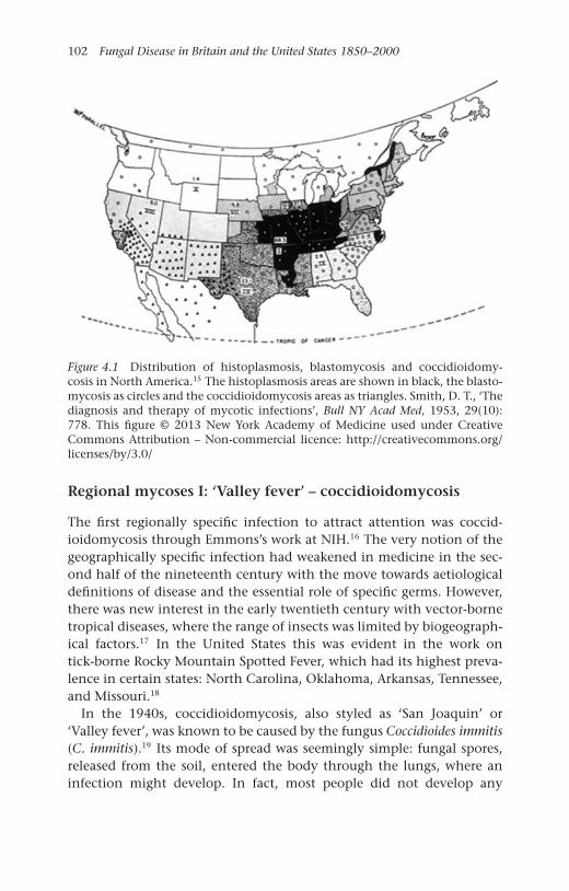
102 Fungal Disease in Britain and the United States 1850–2000
Figure 4.1 Distribution of histoplasmosis, blastomycosis and coccidioidomy-cosis in North America.15 The histoplasmosis areas are shown in black, the blasto-mycosis as circles and the coccidioidomycosis areas as triangles. Smith, D. T., ‘Thediagnosis and therapy of mycotic infections’, Bull NY Acad Med, 1953, 29(10):778. This figure © 2013 New York Academy of Medicine used under CreativeCommons Attribution – Non-commercial licence: http://creativecommons.org/licenses/by/3.0/
Regional mycoses I: ‘Valley fever’ – coccidioidomycosis
The first regionally specific infection to attract attention was coccid-ioidomycosis through Emmons’s work at NIH.16 The very notion of thegeographically specific infection had weakened in medicine in the sec-ond half of the nineteenth century with the move towards aetiologicaldefinitions of disease and the essential role of specific germs. However,there was new interest in the early twentieth century with vector-bornetropical diseases, where the range of insects was limited by biogeograph-ical factors.17 In the United States this was evident in the work ontick-borne Rocky Mountain Spotted Fever, which had its highest preva-lence in certain states: North Carolina, Oklahoma, Arkansas, Tennessee,and Missouri.18
In the 1940s, coccidioidomycosis, also styled as ‘San Joaquin’ or‘Valley fever’, was known to be caused by the fungus Coccidioides immitis(C. immitis).19 Its mode of spread was seemingly simple: fungal spores,released from the soil, entered the body through the lungs, where aninfection might develop. In fact, most people did not develop any
Endemic Mycoses and Allergies: Diseases of Social Change 103
symptoms and those that did experienced a cold or mild flu-like symp-toms. In a tiny fraction of people, especially those with other diseasesand with weakened immune systems, infection spread through thebloodstream to give disseminated coccidioidomycosis.
Research by Ernest C. Dickson and Charles Smith in the 1930s hadrevealed that infection with C. immitis was very common amongst thoseliving in certain areas, and that it was best regarded as an endemic,chronic and benign infection.20 It seemed that residents built up immu-nity from long-term, low-level exposure, hence most infection wassub-clinical and, as would be expected, clinical disease was most com-mon amongst in-migrants who had not had the opportunity to build upimmunity. Population movement and settlement westwards had beengoing on in the United States for many decades and it seemed likelythat coccidioidomycosis emerged at this particularly moment becauseof the new speed and scale of migration, especially families fleeing dustblows in the prairies. Evidence to support this view came in the 1940swhen coccidioidomycosis developed amongst the recruits brought tothe region to train for the United States Air Force.21 As the war effortgrew, more troops arrived, which gave investigators the opportunityto make comparisons of incidence by sex, race and nationality. Forinstance, some 13,000 German prisoners of war were held at Florence,Arizona, where the incidence of coccidioidomycosis became so highthat they were moved away, as United States government officials wor-ried that such a high rate of infection would lead to them beingcharged with violation of the Geneva Convention on treatment of warprisoners.22
After the war, the military presence in the Southwest continued andexpanded. So too did worries about coccidioidomycosis and this ledin 1955 to the establishment of an annual meeting of the VeteransAdministration-Armed Forces Coccidioidomycosis Cooperative StudyGroup (CCSG). This type of cooperative meeting had begun in the late1940s as a way of developing and sharing expertise on the treatment oftuberculosis with streptomycin.23 Initially, the main agencies were theVeterans Administration, the Army, the Navy, Public Health Services andthe National Research Council. While the focus of the early annual con-ferences was squarely on tuberculosis, fungal infections were sometimesdiscussed, as in 1952 when trial coordinators noted that histoplasmosislung infections had complicated their clinical trials by making differen-tial diagnosis with X-rays more difficult.24 The first coccidioidomycosismeeting in 1955 came from a direct concern with the growing inci-dence of the infection, especially at air bases, notably Williams AFB
104 Fungal Disease in Britain and the United States 1850–2000
and Luke AFB in Arizona, Edwards AFB in California, and Lemoore NAS,also in California. The problem grew with the investments in militaryinfrastructure that came with the Cold War. Williams and Luke becametraining centres, with a steady stream of non-immune recruits passingthrough, while Lemoore was further developed in the 1960s for strikeaircraft, and Edwards became a centre for research, including rocketry,and eventually was a landing site for the Space Shuttle.
The construction of new runways and other facilities on these mil-itary sites disturbed the subsoil, which together with aircraft take-offsand landings circulated C. immitus spores to those living and workingnearby.25 Some civilian sufferers were treated by military doctors; how-ever, they were mostly dealt with by local physicians, who also treatedmilitary personnel when they were referred to local hospitals for sero-logical and radiological investigation. Thus, the leading authority onthe disease in the military, from the 1940s to the 1960s, was Charles E.Smith, Dean of Public Health at the University of California Berkeley.26
He had developed his expertise during the Second World War at Berke-ley, where he established a research laboratory and diagnostic serologicalservices.
In the 1950s C. immitus biology was found to be more compli-cated than previously recognised and the new understanding was setout in the first book entirely on the disease by Marshall J. Fiese in1958.27 Fiese was based at the Veterans Administration Hospital inFresno and in his ‘Foreword’ to Fiese’s volume, Charles Smith wrotethat the work reflected the author’s deep experience: ‘[Fiese] has seenand viewed . . . . countless roentgenograms[X rays], seen and studied thetragic autopsies, and perhaps most importantly of all, lived for yearsin the coccidioidal countryside.’ An unusual feature of C. immitis wasthat, rather than being spread by spores, it was actually the cells of thehyphae, called conidia, that circulated in the air. Conidia were foundto be tiny and readily carried in dust; hence, the popular represen-tation of coccidioidomycosis as spread by ‘flying conidia’, or ‘flyingchlamydospores’.28 When inhaled, the human body was shown torespond in one of three possible ways. First and most commonly, theconidia were destroyed by the immune system and a degree of immu-nity, albeit variable, to future infection was established. Alternatively,the conidia grew in the lungs in a spherical form, into bodies thatreleased many more such ‘spherules’, producing inflammation and achest infection. Lastly and least common, and only if the lung infectionwas severe, infection could spread in the blood and cause inflammation,especially in the skin and brain. The reported incidence of the severe
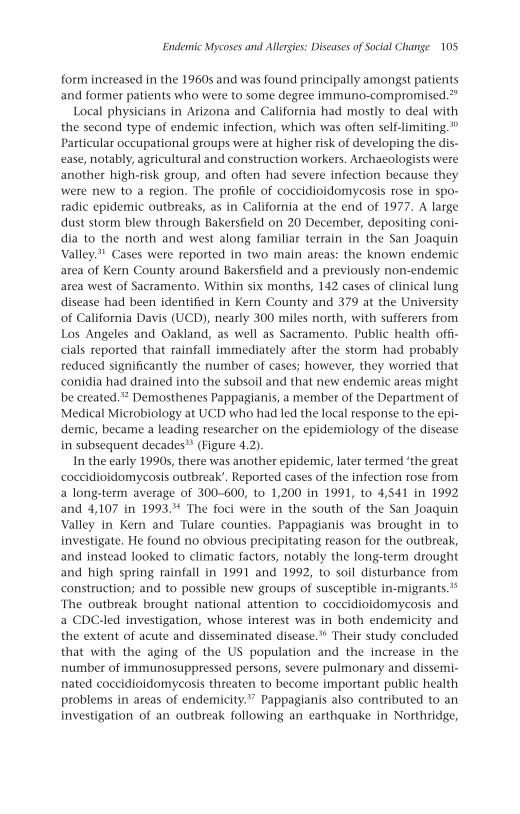
Endemic Mycoses and Allergies: Diseases of Social Change 105
form increased in the 1960s and was found principally amongst patientsand former patients who were to some degree immuno-compromised.29
Local physicians in Arizona and California had mostly to deal withthe second type of endemic infection, which was often self-limiting.30
Particular occupational groups were at higher risk of developing the dis-ease, notably, agricultural and construction workers. Archaeologists wereanother high-risk group, and often had severe infection because theywere new to a region. The profile of coccidioidomycosis rose in spo-radic epidemic outbreaks, as in California at the end of 1977. A largedust storm blew through Bakersfield on 20 December, depositing coni-dia to the north and west along familiar terrain in the San JoaquinValley.31 Cases were reported in two main areas: the known endemicarea of Kern County around Bakersfield and a previously non-endemicarea west of Sacramento. Within six months, 142 cases of clinical lungdisease had been identified in Kern County and 379 at the Universityof California Davis (UCD), nearly 300 miles north, with sufferers fromLos Angeles and Oakland, as well as Sacramento. Public health offi-cials reported that rainfall immediately after the storm had probablyreduced significantly the number of cases; however, they worried thatconidia had drained into the subsoil and that new endemic areas mightbe created.32 Demosthenes Pappagianis, a member of the Department ofMedical Microbiology at UCD who had led the local response to the epi-demic, became a leading researcher on the epidemiology of the diseasein subsequent decades33 (Figure 4.2).
In the early 1990s, there was another epidemic, later termed ‘the greatcoccidioidomycosis outbreak’. Reported cases of the infection rose froma long-term average of 300–600, to 1,200 in 1991, to 4,541 in 1992and 4,107 in 1993.34 The foci were in the south of the San JoaquinValley in Kern and Tulare counties. Pappagianis was brought in toinvestigate. He found no obvious precipitating reason for the outbreak,and instead looked to climatic factors, notably the long-term droughtand high spring rainfall in 1991 and 1992, to soil disturbance fromconstruction; and to possible new groups of susceptible in-migrants.35
The outbreak brought national attention to coccidioidomycosis anda CDC-led investigation, whose interest was in both endemicity andthe extent of acute and disseminated disease.36 Their study concludedthat with the aging of the US population and the increase in thenumber of immunosuppressed persons, severe pulmonary and dissemi-nated coccidioidomycosis threaten to become important public healthproblems in areas of endemicity.37 Pappagianis also contributed to aninvestigation of an outbreak following an earthquake in Northridge,
106 Fungal Disease in Britain and the United States 1850–2000
Figure 4.2 The geographic distribution of coccidioidomycosis. Cross-hatchingindicates the heavily disease-endemic area, single hatching and the moderatelydisease-endemic area.38 Kirkland, T. N. and Fierer, J., ‘Coccidioidomycosis:A reemerging infectious disease’, Emerg Infect Dis [serial on the Internet].1996, Sep. Available from www.nc.cdc.gov/ncidod/eid/vol2no3/kirkland.htmDOI: 10.3201/eid0203.960305. CDC Public domain material.
California in January 1994, to which 203 cases were linked and threecoccidioidomycosis deaths added to the overall toll of 57.39
In addition to Pappagianis, two other researchers stood out in thestudy of coccidioidomycosis during the period. One was John Galgiani,who worked at the Veterans Affairs Medical Center and University ofArizona, Tucson, and was the leading expert on coccidioidomycosis inthat state. His specialism was immune responses and he was amongstthe first to publish on coccidioidomycosis in AIDS sufferers.40 Overmany years he campaigned for recognition that coccidioidomycosis wasbecoming a disease of national importance, because of continuing highrates of in-migration to Southwestern states and the increase in thenumber of immunosuppressed patients. General population mobilitywas also a factor, with many cases presenting outside of endemic areasin out-migrants; indeed, he reported that 46% of coccidioidomycosispatients with AIDS were in non-endemic areas.41 In 1996, coccid-ioidomycosis was being discussed as ‘a re-emerging infectious disease’.42

Endemic Mycoses and Allergies: Diseases of Social Change 107
Theo Kirkland and Joshua Fierer, respectively, from the San DiegoSchool of Medicine and Department of Veterans Affairs Medical Cen-ter, San Diego, wrote that it warranted the label because ‘the numberof cases . . . has increased dramatically, and the clinical symptoms ofthis illness have changed in patients with acquired immunodeficiencysyndrome (AIDS)’.43
The second key researcher was David A. Stevens, who was based in theSanta Clara Valley Medical Center, San José and the Stanford UniversitySchool of Medicine. He became a leading authority on the clinical man-agement of coccidioidomycosis, especially the use of chemotherapy. Hisexpertise, developed along with that on the treatment of aspergillosisand other opportunistic mycoses, was recognised in leadership roles andhonours in the Medical Mycological Society of the Americas (MMSA),International Society for Human and Animal Mycology (ISHAM) andthe American Society for Microbiology (ASM). Stevens’s early careerwork was on viruses, but he switched to fungi in 1974 with a studyof immunity to C. immitis and then to its treatment.44 With HillelB. Levine, he pioneered the use of one of the new azoles, micona-zole, for coccidioidomycosis.45 In 1980, he edited a new textbook onthe disease, contributing chapters on immunology, other syndromes,immuno-compromised hosts, chemotherapy immunotherapy, vaccina-tion and a bibliography.46 Stevens enrolled Pappagianis to write onepidemiology and serology, and Galgiani on ophthalmic disease.
By the 1970s, the Cooperative Study Group meetings, which beganunder military sponsorship, had moved to civilian control, with Stevens,Galgiani and Pappagianis taking leading roles. However, as early as 1957a larger symposium had been called and that met every eight yearsor so, styling itself as ‘international’ for the first time when it met inTucson in 1977.47 This change reflected two developments: first, greaterinterest in the disease in Mexico and South America and second, theexperience of physicians across borders with serological diagnosis andamphotericin B treatment of disseminated disease.48 Furthermore, afterthe Californian coccidioidomycosis outbreak in 1977, a growing num-ber of laboratories had begun to explore vaccine development, utilisingthe tools of the new molecular biology.49 Indeed, a vaccine developedby Pappagianis and Levine went on trial in 1981, but this showed littleor no benefit.50 Attention was also directed to ethnic and social groupsthat were at greater risk from the disease, particularly black men, NativeAmericans, and pregnant women. With the former, researchers exploredthe contribution of racial susceptibility and environmental factors, witha consensus developing around the importance of the latter.

108 Fungal Disease in Britain and the United States 1850–2000
Researchers found that exposure, socio-economic position and accessto health care were the most important determinants of vulnerabilityto infection and the development of disease.51 Pregnant women werethought to be vulnerable because of changes in their immunity and hor-monal levels, but close study showed them to have, at worst, only a tinyadditional likelihood of developing disseminated disease.52 In the 1980s,as would have been expected, more coccidioidomycosis was reported inimmuno-compromised patients in Southwestern states, and also acrossthe United States in people who had lived or travelled to the endemicareas. Thus, the work of regional coccidioidomycosis experts linked upwith that of mycologically minded clinicians nationwide. Their researchand clinical experience put them in a good position to assume nationalleadership roles in the field. For example, David Stevens led the NIHmulticentre clinical trials group on antifungal drugs between 1990 and2000 and chaired committees writing practice guidelines on aspergillosisas well as coccidioidomycosis.53
Regional mycoses II: Blastomycosis – North Americanand otherwise
Blastomycosis was a term invented at the end of the nineteenth cen-tury for eruptive skin lesions or granulomas, which were assumed tobe caused by infective fungi. The prefix ‘blasto-’, from the Greek for‘budding’ or ‘sprouting’, came from the clinical presentation of raisedlesions that were disfiguring, especially so with facial lesions. In the1900s in the United States, the name became associated with so-calledGilchrist’s disease, first thought to be protozoan, but then linked to adimorphic (two forms) fungus named Blastomyces dermatitidis (B. der-matitidis).54 For a while the infection was termed ‘North Americanblastomycosis’ as researchers found that it was restricted to the geo-graphical areas of the Ohio, Mississippi and Missouri River basins andthe western shore of Lake Michigan. There followed, what contempo-raries recognised as, ‘an era of confusion during which the disease wasconfounded with other entities, particularly cryptococcosis and can-didiasis’ and ‘Nineteen new names were suggested for the causativefungus.’55 Consensus on the pathogenesis of the disease followed workby Norman Conant and colleagues at Duke Medical School in the late1930s.56 They endorsed the idea that there was a form of the diseasespecific to North America, though other forms of blastomycosis, dueto other fungal species, were found on other continents. South Ameri-can blastomycosis was a condition with similar symptoms, but produced

Endemic Mycoses and Allergies: Diseases of Social Change 109
by a different fungus – Paracoccidioides brasiliensis. Although blastomy-cosis typically first presented as skin lesions, there were many reportsof disseminated disease, which suggested parallels with coccidioidomy-cosis. Doctors wondered if there was also self-limiting or sub-clinicalpulmonary infection; however, there was no evidence of aerial transmis-sion in the mode of C. immitis. There were few reports of blastomycosisfungus in the environment, and certainly none of concentrations likelyto produce disease in humans. The mystery of the manner in which itspread led some doctors to call it the ‘the enigmatic disease’.57
A study in the mid-1960s by Leo Furcolow, who was based in Kansasin the centre of an endemic area, showed that there had been only 685confirmed cases of blastomycosis across five states between 1912 and1964, a figure which he assumed grossly to under-estimate its actualincidence.58 Furcolow found that infection rates were higher in malesthan females, and there was ‘a slight excess of cases among Negroes’;both factors were linked to the view that blastomycosis was associatedwith outdoor work or sports. A similar geographical distribution of theincidence of B. dermatitidis was found in dogs, so perhaps they werehosts and further credence to this link was suggested by similaritieswith the epidemiology of histoplasmosis. Another difficulty in mappingthe disease, certainly in comparison with coccidioidomycosis, was theabsence of a reliable skin test, which meant that the epidemiologicalpicture relied on symptomatic cases.
Disseminated blastomycosis attracted the attention of cliniciansbecause of its high mortality rate, which was typically 80%, thoughtreatment with amphotericin B brought this down to less than 20%.59
A difference with other endemic fungal diseases was that immunosup-pressed patients were seemingly less likely to become infected, thoughif they did, severe disease was common.60 Epidemiological and clinicalstudies found no significant patterns in the incidence of blastomyco-sis, other than predisposing illness. However, it has been always beenmore common in men, seemingly because of outdoor exposure. Suspi-cions remained that its epidemiology was similar to coccidioidomycosisbecause the lungs were the primary site of infection, and its prevalenceamongst in-migrants and construction workers.61
The low incidence of endemic blastomycosis, along with the limitedsystemic infection, meant that no critical mass of local specialist prac-titioners developed and the expertise of the few people with specialknowledge was not in demand. However, scientists and clinicians in theregions affected were able to develop specialist practices by developingwork on other low-level endemic mycoses, most notably histoplasmosis.
110 Fungal Disease in Britain and the United States 1850–2000
Regional mycoses III: Histoplasmosis62
Histoplasmosis is caused by infection with the fungus Histoplasma cap-sulatum (H. capsulatum), and is associated with specific localities; indeed,it was sometimes referred to as Ohio Valley Fever, though it soonbecame clear its prevalence was wider. Its endemic areas overlapped withthose of other regional fungi and researchers’ work straddled differentmycoses. For example, Leo Furcolow became renowned for his work onblastomycosis and histoplasmosis, and Louis Ajello combined expertiseon coccidioidomycosis and histoplasmosis.63 However, unlike blastomy-cosis, whose low incidence meant less attention from the clinicians,histoplasmosis, similar to the medical history of coccidioidomycosis,gained prominence nationally as social changes led to an increase inci-dence and importance. Moreover, according to Thomas Daniel andGerald L. Baum, a national research network emerged around the dis-ease with work led by federal agencies.64 For Daniel and Baum, one keyfigure in the United States was Jan Schwarz, a Jewish émigré based inCincinnati, Ohio, who worked on tuberculosis, before taking Conant’scourse at Duke and converting to mycology. From his base in Cincin-nati, at the centre of the H. capsulatum endemicity, he became a leadingnational expert alongside his friend Leo Furcolow, who became knownas ‘Mr Histoplasmosis’.
Retrospectively, the first case of histoplasmosis has been identified inthe Panama Canal Zone in 1906, when Samuel Taylor Darling reportedan acute lung infection, with fever and breathlessness, caused by a pro-tozoan that he named Histoplasma capsulatum.65 Sporadic cases werereported over succeeding decades and in 1934 its causal organism wasshown to be a fungus rather than a protozoan.66 However, the diseaseonly attracted the attention of doctors in the 1940s in relation to pul-monary tuberculosis, first in the military and then after the war insanatoria. Recruits for the military were screened for pulmonary tuber-culosis, with both a chest X-ray, which revealed active or healed lesionsin the lungs, and the tuberculin skin test, which through an immuneresponse confirmed either active or previous infection. A significantnumber of recruits showed, perversely, lesions in the lungs with a neg-ative skin test. One possibility was that immunity declined over time;another was that a second disease was causing the lung lesions, and itturned out that histoplasmosis was such a disease. Fortuitously, a meansto investigate the matter became available with the development of atuberculin-type skin test for the infection, using an antigen productcalled histoplasmin. One study showed that of 94 men with healed lung
Endemic Mycoses and Allergies: Diseases of Social Change 111
lesions, 24 cases were due to tuberculosis and 70 to histoplasmosis.67
A later study by Furcolow, published in 1962, showed that histoplas-mosis was responsible for lesions in 7.5% (3,366/44,882) of sanatoriumpatients from across the country, which indicated that nationally some8,200 patients entered sanatoria with evidence of histoplasmosis, andthat of these, 25% might have had active disease with tuberculosis.68
This work, facilitated by the Division of Mycotic Diseases at CDC andthe US Veterans Administration, and orchestrated through a Coopera-tive Study Group from 1952, revealed three features of histoplasmosis.First, it showed that there were certain regions of the United Stateswhere infection with H. capsulatum was very common, but that thedevelopment of symptomatic disease was quite rare. It seemed thatchildren living in endemic areas developed immunity from low-levelexposure and that this gave long-term protection. Thus, those likely todevelop illness were adult in-migrants without previous exposure.
Second, research on the aetiology of the infection was inconclusivefor many years, but eventually yielded that the main source of infec-tion was soil dust and that localities with accumulations of bird and batdroppings were particularly pathogenic. At times, histoplasmosis wasknown as ‘cave disease’. Daniel and Baum describe the ‘detective story’by which the aetiology was solved. Their narrative begins with a case atCamp Gruber in March 1944 when the soil as a source of the fungus wasfirst indicated, but they show that it took several decades to determinea specific aetiology.69 Nonetheless, greater medical awareness of histo-plasmosis and improved serological diagnostic testing led to more casesbeing identified, especially of systemic infection where, as with similarinvasive mycoses, amphotericin B was the treatment of choice.70
By the 1970s, the disease had gained a higher profile, as increased pop-ulation mobility around the United States brought more non-immunepeople to endemic areas, while anyone with active infection who movedanywhere across the country and became immuno-compromised wasvulnerable to severe infection. There were also a series of epidemic out-breaks, often with small numbers affected and typically quite localised,but they were unusual, even bizarre, and attracted press and medicalattention. Daniel and Baum discuss three epidemics: Mason City, Iowain 1962 (returning in 1964) which was traced to bird rookeries; SuwaneeCounty, Florida in 1973, linked to bats; and, the biggest of all, Indi-anapolis in 1978, which returned two years later.71 Final estimates forthe Indianapolis outbreak were that 120,000 people were infected, with448 persons developing clinical illness, 55 with severely disseminateddisease and 19 deaths. Over the 1980s and early 1990s, histoplasmosis
112 Fungal Disease in Britain and the United States 1850–2000
became associated in the public mind and amongst physicians withAIDS, being the commonest endemic mycosis affecting patients.72 Sur-veys in the early 1990s showed that it was found in 2–5% of AIDSsufferers in endemic areas and was the first sign of infection in over halfof these cases. Infection rates amongst AIDS patients as high as 25% werefound in certain cities, notably Indianapolis, Kansas City, Memphis andNashville. Patients had quite general symptoms and treatments with thenew forms of amphotericin B were successful, though maintenance ofantifungals, typically the new azoles, was essential to prevent relapse.
Farmer’s lung and allergic bronchopulmonaryaspergillosis (ABPA)
Unlike the United States, Britain had no regionally specific mycoseslinked to environmental factors of soil and climate, however, therewere geographically localised conditions linked to occupation, whichemerged in the 1950s. The most important was farmer’s lung, whichwas initially linked to the Aspergilli fungi and pulmonary aspergillosis.73
Aspergillosis, the principal cause of which was Aspergillus fumigatus(A. fumigatus), was considered only at the very end of Henrici’s ency-clopaedic Molds, Yeasts and Actinomycetes in 1930, after coccidioidomy-cosis, dermatophytosis, American blastomycosis, histoplasmosis andsporotrichosis.74 However, the 15th edition of Taylor’s Practice of Medicinein 1936 presented aspergillosis as an occupational disease of handlers ofbirds and grain, along with those who sorted human and animal hairfor various products, for example, wigs.75 Also in 1936, Richard Fawcitt,a radiologist in Ulverston, Cumberland, discussed Aspergilli spp. as themain cause of broncho-mycosis in the local farming community.76 Thenumbers affected were small, but this study and one by Fawcitt’s col-league Munro Campbell, are seen in hindsight as the beginning of therecognition of ‘farmer’s lung’ as an occupational disease.77 Fawcitt hadalso found aspergillosis amongst housecleaners, which might have beenlinked to new domestic technologies, such as the vacuum cleaner, whichspread spore-carrying dust from exhaust vents.78
By the end of the 1930s, fungal spores had been added to pollenand house dust as causes of asthma, the prevalence of which was ris-ing and fascinating doctors because of its complex aetiology, variablepresentation, and link between the physiological and the psychologi-cal. At this time the physical basis of asthma was discussed in termsof allergic, hypersensitivity states, where the body reacted abnormallyto certain substances or ‘allergens’.79 The largest group of allergens wasplant and animal matter in the environment, with pollen and aerial
Endemic Mycoses and Allergies: Diseases of Social Change 113
dust understood to be the main exciting causes of allergic symptoms.Doctors debated whether the increased prevalence was due to betterrecognition, or because modern lifestyles made the body more vulner-able and increased exposure to allergens, or because new allergens hadbeen created by modern farming and industry. In 1936, Grafton Brown,a Washington physician who specialised in allergy, pointed out thatfungal spores were ubiquitous, present throughout the atmosphere inhuge numbers and much smaller than pollen grains.80 He was surprisedthat researchers had not considered them as allergens in house dust,especially when scientists were familiar with fungi as contaminants oftheir culture plates and slides in microbiology laboratories. Accordingto Charles Thom and Margaret Church, writing in 1926, ‘the Penicillia,the Aspergilli and the Mucors are the weeds of the culture room’ and,of course, one now celebrated incidence of fungal contamination ledto the discovery of penicillin.81 Surveys of aerial allergens increased inthe 1930s, using aeroplanes to explore high altitudes and remote loca-tions, including flights by Charles Lindberg to survey areas in northernlatitudes.82 While the spores of a large number of fungal species werefound to be potential allergens, they ranked well below pollen and dust.levels of known fungal disease spores found in the atmosphere were wellbelow those of Alternaria, an ascomycete species that caused disease inplants and which emerged as main fungal suspect in causing asthmaattacks.83 Next in importance were the Aspergilli.
In Britain, aspergillosis was discussed in detail in James Duncan’snational survey of fungal disease in 1945, in relation to both pulmonarydisease and farmer’s lung.84 With pulmonary disease, cases were few andfar between, and difficult to diagnose because of confusion with tubercu-losis. However, Duncan was clear that ‘the fungus is an essential factorin the aetiology of farmer’s lung’, but doubted that it was a primaryinflammatory agent.85 In 1953, Thomas Studdert, an assistant physicianat the Cumberland Hospital in Carlisle, contested earlier views,
The currently quoted view that farmer’s lung is an actual fungous(sic) infection of the lungs does not bear close examination. Theexplosive onset, spontaneous clearing, and radiological picture aretotally unlike any true fungous disease, and no real evidence has beenproduced to support this theory.86
Studdert’s alternative, still framed with the possibility of fungal involve-ment, was that farmer’s lung was an allergic reaction ‘to some material inthe fungus-laden dust’.87 By the mid-1950s, the number of fungi consid-ered as allergens increased to include Penicillia, Mucors and other genera,
114 Fungal Disease in Britain and the United States 1850–2000
yet their role was now seen as creating allergenic dust, or mechanicalirritation of the bronchi and alveoli, rather than specific allergic reac-tions. A 1963 report by the British Industrial Injuries Advisory Council,that led to the scheduling of a variety of occupational lung condi-tions, linked farmer’s lung to other ‘dust diseases’, such as thresher’slung, chaff cutter’s lung and bagassosis (sugar cane handler’s disease).88
It maintained the association with mouldy hay and vegetables, but saidnothing about the role of specific fungi.89
A key reason why Aspergilli spp. were dropped from discussions ofasthma and occupational diseases in the 1960s was the creation of thenew specific condition of ABPA.90 In 1952, Kenneth Hinson, with col-leagues at the London Chest Hospital, published a study of eight casesof pulmonary aspergillosis, three of which were said to be an allergictype previously unrecognised and ‘caused by sensitization of the hostto the fungus’.91 The condition was characterised around several symp-toms: a syndrome of recurrent fevers, a changing X-ray pattern showingprogressive lung damage, peripheral blood eosinophilia, and purulentsputum containing the A. fumigatus. Hinson and his colleagues wereseen to have described an unusual type of aspergillosis, itself still veryrare, so their claim was neither challenged nor endorsed; but it remainedon the record. ABPA, as it was later styled, was not characterised bythe invasive growth into tissues, rather fungi simply grew in pulmonaryfluids and on the surface of lung tissues, causing inflammation.
ABPA attracted increasing attention in Britain through the 1950s and1960s as doctors dealt with more patients with chronic lung diseases.It is again a moot point whether the decline in the incidence of pul-monary tuberculosis revealed previously submerged diseases, or whetherthe spectrum of disease that now faced doctors was genuinely new. Overthe 1960s, there was increased incidence of lung cancer, chronic bron-chitis and emphysema, which were linked to the effects of cigarettes,smoke pollution and occupation diseases. The growing incidence ofnon-specific chronic lung disease was captured in 1962 in the creation ofthe new, condition of chronic obstructive pulmonary disease (COPD).92
In both the United States and Britain there was more research on occu-pational diseases, which led to a greater differentiation of causes, linkedto improved prevention and new regulations.93 However, ABPA was onlyreported in Britain and particularly in London, where the concentra-tion of patients with chronic chest conditions allowed Jack Pepys, whowas one of Britain’s leading experts on allergies, to report new find-ings in 1959. He had investigated 145 patients who had A. fumigatusin their sputum, finding that 16 exhibited ABPA, according to Hinson’s1952 criteria.94 Pepys published a second study in 1969, in which
Endemic Mycoses and Allergies: Diseases of Social Change 115
he reported on 111 patients.95 He wrote that the primary indicatorsof ABPA were transitory pulmonary shadows, eosinophilia of bloodand sputum, evidence of allergy to A. fumigatus and fungal mycelia insputum. Corticosteroids were the recommended therapy, having beenshown to be more effective than bronchodilators and other symp-tomatic treatments. The condition was typically debilitating, leading toprogressive deterioration in lung function, though in some patients thecondition ‘burnt out’ and they ‘recovered’.
American allergists and pulmonary specialists were curious about whyABPA was not found across the Atlantic. In 1969, Raymond Slavin andcolleagues at the St. Louis University School asked:
Why is [allergic] aspergillosis such a rarity in the United States?It would seem that the climate and geography of England does notmake a profound difference since A. fumigatus is commonly reportedin air sampling surveys in this country. In addition, as stated previ-ously, secondary aspergillosis is not uncommon. It appears then thata failure of recognition and errors of omission account for the rarityof allergic aspergillosis in the United States . . . With the proper appre-ciation of the characteristics of allergic aspergillosis, both laboratoryand clinical, this disease may be more frequently recognized andtake its place with such hypersensitivity pneumonitides as pigeonbreeders’ disease, bagassosis and farmers’ lung.96
As late as 1977, APBA was being discussed as an ‘emerging disease’ in theUnited States, due ‘to increased awareness by physicians, increased refer-ral, better diagnostic modalities, and earlier bronchography’.97 Stud-ies in the 1970s comparing the incidence of APBA in London andCleveland showed similar levels of sensitivity to A. fumigatus antigensin asthmatics in both cities (23% and 28%, respectively), with the dif-ferences in prevalence attributed to exposure.98 However, the incidenceof the condition was on the rise in both countries, contributing tothe overall increase in asthma, which has been widely discussed andattributed to many factors, from greater awareness to modern lifestyles.ABPA was soon recognised in most countries as the most common formof fungal-induced allergic lung disease and, though a largely chroniccondition, was known to produce acute episodes.99
By the early 1980s, ABPA was a disease defined by eight diagnostic cri-teria, which were: asthma, an immediate positive skin test to Aspergillusantigens; presence of antibodies for A. fumigatus; elevated total serumimmunoglobulin E (IgE); bronchial damage revealed by X-ray; high lev-els of white blood cells; proximal dilatation of the bronchi and elevated
116 Fungal Disease in Britain and the United States 1850–2000
serum immunoglobulin levels of IgE-Af and lgG-Af compared withmould-sensitive asthmatic patients.100 This demanding series, whichcombined clinical, X-ray, laboratory and functional criteria, meant thatdifferential diagnosis against other lung conditions, such as pneumo-nia, bronchiectasis and carcinoma, was difficult in practice. There wereproblems about standardisation between individual clinicians, let aloneacross clinics and countries. Treatment was largely symptomatic, mostlywith prednisone, an anti-inflammatory corticosteroid; however, somedoctors tried antifungal antibiotics, given by inhalation and as well assystemically.101 The chronic character of ABPA led to investigations intothe degree of the destruction of lung tissue in severe cases, recognitionof which led eventually to the designation, by David Denning and hiscolleagues in Manchester, of a group of patients with the new conditionof severe asthma associated with fungal sensitivity (SAFS).102
By the early 1990s, ABPA was mainly identified with two groups:chronic asthma sufferers and people with cystic fibrosis. In both groups,inflamed lung tissues, accumulated exudates, dilated bronchi, impairedbreathing and other factors created the conditions for Aspergilli to growand prompt an allergic response. Epidemiological studies suggested that1–5% of asthma sufferers were affected by ABPA, which was, of course,a fast growing number of individuals. In 1991, the American Academyof Allergy, Asthma and Immunology (AAAAI) formed an ABPA Com-mittee, which attempted to determine the incidence of the condition.Initial results suggested ABPA affected less than 1% of asthma patients,though without a standard diagnosis and poor reporting the figure wasspeculative.103
A higher incidence of ABPA was found amongst people with cys-tic fibrosis, which was a rapidly growing group due to the increase inlife expectancy because of improved management of the condition.104
Studies at the end of the 1970s showed that around 10% of childrenattending the cystic fibrosis clinics had the symptoms of ABPA.105 How-ever, fungal allergy was just one of a number of lung infections thisgroup was vulnerable to and it was far less prevalent than those causedby bacteria and viruses.106 A study published in 1990 reported that themain pathogens affecting people with cystic fibrosis were Pseudomonasaeruginosa (60%) and Staphylococcus aureus (27%), with ABPA in therange of 0.5–11%.107 However, it was more common in older childrenand adults, at around 25%; hence, its importance was likely to grow.108
In 2001, a report by staff at the CDC in Atlanta presented an overviewof mortality from invasive mycoses in the United States from 1980 to
Endemic Mycoses and Allergies: Diseases of Social Change 117
1997.109 The data they collected showed that ‘deaths in which an infec-tious disease was the underlying cause, those due to mycoses increasedfrom the tenth most common in 1980 to the seventh most commonin 1997’, with the annual number of deaths increasing from 1,557 to6,534. They confirmed that there had been ‘a marked upward trendin overall mortality due to the invasive mycoses’, and highlightedthe growing importance of immuno-compromising conditions, particu-larly HIV/AIDS.110 What was interesting was that the regionally specificmycoses discussed in this chapter only registered in the summary whenthey affected patients with HIV/AIDS, indeed, the disease was ‘a majordeterminant of the trend in overall mortality from histoplasmosis’.111
However, the report supported the view that social changes were majorfactors in the fluctuating incidence of fungal disease, as with coccid-ioidomycosis in Arizona, where the increase was due to ‘an influx intothe state of older nonimmune individuals who were susceptible to acuteinfection and more likely to manifest symptomatic illness’.112 In otherplaces, it was not so much the arrival of virgin human soil, but the widerand more intense circulation of the ‘seeds’ of infection, literally thrownup by construction and extreme climate events.
In discussing overall mortality, the CDC report confirmed whatdoctors’ experience had told them:
The two major factors responsible for the emergence of fungal infec-tions have been the HIV disease epidemic and the many advancesof modern medicine (including solid organ and bone marrow trans-plantation) that enable or prolong the survival of critically ill andsusceptible patients. In addition, the aging of the population hasincreased the number of susceptible persons.113
To which should be added greater medical awareness, plus new andmore sensitive diagnostic technologies. However, the report showed thatantifungal drugs had reduced mortality in certain groups and from cer-tain infections; the main exception was aspergillosis, the mycosis mostassociated with ‘advances in modern medicine’, which we move on toin the next chapter.
Except where otherwise noted, this work is licensed under aCreative Commons Attribution 3.0 Unported License. To view
a copy of this license, visit http://creativecommons.org/licenses/by/3.0/
OPEN
5Aspergillosis: A Disease of ModernTechnology
Aspergillosis is the least well known of the diseases we discuss in thisbook, in part because there is no common presentation as with ring-worm and thrush, and in part because it only emerged as a seriouscondition late in the twentieth century. We discussed Aspergillus sporesas allergens in the last chapter and in this chapter we consider its otherforms.1 The first modern English language book on aspergillosis was acollection of essays published in 1985, some 20 years after that for can-didiasis and 150 years after ringworm.2 The first international meetingon aspergillosis was in 1971 and was linked to concern about farmer’slung.3 Serious medical interest in aspergillosis only took off in the 1980s,with the emergence of invasive aspergillosis as an opportunistic, life-threatening infection of immuno-compromised patients. This conditionhad attracted some medical attention from the 1960s as a complicationof leukaemia and its profile grew with the development of high techsurgical and medical interventions, such as transplant surgery, intensivecare and immunosuppressant treatment regimes.4 However, the publicprofile of Aspergilli fungi was much higher, not as infectious diseaseagents, but as producers of toxic chemicals that developed on rottingfoodstuffs and introduced to the world a new class of poisons: myco-toxins. Aspergilli fungi produce compounds called aflatoxins and thedetection of these prompted a debate about whether they were newlyrecognised, or newly produced. This ended with a consensus that theywere genuinely novel and came from the unforeseen consequences ofnew technologies of transporting, storing and processing foodstuffs.
In this chapter, we first discuss what became known as the ‘aflatoxinscare’ of the 1960s, which led to the creation of a new research field offungal toxins more widely, in large part prompted by fears that they weresignificant carcinogens. This episode eventually led to new standards of
118
Aspergillosis: A Disease of Modern Technology 119
food storage and transportation across the world. Scientific and medicalinterest in the Aspergilli developed in the second half of the twenti-eth century for three reasons. Firstly, it came from the close relation ofthe Aspergilli to the Penicillia and technologies of antibiotic production.Secondly, the identification of various forms of aspergillosis throughimproved medical technologies, notably X-rays and antibiotics, whichin patients with tuberculosis, literally and metaphorically opened theirlungs to secondary aspergillosis infection. Finally, by the late twentiethcentury invasive pulmonary aspergillosis (IPA) was the most importantand prevalent form of Aspergillus infection and a major problem in manymedical fields. This infection can be regarded as an exemplary iatrogenicdisease; it became the bane of human transplantation surgery, leukaemiatreatment, and intensive care units and like candidiasis has been styleda ‘disease of the diseased’.
Aspergillus flavus and aflatoxins5
In 1960, 100,000 turkeys died of a mysterious disease in southernEngland, mostly within 80–100 miles of London. Young birds simplycollapsed in pens and fields, while adult birds showed a general malaise,with nervous symptoms, before dying. Post mortems revealed inflam-mation of the gut and liver damage, suggesting poisoning of some typerather than a feared contagion, such as Newcastle disease.6 The outbreakspread to ducks and pheasants, which led to major investigations by theBritish government’s veterinary agencies and agricultural feedstuff com-panies, notably British Oil and Cake Mills (BOCM). The problem wasnot followed in detail by the press, in part because it was localised andin part, because viral fowl pest was a much larger problem at that time.7
Nonetheless, considerable efforts were made by government and corpo-rate laboratories to find the cause of what veterinarians termed turkey‘X’ disease.8
Following outbreaks in other parts of the country, suspicion fell uponfeedstuffs and particularly Brazilian groundnut meal that had beengiven to poultry for the first time in 1960. Experiments at BOCM showedthat the disease could be produced by feeding Brazilian groundnutmeal, however, further work at the Home Office Forensic Laboratoryfailed to find any specific poison. Pathological investigations at UnileverResearch Laboratories, the company that was the major importer andprocessor of groundnuts, first showed that turkey ‘X’ disease mainlyaffected the liver and, if rats were fed groundnuts for six months, somedeveloped liver cancer.9 In December 1961, the same group announced
120 Fungal Disease in Britain and the United States 1850–2000
that they had identified a specific poison for turkey ‘X’ disease, a toxinproduced by A. flavus that they called ‘aflatoxin’.10 In fact, four specifictoxins were soon identified, which were subsequently labelled by theirfluorescent profile with marker chemicals. Those that showed blue weredesignated B1 and B2 and those that were turquoise green G1 and G2.Aflatoxin B1 was shown to be the most toxic form, causing acute hepati-tis, immunosuppression, and hepatocellular carcinoma.11 Research onA. flavus revealed further complexity as, rather than a single species,it turned out to be best characterised as a ‘species complex’. Amongstthe 11 species was A. oryzae, known in Japan as the ‘kõji fungus’, whichrather than being associated with toxins and poisoning, had been usedfor centuries in the production of sake, miso and soy sauce.12 Indeed,it was termed the ‘national fungus’ by Professor Emeritus Eiji Ichishimaof Tohoku University in the journal of the Brewing Society of Japanin 2006.13
The scare in Britain attracted considerable interest across the worldand led to the development of a new field of research on mycotox-ins. In the case of aflatoxins, investigators found the toxin in foodimports sourced from East and West Africa and possibly India. A num-ber of experimental studies showed that liver disease and liver cancercould be produced in a number of animal species, but the threat tohuman health remained unproven. An editorial in the British Medi-cal Journal in February 1962 warned against any panic over foodstuffsimported from developing countries, but called for improved monitor-ing and more research on liver disease in countries where groundnutsand similar foods were dietary staples.14 The following year, aflatoxinwas found in peanuts in the United States, which when fed to ducklingsproduced the characteristic disease syndrome and cancers.15 The NIAIDbegan to fund screening programmes to find and identify mycotoxins.16
While there remained no direct evidence of a threat to human health,in part because any exposure was likely to be sporadic and at low doses,reports of cancer from fungal metabolites grew. Amongst the most wor-rying were those from ‘antibiotic species’: for example, liver tumours inrats fed rice infected with Penicillium islandicum and, to the dismay ofdermatologists, mice fed griseofulvin.17 In 1965, the FDA introduced astandard for safe aflatoxin levels in foodstuffs of 30 parts per billion inpeanut products, with levels above that requiring action. This policy setin chain new research projects on methods of monitoring and changesto regulatory responsibilities.18
The discovery of aflatoxin led to what was soon coined ‘the myco-toxin Gold Rush’, as researchers across the world investigated a whole
Aspergillosis: A Disease of Modern Technology 121
new class of poisons and their health effects.19 The American Societyfor Microbiology held a session on the subject at its annual meeting inJuly 1965 and other conferences followed. Why was so much effort andso many resources mobilised to meet ‘a seemingly obscure toxicity syn-drome in poultry flocks’?20 First, doctors and food producers feared thataflatoxin was the tip of a toxin iceberg and that the long-term effectsof fungal poisons had been overlooked. Reviews suggested that aflatox-ins might have been the cause of previously unexplained outbreaks ofdisease in guinea pigs in 1954, dogs in 1955, and cats and rabbits in1957.21 There was also the possibility that other fungi might be contam-inating different foodstuffs, with most concern about those importedfrom developing countries, where standards of husbandry and storagewere suspect. In addition, the conditions and time taken in transportingproducts with the development of international markets meant therewere more opportunities for deterioration and, hence, the production oftoxins. Thus, there was impetus from state agencies and the public forscientists to develop a better understanding of the problem, and newmethods to test and monitor for toxins. The globalisation of the foodindustry had created a completely new threat to human and animalhealth.
A second factor stoking interest was the fear that mycotoxins werepossibly major causes of cancer; hence, new technologies of foodproduction, storage and transportation might have been contributoryfactors to an increase in the incidence of the disease in the twentiethcentury.22 The 1960s saw growing awareness of the long-term effectsof chemicals in the environment, as the warnings of Rachel Carson’sSilent Spring finally hit home, and as we discussed in Chapter 3, the1960s and 1970s saw the development of clinical ecology and its warn-ings of direct and indirect environmental threats to human health.These concerns had focused on man-made chemicals, but now therewas a further twist; new technologies were turning foodstuffs and nat-ural products into sources of danger. Liver cancer was known to have adistinctive geographical distribution and had become a model for fieldstudies of the causes of cancer.23 The disease was most common in sub-Saharan Africa and Denis Burkitt, already famous for his identificationof what became Burkitt’s lymphoma, highlighted the possible impactof aflatoxin in his survey of cancers in East Africa in 1965.24 In 1966,Richard Doll, well known for his work linking cigarette smoking andlung cancer, estimated that liver cancer rates in developed countriescould be cut 10 to 15-fold, if people were able to reduce their exposure tocarcinogens.25
122 Fungal Disease in Britain and the United States 1850–2000
At the end of the 1960s, John Higginson, a leading cancer epidemi-ologist, reviewed what might be possible in terms of prevention fromall causes, in an analysis that divided aetiology into four categories: cul-tural, occupational, miscellaneous and unknown.26 Unsurprisingly, lungcancer was 90% cultural. Colon and rectal cancers were 99% unknown.Liver cancer was 40% cultural and 60% unknown. With the latter,Higginson associated aflatoxin with DDT as possible triggers, but con-ceded that, though there was experimental evidence of carcinogenesisin animals, there was insufficient evidence of a danger to human healthand he saw no case for tightening regulations.27 While epidemiologicalstudies remained inconclusive, the potency of aflatoxins as carcinogensmade them useful agents for laboratory studies, particularly by inducinghepatomas in rats and rainbow trout.28
In 1979, one review of the burgeoning research on aflatoxins in theprevious decade observed, ‘Unfortunately the amount of factual infor-mation seems to be out of all proportion to the amount of light shedon the particular mechanisms by which aflatoxins damage susceptiblecells.’29 A similar conclusion with regard to safety policy was reachedin a review by a committee constituted at the Institute of Medicine inWashington, and published in 1979 by the National Academy of Sci-ences (NAS).30 The context was concern about whether saccharin, themost commonly used artificial sweetener, was a carcinogen and aflatox-ins, along with mercury and nitrites, were included as comparators.31
The review was very controversial. It recommended that food safety pol-icy be made ‘simpler, more flexible and more comprehensible’, and thatregulatory agencies be granted greater ‘discretionary authority’. The sug-gestion was that the FDA could allow ‘small amounts of carcinogenicadditives in the food supply as it sees fit’. The review also suggestedthat an assessment of health risk versus economic benefit was neces-sary, plus an evaluation of the comparative risk of different foods.32 Butwho would undertake the work and how risks were to be judged wasleft open.
The research trajectory with regard to the necessity for risk assess-ment with aflatoxins exemplified the problems of translating scienceinto policy. Firstly, the link between the toxicity of aflatoxins in animalsand humans was uncertain, and it had proved impossible to establishtolerance levels. Secondly, it seemed likely that there should be dif-ferent safe levels for different foodstuffs, and that this would have totake into account secondary effects, for example, human exposure tomilk from cows fed contaminated grains. Thirdly, the sporadic inci-dence of aflatoxin contamination, along with the potentially high price
Aspergillosis: A Disease of Modern Technology 123
of prevention, meant that benefits seemed unlikely to be worth the cost.Finally, the number of stakeholders involved meant that it had provedvery difficult and time consuming to have changes approved; for exam-ple, the five-year delay in reaching a decision on the 1974 FDA proposalto reduce the tolerance level of aflatoxin from 20 parts per billion (ppb)to 15 ppb.
The NAS review was framed within the overall context of the declinein deaths from infectious diseases, the rise in those from cardiovascu-lar disease and cancer, and the changes to the human environmentbrought by technological development.33 In the specific area of food,the authors pointed to technical advances having shifted hazards ‘awayfrom microbial contamination that produced acute disease soon afterexposure’, to ‘chemicals in small quantities and other hazards intro-duced by environmental contamination as well as by food productionand processing’.34 The recommendation for regulatory flexibility camefrom the aim of allowing agencies to find a middle way between unre-strained use and a ban, as well as a desire to allow them to keep upwith progress in research, production and monitoring.35 In other words,the problem of aflatoxins was being defined largely in terms of the riskscreated by modern methods of food production, distribution and con-sumption; nonetheless, it was expected to be solved by those very sametechnologies.36 One factor, echoing issues with regionally specific fungi,was that risks varied with place and individual lifestyles, particularlydietary, meaning that national, let alone international, standards werelikely to be hard to agree and even harder to enforce.37
Aspergillosis: ‘A Rare Disease’, 1900–1960
Poultry were important in the story of Aspergilli fungi and human healthat both ends of the twentieth century, as aspergillosis was recognised asa lung disease of fowl and cattle, and an occupational disease in humansaround 1900.38 Veterinarians reported the disease to be most commonin birds, including chickens, turkeys and certain waterfowl, where itcaused pneumonia and other lung diseases.39 Mammals were said to beless susceptible, but it was reported in cattle and dogs. Veterinariansblamed contaminated grain, but argued that certain predisposing con-ditions were necessary, such as the animals being in poor general healthor living in insanitary conditions. ‘Brooder pneumonia’, an Americanterm for aspergillosis, came from its presence in intensively reared fowl.In humans, the disease was found mainly in those who worked withbirds and cattle, those who handled grain or worked in dusty condi-tions. The primary presentation in humans was as bronchial or lung
124 Fungal Disease in Britain and the United States 1850–2000
disease, though it was also reported sometimes to affect the cornea ofthe eye and the middle ear. The classic accounts of the disease camefrom Paris and its prevalence amongst ‘les gaveurs de pigeons’, the menwho force-fed the birds being reared for food.40 They did so by mix-ing grain and water in their mouths, chewing the mixture to a pulpand then spitting it forcibly down the gullet of the birds. This practicewas undertaken on an industrial scale, with each man allegedly feeding2,000 birds each day.
Pulmonary aspergillosis attracted the attention of doctors becauseof the similarities of its symptoms and pathology with pulmonarytuberculosis, and over whether it was a cause of a disease known as‘pseudo-tuberculosis’.41 The great anti-tuberculosis campaigns, whichbegan across Europe and North America at the end of the nineteenthcentury, brought more patients with lung diseases into the medical gaze,and doctors were required to differentiate those who would benefit fromthe new initiatives in treatment and care, such as those to be offered thesanatorium treatment.42 There was also unhappiness that ‘pseudo-’ dis-eases still existed in an era when developments in pathology had led togreater specificity and aetiological constructions of disease. The Patho-logical Society of London formed a Committee on Pseudo-tuberculosisin 1899 and its report recommended that the name be dropped fromthe medical lexicon and that only lesions caused by the Koch’s bacillusbe termed tubercles. All other pulmonary lesions were to be referred toas ‘nodules’, one class of which was ‘aspergillar nodules’.43
The first book on Aspergillosis was published in France in 1897 by LouisRénon, who was Chef de Clinique a la Faculté de medècine de Paris.44
Renon’s work was one inspiration for an MD thesis on Aspergillosis sub-mitted to the Victoria University of Manchester by Thomas Rothwellin 1899.45 Rothwell’s supervisor, Sheridan Delépine, Professor of Pathol-ogy, had published on Aspergillus skin infection 1894 and was active intuberculosis research.46 Rothwell reviewed the literature on the diseaseand experimental work on the fungus, before describing his own studieson the inoculations of spores into guinea pigs.47 There had been workin Germany by Paul Grawitz, who was interested in immunity and thequestion of why a seemingly saprophytic organism became pathogenic;he asked: was this due to a change in the fungi, or the resistance of thehost organism?48 Louis Renon’s 300-page book on, what he acknowl-edged was, ‘a rare condition’ discussed the disease in animals and inthe laboratory, before considering ‘Aspergillose de l’homme’.49 Withhuman disease, his main concern was whether infection was primaryor secondary, and he concluded that it could be both, with A. fumigatus
Aspergillosis: A Disease of Modern Technology 125
the most pathogenic species. Rothwell’s thesis had similar conclusions,concentrating on the differences between A. fumigatus and the morebenign A. niger.
Humphry Rolleston, a leading London physician who specialised inpulmonary tuberculosis, published an account of pulmonary aspergillo-sis in 1898.50 He stated that it was almost exclusively a trade diseaseamongst millers, agricultural labourers, and others who worked withcontaminated grains and with processes that created dust. He wasclear that both A. fumigatus and A. niger could also infect the ear andskin, and wondered how many people diagnosed as suffering frompulmonary tuberculosis might really have aspergillosis, or have the fun-gus and bacillus acting synergistically.51 The symptoms of pulmonaryaspergillosis – a cough, purulent expectoration, coughing blood, bron-chitis, consolidation at the top of the lung and raised temperature – weresimilar to those of pulmonary tuberculosis, which left the microscopicexamination of sputum as the only means for differential diagnosis. As aprimary infection, the prognosis with aspergillosis was ‘less grave’ thanfor pulmonary tuberculosis, but as a secondary complication it was saidto be very serious and ‘in fact a terminal complication’.52
In the inter-war period aspergillosis disappeared from the medicalgaze; and when it was discussed it was to admit ignorance, especially ofits relation to pulmonary tuberculosis.53 Instead, the period witnessedgreat interest in the use of the Aspergilli in human food production,building on knowledge from Japan. The Aspergilli became a prime inter-est of industrial chemists, they were termed ‘cell factories’, because oftheir role in the production of citric, gluconic, itaconic and kojic acids,in what would now be termed biotechnology.54 From 1917, A. niger wasthe mainstay of citric acid production, using technologies that werelater adapted for the production of penicillin.55 Scientists at Pfizer, thena fine chemicals business, with industrial as well as pharmaceuticalproducts, had developed large-scale methods to meet the demands ofthe rapidly growing soft drinks and processed food industries.56 In the1930s, fermentation research was developed at the Industrial FarmResearch Division (IFRD) of the United States Department of Agricul-ture. For example, in 1937 scientists from the Division published a papersetting out how to increase the efficiency of gluconic acid productionwith A. niger in submerged cultures.57 This biotechnological applicationof the Aspergilli directly benefited the wartime initiatives with the newantibiotics, for example scientists from the IFRD moved to the North-ern Research Laboratory at Peoria when it was established in December1940 to work on ‘deep culture’ production of penicillin.58 Thus, the link
126 Fungal Disease in Britain and the United States 1850–2000
between Aspergilli and antibiotics was not, as with Candida, that newdrugs opened the body to secondary fungal infections, rather it was thevital role the genus played in the production of antibiotics and hencethe development of modern pharmaceutical industry.
As discussed in Chapter 3, the discovery of penicillin began a searchfor new fungal-derived antibacterials and the botanical similaritiesbetween Penicillium spp. and Aspergillus spp. meant that the latterbecame prime targets. Selman Waksman had first explored the antibac-terial substances produced by A. fumigatus in the 1940s.59 He identifieda compound that he called ‘fumigacin’, which had an antibacterialspectrum similar to penicillin, but was weaker and never developedfor clinical use. No doubt expecting similar products to penicillin,researchers coined the terms ‘Aspergillins’ to refer to the range ofantibiotic substances derived from the Aspergilli. The most success-ful of the compounds was aspergillic acid, which had some effectagainst Mycobacterium tuberculosis.60 Indeed, for many years Aspergillusspp. and Streptomyces spp. were regarded as most likely to produceanti-tuberculosis drugs, and, of course, the latter eventually yieldedstreptomycin.61
While mycologists and industrial biologists worked intensively on thefungus, aspergillosis continued to be largely ignored by doctors andthe only publications were on ‘rare’ and ‘unusual’ cases of invasive dis-ease. The Second World War gave no military, social or epidemiologicalstimulus to the incidence or profile of aspergillosis, as there had beenwith ringworm and troop invalidism, Candida and the spread of antibi-otics, and migration and the geographically specific mycoses. In theUnited States, John Downing and Norman Conant in their 1945 reviewof mycotic infections for the New England Journal of Medicine did notmention aspergillosis.62 Eight years later, in his review in the same jour-nal, Otis Jillson was cursory on the topic, noting several recent reportswhere aspergillosis was usually caused by A. fumigatus and that therewas no effective treatment.63 However, he pointed to the difficultieswith diagnosis, even at autopsy, because A. fumigatus was such a com-mon contaminant in pathological and bacteriological laboratories, andhospital buildings.
In Britain, aspergillosis was discussed in detail in James Duncan’sfungal disease survey in 1945, in relation to both pulmonary diseaseand farmer’s lung.64 With pulmonary disease, cases were few and farbetween, and difficult to diagnose because of confusion with tuber-culosis. However, in the 1950s a new disease entity was recognised:Aspergillus fungus balls, or aspergilloma.65 They were first reported by
Aspergillosis: A Disease of Modern Technology 127
doctors in France, seen in chest X-rays from patients with cavities intheir lungs left by healed tuberculosis lesions. This was a growing groupdue to the effectiveness of triple antibiotic therapy, with streptomycin,para-aminosalycilic acid (PAS) and isoniazid. However, the growths werenot just found in former tubercular patients, they were also associatedwith other chronic lung conditions, of which there was growing aware-ness and investigation; for example, histoplasmosis and cavitating lungcancer.66 A survey in 1968 by the Research Committee of the BritishThoracic Association looked at 544 patients with persistent lung cav-ities larger than 2.5 cm in diameter.67 Using X-rays, they found 11%of patients showed evidence of aspergilloma, with a further 4% proba-ble cases. They also tested their blood serum for Aspergillus precipitins,which revealed 25% of the group were positive, though half thesepatients showed no X-ray evidence of infection. A follow-up study ofthe same group of patients, published in 1970, revealed that the pres-ence of an aspergilloma did not increase mortality.68 The importantfactor in mortality was that aspergilloma patients had worse coughsand were more liable to serious complications, such as haemoptysis.In around 10% of patients the aspergilloma had disappeared spon-taneously, which led to the conclusion that treatment was, perhaps,unnecessary because they were benign, saprophytic growths. Wheretreatments were given, the procedures were similar to those devel-oped for tubercular lesions. Thoracic surgeons, especially in the UnitedStates, removed the infected part of the lung (resection), while physi-cians used antifungal drugs. Only amphotericin B was effective andgiven by intra-cavity injection into the lung.69 By the 1980s treatmenthad become more conservative, with surgery reserved for patients withsevere haemoptysis.70
In the 1990s, aspergilloma was subsumed into the class of chronicpulmonary aspergillosis (CPA), which also included chronic cavitarypulmonary aspergillosis (CCPA), where patients had cavities withoutfungal balls, and chronic fibrosing pulmonary aspergillosis (CFPA),which developed when a primary infection remained untreated andscarred the lungs. In most cases, patients with CPA were found to havean underlying disease, typically tuberculosis, ABPA, lung cancer, COPD,emphysema, asthma or silicosis. As such, CPA typified the view that, likeinvasive candidiasis, aspergillosis was a ‘disease of the diseased’.
Invasive aspergillosis
In the 1960s, the incidence of invasive candidiasis was much higherthan that of invasive aspergillosis, but this changed in the next half

128 Fungal Disease in Britain and the United States 1850–2000
century. Looking back from 1965, Samuel Asper and Andrew Heffernanreviewed the numbers of those diagnosed with aspergillosis in JohnsHopkins University Hospital from 1941 to 1963.71 Their work wasprompted by the aflatoxin scare and based on a re-examination of theautopsy reports of 26 cases, to determine if patients with aspergillosishad also suffered from liver disease.72 They found no correlation, butdetermined to extract something useful from their data they turnedtheir attention to aspergillosis in general, responding to reports thatit was on the rise. At Johns Hopkins, its incidence had increased, butonly slowly and it was still relatively rare. In the 23 years surveyed,there had been 26 cases, amongst 202 patients with fungal infectionsidentified at autopsy. The total number of autopsies was 14,819, so theincidence of recognised fungal infection, which was no doubt an under-estimate, was quite low at 1.3%. However, there was a marked recentincrease in aspergillosis, with 23 cases occurring in the ten years up to1963 (Figure 5.1).
The authors regarded their most interesting finding as the associationof aspergillosis with leukaemia, the incidence of which had nearly
Year1941
1 1 1 1 1 1 114%
6% 6% 6%
3%
8%9%10%9%
10% 10%
13%
18%
13%
22%21%
23%
26%
31%24
22
17
13
1011
77666
5444
500
450
540 556
654628 625
675
779695 704
782
830
884842847 832
779798
595574585
476
2 23 3
8
18
20
11%
5% 5%
100
200
300
400
500
600
700
800
900
42 43 44 45 46 47 48 49 50 51 52 53 54 55 56 57 58 59 60 61 62 63
24681012141618202224
Num
ber
offu
ngal
infe
ctio
ns
Nu
mb
er o
f au
top
sies
Num
ber
ofA
sper
gillu
sin
fect
ions
Figure 5.1 Incidence of fungal infections (including aspergillosis) found atautopsy at the Johns Hopkins Hospital, 1941–1963.73 Asper, S. P. and Heffernan,A. G. A., ‘Insidious fungal disease’, Trans Am Clin Climatol Assoc, 1965, 76:101. This figure © 2013 American Clinical and Climatological Associationused under Creative Commons Attribution – Non-commercial licence: http://creativecommons.org/licenses/by/3.0/
Aspergillosis: A Disease of Modern Technology 129
doubled since the 1940s, during a period when treatments hadintensified.74 Asper and Heffernan observed,
It may well be, as others have suggested, that unique forms of therapyfor leukemia, which alter host-parasite relationships, are the factorsresponsible for the increasing incidence of aspergillosis. In the weeksbefore death, all the leukemic patients had received antibiotics andsteroids and all but one had received cytotoxic agents.75
They did not speculate on which of these treatments might be responsi-ble for predisposing patients to aspergillosis, or the extent to which theleukaemia itself was a factor.
Other doctors had already speculated on the role of fungal infec-tions in leukaemia deaths. Two years previously, in 1963, John Gruhnand John Sansom, of the Mount Sinai Hospital, Chicago, wrote thatmany studies suggested ‘that the profound leukopenia [decrease in thenumber of white blood cells] induced by antileukemic drugs appearedto increase susceptibility to infections’.76 A literature review foundreports of mycoses in leukaemia patients at between 14% and 30%,while their own retrospective look at 25 autopsies found a 24% inci-dence in the period 1941–1961, increasing to 39% in the last fiveyears. In total, candidiasis was found in 19 patients and aspergillo-sis in eight.77 The authors concluded that leukopenia, antibiotics andsteroids were all factors in fungal infections, though suggested thatantibiotics were more important with candidiasis, and steroids withaspergillosis. 78 This conclusion chimed with earlier studies by Her-schel Sidransky in New Orleans, who had explored the relative impor-tance of steroids and antibiotics in experimental studies of aspergillosiswith mice.79
The evidence mounted that the rise in aspergillosis cases was largelydue to the rise in leukaemia cases.80 In 1970, Robert C. Young andcolleagues published on the pattern of aspergillosis found in the 98patients autopsied at the Clinical Centre of the NIH between 1953and 1970.81 Leukaemia and lymph node cancers accounted for 86%of cases, with lung disease the main clinical manifestation. Aspergillo-sis had only rarely been diagnosed ante mortem. Three years afterthe NIH report, Richard Meyer and colleagues at the Memorial Sloan-Kettering Cancer Center, published a study that showed the prevalenceof aspergillosis in leukaemia patients was rising quickly, such that inthe first half of 1971, ‘41 per cent of the patients who died with acuteleukemia had evidence of aspergillosis’.82 A study published in 1972,also from the Mount Sinai Hospital, New York, reported that amongst
130 Fungal Disease in Britain and the United States 1850–2000
65 leukaemia patients: 16 had candidiasis, aspergillosis and four phy-comycetes (common moulds).83 Their finding chimed with those inother hospitals, where fungal complications ranged from 14–30%.84
The increase in the prevalence of fungal infections associated withleukaemias led doctors to seek better diagnostic methods and treat-ments, especially new ways to use amphotericin B, the only antifungaleffective against A. fumigatus at this time.85 In cases of IPA, lookingfor increased levels of the fungus in sputum was an obvious diagnos-tic test, however, as the infection was deep seated in cavities and evencontained within an aspergilloma, it was possible that very few fungiwould reach the throat or mouth. Indeed, sputum tests were regardedonly as indicative, because of the ubiquity of the Aspergilli in the envi-ronment, in hospitals, and in the sputum of healthy individuals. Thealternative test was for A. fumigatus antibodies in blood serum. The testused so-called ‘precipitins’, that is, proteins or other antigenic mate-rial from the fungus that would combine with antibodies to producea cloudy precipitate when mixed with serum. This test had problemsof specificity and sensitivity, again because of the presence of A. fumi-gatus in the environment and because of variable reactions to differentantigens.86
From the early 1970s, aspergilloma was counted as a type of IPA,though it was identified and treated at outpatient chest clinics andframed, alongside pulmonary tuberculosis as a public health problem.In April 1970, the British Medical Journal published an article on theincidence of systemic mycoses in Britain, based on diagnoses made bythe Public Health Laboratory Service in the week ending 3 April 1970,which was said to be typical.87 Aspergilloma and aspergillosis were byfar the largest reported infections: 225 cases of aspergilloma were due toA. flavus, 19 to other Aspergilli, and 15 to all other fungi. As discussedin Chapter 4, the Aspergilli loomed large amongst allergens, but thenumber of patients affected was below that with C. albicans. The reportagain emphasised that fungal diseases were neglected conditions, butthe authors wanted to demonstrate that laboratory testing was availableand that aspergillosis should be on clinicians’ radar.
In the 1960s a new patient group emerged as potential sufferers ofinvasive aspergillosis – renal transplant recipients. The first modernkidney transplant, between identical twins, was performed by JosephMurray in California in 1954.88 As the donor and recipient were genet-ically and immunologically identical there was no problem of organrejection. The number of operations between identical twins and closerelatives increased, with Murray leading the field in the management
Aspergillosis: A Disease of Modern Technology 131
of immunosuppression with local irradiation, specific cytotoxic drugs,initially Azathioprine, and corticosteroids (presnidone). The first renaltransplant operation in Britain was also between identical twins, byMichael Woodruff in Edinburgh in 1960. By the mid-1960s, Murray anda growing cadre of transplant specialists had developed regimes to bet-ter match tissue types and to manage rejection and other complications,which enabled transplants between individual of similar tissue-type andthis led the number of renal transplant operations to increase rapidly.Over the same period, the median survival time of recipients rose,with those receiving organs from live patients doing better than thosereceiving cadaver kidneys.
The major problem in the early years of renal transplantation wasrejection, but as this was better managed and recipients lived longer,other complications came to the fore, principally cardiovascular dis-ease, osteoporosis and infection. The most important infection was withcytomegalovirus, which affected the liver and lungs, and pneumocys-tis pneumonia. A significant minority of recipients developed mycosesdue to a variety of opportunistic fungi: cryptococcosis, candidiasis andaspergillosis.89 With aspergillosis, infection rates were relatively low,but mortality rates were high because of the difficulties with diagno-sis and treatment. However, clinicians’ awareness of the problem ledto the introduction of prophylactic measures, for example, better surgi-cal hygiene and patient care to prevent opportunistic infection and thepre- and post-operative use of antifungal drugs. A particular problemwith renal transplant was that the most effective therapy for invasiveaspergillosis was amphotericin B, where nephrotoxicity was the majorside effect. Aspergillosis was also a problem in heart transplant patients;indeed, the condition reached the pages of the national press in 1969when Britain’s third heart transplant patient, Charles Hendrick, diedfour months after his operation from aspergillosis. The press describedaspergillosis as a ‘very rare’ infection and one that Hendrick’s doctorstook a long time to diagnose, ultimately giving amphotericin B too lateto save his life.90
Amphotericin B had the reputation amongst some doctors of beinga treatment that was worse than the disease; it was often kept back forso-called ‘salvage therapy’ – a final attempt at therapy in the terminallyill or palliation.91 However, in 1973, Bill St Clair Symmers, a pathol-ogist at the Charing Cross Hospital and Medical School, complainedof ‘pharmacophobia’ amongst physicians about amphotericin B.92 Hediscussed five cases of invasive fungal infection, none was aspergillo-sis, where the drug was not used because of fears of nephrotoxicity
132 Fungal Disease in Britain and the United States 1850–2000
and the patients died. He argued that the patients should have beengiven the chance of overcoming the infection and that dialysis mighthave been used ‘to tide the patient over in the event of severe renaldamage’, or that ‘the loss of both kidneys is not necessarily fatal orincompatible with a useful life nowadays’.93 This view received sup-port from Ross Forgan-Smith and J. H. Darrell of the Royal PostgraduateMedical School, who argued that, provided doses were kept low andblood urea monitored, the drug was safe and that kidney damagewas often reversible.94 One reason why physicians were reluctant touse amphotericin B was that diagnosis remained difficult. It seemedthat many of those advocating its use were pathologists and medicalmycologists, who had the benefit of post mortem evidence, which hadnot been available to clinicians. Despite its problems, amphotericinB remained the first choice therapy for invasive aspergillosis into the1980s and was valued because it was also effective against other oppor-tunistic fungal infections that might be present, such as candidiasis andcryptococcosis.
The primary site of aspergillosis remained the lung and inhalationthe main route of infection, thus, chest physicians and infectiousdisease clinicians, who saw most patients of this type, became theexperts on the disease. One issue that remained vexing was whetheraspergillosis was always a secondary infection, with opinion shifting tothe view that it could be primary in patients whose health was poordue to heart or lung problems. Better diagnosis revealed, or perhapsimproved treatments produced, a new type of disease – chronic pul-monary aspergillosis (CPA). The term was first used in the late 1970sto differentiate patients with infection from those with chronic aller-gic reactions, but as noted above was adopted for all types of chronicinfection, including aspergilloma.95
Azoles again
The story of invasive aspergillosis in the last quarter of the twenti-eth century was dominated by the development, trialling and clinicaluse of new antifungal drugs. The biggest change in preventive andtherapeutic possibilities came in the 1980s with the introduction oftriazole antifungal drugs. These compounds, of which the most promis-ing were itraconazole and fluconazole, were produced from the seriesthat began with clotrimazole.96 Itraconazole was found in a programmeof screening by Janssen Pharamceutica in Belgium.97 The compoundwas first used in trials in 1982 and was given final approval in Britainin 1989 and the United States in 1992.98 The momentum behind thesearch for new antifungals was evident in the fact that an international
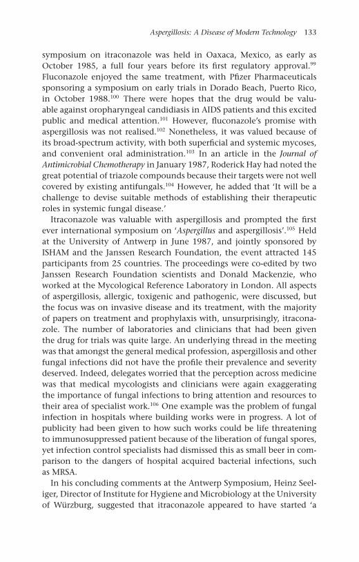
Aspergillosis: A Disease of Modern Technology 133
symposium on itraconazole was held in Oaxaca, Mexico, as early asOctober 1985, a full four years before its first regulatory approval.99
Fluconazole enjoyed the same treatment, with Pfizer Pharmaceuticalssponsoring a symposium on early trials in Dorado Beach, Puerto Rico,in October 1988.100 There were hopes that the drug would be valu-able against oropharyngeal candidiasis in AIDS patients and this excitedpublic and medical attention.101 However, fluconazole’s promise withaspergillosis was not realised.102 Nonetheless, it was valued because ofits broad-spectrum activity, with both superficial and systemic mycoses,and convenient oral administration.103 In an article in the Journal ofAntimicrobial Chemotherapy in January 1987, Roderick Hay had noted thegreat potential of triazole compounds because their targets were not wellcovered by existing antifungals.104 However, he added that ‘It will be achallenge to devise suitable methods of establishing their therapeuticroles in systemic fungal disease.’
Itraconazole was valuable with aspergillosis and prompted the firstever international symposium on ‘Aspergillus and aspergillosis’.105 Heldat the University of Antwerp in June 1987, and jointly sponsored byISHAM and the Janssen Research Foundation, the event attracted 145participants from 25 countries. The proceedings were co-edited by twoJanssen Research Foundation scientists and Donald Mackenzie, whoworked at the Mycological Reference Laboratory in London. All aspectsof aspergillosis, allergic, toxigenic and pathogenic, were discussed, butthe focus was on invasive disease and its treatment, with the majorityof papers on treatment and prophylaxis with, unsurprisingly, itracona-zole. The number of laboratories and clinicians that had been giventhe drug for trials was quite large. An underlying thread in the meetingwas that amongst the general medical profession, aspergillosis and otherfungal infections did not have the profile their prevalence and severitydeserved. Indeed, delegates worried that the perception across medicinewas that medical mycologists and clinicians were again exaggeratingthe importance of fungal infections to bring attention and resources totheir area of specialist work.106 One example was the problem of fungalinfection in hospitals where building works were in progress. A lot ofpublicity had been given to how such works could be life threateningto immunosuppressed patient because of the liberation of fungal spores,yet infection control specialists had dismissed this as small beer in com-parison to the dangers of hospital acquired bacterial infections, suchas MRSA.
In his concluding comments at the Antwerp Symposium, Heinz Seel-iger, Director of Institute for Hygiene and Microbiology at the Universityof Würzburg, suggested that itraconazole appeared to have started ‘a

134 Fungal Disease in Britain and the United States 1850–2000
new area of chemotherapy’.107 Such hopes were soon to prove prema-ture and several issues arose. First, it proved difficult to find sufficientcases for conclusive clinical trials, both to secure regulatory approvaland to evaluate the new drug against amphotericin B.108 When itra-conazole finally passed all the trial stages and gained FDA approvalin 1992, it was essentially as a ‘salvage therapy’. Second, one of thealleged advantages of itraconazole was oral administration. However,Janssen found it hard to produce a formulation that was absorbed wellenough to deliver a consistent effective dose.109 Also, the active princi-ple had to be protected from stomach acids, which meant patients hadto swallow a large, coated capsule. Evidence also emerged in the early1990s of a relatively large number of conflicts with other drugs, espe-cially those likely to be taken by immuno-compromised patients. At thesame time, new forms of amphotericin B were developed to mitigateits toxicity, mostly by binding the drug to lipids to avoid kidney dam-age, but these innovations were not taken up rapidly as they were notsupported by randomised trials.110 Trials with invasive aspergillosis wereproblematic because the number of patients were not only small, butwere dispersed across the country, had different forms of the infectionin different regions of the body, and they suffered from a wide range ofunderlying conditions.111
Nonetheless, by the mid-1990s, whilst still limited, treatment optionsfor invasive aspergillosis had improved. Clinicians had gained betterunderstandings of drug actions, more choices and protocols for differ-ent sites and types of infection. It was still the case, however, that manypatients with the disease were diagnosed too late for effective therapy,though on this front there were promising new approaches from molec-ular biology for earlier and more accurate detection. One hope was thatimproved and standardised methods would make the recognition ofinfection easier for non-specialists. A lot of research was undertaken onantigen tests, but a breakthrough came with microassays to detect sugarsreleased by growing Aspergillus hyphae.112
The profile of invasive aspergillosis grew because of its high mor-tality and the growing number of patients affected due to the greateruse of immunosuppression with transplant, cancer and other patientgroups.113 Aspergillosis was an emerging infection in intensive careunits, where monitoring and maintenance technologies, along withmore aggressive therapies, opened the body to opportunistic infec-tion. There was evidence in the mid-1990s from the United States andGermany that it had replaced candidiasis as the principal hospital-acquired fungal infection.114
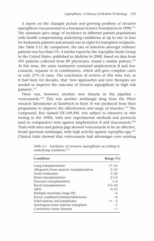
Aspergillosis: A Disease of Modern Technology 135
A report on the changed picture and growing problem of invasiveaspergillosis was presented in a European Science Foundation in 1998.115
The summary gave range of incidence in different patient populationswith health compromising underlying conditions at up to one in fourfor leukaemia patients and around one in eight for transplant recipients.(See Table 5.1): By comparison, the rate of infection amongst ordinarypatients was less than 1%. A similar report by the Aspergillus Study Groupin the United States, published in Medicine in 2000, based on data from595 patients collected from 89 physicians, found a similar pattern.116
At this time, the main treatments remained amphotericin B and itra-conazole, separate or in combination, which still gave complete curesin only 27% of cases. The conclusion of reviews at this time was, asit had been for decades, that ‘new approaches and new therapies areneeded to improve the outcome of invasive aspergillosis in high riskpatients’.117
There was, however, another new triazole in the pipeline –voriconazole.118 This was another antifungal drug from the Pfizerresearch laboratories at Sandwich in Kent. It was produced from theirprogramme to improve the effectiveness and range of triazoles.119 Thecompound, first named UK-109,496, was subject to intensive in vitrotesting in the 1990s, with new experimental methods and protocolsused in comparative tests against amphotericin B and itraconazole.120
Trials with mice and guinea pigs showed voriconazole to be an effective,broad spectrum antifungal, with high activity against Aspergillus spp.121
Clinical trials showed that voriconazole had advantages over existing
Table 5.1 Incidence of invasive aspergillosis according tounderlying condition.122
Condition Range (%)
Lung transplantation 17–16Allogeneic bone marrow transplantation 5–15Acute leukaemia 5–24Heart transplantation 2–13Pancreas transplantation 1–4Renal transplantation 0.5–10AIDS 0–12Multiple myeloma (stage III) ∼4Severe combined immunodeficiency ∼4Solid tumour and lymphoma ∼1 − 3Autologous bone marrow transplant ∼1Connective tissue diseases ∼1
136 Fungal Disease in Britain and the United States 1850–2000
therapies in three areas: antifungal activity, administration and sideeffects. The drug was approved in the United States in May 2002 andin Europe the following year, primarily for the treatment of invasiveaspergillosis. Its superiority over amphotericin B was celebrated in theLancet in October 2002, though the advantages were relative: just overhalf of patients with invasive aspergillosis responded to voriconazolecompared to just under a third on amphotericin.123 Nonetheless, clini-cians continued to be cautiously optimistic, not just about this drug, butbecause it pointed the way forward to more and better antifungals.124
Aspergillosis remains a rare disease, though in the first decade of thetwenty-first century it became the largest cause of death from fungalinfection in Britain and was second only to candidiasis in the UnitedStates.125 Its impact in medicine was significant as it became associ-ated with high profile settings: ‘high tech’ medical and surgical sites,particularly cancer and transplantation clinics, intensive care units andin the many clinical encounters that used immunosuppressant thera-pies. In one sense, aspergillosis was an exemplary iatrogenic condition –an unintended result of medical progress.126 Indeed, the history ofaspergillosis in the twentieth century can be mapped on key concernsand innovations. Pulmonary aspergillosis first attracted attention in thecontext of new interest in pulmonary tuberculosis around 1900 andre-emerged with mass X-ray and effective antibiotic treatment in the1940s. Aspergilli spp. were also part of the rise of allergy and asthma,the development of antibiotics, and the discovery of mycotoxins. Theprofile of the infection was in step with larger epidemiological changesin mortality and especially morbidity: the decline in infections andthe rise of cancers and other degenerative conditions, where chronicityand aggressive treatments opened the body to opportunistic infection.Serious disease was most common in a new type of patient – the post-transplant, immuno-compromised patient, who had been created andhad to be maintained by modern medical technologies.
Except where otherwise noted, this work is licensed under aCreative Commons Attribution 3.0 Unported License. To view
a copy of this license, visit http://creativecommons.org/licenses/by/3.0/
OPEN
Conclusion
Our aim in writing this book, apart from presenting the first history offungal diseases, was also to contribute to the historiography of medicine.In this conclusion, rather than restate and revisit our histories of partic-ular mycoses, we focus on crosscutting themes concerning the history ofinfectious diseases, the limits of the medical gaze, the history of medicalspecialisation and biographies of disease.
Our narrative has shown the value of approaching the history ofinfection in the twentieth century in terms of ‘seed and soil’. Our read-ing of the medical record has been against the grain of the commonfocus on the ‘seeds’ – the specific causative organisms – of disease. It isno surprise that doctors’ histories of infections are in this genre, afterall the major trend in medicine in our period has been to define dis-eases in terms of their causes (aetiology), and from the middle of thecentury, to treat disease by targeting those specific causes with drugs,surgery or other technologies. The history of pulmonary tuberculosisexemplifies this trajectory. First, its medical name changed from phthisis(wasting) or consumption, a symptomatic definition, to TB.1 The lat-ter conflated pathology (‘Tuberculosis’) and aetiology (Tubercle bacillus),and approaching the disease in terms of its ‘seeds’ was reinforced after1950, when antibiotics arrived as the long sought after ‘magic bullets’that selectively killed the T. bacillus in the body. Doctors’ histories ofTB tended to write off pre-1950s treatments as ‘unscientific’ and inef-fective, as they had focused on making the human ‘soil’ unsuitable forthe T. bacillus, either by strengthening the body through the sanato-rium regime, or removing the nidus of infection by surgery. Historiansof medicine have been less judgmental and, in recovering the think-ing and practices of the pre-antibiotic era, have shown the contingentnature of assessments of scientificity and efficacy, and suggested that
137
138 Fungal Disease in Britain and the United States 1850–2000
for many patients, now abandoned treatments that aimed to improvebodily ‘soil’ did ‘work’.2
The role of the ‘soil’ in the history of infectious diseases is implicitin Thomas McKeown’s famous explanation of the decline in mortalityfrom infectious diseases in Britain in the nineteenth century.3 In hisanalysis, he discounted the influence of factors linked to the ‘seeds’of infection, such the changing virulence of germs, along with medi-cal and public health measures targeted at germs, and concluded thatthe principal cause of the decline was rising standards of living andimproved nutrition, which had strengthened the body’s soil and resis-tance to infection. The fact that many people whose body was infectedwith a specific germ did not develop disease was well known to doctorsand especially with TB, as skin tests had long shown that while up to90% of adults had been infected with the bacillus, only a fraction wenton to develop the disease and fewer died.
In our discussion of fungal infections, we have also extended themetaphor of the ‘soil’ beyond its normal references to the individualbody and its vulnerability to infection, to an ecological one that alsoembraces social and geographical settings, and to how these affectedopportunities for the spread of infection as well as susceptibility. Ourchapter sub-titles refer to particular types of ‘soil’ in this extendedsense. Thus, for ringworm in children the ‘soil’ included an institution –schools, and for adults with foot infection, the ‘soil’ was a lifestyle –athleticism. With endemic mycoses, such as coccidioidomycosis, wherethe fungi were in fact literally in the soil, our larger notion of ‘soil’included the social changes that brought in-migration and economicdevelopment. Warwick Anderson has recently highlighted the impor-tance of the ecological tradition in work on infectious diseases in thetwentieth century, but he shows that this expanded approach remainednevertheless predominantly ‘biological’, with any social and techno-logical dimension implicit.4 We have made the latter componentsexplicit.
This book can be characterised as a history of diseases at the peripheryof the medical gaze, or at the ends of the spectrum of infectious diseases.At one end, infections such as ringworm and thrush were ubiquitous,everyday and mostly either self-limited or self-treated, involving at mosta single consultation with a doctor. This is not to deny that such infec-tions can be chronic and hard to eliminate, even since the arrival of oralantifungal drugs. At the other end, infections such as candidaemia andinvasive aspergillosis were rare and unusual, sometimes termed ‘orphandiseases’, and commonly fatal until recent decades.

Conclusion 139
But what can a study of these diseases at the margin contribute toour understanding of the middle, the majority and the mainstream?Above all, our analysis reminds historians that the minor, self-limitingand self-treated conditions are common across medicine and not justwith infectious diseases. Illness ‘ice bergs’, the many episodes that donot reach the medical gaze, were and are present across all areas andmost diseases. Yet, gaze of historians has always tended to be ‘abovethe water’. For example, histories of the ‘Great Influenza Pandemic’ of1918–1919 emphasise that between 20 and 40 million people died of theinfection or complications such as pneumonia, yet the average mortal-ity rate was around 2% (varying between 1% and 10%) amongst thosewho suffered.5 This rate was very high compared to the normal expe-rience of influenza, where case fatality rates was and remains typicallyless than 1%.6 Both figures make our point that the majority patientexperience of influenza was and is one of recovery; with an illness ofvariable severity, which was and is typically self-treated and not requiremedical attention, if indeed, the sufferer had access to, or could afford,professional consultation. Moreover, then, as now, those likely to die ofpneumonia were and are people with underlying health problems; inother words, those with weakened ‘soil’.
The investigation of rare and unusual diseases highlights the impor-tance of the adaptation of mainstream ideas and practices to novelproblems, and the opportunities to ‘experiment’ with new methods ofdiagnosis and treatment. In an era when standardisation and formalprotocols dominate medical practice, our study of ‘orphan mycoses’ hasshown the variability, complexity and individuality of clinical practiceand the many resources, theoretical, practical and material, that doc-tors drew upon, and still draw upon, in all aspects of clinical work.The ways in which uses of amphotericin B, one the earliest antifungalantibiotic drugs, has been reinvented many times exemplifies this adapt-ability and shows the need to think about invention and innovation asprocesses rather than events. The very recognition of ‘orphan diseases’in part derives from novel medical and social technologies of surveil-lance, which have provided new types of recognition of infection, suchas X-rays and immune reactions, and new attitudes to risk associatedwith social, economic and technological changes, such as the negoti-ation of thresholds for intervention in public health and with specificpopulations.
Marginality has been studied by historians and sociologists of sci-ence as a ‘context’ that stimulates innovation.7 These ideas have beencritiqued empirically and for having loose definitions of marginality
140 Fungal Disease in Britain and the United States 1850–2000
and innovation, but in this study we have discussed a group – medicalmycologists – who were routinely designated marginal by their profes-sional peers and saw themselves as such.8 At the end of the twentiethcentury, medical mycology remained a small and marginal field; indeed,some in medicine argued that it was often oversold, with specialistsexaggerating the importance of fungal diseases as causes of morbidityand mortality. We do not want to enter this debate, but instead reflecton the development of medical mycology as a specialism. As we notedin the Introduction, historical studies of specialisation in medicine arenow less teleological and more nuanced, but there remains a focus onmajor specialisms and what might be termed ‘mono-specialists’. As wehave shown, most medical mycologists were ‘multi-specialists’, or hada number of ‘specialist practices’, even in the United States where thesize of agencies, such as CDC and NIH, or the foci of regionally spe-cific infections made mono-specialist careers possible. Indeed, a featureof our story is that the tension, expected in the 1930s and 1940s,between ‘botany types’ and ‘medical types’, which can also be seen asbetween laboratory and clinic, did not develop.9 Cooperation and col-laboration were characteristic as roles co-existed and were combined.In part, this was because of interdependence, especially as cliniciansrelied on laboratory-based experts to confirm and refine their diagnosesand, then after the 1950s, for the development of antifungal antibi-otics. Solidarity was also prompted by size and marginality, which meantthat creating and maintaining critical mass was a priority and specialistorganisations, not least ISHAM which spanned the Americas and notjust North America, were pivotal in this respect. In the United States,a presence at NIH and CDC was a boon. In contrast, medical mycolo-gists in Britain had to be content with a single organisation, the BSMM,though this effectively lobbied national and, latterly, European agen-cies. Needless to say, the relatively small size of the field meant thatindividuals were very important, as too were personal connections andnetworks, which were facilitated by air travel from the 1950s, whichmade international meetings, both disciplinary and those sponsored bypharmaceutical companies on single drugs, more common and betterattended. Our focus on diseases has meant that we have not dwelt on thecareers of individual medical mycologists, though the repeated mentionof names of key individuals, often with many infections and in multiplecontexts, is testimony to their success in combining specialist practices.
The biographical mode is now fashionable in the history of medicine,not only for doctors, scientists and institutions, but diseases too.10 Wehave not termed our narratives of fungal infections biographies, but

Conclusion 141
have kept with ‘histories’. Roger Cooter, while appreciating the richnessof much of the new biographical genre, has criticised it for tending tobe essentialist and singular, taking diseases as given, rather than look-ing at their construction and many identities.11 Cooter was after allmaking these observations in the Lancet, the implication of his remarksbeing that historians of disease need to recognise that ‘modern’, singularnarratives are no longer tenable in ‘the face of contemporary impres-sions of fragmentation and the collapse of universal meanings’.12 Ourstudy supports this view. For example, consider the many views put for-ward on how to explain the rise in the incidence of fungal infectionsin the twentieth century. Some doctors maintain the increase was realand material, some said that it came from new conceptions of whatconstituted an infection, others said it was product of new medicaltechnologies of surveillance and diagnosis, and others that it was iatro-genic. To these views can be added claims that the apparent rise camefrom changing public attitudes to infection and expectations of medicalpower. Our conclusion is that all the above forces were in play in therise of mycoses, shaping, not just epidemiological patterns, but expe-riences and meanings; hence, despite the subtitle of this volume, overthe twentieth century, mycoses can be seen as paradigmatic postmoderndiseases.
Except where otherwise noted, this work is licensed under aCreative Commons Attribution 3.0 Unported License. To view
a copy of this license, visit http://creativecommons.org/licenses/by/3.0/
Notes
Introduction
1. In the book, we use the terms ‘fungal infections’ and ‘mycoses’ (or singularfungal infection and mycosis) interchangeably, mostly following the usageof our historical actors in time and place.
2. The term ‘ringworm’ is very old and came from the circular patches ofpeeled, inflamed skin that characterised the infection. In medicine at least,no one understood it to be associated with worms of any description.
3. Porter, R., ‘The patient’s view: Doing medical history from below’, Theory andSociety, 1985, 14: 175–198; Condrau, F., ‘The patient’s view meets the clinicalgaze’, Social History of Medicine, 2007, 20: 525–540.
4. Burnham, J. C., ‘American medicine’s golden age: What happened to it?’, Sci-ence, 1982, 215: 1474–1479; Brandt, A. M. and Gardner, M., ‘The golden ageof medicine’, in Cooter, R. and Pickstone, J. V., eds, Medicine in the TwentiethCentury, Amsterdam, Harwood, 2000, 21–38.
5. The Oxford English Dictionary states that the first use of ‘side-effect’ was in1939, in H. N. G. Wright and M. L. Montag’s textbook: Materia Med Pharma-col & Therapeutics, 112, when it referred to ‘The effects which are not desiredin any particular case are referred to as “side effects” or “side actions” and, insome instances, these may be so powerful as to limit seriously the therapeuticusefulness of the drug.’
6. Illich, I., Medical Nemesis: The Expropriation of Health, London, Calder &Boyars, 1975.
7. Beck, U., Risk Society: Towards a New Society, New Delhi, Sage, 1992, 56and 63; Greene, J., Prescribing by Numbers: Drugs and the Definition of Dis-ease, Baltimore, Johns Hopkins University Press, 2007; Timmermann, C.,‘To treat or not to treat: Drug research and the changing nature of essentialhypertension’, Schlich, T. and Tröhler, U., eds, The Risks of Medical Innova-tion: Risk Perception and Assessment in Historical Context, London, Routledge,2006, 144.
8. Rippon, J. W., ‘Symposium on medical mycology’, Mycopathologia, 1987, 99:144. The Fusaria is a genus of soil-borne fungi, most of which are harmless,but some are well known to cause wilt in plants. Quorn, a meat-like productrecommended for vegetarians, is produced from Fusarium venenatum.
9. Crook, W. G., The Yeast Connection: A Medical Breakthrough, Jackson, T.,Professional Books, 1983.
10. Dubos, J., Man Adapting, chapters 7, 9 and 10; idem, Mirage of Health: Utopias,Progress, and Biological Change, New Brunswick, Rutgers University Press,1959.
11. Dubos, J. and Dubos, R., The White Plague: Tuberculosis, Man and Society,London, Victor Gollancz, 1952.
142
Notes 143
12. Martin, G. W., ‘Are fungi plants?’, Mycologia, 1955, 47(6): 779–792;Whittaker, R. H., ‘New concepts of Kingdoms of organisms’, Science, 1969,163: 150–161; Hagen, J. B., ‘Five Kingdoms, more or less: Robert Whittakerand the broad classification of organisms’, Bioscience, 2012, 62(1): 67–74.
13. Stringer, J. R. et al, ‘A new name (Pneumocystis jiroveci) for pneumocystis fromhumans’, Emerg Infect Dis, 2002, 8(9): 891–896.
14. Edman, J. C. et al, ‘Ribosomal RNA sequence shows Pneumocystis carinii tobe a member of the fungi’, Nature, 1988, 334: 519–522; Armengol, C. E.,‘A historical review of Pneumocystis carinii’, J Am Med Assoc, 1995, 273(9):747, 750–751.
15. Ainsworth, G. C., Introduction to the History of Medical and Veterinary Mycology,Cambridge, Cambridge University Press, 1987, 1–3.
16. Ainsworth, G. C., ‘Agostino Bassi, 1773–1856’, Nature, 1956, 177: 255–257;Arcieri, G. P., Agostino Bassi in the History of Medical Thought, Florence, L. S.Olschki, 1956.
17. Ainsworth, G. C., Introduction to the History of Plant Pathology, Cambridge,Cambridge University Press, 1981.
18. Bateman, T., A Practical Synopsis of Cutaneous Diseases according to the Arrange-ment of Dr. Willan, Exhibiting a Concise View of the Diagnostic Symptoms andthe Method of Treatment, London: Longman, 1813; idem, Delineations of Cuta-neous Diseases: Exhibiting the Characteristic Appearances of the Principal Generaand Species Comprised in the Classification of the Late Dr. Willan, London,Longman, Hurst, Rees, Orme, and Brown, 1817.
19. Jacyna, S., ‘Pious pathology: J. L. Alibert’s iconography of disease’,Hannaway, C. and La Berge, A., eds, Constructing Paris Medicine, London, ClioMedica/The Wellcome Series in the History of Medicine, 185–219.
20. Lailler, C.-P., Leçons cliniques sur les teignes, faites à l’hôpital Saint-Louis, par leDr C. Lailler, Paris, V.-A. Delahaye, 1878.
21. Worboys, M., Spreading Germs: Disease Theories and Medical Practice in Britain,1856–1900, Cambridge, Cambridge University Press, 2000, 28–42.
22. Zakon, S. J. and Benedek, T., ‘David Gruby and the centenary of medicalmycology, 1841–1941’, Bull His Med, 1944, 16: 155–168; Codell Carter, K.,The Rise of Causal Concepts of Disease, Aldershot, Ashgate, 2003, 32–37 and72–79.
23. Pelling, M., Cholera, Fever and English Medicine, 1825–1865, Oxford, Claren-don Press, 1978, 146–202.
24. Swayne, J. G. and Budd, W., ‘An account of certain organic cells in thepeculiar evacuations of cholera’, Lancet, 1849, 54: 398–399.
25. Worboys, Spreading Germs, passim.26. Sims Woodhead, G. and Hare, A., Pathological Mycology: An Enquiry into the
Etiology of Infectious Diseases, Edinburgh, Young J. Pentland, 1885.27. Muir, R. and Ritchie, J., Manual of Bacteriology, Edinburgh, Young J. Pentland,
1899, 133–138; de Bary, A., Comparative Morphology and Biology of the Fungi,Mycetozoa and Bacteria, Oxford, Clarendon Press, 1887.
28. Hawksworth, D. L., ‘Geoffrey Clough Ainsworth (1905–1998): Mycologi-cal scholar, campaigner, and visionary’, Mycological Research, 2000, 104(1),110–116; Buczacki, S., ‘Ainsworth, Geoffrey Clough (1905–1998)’, OxfordDictionary of National Biography, Oxford University Press, 2004 [http://www.oxforddnb.com/view/article/71235, accessed 3 May 2013].
144 Notes
29. Ainsworth, G. C., Introduction to the History of Mycology, Cambridge, Cam-bridge University Press, 1976; idem, Introduction to the History of PlantPathology, Cambridge, Cambridge University Press, 1981.
30. Ainsworth, Introduction to the History of Medical, ix.31. For an alternative approach to that adopted here, see: Wainwright, M., ‘Some
highlights in the history of fungi in medicine – A personal journey’, FungalBiology Reviews, 2008, 22: 97–102.
32. Espinel-Ingroff, A. V., Medical Mycology in the United States: A Historical Analy-sis (1894–1996), Dordrecht, Kluwer Academic Publishers, 2003. The book wasbased on her 1994 PhD, Espinel-Ingroff, A., Medical Mycology in the UnitedStates: 100 Years of Development as a Discipline, PhD dissertation, VirginiaCommonwealth University, Richmond, 1994. Also see, Espinel-Ingroff, A. V.,‘History of medical mycology in the United States’, Clinical MicrobiologyReviews, 1996, 9(2): 235–272.
33. Daniel, T. M. and Baum, G. L., Drama and Discovery: The Story of Histoplasmo-sis, Westport, CT, Greenwood, Press, 2002. A version of the earlier chapterswas published as Baum, G. L. and Schwarz, J., ‘The history of histoplasmosis1906–1956’, N Engl J Med, 1957, 256: 253–258.
34. Fiese, M. J., Coccidioidomycosis, Springfield, IL, Charles C. Thomas, 1958;Odds, F. C., Candida and Candidosis, Leicester, Leicester University Press,1979.
35. Hischmann, J., ‘The early history of coccidioidomycosis: 1892–1945’, ClinInfect Dis, 2007, 44(9): 1202–1207.
36. However, the ‘The Aspergillus Website’ has an excellent ‘History’ section,with an extensive collection of primary sources that we have used exten-sively in researching this book. ‘The Aspergillus Website’, Historical PapersDatabase and History Section. Accessed 25 March 2013, http://www.aspergillus.org.uk/indexhome.htm?secure/historical_papers/index.php∼main.
37. Rosenthal, T., ‘Perspectives in ringworm of the scalp: Treatment through theages’, Arch Derm Syphilol, 1960, 82(6): 851–856.
38. Seidelman, R. D. et al, ‘ “Healing” the bodies and souls of immigrant chil-dren: The ringworm and Trachoma Institute, Sha’ar ha-Aliyah, 1952–1960’,Journal of Israeli History, 2010, 29(2): 191–211.
39. For works that deal with the issue of specialisation of medicine, see,Rosen, G., The Specialization of Medicine with Particular Reference to Ophthal-mology, New York, Froben Press, 1944; Stevens, R., Medical Practice in ModernEngland: The Impact of Specialization and State Medicine, New Haven, YaleUniversity Press, 1966.
40. Weisz, G., Divide and Conquer: A Comparative History of Medical Specialization,New York, Oxford University Press, 2006, xxix–xxx.
41. This point is made by Stephen Caspar in his review of George Weisz’s Divideand Conquer in Medical History, 2006, 50: 545–547.
42. Digby, A., Making a Medical Living: Doctors and Patients in the English Marketfor Medicine, 1720–1911, Cambridge, Cambridge University Press, 1994.
43. Macfarlane, J. T. and Worboys, M., ‘The changing management of acutebronchitis in Britain, 1940–1970: The impact of antibiotics’, Medical History,2008, 52: 47–72.
44. Scambler, G. and Scambler, A., ‘The illness iceberg and aspects of consultingbehaviour’, in Fitzpatrick, R., ed, The Experience of Illness, London, Tavistock,1984, 35–37.
Notes 145
45. Porter, R., ‘The patient’s view: Doing medical history from below’, Theory andSociety, 1985, 14: 175–198; Condrau, F., ‘The patient’s view meets the clinicalgaze’, Social History of Medicine, 2007, 20: 525–540.
46. Galgiani, J. N., ‘Coccidioidomycosis: Changing perceptions and creatingopportunities for its control’, Ann N Y Acad Sci, 2007, 1111: 1–18.
47. Editorial, ‘Thrush’, JAMA, 1927, 89: 1429–1430; Plass, E. D. et al, ‘Moniliavulvovaginitis’, Am J Obstet Gynecol, 1931, 21: 320.
1 Ringworm: A Disease of Schools and Mass Schooling
1. Sanderson, M., Education, Economic Change and Society in England 1780–1870, Cambridge, Cambridge University Press, 1995; Simon, B., Studies inthe History of Education, London, Lawrence and Wishart, 1966; Digby, A. andSearby, P., eds, Children, School and Society in Nineteenth-Century England,London, Macmillan, 1981; Nasaw, D., Schooled to Order: A Social Historyof Public Schooling in the United States, New York, Oxford University Press,1979.
2. Richardson, N., Typhoid in Uppingham: Analysis of a Victorian Town andSchool in Crisis, 1875–7, London: Pickering & Chatto, 2008.
3. Pusey, W. A., The History of Dermatology, Springfield, Ill., C. C. Thomas,1933; Crissey, J. T. and Parish, L. C., The Dermatology and Syphilology of theNineteenth Century, New York, Praeger, 1981. On Robert Willans see: Brun-ton, D., ‘Willan, Robert (1757–1812)’, Oxford Dictionary of National Biog-raphy, Oxford University Press, 2004. http://www.oxforddnb.com/view/article/29438. Accessed 8 February 2013.
4. On Plumbe see, Rosenthal, T., ‘Samuel Plumbe’, Arch Derm Syphilol, 1937,36(2): 348–354.
5. Plumbe, S., ‘History, Pathology and Treatment, of Ring-worm and Scald-head’, Lancet, 1835, 926.
6. Ibid.7. Brown, H., Ringworm and Some Other Scalp Affections: Their Cause and Cure,
London, J. & A. Churchill, 1899.8. Buchan, W., Domestic Medicine: Or, A Treatise on the Prevention and Cure of
Diseases, London, 11th Edition, London, 1790, 555–556.9. Beeton, I., Beeton’s Book of Household Management, London, S. O. Beeton,
1861, No. 2667–2668.10. Plumbe, S., ‘Remarks on the Contagious Ring-worm of the Scalp’, Lancet,
1835, 858.11. There is an excellent history of ringworm in France, see: Tilles, G. and
Tilles, G., Teignes et Teigneux: Histoire Medicale et Sociale, Paris, Springer,2008.
12. Allan, G. A. T., Christ’s Hospital, London, Town and County, 1984.13. Plumbe, S., ‘History, Pathology and Treatment of Ringworm and Scald-
head’, Lancet, 1835, i: 926–928 and ii: 50–51; Rosenthal, T., ‘SamuelPlumbe’, Arch Dermatol, 2008, 16: 36–43.
14. Lancet, 1835, 683–688.15. Plumbe, ‘History, Pathology’, 928.16. Plumbe, S., An Address to the Governors of Christ’s Hospital, on the Causes and
Means of Prevention of Ring-Worm in That Establishment; To Which Is Attached,
146 Notes
a Few Rules for the Domestic Management of the Scholars during Their Vacations,London, 1834.
17. Mayne, R. G., An Expository Lexicon of the Terms, Ancient And Modern, inMedical and General Science, Pt 2, London: J. Churchill, 1854, 265.
18. Lawrence, C., ‘ “Incommunicable Knowledge”: Science, Technology and theClinical “Art” in Britain, 1850–1910’, Journal of Contemporary History, 1985,20: 503–520.
19. Lancet, 1875, i: 888.20. Ibid.21. Lancet, 1908, i: 822.22. Lawrence, C., Medical Theory, Surgical Practice: Studies in the History of Surgery,
London, Routledge, 1992.23. Rook, A., ‘Dermatology in Britain in the Late Nineteenth Century’, Br J Der-
matol, 1979, 100(1): 3–12.24. Erichsen, J. E., The Science and Art of Surgery, London, J. Walton, 1869, 8.25. Rook, A., ‘James Stratin, Jonathan Hutchinson and the Blackfriars Skin
Hospital’, Br J Dermatol, 1978, 99: 215–219.26. Ibid., 216.27. Russell, B. F., ed, St John’s Hospital for Diseases of the Skin, 1863–1963,
Edinburgh, E. & S. Livingstone, 1963.28. Lancet, 1864, ii: 538. The three supporters withdrew their support when
they realised Milton’s practice was mainly on the treatment of spermator-rhoea, the involuntary discharge of semen.
29. Hadley, R. M., ‘The Life and Works of Sir William James Erasmus Wilson,1809–84’, Medical History, 1959, 3: 215–247; Power, D’A, ‘Wilson, Sir(William James) Erasmus (1809–1884)’, revised Geoffrey L. Aserton, OxfordDictionary of National Biography, Oxford University Press, 2004. [http://www.oxforddnb.com/view/article/29702, accessed 15 August 2008].
30. Quoted in Hogg, J., Parasitic or Germ Theory of Disease: The Skin, Eye, andOther Affections, London, Baillière, Tindall and Cox, 1876, 33.
31. Asherson, G. L., ‘Fox, William Tilbury (1836–1879)’, Oxford Dictionary ofNational Biography, Oxford University Press, 2004. [http://www.oxforddnb.com/view/article/10048, accessed 15 August 2008]; English, M. P., ‘WilliamTilbury Fox and Dermatological Mycology’, Br J Dermat, 1977, 97: 100–112;Cooper, J., ‘Anderson, Sir Thomas McCall (1836–1908)’, revised, J O’DAlexander, Oxford Dictionary of National Biography, Oxford University Press,2006, [http://www.oxforddnb.com/view/article/30414, accessed 6 October2008; Anon, ‘Thomas McCall Anderson, Obituary’, Lancet, 1908, i: 468–471.
32. Tilbury Fox, G., Skin Diseases of Parasitic Origin, London, Robert Hard-wicke, 1863; idem, ‘The True Nature and Meaning of Parasitic Diseasesof the Surface’, Lancet, 1859, ii: 5–7, 31–32, 201, 260–261, 283–284 and507–508; M’Call Anderson, T., On the Parasitic Affections of the Skin, London,Churchill, 1861.
33. Tilbury Fox, Skin Diseases, v–vi; Anderson, On the Parasitic Affections, 1–2.34. Hogg, J., ‘The Vegetable Parasites of the Human Skin’, BMJ, 1859, i: 241;
Hillier, T., ‘On Ringworm and Vegetable Parasites’, BMJ, 1861, ii: 552 and577; Worboys, M., Spreading Germs: Disease Theories and Medical Practice inBritain, 1865–1900, Cambridge, Cambridge University Press, 2000, 73–107.
35. Wilks, S., ‘Address in Medicine’, BMJ, 1872, ii: 146–153.
Notes 147
36. Ibid., 149.37. Colan, T., ‘Parasitic Vegetable Fungi and the Diseases Induced by Them’,
Lancet, 1874, ii: 755–757 and 832–833.38. Liveing, R., ‘Lecture on the Peculiarities of Ringworm and its Treatment’,
Lancet, 1879, ii: 642–644, on 643–644.39. Worboys, Spreading Germs, Passim; M’Call Anderson, T., ‘Introductory Lec-
tures to the Study of the Diseases of the Skin’, Lancet, 1870, i: 149–151.40. Lancet, 1861, ii: 449–450.41. Pelling, M., Cholera, Fever and English Medicine, 1830–1865, Oxford, Claren-
don Press, 1976, 146–202.42. Tilbury Fox, W., ‘On the Identity of Parasitic Fungi Affecting the Human
Surface’, Lancet, 1880, ii: 260–261; Lancet, 1867, ii: 266–267.43. Lancet, 1889, ii: 1232.44. Stark, J., Industrial Illness in Cultural Context: ‘La maladie de Bradford’ in Local,
National and Global Settings, 1878–1919, Unpublished PhD Thesis, Univer-sity of Leeds, 2011; Jamieson, A., An Intolerable Affliction: A History of Lupusvulgaris in Late 19th and Early 20th Century Britain, Unpublished PhD Thesis,University of Leeds, 2010.
45. British Association of Dermatologists, A Biographical History of British Der-matology, London, British Association of Dermatologists, 1995.
46. Tilbury Fox, W., ‘Ringworm in Schools’, Lancet, 1872, i: 5–6.47. Hirst, J. D., ‘Public Health and the Public Elementary Schools, 1870–1907’,
History of Education, 1991, 20: 107–118.48. Harris, B., The Health of the Schoolchild: A History of the School Medical Service
in England and Wales, Buckingham, Open University Press, 1995, 32–47.49. ‘Report of the Lancet Sanitary Commission on the Sanitary Condition of
our Public Schools’, Lancet, 1875, i: 795–796, 859–861 and ii: 111–112,314–315, 422–423, 574–575, 682, and 785–787.
50. See the case of George Beavis who in November 1875 was fined for neglect-ing to send his daughter to school. Hansard, HC Deb 11 February 1876,227: 227–229; Times, 3 February 1876, 5c; 9 March 1876, 6e.
51. Liveing, ‘Lecture on the Peculiarities’, 643–644.52. Mansell, K., Christ’s Hospital in the Victorian Era, Whitton, Ashwater Press,
2011.53. Alder Smith, H., Ringworm: Its Diagnosis and Treatment, London, H. K.
Lewis & Co. Ltd, 1880; 2nd Edition, 1882; 3rd Edition, 1885; Aldersmith,H., Ringworm and Alopecia Areata, 4th Edition, London, H. K. Lewis & Co.Ltd, 1897.
54. Tilbury Fox, W., ‘On Ringworm of the Head and its Management’, Lancet,1877, ii: 643–644.
55. Alder Smith, Ringworm, 1882, vii.56. Many general patent medicines included ringworm as one of the condi-
tions they cured. Specifically, the most widely advertised topical remedieswere Beatson’s Ringworm Lotion, Bateson’s Specific and Cuticura Soap. Hol-loway’s Ointment was advised to be used in conjunction with Holloway’sPills, and those seeking a systemic cure could try was Orange’s UniversalCerate and Vegetable Purifying Pills.
57. Manchester Times, 2 February 1889, 2 and 9 February 1889, 8.58. Morning Post, 10 May 1889, 4; Reynolds’s Newspaper, 26 May 1889, 1.
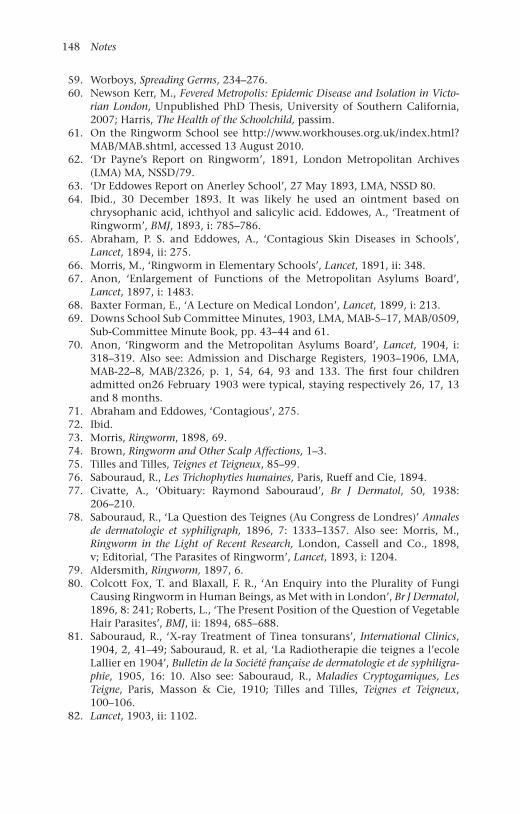
148 Notes
59. Worboys, Spreading Germs, 234–276.60. Newson Kerr, M., Fevered Metropolis: Epidemic Disease and Isolation in Victo-
rian London, Unpublished PhD Thesis, University of Southern California,2007; Harris, The Health of the Schoolchild, passim.
61. On the Ringworm School see http://www.workhouses.org.uk/index.html?MAB/MAB.shtml, accessed 13 August 2010.
62. ‘Dr Payne’s Report on Ringworm’, 1891, London Metropolitan Archives(LMA) MA, NSSD/79.
63. ‘Dr Eddowes Report on Anerley School’, 27 May 1893, LMA, NSSD 80.64. Ibid., 30 December 1893. It was likely he used an ointment based on
chrysophanic acid, ichthyol and salicylic acid. Eddowes, A., ‘Treatment ofRingworm’, BMJ, 1893, i: 785–786.
65. Abraham, P. S. and Eddowes, A., ‘Contagious Skin Diseases in Schools’,Lancet, 1894, ii: 275.
66. Morris, M., ‘Ringworm in Elementary Schools’, Lancet, 1891, ii: 348.67. Anon, ‘Enlargement of Functions of the Metropolitan Asylums Board’,
Lancet, 1897, i: 1483.68. Baxter Forman, E., ‘A Lecture on Medical London’, Lancet, 1899, i: 213.69. Downs School Sub Committee Minutes, 1903, LMA, MAB-5–17, MAB/0509,
Sub-Committee Minute Book, pp. 43–44 and 61.70. Anon, ‘Ringworm and the Metropolitan Asylums Board’, Lancet, 1904, i:
318–319. Also see: Admission and Discharge Registers, 1903–1906, LMA,MAB-22–8, MAB/2326, p. 1, 54, 64, 93 and 133. The first four childrenadmitted on26 February 1903 were typical, staying respectively 26, 17, 13and 8 months.
71. Abraham and Eddowes, ‘Contagious’, 275.72. Ibid.73. Morris, Ringworm, 1898, 69.74. Brown, Ringworm and Other Scalp Affections, 1–3.75. Tilles and Tilles, Teignes et Teigneux, 85–99.76. Sabouraud, R., Les Trichophyties humaines, Paris, Rueff and Cie, 1894.77. Civatte, A., ‘Obituary: Raymond Sabouraud’, Br J Dermatol, 50, 1938:
206–210.78. Sabouraud, R., ‘La Question des Teignes (Au Congress de Londres)’ Annales
de dermatologie et syphiligraph, 1896, 7: 1333–1357. Also see: Morris, M.,Ringworm in the Light of Recent Research, London, Cassell and Co., 1898,v; Editorial, ‘The Parasites of Ringworm’, Lancet, 1893, i: 1204.
79. Aldersmith, Ringworm, 1897, 6.80. Colcott Fox, T. and Blaxall, F. R., ‘An Enquiry into the Plurality of Fungi
Causing Ringworm in Human Beings, as Met with in London’, Br J Dermatol,1896, 8: 241; Roberts, L., ‘The Present Position of the Question of VegetableHair Parasites’, BMJ, ii: 1894, 685–688.
81. Sabouraud, R., ‘X-ray Treatment of Tinea tonsurans’, International Clinics,1904, 2, 41–49; Sabouraud, R. et al, ‘La Radiotherapie die teignes a l’ecoleLallier en 1904’, Bulletin de la Société française de dermatologie et de syphiligra-phie, 1905, 16: 10. Also see: Sabouraud, R., Maladies Cryptogamiques, LesTeigne, Paris, Masson & Cie, 1910; Tilles and Tilles, Teignes et Teigneux,100–106.
82. Lancet, 1903, ii: 1102.
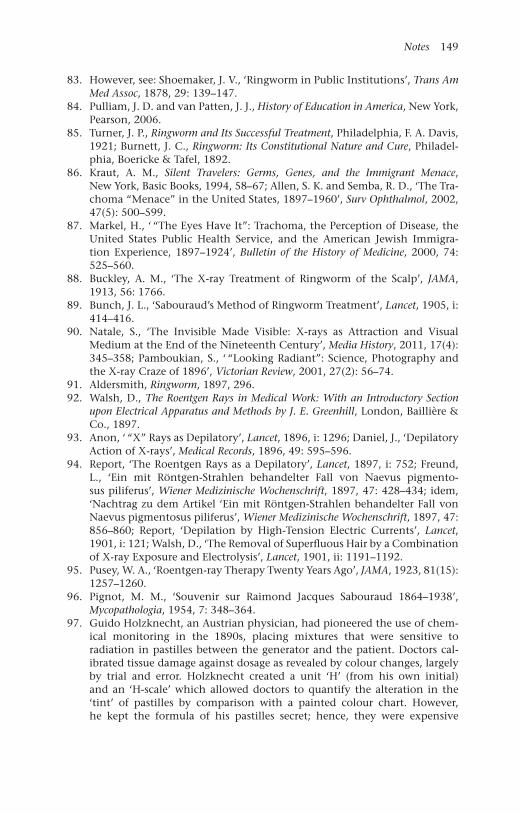
Notes 149
83. However, see: Shoemaker, J. V., ‘Ringworm in Public Institutions’, Trans AmMed Assoc, 1878, 29: 139–147.
84. Pulliam, J. D. and van Patten, J. J., History of Education in America, New York,Pearson, 2006.
85. Turner, J. P., Ringworm and Its Successful Treatment, Philadelphia, F. A. Davis,1921; Burnett, J. C., Ringworm: Its Constitutional Nature and Cure, Philadel-phia, Boericke & Tafel, 1892.
86. Kraut, A. M., Silent Travelers: Germs, Genes, and the Immigrant Menace,New York, Basic Books, 1994, 58–67; Allen, S. K. and Semba, R. D., ‘The Tra-choma “Menace” in the United States, 1897–1960’, Surv Ophthalmol, 2002,47(5): 500–599.
87. Markel, H., ‘ “The Eyes Have It”: Trachoma, the Perception of Disease, theUnited States Public Health Service, and the American Jewish Immigra-tion Experience, 1897–1924’, Bulletin of the History of Medicine, 2000, 74:525–560.
88. Buckley, A. M., ‘The X-ray Treatment of Ringworm of the Scalp’, JAMA,1913, 56: 1766.
89. Bunch, J. L., ‘Sabouraud’s Method of Ringworm Treatment’, Lancet, 1905, i:414–416.
90. Natale, S., ‘The Invisible Made Visible: X-rays as Attraction and VisualMedium at the End of the Nineteenth Century’, Media History, 2011, 17(4):345–358; Pamboukian, S., ‘ “Looking Radiant”: Science, Photography andthe X-ray Craze of 1896’, Victorian Review, 2001, 27(2): 56–74.
91. Aldersmith, Ringworm, 1897, 296.92. Walsh, D., The Roentgen Rays in Medical Work: With an Introductory Section
upon Electrical Apparatus and Methods by J. E. Greenhill, London, Baillière &Co., 1897.
93. Anon, ‘ “X” Rays as Depilatory’, Lancet, 1896, i: 1296; Daniel, J., ‘DepilatoryAction of X-rays’, Medical Records, 1896, 49: 595–596.
94. Report, ‘The Roentgen Rays as a Depilatory’, Lancet, 1897, i: 752; Freund,L., ‘Ein mit Röntgen-Strahlen behandelter Fall von Naevus pigmento-sus piliferus’, Wiener Medizinische Wochenschrift, 1897, 47: 428–434; idem,‘Nachtrag zu dem Artikel ‘Ein mit Röntgen-Strahlen behandelter Fall vonNaevus pigmentosus piliferus’, Wiener Medizinische Wochenschrift, 1897, 47:856–860; Report, ‘Depilation by High-Tension Electric Currents’, Lancet,1901, i: 121; Walsh, D., ‘The Removal of Superfluous Hair by a Combinationof X-ray Exposure and Electrolysis’, Lancet, 1901, ii: 1191–1192.
95. Pusey, W. A., ‘Roentgen-ray Therapy Twenty Years Ago’, JAMA, 1923, 81(15):1257–1260.
96. Pignot, M. M., ‘Souvenir sur Raimond Jacques Sabouraud 1864–1938’,Mycopathologia, 1954, 7: 348–364.
97. Guido Holzknecht, an Austrian physician, had pioneered the use of chem-ical monitoring in the 1890s, placing mixtures that were sensitive toradiation in pastilles between the generator and the patient. Doctors cal-ibrated tissue damage against dosage as revealed by colour changes, largelyby trial and error. Holzknecht created a unit ‘H’ (from his own initial)and an ‘H-scale’ which allowed doctors to quantify the alteration in the‘tint’ of pastilles by comparison with a painted colour chart. However,he kept the formula of his pastilles secret; hence, they were expensive
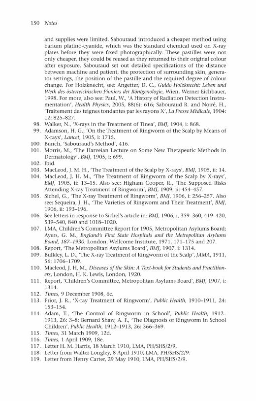
150 Notes
and supplies were limited. Sabouraud introduced a cheaper method usingbarium platino-cyanide, which was the standard chemical used on X-rayplates before they were fixed photographically. These pastilles were notonly cheaper, they could be reused as they returned to their original colourafter exposure. Sabouraud set out detailed specifications of the distancebetween machine and patient, the protection of surrounding skin, genera-tor settings, the position of the pastille and the required degree of colourchange. For Holzknecht, see: Angetter, D. C., Guido Holzknecht: Leben undWerk des österreichischen Pioniers der Röntgenologie, Wien, Werner Eichbauer,1998. For more, also see: Paul, W., ‘A History of Radiation Detection Instru-mentation’, Health Physics, 2005, 88(6): 616; Sabouraud R. and Noiré, H.,‘Traitement des teignes tondantes par les rayons X’, La Presse Mèdicale, 1904:12: 825–827.
98. Walker, N., ‘X-rays in the Treatment of Tinea’, BMJ, 1904, i: 868.99. Adamson, H. G., ‘On the Treatment of Ringworm of the Scalp by Means of
X-rays’, Lancet, 1905, i: 1715.100. Bunch, ‘Sabouraud’s Method’, 416.101. Morris, M., ‘The Harveian Lecture on Some New Therapeutic Methods in
Dermatology’, BMJ, 1905, i: 699.102. Ibid.103. MacLeod, J. M. H., ‘The Treatment of the Scalp by X-rays’, BMJ, 1905, ii: 14.104. MacLeod, J. H. M., ‘The Treatment of Ringworm of the Scalp by X-rays’,
BMJ, 1905, ii: 13–15. Also see: Higham Cooper, R., ‘The Supposed RisksAttending X-ray Treatment of Ringworm’, BMJ, 1909, ii: 454–457.
105. Sichel, G., ‘The X-ray Treatment of Ringworm’, BMJ, 1906, i: 256–257. Alsosee: Sequeira, J. H., ‘The Varieties of Ringworm and Their Treatment’, BMJ,1906, ii: 193–196.
106. See letters in response to Sichel’s article in: BMJ, 1906, i, 359–360, 419–420,539–540, 840 and 1018–1020.
107. LMA, Children’s Committee Report for 1905, Metropolitan Asylums Board;Ayers, G. M., England’s First State Hospitals and the Metropolitan AsylumsBoard, 187–1930, London, Wellcome Institute, 1971, 171–175 and 207.
108. Report, ‘The Metropolitan Asylums Board’, BMJ, 1907, i: 1314.109. Bulkley, L. D., ‘The X-ray Treatment of Ringworm of the Scalp’, JAMA, 1911,
56: 1706–1709.110. Macleod, J. H. M., Diseases of the Skin: A Text-book for Students and Practition-
ers, London, H. K. Lewis, London, 1920.111. Report, ‘Children’s Committee, Metropolitan Asylums Board’, BMJ, 1907, i:
1314.112. Times, 9 December 1908, 6c.113. Prior, J. R., ‘X-ray Treatment of Ringworm’, Public Health, 1910–1911, 24:
153–154.114. Adam, T., ‘The Control of Ringworm in School’, Public Health, 1912–
1913, 26: 3–8; Bernard Shaw, A. F., ‘The Diagnosis of Ringworm in SchoolChildren’, Public Health, 1912–1913, 26: 366–369.
115. Times, 31 March 1909, 12d.116. Times, 1 April 1909, 18e.117. Letter H. M. Harris, 18 March 1910, LMA, PH/SHS/2/9.118. Letter from Walter Longley, 8 April 1910, LMA, PH/SHS/2/9.119. Letter from Henry Carter, 29 May 1910, LMA, PH/SHS/2/9.

Notes 151
120. Cates, J., ‘The Administrative Control of Ringworm’, Public Health,1910–1911, 24: 226–233.
121. On the continuing controversies about the treatment see: Anon, ‘Favus andRingworm among Schoolchildren’, Lancet, i: 1909: 1636.
122. Anon, ‘The Lancet Commission of Ringworm: Its Prevalence, Influence andTreatment’, Lancet, 1910, i: 51–56.
123. Ibid., 55.124. Daily Mirror, 5 November 1901, 16; 8 February 1912, 15c-d; 7 December
1922, 14 a–b.125. Reported in Cates, ‘The Administrative Control’, 232.126. Ernest Dore, S., ‘The Present Position of the X-ray Treatment of Ringworm’,
Lancet, 1911, i: 432.127. Ibid.128. Annual Report for 1908 of the Chief Medical Officer of the Board of Education,
BPP, 1910, Cd. 4986, XXIII: 55–57.129. ‘The Health of School Children’, Times, 30 October 1911: 11a.130. MOH Report for 1912.131. Payne, J. F., ‘An Address on Bacteria in Diseases of the Skin’, Lancet, 1896,
ii: 2–3.132. MOH Report, Manchester.133. ‘Manchester and District’, BMJ, 1913, ii: 205.134. Walker, N., ‘Fifty Years of Dermatology’, Lancet, 1929, ii: 212.135. LMA.136. Adam, ‘The Control of Ringworm’, 3–8; Bernard Shaw, ‘The Diagnosis of
Ringworm’, 366–369.137. Report, ‘Medicine in the Schools’, BMJ, 1920, 2: 826.138. Report, ‘School Health in London in 1934’, Public Health, 1935, 48(12): 403;
The Health of the School Child: Annual Report of the Chief Medical Officer of theBoard of Education, 1937, London, HMSO, 1938, 87–88.
139. Between 1922 and 1931 2,426 cases were treated there, only 200 or so peryear. The Health of the School Child: The Annual Report of the Chief MedicalOfficer of the Board of Education for 1925’, London, HMSO, 1926, 41.
140. The Health of the School Child: The Annual Report of the Chief Medical Officerof the Board of Education for 1933, London, HMSO, 1934, 9.
141. Cochrane Shanks, S., ‘Vale Epilation: X-ray Epilation of the Scalp atGoldie Leigh Hospital, Woolwich (1922–1958)’, Br J Dermatol, 19, 79(4):237–238.
142. Walker, ‘Fifty Years’, 211.143. The Health of the School Child, 1933, 86.144. Walker, ‘Fifty Years’, 211; Percival, G. H., ‘The Treatment of Ringworm of
the Scalp with Thallium Acetate’, Br J Dermat, 1930, 42(2): 59–69.145. Editorial, ‘Thallium: A Dangerous Drug’, N Engl J Med, 1931, 204: 1117;
Lewis, D. R. and Lloyd, A. W., ‘Treatment of Ringworm of the Scalp withThallium Acetate’, BMJ, 1933, ii: 99–100.
146. The Health of the School Child: The Annual Report of the Chief Medical Officerof the Board of Education for 1938, London, HMSO, 1939. Also see: Barber,H. W., ‘The Relationship of Dermatology to General Medicine’, Lancet,1929, ii: 363–370, 483–492 and 591–599.
147. Underwood, E. A., ‘National Health and Physical Fitness’, Public Health,1937–1938, 51: 328–333.
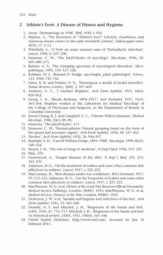
152 Notes
2 Athlete’s Foot: A Disease of Fitness and Hygiene
1. Anon, ‘Dermatology in 1938’, BMJ, 1939, i: 924.2. Majima, A., ‘The Invention of “Athlete’s foot”: Lifestyle, cleanliness, and
American leisure classes in the early twentieth century’, Seikatsugaku ronso,2010, 17: 3–13.
3. Whitfield, A., ‘A Note on some unusual cases of Trichophytic infection’,Lancet, 1908, ii: 237–238.
4. Emmons, C. W., ‘The Jekyll-Hydes of mycology’, Mycologia, 1960, 52:669–680, 671.
5. Bulmer, G. S., ‘The changing spectrum of mycological education’, Myco-pathologia, 1995, 130: 127–128.
6. Robbins, W. J., ‘Bernard O. Dodge, mycologist, plant pathologist’, Science,133, 1960: 741–742.
7. Davis, R. H. and Perkins, D. D., ‘Neurospora: a model of model microbes’,Nature Reviews Genetics, 2002, 3, 397–403.
8. Andrews, G. C., ‘J Gardner Hopkins’, Arch Derm Syphilol, 1951, 64(6):810–812.
9. Georg, L. K., ‘Rhoda Benham, 1894–1957’, Arch Dermatol, 1957, 76(3):363–364. Hopkins worked at the Laboratory for Medical Mycology ofthe College of Physicians and Surgeons, in the Department of Botany atColumbia University.
10. Kwon-Chung, K. J. and Campbell, C. C., ‘Chester Wilson Emmons’, MedicalMycology, 1986, 24(1) 89–90.
11. Emmons, ‘The Jekyll-Hydes’, 671.12. Emmons, C. W., ‘Dermatophytes: Natural grouping based on the form of
the spores and accessory organs’, Arch Derm Syphilol, 1934, 30: 337–362.13. ‘Review’, Arch Derm Syphilol, 1932, 26: 956–957.14. Rudolph, E. D., ‘Carroll William Dodge, 1895–1988’, Mycologia, 1990, 82(2):
160–164.15. Swartz, J. H., ‘The role of fungi in medicine’, N Engl J Med, 1936, 215: 322.16. Ibid., 323.17. Greenwood, A., ‘Fungus diseases of the skin’, N Engl J Med, 193, 213:
363–370.18. Adamson, H. G., ‘On the treatment of scabies and some other common skin
afflictions in soldiers’, Lancet, 1917, i: 222–223.19. MacCormac, H., ‘Skin-diseases under war conditions’, Brit J Dermatol, 1917,
29: 113–131; Adamson, H. G., ‘On the Treatment of Scabies and some othercommon skin affections in soldiers’, Lancet, 1917, i: 221–223.
20. MacPherson, W. G. et al, History of the Great War Based on Official Documents:Medical Services Pathology, London, HMSO, 1923; MacPherson, W. G. et al,Medical Services, Diseases of the War, London, HMSO, 1923.
21. Nickerson, J. W. et al, ‘Sandals and hygiene and infections of the feet’, ArchDerm Syphilol, 1945, 52: 365–368.
22. Ormsby, O. S. and Mitchell, J. H., ‘Ringworm of the hands and feet’,JAMA, 1916, 67: 711–717; Mitchell, J. H., ‘Ringworm of the hands and feet:An historical review’, JAMA, 1951, 146(6): 541–546.
23. Oxford English Dictionary, http://www.oed.com/. Accessed on line 10February 2011.
Notes 153
24. ‘Athlete’s foot’, Literary Digest, 22 December 1928, 17.25. Legge, R. T. et al, ‘Ringworm of the foot: Preliminary report’, JAMA, 1929,
92: 1507–1508.26. Legge, R. T. et al, ‘Incidence of foot ringworm amongst college students: Its
relation to gymnasium hygiene’, JAMA, 1929, 93: 170.27. ‘Bears investigate athlete’s foot’, Los Angeles Times, September 27, 1931, F5.
Also see Maima, ‘Invention of “Athlete’s foot” ’, 7.28. Gould, J. E., ‘Ringworm of the feet’, JAMA, 1931, 96: 1300–1302.29. Anon, ‘An American letter’, Public Health, 1932–1933, 46: 59.30. Swartz, ‘The role of fungi’, 322.31. Blaisdell, J. H., ‘Epidermophytosis’, N Engl J Med, 1930, 202: 1059–1064.32. Maima, ‘Invention of “Athlete’s foot” ’, 7–8.33. Gilman, R. T., ‘The incidence of ringworm of the feet in a university group:
Control and treatment’, JAMA, 1933, 100: 716.34. Underwood G. B. et al, ‘Overtreatment dermatitis of the feet’, JAMA, 1946,
130: 249.35. White, C. J., ‘Fungus disease of the skin, clinical aspects and treatment’,
Arch Derm Syphilol, 1927, 15: 387–414.36. ‘The History of Absorbine’. After Absorbine became widely available across
America, farmers realised that the same liniment that helped their horsesalso helped their own aches and pains. Seeing the need, Wilbur and MaryIda’s son Wilbur II suggested a version for people. In 1904, AbsorbineJr. was introduced, named after Wilbur Jr. Like Absorbine Veterinary Lin-iment, Absorbine Jr. helps the body heal itself by increasing blood flowto the affected area. It has the same analgesic and antiseptic propertiesas the veterinary liniment. It works great to relieve the itch caused byathlete’s foot. W.F. Young, Inc. actually coined the phrase ‘athlete’s foot’.Absorbine Jr. is now widely available at major mass-retailers and smallerpharmacies and stores. http://absorbine.blogspot.com/2010/06/history-of-absorbine.html. Accessed 8 October 2010.
37. Falls, A. I., ‘Doing business as Falls Chemical Co. v. Scholl Mfg. Co., Inc. caseto protect Solvex’, The Trade-Mark Reporter, 27 Trademark Rep. (1937), 444.
38. Pillsbury, D. et al, A Manual of Cutaneous Medicine, Philadelphia, W. B. Saun-ders, 1961, 604. For a later view, see: Bhutani, L. K. et al, ‘Tinea pedis –a penalty of civilization A sample survey of rural and urban population’,Mycoses, 1971, 14: 335–336.
39. Fraser, P. K., ‘Tinea of the foot’, BMJ, 1938, i: 842–844; H. MacCormac,‘Ringworm of the foot’, BMJ, 1940, i: 739–741.
40. ‘Pithead baths’ BMJ, 1931, i: 25; Morgan, W. J., ‘The miners’ welfare fundin Britain 1920–1952’, Social Policy & Administration, 1990, 24: 199–211.
41. Souter, J. C., ‘A Clinical note on fungus infection of the skin of the feet’,Proc Roy Soc Med, 1937, 30: 1107–1116.
42. MacKenna, R. M. B., et al, ‘Dermatological practice in war-time’, in Medicineand Pathology: History of the Second World War, London: Her Majesty’sStationery Office, 1952, 408–419; Pillsbury, D. M. and Livingood, C. S.,‘Experiences in military dermatology: Their interpretation in plans forimproved general medical care’, Arch Derm Syphilol, 1947, 55: 441–462.
43. Dewitt Mackenzie, Men without Guns, Philadelphia, The Blakiston Com-pany, 1943, 15–16 and Plate 1.
154 Notes
44. ‘Report’, JAMA, 1941, 117: 1973.45. De Kruif, P., ‘A working cure for athlete’s foot’, Reader’s Digest, 1942, 40:
46–48.46. Summers, W. C., ‘On the origins of the science in Arrowsmith: Paul De
Kruif, Félix d’Hérelle and phage’, J Hist Med Allied Sci, 1991, 46(3): 315–332.47. See the review of de Kruif’s The Male Hormone in Arch Derm Syphilol, 1945,
52(1): 71.48. Report, ‘Food and drug administration warns o Phenol Camphor mixture,
JAMA, 1942, 119: 713. Also see: Miller, F. G., ‘Poisoning by phenol’, Can MedAssoc J, 1942; 46(6): 615–616; Phillips, B., ‘The phenol-camphor treatmentof dermatophytosis’, Br J Dermatol, 1944, 56(11–12): 219–227.
49. Sanderson, P. H. and Sloper, J. C., ‘Skin disease in the British Army in SEAsia’, Br J Dermatol, 1953, 65, 252–264, 300–309 and 362–372.
50. Ainsworth, G. C., ‘The medical research council’s medical mycology com-mittee (1943–1969): A chapter in the history of medical mycology in theUK’, Sabouraudia, 1978, 16: 1–7.
51. Souter, ‘A clinical note on fungus infection’, 1107.52. Knowles, R. B., ‘Dermatitis in coal-miners: A survey of the factors influenc-
ing its nature and cause’, unpublished M. D. Thesis, University of Sheffield,1943; idem, ‘Factors Influencing dermatitis in coal-miners’, BMJ, 1944, ii:430–432; H. R. Vickers, ‘Arthur Rupert Hallam’, BMJ, 1955, ii: 741–751.
53. Capel, E. H., ‘A medical service for the coal mining industry’, Journal of theRoyal Society for the Promotion of Health, 68, 1948, 525–531, 526. Also see‘Royal sanitary institute congress’, BMJ, 1948, i: 1196.
54. Hodgson, G. A., ‘The history of coal miners’ skin diseases’, in Cule, J. ed,Wales and Medicine: An Historical Survey from Papers Given at the Ninth BritishCongress on the History of Medicine, London: British Society for the Historyof Medicine, 1975, 59.
55. H. R. Vickers was a lecturer at the University of Sheffield by the time ofappointment with the CIE, therefore, it could be that Vickers supervisedKnowles’s study in 1943.
56. However, Lane resigned almost immediately after the launch of theCommittee, giving his reason that ‘industrial medicine was adequatelyrepresented on the Committee by Dr. Rogan’. Committee on Industrial Epi-dermophytosis’, Minutes for the First Meeting, 13 November 1951, MRC.51/739 CIE. Min. 1, Committee on Industrial Epidermophytosis (Foot ring-worm in coal miners and other workers in Great Britain) Manuscripts,Reports, Correspondence 1950–1955. Contemporary Medical Archives Cen-tre, GC1/1, Wellcome Library, p. 3.
57. Cochrane, A. L. and Blythe, M., One Man’s Medicine: An autobiography ofProfessor Archie Cochrane, London, The British Medical Journal, 1989.
58. For a description on the relationship between the NCB and the govern-ment, see McIvor, A. and Johnston, R., Miners’ Lung: A History of Dust Diseasein British Coal Mining, Farnham, Ashgate, 2007, 146–147; Smith, J. H., ‘Thedistribution of power in nationalized industries’, British Journal of Sociol-ogy, 1951, 2(4), 275–293; Presthus, R. V., ‘British public administration: Thenational coal board’, Public Administration Review, 1949, 9(3): 200–210.
59. Committee on Industrial Epidermophytosis’, Minutes for the First Meeting,13 November 1951, MRC. 51/739 CIE. Min. 1, Committee on Industrial

Notes 155
Epidermophytosis (Foot ringworm in coal miners and other workers inGreat Britain) Manuscripts, Reports, Correspondence 1950–1955. Contem-porary Medical Archives Centre, GC1/1, Wellcome Library, p. 1.
60. Rogan, J. M., ‘Epidermophytosis and the coal miners – an introduc-tory note’, 5 November 1951, MRC. 51/683 CIE. 51/1, Committeeon Industrial Epidermophytosis (Foot ringworm in coal miners andother workers in Great Britain) Manuscripts, Reports, Correspondence1950–1955. Contemporary Medical Archives Centre, GC1/1, WellcomeLibrary, p. 2.
61. ‘Committee on industrial epidermophytosis’, Minutes for the First Meeting,13 November, 1951, MRC. 51/739 CIE. Min. 1, Committee on IndustrialEpidermophytosis.
62. Holmes, J. G., ‘Pilot study of epidemiology of epidermophytosis in thecoal-mining industry’, MRC.53/72 CIE.53/1, Committee on Industrial Epi-dermophytosis, p. 2.
63. Ibid.64. Gentles, J. C. and Holmes, J. G., ‘Foot ringworm in coal-miners’, Br J Indust
Med, 1957, 14: 22–29.65. Holmes, ‘Pilot study of epidemiology of epidermophytosis’, 4, p. 9.66. Duncan, J. T., ‘The epidemiology of fungus diseases’, Trans R. Soc Trop Med
Hyg, 1948, 42: 207–216, 209.67. Rosman, N., ‘Infections with Trichophyton rubrum’, Br J Dermatol, 1966,
78(4): 208–212.68. Walker, J., ‘The dermatophytoses of Great Britain: Report of a three year
survey’, Br J Dermatol, 1950, 62: 239–251.69. Sproot, N. A., ‘Athlete’s foot’, BMJ, 1957, ii: 1064 and 1243.70. Holmes, J. G. and Gentles, J. C., ‘Diagnosis of foot ringworm’, Lancet, 1956,
ii: 62–63.71. Peterkin, G. A. G., ‘The diagnosis and treatment of tinea pedis’, Practitioner,
1957, 180: 543–552.72. Cruickshank, R., ‘The epidemiology of some skin infections’, BMJ,
1956, i: 58.73. Pillsbury, D. M. et al, Dermatology, Philadelphia, W. B. Saunders, 1956, 606.
English, M., ‘Trichophyton rubrum infection in families’, BMJ, 1957, i: 746and 755.
74. G. B. Underwood et al, ‘Overtreatment dermatitis of the feet’, JAMA, 1946,130(5): 249–256.
75. Ibid., 249.76. Schwartz, L. et al, ‘Control of the scalp amongst school children’, JAMA,
1946, 132: 58–62; Miller, J. L. et al, ‘Local treatment of tinea capitis’, JAMA,1946, 132: 67–70.
77. Stevens, R. J. and Lynch, F. W., ‘Ringworm of the scalp: A report on the cur-rent epidemic’, JAMA, 1947, 133: 306–309; McKee, G. M. et al, ‘Treatmentof tinea capitis with Roentgen rays’, Arch Derm Syphilol, 1946, 53: 458–470.
78. Bocobo, F. C. et al, ‘Epidemiologic study of tinea capitis caused by Ttonsurans and M audouinii’, Public Health Rep, 1952, 67: 53–56.
79. Modan, B. et al, ‘Thyroid neoplasms in a population irradiated for scalptinea in childhood’, in De Groot, C., ed, Radiation Associated Thyroid Car-cinoma, New York, Grune & Stratton Inc, 1977, pp. 449–459; Hempelman,
156 Notes
L. H. et al, ‘Neoplasms in persons treated with X-rays in infancy: Fourthsurvey in 20 years’, J Natl Cancer Inst, 1975, 55: 519–530.
80. Hornblum, A. M., Acres of Skin: Human Experiments at Holmesburg Prison,New York: Routledge, 1998.
81. Kligman, A. M., ‘The pathogenesis of tinea capitis due to Microsporumaudouini and Microsporum canis’, J Invest Dermatol, 1952, 18, 231–246. Alsosee: Strauss, J. S. and Kligman, A. M., ‘Effect of x-rays on sebaceous glandsof the human face: Radiation therapy of acne’, J Invest Dermatol, 1959, 33:347–356.
82. Ibid., 231.83. Kligman, A. M. and Anderson, W. W., ‘Evaluation of current methods for
the local treatment of tinea capitis’, J Invest Dermatol, 1951, 16: 155–168.84. Strauss, J. S. and Kligman, A. M., ‘An experimental study of tinea pedis and
onychomycosis of the Foot’, AMA Arch Derm, 1957, 76(1): 70–79, 70.85. Gilchrest, B. A. and Leyden, J. L., ‘In memoriam: Mites and the mighty:
The last work and lasting legacy of Albert M. Kligman, PhD, MD’, Journal ofInvestigative Dermatology, 2011, 131, 6–7.
86. Gellene, D., Obituary, Dr Albert M. Kligman’, New York Times, 22 Feb2010, 26a.
87. Weyers, W., ‘Medical experiments on humans and the development ofguidelines governing them: The central role of dermatology’, Clinics inDermatology, 2009, 27(4): 384–394.
88. Georg, L. K., ‘Trichophyton tonsurans ringworm: A new public health prob-lem’, Public Health Rep 1952, 67: 53–56; Bronson, D. M. et al, ‘An epidemicof infection with Trichophyton tonsurans revealed in a 20-year survey offungal infections in Chicago’, J Am Acad Dermatol, 1983, 8(23): 322–330;Rippon, J. W., ‘Forty four years of dermatophytes in a Chicago clinic(1944–1988)’, Mycopathologia, 1992, 119: 25–28.
89. Shockman, J. and Urbach, F., ‘Tinea Capitis in Philadelphia’, Int J Dermatol,1983, 22(9): 522–523.
90. Jawetz, E., ‘The rational use of antimicrobial agents: Reason versus emotionin chemotherapy’, Oral Surg Oral Med Oral Pathol, 8(9): 982–987.
91. Sulzberger, M. B. and Kano, A., ‘Undecylenic and propionic acids in theprevention and treatment of dermatophytosis’, Arch Derm Syphilol 1947,55: 391–395.
92. Hartley, F., ‘Parachlorphenyl-a-glycerol as an antibacterial and antifungalagent of pharmaceutical interest’, Quarterly Journal of Pharmacy and Phar-macology, 1947, 20: 388–395; Petrow, V. and Hartley, Sir F., ‘The rise and fallof British Drug Houses, Ltd.’, Steroids, 1996, 61: 476–482.
93. Anon, ‘Treatment of ringworm of the feet’, Lancet, 1946, I; 95.94. Peterkin, G. A. G., The diagnosis and treatment of tinea pedis’, Practitioner,
1957, 180: 551.95. Ibid., 550.96. The development of the drug by Glaxo and its commercial exploitation
is discussed in detail in Chapter 9 of a company history of Glaxo, see:Davenport-Hines, R. P. T. and Slinn, J., Glaxo: A History to 1962, Cambridge,Cambridge University Press, 1992, 200–222. The chapter is entitled – ‘Thedevelopment and commercial exploitation of griseofulvin’.
97. Oxford, A. E. et al, ‘Griseofulvin, C17H17O6Cl, a metabolic product ofPenicillium griseo-fulvum Dierckx, Biochem J, 33, 1939: 240–248.
Notes 157
98. Bud, R., Penicillin: Triumph and Tragedy, Oxford, Oxford University Press,2007, 26–29; Birkinshaw, J. H., ‘Harold Raistrick, 1890–1971’, Biogr MemsFell R. Soc, 1972, 18: 488–509.
99. Brian, P. W., ‘Studies on the biological activity of Griseofulvin’, Annals ofBotany, 1949, 13: 59–77.
100. The work of A. R. Martin at ICI’s Pharmaceutical Division at AlderleyPark is discussed in Anon, ‘The symposium’, Transactions of the St John’sDermatological Society, 1960, 45: 1.
101. Anon, ‘Fungicide by mouth’, Lancet, 1958, ii: 1216.102. This paragraph is based on the chapter on griseofulvin in Davenport-Hines
and Slinn, Glaxo, 200–222.103. Gentles, J. C., ‘Experimental ringworm in guinea pigs: Oral treatment with
Griseofulvin’, Nature, 1958, 182: 476–477.104. Paget, G. E. and Walpole, A. L., ‘Some cytological effects of griseoful-
vin’, Nature, 1958, 182, 1320–132; idem, ‘The experimental toxicology ofgriseofulvin’, Arch Dermatol, 1960, 81: 750–757.
105. Quintal, D. and Jackson, R., ‘The development of 20th century dermato-logic drugs’, Clinics in Dermatology, 1989, 7 (3), 42–43.
106. Williams, D. I. et al, ‘Oral treatment of ringworm with Griseofulvin’, Lancet,1958, ii: 1212–1213.
107. Ibid., 1212.108. Quoted in Davenport-Hines, Glaxo, 209.109. Gentles, J. C., ‘Experimental ringworm in guinea pigs: Oral treatment with
griseofulvin’, Nature, 1958, 182: 476–477.110. Ibid., 476.111. Gentles, J. C., ‘The treatment of ringworm with Griseofulvin’, Br J Dermatol,
1959, 71: 427–433.112. Williams, D. I. et al, ‘Griseofulvin’, Br J Dermatol, 1959, 71: 434.113. Ibid., 434 and 442.114. Ibid., 442.115. Blank, H. and Roth, J. F., ‘The treatment of dermatomycoses with orally
administered Griseofulvin’, Arch Dermatol, 1960, 81: 259–267.116. Pillsbury, D. M., ‘Griseofulvin therapy in dermatophytic infections’, Trans
Am Clin Climat Assoc, 1959, 71: 52–57.117. Blank, H., ‘Symposium on Griseofulvin’, Arch Dermatol, 1960, 51: 649.
The full proceedings were published as ‘Griseofulvin and dermatomycoses:An international symposium sponsored by University of Miami, October26–27, 1959’, Arch Dermatol, 1960, 81: 649–789.
118. Maibach, H. I. and Kligmann A. M., ‘Short-term treatment of onychomyco-sis with Griseofulvin’, Arch Derm Syphilol, 1960, 81: 733–734; Dillaha, C. J.and Jansen, G., ‘Dosage requirements of Griseofulvin in OnychomycosisDue to Trichophyton Rubrum. Preliminary Report’, Ibid., 790–766.
119. Report, ‘Oral treatment of fungus infections with griseofulvin: An interna-tional symposium’, Transactions of the St John’s Dermatological Society, 1960,45: 1–145.
120. The figure given was £21 for six month’s treatment, which would be £300 in2013. This would not be that expensive today, which says something aboutdrug price inflation.
121. Anon, ‘The symposium’, Transactions of the St John’s Dermatological Society,1960, 45: 1.
158 Notes
122. Frain Bell, W., and Stevenson, J. C., ‘Report on a clinical trial’, Transactions ofthe St John’s Dermatological Society, 1960, 45: 47–53. Russell, B. et al, ‘Chronicringworm infection of the skin and nails treated with griseofulvin: Reportof a therapeutic trial’, Lancet, 1960, i: 1141–1147.
123. Russell, B. F., ‘Correlation of clinical and laboratory findings and the criteriaof cure’, Transactions of the St John’s, 1960, 45: 141.
124. Davenport-Hines and Slinn, Glaxo, 219.125. Ibid.126. Anon, ‘Griseofulvin’, Lancet, 1960, i: 1175–1176.127. English, M. P., ‘Some controversial aspects of tinea pedis’, Br J Dermatol,
1962, 74: 50–56; idem, BMJ, 1961, i: 1086.128. Anon, ‘Tinea pedis’, Lancet, 1962, i: 785–786.129. Blank, H., ‘Antifungal and other effects of griseofulvin’, Am J Med, 1965,
39(5): 831–838; Davies, R. R. and Everall, J. D., ‘Mycological and clini-cal evaluation of griseofulvin for chronic onychomycosis’, BMJ, 1967, ii:464–468; Anon, ‘Today’s drugs: Griseofulvin’, BMJ, 1967, iv: 608–609.
130. Anon, ‘Today’s drugs: Fungal antibiotics’, BMJ, 1963, i: 1659–1660; Anon,‘Antibiotics in dermatology’, BMJ, 1963, ii: 981–982.
131. Blank, H., ‘Antifungal and other effects of Griseofulvin’, Am J Med, 1965,39: 831–838; Report, ‘Today’s drugs: Griseofulvin’, BMJ, 1967, ii: 608–609.
132. Weston Hurst, A., ‘Protoporphyrin, cirrhosis and hepatomata in the liversof mice given griseofulvin’, Br J Dermatol, 1963, 75(3): 105–112; Anderson,D. W., ‘Griseofulvin: Biology and clinical usefulness, a review’, Ann Allergy,1965, 23: 103–110.
133. Conte, N. F. et al, ‘Prophylactic Griseofulvin against Trichophyton mentagro-phytes infections’, in H. M. Robinson, ed, The Diagnosis and Treatment ofFungal Infections, Springfield: Charles C. Thomas, Inc., 1974, 543.
134. Allen, A. M., Internal Medicine in Vietnam: Volume 1, Skin Disease in Vietnam,1965–1972, Washington DC, Office of the Surgeon General, 1972, 59–82.For a summary, see: Allen, A. M. et al, ‘Skin infections in Vietnam’, MilitaryMedicine, 1972, 137: 295–301.
135. Maertens, J. A., ‘History of the development of azole derivatives’, ClinMicrobiol Infect, 2004, 10, Suppl 1: 1.
136. Cartwright, R. Y., ‘Use of antibiotics: Antifungals’, BMJ, 1978, ii: 108–111.137. Montgomery, B. J., ‘Belgian oral antifungal agent looks promising’, JAMA,
1980, 243(1): 12.138. Check, W. A., ‘Oral antifungal agent effective even for widespread infec-
tions’, JAMA, 1980, 244: 2019–2020.139. Restrepo, A. et al, ‘Introduction’, Reviews of Infectious Diseases, 1980,
2(4): 519.140. Dismukes, W. E. et al, ‘Criteria for evaluation of therapeutic response to
antifungal drugs’, Rev Infect Dis, 1980, 2(4): 535–545.141. Borelli, D. et al, ‘Ketoconazole, an oral antifungal: Laboratory and clini-
cal assessment of imidazole drugs’, Postgraduate Medical Journal, 1979, 55:657–661.
142. Dismukes, W., ‘Concluding remarks’, Rev Infect Dis, 1980, 2(4): 688.143. Robertson, M. H. et al, ‘Oral therapy with ketoconazole for dermato-
phyte infections unresponsive to griseofulvin’, Rev Infect Dis, 1980, 2(4):578–581.
Notes 159
144. Hay, R. J., ‘A comparative double blind study of ketoconazole and griseoful-vin in dermatophytosis’, Br J Dermatol, 112, 1985: 691–696.
145. Lambert, D. R. et al, ‘Griseofulvin and ketoconazole in the treatment ofdermatophyte infections’, Int J Dermatol, 1989, 28: 300–304.
3 Candida: A Disease of Antibiotics
1. Macfarlane, G., Alexander Fleming: The Man and the Myth, Oxford, OxfordUniversity Press, 1984.
2. Christensen, C. M., The Molds and Man: An Introduction to the Fungi,Minneapolis, University of Minnesota Press, 1951.
3. The Oxford English Dictionary dates the first use of the term for a disease tonone other than Samuel Pepys and a diary entry for 17 June 1665, when hewrote of someone: ‘He hath a fever – a thrush and a Hickup’, OED online.Accessed 8 August 2012.
4. Vogel, M. J. and Rosenberg, C. E., eds, The Therapeutic Revolution: Essays inthe Social History of American Medicine, Philadelphia, University of Pennsyl-vania Press, 1979.
5. Li, J. W.-H and Vederas, J. C., ‘Drug discovery and natural Products: End ofan era or an endless frontier?’ Science, 2009, 325: 161–165.
6. Medical Research Council, ‘Treatment of pulmonary tuberculosis withstreptomycin and para-aminosalicylic acid’, BMJ, 1950, ii: 1073–1085;Medical Research Council, ‘Various combinations of isoniazid with strepto-mycin or with PAS in the treatment of pulmonary tuberculosis’, BMJ, 1955,ii: 435–445.
7. Jawetz, E., ‘Infectious diseases: Problems of antimicrobial therapy’, Ann RevMed, 1954, 5: 6.
8. Earlier in the century, M. albicans had been known as Oidium albicans.9. Kane, R. L., ‘Iatrogenesis: Just what the doctor ordered’, Journal of Com-
munity Health, 1980, 5(3): 149–158; Whorton, J., ‘Antibiotic abandon: Theresurgence of therapeutic rationalism’, in Parascandola, J., ed, The History ofAntibiotics: A Symposium, Madison, Wis, American Institute of the Historyof Pharmacy, 1980, 125–136.
10. Sáez-Gómez, J. M. and Romero-Maroto, M., ‘Scientific ideas on muguet(Thrush) in the XVIII century’, Journal of Dental Research, 2010, 89: 571–574.
11. Goodhart, J. F., The Diseases of Children, 6th Edn, London, J. & A. Churchill,1899, 114.
12. West Walker, J., ‘On diphtheria’, BMJ, 1863; i: 504, 551 and 660.13. Stuart Wilkinson, J., ‘Some remarks upon the development of epiphytes:
With the description of a new vegetable formation found in connexionwith the human uterus’, Lancet, 1849, ii: 448–451.
14. Pelling, M., Cholera, Fever and English Medicine, Oxford, Clarendon Press,1976.
15. Wilkinson, ‘Some remarks’, 450.16. Bashford, A., Purity and Pollution: Gender, Embodiment and Victorian Medicine,
Palgrave, 1998.17. Hewitt, G., ‘Lectures on the diagnosis and treatment of diseases of women’,
BMJ, 1862, i: 54.
160 Notes
18. Hurley, R., ‘Candidal vaginitis’, Proc R. Soc Med, 1977, 70(Suppl 4): 1–2; Alsosee: Hurley R., and de Louvois, J., ’Candida vaginitis’, Postgrad Med J, 1979,55: 645–647.
19. Plass, E. P. et al, ‘Monilial vulvovaginitis’, Am J Obstet Gynecol, 1931,21: 320.
20. Semon, H. C., ‘The non-venereal affections of the genitalia’, Br J Vener Dis,1929, 5: 114–127.
21. Harkness, A. H., ‘Non-gonococcal urethritis’, Br J Vener Dis, 1933(3),173–186 and 187–191.
22. Sharp, B. B., ‘Vulvo-vaginitis’, Br J Vener Dis, 1930(6): 301.23. Sharman, A., ‘The significance of leucorrhoea’, BMJ, 1935, ii: 1199–1201:
Bland, P. D. et al, ‘Vaginal trichomoniasis in the pregnant woman’, JAMA,1931, 96: 157–163; Solomon, E. and Dockeray, G. C., ‘Vaginal discharges’,Irish Journal of Medical Science 1936–1937, 11, 548–551.
24. See Chapter 2.25. Georg, L. K., ‘Rhoda Benham, 1894–1957’, Arch Dermatol, 1957, 76(3):
363–364.26. Benham, R., ‘Certain Monilias parasitic on man: Their identification by
morphology and by agglutination’, J Infect Dis, 1931, 49: 183–215.27. Ibid., 212. Sprue was a tropical disease of the small intestine.28. Berkhout, M. C., De schimmelgeslachten Monilia, Oidium, Oospora en Torula,
Scheveningen, Edauwe & Johanssen, 1923; Shrewsbury, J. D., ‘The genusMonilia’, J Path Bact, 1934, 38: 213–254,
29. Skinner, C. F., ‘The yeast-like fungi: Candida and Brettomyces’, Bacteriol Rev,1947, 11(4): 227–274.
30. Martin, D. S. et al, ‘A practical classification of the Monilias’, J Bacteriol 34,1937: 99–129; Martin, D. S. and Jones, C. P., ‘Further studies on the practicalclassification of the Monilias’, J Bacteriol, 1940, 39: 609–630.
31. Gay, F. P., Agents of Disease and Host Resistance, Baltimore, CharlesC. Thomas: 1935, 1118–1123.
32. Cruickshank, R. and Sharman, A., ‘Hormones and vaginitis’, J Obst Gynecol,1934, 41: 190.
33. Bland, P. D. et al, ‘Experimental vaginal and cutaneous moniliasis: Clinicaland laboratory studies of certain monilias associated with vaginal, oral andcutaneous thrush’, Arch Derm Syphilol, 1937, 36: 760.
34. Tattersall, R., Diabetes: A Biography, Oxford, Oxford University Press, 2009,82–85.
35. Hesseltine, H. C., ‘Diabetic and mycotic vulvovaginitis: Preliminary report’,JAMA, 1933; 100(3): 177–178.
36. Feudtner, C., Bittersweet: Diabetes, Insulin, and the Transformation of Illness,Chapel Hill, NC, University of North Carolina Press, 2003,
37. Woodruff, P. and Hesseltine, H. C., ‘Relationship of oral thrush to vaginalmycosis and the incidence of each’, Am J Obstet Gynecol, 1938, 36: 467.
38. Liston, G. and Cruickshank, L. G., ‘On thrush, with special reference tovaginal thrush’, Edin Med J, 1940, 47: 369–390; Liston, G. and Cruickshank,L. G., ‘Leucorrhoea in pregnancy: A study of 200 cases’, Br J Obstet Gynaecol,1940, 47: 109–129.
39. Comment, ‘Vaginal thrush’, Lancet, 1940, i: 1174; Editorial, ‘Vaginal dis-charge’, Lancet, 1940, ii: 300. Also see: Bland, P. B. and Rakoff, A. E.,
Notes 161
‘Leukorrea: Clinical and therapeutic aspects’, JAMA, 1940, 115(12):1013–1018.
40. Editorial, ‘Vaginal discharge’, 300.41. Cregor, F. W. and Gastineau, F. M., ‘Stovarsol in the treatment of syphilis:
A preliminary report’, Arch Derm Syphilol, 1927, 15(1): 43–53.42. The Medical Annual 1941: A Year Book of Treatment and Practitioner’s Index,
Bristol, John Wright, 1941.43. Ludlam, G. B. and Henderson, J. L., ‘Neonatal thrush in a maternity
hospital’, Lancet, 1942, i: 64–70.44. Menzies, M. F., ‘Hospital or domiciliary confinement?’, Lancet, 1942, ii:
35–38 and 201.45. McNeil, C., ‘Death in the first month and the first year’, Lancet, 1940, i:
819–821.46. ‘Thrush in infants’, BMJ, ii: 1950: 1430–1431.47. Wagner, J. M. and Kessel, I., ‘Complications of C. albicans infection in
infancy’, BMJ, 1958, ii: 362–366.48. Donald, I., ‘Aetiology and investigation of vaginal discharge’, BMJ, 1952, ii:
1223–1226.49. Russell, C. S., ‘Leucorrhoea’, BMJ, 1953, ii: 91–93. Also see: Anon, ‘Any
questions? Vaginitis’, BMJ, 1952, ii: 813–815 and 1954, i: 59.50. Collins, J. H., ‘Vulvovaginitis’, Obstet Gynecol Surv, 1952, 7(2): 224–227;
Thomas, H. H., ‘Candidal vulvovaginitis: Treatment with mycostatin’,Obstet Gynecol, 1957, 9(2): 163–166.
51. Smith, G., ‘The Subway grate scene in the seven year itch: “The stagingof an appearance-as-disappearance” ’, Cinémas: Journal of Film Studies, 2004,14(2–3): 213–244.
52. Report, ‘Council on pharmacy and medicine’, JAMA, 1951, 145: 1267.53. Bud, R., Penicillin: Triumph and Tragedy, Oxford, Oxford University Press,
2007, 105–107.54. Smith, L. W. and Walker, A. D., Penicillin Decade 1941–1951: Sensitizations
and Toxicities, Washington, Arundel Press, 1951.55. Scales, I. K. et al, ‘Oral fungus infection: Candidiasis albicans’, Oral Surg Oral
Med Oral Pathol, 1956, 9: 970–977.56. Colgan, M. T., ‘The bacterial flora of the intestinal tract: Changes in diar-
rheal disease and following antimicrobial therapy’, Journal of Pediatrics,1956, 49: 214–228; Editorial, ‘Antibiotics and monilial infection’, Lancet,ii, 532.
57. Hirst, H. L. et al, ‘Methods of administration of penicillin’, J Lab Clin Med,1947, 32: 32.
58. Lane, S. L., ‘A review of current opinion on the hazards of indiscriminateantibiotic therapy in dental practice’, Oral Surg Oral Med Oral Pathol, 1956,9: 952–961.
59. Hussar, A. E. and Holley, H. L., Antibiotics and Antibiotic Therapy, New York,Macmillan, 1954, 12–34. Also see: Binns, T. B., ‘Gastro-intestinal complica-tions of oral antibiotics’, Lancet, 1956, i: 336–338.
60. Harris, H. J., ‘Aureomycin and chloramphenicol in Brucellosis: With specialreference to side effects’, JAMA, 1950, 142: 161–165.
61. Woods, J. W. et al, ‘Monilial infections complicating the therapeutic use ofantibiotics’, JAMA, 1951, 145: 207–211.
162 Notes
62. Joekes, T. H. and Simpson, R. H., ‘Bronchomoniliais’, Lancet, 1923, ii:108–111; Anon, ‘An investigation into bronchomoniliais’, Lancet, 1924,i: 714.
63. Greer, A. E., ‘The synergism between mycotic and tuberculous infections ofthe lungs’, Dis Chest, 1948, 14: 33–40; Brown, T. G., Pulmonary mycosis,Edin Med J, 1947, 54: 414–422.
64. Oblath, R. W. et al, ‘Pulmonary moniliasis’, Ann Intern Med, 1951, 35:97–116.
65. Editorial, ‘A danger of the newer antibiotics’, BMJ, 1951, i: 1196.66. Editorial, ‘Fungus infection complicating antibiotic therapy’, JAMA, 1952,
149: 762–763.67. Kligman, A. M., ‘Are fungus infections increasing as a result of antibiotic
therapy’, JAMA, 1952, 149: 979–983.68. Huppert, M. D. et al, ‘Pathogenesis of C. albicans infection following antibi-
otic therapy. I. The effect of antibiotics on the growth of C. albicans’, JBacteriol, 1953, 65: 171–176.
69. Kligman, ‘Are fungus infections’, 979.70. Ibid., 983; Editorial, ‘Antibiotic therapy and fungous infections’, N Engl J
Med, 1952, 247, 491–492.71. Jawetz, ‘Infectious diseases’, 6.72. Finland, M. and Weinstein, L., ‘Complications induced by antimicrobial
agents’, N Engl J Med, 1953, 248, 220–226.73. Weinstein, L. et al, ‘Infections occurring during chemotherapy’, N Engl J
Med, 1954, 251: 251–259.74. Sharp, J. L., ‘The growth of Candida albicans during antibiotic therapy’,
Lancet, 1954, i: 390–392.75. Cannon, P. R., ‘The changing pathologic picture of infection since the intro-
duction of chemotherapy and antibiotics’, Bull N Y Acad Med, 1955, 31:89–91.
76. Kozinn, P. J. et al, ‘Candida albicans: Saprophyte or pathogen? A diagnosticguideline’, JAMA, 1966, 198(2): 170–172.
77. Baldwin, R. S., The Fungus Fighters: Two Women Scientists and Their Discovery,Ithaca, Cornell University Press, 1981.
78. Ibid., 75.79. Hazen, E. L. and Brown, R., ‘Two antifungal agents produced by a soil
actinomycete’, Science, 1950, 112: 423; idem, ‘Fungicidin, an antibioticproduced by a soil actinomycete’, Exp Biol Med, 1951, 76: 93–97.
80. Hazen, E. L. and Brown, R., ‘Nystatin’, Ann N Y Acad Sci, 1960, 27, 89:258–266.
81. Waksman, S. et al, ‘Antifungal antibiotics’, Bull World Hlth Org, 1952, 6:163–172.
82. Garrod, L. P., ‘The sensitivity of actinomyces to antibiotics’, BMJ, 1952, i:263; Hussar and Holley, Antibiotics, 366–369.
83. Espinel-Ingroff, A. V., Medical Mycology in the United States: A Historical Anal-ysis (1894–1996), Dordrecht, Kluwer Academic Publishers, 2003, 86 and94–96.
84. The powder was also used in laboratories as an antifungal agent, to sup-press the contamination of sera, tissue cultures, culture plates, and other invitro media. See: Report, ‘Mycostatin available for laboratories’, Journal ofthe Franklin Institute, 1956, 261: 285.
Notes 163
85. Brabander, J. O. W. et al, ‘Intestinal moniliasis in adults’, Canad Med AssocJ, 1957, 77: 478–483.
86. Shrand, H., ‘Thrush in the newborn’, BMJ, 1961, ii: 1530–1533, i: 186and 567.
87. Sternberg, T. H. and Newcomer, V. D., Therapy of Fungal Diseases: An Inter-national Symposium, Boston, Little Brown & Company, 1956.
88. Ibid., ‘Contents’.89. Sloane, M. B. ‘New antifungal antibiotic: Mycostatin (Nystatin) of the
treatment of moniliasis: A preliminary report’, J Invest Dermat, 1955, 24:569–571.
90. Chesney, J., ‘ “Nystatin” (“Mycostatin”)’, BMJ, 1956, i: 1043–1044.91. Pace, H. and Schantz, S., ‘Nystatin (Mycostatin) in the treatment of monilial
and nonmonilial vaginitis’, JAMA, 162, 1956: 268–271.92. Lang, W. R. et al, ‘Nystatin vaginal tablets in the treatment of Candidal
vulvovaginitis’, Obstet Gynecol, 1956, 8: 364–367.93. Wright, E. T. et al, ‘Treatment of moniliasis with nystatin’, JAMA, 1957, 163:
92–94.94. Barr, W., ‘Nystatin’, Practitioner, 1957, 178: 616–617.95. Baldwin, Fungus Fighters, 99–101.96. Loh, W. P. and Baker, E. E., ‘Fecal flora of man after oral administration of
chlortetracycline or oxytetracycline’, Arch Intern Med, 1955, 95(1): 74–82;McGovern, J. J. et al, ‘The effect of aureomycin and chloramphenicol onthe fungal and bacterial flora of children’, N Engl J Med, 1953, 248(10):397–403.
97. Sharp, ‘The growth of Candida’, 392; Sakewitz, A. B., ‘Treatment of gen-itourinary moniliasis with orally administered nystatin’, Ann Intern Med,1955, 42(6): 1187–1199.
98. Childs, A. J., ‘Effect of nystatin on growth of C. albicans during antibiotictherapy’, BMJ, 1956, i: 660–662; Gimble A. I. et al, ‘Nystatin and tetracy-cline in the treatment of bacterial infections’, Antibiot Annu, 1955–1956;3: 676–680. Lederle introduced a version number the brand ‘Lederstatin’.Anon, ‘Gastrointestinal complications of treatment by broad spectrumantibiotics’, Drugs and Therapeutic Bulletin, 1964, 2: 69–71; Anon, ‘Old andnew tetracyclines’, Drugs and Therapeutic Bulletin, 1967, 5: 77–80.
99. Another practice that reinforced the idea was antibiotic ‘sterilisation’ ofthe colon before surgery with the antibiotic neomycin and nystatin. See:Cohen, I. and Longacre, A. B., ‘Neomycin-Nystatin preoperative prepara-tion of the colon’, Am Surg, 1956, 22: 301–307.
100. Report, ‘Fixed combinations of antimicrobial agents. national academyof sciences – national research council division of medical sciences drugefficacy study’, N Engl J Med, 1969, 280: 1149–1154.
101. McMahon, F. G., ‘Drug combinations: A critique of proposed new federalregulations’, JAMA, 1971, 216: 1008–1010.
102. Report, ‘Council on drugs’, JAMA, 1962, 182(1): 63–66.103. Silverman, M., and Lee, P. R., Pills, Profits and Politics, Berkeley, Uni-
versity of California Press, 1974; Anon, ‘Ebert and Squibb’, HarvardCrimson, 6 December 1972. http://www.thecrimson.com/article/1972/12/6/ebert-and-squibb-pbwbhat-are-the/. Accessed 1 March 2013.
104. Butler, W. T., ‘Pharmacology, toxicity, and therapeutic usefulness of ampho-tericin B’, JAMA, 1966, 195: 371–375.
164 Notes
105. Oura, M. et al, ‘A new antifungal antibiotic, amphotericin B’, Antibioc Annu,1955–1956, 3: 566–573; Dutcher, J. D., ‘The discovery and development ofamphotericin B’, Chest, 1968, 54: 296–298. The soil came from the OrinocoBasin in Venezuela, which illustrates the breadth of the search for newantibiotic compounds.
106. Newcomer, V. D. et al, ‘Current status of amphotericin B. in the treat-ment of the systemic fungus infections’, J Chron Dis, 1959, 9: 354–374;Newcomer, V. D. et al, ‘The treatment of systemic fungus infections withamphotericin B’, Ann N Y Acad Sci, 1960, 89: 221–239.
107. Crounse, R. G. et al, ‘Cryptococcosis; case with unusual skin lesions andfavorable response to amphotericin B. therapy’, Arch Derm, 1958, 77(2):210–215; Derbes, B. and Krafchuk, J. D., ‘Response of North Americanblastomycosis to amphotericin’, Bull Tulane Univ Med Fac, 1958, 17(3):157–163; Littman, B. et al, ‘Coccidioidomycosis and its treatment withamphotericin B’, Am J Med, 1958, 24(4): 568–592; Baum, B. and Schwarz, J.,‘Clinical experiences with amphotericin B’, Antibiot Annu, 1959–1960, 7:638–643.
108. Hurley, R. and Morris, E. D., ‘The pathogenicity of Candida species in thehuman vagina’, Br J Obstet Gynaecol, 1964, 71(5): 692–695.
109. Hurley, R., ‘General discussion’, Proc R Soc Med, 1977, 70, Suppl 4: 30.On Hurley, see: Duerden, B., ‘Obituary, Dame Rosalinde Hurley’, BMJ, 2004,ii: 516.
110. Winner and Hurley, Candida, 139.111. Hurley, ‘Candidal vaginitis’, 2.112. Sobel, J. D. et al, ‘Clotrimazole treatment of recurrent and chronic Candida
vulvovaginitis’, Int J Gynecol Obstet, 1989, 29(4): 386.113. Elewski, B. et al, ‘Long-term outcome of patients with interdigital tinea
pedis treated with terbinafine or clotrimazole’, J Am Acad Dermatol, 1995,32(2–1): 290–292.
114. Winner, H. I. and Hurley, R., Candida albicans, London, Churchill 1964.115. Duerden, B., ‘Obituary: Rosalinde Hurley’, BMJ, 2004, 329: 516. Also see: J
Obstet Gynaecol, 2004, 24(7): 847. Howard Winner was Professor of Bacte-riology at Charing Cross from 1965 to 1982. Wright, D. J. M., ‘Obituary:H. I. Winner’, BMJ, 1993, 306: 1335.
116. Winner and Hurley, Candida, 56–61.117. Winner and Hurley, Candida, 59.118. Hussar and Holley, Antibiotics, 367.119. Winner and Hurley, Candida, 59–60.120. Newcomer, V. D. et al, ‘Current status of Amphotericin B in the treatment
of the systemic fungus infections’, J Chron Dis, 1959, 9: 353–374.121. Winner, H. I. and Hurley, R., A Symposium on Candida Infections, Edinburgh,
E. & S. Livingstone, 1966, 221–244.122. Seelig, M. S., ‘The role of antibiotics in Candida infection’, Amer J Med, 1966,
40: 887–917; Downing, J. G. and Conant, N., ‘Mycotic infections’, N Engl JMed, 233, 153 and 181.
123. Cantor, D., ‘Cortisone and the politics of drama, 1949–1955’, in Pickstone,J. V., ed, Medical Innovation in Historical Perspective, Basingstoke, Macmillan,1992, 225–245.
124. Wikler, A. et al, ‘Mycotic endocarditis: Report of a case’, JAMA, 119, 1942:33; Caplan, H., ‘Monilia (Candida) endocarditis following treatment with
Notes 165
antibiotics’, Lancet, 1955, ii: 95; Utz, J. P. and Roberts, W. C. ‘Candidaendocarditis’, Ann Intern Med, 1961, 54: 1058.
125. Sanger, P. et al, ‘Candida infection as a complication of heart surgery: Reviewof the literature and report of two cases’, JAMA, 1962, 181: 108.
126. Birt, A. R., ‘The increasing problem of drug reactions’, Can Med Assoc J,1957; 77: 709–715; Smith, T. J., ‘Antibiotic-induced disease’, in R. H. Moser,ed, Diseases of Medical Progress, Springfield, Ill, Charles C. Thomas,1963.
127. Plummer, N. S., ‘Candida infection in the lung’, in Winner and Hurley,Symposium, 214.
128. Cohen, A. C., ‘Pulmonary moniliasis’, Amer J Med Sci, 1953, 226: 16–23;Seelig, ‘Role of antibiotics’, 903–906.
129. Cohen, A. C., ‘Pulmonary’, 23.130. Winner and Hurley, 110–113.131. Borel, J. F. et al, ‘The history of the discovery and development of
cyclosporine (Sandimmun®)’ in Vincent, J. et al, eds, The Search for Anti-inflammatory Drugs, Boston, O. Birkhauser 1995; Amor, K. T. et al, ‘Theuse of cyclosporine in dermatology: Part I’, J Am Acad Dermatol, 2010, 63:925–946.
132. Gottlieb, H. S. et al, ‘Pneumocystitis carinii pneumonia and mucosal can-didiasis in previously healthy homosexual men: evidence of a new acquiredcellular immunodeficiency’, N Engl J Med, 1981; 305: 1425–1431; Water-son, A., ‘Acquired immune deficiency syndrome’, BMJ, (Clin Res Ed) 1983;286: 743–746; Seligmann, M. et al, ‘AIDS – An immunologic re-evaluation’,N Engl J Med, 1984; 311: 1286–129.
133. Klein, R. S. et al, ‘Oral candidiasis in high-risk patients as the initial mani-festation of the acquired immunodeficiency syndrome’, N Engl J Med, 1984;311: 354–358; Pranatharhti, H. and Molinari, J. A., ‘Oral candidiasis: Fore-runner of acquired immunodeficiency syndrome (AIDS)?’ Oral Surg OralMed Oral Path, 1985, 60: 532–534.
134. McCarthy, G. M., ‘Host factors associated with HIV-related oral candidiasis:A review’, Oral Surg Oral Med Oral Path, 1992, 73: 181–186.
135. ‘Fluconazole: A major advance for cryptococcal meningitis and othersystemic fungal infections?’ AIDS Treatment News, 1987, 41.
136. Maertens, J. A., ‘History of the development of azole derivatives’, ClinMicrobiol Infect, 2004, 10 Suppl 1: 3–4.
137. Troke, P. F., ‘Efficacy of UK-49,858 (Fluconazole) against Candida albicansexperimental infections in mice’, Antimicrob Agents Chemother, 1985, 28(6):815–818; Odds, F. C. et al, ‘Antifungal effects of fluconazole (UK-49858),a new triazole antifungal, in vitro’, Antimicrob Agents Chemother, 1986, 18:473–478; Hay, R. J., ‘Fluconazole’, J Infect, 1990, 21: 1–6; Richardson, K.et al, ‘Discovery of fluconazole, a novel antifungal agent’, Clin Infect Dis,1990, 12: S267–271.
138. de Wit, S. et al, ‘Comparison of fluconazole and ketoconazole for oropha-ryngeal candidiasis in AIDS’, Lancet, 1989, i: 746–748.
139. Lancet, 1989, i: 1130–1131.140. Anon, ‘Products in development: Progress continues in search for AIDS-
related drugs’, Aids Patient Care, August 1988, 2(4): 28–33; Williams, R. D.,‘Living with AIDS: New treatments give hope’, FDA Consumer, January-February 1992, 26; Lindemann, E., ‘Importing AIDS drugs: Food and drug
166 Notes
administration policy and its limitations’, George Washington Journal ofInternational Law and Economics, 1994, 28: 133–170.
141. British Society for Antimicrobial Chemotherapy Working Party, ‘Antifun-gal chemotherapy in patients with acquired immunodeficiency syndrome’,Lancet 1992: 648–651. Coleman, D. C. et al, ‘Oral Candida in HIV infectionand AIDS: New perspectives/new approaches’, Crit Rev Microbial, 1993; 19:61–82.
142. McNeill, M. et al, ‘Mortality in the United States, 1980–1997, due to can-didiasis, aspergillosis, and other mycoses in persons infected and personsnot infected with HIV trends in mortality due to invasive mycotic diseasesin the United States, 1980–1997’, Clin Infect Dis, 2001, 33(5): 641–647.
143. Odds, Candida, 1988, 5.144. Fisher-Hoch, S. P. and Hutwanger, L., ‘Opportunistic candidiasis: an epi-
demic of the 1980s’, Infect Dis, 1995, 21(4): 897–904.145. Dismukes, W. E., ‘Antifungal therapy: Lessons learned over the past 27
years’, Clin Infect Dis, 2006, 42: 1289–1330.146. Ibid., 1290.147. Bennett, J. E. et al, ‘A comparison of amphotericin B. alone and combined
with flucytosine in the treatment of cryptococcal meningitis’, N Engl J Med,1979, 301(3): 126–131.
148. Dismukes, W. E. et al, ‘Criteria for evaluation of therapeutic response toantifungal drugs’, Rev Infect Dis, 1980, 2(4): 535–545; idem, ‘AntifungalTherapy’, passim. Flucocystine, had been developed as an anti-cancer com-pound in the 1950s, however, it proved ineffective and it was only in the1960s that its antifungal properties were recognised. It was effective againstcryptococcosis and candidiasis, but its value was limited by the develop-ment of resistance, hence combined usage, and toxicity, though it was lessso than amphotericin B. J. E. Bennett, ‘Flucocystine’, Ann Intern Med, 1977,86(3): 319–321.
149. Ayliffe, G. and English, M., Hospital Infection: from Miasmas to MRSA,Cambridge, Cambridge University Press, 2003.
150. Pfaller, M. A., ‘Nosocomial fungal infections: Epidemiology of candidiasis’,Journal of Hospital Infection, 1995, 30 (Suppl): 329–338.
151. Ibid., 330–333.152. Odds, F. C., ‘Epidemiological shifts in opportunistic and nosocomial Can-
dida infections: mycological aspects’, Int J Antimicrob Agents, 1996, 6:141–144.
153. For a popular account of the ‘yeast connection’, see Barrett, S., ‘Unproven“Allergies”: An Epidemic of Nonsense “Environmental illness” and the“yeast connection” ’, in Barrett S. and Jarvis, J. T., eds, The Health Robbers;A Close Look at Quackery in America, New York, Prometheus Press, 1993.
154. Odds, Candida (1988), 233.155. Crook, W. G., The Yeast Connection: A Medical Breakthrough, Jackson, T., Pro-
fessional Books, 1983; O. Truss, The Missing Diagnosis, Birmingham, TheMissing Diagnosis, Inc., 1983.
156. Crook, W. G., Answering Parent’s Questions, Jackson, Professional Books,1963; idem, Your Allergic Child: A Pediatrician’s Guide to Normal Living forAllergic Adults and Children, New York, Medcom Pres, 1973; idem, Can YourChild Read? Is He Hyperactive?: A Pediatrician’s Suggestions for Helping the
Notes 167
Child with Hyperactivity, Behavior and Learning Problems, Jackson, Tenn, Pro-fessional Books 1975; idem, Are You Allergic?: A Guide to Normal Living forAllergic Adults and Children, Jackson, Tenn, Professional Press, 1977.
157. Truss, C. O., ‘The role of Candida Albicans in human illness’, Orthomolecu-lar Psychiatry, 1981, 8: 228–238; Truss, C. O., ‘Tissue injury induced by C.albicans: Mental and neurologic manifestations’, J Orthomol Psychiatr, 1978,1, 17–37.
158. Brief details of Truss’s career are given at http://www.trussmd.com/about.html. Accessed 26 April 2012.
159. Goertzl, T. and Goertzl, B., Linus Pauling: A Life in Science and Politics,New York, HarperCollins, 1995; Hager, T., Linus Pauling and the Chemistryof Life, New York: Oxford University Press, 1998.
160. Pauling, L., ‘Orthomolecular psychiatry: Varying the concentrations of sub-stances normally present in the human body may control mental disease’,Science, 1968, 160: 265–271; Pauling, L. et al, ‘On the orthomolecularenvironment of the mind: orthomolecular theory’, Am J Psychiatry, 1974,131: 1251–1267. Orian Truss was born on 1 December 1922 and died inBirmingham, Alabama on 10 September 2009.
161. Task Force Report 7, Megavitamin and Orthomolecular Therapy in Psychiatry,Washington, DC, APA, 1973; Editorial, ‘Molecules and mental health’, BMJ,1975, i: 296.
162. Williams, R. J. and Wright, D. K., A Physician’s Handbook on OrthomolecularMedicine, New York Pergamon Press, 1977.
163. Hess, D. J., Evaluating Alternative Cancer Therapies: A Guide to the Science andPolitics of An Emerging Medical Field, New Brunswick, NJ, Rutgers UniversityPress, 1999; idem, Can Bacteria Cause Cancer?: Alternative Medicine ConfrontsBig Science, New York, New York University Press, 1997.
164. Truss, C. O., ‘Tissue injury induced by Candida albicans: Mental and neu-rologic manifestations’, J Orthomol Psychiatr, 1978, 7(1): 17: 37; idem,‘Restoration of immunologic competence to C. albicans’, J Orthomol Psy-chiatr, 1980, 9: 287.
165. Ibid., 289.166. Truss, C. O., ‘Restoration of immunologic competence to Candida Albi-
cans’, J Orthomol Psychiatr, 1980, 9: 290.167. Randolph, T. G., Human Ecology and Susceptibility to the Chemical Environ-
ment, Springfield, Ill: Thomas, 1962; Buscher, D., ‘Obituary: Theron G.Randolph, MD, 1906–1995’, Journal of Nutritional & Environmental Medicine,1996, 6: 245–246.
168. Task Force on Clinical Ecology, ‘Clinical ecology – a critical appraisal’, WestJ Med, 1986, 144: 239–245.
169. Crook, W. G., ‘Letters depression associated with C. albicans Infections’,JAMA, 1984: 51: 2928–2929.
170. Crook, Yeast Connection, vi.171. Crook summarises the meeting in Chapter 37 of The Yeast Connection.172. Ibid., vi and Crook, W. G., ‘The coming revolution in medicine’, Journal of
the Tennessee Medical Association, 1983, 76: 145–149.173. Crook, Yeast Connection, viii.174. Quinn, J. P. et al, Ketoconazole and the yeast connection, JAMA, 1986, 255,
3250.
168 Notes
175. Morgan, P. P., ‘Should scientists study “20th-century disease”?’, Can MedAssoc J, 1985, 133: 961–962; Zimmerman, B. and Weber, E., ‘Candida and“20th-century disease” ’, Ibid., 1985 133: 965–966; Maclennan, J. G., ‘Can-dida and “20th-century disease” ’, Ibid., 1986 134: 1112–1113; Lornzani, S.,Candida: A Twentieth Century Disease, New Canaan, Conn, Keats Publishing,1986; Murphy, M., Sick Building Syndrome and the Problem of Uncertainty:Environmental Politics, Technoscience, and Women Workers, Durham, NC,Duke University Press, 2006.
176. For a popular presentation see: Rovner, S., ‘HEALTHTALK: The yeast theory’,Washington Post, Mar 9, 1984: D5.
177. Haas, A. et al, ‘The “Yeast Connection” meets chronic mucocutaneouscandidiasis’, N Engl J Med, 314: 854–855, 1986.
178. Coleman, III, W. P. and Edwards, D. E., ‘Letters depression and Candida –Reply’, JAMA, 1985, 253: 3400.
179. Anderson, J. A. et al, ‘Position statement on candidiasis hypersensitivity’, JAllergy Clin Immunol, 1986, 78: 271–273.
180. Edwards, J. E., ‘Systemic symptoms from Candida in the gut: Real orimaginary?’, Bull N. Y. Acad Med, 1988, 64(6): 544–549.
181. Ibid., 548.182. Infectious Diseases and Immunization Committee of the Canadian Paedi-
atric Society, ‘Candidiasis: current misconceptions’, Can Med Assoc J, 1988,139: 729; Blonz, E. R., ‘Is there an epidemic of chronic candidiasis in ourmidst?’, JAMA, 1986, 256: 3138–3139.
183. Aronowitz, R., ‘From myalgic encephalitis to yuppie flu: a history of chronicfatigue syndromes’, in Rosenberg, C. and Golden, J., eds, Framing Dis-ease: Studies in Cultural History, New Brunswick, NJ: Rutgers UniversityPress, 1992, 155–181; Jenkins, R. and Mowbray, J. F., eds, Post-viral FatigueSyndrome: (myalgic encephalomyelitis), Chichester: John Wiley & Sons,1992.
184. Finlay, S., ‘An illness doctors don’t recognise’, Observer, 1 June 1986, 43.Also see follow up article: Finlay, S., ‘Voice for sufferers’, Observer, 2 August1987, 39; Chaitlow, L., Candida Albicans: Could Yeast be Your Problem?,Wellingborough, Thorsons, 1985.
185. Collings, J., ‘The quiet epidemic that is ruining modern lifestyle’, Guardian,4 February 1988, 13.
186. Crook, The Yeast Connection, v–vi.187. Mildenhall, R., ‘Candida views’, Observer, 26 June 1988, 37.188. Renfro, L. et al, ‘Yeast Connection among 100 patients with chronic
fatigue’, Amer J Med, 1989, 86, 165–168.189. Ibid., 168.190. Dismukes, W. E. et al, ‘A randomized double-blind trial of nystatin therapy
for the candidiasis hypersensitivity syndrome’, N Engl J Med, 1990, 323:1717–1723.
191. Ibid., 1723.192. Correspondence from Crooks, W., Truss, C. O. et al, W. J. Ledger et al, and
M. Crandall, N Engl J Med, 1991, 324: 1592–1594.193. Kay, A. B. and Lessor, M. H., Allergy: Conventional and Alternative Concepts,
London, Royal College of Physicians of London, 1992.
Notes 169
194. Downing D. and Davies, S., ‘ “Allergy: Conventional and Alternative Con-cepts”: A critique of the Royal College of Physicians of London’s Report’,Journal of Nutritional Medicine, 1992, 3: 331–349.
195. Kay, A. B., ‘Alternative allergy and the general medical council’, BMJ, 1993,i: 122–124, 328–331 and 582.
196. Mumby, K., ‘Science or flat earthers? The clinical ecologist replies’, BMJ,1993, ii: 1055–1056.
197. Eisenberg, D. M. et al, ‘Trends in alternative medicine use in the UnitedStates, 1990–1997:Results of a follow-up national survey’, JAMA, 1998, 280:1569–1575.
198. Hyams, K., ‘Developing case definitions for symptom-based conditions: Theproblem of specificity’, Epidemiologic Reviews, 1998, 20(2): 148–156.
199. Crook, W. G., Nature’s Own Candida Cure, Alive Books, 2003; Crook, W. G.et al, The Yeast Connection and Women’s Health, Square One, 2007; Crook,W. G., Tired So Tired: and the Yeast Connection, Square One, 2007.
200. Weig, M. et al, ‘Clinical aspects and pathogenesis of Candida infection’,Trends in Microbiology, 2012, 20(8): 468.
4 Endemic Mycoses and Allergies: Diseases of Social Change
1. Reiss, F., ed, ‘Medical mycology’, Ann N Y Acad Sci, 1950, 50: 1209–1404.2. Conant, N. F., ‘Future developments in mycological investigative methods’,
Ann N Y Acad Sci, 1950, 50: 1245–1249.3. Jackson, M., Asthma: The Biography, Oxford, Oxford University Press, 2009.4. Smith, D. T., ‘The diagnosis and therapy of mycotic infections’, Bull N Y
Acad Med, 1953, 29(10): 778.5. Jillson, O. F., ‘Mycology’, N Engl Med J, 1953, 249: 523–530 and 561–566.6. Salvin, S. B., ‘Public health aspects of fungus infections’, Ann N Y Acad Sci,
1950, 50: 1217.7. Nickerson, W. J., ‘Medical mycology’, Ann Rev Microbiol, 1953, 7: 245–272.8. Vital Statistics of the United States, 1950, Volume 3, Mortality Data, Washing-
ton: US Depart of Health, Education and Welfare, 1953, Table 51, 62, VitalStatistics of the United States, 1960, Volume 2 – Mortality, Part A., Washing-ton: US Department of Health, Education and Welfare, 1963, Table 5–6,5–16.
9. Nickerson, ‘Medical mycology’, 246.10. Henrici, A. T., ‘Characteristics of fungous diseases’, J Bacteriol, 1940, 39:
113–138.11. Fildes, P., ‘Richard Friedrich Johannes Pfeiffer, 1858–1945’, Biog Mem Fell R
Soc, 1956, 2: 237–247; Jackson, M., Allergy: The History of a Modern Malady,London, Reaktion Books, 2006, 87–88.
12. Smith, ‘The diagnosis and therapy’, 782.13. Schwarz, J. and Baum, G. L., ‘A critical review of medical mycology in the
United States, 1946–1956’, Mycopatholgia et Mycologia Applicata, 1957, 8:271–326.
14. Espinel-Ingroff, A. V., Medical Mycology in the United States: A HistoricalAnalysis (1894–1996), Dordrecht, Kluwer Academic Publishers, 2003, 73–78.
15. Smith, ‘The diagnosis and therapy’, 782.

170 Notes
16. Emmons, C. W., ‘Mycology and medicine’, Mycologia, 1961, 53: 1.17. Haynes, D. M., Imperial Medicine: Patrick Manson and the Conquest of Tropical
Disease, Philadelphia, University of Pennsylvania Press, 2001.18. Harden, V. A., Rocky Mountain Spotted Fever: History of a Twentieth-Century
Disease, Baltimore, Johns Hopkins University Press, 1990.19. Hirschmann, J. V., ‘The early history of coccidioidomycosis: 1892–1945’,
Clin Infect Dis, 2007: 44: 1202–1207.20. Dickson, E. C., ‘Valley fever’, Calif West Med,1937, 47: 151–155; idem,
‘Coccidioidomycosis’, JAMA, 1938, 111: 1362–1364; Smith, C. E., ‘Epidemi-ology of acute coccidioidomycosis with Erythema nodosum (“San Joaquin”or “Valley Fever”)’, Am J Publ Health, 1940, 40: 600–611.
21. Saubolle, M. and Sutton, J., ‘Coccidioidomycosis: Centennial year on theNorth American continent’, Clinical Microbiology Newsletter, 1994, 16(18):137–144.
22. Hirschmann, ‘The early history’, 1206; Espinel-Ingroff, Medical Mycology’,40–41.
23. Dowling, H. F., ‘The emergence of the cooperative clinical trial’, Transactionsand Studies of the College of Physicians of Philadelphia, 1975, 43: 22–23; Marks,H. M., The Progress of Experiment: Science and Therapeutic Reform in the UnitedStates, 1900–1990, Cambridge, Cambridge University Press, 1997, 115–128;Tucker, W. B., ‘The evolution of the cooperative studies in the chemother-apy of tuberculosis of the Veterans Administration and Armed Forces ofthe USA: An account of the evolving education of the physician in clinicalpharmacology’, Bibl Tuberc, 1960, 15: 1–68.
24. Comstock, G. W., ‘In memoriam: Carroll Edwards Palmer, 1903–1972’,J Epidemiol, 1972, 95: 305–307.
25. Joffe, B., ‘An epidemic of Coccidioidomycosis probably related to soil’,N Engl Med J., 1960, 262: 720–722.
26. Espinel-Ingroff, Medical Mycology, 39–41, 62–68, 91–92.27. Fiese, M. J., Coccidioidomycosis, Springfield, Ill, Thomas, 1958. Fiese was
killed in a train crash in Bakersfield in March 1960.28. For an accessible introduction to coccidioidomycosis see: Odds, F. C., ‘Coc-
cidioidomycosis: Flying conidia and severed heads’, Mycologist, 2003, 17:37–40.
29. Ajello, L., ‘Coccidioidomycosis and histoplasmosis: A review of the epi-demiology and geographical distribution’, Mycopathologia et MycologiaApplicata, 1971, 45, 221–230.
30. Stevens, D. A., ‘Coccidioidomycosis’, N Engl Med J, 1995, 332: 1077.31. Pappagianis, D. and Einstein, H., ‘Tempest from Tehachapi takes toll or
coccidioides conveyed aloft and afar’, West J Med, 1978, 129: 527–530.32. Ibid., 528–529.33. Pappagianis, D., ‘Epidemiology of coccidioidomycosis’, in Stevens,
D. A., ed, Coccidioidomycosis: A Text, New York: Plenum Book Company,1980.
34. Pappagianis, D., ‘Marked increase in cases of coccidioidomycosis in Cal-ifornia: 1991, 1992, and 1993’, Clin Infect Dis, 1994, 19, Supplement1: S14–S18; Johnson, R. H. et al, ‘The great coccidioidomycosis epidemic:Clinical features’, in Einstein, H. E., and Catanzaro, A., eds, Coccidioidomy-cosis: Proceedings of the 5th International Conference, Washington, NationalFoundation for Infectious Diseases, 1996: 77–87.
Notes 171
35. Pappagianis, ‘Marked increase’, S16–18.36. Rothstein, N. E. et al, ‘Risk factors for severe pulmonary and disseminated
coccidioidomycosis: Kern County, California, 1995–1996’, Clin Infect Dis.,2001, 32(5): 708–714.
37. Ibid., 713.38. Kirkland, T. N. and Fierer, J., ‘Coccidioidomycosis: A reemerging
infectious disease’, Emerg Infect Dis [serial on the Internet]. 1996,Sep. Available from www.nc.cdc.gov/ncidod/eid/vol2no3/kirkland.htmDOI: 10.3201/eid0203.960305.
39. Schneider, E. et al, ‘A coccidioidomycosis outbreak following theNorthridge, California, earthquake’, JAMA, 1997, 277; Jibson, R. W. et al,‘An outbreak of coccidioidomycosis (Valley Fever) caused by landslides trig-gered by the 1994 Northridge, California earthquake’, in Welby, C. W. andGowan, M. E., A Paradox of Power: Voices of Warning and Reason in theGeosciences: Geological Society of America, Reviews in Engineering Geol-ogy, 1998, 53–61; Pappagianis, D., ‘Epidemiology of coccidioidomycosis’,McGinnis, M. R., ed, Current Topics in Medical Mycology, Vol 2, New York:Springer-Verlag, 1988: 199–238.
40. Bronnimann, D. A. et al, ‘Coccidioidomycosis in the acquired immunode-ficiency syndrome’, Ann Intern Med, 1987, 106: 372–379.
41. Galgiani, J. N., ‘Coccidioidomycosis: A regional disease of national impor-tance: rethinking approaches for control’, Ann Intern Med, 1999, 130 (4Part 1): 293–300.
42. Kirkland, T. N., and Fierer, J., ‘Coccidioidomycosis: A reemerging infectiousdisease’, Emerg Infect Dis., 1996, 2(3): 192–199.
43. Ibid., 194–195.44. Deresinski, S. C. et al, ‘Soluble antigens of mycelia and spherules in the in
vitro detection of immunity to Coccidioides immitis’, Infection and Immunity,1977, 10 (4): 700–704; Stevens, D. A. et al, ‘Dermal sensitivity to differentdoses of spherulin and coccidioidin’, Chest, 1974, 65: 530–533; Stevens,D. A. et al, ‘Immunotherapy in recurrent coccidioidomycosis’, Cell Immunol,1974, 12: 37–48.
45. Stevens, D. A. et al, ‘Miconazole in coccidioidomycosis-II: Therapeutic andpharmacologic studies in man’, Am J Med, 1976, 60: 191–202; Levine, H. B.,‘Miconazole in coccidioidomycosis’, Proc R Soc Med, 1977, 70 (Suppl 1):13–17; Cartwright, R Y, ‘Antifungal drugs’, J Antimicrob Chemother, 1975,1: 148–151.
46. Stevens, D. A., Coccidioidomycosis: A text, 1980, New York, Plenum MedicalBook Co., 1980.
47. Proceedings of symposium on coccidioidomycosis, held at Phoenix, Arizona,February 11–13, 1957, Atlanta, Ga., CDC, 1957; Ajello, L., Coccidioidomy-cosis: Current Clinical and Diagnostic Status: A Comprehensive Reference for theClinician and Investigator: Selected Papers from the Third International Coccid-ioidomycosis Symposium, Tucson, Arizona, Miami, Fla., Symposia Specialists,1977.
48. Galgiani, J. N., ‘Coccidioidomycosis: Changing perceptions and cre-ating opportunities for its control’, Ann N Y Acad Sci, 2007, 1111:5–10.
49. Ampel, N. N. et al, ‘Coccidioidomycosis: Clinical update’, Clin Infect Dis,1989, 11(6): 897–911.
172 Notes
50. Pappagianis, D. et al, ‘Evaluation of the protective efficacy of the killedCoccidioides immitis spherule vaccine in humans’, Am Rev Respir Dis, 1993,148: 656–660.
51. Sievers, M. L., ‘Coccidioidomycosis and race’, Am Rev Respir Dis, 1979,119(5): 839; Pappagianis, D. ‘Epidemiology of coccidioidomycosis’, CurrentTopics in Medical Mycology, 1988, 2: 199–238.
52. Vaughan, J. E. and Ramirez, H., ‘Coccidioidomycosis as a complication ofpregnancy’, Calif Med, 1951, 74(2): 121–125; Wack, E. E. et al, ‘Coccid-ioidomycosis during pregnancy: An analysis of ten cases among 47, 120pregnancies’, Chest, 1988, 94: 376–379.
53. Galgiani, J. N. et al, ‘Infectious diseases society of America: Practice guide-line for the treatment of coccidioidomycosis’, Clin Infect Dis, 2000, 30:658–661.
54. Gilchrist, T. C. and Stokes, R. W., ‘A case of pseudolupus vulgaris caused byBlastomyces’, J Exp Med, 1898, 3: 53.
55. Curtis, A. C. and Bocobo, F. C., ‘North American blastomycosis’, J ChronDis, 1957, 5(4): 404.
56. Conant, N. and Howell, A. F., ‘The similarity of the fungi causing SouthAmerican blastomycosis (paracoccidioidal granuloma) and North Americanblastomycosis (Gilchrist’s disease)’, J Invest Dermatol, 1942, 5 (6): 353–370.
57. Tenenbaum, M. J. et al, ‘Blastomycosis’, Crit Rev Microbiol, 1982, 9(3):158–160.
58. Furcolow, M. L. et al, ‘Blastomycosis: An important medical problem inthe Central United States’, JAMA, 1966, 198: 115–118; Chin, D. D. Y.,‘The Kansas city field station’, 1950–1973, Public Health Reports, 1999, 114,377–380.
59. Tenenbaum, ‘Blastomycosis’, 160.60. Pappas, P. G. et al, ‘Blastomycosis in immunocompromised patients’,
Medicine, 1993, 72: 311–325.61. Chapman, S. W. et al, ‘Endemic blastomycosis in Mississippi: Epidemiolog-
ical and clinical studies’, Semin Respir Infect, 1997, 12(3): 219–228: Quillen,J. H. et al, ‘Blastomycosis in North Tennessee’, Chest, 1998, 114(2): 436–443.
62. The condition was also known as cave disease, Darling’s disease, Reticuloen-dotheliosis, Spelunker’s Lung, and Caver’s disease.
63. Ajello, L. et al, eds, Histoplasmosis: Proceedings of the Second NationalConference, Springfield, Ill, Charles C. Thomas Publisher, 1971; Ajello,‘Coccidioidomycosis and histoplasmosis’, 221–230.
64. Daniel, T. M. and Baum, G. L., Drama and Discovery: The Story of Histoplas-mosis, Greenwood, 2002. A version of the earlier chapters was published asBaum, G. L. and Schwarz, J., ‘The history of histoplasmosis 1906–1956’, NEngl Med J, 1957, 256: 253–258.
65. Darling, S. T., ‘A Protozoan general infection producing pseudotuberculosisin the lungs and focal necrosis in the liver, spleen, and lymph nodes’, JAMA,1906, 46: 1283–1285.
66. Daniel and Baum, Drama, 32–36.67. ‘Histoplasmosis cooperative study. I. frequency of histoplasmosis among
adult hospitalized males: Veterans administration cooperation study onhistoplasmosis’, Amer Rev Resp Dis, 1961, 84: 663–668.
Notes 173
68. Furcolow, M. L. et al, ‘Serologic evidence of histoplasmosis in sanatoriumsin the U.S.’, JAMA, 1962, 180: 109–114.
69. Daniel and Baum, Drama, 1–5.70. Rubin, H. et al, ‘The course and prognosis of histoplasmosis’, Am Med J,
1959, 27(2): 278–288; Schwarz J. and Baum, G. L., ‘Histoplasmosis’, 1962’,Arch Intern Med, 1963, 111(6): 710–718.
71. Daniel and Baum, Drama, 79–83.72. Wheat, J., ‘Endemic mycoses in AIDS: A clinical review’, Clin Microbiol Rev,
1995, 8(1): 146–153.73. Rothwell, T. A., Aspergillosis, MD Thesis, Victoria University, Manchester,
1899; Mettam, A. E., ‘Aspergillosis – Aspergillar mycosis’, Transactions of theRoyal Academy of Medicine in Ireland, 1911, 29: 484–494; Lapham, M. E.,‘Aspergillosis of the lungs and its association with tuberculosis’, JAMA,1926, 87: 1031–1033.
74. Henrici, A. T., Molds, Yeasts and Actinomycetes: A Handbook for Students ofBacteriology, New York: J. Wiley & Sons, 1930. Also see similar treatmentin Jacobson, H. P., Fungous Diseases: A Clinic-Mycological Text, London,Baillière, Tindall & Cox, 1932.
75. Taylor, F., ed, The Practice of Medicine, London: J. & A. Churchill, 1936,103–104.
76. Fawcitt, R., ‘Fungoid conditions of the lungs – Part I’, Br J Radiol, 1936, 9:172–195; idem, ‘Fungoid conditions of the lungs – Part II’, Br J Radiol, 1936,9: 354–378.
77. Campbell, M., ‘Acute symptoms following work with hay’, BMJ, 1932, ii:1143–1144.
78. Historians have emphasised how new technologies collected ‘dust’ andmade it more visible, but ignored the fact that cleaning of all types alsospread dust and perhaps it was the finer particles that eluded the dusterand vacuum cleaner. Schwartz Cowan, R., More Work for Mother: The Ironiesof Household Technology from the Open Hearth to the Microwave, New York:Basic Books, 1983; Steadman, C., Dust, Manchester, Manchester UniversityPress, 2002.
79. Jackson, Asthma, 118–136; Jackson, Allergy, 139–147.80. Brown, G. T., ‘Hypersensitiveness to fungi’, J Allergy, 1936, 7(5): 455–470.81. Thom, C. and Church, M. B., The Aspergilli, Baltimore, Williams and
Wilkins, 1926.82. Meier, F. C. with field notes and material by Lindberg, C. A., ‘Collecting
micro-organisms from the Arctic atmosphere’, Scientific Monthly, 1935, 40:5–20.
83. Koerth, C. J. et al, ‘Fungus disease of the lung’, Texas State Journal ofMedicine, 1942, 38: 1–58; Morrow, M B et al, ‘Mold fungi in the etiology ofrespiratory allergic diseases. I. A survey of air-borne molds’, J Allergy, 1942,13: 215–226 and 231–247.
84. Duncan, J. T., ‘Survey of fungous diseases in Great Britain: Results from thefirst eighteenth months’, BMJ, 1945, ii: 716.
85. Ibid.86. Studdert, T. C., ‘Farmer’s lung’, BMJ, 1953, i: 1308.87. Ibid., 1309. Also see: Lancet, 1953, i: 933.
174 Notes
88. Farmer’s Lung: Report by the Industrial Injuries Advisory Council, BPP, 1963–1964, Cmnd 2403: 863–878.
89. Ibid., 869.90. Pickworth, K. H., ‘Farmer’s lung’, Lancet, 1961, ii: 660; Emanuel, D. A. et al,
‘Farmer’s lung: Clinical, pathologic and immunologic study of twenty-fourpatients’, Am J Med, 1961, 37: 392–401.
91. Hinson, K. W. F et al, ‘Broncho-pulmonary aspergillosis: A review and areport of eight new cases’, Thorax, 1952: 7: 317–333.
92. Williams Jr, M. H. and Serff, N. S., ‘Chronic obstructive pulmonary disease:An analysis of clinical, physiologic and roentgenologic features’, Am J Med,1963, 35: 20–30; Report, ‘Chronic obstructive pulmonary disease’, Lancet,1964, ii: 1438–1439.
93. Meiklejohn, A., ‘The development of compensation for occupational dis-eases of the lungs in Great Britain’, Br J Ind Med, 1954, 11: 198–212;Tweedale, G. and Hansen, P., ‘Protecting the Workers: The MedicalBoard and the Asbestos Industry, 1930s–1960s’, Medical History, 1998, 42:439–457.
94. Pepys, J. et al, ‘Clinical and immunological significance of Aspergillusfumigatus in sputum’, Am Rev Respir Dis, 1959, 80: 167.
95. Pepys, J., ‘Allergic broncho-pulmonary aspergillosis’, Clin Allergy, 1971, 1:261–286.
96. Slavin, R. G., ‘Allergic bronchopulmonary aspergillosis – A north Americanrarity’, Am J Med, 1969, 47: 306–313.
97. Rosenberg M. and Paterson, R., ‘Allergic bronchopulmonary aspergillo-sis: An emerging disease’, J Chron Dis, 1977, 30: 193–194; R. G. Slavinand P. Winzenberger, ‘Epidemiologic aspects of allergic aspergillosis’, AnnAllergy, 1977, 38(3): 215–218.
98. Schwartz, H. J. et al, A comparison of the prevalence of sensitization toAspergillus antigens among asthmatics in Cleveland and London, J AllergyClin Immunol, 1978, 62: 9–14; Slavin, R. G., ‘What does fungus among usreally mean?’, J Allergy Clin Immunol, 1978, 62: 1–2.
99. Greenberger, P. A., ‘Allergic bronchopulmonary aspergillosis’, J Allergy ClinImmunol, 1982, 74: 645–652; Slavin, R. G., ‘Allergic bronchopulmonaryaspergillosis’, Clin Rev Allergy Immunol, 1985, 3(2): 167–182.
100. Greenberger, ‘Allergic bronchopulmonary aspergillosis’, 645.101. Wang, J. L. et al, ‘The management of allergic bronchopulmonary
aspergillosis’, Am Rev Respir Dis, 1979, 120(1): 87–92.102. Denning, D. W. et al, ‘The link between fungi and severe asthma: A sum-
mary of the evidence’, Europ Respir J, 2006, 27: 615–626.103. Novey, H., ‘Epidemiology of allergic bronchopulmonary aspergillosis’,
Immunol Allergy Clin N Amer, 1998, 18: 641–653.104. Batten, J., ‘Cystic fibrosis: A review’, Br J Dis Chest, 1965, 59: 1–9; Brueton,
M. J. et al, ‘Allergic bronchopulmonary aspergillosis complicating cysticfibrosis in childhood’, Arch Dis Child, 1980, 55: 348.
105. Nelson, L. et al, ‘Aspergillosis and atopy in cystic fibrosis’, Am Rev Respir Dis,1979, 120: 863; Laufer, P. et al, ‘Allergic bronchopulmonary aspergillosis incystic fibrosis’, J Allergy Clin Immunol, 1984, 73: 44–48.
106. See recent reviews: Foweraker, J., ‘Recent advances in the microbiology ofrespiratory tract infection in cystic fibrosis’, Br Med Bull, 2009, 89: 93–110;
Notes 175
LiPuma, J. J., ‘The changing microbial epidemiology of cystic fibrosis’, ClinMicrobiol Rev, 2010, 23: 299–323.
107. Gilligan, P. H., ‘Microbiology of airway disease in patients with cysticfibrosis’, Clin Microbiol Rev, 1991, 4(10): 35–51; Mroueh, S. and Spock, A.,‘Allergic bronchopulmonary aspergillosis in patients with cystic fibrosis’,Chest, 1994, 105(1): 32–36.
108. Stevens, D. A. et al, ‘Allergic bronchopulmonary aspergillosis in cysticfibrosis – state of the art: Cystic fibrosis foundation consensus conference’,Clin Infect Dis 2003, 37 (Suppl 3): S225–S264.
109. McNeill, M. M. et al, ‘Trends in mortality due to invasive mycotic diseasesin the United States, 1980–1997’, Clin Infect Dis, 2001, 33: 641–647.
110. Ibid., 644.111. Ibid.112. Ampel, N. M. et al, ‘Coccidioidomycosis in Arizona: Increase in incidence
from 1990 to 1995’, Clin Infect Dis, 1998, 27: 1528–1530.113. McNeill, ‘Trends in mortality’, 644–645.
5 Aspergillosis: A Disease of Modern Technology
1. Bennett, J. W., ‘Aspergillus: A primer for the novice’, Medical Mycology,2009, 47 (Suppl 1): S5–12; idem, ‘An overview of the genus Aspergillus’, inMachida, M. and Gomi, K., eds, Aspergillus: Molecular Biology and Genomics,Norfolk, Caister Academic Press, 2010, 1–18.
2. Al-Doory, Y. and Wagner, G. E., eds, Aspergillosis, Springfield, Ill.: Thomas,1985.
3. de Haller, R. and Suter, F., Aspergillosis and farmer’s lung in man and animal:Proceedings of the 4th International Symposium, 7th–9th October 1971, Davos,Bern, Hans Huber, 1974.
4. Warnock, D. W., ‘Trends in the epidemiology of invasive fungal infections’,Jpn J Med Mycol, 2007, 48: 1–12.
5. Richard, J. L., ‘Discovery of aflatoxins and significant historical features’,Toxin Reviews, 2008, 27: 171–201.
6. Alexander, D. J., ‘Newcastle disease’, British Poultry Science, 2001, 42(1),5–22. In 1959–1960, 5.2 million birds were slaughtered to control fowl pestand £3.3 million paid in compensation.
7. BPP, Report of the Committee on Fowl Pest Policy, Parl. Papers, 1961–1962,Cmnd.1664, VIII, 271.
8. Blount, W. P., ‘Turkey “X” disease’, Turkeys, March–April 1961: 52–60.9. Spensley, P. C., ‘Aflatoxin, the active principle in turkey “X” disease’,
Endeavour, 1963, 22: 75–79.10. Sargeant, K. et al, ‘Toxicity associated with certain samples of ground-
nuts’, Nature, 1961, 192: 1096–1097; Toxicity Associated with Certain Batchesof Groundnuts-Report of the Interdepartmental Working Party on GroundnutToxicity Research, Department of Scientific and Industrial Research, 1962.
11. Wogan, G. N., ‘Aflatoxin as a human carcinogen’, Hepatology, 1999, 30:573–575.
12. See, for example, Shurtleff, William and Akiko Aoyagi, ‘History of Koji –Grans and/or soybeans enrobed with a mold culture (300 BCE to 2012)’,
176 Notes
17 July 2012, http://www.soyinfocenter.com/books/154. Accessed 3 May2013; Sakaguchi, Kin’ichiro, Nihon no sake, Tokyo, Iwanami Shoten, 2007.Also, for Kikkoman Corporation, of the internationally renowned Japanesesoy sauce brand Kikkoman, its proprietary koji culture has been the mostvalued item essential for its global success. Yates, Ronald E., The KikkomanChronicles: A Global Company with a Japanese Soul, New York, McGraw Hill,1998, 60–61.
13. Quoted at: The Tokyo Foundation, http://www.tokyofoundation.org/en/topics/japanese-traditional-foods/vol.-10-koji-an-aspergillus. Accessed 27May 2012.
14. Editorial, ‘Toxic product in groundnuts’, BMJ, 1962, i: 309.15. Diener, U. L. et al, ‘Toxin-producing Aspergillus isolated from domestic
peanuts’, Science, 1963, 142: 1491–1492.16. Wilson, B. J. and Wilson, C. H., ‘Toxin from Aspergillus flavus: Production
on food materials of a substance causing tremors in mice’, Science, 1964,144: 177–178. The OED states that the first use of the term ‘mycotoxin’ wasin 1962.
17. Editorial, ‘Aflatoxin’, Lancet, 1964, i: 1090.18. Stoloff, L., ‘Molds and mycotoxins: What FDA is doing about the myco-
toxin problem’, Farm Technol. Agri-Fieldman, 1972, 28: 60a.19. Maggon, K. K. et al, ‘Biosynthesis of aflatoxins’, Bacteriol. Rev., 1977, 41:
822–855.20. Wogan, G. N., ‘Chemical nature and biological effects of the Aflatoxins’,
Bacteriol Rev, 1966, 30: 468.21. Heathcote, J. and Hibbert, J. R., Aflatoxins: Chemical and Biological Aspects,
Oxford, Elsevier, 1978, 2.22. Epstein, S. S., ‘Environmental determinants of human cancer’, Cancer Res,
1974, 34: 2425–2435.23. Higginson, J., ‘The geographic pathology of primary liver cancer’, Cancer
Research, 1963, 23: 1624–1633.24. Hutt, M. S. R. and Burkitt, D., ‘Geographical distribution of cancer in East
Africa: A new clinicopathological approach’, BMJ, 1965, 2: 719–722. Alsosee: Korobkin, M. and Williams, E. H., ‘Hepatoma and groundnuts in theWest Nile district of Uganda’, Yale J Biol Med, 1968, 41(1): 69–78. Epstein,A., ‘Burkitt, Denis Parsons (1911–1993)’, Oxford Dictionary of National Biog-raphy, Oxford University Press, 2004 [http://www.oxforddnb.com/view/article/57333, accessed 15 August 2011].
25. Doll, R. et al, Cancer Incidence in Five Continents, Genève, InternationalUnion against Cancer, 1966. On Richard Doll’s work see: Peto, R. andBeral, V., ‘Doll, Sir (William) Richard Shaboe (1912–2005)’, Oxford Dictio-nary of National Biography, Oxford University Press, January 2009, http://www.oxforddnb.com/view/article/95920, accessed 23 May 2013].
26. Higginson, J., ‘Epidemiology of cancer’, Proc R Soc Med, 1968, 61: 724.27. Ibid., 725.28. Wogan, ‘Chemical nature’, 465–466.29. Heathcote and Hibbert, Aflatoxins, 83.30. Food Safety Policy: Scientific and Societal Considerations, Part 2, Committee for
a Study of Saccharin and Food Safety Policy, Washington, National Academyof Science, 1979.

Notes 177
31. de la Peña, C., Empty Pleasures: The Story of Artificial Sweeteners from Saccharinto Splenda, Chapel Hill, University of North Carolina Press, 2010.
32. Smith, R. J., ‘Institute of medicine report recommends complete overhaulof food safety laws’, Science, 1979, 203: 1221–1223.
33. Food safety, S. 3–4.34. Ibid., S-4.35. Park, D. L. and Stoloff, ‘Aflatoxin control – How a regulatory Agency man-
aged risk from an unavoidable natural toxicant in food and feed’, RegulatoryToxicology and Pharmacology, 1989, 9(2): 109–130.
36. Smith, D. F., ‘Food panics in history: Corned beef, typhoid and “risksociety” ’, J Epidemiol Community Health, 2007, 61(7): 566–570.
37. Bruce, R. D., ‘Risk assessment for aflatoxin: II. Implications of humanepidemiology data’, Risk Anal, 1990, 10: 561–569 and 1994, 14: 896–897.
38. Salmon, D. E., The Diseases of Poultry, G. E. Howard & Co., 1899, 75; Taylor,F., A Manual of the Practice of Medicine, London, J. & A. Churchill, 1898, 472.
39. Daison, R. W., Practical Poultry Culture: A Concise and Practical Treatise on theManagement of Poultry for Profit, Indianapolis, Ind., The Epitomist, 1898, 63.
40. Dieulafoy, G. et al, ‘Une pseudo-tuberculose mycosique’, Gaz Hosp Paris,1890, 63: 821.
41. Lancet, 1898, i: 149; Reports, ‘Pseudo-tuberculosis’, BMJ, 1899, i: 471–472.42. Bryder, L., Below the Magic Mountain: A Social History of Tuberculosis in
Twentieth-Century Britain, Oxford, Clarendon Press, 1988.43. Lancet, 1899, i: 1363.44. Rénon, L., Etude Sur L’aspergillose Chez Les Animaux Et Chez L’homme, Paris,
Masson et Cte. 1897.45. Rothwell, T. A., Aspergillosis, MD Thesis, Victoria University, Manchester,
1899.46. Delepine, S., ‘A case of melanomycosis of the skin’, Trans Path Soc Lond,
1891, 58: 131; Worboys, M., ‘Delépine, Auguste Sheridan (1855–1921)’,Oxford Dictionary of National Biography, Oxford University Press, 2004[http://www.oxforddnb.com/view/article/57113, accessed 5 August 2011].
47. Gradmann, C., Laboratory Disease: Robert Koch’s Medical Bacteriology, Balti-more, Johns Hopkins University Press, 2009.
48. Kohn, H., ‘Ein Fall von Pneumonomycosis aspergillina’, Deutsche Medicinis-che Wochenschrift, 1893, 50: 1332–1333; Podack, M., ‘Zur Kenntnissder Aspergillusmykosen im menschlichen Respirationsapparat’, Virchow’sArchiv, 1895, 139(2): 260–281.
49. Rénon, L., Recherches Cliniques et Experimentales sur las pseudo-tuberculoseaspergillarie, Paris, G. Steinheil, 1893.
50. Rolleston, H. D., ‘Pulmonary aspergillosis’, in C. Allbutt, ed, A System ofMedicine, London, Macmillan, 1898.
51. Worboys, M., ‘Unsexing gonorrhoea: Bacteriologists, gynaecologists andsuffragists in Britain, 1860–1920’, Soc Hist Med, 17(1), 31–59.
52. Mettam, A. E., ‘Aspergillosis – Aspergillar mycosis’, Trans R Acad Med Irel,1911, 29: 484–494.
53. Lapham, M. E., ‘Aspergillosis of the lungs and its association with tubercu-losis’, JAMA, 1926, 87(13): 1031–1033.
54. Bud, R., The Uses of Life: A History of Biotechnology, Cambridge, CambridgeUniversity Press, 1993.
178 Notes
55. Wells, P. A. and Herrick, H. T., ‘Citric acid industry’, Ind Eng Chem, 1938,30(3): 255–262.
56. Hines, S., Pfizer . . . An Informal History, New York, Pfizer, 1978; Anon, ThePasteur Fermentation Centennial: 1857–1957, New York, 1958.
57. Bud, R., Penicillin: Triumph and Tragedy, Oxford, Oxford University Press,2007, 34–35.
58. Neushul, P., ‘Science, government and the mass production of penicillin’,J Hist Med Allied Sci, 1993, 48(4): 371–395.
59. Waksman, S. A. and Schatz, A., ‘Strain specificity and production ofantibiotic substances’, PNAS, 1943, 29(2): 74–79.
60. Benedict, R. G. and Langlykke, A. F., ‘Antibiotics’, Annual Review, 205–206;Tobie, W. C., ‘Aspergillin: A name misapplied to several different antibi-otics’, Nature, 1946, 158, 709.
61. Dulaney, E. L., ‘Penicillin production by the Aspergillus nidulans group’,Mycologia, 1947, 39: 582–586; Raper, K. B., ‘The development of improvedpenicillin producing molds’, Ann N Y Acad Sci, 1946, 48: 41–56; Sheehan,J. C., The Enchanted Ring: The Untold Story of Penicillin, Cambridge, Mass,MIT Press, 1982.
62. Downing, J. G. and Conant, N. F., ‘Medical progress: Mycotic infections’,N Engl J Med, 1945, 233: 153–161 and 181–188.
63. Jillson, O. F., ‘Medical progress: Mycology’, N Engl J Med, 1953, 249: 527.64. Duncan, J. T., ‘Survey of fungous diseases in Great Britain: Results from the
first eighteenth months’, BMJ, 1945, ii: 716.65. Monod, O. et al, ‘New form of pulmonary aspergillosis: Aspergilloma caus-
ing bronchiectasis’, Bull Acad Natl Med. 1951, 135(29–30): 508–511; Pesle,G. D. and Monod, O., ‘Bronchiectasis due to aspergilloma’, Chest, 1954,25(2): 172–183. Also see: Gerstl B. et al, ‘Pulmonary aspergillosis: Report oftwo cases’, Ann Intern Med, 1948, 28: 662–665.
66. Ramires, J., ‘Pulmonary aspergilloma: Endobronchial treatment’, N EnglJ Med, 1964, 271: 1281–1285; Higgins, I. T. T., ‘The epidemiology of chronicrespiratory disease’, Preventive Medicine, 1973, 2 (1): 14–33.
67. British Tuberculosis Association, ‘Aspergillus in persistent lung cavitiesafter tuberculosis: A report from the research committee of the Britishtuberculosis association’, Tubercle, 1968, 49: 1–11.
68. British Thoracic and Tuberculosis Association, ‘Aspergilloma and residualtuberculosis cavities – The results of a resurvey’, Tubercle, 1970, 51: 227–245.
69. Ikemoto, H. B., ‘Treatment of pulmonary aspergilloma with amphotericinB’, Arch Intern Med., 1965, 115: 598–601; Varkey, H. B. and Rose, H. D., ‘Pul-monary aspergilloma: A rational approach to treatment’, Am J Med, 1976,61: 626–631.
70. Jewkes, J. et al, ‘Pulmonary aspergilloma: Analysis of prognosis in relationto haemoptysis and survey of treatment’, Thorax, 1983, 38: 572–578.
71. Asper, S. P. and Heffernan, A. G. A., ‘Insidious fungal disease’, Trans Am ClinClimatol Assoc, 1965, 76: 99–105.
72. Ibid., 99.73. Ibid., 101.74. Fraumeni, F. J. and Miller, R. W., ‘Epidemiology of human leukaemia:
Recent observations’, J Natl Cancer Inst, 1967, 38: 593–605.75. Asper, ‘Insidious fungal’, 102.
Notes 179
76. Gruhn, J. G. and Sansom, J., ‘Mycotic infection of leukemia patients atautopsy’, Cancer, 1963, 16: 61.
77. Ibid., 68–71; Bodey, G. P., ‘Fungal infections complicating acute leukemia’,J Chron Dis, 1966, 19: 667–687.
78. Gruhn, ‘Mycotic infection’, 72.79. Sidransky, H. and Friedman L., ‘The effect of cortisone and antibiotic
agents on experimental pulmonary aspergillosis’, Amer J Path, 1959, 35:169; Sidransky, H. et al, ‘Experimental pulmonary aspergillosis’, Arch Path,1965, 79: 299.
80. Bodey, ‘Fungal infections’, 667.81. Young, R. C., ‘Aspergillosis: The spectrum of disease in 98 patients’,
Medicine, 1970, 49(2): 147–173.82. Meyer, R. et al, ‘Aspergillosis complicating neoplastic disease’, Am J Med,
1973, 54: 6–15.83. Mirsky, H. S. and Cuttner, J., ‘Fungal infection in acute leukemia’, Cancer,
1972, 30: 348–352.84. Degregorio, M. et al., ‘Fungal infections in patients with acute leukemia’,
Am J Med, 1982, 73: 543–548.85. Meyer, ‘Aspergillosis complicating’, 12.86. Campbell, J. et al, ‘Bronchopulmonary aspergillosis: A correlation of the
clinical and laboratory findings in 272 patients investigated for bron-chopulmonary aspergillosis’, Am Rev Resp Dis, 1964, 89: 186.
87. Report, ‘Mycoses’, BMJ, 1970, i: 185.88. Peitzman, S., Dropsy, Dialysis, Transplant: A Short History of Failing Kidneys,
Baltimore, Johns Hopkins University Press, 2007.89. Gallis, H. A. et al, ‘Fungal infection following renal transplantation’, Arch
Intern Med, 1975, 135: 1163–1172; Walker, P. R. and Moorhead, J. F., ‘Infec-tion in the renal transplant patient’, J R Soc Med, 1978, 71: 84–85; Howard,R. J. et al, ‘Fungal infections in renal transplant recipients’, Ann Surg, 1978,188: 598–605.
90. Report, ‘Fungus infection identified’, Guardian, 19 August 1969: 18; Roper,J., ‘Heart graft Briton dies’, Times, 1 September 1969: 1a.
91. For a review of rent work see: Bennett, J. E., ‘Salvage therapy for aspergillo-sis’, Clin Infect Dis, 2005, 41 (Suppl 6): S387–8.
92. St Clair Symmers, W., ‘Amphotericin pharmacophobia’, BMJ, 1973, iv:460–463.
93. Ibid., 463.94. Forgan-Smith, R. and Darrell, J. H., ‘Amphotericin pharmacophobia and
renal toxicity’, BMJ, 1974, i: 244.95. Malo, J. L. et al, ‘Studies in chronic pulmonary aspergillosis’, Thorax, 1977,
32: 254–261.96. Odds, F. C., ‘Laboratory evaluation of antifungal agents: A comparative
study of five imidazole derivatives of clinical importance’, J AntimicrobChemother, 1980, 6: 749–761.
97. Van Cutsem, J. et al, ‘Itraconazole, a new triazole in that is orally active inaspergillosis’, Antimicrob Agents Chemother, 1984, 26: 527–534.
98. Cauwenburgh, G. et al, ‘Itraconazole in the treatment of human mycoses:Review of three years of clinical experience’, Rev Infect Dis, 1987, 9:S146–52.
180 Notes
99. Hay, R. J., The First International Symposium on Itraconazole was held inOaxaca, Mexico in October 1985, Rev Inf Dis, 1987, 9: S1–152.
100. Bennett, J. E., ‘Fluconazole: A novel advance in therapy for systemic fungalinfections: Overview of the symposium’, Rev Infect Dis, 1990, 12 (Suppl):S263–266.
101. Epstein, S., Impure Science: AIDS, Activism, and the Politics of Knowledge,Berkeley, University of California Press, 1998.
102. Patterson, T. F. et al, ‘Efficacy of fluconazole in experimental invasiveaspergillosis’, Rev Infect Dis, 1990, 12 (Suppl): S281–S285.
103. Richardson, K. et al, ‘Discovery of fluconazole: A novel antifungal agent’,Rev Infect Dis, 1990, 12: S267–271.
104. Hay, R. J., ‘The azole antifungals’, J Antimicrob Chemother, 1987, 20: 5.105. Vanden Bossche, H. et al, eds, Aspergillus and Aspergillosis, New York, Plenum
Press, 1978. The meeting was also the Second International Symposium inTopics in Mycology.
106. Seeliger, H. P. R., ‘Summary and outlook’, in Vanden Bossche, Aspergillus,300.
107. Ibid., 301.108. Denning, D. W. et al, ‘Treatment of invasive aspergillosis with itracona-
zole’, Am J Med, 1989, 86: 791–800; Tucker, R. M. et al, ‘Adverse eventsassociated with itraconazole in 189 patients on chronic therapy’, J Antimi-crob Chemother, 1990, 26: 561–566; Denning, D. W. and Stevens, D. A.,‘Antifungal and surgical treatment of invasive aspergillosis: Review of 2,121published cases’, Rev Inf Dis, 1990, 12: 1147–1201.
109. Hay, ‘The azole antifungals’, 1–5.110. Graybill, J. R., ‘Lipid formulations of Amphotericin B: Does the emperor
need new clothes?’, Ann Intern Med, 1996, 124: 921–923.111. Denning, D. W., ‘Therapeutic outcome of invasive aspergillosis’, Clin Inf
Dis, 1996, 23: 607–615.112. Denning, D., ‘Early diagnosis of invasive aspergillosis’, Lancet, 2000, i: 423.113. Richardson, M. D., ‘Changing patterns and trends in systemic fungal
infections’, J Antimicrob Chemother, 2005, 56: S1, i5–i11.114. Groll, A. H. et al, ‘Trends in the post-mortem epidemiology of invasive
fungal infections at a university hospital’, J Infect, 1996, 33: 23–32, 25.115. Denning, D. W. et al, Report on a European Science Foundation Workshop on
Invasive aspergillosis, 21–28 October 1998. http://www.aspergillus.org.uk.Accessed 4 December 2011.
116. Patterson, T. F. et al, ‘Invasive aspergillosis: Disease spectrum, treatmentpractices, and outcomes’, Medicine, 2000, 79(4): 250–260.
117. Ibid., 258.118. Barlett, J. G., ‘Aspergillus Update’, Medicine, 2000, 79: 282.119. Hitchcock, C. A. et al, ‘UK-109,496. ‘A novel wide-spectrum triazole deriva-
tive for the treatment of fungal infections: antifungal activity and selectiv-ity in vitro’, in Proceedings and Abstracts of the 35th Intersciences Conference onAntimicrobial Agents and Chemotherapy. Washington DC: American Societyfor Microbiology, 1995, 125.
120. Cuenca-Estrella, M. et al, ‘Comparison of the in vitro activity of voricona-zole (UK-109,496), itraconazole and amphotericin B against clinical isolatesof Aspergillus fumigatus’, J Antimicrob Chemother, 1998, 42: 531–533.
Notes 181
121. Martin, M. V., ‘Comparison of voriconazole (UK-109,496) and itraconazolein prevention and treatment of Aspergillus fumigatus endocarditis in guineapigs’, Antimicrob Agent Chemother, 1997, 41: 13–16.
122. Also see: Denning, D. W., ‘Invasive aspergillosis: The state of the art’, ClinInfect Dis, 1998, 26: 786.
123. Ahmad, K., ‘Voriconazole victory versus amphotericin’, Lancet Infect Dis,2002, 2(10): 588.
124. Denning, D. W. et al, ‘Efficacy and safety of voriconazole in the treat-ment of acute invasive aspergillosis’, Clin Infect Dis, 2002, 34(5): 563–571;Ghannoum, M. W. and Kuhn, D. M., ‘Voriconazole: Better chances forpatients with invasive mycoses’, Eur J Med Res, 2002, 7(5): 242–256; Enoch,D. A., ‘Invasive fungal infections: A review of epidemiology and manage-ment options’, J of Medical Microbiology, 2006, 55: 809–818; Walsh, T. J.,‘Treatment of aspergillosis: Clinical practice guidelines of the infectiousdiseases society of America’, Clin Infect Dis, 2008, 46: 327–360; Denning,D. W., ‘Echinocandin antifungal drugs’, Lancet, 2003, 362: 1142–1151;Nailor, M. D. and Sobel, J. D., ‘Progress in antifungal therapy: Echinocan-dins versus azoles’, Drug Discovery Today: Therapeutic Strategies, 2006, 3(2):221–226.
125. Office of National Statistics, Mortality statistics: Deaths registered in 2010(Series DR) Table 5. http://www.ons.gov.uk/ons/publications/re-reference-tables.html?edition=tcm per cent3A77–230730. Accessed 28 May 2012;CDC Underlying Causes of Death, 1999–2009, http://wonder.cdc.gov/controller/datarequest/D76. Accessed 28 May 2012.
126. Bazan, E. and Waks, L. J., ‘The iatrogenic body and beyond: The illich-duden research program’, Bull Sci Tech Soc, 1986, 6: 17–18.
Conclusion
1. Worboys, M., Spreading Germs: Disease Theories and Medical Practice in Britain,1865–1900, Cambridge, Cambridge University Press, 2000, 193–223.
2. Such assessments have proved controversial, see Wilson, L. G., ‘The historicaldecline of tuberculosis in Europe and America: Its causes and significance’,J Hist Med Allied Sci, 1990, 45(3): 366–396; Bryder, L., ‘Correspondence’, J HistMed Allied Sci, (1991) 46(3): 358–362.
3. McKeown, T. and Record, R. G., ‘Reasons for the decline of mortality inEngland and Wales during the nineteenth century’, Population Studies, 1964,16(2): 94–122; McKeown, T., The Role of Medicine: Dream, Mirage, or Nemesis?Oxford, Blackwell, 1979; Rosenberg, C. E., ‘Pathologies of progress: The ideaof civilization as risk’, Bull Hist Med, 1998, 72(4): 725–726.
4. Anderson, W., ‘Natural histories of infectious disease: Ecological vision intwentieth-century biomedical science’, Osiris, 2004, 19: 39–61.
5. Johnson, N. P. A. S. and Mueller, J., ‘Updating the accounts: Global mortalityof the 1918–1920 “Spanish” influenza pandemic’, Bull Hist Med, 2002, 76(1):108–115.
6. Taubenberger, J. K. and Morens, D. M. ‘1918 Influenza: The mother of allpandemics’, Rev Biomed, 2006, 17: 69–79.
7. Mulkay, M. J., The Social Process of Innovation, London, Macmillan, 1972.

182 Notes
8. Gieryn, T. F. and Hirsh, R. F., ‘Marginality and innovation in science’, SocialStudies of Science, 1983, 13(1): 87–106 and responses Soc Stud Sci, 1984, 14(4):612–614.
9. On the history of laboratory and clinic see Study, S., ‘Looking for trou-ble: Medical science and clinical practice in the historiography of modernmedicine’, Soc Hist Med, 2011, 24(3): 739–757.
10. Reverby S. M. and Rosner D., ‘ “Beyond the great doctors” revisited: a gener-ation of the “new” social history in medicine’, in Huisman F. and WarnerJ. H., eds, Locating Medical History: Stories and Their Meanings, Baltimore,Johns Hopkins University Press, 2004, 167–193; Linker, B., ‘Resuscitating the“Great Doctor”: The career of biography in medical history’, in Söderqvist, T.,ed, Poetics of Biography in Science, Technology, and Medicine, Aldershot, AshgatePress, 2007, 221–239.
11. Cooter, R., ‘The life of a disease?’, Lancet, 2010, i: 111–112.12. Ibid., 112.
Bibliography
Ainsworth, G. C., Introduction to the History of Medical and Veterinary Mycology,Cambridge, Cambridge University Press, 1987.
Ainsworth, G. C., Introduction to the History of Mycology, Cambridge, CambridgeUniversity Press, 1976.
Ainsworth, G. C., Introduction to the History of Plant Pathology, Cambridge,Cambridge University Press, 1981.
Ajello, L., Coccidioidomycosis: Current Clinical and Diagnostic Status: A Comprehen-sive Reference for the Clinician and Investigator: Selected Papers from the Third Inter-national Coccidioidomycosis Symposium, Tucson, Arizona, Miami, Fla., SymposiaSpecialists, 1977.
Ajello, L. et al, eds, Histoplasmosis: Proceedings of the Second National Conference,Springfield, Ill, Charles C Thomas Publisher, 1971.
Alder Smith, H., Ringworm: Its Diagnosis and Treatment, London, H. K. Lewis & Co.Ltd, 1880, 2nd Edition, 1882, 3rd Edition, 1885.
Aldersmith, H., Ringworm and Alopecia Areata, 4th Edition, London, H. K. Lewis &Co. Ltd, 1897.
Al-Doory, Y. and Wagner, G. E., eds, Aspergillosis, Springfield, Ill., Thomas, 1985.Allan, G. A. T., Christ’s Hospital, London, Town and County, 1984.Allbutt, C., ed, A System of Medicine, Vol., London, Macmillan, 1898.Allen, A. M., Internal Medicine in Vietnam: Volume 1, Skin Disease in Vietnam,
1965–72, Washington DC, Office of the Surgeon General, 1972.Arcieri, G. P., Agostino Bassi in the History of Medical Thought, Florence, L. S.
Olschki, 1956.Ayers, G. M., England’s First State Hospitals, Berkeley, University of California Press,
1971.Ayliffe, G. and English, M., Hospital Infection: From Miasmas to MRSA, Cambridge,
Cambridge University Press, 2003.Baldwin, R. S., The Fungus Fighters: Two Women Scientists and Their Discovery,
Ithaca, Cornell University Press, 1981.Bashford, A., ed, Medicine at the Border: Disease, Globalization, and Security, 1850 to
the Present, Basingstoke, Palgrave, 2006.Bashford, A., Purity and Pollution: Gender, Embodiment and Victorian Medicine,
Basingstoke, Palgrave, 1998.Beeton, I., Beeton’s Book of Household Management, London, S O Beeton, 1861.Berkhout, M. C., De schimmelgeslachten Monilia, Oidium, Oospora en Torula,
Scheveningen, Edauwe & Johanssen, 1923.British Association of Dermatologists, A Biographical History of British Dermatology,
London, British Association of Dermatologists, 1995.Brown, H., Ringworm and Some Other Scalp Affections: Their Cause and Cure,
London, J. & A. Churchill, 1899.Bryder, L., Below the Magic Mountain: A Social History of Tuberculosis in Twentieth-
Century Britain, Oxford, Clarendon Press, 1988.
183
184 Bibliography
Bud, R., Penicillin: Triumph and Tragedy, Oxford, Oxford University Press, 2007.Bud, R., The Uses of Life: A History of Biotechnology, Cambridge, Cambridge
University Press, 1993.Burnett, J. C., Ringworm: Its Constitutional Nature and Cure, Philadelphia, Boer-
icke & Tafel, c1892.Bynum, H., Spitting Blood: The History of Tuberculosis, Oxford, Oxford University
Press, 2012.Chaitlow, L., Candida Albicans: Could Yeast Be Your Problem?, Wellingborough,
Thorsons, 1985.Christensen, C. M., The Molds and Man: An Introduction to the Fungi, Minneapolis,
University of Minnesota Press, 1951.Cochrane, A. L. and Blythe, M., One Man’s Medicine. An Autobiography of Professor
Archie Cochrane, London, The British Medical Journal, 1989.Codell Carter, K., The Rise of Causal Concepts of Disease, Aldershot, Ashgate,
2003.Collings, J., ‘The quiet epidemic that is ruining modern lifestyle’, Guardian,
4 February 1988, 13.Conant, N. F. et al, Manual of Clinical Mycology, Philadelphia, W. B. Saunders,
1944 and 1954.Crissey, J. T. and Parish, L. C., The Dermatology and Syphilology of the Nineteenth
Century, New York, Praeger, 1981.Crook, W. G. et al, The Yeast Connection and Women’s Health, Square One, 2007.Crook, W. G., Answering Parent’s Questions, Jackson, Professional Books, 1963.Crook, W. G., Are You Allergic? A Guide to Normal Living for Allergic Adults and
Children, Jackson, Tenn, Professional Press, 1977.Crook, W. G., Can Your Child Read? Is He Hyperactive?: A Pediatrician’s Suggestions
for Helping the Child with Hyperactivity, Behavior and Learning Problems, Jackson,Tenn, Professional Books, 1975.
Crook, W. G., Nature’s Own Candida Cure, Alive Books, 2003.Crook, W. G., The Yeast Connection: A Medical Breakthrough, Jackson, T., Profes-
sional Books, 1983.Crook, W. G., Tired So Tired: and the Yeast Connection, Square One, 2007.Crook, W. G., Your Allergic Child:A Pediatrician’s Guide to Normal Living for Allergic
Adults and Children, New York, Medcom Pres, 1973.Daison, R. W., Practical Poultry Culture: A Concise and Practical Treatise on the
Management of Poultry for Profit, Indianapolis, Ind., The Epitomist, 1898.Daniel, T. M. and Baum, G. L., Drama and Discovery: The Story of Histoplasmosis,
Westport, Conn, Greenwood, 2002.Davenport-Hines, R. P. T. and Slinn, J., Glaxo: A History to 1962, Cambridge,
Cambridge University Press, 1992.de Haller, R. and Suter, F., Aspergillosis and Farmer’s Lung in Man and Animal: Pro-
ceedings of the 4th International Symposium 7th–9th October 1971, Davos, Bern,Hans Huber, 1974.
de la Peña, C., Empty Pleasures: The Story of Artificial Sweeteners from Saccharin toSplenda, Chapel Hill, University of North Carolina Press, 2010.
Digby, A., Making A Medical Living: Doctors and Patients in the English Market forMedicine, 1720–1911, Cambridge, Cambridge University Press, 1994.
Digby, A. and Searby, P., eds, Children, School and Society in Nineteenth-CenturyEngland, London, Macmillan, 1981.
Bibliography 185
Doll, R. et al, Cancer Incidence in Five Continents, Genève, International Unionagainst Cancer, 1966.
Epstein, S., Impure Science: AIDS, Activism, and the Politics of Knowledge, Berkeley,University of California Press, 1998.
Erichsen, J. E., The Science and Art of Surgery, London, J. Walton, 186.Espinel-Ingroff, A. V., Medical Mycology in the United States: A Historical Analysis
(1894–1996), Dordrecht, Kluwer Academic Publishers, 2003.Evans, R. J., Death in Hamburg: Society and Politics in the Cholera Years, 1830–1910,
Cambridge, Cambridge University Press, 1987.Farley, J., Bilharzia: A History of Imperial Tropical Medicine, Cambridge, Cambridge
University Press, 1991.Feudtner, C., Bittersweet: Diabetes, Insulin, and the Transformation of Illness, Chapel
Hill, NC, University of North Carolina Press, 2003.Fiese, M. J., Coccidioidomycosis, Springfield, Ill, Thomas, 1958.Gay, F. P., Agents of Disease and Host Resistance. Baltimore, Charles C. Thomas,
1935.Goertzl, T. and Goertzl, B., Linus Pauling: A Life in Science and Politics, New York,
HarperCollins, 1995.Goodhart, J. F., The Diseases of Children, 6th Edition, London, J & A Churchill,
1899.Gradmann, C., Laboratory Disease: Robert Koch’s Medical Bacteriology, Baltimore,
Johns Hopkins University Press, 2009.Hager, T., Linus Pauling and the Chemistry of Life, New York, Oxford University
Press, 1998.Hamlin, C., Cholera: The Biography, Oxford, Oxford University Press, 2009.Harden, V. A., Rocky Mountain Spotted Fever: History of a Twentieth-Century Disease,
Baltimore, Johns Hopkins University Press, 1990.Harris, B., The Health of the Schoolchild: A History of the School Medical Service in
England and Wales, Buckingham, Open University Press, 1995.Haynes, D. M., Imperial Medicine: Patrick Manson and the Conquest of Tropical
Disease, Philadelphia, University of Pennsylvania Press, 2001.Heathcote, J. and Hibbert, J. R., Aflatoxins: Chemical and Biological Aspects, Oxford,
Elsevier, 1978.Henrici, A. T., Molds, Yeasts and Actinomycetes: A Handbook for Students of Bacteri-
ology, New York, J. Wiley & Sons, 1930.Hess, D. J., Can Bacteria Cause Cancer?: Alternative Medicine Confronts Big Science,
New York, New York University Press, 1997.Hess, D. J., Evaluating Alternative Cancer Therapies: A Guide to the Science and Politics
of An Emerging Medical Field, New Brunswick, NJ: Rutgers University Press, 1999.Hines, S., Pfizer . . . An Informal History, New York, Pfizer, 1978.Hogg, J., Parasitic or Germ Theory of Disease: The Skin, Eye, and Other Affections,
London, Baillière, Tindall and Cox, 1876.Hopkins, D. R., The Greatest Killer: Smallpox in History, Chicago, University of
Chicago Press, 2002.Hornblum, A. M., Acres of Skin: Human Experiments at Holmesburg Prison,
New York, Routledge, 1998.Hussar, A. E. and Holley, H. L., Antibiotics and Antibiotic Therapy, New York,
Macmillan, 1954.
186 Bibliography
Inglis, B., The Diseases of Civilization, Chicago, Academy Chicago Publishers,1981.
Jackson, M., Allergy: The History of a Modem Malady, London, Reaktion Books,2006.
Jackson, M., Asthma: The Biography, Oxford, Oxford University Press, 2009.Jackson, M., The Age of Stress: Science and the Search for Stability, Oxford, Oxford
University Press, 2013.Jacobson, H. P., Fungous Diseases: A Clinic-Mycological Text, London, Baillière,
Tindall & Cox, 1932.Jenkins R. and Mowbray, J. F., eds, Post-Viral Fatigue Syndrome: (myalgic
encephalomyelitis), Chichester, John Wiley & Sons, 1992.Jones, S. D., Death in a Small Package: A Short History of Anthrax, Baltimore, Johns
Hopkins University Press, 2010.Kay, A. B. and Lessor, M. H., Allergy: Conventional and Alternative Concepts,
London, Royal College of Physicians of London, 1992.Kraut, A. M., Silent Travelers: Germs, Genes, and the Immigrant Menace, New York,
Basic Books, 1994.Lailler, C.-P., Leçons cliniques sur les teignes, faites à l’hôpital Saint-Louis, par le
Dr C. Lailler, Paris, V-A Delahaye, 1878.Lawrence, C., Medical Theory, Surgical Practice: Studies in the History of Surgery,
London, Routledge, 1992.Lornzani, S., Candida: A Twentieth Century Disease, New Canaan, Conn, Keats
Publishing, 1986.M’Call Anderson, T., On the Parasitic Affections of the Skin, London, Churchill,
1861.Macfarlane, G., Alexander Fleming: The Man and the Myth, Oxford, Oxford
University Press, 1984.Macleod, J. H. M., Diseases of the Skin: A Text-book for Students and Practitioners,
London, H. K. Lewis, London, 1920.MacPherson, W. G. et al, History of the Great War Based on Official Documents:
Medical Services Pathology, London, HMSO, 1923.MacPherson, W. G. et al, Medical Services, Diseases of the War, London, HMSO,
1923.Mansell, K., Christ’s Hospital in the Victorian Era, Whitton, Ashwater Press,
2011.Marks, H. M., The Progress of Experiment: Science and Therapeutic Reform in the
United States, 1900–1990, Cambridge, Cambridge University Press, 1997.Mayne, R. G., An Expository Lexicon of the Terms, Ancient And Modern, in Medical
and General Science, Pt 2, London, J Churchill, 1854.McIvor, A. and Johnston, R., Miners’ Lung: A History of Dust Disease in British Coal
Mining, Farnham, Ashgate, 2007.Morris, M., Ringworm in the Light of Recent Research, London, Cassell and Co.,
1898.Moser, R. H., ed, Diseases of Medical Progress, Springfield, Ill, Charles C Thomas,
1963.Mulkay, M. J., The Social Process of Innovation, London, Macmillan, 1972.McKeown, T., The Role of Medicine: Dream, Mirage, or Nemesis?, Oxford, Blackwell,
1979.
Bibliography 187
Murphy, M., Sick Building Syndrome and the Problem of Uncertainty: EnvironmentalPolitics, Technoscience, and Women Workers, Durham, NC, Duke University Press,2006.
Nasaw, D., Schooled to Order: A Social History of Public Schooling in the United States,New York, Oxford University Press, 1979.
Neill, D. J., Networks in Tropical Medicine: Internationalism, Colonialism, and theRise of a Medical Specialty, 1890–1930, Stanford, Stanford University Press,2012.
O. Truss, The Missing Diagnosis, Birmingham, The Missing Diagnosis, Inc.,1983.
Odds, F. C., Candida and Candidosis, Leicester, Leicester University Press, 1979.Odds, F. C., Candida and Candidosis, London, Bailliere Tindall 1988.Packard, R. M., The Making of a Tropical Disease: A Short History of Malaria,
Baltimore, Johns Hopkins University Press, 2007.Peitzman, S., Dropsy, Dialysis, Transplant: A Short History of Failing Kidneys,
Baltimore, Johns Hopkins University Press, 2007.Pelling, M., Cholera, Fever and English Medicine, 1830–1865, Oxford, Clarendon
Press, 1976.Pernick, M. J., A Calculus of Suffering: Pain, Professionalism and Anaesthesia in
Nineteenth Century America, New York, Columbia University Press, 1985.Picard, A., Making the American Mouth: Dentists and Public Health in the Twentieth
Century, New Brunswick, NJ: Rutgers University Press, 2009.Pillsbury, D. et al, A Manual of Cutaneous Medicine, Philadelphia, W. B. Saunders,
1961.Pillsbury, D. M. et al, Dermatology, Philadelphia, W B Saunders, 1956.Plumbe, S., An Address to the Governors of Christ’s Hospital, on the Causes and Means
of Prevention of Ring-Worm in That Establishment; To Which Is Attached, a FewRules for the Domestic Management of the Scholars during Their Vacations, London,1834.
Proceedings of Symposium on Coccidioidomycosis, held at Phoenix, Arizona, February11–13, 1957, Atlanta, Ga., CDC, 1957.
Pusey, W. A., The History of Dermatology, Sprinagfield, Ill., C.C. Thomas, 1933.Randolph, T. G., Human Ecology and Susceptibility to the Chemical Environment,.
Springfield, Ill, Thomas. 1962.Rénon, L., Etude Sur L’aspergillose Chez Les Animaux Et Chez L’homme, Paris, Masson
et Cte. 1897.Rénon, L., Recherches Cliniques et Experimentales sur las pseudo-tuberculose
aspergillarie, Paris, G Steinheil, 1893.Richardson, N., Typhoid in Uppingham: Analysis of a Victorian Town and School in
Crisis, 1875–7, London, Pickering & Chatto, 2008.Rosen, G., The Specialization of Medicine with Particular Reference to Ophthalmology,
New York, Froben Press, 1944.Russell, B. F., ed, St. John’s Hospital for Diseases of the Skin, 1863–1963, Edinburgh,
E. & S. Livingstone, 1963.Sabouraud, R., Les Trichophyties humaines, Paris, Rueff and Cie, 1894.Sabouraud, R., Maladies Cryptogamiques, Les Teigne, Paris, Masson & Cie, 1910.Sakaguchi, Kin’ichiro, Nihon no sake (Tokyo, Iwanami shoten, 2007).Salmon, D. E., The Diseases of Poultry, G. E. Howard & Co., 1899.
188 Bibliography
Sanderson, M., Education, Economic Change and Society in England 1780–1870,Cambridge, Cambridge University Press, 1995.
Schwartz Cowan, R., More Work for Mother: The Ironies of Household Technology fromthe Open Hearth to the Microwave, New York, Basic Books, 1983.
Sheehan, J. C., The Enchanted Ring: The Untold Story of Penicillin, Cambridge, MA,M.I.T. Press, 1982.
Silverman, M., and Lee, P. R., Pills, Profits and Politics, Berkeley, University ofCalifornia Press, 1974.
Simon, B., Studies in the History of Education, London, Lawrence and Wishart,1966.
Smith, L. W. and Walker, A. D., Penicillin Decade 1941–1951. Sensitizations andToxicities, Washington, Arundel Press, 1951.
Steadman, C., Dust, Manchester, Manchester University Press, 2002.Sternberg T. H. and Newcomer, V. D., Therapy of Fungal Disease: An International
Symposium, Boston, Little brown & Company, 1956.Stevens, D. A., Coccidioidomycosis: A Text, New York, Plenum Medical Book Co.,
1980.Stevens, R., Medical Practice in Modern England: The Impact of Specialization and
State Medicine, New Haven, Yale University Press, 1966.Tattersall, R., Diabetes: A Biography, Oxford, Oxford University Press, 2009.Taylor, F., A Manual of the Practice of Medicine, London, J & A Churchill, 1898.The Medical Annual 1941: A Year Book of Treatment and Practitioner’s Index. Bristol,
John Wright, 1941.The Pasteur Fermentation Centennial, 1857–1957, New York, 1958.Thom, C. and Church, M. B., The Aspergilli, Baltimore, Williams and Wilkins,
1926.Tilbury Fox, G., Skin Diseases of Parasitic Origin, London, Robert Hardwicke, 1863.Tilles, G. and Tilles, G, Teignes et Teigneux: Histoire Medicale et Sociale, Paris,
Springer, 2008.Turner, J. P., Ringworm and Its Successful Treatment, Philadelphia, F.A. Davis, 1921.Vanden Bossche, H. et al, eds, Aspergillus and Aspergillosis, New York, Plenum
Press, 1978.Vogel, M. J. and Rosenberg, C. E., eds, The Therapeutic Revolution: Essays in the
Social History of American Medicine, Philadelphia, University of PennsylvaniaPress, 1979.
Walsh, D., The Roentgen Rays in Medical Work: With an Introductory Section uponElectrical Apparatus and Methods by J. E. Greenhill, London, Baillière & Co.,1897.
Weisz, G., Divide and Conquer: A Comparative History of Medical Specialization,New York, Oxford University Press, 2006.
Williams, R. J. and Wright, D. K., A Physician’s Handbook on OrthomolecularMedicine, New York, Pergamon Press, 1977.
Wilson, D. J., Living with Polio: The Epidemic and Its Survivors, Chicago, Universityof Chicago Press, 2005.
Wilson, J. W., Clinical and Immunologic Aspects of Fungous Diseases, Springfield,Charles C. Thomas, 1957.
Winner, H. I. and Hurley, R., A Symposium on Candida Infections, Edinburgh, E. &S. Livingstone, 1966.
Winner, H. I. and Hurley, R., Candida albicans, London, Churchill 1964.
Bibliography 189
Worboys, M., Spreading Germs: Disease Theories and Medical Practice in Britain,1865–1900, Cambridge, Cambridge University Press, 2000.
Yates, Ronald E., The Kikkoman Chronicles: A Global Company with a Japanese Soul,New York: McGraw Hill, 1998.
Unpublished PhD Theses
Jamieson, A., An Intolerable Affliction: A History of Lupus vulgaris in Late 19th andEarly 20th Century Britain, Unpublished PhD Thesis, University of Leeds, 2010.
Knowles, R. B., Dermatitis in Coal-miners: A Survey of the Factors Influencing ItsNature and Cause, Unpublished MD Thesis, University of Sheffield, 1943.
Newson Kerr, M., Fevered Metropolis: Epidemic Disease and Isolation in VictorianLondon, Unpublished PhD Thesis, University of Southern California, 2007.
Rothwell, T. A., Aspergillosis, MD Thesis, Victoria University, Manchester, 1899.Russell, R., Nausea and Vomiting: A History of Signs, Symptoms and Sickness in
Nineteenth-Century Britain, Unpublished PhD Thesis, University of Manchester,2012.
Stark, J., Industrial Illness in Cultural Context: ‘La maladie de Bradford’ in Local,National and Global Settings, 1878–1919, Unpublished PhD Thesis, Universityof Leeds, 2011.
Government Publications
Annual Report for 1908 of the Chief Medical Officer of the Board of Education, Parl.Papers, 1910, Cd. 4986, XXIII: 1.
Farmer’s Lung: Report by the Industrial Injuries Advisory Council, BPP, 1963–1964,Cmnd 2403: 863–878.
Food Safety Policy: Scientific and Societal Considerations, Part 2, Committee for aStudy of Saccharin and Food Safety Policy, Washington, National Academy ofScience, 1979.
Mortality Statistics: Deaths Registered in 2010 (Series DR) Table 5. Officeof National Statistics, http://www.ons.gov.uk/ons/publications/re-reference-tables.html?edition=tcm%3A77-230730. Accessed 28 May 2012.
Megavitamin and Orthomolecular Therapy in Psychiatry, Washington, DC, APA,1973.
Report of the Committee on Fowl Pest Policy, Parl. Papers, 1961–1962, Cmnd.1664,VIII, 271.
The Health of the School Child: Annual Report of the Chief Medical Officer of the Boardof Education, 1937, London, HMSO, 1938.
The Health of the School Child: The Annual Report of the Chief Medical Officer of theBoard of Education for 1925, London, HMSO, 1926.
The Health of the School Child: The Annual Report of the Chief Medical Officer of theBoard of Education for 1933, London, HMSO, 1934.
The Health of the School Child: The Annual Report of the Chief Medical Officer of theBoard of Education for 1938, London, HMSO, 1939.
Toxicity Associated with Certain Batches of Groundnuts-Report of the Interdepartmen-tal Working Party on Groundnut Toxicity Research, Department of Scientific andIndustrial Research, London, HMSO, 1962.
190 Bibliography
Underlying Causes of Death, 1999–2010, Atlanta, CDC, http://wonder.cdc.gov/wonder/help/ucd.html. Accessed 2 August 2013.
Vital Statistics of the United States, 1950, Volume 3, Mortality Data, Washington:US Depart of Health, Education and Welfare, 1953.
Vital Statistics of the United States, 1960, Volume 2 – Mortality, Part A, Washington:US Depart of Health, Education and Welfare, 1963.
Articles and Book Chapters
Abraham, P. S. and Eddowes, A., ‘Contagious skin diseases in schools’, Lancet,1894, ii: 275.
Adam, T., ‘The control of ringworm in school’, Public Health, 1912–1913, 26: 3–8.Adamson, H. G., ‘On the treatment of ringworm of the scalp by means of X-rays’,
Lancet, 1905, i: 1715.Adamson, H. G., ‘On the treatment of scabies and some other common skin
affections in soldiers’, Lancet, 1917, i: 221–223.Ahmad, K., ‘Voriconazole victory versus amphotericin’, Lancet Infect Dis, 2002,
2(10): 588.Ainsworth, G. C., ‘The medical research council’s medical mycology commit-
tee (1943–1969): A chapter in the history of medical mycology in the UK’,Sabouraudia, 1978, 16: 1–7.
Ajello, L., ‘Coccidioidomycosis and histoplasmosis: A review of the epidemiologyand geographical distribution’, Mycopathologia et Mycologia Applicata, 1971, 45:221–230.
Alexander, D. J., ‘Newcastle disease’, British Poultry Science, 2001, 42(1): 5–22.Allen, A. M. et al, ‘Skin infections in Vietnam’, Military Medicine, 1972, 137:
295–301.Allen, S. K. and Semba, R. D., ‘The trachoma “Menace” in the United States,
1897–1960’, Survey of Ophthalmology, 2002, 47(5): 500–599.Amor, K. T. et al, ‘The use of cyclosporine in dermatology: Part I’, J Am Acad
Dermatol, 2010, 63: 925–946.Ampel, N. M. et al, ‘Coccidioidomycosis in Arizona: Increase in incidence from
1990 to 1995’, Clin Infect Dis, 1998, 27: 1528–1530.Ampel, N. N. et al, ‘Coccidioidomycosis: Clinical update’, Clin Infect Dis, 1989,
11(6): 897–911.Anderson, D. W., ‘Griseofulvin: Biology and clinical usefulness, a review’, Ann
Allergy, 1965, 23: 103–110.Anderson, J. A. et al, ‘Position statement on candidiasis hypersensitivity’, J Allergy
Clin Immunol, 1986, 78: 271–273.Anderson, W., ‘Natural histories of infectious disease: Ecological vision in
twentieth-century biomedical science’, Osiris, 2004, 19: 39–61.Andrews, G. C., ‘J Gardner Hopkins’, Arch Derm Syphilol, 1951, 64(6): 810–812.Armengol, C. E., ‘A historical review of Pneumocystis carinii’, JAMA, 1995, 273(9):
747, 750–751.Aronowitz, R., ‘From myalgic encephalitis to yuppie flu: A history of chronic
fatigue syndromes’, in Rosenberg, C. and Golden, J., eds, Framing Disease:Studies in Cultural History, New Brunswick, NJ: Rutgers University Press, 1992,155–181.
Bibliography 191
Arthur Whitfield, ‘A note on some unusual cases of Trichophytic infection’,Lancet, 1908, ii: 237–238.
Asherson, G. L., ‘Fox, William Tilbury (1836–1879)’, Oxford Dictionary of NationalBiography, Oxford University Press, 2004. [http://www.oxforddnb.com/view/article/10048, accessed 15 August 2008].
Asper, S. P. and Heffernan, A. G. A., ‘Insidious fungal disease’, Trans Am ClinClimatol Assoc, 1965, 76: 99–105.
Banham, R., ‘Certain Monilias parasitic on man: Their identification by morphol-ogy and by agglutination’, J Infect Dis, 1931, 49: 183–215.
Barber, H. W., ‘The relationship of dermatology to general medicine’, Lancet,1929, ii: 363–370, 483–492 and 591–599.
Barlett, J. G., ‘Aspergillus Update’, Medicine, 2000, 79: 282.Barr, W., ‘Nystatin’, Practitioner, 1957, 178: 616–617.Barrett, S., ‘Unproven “Allergies”: an epidemic of nonsense “environmental ill-
ness” and the “yeast connection” ’, in Barrett, S and Jarvis, J. T., eds, The HealthRobbers: A Close Look at Quackery in America, New York, Prometheus Press, 1993.
Batten, J., ‘Cystic fibrosis: A review’, Br J Dis Chest, 1965, 59: 1–9.Baum, B. and Schwarz, J., ‘Clinical experiences with amphotericin B’, Antibiot
Annu, 1959–1960, 7: 638–643.Baum, G. L. and Schwarz, J., ‘The history of histoplasmosis 1906–1956’, N Engl
J Med, 1957, 256: 253–258.Baxter Forman, E., ‘A lecture on medical London’, Lancet, 1899, i: 213.Bazan, E. and Waks, L. J., ‘The iatrogenic body and beyond: The Illich-Duden
research program’, Bull Sci Tech Soc, 1986, 6: 17–18.Benedict, R. G. and Langlykke, A. F., ‘Antibiotics’, Annual Review of Microbiology,
1947, 1: 193–236.Benham, R., ‘Certain Monilias parasitic on man: Their identification by morphol-
ogy and by agglutination’, J Infect Dis, 1931, 49: 183–215.Benham, R. W., ‘Manual of clinical mycology’, Am J Public Health, 1945, 35: 1091.Bennett, J. E., ‘Flucocystine’, Ann Intern Med, 1977, 86(3): 319–321.Bennett, J. E., ‘Fluconazole: A novel advance in therapy for systemic fungal
infections: Overview of the symposium’, Rev Infect Dis, 1990, 12, Suppl.:S263–266.
Bennett, J. E., ‘Salvage therapy for aspergillosis’, Clin Infect Dis, 2005, 41, Suppl 6:S387–388.
Bennett, J. W., ‘An overview of the genus Aspergillus’, in Machida, M. andGomi, K., eds, Aspergillus: Molecular Biology and Genomics, Norfolk, CaisterAcademic Press, 2010, 1–18.
Bennett, J. W., ‘Aspergillus: A primer for the novice’, Medical Mycology, 2009, 47Suppl. 1: S5–12.
Bennett, J. E. et al, ‘A comparison of amphotericin B alone and combined withflucytosine in the treatment of cryptoccal meningitis’, N Engl J Med, 1979,301(3): 126–131.
Bernard Shaw, A. F., ‘The diagnosis of ringworm in school children’, Public Health,1912–1913, 26: 366–369.
Bhutani, L. K. et al, ‘Tinea pedis – a penalty of civilization A sample survey ofrural and urban population’, Mycoses, 1971, 14: 335–336.
Binns, T. B., ‘Gastro-intestinal complications of oral antibiotics’, Lancet, 1956, i:336–8.
192 Bibliography
Birkinshaw, J. H., ‘Harold Raistrick, 1890–1971’, Biogr Mems Fell R Soc, 1972, 18:488–509.
Birt, A. R., ‘The increasing problem of drug reactions’, Can Med Assoc J, 1957, 77:709–715.
Blaisdell, J. H., ‘Epidermophytosis’, N Engl J Med, 1930, 202: 1059–1064.Bland, P. B. and Rakoff, A. E., ‘Leukorrea: Clinical and therapeutic aspects’, JAMA,
1940, 115(12): 1013–1018.Bland, P. D. et al, ‘Experimental vaginal and cutaneous moniliasis: Clinical
and laboratory studies of certain monilias associated with vaginal, oral andcutaneous thrush’, Arch Derm Syphilol, 1937, 36: 760–780.
Bland, P. D. et al, ‘Vaginal trichomoniasis in the pregnant woman’, JAMA, 1931,96: 157–163.
Blank, H., ‘Antifungal and other effects of griseofulvin’, Am J Med, 1965, 39(5):831–838.
Blank, H., ‘Symposium on griseofulvin’, Arch Dermatol, 1960, 51: 649.Blank, H. and Roth, J. F., ‘The treatment of dermatomycoses with orally adminis-
tered Griseofulvin’, Arch Derm Syphilo, 1959, 81: 259–267.Blount, W. P., ‘Turkey “X” disease’, Turkeys, March–April 1961: 52–60.Blonz, E. R. ‘Is there an epidemic of chronic candidiasis in our midst?’, JAMA,
1986, 256: 3138–3139.Bocobo, F. C. et al, ‘Epidemiologic study of tinea capitis caused by T tonsurans
and M audouinii’, Public Health Rep, 1952, 67: 53–56.Bodey, G. P., ‘Fungal infections complicating acute leukemia’, J Chronic Dis, 1966,
19: 667–687.Borel, J. F. et al, ‘The History of the discovery and development of cyclosporine
(Sandimmun®)’, in Vincent, J et al, eds, The Search for Anti-inflammatory Drugs,Boston, O Birkhauser 1995.
Borelli, D. et al, ‘Ketoconazole, an oral antifungal: Laboratory and clinicalassessment of imidazole drugs’, Postgrad Med J, 1979, 55: 657–661.
Boseley, S., ‘Are you ready for a world without antibiotics?’, Guardian,12 August 2010.
Brabander, J. O. W. et al, ‘Intestinal moniliasis in adults’, Can Med Assoc J, 1957,77: 478–83.
Brandt, A. M. and Gardner, M., ‘The golden age of medicine’, in Cooter, R. andPickstone, J. V., eds, Medicine in the Twentieth Century, Amsterdam, Harwood,2000: 21–37.
Brian, P. W., ‘Studies on the biological activity of griseofulvin’, Annals of Botany,1949, 13: 59–77.
British Society for Antimicrobial Chemotherapy Working Party, ‘Antifungalchemotherapy in patients with acquired immunodeficiency syndrome’, Lancet,1992: 648–651.
British Thoracic and Tuberculosis Association, ‘Aspergilloma and residual tuber-culosis cavities – The results of a resurvey’, Tubercle, 1970: 51: 227–245.
British Tuberculosis Association, ‘Aspergillus in persistent lung cavities after tuber-culosis: A report from the Research Committee of the British TuberculosisAssociation’, Tubercle, 1968, 49: 1–11.
Bronnimann, D. A. et al, ‘Coccidioidomycosis in the acquired immunodeficiencysyndrome’, Ann Intern Med, 1987, 106: 372–379.

Bibliography 193
Bronson, D. M. et al, ‘An epidemic of infection with Trichophyton tonsuransrevealed in a 20-year survey of fungal infections in Chicago’, J Amer AcadDermatol, 1983, 8(23): 322–330.
Brown, G. T., ‘Hypersensitiveness to fungi’, J Allergy, 1936, 7(5): 455–470.Brown, T. G., ‘Pulmonary mycosis’, Edin Med J, 1947, 54: 414–422.Bruce, R. D., ‘Risk assessment for Aflatoxin: II. Implications of human epidemiol-
ogy data’, Risk Analysis, 1990, 10: 561–569 and 1994, 14: 896–897.Brueton, M. J. et al, ‘Allergic bronchopulmonary aspergillosis complicating cystic
fibrosis in childhood’, Arch Dis Child, 1980, 55: 348–353.Brunton, D., ‘Willan, Robert (1757–1812)’, Oxford Dictionary of National Biogra-
phy, Oxford University Press, 2004. Accessed 8 February 2013, http://www.oxforddnb.com/view/article/29438.
Bryder, L., ‘Correspondence’, J Hist Med Allied Sci, 1991, 46(3): 358–362.Bulkley, L. D., ‘The X-ray treatment of ringworm of the scalp’, JAMA, 1911, 56:
1706–1709.Bulmer, G. S., ‘The changing spectrum of mycological education’, Mycopathologia,
1995, 130: 127–128.Bunch, J. L., ‘Sabouraud’s method of ringworm treatment’ Lancet, 1905, i:
414–416.Burnham, J. C., ‘American medicine’s golden age: What happened to it?’, Science,
1982, 215: 1474–1479.Buscher, D., ‘Obituary: Theron G Randolph, M. D., 1906–1995’, Journal of
Nutritional & Environmental Medicine, 1996, 6: 245–246.Butler, W. T., ‘Pharmacology, toxicity, and therapeutic usefulness of amphotericin
B’, JAMA, 1966, 195: 371–375.Campbell, J. et al, ‘Bronchopulmonary aspergillosis.a correlation of the clinical
and laboratory findings in 272 patients investigated for bronchopulmonaryaspergillosis, Am Rev Resp Dis, 1964, 89: 186.
Campbell, M., ‘Acute symptoms following work with hay’, BMJ, 1932, ii:1143–1144.
Cannon, P. R., ‘The changing pathologic picture of infection since the intro-duction of chemotherapy and antibitoics’, Bull N Y Acad Med, 1955, 31:89–91.
Cantor, D., ‘Cortisone and the politics of drama, 1949–55’ in Pickstone, J. V., ed,Medical Innovation in Historical Perspective, Basingstoke, Macmillan, 1992,225–245.
Capel, E. H., ‘A Medical Service for the Coal Mining Industry’, Journal of the RoyalSociety for the Promotion of Health, 1948, 68: 525–531.
Caplan, H., ‘Monilia (Candida) endocarditis following treatment with antibiotics’,Lancet 1955, ii: 95.
Carlet, J. et al, ‘Ready for a world without antibiotics? The pensières antibioticresistance call to action’, Antimicrobial Resistance and Infection Control, 2012,1(1): 1–11.
Cartwright, R. Y., ‘Antifungal drugs’, J Antimicrob Chemother, 1975, 1:148–151.
Cartwright, R. Y., ‘Use of antibiotics: Antifungals’, BMJ, 1978, ii: 108–111.Cates, J., ‘The administrative control of ringworm’, Public Health, 1910–1911, 24:
226–233.
194 Bibliography
Cauwenburgh, G. et al, ‘Itraconazole in the treatment of human mycoses: Reviewof three years of clinical experience’, Rev Infect Dis, 1987, 9: S146–152.
Chapman, S. W. et al, ‘Endemic blastomycosis in Mississippi: epidemiological andclinical studies’, Semin Respir Infect, 1997, 12(3): 219–228.
Check, W. A., ‘Oral antifungal agent effective even for widespread infections’,JAMA, 1980, 244: 2019–2020.
Chesney, J., ‘ “Nystatin” (“Mycostatin”)’, BMJ, 1956, i: 1043–1044.Childs, A. J., ‘Effect of Nystatin on growth of C. albicans during antibiotic
therapy’, BMJ, 1956, i: 660–662.Chin, D. D. Y., ‘The Kansas city field station’, 1950–1973, Public Health Reports,
1999, 114: 377–380.Civatte, A., ‘Obituary: Raymond Sabouraud’, Br J Dermatol, 1938, 50: 206–210.Cochrane Shanks, S., ‘Vale Epilation: X-ray epilation of the scalp at Goldie Leigh
Hospital, Woolwich (1922–1958)’, Br J Dermatol, 1967, 79(4): 237–238.Cohen, A. C., ‘Pulmonary moniliasis’, Am J Med Sci, 1953, 226: 16–23.Cohen, I and Longacre, A. B., ‘Neomycin-Nystatin preoperative preparation of
the colon’, Am Surg, 1956, 22: 301–307.Colan, T., ‘Parasitic vegetable fungi and the diseases induced by them’, Lancet,
1874, ii: 755–757 and 832–833.Colcott Fox, T. and Blaxall, F. R., ‘An enquiry into the plurality of fungi causing
ringworm in human beings, as met with in London’, Br J Dermatol, 1896, 8: 241.Coleman, D. C. et al, ‘Oral Candida in HIV infection and AIDS: new perspec-
tives/new approaches’, Critical Reviews in Microbiology, 1993, 19: 61–82.Coleman III, W. P. and Edwards, D. E., ‘Letters depression and Candida-reply’,
JAMA, 1985, 253: 3400.Colgan, M. T., ‘The bacterial flora of the intestinal tract: Changes in diarrheal
disease and following antimicrobial therapy’, Journal of Pediatrics, 1956, 49:214–228.
Collins, J. H., ‘Vulvovaginitis’, Obstet Gynecol Surv, 1952, 7(2): 224–227.Comstock, G. W., ‘In memoriam: Carroll Edwards Palmer, 1903–1972’, J Epi-
demiol, 1972, 95: 305–307.Conant, N and Howell, A. F., ‘The similarity of the fungi causing South American
blastomycosis (paracoccidioidal granuloma) and North American blastomyco-sis (Gilchrist’s disease)’, J Invest Dermatol, 1942, 5(6): 353–370.
Conant, N. F., ‘Future developments in mycological investigative methods’, AnnN Y Acad Sci, 1950, 50: 1245–1249.
Condrau, F., ‘The patient’s view meets the clinical gaze’, Soc Hist Med, 2007, 20:525–540.
Conte, N. F. et al, ‘Prophylactic griseofulvin against Trichophyton mentagrophytesinfections’, in H. M. Robinson, ed, The Diagnosis and Treatment of FungalInfections, Springfield: Charles C. Thomas, Inc., 1974, 543.
Cooper, J., ‘Anderson, Sir Thomas McCall (1836–1908)’, revised. J O’D Alexander,Oxford Dictionary of National Biography, Oxford University Press, 2006. Accessed6 October 2008, http://www.oxforddnb.com/view/article/30414.
Cooter, R., ‘The life of a disease?’, Lancet, 2010, i: 111–112.Cregor, F. W. and Gastineau, F. M., ‘Stovarsol in the treatment of syphilis:
A preliminary report, Arch Derm Syphilol, 1927, 15(1): 43–53.Crook, W. G., ‘Letters depression associated with C. albicans infections’, JAMA,
1984, 51: 2928–2929.
Bibliography 195
Crook, W. G., ‘The coming revolution in medicine’, Journal of the TennesseeMedical Association, 1983, 76: 145–149.
Crounse, R. G. et al, ‘Cryptococcosis: case with unusual skin lesions andfavorable response to amphotericin B therapy’, Arch Dermatol, 1958, 77(2):210–215.
Cruickshank, R., ‘The epidemiology of some skin infections’, BMJ, 1956, i: 58.Cruickshank, R. and Sharman, A., ‘Hormones and vaginitis’, J Obstet Gyneacol,
1934, 41: 190.Cuenca-Estrella, M. et al, ‘Comparison of the in vitro activity of voriconazole (UK-
109,496), itraconazole and amphotericin B against clinical isolates of Aspergillusfumigatus, J Antimicrob Chemother, 1998, 42: 531–533.
Curtis, A. C. and Bocobo, F. C., ‘North American blastomycosis’, J Chron Dis, 1957,5(4): 404.
Daniel, J., ‘Depilatory action of X-rays’, Medical Records, 1896, 49: 595–596.Darling, S. T., ‘A protozoan general infection producing pseudotuberculosis in the
lungs and focal necrosis in the liver, spleen, and lymph nodes’, JAMA, 1906, 46:1283–1285.
Davies, R. R. and Everall, J. D., ‘Mycological and clinical evaluation of griseofulvinfor chronic onychomycosis’, BMJ, 1967, ii: 464–468.
Davis, R. H. and Perkins, D. D., ‘Neurospora: A model of model microbes’, NatureReviews Genetics, 2002, 3: 397–403.
De Kruif, P., ‘A working cure for athlete’s foot’, Reader’s Digest, 1942, 40: 46–48.de Wit, S. et al, ‘Comparision of fluconazole and ketoconazole for oropharyngeal
candidiasis in AIDS’, Lancet, 1989, i: 746–748.Degregorio, M. et al, ‘Fungal infections in patients with acute leukemia’, Am J
Med, 1982, 73: 543–548.Delepine, S., ‘A case of melanomycosis of the skin’, Transactions of the Pathological
Society of London, 1891, 58: 131.Denning, D., ‘Early diagnosis of invasive aspergillosis’, Lancet, 2000, i: 423.Denning, D. W., ‘Echinocandin antifungal drugs’, Lancet, 2003, 362: 1142–1151.Denning, D. W., ‘Invasive aspergillosis: The state of the art’, Clin Infect Dis, 1998,
26: 781–803, 786.Denning, D. W., ‘Therapeutic outcome of invasive aspergillosis’, Clin Infect Dis,
1996, 23: 607–615.Denning, D. W. and Stevens, D. A., ‘Antifungal and surgical treatment of inva-
sive aspergillosis: Review of 2, 121 published cases’, Rev Infect Dis, 1990, 12:1147–1201.
Denning, D. W. et al, ‘Efficacy and safety of voriconazole in the treatment ofacute invasive aspergillosis’, Clin Infect Dis, 2002, 34(5): 563–571.
Denning, D. W. et al, ‘The link between fungi and severe asthma: A summary ofthe evidence’, Europ Respir J, 2006, 27: 615–626.
Denning, D. W. et al, ‘Treatment of invasive aspergillosis with itraconazole’, AmJ Med, 1989, 86: 791–800.
Denning, D. W. et al, Report on a European Science Foundation Workshop on Inva-sive Aspergillosis, 21–28 October 1998. http://www.aspergillus.org.uk. Accessed4 December 2011.
Derbes, B. and Krafchuk, J. D., ‘Response of North American blastomycosis toamphotericin’, Bulletin of the Tulane University Medical Faculty, 1958, 17(3):157–163.
196 Bibliography
Deresinski, S. C. et al, ‘Soluble antigens of mycelia and spherules in the in vitrodetection of immunity to coccidioides immitis’, Infection and Immunity, 1977,10(4): 700–704.
Dickson, E. C., ‘Coccidioidomycosis’, JAMA, 1938, 111: 1362–1364.Dickson, E. C., ‘Valley Fever’, California and Western Medicine, 1937, 47: 151–155.Diener, U. L. et al, ‘Toxin-producing Aspergillus isolated from domestic peanuts’,
Science, 1963, 142: 1491–1492.Dieulafoy, G. et al, ‘Une pseudo-tuberculose mycosique’, Gazette Hospital de Paris,
1890, 63: 821.Dillaha, C. J. and Jansen, G., ‘Dosage requirements of griseofulvin in onychomy-
cosis due to Trichophyton rubrum. Preliminary Report’, Arch Dermatol, 1960, 81:790–796.
Dismukes, W. E., ‘Antifungal Therapy: Lessons Learned over the Past 27 Years’,Clin Infect Dis, 2006, 42: 1289–1330.
Dismukes, W. E. et al, ‘A randomized double-blind trial of nystatin therapy for thecandidiasis hypersensitivity syndrome’, N Engl J Med, 1990, 323: 1717–1723.
Dismukes, W. E. et al, ‘Criteria for evaluation of therapeutic response to antifun-gal drugs’, Rev Infect Dis, 1980, 2(4): 535–545.
Dolan, B., ‘Conservative politicians, radical philosophers and the aerial rem-edy for the diseases of civilization’, History of the Human Sciences, 2002, 15:35–54.
Donald, I., Aetiology and Investigation of Vaginal Discharge’, BMJ, 1952, ii:1223–1226.
Dore, E. S., ‘The present position of the X-ray treatment of ringworm’, Lancet,1911, i: 432.
Dorman, S. E. and Chaisson, R. E., ‘From magic bullets back to the magic moun-tain: The rise of extensively drug-resistant tuberculosis’, Nature Medicine, 2007,3: 295–298.
Dowling, H. F., ‘The emergence of the cooperative clinical trial’, Transactions andStudies of the College of Physicians of Philadelphia, 1975, 43: 20–29.
Downing, D. and Davies, S., ‘ “Allergy: Conventional and Alternative Concepts”:A critique of the royal college of physicians of London’s report’, Journal ofNutritional Medicine, 1992, 3: 331–349.
Downing, J. G. and Conant, N. F., ‘Medical progress: Mycotic infections’, N EnglJ Med, 1945, 233: 153–161 and 181–198.
Dubos, R., ‘The diseases of civilization’, Milbank Memorial Fund Quarterly, 1969,47(3): 327–339.
Duerden, B., ‘Obituary, Dame Rosalinde Hurley’, BMJ, 2004, ii: 516.Duerden, B., ‘Obituary: Rosalinde Hurley’, BMJ, 2004, 329: 516.Dulaney, E. L., ‘Penicillin production by the Aspergillus nidulans group’, Mycolo-
gia, 1947, 39: 582–586.Duncan, J. T., ‘Survey of fungous diseases in Great Britain: Results from the first
eighteenth months’, BMJ, 1945, ii: 716.Duncan, J. T., ‘The epidemiology of fungus diseases’, Transactions of the Royal
Society of Tropical Medicine and Hygiene, 1948, 42: 207–216, 209.Dutcher, J. D., ‘The discovery and development of Amphotericin B’, Chest, 1968,
54: 296–298.Eddowes, A., ‘Treatment of ringworm’, BMJ, 1893, i: 785–786.Editorial, ‘A danger of the newer antibiotics’, BMJ, 1951, i: 1196.
Bibliography 197
Editorial, ‘Aflatoxin’, Lancet, 1964, i: 1090.Editorial, ‘Antibiotic therapy and fungous infections’, N Engl J Med, 1952, 247:
491–492.Editorial, ‘Antibiotics and monilial infection’, Lancet, ii: 532.Editorial, ‘Fungus infection complicating antibiotic therapy’, JAMA, 1952, 149:
762–763.Editorial, ‘Molecules and mental health’, BMJ, 1975, i: 296.Editorial, ‘Thallium: A dangerous drug’, N Engl J Med, 1931, 204: 1117.Editorial, ‘The parasites of ringworm’, Lancet, 1893, i: 1204.Editorial, ‘Thrush’, JAMA, 1927, 89: 1429–1430.Editorial, ‘Toxic product in groundnuts’, BMJ, 1962, i: 309.Editorial, ‘Vaginal discharge’, Lancet, 1940, ii: 300.Edman, J. C. et al, ‘Ribosomal RNA sequence shows Pneumocystis carinii
to Be a Member of the Fungi’, Nature, 1988, 334: 519–522.Edwards, J. E., ‘Systemic symptoms from Candida in the gut: Real or Imaginary?’,
Bull N Y Acad Med, 1988, 64(6): 544–549.Eisenberg, D. M. et al, ‘Trends in alternative medicine use in the United
States, 1990–1997: Results of a follow-up national survey’, JAMA, 1998, 280:1569–1575.
Elewski, B. et al, ‘Long-term outcome of patients with interdigital tinea pedistreated with terbinafine or clotrimazole’, J Amer Acad Dermatol, 1995, 32(2–1):290–292.
Emanuel, D. A. et al, ‘Farmer’s lung: Clinical, pathologic and immunologic studyof twenty-four patients’, Am J Med, 1961, 37: 392–401.
Emmons, C. W., ‘Dermatophytes: Natural grouping based on the form of thespores and accessory organs’, Arch Derm Syphilol, 1934, 30: 337–362.
Emmons, C. W., ‘Mycology and medicine’, Mycologia, 1961, 53: 1.Emmons, C. W., ‘The Jekyll-Hydes of mycology’, Mycologia, 1960, 52:
669–680, 671.English, M. P., ‘Some controversial aspects of tinea pedis’, Br J Dermatol, 1962, 74:
50–56.English, M. P., ‘William Tilbury fox and dermatological mycology’, Br J Dermatol,
1977, 97: 100–112.English, M., ‘Trichophyton rubrum infection in families’, BMJ, 1957, i: 746
and 755.Enoch, D. A., ‘Invasive fungal infections: A review of epidemiology and manage-
ment options’, J Med Microbio, 2006, 55: 809–818.Epstein, A., ‘Burkitt, Denis Parsons (1911–1993)’, Oxford Dictionary of National
Biography, Oxford University Press, 2004 [http://www.oxforddnb.com/view/article/57333, accessed 15 August 2011].
Epstein, S. S., ‘Environmental determinants of human cancer’, Cancer Res, 1974,34: 2425–2435.
Espinel-Ingroff, A ‘History of medical mycology in the United States’, ClinicalMicrobiology Reviews, 1996, 9(2): 235–272.
Falls, A. I., ‘Doing business as falls chemical Co. v. Scholl Mfg. Co., Inc. case toprotect Solvex’, The Trade-Mark Reporter, 27 Trademark Rep., 1937, 444.
Fawcitt, R., ‘Fungoid conditions of the lungs – Part I and Part II’, Br J Radiol, 1936,9: 172–195 and 354–378.
Fiese, M. J., Coccidioidomycosis, Springfield, IL, Charles C. Thomas, 1958.
198 Bibliography
Finland, M. and Weinstein, L., ‘Complications induced by antimicrobial agents’,N Engl J Med, 1953, 248, 220–246.
Finlay, S., ‘An illness doctors don’t recognise’, Observer, 1 June 1986, 43.Finlay, S., ‘Voice for sufferers’, Observer, 2 August 1987, 39.Fisher-Hoch, S. P. and Hutwanger, L., ‘Opportunistic candidiasis: An epidemic of
the 1980s’, Infect Dis, 1995, 21(4): 897–904.Forgan-Smith, R. and Darrell, J. H., ‘Amphotericin pharmacophobia and renal
toxicity’, BMJ, 1974, i: 244.Foweraker, J., ‘Recent advances in the microbiology of respiratory tract infection
in cystic fibrosis’, British Medical Bulletin, 2009, 9: 93–110.Frain Bell, W. and Stevenson, J. C., ‘Report on a clinical trial’, Transactions of the
St John’s Dermatological Society, 1960, 45: 47–53.Fraser, P. K., ‘Tinea of the foot’, BMJ, 1938, i: 842–844.Fraumeni, F. J. and Miller, R. W., ‘Epidemiology of human leukaemia: recent
observations’, Journal of the National Cancer Institute, 1967, 38: 593–605.Freund, L., ‘Ein mit Röntgen-Strahlen behandelter Fall von Naevus pigmentosus
piliferus’, Wiener Medizinische Wochenschrift, 1897, 47: 428–434.Freund, L., ‘Nachtrag zu dem Artikel ‘Ein mit Röntgen-Strahlen behandelter Fall
von Naevus pigmentosus piliferus’, Wiener Medizinische Wochenschrift, 1897, 47:856–860.
Furcolow, M. L. et al, ‘Blastomycosis: An important medical problem in theCentral United States’, JAMA, 1966, 198: 115–118.
Furcolow, M. L. et al, ‘Serologic evidence of histoplasmosis in sanatoriums in theU.S.’, JAMA, 1962, 180: 109–114.
Galgiani, J. N., ‘Coccidioidomycosis: A regional disease of national importance:Rethinking approaches for control’, Ann Intern Med, 1999, 130 (4 Part 1):293–300.
Galgiani, J. N., ‘Coccidioidomycosis: Changing perceptions and creating oppor-tunities for its control’, Ann N Y Acad Sci, 2007, 1111: 1–18.
Galgiani, J. N. et al, ‘Infectious diseases society of America: Practice guide-line for the treatment of coccidioidomycosis’, Clin Infect Dis, 2000, 30:658–661.
Gallis, H. A. et al, ‘Fungal infection following renal transplantation’, Arch InternMed, 1975, 135: 1163–1172.
Garrod, L. P., ‘The sensitivity of Actinomyces to antibiotics’, BMJ, 1952, i: 263.Gellene, D., Obituary, ‘Dr Albert M Kligman’, New York Times, 22 Febuary 2010.Gentles, J. C., ‘Experimental ringworm in guinea pigs: Oral treatment with
griseofulvin’, Nature, 1958, 182: 476–477.Gentles, J. C., ‘The treatment of ringworm with Griseofulvin’, Br J Dermatol, 1959,
71: 427–433.Gentles, J. C. and Holmes, J. G., ‘Foot ringworm in coal-miners’, Br J Indust Med,
1957, 14: 22–29.Georg, L. K., ‘Rhoda Benham, 1894–1957’, Arch Dermatol, 1957, 76(3): 363–364.Georg, L. K., ‘Trichophyton tonsurans ringworm: A new public health problem’,
Public Health Rep 1952, 67: 53–56.Gerstl, B. et al, ‘Pulmonary aspergillosis: Report of two cases’, Ann Intern Med,
1948, 28: 662–665.Ghannoum, M. W. and Kuhn, D. M., ‘Voriconazole: Better chances for patients
with invasive mycoses’, Eur J Med Res, 2002, 7(5): 242–256.
Bibliography 199
Gieryn, T. F. and Hirsh, R. F., ‘Marginality and innovation in science’, Social stud-ies of science, 1983, 13(1): 87–106 and responses’, Soc Stud Sci, 1984, 14(4):612–614.
Gilchrest, B. A. and Leyden, J. L., ‘In memoriam: Mites and the mighty: The lastwork and lasting legacy of Albert M Kligman, PhD, MD’, J Invest Dermatol, 2011,131: 6–7.
Gilchrist, T. C. and Stokes, R. W., ‘A case of pseudolupus vulgaris caused byBlastomyces’, J Exp Med, 1898, 3: 53.
Gilligan, P. H., ‘Microbiology of airway disease in patients with cystic fibrosis’,Clinical Microbiology Reviews, 1991, 4(10): 35–51.
Gilman, R. T., ‘The incidence of ringworm of the feet in a university group:Control and treatment’, JAMA, 1933, 100: 716.
Gimble, A. I. et al, ‘Nystatin and tetracycline in the treatment of bacterialinfections’, Antibiot Annu, 1955–1956, 3: 676–680.
Gordon Taylor, G., ‘Sir William Fergusson, FRCS, FRS’, Medical History, 1961, 5:1–14.
Gottlieb, H. S. et al, ‘Pneumocystitis carinii pneumonia and mucosal candidiasisin previously healthy homosexual men: Evidence of a new acquired cellularimmunodeficiency’, N Engl J Med 1981; 305: 1425–1431.
Gould, J. E., ‘Ringworm of the feet’, JAMA, 1931, 96: 1300–1302.Graybill, J. R., ‘Lipid formulations of Amphotericin B: Does the emperor need
new clothes?’, Annals of Internal Medicine, 1996, 124: 921–923.Greenberger, P. A., ‘Allergic bronchopulmonary aspergillosis’, J Allerg Clin Immun,
1982, 74: 645–652.Greer, A. E., ‘The synergism between mycotic and tuberculous infections of the
lungs’, Diseases of the Chest 1948, 14: 33–40.Groll, A. H. et al, ‘Trends in the post-mortem epidemiology of invasive fungal
infections at a university hospital’, J Infection, 1996, 33: 23–32, 25.Gruhn, J. G. and Sansom, J., ‘Mycotic infection of leukemia patients at autopsy’,
Cancer, 1963, 16: 61–73.Haas, A et al, ‘The “Yeast Connection” meets chronic mucocutaneous candidia-
sis’, N Engl J Med, 1986, 314: 854–855.Hadley, R. M., ‘The life and works of Sir William James Erasmus Wilson, 1809–
1884’, Medical History, 1959, 3: 215–247.Hagen, J. B., ‘Five Kingdoms, more or less: Robert Whittaker and the broad
classification of organisms’, Bioscience, 2012, 62(1): 67–74.Hamilton, D. and Lamb, M., ‘Surgeons and surgery’, in O. Checkland and
M. Lamb, eds, Health Care as Social History, Aberdeen, Aberdeen UniversityPress, 74–80.
Hanlon, P. and Carlisle, S., ‘Do we face a third revolution in human his-tory? If so, how will public health respond?’ J of Publ Health, 2008, 30(4):355–361.
Harkness, A. H., ‘Non-gonococcal urethritis’, Br J Vener Dis, 1933, 3: 173–186 and187–191.
Harris, H. J., ‘Aureomycin and cloramphenicol in Brucellosis: With specialreference to side effects’, JAMA, 1950, 142: 161–165.
Hartley, F., ‘Parachlorphenyl-a-glycerol as an antibacterial and antifungal agent ofpharmaceutical interest’, Quarterly Journal of Pharmacy and Pharmacology, 1947,20: 388–395.
200 Bibliography
Hawksworth, D. L., ‘Geoffrey Clough Ainsworth (1905–1998): Mycologicalscholar, campaigner, and visionary’, Mycological Research, 2000, 104(1):110–116.
Hay, R. J., ‘A comparative double blind study of ketoconazole and griseofulvin indermatophytosis’, Br J Dermatol, 1985, 112: 691–696.
Hay, R. J., ‘Fluconazole’, J Infect, 1990, 21: 1–6.Hay, R. J., ‘The azole antifungals’, J Antimicrob Chemother, 1987, 20: 1–5.Hay, R. J., ‘The first international symposium on Itraconazole’, Rev Infect Dis,
1987, 9: S1–152.Hazen, E. L. and Brown, R., ‘Fungicidin, an antibiotic produced by a soil
Actinomycete’, Exp Biol Med, 1951, 76: 93–97.Hazen, E. L. and Brown, R., ‘Nystatin’, Ann N Y Acad Sci, 1960, 27(89): 258–266.Hazen, E. L. and Brown, R., ‘Two antifungal agents produced by a soil actino-
mycete’, Science, 1950, 112: 423.Hempelman, L. H. et al, ‘Neoplasms in persons treated with x-rays in infancy:
Fourth survey in 20 years’, J Natl Cancer Inst, 1975, 55: 519–530.Henrici, A. T., ‘Characteristics of fungous diseases’, J Bacteriol, 1940, 39: 113–138.Hesseltine, H. C., ‘Diabetic and myctoic vulvovaginitis: Preliminary Report’,
JAMA, 1933, 100(3): 177–178.Hewitt, G., ‘Lectures on the diagnosis and treatment of diseases of women’, BMJ,
1862, i: 54.Higgins, I. T. T., ‘The epidemiology of chronic respiratory disease’, Preventive
Medicine, 1973, 2(1): 14–33.Higginson, J., ‘Epidemiology of cancer’, Proc R Soc Med, 1968, 61: 724.Higginson, J., ‘The geographic pathology of primary liver cancer’, Cancer Research,
1963, 23: 1624–1633.Higham Cooper, R., ‘The Supposed risks attending x-ray treatment of ringworm’,
BMJ, 1909, ii: 454–457.Hillier, T., ‘On ringworm and vegetable parasites’, BMJ, 1861, ii: 552 and 577.Hinson, K. W. F. et al, ‘Broncho-pulmonary aspergillosis: A review and a report of
eight new cases’, Thorax, 1952, 7: 317–333.Hirschmann, J. V., ‘The early history of coccidioidomycosis: 1892–1945’, Clin
Infect Dis, 2007, 44: 1202–1207.Hirst, H. L. et al, ‘Methods of administration of penicillin’, J Lab Clin Med, 1947,
32: 32.Hirst, J. D., ‘Public health and the public elementary schools, 1870–1907’, History
of Education, 1991, 20: 107–118.Hischmann, J., ‘The early history of coccidioidomycosis: 1892–1945’, Clinical
Infectious Diseases, 2007, 44(9): 1202–1207.Hitchcock, C. A. et al, ‘UK-109,496. ‘A novel wide-spectrum triazole derivative for
the treatment of fungal infections: Antifungal activity and selectivity in vitro’,in Proceedings and Abstracts of the 35th Intersciences Conference on AntimicrobialAgents and Chemotherapy, Washington DC: American Society for Microbiology,1995: 125.
Hodgson, G. A., ‘The history of coal miners’ skin diseases’, in Cule, J. ed, Walesand Medicine: An Historical Survey from Papers Given at the Ninth British Congresson the History of Medicine, London: British Society for the History of Medicine,1975, 59.
Hogg, J., ‘The vegetable parasites of the human Skin’, BMJ, 1859, i: 241.
Bibliography 201
Holmes, J. G. and Gentles, J. C., ‘Diagnosis of foot ringworm’, Lancet, 1956, ii:62–63.
Homei, A., ‘Specialization and medical mycology in the US, Britain and Japan’,Stud Hist Philos Biol Biomed, 2008, 39(1): 80–92.
Howard, R. J. et al, ‘Fungal infections in renal transplant recipients’, Ann Surg,1978, 188: 598–605.
Huppert, M. D. et al, ‘Pathogenesis of C. albicans infection following antibiotictherapy, I: The effect of antibiotics on the growth of C. albicans’, J Bacteriol,1953, 65: 171–176.
Hurley R., and de Louvois, J., ‘Candida vaginitis’, Postgrad Med J, 1979, 55:645–647.
Hurley, R. and Morris, E. D., ‘The pathogenicity of Candida species in the humanvagina’, Br J Obstet Gyneacol, 1964, 71(5): 692–695.
Hurley, R., ‘Candidal vaginitis’, Proc R Soc Med, 1977, 70(Suppl 4): 1–2.Hurley, R., ‘General discussion’, Proc R Soc Med, 1977, 70(Suppl 4): 30.Hutt, M. S. R. and Burkitt, D., ‘Geographical distribution of cancer in East Africa:
A new clinicopathological approach’, BMJ, 1965, ii: 719–722.Hyams, K., ‘Developing case definitions for symptom-based conditions: The
problem of specificity’, Epidemiologic Reviews, 1998, 20(2): 148–156.Ikemoto, H. B., ‘Treatment of pulmonary aspergilloma with amphotericin B’, Arch
Intern Med, 1965, 115: 598–601.Infectious Diseases and Immunization Committee of the Canadian Paedi-
atric Society, ‘Candidiasis: current misconceptions’, Can Med Assoc J, 1988,139: 729.
Jacyna, S., ‘Pious pathology: J. L. Alibert’s iconography of disease’, in Hannaway,C and La Berge, A., eds, Constructing Paris Medicine, London, Clio Medica/TheWellcome Series in the History of Medicine, 185–219.
Jawetz, E., ‘Infectious diseases: Problems of antimicrobial therapy’, Ann Rev Med,1954, 5: 1–26.
Jawetz, E., ‘The rational use of antimicrobial agents: Reason versus emotion inchemotherapy’, Oral Surg Oral Med Oral Pathol, 8(9): 982–987.
Jewkes, J. et al, ‘Pulmonary aspergilloma: analysis of prognosis in relation tohaemoptysis and survey of treatment’, Thorax, 1983, 38: 572–578.
Jibson, R. W. et al, ‘An outbreak of coccidioidomycosis (Valley Fever) caused bylandslides triggered by the 1994 Northridge, California Earthquake’, in Welby,C. W., and Gowan, M. E., A Paradox of Power: Voices of Warning and Reason inthe Geosciences: Geological Society of America, Reviews in Engineering Geology,1998, 53–61.
Jillson, O. F., ‘Mycology’, N Engl J Med, 1953, 249: 523–530 and 561–566.Joekes, T. H. and Simpson, R. H., ‘Bronchomoniliais’, Lancet, 1923, ii: 108–111.Joffe, B., ‘An epidemic of coccidioidomycosis probably related to soil’, N Engl
J Med, 1960, 262: 720–722.Johnson, N. P. A. S. and Mueller, J., ‘Updating the accounts: Global mortality
of the 1918–1920 “Spanish” Influenza Pandemic’, Bull Hist Med, 2002, 76(1):105–115.
Johnson, R. H. et al, ‘The great coccidioidomycosis epidemic: Clinical features’,in Einstein, H. E., and Catanzaro, A., eds, Coccidioidomycosis: Proceedings of the5th International Conference, Washington, National Foundation for InfectiousDiseases, 1996: 77–87.
202 Bibliography
Kane, R. L., ‘Iatrogenesis: Just what the doctor ordered’, Journal of CommunityHealth, 1980, 5(3): 149–158.
Kay, A. B., ‘Alternative allergy and the general medical council’, BMJ, 1993, i:122–124, 328–31 and 582.
Kirkland, T. N., and Fierer, J., ‘Coccidioidomycosis: a reemerging infectiousdisease’, Emerg Infect Dis, 1996, 2(3): 192–199.
Klein, R. S. et al, ‘Oral candidiasis in high-risk patients as the initial manifesta-tion of the acquired immunodeficiency syndrome’, N Engl J Med, 1984, 311:354–358.
Kligman, A. M., ‘The pathogenesis of tinea capitis due to Microsporum audouiniand Microsporum canis’, J Invest Dermatol, 1952, 18: 231–246.
Kligman, A. M. and Anderson, W. W., ‘Evaluation of current methods for the localtreatment of tinea capitis’, J Invest Dermatol, 1951, 16: 155–168.
Kligman, A. M., ‘Are fungus infections increasing as a result of antibiotic therapy’,JAMA, 1952, 149: 979–983.
Knowles, R. B., ‘Factors Influencing dermatitis in coal-miners’, BMJ, 1944, ii:430–432.
Koerth, C. J. et al, ‘Fungus disease of the lung’, Texas State Journal of Medicine,1942, 38: 1–58.
Kohn, H., ‘Ein fall von pneumonomycosis aspergillina’, Deutsche MedicinischeWochenschrift, 1893, 50: 1332–1333.
Korobkin, M. and Williams, E. H., ‘Hepatoma and groundnuts in the West Niledistrict of Uganda, Yale J Biol Med, 1968, 41(1): 69–78.
Kozinn, P. J. et al, ‘Candida albicans: Saprophyte or pathogen? A diagnosticguideline’, JAMA, 1966, 198(2): 170–172.
Kwon-Chung, K. J. and Campbell, C. C., ‘Chester Wilson Emmons’, MedicalMycology, 1986, 24(1) 89–90.
Lambert, D. R. et al, ‘Griseofulvin and ketoconazole in the treatment of dermato-phyte infections’, Int J Dermatol, 1989, 28: 300–304.
Lane, S. L., ‘A review of current opinion on the hazards of indiscriminateantibiotic therapy in dental practice’, Oral Surg Oral Med Oral Pathol, 1956, 9:952–961.
Lang W. R. et al, ‘Nystatin vaginal tablets in the treatment of Candidal vulvo-vaginitis’, Obstet Gynecol, 1956, 8: 364–367.
Lapham, M. E., ‘Aspergillosis of the lungs and its association with tuberculosis’,JAMA, 1926, 87(13): 1031–1033.
Laufer, P. et al, ‘Allergic bronchopulmonary aspergillosis in cystic fibrosis’, JAllergy Clin Immunol, 1984, 73: 44–48.
Lawrence, C., ‘ “Incommunicable Knowledge”: Science, technology and the clin-ical “Art” in Britain, 1850–1910’, Journal of Contemporary History, 1985, 20:503–520.
Lederberg, J., ‘Infectious history’, Science, 2000, 288: 287–293.Legge, R. T. et al, ‘Incidence of foot ringworm amongst college students: Its
relation to gymnasium hygiene’, JAMA, 1929, 93: 170.Legge, R. T. et al, ‘Ringworm of the foot: Preliminary Report’, JAMA, 1929, 92:
1507–1508.Levine, H. B., ‘Miconazole in coccidioidomycosis’, Proc R Soc Med, 1977,
70(Suppl 1): 13–17.
Bibliography 203
Lewis, D. R. and Lloyd, A. W., ‘Treatment of ringworm of the scalp with thalliumacetate’, BMJ, 1933, ii: 99–100.
Li, J. W.-H. and Vederas, J. C., ‘Drug discovery and natural products: End of anera or an endless frontier?’ Science, 2009, 325: 161–165.
Lindemann, E., ‘Importing AIDS drugs: Food and drug administration policy andits limitations’, George Washington Journal of International Law and Economics,1994, 28: 133–170.
Linker, B., ‘Resuscitating the “Great Doctor”: The career of biography in medi-cal history’, in Söderqvist, T., ed, Poetics of Biography in Science, Technology, andMedicine, Aldershot, Ashgate Press, 2007, 221–239.
LiPuma, J. J., ‘The changing microbial epidemiology of cystic fibrosis’, ClinMicrobiol Rev, 2010, 23: 299–323.
Liston, G. and Cruickshank, L. G., ‘Leucorrhoea in pregnancy: A study of 200cases’, Brit J Obstet Gynaecol, 1940, 47: 109–129.
Liston, G. and Cruickshank, L. G., ‘On thrush, with special reference to vaginalthrush’, Edin Med J, 1940, 47: 369–390.
Littman B. et al, ‘Coccidioidomycosis and its treatment with amphotericin B’, AmJ Med, 1958, 24(4): 568–592.
Liveing, R., ‘Lecture on the peculiarities of ringworm and its treatment’, Lancet,1879, ii: 642–644, on 643–644.
Loh, W. P. and Baker, E. E., ‘Fecal flora of man after oral administra-tion of chlortetracycline or oxytetracycline’, Arch Intern Med, 1955, 95(1):74–82.
Ludlam, G. B. and Henderson, J. L., ‘Neonatal thrush in a maternity hospital’,Lancet, 1942, i: 64–70.
M’Call Anderson, T., ‘Introductory lectures to the study of the diseases of theskin’, Lancet, 1870, i: 149–151.
MacCormac, H., ‘Ringworm of the foot’, BMJ, 1940, i: 739–741.MacCormac, H., ‘Skin-diseases under war conditions’, Brit J Dermatol, 1917, 29:
113–131.Macfarlane, J. T. and Worboys, M., ‘The changing management of acute bronchi-
tis in Britain, 1940–1970: The impact of antibiotics’, Medical History, 2008, 52:47–72.
MacKenna, R. M. B. et al, ‘Dermatological practice in war-time’, in Medicine andPathology: History of the Second World War, London, Her Majesty’s StationeryOffice, 1952.
Maclennan, J. G., ‘Candida and “20th-century disease” ’, Can Med Assoc J, 1986,134: 1112–1113.
MacLeod, J. H. M., ‘The treatment of ringworm of the scalp by X-rays’, BMJ, 1905,ii: 13–15.
Maertens, J. A., ‘History of the development of azole derivatives’, Clin MicrobiolInfect, 2004, 10, Suppl 1: 1–10.
Maertens, J. A., ‘History of the development of azole derivatives’, Clin MicrobiolInfect, 2004, 10 Suppl 1: 3–4.
Maggon, K. K. et al, ‘Biosynthesis of aflatoxins’, Bacteriol Rev, 1977, 41:822–855.
Maibach, H. I. and Klgman, A. M., Short-term treatment of onychomycosis withgriseofulvin’, Arch Dermatol, 1960, 81: 733–734.
204 Bibliography
Majima, A., ‘The invention of “athlete’s foot”: Lifestyle, cleanliness, and ameri-can leisure classes in the early twentieth century’, Seikatsugaku ronso, 2010, 17:3–13.
Malo, J. L. et al, ‘Studies in chronic pulmonary aspergillosis’, Thorax, 1977, 32:254–261.
Markel, H., ‘ “The Eyes Have It”: Trachoma, the perception of disease, the UnitedStates public health service, and the American Jewish immigration experience,1897–1924’, Bull Hist Med, 2000, 74: 525–560.
Martin, D. S. and Jones, C. P., ‘Further studies on the practical classification ofthe Monilias’, J Bacteriol, 1940, 39: 609–630.
Martin, D. S. et al, ‘A practical classification of the Monilias’, J Bacteriol, 1937, 34:99–129.
Martin, G. W., ‘Are fungi plants?’, Mycologia, 1955, 47(6): 779–792.Martin, M. V., ‘Comparison of voriconazole (UK-109,496) and itraconazole in
prevention and treatment of Aspergillus fumigatus endocarditis in guinea pigs’,Antimicrob Agent Chemother, 1997, 41: 13–16.
McCarthy, G. M., ‘Host factors associated with HIV-related oral candidiasis:A review’, Oral Surg Oral Med Oral Pathol, 1992, 73: 181–186.
McGovern, J. J. et al, ‘The effect of aureomycin and chloramphenicol on thefungal and bacterial flora of children’, N Engl J Med, 1953, 248(10): 397–403.
McKee, G. M. et al, ‘Treatment of tinea capitis with Roentgen rays’, Arch DermSyphilol, 1946, 53: 458–470.
McKeown, T. and Record, R. G., ‘Reasons for the decline of mortality in Eng-land and Wales during the nineteenth century’, Population Studies, 1964, 16(2):94–122.
McMahon, F. G., ‘Drug combinations: A critique of proposed new federalregulations’, JAMA, 1971, 216: 1008–1010.
McNeil, C., ‘Death in the first month and the first year’, Lancet, 1940, i:819–821.
McNeill, M. et al, ‘Mortality in the United States, 1980–1997, due to candidiasis,aspergillosis, and other mycoses in persons infected and persons not infectedwith HIV Trends in Mortality Due to Invasive Mycotic Diseases in the UnitedStates, 1980–1997’, Clin Infect Dis, 2001, 33(5): 641–647.
Medical Research Council, ‘Treatment of pulmonary tuberculosis with strepto-mycin and para-aminosalicylic acid’, BMJ 1950, ii: 1073–1085.
Medical Research Council, ‘Various combinations of isoniazid with strepto-mycin or with PAS in the treatment of pulmonary tuberculosis’, BMJ, 1955, i:1005–1014.
Meier, F. C. with field notes and material by Lindberg, C. A. ‘Collecting micro-organisms from the Arctic atmosphere, Scientific Monthly, 1935, 40: 5–20.
Meiklejohn, A., ‘The development of compensation for occupational diseases ofthe lungs in Great Britain’, Br J Ind Med, 1954, 11: 198–212.
Menzies, M. F., ‘Hospital or domiciliary confinement?’, Lancet, 1942, ii: 35–38and 201.
Mettam, A. E., ‘Aspergillosis – Aspergillar mycosis’, Trans R Acad Med Irel, 1911,29: 484–494.
Meyer, R. et al, ‘Aspergillosis complicating neoplastic disease’, Am J Med, 1973,54: 6–15.
Mildenhall, R., ‘Candida views’, Observer, 26 June 1988, 37.
Bibliography 205
Miller, F. G., ‘Poisoning by phenol’, Can Med Assoc J, 1942, 46(6): 615–616.Miller, J. L. et al, ‘Local treatment of tinea capitis’, JAMA, 1946, 132: 67–70.Mirsky, H. S. and Cuttner, J., ‘Fungal infection in acute leukemia’, Cancer, 1972,
30: 348–352.Mitchell, J. H., ‘Ringworm of the hands and feet: An historical review’, JAMA,
1951, 146(6): 541–546.Modan, B. et al, ‘Thyroid neoplasms in a population irradiated for scalp tinea in
childhood’, in De Groot, ed, Radiation Associated Thyroid Carcinoma, New York,Grune & Stratton Inc, 1977, 449–459.
Monod, O. et al, ‘New form of pulmonary aspergillosis: aspergilloma causingbronchiectasis’, Bull Acad Natl Med, 1951, 135(29–30): 508–511.
Montgomery, B. J., ‘Belgian oral antifungal agent looks promising’, JAMA, 1980,243(1): 12.
Morgan, P. P., ‘Should scientists study “20th-century disease”?’, Can Med Assoc J,1985, 133: 961–962.
Morgan, W. J., ‘ “The Miners” Welfare Fund in Britain 1920–1952’, Social Policy &Administration, 1990, 24: 199–211.
Morris, M., ‘Ringworm in elementary schools’, Lancet, 1891, ii: 348.Morris, M., ‘The Harveian lecture on some new therapeutic methods in derma-
tology’, BMJ, 1905, i: 699.Morrow, M. B. et al, ‘Mold fungi in the etiology of respiratory allergic diseases, I:
A survey of air-borne molds’, J Allergy, 1942, 13: 215–226 and 231–247.Mroueh S. and Spock, A., ‘Allergic bronchopulmonary aspergillosis in patients
with cystic fibrosis’, Chest, 1994, 105(1): 32–36.Mumby, K., ‘Science or flat earthers? The clinical ecologist replies’, BMJ, 1993,ii:
1055–1056.Nailor, M. D. and Sobel, J. D., ‘Progress in antifungal therapy: Echinocan-
dins versus azoles’, Drug Discovery Today: Therapeutic Strategies, 2006, 3(2):221–226.
Natale, S., ‘The invisible made visible: X-rays as Attraction and Visual Medium atthe End of the Nineteenth Century’, Media History, 2011, 17(4): 345–358.
Nelson, L. et al, ‘Aspergillosis and atopy in cystic fibrosis’, Am Rev Respir Dis, 1979,120: 863.
Neushul, P., ‘Science, government and the mass production of penicillin’, J HistMed Allied Sci, 1993, 48(4): 371–395.
Newcomer, V. D. et al, ‘Current status of amphotericin B in the treatment of thesystemic fungus infections’, J Chron Dis, 1959, 9: 354–374.
Newcomer, V. D. et al, ‘The treatment of systemic fungus infections withamphotericin B’, Ann N Y Acad Sci, 1960, 89: 221–239.
Nickerson, W. J., ‘Medical mycology’, Ann Rev of Microbiol, 1953, 7: 245–272.Nickerson, J. W. et al, ‘Sandals and hygiene and infections of the feet’, Arch Derm
Syphilol, 1945, 52: 365–368.Novey, H., ‘Epidemiology of allergic bronchopulmonary aspergillosis’, Immunol
Allergy Clin N Amer, 1998, 18: 641–653.Oblath R. W. et al, Pulmonary moniliasis’, Ann Intern Med, 1951, 35: 97–116.Odds, F. C., ‘Coccidioidomycosis: Flying conidia and severed heads’, Mycologist,
2003, 17: 37–40.Odds, F. C., ‘Epidemiological shifts in opportunistic and nosocomial Candida
infections: mycological aspects’, Int J of Antimicrob Agents, 1996, 6: 141–144.
206 Bibliography
Odds, F. C., ‘Laboratory evaluation of antifungal agents: A comparative study offive imidazole derivatives of clinical importance’, J Antimicrob Chemother, 1880,6: 749–761.
Odds, F. C. et al, ‘Antifungal effects of fluconazole (UK-49858), a new triazoleantifungal, in vitro, Antimicrob Agents Chemother, 1986, 18: 473–478.
Ormsby, O. S. and Mitchell, J. H., ‘Ringworm of the hands and feet’, JAMA, 1916,67: 711–717.
Oura, M. et al, ‘A new antifungal antibiotic, Amphotericin B’, Antibiot Annu,1955–1956, 3: 566–573.
Oxford, A. E. et al, ‘Griseofulvin, C17H17O6Cl, a metabolic product of Penicilliumgriseo-fulvum Dierckx, Biochem J, 1939, 33: 240–248.
Pace, H. and Schantz, S., ‘Nystatin (Mycostatin) in the treatment of Monilial andNonMonilial vaginitis’, JAMA, 1956, 162: 268–271.
Paget, G. E., ‘The experimental toxicology of griseofulvin’, Arch Dermatol, 1960,81: 750–757.
Paget, G. E. and Walpole, A. L., ‘Some cytological effects of griseofulvin’, Nature,1958, 182: 1320–132.
Pamboukian, S., ‘ “Looking Radiant”: Science, photography and the X-ray crazeof 1896’, Victorian Review, 2001, 27(2): 56–74.
Pappagianis, D., ‘Epidemiology of coccidioidomycosis’, Current Topics in MedicalMycology, 1988, 2: 199–238.
Pappagianis, D., ‘Epidemiology of coccidioidomycosis’, in McGinnis, M. R., ed,Current Topics in Medical Mycology, Vol 2, New York: Springer-Verlag, 1988:199–238.
Pappagianis, D., ‘Epidemiology of coccidioidomycosis’, in Stevens, D. A., ed,Coccidioidomycosis: A Text, New York: Plenum Book Company, 1980, 63–81.
Pappagianis, D., ‘Marked increase in cases of coccidioidomycosis in California:1991, 1992, and 1993’ Clin Infect Dis, 1994, 19, Supplement 1: S14–S18.
Pappagianis, D. and Einstein, H., ‘Tempest from Tehachapi takes toll or coccid-ioides conveyed aloft and afar’, West J Med, 1978, 129: 527–530.
Pappagianis, D. et al, Evaluation of the protective efficacy of the killed Coccidioidesimmitis spherule vaccine in humans’, Am Rev Respir Dis, 1993, 148: 656–660.
Pappas, P. G., et al, ‘Blastomycosis in immunocompromised patients’, Medicine,1993, 72: 311–325.
Park, D. L. and Stoloff, ‘Aflatoxin control – How a regulatory agency managed riskfrom an unavoidable natural toxicant in food and feed’, Regulatory Toxicologyand Pharmacology, 1989, 9(2): 109–130.
Patterson, T. F. et al, ‘Efficacy of fluconazole in experimental invasive aspergillo-sis’, Rev Infect Dis, 1990, 12(Suppl): S281–S285.
Patterson, T. F. et al, ‘Invasive aspergillosis: Disease spectrum, treatment practices,and outcomes’, Medicine, 2000, 79(4): 250–260.
Pauling, L. et al, ‘On the orthomolecular environment of the mind: Orthomolec-ular theory’, Am J Psychiat, 1974, 131: 1251–1267.
Pauling, L., ‘Orthomolecular psychiatry: Varying the concentrations of sub-stances normally present in the human body may control mental disease’,Science, 1968, 160: 265–271.
Payne, J. F., ‘An address on bacteria in diseases of the skin’, Lancet, 1896, ii: 2–3.Pepys, J., ‘Allergic broncho-pulmonary aspergillosis’, Clinical Allergy, 1971, 1:
261–286.
Bibliography 207
Pepys, J. et al, ‘Clinical and immunological significance of Aspergillus fumigatusin sputum’, Am Rev Resp Dis, 1959, 80: 167–180.
Percival, G. H., ‘The treatment of ringworm of the scalp with Thallium acetate’,Br J Dermatol, 1930, 42(2): 59–69.
Pesle, G. D. and Monod, O., ‘Bronchiectasis due to aspergilloma’, Chest, 1954,25(2): 172–183.
Peterkin, G. A. G., ‘The diagnosis and treatment of tinea pedis’, Practitioner, 1957,180, 543–552.
Peto, R. and Beral, V., ‘Doll, Sir (William) Richard Shaboe (1912–2005)’, OxfordDictionary of National Biography, Oxford University Press, January 2009, [http://www.oxforddnb.com/view/article/95920, accessed 23 May 2013].
Petrow, V. and Hartley, Sir Frank, ‘The rise and fall of British drug houses, Ltd.’,Steroids, 1996, 61: 476–482.
Pfaller, M. A., ‘Nosocomial fungal infections: Epidemiology of candidiasis’,Journal of Hospital Infection, 1995, 30 (Suppl): 329–338.
Phillips, B., ‘The Phenol-Camphor treatment of dermatophytosis’, Br J Dermatol,1944, 56(11–12): 219–227.
Pickworth, K. H., ‘Farmer’s lung’, Lancet, 1961, ii: 660.Pignot, M. M., ‘Souvenir sur Raimond Jacques Sabouraud 1864–1938’, Myco-
pathologia, 1954, 7: 348–364.Pillsbury, D. M., ‘Griseofulvin therapy in dermatophytic infections’, Trans Am
Clin Climat Assoc, 1959, 71: 52–57.Pillsbury, D. M. and Livingood, C. S., ‘Experiences in military dermatology: Their
interpretation in plans for improved general medical care’, Arch Derm Syphilol,1947, 55: 441–462.
Plass, E. D. et al, ‘Monilia vulvovaginitis’, Am J Obstet Gynecol, 1931, 21: 320.Plumbe, S., ‘History, pathology and treatment of ringworm and scald-head’,
Lancet, 1835, i: 926–928 and ii: 50–51.Plumbe, S., ‘Remarks on the contagious ring-worm of the scalp’, Lancet,
1835, 858.Podack, M., ‘Zur Kenntniss der Aspergillusmykosen im menschlichen Respira-
tionsapparat’, Virchow’s Archiv, 1895, 139(2): 260–281.Porter, R., ‘Diseases of civilization’, in Bynum, W. F. and Porter, R., Companion
Encyclopaedia of the History of Medicine, London, Routledge, 1993, 585–600.Porter, R., ‘The patient’s view: Doing medical history from below’, Theory and
Society, 1985, 14: 175–198.Power, D’A., ‘Wilson, Sir (William James) Erasmus (1809–1884)’, revised Geoffrey
L Aserton Oxford Dictionary of National Biography, Oxford University Press, 2004.Accessed 15 August 2008, http://www.oxforddnb.com/view/article/29702.
Pranatharhti, H. and Molinari, J. A., ‘Oral candidiasis: Forerunner of acquiredimmunodeficiency syndrome (AIDS)?’, Oral Surg Oral Med Oral Pathol, 1985,60: 532–534.
Presthus, R. V., ‘British public administration: The national coal board’, PublicAdministration Review, 1949, 9(3): 200–210.
Prior, J. R., ‘X-ray treatment of ringworm’, Public Health, 1910–1911, 24: 153–154.Pusey, W. A., ‘Roentgen-ray therapy twenty years ago, JAMA, 1923, 81(15):
1257–1260.Quillen, J. H. et al, ‘Blastomycosis in North Tennessee’, Chest, 1998, 114(2):
436–443.
208 Bibliography
Quinn, J. P. et al, Ketoconazole and the yeast connection, JAMA, 1986, 255:3250.
Quintal, D. and Jackson, R., ‘The development of 20th century dermatologicdrugs’, Clinics in Dermatology, 1989, 7(3), 42–43.
Ramires, J., ‘Pulmonary aspergilloma: Endobronchial treatment’, N Engl J Med,1964, 271: 1281–1285.
Raper, K. B., ‘The development of improved penicillin producing molds’, Ann N YAcad Sci, 1946, 48: 41–56.
Reiss, F., ed, ‘Medical mycology’, Ann N Y Acad Sci, 1950, 50: 1209–1404.Renfro, L. et al, ‘Yeast connection among 100 patients with chronic fatigue’, Amer
J Med, 1989, 86: 165–168.‘Report of the lancet sanitary commission on the sanitary condition of our public
schools’, Lancet, 1875, i: 795–796, 859–861 and ii: 111–112, 314–315, 422–423,574–575, 682, and 785–787.
Report, ‘Athlete’s foot’, Literary Digest, 22 December 1928, 17.Report, ‘Bears investigate athlete’s foot’, Los Angeles Times, September 27,
1931, F5.Report, ‘Fixed combinations of antimicrobial agents: National academy of
sciences – national research council division of medical sciences drug efficacystudy, N Engl J Med, 1969, 280: 1149–1154.
Report, ‘Fluconazole: A major advance for cryptococcal meningitis and othersystemic fungal infections?” AIDS Treatment News, 1987, 41.
Report, ‘Food and drug administration warns of phenol camphor mixture’, JAMA,1942, 119: 713.
Report, ‘Griseofulvin and dermatomycoses: An international symposium spon-sored by university of miami, October 26–27, 1959’, Arch Dermatol, 1960, 51:649–789.
Report, ‘Histoplasmosis cooperative study. I. frequency of histoplasmosis amongadult hospitalized males: Veterans administration cooperation study on histo-plasmosis’, Amer Rev Resp Dis, 1961, 84: 663–668.
Report, ‘Mycostatin available for laboratories’, Journal of the Franklin Institute,1956, 261: 285.
Restrepo, A. et al, ‘Introduction’, Rev Infect Dis, 1980, 2(4): 519.Reverby, S. M. and Rosner, D., ‘ “Beyond the great doctors” revisited: a generation
of the “new” social history in medicine’, in Huisman F. and Warner J. H., eds,Locating Medical History: Stories and Their Meanings, Baltimore, Johns HopkinsUniversity Press, 2004, 167–193.
Richard, J. L., ‘Discovery of aflatoxins and significant historical features’, ToxinReviews, 2008, 27: 171–201.
Richardson, K. et al, ‘Discovery of fluconazole, a novel antifungal agent’, ClinInfect Dis, 1990, 12: S267–71.
Richardson, K. et al, ‘Discovery of fluconazole: A novel antifungal agent’, RevInfect Dis, 1990, 12: S267–71.
Richardson, M. D., ‘Changing patterns and trends in systemic fungal infections’,J Antimicrob Chemother, 2005, 56: S1, i5–i11.
Rippon, J. W., ‘Forty four years of dermatophytes in a Chicago clinic (1944–1988)’, Mycopathologia, 1992, 119: 25–28.
Rippon, J. W., ‘Symposium on medical mycology’, Mycopathologia, 1987,99: 144.

Bibliography 209
Robbins, W. J., ‘Bernard O Dodge, mycologist, plant pathologist’, Science, 1960,133: 741–2.
Roberts, L., ‘The present position of the question of vegetable hair parasites’, BMJ,ii: 1894, 685–688.
Robertson, M. H. et al, ‘Oral therapy with ketoconazole for dermatophyteinfections unresponsive to griseofulvin’, Rev Inf Dis, 1980, 2(4): 578–581.
Rook, A., ‘Dermatology in Britain in the late nineteenth century’, Br J Dermatol,1979, 100(1): 3–12.
Rook, A., ‘James Stratin, Jonathan Hutchinson and the blackfriars skin hospital’,Br J Dermatol, 1978, 99: 215–219.
Roper, J., ‘Heart graft Briton dies’, Times, 1 September 1969: 1a.Rosenberg, C. E., ‘Pathologies of progress: The idea of civilization as risk’, Bull
Hist Med, 1998, 72(4): 714–730.Rosenberg, M. and Paterson, R., ‘Allergic bronchopulmonary aspergillosis:
An emerging disease’, J Chron Dis, 1977, 30: 193–194.Rosenthal, T., ‘Perspectives in ringworm of the scalp: Treatment through the
ages’, Archives of Dermatology, 1960, 82(6): 851–856.Rosenthal, T., ‘Samuel Plumbe’, Arch Derm Syphilol, 1937, 36(2): 348–354.Rosenthal, T., ‘Samuel Plumbe’, Arch Dermatol, 2008, 16: 36–43.Rosman, N., ‘Infections with Trichophyton rubrum’, Br J Dermatol, 1966, 78(4):
208–212.Rothstein, N. E. et al, ‘Risk factors for severe pulmonary and disseminated coccid-
ioidomycosis: Kern county, California, 1995–1996’, Clin Infect Dis, 2001, 32(5):708–714.
Rovner, S., ‘HEALTHTALK: The yeast theory’, Washington Post, March 9,1984: D5.
Rubin H et al, ‘The course and prognosis of histoplasmosis, Am Med J, 1959, 27(2):278–288.
Rudolph, E. D., ‘Carroll William Dodge, 1895–1988’, Mycologia, 1990, 82(2):160–164.
Russell, B. et al, ‘Chronic ringworm infection of the skin and nails treated withgriseofulvin: Report of a therapeutic trial’, Lancet, 1960, i: 1141–1147.
Russell, C. S., ‘Leucorrhoea’, BMJ, 1953, ii: 91–93.Sabouraud, R., ‘La Question des Teignes (Au Congress de Londres)’ Annales de
dermatologie et syphiligraph, 1896, 7: 1333–1357.Sabouraud, R., ‘X-ray Treatment of Tinea tonsurans’, International Clinics, 1904,
2: 41–49.Sabouraud, R. and Noiré, H., ‘Traitement des teignes tondantes par les rayons X’,
La Presse Mèdicale, 1904, 12: 825–827.Sabouraud, R. et al, ‘La Radiotherapie die teignes a l’ecole Lallier en 1904’, Bulletin
de la Société française de dermatologie et de syphiligraphie, 1905, 16: 10.Sáez-Gómez JM and Romero-Maroto, M., ‘Scientific ideas on muguet (Thrush) in
the XVIII century’, Journal of Dental Research, 2010, 89: 571–574.Sakewitz, A. B., ‘Treatment of genitourinary moniliasis with orally administered
nystatin’, Ann Intern Med, 1955, 42(6): 1187–1189.Salvin, S. B., ‘Public health aspects of fungus infections’, Ann N Y Acad Sci, 1950,
50: 1217.Sanderson, P. H. and Sloper, J. C., ‘Skin disease in the British Army in SE Asia’, Br
J Dermatol, 1953, 65: 252–264, 300–309 and 362–372.
210 Bibliography
Sanger, P. et al, ‘Candida infection as a complication of heart surgery: Review ofthe literature and report of two cases’, JAMA, 1962, 181: 108.
Sargeant, K. et al, ‘Toxicity associated with certain samples of groundnuts’, Nature,1961, 192: 1096–1097.
Saubolle, M. and Sutton, J., ‘Coccidioidomycosis: Centennial year on the NorthAmerican continent’, Clinical Microbiology Newsletter, 1994, 16(18): 137–144.
Scales, I. K. et al, ‘Oral fungus infection: Candidiasis albicans’, Oral Surg Oral MedOral Pathol, 1956, 9: 970–977.
Scambler, G. and Scambler, A., ‘The illness iceberg and aspects of consultingbehaviour’, in Fitzpatrick, R., ed, The Experience of Illness, London, Tavistock,1984, 35–37.
Schneider, E. et al, ‘A coccidioidomycosis outbreak following the Northridge,California, Earthquake’, JAMA, 1997, 277: 904–908.
Schwartz, H. J. et al, ‘A comparison of the prevalence of sensitization to Aspergillusantigens among asthmatics in Cleveland and London’, J Allergy Clin Immunol1978, 62: 9–14.
Schwartz, L. et al, ‘Control of the scalp amongst school children’, JAMA, 1946,132: 58–62.
Schwarz, J. and Baum, G. L., ‘Histoplasmosis’, 1962’, Arch Intern Med, 1963,111(6): 710–718.
Schwarz, J. and Baum, G. L., ‘A critical review of medical mycology in the UnitedStates, 1946–1956’, Mycopatholgia et Mycologia Applicata, 1957, 8: 271–326.
Seelig, M. S., ‘The role of antibiotics in Candida infection’, Amer J Med, 1966, 40:887–917.
Seidelman, R. D. et al, ‘ “Healing” the bodies and souls of immigrant children:The ringworm and Trachoma Institute, Sha’ar ha-Aliyah, 1952–1960’, Journalof Israeli History, 2010, 29(2): 191–211.
Seligmann M. et al, ‘AIDS – An immunologic reevaluation’, N Engl J Med, 1984,311: 1286–1292.
Semon, H. C., ‘The non-venereal affections of the genitalia’, Br J Vener Dis, 1929,5: 114–127.
Sequeira, J. H., ‘The varieties of ringworm and their treatment’, BMJ, 1906ii:193–196.
Sharman, A., ‘The significance of leucorrhoea’, BMJ, 1935, ii: 1199–1201.Sharp, B. B., ‘Vulvo-vaginitis’, Br J Vener Dis, 1930, 6(4): 301–317.Sharp, J. L., ‘The growth of Candida ablicans during antibiotic therapy’, Lancet,
1954, i: 390–392.Shockman, J. and Urbach, F., ‘Tinea Capitis in Philadelphia’, Internat J Dermatol,
1983, 22(9): 522–523.Shoemaker, J. V., ‘Ringworm in public institutions’, Trans Am Med Assoc, 1878,
29: 139–147.Shrand, H., ‘Thrush in the newborn’, BMJ, 1961, ii: 1530–3, and 1962, i: 186
and 567.Shrewsbury, J. D., ‘The genus Monilia’, J Path Bact, 1934, 38: 213–254.Shurtleff, William and Akiko Aoyagi, ‘History of Koji – Grans and/or Soybeans
Enrobed with a Mold Culture (300 BCE to 2012)’, 17 July 2012, http://www.soyinfocenter.com/books/154, accessed 3 May 2013.
Sichel, G., ‘The X-ray treatment of ringworm’, BMJ, 1906, i: 256–257.
Bibliography 211
Sidransky, H. and Friedman L., ‘The effect of cortisone and antibiotic agents onexperimental pulmonary aspergillosis’, Amer J Path, 1959, 35: 169–183.
Sidransky, H. et al, ‘Experimental pulmonary aspergillosis’, Arch Path, 1965,79: 299.
Sievers, M. L., ‘Coccidioidomycosis and race’, Am Rev Respir Dis, 1979, 119(5):839.
Skinner, C. F., ‘The yeast-like fungi: Candida and Brettanomyces, Bacteriol Rev,1947, 11(4): 227–274.
Slavin, R. G., ‘Allergic bronchopulmonary aspergillosis’, Clin Rev Allerg Immu,1985, 3(2): 167–182.
Slavin, R. G., ‘Allergic bronchopulmonary aspergillosis – A north Americanrarity’, Am J Med, 1969, 47: 306–313.
Slavin, R. G., ‘What does fungus among us really mean?’, J Allergy Clin Immunol,1978, 62: 1–2.
Slavin, R. G. and P Winzenberger, ‘Epidemiologic aspects of allergic aspergillosis’,Ann Allergy, 1977, 38(3): 215–218.
Sloane, M. B. ‘New antifungal antibiotic: Mycostatin (Nystatin) of the treatmentof Moniliasis: A preliminary report’, J Invest Dermatol, 1955, 24: 569–571.
Smith, C. E., ‘Epidemiology of acute Coccidioidomycosis with Erythema nodosum(“San Joaquin” or “Valley Fever”)’, Am J Publ Health, 1940, 40: 600–611.
Smith, D. F., ‘Food panics in history: corned beef, typhoid and “risk society” ’,J Epidemiol Community Health, 2007, 61(7): 566–570.
Smith, D. T. ‘The diagnosis and therapy of mycotic infections’, Bull N Y Acad Med,1953, 29(10): 778–795.
Smith, G., ‘The subway grate scene in tThe seven year itch: The staging of anappearance-as-disappearance’, Cinémas: Journal of Film Studies, 2004, 14(2–3):213–244.
Smith, J. H., ‘The distribution of power in nationalized industries’, Brit J Sociol,1951, 2(4): 275–293.
Smith, R. J., ‘Institute of medicine report recommends complete overhaul of foodsafety laws’, Science, 1979, 203: 1221–1223.
Smith, T. J., ‘Antibiotic-induced disease’, in R. H. Moser, ed, Diseases of MedicalProgress, Springfield, Ill, Charles C Thomas, 1963, 16.
Sobel, J. D. et al, ‘Clotrimazole treatment of recurrent and chronic candidavulvovaginitis’, Intern J Gynecol Obstet, 1989, 29(4): 386.
Solomon, E. and Dockeray, G. C., ‘Vaginal discharges’, Irish Journal of MedicalScience, 1936–1937, 11: 548–551.
Souter, J. C., ‘A Clinical note on fungus infection of the skin of the feet’, Proc RSoc Med, 1937, 30: 1107–1116.
Spensley, P. C., ‘Aflatoxin, the active principle in turkey ‘X’ disease’, Endeavour,1963, 22: 75–79.
Sproot, N. A., ‘Athlete’s foot’, BMJ, 1957, ii: 1064 and 1243.St Clair Symmers, W., ‘Amphotericin pharmacophobia’, BMJ, 1973, ii: 460–463.Stearns, S. S., ‘Issues in evolutionary medicine: Pearl Memorial Lecture’, Am J of
Human Biol, 2005, 17: 131–140.Stefan Buczacki, ‘Ainsworth, Geoffrey Clough (1905–1998)’, Oxford Dictionary of
National Biography, Oxford University Press, 2004 [http://www.oxforddnb.com/view/article/71235, accessed 3 May 2013].
212 Bibliography
Stevens, D. A. et al, ‘Allergic bronchopulmonary aspergillosis in cystic fibrosis –state of the art’: Cystic Fibrosis Foundation Consensus Conference, Clin InfectDis, 2003, 37(Suppl 3): S225–S264.
Stevens, D. A. et al, ‘Dermal sensitivity to different doses of spherulin andcoccidioidin’, Chest, 1974, 65: 530–533.
Stevens, D. A. et al, ‘Immunotherapy in recurrent coccidioidomycosis’, CellImmunol, 1974, 12: 37–48.
Stevens, D. A., ‘Coccidioidomycosis’, N Engl J Med, 1995, 332: 1077.Stevens, D. A. et al, ‘Miconazole in coccidioidomycosis-II: Therapeutic and
pharmacologic studies in man’, Am J Med, 1976, 60: 191–202.Stevens, R. J. and Lynch, F. W., ‘Ringworm of the scalp: A report on the current
epidemic’, JAMA, 1947, 133: 306–309.Stoloff, L., ‘Molds and mycotoxins: What FDA is doing about the mycotoxin
problem’, Farm Technol Agri-Fieldman, 1972, 28: 60a.Strauss, J. S. and Kligman, A. M., ‘An experimental study of tinea pedis and
onychomycosis of the foot’, AMA Arch Derm, 1957, 76(1): 70–79.Strauss, J. S. and Kligman, A. M., ‘Effect of x-rays on sebaceous glands of
the human face: radiation therapy of acne’, J Invest Dermatol, 1959, 33:347–356.
Stringer, J. R. et al, ‘A new name (Pneumocystis jiroveci) for pneumocystis fromhumans’, Emerging Infectious Diseases, 2002, 8(9): 891–896.
Stuart Wilkinson, J., ‘Some remarks upon the development of epiphytes: With thedescription of a new vegetable formation found in connexion with the humanuterus’, Lancet, 1849, ii: 448–451.
Studdert, T. C., ‘Farmer’s lung’, BMJ, 1953, i: 1305–1309.Study, S., ‘Looking for trouble: Medical science and clinical practice in the
historiography of modern medicine’, Soc Hist Med, 2011, 24(3): 739–757.Sulzberger, M. B. and Kano, A., ‘Undecylenic and propionic acids in the pre-
vention and treatment of dermatophytosis’, Arch Derm Syphilo. 1947, 55:391–395.
Summers, W. C., ‘On the origins of the science in Arrowsmith: Paul De Kruif,Félix d’Hérelle and phage’, J Hist Med Allied Sci, 1991, 46(3): 315–332.
Swartz, J. H., ‘The role of fungi in medicine’, N Engl J Med, 1936, 215, 322–330.Swayne, J. G. and Budd, W., ‘An account of certain organic cells in the peculiar
evacuations of cholera’, Lancet, 1849, ii: 398–399.Task Force on Clinical Ecology, ‘Clinical ecology – a critical appraisal’, West J Med,
1986, 144: 239–245.Taubenberger, J. K. and Morens, D. M. ‘1918 Influenza: the mother of all
pandemics’, Rev Biomed, 2006, 17: 69–79.Tenenbaum, M. J. et al, ‘Blastomycosis’, Crit Rev Microbiol, 1982, 9(3): 158–160.Thomas, H. H., ‘Candidal vulvovaginitis: Treatment with mycostatin’, Obstet
Gynecol, 1957, 9(2): 163–166.Tilbury Fox, G., ‘The true nature and meaning of parasitic diseases of the surface’,
Lancet, 1859, ii: 5–7, 31–32, 201, 260–261, 283–284 and 507–508.Tilbury Fox, W., ‘On ringworm of the head and its management’, Lancet, 1877,
ii: 643–644.Tilbury Fox, W., ‘On the identity of parasitic fungi affecting the human surface’,
Lancet, 1880, ii: 260–261.Tilbury Fox, W., ‘Ringworm in schools’, Lancet, 1872, i: 5–6.
Bibliography 213
Timmermann, C., ‘To treat or not to treat: Drug research and the changing natureof essential hypertension’, in Schlich, T and Tröhler, U., eds, The Risks of Med-ical Innovation: Risk Perception and Assessment in Historical Context, London,Routledge, 2006, 133–147.
Tobie, W. C., ‘Aspergillin: A name misapplied to several different antibiotics’,Nature, 1946, 158: 709.
Troke, P. F., ‘Efficacy of UK-49,858 (Fluconazole) against Candida albicansexperimental infections in mice’, Antimicrob Agents Chemother, 1985, 28(6):815–818.
Truss, C. O., ‘Restoration of immunologic competence to C. albicans’, J OrthomolPsychiatr, 1980, 9: 287–301.
Truss, C. O., ‘The role of candida albicans in human illness’, OrthomolecularPsychiatry, 1981, 8: 228–238.
Truss, C. O., ‘Tissue injury induced by C. albicans: Mental and neurologicmanifestations’, J Orthomol Psychiatr 1978, 1: 17–37.
Tucker, R. M. et al, ‘Adverse events associated with itraconazole in 189 patientson chronic therapy’, J Antimicrob Chemother, 1990, 26: 561–566.
Tucker, W. B., ‘The evolution of the cooperative studies in the chemother-apy of tuberculosis of the Veterans administration and Armed Forces of theU.S.A.: An account of the evolving education of the physician in clinicalpharmacology’, Bibl Tuberc, 1960, 15: 1–68.
Tweedale, G. and Hansen, P., ‘Protecting the workers: The medical board and theasbestos industry, 1930s-1960s’, Medical History, 1998, 42: 439–457.
Underwood, E. A., ‘National health and physical fitness’, Public Health, 1937–1938, 51: 328–333.
Underwood, G. B. et al, ‘Overtreatment dermatitis of the feet’, JAMA, 1946,130(5): 249–256.
Utz, J. P. and Roberts, W. C. ‘Candida endocardititis’, Ann Intern Med, 1961, 54:1058.
Van Cutsem, J. et al, ‘Itraconazole, a new triazole in that is orally active inaspergillosis’, Antimicrob Agents Chemother, 1984, 26: 527–534.
Varkey, H. B. and Rose, H. D., ‘Pulmonary aspergilloma: A rational approach totreatment’, Am J Med, 1976, 61: 626–631.
Vaughan, J. E. and Ramirez, H., ‘Coccidioidomycosis as a complication ofpregnancy’, Calif Med, 1951, 74(2): 121–125.
Vickers, H. R., ‘Arthur Rupert Hallam’, BMJ, 1955, ii: 741–751.Wack, E. E., et al, ‘Coccidioidomycosis during pregnancy: An analysis of ten cases
among 47, 120 pregnancies’, Chest, 1988, 94: 376–379.Wagner, J. M. and Kessel, I., ‘Complications of C. albicans infection in infancy’,
BMJ, 1958, ii: 362–366.Wainwright, M., ‘Some highlights in the history of fungi in medicine – A personal
journey’, Fungal Biology Reviews, 2008, 22: 97–102.Waksman, S. A. and Schatz, A., ‘Strain specificity and production of antibiotic
substances’, PNAS, 1943, 29(2): 74–79.Waksman, S. et al, ‘Antifungal antibiotics’, Bull World Hlth Org, 1952, 6:
163–172.Walker, J., ‘The dermatophytoses of Great Britain: Report of a three year survey’,
Br J Dermatol, 1950, 62: 239–251.Walker, N., ‘Fifty years of dermatology’, Lancet, 1929, ii: 212.
214 Bibliography
Walker, N., ‘X-rays in the treatment of tinea’, BMJ, 1904, i: 868.Walker, P. R. and Moorhead, J. F., ‘Infection in the renal transplant patient’, J R
Soc Med, 1978, 71: 84–85.Walsh, D., ‘The removal of superfluous hair by a combination of X-ray exposure
and electrolysis, Lancet, 1901, ii: 1191–1192.Walsh, T. J., ‘Treatment of aspergillosis: Clinical practice guidelines of the
infectious diseases society of America’, Clin Infect Dis, 2008, 46: 327–360.Wang, J. L. et al, ‘The management of allergic bronchopulmonary aspergillosis’,
Am Rev Respir Dis, 1979, 120(1): 87–92.Warnock, D. W., ‘Trends in the epidemiology of invasive fungal infections’, Jpn
J Med Mycol, 2007, 48: 1–12.Waterson, A., ‘Acquired immune deficiency syndrome’, BMJ, (Clin Res Ed) 1983,
286: 743–746.Weig, M. et al, Clinical aspects and pathogenesis of Candida infection’, Trends in
Microbiology, 2012, 20(8): 468–470.Weinstein L. et al, ‘Infections occurring during chemotherapy’, N Engl J Med,
1954, 251: 251–259.Wells, P. A. and Herrick, H. T., ‘Citric Acid Industry’, Ind Eng Chem, 1938, 30(3):
255–262.West Walker, J., ‘On diphtheria’, BMJ, 1863, i: 504–508.Weston Hurst, A., ‘Protoporphyrin, cirrhosis and hepatomata in the livers of mice
given griseofulvin’, Br J Dermatol, 1963, 75(3): 105–112.Weyers, W., ‘Medical experiments on humans and the development of guidelines
governing them: the central role of dermatology’, Clinics in Dermatology, 2009,27(4): 384–394.
Wheat, J., ‘Endemic mycoses in AIDS: A clinical review’, Clin Microbiol Rev 1995,8(1): 146–153.
White, C. J., ‘Fungus disease of the skin, clinical aspects and treatment, Arch DermSyphilol, 1927, 15: 387–414.
Whitfield, A., ‘A note on some unusual cases of trichophytic infection’, Lancet,1908, ii: 237–238.
Whittaker, R. H., ‘New concepts of kingdoms of organisms’, Science, 1969, 163:150–161.
Whorton, J., ‘Antibiotic abandon: The resurgence of therapeutic rationalism’,in Parascandola, J., ed, The History of Antibiotics: A Symposium, Madison, Wis,American Institute of the History of Pharmacy, 1980, 125–136.
Wikler, A. et al, ‘Mycotic endocarditis: report of a case’, JAMA, 1942, 119, 33.Wilks, S., ‘Address in medicine’, BMJ, 1872, ii: 146–153.Williams Jr, M. H. and Serff, N. S., ‘Chronic obstructive pulmonary disease:
An analysis of clinical, physiologic and roentgenologic features’, Am J Med,1963, 35: 20–30.
Williams, D. I. et al, ‘Griseofulvin’, Br J Dermatol, 1959, 71: 434.Williams, D. I. et al, ‘Oral treatment of ringworm with Griseofulvin’, Lancet, 1958,
ii: 1212–1213.Williams, R. D., ‘Living with AIDS: New treatments give hope’, FDA Consumer,
January-February 1992, 26.Wilson, B. J. and Wilson, C. H., ‘Toxin from Aspergillus flavus: Production on food
materials of a substance causing tremors in mice’, Science, 1964, 144: 177–178.
Bibliography 215
Wilson, L. G., ‘The historical decline of tuberculosis in Europe and America: Itscauses and significance’, J Hist Med Allied Sci, 1990, 45(3): 366–396.
Wogan, G. N., ‘Aflatoxin as a human carcinogen’, Hepatology, 1999, 30: 573–575.Wogan, G. N., ‘Chemical nature and biological effects of the Aflatoxins’, Bacteriol
Rev, 1966, 30: 460–470.Woodruff, P. and Hesseltine, H. C., ‘Relationship of oral thrush to vaginal mycosis
and the incidence of each’, Am J Obstet Gynecol, 1938, 36: 467–471.Woods, J. W. et al, ‘Monilial infections complicating the therapeutic use of
antibiotics’, JAMA, 1951, 145: 207–211.Worboys, M., ‘Unsexing gonorrhoea: Bacteriologists, gynaecologists and suffrag-
ists in Britain, 1860–1920’, Soc Hist Med, 17(1), 31–59.Worboys, M., ‘Delépine, Auguste Sheridan (1855–1921)’, Oxford Dictionary of
National Biography, Oxford University Press, 2004 [http://www.oxforddnb.com/view/article/57113, accessed 5 August 2011].
Wright, D. J. M., ‘Obituary: H I Winner’, BMJ, 1993, 306: 1335.Wright, E. T. et al, ‘Treatment of moniliasis with nystatin’, JAMA, 1957, 163:
92–94.Young, R. C., ‘Aspergillosis: The spectrum of disease in 98 patients’, Medicine,
1970, 49(2): 147–173.Zakon, S. J. and Benedek, T., ‘David Gruby and the centenary of medical
mycology, 1841–1941’, Bull Hist Med, 1944, 16: 155–168.Zimmerman, B. and Weber, E., ‘Candida and “20th-century disease” ’, Can Med
Assoc J, 1985, 133: 965–966.
Index
ABPA, see allergic bronchopulmonaryaspergillosis
Abraham, Phineas, 27–8‘Absorbine Jr.’, 49–50Actinomycetes, 79, 83adrenocorticotropic hormone
(ACTH) 84aflatoxin, 112–19
liver cancer, 120safe levels in foodstuffs, 122turkey ‘X’ disease, 119–20types – B1, B2, G1 G2, 120
AIDS, see HIV/AIDSAinsworth, G. C., 6, 9, 53Ajello, Louis, 101, 110Aldersmith, Herbert, 26, 29, 31Alibert, Jean Louis, 7allergic bronchopulmonary
aspergillosis (ABPA), 99, 112–16‘emerging disease’ in the United
States, 115American Academy of Allergy, Asthma
and Immunology (AAAAI),93, 116
American Medical Association (AMA)Council on Pharmacy and
Chemistry, 75–7American Society for Microbiology
(ASM), 11, 107, 121amphotericin B, 10, 14, 81, 83, 86,
107, 109, 111, 130, 134–6, 139aspergillosis, 131aspergilloma, 127blastomycosis, 82candidiasis, 132coccidioidomycosis, 82cryptococcosis, 82, 132histoplasmosis, 82nephrotoxicity, 131
Anderson, Warwick, 138antibiotics, 44, 67–97
‘Antibiotic Era’, 68Combination with antifungals, 81‘Mysteclin’, 81resistance, 2, 68
antifungal drugs, see azoles,amphotericin B, griseofulvin,itraconazole, ketoconazole,nystatin
Asper, Samuel, 128–9Aspergilli spp., 112, 116, 118–19, 123,
125–6, 130, 136Aspergillus spp. derived
antibiotics, 126‘Aspergillins’, 126‘fumigacin’, 126
aspergilloma, 126–7, 130–2aspergillosis, 3, 16, 108, 118–36
‘Brooder pneumonia’, 123chronic cavitary pulmonary
aspergillosis (CCPA), 127chronic fibrosing pulmonary
aspergillosis (CFPA), 127chronic pulmonary aspergillosis
(CPA), 132invasive pulmonary aspergillosis
(IPA), 15, 119, 133–4pulmonary tuberculosis, 123–6transplantation, 130–2treatment in 1990s, 133–4twentieth-century disease, 136
Aspergillus Study Group, UnitedStates, 135
asthma, 112–15, 127athlete’s foot, 1, 3, 11, 13, 15, 43–6,
44, 47–55, 58, 62–4, 66, 83, 100‘Absorbine Jr.’, 49–50alternative terms, 47–9, 52‘chemical abuse’ of the feet, 55–6‘dhobi itch’, 47‘foot itch’, 49‘golfer’s itch’, 49‘gym foot’, 49
216
Index 217
‘gymnasium malady’, 48Lassar’s paste, 59Whitfield’s ointment, 49, 59zinc undecylenate treatment, 58also see tinea pedis
aureomycin, 75–6azathioprine, 131azoles, 14, 44, 64–6, 86, 91, 97, 107,
112, 132clotrimazole, 14, 64, 83, 132fluconazole (UK–49,858), 86, 88,
132–3itraconazole, 14, 65, 132–6ketoconazole, 14, 64–5, 86, 91, 93miconazole, 64–5, 107posaconazole, 88triazoles, 14, 66, 132–5voriconazole, 88, 135–6
bacteria, 1–4, 7, 23–4, 31, 44, 49,58, 67
Schizomycetes, splitting fungi, 7bacteriology, 13, 25, 46Baker, R. D., 52Bakersfield, California, 105Barr, William, 80Bassi, Agostino, 6Bateman, Thomas, 6Baum, Gerald L., 11, 110–11Bayer, 64, 83
‘Canesten’, 83miconazole, 64
Benham, Rhoda W., 45, 71, 78, 98Bennett, John, 87biographies of disease, 140Bland, Brooke, 72Blank, Harvey, 60–1Blastomyces dermatitidis
(B. dermatitidis), 108blastomycosis, 3, 11, 46, 82, 98, 99,
101–2, 108–10, 112disseminated, 109Gilchrist’s disease, 108South American blastomycosis,
108–9Blaxall, Frank, 29Borts, Irving, 70Bostock Hill, Alfred, 38Bridge School, Witham, 28, 34
British Dermatological Society, 60British Drug Houses (BDH), 58British Industrial Injuries Advisory
Council, 114British Oil and Cake Mills
(BOCM), 119British Society for Allergy and
Environmental Medicine, 95British Society for Medical
Mycology, 140British Society for Nutritional
Medicine, 95British Thoracic Association, Research
Committee, 127bronchopulmonary candidiasis, 85Brown, Grafton, 113Brown, Rachel F., 78–9, 98Brown-Hazen Fund, 79Brylcreem, 42Buchan, William, 19Büchel, Karl Heinz, 83Bulmer, Glenn S., 45Burkitt, Denis, 121
Candida albicans (C. albicans) 3, 8, 15,67–97, 130
overgrowth, 69previously Monilia albicans, 68, 71
California Medical Association(CMA), 91
Callaway, J. L., 52Campbell, Munro, 112Candida infection, 67–97, 108, 119
AIDS patients, 86Bronchopulmonary, 85candidaemia, 88, 138candidal endocarditis, 84candidiasis hypersensitivity
syndrome (CHS), 93candidosis, 68chronic candidiasis syndrome
(CHS), 90, 93, 95–7cookbooks, 96infections, 59overgrowth, 69, 75–80, 94–7systemic candidiasis, 83, 85, 87
Canadian Paediatric Society, 94candidiasis, see Candida infectioncandidicin, 83
218 Index
‘Canesten’, 83carbol-fuchsin paint, 49Carrión, Arturo, 46Carson, Rachel, Silent Spring, 121Castellani, Aldo, 49CCPA, see chronic cavitary pulmonary
aspergillosisCenters for Disease Prevention and
Control (CDC), 101, 105, 116,117, 140
Division of Mycotic Diseases, 111Cephalosporin, 67Chaitow, Leon, 94Charing Cross Hospital, 34, 83, 131Childs, Andrew, 81chloramphenicol, 75–6‘cholera fungus’, 7Christ’s Hospital School London,
19, 26chronic candidiasis syndrome (CHS),
90, 93, 95–7chronic fatigue syndrome (CFS), 3, 96chronic fibrosing pulmonary
aspergillosis (CFPA), 127chronic obstructive pulmonary disease
(COPD), 114chronic pulmonary aspergillosis
(CPA), 132CHS, see chronic candidiasis syndromeChurch, Margaret, 113clotrimazole, 14, 64, 83, 132coalminers, 51, 53–4, 66Coccidioides immitis (C. immitis), 102
‘flying chlamydospores’, 104‘flying conidia’, 104
coccidioidomycosis, 102–8, 110AIDS, 106–8German prisoners of war, 103
Cochrane, Archibald L., 53Columbia-Presbyterian Medical
Center (C-PMC), 45, 71Committee on Industrial
Epidermophytosis (CIE), 53Commonwealth Mycological
Institute, 9Conant, Norman, 51, 84, 98–9, 126Cooperative Study Group, 111Cooter, Roger, 141
COPD, see chronic obstructivepulmonary disease
corticosteroids (presnidone), 131cortisone, 84Crook, William G., 8, 89
The Yeast Connection, 88–96croton oil, 26Cruickshank, Lewis, 73cryptococcosis, 64, 108Cumberland Hospital, Carlisle, 113cyclosporine, 85cystic fibrosis, 116
‘Dan Freeman Report’ CNN, 92Daniel, Thomas, 11, 110–11Darling, Samuel Taylor, 110Darrell, J. H., 132De Bary, Anton, 8de Kruif, Paul, 52Delépine, Sheridan, 124Denning, David, 116dermatology, 13–20dermatophytosis, 3, 43, 64Dickson, Ernest C., 103‘disease of modernity’, 3, 8, 66, 97Dismukes, William, 65, 87Dodge, Bernard O., 45–6Dodge, Carroll W., 46, 98Doll, Richard, 121Donald, Ian, 74Dore, Ernest, 38Downing, John, 84, 126Downs Ringworm School,
Sutton, 28, 34Dr Scholl’s ‘Solvex’, 49Dubos, René and Jean, 4Duke Medical School, 52, 72, 99, 108Duma, Richard, 87Duncan, James, 113, 126
Echinocandins, 14eczema, 23Eddowes, Alfred, 27Edinburgh
Edinburgh Royal Infirmary, 37,59, 73
ringworm in, 39Edwards Air Force Base, California,
103–4
Index 219
Edwards, John E., 94Eli Lilly, 73Ellis Island, 30Emmons, Chester W., 45, 102emphysema, 127English, Mary, 63Epidermophyton, 46E. R. Squibb & Sons, 79, 81, 82, 84
‘Mysteclin’, 81Squibb Institute for Medical
Research, 81Espinel-Ingroff, Ana Victoria, 10,
12, 101European Science Foundation, 135Evelina Hospital for Sick Children, 38
farmer’s lung, 99, 112, 114, 118favus (Achorion schoenleinii), 30Fawcitt, Richard, 112Fierer, Joshua, 107Fiese, Marshall J., 104Finland, Lois, 77Finsen lamp, 31First World War, 47–8, 71Fleming, Alexander, 59, 67fluconazole (UK–49,858), 86, 88,
132–3Food and Drugs Administration, 13,
52, 86, 120aflatoxins, 122–3
Forgan-Smith, Ross, 132Fox, Thomas Colcott, 29, 34Freund, Leopold, 31–2Frodsham, J. Mill, 22fungaemia, 88fungal allergens, 112fungal disease
historiography of medicine, 1,10–14, 137
immunocompromised patients, 2,65, 68, 78, 85, 87, 105, 107–8,111, 134, 136
leukaemias, 130mortality, 100‘overgrowth’, 8, 69, 75–80, 85, 92–7types of disease, 8‘seed and soil’, 4, 18, 137–8
fungal theories of disease, 23–4, 70
fungi, 4–5‘good’ and ‘bad’, 5as germs, 23
Furcolow, M. Leo, 109–11
Galgiani, John, 106–7Gallis, Harry, 87Gay, Frederick, 72gentian violet, 1, 79–80Gentles, Jimmy, 53, 60–1Georg, Lucille, 98germ theories of disease, 23, 27
‘seed and soil’, 4, 55, 101Gilchrist’s disease, 108, see
blastomycosisGlasgow Western Infirmary, 23Glaxo, 59–60Goldie Leigh Cottage Children’s
Homes, Woolwich, 40gonorrhoea, 71, 73Gould, W. L., 48Grawitz, Paul, 124Greenpoint Hospital Brooklyn, 48Greenwood, Arthur, 47griseofulvin, 10, 44, 58–64, 65, 120
‘Fulcin’, ICI, 62‘Grisovin’, Glaxo, 62
Gruby, David, 6–7Gruhn, John, 129Gulf War syndrome, 96
Hallier, Ernst, 24Hamycin, 83Hare, Arthur, 8Harper, Robert A., 45Harris, Harold, 76Hartley, Frank, 58Harvard Medical School, 81Harvard University, 46, 51Hay, Roderick, 133Hazen, Elizabeth L., 78–9, 98Heffernan, Andrew, 128–9Henderson, J. L., 73Hendrick, Charles, 131Henrici, Arthur, 100, 112Hesseltine, Henry, 70Hewitt, Grailly, 70Higginson, John, 122Hinson, Kenneth, 114–15
220 Index
Hirschmann, Jan, 11Histoplasma capsulatum
(H. capsulatum), 76, 110Histoplasmin, 110Histoplasmosis, 3, 11, 64, 82, 86,
98–9, 102–3, 109, 110–12, 117,127, 133, 135
HIV/AIDS, 112HIV/AIDS, 6, 11, 68, 85, 88–9, 94, 97,
106–7, 112, 117coccidioidomycosis, 86cryptococcosis, 86histoplasmosis, 86, 112oral and oesophageal candidiasis, 85oropharyngeal candidiasis, 133pneumocystis pneumonia, 86
Holmes, J. G., 53Home Office Forensic Laboratory, 119Hôpital Saint Louis, Paris, 7, 29Hopkins, J. Gardner, 45Hornblum, Arthur M., 56Hospital for Diseases of the Skin (later
the Blackfriars SkinHospital), 22, 28
Howell, T. E., 53human transplantation surgery, 119Hurley, Rosalinde, 82–5
iatrogenesis, 1, 68, 141Ichishima, Eiji, 120Illich, Ivan, 2illness ‘ice berg’, 139Imperial Chemical Industries,
59–60, 62Alderley Park Laboratories, 59Butterwick Research Laboratories, 59
Imperial Mycological Institute, 9immunocompromised patients, 2, 65,
68, 78, 85, 87, 105, 107–8, 111,134, 136
impetigo, 30Industrial Farm Research Division
(IFRD) United States Departmentof Agriculture, 125
inflammatory bowel disease (IBD), 3influenza pandemic 1918–1919, 139Ingram, John T., 53Institut Pasteur, 29
Institute for Hygiene andMicrobiology, University ofWürzburg, 133
Institute of Medicine,Washington, 122
Institute of Obstetrics andGynaecology, London, 74
intensive care units, 87, 119International Society for Human and
Animal Mycology (ISHAM), 14,107, 113, 140
irritable bowel syndrome (IBS), 96ISHAM, see International Society for
Human and Animal Mycologyitraconazole, 14, 65, 132–6
Jackson-Madison County GeneralHospital, West Tennessee, 89
Jacobson, Harry, 46Janssen Pharamceutica, Belgium,
64–5, 132Janssen Research Foundation, 133Jawetz, Ernest, 68Jefferson Hospital, Philadelphia, 80Jefferson Medical College,
Philadelphia, 72Jennison, Roy, 80Jillson, Otis, 99, 126Johns Hopkins University
Hospital, 128Jones, Claudius, 72
Kay, Barry, 96Kern County, see Bakersfieldketoconazole, 14, 64–5, 86, 91, 93
‘Nizoral’, 64–5, 93King’s College Hospital, London,
43, 60Kirkland, Theo, 107Kligman, Albert, 56–7, 77Knowles, R. B., 53Koch, Robert, 25Koch’s postulates, 85
Lallier, Charles-Philippe, 7Lancet Commission on Ringworm,
1910, 37–8
Index 221
Lancet Commission on the SanitaryCondition of Our PublicSchools, 25
Lane, R. E., 53Lang, Warren, 80Lassar’s paste – athlete’s foot
treatment, 59Lemoore Navy Air Station,
California, 104leprosy, 25, 90leucorrhoea, 69–70
‘The Whites’, 69leukaemia, 68, 78, 119Levine, Hillel B., 107Lindberg, Charles, 113Lister, Joseph, 24Liston, Glen, 72liver cancer, 120Living, Robert, 26Llywelyn-Jones, J. D., 80London Chest Hospital, 114London County Council Board of
Education, 34–5London Orphan Asylum, 20London School of Hygiene and
Tropical Medicine, 9, 53, 59Los Angeles, 1932 Olympic
Games, 49Ludlam, G. B., 73Luke Air Force base, Arizona, 103–4lung cancer, 127lupus, 25
Mackenzie, Donald, 133MacLeod, John, 34Majima, Ayu, 49Mapother, Edward Dillon, 21Martin, Donald S., 52, 72Mason City, Iowa, 111Massachusetts General Hospital, 47M’Call Anderson, Thomas, 7, 23–4McKeown, Thomas, 138McNeil Laboratories, Johnson and
Johnson, 61Medical Mycological Society of the
Americas (MMSA), 107medical mycology, 12–15, 44, 45–51,
64, 71, 78–9, 82, 98, 101, 139–40botany and botanists, 13, 45
history, 9–11‘orphan science’, 44–7
Medical Mycology Committee, 52, 54Medical Research Council (MRC), 52Medicalisation, 2Medizinische Universität Wien, 31Medoff, Gerald, 87Memorial Sloan-Kettering Cancer
Center, 129Mercury, Freddie, 6Methicillin-Resistant Staphylococcus
aureus (MRSA), 87Metropolitan Asylums Board
(MAB), 28Meyer, Richard, 129Michael-Shaw, Mary, 73Michigan State University, 101miconazole, 64–5, 107microbiologists, 13Microsporon audouinii, 56–7Microsporon canis, 56, 62Milton, John Laws, 22miner’s dermatitis, 51minor illness, 1, 11, 13Mississippi River basin, 108Missouri River basin, 108Mitchell, Mitchell, 48Monilia albicans (M. albicans), 68, 71Monilia albicans, see Candida albicans,
71–2moniliasis, 45
also see candidiasisMorgan, T. H., 45Morris, Malcolm, 28, 32Mount Sinai Hospital, Chicago, 129Mount Sinai Hospital, New York, 129Medical Research Council, 52–3, 60
Committee on IndustrialEpidermophytosis (CIE), 53
Industrial Health Research Board(IHRB), 53
Pneumoconiosis Research Unit,Penarth, 53
Muir, Robert, 8Mumby, Keith, 96Murray, Joseph, 130myalgic encephalomyelitis (ME), 94Mycological Reference Laboratory,
London, 133
222 Index
mycology, 4–6mycoses, see fungal diseaseMycoses Study Group, 65mycotoxins, see aflatoxin
‘mycotoxin Gold Rush’, 120–1
National Society for the Protection ofChildren (NSPCC), 38
National Academy of Science, 78, 81,122–3
National Coal Board (NCB), 53, 60National Institute of Allergy and
Infectious Diseases (NIAID), 87,101, 120
National Institutes of Health (NIH),45, 46, 65, 87, 100–2, 108,129, 140
Mycoses Study Group, 65National Microbiological
Institute, 101National Organization for Rare
Disorders, 13National Research Council, 81, 103Negatan (Negatol or Monsol), 73New Orleans, 129New York Academy of Medicine, 76New York Academy of Science
(NYAS), 98New York Botanical Gardens, 45New York Medical College, 84Newman, George, 39–40Nickerson, Walter, 100Noiré, Henri, 32non-gonococcal urethritis (NGU), 71Norman Walker, 40Northern Research Laboratory,
Peoria, 125Northridge, Callifornia, 105Novartis Lamisil, 66nystatin, 10, 14–15, 58–64, 66, 78–83,
86, 88, 91–3, 95–6, 98, 100candidicin, 83hamycin, 83‘Mycostatin’, 79–80primacin, 83‘Sporotacin’, 83
Oblath, Robert, 76Odds, Frank, 86–7
Ohio River basin, 108Ohio Valley Fever, 110onychomycosis, 65–6Ormsby, Oliver, 48‘orphan diseases’, 11, 13, 138–9Orphan Drug Act, 1983, 13orthomolecular medicine, 89
Pabst, Charles F., 48Pace, Harry, 80Pappagianis, Demosthenes, 105–7Paracoccidioides brasiliensis, 109Pasteur, Louis, 6–7Pauling, Linus, 89Payne, Joseph, 27pediculosis, 30Penicillia spp., 59, 67, 119penicillin, 7, 58, 61–3, 75, 79, 125–6
Penicillium griseo-fulvum, 59People with AIDS Health Group, 86Pepys, Jack, 114Percival, George H., 53Peterkin, Grant, 59Pfeiffer, Richard, 101Pfizer Pharmaceuticals, 86, 125, 133–5Pignot, Maurice, 32Pillsbury, David, 56pimaracine, 83Plass, Everett, 70Plumbe, Samuel, 18–20Pneumocystis carinii pneumonia (PCP),
5–6, 86Poor Law North Surrey Board School,
Anerley, 27Porrigo, 17posaconazole, 88post-viral fatigue syndrome
(PVFS), 94Public Health Laboratory Service,
London, 130pulmonary tuberculosis, 4, 76, 110,
114, 124–6, 130, 136, 137
Queen Charlotte’s Maternity Hospital,London, 82
Raistrick, Harold, 59, 62Rakoff, Abraham, 72Randolph, Theron G., 91
Index 223
Reid, D D., 53Renfro, Lisa, 95Rénon, Louis, 124Research Corporation for Scientific
Advancement (RCSA), 79Richard Baldwin, The Fungus Fighters:
Two Women Scientists and TheirDiscovery, 78
Riehl, Gustav, 60ringworm, 3, 11, 14, 17–42
caps, 25popular topical treatments, 26–7‘scald-head’, 17in schools, 25also see tinea capitis
Rippon, John W., 3Ritchie, James, 8Roberts, Leslie, 29Rockefeller Foundation, 45Rocky Mountain Spotted Fever, 102Rogan, J. M., 53Rolleston, Humphry, 125Rosen, George, 12Rothwell, Thomas, 124Royal Army Medical Corps, 52Royal College of Physicians, 96Royal Free Hospital, 73Royal London and Westminster
Infirmary for the Treatment ofCutaneous Diseases, 22
Royal Naval School, 20Royal Navy, 51Royal Postgraduate Medical
School, 132Ruchill Hospital, Glasgow, 81Russell, Brian, 62Russell, Scott, 74Rutgers University, 78, 100
Sabouraud, Raimond, 29–30, 32, 34,47, 51, 57
Sacramento, 105salvage therapy, 14, 82, 131, 134Salvation Army Mothers’ Hospital,
London, 73Salvin, Samuel B., 100Sande, Merle, 87San Diego School of Medicine, 107
Sandoz Company, Basel,Switzerland, 85
San Joaquin Valley fever, 102Sankey Commissions, 51Sansom, John, 129Santa Clara Valley Medical Center, San
José, 107Scabies, 30Scald head, 18Schantz, Samuel, 80Schiff, Eduard, 32Schizomycetes, 24Schwarz, Jan, 110Second World War, 9, 51, 76, 104, 126Seelig, Mildred, 84Seeliger, Heinz, 133Severe asthma associated with fungal
sensitivity (SAFS), 116Sharp, Jessie, 77Sidransky, Herschel, 129silicosis, 127Sims Woodhead, German, 8Slavin, Raymond, 115Smith, Charles E., 103–4Smith, David T., 52, 99, 101Souter, Surgeon-Commander J. C., 51South American blastomycosis, 108–9specialisation, 11, 139
specialist practice, 12–13, 22–3,109, 140
Squibb, see E. R. Squibb & SonsSt. Clair Symmers, Bill, 131St. John’s Hospital Dermatological
Society, 22, 62–3St. Mary’s Maternity Hospital,
Manchester, 80St. Pierre University Hospital,
Brussels, 86Stanford University School of
Medicine, 107Staphylococcus aureus, 2, 39, 75, 77,
87, 116Steroids, 2, 68Stevens, David, 107–8stovarsol (branded as Spirocid and
Arsetosone), 73Strauss, John, 57streptomin, 9, 67–8, 103, 126Streptomyces noursei, 78
224 Index
Studdert, Thomas, 113superinfections, 2, 75, 77Suwanee County, Florida, 111Swartz, Jacob H., 46, 47, 49syphilis, 21–2, 25, 45, 61,
71–5, 78
Trichophyton, 29, 46–7T. interdigitale, 47, 62T. mentagophyte, 62T. rubrum, 47, 54, 60–2T. tonsurans, 57
tea taster’s cough, 85terbinafin (‘Novartis’), 66terramycin, 75, 76tetracycline, 76, 78–80thallium acetate, 32, 41Thaxter, Roland, 46Thom, Charles, 113thrush, 1–3, 15, 67–9
children, 69diabetes, 72mothers and babies, 72–5nylon underwear, 74–5pregnancy, 72terminally ill, 69vaginal, 69–72also see candidiasis
Tilbury Fox, William, 7, 22–5tinea capitis, 54–8, 62
American schools, 56post Second World War, 54
tinea pedis, 43–4, 47–55, 58, 62–4,66, 83
coalminers, 51students, 48also see athlete’s foot
Tohoku University, 120Tolypocladium inflatum, 85Tomes, Nancy, 41trachoma, 30transplant patients, 2, 8triazoles, 14, 66, 132–5Trichmonas vaginalis, 71, 73Trichomonas vaginitis (TV), 74Truss, C. Orian, 89–92
The Missing Diagnosis, 92Tulane University, 101turkey ‘X’ disease, 119–20
Turner, Dawson, 36Turner, John P., 30
Ulverston, Cumberland, 112Underwood, G. B., 55Unilever Research Laboratories, 119Unitas, Johnny, 58United States Air Force, 103United States Navy, 51United States Public Health
Service, 103University College Hospital, 23University of Alabama School of
Medicine, Birmingham, 65,87, 95
University of Arizona, Tucson, 106University of California – student
health service, 48Berkeley, 104Davis (UCD), 105Medical Center, San Francisco, 68Los Angeles (UCLA), 94, 101
University of Glasgow, 53University of Manchester, 53, 124University of Minnesota, 100University of Oklahoma, 101University of the South West
(Exeter), 9
Vanderbilt Clinic, 45Veterans Administration, 103
Armed Forces CoccidioidomycosisCooperative Study Group(CCSG), 103
Veterans Affairs Medical Center, 107Vickers, H Renwick, 53Victoria Hospital for Children, 34Vietnam War, 64Virginia Commonwealth
University, 101Valley fever, 105von Pirquet, Clements, 101voriconazole, 88, 135–6
Waksman, Selman, 78–9, 126‘fumigacin’, 126
Walker, Hector, 63Walker, Jacqueline, 54Wallace and Tiernan, 58
Index 225
Walter Reed Army Medical Centre,Washington, 51
Watts and McPherson Hospitals,Durham, North Carolina, 76
Weinstein, Louis, 77Weisz, George, 12Wellcome Research Laboratories,
Beckenham, Kent, 9, 58Western Infirmary, Glasgow, 80W.F. Young, Inc., 49Whitfield, Arthur, 43
Whitfield’s ointment, 49, 59Wilkinson, Stuart, 69Wilks, Samuel, 23Willans, Robert, 17–18, 20Williams Air Force Base, Arizona,
103–4
Williams, David, 60–1Wilson Erasmus, 22Winner, Howard, 83–5Woodruff, Michael, 131Woods, James, 76
X ray treatment of ringworm, 11,30–40, 54
dangers, 36parents reactions, 37
Yale Medical School, 81Yeast Connection, The, 8, 88–97‘yeast connection’, 3, 8, 15, 69, 88–97Young, Robert C., 129
Zinser, Hans, 45