FUNCTIONS OF THE CUTICLE
49
FUNCTIONS OF THE CUTICLE The plant cuticle is most typically associated withproviding a fixed barrier to excessive transpirationalwater loss, allowing gas exchange and transpiration tobe dynamically controlled by stomata. However, it hasevolved a number of secondary functions that are consistentwith its place as the outermost layer of primaryaerial organs: it forms a physical barrier that is the firstline of defense against pests and pathogens; in manyspecies,elaborate epicuticular crystals help to form aself-cleaning surface, preventing dust and other debrisfromblockingsunlight; in some cases, it can act to screenexcessive UV light; finally, as a defining feature of theepidermis, it plays a central role in development byphysically establishing organ boundaries. Cuticle Structure and Water Barrier Properties A common perception is that a thick cuticle is associatedwith a lower water permeability and thus increasedtolerance to water stress. However, comparative studiesof the water permeability of cuticles from diverse specieshave indicated that there is no correlation with either thethickness of the cuticle or the amount of wax (RiedererandSchreiber, 2001). Similarly, the amount of cutin is notnecessarily an indication of cuticular water permeability(CWP). For example, studies of three tomato mutants(cd1–cd3), each of which has a greater than 95% reductionin fruit cutin levels, revealed only minor increases 1
Transcript of FUNCTIONS OF THE CUTICLE

FUNCTIONS OF THE CUTICLE
The plant cuticle is most typically associated
withproviding a fixed barrier to excessive transpirationalwater
loss, allowing gas exchange and transpiration tobe dynamically
controlled by stomata. However, it hasevolved a number of
secondary functions that are consistentwith its place as the
outermost layer of primaryaerial organs: it forms a physical
barrier that is the firstline of defense against pests and
pathogens; in manyspecies,elaborate epicuticular crystals help
to form aself-cleaning surface, preventing dust and other
debrisfromblockingsunlight; in some cases, it can act to
screenexcessive UV light; finally, as a defining feature of
theepidermis, it plays a central role in development
byphysically establishing organ boundaries.
Cuticle Structure and Water Barrier Properties
A common perception is that a thick cuticle is
associatedwith a lower water permeability and thus
increasedtolerance to water stress. However, comparative
studiesof the water permeability of cuticles from diverse
specieshave indicated that there is no correlation with either
thethickness of the cuticle or the amount of wax
(RiedererandSchreiber, 2001). Similarly, the amount of cutin is
notnecessarily an indication of cuticular water
permeability(CWP). For example, studies of three tomato
mutants(cd1–cd3), each of which has a greater than 95%
reductionin fruit cutin levels, revealed only minor increases
1

in therate of water loss, and even among the mutants there
wasno clear correlation between cutin amount and
susceptibilityto desiccation (Isaacson et al., 2009). However,
cutindeficiency that leads to organizational defects can
bedetrimental to the cuticle permeability (Bessire et
al.,2011). In contrast to the lack of association with
cutin,extensive removal of wax from tomato fruit,
accomplishedby brief immersion of the fruit in an
organicsolvent, indicates that waxes contribute
approximately95% of the cuticlemediated resistance to water
diffusion,at least in tomato fruit (Leide et al., 2007).
Specific compound classes appear to be associatedwith
water barrier properties of the cuticle; notably,
themorenonpolar components, such as alkanes, tend to
beassociated with decreased CWP, while nonaliphatic wax
compounds, such as triterpenoids, are likely a less
effectivewater barrier (Leide et al., 2007; Buschhaus
andJetter, 2012). This is consistent with a model in
whichcuticular waxes localize within either crystalline
oramorphousdomains of the cuticle, with aliphatic
compoundsforming crystallite “rafts” that are impervious
towater, forcing water, and other polar metabolites, todiffuse
by a circuitous route through the amorphousdomains that are
formed by more polar and cyclicwaxes (Riederer and Schreiber,
1995). The idea that theproportion of alkanes and not the total
wax amount hasthe most significant effect on CWP was
illustrated by arecent study with a backcrossed population of
Capsicumannum and Capsicum chinense, two pepper species
withhigh and low postharvest water loss rates, respectively.In
2

20 backcrossed families, CWP was inversely correlatedwith the
amount of alkanes in the wax but not thetotal amount of wax,
and the more rapidly desiccatingparent had three times the wax
coverage as the parentthat exhibited low postharvest water loss
(Parsons et al.,2012). In summary, resistance to water loss is
primarilyattributed to wax and not cutin, but there is not a
directcorrelation between the amount of either componentand
CWP. Rather, it appears that CWP is primarilydetermined by the
particular mixture of intracuticularand epicuticular waxes and
by their packing and organizationwithin the cuticle
architecture.
The Lotus Effect
A striking feature of many plant leaves is that watertends
to bead into drops and roll to the ground,collecting and
washing particles and debris from the leafsurface. The
efficiency of this self-cleaning mechanism,termed the “lotus
effect,” varies between species andduring organ ontogeny, but
it has been correlated withthe abundance of epicuticular wax
crystals that repelwater and allow a pocket of air to form
beneath thedroplets (Barthlott and Neinhuis, 1997). It is
thought thatthis self-cleaning surface helps to prevent the
buildup ofdust thatwould block sunlight and slow
photosynthesisand that this could also play an important role
inwashing awaypathogen spores before they germinate.Despite the
apparent advantages of a self-cleaning surface,there is not a
clear example of this trait conferringan adaptive advantage. In
terms of photosynthesis, thereis likely a tradeoff between a
self-cleaning surface andthe increased dispersion of light by3

epicuticular waxcrystals, as discussed below. Nevertheless,
based onthe discovery of this effect, surfaces with high
degreesof hydrophobicity andmicroscopic texture have
beenemployed as effective biomimetic technical
materials(Bhushan, 2012), and improved self-cleaning surfacesin
agricultural crops may be a productive avenue ofresearch.
The Cuticle as a Barrier against Pests and Pathogens
The plant cuticle presents a physical barrier to
pathogensthat do not otherwise enter the plant by way ofthe
stomata, wounds, or vectors. However, fungalpathogens have been
shown to breach the cuticle using acombination ofenzymatic
degradation and mechanicalrupture. The latter is often
accomplished by the formationof a swollen appressorium
structure that extends aninfectious peg via turgor pressure
(Deising et al., 2000).While mechanical rupture may be
sufficient for cuticlepenetration, particularly of thinner
cuticles (Tenberge,2007), most fungal pathogens also secrete
cutinases, aclass of small, nonspecific esterases that
hydrolyze thecutin polyester and releasefree cutin monomers
(Longhiand Cambillau, 1999). The cutin monomers that
arereleased during polymeric cutin hydrolysis can act
aselicitors of plant defense responses and are thus
classifiedas damage-associated molecular patterns. At
micromolarconcentrations, these compounds induce theproduction
of hydrogen peroxide and other defenseresponses (Schweizer et
al., 1996; Kauss et al., 1999).However, the mechanism of plant
perception of freecutin monomers is currently unknown (Boller
and Felix,2009).4
Cutin appears to be more important than wax forforming a
barrier to pathogen entry, although there isnot a consistent
correlation between cutin amount andpathogen resistance. In
tomato fruit, severely decreasedcutin levels in three cd
mutants was associated withincreased susceptibility to
infection by Botrytis cinereasurface inoculation and also to
opportunistic microbes(Isaacson et al., 2009). However, in
Arabidopsis, anumber of cutin-deficient mutants and plants that
ectopicallyoverexpress fungal cutinases exhibit enhanced
resistance to B. cinerea (Bessire et al., 2007, 2011; Chassotet
al., 2007; Tang et al., 2007). In this case, increasedcuticular
permeabilityappears to enhance the diffusionof inoculum-derived
elicitors that induce the productionof small, polar antifungal
compounds, which inturn inhibit B. cinerea growth (Bessire et
al., 2007). Conversely,the Arabidopsis lacs2 mutant and
cutinaseoverexpressers exhibited no alteration in their
susceptibilityto a range of other fungal pathogens (Bessire et
al.,2007), and the lacs2 mutation also increased
susceptibilityto a normally avirulent strain of Pseudomonas
syringae(Tang et al., 2007). Thus, cutin plays an important
role asa physical barrier to many pathogens, yet extreme
deficienciesin Arabidopsis can result in increased resistanceto
some pathogens by way of a secondary, but not wellunderstood,
mechanism that involves the induction ofplant defenses. An
additional layer of complexity wassuggested by the observation
that cutin can induce geneexpression in plant pathogens and has
been shown toinduce appressorium expression in Colletotrichum
trifolii(Dickman et al., 2003). This highlights the
competingselective pressures to generate and breach cuticle
5

barriersat the frontier of the plant surface (Chassot
andMetraux,2005).
Despite the importance of cutin in plant-
pathogeninteractions, the first surface encountered by
foliarpathogens is formed by epicuticular wax crystals
andfilms. In addition to the lotus effect that promotes
thewashingof spores from the plant surface before
germination,there are several indications that the
epicuticularwax structures and composition are importantin
determining fungal pathogen development and,thus,
pathogenicity. The C26 aldehyde n-hexacosanyl,a component of
cuticular wax in many species of thePoaceae, can induce in
vitro appressorium formationby the powdery mildew Blumeria
graminis (Tsuba et al.,2002; Ringelmann et al., 2009;Hansjakob
et al., 2010).This observation is further corroborated by
studies ofthe maize mutant glossy1, which does not
accumulatealdehydes in its wax complement. B. graminis
appressoriumformation is substantially reduced on the
leafsurface of the glossy1 mutant but can be restored tonormal
levels by the application of n-hexacosanyl(Hansjakob et al.,
2011). Another example of the influenceof waxes on
pathogenicity is provided by theinhibitor ofrust tube
germination1 (irg1) mutant ofM. truncatula, which exhibits
decreased amounts ofepicuticular wax crystalson the abaxial
leaf surface,corresponding to a substantial decrease in wax
primaryalcohol groups. This surface alteration wasshown to
reduce spore differentiation of the rust fungalpathogens
Phakopsora pachyrhizi and Pucciniaemaculata and the anthracnose
fungus C. trifolii, resultingin nonhost resistance (Uppalapati
6

et al., 2012). TheIRG1 gene was found to encode a C2H2 zinc
fingertranscription factor that had previously been
identifiedas a regulator of dissected leaf morphology (Chen et
al.,2010). Reduced transcript levels of putative MYB96 andCER4
orthologs were also observed in the irg1 mutant,which is
consistent with the wax phenotype. Thesignificance ofwaxes and
cutin in pathogen resistance,therefore, is suggested in a
general sense, but, as withcuticle permeability, little is
known about the relativeimportance of specific molecular
classes or their intermolecularassociations and packing within
the architectureof the cuticle.
Epicuticular waxes may also play an important role
inplant-insect interactions; indeed, epicuticular wax
crystalscan form an unstable surface that prevents
insectattachment or locomotion on plant surfaces (Borodichet
al., 2010). A striking example of this is seen in
thecarnivorous pitcher plants (Nepenthes spp.), which
catchinsects by way of a slippery interior surface that is
coatedwith epicuticular wax crystals (Riedel et al., 2007). For
amore detailed review of cuticle chemical ecology, seeMüller
and Riederer (2005).
The Cuticle and Development
In addition to providing physical barriers to waterand
microbes, the cuticle appears to play an importantrole in
defining organ boundaries during development,since plants with
cuticles showing increased permeabilityand structural defects
often exhibit numerousectopic organ fusions. This phenomenon
has beenobserved in a wax-deficient tomato mutant (Smirnovaet7
al., 2013), a range of Arabidopsis mutants with
abnormalcuticles (Yephremov et al., 1999; Wellesen et al.,2001;
Kurdyukov et al., 2006a; Bird et al., 2007), andtransgenic
Arabidopsis plantsoverexpressing a secretedfungal cutinase
(Sieber et al., 2000). The fusion zones areoften marked by two
adjacentpolysaccharide cell wallswith no visible cuticle
separating the two organs, althoughthe fused epidermal layers
maintain their identity,as indicated by the differentiation of
internalnonfunctional stomata within fusion zones (Sieber et
al.,2000). In each of three Arabidopsis mutants exhibitingorgan
fusions, lacerata, bodyguard, andfiddlehead, ectopicorgan
fusions and cuticular permeability defectscould be partially
suppressed by a secondmutation inSERRATE (Voisin et al., 2009).
SERRATE is a C2H2 zincfinger protein that is required for
microRNA biogenesis,and hypomorphic alleles exhibit numerous
developmentaldefects, including serrated leaf margins (Donget
al., 2008). While the mechanism of SERRATE action asa
suppressor of cuticle fusions remains unclear, this
resultsuggests the existence of a cuticle integrity pathwaythat
is integrated with epidermal developmental programs.The
identification of additional suppressors ofcuticle mutant-
associated developmental phenotypesshould be informative in
elucidating the cuticle integritypathway.
Protection against UV Radiation
UV light in the UV-B spectrum is a considerableportion of
the daylight that reaches the terrestrialsurface, and it can
threaten plant life by damaging DNA, the photosynthetic
apparatus, and membrane lipids (Rozema et al., 1997). As a8

result, plants haveevolved a number of strategies for screening
UV-B radiation.These include a varietyof soluble flavonoid
pigmentsthat are typically localized within the vacuoles
ofepidermal cells, phenoliccompounds present in the
polysaccharidecell wall, and lipophilic phenolic moleculesthat
are covalently bound to cutin or associated withwaxes (Pfündel
et al., 2006). A survey of isolated cuticlesfrom a range of
species indicated generally effectivescreening of the UV-B
spectrum but consistently hightransmittance in the higher
wavelengths that are photosyntheticallyactive (Krauss et al.,
1997). In addition toabsorbing light, the plant cuticle can
reflect light to somedegree, presumably depending on the
abundance ofepicuticular wax crystals. For example, Dudleya
brittonniican reflect up to 83% of UV-B, but this value is
substantiallyreduced when epicuticular waxes are
removed(Mulroy, 1979). Smooth, glossy “glabrous” cuticles
typicallyreflect only small amounts of light (less than10%),but
glaucous plant surfaces are moderately reflective andgenerally
show approximately 20% to 30% reflectance inthe UV and visible
spectra (Pfündel et al., 2006). Waxesreflect both UV and
visible light, but not necessarily to thesame extent, and the
reflectance of UV has been reportedto be greater in some cases
(Holmes and Keiller, 2002).While light reflection provides an
important protectivemechanism, especially by limiting
damagingUV radiation,there is likely a tradeoff with
photosynthetic efficiencyunder conditions when light intensity
is limiting(Pfündel et al., 2006). In this regard, an
interesting areaof future research might to determine
whetherrelativeproportions of UV and visible light reflection
9

can bepredictively changed by altering the composition
ofepicuticularwaxes.
CONCLUSION AND PERSPECTIVES
As described above, several key areas of cuticle
biogenesisremain poorly understood. First, the mechanismof
intracellular and extracellular transport of wax andcutin
precursors remains unknown, although key ABCtransporters
required for their export across the plasmamembrane have been
identified (Pighin et al., 2004; Birdet al., 2007; Chen et al.,
2011). The first cutin synthase hasbeen identified (Girard et
al., 2012; Yeats et al., 2012b),but there are certainly
additional cutin synthases, andwhether they are closely related
to CD1 or belong todistinct protein families remains to be
discovered. Aftercutin is polymerized, is modification of the
polymericstructurerequired to accommodate organ expansion?
Ifso, which enzymes are involved in this process?While
ourunderstanding of cuticle biosynthesis at themolecular level
remains incomplete, recent progress indeciphering these
pathways is bringing us closer thanever to an ability to
selectively modify cuticle propertiesin order to improve
agricultural productivity. However,the ability to make such
modifications rationally willrequire an understanding of the
complexity of cuticlefunction at the molecular level, and far
less progress hasbeen made in this regard. To this end, further
workaimed at understanding the ecophysiological functionsof the
cuticle in defined mutantbackgrounds, as well asin genetically
tractable wild species, will provide a frameworkfor
understanding thecomplex interaction of structure,composition,10
and function of cuticles (Yeats et al.,2012a). While the past
decade has seen unprecedentedprogress in the molecular biology
of cuticle biogenesis,many studies have revealed complexities
in cuticle functionthat underscore the fact that the cuticle is
much more thanjust a preformed barrier towater loss.
ACKNOWLEDGMENTS
We thank Drs. Gregory Buda, Christiane Nawrath, and Lacey
Samuels forgenerously providing microscopy images and Eric
Fich, Laetitia Martin, andDr. Iben Sørensen for helpful
comments and discussion.Received June 5, 2013; accepted July
25, 2013; published July 26, 2013.
DISCUSSIONQuestions :
1 . Adi Maladona
How cuticle to absorb radiation from the sun ?
UV light in the UV - B spectrum is quite part of the day
to reach the terrestrial surface , and it can threaten the
life of the plant by damaging DNA , an essential part of
photosynthesis , and membranlipid ( Rozema et al . , 1997)
. As a result , plants have evolved a number of strategies
for screening UV - B radiation .
cuticle isolated from various species showed generally
effective filtering of UV - B spectrum but consistently
high transmittance in the wavelength the higher the
photosynthetically active ( Krauss et al . , 1997) . In
addition to absorbing light , the plant cuticle can
11

reflect light to some degree , may depend on the abundance
of epicuticular wax crystals .
2 . Rukiah
What are the benefits studying plant cuticles for humans ? (
Rukiah )
One function of the cuticle is known as the lotus effect .
A striking view of a lot of water that leaves the plant is
likely to drip to the ground , collecting and washing the
particles and the debris from the leaf surface .
Efficiency of the self-cleaning mechanism , referred to as
the " lotus effect ,
Epicuticular wax may also play an important role in plant
- insect interactions , indeed , epicuticular wax crystals
can form an unstable surface that prevents insect
attachment or motion on the surface of plants .
In nature , plants have made a good protection against
attacks from living organisms and harmful UV radiation
that humans and animals consume plants spared from harm
caused.
12
FUNGSI KUTIKULA
Kutikula tanaman yang paling biasanya terkait dengan
menyediakan penghalang tetap untuk berlebihan transpirational
kehilangan air, yang memungkinkan pertukaran gas dan
transpirasi untuk secara dinamis dikendalikan oleh stomata.
Namun, ia memiliki berevolusi sejumlah fungsi sekunder yang
konsisten dengan tempatnya sebagai lapisan terluar primer organ
udara: membentuk penghalang fisik yang pertama garis pertahanan
terhadap hama dan patogen, dalam banyak spesies, kristal
epicuticular rumit membantu untuk membentuk membersihkan diri
permukaan, mencegah debu dan kotoran lainnya sinar matahari
fromblocking, dalam beberapa kasus, dapat bertindak untuk layar
sinar UV yang berlebihan, akhirnya, sebagai ciri dari
epidermis, memainkan peran sentral dalam pembangunan oleh
menetapkan batas-batas fisik organ.
Struktur kutikula dan Penyusun Pelindung Air
Sebuah persepsi umum adalah bahwa kutikula tebal dikaitkan
dengan permeabilitas air yang lebih rendah dan dengan demikian
meningkatkan toleransi terhadap stres air. Namun , studi
banding dari permeabilitas air kutikula dari spesies yang
beragam telah menunjukkan bahwa tidak ada korelasi dengan baik
ketebalan kutikula atau jumlah lilin ( Riederer dan Schreiber ,
2001) . Demikian pula , jumlah cutin tidak tentu merupakan
indikasi permeabilitas air kutikula ( CWP ) . Sebagai contoh,
studi dari tiga mutan tomat ( cd1 - CD3 ) , masing-masing
memiliki penurunan lebih besar dari 95 % tingkat cutin buah,
mengungkapkan hanya meningkatkan kecil dalam tingkat kehilangan
13
air , dan bahkan di antara mutan ada ada korelasi yang jelas
antara jumlah cutin dan kerentanan untuk pengeringan ( Isaacson
dkk . , 2009) . Namun , cutin kekurangan yang mengarah ke cacat
organisasi dapat merugikan permeabilitas kutikula ( Bessire et
al . , 2011) . Berbeda dengan kurangnya hubungan dengan cutin ,
penghapusan luas lilin dari buah tomat , dicapai dengan
perendaman singkat buah dalam organic pelarut , menunjukkan
bahwa lilin memberikan kontribusi sekitar 95 % dari perlawanan
kutikula dimediasi untuk resapan air , setidaknya dalam buah
tomat ( Leide et al . , 2007) . Kelas senyawa khusus tampaknya
terkait dengan sifat penahan air kutikula , terutama, komponen
yang lebih nonpolar , seperti alkana , cenderung berhubungan
dengan penurunan CWP , sedangkan lilin nonaliphatic senyawa ,
seperti triterpenoid , cenderung kurang efektif penghalang air
( Leide et al , 2007; . Buschhaus dan Jetter , 2012) . Hal ini
konsisten dengan model di mana lilin kutikula melokalisasi
dalam salah kristal atau domain amorf kutikula , dengan senyawa
alifatik membentuk kristal " rakit " yang tahan terhadap air,
memaksa air , dan metabolit polar lainnya , untuk berdifusi
dengan jalan memutar melalui amorf domain yang dibentuk oleh
lebih polar dan siklik lilin ( Riederer dan Schreiber ,
1995 ) . Gagasan bahwa proporsi alkana dan bukan jumlah total
lilin memiliki pengaruh paling signifikan terhadap CWP
digambarkan oleh penelitian terbaru dengan populasi
disilangbalikkan Capsicum tahun dan Capsicum chinense , dua
spesies lada dengan tingkat tinggi dan rendah pascapanen
kehilangan air , masing-masing. Dalam 20 keluarga
disilangbalikkan , CWP berbanding terbalik berkorelasi dengan
jumlah alkana dalam lilin namun tidak jumlah lilin , dan lebih
14

cepat desiccating induk memiliki tiga kali cakupan lilin
sebagai induk (Parsons et al yang dipamerkan kehilangan air
pascapanen rendah ( . , 2012) . Singkatnya , ketahanan terhadap
kehilangan air terutama dikaitkan dengan lilin dan tidak
cutin , tapi tidak ada yang langsung korelasi antara jumlah
salah satu komponen dan CWP . Sebaliknya , tampak bahwa CWP
terutama ditentukan oleh campuran tertentu intracuticular dan
lilin epicuticular dan dengan pengepakan dan organisasi mereka
dalam arsitektur kutikula .
Efek Lotus
Sebuah tampilan mencolok dari banyak daun tanaman adalah
air yang cenderung menetes ke tanah, mengumpulkan dan mencuci
partikel dan puing-puing dari daun permukaan . Efisiensi
mekanisme pembersihan diri , disebut sebagai " efek lotus , "
bervariasi antara spesies dan selama organ ontogeni , tetapi
telah berkorelasi dengan kelimpahan kristal lilin epicuticular
yang mengusir air dan memungkinkan kantong udara untuk
membentuk bawah tetesan ( Barthlott dan Neinhuis , 1997) .
Diperkirakan bahwa ini permukaan membersihkan diri membantu
untuk mencegah penumpukan debu yang akan memblokir sinar
matahari dan fotosintesis lambat dan ini juga bisa memainkan
peran penting dalam membasuh spora patogen sebelum mereka
berkecambah . Meskipun keuntungan nyata dari permukaan
membersihkan diri , tidak ada contoh yang jelas dari sifat ini
berunding keuntungan adaptif . Dalam hal fotosintesis , ada
kemungkinan tradeoff antara permukaan membersihkan diri dan
peningkatan dispersi cahaya oleh lilin epicuticular kristal ,
seperti dibahas di bawah . Namun demikian , berdasarkan
15

penemuan efek ini , permukaan dengan derajat tinggi hidrofobik
dan tekstur mikroskopik telah dipekerjakan sebagai bahan teknis
biomimetik efektif ( Bhushan , 2012) , dan peningkatan
permukaan membersihkan diri pada tanaman pertanian dapat
menjadi jalan produktif penelitian.
Kutikula sebagai Barrier terhadap Hama dan Patogen
Kutikula tanaman menyajikan penghalang fisik terhadap
pathogen yang tidak dinyatakan masuk tanaman dengan cara
stomata , luka , atau vektor . Namun , jamur patogen telah
terbukti melanggar kutikula menggunakan kombinasi degradasi
enzimatik dan mekanik pecah. Yang terakhir ini sering dilakukan
dengan pembentukan struktur appressorium bengkak yang meluas
pasak menular melalui tekanan turgor ( Deising et al . ,
2000) . Sementara pecah mekanik mungkin cukup untuk kutikula
penetrasi , terutama kutikula tipis ( Tenberge , 2007) ,
kebanyakan jamur patogen juga mengeluarkan cutinases , sebuah
kelas kecil , esterase nonspesifik yang menghidrolisis
poliester cutin dan rilis gratis cutin monomer ( Longhi dan
Cambillau , 1999) . Monomer cutin yang dilepaskan selama
polimer cutin hidrolisis dapat bertindak sebagai Elisitor
respon pertahanan tanaman dan dengan demikian diklasifikasikan
sebagai kerusakan terkait pola molekul . pada mikromolar
konsentrasi , senyawa ini menginduksi produksi hidrogen
peroksida dan pertahanan lainnya tanggapan ( Schweizer et al ,
1996; . . Kauss et al , 1999) . Namun, mekanisme persepsi
tanaman gratis cutin monomer saat ini tidak diketahui ( Boller
dan Felix , 2009 ) .
16

Cutin tampaknya lebih penting daripada lilin untuk
membentuk pelindung masuknya patogen , meskipun ada tidak ada
korelasi yang konsisten antara jumlah cutin dan resistensi
patogen . Dalam buah tomat , sangat menurun tingkat cutin dalam
tiga mutan cd dikaitkan dengan peningkatan kerentanan terhadap
infeksi oleh Botrytis cinerea inokulasi permukaan dan juga
untuk mikroba oportunistik ( Isaacson et al . , 2009) . Namun,
dalam Arabidopsis, jumlah mutan cutin -kekurangan dan tanaman
yang ectopically cutinases jamur overexpress menunjukkan
ditingkatkan resistensi terhadap B. cinerea ( Bessire et al ,
2007 , 2011 ; . Chassot et al , 2007; . Tang et al , 2007 ) . .
Dalam hal ini , peningkatan permeabilitas kutikula muncul
untuk meningkatkan difusi dari Elisitor inokulum yang
diturunkan yang menginduksi produksi kecil , senyawa antijamur
polar , yang pada gilirannya menghambat pertumbuhan B. cinerea
( Bessire et al . , 2007) . Sebaliknya, Arabidopsis mutan lacs2
dan cutinase overexpressers menunjukkan tidak ada perubahan
dalam kerentanan mereka ke berbagai jamur patogen lainnya
( Bessire et al . , 2007) , dan mutasi lacs2 juga meningkatkan
kerentanan dengan strain normal avirulent Pseudomonas syringae
( Tang et al . , 2007) . Dengan demikian , cutin memainkan
peran penting sebagai penghalang fisik untuk banyak patogen ,
kekurangan belum ekstrim dalam Arabidopsis dapat mengakibatkan
peningkatan resistensi beberapa patogen dengan cara sekunder ,
tapi tidak baik dipahami , mekanisme yang melibatkan induksi
pertahanan tanaman . Lapisan tambahan kompleksitas adalah
disarankan oleh pengamatan bahwa cutin dapat menginduksi gen
ekspresi dalam patogen tanaman dan telah terbukti menginduksi
ekspresi appressorium di Colletotrichum trifolii ( Dickman et
17

al . , 2003) . Ini menyoroti bersaing tekanan selektif untuk
menghasilkan dan melanggar hambatan kutikula di perbatasan
permukaan tanaman ( Chassot dan Metraux , 2005) .
Meskipun pentingnya cutin di interaksi tnaman-patogen,
permukaan pertama kali ditemukan oleh daun patogen dibentuk
oleh kristal lilin epicuticular dan film . Selain efek lotus
yang mempromosikan mencuci spora dari permukaan tanaman sebelum
perkecambahan , ada beberapa indikasi bahwa epicuticular
struktur dan komposisi lilin penting dalam menentukan
perkembangan jamur patogen dan , dengan demikian, patogenisitas
. The C26 aldehida n - hexacosanyl , komponen lilin kutikula di
banyak spesies dari Poaceae , dapat menginduksi pembentukan
appressorium vitro oleh embun tepung Blumeria graminis ( Tsuba
et al . , 2002; Ringelmann et al , 2009; . Hansjakob et al ,
2010) . Pengamatan ini lebih diperkuat oleh penelitian dari
jagung mutan glossy1 , yang tidak menumpuk aldehida untuk
melengkapi lilin nya . B. graminis appressorium pembentukan
secara substansial berkurang pada daun permukaan glossy1 mutan
tetapi dapat dikembalikan ke tingkat normal oleh aplikasi n –
hexacosanyl ( Hansjakob et al . , 2011) . Contoh lain dari
pengaruh lilin pada patogenisitas disediakan oleh inhibitor
karat tabung germination1 ( irg1 ) mutan M. truncatula , yang
menunjukkan penurunan jumlah kristal lilin epicuticular pada
permukaan daun abaxial , sesuai dengan penurunan substansial
dalam lilin primer gugus alkohol . Perubahan ini adalah
permukaan terbukti mengurangi diferensiasi spora jamur karat
patogen Phakopsora pachyrhizi dan Puccinia emaculata dan
antraknosa jamur C. trifolii , sehingga dalam perlawanan
nonhost ( Uppalapati et al . , 2012) . itu Gen IRG1 ditemukan
18

untuk mengkodekan jari seng C2H2 faktor transkripsi yang
sebelumnya telah diidentifikasi sebagai pengatur morfologi daun
dibedah ( Chen et al . , 2010) . Mengurangi tingkat transkrip
dari putatif MYB96 dan CER4 orthologs juga diamati dalam irg1
mutan , yang konsisten dengan fenotipe lilin . Pentingnya lilin
dan cutin resistensi patogen , Oleh karena itu , disarankan
dalam pengertian umum , tetapi, seperti denganpermeabilitas
kutikula , sedikit yang diketahui tentang relative pentingnya
kelas molekul tertentu atau antarmolekul mereka asosiasi dan
kemasan dalam arsitektur kutikula .
Lilin Epicuticular juga mungkin memainkan peran penting
dalam interaksi tanaman-serangga, memang, kristal lilin
epicuticular dapat membentuk permukaan yang tidak stabil yang
mencegah serangga lampiran atau gerak di permukaan tanaman
(Borodich et al., 2010). Sebuah contoh yang mencolok ini
terlihat dalam tanaman karnivora Kantong Semar (Nepenthes
spp.), yang menangkap serangga dengan cara permukaan interior
licin yang dilapisi dengan kristal lilin epicuticular (Riedel
et al., 2007). Untuk review lebih rinci kutikula ekologi kimia,
lihat Müller dan Riederer (2005).
PERKEMBANGAN KUTIKULA
Selain memberikan hambatan fisik air dan mikroba ,
kutikula tampaknya memainkan peran penting dalam menentukan
batas-batas organ selama pengembangan, karena tanaman dengan
kutikula menunjukkan peningkatan permeabilitas dan cacat
struktural sering menunjukkan berbagai ektopik fusi organ .
Fenomena ini telah diamati dalam mutan tomat lilin -kekurangan
( Smirnova et al . , 2013) , berbagai mutan Arabidopsis dengan19

normal kutikula ( Yephremov et al , 1999; . . Wellesen et al ,
2001; Kurdyukov et al , 2006a , . Bird et al , 2007) , dan.
tanaman transgenik Arabidopsis mengekspresikan disekresikan
cutinase jamur ( Sieber et al . , 2000) . Zona fusi sering
ditandai oleh dua dinding sel polisakarida yang berdekatan
tanpa kutikula terlihat memisahkan dua organ , meskipun lapisan
epidermis menyatu mempertahankan identitas mereka , seperti
yang ditunjukkan oleh diferensiasi internal stomata
nonfungsional dalam zona fusi ( Sieber et al . , 2000) . Dalam
masing-masing tiga mutan Arabidopsis menunjukkan fusi organ ,
lacerata , pengawal , dan fiddlehead , ektopik fusi organ dan
cacat permeabilitas kutikula bisa ditekan sebagian oleh mutasi
kedua di Bergigi ( Voisin et al . , 2009). Bergigi adalah seng
C2H2 protein jari yang diperlukan untuk microRNA biogenesis ,
dan alel hypomorphic menunjukkan banyak perkembangan cacat ,
termasuk margin daun bergerigi ( Dong et al . , 2008) .
Sedangkan mekanisme aksi bergigi sebagai penekan dari fusi
kutikula masih belum jelas , hasil ini menunjukkan adanya jalur
integritas kutikula yang terintegrasi dengan program-program
pembangunan epidermis . Identifikasi penekan tambahan kutikula
fenotipe mutan perkembangan terkait harus informatif dalam
menjelaskan integritas kutikula jalur .
Perlindungan terhadap Radiasi UV
Sinar UV dalam spektrum UV-B adalah cukup bagian dari
siang hari yang mencapai terestrial permukaan, dan dapat
mengancam kehidupan tanaman dengan merusak DNA, bagian penting
fotosintesis, dan membranlipid (Rozema et al., 1997).
Akibatnya, tanaman memiliki berevolusi sejumlah strategi untuk
skrining radiasi UV-B. Ini termasuk berbagai pigmen flavonoid20
larut yang biasanya terlokalisasi dalam vakuola dari sel-sel
epidermis, senyawa fenolik hadir dalam polisakarida dinding
sel, dan molekul fenolik lipofilik yang kovalen terikat cutin
atau berhubungan dengan lilin (Pfündel et al., 2006). Sebuah
survei kutikula terisolasi dari berbagai spesies menunjukkan
umumnya efektif penyaringan dari spektrum UV-B tapi tinggi
secara konsisten transmitansi dalam panjang gelombang yang
lebih tinggi yang photosynthetically aktif (Krauss et al.,
1997). Di samping menyerap cahaya, kutikula tanaman dapat
memantulkan cahaya ke beberapa derajat, mungkin tergantung pada
kelimpahan epicuticular kristal lilin. Misalnya, Dudleya
brittonnii dapat mencerminkan sampai 83% dari UV-B, tetapi
nilai ini secara substansial berkurang ketika lilin
epicuticular dihapus (Mulroy, 1979). Halus, mengkilap "gundul"
kutikula biasanya mencerminkan hanya sejumlah kecil cahaya
(kurang dari 10%), namun permukaan tanaman hijau keabu-abuan
yang cukup reflektif dan umum menunjukkan sekitar 20% sampai
30% reflektansi di UV dan terlihat spektrum (Pfündel et al.,
2006).
Lilin mencerminkan baik UV dan cahaya tampak, tetapi belum
tentu untuk tingkat yang sama, dan reflektansi UV telah
dilaporkan lebih besar dalam beberapa kasus (Holmes dan
Keiller, 2002). Sementara pantulan cahaya memberikan proteksi
yang penting mekanisme, terutama dengan membatasi radiasi UV
yang merusak, ada kemungkinan tradeoff dengan efisiensi
fotosintesis dalam kondisi saat intensitas cahaya adalah
membatasi (Pfündel et al., 2006). Dalam hal ini, sebuah wilayah
yang menarik penelitian di masa depan mungkin untuk menentukan
apakah relatif proporsi UV dan terlihat pantulan cahaya dapat
21

predictively diubah dengan mengubah komposisi epicuticular
lilin.
KESIMPULAN DAN PERSPEKTIF
Seperti dijelaskan di atas , beberapa daerah kunci
kutikula biogenesis tetap kurang dipahami . Pertama , mekanisme
transportasi intraseluler dan ekstraseluler lilin dan cutin
prekursor masih belum diketahui , meskipun tombol ABC
transporter yang diperlukan untuk ekspor mereka di seluruh
plasma membran telah diidentifikasi ( Pighin et al , 2004; .
Burung et al , 2007; . Chen et al , 2011) . . Pertama cutin
synthase memiliki telah diidentifikasi ( Girard et al , 2012;
Yeats et al , 2012b ) , tetapi ada synthases cutin tentu
tambahan, dan apakah mereka terkait erat dengan CD1 atau milik
keluarga protein yang berbeda masih harus ditemukan . setelah
cutin dipolimerisasi , merupakan modifikasi dari polimer
Struktur yang diperlukan untuk mengakomodasi perluasan organ ?
jika jadi , yang enzim yang terlibat dalam proses ini?
Sementara pemahaman kita tentang biosintesis kutikula di
tingkat molekul tetap tidak lengkap , kemajuan terbaru dalam
mengartikan jalur ini adalah membawa kita lebih dekat daripada
pernah suatu kemampuan untuk selektif memodifikasi sifat
kutikula dalam rangka meningkatkan produktivitas pertanian .
Namun , kemampuan untuk membuat modifikasi tersebut rasional
akan memerlukan pemahaman kompleksitas kutikula berfungsi pada
tingkat molekuler , dan kemajuan jauh lebih sedikit
memilikitelah dibuat dalam hal ini. Untuk tujuan ini, pekerjaan
lebih lanjut bertujuan untuk memahami fungsi ekofisiologis dari
kutikula di latar belakang mutan didefinisikan, serta dalam22
spesies liar genetik penurut, akan memberikan kerangka kerja
untuk memahami interaksi yang kompleks dari struktur,
komposisi, dan fungsi kutikula (Yeats et al., 2012a). Sementara
dekade terakhir telah melihat belum pernah terjadi sebelumnya
kemajuan dalam biologi molekuler dari kutikula biogenesis,
banyak penelitian telah mengungkapkan kompleksitas dalam fungsi
kutikula yang menggarisbawahi fakta bahwa kutikula jauh lebih
dari hanya penghalang preformed kehilangan air.
23
DISKUSI
Pertanyaan:
1. Bagaimana kutikula bisa menyerap radiasi dari sinar
matahari? (Adi M)
Sinar UV dalam spektrum UV-B adalah cukup bagian dari
siang hari yang mencapai terestrial permukaan, dan
dapat mengancam kehidupan tanaman dengan merusak DNA,
bagian penting fotosintesis, dan membranlipid (Rozema
et al., 1997). Akibatnya, tanaman memiliki berevolusi
sejumlah strategi untuk skrining radiasi UV-B.
kutikula terisolasi dari berbagai spesies menunjukkan
umumnya efektif penyaringan dari spektrum UV-B tapi
tinggi secara konsisten transmitansi dalam panjang
gelombang yang lebih tinggi yang photosynthetically
aktif (Krauss et al., 1997). Di samping menyerap
cahaya, kutikula tanaman dapat memantulkan cahaya ke
beberapa derajat, mungkin tergantung pada kelimpahan
epicuticular kristal lilin.
2. Apa manfaatnya mempelajari kutikula tumbuhan bagi manusia?
(Rukiah)
Salah satu fungsi kutikula dikenal sebagai efek lotus.
Sebuah tampilan mencolok dari banyak daun tanaman
adalah air yang cenderung menetes ke tanah,
mengumpulkan dan mencuci partikel dan puing-puing dari
daun permukaan . Efisiensi mekanisme pembersihan diri ,
disebut sebagai " efek lotus ,
Lilin Epicuticular juga mungkin memainkan peran penting
dalam interaksi tanaman-serangga, memang, kristal lilin
24

epicuticular dapat membentuk permukaan yang tidak
stabil yang mencegah serangga lampiran atau gerak di
permukaan tanaman.
Secara alamiah, tumbuhan telah membuat perlindungan
diri terhadap serangan baik dari organism hidup maupun
radiasi UV yang berbahaya sehingga manusia maupun hewan
pengkonsumsi tumbuhan terhindar dari bahaya yang
ditmbulkannya.
25

The Formation and Function of PlantCuticles
Trevor H. Yeats2 and Jocelyn K.C. Rose*Department of Plant Biology, Cornell University, Ithaca, New York 14853
The plant cuticle is an extracellular hydrophobic layer that covers theaerial epidermis of all land plants, providing protection against desiccation andexternal environmental stresses. The past decade has seen considerable progress inassembling models for the biosynthesis of its two major components, the polymercutin and cuticular waxes. Most recently, two breakthroughs in the long-soughtmolecular bases of alkane formation and polyester synthesis have allowedconstruction of nearly complete biosynthetic pathways for both waxes and cutin.Concurrently, a complex regulatory network controlling the synthesis of the cuticleis emerging. It has also become clear that the physiological role of the cuticleextends well beyond its primary function as a transpiration barrier, playingimportant roles in processes ranging from development to interaction with microbes.Here, we review recent progress in the biochemistry and molecular biology of cuticlesynthesis and function and highlight some of the major questions that will drivefuture research in this field.
The first plant colonizers of land, approximately 450 million yearsago in the mid-Paleozoic era, faced a daunting set of challenges associatedwith their new terrestrial environment, including desiccation, temperatureextremes, gravity, and increased exposure to UV radiation (Waters, 2003;Leliaert et al., 2011). The transition from an exclusively aquatic to aterrestrial life style, therefore, would have necessitated the evolution ofa toolbox of morphological and physiological features, some of which areapparent through studies of the fossil record or by examining extant plantlineages. For example, the development of architecturally complex cell wallsfor biomechanical support and structural protection, which typify modernland plants, can be traced back to divergence and radiation within theCharophycean green algae, their immediate ancestors (Sørensen et al., 2011).
However, the most critical adaptive trait for survival duringterrestrialization would have been the ability to retain water inincreasingly dehydrating habitats. Consequently, the capacity to synthesize,deposit, and maintain a hydrophobic surface layer, or cuticle, over thesurfaces of aerial organs was arguably one of the most important innovationsin the history of plant evolution. This idea is borne out by both fossilevidence (Edwards, 1993) and the ubiquity of cuticles among all extantembryophytes, from bryophytes (Budke et al., 2012) to angiosperms. Armedwith a protective skin, together with a range of adaptive strategies foracquiring and conserving water, as well as for avoiding or tolerating waterstress, embryophytes now thrive in a wide range of desiccating environments(Ogburn and Edwards, 2010; Aroca et al., 2012; Delaux et al., 2012; Jonesand Dolan, 2012; Obata and Fernie, 2012; Gaff and Oliver, 2013).Accordingly, cuticles from a broad range of species, and in variousecological and agricultural contexts, have been studied from the perspectiveof their role as the primary barrier to transpirational water loss. However,it is now clear that cuticles play numerous other roles in plantdevelopment, physiology, and interactions with the abiotic environment andother organisms. Indeed, in recent years, there have been many instances ofunexpected associations between the cuticle and diverse aspects of plant
26

biology. In parallel, the past decade has seen considerable progress inunderstanding the biosynthesis of the major cuticle components and thecomplex regulatory networks that control cuticle synthesis and assembly.
This review summarizes recent progress in elucidating the biochemistryand molecular biology of cuticle synthesis and function and highlights someof the connections to other aspects of plant biology, including signaling,pathogen defense, and development. Given the broad scope and spacelimitation, not every aspect of cuticle biosynthesis is covered in depth,and recent specialized reviews focusing on cuticle biomechanical properties(Domínguez et al., 2011), defensive functions (Reina-Pinto and Yephremov,2009), and transport barrier properties (Burghardt and Riederer, 2006) maybe of further interest. In addition, key ongoing questions in the field arediscussed, and potential future approaches to resolving those questions aresuggested.
CUTICLE STRUCTURE, BIOSYNTHESIS, AND ASSEMBLY
Plant cuticles are composite structures, composed of a covalentlylinked macromolecular scaffold of cutin and a variety of organic solvent-soluble lipids that are collectively termed waxes. Although the cuticle isusually considered independently from the underlying polysaccharide cellwall of the epidermis, the two structures are physically associated and havesome overlapping functions. Indeed, the cuticle can be considered aspecialized lipidic modification of the cell wall, just as lignifications isa common modification of plant secondary cell walls. The microscopicstructure of the cuticle is often divided into two domains based onhistochemical staining and their presumed chemical composition: a cutin-richdomain with embedded polysaccharides, which is referred to as the “cuticularlayer,” and an overlying layer that is less abundant in polysaccharides butenriched in waxes, referred to as the “cuticle proper” (Fig. 1A). The waxesare either deposited within the cutin matrix (intracuticular wax) oraccumulate on its surface as epicuticular wax crystals, or films. Theseepicuticular waxes can confer distinct macroscopic surface properties:epicuticular films are responsible for the glossy appearance common to manyleaves and fruits, while epicuticular wax crystals account for the dull,glaucous appearance of broccoli (Brassica oleracea) leaves and Arabidopsis(Arabidopsis thaliana) stems. Cuticle architectural organization can bediscerned using a number of microscopic techniques.
Scanning electron microscopy can reveal the elaborate and diversemorphologies of epicuticular wax crystals (Fig. 1B; Jeffree, 2006), whiletransmission electron microscopy shows the distinct patterning of interiorlayers of the cuticle, although this approach does not allow thevisualization of wax structures (Fig. 1C). Cuticles vary considerably intheir architecture and, depending n species and ontogeny, differdramatically in hickness, from the nanometer to the micrometer scale(Jeffree, 2006). In the latter case, light microscopy can be used toelucidate the fine structures of the cuticle and epidermal cell wall (Fig.1D), while histochemical staining coupled with confocal microscopy canfurther resolve three-dimensional cuticle architecture (Buda et al., 2009).
Wax BiosynthesisWax composition can vary substantially with species, ontogeny, and
environmental growth conditions (Jenks and Ashworth, 1999). In most cases,the majority of compounds comprising the cuticular wax are derived from
27
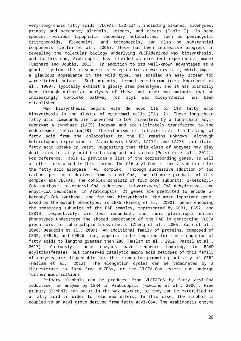
very-long-chain fatty acids (VLCFAs; C20–C34), including alkanes, aldehydes,primary and secondary alcohols, ketones, and esters (Table I). In somespecies, various lipophilic secondary metabolites, such as pentacyclictriterpenoids, flavonoids, and tocopherols, can also be substantialcomponents (Jetter et al., 2006). There has been impressive progress inrevealing the molecular biology underlying VLCFAderived wax biosynthesis,and to this end, Arabidopsis has provided an excellent experimental model(Bernard and Joubès, 2013). In addition to its well-known advantages as agenetic system, the presence of stem epicuticular wax crystals, which imparta glaucous appearance in the wild type, has enabled an easy screen forwaxdeficient mutants. Such mutants, termed eceriferum (cer; Koornneef etal., 1989), typically exhibit a glossy stem phenotype, and it has primarilybeen through molecular analyses of these and other wax mutants that anincreasingly complete pathway for acyl wax biosynthesis has beenestablished.
Wax biosynthesis begins with de novo C16 or C18 fatty acidbiosynthesis in the plastid of epidermal cells (Fig. 2). These long-chainfatty acid compounds are converted to CoA thioesters by a long-chain acyl-coenzyme A synthase (LACS) isozyme and are ultimately transferred to theendoplasmic reticulum(ER). Themechanism of intracellular trafficking offatty acid from the chloroplast to the ER remains unknown, althoughheterologous expression of Arabidopsis LACS1, LACS2, and LACS3 facilitatesfatty acid uptake in yeast, suggesting that this class of enzymes may playdual roles in fatty acid trafficking and activation (Pulsifer et al., 2012).For reference, Table II provides a list of the corresponding genes, as wellas others discussed in this review. The C16 acyl-CoA is then a substrate forthe fatty acid elongase (FAE) complex. Through successive addition of twocarbons per cycle derived from malonyl-CoA, the ultimate products of thiscomplex are VLCFAs. The complex consists of four core subunits: b-ketoacyl-CoA synthase, b-ketoacyl-CoA reductase, b-hydroxyacyl-CoA dehydratase, andenoyl-CoA reductase. In Arabidopsis, 21 genes are predicted to encode b-ketoacyl-CoA synthase, and for wax biosynthesis, the most important gene,based on the mutant phenotype, is CER6 (Fiebig et al., 2000). Genes encodingthe remaining subunits of the FAE complex, represented by KCR1, PAS2, andCER10, respectively, are less redundant, and their pleiotropic mutantphenotypes underscore the shared importance of the FAE in generating VLCFAprecursors for sphingolipid biosynthesis (Zheng et al., 2005; Bach et al.,2008; Beaudoin et al., 2009). An additional family of proteins, composed ofCER2, CER26, and CER26-like, appears to be required for the elongation offatty acids to lengths greater than 28C (Haslam et al., 2012; Pascal et al.,2013). Curiously, these enzymes have sequence homology to BAHDacyltransferases, but conserved catalytic amino acid residues of this familyof enzymes are dispensable for the elongation-promoting activity of CER2(Haslam et al., 2012). The elongation cycles can be terminated by athioesterase to form free VLCFAs, or the VLCFA-CoA esters can undergofurther modifications.
Primary alcohols can be produced from VLCFACoA by fatty acyl-CoAreductase, an enzyme by CER4 in Arabidopsis (Rowland et al., 2006). Freeprimary alcohols can occur in the wax mixture, or they can be esterified toa fatty acid in order to form wax esters. In this case, the alcohol iscoupled to an acyl group derived from fatty acyl-CoA. The Arabidopsis enzyme
28

responsible for this is WSD1, an enzyme of the wax synthase/diacylglycerolacyltransferase family (Li et al., 2008).
A second branch of acyl wax biosynthesis leads to the formation ofaldehydes and, ultimately, alkanes. Interestingly, in Arabidopsis, LACS1,which is also required for C16 cutin monomer biosynthesis, appears to havean additional specificity for C30 VLCFA and is required for the normalaccumulation of downstream wax compounds (Lü et al., 2009). This suggeststhat conversion of an intracellular pool of free VLCFA back to VLCFACoA isan important route to aldehyde and alkane biosynthesis, rather than VLCFA-CoA directly derived from FAE. A long unresolved question in waxbiosynthesis is the enzymatic basis of alkane synthesis.
Classical biochemistry, using crude extracts from pea (Pisum sativum),indicated that the reaction likely occurs via the reduction of VLCFA-CoA toan aldehyde intermediate followed by decarbonylation, yielding an alkanethat is 1C shorter (Cheesbrough and Kolattukudy, 1984; Schneider-Belhaddadand Kolattukudy, 2000). Although this enzyme was not purified andidentified, compelling evidence was recently obtained, through studies ofArabidopsis, that CER1 and CER3 in complex act together to catalyze theformation of alkanes from VLCFA-CoA. It was shown by a split ubiquitin yeasttwo-hybrid assay and an Arabidopsis split luciferase assay that CER1interacts with CER3 as well as several isoforms of cytochrome b5.Furthermore, heterologous expression of the combination of CER1, CER3, acytochrome b5, and LACS1 in yeast resulted in the formation of very-long-chain akanes (Bernard et al., 2012). This strongly suggests that a complexincluding CER1 and CER3 with cytochrome b5 as an electron donor catalyzesthe reduction and decarbonylation of VLCFA-CoA in order to form cuticularalkanes. Aside from being a major component of the wax mixture, alkanes canundergo further modification to form secondary alcohols and ketones. InArabidopsis, both of these oxidations are performed by the cytochrome P450enzyme midchain alkane hydroxylase (MAH1; Greer et al., 2007).
Synthesis of Cutin PrecursorsCutin is typically composed of interesterified hydroxyl fatty acids,
with lesser amounts of glycerol, phenylpropanoids, and dicarboxylic acids(Kolattukudy, 2001). Chemical processes that cleave ester bonds, such assaponification, readily release these monomeric constituents, although insome species an additional lipidic polymer, referred to as cutan, remainsrecalcitrant to such treatments. Cutan is rich in ether and C-C bonds, butits
structure is otherwise unknown, and it appears to be restricted torelatively few extant species (Gupta et al., 2006). The hydroxy fatty acidsof cutin are typically v-hydroxy fatty acids, usually with one or twoadditional midchain hydroxyl groups or an epoxy group (Fig. 3A).
Despite extensive surveys of the chemical composition of plant cutinsin the 1970s and 1980s (Kolattukudy, 2001), the composition of Arabidopsiscutinwas not determined until relatively recently (Bonaventure et al., 2004;Franke et al., 2005). It is important to note that, in this important modelspecies, the cutin of stems and leaves is atypical in that its majorcomponent is a dicarboxylic acid (Fig. 3A), implying that the predominantstructural motif must be a copolymer with an unknown polyhydroxy compound,presumably glycerol (Pollard et al., 2008). However, despite the atypicalcomposition of its cutin, Arabidopsis has proven to be an important modelfor deciphering the pathway of cutin biosynthesis, and more recently, it was
29
discovered that the cutin of its floral organs is more typical, in that itis composed primarily of 10,16- dihydroxyhexadecanoic acid (Li-Beisson etal., 2009).
While there is considerable diversity in the structure of cutinmonomers, the pathway for the biosynthesis of 10,16-dihydroxyhexadecanoicacid-based cutin is the most complete, and the major themes of cutinbiosynthesis are likely shared for other cutin monomers. Here, we summarizethis pathway based on recent molecular genetic and biochemical studies usingArabidopsis and tomato (Solanum lycopersicum).
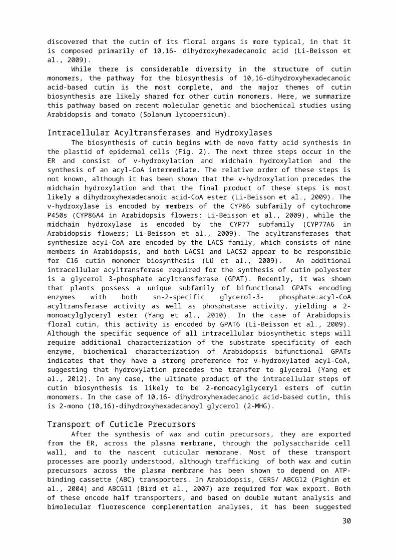
Intracellular Acyltransferases and HydroxylasesThe biosynthesis of cutin begins with de novo fatty acid synthesis in
the plastid of epidermal cells (Fig. 2). The next three steps occur in theER and consist of v-hydroxylation and midchain hydroxylation and thesynthesis of an acyl-CoA intermediate. The relative order of these steps isnot known, although it has been shown that the v-hydroxylation precedes themidchain hydroxylation and that the final product of these steps is mostlikely a dihydroxyhexadecanoic acid-CoA ester (Li-Beisson et al., 2009). Thev-hydroxylase is encoded by members of the CYP86 subfamily of cytochromeP450s (CYP86A4 in Arabidopsis flowers; Li-Beisson et al., 2009), while themidchain hydroxylase is encoded by the CYP77 subfamily (CYP77A6 inArabidopsis flowers; Li-Beisson et al., 2009). The acyltransferases thatsynthesize acyl-CoA are encoded by the LACS family, which consists of ninemembers in Arabidopsis, and both LACS1 and LACS2 appear to be responsiblefor C16 cutin monomer biosynthesis (Lü et al., 2009). An additionalintracellular acyltransferase required for the synthesis of cutin polyesteris a glycerol 3-phosphate acyltransferase (GPAT). Recently, it was shownthat plants possess a unique subfamily of bifunctional GPATs encodingenzymes with both sn-2-specific glycerol-3- phosphate:acyl-CoAacyltransferase activity as well as phosphatase activity, yielding a 2-monoacylglyceryl ester (Yang et al., 2010). In the case of Arabidopsisfloral cutin, this activity is encoded by GPAT6 (Li-Beisson et al., 2009).Although the specific sequence of all intracellular biosynthetic steps willrequire additional characterization of the substrate specificity of eachenzyme, biochemical characterization of Arabidopsis bifunctional GPATsindicates that they have a strong preference for v-hydroxylated acyl-CoA,suggesting that hydroxylation precedes the transfer to glycerol (Yang etal., 2012). In any case, the ultimate product of the intracellular steps ofcutin biosynthesis is likely to be 2-monoacylglyceryl esters of cutinmonomers. In the case of 10,16- dihydroxyhexadecanoic acid-based cutin, thisis 2-mono (10,16)-dihydroxyhexadecanoyl glycerol (2-MHG).
Transport of Cuticle PrecursorsAfter the synthesis of wax and cutin precursors, they are exported
from the ER, across the plasma membrane, through the polysaccharide cellwall, and to the nascent cuticular membrane. Most of these transportprocesses are poorly understood, although trafficking of both wax and cutinprecursors across the plasma membrane has been shown to depend on ATP-binding cassette (ABC) transporters. In Arabidopsis, CER5/ ABCG12 (Pighin etal., 2004) and ABCG11 (Bird et al., 2007) are required for wax export. Bothof these encode half transporters, and based on double mutant analysis andbimolecular fluorescence complementation analyses, it has been suggested
30

that an ABCG11/ABCG12 heterodimer is required for wax secretion (McFarlaneet al., 2010). ABCG11 is also required for cutin accumulation, and since itis also able to dimerize with itself, it has been proposed that thishomodimer is the functional complex responsible for cutin export (McFarlaneet al., 2010). Additionally, a third Arabidopsis half transporter, ABCG13,was shown to be required for cutin deposition in flowers (Panikashvili etal., 2011).
More recently, full transporters required for cutin deposition wereidentified in Arabidopsis (ABCG32; Bessire (et al., 2011) as well as wildbarley (Hordeum spontaneum) and rice (Oryza sativa; Chen et al., 2011).Despite the clear genetic evidence supporting a role for ABC transportersin cuticular lipid export, the substrate specificity of these transportershas not yet been demonstrated in vitro. However, all the ABC transportersthat have been implicated in cuticle biosynthesis to date are members of theABCG subfamily, which has been associated with the transport of lipids andhydrophobic compounds in other systems (Moitra et al., 2011). Moreover, inseveral cases, intracellular lipidic inclusionswere observedinABCtransporter mutants, further supporting their direct involvement incuticular lipid export (Pighin et al., 2004; Bird et al., 2007; Bessire etal., 2011).
Export of some wax compounds also appears to be facilitated byglycosylphosphatidylinositol (GPI)-anchored lipid-transfer proteins (LTPs),LTPG1 and LTPG2, which are bound to the extracellular side of the plasmamembrane (Debono et al., 2009; Lee et al., 2009; Kim et al., 2012). Theseproteins represent a unique class of LTPs, a family of small and typicallysoluble proteins that bind a variety of lipid substrates in vitro (Yeats andRose, 2008). A major remaining question is how hydrophobic cuticleprecursors are transported across the hydrophilic environment of thepolysaccharide cell wall to the cuticle. Apoplastic LTPs have been proposedto play a role, although genetic or biochemical evidence for theirinvolvement in transport is generally lacking (Yeats and Rose, 2008). In thecase of the dihydroxyacyl cutin precursor 2-MHG, the glycerol moiety impartssufficient polarity to allow aqueous solubility at low millimolarconcentrations (Yeats et al., 2012b). This suggests that lipid-bindingproteins or other factors are not necessary in order to facilitate thetransport of this major precursor of cutin biosynthesis. However, thesolubility of glyceryl esters of less polar cutin monomers has not beeninvestigated, and they, along with waxes, may require additional factors toincrease their solubility in the apoplast.
Cutin PolymerizationThe final step of cutin synthesis is incorporation of the hydroxyacyl
monomer into the polymer, but the molecular mechanism of cutinpolymerization has been achieved by studying the tomato mutant cutindeficient1 (cd1) and transgenic tomato plants in which CD1 expression wassuppressed using an RNA interference strategy (Girard et al., 2012; Yeats etal., 2012b). The cd1 mutant exhibits a severe reduction in the amount ofpolymerized cutin in the fruit cuticle (Isaacson et al., 2009), althoughchemical analysis indicated that, unlike wild-type fruit, those of themutant accumulate nonpolymerized 2-MHG (Yeats et al., 2012b). Cloning of themutated gene revealed that it encodes a protein of the GDSL-motiflipase/hydrolase (GDSL) family, which localizes to the developing cuticle
31
(Girard et al., 2012; Yeats et al., 2012b). Despite its similarity tolipolytic enzymes, the recombinant protein acts as an acyltransferase invitro, forming polyester oligomers from 2-MHG (Yeats et al., 2012b).
The identification of CD1 as the first known cutin synthase raisesseveral questions about the specificity and generality of the reaction thatit catalyzes. Phylogenetic analysis of CD1 and homologous genes indicatesthat despite belonging to a very large gene family, the subfamily of GDSLsrepresented by CD1 is relatively small and well conserved, with sequencesrepresented across diverse taxa of land plants (Volokita et al., 2011).
In Arabidopsis, its putative orthologs form a fivemember gene family,and silencing of the expression of two of these (LTL1 and At5g33370)resulted in plants exhibiting floral organ fusions and lacking nanoridges onthe petal surface, phenotypes that are consistent with a cutin deficiency(Shi et al., 2011). An additional putative ortholog of CD1 from Agaveamericana exhibited similar localization and expression, further supportinga conserved mechanism of CD1-like enzymes acting as cutin synthases (Reinaet al., 2007). Despite the presence of a null allele, the cd1 mutant is notcompletely deficient in cutin, so the identity of additional cutinsynthases, or perhaps nonenzymatic mechanisms of cutin synthesis, representsan intriguing line of future research.
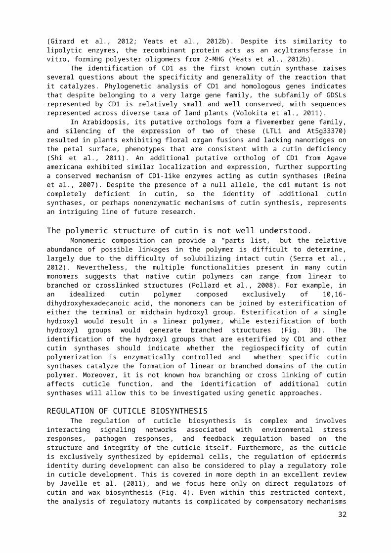
The polymeric structure of cutin is not well understood.Monomeric composition can provide a “parts list,” but the relative
abundance of possible linkages in the polymer is difficult to determine,largely due to the difficulty of solubilizing intact cutin (Serra et al.,2012). Nevertheless, the multiple functionalities present in many cutinmonomers suggests that native cutin polymers can range from linear tobranched or crosslinked structures (Pollard et al., 2008). For example, inan idealized cutin polymer composed exclusively of 10,16-dihydroxyhexadecanoic acid, the monomers can be joined by esterification ofeither the terminal or midchain hydroxyl group. Esterification of a singlehydroxyl would result in a linear polymer, while esterification of bothhydroxyl groups would generate branched structures (Fig. 3B). Theidentification of the hydroxyl groups that are esterified by CD1 and othercutin synthases should indicate whether the regiospecificity of cutinpolymerization is enzymatically controlled and whether specific cutinsynthases catalyze the formation of linear or branched domains of the cutinpolymer. Moreover, it is not known how branching or cross linking of cutinaffects cuticle function, and the identification of additional cutinsynthases will allow this to be investigated using genetic approaches.
REGULATION OF CUTICLE BIOSYNTHESISThe regulation of cuticle biosynthesis is complex and involves
interacting signaling networks associated with environmental stressresponses, pathogen responses, and feedback regulation based on thestructure and integrity of the cuticle itself. Furthermore, as the cuticleis exclusively synthesized by epidermal cells, the regulation of epidermisidentity during development can also be considered to play a regulatory rolein cuticle development. This is covered in more depth in an excellent reviewby Javelle et al. (2011), and we focus here only on direct regulators ofcutin and wax biosynthesis (Fig. 4). Even within this restricted context,the analysis of regulatory mutants is complicated by compensatory mechanisms
32

between cutin and wax biosynthesis and other pleiotropic phenotypes.Nevertheless, a complex regulatory network that responds to developmentaland environmental cues, mediated by hormones, transcription factors, andposttranscriptional regulation, is beginning to emerge.
Environment and HormonesA systematic analysis of both cuticle composition andgene expression
in Arabidopsis indicates that wax synthesis is induced by water deficit,sodium chloride, and abscisic acid (ABA) treatments (Kosma et al., 2009). Incontrast, cutin biosynthesis was reported only to be induced by waterdeficit and not ABA or sodium chloride, suggesting that, at least inArabidopsis, the detection of various osmotic stresses is complex and onlypartially dependent on ABA (Kosma et al., 2009). However, given that ABA isalready well established as a regulator of plant responses to water deficitthrough the regulation of stomatal aperture (Lee and Luan, 2012), ABAregulation of cuticle biosynthesis is an intriguing area for furtherresearch aimed at understanding and engineering drought tolerance in crops.In addition, dark and cold treatments have been shown to reduce theexpression of several components of the FAE complex (Hooker et al., 2002;Joubès et al., 2008). Several wax biosynthetic genes have been shown to beinduced by bacterial pathogens (Raffaele et al., 2008) and duringinfestation of wheat (Triticum aestivum) by the Hessian fly (Mayetioladestructor; Kosma et al., 2010), but in general, the relevance of theinduction of cuticle synthesis to pest or pathogen resistance is poorlyunderstood.
Transcription Factors and Cuticle BiosynthesisThe first transcription factor gene identified as having a role in
regulating cuticle biosynthesis was the AP2 domain-containing WAXINDUCER1/SHINE1 (WIN1/SHN1; Aharoni et al., 2004; Broun et al., 2004).Overexpression of this gene led to glossy leaves with a greater wax loadthan the wild type and lower transpiration, although this was likely due toa reduced density of stomata rather than the wax phenotype (Aharoni et al.,2004). Later studies indicated that cutin levels are also increased inWIN1/SHN1-overexpressing plants and that the up-regulation of genes encodingcutin biosynthetic enzymes precedes the induction of wax biosynthetic genes(Kannangara et al., 2007). WIN1/SHN1 is part of a three-member gene familyin Arabidopsis, and silencing of all three genes led to a reduction in theamount of cutin but not waxes (Shi et al., 2011). These authors alsodemonstrated that these transcription factors directly activate promoters ofseveral cutin biosynthetic genes, further supporting a primary role in cutinregulation with a downstream effect on wax biosynthesis (Shi et al., 2011).In addition to regulating cutin biosynthesis, the SHN transcription factorsalso induced the expression of several pectin-modifying enzymes, suggestinga coordination of the synthesis of the cuticle with the polysaccharide cellwall (Shi et al., 2011). This second function of SHN transcription factorsin regulating the polysaccharide cell wall is further suggested byexperiments in which the overexpression of Arabidopsis SHN2 in rice resultedin a significant increase in the amount of cellulose and a concomitantdecrease in lignin (Ambavaram et al., 2011). On the other hand, a generalrole of WIN1/SHN1-related transcription factors in the regulation of cutinsynthesis is indicated by studies of orthologous genes in barley (Hordeum
33
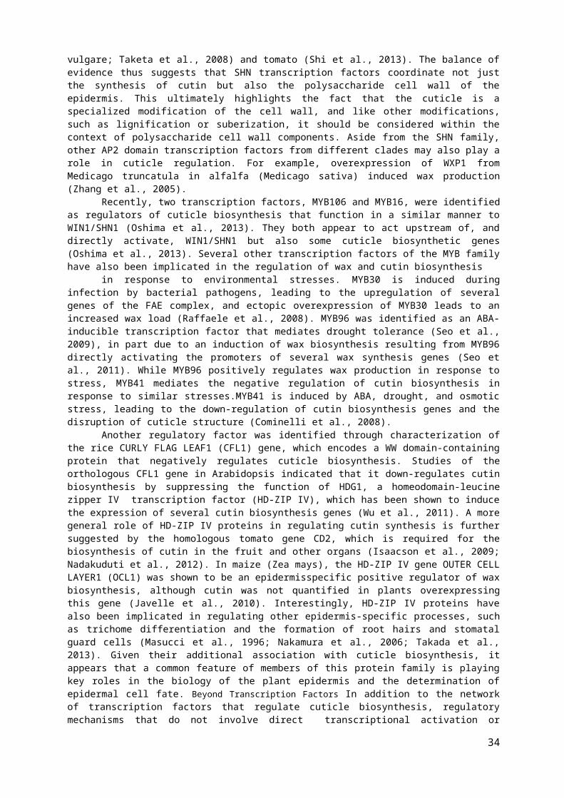
vulgare; Taketa et al., 2008) and tomato (Shi et al., 2013). The balance ofevidence thus suggests that SHN transcription factors coordinate not justthe synthesis of cutin but also the polysaccharide cell wall of theepidermis. This ultimately highlights the fact that the cuticle is aspecialized modification of the cell wall, and like other modifications,such as lignification or suberization, it should be considered within thecontext of polysaccharide cell wall components. Aside from the SHN family,other AP2 domain transcription factors from different clades may also play arole in cuticle regulation. For example, overexpression of WXP1 fromMedicago truncatula in alfalfa (Medicago sativa) induced wax production(Zhang et al., 2005).
Recently, two transcription factors, MYB106 and MYB16, were identifiedas regulators of cuticle biosynthesis that function in a similar manner toWIN1/SHN1 (Oshima et al., 2013). They both appear to act upstream of, anddirectly activate, WIN1/SHN1 but also some cuticle biosynthetic genes(Oshima et al., 2013). Several other transcription factors of the MYB familyhave also been implicated in the regulation of wax and cutin biosynthesis
in response to environmental stresses. MYB30 is induced duringinfection by bacterial pathogens, leading to the upregulation of severalgenes of the FAE complex, and ectopic overexpression of MYB30 leads to anincreased wax load (Raffaele et al., 2008). MYB96 was identified as an ABA-inducible transcription factor that mediates drought tolerance (Seo et al.,2009), in part due to an induction of wax biosynthesis resulting from MYB96directly activating the promoters of several wax synthesis genes (Seo etal., 2011). While MYB96 positively regulates wax production in response tostress, MYB41 mediates the negative regulation of cutin biosynthesis inresponse to similar stresses.MYB41 is induced by ABA, drought, and osmoticstress, leading to the down-regulation of cutin biosynthesis genes and thedisruption of cuticle structure (Cominelli et al., 2008).
Another regulatory factor was identified through characterization ofthe rice CURLY FLAG LEAF1 (CFL1) gene, which encodes a WW domain-containingprotein that negatively regulates cuticle biosynthesis. Studies of theorthologous CFL1 gene in Arabidopsis indicated that it down-regulates cutinbiosynthesis by suppressing the function of HDG1, a homeodomain-leucinezipper IV transcription factor (HD-ZIP IV), which has been shown to inducethe expression of several cutin biosynthesis genes (Wu et al., 2011). A moregeneral role of HD-ZIP IV proteins in regulating cutin synthesis is furthersuggested by the homologous tomato gene CD2, which is required for thebiosynthesis of cutin in the fruit and other organs (Isaacson et al., 2009;Nadakuduti et al., 2012). In maize (Zea mays), the HD-ZIP IV gene OUTER CELLLAYER1 (OCL1) was shown to be an epidermisspecific positive regulator of waxbiosynthesis, although cutin was not quantified in plants overexpressingthis gene (Javelle et al., 2010). Interestingly, HD-ZIP IV proteins havealso been implicated in regulating other epidermis-specific processes, suchas trichome differentiation and the formation of root hairs and stomatalguard cells (Masucci et al., 1996; Nakamura et al., 2006; Takada et al.,2013). Given their additional association with cuticle biosynthesis, itappears that a common feature of members of this protein family is playingkey roles in the biology of the plant epidermis and the determination ofepidermal cell fate. Beyond Transcription Factors In addition to the networkof transcription factors that regulate cuticle biosynthesis, regulatorymechanisms that do not involve direct transcriptional activation or
34

repression by promoter binding have recently been discovered. A recentexample resulted from studies of the Arabidopsis cer9 mutant, which exhibitsalterations in the amount and composition of leaf and stem waxes.
Cloning of the CER9 gene revealed it to encode a protein withsequence similarity to yeast Doa10, an E3 ubiquitin ligase involved in ER-associated degradation of misfolded proteins (Lü et al., 2012). Given the ERlocalization of wax and cutin biosynthetic processes, the authors proposed arole for CER9 in the homeostasis of key cuticle biosynthetic enzyme levels.Experiments further addressing this hypothesis will be particularlyinteresting, given the surprising finding that the cer9 mutant actuallyexhibits enhanced drought tolerance and water use efficiency (Lü et al.,2012).
One of the most intriguing mechanisms of cuticle regulation resultedfrom characterization of the cer7 mutant. CER7 encodes an exosomalexoribonuclease, and the cer7 mutant exhibits reductions in stem wax andtranscription of CER3, a major wax biosynthetic enzyme (Hooker et al.,2007). Recently, two suppressors of cer7 that restore the CER3 transcriptand stem wax levels were identified, and cloning of the respective genesidentified RDR1 and SGS3, two conserved components of the RNA-mediated gene-silencing pathway (Lam et al., 2012). A model was proposed wherein CER7 isinvolved in the degradation of a small RNA species that negatively regulatesthe CER3 transcript. Future work involving the identification of such asmall RNA species and other components of this pathway will be especiallyintriguing, since no known plant small RNA species mapped to the CER7-dependent region of the CER3 promoter (Lam et al., 2012).
ENIGMATIC FACTORS IN CUTICLE BIOSYNTHESISIn addition to the characterized components of cuticle biosynthesis
that can be incorporated into a coherent model, as discussed above, severalgenes/proteins have been identified that are required for cuticle formationbut that lack a clear associated biochemical function that would place themin a specific point in the pathways. One example is HOTHEAD (HTH), a Glc-methanolcholine oxidoreductase family protein that is required for propercuticle organization (Krolikowski et al., 2003).
Chemical analysis indicated that the Arabidopsis hth mutant has wild-type wax levels but abnormal cutin quantity and composition. Specifically,it has decreased levels of dicarboxylic acids and increased amounts of v-hydroxy acids, leading the authors to suggest that HTH may have a role inthe oxidation of v-hydroxy fatty acids to the dicarboxylic acid cutinmonomers that are characteristic of Arabidopsis stem and leaf cuticles(Kurdyukov et al., 2006b). As dicarboxylic acid cutin monomers are unusuallyabundant in Arabidopsis, it will be interesting to see whether HTH-relatedproteins are as essential to cuticle formation in other species where thisclass of monomers is scarce.
Another example of an “orphan” cuticle-associated protein resultedfrom analysis of the Arabidopsis bodyguard (bdg) mutant, which exhibits amicroscopically disorganized cuticle with increased permeability butsignificantly increased levels of wax and cutin (Kurdyukov et al., 2006a).The BDG protein has sequence similarity to the a/b-hydrolase family ofproteins, but no enzymatic activity has been reported. The protein islocalized in the outer cell wall of the epidermis below the cuticle, whichled the authors to propose that BDG may be involved in cutin polymerization,
35

although the increased amounts of polymeric cutin in the mutant would argueagainst this (Kurdyukov et al., 2006a).
Mutation of BDG3, a close homolog of BDG, resulted in thedisorganization of floral nanoridges, petal epidermis structures that arecomposed of cutin (Shi et al., 2011). Moreover, the key cutin regulatorytranscription factors SHN1, SHN2, and SHN3 were shown to activate the BDG3promoter (Shi et al., 2011). Taken together, these results strongly indicatethat BDG proteins are closely linked to cutin polymer formation, althoughtheir mode of action remains mysterious.
Lastly, a defect in the formation of floral nanoridges was alsoidentified in the Arabidopsis mutant defective in cuticular ridges (dcr),which showed a substantial deficiency in floral cutin but a less drasticalteration of leaf and stem cutin (Panikashvili et al., 2009). DCR encodes aprotein of the BAHD acyltransferase family that localizes to the cytoplasm,and it has been proposed that it may be involved in acyl transfer of cutinmonomers to form precursor intermediates or oligomeric structures(Panikashvili et al., 2009). However, DCR was later biochemicallycharacterized and shown to possess in vitro diacylglycerol acyltransferaseactivity, leading to the formation of triacylglycerol (Rani et al., 2010). Arole for cytoplasmic triacylglycerol intermediates in cutin biosynthesis isnot consistent with any known steps in this pathway, yet DCR is clearlyrequired for cutin biosynthesis in Arabidopsis floral organs. Further workwill be needed in order to determine the native substrate and product ofDCR in order for its role in cutin biosynthesis to be elucidated.
FUNCTIONS OF THE CUTICLE
The plant cuticle is most typically associated withproviding a fixedbarrier to excessive transpirationalwater loss, allowing gas exchange andtranspiration tobe dynamically controlled by stomata. However, it hasevolveda number of secondary functions that are consistentwith its place as theoutermost layer of primaryaerial organs: it forms a physical barrier that isthe firstline of defense against pests and pathogens; inmanyspecies,elaborate epicuticular crystals help to form aself-cleaningsurface, preventing dust and other debrisfromblockingsunlight; in somecases, it can act to screenexcessive UV light; finally, as a definingfeature of theepidermis, it plays a central role in development byphysicallyestablishing organ boundaries.
Cuticle Structure and Water Barrier Properties
A common perception is that a thick cuticle is associatedwith a lowerwater permeability and thus increasedtolerance to water stress. However,comparative studiesof the water permeability of cuticles from diversespecieshave indicated that there is no correlation with either thethicknessof the cuticle or the amount of wax (RiedererandSchreiber, 2001). Similarly,the amount of cutin is notnecessarily an indication of cuticular waterpermeability(CWP). For example, studies of three tomato mutants(cd1–cd3),each of which has a greater than 95% reductionin fruit cutin levels,revealed only minor increases in therate of water loss, and even among themutants there wasno clear correlation between cutin amount andsusceptibilityto desiccation (Isaacson et al., 2009). However,cutindeficiency that leads to organizational defects can bedetrimental to
36

the cuticle permeability (Bessire et al.,2011). In contrast to the lack ofassociation with cutin,extensive removal of wax from tomato fruit,accomplishedby brief immersion of the fruit in an organicsolvent, indicatesthat waxes contribute approximately95% of the cuticlemediated resistance towater diffusion,at least in tomato fruit (Leide et al., 2007).
Specific compound classes appear to be associatedwith water barrierproperties of the cuticle; notably, themorenonpolar components, such asalkanes, tend to beassociated with decreased CWP, while nonaliphatic waxcompounds, such as triterpenoids, are likely a less effectivewater barrier(Leide et al., 2007; Buschhaus andJetter, 2012). This is consistent with amodel in whichcuticular waxes localize within either crystallineoramorphousdomains of the cuticle, with aliphatic compoundsformingcrystallite “rafts” that are impervious towater, forcing water, and otherpolar metabolites, todiffuse by a circuitous route through theamorphousdomains that are formed by more polar and cyclicwaxes (Riederer andSchreiber, 1995). The idea that theproportion of alkanes and not the totalwax amount hasthe most significant effect on CWP was illustrated by arecentstudy with a backcrossed population of Capsicumannum and Capsicum chinense,two pepper species withhigh and low postharvest water loss rates,respectively.In 20 backcrossed families, CWP was inversely correlatedwiththe amount of alkanes in the wax but not thetotal amount of wax, and themore rapidly desiccatingparent had three times the wax coverage as theparentthat exhibited low postharvest water loss (Parsons et al.,2012). Insummary, resistance to water loss is primarilyattributed to wax and notcutin, but there is not a directcorrelation between the amount of eithercomponentand CWP. Rather, it appears that CWP is primarilydetermined by theparticular mixture of intracuticularand epicuticular waxes and by theirpacking and organizationwithin the cuticle architecture.
The Lotus Effect
A striking feature of many plant leaves is that watertends to beadinto drops and roll to the ground,collecting and washing particles anddebris from the leafsurface. The efficiency of this self-cleaningmechanism,termed the “lotus effect,” varies between species andduring organontogeny, but it has been correlated withthe abundance of epicuticular waxcrystals that repelwater and allow a pocket of air to form beneaththedroplets (Barthlott and Neinhuis, 1997). It is thought thatthis self-cleaning surface helps to prevent the buildup ofdust thatwould blocksunlight and slow photosynthesisand that this could also play an importantrole inwashing awaypathogen spores before they germinate.Despite theapparent advantages of a self-cleaning surface,there is not a clear exampleof this trait conferringan adaptive advantage. In terms of photosynthesis,thereis likely a tradeoff between a self-cleaning surface andthe increaseddispersion of light by epicuticular waxcrystals, as discussed below.Nevertheless, based onthe discovery of this effect, surfaces with highdegreesof hydrophobicity andmicroscopic texture have beenemployed aseffective biomimetic technical materials(Bhushan, 2012), and improved self-cleaning surfacesin agricultural crops may be a productive avenueofresearch.
The Cuticle as a Barrier against Pests and Pathogens
37

The plant cuticle presents a physical barrier to pathogensthat do nototherwise enter the plant by way ofthe stomata, wounds, or vectors. However,fungalpathogens have been shown to breach the cuticle using acombinationofenzymatic degradation and mechanicalrupture. The latter is oftenaccomplished by the formationof a swollen appressorium structure thatextends aninfectious peg via turgor pressure (Deising et al., 2000).Whilemechanical rupture may be sufficient for cuticlepenetration, particularly ofthinner cuticles (Tenberge,2007), most fungal pathogens also secretecutinases, aclass of small, nonspecific esterases that hydrolyze thecutinpolyester and releasefree cutin monomers (Longhiand Cambillau, 1999). Thecutin monomers that arereleased during polymeric cutin hydrolysis can actaselicitors of plant defense responses and are thus classifiedas damage-associated molecular patterns. At micromolarconcentrations, these compoundsinduce theproduction of hydrogen peroxide and other defenseresponses(Schweizer et al., 1996; Kauss et al., 1999).However, the mechanism of plantperception of freecutin monomers is currently unknown (Boller andFelix,2009).
Cutin appears to be more important than wax forforming a barrier topathogen entry, although there isnot a consistent correlation between cutinamount andpathogen resistance. In tomato fruit, severely decreasedcutinlevels in three cd mutants was associated withincreased susceptibility toinfection by Botrytis cinereasurface inoculation and also to opportunisticmicrobes(Isaacson et al., 2009). However, in Arabidopsis, anumber of cutin-deficient mutants and plants that ectopicallyoverexpress fungal cutinasesexhibit enhanced resistance to B. cinerea (Bessire et al., 2007, 2011;Chassotet al., 2007; Tang et al., 2007). In this case, increasedcuticularpermeabilityappears to enhance the diffusionof inoculum-derived elicitorsthat induce the productionof small, polar antifungal compounds, which inturninhibit B. cinerea growth (Bessire et al., 2007). Conversely,the Arabidopsislacs2 mutant and cutinaseoverexpressers exhibited no alteration in theirsusceptibilityto a range of other fungal pathogens (Bessire et al.,2007),and the lacs2 mutation also increased susceptibilityto a normally avirulentstrain of Pseudomonas syringae(Tang et al., 2007). Thus, cutin plays animportant role asa physical barrier to many pathogens, yet extremedeficienciesin Arabidopsis can result in increased resistanceto somepathogens by way of a secondary, but not wellunderstood, mechanism thatinvolves the induction ofplant defenses. An additional layer of complexitywassuggested by the observation that cutin can induce geneexpression inplant pathogens and has been shown toinduce appressorium expression inColletotrichum trifolii(Dickman et al., 2003). This highlights thecompetingselective pressures to generate and breach cuticle barriersat thefrontier of the plant surface (Chassot andMetraux,2005).
Despite the importance of cutin in plant-pathogeninteractions, thefirst surface encountered by foliarpathogens is formed by epicuticular waxcrystals andfilms. In addition to the lotus effect that promotesthewashingof spores from the plant surface before germination,there areseveral indications that the epicuticularwax structures and composition areimportantin determining fungal pathogen development and,thus, pathogenicity.The C26 aldehyde n-hexacosanyl,a component of cuticular wax in many speciesof thePoaceae, can induce in vitro appressorium formationby the powderymildew Blumeria graminis (Tsuba et al.,2002; Ringelmann et al.,2009;Hansjakob et al., 2010).This observation is further corroborated by
38

studies ofthe maize mutant glossy1, which does not accumulatealdehydes inits wax complement. B. graminis appressoriumformation is substantiallyreduced on the leafsurface of the glossy1 mutant but can be restoredtonormal levels by the application of n-hexacosanyl(Hansjakob et al., 2011).Another example of the influenceof waxes on pathogenicity is provided bytheinhibitor ofrust tube germination1 (irg1) mutant ofM. truncatula, whichexhibits decreased amounts ofepicuticular wax crystalson the abaxial leafsurface,corresponding to a substantial decrease in wax primaryalcoholgroups. This surface alteration wasshown to reduce spore differentiation ofthe rust fungalpathogens Phakopsora pachyrhizi and Pucciniaemaculata and theanthracnose fungus C. trifolii, resultingin nonhost resistance (Uppalapatiet al., 2012). TheIRG1 gene was found to encode a C2H2 zincfingertranscription factor that had previously been identifiedas a regulatorof dissected leaf morphology (Chen et al.,2010). Reduced transcript levelsof putative MYB96 andCER4 orthologs were also observed in the irg1mutant,which is consistent with the wax phenotype. Thesignificance ofwaxesand cutin in pathogen resistance,therefore, is suggested in a general sense,but, as withcuticle permeability, little is known about therelativeimportance of specific molecular classes or theirintermolecularassociations and packing within the architectureof thecuticle.
Epicuticular waxes may also play an important role inplant-insectinteractions; indeed, epicuticular wax crystalscan form an unstable surfacethat prevents insectattachment or locomotion on plant surfaces (Borodichetal., 2010). A striking example of this is seen in thecarnivorous pitcherplants (Nepenthes spp.), which catchinsects by way of a slippery interiorsurface that is coatedwith epicuticular wax crystals (Riedel et al., 2007).For amore detailed review of cuticle chemical ecology, seeMüller andRiederer (2005).
The Cuticle and Development
In addition to providing physical barriers to waterand microbes, thecuticle appears to play an importantrole in defining organ boundaries duringdevelopment,since plants with cuticles showing increased permeabilityandstructural defects often exhibit numerousectopic organ fusions. Thisphenomenon has beenobserved in a wax-deficient tomato mutant (Smirnovaetal., 2013), a range of Arabidopsis mutants with abnormalcuticles (Yephremovet al., 1999; Wellesen et al.,2001; Kurdyukov et al., 2006a; Bird et al.,2007), andtransgenic Arabidopsis plantsoverexpressing a secretedfungalcutinase (Sieber et al., 2000). The fusion zones areoften marked by twoadjacentpolysaccharide cell wallswith no visible cuticle separating the twoorgans, althoughthe fused epidermal layers maintain their identity,asindicated by the differentiation of internalnonfunctional stomata withinfusion zones (Sieber et al.,2000). In each of three Arabidopsis mutantsexhibitingorgan fusions, lacerata, bodyguard, andfiddlehead, ectopicorganfusions and cuticular permeability defectscould be partially suppressed by asecondmutation inSERRATE (Voisin et al., 2009). SERRATE is a C2H2 zincfingerprotein that is required for microRNA biogenesis,and hypomorphic allelesexhibit numerous evelopmentaldefects, including serrated leaf margins(Donget al., 2008). While the mechanism of SERRATE action asa suppressor ofcuticle fusions remains unclear, this resultsuggests the existence of a
39
cuticle integrity pathwaythat is integrated with epidermal developmentalprograms.The identification of additional suppressors ofcuticle mutant-associated developmental phenotypesshould be informative in elucidating thecuticle integritypathway.
Protection against UV Radiation
UV light in the UV-B spectrum is a considerableportion of the daylightthat reaches the terrestrialsurface, and it can threaten plant life bydamagingDNA, the photosynthetic apparatus, and membrane lipids (Rozema etal., 1997). As a result, plants haveevolved a number of strategies forscreening UV-B radiation.These include a varietyof soluble flavonoidpigmentsthat are typically localized within the vacuoles ofepidermal cells,phenoliccompounds present in the polysaccharidecell wall, and lipophilicphenolic moleculesthat are covalently bound to cutin or associated withwaxes(Pfündel et al., 2006). A survey of isolated cuticlesfrom a range of speciesindicated generally effectivescreening of the UV-B spectrum but consistentlyhightransmittance in the higher wavelengths that arephotosyntheticallyactive (Krauss et al., 1997). In addition toabsorbinglight, the plant cuticle can reflect light to somedegree, presumablydepending on the abundance ofepicuticular wax crystals. For example, Dudleyabrittonniican reflect up to 83% of UV-B, but this value issubstantiallyreduced when epicuticular waxes are removed(Mulroy, 1979).Smooth, glossy “glabrous” cuticles typicallyreflect only small amounts oflight (less than10%),but glaucous plant surfaces are moderately reflectiveandgenerally show approximately 20% to 30% reflectance inthe UV and visiblespectra (Pfündel et al., 2006). Waxesreflect both UV and visible light, butnot necessarily to thesame extent, and the reflectance of UV has beenreportedto be greater in some cases (Holmes and Keiller, 2002).While lightreflection provides an important protectivemechanism, especially by limitingdamagingUV radiation,there is likely a tradeoff with photosyntheticefficiencyunder conditions when light intensity is limiting(Pfündel et al.,2006). In this regard, an interesting areaof future research might todetermine whetherrelativeproportions of UV and visible light reflection canbepredictively changed by altering the composition ofepicuticularwaxes.
CONCLUSION AND PERSPECTIVES
As described above, several key areas of cuticle biogenesisremainpoorly understood. First, the mechanismof intracellular and extracellulartransport of wax andcutin precursors remains unknown, although keyABCtransporters required for their export across the plasmamembrane havebeen identified (Pighin et al., 2004; Birdet al., 2007; Chen et al., 2011).The first cutin synthase hasbeen identified (Girard et al., 2012; Yeats etal., 2012b),but there are certainly additional cutin synthases, andwhetherthey are closely related to CD1 or belong todistinct protein familiesremains to be discovered. Aftercutin is polymerized, is modification of thepolymericstructurerequired to accommodate organ expansion? Ifso, whichenzymes are involved in this process?While ourunderstanding of cuticlebiosynthesis at themolecular level remains incomplete, recent progressindeciphering these pathways is bringing us closer thanever to an ability toselectively modify cuticle propertiesin order to improve agriculturalproductivity. However,the ability to make such modifications rationally
40

willrequire an understanding of the complexity of cuticlefunction at themolecular level, and far less progress hasbeen made in this regard. To thisend, further workaimed at understanding the ecophysiological functionsof thecuticle in defined mutantbackgrounds, as well asin genetically tractablewild species, will provide a frameworkfor understanding thecomplexinteraction of structure,composition, and function of cuticles (Yeats etal.,2012a). While the past decade has seen unprecedentedprogress in themolecular biology of cuticle biogenesis,many studies have revealedcomplexities in cuticle functionthat underscore the fact that the cuticle ismuch more thanjust a preformed barrier towater loss.
ACKNOWLEDGMENTS
We thank Drs. Gregory Buda, Christiane Nawrath, and Lacey Samuelsforgenerously providing microscopy images and Eric Fich, Laetitia Martin,andDr. Iben Sørensen for helpful comments and discussion.Received June 5,2013; accepted July 25, 2013; published July 26, 2013.
LITERATURE CITEDAharoni A, Dixit S, Jetter R, Thoenes E, van Arkel G, Pereira A (2004) The
SHINE clade of AP2 domain transcription factors activates wax biosynthesis, alters cuticle properties, and confers drought tolerance when overexpressed in Arabidopsis. Plant Cell 16: 2463–2480
Ambavaram MM, Krishnan A, Trijatmiko KR, Pereira A (2011) Coordinated activation of cellulose and repression of lignin biosynthesis pathways inrice. Plant Physiol 155: 916–931
Aroca R, Porcel R, Ruiz-Lozano JM (2012) Regulation of root water uptake under abiotic stress conditions. J Exp Bot 63: 43–57
Bach L, Michaelson LV, Haslam R, Bellec Y, Gissot L, Marion J, Da Costa M, Boutin JP, Miquel M, Tellier F, et al (2008) The very-long-chain hydroxylfatty acyl-CoA dehydratase PASTICCINO2 is essential and limiting for plant development. Proc Natl Acad Sci USA 105: 14727–14731
Barthlott W, Neinhuis C (1997) Purity of the sacred lotus, or escape from contamination in biological surfaces. Planta 202: 1–8
Beaudoin F, Wu X, Li F, Haslam RP, Markham JE, Zheng H, Napier JA, Kunst L (2009) Functional characterization of the Arabidopsis b-ketoacyl-coenzymeA reductase candidates of the fatty acid elongase. Plant Physiol 150: 1174–1191
Bernard A, Domergue F, Pascal S, Jetter R, Renne C, Faure JD, Haslam RP, Naier JA, Lessire R, Joubès J (2012) Reconstitution of plant alkane biosynthesis in yeast demonstrates that Arabidopsis ECERIFERUM1 and ECERIFERUM3 are core components of a very-long-chain alkane synthesis complex. Plant Cell 24: 3106–3118
Bernard A, Joubès J (2013) Arabidopsis cuticular waxes: advances in synthesis, export and regulation. Prog Lipid Res 52: 110–129
Bessire M, Borel S, Fabre G, Carraça L, Efremova N, Yephremov A, Cao Y, Jetter R, Jacquat AC,Métraux JP, et al (2011) Amember of the PLEIOTROPIC DRUG RESISTANCE family of ATP binding cassette transporters is required for the formation of a functional cuticle in Arabidopsis. Plant Cell 23: 1958–1970
Bessire M, Chassot C, Jacquat AC, Humphry M, Borel S, Petétot JM, Métraux JP, Nawrath C (2007) A permeable cuticle in Arabidopsis leads to a strong
41

resistance to Botrytis cinerea. EMBO J 26: 2158–2168 Bhushan B (2012) Bioinspired structured surfaces. Langmuir 28: 1698–1714
Bird D, Beisson F, Brigham A, Shin J, Greer S, Jetter R, Kunst L, Wu X, Yephremov A, Samuels L (2007) Characterization of Arabidopsis ABCG11/WBC11, an ATP binding cassette (ABC) transporter that is requiredfor cuticular lipid secretion. Plant J 52: 485–498
Boller T, Felix G (2009) A renaissance of elicitors: perception of microbeassociated molecular patterns and danger signals by pattern-recognition receptors. Annu Rev Plant Biol 60: 379–406
Bonaventure G, Beisson F, Ohlrogge J, Pollard M (2004) Analysis of the aliphatic monomer composition of polyesters associated with Arabidopsis epidermis: occurrence of octadeca-cis-6,cis-9-diene-1,18-dioate as the major component. Plant J 40: 920–930
Borodich FM, Gorb EV, Gorb SN (2010) Fracture behaviour of plant epicuticular wax crystals and its role in preventing insect attachment: atheoretical approach. Appl Phys A Mater Sci Process 100: 63–71
Broun P, Poindexter P, Osborne E, Jiang CZ, Riechmann JL (2004) WIN1, a transcriptional activator of epidermal wax accumulation in Arabidopsis. Proc Natl Acad Sci USA 101: 4706–4711
Buda GJ, Isaacson T, Matas AJ, Paolillo DJ, Rose JKC (2009) Threedimensionalimaging of plant cuticle architecture using confocal scanning laser microscopy. Plant J 60: 378–385
Budke JM, Goffinet B, Jones CS (2012) The cuticle on the gametophyte calyptra matures before the sporophyte cuticle in the moss Funaria hygrometrica (Funariaceae). Am J Bot 99: 14–22
Burghardt M, Riederer M (2006) Cuticular transpiration. In M Riederer, C Müller, eds, Biology of the Plant Cuticle. Blackwell, Oxford, pp 292–311
Buschhaus C, Jetter R (2012) Composition and physiological function of the wax layers coating Arabidopsis leaves: b-amyrin negatively affects the intracuticular water barrier. Plant Physiol 160: 1120–1129
Chassot C, Metraux JP (2005) The cuticle as source of signals for plant defense. Plant Biosyst 139: 28–31
Chassot C, Nawrath C, Métraux JP (2007) Cuticular defects lead to full immunity to a major plant pathogen. Plant J 49: 972–980
Cheesbrough TM, Kolattukudy PE (1984) Alkane biosynthesis by decarbonylationof aldehydes catalyzed by a particulate preparation from Pisum sativum. Proc Natl Acad Sci USA 81: 6613–6617
Chen G, Komatsuda T, Ma JF, Nawrath C, Pourkheirandish M, Tagiri A, Hu YG, Sameri M, Li X, Zhao X, et al (2011) An ATP-binding cassette subfamily G full transporter is essential for the retention of leaf water in both wild barley and rice. Proc Natl Acad Sci USA 108: 12354–12359
Chen J, Yu J, Ge L, Wang H, Berbel A, Liu Y, Chen Y, Li G, Tadege M, Wen J, et al (2010) Control of dissected leaf morphology by a Cys(2)His(2) zinc finger transcription factor in the model legume Medicago truncatula. ProcNatl Acad Sci USA 107: 10754–10759
Cominelli E, Sala T, Calvi D, Gusmaroli G, Tonelli C (2008) Overexpression of the Arabidopsis AtMYB41 gene alters cell expansion and leaf surface permeability. Plant J 53: 53–64
Debono A, Yeats TH, Rose JKC, Bird D, Jetter R, Kunst L, Samuels L (2009) Arabidopsis LTPG is a glycosylphosphatidylinositol-anchored lipid transfer protein required for export of lipids to the plant surface. Plant Cell 21: 1230–1238
42
Deising HB, Werner S, Wernitz M (2000) The role of fungal appressoria in plant infection. Microbes Infect 2: 1631–1641
Delaux P-M, Nanda AK, Mathé C, Sejalon-Delmas N, Dunand C (2012) Molecular and biochemical aspects of plant terrestrialization. Perspect Plant Ecol Evol Syst 14: 49–59
Dickman MB, Ha YS, Yang Z, Adams B, Huang C (2003) A protein kinase from Colletotrichum trifolii is induced by plant cutin and is required for appressorium formation. Mol Plant Microbe Interact 16: 411–421
Domínguez E, Heredia-Guerrero JA, Heredia A (2011) The biophysical design ofplant cuticles: an overview. New Phytol 189: 938–949
Dong Z, Han MH, Fedoroff N (2008) The RNA-binding proteins HYL1 and SE promote accurate in vitro processing of pri-miRNA by DCL1. Proc Natl AcadSci USA 105: 9970–9975
Edwards D (1993) Cells and tissues in the vegetative sporophytes of early land plants. New Phytol 125: 225–247
Fiebig A, Mayfield JA, Miley NL, Chau S, Fischer RL, Preuss D (2000) Alterations in CER6, a gene identical to CUT1, differentially affect longchain lipid content on the surface of pollen and stems. Plant Cell 12: 2001–2008
Franke R, Briesen I, Wojciechowski T, Faust A, Yephremov A, Nawrath C, Schreiber L (2005) Apoplastic polyesters in Arabidopsis surface tissues: a typical suberin and a particular cutin. Phytochemistry 66: 2643–2658
Gaff DF, Oliver M (2013) The evolution of desiccation tolerance in angiosperm plants: a rare yet common phenomenon. Funct Plant Biol 40: 315–328
Girard AL, Mounet F, Lemaire-Chamley M, Gaillard C, Elmorjani K, Vivancos J,Runavot JL, Quemener B, Petit J, Germain V, et al (2012) Tomato GDSL1 is required for cutin deposition in the fruit cuticle. Plant Cell 24: 3119–3134
Greer S, Wen M, Bird D, Wu X, Samuels L, Kunst L, Jetter R (2007) The cytochrome P450 enzyme CYP96A15 is the midchain alkane hydroxylase responsible for formation of secondary alcohols and ketones in stem cuticular wax of Arabidopsis. Plant Physiol 145: 653–667
Gupta NS, Collinson ME, Briggs DEG, Evershed RP, Pancost RD (2006) Reinvestigation of the occurrence of cutan in plants: implications for the leaf fossil record. Paleobiology 32: 432–449
Hansjakob A, Bischof S, Bringmann G, Riederer M, Hildebrandt U (2010) Very-long-chain aldehydes promote in vitro prepenetration processes of Blumeria graminis in a dose- and chain length-dependent manner. New Phytol 188: 1039–1054
Hansjakob A, Riederer M, Hildebrandt U (2011) Wax matters: absence of very-long-chain aldehydes from the leaf cuticular wax of the glossy11 mutant of maize compromises the prepenetration processes of Blumeria graminis. Plant Pathol 60: 1151–1161
Haslam TM, Mañas-Fernández A, Zhao L, Kunst L (2012) Arabidopsis ECERIFERUM2is a component of the fatty acid elongation machinery required for fatty acid extension to exceptional lengths. Plant Physiol 160: 1164–1174
Holmes MG, Keiller DR (2002) Effects of pubescence and waxes on the reflectance of leaves in the ultraviolet and photosynthetic wavebands: a comparison of a range of species. Plant Cell Environ 25: 85–93
43

Hooker TS, Lam P, Zheng H, Kunst L (2007) A core subunit of the RNAprocessing/ degrading exosome specifically influences cuticular wax biosynthesis in Arabidopsis. Plant Cell 19: 904–913
Hooker TS, Millar AA, Kunst L (2002) Significance of the expression of the CER6 condensing enzyme for cuticular wax production in Arabidopsis. PlantPhysiol 129: 1568–1580
Isaacson T, Kosma DK, Matas AJ, Buda GJ, He Y, Yu B, Pravitasari A, Batteas JD, Stark RE, Jenks MA, et al (2009) Cutin deficiency in the tomato fruitcuticle consistently affects resistance to microbial infection and biomechanical properties, but not transpirational water loss. Plant J 60:363–377
Javelle M, Vernoud V, Depège-Fargeix N, Arnould C, Oursel D, Domergue F, Sarda X, Rogowsky PM (2010) Overexpression of the epidermis-specific homeodomain-leucine zipper IV transcription factor Outer Cell Layer1 in maize identifies target genes involved in lipid metabolism and cuticle biosynthesis. Plant Physiol 154: 273–286
Javelle M, Vernoud V, Rogowsky PM, Ingram GC (2011) Epidermis: the formationand functions of a fundamental plant tissue. New Phytol 189: 17–39
Jeffree CE (2006) The fine structure of the plant cuticle. In M Riederer, C Müller, eds, Biology of the Plant Cuticle. Blackwell, Oxford, pp 11–125
Jenks MA, Ashworth EN (1999) Plant epicuticular waxes: function, production and genetics. Hortic Rev 23: 1–68
Jetter R, Kunst L, Samuels AL (2006) Composition of plant cuticular waxes. In M Riederer, C Müller, eds, Biology of the Plant Cuticle. Blackwell, Oxford, pp 145–181
Jones VAS, Dolan L (2012) The evolution of root hairs and rhizoids. Ann Bot (Lond) 110: 205–212
Joubès J, Raffaele S, Bourdenx B, Garcia C, Laroche-Traineau J, Moreau P, Domergue F, Lessire R (2008) The VLCFA elongase gene family in Arabidopsis thaliana: phylogenetic analysis, 3D modelling and expression profiling. Plant Mol Biol 67: 547–566
Kannangara R, Branigan C, Liu Y, Penfield T, Rao V, Mouille G, Höfte H, Pauly M, Riechmann JL, Broun P (2007) The transcription factor WIN1/SHN1 regulates cutin biosynthesis in Arabidopsis thaliana. Plant Cell 19: 1278–1294
Kauss H, Fauth M, Merten A, Jeblick W (1999) Cucumber hypocotyls respond to cutin monomers via both an inducible and a constitutive H2O2- generating system. Plant Physiol 120: 1175–1182
Kim H, Lee SB, Kim HJ, Min MK, Hwang I, Suh MC (2012) Characterization of glycosylphosphatidylinositol-anchored lipid transfer protein 2 (LTPG2) and overlapping function between LTPG/LTPG1 and LTPG2 in cuticular wax export or accumulation in Arabidopsis thaliana. Plant Cell Physiol 53: 1391–1403
Kolattukudy PE (2001) Polyesters in higher plants. Adv Biochem Eng Biotechnol 71: 1–49
Koornneef M, Hanhart CJ, Thiel F (1989) A genetic and phenotypic descriptionof eceriferum (cer) mutants in Arabidopsis thaliana. J Hered 80: 118–122
Kosma DK, Bourdenx B, Bernard A, Parsons EP, Lü S, Joubès J, Jenks MA (2009)The impact of water deficiency on leaf cuticle lipids of Arabidopsis.Plant Physiol 151: 1918–1929
44
Kosma DK, Nemacheck JA, Jenks MA, Williams CE (2010) Changes in properties of wheat leaf cuticle during interactions with Hessian fly. Plant J 63: 31–43
Krauss P, Markstädter C, Riederer M (1997) Attenuation of UV radiation by plant cuticles from woody species. Plant Cell Environ 20: 1079–1085
Krolikowski KA, Victor JL,Wagler TN, Lolle SJ, Pruitt RE (2003) Isolation and characterization of the Arabidopsis organ fusion gene HOTHEAD. Plant J 35: 501–511
Kurdyukov S, Faust A, Nawrath C, Bär S, Voisin D, Efremova N, Franke R, Schreiber L, Saedler H, Métraux JP, et al (2006a) The epidermisspecific extracellular BODYGUARD controls cuticle development and morphogenesis inArabidopsis. Plant Cell 18: 321–339
Kurdyukov S, Faust A, Trenkamp S, Bär S, Franke R, Efremova N, Tietjen K, Schreiber L, Saedler H, Yephremov A (2006b) Genetic and biochemical evidence for involvement of HOTHEAD in the biosynthesis of long-chain alpha-,omega-dicarboxylic fatty acids and formation of extracellular matrix. Planta 224: 315–329
Lam P, Zhao L,McFarlane HE, Aiga M, Lam V, Hooker TS, Kunst L (2012) RDR1 and SGS3, components of RNA-mediated gene silencing, are required for theregulation of cuticular wax biosynthesis in developing inflorescence stems of Arabidopsis. Plant Physiol 159: 1385–1395
Lee SB, Go YS, Bae HJ, Park JH, Cho SH, Cho HJ, Lee DS, Park OK, Hwang I, Suh MC (2009) Disruption of glycosylphosphatidylinositolanchored lipid transfer protein gene altered cuticular lipid composition, increased plastoglobules, and enhanced susceptibility to infection by the fungal pathogen Alternaria brassicicola. Plant Physiol 150: 42–54
Lee SC, Luan S (2012) ABA signal transduction at the crossroad of biotic andabiotic stress responses. Plant Cell Environ 35: 53–60
Leide J, Hildebrandt U, Reussing K, Riederer M, Vogg G (2007) The developmental pattern of tomato fruit wax accumulation and its impact on cuticular transpiration barrier properties: effects of a deficiency in a b-ketoacyl-coenzyme A synthase (LeCER6). Plant Physiol 144: 1667–1679
Leliaert F, Verbruggen H, Zechman FW (2011) Into the deep: new discoveries at the base of the green plant phylogeny. Bioessays 33: 683–692
Li F, Wu X, Lam P, Bird D, Zheng H, Samuels L, Jetter R, Kunst L (2008) Identification of the wax ester synthase/acyl-coenzyme A:diacylglycerol acyltransferase WSD1 required for stem wax ester biosynthesis in Arabidopsis. Plant Physiol 148: 97–107
Li-Beisson Y, Pollard M, Sauveplane V, Pinot F, Ohlrogge J, Beisson F (2009)Nanoridges that characterize the surface morphology of flowers require the synthesis of cutin polyester. Proc Natl Acad Sci USA 106: 22008–22013
Longhi S, Cambillau C (1999) Structure-activity of cutinase, a small lipolytic enzyme. Biochim Biophys Acta 1441: 185–196
Lü S, Song T, Kosma DK, Parsons EP, Rowland O, Jenks MA (2009) Arabidopsis CER8 encodes LONG-CHAIN ACYL-COA SYNTHETASE 1 (LACS1) that has overlapping functions with LACS2 in plant wax and cutin synthesis. Plant J 59: 553–564
Lü S, Zhao H, Des Marais DL, Parsons EP, Wen X, Xu X, Bangarusamy DK, Wang G, Rowland O, Juenger T, et al (2012) Arabidopsis ECERIFERUM9 involvementin cuticle formation and maintenance of plant water status. Plant Physiol159: 930–944
45

Masucci JD, Rerie WG, Foreman DR, Zhang M, Galway ME, Marks MD, SchiefelbeinJW (1996) The omeobox gene GLABRA2 is required for position-dependent cell differentiation in the root epidermis of Arabidopsis thaliana. Development 122: 1253–1260
McFarlane HE, Shin JJ, Bird DA, Samuels AL (2010) Arabidopsis ABCG transporters, which are required for export of diverse cuticular lipids, dimerize in different combinations. Plant Cell 22: 3066–3075
Moitra K, Silverton L, Limpert K, Im K, Dean M (2011) Moving out: from sterol transport to drug resistance. The ABCG subfamily of efflux pumps. Drug Metabol Drug Interact 26: 105–111
Müller C, Riederer M (2005) Plant surface properties in chemical ecology. J Chem Ecol 31: 2621–2651
Mulroy TW (1979) Spectral properties of heavily glaucous and nonglaucous leaves of a succulent rosette-plant. Oecologia 38: 349–357
Nadakuduti SS, Pollard M, Kosma DK, Allen CJ Jr, Ohlrogge JB, Barry CS (2012) Pleiotropic phenotypes of the sticky peel mutant provide new insight into the role of CUTIN DEFICIENT2 in epidermal cell function in tomato. Plant Physiol 159: 945–960
Nakamura M, Katsumata H, Abe M, Yabe N, Komeda Y, Yamamoto KT, Takahashi T (2006) Characterization of the class IV homeodomain-leucine zipper gene family in Arabidopsis. Plant Physiol 141: 1363–1375
Obata T, Fernie AR (2012) The use of metabolomics to dissect plant responsesto abiotic stresses. Cell Mol Life Sci 69: 3225–3243
Ogburn RM, Edwards EJ (2010) The ecological water-use strategies of succulent plants. Adv Bot Res 55: 179–225
Oshima Y, ShikataM, Koyama T, Ohtsubo N, Mitsuda N, Ohme-Takagi M (2013) MIXTA-like transcription factors and WAX INDUCER1/SHINE1 coordinately regulate cuticle development in Arabidopsis and Torenia fournieri. Plant Cell 25: 1609–1624 10.1105/tpc.113.110783
Panikashvili D, Shi JX, Schreiber L, Aharoni A (2009) The Arabidopsis DCR encoding a soluble BAHD acyltransferase is required for cutin polyester formation and seed hydration properties. Plant Physiol 151:1773–1789
Panikashvili D, Shi JX, Schreiber L, Aharoni A (2011) The Arabidopsis ABCG13transporter is required for flower cuticle secretion and patterning of the petal epidermis. New Phytol 190: 113–124
Parsons EP, Popopvsky S, Lohrey GT, Lü S, Alkalai-Tuvia S, Perzelan Y, ParanI, Fallik E, Jenks MA (2012) Fruit cuticle lipid composition and fruit post-harvest water loss in an advanced backcross generation of pepper (Capsicum sp.). Physiol Plant 146: 15–25
Pascal S, Bernard A, Sorel M, Pervent M, Vile D, Haslam RP, Napier JA, Lessire R, Domergue F, Joubès J (2013) The Arabidopsis cer26 mutant, likethe cer2 mutant, is specifically affected in the very long chain fatty acid elongation process. Plant J 73: 733–746
Pfündel EE, Agati G, Cerovic ZG (2006) Optical properties of plant surfaces.InM Riederer, C Müller, eds, Biology of the Plant Cuticle. Blackwell, Oxford, pp 216–249
Pighin JA, Zheng H, Balakshin LJ, Goodman IP, Western TL, Jetter R, Kunst L,Samuels AL (2004) Plant cuticular lipid export requires an ABC transporter. Science 306: 702–704
Pollard M, Beisson F, Li Y, Ohlrogge JB (2008) Building lipid barriers: biosynthesis of cutin and suberin. Trends Plant Sci 13: 236–246
46

Pulsifer IP, Kluge S, Rowland O (2012) Arabidopsis long-chain acyl-CoA synthetase 1 (LACS1), LACS2, and LACS3 facilitate fatty acid uptake in yeast. Plant Physiol Biochem 51: 31–39
Raffaele S, Vailleau F, Léger A, Joubès J, Miersch O, Huard C, Blée E, Mongrand S, Domergue F, Roby D (2008) A MYB transcription factor regulates very-long-chain fatty acid biosynthesis for activation of the
hypersensitive cell death response in Arabidopsis. Plant Cell 20: 752–767Rani SH, Krishna TH, Saha S, Negi AS, Rajasekharan R (2010) Defective in
cuticular ridges (DCR) of Arabidopsis thaliana, a gene associated with surface cutin formation, encodes a soluble diacylglycerol acyltransferase. J Biol Chem 285: 38337–38347
Reina JJ, Guerrero C, Heredia A (2007) Isolation, characterization, and localization of AgaSGNH cDNA: a new SGNH-motif plant hydrolase specific to Agave americana L. leaf epidermis. J Exp Bot 58: 2717–2731
Reina-Pinto JJ, Yephremov A (2009) Surface lipids and plant defenses. Plant Physiol Biochem 47: 540–549
Riedel M, Eichner A, Meimberg H, Jetter R (2007) Chemical composition of epicuticular wax crystals on the slippery zone in pitchers of five Nepenthes species and hybrids. Planta 225: 1517–1534
Riederer M, Schreiber L (1995) Waxes: the transport barriers of plant cuticles. In RJ Hamilton, ed, Waxes: Chemistry, Molecular Biology and Functions. Oily Press, Dundee, UK, pp 131–156
Riederer M, Schreiber L (2001) Protecting against water loss: analysis of the barrier properties of plant cuticles. J Exp Bot 52: 2023–2032
Ringelmann A, Riedel M, Riederer M, Hildebrandt U (2009) Two sides of a leafblade: Blumeria graminis needs chemical cues in cuticular waxes of Loliumperenne for germination and differentiation. Planta 230: 95–105
Rowland O, Zheng H, Hepworth SR, Lam P, Jetter R, Kunst L (2006) CER4 encodes an alcohol-forming fatty acyl-coenzyme A reductase involved in cuticular wax production in Arabidopsis. Plant Physiol 142: 866–877
Rozema J, van de Staaij J, Björn LO, Caldwell M (1997) UV-B as an environmental factor in plant life: stress and regulation. Trends Ecol Evol 12: 22–28
Schneider-Belhaddad F, Kolattukudy P (2000) Solubilization, partial purification, and characterization of a fatty aldehyde decarbonylase froma higher plant, Pisum sativum. Arch Biochem Biophys 377: 341–349
Schweizer P, Felix G, Buchala A, Müller C, Métraux JP (1996) Perception of free cutin monomers by plant cells. Plant J 10: 331–341
Seo PJ, Lee SB, Suh MC, Park MJ, Go YS, Park CM (2011) The MYB96 transcription factor regulates cuticular wax biosynthesis under drought conditions in Arabidopsis. Plant Cell 23: 1138–1152
Seo PJ, Xiang F, QiaoM, Park JY, Lee YN, KimSG, Lee YH, ParkWJ, Park CM (2009) The MYB96 transcription factor mediates abscisic acid signaling during drought stress response in Arabidopsis. Plant Physiol 151: 275–289
Serra O, Chatterjee S, Huang W, Stark RE (2012) Mini-review: what nuclear magnetic resonance can tell us about protective tissues. Plant Sci 195: 120–124
Shi JX, Adato A, Alkan N, He Y, Lashbrooke J, Matas AJ, Meir S, Malitsky S, Isaacson T, Prusky D, et al (2013) The tomato SlSHINE3 Plant Physiol. Vol. 163, 2013 19 Plant Cuticle Structures and Functions transcription factor regulates fruit cuticle formation and epidermal patterning. New Phytol 197: 468–480
47
Shi JX, Malitsky S, De Oliveira S, Branigan C, Franke RB, Schreiber L, Aharoni A (2011) SHINE transcription factors act redundantly to pattern the archetypal surface of Arabidopsis flower organs. PLoS Genet 7: e1001388
Sieber P, Schorderet M, Ryser U, Buchala A, Kolattukudy P, Métraux JP, Nawrath C (2000) Transgenic Arabidopsis plants expressing a fungal cutinase show alterations in the structure and properties of the cuticle and postgenital organ fusions. Plant Cell 12: 721–738
Smirnova A, Leide J, RiedererM(2013) Deficiency in a very-long-chain fatty acid b-ketoacyl-coenzyme A synthase of tomato impairs microgametogenesis and causes floral organ fusion. Plant Physiol 161: 196–209
Sørensen I, Pettolino FA, Bacic A, Ralph J, Lu F, O’NeillMA, Fei Z, Rose JKC, Domozych DS, Willats WG (2011) The charophycean green algae provide insights into the early origins of plant cell walls. Plant J 68: 201–211
Takada S, Takada N, Yoshida A (2013) ATML1 promotes epidermal cell differentiation in Arabidopsis shoots. Development 140: 1919–1923
Taketa S, Amano S, Tsujino Y, Sato T, Saisho D, Kakeda K, Nomura M, Suzuki T, Matsumoto T, Sato K, et al (2008) Barley grain with adhering hulls is controlled by an ERF family transcription factor gene regulating a lipid biosynthesis pathway. Proc Natl Acad Sci USA 105: 4062–4067
Tang D, Simonich MT, Innes RW (2007) Mutations in LACS2, a long-chain acyl-coenzyme A synthetase, enhance susceptibility to avirulent Pseudomonas syringae but confer resistance to Botrytis cinerea in Arabidopsis. Plant Physiol 144: 1093–1103
Tenberge KB (2007) Morphology and cellular organisation in Botrytis interactions with plants. In Y Elad, B Williamson, P Tudzynski, N Delen, eds, Botrytis: Biology, Pathology and Control. Springer, Dordrecht, The Netherlands, pp 67–84
Tsuba M, Katagiri C, Takeuchi Y, Takada Y, Yamaoka N (2002) Chemical factorsof the leaf surface involved in the morphogenesis of Blumeria graminis. Physiol Mol Plant Pathol 60: 51–57
Uppalapati SR, Ishiga Y, Doraiswamy V, Bedair M, Mittal S, Chen J, NakashimaJ, Tang Y, Tadege M, Ratet P, et al (2012) Loss of abaxial leaf epicuticular wax in Medicago truncatula irg1/palm1 mutants results in reduced spore differentiation of anthracnose and nonhost rust pathogens. Plant Cell 24: 353–370
Voisin D, Nawrath C, Kurdyukov S, Franke RB, Reina-Pinto JJ, Efremova N, Will I, Schreiber L, Yephremov A (2009) Dissection of the complex phenotype in cuticular mutants of Arabidopsis reveals a role of SERRATE as a mediator. PLoS Genet 5: e1000703
Volokita M, Rosilio-Brami T, Rivkin N, Zik M (2011) Combining comparative sequence and genomic data to ascertain phylogenetic relationships and explore the evolution of the large GDSL-lipase family in land plants.Mol Biol Evol 28: 551–565
Waters ER (2003) Molecular adaptation and the origin of land plants. Mol Phylogenet Evol 29: 456–463
Wellesen K, Durst F, Pinot F, Benveniste I, Nettesheim K, Wisman E, Steiner-Lange S, Saedler H, Yephremov A (2001) Functional analysis of the LACERATA gene of Arabidopsis provides evidence for different roles of fatty acid omega-hydroxylation in development. Proc Natl Acad Sci USA 98:9694–9699
48
Wu R, Li S, He S, Wassmann F, Yu C, Qin G, Schreiber L, Qu LJ, Gu H (2011) CFL1, a WW domain protein, regulates cuticle development by modulating the function of HDG1, a class IV homeodomain transcription factor, in rice and Arabidopsis. Plant Cell 23: 3392–3411
Yang W, Pollard M, Li-Beisson Y, Beisson F, Feig M, Ohlrogge J (2010) A distinct type of glycerol-3-phosphate acyltransferase with sn-2 preference and phosphatase activity producing 2-monoacylglycerol. Proc Natl Acad Sci USA 107: 12040–12045
Yang W, Simpson JP, Li-Beisson Y, Beisson F, Pollard M, Ohlrogge JB (2012) Aland-plant-specific glycerol-3-phosphate acyltransferase family in Arabidopsis: substrate specificity, sn-2 preference, and evolution. PlantPhysiol 160: 638–652
Yeats TH, Buda GJ, Wang Z, Chehanovsky N, Moyle LC, Jetter R, Schaffer AA, Rose JKC (2012a) The fruit cuticles of wild tomato species exhibit architectural and chemical diversity, providing a new model for studying the evolution of cuticle function. Plant J 69: 655–666
Yeats TH, Martin LB, Viart HM, Isaacson T, He Y, Zhao L, Matas AJ, Buda GJ, Domozych DS, Clausen MH, et al (2012b) The identification of cutin synthase: formation of the plant polyester cutin. Nat Chem Biol 8: 609–611
Yeats TH, Rose JKC (2008) The biochemistry and biology of extracellular plant lipid-transfer proteins (LTPs). Protein Sci 17: 191–198
Yephremov A, Wisman E, Huijser P, Huijser C, Wellesen K, Saedler H (1999) Characterization of the FIDDLEHEAD gene of Arabidopsis reveals a link between adhesion response and cell differentiation in the epidermis. Plant Cell 11: 2187–2201
Zhang JY, Broeckling CD, Blancaflor EB, Sledge MK, Sumner LW, Wang ZY (2005)Overexpression of WXP1, a putative Medicago truncatula AP2 domain-containing transcription factor gene, increases cuticular wax accumulation and enhances drought tolerance in transgenic alfalfa (Medicago sativa). Plant J 42: 689–707
Zheng H, Rowland O, Kunst L (2005) Disruptions of the Arabidopsis enoyl- CoAreductase gene reveal an essential role for very-long-chain fatty acid synthesis in cell expansion during plant morphogenesis. Plant Cell 17: 1467–1481
49