
Functional source separation and hand cortical representation for a brain-computer interface feature...
19
J Physiol 580.3 (2007) pp 703–721 703 Topical Review Functional source separation and hand cortical representation for a brain–computer interface feature extraction Franca Tecchio 1,2 , Camillo Porcaro 2 , Giulia Barbati 2 and Filippo Zappasodi 1,2 1 Istituto Scienze e Tecnologie della Cognizione - CNR, Rome, Italy 2 AFaR - Centre of Medical Statistics and IT, Fatebenefratelli Hospital, Rome, Italy A brain–computer interface (BCI) can be defined as any system that can track the person’s intent which is embedded in his/her brain activity and, from it alone, translate the intention into commands of a computer. Among the brain signal monitoring systems best suited for this challenging task, electroencephalography (EEG) and magnetoencephalography (MEG) are the most realistic, since both are non-invasive, EEG is portable and MEG could provide more specific information that could be later exploited also through EEG signals. The first two BCI steps require set up of the appropriate experimental protocol while recording the brain signal and then to extract interesting features from the recorded cerebral activity. To provide information useful in these BCI stages, our aim is to provide an overview of a new procedure we recently developed, named functional source separation (FSS). As it comes from the blind source separation algorithms, it exploits the most valuable information provided by the electro- physiological techniques, i.e. the waveform signal properties, remaining blind to the biophysical nature of the signal sources. FSS returns the single trial source activity, estimates the time course of a neuronal pool along different experimental states on the basis of a specific functional requirement in a specific time period, and uses the simulated annealing as the optimization procedure allowing the exploit of functional constraints non-differentiable. Moreover, a minor section is included, devoted to information acquired by MEG in stroke patients, to guide BCI applications aiming at sustaining motor behaviour in these patients. Relevant BCI features – spatial and time-frequency properties – are in fact altered by a stroke in the regions devoted to hand control. Moreover, a method to investigate the relationship between sensory and motor hand cortical network activities is described, providing information useful to develop BCI feed- back control systems. This review provides a description of the FSS technique, a promising tool for the BCI community for online electrophysiological feature extraction, and offers interesting information to develop BCI applications to sustain hand control in stroke patients. (Resubmitted 26 January 2007; accepted after revision 20 February 2007; first published online 1 March 2007) Corresponding author F. Tecchio: ISTC – CNR, Unit` a MEG, Dipartimento di Neuroscienze – Ospedale ‘Fatebenefratelli’, Isola Tiberina, Rome, Italy. Email: [email protected] A brain computer interface (BCI) can be defined as any system that can track the person’s intent which is embedded in his/her brain activity and, from it alone, translate the intention into commands of a computer (Fig. 1). Thus a BCI creates a link between two adaptive systems. One of these systems is the neural network within the brain, generating signals during the processing of individual intention of an action. This network uses sensory evaluation of action outcomes to adapt and optimize these signals. The second system is the computer, reached by the brain signals provided by the monitoring system. The BCI decoding algorithm must adapt to these signals and their adaptation and optimization to implement the translation of intention into action. Since the two systems are linked, neither is truly independent. Among the brain signal monitoring systems best suited for this challenging task, electro- encephalography (EEG) has the two properties that are essential for most realistic BCIs: non-invasiveness and portability. Magnetoencephalography (MEG) has the C 2007 The Authors. Journal compilation C 2007 The Physiological Society DOI: 10.1113/jphysiol.2007.129163
-
Upload
independent -
Category
Documents
-
view
1 -
download
0
Transcript of Functional source separation and hand cortical representation for a brain-computer interface feature...

J Physiol 580.3 (2007) pp 703–721 703
Top i ca l Rev iew
Functional source separation and hand corticalrepresentation for a brain–computer interface featureextraction
Franca Tecchio1,2, Camillo Porcaro2, Giulia Barbati2 and Filippo Zappasodi1,2
1Istituto Scienze e Tecnologie della Cognizione - CNR, Rome, Italy2AFaR - Centre of Medical Statistics and IT, Fatebenefratelli Hospital, Rome, Italy
A brain–computer interface (BCI) can be defined as any system that can track the person’sintent which is embedded in his/her brain activity and, from it alone, translate the intentioninto commands of a computer. Among the brain signal monitoring systems best suited forthis challenging task, electroencephalography (EEG) and magnetoencephalography (MEG) arethe most realistic, since both are non-invasive, EEG is portable and MEG could provide morespecific information that could be later exploited also through EEG signals. The first twoBCI steps require set up of the appropriate experimental protocol while recording the brainsignal and then to extract interesting features from the recorded cerebral activity. To provideinformation useful in these BCI stages, our aim is to provide an overview of a new procedurewe recently developed, named functional source separation (FSS). As it comes from the blindsource separation algorithms, it exploits the most valuable information provided by the electro-physiological techniques, i.e. the waveform signal properties, remaining blind to the biophysicalnature of the signal sources. FSS returns the single trial source activity, estimates the timecourse of a neuronal pool along different experimental states on the basis of a specific functionalrequirement in a specific time period, and uses the simulated annealing as the optimizationprocedure allowing the exploit of functional constraints non-differentiable. Moreover, a minorsection is included, devoted to information acquired by MEG in stroke patients, to guide BCIapplications aiming at sustaining motor behaviour in these patients. Relevant BCI features –spatial and time-frequency properties – are in fact altered by a stroke in the regions devoted tohand control. Moreover, a method to investigate the relationship between sensory and motorhand cortical network activities is described, providing information useful to develop BCI feed-back control systems. This review provides a description of the FSS technique, a promising toolfor the BCI community for online electrophysiological feature extraction, and offers interestinginformation to develop BCI applications to sustain hand control in stroke patients.
(Resubmitted 26 January 2007; accepted after revision 20 February 2007; first published online 1 March 2007)Corresponding author F. Tecchio: ISTC – CNR, Unita MEG, Dipartimento di Neuroscienze – Ospedale ‘Fatebenefratelli’,Isola Tiberina, Rome, Italy. Email: [email protected]
A brain computer interface (BCI) can be defined asany system that can track the person’s intent which isembedded in his/her brain activity and, from it alone,translate the intention into commands of a computer(Fig. 1). Thus a BCI creates a link between two adaptivesystems. One of these systems is the neural networkwithin the brain, generating signals during the processingof individual intention of an action. This networkuses sensory evaluation of action outcomes to adaptand optimize these signals. The second system is the
computer, reached by the brain signals provided bythe monitoring system. The BCI decoding algorithmmust adapt to these signals and their adaptation andoptimization to implement the translation of intentioninto action. Since the two systems are linked, neither istruly independent. Among the brain signal monitoringsystems best suited for this challenging task, electro-encephalography (EEG) has the two properties thatare essential for most realistic BCIs: non-invasivenessand portability. Magnetoencephalography (MEG) has the
C© 2007 The Authors. Journal compilation C© 2007 The Physiological Society DOI: 10.1113/jphysiol.2007.129163

704 F. Tecchio and others J Physiol 580.3
same non-invasiveness as EEG and, although it is notportable, it allows selective focus on specific aspects thatcould be later exploited by EEG. Invasive BCIs, wherethe brain signal comes from implanted electrodes (e.g.subdural, epidural or intracortical electrocorticogram,ECoG), are, in fact, at their origin since the suitabilityof the recorded signal is lost over a few months (Maynardet al. 1997; Navarro et al. 2005) and the electrodes have tobe removed, although long technological strides have beenmade on implantable electrodes.
The first BCI applications were developed to helppeople with total loss of any control on voluntarymusculature (including respiratory and oculomotor –a situation defined as ‘locked in’) in communicatingwith the environment and other people. In addition,applications could be developed to relieve the sufferingof extremely diseased people, induced by pathologiesleaving unaffected a minimal amount of neuromuscularconnections sustaining patients’ communication abilities(such as amyotrophic lateral sclerosis in its finalstages, cerebrovascular accidents, brain and spinal cordtraumas, severe muscular dystrophies, Parkinson’s disease,severe forms of multiple sclerosis). Moreover, efforts indeveloping BCI provide information that is also useful fordifferent applications, including the control of prostheticlimbs (Prochazka et al. 1997; Lauer et al. 2000a; Craelius,2002; Popovic, 2003) and neuroprostheses (Stein et al.1992; Lauer et al. 2000b; Popovic & Sinkjaer, 2000), orother hybrib bionic systems (HBSs), like exo-skeletons andtele-operated platforms.
Figure 1. BCI definitionSchematic representation of a BCI. The features that could be improved by the information provided in the presentreview are indicated in bold.
For BCI to be effective in real-world applications, manychallenges must be addressed and overcome (Moore,2003). The required cognitive load must be minimizedthrough optimization of the cerebral activity extraction. Infact, while most BCI systems are tested in quiet laboratoryenvironments, where users are able to concentrate onthe task at hand with minimal distractions, BCI usersin the real world have to deal with much more complexsituations, including emotional responses, interactionswith other people, and safety considerations. Every BCI hasits own operational protocol which defines the proceduresfor switching on and off, the continuity or discontinuityof the communication, whether the relevant signal isgenerated consciously by the subject or in response to astimulus triggered by the BCI (event-related), the exactsequence of interactions between the subject and the BCI,and the type of feedback provided to the subject. In reallife the subject must be able to choose the message and tocarry out the switching on and off procedures.
Feature extraction
The feature extraction stage processes the cerebral dataand extracts relevant features to feed the translationalgorithm (Fig. 1). The cerebral data are acquired bysome monitoring system, using appropriate experimentalprotocol designs. Data are then AD converted, usingthe sampling rate suitable to the cerebral signal beingprocessed, at least twice the expected highest relevantfrequency component in the signal of interest. The
C© 2007 The Authors. Journal compilation C© 2007 The Physiological Society

J Physiol 580.3 FSS in hand cortex for BCI 705
digitized signal must be analysed further to extractinformation that is relevant to the mental task underinvestigation. Before the signals are used, however, oftensome unwanted artifacts need to be removed. Withoutexaggeration, thousands of different features have alreadybeen used in BCIs, being divided into three generaldomains: time domain, frequency domain, and jointtime–frequency domain. The features are also likely to belocation specific.
Mandatory for feature extraction success, theexperimental protocol must be designed to suit theapplication and the environment in which the BCI willbe used. This includes choice of mental task, stimulusparameters (e.g. visual scenery timing and constraints), aminimization of unwanted stimuli and distractions thatmay affect the properties of the signals to be monitored,at list in the first phase of initial algorithm training.
Feature extraction in patients
When developing BCI applications devoted to patientsaffected by different pathologies, it is mandatory toconsider that the cerebral processing is hugely distortedby diseases. For instance, in the case of stroke patients,a relevant phenomenon is that different areas start toreplace the function previously played by the damagedareas (plastic modifications characterized by unusualrecruitments: Rossini et al. 1998, 2001; Oliviero et al. 2004;Tecchio et al. 2006d). In the case of peripheral damage,maladaptive (Flor et al. 2006) or adaptive (Tecchio et al.2000a, 2002, 2006d) cerebral reorganizations can occur(Rossini et al. 1994b; Tecchio et al. 2005a, 2006c).
Translation algorithms
The relevant extracted features must be transformedinto commands directed to the device which has toexecute the subject’s intentions. This algorithm can bebased on linear (e.g. standard statistical methods) ornon-linear (e.g. neural networks) techniques. Examples ofartificial intelligence methods used are linear discriminantanalysis, artificial neural networks, genetic algorithms,kernel-based learning methods (support vector machines,kernel Fisher discriminant), Bayesian networks, andhidden Markov models. In order to be really efficient, analgorithm should adapt to the human subject in at leastthree phases: (1) initial training of the algorithm, in whichBCI adapts off-line to the physiological characteristicsof the subject; (2) adaptation by periodic on-lineadjustments; (3) mutual adaptation and reinforcement,where the subject’s control of the physiological signalsused to control the BCI, and the translation algorithm’sability to decode these signals reaches a stable state and anoptimization of low error rate is reached.
Learning
Although BCIs provide alternate communication channelsthat bypass traditional neuromuscular channels, learningto operate a BCI successfully is similar to learning tasksthat involve muscular control. Just as walking or speakingrequires training and practice, the operation of a BCIis an acquired skill that involves many of the samelearning mechanisms. Successful use of BCIs requires thatthe user maintains his/her capability to learn developingnew abilities in controlling not the usual neuromuscularchannels, but the EEG pattern that is recognized as relevantby the BCI. Throughout the repeated execution of anytask, the brain undergoes plastic adaptation that may berelatively short lived or more persistent. From a systempoint of view, this plasticity is in essence a transfer functionchange on the human subject side that inevitably affectsthe overall behaviour of the BCI and requires adaptationon the machine side as well.
Feedback
The BCI requires the subject learning both during therelevant signal production and translation maintenance.The most important element of such learning is thepresence of feedback. During the learning process, eitherwhile walking, speaking, or using a BCI, the subject makesadjustments based on the outcomes produced by their ownefforts in order to eventually hone their skills appropriately.The incoming signal produced by the outcomes is knownas feedback, and it is crucial to the learning process(Graimann et al. 2006). As mentioned above, most BCIsfeedback to the subject the outputs on a computer monitor.The use of this visual information as feedback presentsproblems for fast applications. Treisman & Kanwisher(1998) concluded that it takes at least 100 ms after stimuluspresentation for sensory perception and nearly another100 ms for the information to become conscious. Thisrelatively long delay to process conscious informationcomplicates the use of visual feedback as the only one inreal-time BCIs, e.g. a system devoted to move an arm ina dynamic and complex situation. Among a number ofpossible solutions that may be tried in the future to reducethe problems related to perceptual delays, the use of hapticfeedback and electrotactile sensations seems to be the mostcompelling (Graimann et al. 2006).
Throughput and latency
Two important numerical parameters that allowthe selection of appropriate interfaces for drivingspecific applications are the throughput and latency.(1) Throughput (also called bitrate, bandwidth, orinformation transfer rate) is the rate at which a computeror network sends or receives data. It is a good measure
C© 2007 The Authors. Journal compilation C© 2007 The Physiological Society

706 F. Tecchio and others J Physiol 580.3
of the channel capacity of a communication link – thethroughput unit used in the connections to the internet ishow many bits they pass per second (bit s−1). (2) Latencyis a time delay between the moment something begins,and the moment its effect begins. Up to now, even thebest average information transfer rates for experiencedsubjects and well-tuned BCI systems are relatively low, inthe region of 24 bits min−1 (roughly three characters perminute, Wolpaw et al. 2002). This is too slow for naturalinteractive communication, so, to effectively use BCIs asan alternative to conventional interfaces, it is necessaryto research ways of optimizing selection techniquesand incorporating prediction mechanisms to speed upcommunication.
Signal-to-noise ratio
Like any other communication channel, the BCI systemperformance heavily depends on the signal-to-noise ratio(SNR). Feature extraction success depends on the SNR,specifically estimated as the ratio between the occurrenceof the wanted (by the subject using the BCI) cerebralsignal modulation and the ongoing variation of the samesignal in a certain time period. This ratio will affect thetranslation algorithm efficiency, defined as the ratiobetween the desired and the produced output. Forinstance, in a BCI based on sensorimotor cortex murhythms, the biological noise includes the alpha activityfrom other brain areas (e.g. the visual cortex). It is worthnoting the importance that BCI systems identify andeliminate signals generated outside the central nervoussystem, like the electromyography (EMG) from scalp andface muscles and electroculogram (EOG). A further anddifficult step is the discrimination of signal from noise ifthey have similar topography, amplitude and frequencycontent. For instance, the EOG is more problematic thanEMG for those BCI systems operating on the basis of slowcortical potentials (SCP; Birbaumer et al. 1999) becauseof their frequency overlap; similarly beta-dependent BCIsystems are more sensitive to EMG artifacts (Goncharovaet al. 2003).
Brain signal monitoring systems
EEG and MEG (Niedermeyer & Lopes da Silva, 1997; DelGratta et al. 2001) are non-invasive techniques that detectelectrophysiological signals with the temporal resolutionof a millisecond or better. For both techniques, the origin ofthe signal is mainly the effect of the postsynaptic currentsassociated with synchronous neuronal firing in the brain.EEG detects the electrical potential difference measuredfrom the scalp, i.e. it is a reference-dependent measure.It requires the contact between the recording electrodesand the scalp. MEG detects magnetic fields at the cranial
surface, giving an absolute measure at each point. TheMEG recording system is brought near the head and thesensors are not in contact with the scalp. EEG is equallysensitive to cerebral sources orientated both radially andtangentially to the head surface, whereas MEG is almostselectively sensitive to the latter. Moreover, EEG signalis distorted in space and time by the passage throughthe conductivity discontinuities of cerebrospinal fluid,meninxes, skull and scalp, while MEG signal is transparentto these discontinuities in the first approximation ofspherical head. ECoG (Gastaut, 1952; Keene et al. 2000;Allison et al. 1991) is the invasive recording of corticalpotentials from electrodes placed intracortically or in deepbrain regions. With cortical implanted electrodes it allowsthe mapping of cortical functions; often this investigationis performed intraoperatively, with adjunctive difficultiesdue to anaesthetics. In humans, ECoG can be used only inpathological conditions requiring neuro-surgery or deepbrain stimulation (DBS).
Functional magnetic resonance imaging (fMRI) detectschanges in the concentration of deoxyhaemoglobin,dependent on a complex interplay among blood flow,blood volume and cerebral oxygen consumption (Heegeret al. 2000; Heeger & Ress, 2002). When neurons increasetheir activity with respect to a baseline level, a modulationof the deoxyhaemoglobin concentration is induced,generating the so-called blood oxygen level-dependent(BOLD) contrast (Rees et al. 2000). BOLD dynamics arecharacterized by an initial transient small decrease belowbaseline due to initial oxygen consumption (negative dip),followed by a large increase above baseline, due to an over-supply of oxygenated blood only partially compensatedfor by an increase in the deoxygenated venous bloodvolume. The BOLD signal could reflect both the firingof local neuronal assemblies and also the amount of theirsynchronized input, even if insufficient to evoke an actionpotential spike, as well as fluctuations in firing synchrony,which can increase or decrease without affecting thenet firing rate (Rees et al. 2000; Heeger et al. 2000). Acomparison among different techniques is schematized inTable 1.
This review will focus on cerebral processing featurespivotal for feature extraction step in BCI applicationsaiming at improving hand control. It is formed by twosections. The first will be devoted to the new proceduresdeveloped by our group, which can be applied tonon-invasive electrophysiological signals (EEG, MEG) toextract the relevant signal useful for BCI feature extraction.In particular, the proposed methods allow identificagtionof specific neuronal pool activity on the basis of properfunctional requirements, produce an on-line descriptionof the time course of these neuronal groups – useful toincrease throughput and to reduce latency – and selectonly cerebral physiological activity of interest. Moreover,artifactual signals in phase with the phenomenon of
C© 2007 The Authors. Journal compilation C© 2007 The Physiological Society

J Physiol 580.3 FSS in hand cortex for BCI 707
Table 1. Spatio-temporal characteristics of different brain signal monitoring systems
Spatial TemporalTechnique resolution resolution Advantages Disadvantages
EEG Poor Optimal Non-invasive Not an imaging technique(< 1 ms) Portable
Very low costSleep and operation monitoring
ECoG 1 mm Optimal Low cost Invasive(< 1 ms) Sleep and operation monitoring Not an imaging technique
MEG 5 mm Optimal Non-invasive Very expensive(< 1 ms) Excellent signal frequency–temporal Limited resolution
characteristics for deep structures
fMRI 3 mm Low Excellent resolution Expensive(about 1 s) Non-invasive Limited to activation studies
interest or much stronger (> 103) could be removed –producing a SNR increase. The second section will bedevoted to organizational changes in the cortical areasdevoted to hand control, induced by unilateral strokewithin the middle cerebral artery territory, i.e. useful forBCI feature extraction in stroke patients, and to a briefdescription of the sensorimotor feedback network devotedto the hand control, i.e. useful exploiting of BCI sensoryfeedback.
Section 1. Functional source separation (FSS)
To ultimately discover and translate the subject’sintentions, a BCI starts from cerebral activity as depictedby some brain signal monitoring system. In the case ofEEG and MEG, the sensed signal is a linear mixture ofsource activities. This corresponds to the basic model of theblind source separation (BSS) technique. These algorithmsestimate complete source time courses on the basis ofthe statistical properties of the generated signal, withouttaking into account the physical nature of the generatingphenomenon. Thus, BSS procedures use only informationcontained in the waveform of original signals, a veryconvenient property for electrophysiological techniques(EEG and MEG), which provide the most informativetime–frequency signal from the intact human brain.
In the last decade the BSS techniques, in particularindependent component analysis (ICA) algorithms, havebeen successfully applied to EEG and MEG data to estimatesignal of interest (Makeig et al. 1996, 2002, 2004; Vigarioet al. 1997; Tang et al. 2005; for a comprehensive review onBSS see: Hyvarinen et al. 2001; Cichocki & Amari, 2002).The aim of such techniques is to extract in a ‘blind’ fashion(i.e. without making specific assumptions) meaningfulsignals that have been mixed linearly, without knowing theoriginal signals or the mixing coefficients. There appearsto be something magical about BSS; we are estimating theoriginal source signals without knowing the parametersof mixing and/or characteristics of the sources. In fact,without some a priori knowledge, it is not possible to
uniquely estimate them. However, one can usually estimatethem up to certain indeterminacies. In mathematical termsthese indeterminacies and ambiguities can be expressedas arbitrary scaling, permutation and delay of estimatedsource signals. These indeterminacies preserve, however,the waveforms of the original sources. Although theseindeterminacies seem to be rather severe limitations, in agreat number of applications, like the electrophysiologicalones, these limitations are not essential, since the mostrelevant information about the source signals is containedin their waveforms and not in their amplitudes or in theorder in which they are arranged in the output of thesystem. In particular, the ICA assumption is that a set ofstatistically independent sources s have been mixed linearlyin the recorded data x by means of a mixing matrix A. Theaim is to recover both s and A starting from the observationof the linear mixture x = As without making any particularassumption other than statistical independence of thesources.
Based on the observation that when we deal withreal-world signals we are never completely ‘blind’, in thatwe know (in a more or less detailed and quantitativeway) some of their characteristic features, a new approach,called functional source separation (FSS), has beenrecently proposed by our group (Barbati et al. 2006;Porcaro et al. 2007). The aim of FSS is to enhancethe separation of relevant signals by exploiting some apriori knowledge without renouncing the advantages ofusing only information contained in original signal wave-forms. A modified (with respect to standard ICA) contrastfunction is defined: F = J + λR, where J is the statisticalindex normally used in ICA, while R accounts for the priorinformation of the sources. According to the weightingparameter λ it is possible to adjust the relative weight ofthese two aspects. Moreover, since prior information onthe sources may also be described by a non-differentiablefunction, the new contrast function F is optimized bymeans of simulated annealing. This does not require theuse of derivatives, and performs global optimization, whilegradient-based algorithms usually employed in ICA only
C© 2007 The Authors. Journal compilation C© 2007 The Physiological Society

708 F. Tecchio and others J Physiol 580.3
guarantee local optimization. To separate contributionsrepresenting different sources, the proposed procedurecould be applied in two different ways: by using anorthogonal extraction scheme (as happens in the basic ICAmodel); after having estimated the first source, the secondone is searched in the orthogonal space with respect to thefirst, and so on until the last source is estimated – with astop rule that can be defined according to the data in hand.Since relevant components could not always be reasonablyassumed independent/uncorrelated, in the FSS procedurethe orthogonalization step could be also completelyskipped, producing a non-orthogonal extraction scheme.In this condition, the order of extraction is not significant,because the procedure is applied each time to the originaldata. Different constraints are applied each time to producedifferent sources.
The provided sources are suitable to describe ongoingactivity time courses, which allow, for example, single trialanalysis, instead of describing the activations by averagingall sensors channels and only in specific instants, as isusually done in the standard procedures. Moreover, even ifa source is extracted by exploiting a functional constraintrelated to a specific time portion of the experiment, thecorresponding estimated signal could be studied all alongthe length of the whole session.
Functional constraints. The described optimizationprocedure exploits a wide variety of constraints, whichexpress the functional properties, specific to the sourcesto be estimated (Fig. 2). The BCI applications we areinterested in are devoted to the upper limb movements,
Figure 2. FSS functional constraintsRepresentation of the quantities maximized by thefunctional constraint to obtain the FS in our differentapplications. A, FS responsiveness following thecontralateral median nerve stimulation. The grey areaindicates the time interval around 20 ms where theresponsiveness is maximized (corresponding separatedsource S1a); B, as in A, with the grey area indicating thetime interval around 30 ms where the responsiveness ismaximized (obtaining S1b); C, cortico-muscularcoherence. The grey area indicates the frequencyinterval around 20 Hz where the cortico-muscularcoherence is maximized (obtaining M1); D, FSresponsiveness following the thumb stimulation. Thegrey area indicates the time window from 20 to 40 mswhere the responsiveness is maximized (FST); E, evokedactivity following the little finger stimulation. The greyarea indicates the time window from 20 to 40 ms wherethe responsiveness is maximized (FSL); F, PSD of the FSin the Stimulus\No-stimulus condition. The grey areaindicates the frequency interval from 20 to 70 Hz wherethe spectral difference between Stimulus andNo-stimulus conditions is maximized (obtaining V1).Note that the y axes do not have measurable units, asFSs do not have a physical unit dimension, beforeretro-projection on the original signal space.
in particular to the sensorimotor control of the hand. TheMEG displays an optimal capability to identify cerebralregions devoted to the hand sensory representation,describing the physiological somatotopic organization(Hari et al. 1984; Tecchio et al. 1997; Wikstrom et al.1997; Pizzella et al. 1999; Zappasodi et al. 2006) andits distortion as a consequence of central (Rossini et al.2001; Oliviero et al. 2004; Tecchio et al. 2005a, 2006c,d)and peripheral damage (Tecchio et al. 2002). Moreover,MEG can provide a description of the peripheral–centralconnectivity of the neural network devoted to the hand(Tecchio et al. 2000b, 2005b).
Primary sensory hand areas. In the case of the primarysensory areas, it is often adequate to exploit the cortical arearesponsiveness to the stimuli of the corresponding sensorychannel at appropriate times. In our FSS applications,we obtained the hand representation areas, in particularthe cortical region devoted to the districts innervated bythe median nerve and those representing the thumb andlittle finger of both the hands.
We identified in the primary cortex devoted to thedistricts innervated by the median nerve two functionalsources (FSs) related to the sensory flow induced bymedian nerve stimulation. The first one – named S1a –describes the activity related to the well-known marker ofthe stimulus arrival in the primary sensory cortex (Hari &Kaukoranta, 1985; Allison et al. 1991). This is known tobe mainly generated by excitatory postsynaptic potentialsimpinging on broadman area (BA) 3b pyramidal cells. Asit is maximally recruited at around 20 ms from the stimuli
C© 2007 The Authors. Journal compilation C© 2007 The Physiological Society

J Physiol 580.3 FSS in hand cortex for BCI 709
at the wrist, the functional constraint taking into accountthe ‘reactivity’ to the stimuli to identify S1a was defined as:
RS1a =t20+�2t20∑
t20−�1t20
|EA(t)| −15∑
10
|EA(t)| (1)
with the evoked activity (EA) computed by averagingsource (FS is S1a) signal epochs triggered on the mediannerve stimulus at the wrist (t = 0); t20 is the time pointwith the maximum magnetic field power on the maximaloriginal MEG channel around 20 ms (searched in the[16–24] ms window) after the stimulus arrival: �1t20
(�2t20) is the time point corresponding to a field amplitudeof 50% of the maximal power – by definition in t20 – before(after) t20; the baseline (no response) was computed in thetime interval from 10 to 15 ms. As only one component isextracted at each time, it is possible to avoid the amplitudeindeterminacy inherent to the general ICA method. Oncethe source which optimizes the contrast function F hasbeen obtained, the estimated solution is multiplied bythe Euclidean norm of its weight vector aS1a (aS1a suchas aS1a = aS1a aS1a , with |aS1a| = 1), allowing amplitudecomparisons among sources in a fixed position.
We considered a second cerebral source named S1b,i.e. the source maximally activated in the sensoryareas at around 30 ms from nerve stimulation. Whileinvasive recordings using ECoG in human (Allison et al.1991) showed BA 1 at the crown of the postcentralgyrus contributing to this wave of somatosensory-evokedpotentials, this radial component is poorly detectableby MEG. The component around 30 ms as recorded byMEG is mainly generated by BA 3b inhibitory and BA 4excitatory networks (Wikstrom et al. 1996; Kawamuraet al. 1996; Huang et al. 2000; Tecchio et al. 2005a). Thefunctional constraint to obtain S1b was defined as:
RS1b =t30+�2t30∑
t30−�1t30
|EA(t)| −15∑
10
|EA(t)| (2)
EA was computed as in eqn (1) with FS of S1b; t30
corresponded to the maximum magnetic field poweron the maximal channel around 30 ms (searched in the[26–36] ms window) after the stimulus arrival; �1t30 and�2t30 were as in eqn (1), with t30 instead of t20; the baselinewas defined as in eqn (1). Again, the estimated solution wasmultiplied by the Euclidean norm of its weight vector aS1b
(aS1b such as aS1b = aS1baS1b, with |aS1b| = 1).
Primary sensory finger areas. To identify neural networksdevoted to individual finger central representation, the‘reactivity’ to the stimuli was taken into account in thewhole period including the two early components. It wasdefined as follows: the evoked activity (EA) was computedseparately for the two sensorial stimulations by averagingsignal epochs centred on the corresponding stimulus (EAL,
little finger; EAT, thumb). The reactivity coefficient (Rstim)was then computed as:
Rfinger =40∑
t=20
|EAfinger(t)| −−10∑
t=−30
|EAfinger(t)| (3)
with finger = thumb (T), little finger (L) and t = 0corresponding to the stimulus arrival. The time intervalranging from 20 to 40 ms includes the maximum activation(Allison et al. 1980; Tecchio et al. 1997) and the base-line (no response) was computed in the prestimulus timeinterval (−30 to −10 ms). To estimate the time behaviourof the neural networks devoted to the thumb (FST) andlittle finger (FSL) cortical representations during differentactivation states, each functional source was extractedusing data along the entire recording period, alternatingthese two fingers and median nerve separate stimulation.
As already mentioned, the orthogonality constraint wasremoved and the two finger sources were searched forstarting from the original data. In fact, in the specificand restricted cortical region of interest, neural networksare spatially highly interconnected and superimposed andtemporal overlap of finger sources activation could bereasonably hypothesized.
Primary visual sustained induced activity. In the visualcortex, to observe the cerebral activity induced by asustained stimulus, the robust and temporally inducedpower increase of gamma activity was exploited, by thespectral power band relative variation during the wholeperiod of sustained stimulus and the period of stimulusabsence. The following ad hoc functional constraint R was:
RV 1 =
∑γ
PSDStimulus − ∑γ
PSDNo-stimulus
∑γ
PSDNo−stimulus
(4)
by computing the PSD (power spectrum density) areadifference of the source (FS) between stimulus (from0 to 4.5 s of each trial, t = 0 corresponding to thestimulus onset) and No-stimulus (from −4.5 to 0 s ofeach trial) in the γ (gamma, 20–70 Hz) frequency bandand normalizing this difference with respect to the gammaactivity level at No-stimulus (Barbati et al. 2007).
Primary motor hand area. To identify the source in theprimary motor area devoted to the control of the handmovements – named M1, the coupling of cortical andmuscular rhythmic oscillations in the beta band was takeninto account. In fact, it has been demonstrated that thecomponent of the synchronized cortical activity, coupledto synchronous rhythmic motor-unit firing – assessed bysurface EMG – within this band, characterizes aspectsof cortical control on voluntary movement (Conwayet al. 1995; Gross et al. 2000; Kilner et al. 2000), and
C© 2007 The Authors. Journal compilation C© 2007 The Physiological Society
710 F. Tecchio and others J Physiol 580.3
is generated in the primary motor cortex (Brown et al.1998; Brown, 2000; Gerloff et al. 2006a). A component ofthe synchronized cortical activity has been demonstratedcoupled to synchronous rhythmic motor-unit firingassessed by EMG surface recordings both in animalsand humans (Piper, 1907, 1912; Baker et al. 1997). Inmonkeys, for example, pyramidal neurons of the primarymotor cortex show bursts of oscillations in the beta bandduring a precision grip task, coupled with the rectifiedEMG of the active muscles (Murthy & Fetz, 1992). MEGhas provided the first direct extra-cephalic measurementof such cortical–muscular oscillation coupling (Conwayet al. 1995). This technique has shown systematiccortico-muscular coherence related to the patterns ofmotor output and sensory input, both in healthy subjects(Gross et al. 2000; Brown & Marsden, 2001; Kristeva-Feigeet al. 2002; Tecchio et al. 2006a) and in patients withmotor disorders (Brown et al. 1998, 1999; Volkmann et al.1996; Timmermann et al. 2003; Kristeva et al. 2004). Thecorresponding functional constraint to obtain the M1source was:
RM1 =ωmax+�2ωmax∑
ωmax−�1ωmax
Coh(ω) (5)
where Coh is a function of frequency ω, obtained foreach ω as the amplitude of the cross-spectrum betweenthe M1 source signal and the rectified EMG, normalizedby the root mean square of the power spectral densitiesof these two signals; �1ωmax (�2ωmax) is the frequencypoint corresponding to a coherence amplitude of 50% ofthe maximal value between [13.5–33] Hz – called ωmax –before (after) ωmax. As for S1a and S1b, M1 was obtained bymultiplying it by the Euclidean norm of its weight vectoraM1 (aM1 such as aM1 = aM1aM1, with |aM1| = 1).
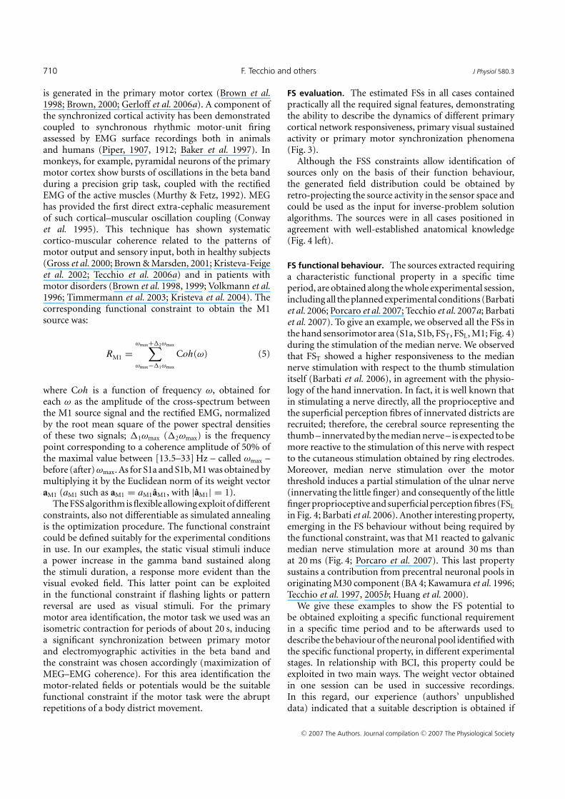
The FSS algorithm is flexible allowing exploit of differentconstraints, also not differentiable as simulated annealingis the optimization procedure. The functional constraintcould be defined suitably for the experimental conditionsin use. In our examples, the static visual stimuli inducea power increase in the gamma band sustained alongthe stimuli duration, a response more evident than thevisual evoked field. This latter point can be exploitedin the functional constraint if flashing lights or patternreversal are used as visual stimuli. For the primarymotor area identification, the motor task we used was anisometric contraction for periods of about 20 s, inducinga significant synchronization between primary motorand electromyographic activities in the beta band andthe constraint was chosen accordingly (maximization ofMEG–EMG coherence). For this area identification themotor-related fields or potentials would be the suitablefunctional constraint if the motor task were the abruptrepetitions of a body district movement.
FS evaluation. The estimated FSs in all cases containedpractically all the required signal features, demonstratingthe ability to describe the dynamics of different primarycortical network responsiveness, primary visual sustainedactivity or primary motor synchronization phenomena(Fig. 3).
Although the FSS constraints allow identification ofsources only on the basis of their function behaviour,the generated field distribution could be obtained byretro-projecting the source activity in the sensor space andcould be used as the input for inverse-problem solutionalgorithms. The sources were in all cases positioned inagreement with well-established anatomical knowledge(Fig. 4 left).
FS functional behaviour. The sources extracted requiringa characteristic functional property in a specific timeperiod, are obtained along the whole experimental session,including all the planned experimental conditions (Barbatiet al. 2006; Porcaro et al. 2007; Tecchio et al. 2007a; Barbatiet al. 2007). To give an example, we observed all the FSs inthe hand sensorimotor area (S1a, S1b, FST, FSL, M1; Fig. 4)during the stimulation of the median nerve. We observedthat FST showed a higher responsiveness to the mediannerve stimulation with respect to the thumb stimulationitself (Barbati et al. 2006), in agreement with the physio-logy of the hand innervation. In fact, it is well known thatin stimulating a nerve directly, all the proprioceptive andthe superficial perception fibres of innervated districts arerecruited; therefore, the cerebral source representing thethumb – innervated by the median nerve – is expected to bemore reactive to the stimulation of this nerve with respectto the cutaneous stimulation obtained by ring electrodes.Moreover, median nerve stimulation over the motorthreshold induces a partial stimulation of the ulnar nerve(innervating the little finger) and consequently of the littlefinger proprioceptive and superficial perception fibres (FSL
in Fig. 4; Barbati et al. 2006). Another interesting property,emerging in the FS behaviour without being required bythe functional constraint, was that M1 reacted to galvanicmedian nerve stimulation more at around 30 ms thanat 20 ms (Fig. 4; Porcaro et al. 2007). This last propertysustains a contribution from precentral neuronal pools inoriginating M30 component (BA 4; Kawamura et al. 1996;Tecchio et al. 1997, 2005b; Huang et al. 2000).
We give these examples to show the FS potential tobe obtained exploiting a specific functional requirementin a specific time period and to be afterwards used todescribe the behaviour of the neuronal pool identified withthe specific functional property, in different experimentalstages. In relationship with BCI, this property could beexploited in two main ways. The weight vector obtainedin one session can be used in successive recordings.In this regard, our experience (authors’ unpublisheddata) indicated that a suitable description is obtained if
C© 2007 The Authors. Journal compilation C© 2007 The Physiological Society

J Physiol 580.3 FSS in hand cortex for BCI 711
Fig. 3. FS discrepancyA, evoked activity during median nerve stimulation. In one representative subject, the two most representativeparietal channels are superimposed in the time window [−10, 80] ms, after averaging on median nerve stimuli,t = 0 being the stimulus arrival at wrist (vertical continous line). The time points corresponding to M20 andM30 components are indicated (vertical dashed lines). Left: original data. Centre: retro-projected data withonly the S1a source (top, MEG˙recS1a) and with only the S1b source (bottom, MEG˙recS1b). Right: original dataminus MEG˙recS1a (top) and original data minus MEG˙recS1b (bottom). The grey area indicates the time interval(�2t20 + �1t20 + 1 top, �2t30 + �1t30 + 1, bottom) where the functional constraint, i.e. the FS responsiveness, ismaximized. Note that both S1a and S1b well explain the generated field at their respective latencies. B, PSD in theStimulus\No-stimulus condition. Left: PSD of one representative MEG sensor signal in the occipital region displayedin the frequency window [0, 80] Hz. Centre: retro-projected channel with the estimated FS (MEG˙rec). Right: PSDof the original MEG data minus MEG˙rec channel. The grey area indicates the frequency interval [20, 70] Hz)where the functional constraint, i.e. the band power difference between Stimulus and No-stimulus conditions,is maximized. C, cortico-muscular coherence during voluntary contraction. The two channels most coherentwith electromyographic activity are chosen. Their coherences with the rectified EMG in the frequency window[0, 45] Hz. The confidence limit is indicated (0.015, horizontal dashed line). Left: original data. Centre:retro-projected channels with only M1 (MEG recM1). The two channels display the same coherence with theEMG signal, as all the channels obtained by retro-projecting only one FS display the same time evolution, unlessa multiplicative factor and the coherence is independent from the signals amplitude. Right: original MEG dataminus MEG recM1 channels. The grey area indicates the frequency interval (�2ωmax + �1ωmax + 1) where theFS–muscular coherence (dimensionless) is calculated.
C© 2007 The Authors. Journal compilation C© 2007 The Physiological Society

712 F. Tecchio and others J Physiol 580.3
similar conditions occur (weight vectors extracted fromsimilar experimental sessions in different days displaycorrelations higher than 0.91); it would be of highinterest to confirm these preliminary positive findings inFS components extracted from a controlled laboratorysetting, later applied to a noisy real-world environment,where competing processes should be expected. Thesecond opportunity is to use the weight vector to isolatethe activity from a specific neuronal pool, in this waybeing more sensitive to volitional modulations of theactivity from those areas. For example, the selection of theactivity from primary visual areas by the above describedfunctional constraint, could allow more sensitivity tovolitional visual imagery.
ICA artifact removal. Muscle contraction on the cephalicdistrict as well as eye movements produce electromagneticsignals which can be recorded from scalp electrodeswith amplitudes that can easily overwhelm the braingenerated signals, particularly on frontal, temporal andoccipital regions (McFarland et al. 1997; Anderer et al.1999; Croft & Barry, 2000; Goncharova et al. 2003).More importantly, the EMG signal from frontal musclescan imitate the frequency of the mu and beta rolandicrhythms and the electro-oculographic (EOG) signal andblinking can resemble the fronto-central theta rhythms.Although FSS could in principle be successful even in thepresence of disturbance signals, if they are uncorrelatedwith the required functional characteristics expressed inthe functional constraint, on some occasions, artifactual
Figure 4. FS positions and behaviourLeft, position in one representative subject ofextracted sources representing the thumbcortical network (FST), the median nerveconnected sensory areas (S1a, S1b), theprimary motor area (M1) and the little fingercortical network (FSL) in the left hemisphereprojected on a representative axial section.Right, dynamics of FSL, S1a, S1b, M1 and FST
activities during contralateral median nervestimulation, shown after averaging the sourcesignal timed on the stimulus onset (t = 0,vertical dashed line) in the [−10, 50] ms timewindow. Note that S1a responsiveness ismaximal around 20 ms, S1b around 30 ms, andM1 responded more at 30 ms than at 20 ms(see text). Note that the y axes do not havemeasurable units, as FSs do not have a physicalunit dimension, before retro-projection on theoriginal signal space.
activities could be as higher as 10–100 times stronger thanthe signal of interest or be in phase with it (Goncharovaet al. 2003). In these cases, it could be very helpful topreviously identify and discard these artifacts. ICA wasproven to be an efficient procedure to remove artifactualactivity avoiding trails exclusion (Vigario et al. 1997; Zieheet al. 2000, 2001; Cao et al. 2000; Delorme & Makeig, 2004).We introduced a suitable strengthening and simplificationof ICA preprocessing data analyses, through an auto-matic detection system of artifactual components (ICs),based on statistical and spectral ICs characteristics (Barbatiet al. 2004). Moreover, the procedure allows recovery ofpart of the non-artifactual signals possibly lost by theblind mechanism, via a control cycle on the differencebetween original data and those reconstructed using onlyICs automatically retained. This step, after automaticpruning, seems to be a suitable way to render negligiblethe risk of loose non-artifactual activity when applyingBSS methods to real data. An emblematic case of artifacts50–100 times larger than the signal of interest comesfrom MEG fetal recordings, where the cerebral activity ofthe fetus is largely overwhelmed by the magnetic signalgenerated by mother heartbeat and partly by the fetalone. In this case, we developed an ad hoc functionalselection procedure of ICs, removing the maternal and fetalcardiac activities, so disclosing the fetal auditory responsesto the external sound stimulation (Porcaro et al. 2006;Fig. 5).
As we saw above, researchers in the BCI field haveattempted to set up experimental protocols and analysis
C© 2007 The Authors. Journal compilation C© 2007 The Physiological Society

J Physiol 580.3 FSS in hand cortex for BCI 713
procedures to increase throughputs and decrease latencies(Xu et al. 2004; Lee et al. 2006). Up to now, BSS algorithmshave been applied in BCI to enhance the feature extractionin motor imagery (Makeig et al. 2000; Qin et al. 2004; Hunget al. 2005), visually induced (Lee et al. 2006) and cognitive(Culpepper & Keller, 2003; Xu et al. 2004; Serby et al. 2005)
Figure 5. ICA artifact removalA, PSD of two channels overlapped before (continous blue line) and after (dotted line) artifacts rejection. The leftchannel is chosen as it is much sensitive to cardiac artifact, and the right one to ocular artifact. B, effects of thead hoc IC artifact removal and IC functional selection procedure. All MEG sensors overlying the fetus head aresuperimposed. Column 1: signal during a 2 s time period; column 2: signal average on the external sounds (dottedvertical line); column 3: spatial magnetic field distribution at the latency of the main component (continous verticalbar). a, original MEG filtered signals: maternal and fetal cardiac peaks are evident in the trace. b, retro-projectionof all ICs but those describing the maternal cardiac activity. c, retro-projection of all ICs but those describing thematernal and fetal cardiac activity; average in c2 still shows a poor quality of morphology, as auditory responselatency and amplitude are not identifiable. d, retro projection of only the ICs describing the auditory response,according to the ad hoc procedure. Latency and amplitude of the auditory response are now clearly identifiable(d2). In this case, the field distribution (d3) shows a dipolar-like shape, indicating a good positioning of the systemwith respect to the fetus head.
tasks. The BSS community is also strongly involved indeveloping online procedures (Serby et al. 2005; Piccioneet al. 2006). Since FSS returns the single trial time coursesof the source of interest without requiring the selectionbetween several components, it could be an even morepromising tool.
C© 2007 The Authors. Journal compilation C© 2007 The Physiological Society

714 F. Tecchio and others J Physiol 580.3
FSS comparison with other source identification methods.The main difference between FSS and the othersource identification methods, ranging from inverseproblem-solving algorithms (single and multiple dipoles:Scherg & Berg, 1991; Multiple Signal Classification(MUSIC): Mosher et al. 1992; recursively applied andprojected-MUSIC (RAP-MUSIC): Mosher & Leahy, 1999;minimum norm estimates: Hamalainen & Ilmoniemi,1994; Low resolution brain electromagnetic tomography(LORETA): Pascual-Marqui et al. 1995) to spatial filteringlike beam forming (for example synthetic aperturemagnetometry, (SAM): Vrba & Robinson, 2001), is thatno information about the physical relationship betweencerebral source generators and generated field distributionis taken into account. Separated FSs provide the sourceactivities in time and the spatial distribution of the fieldthey generate, from which appropriate modelling canto be used to solve the inverse problem to know thesource position. The solution of the inverse problemtheoretically provides in one go both the source positionand its time evolution. Unfortunately, on one side it ishill posed and adjunctive information is to be added,chosen properly time by time. On the other side thesolution is based on the relationship, described by theMaxwell equations, between the current distribution andthat of the generated field. This relation depends onphysical properties, i.e. the shape of conductor volume(the head), the distribution and cytoarchitecture of thecerebral regions (place of the currents generating thefield), the geometrical characteristics and conducibilityvalues of the extra-cerebral tissues (cerebrospinal fluid,meninxs, skull, scalp) none of which are known withprecision. As a consequence, the inverse problem solutionis based on information less accurate provided by theelectrophysiological techniques, while FSS algorithmsdo solve the source identification problem using themost accurate information, i.e. the statistical temporalfrequency properties of the signal. As we said before, oncethe source has been identified, to know its position theproper inverse problem solution can be calculated. In manycases, the scientific interest is in the morphological andtemporal characteristics of the signal and its modulationin the different experimental conditions, and the inverseproblem solving step is not necessary. Whenever thesource position is of specific interest, the advantage is inapplying localization algorithms having isolated the fielddistribution generated by the specific source only.
Section 2. Hand sensorimotor organization and itschanges in stroke patients
Many years of research in functional imaging hasdocumented that the ultimate factor sustaining recoveryis, in parallel with the ‘awakening’ of neurons in theperilesional zone of ischaemic penumbra (Heiss & Graf,1994), the neural ability to change the properties of the
activation induced by inputs from peripheral receptorsand/or other brain areas. This ability is called cerebralplasticity (for review see Calautti et al. 2003; Rossiniet al. 2003; Hummel & Cohen, 2005). When settingup BCI feature extraction, these cerebral reorganizationsshould be taken into account, especially in the electro-physiological counterpart, since EEG and MEG are thebrain signal monitoring systems best suited for mostrealistic BCIs. In particular, MEG is especially suitablein poststroke studies, because the morbid tissue nearthe cerebral generators has minimal effects on the scalpdistribution of the magnetic field (Huang et al. 1990;Maclin et al. 1994). BCI feature extraction is based ontwo main steps: spatial identification of the interestingsignal sources and the time–frequency characterization ofthis signal. To guide BCI applications, it is expected thatthe activity from the areas positively contributing to theclinical recovery are the most appropriate and informative.In patients affected by a monolateral ischaemic lesion in themiddle cerebral artery territory inducing a sensorimotorimpairment of the upper limb, a diverse contribution tothe clinical recovery by several area reorganizations in theaffected (ipsi-lesional ILH) and the unaffected hemisphere(contra-lesional CLH) is going to be recognized. Theactivation of CLH non-primary motor areas was reportedto decrease proportionally with the improvement of motorperformance (Calautti & Baron, 2003; Schaechter, 2004)and CLH SM1 activations were commonly consideredas a marker of poor recovery (Loubinoux et al. 2003;Calautti et al. 2003). Conversely, increasing findingssupport a positive role of non-primary ILH areas inpatients recovering incompletely (Johansen-Berg et al.2002; Loubinoux et al. 2003; Fridman et al. 2004; Gerloffet al. 2006b). Moreover, a similar hypothesis was suggestedfor the recruitment of ILH areas excessively asymmetricalfrom homologous in the CLH (Pineiro et al. 2001; Calauttiet al. 2003; Thickbroom et al. 2004). On the basis ofthe symmetrical organization of primary hand areas,demonstrated by different techniques in healthy subjects(Rossini et al. 1994b; Puce et al. 1995; Hallet, 1996; Tecchioet al. 1997; Wikstrom et al. 1997), the comparison of theILH to the CLH in mono-hemispheric stroke patientsallows a sensitive procedure to identify reorganizationphenomena in the damaged cerebral regions. On this basis,an ad hoc procedure was previously introduced, for theidentification of excessive interhemispheric asymmetriesof primary sensory hand areas (Tecchio et al. 1997,2000b, 2005b). In these studies, a galvanic median nervestimulation was used, a protocol especially suitable inpatients because it equals the input to the two hemi-spheres. It seems, for this reason, more appropriate inorder to detect interhemispheric recruitment asymmetriesof homologous areas, with respect to active motortasks (Kotani et al. 2004) where the functional demandto control the paretic and non-paretic hand cannotbe equalled. In patients in stabilized conditions (more
C© 2007 The Authors. Journal compilation C© 2007 The Physiological Society

J Physiol 580.3 FSS in hand cortex for BCI 715
than 1 year from symptoms onset), primary sensorycortical hand areas topographical modifications andresponsiveness amplitudes were observed (Rossini et al.1998, 27 patients; Rossini et al. 2001, 17 patients), withbrain areas outside the normal boundaries and usually notreached by a dense sensory input from the opposite handacting as somatosensory hand centres. These mechanismswere linked with the hand sensorimotor recovery (Rossiniet al. 1998, 2001; Tecchio et al. 2006d; Altamura et al. 2007).A deeper knowledge of brain mechanisms sustainingrecovery in stroke patients who do not achieve a normalneurological function by standard procedures, is of utmostimportance, since this group could best benefit fromadjunctive rehabilitative-stimulating interventions, likeBCI-mediated procedures.
Once spatial properties of interesting signal sourceshave been identified, BCI will exploit their time–frequencycharacteristics. When studying rest activity in post-acute and chronic phase, perilesional delta activity wascommonly observed (Butz et al. 2004, 23 patients).Moreover, frequency-selective alterations related tospecific dysfunctions were found: global clinical statuswas mostly impaired in patients with increased total andslow band activity powers, whereas hand functionalitywas mostly disrupted in patients with a reduction ofhigh-frequency rhythms (Tecchio et al. 2006c, 56 patients;Tecchio et al. 2006c, 32 patients).
While all the above mentioned studies are most suitableto characterize recruitment changes in patients, theorganization of the movement control remains the abilitythe BCI tools should help to recover. Motor imagery isa protocol design also suitable for patients with moresevere movement impairment. It is clearly indicated by theliterature that the brain activity subtending motor imageryis modified in patients (Sharma et al. 2006). In healthysubjects, movement imagery can focus specific facilitationon the prime-mover muscle for the mentally simulatedmovement (Rossi et al. 1998a) and mental simulationaffects spinal motoneuronal excitability as well, althoughit has been proved that the main effect takes place atcortical level (Rossini et al. 1999). Reviews consideringmotor imagery in healthy subjects and in patients withstroke – which may disrupt the motor imagery network– suggested the encouraging effect of motor imagerytraining on motor recovery after stroke (Lotze & Cohen,2006; Sharma et al. 2006). While in healthy volunteers,robust activation of the non-primary motor structures,but only weak and inconsistent activation of M1 occursduring motor imagery, in patients with stroke, the corticalactivation patterns are proved to involve M1 in both theaffected and unaffected hemispheres (Cicinelli et al. 2006).These results indicate that if an appropriate methodology isimplemented, motor imagery may provide a valuable toolto access the motor network for BCI feature extraction instroke patients.
As mentioned above, the computer-mediated visualfeedback presents problems for fast applications, whilethe haptic feedback and electro-tactile sensationspresent more realistic timing. All studies devoted to theorganization of the sensory and proprioceptive inflowunderline that a successful motor control relies onthe continuous and reciprocal exchange of informationbetween activities of motor areas involved in the taskprogram execution and those elaborating proprioceptivesensory information (Terao et al. 1999; Scott, 2004). Asa matter of fact, subjects with complete deprivation ofproprioceptive feedback (such as in some cases of peri-pheral neuropathy) can move their limbs using visual feed-back (e.g. by looking at the limb), but their movementsare typically sluggish, coarse, require substantial mentalconcentration and attention, and corrections are thereforedelayed and often cause other mistakes (Sainburg et al.1998; Gordon et al. 1995).
Short-term influences on motor cortical organizationhave been demonstrated by modification of sensory inputboth in animal experiments and in healthy humans byusing several types of technology for functional brainimaging (Brazil-Neto et al. 1992; Rossini et al. 1994a;Sadato et al. 1995; Kristeva-Feige et al. 1996). Mechanismsof rapid anaesthesia-related perturbation with changesoccurring at multiple levels of sensory system somatotopywere proven (Nicolelis et al. 1993), underlying the intimaterelationship between primary sensory and motor regions.Experimental findings speak in favour of a significant roleplayed by the tonic sensory flow from the skin receptorsand from the phalangeal joint receptors in energizingthe corticospinal tracts governing muscles (Johansson &Westling, 1987; Rossini et al. 1996a, b; Rossi et al. 1998b).For BCI applications, such evidence indicates the need tosubstitute with compensating feedback mechanisms theabsence of the physiological one, to obtain a satisfactorymovement control.
To optimize knowledge on proprioceptive informationproperties in the region dedicated to hand control, weinvestigated the primary sensory and motor cortices inter-actions, obtaining a simultaneous assessment of sensorycortex activity modulation due to movement and of motorcortex activity modulation due to sensory stimulation(Tecchio et al. 2006b; Fig. 6). The introduced protocolis repeatable and suitable for patients, thus it is suitableto investigate patients eligible for BCI applications. Infact, primary sensory cortical excitability is indexed bythe most repeatable and subject attention-independentcomponents of brain response to the median nervestimulation at the wrist (Hari & Kaukoranta, 1985; Allisonet al. 1991). A relative high-frequency nerve stimulation(around 2 Hz) was used to reduce the recording time. Theprimary motor cortex contribution to movement controlis indexed by cortico-muscular coherence (Brown et al.1998) requiring a motor task both as simple and common
C© 2007 The Authors. Journal compilation C© 2007 The Physiological Society

716 F. Tecchio and others J Physiol 580.3
Figure 6. Sensorimotor feedbackA, experimental protocol: Top from left:(1) subject position during recordings fromscalp positions overlying the peri-rolandic areacontra-lateral to the moved-stimulated hand:(2) non-magnetic device, i.e. the watersphygmomanometer, to control the level ofcontraction: (3) showing the thumb positionduring isometric contraction, with electrodesrecording OP EMG signal, as well as the mediannerve stimulation at wrist. Bottom from top:(1) trigger indicating tone beep forstarting/stopping isometric contraction:(2) trigger indicating sensory stimuli to themedian nerve at wrist: (3) electromyographicsignal, where relax and contraction periods areclearly noticeable, as well as the sensory stimuliartifact: (4) off-line generated signal codedifferentiating the 4 experimental conditions:relax (R), sensory (S), motor (M), sensory-motor(SM). B, FS PSD and intersource coherence. PSDof the S1 and M1 cerebral sources (top) andS1–M1 coherence (bottom) in the fourexperimental conditions in a representativesubject. Note: higher S1 representation in thealpha band and higher M1 beta and gammaband representation; lower S1–M1 coherencein all condition with respect to rest in the threefrequency bands and values above significancethreshold in motor condition (dashed–dottedline). Note that the y axes of S1 and M1 PSDsdo not have measurable units, as FSs do nothave a physical unit dimension. The S1–M1coherence is a dimensionless quantity.
C© 2007 The Authors. Journal compilation C© 2007 The Physiological Society

J Physiol 580.3 FSS in hand cortex for BCI 717
as possible, thus most suitable in patients with upperlimb impairments. In fact, the experimental contraction ispreserved to extremely severe movement impairments, e.g.in diseases affecting the hand motor control, like strokeand Parkinson’s disease or hand dystonia. As describedin Section 1, the ad hoc FSS was developed to extract S1and M1 source activities (Porcaro et al. 2007), allowingcharacterization of the spectral properties, the functionalinvolvement of S1 and M1 and their coupling in the abovedescribed sensorimotor tasks (Fig. 6). It could be veryuseful to reveal alterations in pathological conditions andsetting up feedback control systems for BCI use.
Conclusions
Our review presents innovative methods expected toenhance BCI feature extraction from EEG or MEG signals,for applications devoted to improve hand control. Inparticular, the provided experimental set-up and analysistools could be used to ameliorate a BCI system. Infact, they increase the signal-to-noise ratio by removingnon-cerebral artifacts even when they are many times(10–100) larger than the signal of interest. Moreover, theyoffer a procedure to extract exclusively the sources ofinterest, obtaining their time course along with the wholeexperimental session on the basis of appropriate functionalrequirements taking place in specific time periods. Sincethe FSS procedure is suitable for single trial analysis, itis promising for online developments – increasing BCIthroughput and reducing latency. For applications inpatients, information about the recruitments of unusualareas to control the hand were provided, useful forboth protocol design definition and interesting signalextraction. Finally, information about primary sensoryand motor area relationship was introduced, as a first stepto use the haptic feedback and electrotactile sensationswhich have been indicated as the most compelling forBCI applications aiming at improving hand movementcontrol.
References
Allison T, Goff WR, Williamson PD et al. (1980). On the neuralorigin of the early components of the human somatosensoryevoked potentials. In Progress in Clinical Neurophysiology,ed. Desmedt JD, pp. 51–68. Karger, Basel.
Allison T, McCarthy G, Wood CC et al. (1991). Potentialsevoked in human and monkey cerebral cortex by stimulationof the median nerve. A review of scalp and intracranialrecordings. Brain 114, 2465–2503.
Altamura C, Torquati K, Zappasodi F et al. (2007).fMRI-vs-MEG evaluation of post-stroke interhemisphericasymmetries in primary sensorimotor hand areas. ExpNeurol in press.
Anderer P, Roberts S, Schlogl A et al. (1999). Artifactprocessing in computerized analysis of sleep EEG – a review.Neuropsychobiology 40, 150–157.
Baker SN, Olivier E & Lemon RN (1997). Coherent oscillationsin monkey motor cortex and hand muscle EMG showtask-dependent modulation. J Physiol 501,225–241.
Barbati G, Porcaro C, Hadjipapas A et al. (2007). Functionalsource separation applied to induced visual gamma activity.Hum Brain Mapp in press.
Barbati G, Porcaro C, Zappasodi F et al. (2004). Optimizationof an independent component analysis approach for artifactidentification and removal in magnetoencephalographicsignals. Clin Neurophysiol 115, 1220–1232.
Barbati G, Sigismondi R, Zappasodi F et al. (2006). Functionalsource separation from magnetoencephalographic signals.Hum Brain Mapp 27, 925–934.
Birbaumer N, Ghanayim N, Hinterberger T et al. (1999). Aspelling device for the paralysed. Nature 398,297–298.
Brasil-Neto JP, Cohen LG, Pascual-Leone A et al. (1992). Rapidreversible modulation of human motor outputs aftertransient deafferentation of the forearm: a study withtranscranial magnetic stimulation. Neurology 42,1302–1306.
Brown P (2000). Cortical drives to human muscle: the Piperand related rhythms. Prog Neurobiol 60, 97–108.
Brown P, Farmer SF, Halliday DM et al. (1999). Coherentcortical and muscle discharge in cortical myoclonus. Brain122, 461–472.
Brown P & Marsden JF (2001). Cortical network resonance andmotor activity in humans. Neuroscientist 7, 518–527.
Brown P, Salenius S, Rothwell JC et al. (1998). Corticalcorrelate of the Piper rhythm in humans. J Neurophysiol 80,2911–2917.
Butz M, Gross J, Timmermann L et al. (2004). Perilesionalpathological oscillatory activity in themagnetoencephalogram of patients with cortical brainlesions. Neurosci Lett 355, 93–96.
Calautti C & Baron JC (2003). Functional neuroimagingstudies of motor recovery after stroke in adults: a review.Stroke 34, 1553–1566.
Calautti C, Leroy F, Guincestre JY et al. (2003). Displacementof primary sensorimotor cortex activation after subcorticalstroke: a longitudinal PET study with clinical correlation.Neuroimage 19, 1650–1654.
Cao J, Murata N, Amari S et al. (2000). Single-trialmagnetoencephalographic data decomposition andlocalization based on independent component analysisapproach. IEICE Transactions Fundamentals Electronics,Comms Computer Sci 9, 1757–1766.
Cichocki A & Amari SI (2002). Adaptive Blind Signal and ImageProcessing . John Wiley & Sons, Chichester, UK.
Cicinelli P, Marconi B, Zaccagnini M et al. (2006).Imagery-induced cortical excitability changes in stroke: atranscranial magnetic stimulation study. Cereb Cortex 16,247–253.
Conway BA, Halliday DM, Farmer SF et al. (1995).Synchronization between motor cortex and spinalmotoneuronal pool during the performance of a maintainedmotor task in man. J Physiol 489, 917–924.
Craelius W (2002). The bionic man: restoring mobility. Science295, 1018–1021.
C© 2007 The Authors. Journal compilation C© 2007 The Physiological Society

718 F. Tecchio and others J Physiol 580.3
Croft RJ & Barry RJ (2000). Removal of ocular artifact from theEEG: a review. Neurophysiol Clin 30, 5–19.
Culpepper BJ & Keller RM (2003). Enabling computerdecisions based on EEG input. IEEE Trans Neural SystRehabil Eng 11, 354–360.
Del Gratta C, Pizzella V, Tecchio F & Romani G-L (2001).Magnetoencephalography – a non invasive brain imagingmethod with 1 ms time resolution. Rep Prog Phys 64,1759–1814.
Delorme A & Makeig S (2004). EEGLAB: an open sourcetoolbox for analysis of single-trial EEG dynamics includingindependent component analysis. J Neurosci Meth 134,9–21.
Flor H, Nikolajsen L & Staehelin Jensen T (2006). Phantomlimb pain: a case of maladaptive CNS plasticity? Nat RevNeurosci 7, 873–881.
Fridman EA, Hanakawa T, Chung M, Hummel F, LeiguardaRC & Cohen LG (2004). Reorganization of human premotorcortex after stroke recovery. Brain 127, 747–758.
Gastaut H (1952). Etude electrocorticographique de lareactivite des rythmes rolandiques. Rev Neurol 87,176–182.
Gerloff C, Braun C, Staudt M et al. (2006a). Coherentcorticomuscular oscillations originate from primary motorcortex: evidence from patients with early brain lesions. HumBrain Mapp 27, 789–798.
Gerloff C, Bushara K, Sailer A et al. (2006b). Multimodalimaging of brain reorganization in motor areas of thecontralesional hemisphere of well recovered patients aftercapsular stroke. Brain 129, 791–808.
Goncharova II, McFarland DJ, Vaughan TM et al. (2003). EMGcontamination of EEG: spectral and topographicalcharacteristics. Clin Neurophysiol 114, 1580–1593.
Gordon J, Ghilardi MF & Ghez C (1995). Impairments ofreaching movements in patients without proprioception.J Neurophysiol 73, 347–360.
Graimann B, Townsend G & Pfurtscheller G (2006).Brain-computer communication – A brief introduction(Technical Report). BCI-Info, BCI Laboratory, TechnischeUniversitat Graz, Graz, Austria.
Gross J, Tass PA, Salenius S et al. (2000). Cortico-muscularsynchronization during isometric muscle contraction inhumans as revealed by magnetoencephalography. J Physiol527, 623–631.
Hallet M (1996). Transcranial magnetic stimulation: a tool formapping the central nervous system. Electroencephalogr ClinNeurophysiol Suppl 46, 43–51.
Hamalainen MS & Ilmoniemi RJ (1994). Interpreting magneticfields of the brain: minimum norm estimates. Med Biol EngComput 32, 35–42.
Hari R & Kaukoranta E (1985). Neuromagnetic studies ofsomatosensory system: principles and examples. ProgNeurobiol 24, 233–256.
Hari R, Reinikainen K, Kaukoranta E et al. (1984).Somatosensory evoked magnetic fields from SI and SII inman. Electroencephalogr Clin Neurophyisol 57,254–263.
Heeger DJ, Huk AC, Geisler WS et al. (2000). Spikes versusBOLD: what does neuroimaging tell us about neuronalactivity? Nat Neurosci 3, 631–633.
Heeger DJ & Ress D (2002). What does fMRI tell us aboutneuronal activity? Nat Rev Neurosci 3, 142–151.
Heiss WD & Graf R (1994). The ischemic penumbra. CurrOpin Neurol 7, 11–19.
Huang JC, Nicholson C, Okada Y & C (1990). Distortion ofmagnetic evoked fields and surface potentials byconductivity differences and boundaries in brain tissue.Biophys J 57, 1155–1667.
Huang MX, Aine C, Davis L et al. (2000). Sources on theanterior and posterior banks of the central sulcus identifiedfrom magnetic somatosensory evoked responses usingmultistart spatio-temporal localization. Hum Brain Mapp11, 59–76.
Hummel FC & Cohen LG (2005). Drivers of brain plasticity.Curr Opin Neurol 18, 667–674.
Hung CI, Lee PL, Wu YT et al. (2005). Recognition of motorimagery electroencephalography using independentcomponent analysis and machine classifiers. Ann Biomed Eng33, 1053–1070.
Hyvarinen A, Karhunen J & Oja E (2001). IndependentComponent Analysis. John Wiley & Sons, Chichester, UK.
Johansen-Berg H, Rushworth MF, Bogdanovic MD et al.(2002). The role of ipsilateral premotor cortex in handmovement after stroke. Proc Natl Acad Sci U S A 99,14518–14523.
Johansson RS & Westling G (1987). Signals in tactile afferentsfrom the fingers eliciting adaptive motor responses duringprecision grip. Exp Brain Res 66, 141–154.
Kawamura T, Nakasato N, Seki K et al. (1996). Neuromagneticevidence of pre and post-central cortical sources ofsomatosensory evoked responses. Electroencephalogr ClinNeurophysiol 100, 44–50.
Keene DL, Whiting S & Ventureyra EC (2000).Electrocorticography. Epileptic Disord 2, 57–63.
Kilner JM, Baker SN, Salenius S et al. (2000). Human corticalmuscle coherence is directly related to specific motorparameters. J Neurosci 20, 8838–8845.
Kotani K, Kinomoto Y, Yamada M et al. (2004). Spatiotemporalpatterns of movement-related fields in stroke patients.Neurol Clin Neurophysiol 30, 2004–2063.
Kristeva R, Popa T, Chakarov V et al. (2004). Cortico-muscularcoupling in a patient with postural myoclonus. Neurosci Lett366, 259–263.
Kristeva-Feige R, Fritsch C, Timmer J et al. (2002). Effects ofattention and precision of exerted force on beta rangeEEG-EMG synchronization during a maintained motorcontraction task. Clin Neurophysiol 113, 124–131.
Kristeva-Feige R, Rossi S, Pizzella V et al. (1996). Changes inmovement-related brain activity during transientdeafferentation: a neuromagnetic study. Brain Res 714,201–208.
Lauer R, Peckham P, Kilgore K et al. (2000a). Applications ofcortical signals to neuroprosthetic control: a critical review.IEEE Trans Rehabil Eng 8, 205–208.
Lauer RT, Peckham PH, Kilgore KL et al. (2000b). Applicationsof cortical signals to neuroprosthetic control: a criticalreview. IEEE Trans Rehabil Eng 8, 205–208.
Lee PL, Hsieh JC, Wu CH et al. (2006). The brain computerinterface using flash visual evoked potential and independentcomponent analysis. Ann Biomed Eng 34, 1641–1654.
C© 2007 The Authors. Journal compilation C© 2007 The Physiological Society

J Physiol 580.3 FSS in hand cortex for BCI 719
Lotze M & Cohen LG (2006). Volition and imagery inneurorehabilitation. Cogn Behav Neurol 19, 135–140.
Loubinoux I, Carel C, Pariente J et al. (2003). Correlationbetween cerebral reorganization and motor recovery aftersubcortical infarcts. Neuroimage 20, 2166–2180.
McFarland D, Lefkowicz A & Wolpaw J (1997). Design andoperation of an EEG-based brain–computer interface withdigital signal processing technology. Behav Res Meth InstrumComput 29, 337–345.
Maclin EL, Rose DF, Knight JE et al. (1994). Somatosensoryevoked magnetic fields in patients with stroke.Electroencephalogr Clin Neurophysiol 91, 468–475.
Makeig S, Bell AJ, Jung TP et al. (1996). Independentcomponent analysis of electroencephalographic data. InAdvances in Neural Information Processing Systems, Vol. 8,pp. 145–151, ed. Jordan MI, Kearns MJ & Solla SA. MITPress, Cambridge, MA.
Makeig S, Debener S, Onton J et al. (2004). Miningevent-related brain dynamics. Trends Cogn Sci 8,204–210.
Makeig S, Enghoff S, Jung TP et al. (2000). Natural basis forefficient brain-actuated control. IEEE Trans Rehabil Eng 8,208–211.
Makeig S, Westerfield M, Jung TP et al. (2002). Dynamic brainsources of visual evoked responses. Science 295,690–694.
Maynard EM, Nordhausen CT & Normann RA (1997). TheUtah intracortical electrode array: a recording structure forpotential brain–computer interfaces. Electroencephalogr ClinNeurophysiol 102, 228–239.
Moore MM (2003). Real-world applications for brain–computer interface technology. IEEE Trans Neural SystRehabil Eng 11, 162–165.
Mosher JC & Leahy RM (1999). Source localization usingrecursively applied and projected (RAP) MUSIC. IEEE TransSig Proc 47, 332–345.
Mosher JC, Lewis PS & Leahy RM (1992). Multiple dipolemodeling and localization from spatiotemporal MEG data.IEEE Trans Biomed Eng 39, 541–557.
Murthy VN & Fetz EE (1992). Coherent 25- to 35-Hzoscillations in the sensorimotor cortex of awake behavingmonkeys. Proc Natl Acad Sci U S A 89, 5670–5674.
Navarro X, Krueger T, Lago N et al. (2005). A critical review ofinterfaces with the peripheral nervous system for the controlof neuroprostheses and hybrid bionic systems. J Pher NervSys 10, 229–258.
Nicolelis MA, Lin RC, Woodward DJ et al. (1993). Induction ofimmediate spatiotemporal changes in thalamic networks byperipheral block of ascending cutaneous information.Nature 361, 533–536.
Niedermeyer E & Lopes da Silva F (1997).Electroencephalography: Basic Principles, Clinical Applications,and Related Fields. Williams & Wilkins, Baltimore, MD.
Oliviero A, Tecchio F, Zappasodi F et al. (2004). Brainsensorimotor hand area functionality in acute stroke: insightsfrom magnetoencephalography. Neuroimage 23, 542–550.
Pascual-Marqui RD, Michel CM & Lehman D (1995). Lowresolution electromagnetic tomography: a new method forlocalising electrical activity in the brain. Int J Psychophysiol18, 49–65.
Piccione F, Giorgi F, Tonin P et al. (2006). P300-based braincomputer interface: reliability and performance in healthyand paralysed participants. Clin Neurophysiol 117, 531–537.
Pineiro R, Pendlebury S, Johansen-Berg H et al. (2001).Functional MRI detects posterior shifts in primarysensorimotor cortex activation after stroke: evidence of localadaptive reorganization? Stroke 32, 1134–1139.
Piper HE (1907). Uber den willkurlichen Muskeltetanus.Pflugers Gesamte Physiologie Des Menschen Tiere 119,301–338.
Piper HE (1912). Elektrophysiologie Menschlicher Muskeln.Springer, Berlin.
Pizzella V, Tecchio F, Romani GL et al. (1999). Functionallocalization of the sensory hand area with respect to themotor central gyrus knob. Neuroreport 10, 3809–3814.
Popovic D & Sinkjaer T (2000). Control of Movement for thePhysically Disabled. Springer-Verlag.
Popovic M (2003). Control of neural prostheses for graspingand reaching. Med Engineering Physics 25, 41–50.
Porcaro C, Barbati G, Zappasodi F et al. (2007). Handsensory-motor cortical network assessed by functionalsource separation. Hum Brain Mapp in press.
Porcaro C, Zappasodi F, Barbati G et al. (2006). Fetal auditoryresponses to external sounds and mother’s heart beat:detection improved by independent component analysis.Brain Res 26, 51–58.
Prochazka A, Gauthier M, Wieler M et al. (1997). The bionicglove: An electrical stimulator garment that providescontrolled grasp and hand opening in quadriplegia. ArchPhys Med Rehabilitation 78, 1–7.
Puce A, Constable RT, Luby ML et al. (1995). Functionalmagnetic resonance imaging of the sensory and motorcortex: comparison with electrophysiological localization.J Neurosurg 83, 262–270.
Qin L, Ding L & He B (2004). Motor imagery classification bymeans of source analysis for brain–computer interfaceapplications. J Neural Eng 1, 135–141.
Rees G, Friston K & Koch C (2000). A direct quantitativerelationship between the functional properties of human andmacaque V5. Nat Neurosci 3, 716–723.
Rossi S, Pasqualetti P, Tecchio F et al. (1998a). Corticospinalexcitability modulation during mental simulation of wristmovements in human subjects. Neurosci Lett 243, 147–151.
Rossi S, Pasqualetti P, Tecchio F et al. (1998b). Modulation ofcorticospinal output to human hand muscles followingdeprivation of sensory feedback. Neuroimage 8, 163–175.
Rossini PM, Calautti C, Pauri F et al. (2003). Post-stroke plasticreorganisation in the adult brain. Lancet Neurol 2, 493–502.
Rossini PM, Caltagirone C, Castriota-Scanderbeg A et al.(1998). Hand motor cortical area reorganization in stroke: astudy with fMRI, MEG and TCS maps. Neuroreport 9,2141–2146.
Rossini PM, Martino G, Narici L et al. (1994a). Short-termbrain ‘plasticity’ in humans: transient finger representationchanges in sensory cortex somatotopy following ischemicanaesthesia. Brain Res 642, 169–177.
Rossini PM, Narici L, Martino G et al. (1994b). Analysis ofinterhemispheric asymmetries of somatosensory evokedmagnetic fields to right and left median nerve stimulation.Electroencephalogr Clin Neurophysiol 91, 476–482.
C© 2007 The Authors. Journal compilation C© 2007 The Physiological Society

720 F. Tecchio and others J Physiol 580.3
Rossini PM, Rossi S, Pasqualetti P et al. (1999). Corticospinalexcitability modulation to hand muscles during movementimagery. Cerebral Cortex 9, 161–167.
Rossini PM, Rossi S, Tecchio F et al. (1996a). Focal brainstimulation in healthy humans: motor maps changesfollowing partial hand sensory deprivation. Neurosci Lett214, 191–195.
Rossini PM, Tecchio F, Pizzella V et al. (2001).Interhemispheric differences of sensory hand areas aftermonohemispheric stroke: MEG/MRI integrative study.Neuroimage 14, 474–485.
Rossini PM, Tecchio F, Sabato A et al. (1996b). The role ofcutaneous inputs during magnetic transcranial stimulation.Muscle Nerve 19, 1302–1309.
Sadato N, Zeffiro TA, Campbell G et al. (1995). Regionalcerebral blood flow changes in motor cortical areas aftertransient anesthesia of the forearm. Ann Neurol 37, 74–81.
Sainburg RL, Poizner H & Ghez C (1998). Loss ofproprioception produces deficits in interjoint coordination.J Neurophysiol 82, 1–15.
Schaechter JD (2004). Motor rehabilitation and brain plasticityafter hemiparetic stroke. Prog Neurobiol 73, 61–72.
Scherg M & Berg P (1991). Use of prior knowledge in brainelectromagnetic source analysis. Brain Topogr 4, 143–150.
Scott SH (2004). Optimal feedback control and the neural basisof volitional motor control. Nat Rev Neurosci 5, 532–546.
Serby H, Yom-Tov E & Inbar GF (2005). An improvedP300-based brain–computer interface. IEEE Trans NeuralSyst Rehabil Eng 13, 89–98.
Sharma N, Pomeroy VM & Baron JC (2006). Motor imagery: abackdoor to the motor system after stroke? Stroke 37,1941–1952.
Stein R, Peckham P & Popovic D (1992). Neural Prosthesis.Oxford University Press, Oxford, UK.
Tang AC, Sutherland MT & McKinney CJ (2005). Validation ofSOBI components from high-density EEG. Neuroimage 25,539–553.
Tecchio F, Bicciolo G, De Campora E et al. (2000a). Tonotopiccortical changes following stapes substitution in otoscleroticpatients: a magnetoencephalographic study. Hum BrainMapp 10, 28–38.
Tecchio F, Graziadio S, Barbati G et al. (2007a). Dynamicgamma-band synchrony: a neural code of sensorimotordexterity. Neuroimage, 35, 185–193.
Tecchio F, Padua L, Aprile I et al. (2002). Carpal tunnelsyndrome modifies sensory hand cortical somatotopy: aMEG study. Hum Brain Mapp 17, 28–36.
Tecchio F, Pasqualetti P, Pizzella V et al. (2000b). Morphologyof somatosensory evoked fields: inter-hemispheric similarityas a parameter for physiological and pathological neuralconnectivity. Neurosci Lett 287, 203–206.
Tecchio F, Pasqualetti P, Zappasodi F et al. (2007b). Prognosticvalue of magnetoencephalography parameteres in acutemonohemispheric stroke. J Neurol, 35, 185–193.
Tecchio F, Porcaro C, Zappasodi F et al. (2006a). Corticalshort-term fatigue effects assessed via rhythmicbrain-muscle coherence. Exp Brain Res 174, 144–151.
Tecchio F, Rossini PM, Pizzella V et al. (1997). Spatialproperties and interhemispheric differences of the sensoryhand cortical representation: a neuromagnetic study. BrainRes 767, 100–108.
Tecchio F, Zappasodi F, Melgari JM et al. (2006b).Sensory–motor interaction in primary hand cortical areas: aMEG assessment. Neuroscience 141, 533–542.
Tecchio F, Zappasodi F, Pasqualetti P et al. (2005a). Rhythmicbrain activity at rest from rolandic areas in acutemono-hemispheric stroke: a magnetoencephalographicstudy. Neuroimage 28, 72–83.
Tecchio F, Zappasodi F, Pasqualetti P et al. (2005b). Neuralconnectivity in hand sensorimotor brain areas: an evaluationby evoked field morphology. Hum Brain Mapp 24,99–108.
Tecchio F, Zappasodi F, Pasqualetti P et al. (2006c). Long-termeffects of stroke on neuronal rest activity in rolandic corticalareas. J Neurosci Res 83, 1077–1087.
Tecchio F, Zappasodi F, Tombini M et al. (2006d). Brainplasticity in recovery from stroke: an MEG assessment.Neuroimage 32, 1326–1334.
Terao Y, Ugawa Y, Hanajima R et al. (1999). Air-puff-inducedfacilitation of motor cortical excitability studied in patientswith discrete brain lesions. Brain 122, 2259–2277.
Thickbroom GW, Byrnes ML, Archer SA et al. (2004). Motoroutcome after subcortical stroke correlates with the degreeof cortical reorganization. Clin Neurophysiol 115,2144–2150.
Timmermann L, Gross J, Dirks M et al. (2003). The cerebraloscillatory network of parkinsonian resting tremor. Brain126, 199–212.
Treisman AM & Kanwisher NG (1998). Perceiving visuallypresented objects: Recognition, awareness, and modularity.Current Opinion Neurobiol 8, 218–226.
Vigario R, Jousmaki V, Hamalainen M et al. (eds) (1997).Independent component analysis for identification ofartifacts in magnetoencephalographic recordings. InAdvances in Neural Information Processing Systems,pp. 229–235. MIT Press, Cambridge.
Volkmann J, Joliot M, Mogilner A et al. (1996). Central motorloop oscillations in parkinsonian resting tremor revealed bymagnetoencephalography. Neurology 46, 1359–1370.
Vrba J & Robinson SE (2001). Signal processing inmagnetoencephalography. Methods 25, 249–271.
Wikstrom H, Huttunen J, Korvenoja A et al. (1996). Effects ofinterstimulus interval on somatosensory evoked magneticfields (SEFs): a hypothesis concerning SEF generation at theprimary sensorimotor cortex. Electroencephalogr ClinNeurophysiol 100, 479–487.
Wikstrom H, Roine RO, Salonen O et al. (1997).Somatosensory evoked magnetic fields to median nervestimulation: interhemispheric differences in a normalpopulation. Electroencephalogr Clin Neurophysiol 104,480–487.
Wolpaw JR, Birbaumer N, McFarland DJ et al. (2002).Brain–computer interfaces for communication and control.Clin Neurophysiol 113, 767–791.
Xu N, Gao X, Hong B et al. (2004). BCI Competition 2003 –Data set IIb: enhancing P300 wave detection usingICA-based subspace projections for BCI applications. IEEETrans Biomed Eng 51, 1067–1072.
Zappasodi F, Pasqualetti P, Ercolani M et al. (2006). Handcortical representation at rest and during activation: genderand age effects in the two hemispheres. Clin Neurophysiol117, 1518–1528.
C© 2007 The Authors. Journal compilation C© 2007 The Physiological Society

J Physiol 580.3 FSS in hand cortex for BCI 721
Ziehe A, Nolte G, Sander T et al. (2000). Artifact reduction inmagnetoneurography based on time-delayed second-ordercorrelations. IEEE Trans Biomed Eng 47, 75–87.
Ziehe A, Nolte G, Sander T et al. (2001). A comparison ofICA-based artifact reduction methods for MEG. InProceedings of the 12th International Conference onBiomagnetism, pp. 895–898. Helsinki University ofTechnolology, Espoo, Finland.
Acknowledgements
This work was partially supported by the RBNE01AZ92, PRIN2005027850 and PRIN 2005063547 of the Italian Departmentof University and Research (MIUR) and by the EuropeanIST/FET Integrated Project NEUROBOTICS – the fusion ofNEUROscience and roBOTICS, Project no. 001917 under the6th Framework Programme.
C© 2007 The Authors. Journal compilation C© 2007 The Physiological Society


![[Posterior cortical atrophy]](https://static.fdokumen.com/doc/165x107/6331b9d14e01430403005392/posterior-cortical-atrophy.jpg)


