Finite Element Analysis of the Cingulata Jaw: An Ecomorphological Approach to Armadillo’s Diets
Upload
independentCategory
view
1download
0
r Human Brain Mapping 33:2306–2321 (2012) r
Functional MRI Assessment of OrofacialArticulators: Neural Correlates of Lip, Jaw, Larynx,
and Tongue Movements
Krystyna Grabski,1* Laurent Lamalle,2,3 Coriandre Vilain,1
Jean-Luc Schwartz,1 Nathalie Vallee,1 Irene Tropres,2,4 Monica Baciu,5
Jean-Francois Le Bas,2,6 and Marc Sato1*
1Gipsa-Lab, Departement Parole & Cognition, UMR CNRS 5216, Grenoble Universites, France2Structure Federative de Recherche n�1 ‘‘RMN Biomedicale et Neurosciences’’ & Unite IRM 3T,
Centre Hospitalier Universitaire de Grenoble, France3INSERM, France
4Universite Joseph Fourier, Grenoble, France5Laboratoire de Psychologie et Neurocognition, UMR CNRS 5105 & Universite Pierre Mendes France, France
6Centre Hospitalier Universitaire de Grenoble, France
r r
Abstract: Compared with complex coordinated orofacial actions, few neuroimaging studies haveattempted to determine the shared and distinct neural substrates of supralaryngeal and laryngeal articu-latory movements when performed independently. To determine cortical and subcortical regions associ-ated with supralaryngeal motor control, participants produced lip, tongue and jaw movements whileundergoing functional magnetic resonance imaging (fMRI). For laryngeal motor activity, participantsproduced the steady-state/i/vowel. A sparse temporal sampling acquisition method was used to mini-mize movement-related artifacts. Three main findings were observed. First, the four tasks activated a setof largely overlapping, common brain areas: the sensorimotor and premotor cortices, the right inferiorfrontal gyrus, the supplementary motor area, the left parietal operculum and the adjacent inferior parie-tal lobule, the basal ganglia and the cerebellum. Second, differences between tasks were restricted to thebilateral auditory cortices and to the left ventrolateral sensorimotor cortex, with greater signal intensityfor vowel vocalization. Finally, a dorso-ventral somatotopic organization of lip, jaw, vocalic/laryngeal,and tongue movements was observed within the primary motor and somatosensory cortices using indi-vidual region-of-interest (ROI) analyses. These results provide evidence for a core neural networkinvolved in laryngeal and supralaryngeal motor control and further refine the sensorimotor somatotopicorganization of orofacial articulators. Hum Brain Mapp 33:2306–2321, 2012. VC 2011 Wiley Periodicals, Inc.
Key words: fMRI; sparse sampling; motor control; orofacial articulators; somatotopy; speech production
r r
Additional Supporting Information may be found in the onlineversion of this article.
Contract grant sponsors: CNRS [Centre National de la RechercheScientifique (to M.S.] and Grenoble-INP [BQR ‘‘Modyc: Modelisationdynamique de l’activite cerebrale’’ (to M.S and K.G.).
*Correspondence to: Krystyna Grabski or Marc Sato; GIPSA-LAB,UMR CNRS 5216, Grenoble Universites, 1180, avenue centrale, BP
25, 38040 Grenoble Cedex 9, France. E-mail: [email protected] or [email protected]
Received for publication 25 October 2010; Revised 1 April 2011;Accepted 26 April 2011
DOI: 10.1002/hbm.21363Published online 8 August 2011 in Wiley Online Library(wileyonlinelibrary.com).
VC 2011 Wiley Periodicals, Inc.

INTRODUCTION
The neural correlates of orofacial motor control have beenextensively studied in the context of coordinated actionssuch as mastication (Lund and Kolat, 2006; Nakamura andKatakura, 1995; Onozuka et al., 2007), swallowing (Hamdyet al., 1999; Humbert and Robbins, 2007; Leopold andDaniels, 2009; Martin et al., 2001, 2004, 2007; Peeva et al.,2009; Sawczuk and Mosier, 2005), whistling (Dresel et al.,2005), phonation and vocalization (Brown et al., 2008; Jur-gens, 2002, 2009; Schulz et al., 2005; Simonyan et al., 2009;Smotherman, 2007) as well as speech production (Bohlandand Guenther, 2006; Brown et al., 2005; Brown, Ngan andLiotti, 2008; Chang et al., 2009; Guenther et al., 2006; Lotzeet al., 2000a; Murphy et al., 1997; Ozdemir et al., 2006;Riecker et al., 2000a,b, 2005, 2008; Soros et al., 2006;Terumitsu et al., 2006; Wise et al., 1999).
However, compared with complex coordinated orofacialactions, less is known about the shared and distinct neuralsubstrates of supralaryngeal and laryngeal articulatorymovements when they are performed independently(Brown et al., 2008; Dhanjal et al., 2008; Hesselmann et al.,2004; Lotze et al., 2000a; Pulvermuller et al., 2006;Terumitsu et al., 2006). Previous neuroimaging studies ofsimple orofacial movements have primarily focused on thesomatotopic organization of orofacial articulators withinthe primary sensorimotor cortex (Brown et al., 2008;Hesselmann et al., 2004; Lotze et al., 2000a; Pulvermulleret al., 2006; Terumitsu et al., 2006). Since the early electro-cortical mapping studies during awake neurosurgicaloperations (Penfield and Boldrey, 1937; Penfield andRasmussen, 1950), it is well known that the motor andsomatosensory cortices are broadly arranged somatotopi-cally, with an orderly organization of sensorimotor areasresponsible for motor control and sensory integration ofdifferent parts of the human body. Despite large activationoverlap, Penfield and Rasmussen (1950) observed a dorso-ventral somatotopic organization for the lips, jaw, tongue,and pharynx, respectively. Since then, these results havebeen partly reproduced using functional magnetic reso-nance imaging (fMRI), with recent studies demonstratingtopographically dorso-ventral ordered positions within theprimary sensorimotor cortex for lip and tongue move-
ments (Hesselmann et al., 2004; Lotze et al., 2000a; Pulver-muller et al., 2006). The location of a larynx-specific areain the primary motor cortex appears less clear with tworecent studies reporting different positions for laryngealmovements among lips and/or tongue motor areas andvarying across hemispheres (Brown et al., 2008; Terumitsuet al., 2006). Finally, in our best knowledge, no brain-imaging study attempted to localize an area controllingjaw movements and to place this area in a somatotopiccontext.
While the existence of an orofacial sensorimotor somato-topy per se makes no doubt in light of previous studies,only few of them explored and directly compared withinthe same participants the neural correlates of simplesupralaryngeal and laryngeal movements (Brown et al.,2008; Dhanjal et al., 2008). It appears, however, importantto further precise how the basic orofacial articulators aremutually organized in the human brain. Notably, in thecontext of speech studies, speech production models ofteninvolve detailed sensorimotor maps in which each articu-lator has a specific role and a specific representation(Guenther et al., 2006; Guenther and Vladusich, in press).In this framework, while lips and tongue are often consid-ered as the two basic supralaryngeal articulators forspeech production, jaw is also considered as playing acrucial role in the development of speech motor controlparticularly in relation with the acquisition of syllables(MacNeilage, 1998). The coordination of laryngeal andsupralaryngeal articulators during speech production isalso essential at both the segmental and suprasegmentallevels. From this view, previous neuroimaging studies onorofacial motor control provide ‘‘an overall picture ofsomatotopy with overlap’’ (Takai et al., 2010) and suggesta highly integrated system for laryngeal and supralaryng-eal functions.
In this study, we used fMRI with sparse temporal imageacquisition to further clarify the shared and distinct neuralsubstrates of simple lip, tongue, jaw and vocalic/laryngealmovements. For supralaryngeal articulators, participantsperformed independent lip protrusion, jaw lowering, andtongue retraction movements. To determine laryngealmotor activity, participants produced the steady-state/i/vowel. Although vowel vocalization involves activity ofother orofacial articulators, a larynx-specific area in theprimary motor cortex has been shown to be activated com-parably by vocalic and nonvocalic laryngeal tasks (i.e.,production of the schwa vowel vs. forced adduction of thevocal folds in the absence of any voicing; Brown et al.,2008). Furthermore, vocalization of the unrounded fronthigh/i/vowel likely involved lower activity of the lip andjaw muscles than for other rounded and/or low vowels.In summary, our design improves upon previous studiesby precising at once (1) the shared and distinct neuralsubstrates of supralaryngeal and laryngeal movements inrelation to speech and non-speech orofacial gestures and(2) the sensorimotor somatotopic organization of lip, jaw,vocalic/laryngeal, and tongue movements, using the
Abbreviations
ANOVA analysis of varianceBOLD blood-oxygen-level dependenceCOG centre of gravityEPI echoplanar imagingFIR finite impulse responsefMRI functional magnetic resonance imagingGLM general linear modelMNI Montreal neurological instituteROI region-of-interestTR repetition time
r fMRI Assessment of Orofacial Articulators r
r 2307 r
sparse temporal sampling method to minimize articula-tory-related movement image artifacts.
METHODS
Participants
Thirteen healthy adults (eleven males and two femaleswith a mean age of 29 years, ranging in age from 21 to 44years), native French speakers, participated in the study aftergiving their informed consent. All were right-handed accord-ing to a standard handedness inventory (Oldfield, 1971),had normal or corrected-to-normal vision and reported nohistory of motor, speaking or hearing disorders. Participantswere screened for neurological, psychiatric, other possiblemedical problems and contraindications to MRI. Theprotocol was approved by the Grenoble University EthicalCommittee and was carried out in accordance with theethical standards of the 1964 Declaration of Helsinki.
Tasks
The experimental paradigm of the present study isdepicted in Fig. 1. All participants first performed a vowelvocalization task in three functional runs, as part of alarger speech production study (including other speechstimuli to be produced; data not reported here). Partici-pants were instructed to overtly produce the steady-state/i/vowel, which requires laryngeal activation for phonation.A resting condition, without any movement, served asbaseline. Importantly, although the vowel vocalization taskwas performed to determine laryngeal motor activity, it
also involved activity of tongue and lip muscles. However,the rationale to include this condition was based on the fol-lowing points. First, because some previous studies dealingwith laryngeal activity also used a vowel vocalization task(Brown et al., 2008; Terumitsu et al., 2006), this conditionallowed to compare and to discuss the observed activity inthis condition. Second, compared with other vowels, thesteady-state vocalization of the unrounded/front/high/i/vowel likely involved lower activity of the lip and jawmuscles than for rounded/low vowels. Third, comparedwith a pure motor laryngeal task (as vocal-fold adductionmovements performed in Brown et al.’s study, 2008), the/i/vowel was hypothesized to require minimal involvementof the pharyngeal part of the tongue and to be easier andmore natural to produce. Finally, it is also worthwhile not-ing that a larynx-specific area in the primary motor cortexhas been shown to be activated comparably by nonvocaland vocal laryngeal movements (Brown et al., 2008) andthat vowel vocalization and non-speech orofacial move-ments have been shown to involve very similar activationof the sensorimotor system, besides specific auditory andphonological activations in the bilateral temporal corticesfor vowel vocalization (Soros et al., 2006).
In a subsequent functional run, done within the sameimaging session and using exactly the same acquisitionparameters, three orofacial motor tasks were performed in-dependently and without phonation: a lip protrusion move-ment, a tongue retraction movement (the tongue turned inthe back of the mouth) and a jaw lowering movement. Aspreviously, a resting condition was also added.
For all motor conditions, participants were instructed toinitiate and end each movement from a resting state
Figure 1.
(A) Experimental design. The vowel vocalization task was
performed in three functional runs (as part of a larger speech
production study) preceding a functional run related to the supra-
laryngeal motor tasks. Each motor or resting condition occurred
18 times in a pseudorandomized order. (B) Timeline of a single trial.
For each trial, the time interval between the visual instruction onset
and the midpoint of the following functional scan acquisition was
randomly varied between 4 s, 5 s, or 6 s. In each trial, a 1,000 ms
visual instruction informed the participants about the motor condi-
tion or the resting baseline (blue rectangle). TR: repetition time; TA
= acquisition time. [Color figure can be viewed in the online issue,
which is available at wileyonlinelibrary.com.]
r Grabski et al. r
r 2308 r

position, with the mouth closed and the tongue and jawrelaxed. In each trial, a 1,000 ms visual instructioninformed the participants about the motor condition (‘‘i,’’‘‘tongue,’’ ‘‘lip,’’ ‘‘jaw’’) or the resting baseline (‘‘pause’’)and indicated the onset and offset of the movement or thevowel vocalization for the motor conditions. Participantswere instructed to initiate each motor task as soon as theyperceived the visual instruction and to maintain theproduction/movement until the visual cue disappeared.
Apart from articulatory movements, participants wereinstructed not to move during the whole experimental ses-sion to avoid head-movement artifacts. They were traineda few days before the scanning session and all the motortasks were practiced again just before entering into thescanner. No participant reported any difficulty performingthe tasks.
Data Acquisition
Magnetic resonance images were acquired with a 3Twhole-body MRI scanner (Bruker Medspec S300). Partici-pants laid supine in the scanner with head movementsminimized with a standard birdcage head coil and foamcushions. Visual instructions were presented using Presen-tation software (Neurobehavioral Systems, Albany) anddisplayed on a screen situated behind the scanner andviewed on a mirror fixed above the subject’s eyes.
A high-resolution T1-weighted whole-brain structuralimage was first acquired for each participant (MP-RAGE,sagittal volume of 256 � 224 � 176 mm3 with a 1 mm iso-tropic resolution, inversion time ¼ 900 ms, two segments,segment repetition time ¼ 2,500 ms, segment duration ¼1,795 ms, TR/TE ¼ 16/5 in ms with 35% partial echo, flipangle ¼ 30�). Functional images were then obtained usinga T2*-weighted, echoplanar imaging (EPI) sequence withwhole-brain coverage (TR ¼ 10 s, acquisition time ¼ 2,600ms, TE ¼ 30 ms, flip angle ¼ 90�). Each functional scancomprised forty axial slices parallel to the anteroposteriorcommisural plane acquired in interleaved order (72 �72 matrix; field of view: 216 mm; 3 � 3 mm2 in plane reso-lution with a slice thickness of 3 mm without gap).
To avoid movement artifacts due to vowel vocalizationand articulatory movements (Birn et al., 1999; Bohland andGuenther, 2006; Gracco et al., 2005; Hall et al., 1999; Soroset al., 2006), and to minimize scanner noise, a ‘‘sparse sam-pling’’ acquisition paradigm was used (see Fig. 1). This ac-quisition technique is based on neurophysiologicalproperties of the slowly rising hemodynamic response,which is estimated to occur with a 4–6 s delay in case ofovert speech sequences or articulatory movements (Boh-land and Guenther 2006; Gracco et al., 2005; Soros et al.,2006). In this study, functional scanning therefore occursonly during a fraction of the TR, alternating with silentinterscanning periods, where participants produced oralmovements. For each TR, the time interval between thevisual instruction onset and the midpoint of the following
functional scan acquisition was varied between 4 s, 5 s or6 s. The order of delay times was pseudorandomly coun-terbalanced within both runs and conditions (the samedelay of acquisition never occurred twice in successivescans). In addition, each motor or resting conditionoccurred 18 times in a pseudorandomized order (the samecondition never occurring twice in succession). Altogether,108 functional scans were therefore acquired (4 þ 2 condi-tions � 18 trials). Three ‘‘dummy’’ scans at the beginningof each run were added to allow for equilibration of theMRI signal and were removed from the analyses.
Data Analysis
Data were analyzed using the SPM5 software package(Wellcome Department of Imaging Neuroscience, Instituteof Neurology, London, UK) running on Matlab 7.1 (Math-works, Natick, MA). Brain activated regions were labeledusing the SPM Anatomy toolbox (Eickhoff et al., 2005)and, when necessary, using the Talairach Daemon soft-ware (Lancaster et al., 2000). Region-of-interest (ROI) anal-yses were performed using the SPM PickAtlas Toolbox(Maldjian et al., 2003). For visualization, activation mapswere superimposed on a standard brain template usingthe MRICRON software (http://www.sph.sc.edu/comd/rorden/mricron/).
Data preprocessing
Before statistical analyses, the first three volumes ofeach run (‘dummy’ scans) were discarded and 108 vol-umes were used for the analyses. For each participant, thefunctional series were first realigned by estimating the sixmovement parameters of a rigid-body transformation tocontrol for head movements between scans. After segmen-tation of the T1 structural image and coregistration to themean functional image, all functional images were spa-tially normalized into standard stereotaxic space of theMontreal neurological institute (MNI) using segmentationparameters of the T1 structural image. All functionalimages were then smoothed using a 6 mm full-width athalf maximum Gaussian kernel, to improve the signal-to-noise ratio and to compensate for the anatomical variabili-ty among individual brains.
Group analysis
For each participant, the neural correlates related to themotor tasks were analyzed using the general linear model(GLM; Friston et al., 1995). The GLM included regressors ofinterest related to the four conditions (vowel, lip, tongue,and jaw conditions) and realignment parameters, with thesilent trials forming an implicit baseline. Each condition(regressors of interest and baseline) included 18 functionalimages. The blood-oxygen-level dependence (BOLD)response for each event was modeled using a single-binfinite impulse response (FIR) basis function spanning the
r fMRI Assessment of Orofacial Articulators r
r 2309 r

time of acquisition (2.6 s). Before estimation, a high-passfiltering with a cutoff period of 128 s was applied. Betaweights associated with the modeled FIR responses werethen computed to fit the observed BOLD signal time coursein each voxel for each condition. Individual statistical mapswere calculated for each condition contrasted with the base-line and subsequently used for group statistics. To drawpopulation-based inferences (Friston et al., 1999), a second-level random effect group analysis was carried-out. A one-way repeated measures analysis of variance (ANOVA) wasperformed, with the ‘‘motor’’ condition as within-subjectfactor with four levels (lips, tongue, jaw, vowel) while ‘‘sub-jects’’ variable was considered as a random factor. Due tothe separation of the motor tasks into different runs, thelevel measurements were defined as having unequal var-iance. Covariance components were therefore estimatedusing restricted maximum likelihood and used to adjust thestatistics and degrees of freedom during inferences.
Four t-contrasts were calculated to determine brainregions specifically activated for each of the orofacial motortasks (versus baseline). To identify overlapping activationfor all motor conditions, a conjunction analysis (Fristonet al., 1999; Nichols et al., 2005) was subsequently con-ducted. Finally, an F-contrast was calculated to determinethe main effect of the motor factor and to determine brainregions showing significant variation of the MR signalbetween the four motor tasks. All activations for the groupanalysis are reported at a family-wise (FWE; Nichols andHayasaka, 2003) corrected level of P < 0.05 and a clusterextent of at least 10 voxels (T > 5.91 for the t-contrasts andF > 17.71 for the F-contrast). The activation peaks were firstdetermined in each cluster and then labeled according toprobabilistic cytoarchitectonic maps (Eickhoff et al., 2005) asimplemented in the SPM Anatomy toolbox. If a brain regionwas assigned with a probability lower than 50% or if it wasnot specified in the SPM Anatomy toolbox, the coordinatesof the activation peak was converted from MNI space to thestandard stereotactic space of Talairach and Tournoux(1988) and the related brain region determined using theTalairach Daemon software (Lancaster et al., 2000).
In addition, a second analysis was performed todetermine motor activations related to the three delays ofimage acquisition and irrespective of the articulator (seeSupporting Information).
Individual sensorimotor somatotopy analysis
Individual ROI analyses were carried out to test forsomatotopic organization of motor activations in the pri-mary motor and somatosensory cortices for each partici-pant individually. Three specific cytoarchitectonic ROIsrelated to the primary motor and somatosensory corticeswere first created using the SPM anatomy toolbox (Eickh-off et al., 2005; see Fig. 4a). The first ROI was related tothe primary motor cortex (combined cytoarchitectonicmaps of areas 4a and 4p; see Gayer et al., 1996). Given the
anatomical locations of BA3 and BA1 (in the fundus of thecentral sulcus and on the crown of the postcentral gyrus,respectively; see Geyer et al., 1999), two distinct ROIsrelated to the primary somatosensory cortex were created(combined cytoarchitectonic maps of areas BA3a andBA3b and cytoarchitectonic map of area BA1). ROI analyseswere then performed using the SPM Anatomy toolbox foreach participant and each task, with small volume correc-tion applied on each ROI at a threshold of P ¼ 0.0001 uncor-rected. MNI coordinates of the maximum activation peakand the centre of gravity (COG) within the primary motorand somatosensory cortices, (areas BA4, BA3 and BA1) cor-tex were determined for both hemispheres. Two subjectswere removed from these analyses because no activationswere observed in the three ROIs and in both hemispheres ata threshold of P ¼ 0.0001 uncorrected. For each ROI, two-way repeated measures ANOVAs with the hemisphere (left,right) and the articulator (lips, jaw, larynx, tongue) aswithin-subjects factors were performed on x, y, and z MNIcoordinates of activation peaks (for x coordinates, absolutevalues were used). For all analyses, the significance levelwas set at P ¼ 0.05 and Greenhouse-Geisser corrected whenappropriate. When required, post-hoc analyses wereconducted with Fisher’s protected LSD tests.
RESULTS
Basic Articulatory Network
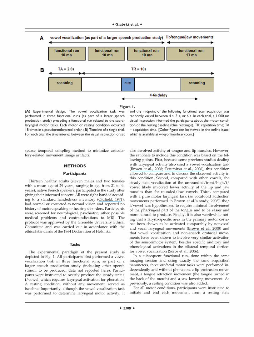
Results from the group analysis showed regions thatwere largely overlapping across the four motor conditions.The conjunction analysis revealed a bilateral set of com-mon brain areas classically involved in orofacial motorcontrol. Table I summarizes activations for each of thefour motor conditions (compared with baseline), as well asactivations provided by the conjunction analysis. Activa-tion maps are illustrated in Figure 2.
The minimal network for supralaryngeal and laryngealmovements (conjunction analysis; Table I and Fig. 2) con-cerned, bilaterally, the activation of the central sulcusextending rostrally onto the precentral gyrus and caudallyonto the postcentral gyrus. Two large bilateral clusters ofactivations enclosed the superior portion of the ventralpremotor, motor, and somatosensory cortices. In addition,two smaller bilateral clusters of activations of the precen-tral gyrus were also observed in the dorsal premotor cor-tex. Moreover, bilateral activations were found in thesupplementary motor area and the superior cerebellarhemispheres (declive region of neocerebellum). Additionalfrontal activation was observed at the border of the rightinferior frontal and middle frontal gyri. Left activationswere also observed around the ventral part of the postcen-tral sulcus, extending rostrally onto the parietal opercu-lum, as well as in the supramarginal gyrus, and in thedorsal striatum of basal ganglia (putamen).
r Grabski et al. r
r 2310 r

TA
BL
EI.
Acti
vati
on
peak
sum
mary
ob
served
ineach
mo
tor
task
(vers
us
base
lin
e)
an
din
the
co
nju
ncti
on
an
aly
sis
(ran
do
m-e
ffect
gro
up
an
aly
sis,
P<
.05,
FW
E
co
rrecte
d,
clu
ster
exte
nt
thre
sho
ldo
f10
vo
xels
,T
>5.9
1)
To
ng
ue
Vo
wel
Jaw
Lip
sC
on
jun
ctio
n
Reg
ion
HB
Ax
yz
TB
Ax
yz
TB
Ax
yz
TB
Ax
yz
TB
Ax
yz
T
Pri
mar
ym
oto
rco
rtex
L4
�20�
2858
7.48
4�
60�
418
15.4
94
�48�
1234
10.5
34
�48�
1236
8.5
74
�48�
1236
8.57
R4
56�
430
19.7
34
52�
632
14.6
74
54�
632
11.5
94
40�
1246
7.95
Pri
mar
yso
mat
ose
nso
ryco
rtex L
1,2
,3�
52�
832
21.9
51,2
,3�
52�
832
15.7
01,2
,3�
52�
832
11.3
31,2
,3�
54�
1642
9.9
71,2
,3�
50�
1640
9.31
R1,2
,344�
830
13.3
11,2
,360�
1034
8.30
Fro
nta
lre
gio
ns
Su
per
ior
fro
nta
lg
yru
sL
6�
18�
468
6.02
6�
22�
266
7.2
3
Su
pp
lem
enta
rym
oto
rar
eaL
60�
658
20.5
86
�4�
462
8.93
6�
10�
270
6.6
06
�4�
462
8.93
R6
8�
460
8.18
62�
658
13.7
86
2�
658
11.8
1
Pre
mo
tor
cort
exL
6�
620
2615
.50
6�
60�
436
11.2
16
�44�
1260
8.97
6�
44�
1260
9.0
46
�56
�4
407.
56R
654�
638
15.5
56
54�
638
11.9
96
54�
640
12.2
36
54�
640
11.9
8In
feri
or
fro
nta
lg
yru
s/L
44/9
�58
830
14.7
644/9
�58
830
8.2
2
Pre
fro
nta
lg
yru
sR
44/9
588
2816
.33
44
588
288.
0744/9
588
308.2
044/9
586
307.
36T
emp
ora
lre
gio
ns
Tra
nsv
erse
tem
po
ral
gy
rus
L41/
42�
42�
24
813
.43
41/4
2�
40�
2612
6.71
R41/
42
50�
16
612
.30
Su
per
ior
tem
po
ral
gy
rus
R22
52�
20
010
.71
Par
ieta
lre
gio
ns
Su
pra
mar
gin
alg
yru
sL
40
�38�
4454
9.24
40
�62�
3034
7.52
40
�48�
2638
9.1
7
R40
58�
26
24
8.18
40
64�
2622
8.50
Par
ieta
lo
per
culu
m/
L43
/40�
46�
2820
10.2
443
/40�
48�
2820
8.8
043
/40�
48�
2820
8.80
Su
pra
mar
gin
alg
yru
sR
43/
4052�
2820
7.31
Par
ieta
lo
per
culu
mL
43
�62
�6
1816
.84
43
�58�
610
13.9
843
�62
�6
189.
33R
43
60�
46
10.3
643
60�
1622
7.8
4
Su
per
ior
par
ieta
llo
bu
leL
7�
20�
5264
7.50
7�
20�
5264
6.7
3R
714�
6456
7.84
Su
bco
rtic
alre
gio
ns
Insu
la/
par
ieta
lo
per
culu
mR
13
3616
87.
0113
46
82
7.06
43/1
3�
38�
1218
6.96
43/1
3�
38�
1420
6.8
6
Cin
gu
late
cort
exL
32
10
14
34
7.14
24
�6
838
7.72
24
�6
838
8.2
3A
my
gd
ala
R26�
2�
88.
37P
uta
men
L�
26�
2�
413
.69
�26�
8�
610
.60
�26
�2�
47.2
8�
26�
2�
47.
28R
248
613
.56
24�
4�
26.
8426
6�
68.
00P
alli
du
mL
�24
0�
48.
69R
18�
8�
49.
0422�
10�
48.
05C
aud
ate
nu
cleu
sR
184
207.
41C
lau
stru
mR
32�
812
7.81
Th
alam
us
L�
14�
20
013
.37
�14�
202
9.35
R16�
20�
28.
2114�
18
49.
5116�
20�
26.
97C
ereb
ellu
mC
ereb
ellu
m(d
ecli
ve)
L�
16�
62�
2014
.76
�12�
64�
16
12.9
5�
16�
62�
188.
56�
16�
62�
186.9
3�
16�
62�
186.
93R
18�
60�
2013
.75
20�
60�
20
12.2
820�
60�
207.
9618�
58�
207.1
318�
58�
207.
13C
ereb
ellu
m(c
ulm
en)
R10�
58�
148.
66O
ccip
ital
reg
ion
sC
alca
rin
eg
yru
s18�
6210
8.6
6
Main Effect of Motor Tasks
The main effect of motor tasks (Table II and Fig. 3)revealed significant activity differences in the ventral pre-central and postcentral gyri (with two activation peakslocated in the ventrolateral primary motor and somatosen-sory cortices) and in the bilateral primary auditory cortex(the anterior and posterior parts of the transverse temporalgyrus) bilaterally. All these regions showed stronger acti-
vation for vowel vocalization compared with the other
tasks. As previously mentioned, due to the separation of
the motor tasks into different runs, these results have to
be interpreted with caution. Nevertheless, our results are
quite coherent with those reported in a previous study
(Soros et al., 2006), with stronger activity observed in the
auditory cortex for the vowel condition compared with
orofacial movements.
Figure 2.
Cerebral networks of lip, jaw, vocalic/laryngeal and tongue
movements, and conjunction map (random-effect group analysis,
t-contrasts, P < 0.05, FWE corrected, cluster extent threshold
of 10 voxels, T > 5.91). Significant activations are rendered on
cortical surfaces (left) and on axial slices covering the orofacial
motor cortex (right—z values refer to planes in MNI space).
The conjunction analysis revealed a bilateral set of largely over-
lapping brain areas classically involved in motor control: the sen-
sorimotor and premotor cortices, bilaterally, the right inferior
frontal gyrus, the supplementary motor area, the left parietal
operculum and the adjacent inferior parietal lobule, the basal
ganglia and the cerebellum. Due to spatial overlapping, no clear
sensorimotor somatotopy of the four articulators was observed
at the group level. [Color figure can be viewed in the online
issue, which is available at wileyonlinelibrary.com.]
r Grabski et al. r
r 2312 r

Individual Articulatory Somatotopy
As no clear sensorimotor somatotopy for the fourarticulators emerged from the group analysis, individualROI analyses within the primary motor and somatosen-sory cortices (areas 4a/p, 3a/b, 1; Geyer et al., 1996,1999) were carried out to further test at the individuallevel a sensorimotor somatotopic organization of supra-laryngeal and laryngeal activations. To this aim, MNIcoordinates in the medio-lateral (x), caudo-rostral (y),and dorso-ventral (z) dimensions of the maximum acti-vation peaks within areas BA4, BA3, and BA1 weredetermined in both hemispheres for each condition (seeTables III and IV and Fig. 4).
For all sensorimotor ROIs, almost all analyses showed asignificant effect of the hemisphere, with a more medial,posterior and dorsal position in the left hemisphere com-pared with the right hemisphere. Regarding a somatotopicorganization, a more posterior position significantlyemerged in the caudo-rostral (y) dimension for lips com-pared with tongue as well as a more dorsal position in thedorso-ventral (z) dimension for lips compared with tongueand vowel and for jaw compared with tongue.
Medio-Lateral (x) dimension
• BA4: Significant effects of the hemisphere and the artic-ulator were observed [F(1,10) ¼ 24.7; P \ 0.001; F(3,30)¼ 5.1; P \ 0.006], with a more medial position in theleft than in the right hemisphere (45 vs. 50 mm) and amore lateral position for lips and tongue comparedwith vowel and jaw (48, 49, 46, 47 mm, respectively).The interaction was not significant [F(3,30) ¼ 2.1].
• BA3: Significant effects of the hemisphere and the artic-ulator were observed [F(1,10) ¼ 20.1; P \ 0.001; F(3,30)¼ 8.7; P \ 0.001], with a more medial position in the left
than in the right hemisphere (48 vs. 53 mm) and a morelateral position for vowel and tongue compared withlips and jaw (53, 53, 48, 48 mm, respectively). The inter-action was not significant (F(3,30) ¼ 0.6).
• BA1: Significant effects of the hemisphere and thearticulator were observed [F(1,10) ¼ 21.9; P \ 0.001;F(3,30) ¼ 3.8; P \ 0.02], with a more medial positionin the left than in the right hemisphere (53 vs. 57 mm)and a more lateral position for tongue compared withlips and jaw (58, 53, 54 mm, respectively). The interac-tion was not significant [F(3,30) ¼ 1.2].
Caudo-rostral (y) dimension
• BA4: Significant effects of the hemisphere and the artic-ulator were observed [F(1,10) ¼ 31.4; P \ 0.001; F(3,30)¼ 6.6; P \ 0.001] with a more posterior position in theleft than in the right hemisphere (212 vs. 27 mm) and amore anterior position for tongue compared with lips,jaw and vowel (28, 210, 211, 210 mm, respectively).The interaction was not significant [F(3,30) ¼ 1.9].
• BA3: Significant effects of the hemisphere and the artic-ulator were observed [F(1,10) ¼ 45.9; P \ 0.001; F(3,30)¼ 12.9; P \ 0.001], with a more posterior position in theleft than in the right hemisphere (213 vs. 28 mm) and amore anterior position for vowel and tongue comparedwith lips and jaw (28, 28, 214, 212 mm, respectively).The interaction was not significant [F(3,30) ¼ 0.9].
• BA1: Significant effects of the hemisphere and thearticulator were observed [F(1,10) ¼ 19.4; P \ 0.001;F(3,30) ¼ 4.7; P \ 0.008], with a more posterior positionin the left than in the right hemisphere (211 vs. 26 mm)and a more anterior position for tongue and vowelcompared with lips (28, 27, 212 mm, respectively).The interaction was not significant [F(3,30) ¼ 0.2].
Dorso-ventral (z) dimension
• BA4: A significant effect of the articulator wasobserved [F(3,30) ¼ 7.0; P \ 0.001], with a more dorsalposition for lips, jaw, and vowel compared withtongue (38, 36, 36, 34 mm, respectively) as well as amore dorsal position for lips compared with vowel.The interaction was not significant [F(3,30) ¼ 1.2]. Theeffect of the hemisphere [F(1,10) ¼ 0.1] and the interac-tion [F(3,30) ¼ 0.8] were not significant.
• BA3: Significant effects of the hemisphere and thearticulator were observed [F(1,10) ¼ 5.3; P \ 0.05; y:F(3,30) ¼ 11.1; P \ 0.001], with a more dorsal positionin the left than in the right hemisphere (35 vs.31 mm)and a more dorsal position for lips and jaw comparedwith vowel and tongue (38, 35, 30, 31 mm, respec-tively). The interaction was not significant [F(3,30) ¼0.5].
• BA1: Significant effects of the hemisphere and the ar-ticulator were observed [F(1,10) ¼ 10.2; P \ 0.009; y:
TABLE II. Activation peak summary observed in the
main effect analysis (random-effect group analysis,
P < .05, FWE corrected, cluster extent threshold of
10 voxels, F > 17.71). Apart from the bilateral superior
temporal gyri for vowel production, the only significant
difference between the four motor tasks was observed
in the left ventro-lateral sensorimotor cortex, with
stronger activity for vowel vocalization than for the
other tasks
Region H
Main effect
BA x y z F
Primarymotor cortex
L 4 �60 �4 18 26.44
Primarysomatosensory cortex
L 3 �52 �8 32 25.66
Transversetemporal gyrus
L 41/42 �52 �16 4 24.55
R 41/42 44 �22 4 20.72
r fMRI Assessment of Orofacial Articulators r
r 2313 r

F(3,30) ¼ 7.5; P \ 0.001], with a more dorsal positionfor lips compared with vowel and tongue (42, 35, 33mm, respectively) as well as a more dorsal positionfor jaw compared with tongue (35, 33 mm, respec-tively). The interaction was not significant [F(3,30) ¼1.1].
DISCUSSION
Using sparse temporal acquisition, the goal of this fMRIstudy was to clarify the neural networks underlying motorcontrol of simple supralaryngeal and laryngeal movementsand to refine the sensorimotor somatotopic organization of
Figure 3.
Main effect of motor task (random-effect group analysis, F-con-
trast, P < 0.05, FWE corrected, cluster extent threshold of 10
voxels, F > 17.71): brain regions showing activity differences
between the four motor tasks (top) and contrast estimates
reflecting percentage BOLD signal change from baseline for the
observed activation peaks and the four motor tasks (bottom).
Compared with the other tasks, stronger activations were
observed in the left ventrolateral sensorimotor cortex and the
bilateral primary auditory cortex for vowel vocalization. SEM
are indicated in transparency. [Color figure can be viewed in the
online issue, which is available at wileyonlinelibrary.com.]
r Grabski et al. r
r 2314 r

lip, jaw, vocalic/laryngeal, and tongue motor representa-tions. Three main findings were observed. First, orofacialmovements activated a set of largely overlapping, commonbrain areas forming a core neural network involved in oro-
facial motor control. Second, apart from the auditory tem-
poral cortex for vowel production, differences between the
motor tasks were restricted to ventrolateral regions of the
primary motor and somatosensory cortices, with greater
signal intensity for vowel vocalization likely reflecting
more complex neuromuscular coordination and sensory
feedback processing. Finally, using individual ROI analy-
ses, a sequential dorso-ventral somatotopic organization of
lip, jaw, vocalic/laryngeal, and tongue movements was
observed in the primary motor and somatosensory
cortices.Before discussing these results, it is important to high-
light an important limitation of this study. As previouslynoted, although the vowel vocalization task was per-formed to determine laryngeal motor activity, it alsorequires muscle activity of the tongue and lips. In addi-tion, jaw movements likely induce tongue movements aswell given the anatomical connection of the jaw and thetongue. Hence, although this study confirms ‘‘an overallpicture of somatotopy with overlap’’ (Takai et al., 2010), amutual contribution of orofacial articulators during vowelproductions and jaw movements cannot be ruled out.
Basic Articulatory Network
The basic neural network for supralaryngeal movementsand vowel vocalization (see Table I and Fig. 2) revealedstrong bilateral activations within the sensorimotor cortex(including the primary motor, ventral and dorsal premo-tor, and somatosensory cortices), the supplementary motor
area and the superior cerebellar hemispheres. Activationswere also found in the right posterior inferior frontalgyrus (pars opercularis), the left parietal operculum andthe adjacent inferior parietal lobule, and the left dorsalstriatum of basal ganglia. The observed basic neural net-work for orofacial motor actions is fully consistent withprevious fMRI studies on orofacial motor control. Notably,common neural activity for jaw and tongue movements(Dhanjal et al., 2008) were observed in the medial and lat-eral prefrontal cortex including the ventral lateral premo-tor cortex, the frontal operculum ventral to the centralsulcus and the adjacent insular cortex, the primary motorand somatosensory cortices, the supplementary motor areaand the cerebellum (lobule VI). Lotze et al. (2000a) havepreviously reported these findings for simple tongue andlip movements (although, surprisingly, without significantactivation observed in the supplementary motor area).
Focusing on orofacial coordinated actions, our resultsalso fit well with studies dealing with swallowing andbreathing (Loucks et al., 2007; Sawczuk and Mosier, 2001),vocalization (Jurgens 2002, 2009) and overt speech produc-tion (Bohland and Guenther, 2006; Brown et al., 2005;Guenther, Ghosh and Tourville 2006; Riecker et al., 2000,2005, 2008; Soros et al., 2006). First, the neural control ofswallowing and breathing has been shown to partly relyon an overlapping neural network, including bilateral acti-vation of the primary sensorimotor cortices, the supple-mentary motor area, the thalamus, the cerebellum, thecaudate nucleus, the globus pallidum, and the medulla(Sawczuk and Mosier, 2001; see also McKay et al., 2003).In line with our results, a number of speech productionstudies have led to the suggestion of a ‘‘minimal networkfor overt speech production’’ (Bohland and Guenther,2006), including mesiofrontal structures (supplementarymotor area and anterior cingulate gyrus), bilateral pre-
TABLE III. Mean y- and z-axis values of COGs and activation peaks in MNI coordinates observed in the ROI
analyses
SensorimotorROIs
Left hemisphere Right hemisphere
Lips Jaw Vowel Tongue Lips Jaw Vowel Tongue
x y z x y z x y z x y z x y z x y z x y z x y z
Activation PeakBA 4a,p �46 �12 37 �43 �14 36 �43 �13 37 �47 �11 35 51 �8 39 48 �8 37 51 �6 35 51 �5 34BA 3a,b �46 �16 38 �46 �15 36 �50 �10 31 �50 �12 33 48 �11 37 51 �9 33 55 �6 28 56 �5 28BA 1 �52 �14 43 �52 �12 41 �53 �11 39 �57 �9 38 54 �10 41 57 �7 36 59 �5 31 60 �4 31
Meancoordinates
�48 �14 40 �47 �13 38 �49 �11 36 �51 �11 34 51 �10 39 52 �8 35 55 �6 32 55 �5 31
Center of gravity (COG)BA 4a,p �44 �11 39 �43 �12 39 �42 �12 39 �45 �10 36 47 �8 38 47 �8 37 49 �7 36 49 �6 34BA 3a,b �45 �16 39 �47 �14 36 �50 �10 31 �49 �11 33 48 �12 36 51 �9 33 55 �6 28 54 �7 28BA 1 �54 �13 44 �54 �11 42 �55 �11 40 �57 �9 38 55 �11 41 58 �7 35 60 �6 31 60 �7 33
Meancoordinates
�48 �14 40 �48 �12 39 �49 �11 37 �50 �10 36 50 �10 38 52 �8 35 55 �6 32 54 �7 32
r fMRI Assessment of Orofacial Articulators r
r 2315 r

TA
BL
EIV
.R
evie
wo
fm
ain
acti
vati
on
peaks
ob
served
inth
ese
nso
rim
oto
rm
oto
rco
rtex
du
rin
gsi
mp
leart
icu
lato
ry
gest
ure
sin
pre
vio
us
fMR
I
stu
die
s.x,y
,zco
ord
inate
sare
rep
ort
ed
inM
NI
space
Lef
th
emis
ph
ere
Rig
ht
hem
isp
her
e
Lip
sJa
wL
ary
nx
To
ng
ue
Lip
sJa
wL
ary
nx
To
ng
ue
Ref
eren
cex
yz
xy
zx
yz
xy
zx
yz
xy
zx
yz
xy
z
Co
rfiel
det
al.
(199
9)*—
ho
rizo
nta
lto
ng
ue
mo
vem
ent
�63
�7
2464
�5
19
Lo
tze
etal
.(2
000a
)*—
lip
pu
rsin
g,
ver
tica
lto
ng
ue
mo
vem
ent
�53
�18
40�
530
3555
�8
4167
�3
26
Lo
tze
etal
.(2
000b
)*—
lip
pu
rsin
g�
59�
1643
53�
1039
Rie
cker
etal
.(2
000)
—h
ori
zon
tal
ton
gu
em
ov
emen
t�
63�
330
57�
324
Sti
pp
ich
etal
.(2
002)
*—v
erti
cal
ton
gu
em
ov
emen
t�
54�
1234
53�
834
Fes
let
al.
(200
3)*—
ho
rizo
nta
lto
ng
ue
mo
vem
ent
�63
�8
3767
�8
32G
erar
din
etal
.(2
003)
*—li
pp
urs
ing
�58
�15
4561
�9
52S
hin
agaw
aet
al.
(200
3)*—
ton
gu
ep
rotr
usi
on
�48
�14
3451
�8
37S
hin
agaw
aet
al.
(200
3)*—
rig
ht
ton
gu
em
ov
emen
t�
48�
1436
51�
837
Sh
inag
awa
etal
.(2
003)
*—le
ftto
ng
ue
mo
vem
ent
�48
�14
3859
�1
24H
esse
lman
net
al.
(200
4)*—
lip
pu
rsin
g,
ho
rizo
nta
lto
ng
ue
mo
vem
ent
�41
�21
44�
55�
1227
53�
2149
58�
1330
Han
akaw
aet
al.
(200
5)*—
un
ilat
eral
lip
stre
tch
ing
�54
�8
4157
�3
40P
ulv
erm
ull
eret
al.
(200
6)—
up
/d
ow
nto
ng
ue
or
lip
mo
vem
ent
�48
�10
36�
56�
828
Ter
um
itsu
etal
.(2
006)
—to
ng
ue
mo
vem
ents
and
vo
wel
ph
on
atio
n�
40�
1942
�55
�8
3251
�11
3859
�6
30
�56
�3
21V
ince
nt
etal
.(2
006)
*—h
ori
zon
tal/
ver
tica
lto
ng
ue
mo
vem
ent
61�
126
Bro
wn
etal
.(2
008)
*—li
pp
rotr
usi
on
and
ver
tica
lto
ng
ue
mo
vem
ent
�52
�14
36�
64�
1230
58�
1234
58�
1434
Bro
wn
etal
.(2
008)
*—g
lott
alst
op
s�
38�
1634
44�
1236
Bro
wn
etal
.(2
008)
*—v
ow
elp
ho
nat
ion
�40
�12
3244
�10
36S
tip
pic
het
al.
(200
8)*—
ver
tica
lto
ng
ue
mo
vem
ent
�54
�16
4154
�11
39M
ean
coo
rdin
ates
�52
�15
41�
44�
1332
�55
�10
3456
�11
4346
�11
3758
�7
31
(*)I
fo
rig
inal
lyre
po
rted
inT
alai
rach
spac
e,co
ord
inat
esw
ere
con
ver
ted
inM
NI
spac
eu
sin
gth
eT
alai
rach
Dae
mo
nso
ftw
are
(Lan
cast
eret
al.,
2000
)an
da
no
nli
nea
rtr
ansf
or-
mat
ion
ori
gin
ally
des
crib
edb
yM
atth
ewB
rett
(ww
w.m
rc-c
bu
.cam
.ac.
uk
/Im
agin
g/
mn
isp
ace.
htm
l).
r Grabski et al. r
r 2316 r

and postcentral convolutions, extending rostrally into pos-terior parts of the inferior frontal gyrus, the left anteriorinsula as well as bilateral components of the basal ganglia(notably the putamen and the globus pallidus), the cere-bellum (notably the lobule VI, including the declive), thethalamus and the superior temporal gyrus (Bohland andGuenther, 2006; Brown et al., 2005; Guenther et al., 2006;Riecker et al., 2008; Soros et al., 2006). Finally, Chang et al.(2009) showed common activations during speech andnon-speech vocal tract gestures in the posterior inferiorfrontal gyrus, the ventral premotor cortex, the supplemen-tary motor area, the superior temporal gyrus, the insula,the supramarginal gyrus, the cerebellum and the basalganglia, a result suggesting a more general role for theseregions than just speech production. Finally, this basicneural network is also consistent with that observed dur-ing wordless singing (for a review, Brown et al., 2005) andwhistling (Dresel et al., 2005), despite differences in theamygdala, not reported for singing, and in the insula, notreported for whistling.
As the basic neural network for supralaryngeal move-ments and vowel vocalization comprises a similar set ofbrain areas, our results refine the core neural networkinvolved in speech and non-speech orofacial motor con-trol. This network encompasses the ventral sensorimotor
and ventral/dorsal premotor cortices, bilaterally, the rightposterior inferior frontal gyrus (pars opercularis), the sup-plementary motor area, the left parietal operculum and theadjacent inferior parietal lobule, the basal ganglia and thecerebellum. Although specific functions in the motor-coor-dinating neural network of these cortical and subcorticalbrain regions remain largely discussed, they can bebroadly assigned to motor preparation, execution and reg-ulation loops (for reviews, Jurgens, 2002; Riecker et al.,2008). Initiation and suppression of voluntary movementsis traditionally assigned to the supplementary motor area(Pickard and Strick, 2001) while fine-grained motor controlof orofacial gestures is carried out by the primary motorcortex and basal ganglia via the pyramidal and extrapyra-midal pathways (Jurgens, 2002; Riecker et al.; 2005; Wiseet al., 1999). The motor cortex receives motor plans fromthe premotor cortex (Pickard and Strick, 2001), and theadjacent posterior inferior frontal gyrus as well as proprio-ceptive inputs from primary and associative somatosen-sory areas and the inferior parietal cortex (Riecker et al.,2005; Smith, 1998). Importantly, although some previousstudies on speech and lip, tongue and or jaw motor con-trol failed to observe activation in the inferior frontal gyrusand/or the inferior parietal cortex (Corfield et al.,1997;Dhanjal et al., 2008; Lotze et al., 2000a; Murphy et al.,
Figure 4.
Individual sensorimotor somatotopy (ROI analysis). (A) ROIs related
to BA4a,p (red), BA3a,b (green), and BA1 (yellow). (B) Mean y- and
z-axis values of activation peaks in MNI coordinates and their
projections for the four articulators. Regarding a somatotopic orga-
nization, a more posterior position emerged in the caudo-rostral (y)
dimension for lips compared with tongue as well as a more dorsal
position in the dorso-ventral (z) dimension for lips compared with
tongue and vowel and for jaw compared with tongue. [Color figure
can be viewed in the online issue, which is available at
wileyonlinelibrary.com.]
r fMRI Assessment of Orofacial Articulators r
r 2317 r

1997; Nota and Honda, 2004; Wise et al., 1999), the presentresults and that of Chang et al. (2009) support theseregions as part of the core neural network involved inorofacial motor control. Finally, the basal ganglia play animportant role not only in the selection but also in the reg-ulation of motor commands via thalamo-motor projections(Houk et al., 2007). In parallel, the cerebellum receivesmotor and sensory information and is involved in finemuscular coordination of the intended movement (Houket al., 2007; Thach, 1998).
Main Effect of Motor Tasks
In this study, the neural networks involved in simpleorofacial movements strongly overlap and share similarcortical and subcortical brain areas. Apart from the bilat-eral superior temporal gyri for vowel production, the onlysignificant difference between the four motor tasks wasobserved in the left ventrolateral sensorimotor cortex, withstronger activity for vowel vocalization than for the othertasks (see Table II and Fig. 3).
Due to the separation of the supralaryngeal andvocalic/laryngeal motor tasks into different runs, thisresult has to be interpreted with caution. It is tempting tohypothesize that these regions might be related specificallyto laryngeal movements during vowel production. In thatcase, the closely located activation peaks observed in theindividual ROI analyses for vowel vocalization and tonguemovement (see below, section ‘‘articulatory somatotopy’’),slightly dorsal than the present activations, would likelyreflect the contribution of tongue muscles during vocaliza-tion, rather than pure laryngeal activity. On the otherhand, these two activation peaks in the left ventrolateralprimary motor and somatosensory cortices were alsofound to be activated during jaw and tongue movement(see Fig. 2), albeit at a lower extent. Although tongue re-traction movements also induce prominent sensory feed-back, an alternative interpretation is that this strongersensorimotor activity would be due to more complex sen-sorimotor interactions required for steady-state vowelvocalization. To accurately reach and maintain the precisetarget speech sound, the left ventrolateral primary motorand somatosensory cortices would be strongly engaged inonline motor control and registration of orosensory andauditory feedback (Guenther, 2006; Guenther and Bohland,2006; Guenther and Vladusich, in press).
Articulatory Somatotopy
Due to significant activation overlap between the fourarticulators, no clear sensorimotor somatotopy wasobserved at the group level. To better take into accountpossible individual anatomical and functional differencesacross participants, individual ROI analyses on the bilat-eral primary motor and somatosensory cortices were car-
ried out to evaluate individual somatotopic organizationof supralaryngeal and laryngeal articulators.
Overall, the observed coordinates for lip, jaw, tongue,and vocalic/laryngeal representations in both hemispheresand the three sensorimotor ROIs (averaged across partici-pants) fit very well with those observed in previous fMRIstudies of simple articulatory gestures (see mean coordi-nates in Tables III and IV). For all sensorimotor ROIs, amore posterior position emerged in the caudo-rostral (y)dimension for lips compared with tongue as well as amore dorsal position in the dorso-ventral (z) dimensionfor lips compared with tongue and vowel and for jawcompared with tongue. The main finding from these ROIanalyses is therefore a dorso-ventral somatotopic organiza-tion of lips, vowel, and tongue, respectively, with the sen-sorimotor representations of lip movements occupying themost dorso-caudal position and that of vocalic/laryngealand tongue movements the most ventrorostral positions,the jaw position being dorsal to the tongue position. Theseresults appear consistent with the well-established moredorsal position for the lip area than for the tongue area inthe human sensory and motor homunculi, as shown byprevious electrocortical stimulation and fMRI studies(Hesselmann et al., 2004; Lotze et al., 2000a; Penfield andRasmussen, 1950; Pulvermuller et al., 2006). In agreementwith earlier electrocortical mapping by Penfield andRasmussen (1950), we also observed the jaw area located alittle more ventral than the lip area and more dorsal thanthe larynx and tongue areas (although no significant dif-ferences were observed between lips and jaw positionsand the only significant differences between jaw andvowel was observed in BA3). A previous fMRI study alsoshowed a little more medial and dorsal position in thesensorimotor cortex for jaw (opening/closing movement)compared with that for the tongue (vertical movement),but unfortunately did not report precise activation peaksseparately for the two articulators (Dhanjal et al., 2008).Finally, the motor location of the vocalic/laryngeal move-ment was found to be located more dorsally than thetongue position. Brown et al. (2008) reported similar acti-vation peaks within the left motor cortex for both nonvocaland vocal laryngeal movements (vocal-fold abduction andvowel vocalization) and located between those observedfor lip and tongue movements. However, they report amore dorsal position for the larynx area than for bothtongue and lip areas in the right hemisphere. We notehowever that in their study the right lip and tongueactivation peak in the primary cortex are quite exactly thesame (see Tables III and IV), a result which does notcorrespond to previous studies showing the lip motor areaabove the tongue area in both hemispheres (Hesselmannet al., 2004; Lotze et al., 2000a, see Tables III and IV).Another study using both vowel vocalization and tonguemovement also observed a more dorsal position in bothhemispheres for the larynx area than for the tongue area(Terumitsu et al., 2006), although another activation peakwas found in the left primary motor cortex more ventral
r Grabski et al. r
r 2318 r

than the tongue area. Altogether, the somatotopy of lip,jaw, vocalic/laryngeal, and tongue movements displayed inthis study could be of importance for both betterspecification of speech production models and theirbrain correlates (Guenther et al., 2006) and for future brain-imaging studies investigating speech motor control and dis-orders as well as cortical reorganization following injury.
Finally, irrespective of the articulators, results alsoshowed a significant hemispheric asymmetry of sensori-motor representations, with activation sites globally moremedial, posterior and dorsal in the left than in the righthemisphere. A review of fMRI studies of simple lip,tongue, and larynx movements show a similar hemi-spheric sensorimotor asymmetry for all articulators, with amore medial and posterior position of the activation site inthe left hemisphere (see mean coordinates in Tables IIIand IV). This result might therefore suggest some anatomi-cal and/or functional asymmetry of the primary sensori-motor cortex (Sowman et al., 2009) irrespective ofparticipants’ handedness or gender (Amunts et al., 2000).
CONCLUSION
At the group level, a core functional motor network con-trolling laryngeal and supralaryngeal movements wasobserved, including the sensorimotor and premotor corti-ces, bilaterally, the right inferior frontal gyrus, the supple-mentary motor area, the left parietal operculum and theadjacent inferior parietal lobule, the basal ganglia, and thecerebellum. Apart from the auditory temporal cortex forvowel production, activity differences across motor taskswere found in the left ventrolateral sensorimotor cortexwith greater signal intensity for vowel vocalization.Finally, although no clear sensorimotor somatotopy of thefour articulators emerged at the group level due to largespatial overlapping, a sequential dorso-ventral somatotopicorganization of lips, jaw, vocalic/laryngeal, and tonguemovements was observed using individual ROI analyseswithin the primary sensory and motor cortices. Theobserved core neural network of orofacial movements andtheir sequential dorso-ventral somatotopic organizationmay be of particular interest for future brain-imaging stud-ies investigating speech motor control and disorders aswell as cortical reorganization following injury.
ACKNOWLEDGMENTS
The authors thank Vincent Gracco and two Reviewersfor their insightful and constructive comments as well asall participants of the study.
REFERENCES
Ackermann H, Riecker A (2004): The contribution of the insula tomotor aspects of speech production: A review and a hypothe-sis. Brain Lang 89:320–328.
Alkadhi H, Crelier GR, Boendermaker SH, Golay X, Hepp-Rey-mond MC, Kollias SS (2002): Reproducibility of primary motorcortex somatotopy under controlled conditions. Am J Neurora-diol 23:1524–1532.
Amunts K, Jancke L, Mohlberg H, Steinmetz H, Zilles K (2000):
Interhemispheric asymmetry of the human motor cortex related
to handedness and gender. Neuropsychologia 38:304–312.
Bohland JW, Guenther FH (2006): An fMRI investigation of sylla-ble sequence production. NeuroImage 32:821–841.
Birn RM, Bandettini PA, Cox RW, Shaker R (1999): Event-relatedfMRI of tasks involving brief motion, Human Brain Mapp7:106–114.
Brown S, Ingham RJ, Ingham JC, Laird AR, Fox PT (2005): Stut-tered and fluent speech production: An ALE meta-analysis offunctional neuroimaging studies. Hum Brain Mapp 25:105–117.
Brown S, Ngan E, Liotti M (2008): A larynx area in the humanmotor cortex. Cerebral Cortex 18:837–845.
Brown S, Laird A, Pfordresher PQ, Thelen SM, Turkeltaub P, LiottiM (2009): The somatotopy of speech. Phonation and articulationin the human motor cortex. Brain Cognition 70:31–41.
Bunton K (2008): Speech versus nonspeech: Different tasks, differ-ent neural organization. Semin Speech Lang 29:267–275.
Chang SE, Kenney MK, Loucks TM, Poletto CJ, Ludlow CL(2009): Common neural substrates support speech and non-speech vocal tract gestures. Neuroimage 47:314–325.
Corfield DR, Murphy K, Josephs O, Fink GR, Frackowiak RS, Guz
A, Adams L, Turner, Desmuget, Grethe R (1999): Cortical and
subcortical control of tongue movement in humans: A functional
neuroimaging study using fMRI. J Appl Physiol 86:1468–1477.
Dhanjal NS, Handunnetthi L, Patel MC, Wise RJ (2008): Perceptualsystems controlling speech production. J Neurosci 28:9969–9975.
Dresel C, Castrop F, Haslinger B, Wohlschlaeger AM, Hennenlot-
ter A, Ceballos-Baumann AO (2005): The functional neuroanat-
omy of coordinated orofacial movements: Sparse sampling
fMRI of whistling. Neuroimage 28:588–597.Dronkers NF (1996): A new brain region for coordinating speech
articulation. Nature 384:159–161.Ehrsson HH, Geyer S, Naito E (2003): Imagery of voluntary move-
ment of fingers, toes, and tongue activates correspondingbody-part-specific motor representations. J Neurophysiol 90:3304–3316.
Eickhoff SB, Stephan KE, Mohlberg H, Grefkes C, Fink GR,Amunts K, Zilles K (2005): A new SPM toolbox for combiningprobabilistic cytoarchitectonic maps and functional imagingdata. NeuroImage 25:1325–1335.
Fesl G, Moriggl B, Schmid UD, Naidich TP, Herholz K, YousryTA (2003): Inferior central sulcus: Variations of anatomy andfunction on the example of the motor tongue area. Neuro-Image 20:601–610.
Friston KJ, Holmes AP, Poline JB, Grasby PJ, Williams SC, Fracko-wiak RS, Turner, Desmuget, Grethe R (1995): Analysis of fMRItime-series revisited. Neuroimage 2:45–53.
Friston KJ, Holmes AP, Worsley KJ (1999): How many subjectsconstitute a study? NeuroImage 10:1–5.
Friston KJ, Holmes AP, Price CJ, Buchel C, Worsley KJ (1999):
Multisubject fMRI studies and conjunction analyses. Neuro-
Image 10:385–396.
Gaab N, Gaser C, Zaehle T, Jancke L, Schlaug G (2003): Functional
anatomy of pitch memory: An fMRI study with sparse tempo-
ral sampling. Neuroimage 19:1417–1426.
r fMRI Assessment of Orofacial Articulators r
r 2319 r

Georgopoulos AP (1995): Current issues in directional motor con-
trol. Trends Neurosci 18:506–510.
Genovese CR, Lazar NA, Nichols T (2002): Thresholding of statis-tical maps in functional neuroimaging using the false discov-ery rate. Neuroimage 15:870–878.
Gerardin E, Lehericy S, Pochon JB, Tezenas du Montcel S, ManginJF, Poupon F, Agid Y, Le Bihan D, Marsault C (2003): Foot,hand, face and eye representation in the human striatum.Cerebral Cortex 13:162–169.
Geyer S, Ledberg A, Schleicher A, Kinomura S, Schormann T,Burgel U, Klingberg T, Larsson J, Zilles K, Roland PE (1996):Two different areas within the primary motor cortex of man.Nature 382:805–807.
Geyer S, Schleicher A, Zilles K (1999): Areas 3a, 3b, and 1of human primary somatosensory cortex: 1. Microstructuralorganization and interindividual variability. NeuroImage 10:63–83.
Gracco VL, Tremblay P, Pike GB (2005): Imaging speech produc-tion using fMRI. NeuroImage 26:294–301.
Grafton ST, Woods RP, Mazziotta JC, Phelps ME (1991): Somato-topic mapping of the primary motor cortex in humans: Activa-tion studies with cerebral blood flow and positron emissiontomography. J Neurophysiol 66:735–743.
Guenther FH, Vladusich T: A neural theory of speech acquisitionand production (in press).
Guenther FH, Ghosh SS, Tourville JA (2006): Neural modelingand imaging of the cortical interactions underlying syllableproduction. Brain Lang 96:280–301.
Hall DA, Haggard MP, Akeroyd MA, Palmer AR, SummerfieldAQ, Elliott MR, Gurney EM, Bowtell RW (1999): ‘‘Sparse’’temporal sampling in auditory fMRI. Hum Brain Mapp 7:213–223.
Hamdy S, Mikulis DJ, Crawley A, Xue S, Lau H, Henry S, Dia-mant NE (1999): Cortical activation during human volitionalswallowing: An event-related fMRI study. Am J Physiol277:G219–G225.
Hanakawa T, Parikh S, Bruno MK, Hallett M (2005): Finger andface representations in the ipsilateral precentral motor areas inhumans. J Neurophysiol 93:2950–2958.
Hesselmann V, Sorger B, Lasek K, Guntinas-Lichius O, Krug B,Sturm V, Goebel R, Lackner K (2004): Discriminating the corti-cal representation sites of tongue and lip movement by func-tional MRI. Brain Topography 16:159–167.
Houk JC, Bastianen C, Fansler D, Fishbach A, Fraser D, Reber PJ,Roy SA, Simo LS (2007): Action selection and refinement insubcortical loops through basal ganglia and cerebellum. PhilosTrans R Soc Lond B Biol Sci 362:1573–1583.
Humbert IA, Robbins J (2007): Normal swallowing and functionalmagnetic resonance imaging: A systematic review. Dysphagia22:266–275.
Jurgens U (2002): Neural pathways underlying vocal control. Neu-rosci Biobehav Rev 26:235–258.
Jurgens U (2009): The neural control of vocalization in mammals:A review. J Voice 23:1–10.
Karni A, Meyer G, Rey-Hipolito C, Jezzard P, Adams MM, TurnerR, Ungerleider LG (1998): The acquisition of skilled motor per-formance: Fast and slow experience-driven changes in primarymotor cortex. Proc Natl Acad Sci U S A 95:861–868.
Lancaster JL, Woldorff MG, Parsons LM, Liotti M, Freitas CS,Rainey L, Kochunov PV, Nickerson D, Mikiten SA, Fox PT(2000): Automated Talairach atlas labels for functional brainmapping. Hum Brain Mapp 10:120–131.
Leopold NA, Daniels SK.2010. Supranuclear control of swallow-ing. Dysphagia 25:250–257.
Logothetis NK, Pauls J, Augath M, Trinath T, Oeltermann A(2001): Neurophysiological investigation of the basis of thefMRI signal. Nature 412:150–157.
Lotze M, Seggewies G, Erb M, Grodd W, Birbaumer N (2000a):The representation of articulation in the primary sensorimotorcortex. Neuroreport 11:2985–2989.
Lotze M, Erb M, Flor H, Huelsmann E, Godde B, Grodd W(2000b): fMRI evaluation of somatotopic representation inhuman primary motor cortex. Neuroimage 11:473–481.
Loucks TMJ, Poletto CJ, Simonyan K, Reynolds CL, Ludlow CL(2007): Human brain activation during phonation and exhala-tion: Common volitional Control for two upper airway func-tions. Neuroimage 36:131–143.
Lund JP, Kolta A (2006): Brainstem circuits that control mastica-tion: Do they have anything to say during speech? J CommunDisord 39(5),381–390.
Maldjian JA, Laurienti PJ, Burdette JB, Kraft RA (2003): An auto-mated method for neuroanatomic and cytoarchitectonic atlas-based interrogation of fMRI data sets. NeuroImage 19:1233–1239.
Martin RE, Goodyear BG, Gati JS, Menon RS (2001): Cerebral cort-ical representation of automatic and volitional swallowing inhumans. J Neurophysiol 85:938–950.
Martin RE, MacIntosh BJ, Smith RC, Barr AM, Stevens TK, Gati JS,Menon RS (2004): Cerebral areas processing swallowing andtongue movement are overlapping but distinct: A functionalmagnetic resonance imaging study. J Neurophysiol 92:2428–2443.
Martin R, Barr A, MacIntosh B, Smith R, Stevens T, Taves D, GatiJ, Menon R, Hachinski V (2007): Cerebral cortical processing ofswallowing in older adults. Exp Brain Res 176:12–22.
McKay LC, Evans KC, Frackowiak RS, Corfield DR (2003): Neuralcorrelates of voluntary breathing in humans. J Appl Physiol95:1170–1178.
MacNeilage PF (1998): The frame/content theory of evolution ofspeech production. Behav Brain Sci 21:499–546.
Murphy K, Corfield DR, Guz A, Fink GR, Wise RJ, Harrison J,Adams L (1997): Cerebral areas associated with motor controlof speech in humans. J Appl Physiol 83:1438–1447.
Nakamura Y, Katakura N (1995): Generation of masticatoryrhythm in the brainstem. Neurosci Res 23:1–19.
Nota Y, Honda K (2004): Brain regions involved in motor controlof speech. Acoust Sci Technol 25:286–289.
Nichols T, Hayasaka S (2003): Controlling the familywise errorrate in functional neuroimaging: A comparative review. StatMethods Med Res 12:419–446.
Oldfield RC (1971): The assessment and analysis of handedness:The Edinburgh inventory. Neuropsychologia 9:97–114.
Onozuka M, Hirano Y, Tachibana A, Kim W, Ono Y, Sasaguri K,Kubo K, Niwa M, Kanematsu K, Watanabe K (2007): Interac-tions between chewing and brain activity in humans. In Ono-zuka M, Yen CT, editors. Novel Trends in Brain Science,Springer. pp 99–113.
Ozdemir E, Norton A, Schlaug G (2006): Shared and distinct neu-ral correlates of singing and speaking. NeuroImage 33:628–635.
Peeva MG, Guenther FH, Tourville JA, Nieto-Castanon A, AntonJL, Nazarian B, Alario FX (2009): Distinct representations ofphonemes, syllables, and supra-syllabic sequences in thespeech production network. Neuroimage 50:626–638.
Penfield W, Boldrey E (1937): Somatic motor and sensory repre-sentation in the cerebral cortex of man as studied by electricalstimulation. Brain 60:389–443.
r Grabski et al. r
r 2320 r

Penfield W, Rasmussen T. 1950. The Cerebral Cortex of Man.New York: Macmillan.
Pickard N, Strick P (2001): Imaging the premotor areas. Curr OpinNeurobiol 11:663–672.
Pulvermuller F, Huss M, Kherif F, Moscoso del Prado Martin F,Hauk O, Shtyrov Y (2006): Motor cortex maps articulatory fea-tures of speech sounds. PNAS 103:7865–7870.
Rao SM, Binder JR, Hammeke TA, Bandettini PA, Bobholz JA,Frost JA, Myklebust BM, Jacobson RD, Hyde JS (1995): Somato-topic mapping of the human primary motor cortex with func-tional magnetic resonance imaging. Neurology 45:919–924.
Riecker A, Ackermann H, Wildgruber D, Meyer J, Dogil G, HaiderH, Grodd W (2000a): Articulatory/phonetic sequencing at thelevel of the anterior perisylvian cortex: A functional magneticresonance imaging (fMRI) study. Brain Lang 75:259–276.
Riecker A, Ackermann H, Wildgruber D, Dogil G, Grodd W(2000b): Opposite hemispheric lateralization effects duringspeaking and singing at motor cortex, insula and cerebellum.Neuroreport 11:1997–2000.
Riecker A, Mathiak K, Wildgruber D, Erb M, Hertrich I, Grodd W,Ackermann H (2005): fMRI reveals two distinct cerebral net-works subserving speech motor control. Neurology 64:700–706.
Riecker A, Brendel B, Ziegler W, Erb M, Ackermann H (2008):The influence of syllable onset complexity and syllable fre-quency on speech motor control. Brain Lang 107:102–113.
Sawczuk A, Mosier KM (2001): Neural control of tongue move-ment with respect to respiration and swallowing. Crit RevOral Biol Med 12:18–37.
Schulz GM, Varga M, Jeffires K, Ludlow CL, Braun AR (2005):Functional neuroanatomy of human vocalization: An H215OPET study. Cereb Cortex 15:1835–1847.
Shinagawa H, Ono T, Ishiwata Y, Honda E, Sasaki T, Taira M,Iriki A, Kuroda T (2003): Hemispheric dominance of tonguecontrol depends on the chewing-side preference. J Dental Res82:278–283.
Simonyan K, Ostuni J, Ludlow CL, Horwitz B (2009): Functionalbut not structural networks of the human laryngeal motor cor-tex show left hemispheric lateralization during syllable but notbreathing production. J Neurosci 29:14912–14923.
Smith A (1998): The control of orofacial movements in speech.Crit Rev Oral Biol Med 3:233–267.
Smotherman MS (2007): Sensory feedback control of mammalianvocalizations. Behav Brain Res 182:315–326.
Sowman PF, Flavel SC, McShane CL, Sakuma S, Miles TS, Nord-strom MA (2009): Asymmetric activation of motor cortex con-trolling human anterior digastric muscles during speech andtarget-directed jaw movements. J Neurophysiol 102:159–166.
Soros P, Sokoloff LG, Bose A, McIntosh AR, Graham SJ, Stuss DT(2006): Clustered functional MRI of overt speech production.NeuroImage 32:376–387.
Stippich C, Ochmann H, Sartor K (2002): Somatotopic mapping ofthe human primary sensorimotor cortex during motor imageryand motor execution by functional magnetic resonance imag-ing. Neurosci Lett 331:50–54.
Stippich C, Blatow M, Durst A, Dreyhaupt J, Sartor K (2007):Global activation of primary motor cortex during voluntarymovements in man. Neuroimage 34:1227–1237.
Takai,O, Brown,S, Liotti,M (2010): Representation of the speecheffectors in the human motor cortex: Somatotopy or overlap?Brain Lang 113:39–44.
Talairach J, Tournoux P.1988. Co-planar Stereotaxic Atlas of theHuman Brain. New York: Thieme Medical Publishers.
Terumitsu M, Fujii Y, Suzuki K, Kwee IL, Nakada T (2006):Human primary motor cortex shows hemispheric specializa-tion for speech. Neuroreport 17:1091–1095.
Thach WT (1998): A role for the cerebellum in learning movementcoordination. Neurobiol Learn Mem 70:177–188.
Vincent DJ, Bloomer CJ, Hinson VK, Bergmann KJ (2006): Therange of motor activation in the normal human cortex usingbold FMRI. Brain Topogr 18:273–280.
Watanabe J, Sugiura M, Miura N, Watanabe Y, Maeda Y, MatsueY, Kawashima R (2004): The human parietal cortex is involvedin spatial processing of tongue movement: An fMRI study.NeuroImage 21:1289–1299.
Wildgruber D, Ackermann H, Klose U, Kardatzki B, Grodd W(1996): Functional lateralization of speech production at pri-mary motor cortex: A fMRI study. Neuroreport 7:2791–2795.
Wise RJ, Greene J, Buchel C, Scott SK (1999): Brain regionsinvolved in articulation. Lancet 353:1057–1061.
Yetkin FZ, Haughton VM, Cox RW, Hyde J, Birn RM, Wong EC,Prost R (1996): Effect of motion outside the field of view onfunctional MR. Am J Neuroradiol 17:1005–1009.
r 2321 r
r fMRI Assessment of Orofacial Articulators r
Copyright © 2022 FDOKUMEN