IL6 Contributes to the Expression of RAGs in Human Mature B Cells
Upload
independentCategory
view
2download
0
Functional domains of the 50S subunit mature latein the assembly processAhmad Jomaa1y Nikhil Jain2y Joseph H Davis3y James R Williamson3
Robert A Britton2 and Joaquin Ortega1
1Department of Biochemistry and Biomedical Sciences and MG DeGroote Institute for Infectious DiseasesResearch McMaster University 1280 Main Street West Hamilton Ontario L8S4K1 Canada 2Department ofMicrobiology and Molecular Genetics Michigan State University East Lansing MI 48824 USA and3Department of Integrative Structural and Computational Biology Department of Chemistry and The SkaggsInstitute for Chemical Biology The Scripps Research Institute La Jolla CA 92037 USA
Received September 16 2013 Revised November 17 2013 Accepted November 21 2013
ABSTRACT
Despite the identification of many factors that facili-tate ribosome assembly the molecular mechanismsby which they drive ribosome biogenesis are poorlyunderstood Here we analyze the late stages ofassembly of the 50S subunit using Bacillus subtiliscells depleted of RbgA a highly conserved GTPaseWe found that RbgA-depleted cells accumulate lateassembly intermediates bearing sub-stoichiometricquantities of ribosomal proteins L16 L27 L28 L33aL35 and L36 Using a novel pulse labelingquantitativemass spectrometry technique we show that thisparticle is physiologically relevant and is capable ofmaturing into a complete 50S particle Cryo-electronmicroscopy and chemical probing revealed that thecentral protuberance the GTPase associating regionand tRNA-binding sites in this intermediate areunstructured These findings demonstrate that keyfunctional sites of the 50S subunit remain unstruc-tured until late stages of maturation preventing theincomplete subunit from prematurely engaging intranslation Finally structural and biochemicalanalysis of a ribosome particle depleted of L16indicate that L16 binding is necessary for the stimu-lation of RbgA GTPase activity and in turn release ofthis co-factor and for conversion of the intermediateto a complete 50S subunit
INTRODUCTION
In bacteria ribosome biogenesis requires the synthesisfolding chemical modification and assembly of 3 large
RNAs and 55 proteins This complex process is completedrapidly and efficiently with bacterial cells synthesizing upto 50 000 ribosomes per generation (1) Several decadesago the groups of Nomura (2ndash4) and Nierhaus (56)determined the thermodynamic binding dependencies forribosomal proteins (r-proteins) in the 30S and 50S bacter-ial subunits and used these data to reconstruct assemblymaps depicting r-protein binding These assembly mapssuggest that assembly proceeds in stages with someproteins binding directly to the RNA while othersrequire the pre-binding of other r-proteinsMore recently work from the Williamson (7) and
Woodson (8) laboratories established that assembly ofthe 30S subunit does not flow through a single pathwaybut instead utilizes multiple alternative pathways eachexhibiting hierarchical binding as predicted by Nomuraet al Although similar in vitro studies have not beenreported for the 50S subunit the existence of multipleparallel pathways has also been suggested from in vivostudies (9) Overall the assembly of the 50S subunit isless understood than that of the 30S subunit due to itslarger number of components more intricate structureand the lack of simple highly efficient in vitro reconstruc-tion protocolIn the cell 50S ribosomal subunit assembly is assisted
by a variety of protein factors including a critically im-portant class of GTPases (10) While the precise roles ofmany of these factors are still unknown genetic studiessuggest that these GTPases act late in 50S assembly(11ndash14) It is hypothesized that during ribosomalassembly these GTPases may facilitate proteinndashRNAinteractions participate in rRNA processing or otherwiseensure that the assembly process progresses quickly andefficiently by limiting the rRNA folding landscape andpreventing the assembling subunit from entering kinetic
To whom correspondence should be addressed Tel +1 905 525 9140 (ext 22703) Fax +1 905 522 9033 Email ortegajmcmastercaCorrespondence may also be addressed to Robert A Britton Tel +1 517 884 5395 Email rbrittonmsuedu
yThese authors contributed equally to the paper as first authors
Published online 13 December 2013 Nucleic Acids Research 2014 Vol 42 No 5 3419ndash3435doi101093nargkt1295
The Author(s) 2013 Published by Oxford University PressThis is an Open Access article distributed under the terms of the Creative Commons Attribution Non-Commercial License (httpcreativecommonsorglicensesby-nc30) which permits non-commercial re-use distribution and reproduction in any medium provided the original work is properly cited For commercial re-use please contact journalspermissionsoupcom
traps common in the folding of large RNA molecules Bycoupling their enzymatic activity to guanine nucleotideconcentrations in the cell these enzymes may alsoprovide cells with a rapid and direct mechanism to shutdown ribosome biogenesis in response to decreases incellular energy levels (1011)The focus of this study was to gain insights into the
events occurring during the late stages of 50S subunitassembly and to understand the role of the essential mat-uration factor RbgA in this process This protein (alsoknown as YlqF) is a widely conserved GTPase found inall three kingdoms of life Although Escherichia coli lacksan RbgA homolog this protein is found in most Gram-positive and Gram-negative bacteria as well as in alleukaryotes (11)Cells depleted of RbgA grow at a significantly decreased
rate exhibit dramatically reduced levels of 70S ribosomesand completely lack 50S particles Instead they accumu-late a large subunit intermediate that migrates as a 45Sparticle in sucrose gradients (1115) The 45S particleexhibits disordered functional centers as visualized byelectron microscopy and is severely depleted of thetertiary-binding r-proteins L16 L27 and L36 (111516)suggesting it may be a late stage assembly intermediateof the 50S subunit Depletion of other highly conservedGTPases including ObgE YsxC (YihA) and YphC(EngA or Der) (12ndash1417) also causes the accumulationof immature large ribosomal subunits However it is pres-ently unknown whether any of these ribosomal particlesare physiological assembly intermediates competent forassembly or instead simply terminal dead-end productsWe hypothesized that if the 45S particles from RbgA-
depleted Bacillus subtilis cells are in fact competent forassembly their analysis could elucidate the conform-ational changes occurring during the late stages of largeribosomal subunit maturation Such an analysis could alsoreveal how RbgA assists this process and would constitutean important first step toward characterizing any func-tional interplay between RbgA and the other assemblyfactor GTPasesTo this end we performed pulse-labeling experiments
and determined that the 45S particle could mature intofunctional 70S particles Quantitative mass spectrometry(qMS) and cryo-EM experiments revealed that the 45Sparticles lacked several r-proteins and structurally ex-hibited substantial distortion in multiple key functionalsites of the ribosome These results suggest that the func-tional core is the last region of the 50S subunit to matureduring the assembly process and provide a mechanisticexplanation to how the cell ensures that assembly iscomplete before large ribosomal particles are allowed toinitiate translation We additionally provide evidence thatL16 plays a key role in this late structural rearrangementOur results indicate that the entry of this r-protein directlyor indirectly requires RbgA activity and its bindingtriggers most of the conformational changes that the 45Sparticle undergoes to become a mature 50S subunit Inaddition binding of L16 to the maturing large ribosomalsubunit also triggers the GTPase activity of RbgA and thesubsequent release of this assembly factor
MATERIALS AND METHODS
Ribosome and 50S subunit purifications
The 50S and 45S subunits from Bacillus subtilis werepurified from IF2-depleted (RB419) and RbgA-depleted(RB301) strains respectively Construction of these twostrains has been previously described (11) In thesecells transcription of infB (encoding IF2) and ylqF(encoding RbgA) is under the control of an isopropyl-beta-D-thiogalactopyranoside (IPTG) inducible Pspank
promotor To minimize the probability of genetic suppres-sors arising strains were first grown at 37C on LB agarplates containing 5 mgml of chloramphenicol and 1mMIPTG To initiate depletion cells were then scraped offthe plate and resuspended in 1ml of LB media which wasthen used to inoculate 50ml of pre-warmed LB medialacking IPTG and cultures were grown to an OD600 of005 Cultures were incubated at 37C with agitation untilthey reached 150min doubling time Cells were notallowed to grow beyond an OD600 of 05 thus if at thispoint cells were still exhibiting doubling times 150minthen the culture was diluted again in 50ml of new freshpre-warmed LB media to an OD600 of 005 Typically twocycles of growth dilution were required before the cellsreached 150min doubling time At that point cultureswere used to inoculate 1 l of pre-warmed media to anOD600 of 005 Full depletion of the IF2 or RbgAproteins was obtained in cells with a doubling time of120ndash150min At OD600 04ndash05 these cells wereharvested via centrifugation at 8500 g for 15min Cellpellets were processed as previously described (18) toobtain the crude ribosomes with the only exception thatbuffers contained 15mM magnesium acetate (versus10mM) To obtain the 50S or 45S fraction the cruderibosome pellet was resuspended in buffer containing10mM TrisndashHCl at pH 75 15mM magnesium acetate60mM NH4Cl and 2mM 2-mercaptoethanol (non-dissociating conditions) About 50ndash60 A260 units of thesubunit suspension were layered onto a 32-ml 10ndash30(wtvol) sucrose gradient made up in the same bufferand centrifuged at 43 000 g for 16 h using a BeckmanSW32 Ti rotor Gradients were fractionated using aBrandel fractionator apparatus and an AKTAprimeFPLC system (GE Healthcare) The elution profile wasmonitored by UV absorbance at A260 and fractions cor-responding to the 50S or 45S subunit peaks were collectedand pooled Subunits were removed from the sucrosebuffer by centrifugation at 100 000 g for 16 h and thepellet resuspended in storage buffer (10mM TrisndashHClpH 75 10mM magnesium acetate 60mM NH4Cl3mM 2-mercaptoethanol) and kept at 80C
L16-depleted 50S subunits were obtained by performingthe cell lysis step in higher volume In this case cell pelletsfrom 200ml cultures were first washed with buffer con-taining 10mM TrisndashHCl pH 75 10mM MgCl2 and60mM KCl Subsequently cell pellets were resuspendedin 10ml of lysis buffer (versus 25ml for control 50Ssubunit) containing 10mM TrisndashHCl pH 75 10mMMgCl2 60mM KCl 1mM DTT 05 Tween 20 (vv)protease inhibitor cocktail (Roche) and 27 units of RNAsefree DNAse (Qiagen) Cell lysis was performed by three
3420 Nucleic Acids Research 2014 Vol 42 No 5
consecutive passes of the cells suspension through a cellpressure cell at 1200ndash1400 psi Lysate was clarified bycentrifuging at 16 000 g for 20min Crude ribosomeswere pelleted by centrifugation at 166 880 g for 2 h and25min and the pellet was dissolved in 1ml of HMA 06buffer (20mM TrisndashHCl pH 75 10mM MgCl2 30mMNH4Cl and 1mM DTT) Subsequently 1ml of high saltHMA 08 buffer (20mM TrisndashHCl pH 75 10mM MgCl2800mM NH4Cl and 1mM DTT) was added and themixture was incubated on ice for 1 h Then ribosomeswere pelleted again by centrifugation at 166 880 g for 2 hand 25min and resuspended in 500 ml of HMA06 bufferbefore loading them onto a 35-ml 18ndash43 sucrosegradient made in buffer 10mM TrisndashHCl pH 7510mM MgCl2 50mM NH4Cl and 1mM DTTGradients were centrifuged at 81 991 g for 14 h and frac-tionation was monitored by absorbance at A260 The peakcorresponding to 50S subunit was concentrated andbuffer was exchanged to buffer containing 10mM TrisndashHCl pH 75 10mM MgCl2 60mM KCl using AmiconUltra-15 centrifugal concentrators (Millipore)
Quantitative mass spectrometry
15N-labeled 70S ribosomes were prepared by growingB subtilis ATCC 6051 strain aerobically in 1 l MSpitz9medium (80mM K2HPO4 44mM KH2PO4 039mMNa3-citrate 10mM MgCl2 10mM MgSO4 56mMglucose 50 mM Na3middotEDTA 25mM CaCl2 50 mMFeCl3 05 mM ZnSO4 05 mM CuSO4 05 mM MnSO405 mM CoCl2 004 mM d-biotin 002 mM folic acid008 mM vitamin B1 011 mM calcium pantothenate04 nM vitamin B12 02 mM nicotinamide 007 mM ribo-flavin and 76mM (15NH4)2SO4) at 37
C Cells were har-vested at OD600 04 by directly adding the culture to 1 lof ice After centrifugation at 6000 g for 20min cells wereresuspended in buffer containing 10mM Tris (pH 78)20mM MgCl2 100mM NH4Cl 6mM 2-mercaptoethanoland lysed using a BioSpec mini-bead beater The lysatewas clarified by centrifugation (30 000 g for 40min) andloaded onto a 10ndash40 (wv) sucrose gradient made upin the same buffer The gradient was then centrifuged at4C in a Beckman SW32 Ti rotor at 83 000 g for 28 hFractions were collected as described above and thosecontaining 70S particles were pooled and stored at 4C
14N-labeled 50S subunits and 45S particles producedand purified as described above were prepared forLCMS analysis using published protocols (19) Briefly20 pmol of 15N-labeled 70S spike was mixed with eachexperimental sample (20 pmol) before addition of tri-chloroacetic acid (TCA) to a final concentration of 13(vv) Samples were incubated on ice overnight andprecipitated material was isolated by centrifugationwashed with 10 TCA followed by acetone and finallyresuspended in 40 ml buffer B (100mM NH4CO3 5acetonitrile and 5mM dithiotreitol) After incubation for10min at 65C 10mM iodoacetamide was added andsamples were incubated at 30C for 30min Sampleswere then digested using 02 mg trypsin at 37C overnightPeptides were injected onto a C18 column and were elutedover 105min across a concave 5ndash50 acetonitrile
gradient and detected on an Agilent G1969A ESI-TOFmass spectrometer using a detection range set at250ndash1300mz Unique ribosomal peptides were identifiedin the raw data using a custom suite of software designedto identify 14N15N peak pairs given a theoretical digest(20) For each peptide the quantities of 14N or 15N specieswere calculated by fitting the isotope distribution using aLeast Square Fourier Transform Convolution algorithm(21) The relative protein level for each r-protein wascalculated as the 14N fitted isotope distribution amplitudedivided by the sum of the 14N and 15N fitted isotope dis-tribution amplitudes [14N(14N+15N)] Each isotope dis-tribution and its local chromatographic contour map werecarefully examined and fits with low signal-to-noise ratioswere excluded To account for differences in the totalamount of each experimental sample analyzed eachrelative protein level was normalized to that of L24 aprimary binding r-protein shown to be present in stoichio-metric quantities in each sample analyzedTo improve protein coverage and quantification
accuracy samples were run multiple times and averagedacross runs Because proteins that are completely absentfrom an experimental sample cannot be identified by the14N15N peak pair each experimental sample was alsoanalyzed on a ABSciex 5600 Triple-TOF run in MS2
mode Peptides were eluted from analytical C18 nano-column across a 120-min linear 5ndash40 acetonitrilegradient at 300 nlmin Precursor ions were detected in a400ndash1250mz mass window and product ions weredetected with a 100ndash1800mz mass window Precursorpeptides were identified from the fragmentation datausing Mascot (Matrix Science) and relevant MS1 scanswere inspected to quantify the 14N15N species asdescribed above
Pulse-labeling experiments and quantitation of r-proteinlabeling kinetics
In these experiments the RbgA-depleted cells were grownexponentially in 14N-labeled minimal media in thepresence of limiting inducer (10 mM IPTG) Control cellswere grown in the presence of 1mM IPTG During mid-log phase growth the cells were pulsed by the addition ofan equal volume of 15N-labeled media Samples weretaken at multiple time points over a 80-min period andrapidly frozen for storage After lysis either 45S or 70Sparticles were purified on a sucrose gradient mixed with a15N-labeled 70S internal standard and the mixture ofproteins were prepared for mass spectrometry analysis asdescribed above For each peptide three isotope envelopeswere observed corresponding to proteins synthesizedbefore the pulse (0 14N lowest mz) after the pulse(50 15N middle mz) or the internal standard (10015N highest mz) The fraction of material labeledwhich was calculated as [50 15N(50 15N+10014N)] increased monotonically as a function of time con-sistent with proteins synthesized after the pulseconstituting a greater fraction of the 70S particles astime progresses To ease comparison between the 1mMand 10 mM IPTG conditions each time course was scaledby the corresponding growth rate For each protein the
Nucleic Acids Research 2014 Vol 42 No 5 3421
pulse-labeling time course was fit to Equation (1) asdescribed in Chen et al (22)
frethtTHORN frac14 1+P exp k 1+1
P
t
eth1+PTHORN exp k tfrac12
eth1THORN
where P is the pool size t is the time k is the growth rateand fr is the fraction labeled and P is the only free par-ameter The max lab curves were calculated usingEquation (2)
fmaxethtTHORN frac14 1 expfrac12k t eth2THORN
Cryo-electron microscopy
Ribosomal subunits were diluted to a concentration of16 nM in storage buffer and 36 ml of this dilution wasapplied in freshly coated holey carbon grids with an add-itional layer (5ndash10 nm) of thin carbon Before the samplewas applied the grid was glow discharged at 5mA for 15 sVitrification of the specimen was performed in a Vitrobot(FEI) by blotting the grids twice 7 s each time before theywere plunged into liquid ethane Grids were transferred toa JEOL 2010F electron microscope operated at 200 kVusing a Gatan 914 cryo-holder Micrographs were col-lected at a nominal magnification of 50 000 and arange of defocus from 15 to 39 mm and digitalizedwith a step size of 127 mm in a Nikon Supercool Scan9000 The sampling of the images was 254 Apixel
Image processing
Projection images of the ribosomal subunits were pickedmanually using Boxer (23) The total of number of par-ticles picked for the mature 50S subunit 45S particles andL16-depleted 50S subunit were 53 053 76 499 and 51 016particles respectively The contrast transfer functionof the micrographs was estimated using CTFFINDsoftware (24) and corrected using the Xmipp softwarepackage (25)The three-dimensional (3D) reconstructions of the
mature 50S subunit were calculated using 3D projectionmatching alignment procedures as implemented in theXmipp software package (25) The reference map usedto refine all three reconstructions was the X-ray structureof the E coli 50S subunit (PDB ID 2AW4) (26) low-passfiltered to 25 A resolution The resulting map wassharpened using a B-factor calculated using a previouslydescribed method (27) implemented in the Xmippsoftware package (28)The dataset of immature 45S subunits was first sub-
jected to a maximum likelihood-based classificationapproach following previously published protocols (29ndash32) The 3D reconstruction of the mature 50S subunitlow-pass filtered to 70 A was used as an initial referenceto produce four seeds from random subsets of experimen-tal images by performing one iteration of maximumlikelihood optimization Using these four seeds we per-formed 30 iterations of maximum likelihood-based classi-fication This approach rendered four homogeneoussubpopulations of particles Increasing the number of
classes in the ML3D classifications produced classes withsimilar features The total number of particles in each classwas 20 269 10 990 21 178 and 24 059 The 3D reconstruc-tions of the four conformational populations of the 45Sparticle were obtained from these groups of particles usingprojection matching approaches The reference maps forthe projection matching refinements were produced fromthe particles in each subclass using a maximum likelihood-based refinement approach (3033) to prevent model bias
In the case of the L16-depleted 50S subunit particleimages in the dataset were first used to produce aninitial reference map using a maximum likelihood basedrefinement approach (3033) The initial seed for this pro-cedure was the cryo-EM map of B subtilis IF2-depleted50S subunit low-pass filtered to 50 A The 3D structurewas refined against this initial model using a projectionmatching approach (25)
To estimate the resolution of the 3D reconstructionseven and odd-numbered particles from the last cycle ofrefinement were used to produce two 3D maps fromwhich the Fourier Shell Correlation plot was calculatedResolution values were obtained from this plot using the05 criteria and these values were used to low-pass filterthe corresponding 3D reconstructions
Visualization and docking of X-ray structures into thecryo-EM maps was done using the Chimera program (34)
Chemical probing
Chemical probing of the 23S rRNA was performed withdimethyl sulfate (DMS) and kethoxal DMS methylatesspecifically unpaired and exposed adenine and cytosinein RNA while kethoxal methylates gunanine Thesite of methylation is subsequently identified by reversetranscription of the RNA using fluorescent primersA minimum cutoff of at least a 2-fold difference inchemical reactivity was employed to select bases thathad a significant change in structure when comparingthe 45S particle and 50S subunit We also required thatthese changes were observed in a minimum of threebiological replicates of the experiment and report thearithmetic mean and standard error for each base Tostatistically test the significance of the data we log trans-formed the ratios and calculated the geometric mean andstandard deviation and performed a one-sample two-tailed Studentrsquos t-test
Approximately 100 pmol of 50S subunits or 45S particleswere incubated with 4ml of 088M DMS solution preparedin 95 ethanol Reactions were incubated on ice for1 h and stopped by adding DMS stop solution containing1 M TrisndashHCl at pH 75 01 M EDTA and 1 M2-mercaptoethanol DMS modified subunits wereprecipitated by addition of subunit precipitation buffer(85 ethanol 01 M Na-acetate and 25mgml glycogen)For kethoxal modification 100pmol of ribosomalsubunits were incubated with 25ml of a 25M kethoxalsolution on ice for 1 h In this case reactions wereterminated by addition of subunit precipitation buffer(85 ethanol 01M Na-acetate and 25mgml glycogen)supplemented with 25mM potassium borate All furthersolution used for kethoxal treated rRNA had 25mM
3422 Nucleic Acids Research 2014 Vol 42 No 5
potassium borate to maintain the stable modification Thevolume of all reactions was 100ml Subsequent to thechemical modification the 23S rRNA from the ribosomalsubunits was prepared by ethanol precipitation and phenolextraction Precipitation was obtained by centrifugation at16 000 g for 20min after addition of 1ml of subunit precipi-tation buffer Pellets containing the ribosomal subunitswere dissolved in RNA extraction buffer (05 SDS5mM EDTA pH 8 03 M Na-acetate) Proteins wereremoved by two rounds of PCI solution (phenol chloro-form Isoamylalcohol 25241) treatment and traces ofphenol were removed by chloroform treatment RNA wasprecipitated from the aqueous layer by using 100 ethanolThe RNA pellet was air dried and dissolved in water
The reverse transcriptase reaction was carried out using50 6-carboxyfluorescein (6-FAM) labeled primers listed inSupplementary Table S3 that were complementary to aportion of the 23S rRNA sequence Reactions containing5 ml of 03 mM modified rRNA 2 ml of 1 mM 50 6-FAMlabeled primer and 2 ml of 45 hybridization buffer(225mM HEPES pH 7 450mM KCl) wereincubated for 5min at 75C A volume of 11 ml ofMaster mix containing 05 ml (100 units) Reverse tran-scriptase (Superscript III reverse transcriptase from Lifetechnologies) 05 ml of RNase Inhibitor (Roche) 1 ml10mM dNTP 4 ml 5 buffer 1 ml 100mM DTT and 4 mlwater was added to the reaction Reactions were incubatedat 55C for 1 h Then 2 ml chase solution (1mM dNTP)was added to the reaction and incubated for another30min Then 2 units of RNase H was added to the reac-tions and incubated at 37C for 1 h Reactions werecleaned using DTR edgebio ultra gel filtration cartridgeand incubated with 5 ml of formamide for 2min at 70CThen they were rapidly transferred to ice Sequencing wasdone on ABI PRISM 3130 Genetic Analyzer (AppliedBiosystems) and sequence analysis was carried out usingABI peak-scanner software (Applied Biosystems) Morethan 2-fold difference in modification between the 50Ssubunit and 45S particle was considered for furtheranalysis To eliminate peaks due to spontaneous reversetranscriptase stops the difference cut off between controland modified j45S R-50S Rj versus j45S C-50SCjgt 5-foldwas considered as genuine modification The modifica-tions reported here were observed in a minimum of threebiological replicates of the experiment To find theposition of a modified base it was compared withdi-deoxysequencing reaction carried out using cDNA of23S rRNA using Thermo Sequenase Cycle Sequencing Kit(Affymetrix) using fluorescent primer and positions weredetermined within three bases Nucleotide bases weremapped on the 23S rRNA secondary structure map ofB subtilis which was obtained from comparative RNAWeb (CRW) Site and project (httpwwwrnaccbbutexasedu) Corresponding bases in B subtilis weremapped in the X-ray structure of the 50S subunit fromE coli (PDB ID 2AW4) using Chimera (34)
GTPase activity assays
The GTPase activity of RbgA in the presence of mature50S subunit 45S particle and L16-depleted 50S subunits
was determined by incubating the protein with GTPand then measuring released free phosphate by the mal-achite greenammonium molybdate colorimetric assay(BioAssays Systems) according to manufacturer proto-cols RbgA protein for these experiments was expressedand purified as described (11) Mixtures contained 05 mMof RbgA and the same concentration of ribosomalsubunits in reaction buffer (50mM TrisndashHCl pH 75250mM KCl and 5mM MgCl2) were incubated at 37Cfor 30min Reaction was started by adding GTP to a finalconcentration of 200 mM At this GTP concentration theobtained values are within the linear range of the assay(35) The GTPase activity obtained for the RgbA proteinby itself was defined as 1 and the GTPase activity forRbgA in the presence of each ribosomal subunits wasexpressed and plotted with respect to this standard
RESULTS
The 45S particle from RbgA-depleted B subtilis cellslacks several late-binding r-proteins
The 45S particles analyzed in this study were obtainedby depleting B subtilis cells of RbgA using a strain inwhich the rbgA gene was under the control of the IPTG-inducible Pspank promoter (RB301) (11) In the presenceof 1mM IPTG these cells produced a ribosome profileindistinguishable from wild-type cells (SupplementaryFigure S1 bottom panel) In contrast withholdingIPTG caused depletion of RbgA and led to a decreasein the levels of 70S ribosomes and the accumulation offree 45S particles (Supplementary Figure S1 top panel)(1115)We analyzed the protein complement of the 45S par-
ticles by qMS Cells were grown in 14N-labeled mediaand 45S particles were purified under non-dissociatingconditions (see Materials and Methods) that preventedcontamination with mature 50S subunits (SupplementaryFigure S1 top panel) These purified 45S particles werethen spiked with a fixed concentration of 70S particlespurified from cells grown in 15N-labeled media Due tothe isotope shift that results from growth in 15N-labeledmedia we could independently quantify the r-proteinlevels in the experimental sample (45S from RbgA-depleted cells) and the reference sample (70S particlesfrom wild-type cells)We also quantified the protein composition of 50S
subunits purified under identical conditions from IF-2depleted cells (RB419) to allow for a direct comparisonbetween immature (45S) and mature particles (50S) Wefound that multiple proteins were completely absent (L16L28 and L36) or dramatically depleted (L27 L33a andL35) from 45S particles (Figure 1A and B red) All sixof these proteins displayed normal levels in the 50Sparticle isolated from IF-2 depleted cells confirming thatthe observed effect was specific to the 45S particle and nota result of our mass spectrometry based quantificationprocedure (Figure 1A and B blue)Depletion of L16 L27 and L36 from the 45S particles
has been reported in earlier biochemical studies (1115)However the higher sensitivity of these qMS experiments
Nucleic Acids Research 2014 Vol 42 No 5 3423
identified additional r-proteins including L28 L33a andL35 are depleted from the 45S particle All these r-proteins(except for L28) cluster to the base of the central protu-berance (CP) (3637) (Figure 1C) and bind late in 50S
subunit reconstitution experiments (SupplementaryFigure S2) (56193839) indicating that the 45S particlesisolated from RbgA-depleted strains represent a lateassembly intermediate of the 50S subunit
Figure 1 Protein complement of the 45S particle (A) Relative protein abundance in 14N-labeled 50S subunits (blue) and 45S particles (red) withrespect to 15N-labeled 50S subunits from functional 70S ribosomes The 50S subunits and 45S particles were purified from IF2-depleted and RbgA-depleted cells respectively The 70S subunits used as reference were purified from wild-type cells Each marker represents a unique measurement of apeptide resulting from a tryptic digest of the parent protein Each sample was analyzed in quadruplicate and datasets were merged to improveproteomic coverage (B) Representative mass chromatograms of proteins present stochiometrically (left) completely absent (middle) or depleted(right) from the 45S particle are shown in red with mass chromatograms from the reference particles in brown Equivalent chromatograms for the50S subunits are shown in blue (C) Transparent surface representation of the control cryo-EM map obtained for the mature 50S subunit The X-raystructure from T thermophilus (PDB ID 2Y11) was docked into the cryo-EM to show the r-proteins that were found absent or depleted in the 45Sparticles Individual proteins are shown as a surface The central protuberance is labeled as CP
3424 Nucleic Acids Research 2014 Vol 42 No 5
The 45S particle can mature into a complete ribosome
We performed in vivo pulse-labeling experiments to deter-mine whether the 45S particles that accumulate in RbgA-depleted cells can mature into functional subunits or ifthey are simply dead-end assembly products (Figure 2Aand B) In these experiments RB301 cells (Pspank-rbgA)were grown in 14N-labeled media either in the presenceof 1mM or 10 mM IPTG When grown with 1mMIPTG cells displayed a wild-type growth rate (48-mindoubling time) and ribosome profile (Figure 2C blue)In contrast when grown with 10 mM IPTG they grewslower (85-min doubling time) and their ribosome pro-file contained significant quantities of 45S particles(Figure 2C red) At mid-log phase cells were pulsedwith 15N-labeled media and at various times post-pulse70S ribosomes were purified on a sucrose gradient UsingqMS we determined the fraction of label incorporated asa function of time for each r-protein by quantifying theisotope distributions of the 14N (protein synthesizedbefore the pulse) and 50 15N material (proteinssynthesized after the pulse) (Supplementary Figure S3Aand C) The resulting label incorporation time coursewas then fit to a previously described theoretical pulse-labeling model (22) which allowed us to accurately calcu-late a lsquorelative precursor pool sizersquo (P) for each r-protein(Figure 2D Supplementary Figure S3D) Here the par-ameter (P) reports the quantities of particles in theprecursor pool relative to those in the fully mature ribo-somal pool
The conceptual framework of this model is presented inFigure 2A and B If the 45S particle is a dead-end forassembly labeled nitrogen accumulates in 45S particlesbut is never incorporated in 70S subunits (Figure 2A)and therefore does not contribute to the measured(P) value Conversely if the generated 45S particles arecompetent for maturation then labeled material mustwash through this 45S precursor pool which will delaylabel incorporation into 70S ribosomes (Figure 2B)Thus if the 45S particles are competent for maturationthe model predicts that all of the r-proteins incorporatedinto this particle will show increased (P) values in theRbgA-limiting condition
Using the described pulse-labeling time courses wedetermined the precursor pool size for each r-proteinunder each condition (Supplementary Figure S3D) Thedata fit well using this theoretical model and mostr-proteins present in the 45S particle clearly showed anincrease in the precursor pool under RbgA-limiting con-ditions [Figure 2D (L3 L21 L22)] Small subunit proteinsdid not exhibit appreciable pools in either condition (datanot shown) These data are inconsistent with a dead-endintermediate instead confirming that the 45S particlecan mature into a functional 70S particle and is likelya genuine on-pathway intermediate (SupplementaryFigure S4A) However these results cannot distinguishbetween several similar mechanistic models in which the45S particle is competent for maturation (SupplementaryFigure S4B)
For proteins absent from the 45S particle (except L36)we observed very small pools [Figure 2D (L16 L35
L33a)] and in some cases (ie L28 L33a) we observedlsquoover-labelingrsquo which is consistent with cellular turnoverof these proteins [Figure 2D (L33a)] Because our poolparameter (P) measures free proteins as well as all inter-mediates upstream of the 70S this result indicates that thebiosynthesis andor degradation of these proteins must betightly regulated Were it not large quantities of freeproteins would accumulate in the cell and contribute tothe observed precursor poolTo determine the relationship between protein occu-
pancy in the 45S and precursor pool size upon RbgAdepletion we plotted these quantities against oneanother (Figure 2E) This treatment generally divides ther-proteins into two groups those absent from the 45Swhich exhibit small precursor pools and those present inthe 45S which exhibit large precursor pools The stalkproteins L10 and L7L12 are known to exchange duringcellular growth and translation likely explaining theobserved small precursor pools (2240) Unregulated syn-thesis of L36 may account for its relatively large precursorpoolOur pulse-labeling model also makes clear predictions
for the labeling kinetics of the intermediate itself In par-ticular if the 45S particle is competent for maturation weexpect significant over-labeling of material purified fromthe 45S peak Alternatively if this material is a lsquodead-endrsquoparticle that is not turned-over we expect proteins purifiedfrom this peak to label at the cellular growth rate (seeChen et al for a theoretical treatment of these predic-tions) After pulse-labeling cells as described above wepurified material from the 45S peak and analyzed labelincorporation as a function of time Consistent with con-version to a mature subunit we observe significant over-labeling of the 45S particle (Supplementary Figure S3B)Because our model reports pool sizes as a fraction of
completed ribosomes we can estimate the fraction of the45S peak that is competent for maturation by integratingthe 45S and 70S peaks in the sucrose profile andcomparing this ratio to the fit pool size For proteins inthe 45S the average pool size (11 Figure 2E) is similar tothe 45S70S integrated peaks ratio observed in the sucrosegradients (14 Figure 2C) implying that the vast majorityof the 45S particles purified from the sucrose gradienteventually form mature 50S subunits Notably themeasured pool size upon RbgA-depletion (11) representsa 40-fold increase relative to the average pool size ofcells bearing RbgA (003)
The central protuberance in the 45S particleis unstructured
To visualize the large structural changes that occur in the50S subunit during the late stages of maturation weobtained cryo-EM structures of the mature 50S subunitand 45S particle and we complemented this analysis bymeasuring each particlersquos reactivity to chemical probesThe cryo-EM structure of the 50S subunit from
IF2-depleted cells was similar to that from E coli orThermus thermophilus (Figure 3A) As expected ourcryo-EM maps lacked density corresponding to thestress specific L25Ctc protein consistent with having
Nucleic Acids Research 2014 Vol 42 No 5 3425
harvested cells growing exponentially (41) In analyzingthe 45S particles we classified (18303133) the entiredataset and found four predominant conformations ofthe purified 45S particles (Figure 3B) The 3D structuresof the 45S particle conformers showed that most of the
subunit domains resembled the fully mature state consist-ent with a late-stage intermediate However the CP dis-played significant conformational alterations from thenative structure with deviations and missing densitiesgradually increasing from class 1 to class 4 (Figure 3B)
Figure 2 The 45S particle can mature into a complete ribosome Cells were pulsed with a stable isotope label and at various times post-pulse cellswere harvested 70S particles were purified on a sucrose gradient and the degree of label incorporation was quantified using qMS The pulsersquos arrivaltime in 70S particles is a function of the size of the precursor pools through which the label must transit (A) Dead-end intermediates do not alter thepulse transit time as the label is able to directly progress via the green arrows to the 70S particle 15N-labeled amino acids that are incorporated in thedead-end intermediate (red arrows) are never detected as according to the off-pathway model these particles do not mature (B) Intermediatescompetent for maturation will increase the pulse transit time as the labeled material must wash through the large 45S precursor pool in this model(C) Sucrose gradient profiles corresponding to cells in which transcription of rbgA is under the control of an IPTG inducible Pspank promoter Cellswere grown in either the presence of 1mM (blue) or 10 mM IPTG (red) To calculate the abundance of the precursor relative to that of the 70Sparticles 45S and 50S peaks were integrated corrected using the appropriate rRNA extinction coefficient and normalized by the integrated 70S peakintensity (shading and inset text) (D) Representative pulse-labeling kinetics for various proteins for cells grown with RbgA induced (blue 1mMIPTG) or RbgA limited (red 10 mM IPTG) Cellular doubling times were 48 and 85min for the RbgA-induced and RbgA-limited conditionsrespectively To ease comparison the time domain has been normalized by the doubling rate for each condition The maximum labeling rate in theabsence of protein turnover is strictly a function of the growth rate and is depicted by the gray line Each experimental time course was fit (dashedlines) to a previously described pulse-labeling model (22) resulting in a lsquorelative precursor pool sizersquo (P) for the r-protein (E) Relationship betweenprecursor pool size and protein abundance in the 45S particle With the exception of exchangeable proteins L10 and L12 proteins present in the 45Sparticle also have large precursor pools upon RbgA-limitation (group II) With the sole exception of L36 proteins depleted from the 45S particle(group I) exhibit no pronounced increase in precursor pool size upon RbgA-limitation
3426 Nucleic Acids Research 2014 Vol 42 No 5
Consequently we believe these maps illustrate the pro-gressive evolution of the CP toward the mature stateduring the late stages of assemblyTo identify the specific components in the CP that
were either missing or in a different conformation theX-ray structure of the 50S subunit from T thermophilus(PDB IDs 2Y11) was docked as a rigid body into thecryo-EM maps obtained for the four conform-ational subpopulations of the 45S particle (Figure 3CSupplementary Figure S6) We found that proteinslocated at the base of the CP including L16 L27 L33aL35 and L36 lacked corresponding density in the cryo-EMmaps obtained for the four conformational subpopulationsof the 45S particle (Figure 4 Supplementary Figure S7)This finding was consistent with our qMS analysis(Figure 1A) Careful inspection revealed that the amountof density missing for these r-proteins varied among thefour conformational subpopulations of the 45S particleagain suggesting that these maps represent various stagesof CP maturation (Supplementary Figure S7)Density corresponding to protein L5 was missing from
maps for classes 3 and 4 (Figure 5A SupplementaryFigure S8) despite the observed stoichiometric occupancyby qMS (Figure 1A) This discrepancy suggested enhancedflexibility of L5 in these two classes L5 is a central organ-izer of the CP assisting the entry of all other r-proteins inthis domain and promoting the association of the 5SrRNA (42) Our maps showed that while most of the5S rRNA density is present its location deviated slightlyfrom that observed in the mature 50S subunit (Figure 5ASupplementary Figure S8 top panels) Similarly helices81 84 and 85 located at the core of the CP adopted con-formations distinct from the mature structure (Figure 5ASupplementary Figure S8) These results suggested that inaddition to L5 the r-proteins at the base of the CP alsostabilize the mature conformation of the rRNA at the CPMore importantly this structural analysis revealed thatthe CP is one of the last structural motifs to mature
The structure of the 45S particle is incompatible withtRNA binding
Helices 38 69 and 89 in the 23S rRNA stabilize tRNAbinding to the A and P sites of the mature 50S subunitHelix 38 (A-site finger) contacts the elbow D and T loopsof the A-tRNA whereas helix 89 interacts with itsacceptor arm In addition helix 69 interacts with the Darm of the A and P-tRNA Helices 38 and 89 also create abinding cleft for L16 which makes direct contacts with theA-site tRNA (Figure 5B top panel) These helices all dis-played severe distortions in the 45S particle structures with
Figure 3 Cryo-EM structure of the 45S particle (A) Crown view ofthe cryo-EM structure of the mature 50S subunits refined to 11 A(Supplementary Figure S5) represented as a semitransparent surfaceThe X-ray structure of the 50S subunit from T thermophilus (PDBID 2Y11) is shown docked into the cryo-EM map The central protu-berance is labeled as CP L25Ctc r-protein for which a correspondingdensity was not found in the cryo-EM map is labeled The rRNA isrepresented as a blue ribbon The r-proteins are shown in red except forthose that were found missing or depleted in the 45S particle that arecolored as in Figure 1C The six domains of the 23S rRNA areindicated in the structure (B) Crown view of the cryo-EM mapsobtained for the four conformational subpopulations (labeled fromCL1 to CL4) of the 45S particles refined to a resolution that rangedbetween 13 and 15 A (Supplementary Figure S5) The particle distribu-tion between the CL1 CL2 CL3 and CL4 classes was 27 14 28and 31 respectively Landmarks of the subunit including the CP L1
Figure 3 Continuedand L7L12 stalks are indicated (C) The cryo-EM map for one of theconformational subpopulations of the 45S particle (CL1) is shown withthe X-ray structure of the 50S subunit from T thermophilus (PDB ID2Y11) being docked Labeled r-proteins and in a color different fromred are those for which a corresponding density was not found in thecryo-EM map of the 45S particle and were also found either depletedor absent in the qMS analysis Color coding of these proteins is as inFigure 1C
Nucleic Acids Research 2014 Vol 42 No 5 3427
their corresponding densities in the EM maps eitherabsent or shifted from their positions in the mature50S subunit (Figure 5B Supplementary Figure S9) Inaddition a prominent density likely resulting from dis-placement of rRNA in this region occupied part of theA site (Figure 5B asterisk Figure 5C SupplementaryFigure S9 asterisk)
The acceptor stem of the E-site tRNA binds in a pocketformed by helices 68 74 75 76 and 88 and the twor-proteins L28 and L35 (43) These two proteins weredepleted from the 45S particles according to qMSanalysis (Figure 1A) and the cryo-EM maps for two ofthe conformational populations of the 45S particle (CL1and CL4) lacked density corresponding to these proteins(Figure 4 Supplementary Figure S7) In addition the50 A-long helix 68 found at the cleft between the L1stalk and the CP was missing (Figure 5B) Takentogether these distortions are incompatible with tRNAmolecules binding at the A P or E sites of the 45S particle
To probe for nucleotide-level structural differencesbetween the 45S particle and the mature 50S subunit weperformed chemical modifications of the two particlesusing DMS or kethoxal followed by primer extensionConsistent with the cryo-EM maps five bases in helix38 G911(864) G916(869) G953(906) C955(908) andC961(914) (number in brackets correspond to E coli num-bering of the 23S rRNA) all displayed dramaticallyincreased reactivity in the 45S particle (SupplementaryFigure S10 Supplementary Tables S1 and S2) Fourresidues that exhibited significantly increased modificationin the 45S (4- to 11-fold) make direct contact withL16 (Figure 6 Supplementary Tables S1 and S2) Wehypothesized that the increased reactivity in the 45Sparticle was due to the absence of L16 andor local con-formational differences in the central part of helix 38Nucleotide A2281 (2252) of helix 80 which makes adirect contact with the P-site tRNA (Figure 6) was lessreactive (58-fold) in the 45S particle (SupplementaryFigure S11 Supplementary Table S2) This indicatesthat this base pair is less available for contact with theP-site tRNA Finally 14 bp in helix 68 had enhancedmodifications (5 - to 34-fold) in the immature particle(Figure 6 Supplementary Figure S11 SupplementaryTables S1 and S2) Therefore the chemical probinganalysis confirmed the severe structural limitations ofthe 45S particle to bind tRNA in the A P and E sites
Key intersubunit bridges are not properly structured inthe 45S particle
Previous work has shown that 45S particles do not asso-ciate with 30S subunits in RbgA-depleted cells (1115)however the detailed structural underpinnings of theseresults have not been explored Crystal structures of the70S particles show that near the CP three intersubunitbridges mediate the association of the large and smallsubunits Bridge B1a utilizes helix 38 and connects theCP to protein S13 in the head of the 30S subunit bridgeB2a an essential interaction for 70S stability connectshelix 69 and the decoding site of the 30S subunit andbridge B7a links helix 68 with helix 23 in the 30S
Figure 4 Late binding r-proteins missing from the 45S subunit Close-up view of the densities representing the r-proteins that qMS founddepleted or absent in the cryo-EM map of the 45S particles (rightpanels) The panels in the left are from the control cryo-EM map forthe mature 50S subunit The map shown for the 45S particle corres-ponds to conformation subpopulation lsquoCL1rsquo The X-ray structure ofthe 50S subunit from T thermophilus (PDB ID 2Y11) was docked intothe cryo-EM maps Proteins are colored as indicated by the color codeand 23S rRNA is shown as a blue ribbon
3428 Nucleic Acids Research 2014 Vol 42 No 5
subunit (2644) In E coli A1848 in helix 68 interacts withA702 in helix 23 of the 16S rRNA to form this lastintersubunit bridge (Figure 6) As described earlierhelices 38 68 and 69 in the 23S rRNA were significantlydistorted in the cryo-EM maps of the 45S structure(Figure 5A and B) Moreover chemical probing showedsignificantly increased reactivity for multiple nucleotidesin helices 38 and 68 as well as a 17-fold reactivityincrease for residue A1877 (1848 in E coli) in the 45Sparticle (Figure 6 Supplementary Figures S11 and S12Supplementary Tables S1 and S2) The severe structuraldistortion observed in these critical regions likely explainsthe inability of the immature 45S to associate with 30Ssubunits
The GTPase associated region is not properly structuredin the 45S particles
Given the distortions observed in the CP and intersubunitbridges we used our highly sensitive chemical probingapproach to analyze whether other smaller structural dif-ferences were present in the 45S using primers covering theentire 23S rRNA (Supplementary Table S3) This analysisrevealed altered reactivity in five bases of helix 43 inthe 23S rRNA (Figure 6 Supplementary Figure S10Supplementary Tables S1 and S2) This helix togetherwith helix 44 forms the GTPase associating region(GAR) (Figure 6) which serves as an interaction site fortranslation factors (ie EF-Tu and EF-G) Three of thebases with altered reactivity G1105(1059) C1110(1064)and C1118(1072) were less reactive when compared tothe 50S subunit while the other two bases G1102(1056)and G1114(1068) were more reactive In addition wefound significantly increased modification of nucleotidesC1098(1052) and G1166(1120) in helix 42 (Figure 6 topright panel Supplementary Tables S1 and S2) which isproposed to provide flexibility and orientation specificityto the GAR While these structural changes are moresubtle than those observed in the CP these data indicatesthat the GAR is also malformed in this immature particle
L16 enables the 50S subunit to stimulate the GTPaseactivity of RbgA
Mature 50S subunits but not the 45S particles signifi-cantly stimulate RbgA GTPase activity (35) Given thenumerous differences between the 45S and 50S particles
Figure 5 The CP and tRNA-binding sites in the 45S particle are dis-torted (A) Side view from the L712 stalk (top panels) and front view(bottom panels) of the CP of the mature 50S subunit (left panels) andthe 45S particle (right panels) The cryo-EM map of the 45S particledisplayed in this figure corresponds to the conformationalsubpopulation lsquoCL3rsquo Helices from the rRNA for which a correspondeddensity was not found or the existing density deviated from the mature
Figure 5 Continued50S structure are labeled The X-ray structure shown docked into themap corresponds to the 50S subunit from T thermophilus (PDB ID2Y11) (B) Top view of the canyon containing the A P and E tRNA-binding sites in the cryo-EM map of the mature 50S subunit and 45Sparticle (conformational subpopulation lsquoCL4rsquo) The tRNA molecules inthe A (gray) P (green) and E (red) sites are placed for reference in thetwo maps Location of the tRNA molecules was determined by dockingthe X-ray structure of the 50S subunit from T thermophilus (PDB ID1GIY) Important landmarks and rRNA helices that showed distor-tions in the 45S particles are labeled A prominent density occupyingpart of the A site is labeled with an asterisk (C) A side view of the AtRNA-binding site in the 45S particle (conformational subpopulationlsquoCL4rsquo) partially occupied with the prominent density is shown in theright panel and compared to a similar view of the 50S subunit cryo-EMmap (left panel)
Nucleic Acids Research 2014 Vol 42 No 5 3429
described above we attempted to isolate subtly perturbed50S-like particles that might illuminate the specific struc-tural features required for RbgA GTPase stimulationFortuitously we found that purification of the 50Ssubunit from a dilute cellular lysate caused partial dissoci-ation of L16 from the subunit Analysis of these lsquoL16-depleted 50S subunitsrsquo by qMS (Figure 7A) showed thatall other r-proteins were present in nearly stoichiometricamounts except for L28 and L7L12 which were alsopartially depleted in the control 50S subunits purifiedfrom a more concentrated lysate Despite the depletionof L16 we found that these subunits co-migrated withmature 50S particles (Figure 7B)Surprisingly these nearly mature 50S-like particles only
weakly stimulated RbgA GTPase activity behaving muchlike the immature 45S subunits (Figure 7C) 50S particlespurified from the more concentrated lysate exhibited stoi-chiometric L16 incorporation and maximally stimulatedRbgA GTPase activity suggesting L16 incorporation is
necessary for this activity Notably L16 is the largest ofthe r-proteins depleted from the 45S particles and has beenimplicated in driving major conformational changes in the50S subunit during in vitro assembly (45)
To inspect structural changes in the L16-depleted50S subunit we performed a cryo-EM reconstruction(Figure 7D) Structurally these particles were similar tothe least distorted conformational subpopulation of the45S particle (CL1) (Figure 3B) Consistent with the qMSanalysis we could not observe density corresponding toL16 L28 (not shown) and L7L12 and much like the 45Sstructures part of the density corresponding to L6 wasmissing suggesting enhanced flexibility of this r-proteinLike in the 45S structure we also noted partial displace-ment of helices 38 68 and 89 which form the bindingpocket for L16 suggesting that L16 affords stabilizingcontacts to these RNA structures
These results suggested that the presence of L16enabled the large ribosomal subunit to stimulate RbgA
Figure 6 Bases with increased and decreased chemical reactivity in the 45S particle The center of the figure shows a top view of the 50S subunitcryo-EM map The X-ray structure of the 50S subunit from E coli (PDB ID 2AW4) is shown docked into the cryo-EM maps The r-proteins fromthe X-ray structure have been removed for clarity except L16 that is displayed as a yellow ribbon Helices 68 38 and GAR (helices 42ndash44) thatshowed the highest levels of chemical modification are colored in red Bases with increased and decreased reactivity to DMS and kethoxal in thesehelices are displayed in the 3D structure as red and blue spheres respectively They are also mapped into the secondary structure of the 23S rRNADetailed views of the framed areas are shown at both sides In these views the cryo-EM map displayed is that from the 45S particle conformationsubpopulation lsquoCL2rsquo for easier comparison between the chemical modification data and the structural distortions observed in the immature ribo-somal subunit Landmarks of the 50S structure including the CP and L1 stalk are indicated A P and E tRNA molecules are also shown fororientation purposes
3430 Nucleic Acids Research 2014 Vol 42 No 5
Figure 7 Depletion of L16 from the mature 50S subunit partially reverts the subunit to an immature state (A) Relative protein abundance in14N-labeled 50S subunit purified using standard (25ml blue) or higher (10ml red) lysis volumes with respect to 15N-labeled 50S subunits fromfunctional 70S ribosomes The 50S subunits were purified from IF2-depleted cells (see methods) The 70S subunits used as reference were purifiedfrom wild-type cells Each marker represents a unique measurement of a peptide resulting from a tryptic digest of the parent r-protein Each samplewas analyzed in duplicate and datasets were merged to improve proteomic coverage 50S subunits purified using a higher lysis volume showed aspecific depletion of L16 and these particles were called lsquoL16-depleted 50S subunitsrsquo (B) Sedimentation profiles of L16-depleted 50S subunits insucrose gradients Cells grown as described in (B) were lysed either in 10ml buffer (top panel) or in 25ml of buffer (bottom panel) Analysis ofsedimentation profiles in a sucrose gradient revealed that in each instance large subunit particles co-migrated at 50S (C) GTPase activity of RbgA inthe presence of L16-depleted 50S subunit 50S subunits purified using standard lysis volume and 45S particles In each case the GTPase activity was
Nucleic Acids Research 2014 Vol 42 No 5 3431
(continued)
GTPase activity In addition our finding that the loss ofL16 (and perhaps L28) caused a partial reversion of thestructure of the mature 50S subunit to that of theimmature 45S particle suggested that L16 contributes sub-stantially to the conformational changes occurring duringthe late stages of large subunit maturation Takentogether these results show that L16 binding eitherdirectly or indirectly requires RbgA activity and thatGTPase stimulation and release of RbgA are directly orindirectly dependent on L16 binding to the 50S subunitWe hypothesize that together these two events (L16binding and RbgA release) then result in substantial con-formational changes that structure the tRNA-binding sitesand intersubunit bridges effectively transitioning theassembling particle into the mature structure (Figure 7E)
DISCUSSION
In this study we found that the 45S particle from RbgA-depleted cells is a physiologically relevant assembly inter-mediate that is able to mature into the 50S ribosomalsubunit Structural analysis revealed the conformationalchanges that the large ribosomal subunit undergoesduring the late stages of maturation We found that the45S particle contains an incomplete protein comple-ment and presents severe distortions in the CP and tRNAbinding sites by cryo-EM and chemical probing Incontrast few nucleotides in the body of the subunit showedsignificantly altered reactivity in chemical probing experi-ments (Supplementary Tables S1 and S2) This finding wasconsistent with our cryo-EM structures which show thatthe body of the immature 45S particle is folded in a con-formation closely resembling the structure of the mature50S subunit Overall these results indicate that the func-tional core of the 50S subunit is the last region to becomestructured and that RbgA activity is required for the mat-uration of these motifs and for the incorporation of latebinding proteins Because homologs of RbgA are involvedin mitochondrial chloroplast and eukaryotic ribosomeassembly (10) it is likely our studies have relevance forthe assembly of these ribosomes as wellInteresting parallels are revealed by comparison of the
45S structures reported here with recently described struc-tures of 30S subunit assembly intermediates (183246) Ineach case the main structural domains of the immatureparticles are fully formed in late intermediates whereasthe functional core (the decoding center in the 30S thetRNA-binding sites in the 50S) is severely distortedThese structural distortions likely prevent the immature30S and 50S ribosomal subunits from assembling into70S ribosomes and prematurely engaging in translation
Evidence for terminal maturation of ribosomal particlersquosfunctional elements can also be found in cryo-EM struc-tures of the late cytoplasmic 40S ribosome assembly inter-mediate from Saccharomyces cerevisiae which bears adistorted decoding center (47) Further highlightingthe need to keep inactive ribosome particles out of thetranslating pool recent work has identified qualitycontrol checkpoints in yeast that ensure that newlyformed ribosome subunits are fully functional beforeengaging in translation (4849) That this generalassembly principle is conserved for both ribosomalsubunits and between bacteria and eukaryotes suggeststhat allowing immature particles to initiate translationresults in a major stress for the cell
The structural rearrangement that the 45S particleundergoes to become a mature subunit seems to beinduced by the combined effect of RbgA binding andthe entrance of late binding r-proteins into the assemblyOur structural and biochemical results in the L16-depleted50S subunits suggest that L16 is a key player in the struc-tural rearrangements that the 45S particles undergoes onthe pathway to become a mature subunit L16 was previ-ously shown to be important for subunit association (50)and incorporation of L16 during in vitro 50S assemblycauses a large-scale conformational change (45) Inaddition the yeast homolog of L16 Rpl10p is one ofthe last r-proteins to be incorporated into the 60Ssubunit and its binding is controlled by the RbgAhomolog Lsg1p (51) Thus it appears that this late stageincorporation of L16 is an evolutionarily conservedprocess that may control the level of functional ribosomesin both bacteria and eukaryotes
The lack of L27 L28 and L36 also has significant con-sequences for ribosome structure and function Removal ofL27 impairs peptidyl transferase activity and P-site tRNAbinding (52) and elimination of L28 in E coli cells results inaccumulation of a 47S particle (53) Finally E coli 50Ssubunits lacking L36 contain distortions that extend fromthe L36 binding site 60 A to the peptidyl transferasecenter (54) When combined with results from our L16-depleted 50S subunits which contain normal quantities ofL27 and L36 these studies suggest that the distortionsobserved in the 45S particle are caused by the absence ofL16 and possibly to a lesser extent L28
While this paper was in preparation a study reportingthe cryo-EM reconstruction of a 45S particle also purifiedfrom RbgA-depleted B subtilis cells was published (16)The structural distortions exhibited by these structures aresimilar to those observed in our cryo-EM reconstructionsof the 45S particles The work presented here significantlyadds to this initial characterization of the 45S particles
Figure 7 Continuednormalized to that of the RbgA protein in isolation Plotted values represent the average and standard deviation obtained from three replicas of eachreaction (D) Cryo-EM structure of the L16-depleted 50S subunit refined to 13 A resolution (Supplementary Figure S12) The X-ray structure of the50S subunit from T thermophilus (PDB ID 2Y11) was docked in the cryo-EM map L1 and L25Ctc have been removed for clarity L16 L6 and therRNA helices that exhibited conformational deviations from the mature 50S subunit are labeled The central protuberance is labeled as CP(E) Model for the functional interplay between RgbA and L16 during the last stages of maturation of the 50S subunit Under this model RbgAdirectly or indirectly mediates binding of L16 to the 45S particle (red) which in turn releases RbgA (upon GTP hydrolysis) Binding of L16 andrelease of RbgA induce conformational changes that transition the assembling particle toward the mature structure (blue)
3432 Nucleic Acids Research 2014 Vol 42 No 5
We show that the 45S particle is indeed a physiologicallyrelevant assembly intermediate competent to become amature 50S subunit In addition we have also extendedthe structural analysis of the 45S particle to nucleotideresolution with chemical probing experiments Finallywe found that the presence of L16 in the large ribosomalsubunit is required for the stimulation of RbgA GTPaseactivity either through direct contacts between L16 andRbgA or indirectly through the large conformationalchanges induced upon L16 binding
The main question that remains to be answered forRbgA and other essential GTPase ribosome assemblyfactors is precisely how they facilitate late stages of ribo-somal subunit assembly Li et al (16) assigns RbgA therole of an rRNA chaperone that likely facilitates a struc-tural remodeling of the large subunit contributing to there-orientation of helix 38 to its native position Howeverintriguingly one-third of the 45S particles in their RbgA-depleted cells displayed helix 38 in a native-like conform-ation (state I) This finding is difficult to reconcile with theessential role of RbgA being involved in positioning helix38 during 50S subunit maturation They further proposethat this re-orientation of helix 38 is a rate-limiting pre-requisite for the conformational maturation of the CP andtRNA binding sites In contrast our structural analysis ofthe L16-depleted 50S subunits demonstrated that despitehelix 38 adopting a proper overall orientation somehelices in the CP (helices 81 85 and 89) and tRNA-binding sites (helix 68) still adopt non-mature conform-ations (Figure 7D) These data indicate that re-orientationof helix 38 alone is not sufficient for maturation of thesefunctional sites While the orientation of helix 38 may playan important role in a late stage of assembly at this timewe feel there is not enough evidence to distinguish betweenthis activity and a model in which RbgA aids in the in-corporation or conformational positioning of late-bindingproteins such as L16
Despite differences in our proposed mechanistic modelsthe presented data and that from Li et al (16) firmlyestablish RbgArsquos role as as key player in the structuralmaturation of the 50S subunitrsquos functional core and alsosuggest that bacteria could use this assembly factor tocontrol the number of translationally competent ribo-somes as a function of cellular GTP concentrationsIndeed RbgA binds the 45S subunit tightly only in thepresence of GTP (35) and thus a drop in cellular GTPconcentration could be used to inhibit RbgArsquos activityand halt maturation of the functional core of the riboso-mal subunits (10) Because cellular GTP levels are linkedto nutrient availability such a mechanism could provide arelatively early checkpoint and limit the number of ribo-somes entering the translational pool during conditions ofnutrient deprivation
ACCESSION NUMBERS
EMDB IDs for the conformational subpopulations CL1CL2 CL3 and CL4 of the 45S particle are 5789 57905791 5792 respectively The cryo-EM map of the
mature 50S subunit and the L16-depleted 50S subunithas been assigned the EMDB IDs 5787 5788 respectively
SUPPLEMENTARY DATA
Supplementary Data are available at NAR Online
ACKNOWLEDGEMENTS
We are grateful to Vivian Leong for technical assistanceand to the staff at the EM Facility of the Faculty ofHealth Sciences and at Canadian Centre for ElectronMicroscopy at McMaster We are also thankful toRichard Fekete for suggestions about the primer extensionmethod We also acknowledge Brian King andChristopher Radek for early work in the project
FUNDING
Canadian Institutes of Health Research [MOP-82930 toJO] CAREER Award from the National ScienceFoundation [0643565 to RAB] National Institutesof Health [R37-GM-053757 to JRW] CanadianInstitutes of Health Research Doctoral Research Awardsupported (to AJ) Jane Coffin Childs Fund PostdoctoralFellowship supported (to JHD) The funders had no rolein study design data collection and analysis decision topublish or preparation of the manuscript Funding foropen access charge Canadian Institute of HealthResearch
Conflict of interest statement None declared
REFERENCES
1 BremerH and DennisPD (1996) Modulation of chemicalcomposition and other parameters of the cell by growth rateIn NeidhardtFC (ed) Escherichia coli and Salmonella ASMPress Washington DC pp 1553ndash1569
2 TraubP and NomuraM (1968) Structure and function ofEscherichia coli ribosomes I Partial fractionation of thefunctionally active ribosomal proteins and reconstitution ofartificial subribosomal particles J Mol Biol 34 575ndash593
3 TraubP and NomuraM (1968) Structure and function of E coliribosomes V Reconstitution of functionally active 30S ribosomalparticles from RNA and proteins Proc Natl Acad Sci USA59 777ndash784
4 TraubP and NomuraM (1969) Studies on the assembly ofribosomes in vitro Cold Spring Harb Symp Quant Biol 3463ndash67
5 RohlR and NierhausKH (1982) Assembly map of the largesubunit (50S) of Escherichia coli ribosomes Proc Natl Acad SciUSA 79 729ndash733
6 HeroldM and NierhausKH (1987) Incorporation of sixadditional proteins to complete the assembly map of the 50 Ssubunit from Escherichia coli ribosomes J Biol Chem 2628826ndash8833
7 TalkingtonMW SiuzdakG and WilliamsonJR (2005) Anassembly landscape for the 30S ribosomal subunit Nature 438628ndash632
8 AdilakshmiT BellurDL and WoodsonSA (2008) Concurrentnucleation of 16S folding and induced fit in 30S ribosomeassembly Nature 455 1268ndash1272
9 SykesMT and WilliamsonJR (2009) A complex assemblylandscape for the 30S ribosomal subunit Annu Rev Biophys 38197ndash215
Nucleic Acids Research 2014 Vol 42 No 5 3433
10 BrittonRA (2009) Role of GTPases in bacterial ribosomeassembly Annu Rev Microbiol 63 155ndash176
11 UickerWC SchaeferL and BrittonRA (2006) The essentialGTPase RbgA (YlqF) is required for 50S ribosome assembly inBacillus subtilis Mol Microbiol 59 528ndash540
12 SchaeferL UickerWC Wicker-PlanquartC FoucherAEJaultJM and BrittonRA (2006) Multiple GTPases participatein the assembly of the large ribosomal subunit in Bacillus subtilisJ Bacteriol 188 8252ndash8258
13 HwangJ and InouyeM (2006) The tandem GTPase Der isessential for the biogenesis of 50S ribosomal subunits inEscherichia coli Mol Microbiol 61 1660ndash1672
14 JiangM DattaK WalkerA StrahlerJ BagamasbadPAndrewsPC and MaddockJR (2006) The Escherichia coliGTPase CgtAE is involved in late steps of large ribosomeassembly J Bacteriol 188 6757ndash6770
15 MatsuoY MorimotoT KuwanoM LohPC OshimaT andOgasawaraN (2006) The GTP-binding protein YlqF participatesin the late step of 50 S ribosomal subunit assembly in Bacillussubtilis J Biol Chem 281 8110ndash8117
16 LiN ChenY GuoQ ZhangY YuanY MaC DengHLeiJ and GaoN (2013) Cryo-EM structures of the late-stageassembly intermediates of the bacterial 50S ribosomal subunitNucleic Acids Res 41 7073ndash7083
17 MorimotoT LohPC HiraiT AsaiK KobayashiKMoriyaS and OgasawaraN (2002) Six GTP-binding proteins ofthe EraObg family are essential for cell growth in Bacillussubtilis Microbiology 148 3539ndash3552
18 JomaaA StewartG Martin-BenitoJ ZielkeR CampbellTLMaddockJR BrownED and OrtegaJ (2011) Understandingribosome assembly the structure of in vivo assembled immature30S subunits revealed by cryo-electron microscopy RNA 17697ndash709
19 ChenSS and WilliamsonJR (2013) Characterization of theribosome biogenesis landscape in E coli using quantitative massspectrometry J Mol Biol 425 767ndash779
20 BunnerAE TraugerSA SiuzdakG and WilliamsonJR(2008) Quantitative ESI-TOF analysis of macromolecularassembly kinetics Anal Chem 80 9379ndash9386
21 SperlingE BunnerAE SykesMT and WilliamsonJR (2008)Quantitative analysis of isotope distributions in proteomic massspectrometry using least-squares Fourier transform convolutionAnal Chem 80 4906ndash4917
22 ChenSS SperlingE SilvermanJM DavisJH andWilliamsonJR (2012) Measuring the dynamics of E coliribosome biogenesis using pulse-labeling and quantitative massspectrometry Mol Biosyst 8 3325ndash3334
23 LudtkeSJ BaldwinPR and ChiuW (1999) EMANsemiautomated software for high-resolution single-particlereconstructions J Struct Biol 128 82ndash97
24 MindellJA and GrigorieffN (2003) Accurate determination oflocal defocus and specimen tilt in electron microscopy J StructBiol 142 334ndash347
25 ScheresSH Nunez-RamirezR SorzanoCO CarazoJM andMarabiniR (2008) Image processing for electron microscopysingle-particle analysis using XMIPP Nat Protoc 3 977ndash990
26 SchuwirthBS BorovinskayaMA HauCW ZhangWVila-SanjurjoA HoltonJM and CateJH (2005) Structuresof the bacterial ribosome at 35 A resolution Science 310827ndash834
27 RosenthalPB and HendersonR (2003) Optimal determinationof particle orientation absolute hand and contrast loss insingle-particle electron cryomicroscopy J Mol Biol 333721ndash745
28 FernandezJJ LuqueD CastonJR and CarrascosaJL (2008)Sharpening high resolution information in single particle electroncryomicroscopy J Struct Biol 164 170ndash175
29 ScheresSH MarabiniR LanzavecchiaS CanteleF RuttenTFullerSD CarazoJM BurnettRM and San MartinC (2005)Classification of single-projection reconstructions for cryo-electronmicroscopy data of icosahedral viruses J Struct Biol 15179ndash91
30 ScheresSH ValleM NunezR SorzanoCO MarabiniRHermanGT and CarazoJM (2005) Maximum-likelihood
multi-reference refinement for electron microscopy images J MolBiol 348 139ndash149
31 ScheresSH GaoH ValleM HermanGT EggermontPPFrankJ and CarazoJM (2007) Disentangling conformationalstates of macromolecules in 3D-EM through likelihoodoptimization Nat Methods 4 27ndash29
32 LeongV KentM JomaaA and OrtegaJ (2013) Escherichiacoli rimM and yjeQ null strains accumulate immature 30Ssubunits of similar structure and protein complement RNA 19789ndash802
33 ScheresSH ValleM and CarazoJM (2005) Fast maximum-likelihood refinement of electron microscopy imagesBioinformatics 21(Suppl 2) ii243ndashii244
34 PettersenEF GoddardTD HuangCC CouchGSGreenblattDM MengEC and FerrinTE (2004) UCSFChimeramdasha visualization system for exploratory research andanalysis J Comput Chem 25 1605ndash1612
35 AchilaD GulatiM JainN and BrittonRA (2012)Biochemical characterization of ribosome assembly GTPase RbgAin Bacillus subtilis J Biol Chem 287 8417ndash8423
36 SchmeingTM VoorheesRM KelleyAC andRamakrishnanV (2011) How mutations in tRNA distant fromthe anticodon affect the fidelity of decoding Nat Struct MolBiol 18 432ndash436
37 DunkleJA XiongL MankinAS and CateJH (2010)Structures of the Escherichia coli ribosome with antibioticsbound near the peptidyl transferase center explain spectraof drug action Proc Natl Acad Sci USA 107 17152ndash17157
38 NomuraM and ErdmannVA (1970) Reconstitution of 50Sribosomal subunits from dissociated molecular componentsNature 228 744ndash748
39 FahnestockS HeldW and NomuraM (1973) The assembly ofbacterial ribosomes In MarkhamR BancroftJB DaviesDRHopwoodDE and HorneRW (eds) Generation of Sub-cellularStructures North-Holland Amsterdam pp 179ndash217
40 DerooS HyungSJ MarcouxJ GordiyenkoYKoripellaRK SanyalS and RobinsonCV (2012)Mechanism and rates of exchange of L7L12 betweenribosomes and the effects of binding EF-G ACS Chem Biol 71120ndash1127
41 SchmalischM LangbeinI and StulkeJ (2002) The general stressprotein Ctc of Bacillus subtilis is a ribosomal protein J MolMicrobiol Biotechnol 4 495ndash501
42 KorepanovAP KorobeinikovaAV ShestakovSAGarberMB and GongadzeGM (2012) Protein L5 is crucial forin vivo assembly of the bacterial 50S ribosomal subunit centralprotuberance Nucleic Acids Res 40 9153ndash9159
43 WilsonDN and NierhausKH (2006) The E-site story theimportance of maintaining two tRNAs on the ribosome duringprotein synthesis Cell Mol Life Sci 63 2725ndash2737
44 YusupovMM YusupovaGZ BaucomA LiebermanKEarnestTN CateJH and NollerHF (2001) Crystalstructure of the ribosome at 55 A resolution Science 292883ndash896
45 TeraokaH and NierhausKH (1978) Protein L16 induces aconformational change when incorporated into a L16-deficientcore derived from Escherichia coli ribosomes FEBS Lett 88223ndash226
46 GuoQ GotoS ChenY FengB XuY MutoA HimenoHDengH LeiJ and GaoN (2013) Dissecting the in vivoassembly of the 30S ribosomal subunit reveals the role of RimMand general features of the assembly process Nucleic Acids Res41 2609ndash2620
47 StrunkBS LoucksCR SuM VashisthH ChengSSchillingJ BrooksCL 3rd KarbsteinK and SkiniotisG (2011)Ribosome assembly factors prevent premature translationinitiation by 40S assembly intermediates Science 333 1449ndash1453
48 StrunkBS NovakMN YoungCL and KarbsteinK (2012) Atranslation-like cycle is a quality control checkpoint for maturing40S ribosome subunits Cell 150 111ndash121
49 KarbsteinK (2013) Quality control mechanisms during ribosomematuration Trends Cell Biol 23 242ndash250
50 KazemieM (1975) The importance of Escherichia coli ribosomalproteins L1 L11 and L16 for the association of ribosomal
3434 Nucleic Acids Research 2014 Vol 42 No 5
subunits and the formation of the 70-S initiation complexEur J Biochem 58 501ndash510
51 WestM HedgesJB ChenA and JohnsonAW (2005) Definingthe order in which Nmd3p and Rpl10p load onto nascent 60Sribosomal subunits Mol Cell Biol 25 3802ndash3813
52 MaguireBA BeniaminovAD RamuH MankinAS andZimmermannRA (2005) A protein component at the heart ofan RNA machine the importance of protein l27 for the functionof the bacterial ribosome Mol Cell 20 427ndash435
53 MaguireBA and WildDG (1997) The roles of proteins L28and L33 in the assembly and function of Escherichia coliribosomes in vivo Mol Microbiol 23 237ndash245
54 MaederC and DraperDE (2005) A small protein uniqueto bacteria organizes rRNA tertiary structure over anextensive region of the 50 S ribosomal subunit J Mol Biol 354436ndash446
Nucleic Acids Research 2014 Vol 42 No 5 3435
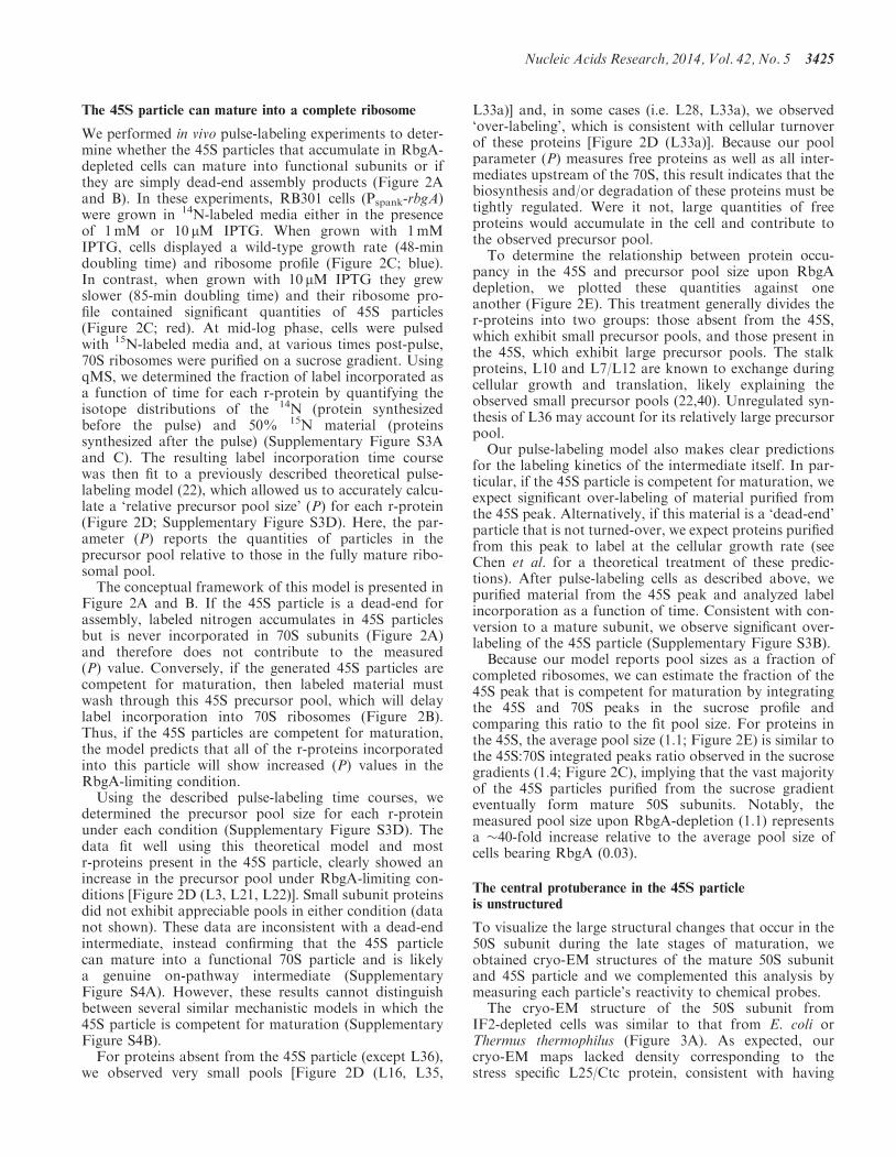
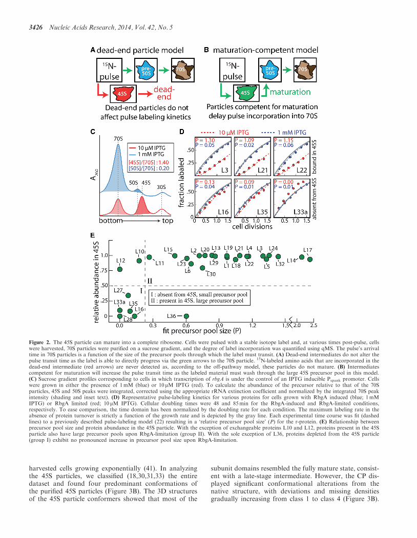
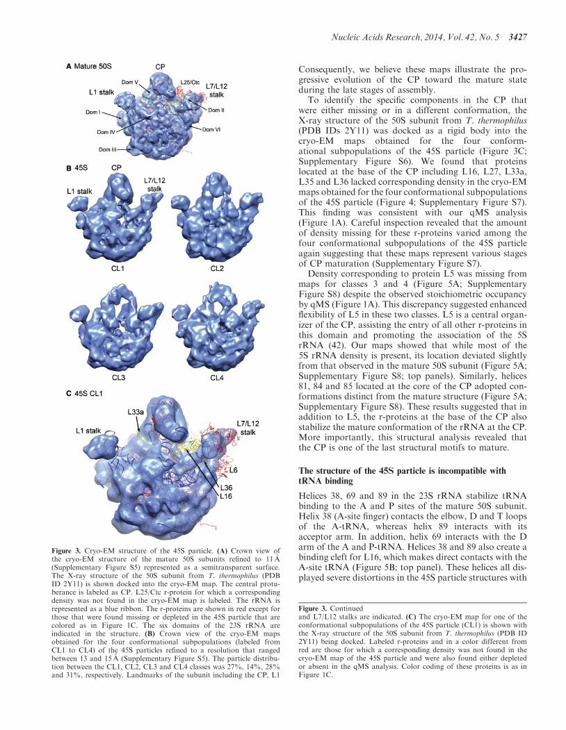
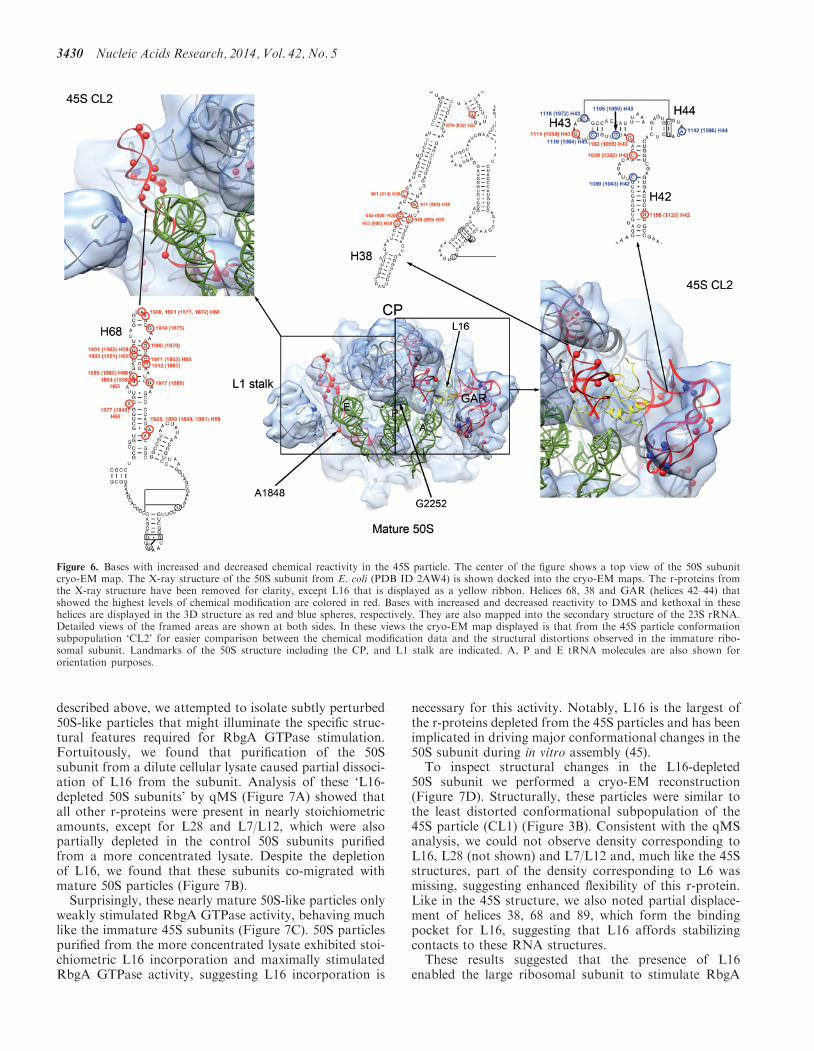
Copyright © 2022 FDOKUMEN