
Functional delineation of the human occipito-temporal areas related to face and scene processing: A...
10
Brain (2000), 123, 1903–1912 Functional delineation of the human occipito- temporal areas related to face and scene processing A PET study K. Nakamura, 1 R. Kawashima, 2 N. Sato, 1 A. Nakamura, 3 M. Sugiura, 2 T. Kato, 3 K. Hatano, 3 K. Ito, 3 H. Fukuda, 2 T. Schormann 4 and K. Zilles 4,5 1 Department of Behavioral and Brain Sciences, Primate Correspondence to: Dr Katsuki Nakamura, Department of Research Institute, Kyoto University, Inuyama, 2 Department Behavioral and Brain Sciences, Primate Research Institute, of Nuclear Medicine and Radiology, IDAC, Tohoku Kyoto University, Kanrin, Inuyama, Aichi 484-8506, Japan University, Sendai, 3 National Institute for Longevity E-mail: knakamur@pri.kyoto-u.ac.jp Sciences, Obu, Japan, 4 C. O. Vogt Institute of Brain Research, University of Du ¨sseldorf, Du ¨sseldorf and 5 Institute of Medicine, Research Center, Julich, Germany Summary By measuring regional cerebral blood flow using PET, we delineated the roles of the occipito-temporal regions activated by faces and scenes. We asked right-handed normal subjects to perform three tasks using facial images as visual stimuli: in the face familiar/unfamiliar discrimination (FF) task, they discriminated the faces of their friends and associates from unfamiliar ones; in the face direction discrimination (FD) task, they discriminated the direction of each unfamiliar face; in the dot location discrimination (DL) task, they discriminated the location of a red dot on a scrambled face. The activity in each task was compared with that in the control fixation (CF) task, in which they fixated on the centre of a display without visual stimuli. The DL task activated the occipital cortices and posterior fusiform gyri bilaterally. During the FD task, the activation extended anteriorly in the right fusiform gyrus and laterally to the right inferior Keywords: brain; visual; recognition; temporal pole; familiar Abbreviations: CF control fixation task; DL dot location discrimination task; FD face direction discrimination task; FF face familiar/unfamiliar discrimination task; rCBF regional cerebral blood flow; SF scene familiar/unfamiliar discrimination task Introduction It has been suggested that the human brain contains a specialized system for face recognition, and damage to the system produces the inability to recognize faces (prosopagnosia or face agnosia) despite intact intellectual functioning and apparently intact visual recognition of other stimuli (e.g. Meadows, 1974; Damasio et al., 1982; © Oxford University Press 2000 temporal cortex. The FF task further activated the right temporal pole. To examine whether the activation due to faces was face-specific, we used a scene familiar/unfamiliar discrimination (SF) task, in which the subjects dis- criminated familiar scenes from unfamiliar ones. Our results suggest that (i) the occipital cortices and posterior fusiform gyri non-selectively respond to faces, scrambled faces and scenes, and are involved mainly in the extraction of physical features of complex visual images; (ii) the right inferior temporal/fusiform gyrus responds selectively to faces but not to non-face stimuli and is involved in the visual processing related to face perception, whereas the bilateral parahippocampal gyri and parieto-occipital junctions respond selectively to scenes and are involved in processing related to scene perception; and (iii) the right temporal pole is activated during the discrimination of familiar faces and scenes from unfamiliar ones, and is probably involved in the recognition of familiar objects. Shuttleworth et al., 1982; Damasio et al., 1990; Farah, 1990). The neuropathology of prosopagnosia has received much attention. Focal damage to various regions of the occipito- temporal areas can impair the ability to recognize faces (Damasio et al., 1990; Farah, 1990; De Renzi et al., 1991; Farah et al., 1995). The lesion site and the nature of the
Transcript of Functional delineation of the human occipito-temporal areas related to face and scene processing: A...
Brain (2000), 123, 1903–1912
Functional delineation of the human occipito-temporal areas related to face and sceneprocessingA PET study
K. Nakamura,1 R. Kawashima,2 N. Sato,1 A. Nakamura,3 M. Sugiura,2 T. Kato,3 K. Hatano,3 K. Ito,3
H. Fukuda,2 T. Schormann4 and K. Zilles4,5
1Department of Behavioral and Brain Sciences, Primate Correspondence to: Dr Katsuki Nakamura, Department ofResearch Institute, Kyoto University, Inuyama, 2Department Behavioral and Brain Sciences, Primate Research Institute,of Nuclear Medicine and Radiology, IDAC, Tohoku Kyoto University, Kanrin, Inuyama, Aichi 484-8506, JapanUniversity, Sendai, 3National Institute for Longevity E-mail: [email protected], Obu, Japan, 4C. O. Vogt Institute of BrainResearch, University of Dusseldorf, Dusseldorf and5Institute of Medicine, Research Center, Julich, Germany
SummaryBy measuring regional cerebral blood flow using PET,we delineated the roles of the occipito-temporal regionsactivated by faces and scenes. We asked right-handednormal subjects to perform three tasks using facial imagesas visual stimuli: in the face familiar/unfamiliardiscrimination (FF) task, they discriminated the faces oftheir friends and associates from unfamiliar ones; in theface direction discrimination (FD) task, they discriminatedthe direction of each unfamiliar face; in the dot locationdiscrimination (DL) task, they discriminated the locationof a red dot on a scrambled face. The activity in eachtask was compared with that in the control fixation (CF)task, in which they fixated on the centre of a displaywithout visual stimuli. The DL task activated the occipitalcortices and posterior fusiform gyri bilaterally. Duringthe FD task, the activation extended anteriorly in theright fusiform gyrus and laterally to the right inferior
Keywords: brain; visual; recognition; temporal pole; familiar
Abbreviations: CF � control fixation task; DL � dot location discrimination task; FD � face direction discrimination task;FF � face familiar/unfamiliar discrimination task; rCBF � regional cerebral blood flow; SF � scene familiar/unfamiliardiscrimination task
IntroductionIt has been suggested that the human brain contains aspecialized system for face recognition, and damage to thesystem produces the inability to recognize faces(prosopagnosia or face agnosia) despite intact intellectualfunctioning and apparently intact visual recognition of otherstimuli (e.g. Meadows, 1974; Damasio et al., 1982;
© Oxford University Press 2000
temporal cortex. The FF task further activated the righttemporal pole. To examine whether the activation due tofaces was face-specific, we used a scene familiar/unfamiliardiscrimination (SF) task, in which the subjects dis-criminated familiar scenes from unfamiliar ones. Ourresults suggest that (i) the occipital cortices and posteriorfusiform gyri non-selectively respond to faces, scrambledfaces and scenes, and are involved mainly in the extractionof physical features of complex visual images; (ii) the rightinferior temporal/fusiform gyrus responds selectively tofaces but not to non-face stimuli and is involved in thevisual processing related to face perception, whereasthe bilateral parahippocampal gyri and parieto-occipitaljunctions respond selectively to scenes and are involvedin processing related to scene perception; and (iii) theright temporal pole is activated during the discriminationof familiar faces and scenes from unfamiliar ones, and isprobably involved in the recognition of familiar objects.
Shuttleworth et al., 1982; Damasio et al., 1990; Farah, 1990).The neuropathology of prosopagnosia has received muchattention. Focal damage to various regions of the occipito-temporal areas can impair the ability to recognize faces(Damasio et al., 1990; Farah, 1990; De Renzi et al., 1991;Farah et al., 1995). The lesion site and the nature of the
1904 K. Nakamura et al.
deficit vary from patient to patient. Some patients cannotdiscriminate faces from non-face objects, but others can doso but may not recognize familiar faces. Recent brain imagingstudies have reported that face stimuli can activate the rightfusiform gyrus, suggesting its involvement in face perception(Sergent et al., 1992; Haxby et al., 1994; Puce et al., 1995;Kanwisher et al., 1997; McCarthy et al., 1997; Farah andAguirre, 1999; Haxby et al., 1999). However, in our sociallives the visual recognition of familiar faces based on memory,such as the recognition of the faces of our parents andfriends, etc., is important. Which brain regions are involved inthe visual recognition of familiar faces remains under debate.
The objective of the present study was to determine thebrain regions associated with the recognition of familiar facesby measuring regional cerebral blood flow (rCBF) using PETwhile normal subjects performed visual discrimination tasks.We addressed the following two questions: (i) which brainregions are associated with the recognition of familiar facesand which with the perception of faces; and (ii) is the activityof each brain region face-specific? To answer the firstquestion, we used three face tasks which required differentlevels of face processing and determined the brain regionsactivated by the recognition of familiar faces but not by theperception of unfamiliar faces, as well as those activated byfaces but not by non-face stimuli. To answer the secondquestion, we used a scene discrimination task and comparedthe activity between face and scene tasks, as faces and scenesare reportedly processed differently (Aguirre et al., 1996;Aguirre and D’Esposito, 1997; Epstein and Kanwisher, 1998;Maguire et al., 1998; Haxby et al., 1999).
MethodsSubjectsSeven right-handed normal male volunteers (aged 23–29years) participated in this study. Written informed consentwas obtained from each subject in accordance with guidelinesapproved by the ethics committee of National Institute forLongevity Sciences and the Declaration of Helsinki (1991).All of the subjects were healthy, with no history of psychiatricor neurological illness, and were not on any medication.
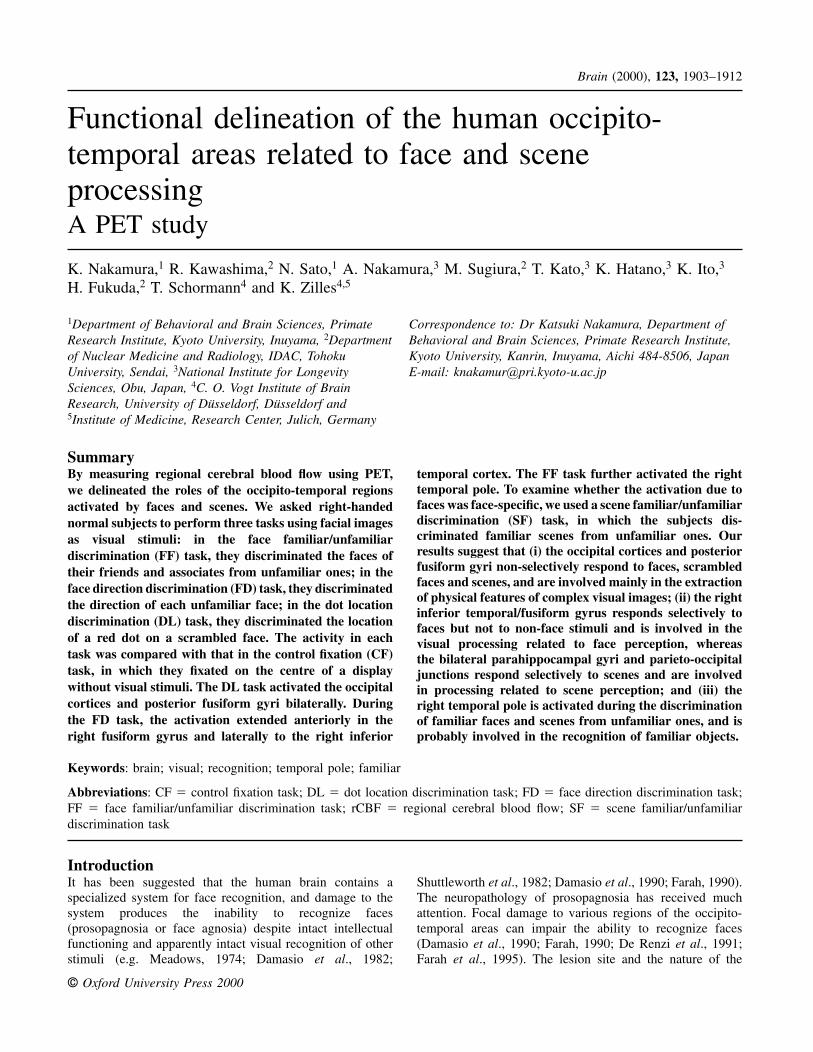
Behavioural tasksThe stimuli were digitized coloured images of faces presentedat various orientations and scrambled images of the faceswith a red dot near one of the four corners, and colouredimages of a scene (Fig. 1). To delineate the roles of theregions activated by faces, we used three face tasks whichrequired different levels of processing of facial images. Inthe face familiar/unfamiliar discrimination (FF) task, thesubjects were required to discriminate the faces of theirfriends and associates from unfamiliar faces. All of thefamiliar and unfamiliar faces were presented at variousorientations. Each subject was instructed to click a computer
Fig. 1 Examples of stimuli. The stimuli were digitized colouredimages of a face presented at various orientations (top), scrambledimages of the same face (middle) with a red dot near one of thefour corners, and coloured images of a scene (bottom).
mouse during visual stimulation when the face was familiarto him in the FF task. In the face direction discrimination (FD)task, the subjects were asked to discriminate the direction ofeach face and to click the mouse when the face was facingright (or left). All of the faces were unfamiliar to thesubjects. In the dot location discrimination (DL) task, theydiscriminated the location of a red dot on a scrambled faceimage and clicked the mouse when the red dot was locatedto the right (or left). We used scrambled images of the samefaces to control for luminance, colour and local features(Allison et al., 1994a, b). The red dot had no significance inthe FF and FD tasks. We assumed the following cognitiveprocesses: complex visual stimulation in all three tasks;perception of unfamiliar faces in the FD and FF tasks;recognition of familiar faces and their discrimination fromunfamiliar ones only in the FF task. In the FF, FD and DLtasks, 89% (75–98%), 91% (78–100%) and 99% (98–100%)of the responses were correct, respectively. The DL testtended to be easier than the FF test (Wilcoxon matched-pairssigned-ranks test, P � 0.03). There were no significantdifferences between the FF and FD tests or between the FDand DL tests. To examine whether the activation due tofamiliar faces was face-specific, we used an analogous taskin which the subjects viewed familiar and unfamiliar scenes.In this scene familiar/unfamiliar discrimination (SF) task, thesubjects discriminated familiar scenes, e.g. their universityor town railway station, from unfamiliar scenes. In the SFtask, 70% (56–90%) of the responses were correct, and therewere no significant differences between the percentagescorrect in the SF and FF tasks (Wilcoxon test, P � 0.05). Inall tasks, images were presented for 1.0 s on a face-mounteddisplay at 1.0 s intervals. Each image had 200 � 200 pixelsand subtended horizontal and vertical visual angles of 10°.The activity in each task was compared with that in the controlfixation (CF) task, in which the subjects were instructed tofixate on the centre of a display without visual stimuli. Each
Face and scene processing in the human brain 1905
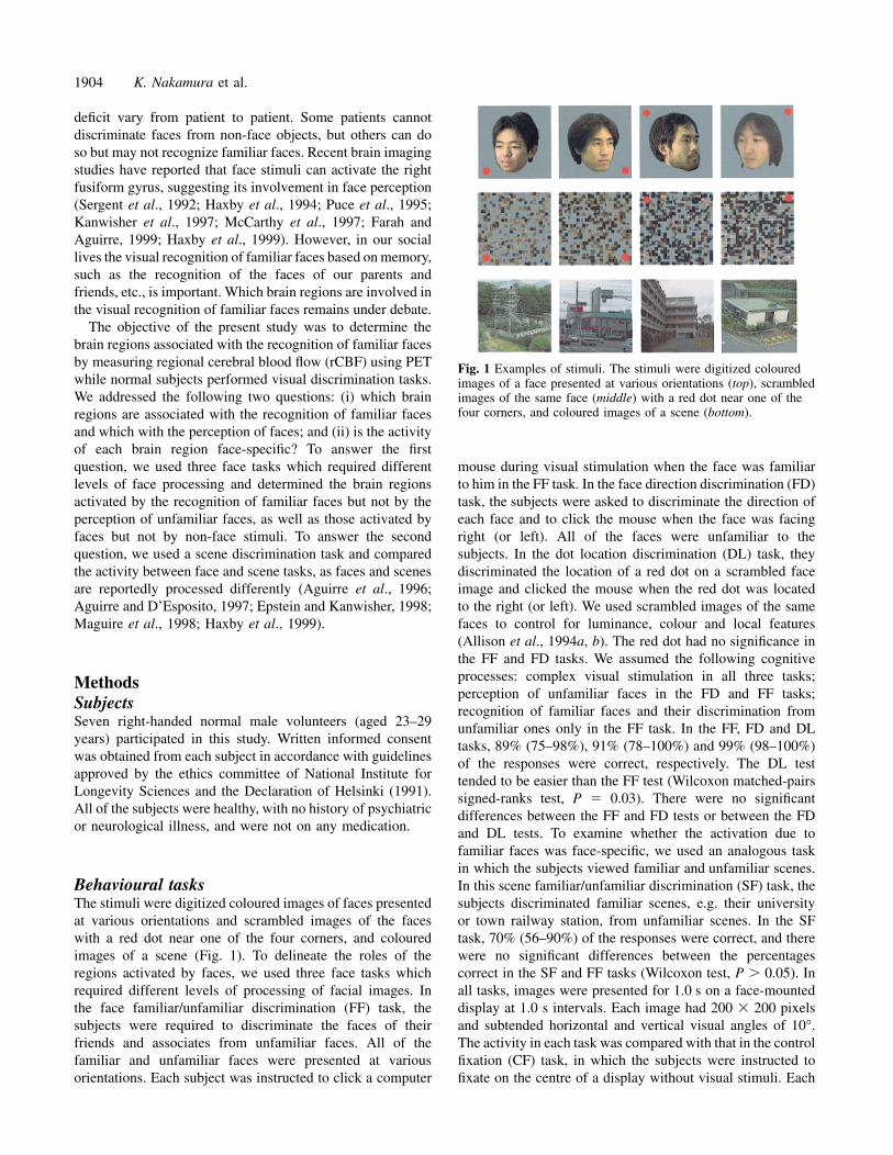
Fig. 2 Regions of increased rCBF in the face tasks on 3D-reconstructed standard brains. Only regionsof significantly increased rCBF after correction for multiple comparisons are shown (red). Regionsactivated during the dot location discrimination (DL, A), face direction discrimination (FD, B) and facefamiliar/unfamiliar discrimination (FF, C) compared with the control fixation (CF) task, and thoserevealed by conjunction of (FD–DL) with (FD–CF) (D) and by conjunction of (FF–CF) with (FF–FD)(E). Back � back view; Left � left view; Right � right view; Ventral � ventral view.
task lasted 1.5 min. Only the CF task was administered twice,i.e. at the beginning and at the end of the experiment foreach subject. The order of performing the three face tasksvaried, and the responses to the right and left in the FD andDL tasks were counterbalanced among subjects. The SF taskwas introduced randomly into the task sequence. Each faceor scene was presented only once, to avoid the effect ofrepetition of the same image on neural activity. The subjectswere instructed to focus at the centre of the display throughoutthe experiments. During PET scans, horizontal and verticalmonopolar electro-oculograms were recorded. The signalsfrom the surface electrodes, placed in the vicinity of theouter and infraorbital parts of each eye, were fed into anamplifier. There were no significant differences in the numbersof saccadic eye movements among the tasks.
Data acquisition and analysisEach subject was positioned in a PET scanner (Siemens/CTIECAT EXACT HR) (Wienhard et al., 1994). The emission
scan was started immediately after the administration ofbolus injection of approximately 15 mCi (555 MBq) H2
15O,using the three dimensional (3D) collection mode. Allinjections were performed in a single scanning session foreach subject. Each PET image was anatomically normalizedusing Automated Image Registration (Woods et al., 1998)and Elastic Transformation (Schormann et al., 1996); eachsubject’s MRI was normalized to the standard brain MRI ofthe Human Brain Atlas (Roland et al., 1994) by affinetransformation of Automated Image Registration followedby application of the 3D-deformation field of elastictransformation. These parameters were used subsequently totransform each subject’s PET image and MRI into thestandard brain anatomy.
Statistical parametric mapping software (SPM96,Wellcome Department of Cognitive Neurology, London)(Friston et al., 1995) was used for smoothing and statisticalanalysis. A 3D 16 mm Gaussian filter was used. Significantthresholds were set at P � 0.05, corrected for multiplecomparisons for both the height and extent of activation. The

1906 K. Nakamura et al.
Table 1 Brain regions showing statistically significant activation in the face tasks
Region Cluster DL–CF FD–CF FF–CF (FD–CF) (FF–CF)with (FD–DL) with (FF–FD)
R LG A (C) 12, –77, –13 14, –81, –14 12, –78, –14(6.81) (5.88) (6.59)
L LG A –8, –78, –11 –12, –78, –18(5.13) (5.75)
R LOC A (A) 15, –94, 1 13, –94, –1 15, –92, 0(6.77) (5.91) (6.51)
R FG A (D) 28, –65, –16 30, –64, –16 34, –62, –19(6.75) (7.07) (6.67)
L FG A (F) –36, –43, –18(4.36)
L OCT A –36, –79, –20 –37, –81, –19(5.28) (6.13)
R IT/FG D (E) 47, –49, –21 45, –49, –19(6.42) (6.37)
L IT/FG A –41, –66, –15(5.44)
R PR D (I) 29, –6, –29 28, –7, –29(4.36) (4.31)
R TP C (J) 34, 23, –27 37, 23, –27(5.15) (4.52)
The table lists the locations of peak activation in at least one of the face tasks in comparison with the CF task that were significant aftercorrection for multiple comparisons. Coordinates (x, y, z) and Z scores (in parentheses) are shown. In the column headed Region, the firstcharacter (R or L) indicates the right or left hemisphere. Brain regions: FG � fusiform gyrus; LG � lingual gyrus; LOC � lateraloccipital cortex; IT/FG � inferior temporal gyrus/fusiform gyrus; OCT � occipito-temporal junction; PR � perirhinal cortex; TP �temporal pole. Characters in the column headed Cluster represent the name of the clusters defined by the cluster analysis shown inFig. 4. Characters in parentheses correspond to the labels of the panels of Fig. 5. CF � control fixation task; DL � dot locationdiscrimination task; FD � face direction discrimination task; FF � face familiar/unfamiliar discrimination task.
activated areas were localized anatomically in relation tothe mean reformatted MRI of the seven subjects. To extractthe activation concerning each cognitive process and toeliminate the confounding effects of deactivation orsuppression, we used conjunction analysis (Friston, 1997).Significant thresholds for the conjunction analysis were setat P � 0.05 (corrected). The conjunction analysis was appliedby masking, whereby the second subtraction was tested onlyin pixels that reached significance (P � 0.01) in both thefirst and the second subtraction.
To examine the similarity of the response profiles amongthe activated foci, we calculated Pearson’s correlationcoefficient for the adjusted rCBF value in each task amongthe foci and made a correlation matrix representing thedistances among the foci. Then, non-metric multidimensionalscaling (Kruskal and Wish, 1978) and cluster analysis wereapplied to the data. In the multidimensional scaling,configurations in one to four dimensions were produced sothat analyses in different dimensions could be compared ina scree test (Cattell, 1966; Kruskal and Wish, 1978). Thescree test can reveal the extent to which a two-dimensionalsolution explains the variability in the data. If there weremultiple foci of significant activation in the same anatomicalarea, data for all of the foci were used in this analysis, sothat we could examine whether multiple foci in the sameanatomical area showed a similar response profile. In thecluster analysis, we used a complete linkage method usingthe correlation coefficient.
ResultsRegions involved in face discriminationTo delineate the roles of the regions activated by faces, weused three tasks using facial images; the FF, FD and DLtasks. The activity in each task was compared with that inthe CF task. Compared with the CF task, the DL task activatedthe occipital cortices bilaterally, including the lingual andlateral occipital gyri, and the posterior fusiform gyri (Fig. 2A).When the FD task was compared with the CF task, theactivated regions extended anteriorly in the fusiform gyriand laterally beyond the occipito-temporal sulcus to theposterior portion of the inferior temporal gyri, in addition tothe occipital cortices and posterior fusiform gyri (Fig. 2B).The response was more prominent on the right side than onthe left. The FF task further activated the right temporal poleand perirhinal cortex (Fig. 2C).
To elucidate the functional characteristic of each region,direct comparisons among face tasks were performed (seeData acquisition and analysis). The brain regions more activein the FD task than in the DL or CF task, revealed byconjunction of (FD–DL) with (FD–CF), were the right inferiortemporal and fusiform gyri (Fig. 2D). The activation wasobserved in the right inferior temporal and fusiform gyri,and the peak location was near the occipito-temporal sulcus.We refer to the anatomical region of this activation as theright inferior temporal/fusiform gyri. The brain region moreactive in the FF task than in either the FD or the CF task,
Face and scene processing in the human brain 1907
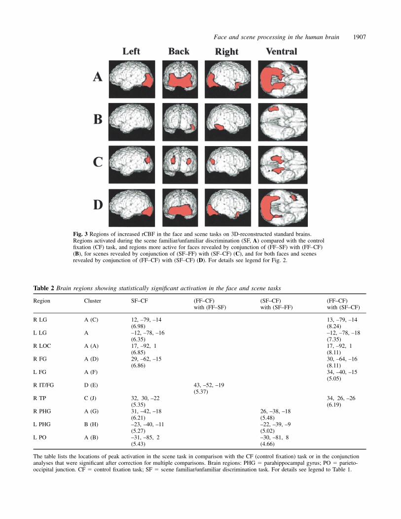
Fig. 3 Regions of increased rCBF in the face and scene tasks on 3D-reconstructed standard brains.Regions activated during the scene familiar/unfamiliar discrimination (SF, A) compared with the controlfixation (CF) task, and regions more active for faces revealed by conjunction of (FF–SF) with (FF–CF)(B), for scenes revealed by conjunction of (SF–FF) with (SF–CF) (C), and for both faces and scenesrevealed by conjunction of (FF–CF) with (SF–CF) (D). For details see legend for Fig. 2.
Table 2 Brain regions showing statistically significant activation in the face and scene tasks
Region Cluster SF–CF (FF–CF) (SF–CF) (FF–CF)with (FF–SF) with (SF–FF) with (SF–CF)
R LG A (C) 12, –79, –14 13, –79, –14(6.98) (8.24)
L LG A –12, –78, –16 –12, –78, –18(6.35) (7.35)
R LOC A (A) 17, –92, 1 17, –92, 1(6.85) (8.11)
R FG A (D) 29, –62, –15 30, –64, –16(6.86) (8.11)
L FG A (F) 34, –40, –15(5.05)
R IT/FG D (E) 43, –52, –19(5.37)
R TP C (J) 32, 30, –22 34, 26, –26(5.35) (6.19)
R PHG A (G) 31, –42, –18 26, –38, –18(6.21) (5.48)
L PHG B (H) –23, –40, –11 –22, –39, –9(5.27) (5.02)
L PO A (B) –31, –85, 2 –30, –81, 8(5.43) (4.66)
The table lists the locations of peak activation in the scene task in comparison with the CF (control fixation) task or in the conjunctionanalyses that were significant after correction for multiple comparisons. Brain regions: PHG � parahippocampal gyrus; PO � parieto-occipital junction. CF � control fixation task; SF � scene familiar/unfamiliar discrimination task. For details see legend to Table 1.
1908 K. Nakamura et al.
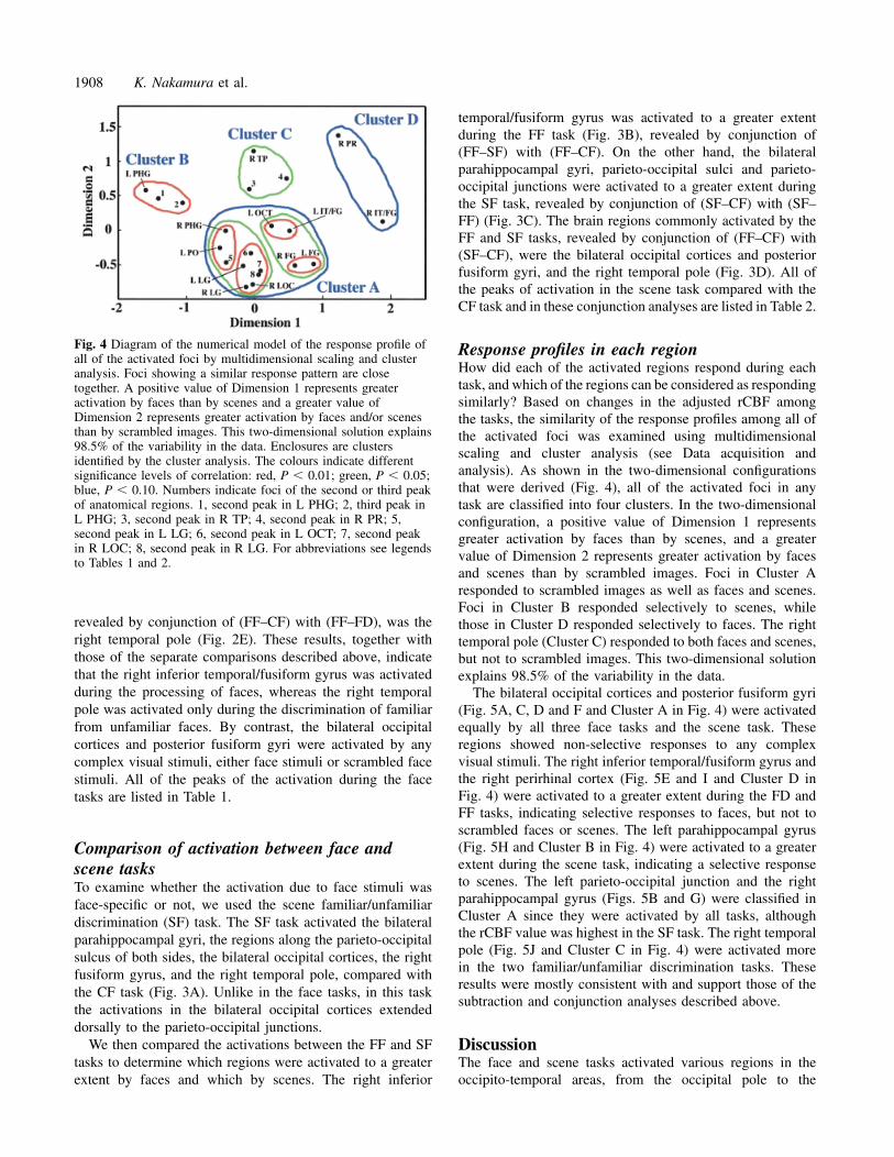
Fig. 4 Diagram of the numerical model of the response profile ofall of the activated foci by multidimensional scaling and clusteranalysis. Foci showing a similar response pattern are closetogether. A positive value of Dimension 1 represents greateractivation by faces than by scenes and a greater value ofDimension 2 represents greater activation by faces and/or scenesthan by scrambled images. This two-dimensional solution explains98.5% of the variability in the data. Enclosures are clustersidentified by the cluster analysis. The colours indicate differentsignificance levels of correlation: red, P � 0.01; green, P � 0.05;blue, P � 0.10. Numbers indicate foci of the second or third peakof anatomical regions. 1, second peak in L PHG; 2, third peak inL PHG; 3, second peak in R TP; 4, second peak in R PR; 5,second peak in L LG; 6, second peak in L OCT; 7, second peakin R LOC; 8, second peak in R LG. For abbreviations see legendsto Tables 1 and 2.
revealed by conjunction of (FF–CF) with (FF–FD), was theright temporal pole (Fig. 2E). These results, together withthose of the separate comparisons described above, indicatethat the right inferior temporal/fusiform gyrus was activatedduring the processing of faces, whereas the right temporalpole was activated only during the discrimination of familiarfrom unfamiliar faces. By contrast, the bilateral occipitalcortices and posterior fusiform gyri were activated by anycomplex visual stimuli, either face stimuli or scrambled facestimuli. All of the peaks of the activation during the facetasks are listed in Table 1.
Comparison of activation between face andscene tasksTo examine whether the activation due to face stimuli wasface-specific or not, we used the scene familiar/unfamiliardiscrimination (SF) task. The SF task activated the bilateralparahippocampal gyri, the regions along the parieto-occipitalsulcus of both sides, the bilateral occipital cortices, the rightfusiform gyrus, and the right temporal pole, compared withthe CF task (Fig. 3A). Unlike in the face tasks, in this taskthe activations in the bilateral occipital cortices extendeddorsally to the parieto-occipital junctions.
We then compared the activations between the FF and SFtasks to determine which regions were activated to a greaterextent by faces and which by scenes. The right inferior
temporal/fusiform gyrus was activated to a greater extentduring the FF task (Fig. 3B), revealed by conjunction of(FF–SF) with (FF–CF). On the other hand, the bilateralparahippocampal gyri, parieto-occipital sulci and parieto-occipital junctions were activated to a greater extent duringthe SF task, revealed by conjunction of (SF–CF) with (SF–FF) (Fig. 3C). The brain regions commonly activated by theFF and SF tasks, revealed by conjunction of (FF–CF) with(SF–CF), were the bilateral occipital cortices and posteriorfusiform gyri, and the right temporal pole (Fig. 3D). All ofthe peaks of activation in the scene task compared with theCF task and in these conjunction analyses are listed in Table 2.
Response profiles in each regionHow did each of the activated regions respond during eachtask, and which of the regions can be considered as respondingsimilarly? Based on changes in the adjusted rCBF amongthe tasks, the similarity of the response profiles among all ofthe activated foci was examined using multidimensionalscaling and cluster analysis (see Data acquisition andanalysis). As shown in the two-dimensional configurationsthat were derived (Fig. 4), all of the activated foci in anytask are classified into four clusters. In the two-dimensionalconfiguration, a positive value of Dimension 1 representsgreater activation by faces than by scenes, and a greatervalue of Dimension 2 represents greater activation by facesand scenes than by scrambled images. Foci in Cluster Aresponded to scrambled images as well as faces and scenes.Foci in Cluster B responded selectively to scenes, whilethose in Cluster D responded selectively to faces. The righttemporal pole (Cluster C) responded to both faces and scenes,but not to scrambled images. This two-dimensional solutionexplains 98.5% of the variability in the data.
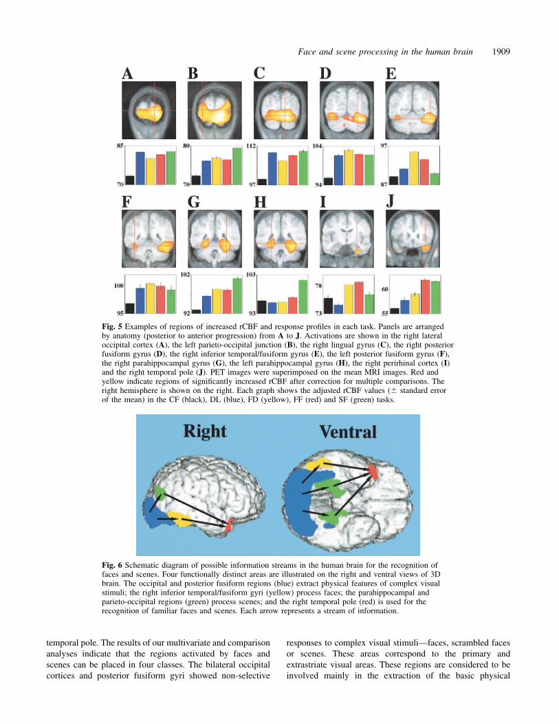
The bilateral occipital cortices and posterior fusiform gyri(Fig. 5A, C, D and F and Cluster A in Fig. 4) were activatedequally by all three face tasks and the scene task. Theseregions showed non-selective responses to any complexvisual stimuli. The right inferior temporal/fusiform gyrus andthe right perirhinal cortex (Fig. 5E and I and Cluster D inFig. 4) were activated to a greater extent during the FD andFF tasks, indicating selective responses to faces, but not toscrambled faces or scenes. The left parahippocampal gyrus(Fig. 5H and Cluster B in Fig. 4) were activated to a greaterextent during the scene task, indicating a selective responseto scenes. The left parieto-occipital junction and the rightparahippocampal gyrus (Figs. 5B and G) were classified inCluster A since they were activated by all tasks, althoughthe rCBF value was highest in the SF task. The right temporalpole (Fig. 5J and Cluster C in Fig. 4) were activated morein the two familiar/unfamiliar discrimination tasks. Theseresults were mostly consistent with and support those of thesubtraction and conjunction analyses described above.
DiscussionThe face and scene tasks activated various regions in theoccipito-temporal areas, from the occipital pole to the
Face and scene processing in the human brain 1909
Fig. 5 Examples of regions of increased rCBF and response profiles in each task. Panels are arrangedby anatomy (posterior to anterior progression) from A to J. Activations are shown in the right lateraloccipital cortex (A), the left parieto-occipital junction (B), the right lingual gyrus (C), the right posteriorfusiform gyrus (D), the right inferior temporal/fusiform gyrus (E), the left posterior fusiform gyrus (F),the right parahippocampal gyrus (G), the left parahippocampal gyrus (H), the right perirhinal cortex (I)and the right temporal pole (J). PET images were superimposed on the mean MRI images. Red andyellow indicate regions of significantly increased rCBF after correction for multiple comparisons. Theright hemisphere is shown on the right. Each graph shows the adjusted rCBF values (� standard errorof the mean) in the CF (black), DL (blue), FD (yellow), FF (red) and SF (green) tasks.
Fig. 6 Schematic diagram of possible information streams in the human brain for the recognition offaces and scenes. Four functionally distinct areas are illustrated on the right and ventral views of 3Dbrain. The occipital and posterior fusiform regions (blue) extract physical features of complex visualstimuli; the right inferior temporal/fusiform gyri (yellow) process faces; the parahippocampal andparieto-occipital regions (green) process scenes; and the right temporal pole (red) is used for therecognition of familiar faces and scenes. Each arrow represents a stream of information.
temporal pole. The results of our multivariate and comparisonanalyses indicate that the regions activated by faces andscenes can be placed in four classes. The bilateral occipitalcortices and posterior fusiform gyri showed non-selective
responses to complex visual stimuli—faces, scrambled facesor scenes. These areas correspond to the primary andextrastriate visual areas. These regions are considered to beinvolved mainly in the extraction of the basic physical
1910 K. Nakamura et al.
features of complex visual stimuli, although some studieshave reported selective responses of the occipital areas tofaces (e.g. Linkenkaer-Hansen et al., 1998). The right inferiortemporal/fusiform gyrus and the right perirhinal cortex wereactivated to a greater extent by faces, indicating selectiveresponses to faces, but not by scrambled faces or scenes. Onthe other hand, the bilateral parahippocampal gyri and parieto-occipital regions were activated to a greater extent by scenes.The right temporal pole was active during the discriminationof familiar faces and scenes from unfamiliar ones.
Neuropsychological studies have suggested multiple,distinct neural systems for the processing of different objectcategories, such as faces (Damasio et al., 1990; De Renziet al., 1991; Farah et al., 1995), living things (Warringtonand Schallice, 1984; Farah et al., 1991), man-made objects(Warrington and McCarthy, 1994; Moscovitch et al., 1997)and scenes (De Renzi et al., 1977; Whiteley and Warrington,1978; Hecaen et al., 1980; Habib and Sirigu, 1987; Maguireet al., 1996; McCarthy et al., 1996). Many functional imagingstudies (Sergent et al., 1992; Haxby et al., 1994; Puce et al.,1995; Kanwisher et al., 1997; McCarthy et al., 1997; Farahand Aguirre, 1999; Haxby et al., 1999) and recording studies(Allison et al., 1994a, b, 1999) reported that the right inferiortemporal/fusiform gyrus responded more to faces than toother complex visual stimuli. We confirmed these results.Our present data demonstrate that the right perirhinal cortexresponds selectively to faces. A recent recording study(Allison et al., 1999) also found activation in the rightperirhinal cortex by faces. These data suggest that the rightperirhinal cortex and the right inferior temporal/fusiform gyriselectively process facial features, leading to the perceptionof faces. Several neuroimaging studies have reported that thebilateral parahippocampal gyri predominantly process scenes(Aguirre et al., 1996; Aguirre and D’Esposito, 1997; Epsteinand Kanwisher, 1998; Maguire et al., 1998; Haxby et al.,1999). The present results further show that the bilateralparieto-occipital junctions and parieto-occipital sulciresponded selectively to scenes, suggesting the involvementof temporal and parietal regions in the processing of localenvironments (Aguirre and D’Esposito, 1997; Maguire et al.,1998; Sato et al., 1999). All of these data confirm the ideathat the human brain contains multiple systems for theprocessing of the different object categories that have beensuggested by neuropsychological data.
The major finding of the present study is that the righttemporal pole functions in the recognition of familiar facesand their discrimination from unfamiliar faces on the basisof memory. Activation in the temporal pole has been reportedin some imaging studies using famous faces as stimuli(Sergent et al., 1992; Damasio et al., 1996; Gorno-Tempiniet al., 1998), whereas the right inferior temporal/fusiformgyrus exhibited stable responses to faces (Sergent et al.,1992; Haxby et al., 1994; Puce et al., 1995; Kanwisher et al.,1997; McCarthy et al., 1997; Farah and Aguirre, 1999;Haxby et al., 1999). This inconsistency among previousstudies in the observation of activation in the temporal pole
can be explained by the nature of face stimuli, famous(familiar) or unfamiliar. However, the activation of the righttemporal pole was not face-specific. The right temporal polewas also activated during the scene task. One explanation isthat the right temporal pole is associated with the processingof scenes as well as face recognition. This is unlikely, as noprevious studies using scene stimuli have reported activationof the right temporal pole. Another explanation is that theright temporal pole is associated with the recognition offamiliar objects regardless of the object categories, at leastfor faces and scenes. Indeed, damage to the right anteriortemporal cortex including the temporal pole can impair therecognition of famous faces, scenes or buildings, suggestingthe loss of memory (Ellis et al., 1989; Kapur et al., 1992;Markowitsch et al., 1993; Nakamura and Kubota, 1996;Tranel et al., 1997). The right temporal pole may be thestorehouse of personal memory.
The occipito-temporal cortical areas have been implicatedin visual processing for object recognition and are wellknown as the ‘ventral visual pathway’ (Gross, 1973, 1992;Ungerleider and Mishkin, 1982; Desimone and Ungerleider,1989; Farah, 1990; Milner and Goodale, 1995). In monkeys,the processing of complex visual stimuli progresses anteriorlyalong the anterior–posterior axis (Mishkin, 1982; Desimoneand Ungerleider, 1989; Gross, 1992; Tanaka, 1996), and theanterior portion of the inferior temporal cortex is moreassociated with memory functions (Miyashita, 1993;Nakamura and Kubota, 1996; Suzuki, 1996). The presentresults suggest that visual processing progresses similarly inthe human occipito-temporal areas. Figure 6 illustrates thepresent results schematically. In the human brain, there existsa common neural substrate (the right temporal pole, shownin red) for the visual recognition of familiar objects basedon memory, whereas multiple systems, such as the rightinferior temporal/fusiform areas and the bilateral para-hippocampal and parieto-occipital areas (yellow and green,respectively), are involved in the processing of differentobject categories, such as faces and scenes. The right temporalpole was also activated during listening to sentencescontaining information about the subjects’ own past (Finket al., 1996). Thus, the temporal pole seems to function inauditory as well as visual recognition based on memory. Thelocation of peak activation in the right temporal pole of thestudy of Fink and colleagues (38, 6, –12) (Fink et al., 1996)is dorsal to that of our present study (34, 23, –27). Theremay be functional subregions in the right temporal pole: itsventral portion for visual processing and its dorsal portionfor auditory processing. Further experiments are needed toclarify this issue.
AcknowledgementsThis work was supported by JSPS-RFTF (97L00202), Grant-in-Aid for Scientific Research on Priority Areas from theMinistry of Education, Science, Sports and Culture (1114522),

Face and scene processing in the human brain 1911
the Fund for Comprehensive Research on Aging and Healthfrom the Ministry of Welfare, and an SFB grant.
ReferencesAguirre GK, D’Esposito M. Environmental knowledge is subservedby separable dorsal/ventral neural areas. J Neurosci 1997; 17:2512–8.
Aguirre GK, Detre JA, Alsop DC, D’Esposito M. The para-hippocampus subserves topographical learning in man. Cereb Cortex1996; 6: 823–9.
Allison T, Ginter H, McCarthy G, Nobre AC, Puce A, Luby M,et al. Face recognition in human extrastriate cortex. J Neurophysiol1994a; 71: 821–5.
Allison T, McCarthy G, Nobre A, Puce A, Belger A. Humanextrastriate visual cortex and the perception of faces, words,numbers, and colors. [Review]. Cereb Cortex 1994b; 4: 544–54.
Allison T, Puce A, Spencer DD, McCarthy G. Electrophysiologicalstudies of human face perception. I. Potentials generated inoccipitotemporal cortex by face and non-face stimuli. Cereb Cortex1999; 9: 415–30.
Cattell RB. The scree test for the number of factors. Multivar BehavRes 1966; 1: 245–76.
Damasio H, Grabowski TJ, Tranel D, Hichwa RD, Damasio AR.A neural basis for lexical retrieval. Nature 1996; 380: 499–505.
Damasio AR, Tranel D, Damasio H. Face agnosia and the neuralsubstrates of memory. [Review]. Annu Rev Neurosci 1990; 13:89–109.
Damasio AR, Damasio H, Van Hoesen GW. Prosopagnosia: anatomicbasis and behavioral mechanisms. Neurology 1982; 32: 331–41.
De Renzi E, Faglioni P, Grossi D, Nichelli P. Apperceptive andassociative forms of prosopagnosia. Cortex 1991; 27: 213–21.
De Renzi E, Faglioni P, Villa P. Topographical amnesia. J NeurolNeurosurg Psychiatry 1977; 40: 498–505.
Desimone R, Ungerleider LG. Neural mechanisms of visualprocessing in monkeys. In: Boller F, Grafman J, editors. Handbookof neuropsychology, Vol. 2. Amsterdam: Elsevier; 1989. p. 267–99.
Ellis AW, Young AW, Critchley EM. Loss of memory for peoplefollowing temporal lobe damage. Brain 1989; 112: 1469–83.
Epstein R, Kanwisher N. A cortical representation of the localvisual environment. Nature 1998; 392: 598–601.
Farah MJ. Visual agnosia. Disorders of object recognition and whatthey tell us about normal vision. Cambridge (MA): MIT Press; 1990.
Farah MJ, Aguirre GK. Imaging visual recognition: PET and fMRIstudies of the functional anatomy of human visual recognition.Trends Cogn Sci 1999; 3: 179–86.
Farah MJ, McMullen PA, Meyer MM. Can recognition of livingthings be selectively impaired? Neuropsychologia 1991; 29: 185–93.
Farah MJ, Wilson KD, Drain HM, Tanaka JR. The inverted faceinversion effect in prosopagnosia: evidence for mandatory, face-specific perceptual mechanisms. Vision Res 1995; 35: 2089–93.
Fink GR, Markowitsch HJ, Reinkemeier M, Bruckbauer T, Kessler J,Heiss WD. Cerebral representation of one’s own past: neuralnetworks involved in autobiographical memory. J Neurosci 1996;16: 4275–82.
Friston KJ. Imaging cognitive anatomy. Trends Cogn Sci 1997; 1:21–7.
Friston KJ, Holmes AP, Worsley KJ, Poline J-P, Frith CD,Frackowiak RSJ. Statistical parametric maps in functional imaging:a general linear approach. Hum Brain Mapp 1995; 2: 189–210.
Gorno-Tempini ML, Price CJ, Josephs O, Vandenberghe R, CappaSF, Kapur N, et al. The neural systems sustaining face and proper-name processing. Brain 1998; 121: 2103–18.
Gross CG. Visual functions of inferotemporal cortex. In: Jung R,editor. Handbook of sensory physiology, Vol. VII/3. Centralprocessing of visual information. Berlin: Springer-Verlag; 1973.p. 451–82.
Gross CG. Representation of visual stimuli in inferior temporalcortex. [Review]. Philos Trans R Soc Lond B Biol Sci 1992; 335:3–10.
Habib M, Sirigu A. Pure topographical disorientation: a definitionand anatomical basis. Cortex 1987; 23: 73–85.
Haxby JV, Horwitz B, Ungerleider LG, Maisog JM, Pietrini P,Grady CL. The functional organization of human extrastriate cortex:a PET-rCBF study of selective attention to faces and locations.J Neurosci 1994; 14: 6336–53.
Haxby JV, Ungerleider LG, Clark VP, Schouten JL, Hoffman EA,Martin A. The effect of face inversion on activity in human neuralsystems for face and object perception. Neuron 1999; 22: 189–99.
Hecaen H, Tzortzis C, Rondot P. Loss of topographic memory withlearning deficits. Cortex 1980; 16: 525–42.
Kanwisher N, McDermott J, Chun MM. The fusiform face area: amodule in human extrastriate cortex specialized for face perception.J Neurosci 1997; 17: 4302–11.
Kapur N, Ellison D, Smith MP, McLellan DL, Burrows EH. Focalretrograde amnesia following bilateral temporal lobe pathology.Brain 1992; 115: 73–85.
Kruskal, JB, Wish, M. Multidimensional scaling. Beverly Hills:Sage Pubications; 1978.
Linkenkaer-Hansen K, Palva JM, Sams M, Hietanen JK, AronenHJ, Ilmoniemi RJ. Face-selective processing in human extrastriatecortex around 120 ms after stimulus onset revealed by magneto-and electroencephalography. Neurosci Lett 1998; 253: 147–50.
Maguire EA, Burke T, Phillips J, Staunton H. Topographicaldisorientation following unilateral temporal lobe lesions in humans.Neuropsychologia 1996; 34: 993–1001.
Maguire EA, Burgess N, Donnett JG, Frackowiak RS, Frith CD,O’Keefe J. Knowing where and getting there: a human navigationnetwork. Science 1998; 280: 921–4.
Markowitsch HJ, Calabrese P, Liess J, Haupts M, Durwen HF,Gehlen W. Retrograde amnesia after traumatic injury of the fronto-temporal cortex. J Neurol Neurosurg Psychiatry 1993; 56: 988–92.

1912 K. Nakamura et al.
McCarthy RA, Evans JJ, Hodges JR. Topographic amnesia: spatialmemory disorder, perceptual dysfunction, or category specificsemantic memory impairment? J Neurol Neurosurg Psychiatry 1996;60: 318–25.
McCarthy G, Puce A, Gore JC, Allison T. Face-specific processingin the human fusiform gyrus. J Cogn Neurosci 1997; 9: 605–10.
Meadows JC. The anatomical basis of prosopagnosia. J NeurolNeurosurg Psychiatry 1974; 37: 489–501.
Milner AD, Goodale MA. The visual brain in action. Oxford:Oxford University Press; 1995.
Mishkin M. A memory system in the monkey. Phil Trans R SocLond B Biol Sci 1982; 298: 85–95.
Miyashita Y. Inferior temporal cortex: where visual perceptionmeets memory. [Review]. Annu Rev Neurosci 1993; 16: 245–63.
Moscovitch M, Winocur G, Behrmann M. What is special aboutface recognition? Nineteen experiments on a person with visualobject agnosia and dyslexia but normal face recognition. J CognNeurosci 1997; 9: 555–604.
Nakamura K, Kubota K. The primate temporal pole: its putativerole in object recognition and memory. [Review]. Behav Brain Res1996; 77: 53–77.
Puce A, Allison T, Gore JC, McCarthy G. Face-sensitive regions inhuman extrastriate cortex studied by functional MRI. J Neurophysiol1995; 74: 1192–9.
Roland PE, Graufelds CJ, Wahlin J, Ingelman L, Andersson M,Ledberg A, et al. Human brain atlas: for high-resolution functionaland anatomical mapping. Hum Brain Mapp 1994; 1: 173–84.
Sato N, Nakamura K, Nakamura A, Sugiura M, Ito K, Fukuda H,et al. Different time course between scene processing and faceprocessing: a MEG study. Neuroreport 1999; 10: 3633–7.
Schormann T, Henn S, Zilles K. A new approach to fast elastic
alignment with applications to human brains. Lecture Notes ComputSci 1996; 1131: 337–42.
Sergent J, Ohta S, MacDonald B. Functional neuroanatomy of faceand object processing. Brain 1992; 115: 15–36.
Shuttleworth EC, Syring V, Allen N. Further observations on thenature of prosopagnosia. Brain Cogn 1982; 1: 302–32.
Suzuki WA. The anatomy, physiology and functions of the perirhinalcortex. [Review]. Curr Opin Neurobiol 1996; 6: 179–86.
Tanaka K. Inferotemporal cortex and object vision. [Review]. AnnuRev Neurosci 1996; 19: 109–39.
Tranel D, Damasio H, Damasio AR. A neural basis for the retrievalof conceptual knowledge. Neuropsychologia 1997; 35: 1319–27.
Ungerleider LG, Mishkin M. Two cortical visual systems. In: IngleDJ, Goodale MA, Mansfield RJW, editors. Analysis of visualbehavior. Cambridge (MA): MIT Press; 1982. p. 549–86.
Warrington EK, McCarthy RA. Multiple meaning systems in thebrain: a case for visual semantics. Neuropsychologia 1994; 32:1465–73.
Warrington EK, Schallice T. Category specific semanticimpairments. Brain 1984; 107: 829–54.
Whiteley AM, Warrington EK. Selective impairment oftopographical memory: a single case study. J Neurol NeurosurgPsychiatry 1978; 41: 575–8.
Wienhard K, Dahlbom M, Eriksson L, Michel C, Bruckbauer T,Pietrzyk U, et al. The ECAT EXACT HR: performance of a newhigh resolution positron scanner. J Comput Assist Tomogr 1994;18: 110–8.
Woods RP, Grafton ST, Watson JD, Sicotte NL, Mazziotta JC.Automated image registration: II. Intersubject validation of linearand nonlinear models. J Comput Assist Tomogr 1998; 22: 153–65.
Received January 6, 2000. Revised March 30, 2000.Accepted May 11, 2000