Free Radicals in Biology and Medicine - This student paper ...
11
77:222 Spring 2003 Free Radicals in Biology and Medicine Page 0 This student paper was written as an assignment in the graduate course Free Radicals in Biology and Medicine (77:222, Spring 2003) offered by the Free Radical and Radiation Biology Program B-180 Med Labs The University of Iowa Iowa City, IA 52242-1181 Spring 2003 Term Instructors: GARRY R. BUETTNER, Ph.D. LARRY W. OBERLEY, Ph.D. with guest lectures from: Drs. Freya Q . Schafer, Douglas R. Spitz, and Frederick E. Domann The Fine Print: Because this is a paper written by a beginning student as an assignment, there are no guarantees that everything is absolutely correct and accurate. In view of the possibility of human error or changes in our knowledge due to continued research, neither the author nor The University of Iowa nor any other party who has been involved in the preparation or publication of this work warrants that the information contained herein is in every respect accurate or complete, and they are not responsible for any errors or omissions or for the results obtained from the use of such information. Readers are encouraged to confirm the information contained herein with other sources. All material contained in this paper is copyright of the author, or the owner of the source that the material was taken from. This work is not intended as a threat to the ownership of said copyrights.
-
Upload
khangminh22 -
Category
Documents
-
view
0 -
download
0
Transcript of Free Radicals in Biology and Medicine - This student paper ...
77:222 Spring 2003 Free Radicals in Biology and Medicine Page 0
This student paper was written as an assignment in the graduate course
Free Radicals in Biology and Medicine
(77:222, Spring 2003)
offered by the
Free Radical and Radiation Biology Program
B-180 Med Labs The University of Iowa
Iowa City, IA 52242-1181 Spring 2003 Term
Instructors:
GARRY R. BUETTNER, Ph.D. LARRY W. OBERLEY, Ph.D.
with guest lectures from:
Drs. Freya Q . Schafer, Douglas R. Spitz, and Frederick E. Domann The Fine Print: Because this is a paper written by a beginning student as an assignment, there are no guarantees that everything is absolutely correct and accurate. In view of the possibility of human error or changes in our knowledge due to continued research, neither the author nor The University of Iowa nor any other party who has been involved in the preparation or publication of this work warrants that the information contained herein is in every respect accurate or complete, and they are not responsible for any errors or omissions or for the results obtained from the use of such information. Readers are encouraged to confirm the information contained herein with other sources. All material contained in this paper is copyright of the author, or the owner of the source that the material was taken from. This work is not intended as a threat to the ownership of said copyrights.

K.J. Krager Bilirubin 1
Bilirubin
By Kimberly Krager
B 180 ML
Free Radical and Radiation Biology The University of Iowa
Iowa City, IA 52242-1181
For 77:222, Spring 2003
27. February 2003
Abbreviations: AMVN: 2,2’-azobis(2,4-dimethylvaleronitrile) GSH: Glutathione HPLC: High performance liquid chromatography
K.J. Krager Bilirubin 2
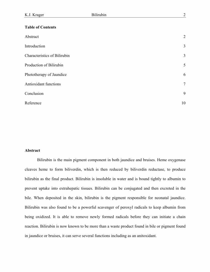
Table of Contents Abstract 2
Introduction 3
Characteristics of Bilirubin 3
Production of Bilirubin 5
Phototherapy of Jaundice 6
Antioxidant functions 7
Conclusion 9
Reference 10
Abstract
Bilirubin is the main pigment component in both jaundice and bruises. Heme oxygenase
cleaves heme to form biliverdin, which is then reduced by biliverdin reductase, to produce
bilirubin as the final product. Bilirubin is insoluble in water and is bound tightly to albumin to
prevent uptake into extrahepatic tissues. Bilirubin can be conjugated and then excreted in the
bile. When deposited in the skin, bilirubin is the pigment responsible for neonatal jaundice.
Bilirubin was also found to be a powerful scavenger of peroxyl radicals to keep albumin from
being oxidized. It is able to remove newly formed radicals before they can initiate a chain
reaction. Bilirubin is now known to be more than a waste product found in bile or pigment found
in jaundice or bruises, it can serve several functions including as an anitoxidant.

K.J. Krager Bilirubin 3
Introduction
Georg Stadeler, in 1864, originally defined bilirubin as the dark red pigment in bile [2].
He also discovered the accumulation of bilirubin was the cause of the yellow color in jaundice
patients. It was speculated that bilirubin was structural similar to both biliverdin and haematoidin
[2]. Bilirubin was also found to be in two forms; direct reacting bilirubin found in the bile, and
indirect found in blood [2]. Bilirubin when in the blood is bound tightly to albumin in a 1:1
stoichiometry, and is insoluble in water at physiological pH [1]. Bilirubin was also found to be a
powerful scavenger of peroxyl radicals to keep albumin from being oxidized [1]. Phototherapy
has been key in the treatment of accumulation of bilirubin in neonatal jaundice. Bilirubin was
also found to be a powerful scavenger of peroxyl radicals.
Characteristics of Bilirubin The crystal structure of bilirubin was determined to be composed of a carbon backbone
with four pyrrole rings containing nitrogen. The unconjugated form contained internal hydrogen
oxygen bonds that cause it to be convoluted and infolded in a ridge-tile conformation and
therefore insoluble in water (Figure 1) [3]. The unconjugated or indirect form binds to albumin
and is soluble in the lipid bilayers of the cell membrane.
Figure 1. Molecular structure of the unconjugated form of bilirubin. The form is insoluble in water due to the internal hydrogen oxygen bonds [3]. M= −CH3 V= −CH=CH2.
K.J. Krager Bilirubin 4
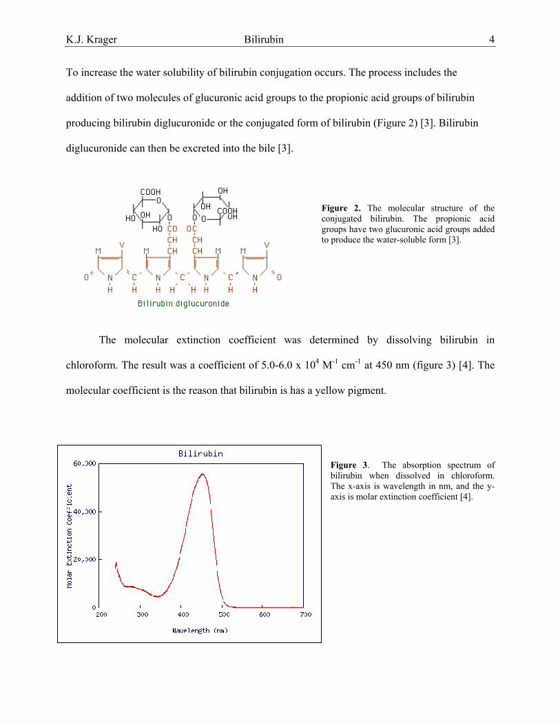
To increase the water solubility of bilirubin conjugation occurs. The process includes the
addition of two molecules of glucuronic acid groups to the propionic acid groups of bilirubin
producing bilirubin diglucuronide or the conjugated form of bilirubin (Figure 2) [3]. Bilirubin
diglucuronide can then be excreted into the bile [3].
Figure 2. The molecular structure of the conjugated bilirubin. The propionic acid groups have two glucuronic acid groups added to produce the water-soluble form [3].
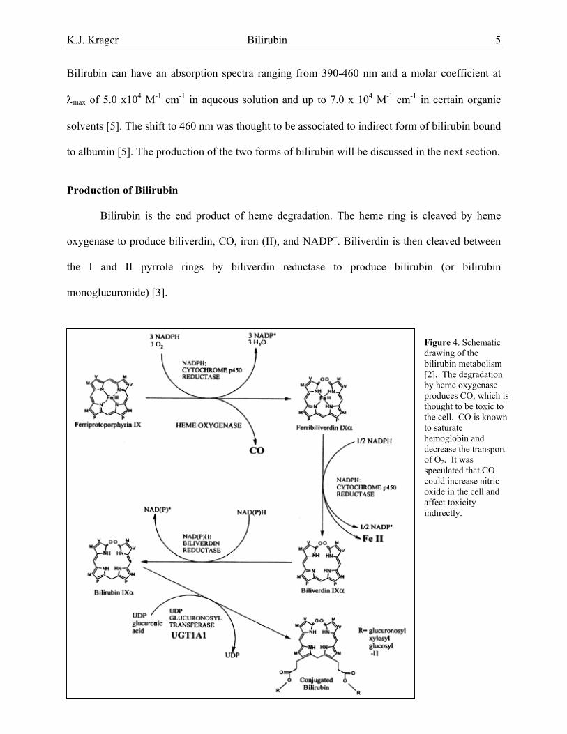
The molecular extinction coefficient was determined by dissolving bilirubin in
chloroform. The result was a coefficient of 5.0-6.0 x 10⁴ M-1 cm-1 at 450 nm (figure 3) [4]. The
molecular coefficient is the reason that bilirubin is has a yellow pigment.
Figure 3. The absorption spectrum of bilirubin when dissolved in chloroform. The x-axis is wavelength in nm, and the y-axis is molar extinction coefficient [4].
K.J. Krager Bilirubin 5
Bilirubin can have an absorption spectra ranging from 390-460 nm and a molar coefficient at
λmax of 5.0 x10⁴ M-1 cm-1 in aqueous solution and up to 7.0 x 10⁴ M-1 cm-1 in certain organic
solvents [5]. The shift to 460 nm was thought to be associated to indirect form of bilirubin bound
to albumin [5]. The production of the two forms of bilirubin will be discussed in the next section.
Production of Bilirubin Bilirubin is the end product of heme degradation. The heme ring is cleaved by heme
oxygenase to produce biliverdin, CO, iron (II), and NADP+. Biliverdin is then cleaved between
the I and II pyrrole rings by biliverdin reductase to produce bilirubin (or bilirubin
monoglucuronide) [3].
Figure 4. Schematic
drawing of the bilirubin metabolism [2]. The degradation by heme oxygenase produces CO, which is thought to be toxic to the cell. CO is known to saturate hemoglobin and decrease the transport of O2. It was speculated that CO could increase nitric oxide in the cell and affect toxicity indirectly.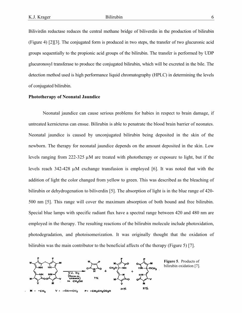
K.J. Krager Bilirubin 6
Bilivirdin reductase reduces the central methane bridge of biliverdin in the production of bilirubin
(Figure 4) [2][3]. The conjugated form is produced in two steps, the transfer of two glucuronic acid
groups sequentially to the propionic acid groups of the bilirubin. The transfer is performed by UDP
glucuronosyl transferase to produce the conjugated bilirubin, which will be excreted in the bile. The
detection method used is high performance liquid chromatography (HPLC) in determining the levels
of conjugated bilirubin.
Phototherapy of Neonatal Jaundice
Neonatal jaundice can cause serious problems for babies in respect to brain damage, if
untreated kernicterus can ensue. Bilirubin is able to penatrate the blood brain barrier of neonates.
Neonatal jaundice is caused by unconjugated bilirubin being deposited in the skin of the
newborn. The therapy for neonatal jaundice depends on the amount deposited in the skin. Low
levels ranging from 222-325 µM are treated with phototherapy or exposure to light, but if the
levels reach 342-428 µM exchange transfusion is employed [6]. It was noted that with the
addition of light the color changed from yellow to green. This was described as the bleaching of
bilirubin or dehydrogenation to biliverdin [5]. The absorption of light is in the blue range of 420-
500 nm [5]. This range will cover the maximum absorption of both bound and free bilirubin.
Special blue lamps with specific radiant flux have a spectral range between 420 and 480 nm are
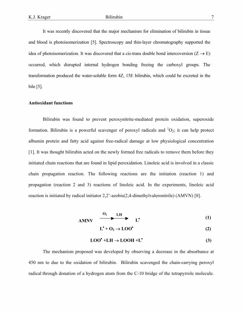
employed in the therapy. The resulting reactions of the bilirubin molecule include photoxidation,
photodegradation, and photoisomerization. It was originally thought that the oxidation of
bilirubin was the main contributor to the beneficial affects of the therapy (Figure 5) [7].
Figure 5. Products of bilirubin oxidation [7].
K.J. Krager Bilirubin 7
It was recently discovered that the major mechanism for elimination of bilirubin in tissue
and blood is photoisomerization [5]. Spectroscopy and thin-layer chromatography supported the
idea of photoisomerization. It was discovered that a cis-trans double bond intercoversion (Z → E)
occurred, which disrupted internal hydrogen bonding freeing the carboxyl groups. The
transformation produced the water-soluble form 4Z, 15E bilirubin, which could be excreted in the
bile [5].
Antioxidant functions
Bilirubin was found to prevent peroxynitrite-mediated protein oxidation, superoxide
formation. Bilirubin is a powerful scavenger of peroxyl radicals and 1O2; it can help protect
albumin protein and fatty acid against free-radical damage at low physiological concentration
[1]. It was thought bilirubin acted on the newly formed free radicals to remove them before they
initiated chain reactions that are found in lipid peroxidation. Linoleic acid is involved in a classic
chain propagation reaction. The following reactions are the initiation (reaction 1) and
propagation (reaction 2 and 3) reactions of linoleic acid. In the experiments, linoleic acid
reaction is initiated by radical initiator 2,2’-azobis(2,4-dimethylvaleronitrile) (AMVN) [8].
LH AMNV
L
LOO
The mechanism proposed was
450 nm to due to the oxidation of b
radical through donation of a hydroge
O2
(1)L• • + O2 → LOO• (2)
• +LH → LOOH +L• (3)
developed by observing a decrease in the absorbance at
ilirubin. Bilirubin scavenged the chain-carrying peroxyl
n atom from the C-10 bridge of the tetrapyrrole molecule.

K.J. Krager Bilirubin 8
The donation of the hydrogen atom creates a carbon-centered radical (BR•), the radical could
then react with the second peroxyl radical to produce another nonradical or oxygen (refer to
reactions 4, 5, & 6) [8]. The final reaction produced biliverdin, which is quickly recycled back
to bilirubin.
LOO• + BR → LOOH + BR• (4)
BR• +LOO• → BR-OOL (5)
BR• +O2 ↔ BR-OO• (6)
Bilirubin contains numerous conjugated double bonds that could contribute to the nonreactive of
the bilirubin free radical (BR•) (Figure 6). The arrow denotes one of the few bis-allelic bonds of
bilirubin, the weak attributes of this bond makes it a perfect canidate for donating the hydrogen
atom [9].
Figure 6. Schmatic of bilirubin the arrow denotes the weak bis-allelic
h
o
r
bond found in bilirubin which could donate a hydrogen atom [2, 9].
Another report stated that as little as 10 nM of bilirubin would protect against 10,000 fold
igher concentrations of H2O2 [10]. This finding suggested that bilirubin was acting in a way
ther than as a stoichiometric antioxidant. The group then went on to hypothesize that the
eaction of bilirubin with two peroxyl radicals to form biliverdin was then rapidly reduced back
K.J. Krager Bilirubin 9
to bilirubin. It was this redox cycle that accounted for the 10,000-fold amplification [10]. They
were able to show in experiments when bilirubin was reduced or inhibited that a greater
production of reactive oxygen species was observed [10]. Bilirubin acts much like glutathione
(GSH) in the respect that it exists in two forms, an oxidized form and a reduced form. The
greatest difference is the fact that bilirubin exists in less amounts [10]. Another report showed
bilirubin-albumin complexes could react with peroxyl radicals 3.1 times faster than uric acid but
not as efficiently as vitamin C [11]. It most be noted that most of the experiments that showed
bilirubin as a potent antioxidant was performed in vitro.
Conclusions
Bilirubin was discovered to be the end product of heme degradation. The unconjugated
form of bilirubin is tightly bound to albumin and is insoluble. This form is found deposited in the
skin of neonates where it can lead to jaundice. The esterification of the proprionic acid side
chains leads to the formation of conjugated form. Conjugated bilirubin is soluble and is excreted in
the bile. The absorption of light in the blue range of 420-500 nm lead to the photoisomerization of
bilirubin into the 4Z, 15E bilirubin that can be easily excreted. The antioxidant functions of
bilirubin showed a greater ability than glutathione in vitro and in recent in vivo experiments due
to the fact little was needed. It can react with two peroxyl radicals to form biliverdin and then be
rapidly reduced back to bilirubin in a classic redox cycle. The mechanism was suggested to be
the scavenging of newly formed free radicals before they could initiate a chain propagation
reaction. This recent finding could prove bilirubin as a better antioxidant then most known today.

K.J. Krager Bilirubin 10
References
1. Halliwell B, Gutteridge J. (1999) Free Radicals in Biology and Medicine: Oxford University Press Oxford New York:189-191.
2. Bosma P. (2003) Inherited disorders of bilirubin metabolism: a review. Journal of Hepatology. 38:107-117.
3. World wide web at http://medlib.med.utah.edu/NetBiochem/hi7c.htm (1995) Heme Degradation.
4. Giovanni A, Franco F. (1990) New trends in photobiology recent advances in bilirubin photophysics. J Photochem Photobiol. 7:1-14.
5. Ostrow J. (1986) Bile Pigments and Jaundice. Marcel Dekker, Inc New York, NY.
6. Mantle T. (2002) Haem degradation in animals and plants. Biochemical Society Transactions. 30:630-633.
7. Rodgers M, Powers E. (1981) Oxygen and Oxy-Radicals in Chemistry and Biology. New York, NY: Academic Press:434-435.
8. Stocker R, Yamamoto Y, McDonagh A, Glazer A, Ames B. (1987) Bilirubin is an antioxidant of possible physiological importance. Science. 235:1043-1046.
9. Buettner G. (2003) personal communication.
10. Baranano D, Rao M, Ferris C, Snyder S. (2002) Biliverdin reductase: a major physiologic cytoprotectant. Proc Natl Acad Sci USA. 99:16093-16098.
11. Stocker R, Glazer A, Ames B. (1987) Antioxidant activity of albumin-bound bilirubin. Proc Natl Acad Sci USA. 84:5918-5922.