Fossil woods from early Miocene sediments of the El Cien Formation, Baja California Sur, Mexico
23
Fossil woods from early Miocene sediments of the El Cien Formation, Baja California Sur, Mexico Hugo. I. Martínez-Cabrera a , Sergio R.S. Cevallos-Ferriz b, ⁎ , Imogen Poole c a Posgrado en Ciencias Biológicas, Instituto de Geología, Universidad Nacional Autónoma de México, Ciudad Universitaria, Circuito de la Investigación Científica, Del. Coyoacan, 04510 México D.F., Mexico b Departamento de Paleontología, Instituto de Geología, Universidad Nacional Autónoma de México, Ciudad Universitaria, Circuito de la Investigación Científica, Del. Coyoacan, 04510 México D.F., Mexico c Wood Anatomy Section, National Herbarium of The Netherlands, University of Utrecht Branch, P.O. Box 80102, 3585 CS Utrecht, The Netherlands Received 29 November 2004; received in revised form 3 December 2005; accepted 3 January 2006 Available online 20 February 2006 Abstract Five fossil woods are described from the Miocene El Cien Formation located in Baja California Sur, Mexico. Their anatomical characters suggest affinity to the Burseraceae, Euphorbiaceae, Leguminosae and Moraceae. The fossil species described here, together with previous records, demonstrate a high familial and generic floristic similarity with the extant vegetation of western Mexico (e.g. Jalisco). This floristic link suggests a landscape dominated by a tropical deciduous or semi-deciduous forest. However, the presence of additional taxa whose living relatives thrive in relatively moist conditions today suggests a relatively humid environment. The floristic similarity between the floras growing in this region today and those existing during the Miocene can be explained by the palaeogeographic link between these two landmasses (Baja California Sur and Western Mexico) that existed until approximately 5 mybp. © 2006 Elsevier B.V. All rights reserved. Keywords: Baja California Peninsula; Miocene; El Cien Formation; fossil woods; Mexico 1. Introduction Wegener (1922) hypothesized that the Peninsula of Baja California and the main American continent had a common origin. Now, after much research in the area, the tectonic events that gave rise to the present geographical configuration of Baja California are relatively well known (e.g. McKenzie and Morgan, 1969; Atwater, 1970; Mammerickx and Klitgord, 1982; Hausback, 1984). A complex series of geological events (detailed in, for example, Hausback, 1984) resulted in the severance of the Peninsula from North America, its eventual integration with the Pacific Plate, and the subsequent opening of the Gulf of California. During the deposition of sediments forming the El Cien Formation, Baja California was a structurally stable continental margin of western Mexico (McKenzie and Morgan, 1969; Atwater, 1970; Hausback, 1984). For this reason the fossil plants preserved in the El Cien Formation provide a unique opportunity to evaluate the floristic and environmental changes along the Review of Palaeobotany and Palynology 138 (2006) 141 – 163 www.elsevier.com/locate/revpalbo ⁎ Corresponding author. E-mail addresses: [email protected] (H.I. Martínez-Cabrera), [email protected] (S.R.S. Cevallos-Ferriz), [email protected] (I. Poole). 0034-6667/$ - see front matter © 2006 Elsevier B.V. All rights reserved. doi:10.1016/j.revpalbo.2006.01.001
Transcript of Fossil woods from early Miocene sediments of the El Cien Formation, Baja California Sur, Mexico

nology 138 (2006) 141–163www.elsevier.com/locate/revpalbo
Review of Palaeobotany and Paly
Fossil woods from early Miocene sediments of the El CienFormation, Baja California Sur, Mexico
Hugo. I. Martínez-Cabrera a, Sergio R.S. Cevallos-Ferriz b,⁎, Imogen Poole c
a Posgrado en Ciencias Biológicas, Instituto de Geología, Universidad Nacional Autónoma de México, Ciudad Universitaria,Circuito de la Investigación Científica, Del. Coyoacan, 04510 México D.F., Mexico
b Departamento de Paleontología, Instituto de Geología, Universidad Nacional Autónoma de México, Ciudad Universitaria,Circuito de la Investigación Científica, Del. Coyoacan, 04510 México D.F., Mexico
c Wood Anatomy Section, National Herbarium of The Netherlands, University of Utrecht Branch, P.O. Box 80102,3585 CS Utrecht, The Netherlands
Received 29 November 2004; received in revised form 3 December 2005; accepted 3 January 2006Available online 20 February 2006
Abstract
Five fossil woods are described from the Miocene El Cien Formation located in Baja California Sur, Mexico. Their anatomicalcharacters suggest affinity to the Burseraceae, Euphorbiaceae, Leguminosae and Moraceae. The fossil species described here,together with previous records, demonstrate a high familial and generic floristic similarity with the extant vegetation of westernMexico (e.g. Jalisco). This floristic link suggests a landscape dominated by a tropical deciduous or semi-deciduous forest.However, the presence of additional taxa whose living relatives thrive in relatively moist conditions today suggests a relativelyhumid environment. The floristic similarity between the floras growing in this region today and those existing during the Miocenecan be explained by the palaeogeographic link between these two landmasses (Baja California Sur and Western Mexico) thatexisted until approximately 5 mybp.© 2006 Elsevier B.V. All rights reserved.
Keywords: Baja California Peninsula; Miocene; El Cien Formation; fossil woods; Mexico
1. Introduction
Wegener (1922) hypothesized that the Peninsula ofBaja California and the main American continent had acommon origin. Now, after much research in the area,the tectonic events that gave rise to the presentgeographical configuration of Baja California arerelatively well known (e.g. McKenzie and Morgan,
⁎ Corresponding author.E-mail addresses: [email protected]
(H.I. Martínez-Cabrera), [email protected](S.R.S. Cevallos-Ferriz), [email protected] (I. Poole).
0034-6667/$ - see front matter © 2006 Elsevier B.V. All rights reserved.doi:10.1016/j.revpalbo.2006.01.001
1969; Atwater, 1970; Mammerickx and Klitgord, 1982;Hausback, 1984). A complex series of geological events(detailed in, for example, Hausback, 1984) resulted inthe severance of the Peninsula from North America, itseventual integration with the Pacific Plate, and thesubsequent opening of the Gulf of California. Duringthe deposition of sediments forming the El CienFormation, Baja California was a structurally stablecontinental margin of western Mexico (McKenzie andMorgan, 1969; Atwater, 1970; Hausback, 1984). Forthis reason the fossil plants preserved in the El CienFormation provide a unique opportunity to evaluatethe floristic and environmental changes along the

142 H.I. Martínez-Cabrera et al. / Review of Palaeobotany and Palynology 138 (2006) 141–163
northwestern coast of Mexico associated with tectonicand geographic processes that have guided the evolutionof this area.
Although studies of fossil floras in Mexico aresomewhat limited, information determined from themacrofossil angiosperm remains have provided inter-esting data pertaining the biodiversity during theCretaceous (e.g. Cevallos-Ferriz and Weber, 1992;Cevallos-Ferriz and Ricalde-Moreno, 1995; Rodrí-guez-de la Rosa and Cevallos-Ferriz, 1994; Hernán-dez-Castillo and Cevallos-Ferriz, 1998; Estrada-Ruiz,2004) and Tertiary (e.g. Magallón-Puebla and Cevallos-Ferriz, 1993, 1994a,b,c; Velasco de León and Cevallos-Ferriz, 1997; Velasco de León et al., 1998; Ramírez-Garduño, 1999; Calvillo-Canadell and Cevallos-Ferriz,2002; Ramírez and Cevallos-Ferriz, 2002) as well as thefloristic evolution (Cevallos-Ferriz and Ramírez-Gar-duño, 1998), continental palaeoclimates (Velasco deLeón, 1999) and biogeography (e.g. Ramírez et al.,2000; Martínez-Millán, 2000, 2003). This study focuseson the Baja California wood flora complementing thepalaeoecological data already available and providesimportant new information regarding the Miocenevegetation of Mexico.
2. Materials
In the southern part of the Baja California Peninsula,approximately 100 km to the northwest of La Paz, BajaCalifornia Sur (Fig. 1), the clastic sedimentary rocks ofthe El Cien Formation are exposed (Applegate, 1986).The El Cien Formation is a sedimentary sequence
Fig. 1. Maps showing the location of the El Cien
deposited during the late Oligocene–early Miocene(Applegate, 1986; Fischer et al., 1995). It restsunconformably on the sandstone of the TepetateFormation and is overlain by the volcanic rocks of theComondú Formation (Applegate, 1986; Fischer et al.,1995). Although Applegate (1986), who originallyproposed the El Cien Formation as a stratigraphic unit,divided it into three members, the Cerro Tierra Blanca,San Hilario and Cerro Colorado, the general consensusis that the two basal members form a single geologicalunit. The El Cien Formation corresponds to the (1)Monterrey Formation (Darton, 1921; Heim, 1922; Beal,1948; Mina, 1957; Ojeda, 1959; Alatorre, 1988), (2) SanGregorio Formation (Hausback, 1984; Kim and Barron,1986), and (3) the basal member of the El CienFormation (San Juan Member) sensu Schwennicke(1994) and Fischer et al. (1995).
The woods described here are preserved as silicapermineralizations and were collected from the upperCerro Colorado Member of the El Cien Formation nearRancho Matanzas and Cañada El Canelo, located 5 kmnortheast and 3.5 km southwest from El Cien town,respectively (Fig. 1). The sediments of this Member arecomposed mainly of fine to coarse-grained sandstones,tuffaceous sandstones and conglomerates (Applegate,1986; Fischer et al., 1995) and represent a prograda-tional sequence from offshore to non-marine environ-ments (Gidde, 1992). The change in depositionalenvironment is evidenced by the presence of primarysedimentary structures and ichnofossils at the base–indicative of near shore environments–and by root castsof fossil plants and fossil caliche deposits towards the
Formation and the fossil wood localities.

143H.I. Martínez-Cabrera et al. / Review of Palaeobotany and Palynology 138 (2006) 141–163
top, indicating the change from lagoonal to terrestrialenvironments.
There has been no radiometric dating of the CerroColorado Member itself. However, K–Ar radiometricdates for the underlying member give an age of 25.5±0.4 my, whilst the overlying volcanic rocks have beendated at 21±0.4 my (Hausback, 1984). Therefore, weassume the Cerro Colarado Member is approximately25–20 my old.
3. Methods
The fossil woods were thin-sectioned in transverse,tangential and radial sections using standard techniquesfor petrified woods (e.g. Haas and Rowe, 1999). Thedescriptions of the five morphospecies are based on theobservations of 19 different trees. The group of slidesrepresenting each tree is given its own IGM-LPB seriesnumber. With the exception of vessel frequency, whichwas calculated based on counts of 15 fields of view, thequantitative characteristics are the result of at least 25measurements with the mean (x) and standard deviation(s) provided. Measurement techniques and terminologyfollow, in general, that described by an IAWACommittee (1989).
Familial affinities were determined by consultingreference descriptions such as Metcalfe and Chalk(1950), Détienne and Jacquet (1983), Ilic (1987, 1991),Terrazas (1994) and searches of the computerisedOPCN (Wheeler et al., 1986; LaPasha and Wheeler,1987) and CSIRO Family Key (Ilic, 1987) wooddatabases. Comparisons were then made with extantwood samples housed in the National Xilothec of theInstitute of Biology, UNAM, and the National Herbar-ium of The Netherlands, Utrecht University Branch(sample numbers prefixed by Uw). All fossil specimensare deposited in the National Paleontological Collection,Institute of Geology, Universidad Nacional Autónomade Mexico (UNAM), Mexico.
4. Systematic palaeobotany
Family: BurseraceaeGenus: Tetragastroxylon gen. nov.Generic diagnosis: Fossil angiosperm wood. Vesselspredominantly solitary; perforation plates simple, inter-vascular pits alternate, oval to polygonal, with lenticularapertures; vessel-ray pits elongated with reduced tosimple borders; fibers septate; axial parenchyma absent;rays heterocellular, predominantly uniseriate, occasion-ally multiseriate; radial canals present in multiseriaterays.
Type species: Tetragastroxylon magniporus gen. et sp.nov. Plate I, 1–8.Etymology: The generic name highlights the anatomicalresemblance of the fossil with the wood of Tetragastris.The specific epithet reflects the large lumen of thevessels.Holotype: LPB 259–267(ca. 4 cm).Additional material: LPB 1183–1192 (14 cm).Repository: Paleontological Collection of the Institutode Geología, UNAM, Mexico.Stratigraphic horizon: Cerro Colorado Member of theEl Cien Formation, early Miocene.Locality: Rancho Matanzas, Mexico (Fig. 1).Species diagnosis: Wood diffuse porous, growth ringsindistinct; vessels solitary or in radial multiples of 2–4with average vessel diameter N200 μm; perforationplates simple, intervascular pits alternate, oval to poly-gonal, with lenticular apertures; vessel-ray pits withreduced to simple borders, slightly or prominently elon-gated; fibers septate with 1–4 septa per fiber; axialparenchyma absent; rays heterocellular, mostly uniseri-ate, some multiseriate (2–4 cells) with one or two radialcanals per ray; druses and spherical contents in radialtissue.Description: This description is based on two pieces ofwood with estimated diameters of 4 and 14 cm.Diffuse porous wood and indistinct growth rings(Plate I, 1). Vessels circular in outline, solitary and inradial multiples of 2–4, with vessel grouping index of1.37. Vessel element tangential diameter ranges from140 to 327 μm (x=226, s=45), length varies from 250to 640 μm (x=366, s=70 μm), wall thickness rangesfrom 5 to 13 μm (x=8.6, s=1.9 μm), and frequencyvaries from (5) 7 to 16 mm−2 (x=9.5, s=2.6 mm−2).Abundant thin-walled tyloses (Plate I, 1, 3). Perfora-tion plates exclusively simple with slightly obliqueend walls. Intervascular pits alternate and oval topolygonal in outline, with lenticular apertures, 4–8 μm(x=5.8, s=1.1 μm) in diameter. Vessel-ray pitsslightly to definitely horizontally elongated withreduced or simple borders (Plate I, 7, 8). Fibers with1–4 septate per fiber (Plate I, 3, 6), tangentialdiameter from 5–18 μm (x=12.3, s=3.6 μm) andwall-thickness between 1 and 4 μm (x=2, s=1.1), pitsnot observed in fiber walls. Axial parenchyma absent(Plate I, 1). Predominantly uniseriate rays (99%) (PlateI, 3, 6), 5–11 rays per mm. Multiseriate raysheterocellular (Plate I, 3–6) with ray body composedof procumbent cells and 2–4 marginal rows ofupright/square cells, one or two canals per ray, 50–95 μm (x=61, s=17) in diameter (Plate I, 2).Uniseriate rays are 6–14 (x=8.3, s=2.1) cells high

144 H.I. Martínez-Cabrera et al. / Review of Palaeobotany and Palynology 138 (2006) 141–163
and composed of procumbent and square cells. Somedruses and spherical contents resembling oil drops inprocumbent parenchyma cells (Plate I, 4).Affinities:The presence of septate fibers, heterocellularrays and radial canals, plus the absence of (or verypoorly developed) parenchyma suggest similarity of thisfossil to the woods of Anacardiaceae and Burseraceae.
The high degree of similarity in wood charactersbetween extant members of these two families supportsthe suggestion of a common ancestry for these twofamilies based on molecular characters (rbc L) andmorphological/anatomical analyses (Terrazas, 1994;Martínez-Millán, 2000, 2003). The high percentage ofuniseriate rays in combination with the presence of

Table 1Wood characteristics used in the identification of the fossil materialfrom selected genera of Anacardiaceae and Burseraceae
Family Genera Radialcanals
Uniseriaterays
Septatefibers
Anacardiaceae Astronium (2 spp) + + − +Anacardium (2 spp) − +(−) −Buchanania latifolia − − −Choerospondiasaxillaris
− − + −
Cotinus coggygria − − −Cyrtocarpavelutinifolia
+ − +
Dracontomeloum dao − − − +Gluta rhengas + + (−) −Lannea welwitschii + − −Loxopterigyumsagotti
+ − + +
Mangifera indica − − −Pistacia chinensis + − −Pseudospondiasmicrocarpa
− − −
Rhus typhina − − −Sclerocarya birrea − − −Spondias (2 spp) + − −Thyrsodiumguianenese
+ − + −
Burseraceae Aucoumea klaineana − − +CanariumShweinfurtii
− − +
Commiphora (2 spp) + − +Dacryodes (3 spp) − − +Hemicrepidospermumrhoifolium
− + (−) +
Protium (12 spp) + − + (−) +Tetragastris (3 spp) + + (−) +Trattinickia (2 spp) − − +
145H.I. Martínez-Cabrera et al. / Review of Palaeobotany and Palynology 138 (2006) 141–163
radial canals, septate fibers and absence of axialparenchyma are the most important characteristicslinking the fossil wood with extant members of thesefamilies (Table 1).
In some Anacardiaceae, such as Anacardium,Swintonia (Terrazas, 1994), Gluta, Astronium andLoxopterygium, uniseriate rays dominate. However, incontrast with the fossil, Anacardium and Gluta haveabundant axial parenchyma and Swintonia has non-septate fibers (Terrazas, 1994). Some specimens ofLoxopterygium (e.g. L. sagotii Uw 4239) and Astroniumhave high proportions of uniseriate rays (similar to thoseseen in the fossil wood), but differ since they arepredominantly heterogeneous IIB rays (Terrazas, 1994).The presence of predominantly or exclusively uniseriaterays is relatively common in many species of Burser-aceae including the majority of Dacryodes, Protium andTetragastris species reviewed during the course of thisresearch (Table 1).
Axial parenchyma is generally less abundant in theBurseraceae when compared with the Anacardiaceae.In particular, many species of Protium and Tetragas-tris (Burseraceae) have scanty paratracheal parenchy-ma along with exclusively uniseriate rays (Tables 1and 2), septate fibers and radial canals (Plate I, 9–12),thus resembling the overall anatomical condition ofthe fossil woods. Although the number of cells peruniseriate ray in the fossil wood agrees with thenumber of cells per ray in Protium, the fossil woodrays are more similar to those of Tetragastris (Table2) even though the size of the ray cells and proportionof square cells are greater in the fossil when comparedwith the two extant genera. This latter character be-comes more obvious if the ratio between the numberof cells and ray height is calculated. The quotient forthe fossil (22) is greater than that of the extant spe-cies, T. panamensis (9) and T. altisima (11), studied.The rays in the fossil have the same frequency (2)
Plate I. Light micrographs of Tetragastroxylon magniporus (Figs. 1–8) and
1. Transverse section (Ts). Diffuse porous wood and axial parench2. Tangential section (Tg). Radial canals in a multiseriate ray. LPB3. Tg. Exclusively uniseriate rays and vessels filled with thin-wal4. Radial section (Rs). Spherical contents in ray parenchyma cells5. Rs. Procumbent and square cells in the radial tissue. LPB-IGM6. Tg. Uniserate rays and septate fibers. LPB-IGM 266. Scale bar7, 8. Rs. Horizontally elongated vessel-ray pits, note the reduced bo9. Ts. Diffuse porous wood and scanty paratracheal axial parench10. Tg. Radial canals in a multiseriate rays. Scale bar=100 μm.11. Rs. Heterocellular ray. Scale bar=150 μm.12. Tg. Uniseriate rays and septate fibers. Scale bar=150 μm.
of canals per ray as seen in Tetragastris (Table 2;Plate I, 2, 10). However, there are certain anatomicaldifferences between the fossil wood and Tetragarisand Protium to which it shows greatest similarity.
Tetragastis altissima Uw 658 (Figs. 9–12).
yma absent. LPB-IGM 260. Scale bar=1 mm.-IGM 265. Scale bar=100 μm.
led tyloses. LPB-IGM 265. Scale bar=1 mm.. LPB-IGM 267. Scale bar=100 μm.267. Scale bar=150 μm.=100 μm.rders. LPB-IGM 267. Scale bar=10 μm.yma. Scale bar=1 mm.

Table 2Anatomical comparison between Tetragastroxylon magniporus and selected extant Tetragastris and Protium species exhibiting similar anatomy.V=vessel; r= rays; p=procumbent; s=square; u=upright
Character Tetragastroxylonmagniporus
Tetragastris altísimaUw 658
Tetragastrispanamensis Uw 11062
Protium alstoniiUw 1845
Protium polybotryumUw 4589
Porosity Diffuse Diffuse Diffuse Diffuse Diffuse
Growthrings
Indistinct Delimited by 2–3flattened latewoodfibers
Weakly delimitedflattened latewoodfibers
Indistinct Indistinct
V/Frequency(v/mm2)
9.5 (7–16) 15.9 (14–19) 12.3 (4–20) 12.1 (7–11–15) 14 (11–18)
Diameter(μm)
225 (140–327) 79 (56–103) 98 (61–124) 98 (67–122) 105 (72–139)
Grouping 1.37 (77% solitary) 1.28 (80% solitary) 1.32 (85% solitary) 1.18 (89% solitary) 1.53 (77% solitary)
Wallthickness(μm)
8.6 (5–13) 4.2 (2.9–7) 3.5 (2.9–6) 2.8 (2.3–3.4) 3.2 (2.3–5.8)
Intervessel pits(μm)
Alternate, oval topolygonal, withlenticular apertures.5.8 (4–8) diameter
Alternate, oval topolygonal, lenticularapertures. 6.3(4.3–8.7) diameter
Alternate, oval topolygonal, lenticularapertures. 5.8(3–8.7) diameter
Alternate oval topolygonal 6.4(5.5–8.7) diameter
Alternate oval topolygonal 7(5.8–8.7) diameter
Fibers Septate 1–3 septaper fiber
Septate 3–4 septaper fiber
Septate 3–4 septa perfiber
Septate 2–4 septaper fiber
Septate 3–5 septaper fiber
Ray composition Uniseriate,multiseriate less than1%. Uniseriate rayscomposed of p and scells, some u cells
Mostly uniseriate,multiseriate 18%.Uniseriate rayscomposed of p,s and u cells
Mostly uniseriate,multiseriate less than20% composed of p,s and u cells
Mostly uniseriate,multiseriate less than10% composed of p,u cells, high proportionof s cells (comparedwith upright)
Mostly uniseriate,biseriate less than10% composed ofp and u cells, highproportion of s cells(compared with u)
Frequency(r/mm)
7 (5–11) 6.4 (4–9) 7.9 (6–11) 5.2 (3–4–8) 5.8 (4–8)
Height uniseriater (# cells, μm)
4–14 308 (145–555) (2)4–11(16)179 (70–324)
(2)4–17229 (94–427)
3–23 275 (89–446) (2)3–18 242 (66–381)
Radial canal One sometimes twocanals per ray, 60 μm(50–95 μm)
One sometimes twocanals per ray 28 μm(23–32 μm)
One sometimes twocanals per ray52.6 μm (28–66 μm)
One canal per ray89.3 μm (70–94 μm)
One canal per ray42.3 μm (38–47 μm)
Vessel-raypits
Reduced borders tosimple, round orirregular, horizontallyelongated
Reduced borders tosimple, round or,horizontallyelongated
Reduced borders tosimple, round or,horizontallyelongated
Reduced borders tosimple, round or,horizontally elongatedto irregular (some)
Reduced borders tosimple, round or,horizontally elongatedto irregular (some)
Axialparenchyma
Absent Scanty paratracheal Scanty paratracheal Scanty paratrachealand diffuse apotracheal
Scantyparatracheal
146 H.I. Martínez-Cabrera et al. / Review of Palaeobotany and Palynology 138 (2006) 141–163
The fossils have a higher conductive area up to twotimes greater the mean vessel diameter and a lowermean vessel frequency (Table 2). Another differenceis the absence of axial parenchyma in the fossilmaterial, but is present in the extant material. Eventhough Protium, Tetragastris and this fossil morpho-type show very similar anatomical characteristics,based on the quantitative and qualitative similarities/
differences discussed above and summarised in Table2, the fossils are most similar anatomically to speciesof Tetragastris.
Several other fossil woods with anatomy similar tospecies of Burseraceae, and Tetragastris in particular,have been collected from Europe and Asia (e.g. DenBergen, 1923; Dayal, 1964; Prakash et al., 1974;Prakash and Tripathi, 1975; Ghosh and Roy, 1978;

147H.I. Martínez-Cabrera et al. / Review of Palaeobotany and Palynology 138 (2006) 141–163
Lakhanpal et al., 1981; Bande and Prakash, 1983;Trivedi and Srivastava, 1985; Awasthi and Srivastava,1989; Awasthi and Mehrotra, 1993). The majordistinction between the El Cien Formation fossil woodand the Asian fossil woods relates to the proportion ofuniseriate rays and occurrence of axial parenchyma:Boswellioxylon indicum (Dayal, 1964) and Sumatrox-ylon molli (Den Bergen, 1923) have obvious axialparenchyma, few uniseriate rays and multiseriate raysup to 6 cells wide. Species of Burseroxylon (B.preserratum and B. garugoides; Prakash and Tripathi,1975; Lakhanpal et al., 1981, respectively) lack radialcanals, have axial parenchyma and predominantlymultiseriate rays. In Canarioxylon ceskobudejovicense(Prakash et al., 1974), from Czech Republic, and C.indicum from India (Ghosh and Roy, 1978), radialcanals are absent. Awasthi and Srivastava (1989)describe a wood, Canarium palaeoluzonicum, fromthe Neogene flora of the Kerala Basin that they believeis related to Canarium. This has great similarity with thefossil from El Cien Formation; although, Canariumpalaeoluzonicum (Awasthi and Srivastava, 1989) has ahigh frequency of uniseriate rays, they are moreabundant (N90%) in the fossil from the El CienFormation.
The absence of axial parenchyma in the fossil woodsdescribed here, coupled with the particularly highincidence of the uniseriate rays and its closer similarityto extant members of Protium and particularly Tetra-gastris (Burseraceae) than to fossil burseraceous woodspreviously described, support the erection of a newmorphogenus.
Family: EuphorbiaceaeGenus: ParaphyllanthoxylonBailey (1924)Species: Paraphyllanthoxylon mennegae sp. nov. PlateII, 1–8.Etymology: The specific epithet is after A.M.W.Mennega in recognition of her work on the woodanatomy of the Euphorbiaceae.Holotype: LPB 227–258 (4 cm).Additional material: LPB 4120–4134 (ca. 6 cm); LPB4203–4217 (ca. 9.5 cm); LPB 3573–3574 (10.5 cm);LPB 982–989, 991, 993–1000 (ca. 6.2 cm).Repository: Paleontological Collection of the Institutode Geología, UNAM, Mexico.Stratigraphic horizon: Cerro Colorado Member of theEl Cien Formation, early Miocene.Locality: Rancho Matanzas (LPB 227–258; LPB 982–989, 991, 993–1000) and Cañada El Canelo (LPB4120–4134; LPB 4203–4217; LPB 3573–3574), Mex-ico (Fig. 1).
Species diagnosis: Diffuse porous, growth ringsindistinct; vessels solitary or in radial multiples of 2–4 (5) with average tangential diameter b100 μm;perforation plates simple, intervascular pits alternate,oval to polygonal; vessel-ray pits oval to irregularlyshaped, horizontally elongated to scalariform; 2–3septa per fiber; axial parenchyma absent; rays he-terocellular, uniseriate and multiseriate; uniseriate rayscomposed exclusively of upright cells, 4–14 cells high;multiseriate rays 2–3, exceptionally 4 cells wide,b1500 μm high and composed predominantly ofupright and square cells with few procumbent cells;uniseriate margins composed of 1–6 cells.Description: This description is based on five pieces ofwood with estimated diameters ranging from 4 to 10.5cm. Diffuse porous wood with indistinct growth rings.Vessels oval in cross section, solitary and in radialmultiples of 2–4 (5), with 1.8 vessels per group (Plate II,1). Vessel element tangential diameter ranges from 40 to97.5 μm (x=71 μm, s=16), length varies from 215 to795 μm (x=469 μm, s=120), wall thickness ranges from2 to 5 μm (x=3.2 μm, s=0.88), and frequency variesfrom 23 to 66 mm−2 (x=45.9 mm−2, s=10). Perforationplates simple with oblique end walls (Plate II, 5).Intervascular pits oval and alternate with circular orelliptic apertures (Plate II, 7), 3–7 μm (x=4.8 μm,s=1.1) in diameter. Vessel-ray pits horizontally elongat-ed with reduced to simple borders (Plate II, 4, 6). Thin-walled tyloses. Fibers with 2–3 septa per fiber (Plate II,2, 3, 8). Fiber tangential diameter ranges from 6 to 24 μm(x=13.6 μm, s=5.5) and wall-thickness varies from 1 to8 μm (x=4.1 μm, s=1.6). Axial parenchyma absent. Rayfrequency 6–9 per mm (x=7.5 per mm, s=0.83/mm).Rays heterocellular (Plate II, 2–4, 8), uniseriate andmultiseriate. Multiseriate rays 2–3 (–4) cells wide and310–1450 μm (x=755 μm, s=316 μm) high. Body ofmultiseriate rays composed of predominantly square andupright cells, with uniseriate extensions composed of 1–6 (x=2.4, s=1.3) rows of upright cells. Uniseriate rayscomposed entirely of upright cells, 4–14 (x=6.5, s=2.9)cells high. Dark contents in ray cells common.
Species: Paraphyllanthoxylon coloradensis sp. nov.Plate III, 1–8.Etymology: The specific epithet reflects the geologicunit from which the fossils were collected.Holotype: LPB 1044–1063 (5.4 cm).Additional material: LPB 3585 (ca. 7 cm); LPB 1002–1004 (7.5 cm); LPB 1196–1204 (2.2 cm); LPB 1174–1182 (ca. 14 cm).Repository: Paleontological Collection of the Institutode Geología, UNAM, Mexico.

148 H.I. Martínez-Cabrera et al. / Review of Palaeobotany and Palynology 138 (2006) 141–163
Stratigraphic horizon: Cerro Colorado Member of theEl Cien Formation, early Miocene.Locality: Rancho Matanzas (LPB 1044–1063; LPB1002–1004. LPB 1196–104. LPB 1174–1182) andCañada El Canelo (LPB 3585), Mexico (Fig. 1).
Species diagnosis: Wood diffuse porous, growth ringsindistinct; vessels solitary or in radial multiples of 2–4,sometimes more, average tangential diameter N100 μm;perforation plates simple, intervascular pitting alternate,oval to polygonal; vessel-ray pits horizontally elongated

149H.I. Martínez-Cabrera et al. / Review of Palaeobotany and Palynology 138 (2006) 141–163
to scalariform; fibers septate, 2–3 septa per fiber, or non-septate; axial parenchyma absent; rays uniseriate andmultiseriate; uniseriate rays homocellular composedexclusively of upright cells, 3–14 cells high; multi-seriate rays heterocellular, 2–3 cells wide and up to3400 μm high, composed predominantly of procumbentcells with some upright and square cells; some rays havebiseriate and uniseriate portions within a constant width,and uniseriate margins 1–8 cells high.Description: This description is based on five pieces ofwood with estimated diameters of ∼2 to 14 cm. Diffuseporous wood with indistinct growth rings (Plate III, 1).Vessels in cross section oval, solitary and in radialmultiples of 2–4, sporadically longer, vessel groupingindex of 1.58. Vessel element tangential diameter variesfrom 75 to 185 μm (x=131 μm, s=23.6 μm), lengthranges from 410 to 830 μm (x=627 μm, s=124 μm),wall-thickness between 2 and 6 μm (x=3.5 μm,s=1.1), and frequency ranges from 15 to 36 mm−2
(x=24.4 mm−2, s=5.9). Perforation plates simple withoblique end walls (Plate III, 7, 8). Intervascular pitsoval to polygonal, alternate, and 10–13 μm (x=11.5μm, s=1) in diameter. Vessel-ray parenchyma pitshorizontally elongate, with simple to reduced borders(Plate III, 5, 6). Thin-walled tyloses. Fibers with 2–3septa per fiber, or non-septate (Plate III, 2–4), withtangential diameter ranging from 10 to 28 μm (x=18.2μm, s=5.6) and wall-thickness varies from 1 to 6 μm(x=2 μm, s=1.4). Axial parenchyma absent (Plate III,1). Rays uniseriate and multiseriate (Plate III, 2–4),with frequency of 5–11 per mm (x=8.1 per mm,s=1.6). Multiseriate rays heterocellular, 2–3 cellswide and 600–3400 μm (x=1586 μm, s=624) high,with body composed predominantly of procumbentcells although upright and square cells can be present.Uniseriate extensions of multiseriate rays composed of1–8 (x=4.1, s=2.1) upright cells. Uniseriate rayshomocellular, 3–14 (x=7.5, s=2.6) cells high andcomposed exclusively of upright cells. Some rays
Plate II. Light micrographs of Paraphyllanthoxylon mennegae (Figs. 1–8) an
1. Ts. Diffuse porous wood, mostly radial multiples of two cells a2, 8. Tg. 1–3 seriate heterocellular rays and septate fibers. LPB-IGM3. Tg. Septate fibers. LPB-IGM 238. Scale bar=50 μm.4. Rs. Heterocellular ray, note the upright cells and the scalariform5. Rs. Simple perforation plates. LPB-IGM 243. Scale bar=70 μm6. Rs. Oval to horizontally elongated vessel-ray pits. LPB-IGM 47. Rs. Alternate intervascular pitting. LPB-IGM 4209. Scale bar=9. Ts. Long radial multiples. Scale bar=500 μm.10, 11. Tg. Heterocellular and septate fibers. Scale bar=100 μm.12. Rs. Oval to scalariform vessel-ray pits in upright cells. Scale ba
have alternating biseriate and uniseriate portions withconstant width.Affinities: The Paraphyllanthoxylon wood type was firstdescribed by Bailey (1924) and the organ genus hasbeen used repeatedly for fossil woods characterised byweakly differentiated growth rings, simple perforationplates, alternate intervascular pits, large vessel-ray pits,scanty to absent axial parenchyma, septate fibers,heterocellular rays and tyloses. The fossil woodsdescribed here fit well with the suite of diagnosticcharacters given above and thus the woods are assignedto Paraphyllanthoxylon. The two morphotypes from theEl Cien Formation can be separated based on the relativevessel diameter, amount of septate fibers, multiseriateray height and cellular composition and the presence ofuniseriate and biseriate portions in some rays withconstant ray width.
The Paraphyllanthoxylon wood type is common inthe sedimentary record, both in time and space (e.g.Bailey, 1924; Spackman, 1948; Mädel, 1962; Prakash,1958; Cahoon, 1972; Thayn et al., 1983; Prakash et al.,1986; Wheeler et al., 1987, 1995; Wheeler, 1991;Herendeen, 1991; Cevallos-Ferriz and Weber, 1992;Meijer, 2000). The anatomy exhibited by Paraphyl-lanthoxylon is commonly associated with that ofAnacardiaceae, Burseraceae, Elaeocarpaceae, Euphor-biaceae, Flacourtiaceae and Lauraceae, amongst others.While many authors accept that the anatomy ofParaphyllanthoxylon is closest to the Euphorbiaceae(Mädel, 1962; Privé, 1975; Thayn et al., 1983; Thaynand Tidwell, 1984; Prakash et al., 1986), not allresearchers agree in considering the similarity enoughto assign it to this family (Bailey, 1924; Spackman,1948; Cahoon, 1972; Wheeler, 1991).
Based on the occurrence and number of marginal cellsassociated with the multiseriate rays and the amount ofthe uniseriate rays, Wheeler et al. (1987) and Herendeen(1991) recognise two anatomical groups within Para-phyllanthoxylon: (1) those Paraphyllanthoxylon species
d its living analogue Phyllanthus sellowianus Uw 14074 (Figs. 9–12).
nd axial parenchyma absent. LPB-IGM 227. Scale bar=1 mm.238. Scale bar=300 μm, 100 μm respectively.
vessel-ray pitting. LPB-IGM 243. Scale bar=50 μm..
209. Sacle bar=20 μm.20 μm.
r=50 μm.
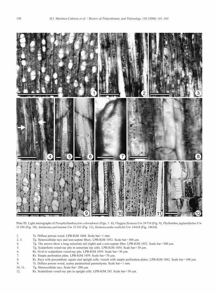
Plate III. Light micrographs of Paraphyllanthoxylon coloradensis (Figs. 1–8), Fluggea flexuosa Uw 24716 (Fig. 9), Phyllanthus juglandifolius Uw31189 (Fig. 10), Antidesma pulvinatum Uw 31193 (Fig. 11), Hymenocardia wallichii Uw 14634 (Fig. 14634).
1. Ts. Diffuse porous wood. LPB-IGM 1044. Scale bar=1 mm.2, 3. Tg. Heterocellular rays and non-septate fibers. LPB-IGM 1052. Scale bar=300 μm.4. Tg. The arrows show a long uniseriate tail (right) and a non-septate fiber. LPB-IGM 1052. Scale bar=300 μm.5. Tg. Scalariform vessel-ray pits in uniseriate ray cells. LPB-IGM 1054. Scale bar=30 μm.6. Rs. Oval to scalariform vessel-ray pits. LPB-IGM 1059. Scale bar=30 μm.7. Rs. Simple perforation plate. LPB-IGM 1059. Scale bar=70 μm.8. Rs. Rays with procumbent, square and upright cells; vessels with simple perforation plates. LPB-IGM 1062. Scale bar=100 μm.9. Ts. Diffuse porous wood, scanty paratracheal parenchyma. Scale bar=1 mm.10, 11. Tg. Heterocellular rays. Scale bar=200 μm.12. Rs. Scalariform vessel-ray pits in upright cells. LPB-IGM 243. Scale bar=30 μm.
150 H.I. Martínez-Cabrera et al. / Review of Palaeobotany and Palynology 138 (2006) 141–163
151H.I. Martínez-Cabrera et al. / Review of Palaeobotany and Palynology 138 (2006) 141–163
with few, short marginal extensions in the multiseriaterays and few or rare uniseriate rays (P. utahense, P.idahoense, P. marylandense, P. alabamense, P. capense)have greater affinity with Lauraceae, Elaeocarpaceae,Anacardiaceae, Burseraceae and Verbenaceae; and (2)Paraphyllanthoxylon species with abundant uniseriaterays and long uniseriate extensions of the multiseriaterays (i.e. P. arizonense and P. illinoisense) areanatomically closer to Ulmaceae, Flacourtiaceae,Euphorbiaceae and Simaroubaceae (Herendeen, 1991).It is, therefore, possible that Paraphyllanthoxylonrepresents more than a single taxonomic group (Here-ndeen, 1991; Wheeler, 1991). By comparing the woodof P. marylandense (of group 1) with the xylem of theinflorescence axis of Mauldinia mirabilis, Herendeen(1991) demonstrated that closest anatomical similarityof this morphospecies lies with the Lauraceae. Incontrast, Wheeler et al. (1987) consider the markedlyheterocellular condition of the rays exhibited by P.arizonense and P. illinoisense (group 2) as indicative ofanatomical similarity to the Euphorbiaceae. However,Wheeler et al. (1987) also stated that these Paraphyl-lanthoxylon species also resemble the Flacourtiaceaeand Violaceae in other characters.
When the fossil woods from El Cien Formation arecompared with the two groups of Paraphyllanthoxylon,it is clear that the fossils are anatomically closer to group2 since they share long uniseriate margins in multiseriaterays and abundant uniseriate rays. From a comparison ofthese fossil woods with the wood from the extantfamilies listed above, closest anatomical similarity isfound with members of Euphorbiaceae and Flacourtia-ceae which also exhibit a combination of septate fibers,heterocellular rays (heterogeneous Kribs type I), multi-seriate rays with large uniseriate extensions, absence ofaxial parenchyma (or scanty paratracheal), simpleperforation plates, alternate intervessel pits and largevessel-ray pits.
Similarity can be recognised in some genera withinthe Gloichidion group. For example, Bischoffia, Phyl-lanthus and Antidesma are difficult to distinguish fromCaloncoba in the Euphorbiaceae, or Erythrospermumand Kiggelaria of the Flacourtiaceae (Metcalfe andChalk, 1950; Miller, 1975). Miller (1975) also noted thesimilarity of other Euphorbiaceae taxa (Acalypha,Aporocella, Gloichidion and Hymenocardia) to mem-bers of the Flacourtiaceae genera in the subtribesCasearieae and Homalieae. The suite of characters listedabove is common to all Flacourtiaceae (Miller, 1975),but in Euphorbiaceae they are restricted to certaingroups such as the Phyllanthoidae (especially theGloichidion group; Metcalfe and Chalk, 1950; Men-
nega, 1987). The combination of (i) simple perforationplates, (ii) exclusively alternate intervascular pits, and(iii) ray-vessel pits large, oval, irregular and/or horizon-tally elongated with a scalariform pattern, along with thefeatures already mentioned above, restricts the similarityof the fossil woods to different tribes within Flacourtia-ceae (i.e. Ahernia [Berberidopsideae], Oncoba andLindackeria [Oncobeae], Pineda [Banareae], Poliothyr-sis and Itoa [Flacourtiaeae]) and Euphorbiaceae (i.e.Spondiantheae, Antidesma, Phyllantheae–only Flueg-geinae–, Hymenocardieae and Bischofieae) (Miller,1975).
In Flacourtiaceae the ground tissue is composedeither of tracheids, or both fiber-tracheids and/orlibriform fibers which are predominantly septate (Miller,1975). In contrast, but with the exception of Hyeronima(Mennega, pers. com., 2003), the ground tissue of thePhyllanthoideae is almost always composed of libriformfibers. Therefore, those Flacourtiaceae with fiber-trac-heids forming the ground tissue may be excluded fromfurther discussion. Those members of the Flacourtiaceaewith exclusively libriform fibers (such as Trimeria,Scolopia, Grossypiospermum, Ophiobotrys, Neoptycho-carpus) can also be distinguished from the El CienParaphyllanthoxylon fossils based on the small ray-vessel pits that are similar to the intervessel pits.Therefore, the new fossils share greater anatomicalsimilarity with the Euphobiaceae, and in particular withPhyllanthoideae.
Within the Phyllanthoideae two different anatomicaltypes can be distinguished: (1) the Gloichidion type and(2) the Aporusa type (Metcalfe and Chalk, 1950;Mennega, 1987). These types can be distinguished bythe presence of predominantly simple perforation plates,thin-walled septate fibers, and either no axial parenchy-ma or very scarce paratracheal parenchyma in theGloichidion type (Mennega, 1987). The anatomicalpattern of the fossils has greatest similarity to theGloichidion group and Antidesma (anatomically similarto the Gloichidion group but belonging to the Aporusawood type). Within the Gloichidion group, Spon-diantheae, Bischofieae, Dicoelieae and Bridelieae canbe distinguished from the other tribes and the El CienFormation fossils based on their relatively wide (interms of cell numbers) multiseriate rays, whilstPoranthereae and Uapaceae differ in having exclusivelynon-septate libriform fibers (Mennega, 1987). Secur-inega of the tribe Phyllantheae can also be eliminatedbecause of the presence of exclusively scalariformperforation plates, vessel-ray pits similar to intervas-cular pits and abundant axial parenchyma. Both El CienParaphyllanthoxylon fossil types share characters with

Table 3Anatomical comparison of Paraphyllanthoxylon coloradensis and Paraphyllanthoxylon mennegae with selected extant members of the subfamily Phyllanthoideae exhibiting similar anatomy. N,number of cells; O, oval; I, irregular; He, horizontally elongated; S, scalariform; Oe, oblique elongated; s/f, septa per fiber
Character AntidesmamembranaceaousUw 15897
AntidesmamontanumUw 31312
AntidesmapulvinatusUw 31193
Margaritariaindica Uw10905
Hymenocardiawalichii Uw14634
FluggeaflexuosaUw 24716
PhyllanthusjuglandifloliusUw 31189
PhyllanthusgrandifoliusUw 875
PhyllanthussellowianusUw 14074
Paraphyllanthoxyloncoloradensis
Paraphyllanthoxylonmennegae
Vesseldiameter(μm)
56–94(75)
28–51(36)
47–94(69)
61–136(93)
32–51(43)
94–136(114)
61–141(101)
70–117(92)
34–85(63)
75–170(129)
40–97(71)
Intervascularpits diameter(μm)
6 6–9 6–12 6–15 6 3–4 6–9 6–12 6–8 6–13 5–9
Height ofmultiseriaterays (μm)
729–4680(2225)
648–2640(1424)
480–2280(1093)
468–2064(968)
720–2280(1471)
480–1920(1187)
420–3240(1466)
864–2760(1720)
348–984(634)
600–3400(1586)
310–1450(755)
Uniseriateextensions(μm)
72–1440(530)
60–900(356)
60–624(304)
108–468(252)
144–504(341)
48–840(345)
84–1080(245)
180–1560(482)
36–300(165)
110–1100(458)
35–580(143)
Uniseriateextensions(N)
1–16(5.9)
1–14(6.8)
1–12(5.4)
2–10(5.3)
3–12(6)
1–14(5.9)
1–16(4)
2–19(6)
1–6(3)
1–8 (4) 1–6(2.4)
Height ofuniseriaterays (μm)
132–1680(871)
156–1080(552)
2 1 6 – 7 9 2(526)
132–480(310)
180–1200(598)
180–1080(472)
180–1008(485)
240–1200(563)
252–1250(384)
310–1300(808)
190–700(361)
Height ofuniseriaterays (N)
2–18 (9) 2–19(9.4)
2–17(10)
3–11(7)
6–22(11)
2–19(8)
3–20(8)
3–15(8.3)
4–10(7.5)
3–14(7.5)
4–14(6.5)
Rays per mm 7.6 14.5 12.5 13 14 11.8 6.3 5.6 10.1 8.1 7.5Ray width (N) 2–3 (2) 2–3 (2) 2–3 (2) 2–3 (2) 2–4 (2–3) 2–4 (3) 2–4 (2–3) 2–4 (2–3) 2–4 (2–3) 2–3 2–4 (2–3)Vessel-ray pits O, I, He, S. O, I, He, S O, I, He, S O, some
slightlyelongated
O, I, He, S O, I, He, S O, I, He,S to Oe
O, I, He, S O, I, He, S O, I, He, S O, I, He,S E
Sheath cells Present Present Present Present Present Present Present Present Present Present PresentFibers Partially
septatePartiallyseptate2–3 s/f
Septate3 s/f
Septate2–3 s/f
Septate,2–4 s/f
Septate2–4 s/f
Septate2 s/f
Septate2–3 s/f
2–3(3) s/f
Partially septate2–3 s/f
Septate2–3 s/f
Axialparenchyma
Absent Absent Scantyparatracheal
Absent Absent Scantyparatracheal
Absent Scantyparatracheal
Absent Absent Absent
152H.I.
Martínez-C
abreraet
al./Review
ofPalaeobotany
andPalynology
138(2006)
141–163

153H.I. Martínez-Cabrera et al. / Review of Palaeobotany and Palynology 138 (2006) 141–163
the subtribe Flueggeinae of the Hymenocardieae andAntidesma that include the absence of axial parenchy-ma (sometimes very scarce in Flueggeinae andAntidesma), septate fibers (partially in Antidesma) andlarge oval and horizontally elongated vessel-ray pitsoften with a scalariform pattern.
Very similar anatomical characters are found inwoods of Phyllanthus, Margaritaria, Flueggea (sub-tribe Flueggeinae) and Hymenocardia (Hymenocar-dieae) and Antidesma (Antidesmeae) (Table 3). Thesetaxa are difficult to distinguish from one another basedon their xylem structure alone. Phyllanthus hasmarked anatomical variations among its species, thuscan show greater similarity with other members of theFlueggeinae, Hymenocardia or Antidesma. AlthoughParaphyllanthoxylon coloradensis does exhibit closeanatomical similarity to Antidesma montanum based onthe presence of septate and non-septate fibers, bothnew fossil Paraphyllanthoxylon morphotypes can bedistinguished from Antidesma by the presence ofrelatively large vessel elements (Mennega, 1987) andpredominantly biseriate rays in Antidesma (Table 3).Margaritaria (e.g. M. indica) can also be distinguishedfrom the fossils, given that biseriate rays dominate[personal observations of NHN Utrecht slide (Uw10905)] and Hymenocardia wallichii (Uw 14634)differs from the fossils in that it has 4 septa perfiber.
In summary, although the anatomical characters ofthe El Cien Paraphyllanthoxylon morphospecies alsooccur within the Flacourtiaceae, greatest anatomicalsimilarity is shared with the Glochidion-group of thePhyllanthoideae, the Flueggeinae (of the Hymeno-cardieae) and Antidesma, all belonging to theEuphorbiaceae.
Family: LeguminosaeGenus: Andiroxylon Müller-Stoll and Mädel (1967)Species: Andiroxylon cinnamomeus sp. nov. Plate IV.Etymology: The specific epithet refers to the cinnamoncolour of the samples.Holotype: LPB 4115–4126 (18 cm).Additional material: LPB 4218–4227 (ca. 11.5 cm).Repository: Paleontological Collection of the Institutode Geología, UNAM, Mexico.Stratigraphic horizon: Cerro Colorado Member of theEl Cien Formation, early Miocene.Locality: Cañada El Canelo, Mexico (Fig. 1).Species diagnosis: Wood diffuse porous, indistinctgrowth rings; vessels mostly solitary and in radialpairs, sporadically in multiples of four; perforationplates simple, intervascular pits oval to polygonal;
vessel-ray pits and vessel-parenchyma pits similar tointervascular pits; fibers non-septate; axial parenchymaaliform and confluent to banded; parenchyma strands(3–)4(–6) cells high; axial parenchyma and raysirregularly storied; rays heterocellular, predominantlymultiseriate with rare uniseriates; multiseriate rays 2–4cells wide, body composed of procumbent cells, 0–3marginal rows of square or weakly procumbent cells.Description: This description is based on two pieces ofwood with estimated diameters of ca. 11.5 and 18 cm.Diffuse porous wood with indistinct growth rings(Plate IV, 1–3). Vessels round to oval in outline,mostly solitary and in radial pairs, but sporadicallyform larger groups (up to 4), with vessel groupingindex of 1.25. Vessel element tangential diametervaries from 120 to 225 μm (x=158 μm, s=25.4),length measures 185–400 μm (x=335 μm, s=48),wall-thickness varies from 4 to 12 μm (x=7.3 μm,s=1.9) and frequency varies from 0 to 6 mm−2 (x=2.8mm−2, s=1.3). Perforation plates simple with almosttransverse end walls. Intervascular pits oval topolygonal (Plate IV, 10), 5–8 μm (x=6.2 μm, s=1)in diameter. Vessel-ray and vessel-parenchyma pitssimilar to intervascular pits, sometimes alternate withno conspicuous border reduction (Plate IV, 11). Thin-walled tyloses present. Libriform non-septate fiberspresent with tangential diameters varying from 3 to 17μm (x=9.2 μm, s=3.1) and wall-thickness rangingbetween 2 and 7 μm (x=4 μm, s=1.2), some with verythick walls (Plate IV, 9). Axial parenchyma is aliformwith either short or long wings, often confluent,frequently banded due to wing convergence (PlateIV, 1–3). In longitudinal section axial parenchymastrands mainly (3)–4–(6) cells high (Plate IV, 6, 7) andirregularly storied. Rays heterocellular, predominantlymultiseriate but some uniseriate (Plate IV, 4, 5) andirregularly storied, frequency of 6–10 per mm (x=8.5per mm, s=1.2). Multiseriate rays 2–3(4) cells wideand 190–345 μm (x=259 μm, s=44) high. Body ofmultiseriate rays composed of exclusively procumbentcells with 0–3 (x=1.2, s=0.6) rows of square orweakly procumbent cells forming uniseriate extensions(Plate IV, 8); uniseriate rays composed exclusively ofprocumbent cells.Affinities: The characters shared by these fossils withmembers of the Leguminoseae are the storied nature ofthe axial and radial parenchyma, abundance of the axialparenchyma, similarity of the vessel-ray and vessel-parenchyma pits to intervascular pits and presence ofsimple perforation plates.
Although the structure of the secondary xylem inlegumes is extremely variable (Metcalfe and Chalk,

Plate IV. Light micrographs of Andiroxylon cinnamomeus.
1, 2, 3. Ts. Variability of the axial parenchyma types. Aliform to confluent and banded. LPB-IGM 4120. Scale bar=1 mm.4, 5. Tg. Storied and heterocellular rays. LPB-IGM 4116, 4221. Scale bar=1 mm and 250 μm.6, 7. Tg. 3–6 celled parenchyma strands, storied axial parenchyma. LPB-4117. Scale bar=150 μm.8. Rs. Heterocellular ray, note the square and weakly procumbent marginal cells. LPB-4123. Scale bar=150 μm.9. Ts. Very thick-walled fibers with virtually no lumen. LPB-4121. Scale bar=15 μm.10. Rs. Alternate intervascular pitting. LPB-4222. Scale bar=50 μm.11. Rs. Vessel-ray pits. LPB-4222. Scale bar=50 μm.
154 H.I. Martínez-Cabrera et al. / Review of Palaeobotany and Palynology 138 (2006) 141–163
1950; Baretta-Kuipers, 1981), the relative presence ofcertain characters has been valuable in the recognitionof anatomical trends at the subfamily level. Baretta-Kuipers (1981) has documented the significance of
ray- and axial parenchyma structure and cell compo-sition in the taxonomy of the family. According tothese trends, Mimosoideae may be distinguished fromPapilionoideae and Caesalpinioideae in having shorter

155H.I. Martínez-Cabrera et al. / Review of Palaeobotany and Palynology 138 (2006) 141–163
homocellular rays, lower proportion of axial paren-chyma bands N4 cells wide, greater proportion ofseptate fibers and absence of storied structures.Conversely, the Papilionoideae differs from Caesalpi-nioideae in having species with greater occurrence ofstoried structure elements, smaller proportion ofheterocellular rays, and relatively shorter rays (Bare-tta-Kuipers, 1981).
The anatomical pattern of the El Cien Formationwood is particularly similar to the wood anatomicalcharacters displayed by members of the tribes Swart-zieae and Dalbergieae (and Sophoreae) of the Papilio-noideae. Within the tribe Swartzieae the fossil woodshares greatest similarity with Aldina, Cordyla andMildbraediodendron excelsum, namely: (i) thick walledfibers (with virtually no lumen), (ii) abundance of axialparenchyma, (iii) regular/irregular storeying of axial andradial parenchyma, and (iv) height, width and cellcomposition of the rays. However, the number of cellsthat form the axial parenchyma strands in the fossil (i.e.3–6 cells) differs from that in these genera: Aldina andCordyla have strands of 2–4 cells (Gasson, 1996;Angyalossy-Alfonso and Miller, 2002) andM. excelsumhas strands of 3–5 cells (Gasson, 1996; or 2–4 cellsaccording to Angyalossy-Alfonso and Miller, 2002)(Table 4).
Within the Dalbergieae anatomical similarity isclosest to a group of four genera whose wood anatomyis atypical within this tribe: Andira, Hymenolobium,Vatairea and Vataireopsis. Based on the virtual absenceof uniseriate rays and the absence of strictly storiedstructure in all axial parenchyma and rays, this group isanatomically closer to members of the Sophoreae(Baretta-Kuipers, 1981; Gasson, 1996). Baretta-Kuipers
Table 4Anatomical comparison of selected Andira species and Andiroxylon cinnamohighest number of ray cells; [ ] extreme values; − absent; +, ++, +++ axial p
Character Andira coriaceaUw 11500
Andira inermisUw 7077
Andira reUw 9129
Vessel diameter(μm)
259 230 196
Vessel frequency(v/mm2)
1.7 2.7 1.8
Ray frequency(mm)
4.3 5.5 8
Ray height (μm) 615 334 293Ray height (cells)⁎ 50 21 18Ray width (cells) 2–4, 3–4 2–5, 3–4 2–4, 2–3Strands (cells) (3) 4–6 (3) 4–6 [4] 3–4 (5) [Axial parenchymaAliform +++ − +++Confluent + +++ +++Banded − +++ +
(1981) relates them directly with Ormosia. The fossilsfrom the El Cien Formation differ from Hymenolobium,Vatairea and Vataireopsis in the number of cells peraxial parenchyma strand (2–4 cells, sometimes 2–8 cells high in Vatairea guinensis [Reinders-Gouwentak,1955]) and in the absence of septate fibers (in thecomparative material studied, septate fibers wereobserved only in Vatairea guinensis even thoughGasson, 1999 indicates that they are present in allthree genera).
The species of Andira that were available forcomparative study can be subdivided in two groupsbased on wood characteristics (Table 4). Andiracoriacea (Plate V, 4), A. inermis and A. villosa (PlateV, 5) are notably different from A. retusa and A.surinamensis (Plate V, 1–3, 6) in both the size of themultiseriate rays and the upper limit of cells perparenchyma strand. In A. coriacea most multiseriaterays are extremely high with only the shorter rayshaving a slight tendency towards a storied nature. Rays5 cells wide (wider than in the fossil) are extremelycommon in A. villose and A. surinamensis (Table 4).These three species of Andira have 3–6 cells per axialparenchyma strand, commonly 4 cells, but up to 7 in A.villose. Rays of A. retusa and A. surinamensis do notexceed the 20 cells in height and 4 cells in width, andaxial parenchyma strands are shorter (Table 4).Although the combination of anatomical features inthe new fossil legume morphotype from the El CienFormation does vary slightly from those observed in theextant species of Andira, the number of cells in the axialparenchyma strands in the fossil is closer to A. coriacea,A. inermis and A. villose, while the size of the rays(height and width) is similar to that of A. retusa and A.
meus. Bold type denotes the most frequently encountered condition; ⁎
arenchyma abundance (relatively few to relatively abundant)
tusa Andira surinamensisUw 57, 14476
Andira villoseUw 1384
Andiroxyloncinnamomeus
229 224 157
1.8 2.1 2.8
5 4.9 8.5
367 394 25917 27 182–4, 2–3 2–5, 3–4 2–4, 2–3
4] 3–4 [4] 3–6–(7) [4] (3) 4–6 [4]
+ ++ ++++++ +++ +++++ ++ +

Plate V. Light micrographs of Andira. Andira surinamensis Uw. 14476 (Figs. 1–3, 6), Andira coriacea Uw 11500 (Fig. 4), Andira villosa Uw 1384(Fig. 5).
1, 4, 5. Ts. Axial parenchyma types in different species of Andira. Scale bar= 1 mm (Fig. 1), 500 μm (Figs. 4 and 5)2. Tg. Storied rays. Scale bar=500 μm.3. Tg. Storied rays and axial parenchyma. 4–6 celled parenchyma strands. Scale bar=500 μm.6. Rs. Heterocellular ray with one row of weakly procumbent cells. LPB-4117. Scale bar=250 μm.
156 H.I. Martínez-Cabrera et al. / Review of Palaeobotany and Palynology 138 (2006) 141–163
surinamensis (Table 4). Overall the fossil is most similarto A. retusa, with which it shares comparable quantita-tive and qualitative vessel, ray, and axial parenchymafeatures. The only difference lies in the length of theaxial parenchyma strands. Thus, we consider this fossilmorphotype exhibits anatomy closest to that of extantAndira and have therefore placed it in the fossil genusAndiroxylon erected for fossil woods with anatomicalsimilarity to extant Andira (Müller-Stoll and Mädel,1967).
In the fossil record only one species has beendescribed with anatomical similarity to extant Andira,namely the type species, A. biseriatum Müller-Stoll and
Mädel (1967), from Kenya. The major distinctionbetween the El Cien woods and A. biseriatum includesthe number of cells per parenchyma strand, the absenceof crystals and the parenchyma distribution. Althoughrays of A. biseriatum were defined by Müller-Stoll andMädel (1967) as being homocellular, they describedthem as having “marginal cells only a little bit higherthan cells of the body”, therefore they are equivalent tothe weakly procumbent cells seen in the El Cien wood.The number of cells per parenchyma strand in the ElCien woods (3–6 cells) is closer to the extant speciesstudied [i.e. 3–6(–7); Table 4] than to A. biseriatum (4–8). Additionally, the presence of crystals in the axial

157H.I. Martínez-Cabrera et al. / Review of Palaeobotany and Palynology 138 (2006) 141–163
parenchyma and the relative abundance of axialparenchyma in A. biseriatum distinguish this speciesfrom the El Cien woods. Therefore, the erection of anew species, A. cinnamomeous, for the El Cien woods isjustified.
Family: MoraceaeGenus: Ficoxylon Kaiser (1880).Species: Ficoxylon bajacaliforniense sp. nov. Plate VI,1–9; Plate VII, 1, 2.Etymology: The specific epithet indicates theMexican State in which the fossil samples werecollected.Holotype: LPB-IGM 1205–1210 (ca. 17.3 cm).Additional material: LPB-IGM 1236–1239 (5.8 cm);LPB-IGM 3557–3560 (14.2 cm); LPB-IGM 4276–4291(ca. 12.5 cm); LPB-IGM 5475–5476 (ca. 8 cm).Repository: Paleontological Collection of the Institutode Geología, UNAM, Mexico.Stratigraphic horizon: Cerro Colorado Member of theEl Cien Formation, early Miocene.Locality: Rancho Matanzas (LPB-IGM 1205–1210;LPB-IGM 1236–1239) and Cañada El Canelo (LPB-IGM 3557–3560; LPB-IGM 4276–4291; LPB-IGM5475–5476), Mexico (Fig. 1).Species diagnosis: Wood diffuse porous, growth ringsindistinct; vessels mainly solitary and in radial multiplesof 2–3 (4), average diameter of ca. 150 μm and meanfrequency of ca. 3 per mm−2; perforation plates simple,intervascular pits alternate, oval to polygonal; vessel-rayand vessel-parenchyma pits larger than intervascularpits, round to oval with reduced borders; libriform fibersnon-septate; axial parenchyma paratracheal vasicentric,concentric apotracheal bands, and 4–6 cells in height;up to 13 rhombic crystals in chambered parenchymacells; rays homocellular, mostly multiseriate, 2–8 cellswide; some sheath cells present.Description: This description is based on five pieces ofwood with estimated diameters ca. 6 to N17 cm. Diffuseporous wood with indistinct growth rings; vessel roundto oval in cross section, solitary, and in radial multiplesof 2–3 (4) with vessel grouping index of 1.7 (Plate VI, 1,2). Vessel element tangential diameter ranges from 90 to215 μm (x=154 μm, s=33), length measures 195–375μm (x=290 μm, s=61), wall thickness measures 6–15μm (x=9.7 μm, s=2.6) and frequency varies from 0 to 6mm−2 (x=3 mm−2, s=1.8). Perforation plates simplewith almost transverse end walls (Plate VI, 9).Intervascular pits alternate, oval to polygonal (PlateVI, 3), and 3–7 μm (x=5 μm, s=1.2) in diameter.Vessel-ray and vessel-parenchyma pits round to ovalwith reduced borders and larger than intervascular pits
(Plate VII, 1, 2). Thin-walled tyloses. Non-septatelibriform fibers form the ground tissue, with tangentialdiameter measuring 1–8 μm (x=4 μm, s=1.7) and wall-thickness of 3–9 μm (x=5.4 μm, s=1.3). Axialparenchyma apotracheal in concentric bands of 5–12cells wide and 2–3 per mm, and paratracheal vasicentric(Plate VI, 1, 2). In longitudinal section axial parenchy-ma strands up to 4–6 cells in height (Plate VI, 8).Crystalliferous strands up to 15 crystals, one perchamber in axial parenchyma (Plate VI, 6, 7). Rayshomocellular (Plate VI, 4, 5, 8), mainly multiseriate withfrequency of 4–8 per mm (x=5.8 per mm, s=1.2).Multiseriate rays 2–6 (–8) cells wide and 130–1110 μm(x=632, s=293 μm) high, composed predominantly ofprocumbent cells although some have slightly squarercells. Some sheath cells present. The uniseriate exten-sions of multiseriate rays composed of 1–7 (x=2, s=1)rows of procumbent cells. Uniseriate rays represent lessthan 10% of ray tissue and are 5–15 (x=8.3, s=3.4)cells high.Affinities: Ficus is the largest genus of theMoraceae withmore than 900 species in Africa, America andAustralasia, and although it grows in a wide diversityof habitats (from humid forest to relatively dry zones)and exhibits a variety of habits, its wood anatomy isremarkably homogeneous (Koek-Noorman et al.,1984). Ficus can be distinguished from the othermembers of the Moraceae by the presence of few,relatively large vessels, long apotracheal bands, 3–15cells wide (Plate VI, 10), non-septate libriform fibers(sporadically some septate) and the rhomboidalcrystals in marginal ray cells and axial parenchyma(Koek-Noorman et al., 1984). The conspicuousparenchyma bands that in some cases make up 50%of the axial tissue are rarely found in other Moraceaegenera (Koek-Noorman et al., 1984). The otherMoraceae genera with relatively large amounts ofaxial parenchyma and homocellular rays are Clarsia,Parartocarpus and Morus. However, they can bedifferentiated by the number of cells per parenchymastrand: Morus and Clarisia have 2–4 cells per strand(ter Welle et al., 1986a,b), whereas Parartocarpus has3–4 cells per strand (ter Welle et al., 1986b). Both theEl Cien woods and the extant species of Ficus havelonger parenchyma strands, i.e. 4–6 cells and 4–6 [10]cells (Koek-Noorman et al., 1984), respectively. Thefossil woods of the El Cien Formation is bothquantitatively and qualitatively very similar to theextant species of Ficus (Plate VI, 10–12) with onlyslight deviations in fiber lumen and wall thickness andin the number of cells in the uniseriate extensions ofthe multseriate rays.

158 H.I. Martínez-Cabrera et al. / Review of Palaeobotany and Palynology 138 (2006) 141–163
Several fossil woods have been assigned to Ficox-ylon, a fossil organ genus erected by Kaiser (1880) forwoods with anatomical similarity to extant Ficus. Themajority of the fossil woods assigned to this genusoriginate from northern Africa (e.g. Schenk, 1883;Kamal El-Din, 2003), but it has been also found inColombia (Boureau and Salard, 1962). Anatomical
differences between the wood from the El CienFormation and those Ficus assigned to other speciescan be noted. The species with greatest anatomicalsimilarity to the El Cien plant is F. cretaceum from theCretaceous of northern Africa. It has relatively greaterabundance of uniseriate rays, narrower (up to three cellswide; Schenk, 1883; Boureau and Salard, 1962; Kamal

Plate VII. Light micrographs of Ficoxylon bajacaliforniense LPB-IMG 4287 (Figs. 1 and 2), Ficus glauscensMEXU-139 (Fig. 3) and F. goldmaniiMEXU-486 (Fig. 4)
1, 2. Rs. Vessel-ray and vessel-parenchyma pits (arrowed) respectively. Scale bar=50 μm.3, 4. Rs. Vessel parenchyma and vessel-ray pits respectively. Scale bar=50 μm.
159H.I. Martínez-Cabrera et al. / Review of Palaeobotany and Palynology 138 (2006) 141–163
El-Din, 2003) multiseriate rays and sometimes non-septate fibers (see Schenk, 1883; Kamal El-Din, 2003for further details), thus we believe that there issufficient anatomical distinction to warrant the erectionof a new species for the Ficus-like woods from the ElCien Formation.
5. Discussion
The El Cien Formation has yielded a well-preserved fossil wood flora. In total 23 plant typeshave been recorded but only five have been inprevious works. These taxa include several legumes(Mimosoxylon tenax [Felix] Müller-Stoll and Mädel,1967, Bajacalifornioxylon cienense, Copaiferoxylonmatanzensis Cevallos-Ferriz and Barajas-Morales,1994), one moraceous wood (Maclura; Martínez-Cabrera, 2002) and an anacardiaceous plant (Tapirirapeninsularis; Martínez-Cabrera and Cevallos-Ferriz,2004). Other five are described here. A high percentageof near living relatives of the plants identified from the El
Plate VI. Light micrographs of Ficoxylon bajacaliforniense (Figs. 1–9) and
1, 2. Ts. Diffuse porous wood with indistinct growth rings. Note the bLPB-IGM 1205. Scale bar=1 mm and 200 μm.
3. Tg. Alternate intervascular pitting. LPB-IGM 3557. Scale bar=4. Tg. General view or the radial tissue; 1–6 seriate rays. LPB-IG5. Tg. Detail of a wide ray. LPB-IGM 3557. Scale bar=200 μm.6, 7. Tg. Crystalliferous strand in the axial parenchyma cells, note
bar=100 and 50 μm.8. Rs. Homogeneous radial tissue (vertical arrow); axial parenchy9. Tg. Simple perforation plate (arrowed). LPB-IGM 3557. Scale10. Ts. Diffuse porous wood; apotracheal parenchyma bands. Scale11. Tg. General view of the rays. Scale bar=500 μm.12. Homocellular rays. Scale bar=500 μm.
Cien Formation assemblage are found today in thedeciduous tropical forests on the western coast ofMexico. Greatest forest composition similarity is foundin the biological station of Chamela (UNAM), located inthe state of Jalisco. Based on the material so far identifiedin El Cien localities, Leguminosae is the most diversetaxonomic group in terms of number of species, followedclosely by Euphorbiaceae along with Moraceae. Signif-icantly, the Leguminosae and Euphorbiaceae are alsothe most diverse families in Chamela (Lott, 1985; Lottand Atkinson, 2002). Although there may be doubtsconcerning the inclusion of the Paraphyllanthoxylonspecies within the Euphorbiaceae. This brings the totalof taxa described from the El Cien Formation andshared with those from Chamela to seven, with one ofthe most diverse genera at Chamela, Mimosa, alsopresent in the fossil flora (Table 5).
The extant relatives of the El Cien Formation plantspresent in Chamela represent both deciduous taxa,restricted to the deciduous tropical forest (e.g. Maclura;Lott, 2002), and evergreen trees mainly distributed in
Ficoxylon yoponensis MEXU 128 (Figs. 10–12)
road apotracheal axial parenchyma bands and paratracheal vasicentric.
50 μm.M 3557. Scale bar=1 mm.
the rhombic shape of the crystals (arrowed). LPB-IGM 3557. Scale
ma strands (horizontal arrow). LPB-IGM 4288. Scale bar=200 μm.bar=150 μm.bar=1 mm.

Table 5Habitat preference, habit and phenology of the nearest living relatives of the fossil woods from the El Cien Formation
Fossil genus Living relative Phenology Habit Habitat
Andiroxylon Andira a Evergreen t, s(10–35 m)+ TSDF+, TRFCopaiferoxylon Copaifera Evergreen t (15–35 m) DF, TRFFicoxylon Ficus a E, D t, hem (8–18 m)+ Ri+, TDF+, TSDF+Maclura Maclura a Deciduous t (6–20 m)+ TDF+Mimosoxylon Mimosa a Deciduous l,.c, s, t (1, 4–8)+ Ri+, TDF+, TSDF+Paraphyllanthoxylonmennegae
Phyllanthus a Deciduous t, s, h (1–12 m) + TDF+, Hal+, Ri+
P. coloradensis Phyllantoidae− – – –Tapirira Tapirira Evergreen t (up to 40 m) TRF, MFTetragastroxylon Tetragastris Evergreen t (20–30 m) TRf
a Nearest living relatives present at Chamela; in the habit column + represents the height and habit of the nearest living relatives present at Chamela;the habitat column provides the vegetation type of the nearest relative at Chamela; E, evergreen; D, deciduous;−, taxonomic relationship uncertain; t,trees; s, shrubs; hem, hemiepiphyte, h, herb; c, woody climbers; l, lianas. TDF, tropical deciduous forest; TRF, tropical rain forest; DF, dry forest; Ri,riparian; Hal, halophyte; MF, mountain forest (data from Chamela after Lott, 2002).
160 H.I. Martínez-Cabrera et al. / Review of Palaeobotany and Palynology 138 (2006) 141–163
the sub-deciduous tropical forest (e.g. Andira; Lott,2002) (Table 5). In addition there are other taxa in thefossil assemblage that can be found in either both ofthese vegetation types and/or in riparian environments(e.g. Ficus, Mimosa; Lott, 2002) (Table 5). Moreover,the fossil assemblage contains other taxa whose closerliving relatives, Tapirira, Copaifera and Tetragastris (orProtium), do not occur at this latitude along the Pacificcoast today, but are restricted to more humid areas to thesouth. Tapirira lives in more humid environments atmid-elevations either in the mountain forests (T.mexicana) or rain forest (T. chimalapana), essentiallyin southeastern Mexico and Central and South America(Terrazas and Wendt, 1995). Copaifera, native of SouthAmerica and Africa, grows in dry and humid tropicaland sub-tropical areas with precipitation and tempera-ture tolerance ranges from 1000 to 4000 mm/yr and 20to 27 °C, respectively. Nevertheless, the majority of theCopaiferas occurs in areas with 3500 mm or more ofprecipitation and ca. 27 °C of MAT (http://www.hort.purdue.edu, 2003). Tetragastris is distributed in humidor gallery forests of South America (http://scisun.nybg.org, 2003; http://ctrs.si.edu, 2003) and southern Mexico(Martínez, 1987). Evidently, the moisture requirementsof the latter three extant taxa are greater than the othernearest living relatives of other fossil taxa found in theEl Cien Formation. Assuming that the ecologicaltolerances of these taxa have not changed appreciablyover the last 20–25 my, the El Cien flora may havegrown in an environment characterised by higherhumidity compared with the current conditions inChamela and the Baja California Peninsula. Thetolerances of Andira, Ficus and Mimosa may overlapwith those of Tetragastris, Tapirira and Copaifera, butother taxa such asMaclura tinctoria grow in Chamela at
relatively higher water stress levels (Lott, 2002); eventhough, M. tinctoria can thrive in more humid forests(tropical rain forest) as demonstrated by its presence inManaus (Brazil), where a mean annual precipitation of2275 mm prevails.
The extant vegetation of Chamela is dominated bydeciduous tropical forest, although sub-deciduous forestassociated with wide valleys with rivers is also present inthe natural reserve (Durán et al., 2002). Based on theabove preliminary comparison we suggest that thelandscape at the time of deposition of the El CienFormation, was composed of a mixture of evergreen anddeciduous plants, similar to those growing in Chamela,but with some additional taxa that grow today undermorehumid conditions in different vegetation types (Table 5).
In order to more fully determine the palaeoecology ofthe El Cien Formation, the wood of its 23 distinct planttypes was subject to statistical analyses (MartínezCabrera, 2004). The results strongly support thepresence of a moist/humid tropical environment in thisregion during the early Miocene (25–20 my), support-ing plants with a deciduous or semi-deciduous habit(Martínez Cabrera, 2004).
By about 3.5 my the Baja California region was cutoff from the North American Plate and subsequentlybecame incorporated onto the Pacific Plate (Hausback,1984). Therefore, since these geographic areas share acommon history the floristic similarity between the ElCien Formation and the western coast Mexico is notsurprising. However, the displacement of the BajaCalifornia Peninsula following its severance with theNorth American Plate, resulted in the Peninsulabecoming much drier. Today, in the vicinity of the ElCien Formation outcrop the characteristic vegetation is axeric thicket, but to the east of the outcrops, restricted to

161H.I. Martínez-Cabrera et al. / Review of Palaeobotany and Palynology 138 (2006) 141–163
a thin strip in the lower eastern third of Southern BajaCalifornia, dry tropical forest (drier than Chamela) stillprevails (Rzedowski, 1978).
This work adds new information to the biodiversityof Baja California Sur, Mexico, 25–20 my ago.Moreover, the evidence presented here supports thehypothesis of the existence of a relatively humid tropicalclimate during the early Miocene that became progres-sively drier during the later Miocene and into thePliocene. This had major implications for the vegetationin this region. With more studies focusing on both thewoods from the El Cien Formation and floras fromyounger deposits, the picture of environmental changein this area of Mexico over the last ∼25 my can befurther developed and the evolution of the Mexican floramore fully understood.
Acknowledgements
We would like to acknowledge to Dr. A.M.W.Mennega and Dr. J. Koek-Noorman for their helpfulcomments on the wood anatomy of Euphorbiaceae andMoraceae, respectively. We also thank Dr. E.A. Wheelerand an anonymous reviewer for their comments on aprevious version of the manuscript. Enoch Ortiz,Roberto Cabrera, Carlos Castañeda, Emilio Estrada(IGUNAM) and Dew Makhan (NHN, Utrecht) areacknowledged for their help in sectioning the fossilwoods and extant material for anatomical comparison.Josefina Barajas-Morales and Paul Maas are thanked forallowing access to the slide collections at MEXU andNHN (Utrecht), respectively. H.I.M.C. thanks thePosgrado en Ciencias Biológicas and Instituto deGeología, UNAM, for providing funds to visit theNHN, University of Utrecht Branch. Comments by Mrs.Magdalena Alcayde, Editorial Department, Instituto deGeología, UNAM, improved a preliminary Englishversion. This study is based in part on the Master thesisof H.I.M.C. at Posgrado en Ciencias Biológicas,UNAM. Funds by CONACyT (1005PT) and DGAPA-UNAM (IN208500 and IN201103) through grants to S.R.S.C.F. and a CONACyT student loan 167130 to H.I.M.C are appreciated.
References
Alatorre, A.E., 1988. Stratigraphy and depositional environments ofthe phosphorite-bearing Monterrey Formation in Baja CaliforniaSur. Econ. Geol. 83, 1918–1930.
Angyalossy-Alfonso, V., Miller, R.B., 2002. Wood anatomy of theBrazilian species of Swartzia and considerations within the tribeSwartzieae. IAWA J. 23, 359–390.
Applegate, S.P., 1986. The El Cien Formation, strata of Oligocene andearly Miocene age in Baja California Sur. Revista del Instituto deGeología de la Universidad Nacional Autónoma de Mexico 6,145–162.
Atwater, T., 1970. Implications of plate tectonics for the Cenozoictectonic evolution of western North America. Geol. Soc. Amer.Bull. 81, 125–132.
Awasthi, N., Mehrotra, R.C., 1993. Further contribution to Neogeneflora of northeast India and significance of the occurrence ofAfrican element. Geophytology 23, 81–92.
Awasthi, N., Srivastava, R., 1989. Canarium Palaeoluzonicum, a newfossil wood from the Neogene of Kerala with remarks on thenomenclature of fossil woods of Burseraceae. The Palaeobotanist37 (2), 173–179.
Bailey, I.W., 1924. The problem of identifying the wood of Cretaceousand later dicotyledons: Paraphyllanthoxylon arizonense. Ann. Bot.38, 439–451.
Bande, M.B., Prakash, U., 1983. Fossil dicotyledonous woods fromDeccan Intertrappean beds near Shahpura, Mandla District,Madhya Pradesh. The Paleobotanist 31, 13–29.
Baretta-Kuipers, T., 1981. Wood anatomy of Leguminosae: itsrelevance to Taxonomy. In: Polhill, R.M., Raven, P.H. (Eds.),Advances in Legume Systematics: Part 2. Royal Botanic Gardens,Kew, pp. 667–705.
Beal, C., 1948. Reconnaissance of geology and oil possibilities of BajaCalifornia, Mexico. Geol. Soc. Amer. Bull. 31, 1–138.
Boureau, E., Salard, M., 1962. Sur un bois fossile du Départament deBolívar (Colombie). Boletin de Geología, Universidad Industrialde Santander 11, 35–44.
Cahoon, E.J., 1972. Paraphyllanthoxylon alabamense—a new speciesof fossil dicotyledonous wood. Am. J. Bot. 59 (1), 5–11.
Calvillo-Canadell, L., Cevallos-Ferriz, S.R.S., 2002. Bauhcismoranii gen. et sp. nov. (Cercideae Caesalpinieae) an Oligoceneplant from Tepexi de Rodríguez, Puebla, Mexico with leafarchitecture similar to Bauhinia and Cercis. Rev. Palaeobot.Palynol. 122, 171–184.
Cevallos-Ferriz, S.R.S., Barajas-Morales, J., 1994. Fossil woods fromthe El Cien Formation in Baja California Sur: leguminosae. IAWABull. n.s. 15, 229–245.
Cevallos-Ferriz, S.R.S., Ramírez-Garduño, J.L., 1998. Las plantas conflor en el registro fósil. Ciencias 52, 46–57.
Cevallos-Ferriz, S.R.S., Ricalde-Moreno, O.S., 1995. Palmerasfósiles del norte de México. Anales del Instituto de Biología,Universidad Nacional Autónoma de México, Serie Botánica 66,37–106.
Cevallos-Ferriz, S.R.S., Weber, R., 1992. Dicotyledonous wood fromthe Upper Cretaceous (Maastrichtian) of Coahuila. Rev. Inst. Geol.UNAM. 10, 65–70.
Darton, N.H., 1921. Geologic reconnaissance in Baja California.J. Geol. 29, 720–748.
Dayal, R., 1964. Occurrence of Boswellia in the Deccan Intertrappeanbeds of Keria, Madhya Pradesh. Curr. Sci. 33, 683–684.
Den Bergen, L.G., 1923. Fossiele Houtsoorten uit het Tertiair von ZuidSumatra. Verb. Geol. Mijnb. Genoot Ned. 7, 143–148.
Détienne, P., Jacquet, P., 1983. Atlas d'identification des bois del'Amazonie et des régions voisines. Centre Technique ForestierTropical.
Durán, E., Balvanera, P., Lott, E., Segura, G., Pérez-Jiménez, A., Islas,A., Franco, M., 2002. Estructura, Composición y dinámica de lavegetación. In: Noguera, F.N., Vega Rivera, J.H., García Alderete,A.N., Quesada Avendaño, M. (Eds.), Historia Natural de Chamela.

162 H.I. Martínez-Cabrera et al. / Review of Palaeobotany and Palynology 138 (2006) 141–163
Inst. Biol., Universidad Nacional Autónoma de Mexico, Mexico,pp. 443–472.
Estrada-Ruiz, E., 2004. Frutos permineralizados de la FormaciónCerro del Pueblo (Maastrichtiano), Coahuila, México. MSc Thesis,Universidad Nacional Autónoma de México, México.
Fischer, R., Glli-Olivier, C., Gidde, A., Schwennicke, T., 1995. The ElCien Formation of southern Baja California, Mexico: stratigraphicprecisions. Newsl. Stratigr. 32, 137–161.
Gasson, P., 1996. Wood anatomy of the tribe Swartzieae withcomments on related Papilionoid and Caeasalpinoid Legume.IAWA J. n.s. 17, 45–75.
Gasson, P., 1999. Wood anatomy of the tribe Dipteygeae withcomments on related Papilionoid and Caesalpinioid Leguminosae.IAWA J. n.s. 20, 441–455.
Ghosh, P.K., Roy, S.K., 1978. Fossil woods of Canarium from theTertiary of west Bengala, India. Curr. Sci. 47, 804–805.
Gidde, A., 1992. Sedimentology of the Miocene Cerro ColoradoMember (upper part of the El Cien Formation in Baja CaliforniaSur, Mexico). Zbl. Geol. Paläont. Teil. 1991, 1467–1477.
Haas, H., Rowe, N.P., 1999. Thin sections and wafering. In: Jones, T.P., Rowe, N.P. (Eds.), Fossil Plants and Spores: ModernTechniques. Geological Society, London, pp. 76–81.
Hausback, P.B., 1984. Cenozoic volcanic and tectonic evolution ofBaja California Sur, Mexico. In: Frizze, V. (Ed.), Geology of theBaja California Peninsula. The Pacific Section Society ofEconomic Paleontologists and Mineralogists, Los Angeles Cali-fornia, pp. 219–237.
Heim, A., 1922. Notes on the Tertiary of southern lower California.Geol. Mag. 59, 529–547.
Herendeen, P.S., 1991. Lauraceous wood from the mid-CretaceousPotomac group of eastern North America: Paraphyllanthoxylonmarylandense sp. nov. Rev. Palaeobot. Palynol. 69, 277–290.
Hernández-Castillo, G., Cevallos-Ferriz, S.R.S., 1998. A plantpermineralized with Haloragaceae affinity from the UpperCretaceous of Sonora, Mexico. Am. J. Bot. (Abstract) 84, 134.
IAWA Committee, 1989. List of microscopic features for hardwoodidentification. IAWA Bull. n.s. 10, 219–329.
Ilic, J., 1987. The CSIRO Family Key for Hardwood Identification. EJBrill, Leiden.
Ilic, J., 1991. CSIRO Atlas of Hardwoods. Springer-Verlag.Kaiser, P.E.E., 1880. Ficoxylon bohemicum, ein neues fossiles
Laubholz. Zeitschr. Gesamt. Naturw. 53, 309–317.Kamal El-Din, M.M., 2003. Petrified wood from the Farafra Oasis,
Egypt. IAWA J. n.s. 24, 163–172.Kim, W.H., Barron, J.A., 1986. Diatom biostratigraphy of the upper
Oligocene to lowermost Miocene San Gregorio Formation, BajaClifornia Sur, Mexico. Diatom Res. 1, 169–187.
Koek-Noorman, J., Topper, S.M.C., ter Welle, B.J.H., 1984. Thesystematic wood anatomy of Moraceae (Urticales): III. TribeFiceae. IAWA Bull. n.s. 5, 330–334.
Lakhanpal, R.N., Prakash, U., Awasthi, N., 1981. Some moredicotyledonous woods from the Tertiary of Deomali, ArunachalPradesh, India. The Palaeobotanist 27, 232–252.
LaPasha, C.A., Wheeler, E.A., 1987. A microcomputer basedsystem for computer-aided wood identification. IAWA Bull. 8,347–354.
Lott, E.J., 1985. Listados florísticos de México III. La Estación deBiologíde Chamela, Jalisco. Herbario Nacional. Ins. Biol.,Universidad Nacional Autónoma de Mexico, Mexico.
Lott, E.J., 2002. Lista anotada de las plantas vasculares de Chamela-Cuixmala. In: Noguera, F.N., Vega Rivera, J.H., García Alderete,A.N., Quesada Avendaño, M. (Eds.), Historia Natural de Chamela.
Inst. Biol., Universidad Nacional Autónoma de Mexico, Mexico,pp. 99–136.
Lott, E.J., Atkinson, T.H., 2002. Biodiversidad y fitogeografía deChamela-Cuixmala, Jalisco. In: Noguera, F.N., Vega Rivera, J.H.,García Alderete, A.N., Quesada Avendaño, M. (Eds.), HistoriaNatural de Chamela. Inst. Biol., Universidad Nacional Autónomade Mexico, Mexico, pp. 83–97.
Mädel, E., 1962. Die fossilen Euphorbiaceen-hölzer mit besondererbercksichtigung neuer Funde aus der Oberkreide Sd Afrikas.Senckenberg. Lethaia 43 (4), 283–321.
Magallón-Puebla, S., Cevallos-Ferriz, S.R.S., 1993. A fossil earthstar(Geasteraceae; Gasteromycetes) from the late Cenozoic of Puebla,Mexico. Am. J. Bot. 80, 1162–1167.
Magallón-Puebla, S., Cevallos-Ferriz, S.R.S., 1994a. Latest occur-rence of the extinct genus Cedrelospermum (Ulmaceae) in NorthAmerica: Cedrelospermum manchesteari from Mexico. Rev.Palaeobot. Palynol. 81, 115–128.
Magallón-Puebla, S., Cevallos-Ferriz, S.R.S., 1994b. Fossil legumefruits from Tertiary strata of Puebla, Mexico. Can. J. Bot. 72,1027–1038.
Magallón-Puebla, S., Cevallos-Ferriz, S.R.S., 1994c. Eucommiaconstans n. sp. fruits from upper Cenozoic strata of PueblaMexico: morphological and anatomical comparison with Eucom-mia ulmoides Oliver. Int. J. Plant Sci. 155, 80–95.
Mammerickx, J., Klitgord, K.D., 1982. Northern east Pacific rise:evolution from 25 m.y.b.p. to the present. J. Geophys. Res. 87,6751–6759.
Martínez, M., 1987. Catálogo de Nombres Vulgares y Científicos dePlantas Mexicanas. Fondo de Cultura Económica, Mexico.
Martínez-Cabrera, H.I., 2002. Maderas de la Formación El Cien(Oligoceno–Mioceno) Baja California Sur, Mexico. Determina-ción taxonómica (Anacardiaceae y Moraceae) y biogeografíahistórica de Moraceae. Bachelor Thesis, Universidad NacionalAutónoma de Mexico, Mexico.
Martínez Cabrera, H.I., 2004. Caracteres anatómicos de la maderacomo indicadores ambientales: el caso de la flora de la FormaciónEl Cien (Oligoceno–Mioceno) Baja California Sur. Master Thesis,Universidad Nacional Autónoma de México, México.
Martínez-Cabrera, H.I., Cevallos-Ferriz, S.R.S., 2004. A new speciesof Tapirira (Anacardiaceae) from early Miocene sediments of theEl Cien Formation, Baja California Sur, Mexico. IAWA J. 25,103–117.
Martínez-Millán, M., 2000. Biogeografía histórica (Terciario yCuaternario) de Anacardiaceae con base en caracteres anatómicosde la madera. Bachelor Thesis, Universidad Nacional Autónomade Mexico, Mexico.
Martínez-Millán, M., 2003. Biogeografia histórica (Terciario) deAnacardiaceae con base en arquitectura foliar. MSc Thesis,Universidad Nacional Autónoma de México, México.
McKenzie, D.P., Morgan, W.J., 1969. Evolution of triple junctions.Nature 224, 125–133.
Meijer, J.J.F., 2000. Fossil wood flora from the Late CretaceousAachen Formation. Rev. Palaeobot. Palynol. 112, 297–336.
Mennega, A.M.W., 1987. Wood anatomy of the Euphorbiaceae, inparticular of the subfamily Phyllanthoideae. Bot. J. Linn. Soc. 94,111–126.
Metcalfe, C.R., Chalk, L., 1950. Anatomy of Dicotyledons, vols. 1 and2. Clarendon Press, Oxford.
Miller, R., 1975. Systematic anatomy of the xylem and comments onthe relationships of Flacourtiaceae. J. Arnold Arbor. 56, 20–102.
Mina, U.F., 1957. Bosquejo geológico del territorio sur de la BajaCalifornia. Assoc. Mex. Geól. Petrol. 9, 141–269.

163H.I. Martínez-Cabrera et al. / Review of Palaeobotany and Palynology 138 (2006) 141–163
Müller-Stoll, W.R., Mädel, E., 1967. Die fossilen leguminosen-hölzer.Eine revision der mit leguminosen verglichenen fossilen hölzerund beschreibung älterer und neuer arten. Palaeontographica 119B,95–174.
Ojeda, J., 1959. Resumen de datos estratigráficos y estructurales de laFormación Monterrey que aflora en el área de San Hilario, BajaCalifornia. Sur. Revista Geomimet. 100, 51–83.
Prakash, U., 1958. Studies in the Deccan Intertrappean flora, 4: twosilicified woods from Madhya Pradesh. The Palaeobotanist 7,12–20.
Prakash, U., Tripathi, P.P., 1975. Fossil dicotyledonous woods fromTertiary of eastern India. The Palaeobotanist 22, 51–62.
Prakash, U., Brezinova, D., Awasthi, N., 1974. Fossil woods from theTertiary of south Bohemia, Czechoslovakia. Palaeontographica147B, 107–123.
Prakash, U., Bande, M.B., Lalitha, V., 1986. The genus Phyllanthusfrom Tertiary of India with critical remarks on the nomenclature offossil woods of Euphorbiaceae. The Paleobotanist 35, 106–114.
Privé, C., 1975. Sur la présence d'un bois fossile d' Euphorbiaceaedans l'Oligocène du Cantal. Act. 95e Natl. Congr. Soc. Sav. 1970,111–125.
Ramírez-Garduño, J.L., 1999. Análisis foliar de Anacardiaceae,Berberidaceae y Salicaceae en Los Ahuehuetes (Oligoceno),Tepexi de Rodríguez, Puebla. MSc Thesis, Universidad NacionalAutónoma de México, México.
Ramírez, J.L., Cevallos-Ferriz, S.R.S., 2002. A diverse assemblage ofAnacardiaceae from Oligocene sediments, Tepexi de Rodríguez,Puebla, Mexico. Am. J. Bot. 89, 535–545.
Ramírez, J.L., Cevallos-Ferriz, S.R.S., Silva Pineda, A., 2000.Reconstruction of leaves of two new species of Pseudosmodin-gium from Oligocene strata of Puebla, Mexico. Int. J. Plant Sci.161, 509–519.
Reinders-Gouwentak, C.A., 1955. The storied structure–features andthe taxonomic rank of the leguminous taxa. Acta Bot. Neerlandica.4, 460–470.
Rodríguez-de la Rosa, R., Cevallos-Ferriz, S.R.S., 1994. UpperCretaceous zingiberalean fruits with in situ seeds from southeast-ern Coahuila, Mexico. Int. J. Plant Sci. 155, 786–805.
Rzedowski, J., 1978. Vegetación de México. Limusa, México.Schenk, A., 1883. Fossile hoelzer. In: Dunken, W., Zittel, K.A. (Eds.),
Beitraege zur geologie und paleontologie der libyschen wsche undder angrenzenden gebiete von AegyptenPalaeontographica Bb.,vol. 30, pp. 1–17.
Schwennicke, T., 1994. Deep and shallow water phosphorite bearingstrata of the upper Oligocene of Baja California, Mexico (San JuanMember, El Cien Formation). Zbl. Geol. Palaont. I, 567–580.
Spackman Jr., W., 1948. A dicotyledonous wood found associatedwith the Idaho Tepskyas. Ann. Mo. Bot. Gard. 35, 107–116.
ter Welle, B.J.H., Koek-Noorman, J., Topper, S.M.C., 1986a. Thesystematic wood anatomy of Moraceae (Urticales): IV. Genera ofthe tribe Moreae with urticaceous stamens. IAWA Bull. n.s. 7,91–128.
ter Welle, B.J.H., Koek-Noorman, J., Topper, S.M.C., 1986b. Thesystematic wood anatomy of Moraceae (Urticales): V. Genera ofthe tribe Moreae without urticaceous stamens. IAWA Bull. n.s. 7,175–192.
Terrazas, S.T., 1994. Wood anatomy of the Anacardiaceae: ecologicaland phylogenetic interpretation. Unpublished PhD Thesis, Uni-versity of North Carolina, U.S.A.
Terrazas, S.T., Wendt, T., 1995. Systematic wood anatomy of thegenus Tapirira Aublet (Anacardiaceae)—a numerical approach.Brittonia 47, 109–129.
Thayn, G.F., Tidwell, W.D., 1984. A review of the genusParaphyllanthoxylon. Rev. Palaeobot. Palynol. 43, 321–335.
Thayn, G.F., Tidwell, W.D., Stokes, W.L., 1983. Flora of the LowerCretaceous Cedar Mountain Formation of Utah and Colorado: PartI. Paraphyllanthoxylon utahense. Great Basin Nat. 43, 349–402.
Trivedi, B.S., Srivastava, K., 1985. Canarioxylon shahpuraensis fromthe Deccan Intertrappean beds of Shahpura, District Mandla, India.Geophytology 15, 27–32.
Velasco de León, P., 1999. Estudio paleoecológico de una comunidaddel Terciario en el Estado de Puebla. PhD Thesis, UniversidadNacional Autónoma de México, México.
Velasco de León, P., Cevallos-Ferriz, S.R.S., 1997. Leaves ofCercocarpus (Rosaceae) in Tertiary sediments of Tepexi deRodríguez, Puebla. Am. J. Bot. (Abstracts) 84, 144.
Velasco de León, P., Cevallos-Ferriz, S.R.S., Silva-Pineda, A., 1998.Leaf of Karwinskia axamilpense n. sp. (Rhamnaceae) fromOligocene sediments near Tepexi de Rodríguez, Puebla, Mexico.Can. J. Bot. 76, 410–419.
Wegener, A., 1922. Die Entstehung der Kontinente und Ozeane.Braunschweing, vieweg.
Wheeler, E.A., 1991. Paleocene dicotyledonous trees from Big BendNational Park, Texas: variability in wood types common in theLate Cretaceous and early Tertiary, and ecological inferences. Am.J. Bot. 78, 658–671.
Wheeler, E.A., Pearson, R.G., LaPasha, C.A., Zack, T., Hatley, W.,1986. Computer-aided wood identification. N. C. Agric. Res. Serv.Bull. 474. Raleigh.
Wheeler, E.A., Lee, M., Matten, L.C., 1987. Dicotyledonous woodsfrom the Upper Cretaceous of southern Illinois. Bot. J. Linn. Soc.94, 111–126.
Wheeler, E.A., McClammer, J., LaPasha, C.A., 1995. Similarities anddifferences in dicotyledonous woods of the Cretaceous andPaleocene, San Juan Basin, New Mexico, USA. IAWA J. n.s. 16,223–254.