Formulation Strategies to Enhance Nose-to-Brain Delivery of ...
283
Sasi Bhushan Yarragudi A thesis submitted for the degree of Doctor of Philosophy at the University of Otago Dunedin, New Zealand June 2018 Formulation Strategies to Enhance Nose-to-Brain Delivery of Drugs
-
Upload
khangminh22 -
Category
Documents
-
view
2 -
download
0
Transcript of Formulation Strategies to Enhance Nose-to-Brain Delivery of ...
Sasi Bhushan Yarragudi
A thesis submitted for the degree of
Doctor of Philosophy
at the University of Otago
Dunedin, New Zealand
June 2018
Formulation Strategies to Enhance
Nose-to-Brain Delivery of Drugs

Abstract
i
Abstract
Delivery of drugs via the intranasal olfactory route is a non-invasive and practical method
of bypassing the blood-brain barrier (BBB). However, targeted delivery and retention of
drugs to the olfactory region is a significant challenge due to the geometrical complexity of
the nasal cavity and mucociliary clearance. Formulating drugs into particulate-carriers,
specifically, carriers with mucoadhesive properties can potentially overcome this challenge
by enabling targeted deposition and retention of the drug onto the olfactory epithelium for
subsequent nose-to-brain transport. Recent modeling data indicates that particles around
10 μm in size show maximum deposition in the olfactory region, the target site for nose-to-
brain drug absorption. Therefore, the primary objectives of this thesis was to develop and
characterize 10 μm-sized mucoadhesive microparticles for selective drug deposition in the
olfactory region and enhanced nose-to-brain delivery. Furthermore, recently several drug
delivery devices that aim to target drug formulations to the olfactory region in the nasal
cavity are making their way to the market. Therefore, the second objective of this thesis was
to explore if the formulative approach of making particles to a specific size and combining
it with a targeting device could augment olfactory targeting and further enhance
nose-to-brain delivery of therapeutic molecules. Consequently, the effect of particle size
combined with a bi-directional delivery technique on the olfactory deposition of
microparticles in the human nasal cavity was investigated.
A naturally occurring mucoadhesive polymer, tamarind seed polysaccharide (TSP), was
spray-dried with model drugs, FITC-Dextrans of molecular weight (MW) 3 to 40 kDa. The
spray-drying process was optimized by the Box-Behnken experimental design to produce
particles with 10 µm size. In-vitro and ex-vivo characterization demonstrated mucoadhesive
potential and successful drug loading of TSP-microparticles. Particles of 10 µm in size
demonstrated higher olfactory deposition compared to 2 µm sized particles in a 3D-human
nasal replica, at standard inhalation airflow rate. The nose-to-brain delivery efficiency of the
mucoadhesive TSP-microparticles was tested in-vivo in a rodent model. An anti-epileptic
drug (AED) phenytoin was loaded into TSP-microparticles and administered intranasally to
rats with an insufflator. The analysis of phenytoin concentrations in the rat brain revealed a

Abstract
ii
three-fold greater direct transport of phenytoin with the TSP-microparticles compared to the
intranasal solution at the end of 60 min. The results from this study demonstrated the
potential of TSP-microparticles to improve direct transport of drugs to the brain by
enhancing the nasal residence time of phenytoin due to mucoadhesion.
In-silico computational fluid and particle dynamics (CFPD) techniques were utilized to
identify the variability in olfactory deposition of microparticles between three human
subjects with the inhalational delivery technique. Three normal human nasal cavities
reconstructed using computerized tomography (CT)-scans were used to study the deposition
of particles. The results identified that particles around 10 µm have consistently high
deposition in the olfactory regions of three human subjects without any significant
inter-subject variability. CFPD techniques were also used to study the effect of particle size
in combination with a novel bi-directional delivery technique (used in the ‘OPTINOSE®’
targeting device) on the deposition of particles in the human nasal cavities. The deposition
of particles in the olfactory region was found to be a function of particle size. The bi-
directional delivery technique demonstrated significantly higher deposition of particles in
the olfactory region compared to standard inhalation. The results identified a particle size
range of 14 to 18 µm can significantly enhance the olfactory deposition of particles when
administered with bi-directional delivery technique without any inter-subject variability.
In summary, this thesis demonstrated that formulation strategies can augment olfactory
deposition and enhance nose-to-brain delivery of therapeutic molecules. This thesis
integrated data from in-vitro, in-vivo and in-silico studies to refine and optimize a size
tailored mucoadhesive microparticle delivery system that has promising potential in the
nose-to-brain drug delivery.

iii
అంకితం
అమ్మానాన్నలకు, అనుబంధాలకు, ఆచార్యులకు
అమ్ా భాషకు
To
Parents, teachers & Telugu language

iv

Acknowledgements
v
Acknowledgments
Ph.D. has been a life-changing experience. The journey has been incredible, and the
memories from New Zealand stay with me forever. First and foremost I am grateful to my
supervisors Shakila, Hari, Greg, and Andrew. Shakila, I would not be what I am today
without you. Thanks for trusting me and providing me an opportunity to be a Ph.D. student.
I am forever grateful for the support and opportunities you gave me during this journey. You
have taught me the importance of work-life balance and inspired me to become a responsible
researcher and person. I cannot thank you enough. Hari, you have been an excellent teacher
and a nice person. I am highly indebted for your support and patience during my learning
phase of the engineering topics. Thanks for all those late night meetings and weekend
corrections. You inspired me to work harder and smile no matter what the situation is. Greg,
I am grateful for your support and ideas during my Ph.D. My journey wouldn’t be same
without your inputs and suggestions. Andrew, thanks for the corrections and suggestions
during my Ph.D. I would like to thank Dr Ravi Jain and Dr Richard Douglas from
Department of surgery at University of Auckland, for sharing CT images of healthy normal
human subjects used in this study.
My gratitude also extends to our technical and administration team at the school of
pharmacy. Kevin, you are a genius and a problem solver. Pummy, your dedication, and
experience are impeccable, and you are an inspiration. Blake, Jo, and Kathy, you guys, are
sunshine to the school of pharmacy, you inspired me with your smile and time management
skills. Denise, thanks for helping me out in all the administration process. My sincere thanks
to Tim for his support with always troubling computers and software. Special thanks to Dot
for feeding me on most of my late night experiment days.
I would like to thank my friends in Dunedin who made this journey memorable. Sid, you
are the best happened to me in this journey. You aren’t just a friend, you and Mona are
family. Younus, you are a menace, you are like a bad habit that I cannot get rid of. I enjoyed
and hated the conversations and time we spent together. Jackmil, you are my partner in crime
thanks for the support and time. You are a strong woman and an inspiration. Words aren’t

Acknowledgements
vi
enough to express my love towards you three guys. The memories we made together will
rule my heart forever, Thanks.
I am thankful for the fantastic friend gang I have had here. Basanth & Raji, Vijayanna &
Swarna, Shivaji, Manoj the traveling and potlucks we have had together will be the memories
that I cherish all my life. Siri, my little angel, I can never forget you, and I am proud that the
first name you spoke was mine. Special thanks to my 17 grange buddies, Dinesh & Anurag
who weren’t just flatmates but brothers in crisis. Raadha and Priya you are amazing women,
I am grateful for the conversations we have had. Thanks to Anita for helping me with animal
experiments. Thanks to Helen lee, Katrin voos, Sumith, Sameek, Sujitha, Deji, Harry, Rinky,
Swethakka, Lohitha, Vijaybhaskar, Hanisah, Kristina, Hendrick, Nirmal and Martin for
being part of my journey, it has been amazing to know you guys. Special thanks to Richard
for his valuable suggestions, help and corrections in my thesis. To Robert, who taught me
the essence of traveling and joy of being honest, and Arnold, an engineering soul trapped in
pharmacist who helped me with his incredible 3D printing skills, you both deserve credit in
the success of my experiments, Thank you guys. I would like to thank my teachers and
friends back in India, especially, Kranthi and Deepika who always encouraged me to pursue
my career and been there in my low times. I am thankful to university of Otago doctoral
scholarship for supporting my studies in Dunedin, also I am thankful to School of pharmacy
for six months scholarship and fourth year tuition fees.
I am highly indebted to my parents, who sacrificed everything for me. Amma, I love you,
and this thesis is dedicated to you. I haven’t seen a beautiful and stronger woman, you are
my strength. Daddy, I miss you. I wish you are here to see my success. It is the patience,
smile, and love for the Telugu language I inherited from you made me who I am. Thanks for
providing me the opportunities to pursue my goals. I hope I made you both happy. Last but
not least, the Almighty, force beyond imagination. Thank you.

Publications
vii
Publications arising from this thesis
Refereed journal articles
Sasi B. Yarragudi, Robert Richter, Helen Lee, Greg F. Walker, Andrew N. Clarkson,
Haribalan Kumar, Shakila B. Rizwan, Formulation of olfactory-targeted microparticles
with tamarind seed polysaccharide to improve nose-to-brain transport of drug,
Carbohydrate Polymers, 2017, 163: 216–226.
Sasi B Yarragudi, Richard F Prentice, and Shakila B Rizwan, In-vivo brain uptake of
phenytoin following the intranasal administration of the size tailored microparticles,
Journal of controlled release. (Manuscript in preparation)
Sasi B Yarragudi, and Shakila B Rizwan, Solubility enhancement of phenytoin by
formulating into mucoadhesive microparticles, International journal of pharmaceutics.
(Manuscript in preparation)
Sasi B Yarragudi, and Shakila B Rizwan, A review on role of microparticles in nose-to-
brain delivery of drugs so far, European journal of pharmaceutics and
biopharmaceutics. (Manuscript in preparation)
Sasi B Yarragudi, Haribalan Kumar, and Shakila B Rizwan, Predicting fate of
mucoadhesive microparticles after deposition in the human nasal cavities: An in-silico
model development, Journal of aerosol medicine and pulmonary drug delivery.
(Manuscript in preparation)
Conference contributions
Oral presentation
Biodegradable, mucoadhesive microparticles of tamarind seed polysaccharide for
enhanced olfactory deposition” at ‘The globalization of pharmaceutics education
network’ (GPEN), 2016, Kansas, USA.

Publications
viii
Poster presentations
“Direct nose-to-brain-delivery of drugs: size matters” at BHRC annual meeting,
Dunedin, NZ.
“Olfactory targeted mucoadhesive microparticles for enhanced brain uptake of
phenytoin” at APSA-ASCEPT meeting, 2017, Brisbane, Australia.
“Modelling of inhalation and bi-directional airflow effects on olfactory targeted micro-
particle deposition in human nasal cavities” at BMES annual meeting, 2017, Phoenix,
USA.
“Battle to bypass the blood-brain–barrier: mucoadhesive microparticles with enhanced
olfactory deposition for nose-to-brain drug delivery” at AAPS annual meeting, 2016,
Denver, USA.
“Formulation of olfactory-targeted microparticles of tamarind seed polysaccharide for
nose-to-brain delivery of drug” at DDA meeting, 2016, Sydney, Australia.
“Scanning electron microscopy as a tool to understand the effects of process variables
on microparticle formulation” at Microscopy New Zealand, 2015, Dunedin, NZ.
“Olfactory-targeted microparticles: a novel approach to bypass the blood-brain barrier”
at AWCBR, 2015, Queenstown, NZ.
Awards
Winner of the best poster presentation at APSA-ASCEPT meeting (2017), Brisbane,
Australia.
Silver medal in three-minute thesis competition (2016), University of Otago, Dunedin,
NZ.
Awardee of Dean’s Fund (2014), School of Pharmacy, University of Otago, Dundein,
NZ.

Table of contents
ix
Table of contents
Abstract ............................................................................................................................. i
Acknowledgments ........................................................................................................... v
Publications arising from this thesis .............................................................................. vii
Table of contents ............................................................................................................ ix
List of figures ................................................................................................................ xv
List of tables ................................................................................................................. xix
List of abbreviations ..................................................................................................... xxi
1 Introduction ............................................................................................................... 1
1.1 Rationale ............................................................................................................ 1
1.2 Nose-to-brain drug delivery .............................................................................. 2
1.2.1 Anatomical and physiological aspects ...................................................... 2
1.2.2 Pathways and mechanisms for nose-to-brain transport of drugs .............. 7
1.2.3 Mucociliary clearance of drugs in nasal passages .................................... 9
1.2.4 Current status of nose-to-brain drug delivery ......................................... 10
1.2.5 Challenges and strategies for efficient nose-to-brain drug delivery ....... 13
1.3 Delivery systems for nose-to-brain drug delivery ........................................... 15
1.3.1 Nanocarrier drug delivery systems ......................................................... 16
1.3.2 Microparticle drug delivery system ........................................................ 17
1.3.3 Nanocarrier versus microparticles for nose-to-brain drug delivery ........ 18
1.3.4 Microparticles for nose-to-brain delivery ............................................... 22
1.3.5 Application of microparticles to overcome the challenges of nose-to-brain
delivery.................................................................................................... 25

Table of contents
x
1.4 Size tailored, mucoadhesive microparticles for nose-to-brain drug delivery .. 29
1.5 Hypothesis........................................................................................................ 30
1.5.1 Aims ........................................................................................................ 30
2 General Materials and Methods ............................................................................... 33
2.1 Materials .......................................................................................................... 33
2.2 Isolation of Tamarind seed polysaccharide ...................................................... 33
2.3 In-vitro and ex-vivo studies .............................................................................. 34
2.3.1 Preparation of TSP-FITC-Dextran microparticles .................................. 34
2.3.2 Preparation of TSP-phenytoin sodium microparticles ............................ 35
2.3.3 Particle size analysis ................................................................................ 35
2.3.4 Morphology of microparticles ................................................................. 35
2.3.5 Determination of FITC-Dextran content ................................................. 36
2.3.6 Determination of phenytoin sodium content ........................................... 36
2.3.7 Drug loading and Encapsulation efficiency ............................................ 37
2.3.8 Fluorescence microscopy ........................................................................ 37
2.3.9 In-vitro mucoadhesion under breathing conditions ................................. 38
2.3.10 Ex-vivo mucoadhesion ............................................................................. 39
2.3.11 Cryogenic field emission scanning electron microscopy (cryo-FESEM).
................................................................................................................. 40
2.3.12 Crystallinity of microparticles ................................................................. 40
2.3.13 Thermal behavior of microparticles ........................................................ 41
2.3.14 Moisture content of microparticles .......................................................... 41
2.3.15 Stability studies ....................................................................................... 41
2.3.16 In-vitro release studies ............................................................................. 42
2.3.17 Ex-vivo permeation studies ...................................................................... 43

Table of contents
xi
2.3.18 Histopathological examination of toxicity to the nasal mucosa ............. 43
2.3.19 Olfactory deposition of TSP-microparticles in 3D printed human nasal cast
................................................................................................................. 44
2.4 In-vivo studies .................................................................................................. 45
2.4.1 Construction of insufflator for the intranasal delivery of microparticles 46
2.4.2 Evaluation of nasal toxicity and drug-related behavioral adverse effects of
phenytoin loaded TSP-microparticles. .................................................... 46
2.4.3 In-vivo drug administration ..................................................................... 47
2.4.4 Analysis of phenytoin content in rat plasma and tissues by HPLC ........ 49
2.5 Statistical analysis ........................................................................................... 52
3 Formulation and evaluation of TSP-microparticles................................................. 55
3.1 Selection of a mucoadhesive polymer ............................................................. 55
3.1.1 Tamarind seed polysaccharide (TSP) ..................................................... 56
3.2 Method of preparation of microparticles ......................................................... 64
3.2.1 Spray-drying............................................................................................ 64
3.3 Design of experiments (DOE) ......................................................................... 65
3.4 Model drugs ..................................................................................................... 66
3.4.1 Spray-dried TSP microparticles .............................................................. 67
3.5 Hypothesis ....................................................................................................... 68
3.5.1 Chapter aims ........................................................................................... 68
3.6 Results ............................................................................................................. 69
3.6.1 Extraction of TSP from tamarind seed gum ............................................ 69
3.6.2 Formulation of 10 µm sized TSP-microparticles loaded with
FITC-Dextrans ........................................................................................ 72
3.6.3 The mucoadhesive potential of TSP-microparticles ............................... 83
3.6.4 The release of FITC-Dextrans from TSP-microparticles ........................ 86

Table of contents
xii
3.6.5 Permeability of FITC-Dextrans across a porcine nasal mucosa .............. 87
3.6.6 Olfactory deposition of 10 µm sized TSP-microparticles containing FITC-
Dextrans ................................................................................................... 89
3.6.7 Safety of TSP-microparticles for nasal administration ........................... 92
3.7 Discussion ........................................................................................................ 94
3.8 Key findings ................................................................................................... 100
3.9 Future directions ............................................................................................ 100
4 Formulation and in-vivo characterization of TSP-microparticles loaded with
phenytoin............................................................................................................. 105
4.1 Introduction .................................................................................................... 105
4.1.1 Sub therapeutic brain concentrations of drugs in epilepsy .................... 105
4.1.2 Strategies to enhance the brain concentrations of drugs in epilepsy ..... 106
4.1.3 Phenytoin ............................................................................................... 108
4.1.4 TSP-microparticles loaded with phenytoin for nose-to-brain delivery . 109
4.2 Hypothesis...................................................................................................... 110
4.2.1 Chapter Aims ......................................................................................... 110
4.3 Results ............................................................................................................ 111
4.3.1 Formulating 10 µm-sized TSP-microparticles containing phenytoin by
spray-drying ........................................................................................... 111
4.3.2 Mucoadhesive properties of TSP-microparticles containing phenytoin 118
4.3.3 Release of phenytoin from TSP-microparticles in-vitro ....................... 119
4.3.4 The permeability of phenytoin across porcine nasal mucosa ex-vivo ... 122
4.3.5 Olfactory deposition of TSP-microparticles containing phenytoin ....... 123
4.3.6 Stability of TSP-microparticles containing phenytoin .......................... 124
4.3.7 Evaluation of nasal toxicity and drug-related behavioral adverse effects of
phenytoin loaded TSP-microparticles after intranasal administration .. 126
Table of contents
xiii
4.3.8 In-vivo characterization of TSP-microparticles containing phenytoin . 128
4.4 Discussion ...................................................................................................... 145
4.4.1 Key findings .......................................................................................... 154
4.4.2 Future directions ................................................................................... 154
5 Computational measurements of microparticle deposition in human nasal cavities
............................................................................................................................ 157
5.1 Introduction ................................................................................................... 157
5.1.1 Nebulized (inhalation-driven) deposition in the nasal cavity ............... 158
5.1.2 Particle deposition in the nasal cavity ................................................... 159
5.1.3 Modern targeting devices ...................................................................... 164
5.1.4 Computational simulations in literature ................................................ 166
5.2 Objectives ...................................................................................................... 167
5.2.1 Chapter aims ......................................................................................... 167
5.3 Methods ......................................................................................................... 168
5.3.1 Subjects used in this study .................................................................... 168
5.3.2 Image-based reconstruction of human nasal airways and mesh generation
............................................................................................................... 168
5.3.1 Governing equations ............................................................................. 169
5.3.2 Simulation of airflow and particle deposition ....................................... 170
5.3.3 Statistical analysis ................................................................................. 171
5.4 Results ........................................................................................................... 172
5.4.1 Subjects used in this study .................................................................... 172
5.4.2 Model validations .................................................................................. 175
5.4.3 Simulation of airflow with nebulization and bi-directional delivery
techniques.............................................................................................. 180
5.4.4 Results of particle deposition under nebulization ................................. 184

Table of contents
xiv
5.4.5 Identification of suitable particle size to maximize olfactory deposition
with the bi-directional delivery technique ............................................. 189
5.5 Discussion ...................................................................................................... 193
5.6 Key findings ................................................................................................... 199
5.7 Limitations and future directions ................................................................... 199
6 Summary and future outlook ................................................................................. 203
7 References .............................................................................................................. 211
8 Appendices ............................................................................................................. 247
A. Clinical trials on nose-to-brain drug delivery ................................................ 247
B. Calculation of %DTP values for microparticle formulations ........................ 247
C. Ex-vivo mucoadhesion apparatus ................................................................... 249
D. Microparticle insufflation studies .................................................................. 250
E. HPLC method validation data for peripheral tissues ..................................... 251
F. Airflow in nasal cavities of three human subjects at different nebulization
velocities ....................................................................................................... 256

List of figures
xv
List of figures
Figure 1.1 A sagittal view of human nasal cavity displaying the location of the olfactory region and
turbinate’s narrow airways for the inhaled air. ...................................................................... 3
Figure 1.2 Drug transport pathways and organization of the olfactory mucosa and trigeminal
innervation and organization of the respiratory mucosa. ....................................................... 6
Figure 1.3 Nose-to-brain direct pathways from olfactory and respiratory mucosa via olfactory nerve
and trigeminal nerves. ............................................................................................................ 8
Figure 1.4 Comparison of drug targeting efficacy via neuronal pathways of nanocarriers and
microparticles. ..................................................................................................................... 20
Figure 1.5 Deposition of microparticles in the olfactory region as a function of particle size. ........ 26
Figure 1.6 Delivery devices to target the drug to the olfactory region in the human nasal cavity. . 27
Figure 2.1 Schematic representation of apparatus used for the evaluation of in-vitro mucoadhesion.
............................................................................................................................................. 38
Figure 2.2 Cast used for in-vitro mucoadhesion experiments. ......................................................... 39
Figure 2.3 A 3D printed cast of the human nasal cavity showing the the approximate location of the
olfactory region. ................................................................................................................... 44
Figure 2.4 Schematic of experimental setup used for particle deposition experiments. ................... 45
Figure 2.5 Custom built insufflator used for intranasal administration of solid microparticles to rats.
............................................................................................................................................. 46
Figure 2.6. Administration of phenytoin microparticles to the rat nasal cavity using the custom built
insufflator. ............................................................................................................................ 48
Figure 3.1 Chemical structure of TSP with xylose and galactose side chains attached on to the glucose
backbone. ............................................................................................................................. 56
Figure 3.2 Structure of FITC-Dextran, the molecular weight depends on the number of dextran
molecules linked to each other............................................................................................. 67
Figure 3.3 IR spectrum and X-ray diffractogram of TSP extracted from the tamarind gum. .......... 70
Figure 3.4 Representative micrograph showing the morphology of TSP extracted from tamarind seed
gum. ..................................................................................................................................... 71
Figure 3.5 Contour plots are showing the effect of the atomizing airflow and aspiration on the mode
size of TSP-microparticles. .................................................................................................. 76
Figure 3.6 Representative particle size distribution of TSP-microparticles spray-dried from optimized
formulation parameters. ....................................................................................................... 77
List of figures
xvi
Figure 3.7 Representative SEM micrographs from the pilot studies showing the microparticle
formation with TSP alone. ................................................................................................... 78
Figure 3.8 Representative SEM micrographs of TSP-microparticles containing FITC-Dextrans.. .. 79
Figure 3.9 Representative electron micrographs of TSP-microparticles smooth rupture free surface
and showing the hollow core. .............................................................................................. 80
Figure 3.9 Standard curves and r2 values of FITC-Dextrans. ........................................................... 81
Figure 3.10 Representative fluorescence micrographs of TSP-microparticles containing FITC-
Dextran. ................................................................................................................................ 82
Figure 3.11 Mucoadhesive properties of FITC-Dextran containing TSP-microparticles. ................ 84
Figure 3.12 Representative cryo-FESEM micrographs of a 2 % w/v solution of mucin and mucin
deposited with TSP-microparticles over 60 min. ................................................................. 85
Figure 3.13 Cumulative release over time of FITC-Dextrans formulated in TSP-microparticles or as
an unformulated solution. .................................................................................................... 86
Figure 3.14 Permeability across porcine nasal mucosa over time of FITC-Dextrans formulated in
TSP-microparticles or as an unformulated solution... .......................................................... 88
Figure 3.15 Experimental set up used for particle deposition experiments. ..................................... 89
Figure 3.16 Representative picture showing the deposition of TSP-microparticles containing FITC-
Dextrans in the nasal cast. .................................................................................................... 90
Figure 3.17 Deposition efficiency of 10 µm versus 2 µm. ............................................................... 91
Figure 3.18 Representative light micrographs of porcine olfactory nasal mucosa after exposure to
TSP-microparticles containing FITC-Dextran. .................................................................... 93
Figure 4.1 Localization of multidrug efflux transporters at the BBB. ............................................ 106
Figure 4.2 Chemical structure of phenytoin. ................................................................................... 109
Figure 4.3 Representative micrographs of crystalline unformulated phenytoin powder and spray-
dried phenytoin. ................................................................................................................. 115
Figure 4.4 Representative micrographs showing the morphology of unformulated phenytoin powder
spray dried phenytoin and TSP-microparticles containing phenytoin. .............................. 116
Figure 4.5 HPLC chromatogram showing the phenytoin peak and standard curve of phenytoin. . 117
Figure 4.6 In-vitro and ex-vivo mucoadhesive properties of TSP-microparticles containing phenytoin
compared to the unformulated phenytoin sodium powder and solution. ........................... 118
Figure 4.7 Cumulative release of phenytoin as a function of time from solution, unformulated powder
and microparticles. ............................................................................................................. 119
Figure 4.8 XRPD diffractograms and DSC thermograms of unformulated phenytoin powder and
TSP-microparticles containing phenytoin. ......................................................................... 121
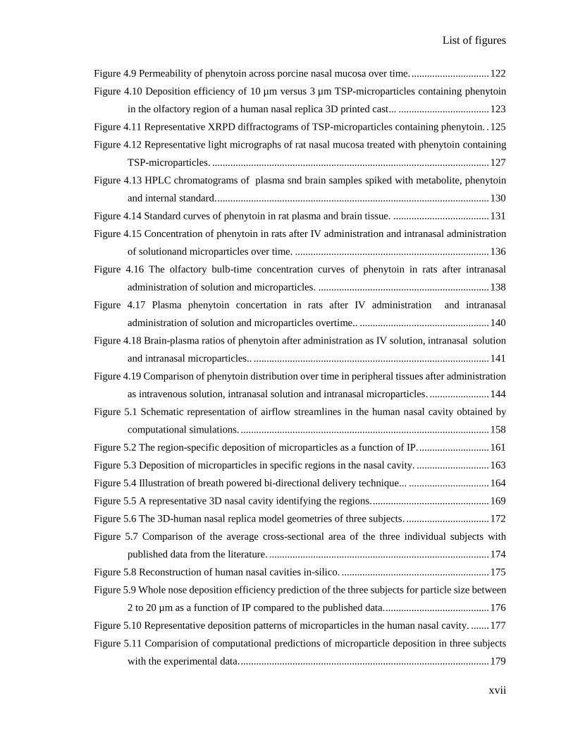
List of figures
xvii
Figure 4.9 Permeability of phenytoin across porcine nasal mucosa over time. .............................. 122
Figure 4.10 Deposition efficiency of 10 µm versus 3 µm TSP-microparticles containing phenytoin
in the olfactory region of a human nasal replica 3D printed cast... ................................... 123
Figure 4.11 Representative XRPD diffractograms of TSP-microparticles containing phenytoin. . 125
Figure 4.12 Representative light micrographs of rat nasal mucosa treated with phenytoin containing
TSP-microparticles. ........................................................................................................... 127
Figure 4.13 HPLC chromatograms of plasma snd brain samples spiked with metabolite, phenytoin
and internal standard. ......................................................................................................... 130
Figure 4.14 Standard curves of phenytoin in rat plasma and brain tissue. ..................................... 131
Figure 4.15 Concentration of phenytoin in rats after IV administration and intranasal administration
of solutionand microparticles over time. ........................................................................... 136
Figure 4.16 The olfactory bulb-time concentration curves of phenytoin in rats after intranasal
administration of solution and microparticles. .................................................................. 138
Figure 4.17 Plasma phenytoin concertation in rats after IV administration and intranasal
administration of solution and microparticles overtime.. .................................................. 140
Figure 4.18 Brain-plasma ratios of phenytoin after administration as IV solution, intranasal solution
and intranasal microparticles.. ........................................................................................... 141
Figure 4.19 Comparison of phenytoin distribution over time in peripheral tissues after administration
as intravenous solution, intranasal solution and intranasal microparticles. ....................... 144
Figure 5.1 Schematic representation of airflow streamlines in the human nasal cavity obtained by
computational simulations. ................................................................................................ 158
Figure 5.2 The region-specific deposition of microparticles as a function of IP. ........................... 161
Figure 5.3 Deposition of microparticles in specific regions in the nasal cavity. ............................ 163
Figure 5.4 Illustration of breath powered bi-directional delivery technique... ............................... 164
Figure 5.5 A representative 3D nasal cavity identifying the regions. ............................................. 169
Figure 5.6 The 3D-human nasal replica model geometries of three subjects. ................................ 172
Figure 5.7 Comparison of the average cross-sectional area of the three individual subjects with
published data from the literature. ..................................................................................... 174
Figure 5.8 Reconstruction of human nasal cavities in-silico. ......................................................... 175
Figure 5.9 Whole nose deposition efficiency prediction of the three subjects for particle size between
2 to 20 µm as a function of IP compared to the published data. ........................................ 176
Figure 5.10 Representative deposition patterns of microparticles in the human nasal cavity. ....... 177
Figure 5.11 Comparision of computational predictions of microparticle deposition in three subjects
with the experimental data. ................................................................................................ 179

List of figures
xviii
Figure 5.12 Representative velocity profile of the air in olfactory/turbinate region with nebulization.
........................................................................................................................................... 181
Figure 5.13 Representative velocity profile of the airflow in olfactory/turbinate region with bi-
directional delivery technique. ........................................................................................... 182
Figure 5.14 Representative airflow streamlines of nebulization and bi-directional delivery methods
in three subjects. ................................................................................................................ 183
Figure 5.15 Regional specific deposition in nasal cavities of three human subjects. ..................... 186
Figure 5.16 Region-specific deposition of particles with different flow rates. ............................... 188
Figure 5.17 Effect of particle size on region-specific deposition in the nasal cavities of three human
subjects with bi-directional delivery technique at an air flow rate of 6 Lmin-1 when bi-
directional airflow was simulated from left to right........................................................... 191
Figure 5.18 Effect of particle size on region-specific deposition in the nasal cavities of three human
subjects with bi-directional delivery technique at an air flow rate of 6 L min-1 when bi-
directional airflow was simulated from right to left........................................................... 192
Figure 5.19 Comparison of maximum olfactory deposition by nebulization vs. bi-directional delivery
techniques.. ........................................................................................................................ 197
Figure 8.1 Tensile strength apparatus used to measure the ex-vivo mucoadhesion strength of the TSP-
microparticles. .................................................................................................................... 249
Figure 8.2 Representative airflow streamlines of nebulization and bi-directional delivery methods in
three subjects at different nebulization airflow rates. ........................................................ 256
List of tables
xix
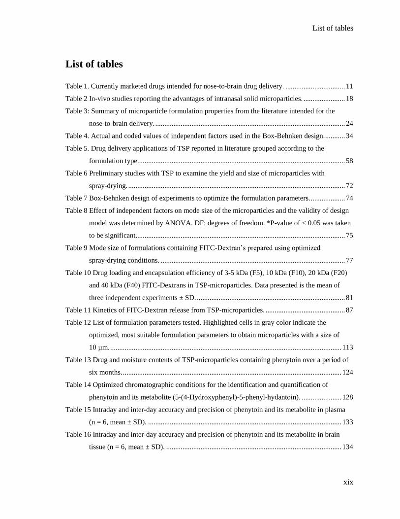
List of tables
Table 1. Currently marketed drugs intended for nose-to-brain drug delivery. ................................. 11
Table 2 In-vivo studies reporting the advantages of intranasal solid microparticles. ....................... 18
Table 3: Summary of microparticle formulation properties from the literature intended for the
nose-to-brain delivery. ......................................................................................................... 24
Table 4. Actual and coded values of independent factors used in the Box-Behnken design. ........... 34
Table 5. Drug delivery applications of TSP reported in literature grouped according to the
formulation type. .................................................................................................................. 58
Table 6 Preliminary studies with TSP to examine the yield and size of microparticles with
spray-drying. ........................................................................................................................ 72
Table 7 Box-Behnken design of experiments to optimize the formulation parameters. ................... 74
Table 8 Effect of independent factors on mode size of the microparticles and the validity of design
model was determined by ANOVA. DF: degrees of freedom. *P-value of < 0.05 was taken
to be significant.................................................................................................................... 75
Table 9 Mode size of formulations containing FITC-Dextran’s prepared using optimized
spray-drying conditions. ...................................................................................................... 77
Table 10 Drug loading and encapsulation efficiency of 3-5 kDa (F5), 10 kDa (F10), 20 kDa (F20)
and 40 kDa (F40) FITC-Dextrans in TSP-microparticles. Data presented is the mean of
three independent experiments ± SD. .................................................................................. 81
Table 11 Kinetics of FITC-Dextran release from TSP-microparticles. ............................................ 87
Table 12 List of formulation parameters tested. Highlighted cells in gray color indicate the
optimized, most suitable formulation parameters to obtain microparticles with a size of
10 µm. ................................................................................................................................ 113
Table 13 Drug and moisture contents of TSP-microparticles containing phenytoin over a period of
six months. ......................................................................................................................... 124
Table 14 Optimized chromatographic conditions for the identification and quantification of
phenytoin and its metabolite (5-(4-Hydroxyphenyl)-5-phenyl-hydantoin). ...................... 128
Table 15 Intraday and inter-day accuracy and precision of phenytoin and its metabolite in plasma
(n = 6, mean ± SD). ........................................................................................................... 133
Table 16 Intraday and inter-day accuracy and precision of phenytoin and its metabolite in brain
tissue (n = 6, mean ± SD). ................................................................................................. 134

List of tables
xx
Table 17 P-values indicating the statistical significance of brain phenytoin concentrations after (IN;
intranasal and IV; intravenous) over time. ......................................................................... 136
Table 18 P-values indicating the statistical significance of olfactory bulb phenytoin concentrations.
(IN: intranasal) over time. .................................................................................................. 138
Table 19 P-values indicating the statistical significance of plasma phenytoin concentrations (IN;
intranasal and IV; intravenous). ......................................................................................... 140
Table 20 Geometrical surface area of the nasal regions in the 3D-human nasal cavity models. .... 173
Table 21 %DTP values calculated for the microparticle formulations from the reported literature.
........................................................................................................................................... 248
Table 22. Microparticle expulsion from insufflator device. ........................................................... 250
Table 23 Intraday and inter-day accuracy and precision of phenytoin and its metabolite in lung
tissue (n = 6, mean ± SD)................................................................................................... 251
Table 24 Intraday and inter-day accuracy and precision of phenytoin and its metabolite in liver
tissue (n = 6, mean ± SD)................................................................................................... 252
Table 25 Intraday and inter-day accuracy and precision of phenytoin and its metabolite in kidney
tissue (n = 6, mean ± SD)................................................................................................... 253
Table 26 Intraday and inter-day accuracy and precision of phenytoin and its metabolite in spleen
tissue (n = 6, mean ± SD)................................................................................................... 254
Table 27 Intraday and inter-day accuracy and precision of phenytoin and its metabolite in heart
tissue (n = 6, mean ± SD)................................................................................................... 255
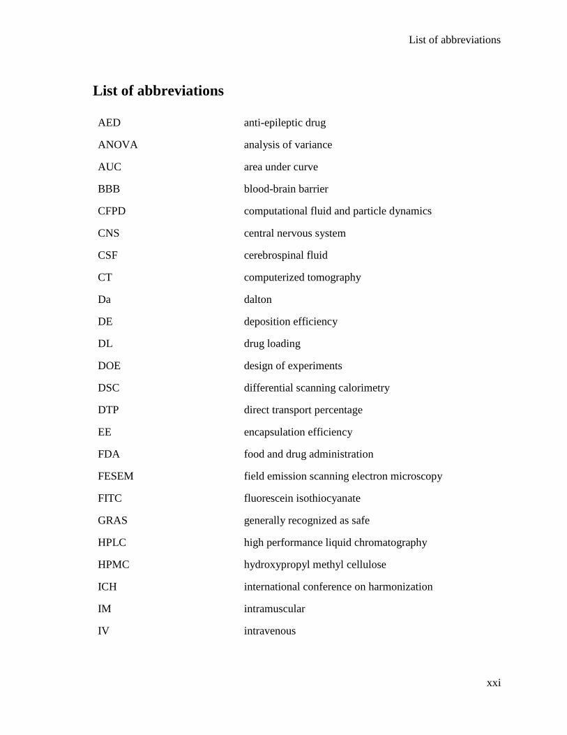
List of abbreviations
xxi
List of abbreviations
AED anti-epileptic drug
ANOVA analysis of variance
AUC area under curve
BBB blood-brain barrier
CFPD computational fluid and particle dynamics
CNS central nervous system
CSF cerebrospinal fluid
CT computerized tomography
Da dalton
DE deposition efficiency
DL drug loading
DOE design of experiments
DSC differential scanning calorimetry
DTP direct transport percentage
EE encapsulation efficiency
FDA food and drug administration
FESEM field emission scanning electron microscopy
FITC fluorescein isothiocyanate
GRAS generally recognized as safe
HPLC high performance liquid chromatography
HPMC hydroxypropyl methyl cellulose
ICH international conference on harmonization
IM intramuscular
IV intravenous
List of abbreviations
xxii
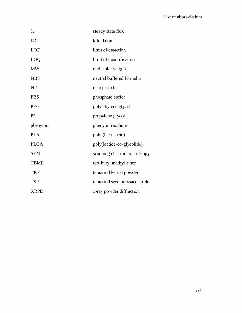
Jss steady state flux
kDa kilo dalton
LOD limit of detection
LOQ limit of quantification
MW molecular weight
NBF neutral buffered formalin
NP nanoparticle
PBS phosphate buffer
PEG polyethylene glycol
PG propylene glycol
phenytoin phenytoin sodium
PLA poly (lactic acid)
PLGA poly(lactide-co-glycolide)
SEM scanning electron microscopy
TBME tert-butyl methyl ether
TKP tamarind kernel powder
TSP tamarind seed polysaccharide
XRPD x-ray powder diffraction

Chapter 1
Introduction


Chapter 1
1
1 Introduction
1.1 Rationale
The blood-brain barrier (BBB) serves as a barrier to restrict the entry of potential drugs
from the systemic circulation into the central nervous system (CNS). The discovery of direct
drug transport pathways found in the olfactory region of the nasal cavity that can bypass the
BBB has provided a non-invasive approach to treat complex CNS disorders. However,
exploiting these direct nose-to-brain pathways and translating them into effective treatment
strategies for CNS disorders has been challenging. In last 20 years, the vast amount of
literature published on direct nose-to-brain delivery has led to approximately 165 clinical
trials, and from this, only five drugs have been approved by the US food and drug
administration (FDA) for use in the clinic (Appendix A). One of the main reasons behind
the failure to translate the preclinical research to the clinical stage has been the
subtherapeutic concentration of drugs reaching the brain following intranasal administration.
There has been intensive research and development in innovative drug formulation
technologies to address the inadequate therapeutic drug levels in the brain. Much of this
research has resulted in the evolution of polymer-based, mucoadhesive, microparticle and
nanocarrier drug delivery systems. Each of these systems has been shown to deliver higher
drug concentrations to the brain by sustained release and improved residence time of drugs
in the nasal cavity. However, to obtain sufficient therapeutic drug levels in the brain, it is
necessary for a delivery system to target the site of nose-to-brain transport (the olfactory
region) in the nasal cavity.
The complex geometry and narrow airways (Figure 1.1) of the nasal cavity makes
targeted delivery of drug formulations to the olfactory mucosa a significant challenge1.
Drugs administered as solutions often cannot be retained by the upper, neuron-containing,
region of the nasal passage and are instead cleared by nasal mucociliary clearance or
swallowed or lost to dripping2. Drugs formulated as mucoadhesive microparticles might
have advantages in targeting and retaining the drugs in the olfactory region due to their size

Chapter 1
2
and mucoadhesive properties. Therefore, the overall focus and aim of this thesis is the
development of an olfactory-targeted mucoadhesive microparticulate delivery system, with
the idea that this will offer an improved nose-to-brain delivery of drugs.
1.2 Nose-to-brain drug delivery
Considering the potential advantages microparticles can offer in nose-to-brain drug
delivery, it may be hard to understand their poor representation in the pharmaceutical market.
Nasal formulations intended for nose-to-brain delivery are predominantly available as liquid
formulations, for example, Fentanyl citrate nasal spray (Lazanda®), Ketorolac tromethamine
(Sprix®), Zolmitriptan (Zomig®) and Sumatriptan nasal spray (Imitrex®) to treat pain and/or
a migraine3. To facilitate the discussion on the application of microparticles to overcome the
challenges of nose-to-brain delivery, some fundamental concepts will be introduced first.
1.2.1 Anatomical and physiological aspects
The physiology of the human nose is intended for warming and moistening of inspired
air and olfaction. The nasal cavity is divided into two separate passages by the nasal septum.
Each nasal passage ends anteriorly in a nostril and posteriorly at the nasopharynx. The nasal
passage contains comma-shaped, bony turbinates protruding into the cavity allowing only a
narrow pathway for the inspired air4,5 as shown in Figure 1.1 B. These geometrically
complex airways act as a filter for inhaled particles at the same time and help in conditioning
the inhaled air so that air reaching the lungs is warm and moist6.
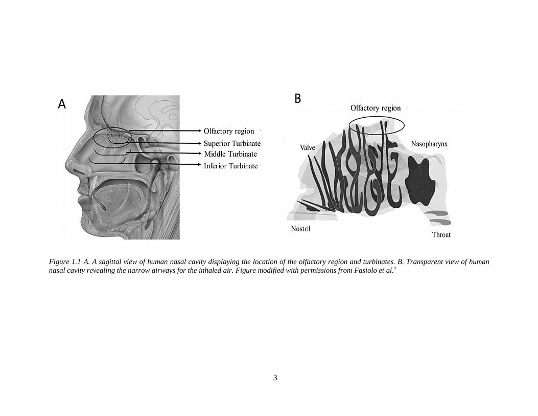
3
Figure 1.1 A. A sagittal view of human nasal cavity displaying the location of the olfactory region and turbinates. B. Transparent view of human
nasal cavity revealing the narrow airways for the inhaled air. Figure modified with permissions from Fasiolo et al.7

Chapter 1
4
The nose is a direct entry point to our internal body. To help protect our body from the
inhaled material, the nasal cavity is lined with mucosa along the nasal passage. Depending
on where we look in the nasal cavity, three different mucosae are present, which serve
different purposes. Near to the nostrils, stratified squamous epithelium is found and
gradually transforms into a pseudostratified columnar epithelium (respiratory mucosa). The
respiratory mucosa is innervated by the trigeminal nerve and covers most of the nasal cavity.
The pseudostratified columnar epithelium in the olfactory region (olfactory mucosa)
contains olfactory neurons responsible for the detection of smell4,8. The intranasal
administration of drugs provides access to the olfactory neurons in the olfactory mucosa and
trigeminal nerves in the respiratory mucosa.
The olfactory region is situated in the upper deep posterior region of the nasal passage
as shown in Figure 1.1. The surface area of the olfactory region is small and covers
approximately 10 cm2 of the total nasal cavity8. Due to curved, narrow airways and the
location of the olfactory region situated deep in the nasal cavity, it is difficult for inhaled
particles to reach this region. The CNS opens into the nasal cavity through the olfactory
neurons in the olfactory mucosa as shown in Figure 1.2 A. The olfactory neurons contain
several non-motile cilia with odorant receptors that extend into the overlying mucus. The
unmyelinated axons from olfactory neurons extend through the basal lamina and converge
with axons from other olfactory neurons to form nerve bundles called fila olfactoria.
Olfactory ensheathing cells and fibroblasts enclose fila olfactoria to form a perineural like a
sheath. These ensheathed fila olfactoria form the olfactory nerve and travel through the
cribriform plate into the mitral, periglomerular, and tufted cells in glomeruli of the olfactory
bulb. Axons from the olfactory bulb project to a number of rostral areas in the brain including
piriform cortex, amygdala, and entorhinal cortex, forming a channel from the olfactory
region within the nasal cavity to the brain9–11. The olfactory mucosa also contains supporting
cells, microvillar cells secured in the basement membrane. The submucosa under the
basement membrane is highly vascularized, it contains Bowman glands and a variety of other
cells including progenitor cells such as globose and horizontal basal cells12–15.
Chapter 1
5
In addition to the olfactory neurons, the CNS opens into the nasal cavity through
trigeminal nerve endings in the respiratory mucosa. The free trigeminal nerve endings are
extended into the respiratory region as shown in Figure 1.2 B. The axons of the trigeminal
nerve project into the brainstem through the pons and enter into the forebrain through the
cribriform plate16. Therefore the trigeminal nerve connects to both the caudal and rostral
parts of the brain, consequently, forming a channel from the respiratory mucosa in the nasal
cavity to the CNS16,17. The respiratory region contains ciliated and non-ciliated columnar
cells, mucus-producing goblet cells with tight junctions. The movements of cilia in the
respiratory region are responsible for the mucociliary clearance in the nasal cavity. The
submucosa is highly vascularized9.

6
Figure 1.2 A. Drug transport pathways and organization of the olfactory mucosa B. Trigeminal innervation and the organization of the respiratory
mucosa. Figure modified with permissions from Thorne et al9
Chapter 1
7
The trigeminal nerve and olfactory bulb were reported to be connected by the sensory
endings of the trigeminal ganglion cells located in the nasal epithelium. The sensory endings
were found to send collaterals directly into the olfactory bulb18. Along with trigeminal and
olfactory nerves, the nasal passage also contains the nervus terminalus and the vomeronasal
nerve. However, their role in drug transport to CNS has not been established9.
1.2.2 Pathways and mechanisms for nose-to-brain transport of drugs
The exact pathways and mechanisms of direct drug transport into the brain following
intranasal administration have not been fully characterized. However, specific pathways
through olfactory, trigeminal nerves, and nasal mucosa have been proposed by experimental
evidence obtained from published literature.
1.2.2.1 Neuronal pathways
Intracellular uptake of molecules into olfactory neurons and trigeminal nerves leads to
axonal transport of the molecules that have been taken up into the brain. Large protein
molecules such as horseradish peroxidase and wheat germ agglutinin-horseradish peroxidase
have been previously reported to be taken up into olfactory neurons via pinocytosis and
adsorptive endocytosis19. Wheat germ agglutinin-horseradish peroxidase is also reported to
be endocytosed into the trigeminal nerves and transported to the brain stem by axonal
transport20,21. Endocytosis of proteins, viruses, and bacteria by the olfactory and trigeminal
nerves and their subsequent intracellular transport to the brain has been previously
reported22–26.

Chapter 1
8
Figure 1.3 Nose-to-brain direct pathways from olfactory and respiratory mucosa via olfactory nerve
and trigeminal nerves. Figure adapted with permissions from Thorne et al9
The olfactory and trigeminal pathways potentially occur in the olfactory and respiratory
regions of the nasal cavity. The olfactory neuronal transport target the rostral area of the
brain and the trigeminal transport target both the rostral and caudal areas of the brain27 as
shown in Figure 1.3. Specifically, olfactory pathway contributes a vital role in direct nose-
to-brain transport, hence targeting drugs to olfactory mucosa/region can aid nose-to-brain
delivery28.
1.2.2.2 Mucosal pathways
In addition to neuronal pathways, drugs can also cross nasal mucosa in the olfactory
region to access the perineural space and thereby reach the brain via extracellular pathways
(Figure 1.2 B). Transcytosis and paracellular diffusion across the nasal mucosa have been
proposed as the extracellular pathways29–32.
Receptor-mediated endocytosis is involved in the transcytosis pathway. Size of the
molecule determines the mechanism of endocytosis, for example, molecules less than
200 nm follow clathrin-dependent endocytosis and molecules with 100-200 nm are

Chapter 1
9
transported by caveolae-mediated endocytosis33,34. Cell type, surface charge, and
concentration are the other important factors that influence the mechanism of endocytosis in
transcytosis35.
Paracellular transport involves opening of tight junctions between the cells in the
olfactory and respiratory mucosa. The continuous turnover of basal cells as a part of the life
cycle of biological membranes causes loosening and the opening of tight junctions and
promotes paracellular transport36,37. Drugs crossing nasal mucosa via extracellular pathways
may enter the systemic circulation or lymphatic system or can access the perineural spaces.
Perineural distribution of potassium ferrocyanide, ammonium citrate and fluorescein-
Dextran (3 kDa) in olfactory nerve bundles and their subsequent entry into the brain
following the intranasal administration was reported38,39.
Distribution of drugs to the different areas of the brain from the site of entry occurs by
bulk flow within perivascular spaces of cerebral blood vessels27,40. Wider distribution of
fluorescent liposomes in the brain parenchyma with increased blood pressure after
intrastriatal injection demonstrated the role of bulk flow in the distribution of the drug
molecules41. Cerebrospinal fluid (CSF) flow also plays a role in distributing the drug into
widespread areas of the brain. Rapid distribution following tracer application into the CSF
was reported42,43. Drugs reaching the perineural spaces are distributed to the CSF, and the
flow of CSF can further transport the drug to more distant sites of the brain. However, the
barrier between the perineural space and CSF is shown to be selective27, and the
physiological aspects of this distribution are yet to be completely understood.
1.2.3 Mucociliary clearance of drugs in nasal passages
The nasal passage is covered with mucus produced by submucosal glands and goblet
cells in the respiratory mucosa. The mucus in nasal passages are composed of >90 % water,
0.5-5 % mucus glycoproteins, 1-2 % salts and 0.5-1 % free proteins and is slightly acidic
with a pH of 5.5-6.56,44. The overlying mucus is between 10-20 µm in depth and consists of
two distinct layers, a lower less viscous (sol) and an upper high viscous (gel) layer45,46.
Mucus glycoproteins are responsible for the gel-like structure of the mucus; they are formed

Chapter 1
10
from a protein core surrounded by carbohydrate chains47. Many columnar cells in the
respiratory mucosa possess hair-like protrusions called cilia that are around 5-10 µm long
and width from 0.1-0.3 µm extending into the sol layer. The number of cilia per cell is
approximately 30048,49. The synchronized movement of cilia in the sol layer causes the
transport of the upper gel layer towards the nasopharynx where it is swallowed50,51. The
human nasal cilia beat with a frequency of 10 Hz, and the average velocity of the mucus
transport is around 8 mm/min and can vary between 3-25 mm/min52,53. Airborne particles
entering the nasal passages are entrapped in the mucus layer and get transported along with
it to the nasopharynx and are eventually cleared from the nasal cavity. The combined action
of the cilia and mucus layers is called mucociliary clearance.
Mucociliary clearance is an essential physiological defense mechanism to protect the
nasal cavity against noxious inhaled particles. On the other hand, it is responsible for the
rapid clearance of the drugs and formulations after intranasal administration. The average
nasal clearance half-life for drugs is about 12-15 mm/min. Mucociliary clearance decreases
the contact time with the nasal mucosa, ultimately leading to decreased drug delivery to the
brain54,48.
1.2.4 Current status of nose-to-brain drug delivery
Conventionally, the nasal route has been used for the systemic delivery of drugs, but now
it has become an increasingly popular method to bypass the BBB and blood circulation to
deliver neurotherapeutics to the brain/CNS24,55. The published literature now includes
substantial evidence for the advantages of nose-to-brain delivery over the other routes of
administration. Most importantly, a wide range of therapeutic agents including proteins,
plasmids, gene vectors and stem cells have been shown to reach the brain following
intranasal administration via olfactory and trigeminal pathways32,56–58.
Chapter 1
11
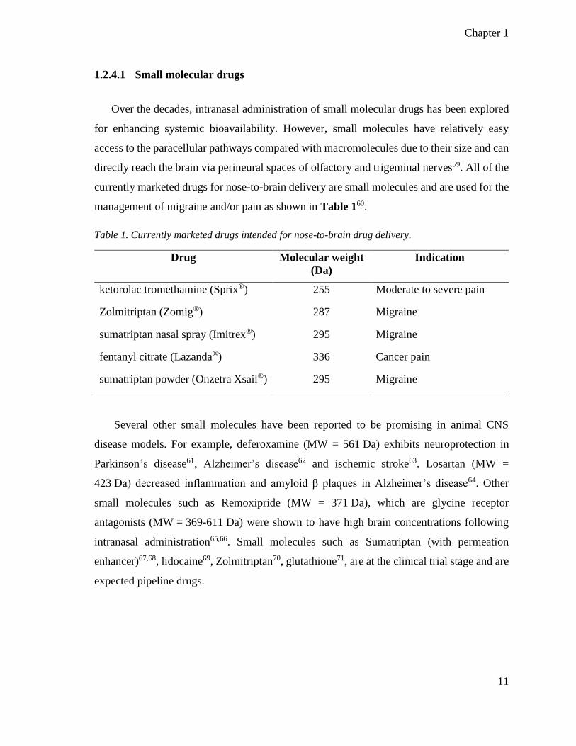
1.2.4.1 Small molecular drugs
Over the decades, intranasal administration of small molecular drugs has been explored
for enhancing systemic bioavailability. However, small molecules have relatively easy
access to the paracellular pathways compared with macromolecules due to their size and can
directly reach the brain via perineural spaces of olfactory and trigeminal nerves59. All of the
currently marketed drugs for nose-to-brain delivery are small molecules and are used for the
management of migraine and/or pain as shown in Table 160.
Table 1. Currently marketed drugs intended for nose-to-brain drug delivery.
Drug Molecular weight
(Da)
Indication
ketorolac tromethamine (Sprix®) 255 Moderate to severe pain
Zolmitriptan (Zomig®) 287 Migraine
sumatriptan nasal spray (Imitrex®) 295 Migraine
fentanyl citrate (Lazanda®) 336 Cancer pain
sumatriptan powder (Onzetra Xsail®) 295 Migraine
Several other small molecules have been reported to be promising in animal CNS
disease models. For example, deferoxamine (MW = 561 Da) exhibits neuroprotection in
Parkinson’s disease61, Alzheimer’s disease62 and ischemic stroke63. Losartan (MW =
423 Da) decreased inflammation and amyloid β plaques in Alzheimer’s disease64. Other
small molecules such as Remoxipride (MW = 371 Da), which are glycine receptor
antagonists (MW = 369-611 Da) were shown to have high brain concentrations following
intranasal administration65,66. Small molecules such as Sumatriptan (with permeation
enhancer)67,68, lidocaine69, Zolmitriptan70, glutathione71, are at the clinical trial stage and are
expected pipeline drugs.

Chapter 1
12
1.2.4.2 Macromolecular drugs
Macromolecular drugs like proteins and peptides have limited permeation across
biological membranes and are prone to metabolic degradation in the blood and tissues.
Intranasal administration provides a promising route for such molecules. Melanocortin,
arginine-vasopressin and insulin were among the first peptides determined in CSF following
intranasal administration, all these peptides were detected within 30 min in CSF of human
volunteers72. Detectable brain concentrations of growth factors73–76, neuropeptides77,78,
neurotrophic factors79,80,81 and other macromolecules82 have been reported following
intranasal administration in animal models. Higher CNS levels of intranasal insulin
compared to subcutaneous injection were reported in mice. Highest levels were found in the
trigeminal nerve and olfactory bulbs suggesting the transport to the CNS through neuronal
pathways83. Clinical trials in Alzheimer’s disease patients showed intranasal insulin
improved memory and preserved general cognition84,85 and had rapid action86. Efficacy of
intranasal insulin in treating obesity in humans and mice has been reported87. Intranasal
administration of sleep-related peptide orexin-A has shown improved brain metabolic
activity in rhesus monkeys and stabilized REM sleep in humans88–90.
An increasing number of protein drugs for the treatment of CNS diseases and recent
discoveries of essential brain functions have stimulated this area of research. Analysis of the
literature shows a doubling of the number of publications on nose-to-brain peptide delivery
reporting in-vivo studies in the last seven years (55 versus 120 reports [pubmed database]).
Currently, many intranasal peptide and protein drugs are undergoing clinical trials and
represent the most promising group of pipeline drugs to treat complex CNS diseases. Insulin
is under clinical trials for the treatment of Schizophrenia (Phase I)91, Alzheimer’s disease
(Phase I and Phase III)92–95, obesity (Phase II)96 and major depressive disorder (Phase II)97.
Oxytocin is being examined for the treatment of schizophrenia (Phase I)98–100, autism
spectrum disorder (Phase I and Phase III)101–103 and post-traumatic stress disorder (Phase I,
II and Phase III)104–106. The efficacy of Arginine-vasopressin in the treatment of cognitive
and behavior disorders (Phase I)107,108, Neuropeptide Y in the treatment of obesity
(Phase I)96, insulin-like growth factor-I in the treatment of obesity and diabetes (Phase I)109
Chapter 1
13
are also being investigated. Other potential peptide drugs such as melanocortin,
cholecystokinin, NAP neuropeptide and hexarelin are also under clinical trials for nose-to-
brain delivery82. Although intranasal administration is a potential route for peptide drugs,
many reported studies use simple drug solutions and the drugs are failing to reach the
brain/CNS in therapeutic concentrations. Nevertheless, the trend is now changing to the
carrier-based formulation of peptide drugs to improve the efficacy of such drugs.
1.2.4.3 Cell-based therapies
Targeting of stem cells to the brain via intranasal endocytic neuronal pathways represents
the most recent group of intranasal drugs. Mesenchymal cells administered intranasally in
mice have shown therapeutic potential in Parkinson’s disease and several models of
stroke110–113. Fluorescently labeled mesenchymal stem cells has been transported to the brain
via neuronal pathways 1 h after intranasal delivery in mice64. The therapeutic potential of
intranasal neural stem/progenitor cells has been recently identified; with intranasal delivery
providing direct transport of neural stem/progenitor cells to intracerebral gliomas in six h58.
Finally, intranasal administration of genetically engineered T-cells has been successful in
suppressing the inflammation in mouse models of multiple sclerosis114. The potential of such
cell-based therapies has only recently been identified and is yet to be fully explored.
1.2.5 Challenges and strategies for efficient nose-to-brain drug delivery
As discussed in the previous sections, nose-to-brain administration has excellent
potential for delivery of novel therapies for brain diseases. However, achieving sufficient
therapeutic drug levels in the brain is still a challenge. In addition to the fast mucociliary
clearance and loss of drug to the systemic and lymphatic circulation, targeting drugs to
neuronal rich regions like the olfactory region in the nasal cavity and facilitating the transport
across mucosal membrane are the significant challenges for attaining therapeutic drug levels
in the brain via nose-to-brain delivery. Hydrophilic small molecular drugs have a
bioavailability of 10 %, and peptide and protein drugs have a bioavailability of about 1 %
following intranasal administration115. Therefore, to achieve efficient absorption and

Chapter 1
14
therapeutic efficacy, approaches to target the olfactory region in the nasal cavity, increase
the contact time of drugs with nasal mucosa, and facilitate the transport of drugs across the
mucosa are required.
Many strategies such as, use of permeation enhancers to facilitate the drug transport
across the nasal mucosa, use of vasoactive agents (bradykinin) to chemically disturb the
BBB116,117, use of microbubble facilitated focus ultrasound waves to open the BBB118,
implanting polymeric wafers119,120 & programmable microchips121, and chemically
modifying the drug to increase the permeability and water solubility122,123, to improve the
drug delivery to the brain are being researched upon. As stated in Section 1.1, much of the
research has led to development of nano/microparticle delivery systems. The objective of
this thesis is not, however, to review the many exciting aspects of these strategies as many
of them are either invasive28 or associated with side effects124. Nevertheless, a brief
comparison of the strengths and weaknesses of non-particle approaches to
nano/microparticle delivery systems to increase drug transport across the nasal mucosa is
presented here.
Administration of drug solutions with permeation enhancers to open the tight junctions
in the epithelial cells and enhance the transport across the nasal mucosa has been reported115.
However, the permeation enhancers used in the studies such as surfactants, phospholipids,
bile acids, etc. are reported to produce toxic effects on the nasal mucosa including decreased
ciliary beat frequency, tissue damage, and irreversible ciliotoxicity125. On the contrary,
nanoparticles can provide enhanced transport across the nasal mucosa without the use of an
enhancer due to their size. Nanoparticles less than 20 nm can pass the epithelial tight
junctions, while larger nanoparticles can cross the mucosal barrier transcellularly, by
entering into the cell by the process of endocytosis or phagocytosis115.
Another strategy, is to chemically modify the drug to improve solubility and permeability
which has been reported for nose-to-brain delivery126. Producing prodrugs by chemical
modification of a drug to change its lipophilicity to increase permeability and water solubility
has been reported127–129. However, prodrug synthesis requires challenging skills involving
reduction of polar groups or linking a lipophilic moiety to the drugs and in general is not
fruitful28. On the contrast, nano/microparticles can be produced by single step processes like
Chapter 1
15
spray-drying and have given promising results in pre-clinical studies to improve the brain
delivery of a variety of drugs such as brimocriptine130, rokitamycin131, rivastigimin132,
venlafaxine133,134, tizanidine135.
Formulating drugs as nano/microparticle delivery systems, in general, has numerous
advantages for nose-to-brain delivery of drugs. These delivery systems can control the
release of drugs at a predetermined rate and desired drug levels can be maintained136.
Nano\microparticles can prevent drug loss and degradation and improve drug solubility137 in
the nasal cavity28. Nano/microparticle delivery systems can reduce the mucociliary
clearance, increase residency time, enhance the permeation of drug through nasal mucosa,
and promote large molecular138/phytochemical139 drug delivery across the nasal cavity28.
Due to their advantages, these delivery systems are being extensively researched and are
delivering promising results in pre-clinical studies28,140.
Although nano\microparticle delivery systems have several advantages, a few limitations
of these delivery systems exist in nose-to-brain delivery. Mucosal damage, nasal irritation,
the effect of patients position during administration on brain uptake, low entrapment
efficiency and storage related problems need to be addressed by further investigations to
exploit the full potential of these delivery systems28,115.
1.3 Delivery systems for nose-to-brain drug delivery
Drug delivery systems intended for nose-to-brain delivery are designed to transport the
drug to the brain, attain a desired therapeutic level and maintain the drug concentration
within the therapeutic window. Furthermore, the desired characteristics of an ideal drug
delivery system are to reduce the dose of the drug, have minimal side effects and ease of
administration. Based on the size, drug delivery systems for nose-to-brain can be divided
into nanocarriers and microparticles. Nanocarriers and microparticles have a potential to
achieve the desired therapeutic level of drug in the brain by controlling and/or sustaining the
release rate of encapsulated drug and nasal residence time141.

Chapter 1
16
Nanocarrier drug delivery system include polymeric nanoparticles, nanoemulsions,
micelles, liposomes and nanogels. These delivery systems provide a versatile platform with
great potential in overcoming the challenges associated with nose-to-brain delivery.
However, the focus of this thesis is microparticles and not the nanocarriers; nonetheless, a
short introduction on the impact of the nanocarrier drug delivery system will be provided to
facilitate further discussions.
1.3.1 Nanocarrier drug delivery systems
The rapid increase in the number of publications reporting nanocarrier systems for
nose-to-brain delivery demonstrates the enormous potential of such delivery systems in
recent times142. Polymeric nanoparticles (NPs) have been successful in the delivery of small
molecules to the brain. For example, chitosan NPs were shown to increase the concentration
of two small MW drugs rasagiline and bromocriptine significantly in the brain after
intranasal administration compared to intravenous (IV) adminsitation143,130. Use of other
polymers such as polyethylene glycol (PEG), PLGA, poly (lactic acid) (PLA) and gelatin to
formulate the NPs has also been reported28,82,144.
Polymeric NPs have also been reported for their success in encapsulating and delivering
macromolecular protein and peptide drugs such as a thyrotropin-releasing hormone, which
when encapsulated in PLA NPs successfully suppressed seizures in epileptic rats145,146. Other
peptides and protein drugs such as leucine–encephalin147, osteopontin148, and vasoactive
intestinal peptide149 were reported to show higher brain concentrations with polymeric
NPs82. Similarly, the ability of other nanocarrier systems such as nanoemulsions150,
micelles151,152, liposomes153,154, and nanogels155 to enhance nose-to-brain delivery has been
reported. Polymers employed in the preparation of nanoparticles and their size are found to
be two critical factors that influence drug targeting to the brain after intranasal
administration156.
As implied above, nanocarrier systems could be an excellent platform for drug delivery
to treat CNS diseases. However, still, no nanocarrier delivery system has reached the clinical

Chapter 1
17
development phase. More conclusive studies, pharmacokinetic and pharmacodynamic data
are required to unveil the full potential of such nanocarrier delivery systems.
1.3.2 Microparticle drug delivery system
As the name suggests, microparticles are tiny particles with size ranging from
1 to 1000 µm. In the drug delivery field, microparticles are vesicles made up of a polymer
into which drugs can be loaded for protection, transport, targeting, and release. Due to their
size, microparticles provide an increased surface area which is advantageous for drug
delivery. The high surface area of microparticles can aid faster drug diffusion into the
external environment. Also, by changing the characteristics of the polymer drug release from
the microparticles can be tuned to meet therapeutic requirements157.
Microparticles can encapsulate many types of drugs including small molecules and
peptide and protein drugs. They are biocompatible and can provide sustained release of the
drug over long periods of time. For example, the commercially available PLGA
microparticle products Nutropin Depot® and Lupron Depot® can sustain the release of
macromolecular drugs somatropin and leuprolide respectively, for over a month replacing
the need for daily injections158.
Microparticle formulations can be administered as suspensions or solids (powders), and
are administered via oral159, pulmonary160 and parenteral (intramuscular (IM)161,
Subcutaneous159) routes. However, for nasal administration, solid particles can have good
patient compliance, deposition in the nasal cavity and provide high dose without the
necessity of a liquid vehicle162,163. Also, solid particles can enhance drug diffusion and
absorption across the mucosa164. Many studies have reported in-vivo the advantage of
administering drugs formulated as solid microparticles for intranasal administration, a few
examples are reported in Table 2.

Chapter 1
18
Table 2 In-vivo studies reporting the advantages of intranasal solid microparticles.
Drug Animal
model
Particle
size (µm)
Polymer Observation Ref
Carvedilol Rabbits 20 to 50 Chitosan
Microparticles enhanced
bioavailability (72.29 %) after
intranasal administration vs
IV (67.87 %)
165
Repaglinide Rats 1 to 5
Pectin
Gellan gum
Dextran
sulfate
Intranasal administration of
microparticles produced a
prolonged hypoglycemic
effect (2-6 h) vs. IV (1 h)
166
Gentamicin Rabbits 19 to 46
Chitosan
Hyaluronic
acid
Intranasal microparticles
enhanced the bioavailability
of gentamicin by two-fold
compared to the intranasal
solution
167
Insulin Rats 1 to 80 Thiolated
chitosan
Intranasal microparticles
produced 4-5 fold higher
bioavailability compared to
the control (mannitol), and the
hypoglycemic effect lasted for
more than 2 h vs. IV (15 min)
168
Verapamil Rabbits 21 to 53 Chitosan
Intranasal microparticles
produced higher
bioavailability (58.6 %) than
the intranasal solution
(47.8 %)
169
1.3.3 Nanocarrier versus microparticles for nose-to-brain drug delivery
It is an important question to reflect on as to why microparticles but not nanocarriers are
used in this thesis for nose-to-brain delivery, especially, after having presented the success
of nanocarrier delivery systems. In this case, it’s a matter of size perhaps literally, as will be
discussed in Section 1.3.5.1.1. This section compares microparticles and nanocarrier
delivery systems for nose-to-brain delivery of drugs.

Chapter 1
19
Estimation of drug transported through direct neuronal pathways as a function of the
formulation can represent the efficiency of the formulation. Nose-to-brain direct transport
percentage (%DTP) is the amount of the drug dose that is estimated to reach the brain via
direct routes. The %DTP offers an interpretation of drug fraction transported through the
neuronal pathways and has been used to represent the efficiency of formulations and delivery
systems142,170,171. The values of %DTP can range from -∞ to 100, values between -∞ to 0
indicate the drug transport is only through IV route, higher than 0 indicates drug transport
through direct nose-to-brain neuronal pathways171.
The %DTP is calculated using Equation 1
%DTP =AUCbrainintranasal − F
AUCbrainintranasal∗ 100 |𝐸𝑞𝑢𝑎𝑡𝑖𝑜𝑛 1
Where AUC is the area under the curve representing drug concentration variation over
time in the brain and F is given by Equation 2 below:
F =AUCbrainIV
AUCbloodIV∗ AUCblood intranasal |𝐸𝑞𝑢𝑎𝑡𝑖𝑜𝑛 2
With an aim to compare the efficiencies of nanoparticle systems and microparticles,
%DTP values from the reported literature of the respective delivery systems were calculated.
Initially, scientific publications reporting quantitative data on drugs by nose-to-brain
delivery of microparticle delivery systems were identified. Specific keywords were used to
search the PubMed database, and the search resulted in 303 articles. Further screening was
performed based on their title, abstract and subsequently full text to identify publications
reporting in-vivo brain or CSF concentrations of the drug. A total of 45 publications were
identified reporting the formulation of drugs into microparticles for nose-to-brain delivery,
out of which only seven had quantitative brain/CSF drug concentrations. The %DTP values
were calculated using the Equation 1 and Equation 2. The %DTP values for nanocarrier
systems were obtained from a recently published scientific article by Pires C.P and Santos
O.A.142.
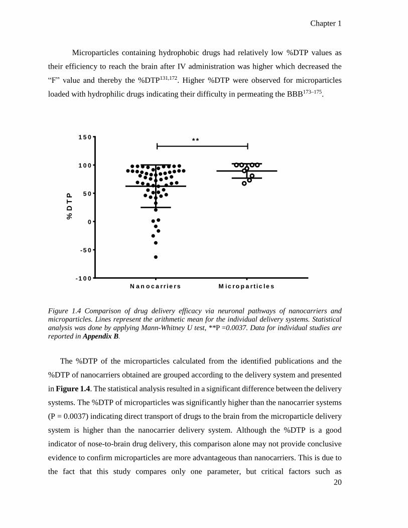
Chapter 1
20
Microparticles containing hydrophobic drugs had relatively low %DTP values as
their efficiency to reach the brain after IV administration was higher which decreased the
“F” value and thereby the %DTP131,172. Higher %DTP were observed for microparticles
loaded with hydrophilic drugs indicating their difficulty in permeating the BBB173–175.
N a n o c a r r ie rs M ic ro p a r t ic le s
-1 0 0
-5 0
0
5 0
1 0 0
1 5 0
%D
TP
* *
Figure 1.4 Comparison of drug delivery efficacy via neuronal pathways of nanocarriers and
microparticles. Lines represent the arithmetic mean for the individual delivery systems. Statistical
analysis was done by applying Mann-Whitney U test, **P =0.0037. Data for individual studies are
reported in Appendix B.
The %DTP of the microparticles calculated from the identified publications and the
%DTP of nanocarriers obtained are grouped according to the delivery system and presented
in Figure 1.4. The statistical analysis resulted in a significant difference between the delivery
systems. The %DTP of microparticles was significantly higher than the nanocarrier systems
(P = 0.0037) indicating direct transport of drugs to the brain from the microparticle delivery
system is higher than the nanocarrier delivery system. Although the %DTP is a good
indicator of nose-to-brain drug delivery, this comparison alone may not provide conclusive
evidence to confirm microparticles are more advantageous than nanocarriers. This is due to
the fact that this study compares only one parameter, but critical factors such as
Chapter 1
21
pharmacological efficacy, toxicity, and therapeutic outcomes must be compared to prove the
advantage of microparticles over nanocarrier systems.
This comparison of %DTP values also has its own limitations. For instance, the
difference in number of reports being compared between microparticles and nanocarriers
(7 vs. 56) and the presence of outliers with very low %DTP values for certain nanocarrier
systems176–178 are two major downsides of the study. In addition, formulation and delivery
factors such as differences among administered formulations (suspension, powder etc.),
difference among the size of nanoparticles (100 nm-1 µm), variety of drug delivery methods
and apparatus employed for drug delivery to animals and site of drug delivery (tip of the
nostril/inside the nasal cavity) might have affected the %DTP values. Also, a variety of
animal models employed, variable factors among the animals that can affect the drug kinetics
such as age, body weight, breed, etc. might have contributed to the %DTP values. In
summary, consideration of more factors and data from extensive studies are required to
identify the ideal delivery system for nose-to-brain delivery of drugs. However, in this thesis,
a microparticle delivery system is studied due to its size as will be discussed in
Section 1.3.5.1.1
Interestingly, this study revealed a very low number of preclinical studies utilizing the
microparticle delivery system for the nose-to-brain delivery of drugs, identifying shortage in
critical pharmacokinetic and pharmacodynamic data required for clinical translation of
microparticle systems. Though many scientific articles reported formulation and in-vitro
characterization of drugs for nose-to-brain delivery of drugs, only a few reported results from
in-vivo studies. This study could not identify the exact reason for this poor translation of
microparticles compared to nanocarriers in nose-to-brain drug delivery. However, based on
the literature, a few assumptions can be made. The advantageous characteristics of
nanocarriers in nose-to-brain delivery such as the high surface area to volume, ability to
bypass biological barriers (enhanced drug absorption) due to size and ability to enter cells
by pinocytosis (enhances drug absorption)179 might have contributed to their comparatively
better translation into preclinical studies.
Chapter 1
22
Despite the advantages of microparticle delivery systems as stated in previous sections,
the absence of well-distributed data indicates that this is an area where more research is
needed to fully characterize and exploit its potential in nose-to-brain delivery of drugs.
1.3.4 Microparticles for nose-to-brain delivery
As stated in the previous section, few scientific articles are identified in the published
literature reporting microparticles intended for nose-to-brain delivery. Among these, 51 %
reported only in-vitro data and only one article reported the extent of pharmacological
effects180, which represents the significant lack of critical data in this field.
Among the studies reporting in-vivo pharmacokinetic data, only seven studies quantified
the drug concentrations in the brain/CSF. However, these studies reported encouraging
results, for example, the antiviral prodrug zidovudine formulated into chitosan and stearic
acid microparticles had a six-fold increase in CSF uptake through the neuronal
pathways181,174. Faster CSF uptake of the antimigraine drug zolmitriptan and reduction in the
peripheral drug levels have been reported from chitosan and hydroxypropyl methylcellulose
(HPMC) microparticles182. Enhanced CSF uptake of deferoxamine mesylate from
methyl-β-cyclodextrin microparticles has been reported as the first formulative approach to
increase the nose-to-brain transport of deferoxamine mesylate175. Also, increased CSF
uptake of N6-cyclopentyladenosine, rokitamycin from mannitol-lecithin, chitosan and
chitosan glutamate microparticles has been reported131,173. Most of these studies report that
the presence of mucoadhesive polymers like chitosan and HPMC increase the residence time
of the formulations in the nasal cavity and lead to enhanced brain/CSF drug transport. The
mechanism of mucoadhesion and its effect on the bioavailability of drugs will be described
in detail in Section 1.3.5.2.
Mucoadhesive microparticles have been studied in three in-vivo studies in humans183 and
rabbits184,185. These studies reported increased residence time of microparticle formulations
in the nasal cavity due to the mucoadhesive nature of the polymers used. One study reported
increased accumulation of the analgesic tramadol in the brain by HPMC microparticles. The
mucoadhesive strength of HPMC microparticles retained tramadol in the rabbit nasal cavity

Chapter 1
23
for extended time periods consequently increasing tramadol accumulation in the brain as
observed by gamma scintigraphy studies186. Sustained drug release resulting in constant
plasma profiles of the antiemetic drug ondansetron was also reported with chitosan, PLA,
and PLGA microparticles187,188. Rapid absorption of the drug and high bioavailability was
reported with carbamazepine loaded chitosan glutamate microparticles189 and lorazepam95.
Only one study reported no difference in the bioavailability of drug between starch
microparticles and control in a rabbit model190. Moreover, one study has emphasized the
importance of drugs reaching the olfactory region for obtaining therapeutically sufficient
brain concentration. Sun et al. reported that methotrexate loaded chitosan microparticles
enhanced brain tissue uptake of the drug. However, the levels were still not therapeutic due
to low amounts of drug reaching the olfactory region172. A substantial fraction of the reported
in-vivo studies focused on the formulation of small MW drugs (232-827 Da), with
insulin191,192 and α-corobotoxin180 being the only two macromolecules reported.
Drug and characteristics of polymers have been shown to have a significant impact on
nose-to-brain delivery of microparticles. Although particle size and method of
manufacturing can impact drug loading and delivery efficiencies, their effect has not been
discussed extensively in the reported literature. Spray-drying is the most popular method of
microparticle preparation, and chitosan is the most used polymer for the formulation of
microparticles intended for nose-to-brain delivery. Almost half of the reported studies used
chitosan and/or chitosan derivatives and this can be attributed to mucoadhesive nature. A
summary of formulation properties is given in Table 3.

Chapter 1
24
Table 3: Summary of microparticle formulation properties from the literature intended for the
nose-to-brain delivery.
Drug Polymer Particle
Size (µm)
Method Ref
Levodopa Gelatin 16
water-in-oil (w/o)
emulsification
solvent extraction
193
Promethazine Ethyl Cellulose and
chitosan 2-3 Spray-drying 194
Metoclopramide Methylpyrrolidinone
chitosan 5-8 Spray-drying 195
Lorazepam HPMC and Carbomer 2-3 Spray-drying 196
Rokitamycin Chitosan 2-5 Spray-drying 197
Zolmitriptan Chitosan 1-4 Spray-drying 198
Rokitamycin Chitosan derivatives 1-6 Spray-drying 199
Sumatriptan HPMC 20-35 Spray-drying 200
Ondansetron Gellan gum 9-11 Spray-drying 201
β-cyclodextrin
or
hydroxypropyl-
βcyclodextrin
Chitosan and Alginate 1-3 Spray-drying 202
Methotrexate Chitosan 2-5 Spray-drying 203
Ketorolac
Carbopol,
Polycarbophil, and
Chitosan
14-46
w/o
emulsification
solvent extraction
204
Phosphorothioate
antisense
oligonucleotides
Polycarbophil-cysteine
reduced glutathione 30
w/o
emulsification
solvent
evaporation
205
Granisetron Sodium CMC 7-15 Freeze drying 206
Lysozyme
as model protein
HPMC and water-
soluble chitosan 7-12 Freeze drying 207
No drug Chitosan 28-46 emulsification-
crosslinking 208
Lorazepam Chitosan microparticles
in pluronic gel NA
emulsion
crosslinking 137
Metoclopramide Sodium alginate and
Chitosan 3-10 Spray-drying 209
Ropinirole
hydrochloride
PLGA/DPPC/ trimethyl
chitosan 2 Spray-drying 210
Metoclopramide Gellan gum 9-10 Spray-drying 211
Tacrine
hydrochloride Chitosan/pectin 1-19 Spray-drying 212

Chapter 1
25
1.3.5 Application of microparticles to overcome the challenges of nose-to-brain
delivery
As described in Section 1.2.5, targeting drug to the olfactory region, overcoming
mucociliary clearance and facilitating drug transport across the mucosal membrane remain
significant challenges for nose-to-brain drug delivery. In this thesis, formulating drugs into
microparticles is investigated as a strategy to address these delivery challenges.
1.3.5.1 Targeting the olfactory region
1.3.5.1.1 Effect of particle size
It has been reported that the size of a particle defines its aerodynamics and deposition in
the geometrically complex human nasal cavity. Particles larger than 20 µm show preferential
deposition in the anterior part of the nasal cavity due to high inertial impaction, while the
majority of particles less than 5 µm escape the nasal cavity, following the air streamlines,
and deposit in the lungs as shown by low deposition fraction in the nasal cavity213.
With the advancements in computational power, recently, in-silico modeling has become
a critical tool in characterizing various factors affecting deposition of particles in the nasal
cavity. Recent modeling data from two reports suggested that particles around 10 µm in size
show high deposition in the olfactory region when administered intranasally at normal
inhalation rates of around 20 L/min214,215. Figure 1.5 shows the deposition of particles in the
olfactory region as a function of size in one human subject from a modeling study. These
studies have triggered the idea of controlling particle size to target specific sites in the nasal
cavity. Therefore, this thesis postulates that formulating microparticles which are around
10 µm in size and loaded with drugs can potentially target the drug to the olfactory mucosa
after inhalation.

Chapter 1
26
Figure 1.5 Deposition of microparticles in the olfactory region as a function of particle size. The
particles with size around 10 µm are shown to enhance the particle deposition in the olfactory region.
Solid line represents computational simulation modeling studies and diamond symbols represent the
study in a 3D human nasal cast. Figure adapted with permissions from Schroeter et al214
1.3.5.1.2 Targeting devices
Use of novel and effective drug delivery device technologies for targeting drug
formulations to the olfactory region are reported in the literature. Among such systems,
pressurized olfactory delivery technology (POD, Impel neuropharma), controlled particle
dispersion technology (Kurve) and breath powered bi-directional technology (OPTINOSE)
have shown potential for nose-to-brain drug delivery216–218.
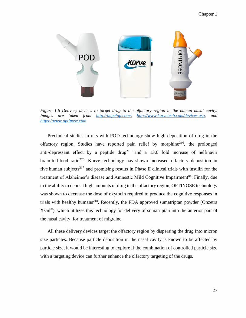
Chapter 1
27
Figure 1.6 Delivery devices to target drug to the olfactory region in the human nasal cavity.
Images are taken from http://impelnp.com/, http://www.kurvetech.com/devices.asp, and
https://www.optinose.com
Preclinical studies in rats with POD technology show high deposition of drug in the
olfactory region. Studies have reported pain relief by morphine216, the prolonged
anti-depressant effect by a peptide drug219 and a 13.6 fold increase of nelfinavir
brain-to-blood ratio220. Kurve technology has shown increased olfactory deposition in
five human subjects217 and promising results in Phase II clinical trials with insulin for the
treatment of Alzheimer’s disease and Amnestic Mild Cognitive Impairment84. Finally, due
to the ability to deposit high amounts of drug in the olfactory region, OPTINOSE technology
was shown to decrease the dose of oxytocin required to produce the cognitive responses in
trials with healthy humans218. Recently, the FDA approved sumatriptan powder (Onzetra
Xsail®), which utilizes this technology for delivery of sumatriptan into the anterior part of
the nasal cavity, for treatment of migraine.
All these delivery devices target the olfactory region by dispersing the drug into micron
size particles. Because particle deposition in the nasal cavity is known to be affected by
particle size, it would be interesting to explore if the combination of controlled particle size
with a targeting device can further enhance the olfactory targeting of the drugs.

Chapter 1
28
1.3.5.2 Mucoadhesion
In addition to targeting the olfactory region, it is helpful if the drug formulation is
retained in the olfactory region long enough for absorption to take place. Many polymers
used in microparticle formulations can adhere to mucus, therefore, prolonging the residence
time of drug in the olfactory region.
Two critical steps in the mucoadhesion process are the initial contact stage and then the
consolidation stage. For solid microparticles, the contact stage is most likely achieved by
water movement from the mucosa into the formulation and hydration of polymer chains221.
Consequently, the nasal mucosa de-hydrates causing the mucus viscosity to increase. In the
second stage, consolidation between polymer and mucin happens by interpenetration of
polymer chains into mucin to form entanglements, the formation of van der Waals, hydrogen
and/or electrostatic bonds such as disulfide bonds222. These chemical bonds and molecular
entanglements create regions where mucus viscosity is further increased leading to high
resistance to the ciliary movement, resulting in decreased mucociliary clearance223–225 and
providing a prolonged contact time for the drug in the nasal cavity. As solid microparticles
can de-hydrate the nasal mucosa leading to an increase in mucous viscosity, they would
lower the usual requirements for the formation of secondary chemical bonds compared to
the pre-hydrated suspensions54,226. Hence, solid microparticles engineered with
mucoadhesive polymers are exceptionally well suited for nose-to-brain drug delivery.
1.3.5.3 Facilitating transport across the mucosa
Solid microparticles are shown to increase the paracellular absorption of drugs loaded
into them by opening tight junctions. For example, dry starch microparticles containing
insulin temporarily opened tight junctions between epithelial cells and enhanced the
absorption of insulin compared to pre-hydrated microparticles227–229. The opening of tight
junctions by dry starch microparticles was thought to be caused by the initial dehydration of
the mucosa which was not required in the case of the pre-hydrated microparticles230. Also,
polymers such as chitosan and polycarbophil used for microparticle preparation were shown
to open the tight junctions reversibly by inducing changes in transmembrane proteins and

Chapter 1
29
chelation of calcium ions respectively and may have potential to increase extracellular
transport along neurological pathways into the brain231,232. Finally, solid microparticles can
increase absorption due to the higher concentration gradient of the drugs released to external
environments as a virtue of their high surface area. Considering the advantages of solid
particles in nasal delivery and the ability of 10 µm sized particles to target the olfactory
region, formulating drugs as solid microparticles for nose-to-brain delivery can be beneficial.
1.4 Size tailored, mucoadhesive microparticles for nose-to-brain drug
delivery
The introduction highlighted the potential of microparticles as one promising strategy to
provide enhanced drug delivery to the brain, with the potential to treat complex CNS
disorders. The versatility of microparticle delivery systems, i.e., the ability to tailor their size
and choice of mucoadhesive polymers, therefore poses the research question of whether such
microparticles can be optimized to deliver drugs efficiently to the brain through the intranasal
route.
Chapter 1
30
1.5 Hypothesis
This thesis will explore the following hypotheses
1. That a combined formulative approach of packaging drugs into solid mucoadhesive
microparticles and size tailoring them to 10 µm can enhance deposition and retention
in the olfactory region and increase brain uptake of the encapsulated drug.
2. That inter-subject variability does not affect the preferential deposition of 10 µm
particles in the human nasal cavity.
3. Combination of controlled particle size with an intranasal device can further enhance
the olfactory targeting of microparticles.
1.5.1 Aims
In order to investigate the above hypotheses, the aims of this thesis are
1. To develop mucoadhesive 10 µm sized microparticles and characterize their
olfactory deposition in-vitro.
2. To develop a model drug-loaded mucoadhesive 10 µm sized microparticles and
investigate its brain uptake of the drug in a rat model.
3. To evaluate inter-subject variability in olfactory deposition of particles in-silico.
4. To identify the effect of particle size on olfactory deposition in the human nasal
cavity combined with a delivery device in-silico.

Chapter 2
General Materials & Methods

Chapter 2
33
2 General Materials and Methods
2.1 Materials
Mucin (bovine submaxillary glands Type I-S, M3895), cellulose acetate membrane (MW
cut off 50,000 Da), propylene glycol (PG), three-way stopcock, phenytoin sodium,
5-(4-methyl phenyl)-5-phenyl-hydantoin, 5-(4-hydroxyphenyl)-5-phenyl-hydantoin and
fluorescein isothiocyanate (FITC)-labelled dextrans with MW of 3-5, 10, 20 and 40 kDa
were purchased from Sigma-Aldrich (New Zealand). Tamarind gum powder was purchased
from Xi’an Jiatian Biotechnology (China). HPLC-grade acetonitrile, ethanol, and methanol
were purchased from Merck KGaA (Darmstadt, Germany). A manual anemometer (N1287)
was purchased from Digitech (Australia). Water was ion-exchanged distilled and passed
through a Milli-Q water purification system (Millipore, Bedford, MA, USA) and was used
for all the studies, sample preparation and HPLC analysis.
2.2 Isolation of Tamarind seed polysaccharide
A naturally mucoadhesive Tamarind seed polysaccharide (TSP) was used as the polymer
in this thesis. A detailed discussion on the purpose and advantages of TSP as a polymer in
drug delivery is provided in Chapter 3, Section 3.1.1. TSP was isolated from tamarind gum
powder using methods described by Rao et al233. Tamarind gum powder (20 g) was
dispersed in 200 mL of Milli-Q water to obtain a slurry, which was then mixed with a further
800 mL of Milli-Q water and boiled for 20 min under constant stirring (800 rpm). This
dispersion was kept overnight to allow proteins and fibers to sediment and subsequently
centrifuged at 5000 rpm for 20 min. The supernatant was separated and mixed with twice
the volume of absolute ethanol under constant stirring. Since the TSP is soluble in water and
insoluble in most of the organic solvents234,235 this led to a precipitate of TSP, which was
washed with Milli-Q water and dried at 60 °C for 12 h to obtain a film of TSP. The film was
crushed using a mortar and pestle to a fine powder and stored in a desiccator at room
temperature until required.

Chapter 2
34
2.3 In-vitro and ex-vivo studies
2.3.1 Preparation of TSP-FITC-Dextran microparticles
All microparticle formulations in the study were prepared by spray-drying methods
based on literature203,236,237 using a laboratory-scale mini Spray Dryer (Büchi B-290, Büchi
Labortechnik AG, Switzerland).
The effect of the spray-drying process variables; atomizing air flow, aspiration air flow
rate, FITC-Dextran concentration and inlet temperature (independent factors) on the mode
size of microparticles (response) was determined using a 4 factor (independent factors),
3 level (low (-1), base (0) and high (+1)) Box-Behnken design238,239 (Table 4). A total of
29 experiments were designed and analyzed using Design-Expert® Software Version 9
(Stat-Ease Inc., MN, USA). The effect of independent variables upon the responses was
modeled using a linear mathematical model. One-way Analysis of Variance (ANOVA) was
applied to determine the significance of the model (P < 0.05). The optimized spray-drying
conditions for producing 10 µm sized particles was determined by applying the numerical
optimization methodology using Design-Expert® Software. The microparticles were
prepared, and their size was measured to validate the optimized spray-drying conditions
obtained from Design Expert®.
Table 4. Actual and coded values of independent factors used in the Box-Behnken design.
Independent factor Coded level
Low (-1) Base (0) High (+1)
Atomizing air flow (L/h) 246 494 742
Aspiration air flow rate (%) 50 60 70
Amount of FITC-Dextran
(%w/w) 0.10 0.25 0.50
Inlet temperature (°C) 100 110 120
Feed solutions for spray-drying were prepared as follows. TSP was dissolved in water
under constant stirring at 60 oC for 2 h and cooled to room temperature. To this solution
different amounts of FITC-Dextrans were added and mixed for a further 1 h. The total

Chapter 2
35
amount of solids in the feed solution was 2 % w/w. The solutions were spray-dried with a
standard nozzle cap with an orifice diameter of 0.7 mm and feed solution flow rate of
2 mL/min. The dried microparticles were collected and stored in a desiccator until required.
2.3.2 Preparation of TSP-phenytoin sodium microparticles
The feed solutions were obtained as follows; Phenytoin sodium was dissolved in the
water and different amounts of propylene glycol (PG) mixture by sonicating for 8 min.
Different amounts of this solution was added to the TSP solution (prepared as described
above) under stirring at room temperature and allowed to mix for 1 h. The total amount of
solids in the feed solution was 1.5 % w/w. These solutions were spray-dried, and the
microparticles were collected and stored in a desiccator at room temperature until further
analysis.
2.3.3 Particle size analysis
The sizes of all microparticle formulations were determined using laser diffraction
(Horiba™ Particle LA-950V2, HORIBA, Ltd. Kyoto, Japan). A 5 mg sample of
microparticles was dispersed in ethanol (5 mL), from which a 200 µL aliquot was diluted
with 10 mL of ethanol prior to analysis. Mode size (most common size in the particle
population) of each formulation was determined and reported. All measurements were
performed at 25 °C and results are presented as the mean ± standard deviation (SD) of at
least three independent experiments.
2.3.4 Morphology of microparticles
Microparticles were adhered to double-sided adhesive carbon tape on metallic stubs,
coated with a thin layer of platinum-palladium (Emtech K575X, Quorum Technologies Ltd,
UK) and viewed under a scanning electron microscope (Zeiss Sigma VP variable-pressure
scanning electron microscope (Carl Zeiss Inc, Oberkocken, Germany) fitted with a HKL

Chapter 2
36
INCA Premium Synergy Integrated ED/EBSD system (Oxford Instruments, Oxfordshire,
UK)) with an electron beam at an acceleration voltage of 1 kV.
2.3.5 Determination of FITC-Dextran content
To determine the amount of FITC-Dextran incorporated into TSP-microparticles, 5 mg
of microparticles were dissolved in 10 mL of Milli-Q water under constant stirring at 60°C
for 2 h. A 1 mL sample was collected and filtered through a membrane filter (0.45 μm). The
filtrates were diluted to 10 mL with Milli-Q water, and the amount of FITC-Dextran
quantified by spectrofluorometry (Hitachi F-7000 Spectrophotometer, High-Technologies
Corporation, Japan) using an excitation and emission wavelength of 498 nm and 520 nm,
respectively. Samples were quantified against calibration curves constructed of standard
FITC-Dextran solutions in the range of 0.5−100 μg (n = 3, r2 > 0.999). The standards were
made by processing the blank TSP-microparticles, which do not contain FITC-Dextran, and
spiking with a known amount of FITC-Dextran to normalize the interference of TSP on
fluorescence. FITC-Dextrans were reported to be stable at the temperatures employed in the
study240,241.
2.3.6 Determination of phenytoin sodium content
For the TSP-Phenytoin sodium (referred to as phenytoin from this point onwards)
microparticles, 25 mg of microparticles were dissolved in 3 mL of Milli-Q water under
constant stirring at 60 °C for 2 h. The solution was allowed to cool down to room temperature
while stirring, then sufficient amount of methanol was added to make it a 70 % methanol
solution with a total volume of 10 mL. A 3 mL aliquot of the solution was collected and
centrifuged at 13000 rpm for 5 min. A 1 mL sample was collected and filtered through a
0.22 µm syringe filter, and the amount of phenytoin was quantified by high-performance
liquid chromatography (HPLC), according to a validated method (Younus et al, unpublished
data) developed from modification of published method242 as described below.
Chapter 2
37
A reverse phase HPLC-system (Agilent 1200 series) with UV detector coupled with
EZChrom Elite software, version 3.2.0 (Agilent Technologies, California, USA), a C-18
(150 X 4.6 mm) column (5 µm particle size) with a C-18 guard cartridge (5 X 4.6 mm)
(Phenomenex, California, USA) kept at 25 ºC were used for the analysis. An isocratic
method was used to quantify the phenytoin. The mobile phase consisted of 70 % (v/v)
methanol and 25 % (v/v) HPLC-grade water with a flow rate of 1 mL/min. A mixture of
methanol and water (90:10, v/v) was used to perform needle wash. The injection volume was
20 μL and detection was at 254 nm resulting in a retention time of 4.3 min for phenytoin.
The standard curve range was 5 to 100 μg/ml (r2 = 1) and a new standard curve was prepared
each day.
2.3.7 Drug loading and Encapsulation efficiency
Drug loading (DL) and encapsulation efficiency (EE) of microparticles were calculated
by using Equation 3 and 4, respectively.
DL ( %) =D (actual)
Weight of TSP–microparticles× 100 |𝐸𝑞𝑢𝑎𝑡𝑖𝑜𝑛 3
EE ( %) =D (actual)
D (theoretical)× 100 |𝐸𝑞𝑢𝑎𝑖𝑡𝑜𝑛 4
Where D (actual) and D (theoretical) are the actual and theoretical amounts of FITC-
Dextran/phenytoin in microparticles, respectively.
2.3.8 Fluorescence microscopy
Fluorescence microscopy was used to confirm the incorporation of the FITC-Dextran in
the microparticle formulations. Samples were prepared by dispersing microparticles (5 mg)
in ethanol (2 mL). A sample of dispersed microparticles was added on to a glass slide and
examined under an inverted microscope with a fluorescence illuminator with a U-FBN filter
block (bandpass = 470-495, barrier filter = 510 (IF) and dichroic mirror = 505) (Olympus
IX53, Olympus Corporation, Japan). Images were captured with cellSensTM software.

Chapter 2
38
2.3.9 In-vitro mucoadhesion under breathing conditions
Mucoadhesion of TSP-microparticles under simulated breathing conditions was
determined in-vitro using a method modified from Gavini et al209. The amount of
FITC-Dextran/phenytoin containing TSP-microparticles adhering to a piece of filter paper
saturated with mucin after application of an air load was quantified. The mucin from bovine
submaxillary glands was compared to human airway mucus in the literature due to its
similarities and is used in this thesis243,244. The schematic representation of the experimental
set-up is shown in Figure 2.1. A 3D printed mucoadhesion apparatus as shown in Figure
2.2 was designed to emulate the human nasal cavity.
Figure 2.1 Schematic representation of the apparatus used for the evaluation of in-vitro
mucoadhesion. LI, Left inlet; RI, Right inlet, RH, removable hatch. Arrows indicate the direction of
airflow. Filter paper saturated with mucin and adhered microparticles is shown in the inset.
The filter paper (d = 1.5 cm, A = 1.78 cm2) was soaked in a solution of mucin (2 % w/v
in distilled water) for 20 min to saturate the filter paper with mucin solution. A 5 mg sample
of microparticles containing FITC-Dextran or phenytoin was placed on the filter paper
saturated with mucin as shown in the inset of Figure 2.1. The filter paper was fixed to the
roof of the apparatus with the help of a removable hatch, and the hatch was sealed as shown
in Figure 2.1. A stream of air with a flux of 6.37 ms-1 was applied over the microparticles

Chapter 2
39
for 5 sec from the left inlet and then repeated from the right inlet. Since the microparticles
contained FITC-Dextran or phenytoin, the amount of microparticles that remained adhered
to the filter paper after the applied air load was quantified by spectrofluorometry or HPLC
respectively. Mucoadhesion of microparticles was expressed as the percentage of
FITC-Dextran or phenytoin remaining on the filter paper after exposure to the air stream as
a function of the FITC-Dextran or phenytoin deposited. Mucoadhesion of unformulated
FITC-Dextran or phenytoin powders was used as a control.
Figure 2.2 Cast used for in-vitro mucoadhesion experiments (A) side view showing the cast and
removable hatch (B) top view showing the setup of the removable hatch.
2.3.10 Ex-vivo mucoadhesion
2.3.10.1 Isolation of porcine nasal mucosa
Nasal mucosa was isolated from porcine snouts obtained from a local slaughterhouse.
The snouts were opened by a longitudinal incision to expose the septum and conchae, and
the nasal mucosa was carefully removed from the underlying bone, transferred to ice-cold
PBS (pH 7.4) and immediately frozen in liquid nitrogen and stored at – 81 ºC until use.

Chapter 2
40
2.3.10.2 Mucoadhesion strength
Measurement of tensile strength to assess mucoadhesion of powders has been previously
reported by Kockish et al.245 and was used in the current study to measure mucoadhesion of
TSP-microparticles. Briefly, microparticles were attached to the moving cylindrical probe
(Stable Microsystems, United Kingdom) of a texture analyzer (TA HD plus) with a
mucoadhesion rig using industrial grade adhesive epoxy. A section of the porcine nasal
mucosa was fixed onto the stationary mucoadhesion rig. TSP-microparticles or unformulated
FITC-Dextran/phenytoin powders (control) were brought into contact with the mucosa for
30 sec and retracted at a constant speed of 1 cm/min. The force required to disrupt the
adhesive bond between microparticles and the mucosa was measured and is reported as
mucoadhesion strength in the present study. The tensile strength apparatus and
mucoadhesion rig used for these studies are shown in Appendix C.
2.3.11 Cryogenic field emission scanning electron microscopy (cryo-FESEM).
The interaction of TSP-microparticles with mucin over time was investigated using cryo-
FESEM. Approximately 30 µL of mucin solution (2 % w/v) was spread on the flat surface
of metallic stubs to create an artificial mucus layer. TSP-microparticles were distributed over
the mucus layers of the stubs, which were subsequently plunge-frozen in liquid nitrogen at
time 0, 15, 30 and 60 min, respectively. The samples were then transferred into the cryo-
chamber (Gatan, Alto 2500, UK) of the microscope (JEOL, JSM-6700F, Japan), which was
maintained at −140 °C. The samples were sublimed at −90 °C for 2–5 min to remove any
surface frost and viewed at −140 °C at an acceleration voltage of 3 kV and a working
distance of 6 mm.
2.3.12 Crystallinity of microparticles
Crystallinity of the TSP, FITC-Dextrans, and phenytoin and microparticle formulations
was examined using an X-ray diffractometer (PANalytical X’Pert PRO MPD PW3040/60
XRD) equipped with a rapid real-time multistrip X’Celerator detector. Approximately

Chapter 2
41
5-10 mg of the powder sample was loaded into the sample holder as a thin layer, and x-ray
powder diffraction (XRPD) patterns were recorded over an angular range of 5−35° (2θ) using
Cu Kα radiation operating at a generator power of 40 kV and current of 40 mA at ambient
room conditions. The results were analyzed using the PANalytical High Score software.
2.3.13 Thermal behavior of microparticles
Thermal behavior of TSP, FITC-Dextrans, phenytoin and microparticle formulations
was analyzed using Differential scanning calorimetry (DSC) Q1000 (TA Instruments, USA)
calibrated with indium (calibration standard, purity > 99.9 %). Approximately 5 mg of the
powder sample was weighed into a standard aluminum pan (TA instruments) and press
sealed with the standard aluminum lid. All the samples were scanned at 1°C/min from 30°C
to 500°C under a nitrogen gas flow (50 mL/min). An empty aluminum pan with lid served
as a control.
2.3.14 Moisture content of microparticles
Moisture content of the microparticles was determined by the oven dry method246,247.
250 mg of freshly prepared TSP-microparticles containing phenytoin were accurately
weighed (W1). Microparticles were placed in a glass petri dish and kept in a hot air oven at
110 ºC overnight to dry. The dried microparticles were accurately weighed (W2). The
% moisture content was determined by the following formula.
2.3.15 Stability studies
Stability studies for the TSP-microparticles containing phenytoin were performed
according to the international conference on harmonization (ICH) guidelines for stability
testing of finished pharmaceutical products248.
Moisture content ( %) =𝑊1 − 𝑊2
𝑊1 × 100 |𝐸𝑞𝑢𝑎𝑡𝑖𝑜𝑛 5
Chapter 2
42
Freshly spray-dried TSP-microparticles containing phenytoin were placed in two glass
Petri dishes (2 g each) and stored at 25 °C ± 2 °C and 60 % ± 5 % relative humidity (RH)
and 40 °C ± 2 °C and 75 % ± 5 % RH in two separate incubators for six months. A sample
of 250 mg of TSP-microparticles was collected on day 1, 30, 60, 90 and 180 from each
incubator. The samples were analyzed for the encapsulation efficiency, moisture content,
and crystallinity as described before. The storage conditions were monitored during the six
month storage time, and any deviations were recorded. Phenytoin has been reported to be
stable at temperatures employed in stability studies249.
2.3.16 In-vitro release studies
Release studies were conducted across a cellulose acetate membrane (MW cut-off
50 kDa) using vertical static diffusion cells with an effective diffusion area of 1.77 cm2 and
a receptor chamber volume of 7 mL. The cellulose acetate membrane was saturated with
phosphate buffer (PBS) (pH7.4) for 30 min and then mounted onto a vertical diffusion cell.
Microparticles containing the equivalent of 1 mg of FITC-Dextran or phenytoin were spread
over the membrane as evenly as possible. The receptor compartment was filled with PBS
(pH 7.4), and the temperature was maintained at 37 °C using a thermostatic water bath. The
PBS in the receptor compartment was magnetically stirred to maintain sample homogeneity.
A 1 mL sample was withdrawn at predetermined time intervals from the receptor
compartment and replaced with the same quantity of fresh PBS. The samples were diluted
with 9 mL of PBS, and the content of FITC-Dextran or phenytoin were quantified using
methods described in Sections 2.3.5 and 2.3.6 and plotted against time. The samples
obtained from blank TSP microparticles were used as a reference to normalize any
interference caused by TSP. The first 60 min of the drug release data were analyzed against
zero-order, first-order, and Higuchi and Korsymeyers-Peppas models to determine the
mechanism of drug release from TSP-microparticles.
Chapter 2
43
2.3.17 Ex-vivo permeation studies
The porcine nasal mucosa was isolated as described in Section 2.3.10.1, and permeability
to FITC-Dextrans or phenytoin were quantified using methods described in Sections 2.3.5
and 2.3.6. The cumulative amount of FITC-Dextrans or phenytoin permeating through the
nasal mucosa per cm2 over time was plotted, and the steady-state flux (Jss) was calculated
from the slope of the linear portion of the curve using Equation 6 as described below.
𝐽𝑠𝑠 =
𝑑𝑄𝑑𝑡𝐴
|𝐸𝑞𝑢𝑎𝑡𝑖𝑜𝑛 6
Where Jss is the steady-state flux, dQ/dt is the slope of the cumulative amount of FITC-
Dextran permeated versus time curve, and A is the diffusional cross-sectional area of the
mucosa. Unless otherwise stated, data presented are the mean ± SD of at least three
independent experiments.
2.3.18 Histopathological examination of toxicity to the nasal mucosa
All porcine nasal mucosa specimens were fixed in neutral buffered formalin for 12 h then
dehydrated by incubating sequentially for 1 h in 70 %, 80 %, 96 % and 100 % alcohol. The
tissue samples were subsequently incubated in methyl benzoate for 24 h then embedded in
paraffin. 4 µm sections were cut using a rotary microtome (Leica Jung RM 2025). The
sections were stained with hematoxylin and eosin and examined under the light microscope
(Olympus Corporation, Tokyo, Japan). The freshly cut porcine nasal mucosa was used as
negative control while nasal mucosa treated with 100 µL of 37 % v/v nitric acid served as
positive control250 for the porcine nasal mucosa studies. Sections were analyzed for any
pathological changes in cell morphology and tissue organization by personnel blinded to the
experimental conditions.

Chapter 2
44
2.3.19 Olfactory deposition of TSP-microparticles in 3D printed human nasal
cast
A human nasal replica cast designed from data obtained by magnetic resonance imaging
scans (80 cross sections spaced 1 mm apart) of a healthy, non-smoking, adult male
constructed using a 3D printer (Dimension Elite 3D printer, Stratasys, USA) with a
resolution of 0.178 mm was provided by Auckland Institute of Bioengineering. The airway
model encompassed the complete nasal passages from the nostrils to the posterior
nasopharynx. The approximate location of the olfactory region where microparticle
deposition was quantified is located in the dorsal posterior region of the nasal cavity, starting
at the anterior middle turbinate and extending along the septal walls towards the back of the
nose as shown in Figure 2.3251.
Figure 2.3 A 3D printed cast of the human nasal cavity showing the left (A) and right nasal passage
(B). The approximate location of the olfactory region where deposition of microparticles was
determined is shown in the dotted region.
A schematic of the experimental setup used for particle deposition experiments is shown
in Figure 2.4. The sectioned nasal replica was coated with a layer of mucin solution
(2 % w/v), assembled, and placed on a metal clamp. The throat side of the nasal cast was
connected to a vacuum pump, and the inhalation flow rate was adjusted to ~20 L/min,
measured with an anemometer. An air compressor system loaded with microparticles was
brought to the right nostril. The microparticles were nebulized into the nasal cast using the
air compressor system for 2 sec. Then the air compressor system was removed from the

Chapter 2
45
nostril. Subsequently, the nasal replica cast was disassembled, and microparticles deposited
in the olfactory region were carefully collected and analyzed for FITC-Dextrans or phenytoin
content. TSP-microparticles of diameter 2 ± 1 µm prepared with spray-drying technique
were used as a control in these experiments. The data is expressed as deposition efficiency
(DE). Only deposition of microparticles in the olfactory region was considered in this study
and is given by Equation 7 as below.
𝐷𝐸( %) = 𝐴𝑚𝑜𝑢𝑛𝑡 𝑜𝑓 𝑑𝑟𝑢𝑔 𝑑𝑒𝑝𝑜𝑠𝑖𝑡𝑒𝑑 𝑖𝑛 𝑡ℎ𝑒 𝑜𝑙𝑓𝑎𝑐𝑡𝑜𝑟𝑦 𝑟𝑒𝑔𝑖𝑜𝑛 (𝑚𝑔)
𝑇𝑜𝑡𝑎𝑙 𝑎𝑚𝑜𝑢𝑛𝑡 𝑜𝑓 𝑑𝑟𝑢𝑔 𝑟𝑒𝑙𝑒𝑎𝑠𝑒𝑑 𝑖𝑛𝑡𝑜 𝑡ℎ𝑒 𝑛𝑎𝑠𝑎𝑙 𝑐𝑎𝑣𝑖𝑡𝑦 (𝑚𝑔) |𝐸𝑞𝑢𝑎𝑡𝑖𝑜𝑛 7
Figure 2.4 Schematic of the experimental setup used for particle deposition experiments. The
microparticles were nebulized into the nasal cast using an air compressor for 2 sec. Arrows indicate
the direction of air flow.
2.4 In-vivo studies
Six to eight week old male Wistar rats were used for all in-vivo studies. All in-vivo
experiments were performed in accordance with an approved protocol by the Animal Ethics
Committee (AEC 72/16), University of Otago, New Zealand.
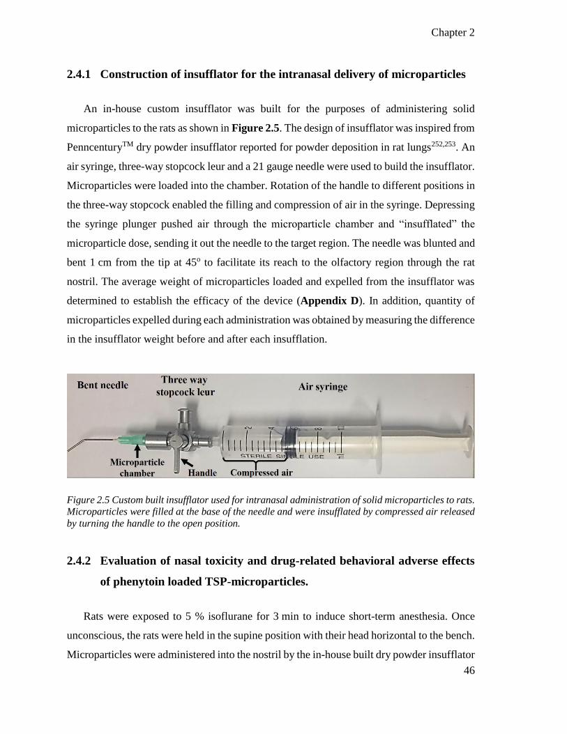
Chapter 2
46
2.4.1 Construction of insufflator for the intranasal delivery of microparticles
An in-house custom insufflator was built for the purposes of administering solid
microparticles to the rats as shown in Figure 2.5. The design of insufflator was inspired from
PenncenturyTM dry powder insufflator reported for powder deposition in rat lungs252,253. An
air syringe, three-way stopcock leur and a 21 gauge needle were used to build the insufflator.
Microparticles were loaded into the chamber. Rotation of the handle to different positions in
the three-way stopcock enabled the filling and compression of air in the syringe. Depressing
the syringe plunger pushed air through the microparticle chamber and “insufflated” the
microparticle dose, sending it out the needle to the target region. The needle was blunted and
bent 1 cm from the tip at 45o to facilitate its reach to the olfactory region through the rat
nostril. The average weight of microparticles loaded and expelled from the insufflator was
determined to establish the efficacy of the device (Appendix D). In addition, quantity of
microparticles expelled during each administration was obtained by measuring the difference
in the insufflator weight before and after each insufflation.
Figure 2.5 Custom built insufflator used for intranasal administration of solid microparticles to rats.
Microparticles were filled at the base of the needle and were insufflated by compressed air released
by turning the handle to the open position.
2.4.2 Evaluation of nasal toxicity and drug-related behavioral adverse effects
of phenytoin loaded TSP-microparticles.
Rats were exposed to 5 % isoflurane for 3 min to induce short-term anesthesia. Once
unconscious, the rats were held in the supine position with their head horizontal to the bench.
Microparticles were administered into the nostril by the in-house built dry powder insufflator

Chapter 2
47
described above. Rats were held in position for 15 sec after administration. Rats were
monitored for any potential signs of nasal irritation such as frequent sneezing, noisy
breathing, nose-rubbing, porphyrin (red) discharge, and general signs of pain/discomfort for
60 min.
Rats were euthanized 60 min after intranasal administration by guillotine decapitation.
Following this, skin and excess tissue around the nasal passage were removed from the head.
A blunt needle was inserted 0.5 cm into the posterior of the nasopharyngeal duct and used
to flush the nasal passage with 10 mL of 10 % neutral buffered formalin (NBF) or sodium
deoxycholate (positive control). The nasal passage was fixed in 50 mL of NBF for 48-72 h.
The fixed nasal passage was subsequently decalcified in 10 % EDTA (pH 7.2) for 2-3 weeks,
sliced coronally into blocks and embedded in paraffin. A microtome was used to cut 5 µm
sections which were subsequently de-paraffinized, stained with hematoxylin and eosin, and
cover-slipped with DPX mounting medium. Sections were imaged on an Aperio ScanScope.
Images were analyzed by personnel blinded to the treatments using Aperio
ImageScope v12.2.2.5015 software. A rat treated with saline was used as a negative control,
and a rat nasal cavity flushed with sodium deoxycholate served as positive control.
2.4.3 In-vivo drug administration
Rats were divided into three treatment groups receiving intranasal phenytoin
microparticles, intranasal phenytoin solution or IV phenytoin sodium injection under short-
term inhaled general anesthesia. The intranasal phenytoin microparticles were administered
by the in-house custom built insufflator as described in Section 2.4.1. The dose of the drug
was determined by weighing the microparticles emitted from an insufflator, an average dose
of 1.5 mg phenytoin was administered as shown in Figure 2.6. Animals to whom
administration was incomplete due to clogging of the microparticles in the needle were
temporarily excluded. Considering the half-life of model drugs254–256 and logistics, animals
were experimented with at a later date, at least 4 days later.

Chapter 2
48
Figure 2.6. Administration of phenytoin microparticles to the rat nasal cavity using the custom-built
insufflator.
The intranasal phenytoin solution was administered using a ‘rat intranasal catheter
device’ (Impel Neuro Pharma, USA). The drug solution was administered to anesthetized
animals in a similar to the procedure described in Section 2.4.2. Drug solution was loaded
into a syringe with a piece of plastic tubing attached. The tubing was inserted about 1 cm
into the rat nasal cavity to reach the olfactory region and depressing the plunger of the syringe
delivered the drug solution through the tubing directly to the olfactory region. Phenytoin
sodium solution was prepared by dissolving 100 mg of phenytoin sodium in 1 mL of 50 %
ethanol-water mixture containing 10 µL of propylene glycol. Considering the limitation of
solution volumes that the nasal cavity can accommodate, a volume of 15 µL solution,
equivalent to 1.5 mg of phenytoin was administered to each rat.
DBLTM Phenytoin injection (Hospira, Australia) was used for IV administration. The
marketed formulation has a 50 mg/mL concentration of phenytoin sodium. A solution
volume of 30 µL, equivalent to 1.5 mg of phenytoin sodium was administered IV by tail-
vein injection.
Chapter 2
49
After administration, the rats were sacrificed at 15, 30, 45 and 60 min time points (n = 5
for each time point) by decapitation as described in Section 2.4.2. Immediately after
decapitation, trunk blood was collected into heparin tubes. The blood was centrifuged at
2000 G for 5 min to collect the plasma. The brain was collected immediately and washed
with fresh saline three times to remove any blood. The liver, spleen, kidneys, lungs, and heart
were also collected and washed with saline. All the tissues and plasma were stored in a
– 81 ºC freezer until further analysis.
2.4.4 Analysis of phenytoin content in rat plasma and tissues by HPLC
2.4.4.1 Equipment and chromatographic conditions
An HPLC (Agilent technologies 1260 infinity) with UV detector, a C 18 column
(150 X 4.6 mm) with 5 µm particle size and a C-18 guard cartridge (5 X 4.6 mm)
(Phenomenex, California, USA) kept at 40 ºC were used for the analysis. The mobile phase
consisted of a mixture of pH 5.8 phosphate buffer (10 mM) and acetonitrile that was
regulated by a gradient profile programmed as follows: a linear gradient from 10 %
acetonitrile to 50 % over 10 min; maintenance at 50 % for 2 min, a linear gradient from 50 %
acetonitrile to 40 % over 1 min; maintenance of acetonitrile at 60 % for 3 min, then a linear
gradient from 40 % acetonitrile to 10 % over 1 min. After each cycle, the column was
conditioned with 10 % acetonitrile and 90 % phosphate buffer for 3 min. The flow rate was
0.2 mL/min. The phenytoin derivative 5-(4-methylphenyl)-5-phenyl-hydantoin was used as
the internal standard for the analysis of blood and tissues (referred to as ‘internal standard’
from now on in this thesis). The major metabolite of phenytoin in blood and tissues is
‘5- (4-Hydroxyphenyl)-5-phenyl-hydantoin’ (referred to as ‘metabolite’ from here onwards
in this thesis) was also quantified along with the phenytoin. The injection volume was 10 µL,
and the UV detection wavelength was 210 nm. The chromatographic data analysis was
performed using the OpenLAB software (Agilent Technologies, California, USA). The
method was validated for its accuracy, selectivity, precision, and repeatability in plasma,
brain, liver, spleen, kidneys, lung, and heart. Chromatographic conditions and the procedure

Chapter 2
50
to extract phenytoin from plasma and tissue samples were optimized by modifying the
reported methods257–259.
2.4.4.2 Stock solutions
Stock solutions of phenytoin, internal standard and metabolite were prepared in methanol
at a concentration of 1 mg/mL. Working solutions (100 µg/mL and 10 µg/mL) were prepared
by serial dilution of stock solutions with methanol.
2.4.4.3 Plasma sample preparation
For the construction of calibration curves, 200 µL aliquots of plasma were added to
1.7 mL Eppendorf tubes. The samples were spiked with varying concentrations of phenytoin
(0.1, 0.5, 1, 2.5, 5, 7.5, and 10 µg/mL) and metabolite (0.5, 1, 2.5, 5, 7.5, 10 and µg/mL) and
a fixed concentration of the internal standard (10 µg/mL) using the working solutions and
vortexed for 30 sec. Following that, 300 µL of chilled acetonitrile and 300 µL of tert-Butyl
methyl ether were added to the spiked plasma samples to precipitate the proteins and extract
the phenytoin from plasma. The samples were centrifuged at 13000 rpm at 4 ºC for 20 min.
The supernatant was then separated and concentrated overnight by evaporation to dryness in
a centrifugal evaporator. The concentrated samples were reconstituted in 200 µL of a mixture
of pH 5.8 phosphate buffer and acetonitrile (60:40) and loaded into the auto sampler for
HPLC analysis.
For quantification of phenytoin from in-vivo studies: the 200 µL plasma samples were
spiked with 10 µL internal standard (1 mg/mL) in Eppendorf tubes and vortexed for 30 sec.
Following that, the same steps for sample preparation as for the calibration curves were
followed and the samples were analyzed for phenytoin content.
2.4.4.4 Tissue sample preparation
All tissues were homogenized in 80 % ethanol solution in water to precipitate proteins
and lipids. A volume of 6 mL of 80 % ethanol solution was used for every 1 g of tissue.

Chapter 2
51
Tissue samples were spiked with varying concentrations of phenytoin (0.1, 0.5, 1, 2.5, 5, 7.5,
10 µg/g) and metabolite (0.5, 1, 2.5, 5, 7.5, 10 µg/g) and a fixed concentration of the internal
standard (10 µg/g) using the working solutions and vortexed for 30 sec. Tissue samples for
constructing calibration curves were then prepared in the same way as the plasma sample
preparations using of tissue homogenates equivalent to 200 µg tissue weight. However,
multiple tissue aliquots were concentrated then pooled together and reconstituted with
200 µL of a mixture of pH 5.8 phosphate buffer and acetonitrile (60:40) for analysis.
For quantification of phenytoin from in-vivo studies: 1 g of tissue homogenates were
spiked with 10 µL internal standard (1 mg/mL) in Falcon tubes and vortexed for 30 sec. The
same steps for sample preparation as for the calibration curves were followed and the
samples were analyzed for phenytoin content.
2.4.4.5 Method validation
The calibration curves were plotted using peak area ratio values obtained from the ratio
of phenytoin to the internal standard. The unknown concentrations of phenytoin were
determined from the calibration curves. Quality control (QC) samples consisted of low
(1 µg/mL) medium (5 µg/mL) and high (10 µg/mL) concentrations of the phenytoin.
2.4.4.5.1 Specificity
The specificity of the method was determined by identifying and separating the
phenytoin, the internal standard and the metabolite chromatographic peaks from each other,
and from the endogenous materials in plasma, brain, and other tissues. The specificity of the
HPLC method is demonstrated by comparing the chromatograms of plasma/tissue samples
with no phenytoin, and plasma/tissue samples spiked with known amounts of phenytoin,
internal standard, and metabolite.

Chapter 2
52
2.4.4.5.2 Linearity
The linearity of the HPLC method was determined using standard solutions spiked with
phenytoin and metabolite with final concentrations in the range of 0.1 to 10 µg/mL and an
internal standard of 10 µg/mL. The calibration curves were plotted with peak area ratio
values obtained from the ratio of phenytoin and metabolite to the internal standard. The linear
regression coefficient (r2) values were calculated and reported.
2.4.4.5.3 Sensitivity
The sensitivity of the method is demonstrated by the lower limit of detection (LOD), and
the lower limit of quantitation (LOQ) obtained from the standard deviation of the y-intercept
and slope of the calibration curve.
2.4.4.5.4 Accuracy and Precision
The amount of phenytoin recovered from the tissue and plasma QC samples after sample
preparation and HPLC analysis were compared to the theoretical values and used to report
the accuracy of the method. The intraday variability was determined by replicate analysis of
QC samples on the same day, by calculating and reporting the %co-variance (CV) and %bias.
The accuracy and precision of the method were based on the %bias and %CV.
The inter-day variability was measured to represent the repeatability of the method. QC
samples were analyzed on different days with one replicate being analyzed each day and the
%CV and %bias were calculated and reported. A %CV and %bias within ±15 % were
considered as acceptable for the plasma and tissue samples.
2.5 Statistical analysis
All results are expressed as mean ± SD. Where applicable, statistical analyses were
conducted using an unpaired t-test or ANOVA on Prism 6 (GraphPad, USA). A P-value of
< 0.05 was taken to be significant.
Chapter 3
Formulation and evaluation of
TSP-microparticles
This chapter has been published as a peer-reviewed article:
Sasi B. Yarragudi, Robert Richter, Helen Lee, Greg F. Walker, Andrew N. Clarkson,
Haribalan Kumar, Shakila B. Rizwan, Formulation of olfactory-targeted microparticles with
tamarind seed polysaccharide to improve nose-to-brain transport of drug, Carbohydrate
Polymers, 2017, 163: 216–226.


Chapter 3
55
3 Formulation and evaluation of TSP-microparticles
As discussed in Chapter 1, this thesis aimed to develop a mucoadhesive microparticle
system with specific size to efficiently target the olfactory region and enhance nose-to-brain
delivery of drugs. In this Chapter, the selection of a mucoadhesive polymer, formulation of
size tailored microparticles and the ability of such a microparticle system to load and release
drugs was investigated.
3.1 Selection of a mucoadhesive polymer
Chitosan is the most popular polymer reported in the literature for the formulation of
nose-to-brain micro and nanoparticle drug delivery systems (Table 5). It is proposed that the
positively charged amino groups of chitosan interact with sialic acids in the mucous layers
to form strong electrostatic bonds causing mucoadhesion. Also, chitosan can reversibly open
the tight junctions by inducing changes in the transmembrane protein claudin-4 and the
C-Jun NH2-terminal kinase-dependent pathway to increase the permeability of the epithelial
cells231,260. These characteristics have made chitosan the first choice among all other
polymers. However, there have been reports of acute mucosal toxicity261 believed to be due
to the positive charge on chitosan, and there is very little known about the effect of long-term
exposure to chitosan on the nasal mucosa.
Recently, tamarind seed polysaccharide (TSP) has gained some attention in drug delivery
applications due to its mucoadhesion properties, broad pH tolerance, biocompatibility and
high drug loading capacity262,263. Moreover, unlike chitosan, TSP is uncharged under
physiological conditions 264 and therefore is less likely to cause nasal toxicity. TSP is
classified as a generally recognized as safe (GRAS) substance and is approved by the FDA.
It is commonly used as an excipient in food and pharmaceutical preparations and is available
in the market as an eye drop formulation for treating dry eyes264–266. TSP is a naturally
occurring and readily available polymer. These distinctive advantages and characteristics of
TSP make it a potential polymer for nose-to-brain drug delivery purposes and hence, this
thesis investigates TSP for the formulation of mucoadhesive microparticles.
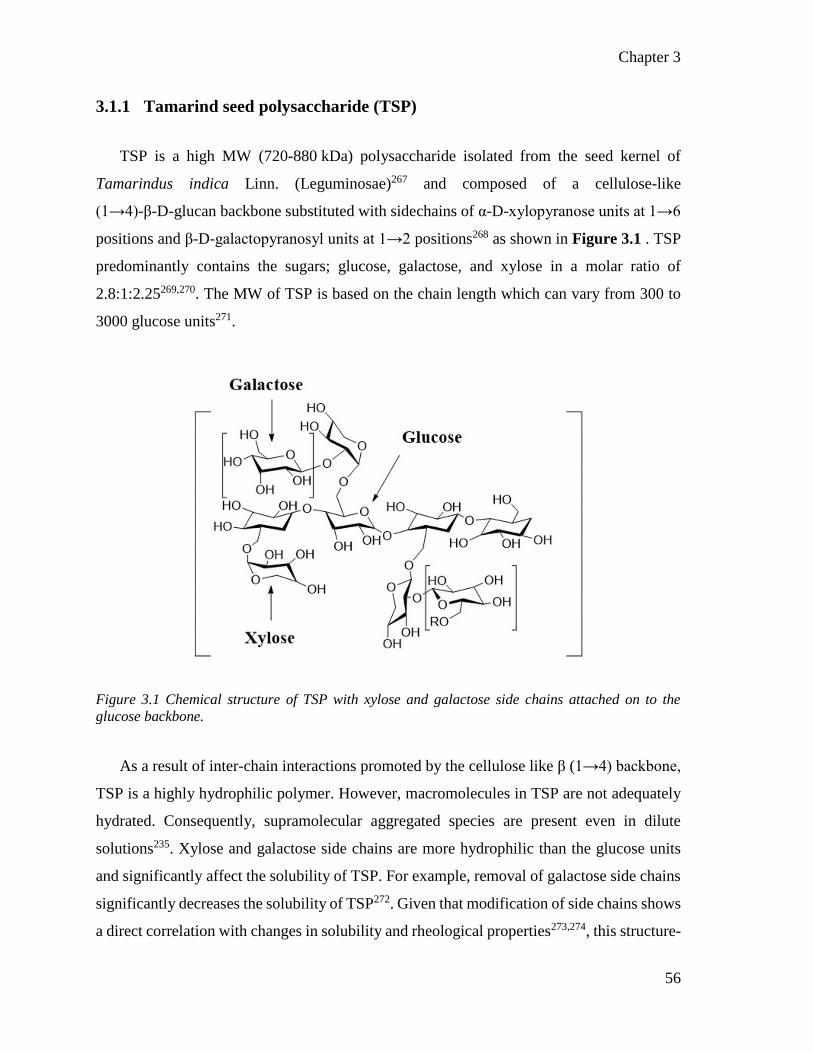
Chapter 3
56
3.1.1 Tamarind seed polysaccharide (TSP)
TSP is a high MW (720-880 kDa) polysaccharide isolated from the seed kernel of
Tamarindus indica Linn. (Leguminosae)267 and composed of a cellulose-like
(1→4)-β-D-glucan backbone substituted with sidechains of α-D-xylopyranose units at 1→6
positions and β-D-galactopyranosyl units at 1→2 positions268 as shown in Figure 3.1 . TSP
predominantly contains the sugars; glucose, galactose, and xylose in a molar ratio of
2.8:1:2.25269,270. The MW of TSP is based on the chain length which can vary from 300 to
3000 glucose units271.
Figure 3.1 Chemical structure of TSP with xylose and galactose side chains attached on to the
glucose backbone.
As a result of inter-chain interactions promoted by the cellulose like β (1→4) backbone,
TSP is a highly hydrophilic polymer. However, macromolecules in TSP are not adequately
hydrated. Consequently, supramolecular aggregated species are present even in dilute
solutions235. Xylose and galactose side chains are more hydrophilic than the glucose units
and significantly affect the solubility of TSP. For example, removal of galactose side chains
significantly decreases the solubility of TSP272. Given that modification of side chains shows
a direct correlation with changes in solubility and rheological properties273,274, this structure-

Chapter 3
57
functional relationship may have potential in controlling the release of drugs when
formulated using TSP.
The mucoadhesive properties of TSP are believed to be due to the secondary hydroxyl
(OH) groups present in the molecule, which can interact with mucus to form hydrogen bonds,
and Van der Walls attractions221. Also, the mucin-like configuration of TSP imparted by its
branched structure aids in the interpenetration of chains between TSP and mucus resulting
in mucoadhesion275,276. Due to the advantages it can offer, TSP has been investigated in drug
delivery applications. Table 5 lists the reported drug delivery applications of TSP and the
critical observations in those studies.
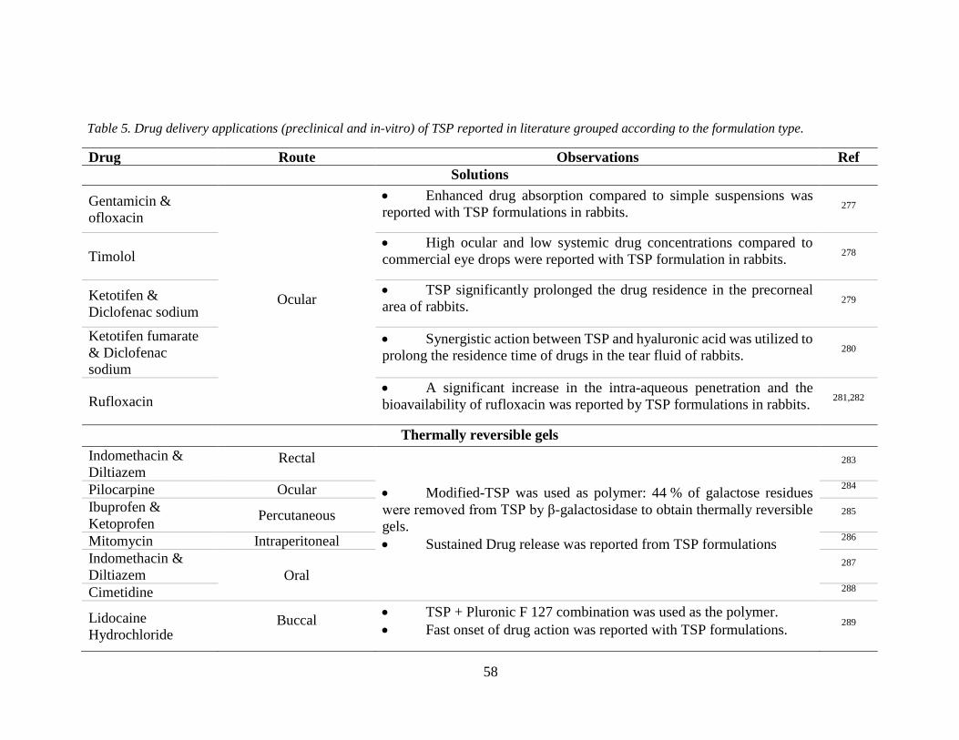
58
Table 5. Drug delivery applications (preclinical and in-vitro) of TSP reported in literature grouped according to the formulation type.
Drug Route Observations Ref
Solutions
Gentamicin &
ofloxacin
Ocular
Enhanced drug absorption compared to simple suspensions was
reported with TSP formulations in rabbits. 277
Timolol High ocular and low systemic drug concentrations compared to
commercial eye drops were reported with TSP formulation in rabbits. 278
Ketotifen &
Diclofenac sodium
TSP significantly prolonged the drug residence in the precorneal
area of rabbits. 279
Ketotifen fumarate
& Diclofenac
sodium
Synergistic action between TSP and hyaluronic acid was utilized to
prolong the residence time of drugs in the tear fluid of rabbits. 280
Rufloxacin A significant increase in the intra-aqueous penetration and the
bioavailability of rufloxacin was reported by TSP formulations in rabbits. 281,282
Thermally reversible gels
Indomethacin &
Diltiazem Rectal
Modified-TSP was used as polymer: 44 % of galactose residues
were removed from TSP by β-galactosidase to obtain thermally reversible
gels.
Sustained Drug release was reported from TSP formulations
283
Pilocarpine Ocular 284
Ibuprofen &
Ketoprofen Percutaneous 285
Mitomycin Intraperitoneal 286
Indomethacin &
Diltiazem Oral 287
Cimetidine 288
Lidocaine
Hydrochloride Buccal
TSP + Pluronic F 127 combination was used as the polymer.
Fast onset of drug action was reported with TSP formulations. 289
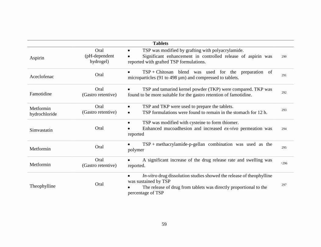
59
Tablets
Aspirin
Oral
(pH-dependent
hydrogel)
TSP was modified by grafting with polyacrylamide.
Significant enhancement in controlled release of aspirin was
reported with grafted TSP formulations.
290
Aceclofenac Oral TSP + Chitosan blend was used for the preparation of
microparticles (91 to 498 µm) and compressed to tablets. 291
Famotidine Oral
(Gastro retentive) TSP and tamarind kernel powder (TKP) were compared. TKP was
found to be more suitable for the gastro retention of famotidine. 292
Metformin
hydrochloride
Oral
(Gastro retentive)
TSP and TKP were used to prepare the tablets.
TSP formulations were found to remain in the stomach for 12 h. 293
Simvastatin Oral
TSP was modified with cysteine to form thiomer.
Enhanced mucoadhesion and increased ex-vivo permeation was
reported
294
Metformin Oral TSP + methacrylamide-p-gellan combination was used as the
polymer 295
Metformin Oral
(Gastro retentive) A significant increase of the drug release rate and swelling was
reported. `296
Theophylline Oral
In-vitro drug dissolution studies showed the release of theophylline
was sustained by TSP
The release of drug from tablets was directly proportional to the
percentage of TSP
297
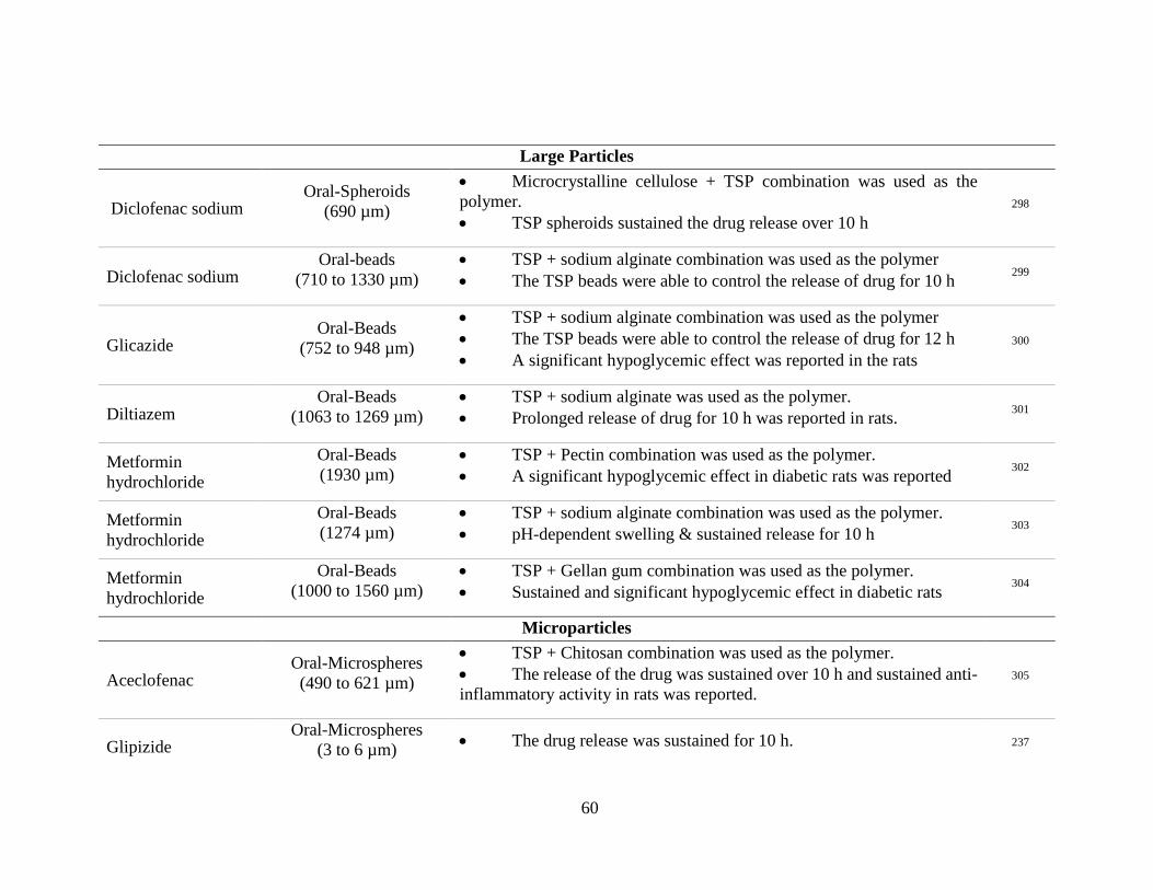
60
Large Particles
Diclofenac sodium Oral-Spheroids
(690 µm)
Microcrystalline cellulose + TSP combination was used as the
polymer.
TSP spheroids sustained the drug release over 10 h
298
Diclofenac sodium Oral-beads
(710 to 1330 µm)
TSP + sodium alginate combination was used as the polymer
The TSP beads were able to control the release of drug for 10 h 299
Glicazide Oral-Beads
(752 to 948 µm)
TSP + sodium alginate combination was used as the polymer
The TSP beads were able to control the release of drug for 12 h
A significant hypoglycemic effect was reported in the rats
300
Diltiazem Oral-Beads
(1063 to 1269 µm)
TSP + sodium alginate was used as the polymer.
Prolonged release of drug for 10 h was reported in rats. 301
Metformin
hydrochloride
Oral-Beads
(1930 µm)
TSP + Pectin combination was used as the polymer.
A significant hypoglycemic effect in diabetic rats was reported 302
Metformin
hydrochloride
Oral-Beads
(1274 µm)
TSP + sodium alginate combination was used as the polymer.
pH-dependent swelling & sustained release for 10 h 303
Metformin
hydrochloride
Oral-Beads
(1000 to 1560 µm)
TSP + Gellan gum combination was used as the polymer.
Sustained and significant hypoglycemic effect in diabetic rats 304
Microparticles
Aceclofenac Oral-Microspheres
(490 to 621 µm)
TSP + Chitosan combination was used as the polymer.
The release of the drug was sustained over 10 h and sustained anti-
inflammatory activity in rats was reported.
305
Glipizide Oral-Microspheres
(3 to 6 µm) The drug release was sustained for 10 h. 237

61
Montelukast sodium
Inhalation
Microspheres
(0.9 to 6 µm)
Modified TSP was used as polymer: 47 % of galactose units were
removed.
Enhanced in-vivo drug residence time in rats was reported
306
Films
Rizatriptan benzoate Buccal TSP + Carbopol combination was used as the polymer. 307
Nystatin Vaginal The sustained release was reported 308
Timolol maleate Ocular TSP films were nonirritant and able to sustain the drug delivery 309
Ciprofloxacin 310
Nanoparticles/Nanoaggregates
Tropicamine Ocular Increased drug absorption was reported in the in-vitro cell studies 311
Doxorubicin NA/in-vitro Enhanced cytotoxic effects were reported in the cells with TSP
formulations 312
Gels
Paracetamol
Oral
Modified-TSP was used as polymer: removal of galactose residues
Sustained release of drugs by TSP gel was reported in rats. 313
Metronidazole Thiol-functionalized TSP was used as the polymer.
Mucoadhesion was increased by 6.85 folds after functionalization 268
Eriochrome Black T Injectable implants Sustained drug release was reported for four days 314

Chapter 3
62
Reports in the literature showcase the potential of TSP as an excipient and an active
ingredient in drug delivery applications. The chemical modification of TSP with
polyacrylamide315, methyl methacrylate316, polycaprolactone317,318 and PLA319 has been
investigated for drug delivery purposes. Semi-synthetic derivatives of TSP such as
thiolated-TSP268,276,320 and carboxymethylated-TSP321 have also been investigated for drug
delivery applications. Several patents on the use of TSP as a polymer for drug delivery in
opthalmic322,323, topical324 and cosmetic325 areas have been filed. In addition to the drug
delivery applications, TSP has also been shown to have wound healing326, anti-tumor327–329,
anti-myocardial infraction330 and immunomodulatory properties329,331.Patent has also been
filed for the use of TSP as a protecting agent for epithelial cells of the respiratory tract from
damages induced by smoking332. TSP has been tested clinically with positive results for the
treatment of dry eye syndrome333 and is used in commercially available Visine Intensiv 1%
eyedrops (Europe)266.
The analysis of literature has shown some interesting trends in the use of TSP for drug
delivery applications.
1. The drug carrying capacity of TSP in oral and ocular routes was well characterized
in the reported publications (Table 5). However, the ability of TSP to act as a drug
carrier via the nasal route has not been well reported.
2. The potential of TSP as a carrier of drug molecules with high MW has not been
explored yet.
3. About 60 % of the reported publications have formulated TSP either with a
combination of another polymer or modification of its structure with an aim to change
properties like mucoadhesion and drug release. Consequently, this has resulted in
considerable variations in reported drug release profiles ranging from 2 h to
four days. Controlled release of drugs formulated in TSP in combination with other
polymers or by chemical modification has been shown for at least 10-12 h in-vitro
(Table 5). While this may be useful for other drug delivery purposes, for the
nose-to-brain delivery purposes, the likelihood that any formulation is retained in the
nasal cavity for periods longer than 12 h despite its mucoadhesive nature is unlikely
and often not desired. Thus, examining unmodified TSP as a carrier of drugs may
Chapter 3
63
decrease the time required to release the encapsulated drugs, offering advantages for
nose-to-brain drug delivery purposes.
3.1.1.1 TSP as a carrier for nose-to-brain drug delivery
Two investigations have reported the potential of TSP as a carrier for brain targeting
drugs via the nasal cavity. Kumar et al.,275 formulated two anti-migraine drugs zolmitriptan
and ketorolac tromethamine into thermally reversible gels using a blend of TSP,
Pluronic F127 and PEG. The formulations were found to be mucoadhesive and sustained
release of both drugs. The histopathological evaluation of sheep nasal tissue treated with the
formulations suggested the formulations were safe to administer. In-vivo studies in rabbits
compared the pharmacokinetic profiles of drugs between intranasal (gel), IV and oral
(solutions) administrations. The TSP formulations were reported to have an almost 1.3-fold
increase in absolute bioavailability compared to the oral route.
In the second study, Mahajan et al.,334 reported the formulation of the anti-nausea agent
ondansetron in a TSP thermally reversible gel. TSP was modified by the β-galactosidase
enzyme to remove 45 % of the galactose units. The removal of galactose units imparted TSP
with thermosensitive gel properties. The formulations were found to be mucoadhesive with
sustain drug release over 6 h. Histopathological evaluation of rabbit nasal tissue treated with
the formulations has shown no evidence of damage. In-vivo absorption studies compared
intranasal (gel) and oral (solution) administration. TSP formulations were reported to
produce an almost two-fold increase in absolute bioavailability.
In both studies, TSP was either chemically modified or used with another polymer. Both
studies reported the safety of TSP for nasal administration and the ability of TSP
formulations to improve residency of drugs in the nasal cavity. However, the drug
concentrations in the brain were not quantified.
The capability of TSP as a mucoadhesive and safe polymer for nasal administration has
been reported. Therefore, due to the advantages and potential of TSP, this thesis utilizes TSP
as a mucoadhesive polymer for the preparation of solid microparticles intended for nose-to-
brain delivery.

Chapter 3
64
3.2 Method of preparation of microparticles
Freeze drying206,207, emulsification-cross linking137,208, and emulsification-solvent
evaporation193,205 are some of the methods reported in the literature for the preparation of
microparticles for nose-to-brain delivery. Spray-drying has been the most popular method
with about 70 % of the reported publications utilizing spray-drying for the formulation of
microparticles intended for nose-to-brain delivery. The high yield rate and simple
methodology of spray-drying are the main reasons for its popularity.
3.2.1 Spray-drying
Spray-drying is a process where a liquid (feed) is converted to a powdered solid (product)
by spraying the feed into a gaseous drying medium335. It has been utilized in the food and
pharmaceutical industry for the production of powders since its first patent in 1872336. There
are three phases involved in the spray-drying process: first, the atomization phase where the
pumped feed is sprayed as small droplets through the nozzle by the force of a compressed
air/gas. The second is the drying phase where the droplets interact with hot gas in a chamber
to evaporate the solvent, and third, a separation phase where the particles are separated from
the gas for collection by filter or cyclone337.
Spray-drying is a one-step method and produces reasonable yields of microparticles338.
By controlling the spray-drying parameters, microparticle characteristics such as particle
size, shape and density can be tailored to suit the requirements339. The parameters of
composition of feed solution, velocity of the feed solution being pumped, velocity of the
compressed air/gas (atomizing air flow), nozzle type, velocity of the drying gas (aspiration)
and temperature of the drying gas (inlet temperature) can be examined.
As reported in Chapter 1 and previous sections spray-drying is the most widely used
method to produce microparticles, which this can be attributed to the numerous advantages
of spray-drying compared to the other methods. For example, compared to a commonly
employed method, freeze-drying, it is economical and less time consuming due to the fact
that spray-drying doesn’t have a deep cooling step which consumes significant time and

Chapter 3
65
energy340–342.Hence, spray-drying is being explored as an alternative to freeze-drying343,344.
In addition, particle size and distribution can be well controlled by spray-drying compared
to emulsification, solvent extraction, and crosslinking methods employed in the literature.
Hence, a spray-drying method promises successful bench-to-bedside translation of
microparticle produciton345.
Although spray-drying has numerous advantages, low yield and loss of nano-sized
particles are major limitations of the spray-drying process345. Low yields are generally
associated with loss of particles in the walls of equipment and smaller particles passing into
exhaust346. The production of nanometer-sized particles by spray-drying is a challenging task
due to the low separation capacity of the cyclone and low forces of liquid
atomization345,347,348. While few studies reported an increased capacity of the cyclone at
industrial settings, more studies are required to address these challenges345,349.
Due to the ability of spray-drying to control the particle size, this thesis employs spray-
drying to investigate the optimum parameters to produce the 10 µm sized particles with TSP
as a mucoadhesive polymer.
3.3 Design of experiments (DOE)
Rational DOE decreases the total number of experiments required to extract critical
information of a complicated process and saves significant experimental time238. In this
thesis variables of an experiment that may have an effect on the characteristics of the end
product are termed ‘independent factors,’ the characteristics that are being affected are
termed ‘responses.’ This chapter employs spray-drying to formulate size tailored TSP
microparticles. The effect of spray-drying parameters at different levels on the particle size
needs to be tested for these purposes to identify the optimum parameters. The relationship
between one or more responses and one or more independent factors can be studied using
response surface methods350.
Central composite design and Box–Behnken experimental designs are two widely
reported methods to identify optimum levels of independent factors to achieve a specific

Chapter 3
66
response238,239. The central composite experimental design has axial points beyond the
design space leading to increased levels of the independent factors to be tested. Due to the
nature of the independent factors tested in this chapter, it might not be beneficial to test points
beyond the design space. In contrast, Box-Behnken design does not require the axial points
beyond design space to be tested. Also, it is experimentally more convenient as it requires
fewer experiments to be performed. The Box-Behnken experimental design also ensures that
all individual factors are not tested at their highest levels, simultaneously putting the
experiments in a safe operating zone350. Thus, in this chapter, Box-Behnken experimental
design is employed to examine and identify the optimum parameters to produce the 10 µm
sized particles with TSP using the spray-drying method.
3.4 Model drugs
In this Chapter, the ability of TSP to load drugs and the release of drugs from the
formulated microparticles are examined by incorporating fluorescein isothiocyanate-labeled
dextran’s (FITC-Dextrans) with MW of 3–5, 10, 20 and 40 kDa as model drugs. Use of
FITC-Dextrans to study the drug release from the formulations and permeation across the
mucosal barriers is well reported in the literature351–353.

Chapter 3
67
Figure 3.2 Structure of FITC-Dextran. The MW depends on the number of dextran molecules linked
to each other. Figure adapted from website https://www.sigmaaldrich.com
Dextran is a polymer of anhydroglucose as shown in Figure 3.2 It is comprised of
approximately 95 % straight-chain α-D-(16) glycosidic linkages and the remaining
α -D-(13) glycosidic linkages account for the branching of dextran354. The FITC is
randomly conjugated to the free hydroxyl groups of the dextran at a frequency of 0.003 to
0.02 moles of FITC per mole of glucose. The number of dextran molecules determines the
MW of the FITC-Dextrans and hence they are available in different MW. The FITC portion
is responsible for the fluorescence of the compound, and hence it is utilized in permeation
and release studies. It fluoresces with an excitation and emission wavelength of 498 nm and
520 nm, respectively.
3.4.1 Spray-dried TSP microparticles
As discussed in the previous sections, the advantages TSP can offer, its unexplored
potential as a carrier of macromolecules and the ability of spray-drying to control the particle
size combined poses the research question of whether drug loaded 10 µm sized TSP-particles
can be formulated by spray-drying.

Chapter 3
68
3.5 Hypothesis
The overall hypothesis tested in this chapter is that drug formulated into 10 µm
TSP-microparticles will have increased olfactory targeting and residence.
3.5.1 Chapter aims
1. To formulate 10 µm sized TSP-microparticles loaded with high MW drugs by
spray-drying.
2. To measure the mucoadhesion of the TSP-microparticles in-vitro and ex-vivo.
3. To establish the drug release profile from the TSP-microparticles.
4. To assess the safety of TSP-microparticles for nasal administration.
5. To assess the olfactory deposition of TSP-microparticles.
Chapter 3
69
3.6 Results
3.6.1 Extraction of TSP from tamarind seed gum
TSP was extracted from tamarind seed gum with an average yield of 51 ± 11 %. The
infrared (IR) spectra and x-ray diffractograms of the extracted TSP were analyzed and
compared with the published data355.
The IR spectra of TSP presented the characteristic broad peaks for stretching of the –OH
group on the glucan backbone at 3331.62 cm-1 and 3282.07 cm-1 and the OH bending peaks
were observed at 526.61 cm-1. The alkane C-H stretching was observed by the peak at
2922.80 cm-1. The cyclic ethers (C-O-C) stretching peak was observed at 1026.24 cm-1.
These peaks (Figure 3.3 A) indicate the characteristic functional groups of TSP and are
agreement with the reported literature355. The x-ray diffractograms of TSP are presented in
Figure 3.3 B. In agreement with the published literature for TSP, no sharp peaks were
observed indicating its amorphous nature355.

Chapter 3
70
Figure 3.3 IR spectrum (A) and X-ray diffractogram (B) of TSP extracted from the tamarind gum.
The red arrows show the characteristic peaks.

Chapter 3
71
The surface morphology of TSP was observed under SEM and found to be similar to the
published literature356.
Figure 3.4 Representative micrograph showing the morphology of TSP (powder obtained from the
crushed film) extracted from tamarind seed gum similar to the extraction procedure followed in this
thesis.

Chapter 3
72
3.6.2 Formulation of 10 µm sized TSP-microparticles loaded with
FITC-Dextrans
3.6.2.1 Pilot studies
Pilot studies were conducted by trial and error to examine the ability of TSP to form
microparticles. Initially, the process variables that might affect the characteristics of resultant
particles were identified. These are referred to as formulation parameters (independent
factors) in this thesis. The identified formulation parameters were: total solids (%) (w/w) in
the feed solution, FITC-Dextran content (%), spray-drying conditions (atomizing airflow
(L/h), inlet temperature and aspiration (%)). Formulation parameters employed in the
preliminary studies and the resulting microparticle size and yield are presented in Table 6.
Table 6 Preliminary studies with TSP to examine the yield and size of microparticles with
spray-drying.
Total
solids (%)
(TSP)
Atomizing
airflow
(L/h),
Inlet
temperature
Aspiration
(%)
Particle size
(µm)
Yield
(%)
1.0 742 130 100 2.1 ± 0.2 21 ± 4
1.5 742 130 100 2.8 ± 0.9 53 ± 1
2.0 742 130 100 2.2 ± 0.1 69 ± 4
1.0 246 100 20 NA NA
1.5 312 110 30 16.3 13.0
2.0 384 120 40 13.2 35.0
2.0 425 130 50 11.9 67.0
TSP concentration higher than 2 % made the feed solution too viscous for spray-drying.
Therefore, a 2 % TSP concentration (total solids content) was employed to prepare the feed
solutions in subsequent studies. The % yield of the microparticles was found to increase with
an increase in the % w/w of total solids.
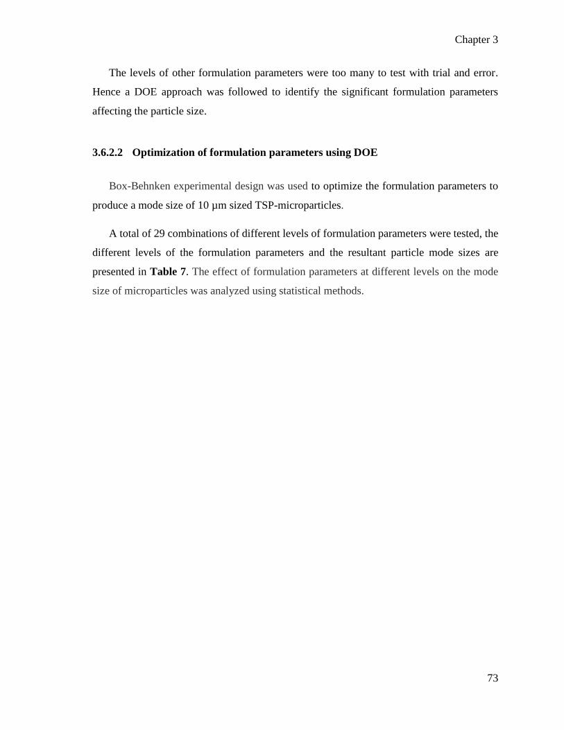
Chapter 3
73
The levels of other formulation parameters were too many to test with trial and error.
Hence a DOE approach was followed to identify the significant formulation parameters
affecting the particle size.
3.6.2.2 Optimization of formulation parameters using DOE
Box-Behnken experimental design was used to optimize the formulation parameters to
produce a mode size of 10 µm sized TSP-microparticles.
A total of 29 combinations of different levels of formulation parameters were tested, the
different levels of the formulation parameters and the resultant particle mode sizes are
presented in Table 7. The effect of formulation parameters at different levels on the mode
size of microparticles was analyzed using statistical methods.

Chapter 3
74
Table 7 Box-Behnken design of experiments to optimize the formulation parameters.
Formulation parameters (Independent Factors) Response
Run Atomizing
Airflow (L/h)
Aspiration
(%)
FITC content
(%)
Inlet
temperature
(ºC)
Mode size
(µm)
1 494 50 0.10 110 12.99
2 246 60 0.20 120 12.98
3 494 60 0.10 120 9.48
4 494 60 0.20 110 8.63
5 494 70 0.20 100 8.61
6 494 60 0.20 110 7.27
7 246 50 0.20 110 11.32
8 742 60 0.10 120 4.79
9 742 70 0.20 110 5.24
10 742 60 0.20 100 5.24
11 494 50 0.25 110 14.14
12 742 50 0.20 110 5.47
13 494 60 0.20 110 12.96
14 494 60 0.10 100 9.46
15 494 60 0.25 120 12.40
16 494 60 0.25 100 6.27
17 742 60 0.20 120 5.47
18 494 60 0.20 110 10.32
19 494 50 0.20 120 12.96
20 246 60 0.20 110 8.23
21 494 60 0.20 110 10.32
22 494 50 0.20 100 11.28
23 494 70 0.20 110 10.32
24 246 60 0.20 110 10.80
25 742 60 0.25 110 8.23
26 494 70 0.25 110 7.89
27 494 60 0.10 110 11.84
28 246 60 0.25 110 12.39
29 494 70 0.10 110 5.50

Chapter 3
75
ANOVA was performed to determine the validity of the experimental design used and
the effect of each of the independent factors on mode size of the particles. The sequential
model sum of squares and lack of fit tests were performed to determine whether the
experimental design model is valid to navigate the design space. Lack of fit provided a mean
response and an estimate of real experimental uncertainty and was found to have a value of
P = 0.5613 (Table 8). An insignificant P-value for lack of fit indicates that any errors caused
by design were too insignificant to influence the desired outcome.
Table 8 Effect of independent factors on mode size of the microparticles and the validity of design
model was determined by ANOVA. DF: degrees of freedom. *P-value of < 0.05 was taken to be
significant.
Source Sum of Squares DF Mean square F-value P-value
Model 128.64 4 32.16 7.49 0.0005*
Atomizing Airflow 92.47 1 92.47 21.53 0.0001*
Aspiration 35.16 1 35.16 8.19 0.0086*
TSP:FITC-Dextran 1.97 1 1.97 0.46 0.5051
Inlet Temperature 3.50 1 3.50 0.81 0.3759
Residual 103.07 24 4.29
Lack of Fit 81.42 19 4.29 0.99 0.5613
Pure Error 21.65 5 4.33
Cur Total 231.71 28
The atomizing airflow and aspiration were found to significantly influence microparticle
size, with P-values of 0.0001 and 0.0086, respectively (Table 8). The three levels of inlet
temperature used in the study were high enough to dry the microparticles in the spray-drying
chamber and did not influence the mode size. Additionally, the amount of FITC-Dextran did
not have a significant effect on the size of the microparticles. The 2D contour plots
representing the effect of significant factors on the mode size are presented in Figure 3.5.

Chapter 3
76
Figure 3.5 Contour plots showing the effect of the atomizing airflow and aspiration on the mode size
of TSP-microparticles (A) which was further optimized to obtain microparticles with a mode size of
10 µm (B). A plot showing the correlation between the predicted mode of TSP-microparticles sizes
against experimentally determined values (C).
Response surface methodology was used predict the optimal formulation parameters
required to produce 10 µm sized microparticles based on their effect on the size. A
correlation between the predicted mode size of TSP-microparticles and experimentally
determined values was found to exist and is presented in Figure 3.5 C. The desirability plot
shown in Figure 3.5 B, constructed based on the predictions of response surface
methodology shows the desirable regression ranges for the atomizing airflow and aspiration
to produce 10 µm microparticles. Optimum values of atomizing airflow and aspiration, to
produce 10 µm microparticles were determined to be 547 L/h and 55 %, respectively, while
the inlet temperature and amount of FITC-Dextran were set at 120 ºC and 0.25 % w/w,
respectively.
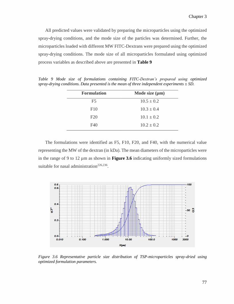
Chapter 3
77
All predicted values were validated by preparing the microparticles using the optimized
spray-drying conditions, and the mode size of the particles was determined. Further, the
microparticles loaded with different MW FITC-Dextrans were prepared using the optimized
spray-drying conditions. The mode size of all microparticles formulated using optimized
process variables as described above are presented in Table 9
Table 9 Mode size of formulations containing FITC-Dextran’s prepared using optimized
spray-drying conditions. Data presented is the mean of three independent experiments ± SD.
Formulation Mode size (µm)
F5 10.5 ± 0.2
F10 10.3 ± 0.4
F20 10.1 ± 0.2
F40 10.2 ± 0.2
The formulations were identified as F5, F10, F20, and F40, with the numerical value
representing the MW of the dextran (in kDa). The mean diameters of the microparticles were
in the range of 9 to 12 µm as shown in Figure 3.6 indicating uniformly sized formulations
suitable for nasal administration226,236.
Figure 3.6 Representative particle size distribution of TSP-microparticles spray-dried using
optimized formulation parameters.

Chapter 3
78
3.6.2.3 Morphology of TSP-microparticles
The morphology of the microparticles was investigated using SEM. The spray-drying
conditions used in pilot studies were able to produce microparticles successfully as shown
in Figure 3.7. The TSP-microparticles were spherical and corrugated in shape. The majority
of the larger particles (> 15 µm) were spherical as shown in Figure 3.7 A and the smaller
particles (< 5 µm) were corrugated in shape (Figure 3.7 B). All particles had a smooth
surface.
Figure 3.7 Representative SEM micrographs from the pilot studies showing the microparticle
formation with TSP alone. Huge variation in particle size (large >15 µm (A) and small < 5 µm (B))
was observed at different levels of formulation parameters.
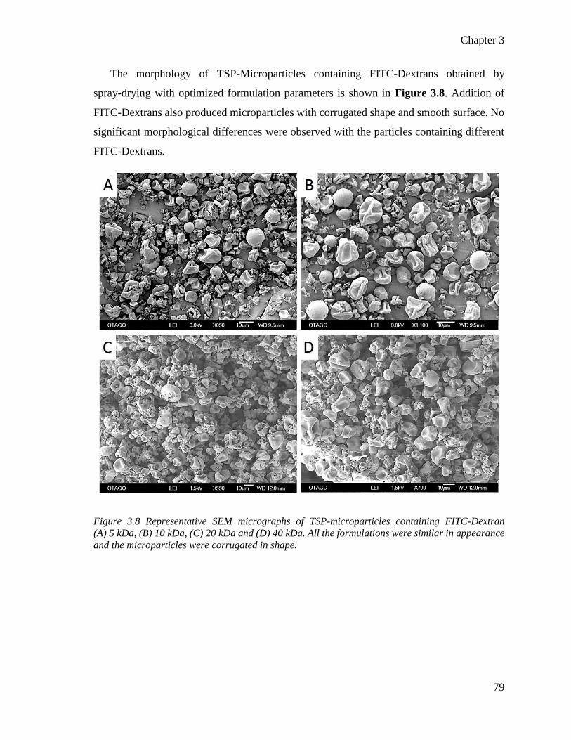
Chapter 3
79
The morphology of TSP-Microparticles containing FITC-Dextrans obtained by
spray-drying with optimized formulation parameters is shown in Figure 3.8. Addition of
FITC-Dextrans also produced microparticles with corrugated shape and smooth surface. No
significant morphological differences were observed with the particles containing different
FITC-Dextrans.
Figure 3.8 Representative SEM micrographs of TSP-microparticles containing FITC-Dextran
(A) 5 kDa, (B) 10 kDa, (C) 20 kDa and (D) 40 kDa. All the formulations were similar in appearance
and the microparticles were corrugated in shape.

Chapter 3
80
Microparticles were physically broken using a mortar and pestle and observed under
SEM to examine their core. The TSP-microparticles produced were hollow (Figure 3.9 B).
Figure 3.9 Representative electron micrographs of TSP-microparticles showing the smooth rupture
free surface (A) and showing the hollow core (B). The microparticles were physically broken and
examined under SEM to reveal the hollow core.
3.6.2.4 FITC-Dextran content
Quantification of FITC-Dextrans encapsulated in TSP-microparticles was carried out by
fluorescence spectroscopy. A series of standard curves were prepared with FITC-Dextrans
and used to calculate the fluorescence of unknown samples, as presented in Figure 3.10. The
background fluorescence from TSP was normalized to construct the standard curves.

Chapter 3
81
Figure 3.10 Standard curves and r2 values of FITC-Dextrans with a MW (A) 3-5 kDa, (B) 10 kDa,
(C) 20 kDa and (D) 40 kDa. (AU = arbitrary units)
The fluorescence was used to quantify the amount of FITC-Dextran entrapped in the
microparticles. Table 10 presents the entrapment efficiency (%) and drug loading (%) of
microparticles for all the FITC-Dextrans used.
Table 10 Drug loading and encapsulation efficiency of 3-5 kDa (F5), 10 kDa (F10), 20 kDa (F20)
and 40 kDa (F40) FITC-Dextrans in TSP-microparticles. Data presented is the mean of three
independent experiments ± SD.
Formulation Drug loading (%) Encapsulation efficiency (%)
F5 20.6 ± 0.1 82.6 ± 0.5
F10 19.8 ± 0.3 79.3 ± 1.1
F20 24.2 ± 0.6 75.7 ± 0.5
F40 18.2 ± 0.3 72.8 ± 1.9
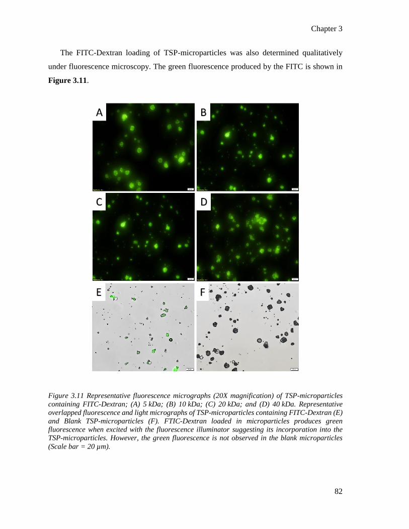
Chapter 3
82
The FITC-Dextran loading of TSP-microparticles was also determined qualitatively
under fluorescence microscopy. The green fluorescence produced by the FITC is shown in
Figure 3.11.
Figure 3.11 Representative fluorescence micrographs (20X magnification) of TSP-microparticles
containing FITC-Dextran; (A) 5 kDa; (B) 10 kDa; (C) 20 kDa; and (D) 40 kDa. Representative
overlapped fluorescence and light micrographs of TSP-microparticles containing FITC-Dextran (E)
and Blank TSP-microparticles (F). FTIC-Dextran loaded in microparticles produces green
fluorescence when excited with the fluorescence illuminator suggesting its incorporation into the
TSP-microparticles. However, the green fluorescence is not observed in the blank microparticles
(Scale bar = 20 µm).

Chapter 3
83
3.6.3 The mucoadhesive potential of TSP-microparticles
A combination of in-vitro and ex-vivo techniques was used to investigate the interaction
of TSP-microparticles with mucin as an indicator of mucoadhesiveness. The percentage of
FITC-Dextran that remained adhered to a piece of filter paper saturated with mucin after
application of an air load was quantified and expressed as a percentage of the original amount
of FITC-Dextran applied (Figure 3.12 A). Applying an air load is a useful way to determine,
in-vitro, the effect breathing may have on the clearance of microparticulate formulations
from the nasal cavity (‘nasal retention’) after administration. The percentage of
FITC-Dextran that remained adhered to the filter paper was comparable across all
microparticle formulations. However, it was higher when compared to unformulated FITC-
Dextran powders, which were used as controls (P < 0.001). Mucoadhesive strength was
determined ex-vivo using porcine mucosa and a tensile strength apparatus and the results are
shown in Figure 3.12 B. The strength required to detach (mucoadhesion strength) the
microparticles from the mucosa was significantly (P < 0.0001) higher than the
mucoadhesion strength of unformulated FITC-Dextran powders. The mucoadhesion strength
of all the formulations containing different MW FITC-Dextrans was comparable and also
was comparable to the in-vitro studies using the mucin-saturated filter paper.

Chapter 3
84
Figure 3.12 Mucoadhesive properties of FITC-Dextran containing TSP-microparticles (triangle) or
unformulated FITC-Dextrans (square) in-vitro (A) and ex-vivo (B) as a function of the MW of the
FITC-Dextran. Data presented is the mean of three independent experiments ± SD. (***P < 0.001
and **** P < 0.0001).
3.6.3.1 Mechanism of mucoadhesion determined by cryo-FESEM
The interaction of TSP-microparticles with mucin was investigated microscopically by
plunge freezing a sample of mucin with microparticles deposited on it. Samples were viewed
at pre-determined time intervals under the cryo-FESEM (Figure 3.13). The time-series
cryo-FESEM micrographs show the initial contact (Figure 3.13 B, white arrowhead)
followed by gradual formation of a gel-like layer due to the interaction between the
TSP-microparticles and the mucin layer (Figure 3.13 C and D, white arrows). The rough
surface features of the mucin layer as shown in Figure 3.13 A gradually changes to a
smoother surface after interacting with TSP-microparticles (Figure 3.13 C and D, red
arrows)

Chapter 3
85
Figure 3.13 Representative cryo-FESEM micrographs of a 2 % w/v solution of mucin only (A) or
mucin over 60 min (T0-T60) after deposition of TSP-microparticles on it (B-D). The initial contact
stage between mucin and TSP-microparticles (B, arrowhead) is followed by interaction of the
microparticles with the mucus layer (consolidation stage, white arrows) and gradual formation of a
smooth gel layer (red arrows) (C and D).
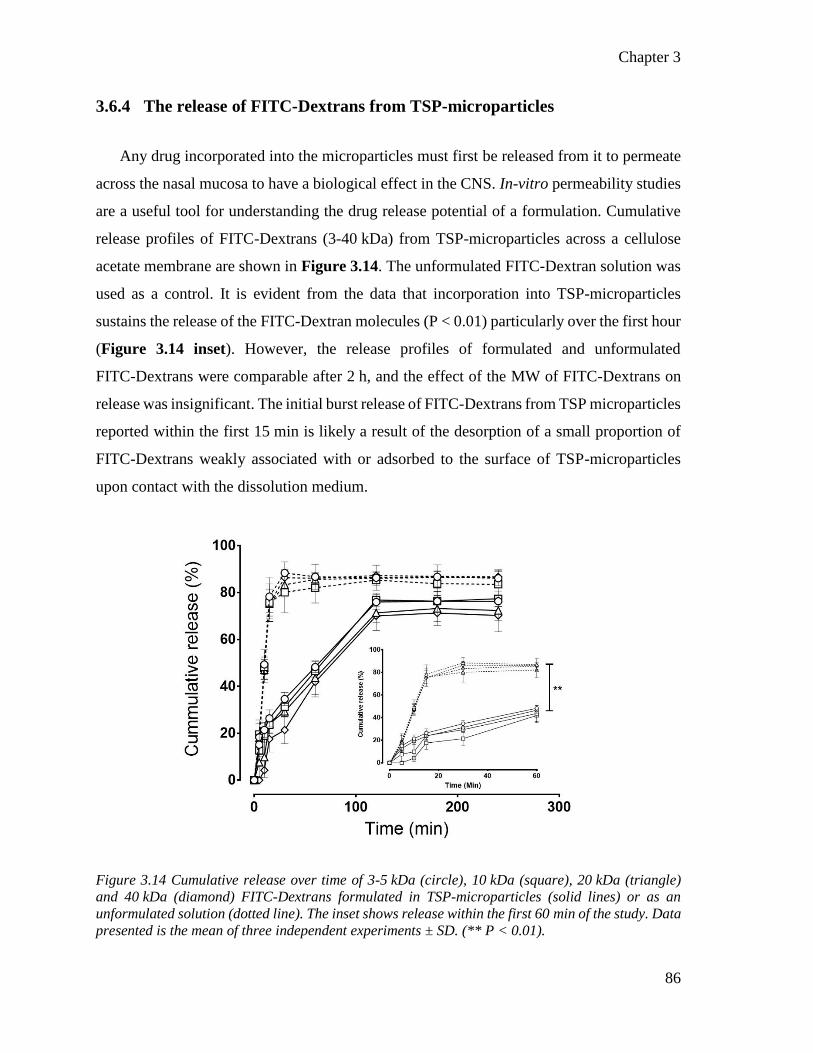
Chapter 3
86
3.6.4 The release of FITC-Dextrans from TSP-microparticles
Any drug incorporated into the microparticles must first be released from it to permeate
across the nasal mucosa to have a biological effect in the CNS. In-vitro permeability studies
are a useful tool for understanding the drug release potential of a formulation. Cumulative
release profiles of FITC-Dextrans (3-40 kDa) from TSP-microparticles across a cellulose
acetate membrane are shown in Figure 3.14. The unformulated FITC-Dextran solution was
used as a control. It is evident from the data that incorporation into TSP-microparticles
sustains the release of the FITC-Dextran molecules (P < 0.01) particularly over the first hour
(Figure 3.14 inset). However, the release profiles of formulated and unformulated
FITC-Dextrans were comparable after 2 h, and the effect of the MW of FITC-Dextrans on
release was insignificant. The initial burst release of FITC-Dextrans from TSP microparticles
reported within the first 15 min is likely a result of the desorption of a small proportion of
FITC-Dextrans weakly associated with or adsorbed to the surface of TSP-microparticles
upon contact with the dissolution medium.
Figure 3.14 Cumulative release over time of 3-5 kDa (circle), 10 kDa (square), 20 kDa (triangle)
and 40 kDa (diamond) FITC-Dextrans formulated in TSP-microparticles (solid lines) or as an
unformulated solution (dotted line). The inset shows release within the first 60 min of the study. Data
presented is the mean of three independent experiments ± SD. (** P < 0.01).

Chapter 3
87
The in-vitro drug release results for the first hour were fitted into zero-order, first-order,
Higuchi, and Korsymeyers-Peppas models to determine the release kinetics and presented in
Table 11.
Table 11 Kinetics of FITC-Dextran release from TSP-microparticles.
Formulation Zero-
order (r2)
First order
(r2) Higuchi (r2)
Korsmeyer-
Peppas (r2)
F 5 0.865 0.874 0.996 0.996
F 10 0.902 0.879 0.998 0.915
F 20 0.905 0.749 0.948 0.822
F 40 0.948 0.691 0.876 0.985
3.6.5 Permeability of FITC-Dextrans across a porcine nasal mucosa
Ex-vivo studies were conducted to determine if drug released from TSP-microparticles
can then permeate across the nasal mucosa. Freshly acquired porcine nasal mucosa was used
in these studies due to its morphological similarities with the human nasal mucosa357. The
permeability of FITC-Dextrans formulated in TSP-microparticles across the porcine nasal
mucosa shown in Figure 3.15 A-D was significantly slower compared to a solution of the
FITC-Dextrans (P < 0.05). The cumulative amount of FITC-Dextran from
TSP-microparticles penetrating the mucosa per unit area (cm2) versus time was plotted.The
steady state flux (Jss) was calculated from the slope of the linear portion of the curves and is
shown in Figure 3.15 E. The plot shows that Jss of FITC-Dextran incorporated in
microparticles across the porcine nasal mucosa is inversely proportional to its size.

88
Figure 3.15 Permeability across porcine nasal mucosa over time of 3-5 kDa (A), 10 kDa (B), 20 kDa (C) and 40 kDa (D) FITC-Dextrans formulated
in TSP-microparticles (solid lines) or as an unformulated solution (dotted line). Flux (Jss) as a function of the MW of FITC-Dextrans across porcine
nasal mucosa (E). Data presented is the mean of three independent experiments ± SD.
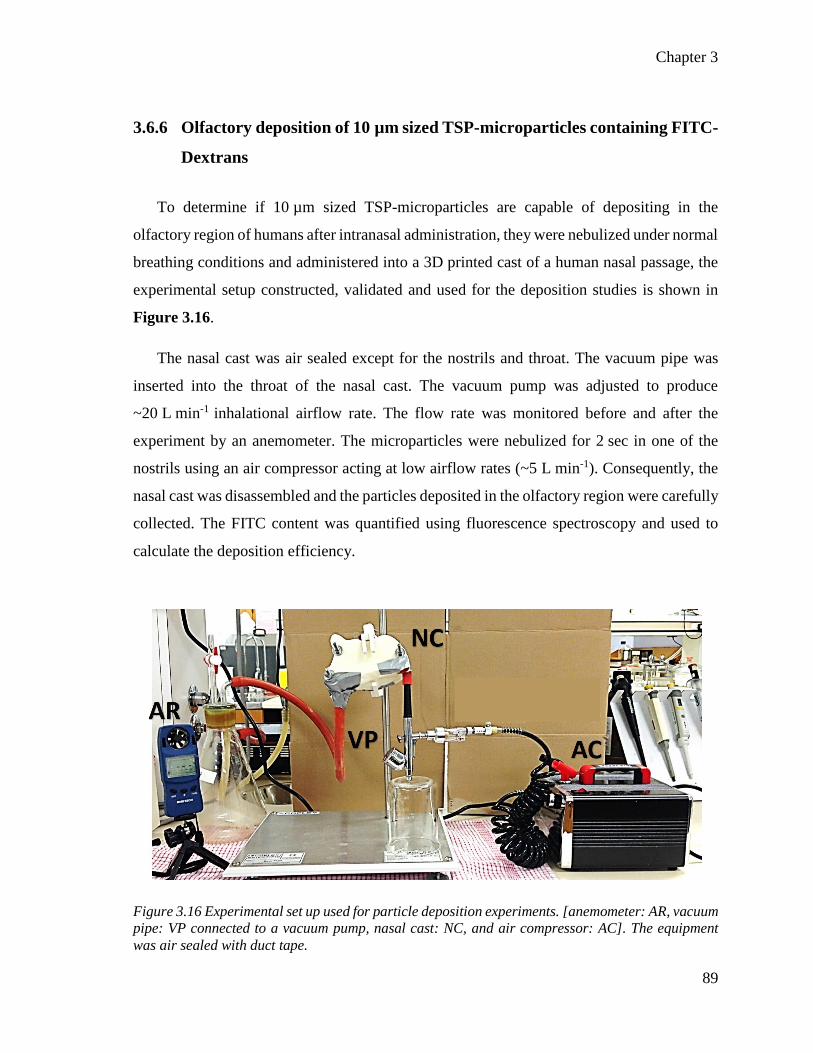
Chapter 3
89
3.6.6 Olfactory deposition of 10 µm sized TSP-microparticles containing FITC-
Dextrans
To determine if 10 µm sized TSP-microparticles are capable of depositing in the
olfactory region of humans after intranasal administration, they were nebulized under normal
breathing conditions and administered into a 3D printed cast of a human nasal passage, the
experimental setup constructed, validated and used for the deposition studies is shown in
Figure 3.16.
The nasal cast was air sealed except for the nostrils and throat. The vacuum pipe was
inserted into the throat of the nasal cast. The vacuum pump was adjusted to produce
~20 L min-1 inhalational airflow rate. The flow rate was monitored before and after the
experiment by an anemometer. The microparticles were nebulized for 2 sec in one of the
nostrils using an air compressor acting at low airflow rates (~5 L min-1). Consequently, the
nasal cast was disassembled and the particles deposited in the olfactory region were carefully
collected. The FITC content was quantified using fluorescence spectroscopy and used to
calculate the deposition efficiency.
Figure 3.16 Experimental set up used for particle deposition experiments. [anemometer: AR, vacuum
pipe: VP connected to a vacuum pump, nasal cast: NC, and air compressor: AC]. The equipment
was air sealed with duct tape.
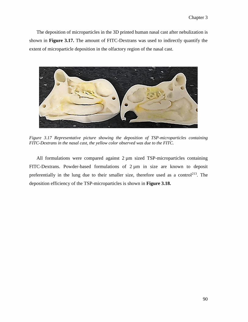
Chapter 3
90
The deposition of microparticles in the 3D printed human nasal cast after nebulization is
shown in Figure 3.17. The amount of FITC-Dextrans was used to indirectly quantify the
extent of microparticle deposition in the olfactory region of the nasal cast.
Figure 3.17 Representative picture showing the deposition of TSP-microparticles containing
FITC-Dextrans in the nasal cast, the yellow color observed was due to the FITC.
All formulations were compared against 2 µm sized TSP-microparticles containing
FITC-Dextrans. Powder-based formulations of 2 µm in size are known to deposit
preferentially in the lung due to their smaller size, therefore used as a control213. The
deposition efficiency of the TSP-microparticles is shown in Figure 3.18.

Chapter 3
91
Figure 3.18 Deposition efficiency of 10 µm versus 2 µm TSP-microparticles containing 5 kDa (A), 10 kDa(B), 20 kDa(C) and 40 kDa(D)
FITC-Dextrans in the olfactory region of a human nasal replica 3D printed cast when nebulized under normal breathing conditions (20 L/min).
Data presented is the mean of 10 independent experiments ± SD. (* P < 0.05).

Chapter 3
92
Deposition in the olfactory region is reported as deposition efficiency and the data is
presented in Figure 3.18. Deposition efficiency ranged from 0 to 1.9 % with 10 µm
microparticles as compared to 0 to 0.7 % with 2 µm microparticles. In some experiments,
olfactory deposition of microparticles was negligible, so for these experiments, the
deposition efficiency is reported as zero. Consequently, a large number of independent
experiments were conducted to ensure reproducible and statistically robust data. Overall,
microparticles with a mode size of 10 µm showed significantly higher deposition efficiency
(****P < 0.0001) in the olfactory region compared to the 2 µm particles.
3.6.7 Safety of TSP-microparticles for nasal administration
Histopathological analysis of porcine nasal mucosa at the completion of the ex-vivo
studies (Section 3.6.5) is shown in Figure 3.19. Freshly acquired, untreated nasal mucosa
was used as a control as shown in Figure 3.19 A. Mucosal structures such as the surface
epithelium, the underlying connective tissue, and serous glands are evident in the
micrographs as shown in Figure 3.19 B-E. The cylindrical appearance of epithelial cells
retaining their overall polarity is a good indicator of structural integrity250. No visual
evidence of epithelial cells detached from the mucosa or large vacuolated regions, necrosis,
hemorrhage or ulceration (large red colour spots), were identified in tissues treated with
TSP-microparticles as shown in Figure 3.19 B-E and these were comparable to the negative
control. Marked alterations to the epithelium with a detachment of the epithelial layer, the
presence of rounded epithelial cells and loss of polarity could be seen in the histopathology
of the positive control as shown in Figure 3.19 F. The histopathlogy of nasal mucosa in this
case is in agreement with other reports of the toxic effect of nitric acid on the nasal
mucosa171,358,359.

93
Figure 3.19 Representative light micrographs of porcine olfactory nasal mucosa after exposure to TSP-microparticles containing FITC-Dextran
(B) 5 kDa, (C) 10 kDa, (D) 20 kDa and (E) 40 kDa. Freshly acquired nasal mucosa was used as negative control (A) while mucosa treated with
37 % v/v nitric acid served as a positive control (F). (SG: serous glands). The arrows and arrowhead indicate histopathology of the olfactory
epithelium and large vacuoles, respectively. (Scale bar = 100 µm).

Chapter 3
94
3.7 Discussion
The potential advantages that TSP formulated as microparticles can offer to the
nose-to-brain delivery of drugs, the unexplored potential of TSP as a macromolecular drug
carrier, and the understated nasal drug carrier properties of TSP are the driving factors for
the research presented in this Chapter.
The majority of the literature has followed the extraction of TSP described by
Rao et al233. Since it is an easy and straightforward method, it was used in this chapter for
the extraction of TSP from tamarind seed gum. The IR spectrum of extracted TSP produced
characteristic functional group IR peaks, and the x-ray diffractograms showed no sharp peaks
indicating the amorphous nature of the TSP. The IR spectrum and the x-ray diffractograms
were comparable to the data available in literature355. Furthermore, the SEM observation of
TSP revealed a smooth surface and was similar to the published literature356. In addition, no
batch variation was observed between TSP extracted on different days, x-ray diffractograms
and IR spectrum were identical. Published scientific reports identified similar properties for
polysaccharides with no batch variation when a similar method of extraction has been
used360,361.
Formulating TSP as microparticles and optimizing the microparticle size to 10 µm was
one of the research aims of this chapter. In this thesis, the size of particles is reported as
“mode size” the most common size within a particle population. Pilot studies and a DOE
approach (Box-Behnken experimental design) was employed to optimize the formulation
parameters. The effect of formulation parameters on the microparticle size was analyzed by
ANOVA to understand their effect and atomizing airflow and aspiration were found to be
significant factors affecting the particle size. The velocity of the compressed air passing
through the nozzle (atomizing air flow) can significantly affect the particle size by
determining the size of droplets formed from the feed solution339,362. The velocity of the
drying gas (aspiration) determines the time of droplet drying in the chamber which might
have affected the size of the particles339. Among the formulation parameters tested,
FITC-Dextran concentration and inlet temperature did not significantly affect particle size.
Chapter 3
95
The reason for this can be attributed to the low amount of FITC-Dextrans used in this study
and that the temperatures were higher and sufficient to dry the solvent (water) quickly,
resulting in an insignificant effect on the resultant microparticle size.
To identify the optimal levels of formulation parameters to produce the 10 µm particles
the Design Expert software was used. Design Expert provides a user interface to target the
particle size to a specific range manually and predicts the optimal levels of formulation
parameters required to produce the target range of particle size. The particle size target was
set to 10 µm and the predictions of formulation parameter levels acquired from Design
Expert. The levels of two significant parameters: atomizing airflow and aspiration, were
predicted to be 547 L/h and 55 % respectively, in order to produce 10 µm sized particles.
These predictions were tested practically and resultant particle size was measured. The
optimized levels of formulation parameters were found to produce the 10 µm sized particles
reproducibly. FITC-Dextran with MW 3-5 kDa was employed for these studies.
Microparticles were prepared using the optimized levels of formulation parameters with all
the other FITC-Dextrans. The MW of the FITC-Dextran did not show any effect on the
particle size. This could have been be due to the constant amount of the FITC-Dextrans
(0.25 % w/w) used in all the formulations. Spray-Drying TSP with FITC-Dextrans of
different MW at the optimized levels of formulation parameters produced 10 µm sized
TSP-microparticles containing FITC-Dextrans.
Spray-dried TSP-microparticles containing FITC-Dextrans were corrugated in shape and
hollow. Corrugated particle shape has been reported to lower the true area of contact between
the particles thereby reducing the cohesiveness between particles which is the desired quality
for intranasal administration363,364. Hollow particles improve the dispersibility of the solid
microparticles339 which can be a desirable property for intranasal delivery end use aimed for
in this thesis. The surfaces of the microparticles observed were rupture free, which is
desirable as such microparticles have been reported to have proper deposition and delayed
clearance from the nasal cavity365. There were no significant differences observed between
the morphology of the TSP-microparticles containing different MW FITC-Dextrans.
The FITC-Dextran content in the microparticles was determined by fluorescence
spectrophotometer. The results indicated that TSP could successfully load high MW dextran
Chapter 3
96
molecules up to 40 kDa. The molecular interactions of dextran chains with TSP and the low
drug loss in the spray-drying process might have contributed to the high encapsulation
efficiency of the TSP-microparticles. The green fluorescence of the FITC in
TSP-microparticles observed with fluorescence microscopy confirmed the loading of
FITC-Dextrans. These results suggest that TSP can be a potential macromolecular drug
carrier for drug delivery applications.
One of the beneficial features of TSP for intranasal drug delivery is its potential ability
to resist mucociliary clearance. The mucoadhesive potential of TSP was evaluated to identify
the potential of TSP-microparticles to increase their residence time in the nasal cavity. In the
present study, two in-vitro approaches and an ex-vivo approach were used to confirm the
mucoadhesive properties of FITC-Dextran loaded TSP-microparticles. Firstly, the amount
of TSP-microparticles containing FITC-Dextran that adhered to a piece of filter paper
saturated with mucin after application of an air load was determined by quantifying the
amount of FITC-Dextran and represented as in-vitro mucoadhesion. It is a quick and useful
approach to determine the effect of air-flow on nasal clearance of microparticulate
formulations after their administration into the nasal cavity. Previously reported techniques
for in-vitro mucoadhesion had applied the air load only one-way to simulate the process of
inhalation but not exhalation209. In, the current study, respiration was simulated by
incorporating the airflow from two directions in an attempt to better represent the respiratory
conditions when a formulation is inhaled. In-vitro, TSP-microparticles showed significantly
higher mucoadhesion compared to the unformulated FITC-Dextran powders. The
mucoadhesive potential of TSP can be attributed to the hydrogen bonding capacity of –OH
groups on the side chains of TSP. These functional groups interact with other electronegative
atoms in mucin and form a hydrogen bond. Furthermore, van-der-Waals bonds may also
arise due to the dispersion forces in TSP contributing to the mucoadhesion.
Ex-vivo mucoadhesion studies using the tensile strength apparatus complemented the
in-vitro study and demonstrated the potential of TSP-microparticles to adhere to the mucosa
in the nasal cavity. Tensile strength is, a widely employed method for the assessment of
mucoadhesion366. In this chapter, the force required to detach the microparticles from porcine
nasal mucosa was represented as mucoadhesive strength. Assessing mucoadhesion of

Chapter 3
97
microparticles is challenging due to the small size of the particles. In this study, an industrial
grade strong adhesive epoxy was employed to ensure the firm adhesion of microparticles to
the probe. This prevented the loss of microparticles onto mucosa when the probe measured
the detaching force. The exclusively designed mucoadhesion rig minimized any equipment
errors. TSP-microparticles demonstrated significantly high mucoadhesion strength
compared to the unformulated FITC-Dextran powders. Mucoadhesive properties were
comparable across all microparticle formulations in in-vitro and ex-vivo studies and are most
likely the result of the constant amount of polymer and FITC-Dextran used in each of the
formulations investigated (2 % of total solids)367.
Cryo-FESEM was used to investigate the interaction between TSP-microparticles and
mucin. This work is the first electron microscopy study to identify the two critical stages
which have been hypothesized to occur during the mucoadhesion process; contact and
consolidation221. In the initial contact stage, contact between microparticles and the mucus
layer was visible, and no apparent changes were seen in the morphology of the microparticles
or the mucin. In the following consolidation stage, the interaction of TSP-microparticles with
the mucus layer was evident. The interaction is most likely due to the moisture present in the
mucin, which allows the TSP to swell and penetrate the mucin layer. Subsequently, the TSP
conforms to the shape of the mucin by forming a gel-like layer most likely by weak Van der
Waals attraction and hydrogen bonding 221,228.
While controlled release of a drug molecule is considered ‘gold standard’ in formulation
science, the optimal release profile of a drug from an intranasal delivery system is dependent
on several parameters, in particular, the drug molecule itself, the therapeutic indication and
the required dose. Controlled release of drugs formulated in TSP in combination with other
polymers has been shown for at least 10 to 12 h in-vitro. However, the release of drug
molecules from TSP-only formulations was reported to be relatively quicker
(approximately 6 h)300,316. The likelihood that any formulation is retained in the nasal cavity
for periods as long as 10 to 12 h despite its mucoadhesive nature is unlikely and often not
desired. Therefore, in the present chapter, release studies over a 4 h period were considered
as a practical time frame which would most likely encompass the timeframe of any nasal
clearance that occurs in-vivo. The mechanism of FITC-Dextran release was found to be

Chapter 3
98
diffusion controlled as the data fit well with the Higuchi model of drug release. The
mechanism of drug release from TSP can be compared to hydrogels as, the microscopy
studies and previous reports in the literature show that TSP forms a gel-like viscous layer on
contact with the mucin by absorbing water and simultaneously releasing the FITC-Dextrans
loaded in the microparticles via diffusion368,369. However, the drug release from the F40
formulation fitted a Korsmeyer-Peppas model indicating that the drug release is by both
diffusion controlled and erosion controlled mechanisms. Why this was the case for the F40
remains unclear.
Furthermore, ex-vivo studies using porcine nasal mucosa were conducted as such studies
provide useful data about the permeability of drug molecules across a mucosal barrier before
testing in-vivo. The permeation of FITC-Dextrans released from TSP-microparticles was
dependent on MW. The FITC-Dextrans employed in this chapter were ≤ 40 kDa in size and
their ex-vivo permeation flux decreased with increase in MW as their permeation across the
mucosa was diffusion dependent. It has been reported that FITC-Dextrans with MW lower
than 40 kDa cross biological barriers primarily by diffusion and in small amounts by
pinocytosis370.
Collectively, the in-vitro data suggest that release of hydrophilic molecules (≤ 40 kDa)
from TSP-microparticles was independent of MW and followed a typical biphasic profile.
The initial burst release reported was most likely surface-associated molecules that dissolved
quickly upon contact with the solvent. This is supported by the near 100 % release reported
with FITC-Dextrans in solution. However, unlike FITC-Dextrans in solution, the release of
FITC-Dextrans formulated in TSP-microparticles was significantly slower which was most
likely because the FITC-Dextrans had to diffuse through the gel layer which is rapidly
formed when TSP-microparticles interact with mucin, as was seen by cryo-FESEM. The
ex-vivo studies show that permeability of molecules across the nasal mucosa is strongly
dependent on their MW. The size-dependent permeability of FITC-Dextrans across the
olfactory mucosa and into the brain after intranasal administration has been demonstrated
in-vivo 371.
Another objective of this Chapter was to investigate the deposition of 10 µm sized
particles in the olfactory region of the human nasal cavity. A 3D human nasal replica cast
Chapter 3
99
coated with 2 % w/w mucin solution inside the nasal passages was employed for this
purpose. Deposition of particles in the geometrically complex human nasal cavity has been
shown to be influenced by particle size, the inhalational flow rate and the position in the
nasal cavity from where the drug is administered76. Particles 10 µm in size have recently
been demonstrated to have an optimum deposition in the olfactory region of the nasal
cavity215. TSP-microparticles (2 and 10 µm) were nebulized at the nostrils in a 3D printed
human nasal cast under a normal inhalational flow rate (20 L/min). Such in-vitro studies
provide useful data on the regional nasal deposition of particles before human testing. The
influence of size on particle deposition in the olfactory region was reported in the present
study, with 10 µm-sized microparticles showing better deposition than 2 µm-sized
microparticles. The better deposition can be attributed to the inertia of the particles, where
particles with 10 µm size have the optimum inertia to reach the upper narrow olfactory
region. Deposition of microparticles was independent of the MW of the incorporated
FITC-Dextrans. This study demonstrated that formulating microparticles to 10 µm size can
enhance their deposition in the olfactory region of the human nasal cavity. In this chapter,
the safety of TSP-microparticles containing FITC-Dextrans was assessed ex-vivo on the
porcine nasal mucosa. Structural integrity and epithelial cell structure were preserved in the
mucosae treated with TSP-microparticles demonstrating the safety of TSP-microparticles for
intranasal administration. These results agree with the previously reported safety data of TSP
for intranasal applications275,334.
Collectively, this Chapter evaluated the potential of TSP as a nasal drug carrier and
macromolecular drug carrier. The combined formulative approach of formulating
mucoadhesive microparticles as 10 µm particles improved their deposition in the olfactory
region and the mucoadhesive potential of TSP may increase the intranasal residence time of
the drug. A significant advantage of TSP is that it is structurally very similar to mucin and
demonstrates mucoadhesive properties. Furthermore, the established tolerability of TSP in
humans makes it a great candidate for a nasal preparation266.
Chapter 3
100
3.8 Key findings
This chapter of the thesis tested a simple formulative approach of incorporating drug
molecules in 10 µm-sized mucoadhesive microparticles to target them to the olfactory region
of the nasal cavity. Collectively, the data reported in this chapter suggest that the TSP can
act as a carrier for the high MW drugs and can be formulated as 10 µm-sized particles by
spray-drying technique. TSP-microparticles have the potential to increase the residence time
of drugs in the nasal cavity by mucoadhesion and are safe to the nasal mucosa after acute
nasal administration. The 10 µm-sized TSP-microparticles can improve the olfactory
deposition at normal inhalation rates.
3.9 Future directions
Although enhanced olfactory deposition with size tailored particles has been reported in
this study, further human studies are required to correlate increased deposition efficiency
with the biological relevance. Critical research quesitons such as, does increased depositon
in the olfactory region affect brain uptake of drugs? Is the increase in depositon sufficient to
produce a biological effect? need to be answered in future studies.
Although the safety of TSP has been reported ex-vivo, further studies investigating the
safety and tolerability of TSP-microparticles in-vivo after acute and chronic intranasal
administration are required to progress this drug-delivery platform. The drug release and
permeation across the nasal mucosa to reach the brain have to be examined in-vivo to further
understand the potential of TSP-microparticles in nose-to-brain drug delivery. In addition,
the FITC-Dextrans used as model drugs in this study are hydrophilic and the MW tested is
only till 40 kDa. Such limitations of this study raise research questions on the potential of
the TSP-microparticle delivery system for use with larger and or hydrophobic drugs, which
need to be addressed by future studies. Also, study of effect of the surface charge of particles
on drug delivery and mucoadhesion needs to be addressed in future work.
Several drug delivery devices that aim to target drug formulations to the olfactory
mucosa are already in various stages of clinical trials. While still at a conceptual stage, it
would be interesting to explore if this formulative approach used in combination with an

Chapter 3
101
intranasal device can augment olfactory targeting and enable enhanced delivery of
therapeutic molecules to the brain via the nose.

Chapter 3
102

Chapter 4
Formulation and In-vivo characterization of TSP-microparticles containing phenytoin


Chapter 4
105
4 Formulation and in-vivo characterization of TSP-
microparticles loaded with phenytoin
4.1 Introduction
Results from Chapter 3 demonstrate that TSP can be formulated as mucoadhesive
microparticles. This chapter investigates the ability of the TSP microparticle system to load
and release an anti-epileptic drug (AED) phenytoin in-vitro and its biodistribution in rats
following intranasal administration.
4.1.1 Sub therapeutic brain concentrations of drugs in epilepsy
More than 70 million people worldwide are affected by epilepsy, making it one of the
most common neurological disorders372,373. Unprovoked and recurrent seizures are observed
in epileptic patients which are either focal or general in nature374. AEDs are the first line of
treatment in controlling seizures. However, about one-third of epilepsy patients suffer from
uncontrolled seizures despite treatment with at least two or three AEDs. Such a condition is
termed ‘drug-resistant epilepsy’375.
One possible reason for treatment failure is inadequate levels of AEDs reaching the brain
due to overexpression of the multidrug efflux transporter P-glycoprotein (Pgp) at the
blood-brain barrier (BBB) in the tissues surrounding the epileptic focus 376–378. Along with
Pgp various types of multidrug efflux transporters present at the BBB are shown in Figure
4.1. It is hypothesized that in patients with drug-resistant epilepsy, multidrug efflux
transporters at the BBB efflux AEDs at a significantly higher rate, which results in lower
brain concentrations of the AEDs376,377,379. Overexpression of such efflux transporters
severely limit the AED penetration into the brain so that the concentrations of AEDs in the
brain cannot become sufficiently high for therapeutic efficacy380,381. Consequently, AEDs
fail to control the seizures in drug-resistant epilepsy. To improve the efficacy of AEDs
simultaneous use of Pgp inhibitors with AEDs has shown promise382. However, the
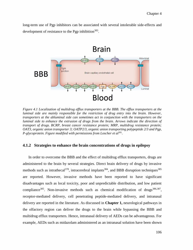
Chapter 4
106
long-term use of Pgp inhibitors can be associated with several intolerable side-effects and
development of resistance to the Pgp inhibition383.
Figure 4.1 Localization of multidrug efflux transporters at the BBB. The efflux transporters at the
luminal side are mainly responsible for the restriction of drug entry into the brain. However,
transporters at the abluminal side can sometimes act in conjunction with the transporters on the
luminal side to enhance the extrusion of drugs from the brain. Arrows indicate the direction of
transport of drugs. BCRP, breast cancer resistance protein; MRP, multidrug resistance protein;
OAT3, organic anion transporter 3; OATP2/3, organic anion transporting polypeptide 2/3 and Pgp,
P-glycoprotein. Figure modified with permissions from Loscher et al381.
4.1.2 Strategies to enhance the brain concentrations of drugs in epilepsy
In order to overcome the BBB and the effect of multidrug efflux transporters, drugs are
administered to the brain by several strategies. Direct brain delivery of drugs by invasive
methods such as intrathecal378, intracerebral implants384, and BBB disruption techniques385
are reported. However, invasive methods have been reported to have significant
disadvantages such as local toxicity, poor and unpredictable distribution, and low patient
compliance385. Non-invasive methods such as chemical modification of drugs386,387,
receptor-mediated delivery, cell penetrating peptide-mediated delivery, and intranasal
delivery are reported in the literature. As discussed in Chapter 1, neurological pathways in
the olfactory region can deliver the drugs to the brain while bypassing the BBB and
multidrug efflux transporters. Hence, intranasal delivery of AEDs can be advantageous. For
example, AEDs such as midazolam administered as an intranasal solution have been shown
Chapter 4
107
to produce enhanced anti-seizure effect compared to IV diazepam in humans388,389.
Administering the AED lamotrigine390 and clobazepam391 intranasally has been reported in
literature to achieve higher in-vivo brain concentrations. Also, intranasal administration of
lorazepam demonstrated favorable pharmacokinetics in humans in relation to IV and IM
routes392.
Formulation strategies to improve AED bioavailability for effective anti-seizure activity
has been reported in the literature. For example, formulation of carbamazepine as a
β-cyclodextrin-carbamazepine complex has been shown to significantly improve the
anti-epileptic activity and bioavailability in rats compared to unformulated AED after
intraperitoneal and oral administrations393,394. Co-polymerization and co-precipitation of
AEDs with polymers have been reported to improve the dissolution and solubility profiles
consequently enhancing the bioavailability395,396. Formulating AEDs such as stiripentol,397
carbamazepine,398–400 rufanimide401 and phenytoin402 as solid dispersions has been shown to
improve their dissolution profiles.
Delivery of AEDs formulated as nanoparticles have been reported with promising results
towards increasing the bioavailability of AEDs403,404. For example, polysorbate-80 coated
nanoparticles of gabapentin have shown an enhanced concentration in the brain after
intraperitoneal administration405. Another study where clonazepam was formulated as solid
lipid nanoparticles demonstrated improved anticonvulsant activity by enhanced BBB
permeability of nanaoparticles406. Wang et al. reported that hydrogel nanoparticles of
phenytoin sodium improve antiepileptic effects by enhancing the brain concentrations407 and
on-demand delivery of phenytoin sodium into the brain by electro responsive nanoparticles
in generalized tonic-clonic seizures408.
Combining formulation strategies with intranasal administration has been reported to
produce high brain targeting of AEDs in the literature. For example, Eskandari et al. reported
that intranasal administration of valproic acid formulated into nanostructures could enhance
the brain concentration and consequently produce high anti-seizure activity compared to
intraperitoneal administration409. Sharma et al. demonstrated enhanced brain delivery of
lorazepam incorporated into nanoparticles after intranasal administration compared to
intranasal solution and intraperitoneal administrations410. Vyas et al. demonstrated

Chapter 4
108
mucoadhesive microemulsions could significantly increase the direct nose-to-brain transport
of clonazepam compared to a solution or non-mucoadhesive microemulsions, into rabbit
brain. The brain targeting efficiency of mucoadhesive microemulsions was tested by
measuring the radioactivity of administered 99mTc‐labeled clonazepam. Mucoadhesive
microemulsions produced higher radioactivity in the brain demonstrating the high targeting
efficiency of the mucoadhesive microparticles411.
Interestingly, the reports on AEDs formulated as intranasal microparticles are scarce.
One study reported spray-dried microspheres loaded with phenytoin. The in-vitro
characteristics of the formulation and the sustained release of phenytoin from microspheres
was reported, but no in-vivo data was provided412. In another study, alginate-chitosan
microparticles of phenytoin were formulated. Nonetheless, the study did not report any
significant advantage of formulating phenytoin as alginate-chitosan microparticles and
concluded that the formulation requires more modifications413.
Considering the advantages of intranasal administration of AED formulations and the
potential of mucoadhesive microparticles as discussed in Chapter 1, this chapter
investigates the formulation of an AED (phenytoin) into microparticles and its intranasal
administration in rats. Phenytoin was selected as a model drug as it is an effective first-line
AED and is a substrate of the Pgp multidrug efflux transporter. Over expression of Pgp in
drug-resistant epilepsy severely limits the uptake of phenytoin into the brain leading to sub
therapeutic levels and treatment failure414. Therefore this Chapter utilizes phenytoin as a
model drug for formulation into the intranasal mucoadhesive microparticles.
4.1.3 Phenytoin
Phenytoin (Figure 4.2) was first developed during the 1930s, and it is still in use for the
treatment of epilepsy despite the invention of many new AEDs415. In addition to its use as
an AED, phenytoin is also used as antiarrhythmic416. Recently, neuroprotective activity of
phenytoin in optic neuritis in patients with multiple sclerosis has been tested with positive
results in a Phase II clinical study and new indications for phenytoin are still emerging415,417.

Chapter 4
109
Phenytoin exhibits anti-epileptic activity by blocking inactive sodium channels and reducing
voltage-gated sodium and calcium channel currents418,419.
Figure 4.2 Chemical structure of phenytoin sodium (phenytoin).
Due to its poor water solubility (80 µmol/L), its sodium salt, phenytoin sodium, and ester
prodrug, fosphenytoin, are used clinically420,421. Phenytoin (Sodium 2,5-dioxo-
4,4-diphenylimidazolidin-1-ide) belongs to Class II of the Biopharmaceutics Classification
System with low solubility and high permeability422. Phenytoin is available as a parenteral
preparation solubilized at 50 mg/mL with 40 % propylene glycol (PG) and 10 % ethanol421.
Sodium salt of phenytoin is a low MW (274.25 Da) crystalline powder, which converts into
phenytoin when administered and exhibits its pharmacological activity. Phenytoin undergoes
metabolism in the liver by cytochrome P450 enzymes to form its major metabolite
‘5-(4-Hydroxyphenyl)-5-phenyl-hydantoin’423. The amide groups (-NH) in the phenytoin
molecule (Figure 4.2) are reported to interact with excipients by hydrogen bonding and
improve the solubility of phenytoin394,421,424.
4.1.4 TSP-microparticles loaded with phenytoin for nose-to-brain delivery
It is evident from a review of literature that there is still a need for studies to explore the
potential of formulation strategies combined with intranasal administration for AEDs. With
the advantages mucoadhesive microparticle formulation can provide as discussed in
Chapter 1, and the supporting results from Chapter 3, formulating AEDs into

Chapter 4
110
TSP-microparticles with 10 µm size could have the potential for enhancing nose-to-brain
drug delivery of phenytoin and therefore forms the focus of this chapter. However, the low
solubility and high crystallinity of phenytoin pose research questions:
Whether phenytoin can be formulated as 10 µm sized TSP-microparticles?
Whether phenytoin can affect the mucoadhesive potential of TSP-microparticles?
How stable are the microparticle formulations?
How much phenytoin administered as intranasal microparticles can reach the brain?
4.2 Hypothesis
The overall hypothesis of this chapter is that incorporating phenytoin into 10 µm sized
TSP-microparticles can enhance nose-to-brain delivery of phenytoin in a rat model.
4.2.1 Chapter Aims
1. To formulate and optimize 10 µm sized TSP-microparticles loaded with phenytoin
by spray-drying.
2. To assess the mucoadhesive potential and phenytoin release profile of
TSP-microparticles.
3. To assess olfactory deposition efficiency of 10 µm sized TSP-microparticles loaded
with phenytoin.
4. To assess nasal mucotoxicity of TSP-microparticles loaded with phenytoin in rats.
5. To assess the stability of microparticle formulations.
6. To develop an HPLC method to accurately quantify phenytoin in rat plasma, brain,
and other tissues.
7. To quantify the uptake of phenytoin in plasma, brain, and peripheral tissues
following intranasal administration of phenytoin in TSP-microparticles, IV and as
an intranasal solution.

Chapter 4
111
4.3 Results
4.3.1 Formulating 10 µm-sized TSP-microparticles containing phenytoin by
spray-drying
4.3.1.1 Optimization of formulation parameters
The initial experimental trials were based on the optimized conditions for the TSP
FITC-microparticles from Chapter 3. The effect of formulation parameters: total solids (%),
TSP:phenytoin ratio, PG (%), and spray-drying conditions (Q-flow (L/h) & aspiration (%))
on particle size was examined. The inlet temperature was kept constant at 120 ºC. Table 12
lists various formulation parameters and the resulting particle size. A total of
12 combinations of different levels of formulation parameters were tested.
The optimized formulation parameters from Chapter 3 failed to produce the same
particle size when the FITC-Dextran was replaced with phenytoin. Instead an increased
particle size of 14 µm was observed. It has been reported that factors like the properties of
drug, excipients, and drug to excipient ratio can affect the spray-drying process362,425.
Phenytoin is a poorly soluble crystalline drug, unlike amorphous hydrophilic dextrans and
the addition of PG may have affected the spray-drying process. Thus suitable formulation
parameters to produce 10 µm sized TSP-particles containing phenytoin were identified by
trial and error.
The analysis of the spray-drying results from Chapter 3 revealed that atomizing airflow
and aspiration are two significant factors that control particle size. As a first step, the
atomizing airflow was changed, and the size of the resultant particles was analyzed. A
decrease of atomizing air flow < 547 L/h lead to larger particle size (> 14 µm). Furthermore,
the phenytoin was not completely formulated into microparticles as identified by SEM
(Figure 4.3). Increasing the atomizing airflow to the next possible level (> 547 L/h) resulted
in very fine atomization of the feed solution and consequently a decrease in the particle size
(6 ± 0.3µm) and its distribution. Hence, the atomizing airflow rate was kept constant at
547 L/h while the aspiration was increased gradually and the resultant particle size was
Chapter 4
112
analyzed. An increase in the velocity of the drying gas decreased particle size gradually, and
at the maximum level of aspiration (100 %), the particle size was 11.6 ± 0.2 µm.
As the amount of drug and excipients can also affect spray-drying, the total solid contents
in the feed solution was decreased from 2 % to 1.5 % without changing the TSP:phenytoin
ratio. This further decreased the particle size to 11.2 ± 0.1 µm. Further decrease in total
solids was not considered due to its potential implications on the mucoadhesive potential and
phenytoin content of the microparticles. Instead, PG content was decreased from 10 % to
5 %, and the resultant particle size was analyzed. These conditions produced the required
10 µm particles. The optimized conditions are presented in Table 12 and are highlighted
with grey color.

Chapter 4
113
Table 12 List of formulation parameters tested. Highlighted cells in gray color indicate the optimized,
most suitable formulation parameters to obtain microparticles with a size of 10 µm. Particle size
data presented is the mean of three independent experiments ± SD.
Total
solids
(%)
TSP:
Phenytoin
Spray-drying conditions PG
(%)
Particle
size (µm) Atomizing airflow
(L/h)
Aspiration
(%)
2.0 3:1 425 55 10 17.8 ± 0.3
2.0 3:1 487 55 10 17.8 ± 1.0
2.0 3:1 547 55 10 14.0 ± 1.0
2.0 3:1 607 55 10 6.0 ± 0.3
2.0 3:1 668 55 10 4.2 ± 0.1
2.0 3:1 547 60 10 19.4 ± 0.1
2.0 3:1 547 75 10 14.3 ± 0.8
2.0 3:1 547 95 10 12.2 ± 0.5
2.0 3:1 547 100 10 11.6 ± 0.2
1.5 3:1 547 100 10 11.2 ± 0.1
1.5 3:1 547 100 5
9.8 ± 0.4
10.1 ± 0.1
10.3 ± 0.3
1.5 1:1 547 100 5 9.7 ± 0.7
1.5 1:3 547 100 5 11.0 ± 1.0
The optimized conditions were validated by analyzing the particle size data obtained
under these conditions on three different days and during three different times within a day.
Statistical analysis of these results by a student t-test identified no significant difference in
the particle size (P>0.05) confirming that these conditions were suitable to produce
10 µm-sized TSP-microparticles containing phenytoin. Then, with an aim to further increase
the drug concentration in microparticles, the concentration of phenytoin was increased from

Chapter 4
114
25 % to 50 % and 75 %. These changes did not result in significant particle size changes
(P >0.05).
Figure 4.3 A & B show the morphology of the unformulated phenytoin powder (A) and
spray-dried phenytoin (B). Figure 4.3 C-F show representative SEM micrographs of
TSP-microparticles containing phenytoin where the effect of formulation parameters can be
observed in the morphological features of the particles. The effect of spray-drying
parameters are shown in Figure 4.3 C & D and indicate that the larger particles are produced
at lower atomizing air flow (≤ 487 L/h) rates. Also, at these levels, phenytoin was not
completely incorporated into TSP-microparticles. The unformulated phenytoin crystals
could be observed as shown in C. Higher atomizing airflow (≥ 607 L/h) led to particles with
smaller size as shown in Figure 4.3 D. Figure 4.3 E & F demonstrate the effect of the
TSP: Phenytoin ratio on microparticles. With increases in the concentration of phenytoin
(50 %), the surface of the microparticles shows crystalline fractions of the phenytoin as a
textured surface, as shown in Figure 4.3 E. Further increase in phenytoin level (75 %) results
in increased fractions of crystalline phenytoin on the surface of the microparticles as shown
in Figure 4.3 F. Large variations in the particle size and presence of crystalline phenytoin in
the formulations and on the surface of microparticles observed under SEM emphasize the
importance of formulation parameters on the particle size and morphology of the
microparticles.

Chapter 4
115
Figure 4.3 Representative micrographs of (A) crystalline unformulated phenytoin powder and (B)
spray-dried phenytoin. Representative micrographs of TSP-microparticles demonstrating the effect
of spray-drying parameters: (C) unformulated crystalline phenytoin observed after spray-drying with
TSP and (D) smaller particle size at the higher levels (≥ 607 L/h) of spray-drying parameters.
Representative micrographs of TSP-microparticles demonstrating the effect of an increase in
phenytoin concentration (E and F). Arrows indicate the visible crystalline fractions of phenytoin.
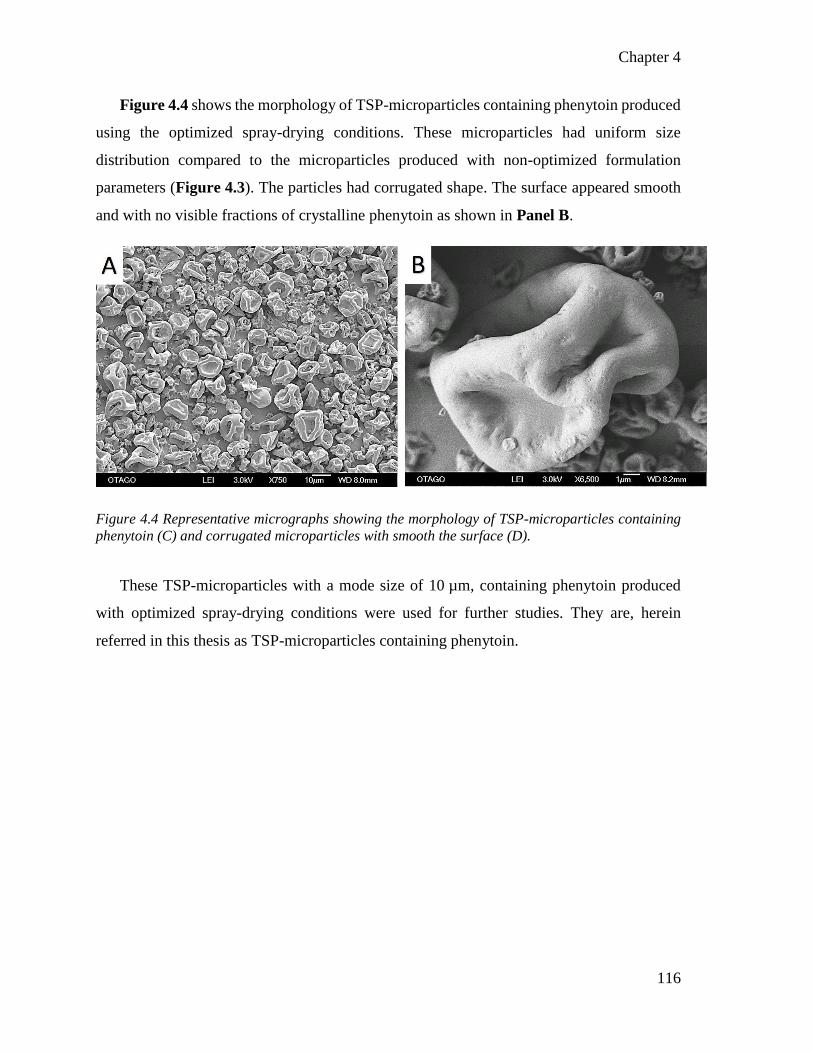
Chapter 4
116
Figure 4.4 shows the morphology of TSP-microparticles containing phenytoin produced
using the optimized spray-drying conditions. These microparticles had uniform size
distribution compared to the microparticles produced with non-optimized formulation
parameters (Figure 4.3). The particles had corrugated shape. The surface appeared smooth
and with no visible fractions of crystalline phenytoin as shown in Panel B.
Figure 4.4 Representative micrographs showing the morphology of TSP-microparticles containing
phenytoin (C) and corrugated microparticles with smooth the surface (D).
These TSP-microparticles with a mode size of 10 µm, containing phenytoin produced
with optimized spray-drying conditions were used for further studies. They are, herein
referred in this thesis as TSP-microparticles containing phenytoin.

Chapter 4
117
4.3.1.2 Analysis of phenytoin content in TSP-microparticles
The amount of phenytoin incorporated in TSP-microparticles was analyzed by a
previously validated HPLC method (Younus et al, unpublished data). Figure 4.5 A, shows
the peak for separation and identification of phenytoin. Phenytoin was quantified using the
standard curve in Figure 4.5 B (r2 = 1). The encapsulation efficiency of optimized
microparticle formulations was found to be 96.13 ± 0.37 %, and the drug loading was
24.20 ± 0.02 %.
Figure 4.5 (A) HPLC chromatogram showing the phenytoin (PHT) peak with a retention time of
4.4 min. (B) A standard curve of phenytoin where data presented are the mean of three independent
experiments ± SD
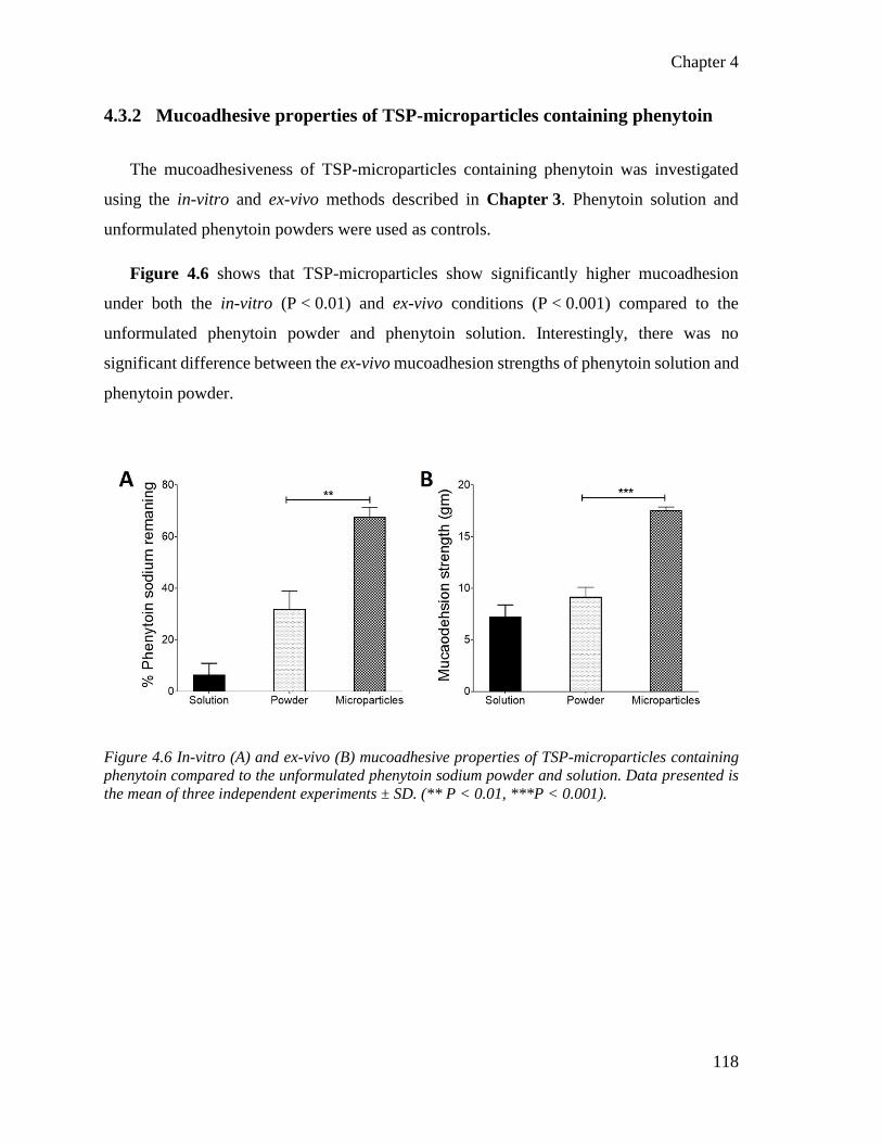
Chapter 4
118
4.3.2 Mucoadhesive properties of TSP-microparticles containing phenytoin
The mucoadhesiveness of TSP-microparticles containing phenytoin was investigated
using the in-vitro and ex-vivo methods described in Chapter 3. Phenytoin solution and
unformulated phenytoin powders were used as controls.
Figure 4.6 shows that TSP-microparticles show significantly higher mucoadhesion
under both the in-vitro (P < 0.01) and ex-vivo conditions (P < 0.001) compared to the
unformulated phenytoin powder and phenytoin solution. Interestingly, there was no
significant difference between the ex-vivo mucoadhesion strengths of phenytoin solution and
phenytoin powder.
Figure 4.6 In-vitro (A) and ex-vivo (B) mucoadhesive properties of TSP-microparticles containing
phenytoin compared to the unformulated phenytoin sodium powder and solution. Data presented is
the mean of three independent experiments ± SD. (** P < 0.01, ***P < 0.001).

Chapter 4
119
4.3.3 Release of phenytoin from TSP-microparticles in-vitro
The cumulative release profile of phenytoin from TSP-microparticles across a cellulose
acetate membrane is presented in Figure 4.7. Unformulated phenytoin powder and
phenytoin solution were used as controls. The results suggest that the microparticles
significantly sustain the release of phenytoin. The initial burst release of phenytoin from
microparticles and faster release in the first 5 min was observed compared to the phenytoin
powder.
Figure 4.7 Cumulative release of phenytoin as a function of time from solution (triangle, dotted line),
unformulated powder (square, dashed line) and microparticles (circle, solid line). The inset shows
the initial burst release from the microparticles due to the amorphous surface drug releasing
instantly upon contact with the medium and sustained release of phenytoin over the first 60 min. Data
presented is the mean ± SD of three independent experiments (* P < 0.05).
The in-vitro drug release results for the first hour were fitted into zero-order, first-order,
Higuchi, and Korsymeyers-Peppas models to determine the release kinetics. The release was
found to follow a diffusion controlled release (Higuchi, r2 = 0.998).
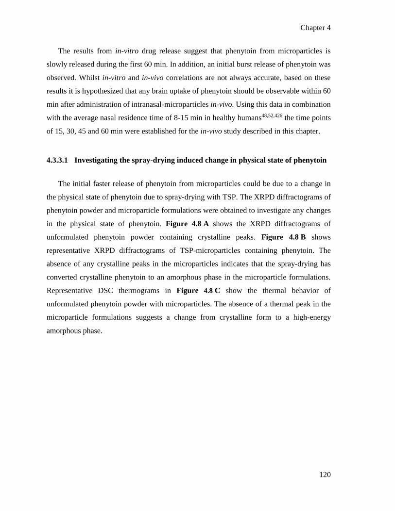
Chapter 4
120
The results from in-vitro drug release suggest that phenytoin from microparticles is
slowly released during the first 60 min. In addition, an initial burst release of phenytoin was
observed. Whilst in-vitro and in-vivo correlations are not always accurate, based on these
results it is hypothesized that any brain uptake of phenytoin should be observable within 60
min after administration of intranasal-microparticles in-vivo. Using this data in combination
with the average nasal residence time of 8-15 min in healthy humans48,52,426 the time points
of 15, 30, 45 and 60 min were established for the in-vivo study described in this chapter.
4.3.3.1 Investigating the spray-drying induced change in physical state of phenytoin
The initial faster release of phenytoin from microparticles could be due to a change in
the physical state of phenytoin due to spray-drying with TSP. The XRPD diffractograms of
phenytoin powder and microparticle formulations were obtained to investigate any changes
in the physical state of phenytoin. Figure 4.8 A shows the XRPD diffractograms of
unformulated phenytoin powder containing crystalline peaks. Figure 4.8 B shows
representative XRPD diffractograms of TSP-microparticles containing phenytoin. The
absence of any crystalline peaks in the microparticles indicates that the spray-drying has
converted crystalline phenytoin to an amorphous phase in the microparticle formulations.
Representative DSC thermograms in Figure 4.8 C show the thermal behavior of
unformulated phenytoin powder with microparticles. The absence of a thermal peak in the
microparticle formulations suggests a change from crystalline form to a high-energy
amorphous phase.
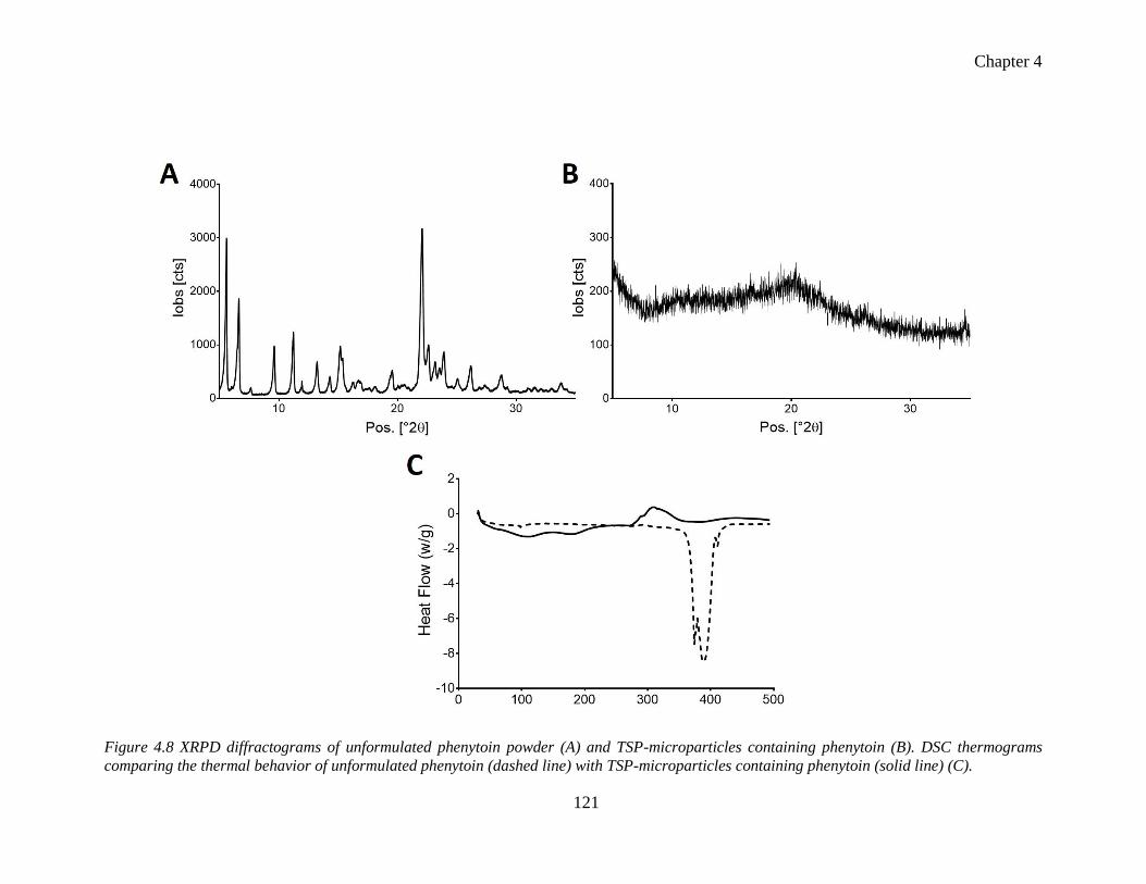
Chapter 4
121
Figure 4.8 XRPD diffractograms of unformulated phenytoin powder (A) and TSP-microparticles containing phenytoin (B). DSC thermograms
comparing the thermal behavior of unformulated phenytoin (dashed line) with TSP-microparticles containing phenytoin (solid line) (C).
Chapter 4
122
4.3.4 The permeability of phenytoin across porcine nasal mucosa ex-vivo
Permeation of phenytoin across the porcine nasal mucosa was studied to determine if
drug released from TSP-microparticles can then permeate a biologically relevant mucus
barrier. Unformulated phenytoin powder and phenytoin solution were used as controls. The
permeability of phenytoin as shown in Figure 4.9 from microparticles during the first 2 h
was significantly slower compared to the powder and solution (P < 0.05). The steady state
flux (Jss) of phenytoin permeation across the porcine mucosa released from
TSP-microparticles was calculated to be 55.9 ±2.808 µg cm-2 h-1. The Jss of phenytoin
solution and phenytoin powder was found to be 210.10 ± 68.89 and 87.99±40.77 µg cm-2 h-
1 respectively.
Figure 4.9 Permeability of phenytoin across porcine nasal mucosa over time. The permeation of
phenytoin formulated as TSP-microparticles (circle, solid line) was compared with phenytoin
solution (triangle, dotted line) and unformulated phenytoin powder (square, dashed line). Data
presented are the mean ± SD of three independent experiments.

Chapter 4
123
4.3.5 Olfactory deposition of TSP-microparticles containing phenytoin
The extent of olfactory targeting by microparticles under normal breathing conditions of
20 Lmin-1 was determined by the amount of phenytoin depositing in the olfactory region of
the 3D nasal cast. The 4 µm sized microparticles produced from one of the trial runs in the
formulation optimization process was used as a control due to their ability to escape
deposition in the nasal cavity. The deposition efficiencies of microparticles are reported in
Figure 4.10. In some experiments, olfactory deposition of microparticles was negligible, so
for these experiments, the deposition efficiency is reported as zero. Overall, microparticles
with a mode size of 10 µm showed significantly higher (P < 0.0001) deposition efficiency
in the olfactory region compared to the 4 µm particles. The results are comparable to the
deposition of TSP-FITC-Dextran microparticles described in Chapter 3.
Figure 4.10 Deposition efficiency of 10 µm versus 3 µm TSP-microparticles containing phenytoin in
the olfactory region of a human nasal replica 3D printed cast when nebulized under normal
breathing conditions (20 L/min). Data presented is the mean ± SD of ten independent experiments.
(**** P < 0.0001).
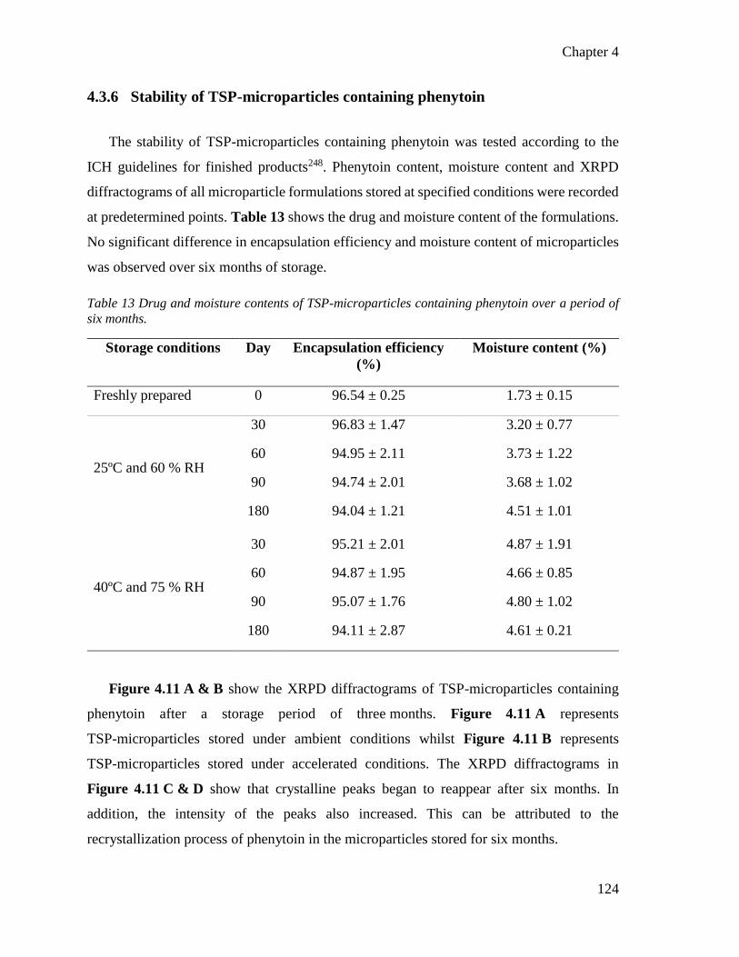
Chapter 4
124
4.3.6 Stability of TSP-microparticles containing phenytoin
The stability of TSP-microparticles containing phenytoin was tested according to the
ICH guidelines for finished products248. Phenytoin content, moisture content and XRPD
diffractograms of all microparticle formulations stored at specified conditions were recorded
at predetermined points. Table 13 shows the drug and moisture content of the formulations.
No significant difference in encapsulation efficiency and moisture content of microparticles
was observed over six months of storage.
Table 13 Drug and moisture contents of TSP-microparticles containing phenytoin over a period of
six months.
Storage conditions Day Encapsulation efficiency
(%)
Moisture content (%)
Freshly prepared 0 96.54 ± 0.25 1.73 ± 0.15
25ºC and 60 % RH
30 96.83 ± 1.47 3.20 ± 0.77
60 94.95 ± 2.11 3.73 ± 1.22
90 94.74 ± 2.01 3.68 ± 1.02
180 94.04 ± 1.21 4.51 ± 1.01
40ºC and 75 % RH
30 95.21 ± 2.01 4.87 ± 1.91
60 94.87 ± 1.95 4.66 ± 0.85
90 95.07 ± 1.76 4.80 ± 1.02
180 94.11 ± 2.87 4.61 ± 0.21
Figure 4.11 A & B show the XRPD diffractograms of TSP-microparticles containing
phenytoin after a storage period of three months. Figure 4.11 A represents
TSP-microparticles stored under ambient conditions whilst Figure 4.11 B represents
TSP-microparticles stored under accelerated conditions. The XRPD diffractograms in
Figure 4.11 C & D show that crystalline peaks began to reappear after six months. In
addition, the intensity of the peaks also increased. This can be attributed to the
recrystallization process of phenytoin in the microparticles stored for six months.

125
Figure 4.11 Representative XRPD diffractograms of TSP-microparticles containing phenytoin stored at 25 ºC and 60 % RH for three (A) and six
months (C) and of microparticles stored at 40ºC and 75 % RH for three months (B) and six months (D).

Chapter 4
126
4.3.7 Evaluation of nasal toxicity and drug-related behavioral adverse effects
of phenytoin loaded TSP-microparticles after intranasal administration
The effect of TSP-microparticles loaded with phenytoin on rat nasal mucosa was
examined in-vivo. No potential signs of nasal irritation such as frequent sneezing, noisy
breathing, red (porphyrin) discharge, nose-rubbing or general signs of pain/discomfort were
observed over the time between administration and euthanasia. The histological micrographs
of the olfactory mucosa of rats 1 h after administration of TSP-microparticles containing
phenytoin is shown in Figure 4.12. The olfactory mucosa of a rat treated with saline was
used as a negative control and is shown in Figure 4.12 A. The structural integrity of the
olfactory mucosa treated with microparticles is evident in the micrographs as shown in
Figure 4.12 B. The structure and appearance of the epithelial cells and tissue integrity is
comparable to the negative control. No evidence of necrosis or structural damage exists,
unlike in the positive control (Figure 4.12 C) which was rat nasal mucosa treated with
sodium deoxycholate. Damage to the epithelial cells, rupture of the epithelial tissue and large
vacuoles are observed in the positive control tissues. These studies suggest that the
TSP-microparticles loaded with the phenytoin do not cause any nasal mucosal toxicity in
rats up to 60 min after administration.

127
Figure 4.12 Representative light micrographs of rat nasal mucosa treated with saline (A) (negative control), phenytoin containing
TSP-microparticles (B), and sodium deoxycholate (C) (positive control). The arrows indicate histopathology of the olfactory mucosa. A, B, C were
captured at 4X magnification (scale bar (blue) = 400 µm) and their zoomed micrographs were captured at 10X magnification
(scale bar (green) = 100 µm).
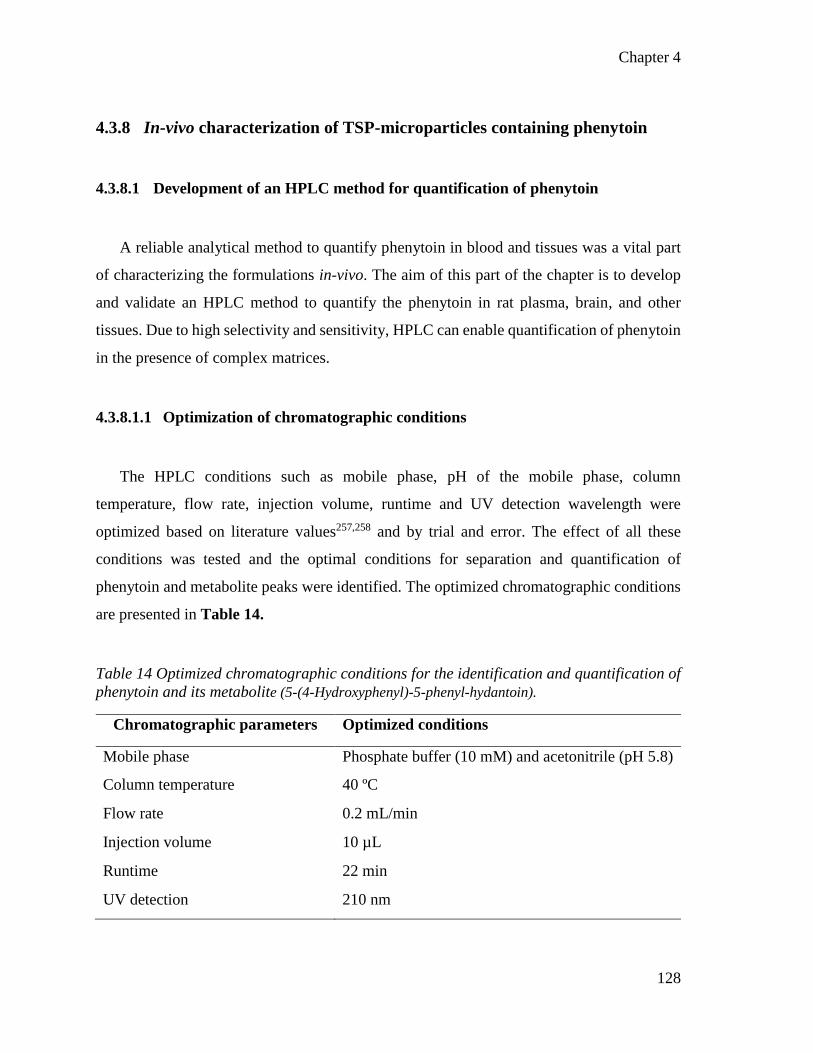
Chapter 4
128
4.3.8 In-vivo characterization of TSP-microparticles containing phenytoin
4.3.8.1 Development of an HPLC method for quantification of phenytoin
A reliable analytical method to quantify phenytoin in blood and tissues was a vital part
of characterizing the formulations in-vivo. The aim of this part of the chapter is to develop
and validate an HPLC method to quantify the phenytoin in rat plasma, brain, and other
tissues. Due to high selectivity and sensitivity, HPLC can enable quantification of phenytoin
in the presence of complex matrices.
4.3.8.1.1 Optimization of chromatographic conditions
The HPLC conditions such as mobile phase, pH of the mobile phase, column
temperature, flow rate, injection volume, runtime and UV detection wavelength were
optimized based on literature values257,258 and by trial and error. The effect of all these
conditions was tested and the optimal conditions for separation and quantification of
phenytoin and metabolite peaks were identified. The optimized chromatographic conditions
are presented in Table 14.
Table 14 Optimized chromatographic conditions for the identification and quantification of
phenytoin and its metabolite (5-(4-Hydroxyphenyl)-5-phenyl-hydantoin).
Chromatographic parameters Optimized conditions
Mobile phase Phosphate buffer (10 mM) and acetonitrile (pH 5.8)
Column temperature 40 ºC
Flow rate 0.2 mL/min
Injection volume 10 µL
Runtime 22 min
UV detection 210 nm

Chapter 4
129
At these optimized conditions, phenytoin and metabolite peaks were identified and
separated as shown in Figure 4.13. Retention time for the phenytoin peak ranged between
13.4 to 13.6 min, the metabolite peak between 10.4-10.6 min and the internal standard peak
between 14.6 min and 14.8 min in various tissues and plasma.
4.3.8.1.2 Validation
The assay was validated in plasma, brain tissue and peripheral tissues such as lung, liver,
spleen, heart, kidney, and spleen. The plasma and brain tissue validation data are presented
in detail in this chapter, and the validation data in other tissues is given in Appendix E.
4.3.8.1.2.1 Specificity
The specificity of the HPLC method was determined by comparing the chromatograms
of plasma and brain samples with no phenytoin, metabolite and internal standard to those
obtained from plasma and brain samples spiked with known amounts of phenytoin,
metabolite and internal standard. The blank samples had no peaks at the retention times
expected for phenytoin, metabolite or internal standard as shown in Figure 4.13 A & C. The
spiked samples had separate peaks for phenytoin, metabolite or internal standard as shown
in Figure 4.13 B & D, suggesting the specificity of the method.

130
Figure 4.13 HPLC chromatograms of (A) blank plasma (B) plasma sample spiked with metabolite, phenytoin (PHT) and internal standard (IS), (C)
blank brain tissue and (D) brain sample spiked with metabolite, phenytoin (PHT) and internal standard (IS). The chromatograms show clear
separation of metabolite, phenytoin and IS in both plasma and brain tissue, only retention times near peaks are shown in the chromatograms.
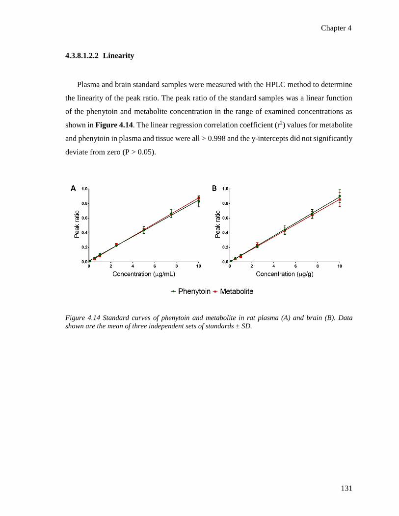
Chapter 4
131
4.3.8.1.2.2 Linearity
Plasma and brain standard samples were measured with the HPLC method to determine
the linearity of the peak ratio. The peak ratio of the standard samples was a linear function
of the phenytoin and metabolite concentration in the range of examined concentrations as
shown in Figure 4.14. The linear regression correlation coefficient (r2) values for metabolite
and phenytoin in plasma and tissue were all > 0.998 and the y-intercepts did not significantly
deviate from zero (P > 0.05).
Figure 4.14 Standard curves of phenytoin and metabolite in rat plasma (A) and brain (B). Data
shown are the mean of three independent sets of standards ± SD.

Chapter 4
132
4.3.8.1.2.3 Sensitivity
The LOD and LOQ are used to assess the sensitivity of the HPLC assay. These were
calculated from the slope and the SD of the y-intercepts from the standard curves. The LOQ
and LOD for phenytoin were 0.016 µg/mL, and 0.048 µg/mL in plasma, and in brain were
0.027 µg/g, and 0.084 µg/g respectively. The LOQ and LOD for metabolite were
0.061 µg/mL, and 0.185 µg/mL in plasma, and in brain were 0.140 µg/g and 0.441 µg/g
respectively.
4.3.8.1.2.4 Accuracy and precision
The accuracy of the assay was determined by comparing the amount of phenytoin and
metabolite recovered in quality control (QC) samples spiked with known concentrations.
The precision of the assay was then determined by calculating the coefficient of variation
(CV) and bias (%). Accuracy and precision of plasma and brain samples are reported in
Table 15 and Table 16. Deviations from theoretical concentrations were less than 8 % for
all the intraday and inter-day QC samples tested. The CV and bias for all QC samples tested
were within 13 % for both the intraday and inter-day. These values for accuracy and
precision are with a range which is acceptable for HPLC analysis according to FDA
guidelines427.
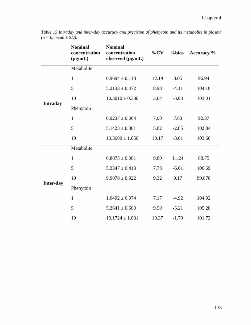
Chapter 4
133
Table 15 Intraday and inter-day accuracy and precision of phenytoin and its metabolite in plasma
(n = 6, mean ± SD).
Nominal
concentration
(µg/mL)
Nominal
concentration
observed (µg/mL)
%CV %bias Accuracy %
Intraday
Metabolite
1 0.9694 ± 0.118 12.19 3.05 96.94
5 5.2133 ± 0.472 8.98 -4.11 104.10
10 10.3010 ± 0.380 3.64 -3.03 103.01
Phenytoin
1 0.9237 ± 0.064 7.00 7.63 92.37
5 5.1423 ± 0.301 5.82 -2.85 102.84
10 10.3600 ± 1.050 10.17 -3.61 103.60
Inter-day
Metabolite
1 0.8875 ± 0.081 9.80 11.24 88.75
5 5.3347 ± 0.413 7.73 -6.61 106.69
10 9.9878 ± 0.922 9.32 0.17 99.878
Phenytoin
1 1.0492 ± 0.074 7.17 -4.92 104.92
5 5.2641 ± 0.500 9.50 -5.21 105.28
10 10.1724 ± 1.031 10.37 -1.70 101.72

Chapter 4
134
Table 16 Intraday and inter-day accuracy and precision of phenytoin and its metabolite in brain
tissue (n = 6, mean ± SD).
Nominal
concentration
(µg/mL)
Nominal
concentration
observed (µg/mL)
%CV %bias Accuracy %
Intraday
Metabolite
1 0.8883 ± 0.065 7.40 11.16 88.83
5 4.7934 ± 0.497 10.25 4.22 95.86
10 9.8697 ± 0.304 3.04 1.41 98.69
Phenytoin
1 1.0633 ± 0.035 3.32 -6.33 106.33
5 4.9074 ± 0.561 11.41 2.00 98.14
10 10.1891 ± 1.276 12.44 -1.77 101.89
Inter-day
Metabolite
1 0.8795 ± 0.089 10.28 12.04 87.95
5 5.0187 ± 0.514 10.18 -0.14 100.37
10 10.1734 ± 0.600 5.82 -1.72 101.73
Phenytoin
1 1.0753 ± 0.084 7.81 -7.52 107.53
5 4.7299 ± 0.491 10.34 5.53 94.59
10 10.2224 ± 1.113 10.87 -2.15 102.22
Chapter 4
135
4.3.8.2 Uptake of phenytoin into the rat brain.
The uptake of phenytoin into the brain 60 min after intranasal administration of
phenytoin-loaded microparticles was compared with IV and intranasal solution
administration and is shown in Figure 4.15.
IV administration showed comparatively low brain uptake of phenytoin with a maximum
phenytoin concentration of 0.21 ± 0.09 µg/g observed at 15 min post administration.
Thereafter, no statistically significant difference between the brain concentrations of
phenytoin over the remaining 45 min was observed. The highest phenytoin concentration of
1.25 ± 0.44 µg/g in the brain was at 30 min after administration of intranasal solution. Again,
like the IV administration, no statistically significant difference in the brain concentrations
of phenytoin over the remaining 45 min was observed. Intranasal microparticles produced a
continuous increase in the brain concentrations of phenytoin over time with a maximum of
1.22 ± 0.39 µg/g at 60 min. Phenytoin concentrations in the brain were significantly higher
at 60 min (**P = 0.0037) compared to initial time points.
The efficiency of the formulation for nose-to-brain delivery of phenytoin was analyzed
by comparing the brain concentrations obtained after administration of intranasal
solution versus intranasal microparticles. The efficiency of the route of administration was
analyzed by comparing brain concentrations after administration as IV
solution versus intranasal solution and the results are reported in Table 17. The statistical
analysis indicates that initially the administration of the intranasal solution led to
significantly higher concentrations of phenytoin in the brain at the 15, 30 and 45 min time
points compared to IV and at 15 and 30 min (P < 0.05) compared to intranasal microparticle
administrations. However, at 60 min post administration, microparticles were shown to
produce significantly higher phenytoin brain concentrations compared to the intranasal
solution at 60 min. The analysis of phenytoin brain concentrations shows the intranasal route
can offer high brain concentrations of phenytoin compared to the IV route and furthermore
administration of phenytoin as intranasal microparticles can preferentially increase the brain
concentrations compared to the intranasal solution.

Chapter 4
136
Figure 4.15 Concentration of phenytoin (PHT) in the rat brains after IV solution administration
(square), intranasal administration of solution (circle) and intranasal microparticles (diamond) over
time. Data expressed are the mean of minimum three independent experiments ± SD with at least
n=5 rats in each time point.
Table 17 P-values indicating the statistical significance of brain phenytoin concentrations after (IN;
intranasal and IV; intravenous) over time.
15 min 30 min 45 min 60 min
IV solution vs
IN solution
*P = 0.0129 **P = 0.0074 *P = 0.0328 P = 0.1072
IN solution vs
IN microparticles
**P = 0.0037 *P = 0.0411 P = 0.8511 * P = 0.0270
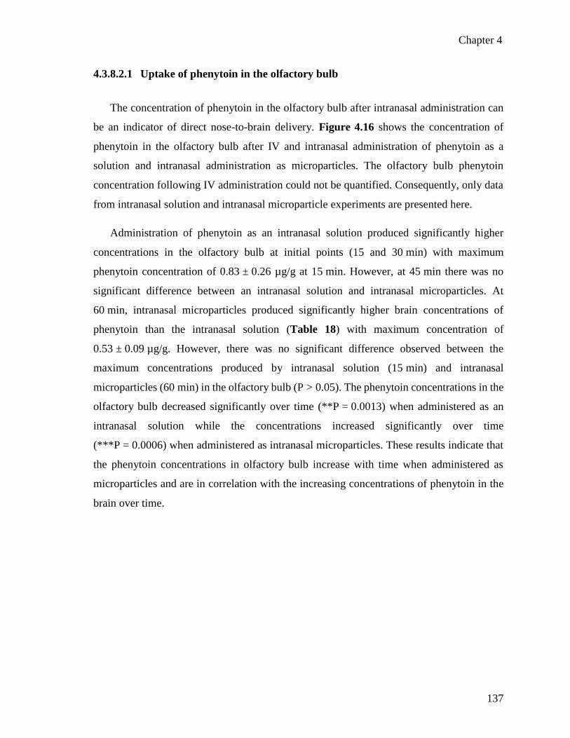
Chapter 4
137
4.3.8.2.1 Uptake of phenytoin in the olfactory bulb
The concentration of phenytoin in the olfactory bulb after intranasal administration can
be an indicator of direct nose-to-brain delivery. Figure 4.16 shows the concentration of
phenytoin in the olfactory bulb after IV and intranasal administration of phenytoin as a
solution and intranasal administration as microparticles. The olfactory bulb phenytoin
concentration following IV administration could not be quantified. Consequently, only data
from intranasal solution and intranasal microparticle experiments are presented here.
Administration of phenytoin as an intranasal solution produced significantly higher
concentrations in the olfactory bulb at initial points (15 and 30 min) with maximum
phenytoin concentration of 0.83 ± 0.26 µg/g at 15 min. However, at 45 min there was no
significant difference between an intranasal solution and intranasal microparticles. At
60 min, intranasal microparticles produced significantly higher brain concentrations of
phenytoin than the intranasal solution (Table 18) with maximum concentration of
0.53 ± 0.09 µg/g. However, there was no significant difference observed between the
maximum concentrations produced by intranasal solution (15 min) and intranasal
microparticles (60 min) in the olfactory bulb (P > 0.05). The phenytoin concentrations in the
olfactory bulb decreased significantly over time (**P = 0.0013) when administered as an
intranasal solution while the concentrations increased significantly over time
(***P = 0.0006) when administered as intranasal microparticles. These results indicate that
the phenytoin concentrations in olfactory bulb increase with time when administered as
microparticles and are in correlation with the increasing concentrations of phenytoin in the
brain over time.
Chapter 4
138
Figure 4.16 The olfactory bulb-time concentration curves of phenytoin (PHT) in rats after intranasal
administration of solution (circle) and microparticles (diamond). Data expressed are the mean of
three independent runs ± SD with at least n=5 rats in each time point.
Table 18 P-values indicating the statistical significance of olfactory bulb phenytoin concentrations.
(IN: intranasal) over time.
15 min 30 min 45 min 60 min
IN solution vs
IN microparticles
* P = 0.0236 ** P = 0.0097 P = 0.2800 ** P = 0.0021
Chapter 4
139
4.3.8.3 The plasma-time concentrations of phenytoin
The plasma concentration of phenytoin over a period of 60 min after IV, intranasal
solution and intranasal microparticle administration was quantified and is shown in Figure
4.17. A trend of increase in plasma concentration of phenytoin over time when administered
as intranasal microparticles and intranasal solution was observed with maximum
concentrations of 1.29 ± 0.36 and 0.97 ± 0.45 µg/mL at 60 min respectively. No significant
difference was observed in plasma phenytoin concentrations for the time points tested with
intranasal solution and intranasal microparticle administrations.
With IV administration, the maximum phenytoin concentration of 2.74 ± 0.27 µg/mL
was observed at the initial 15 min time point, then the plasma concentrations decreased over
time reaching a minimum of 1.85 ± 0.32 µg/mL at 60 min. The plasma phenytoin
concentration decreased significantly (*P = 0.0239) over 60 min after IV administration.
Plasma concentrations of phenytoin with IV administration were compared with intranasal
solution to determine the statistical significance between administration routes. Also,
intranasal solution and intranasal microparticle administrations were compared to assess the
effect of formulation strategy and the results are reported in Table 19. The statistical analysis
revealed that as expected, IV administration led to a significantly higher phenytoin plasma
concentration than the intranasal solution and intranasal microparticles at all the time points.
Significantly higher phenytoin plasma concentrations for intranasal solution compared to
intranasal microparticles for the first 30 min were observed, but intranasal microparticles
were then able to produce similar phenytoin plasma concentrations. The plasma phenytoin
concentrations show that the intranasal route reduces the plasma exposure when compared
to the IV route and furthermore administering intranasal microparticles produces low plasma
phenytoin concentration for 60 min compared to the intranasal solution.

Chapter 4
140
Figure 4.17 Plasma phenytoin (PHT) concertation in rats after IV solution administration (square),
intranasal administration of solution (circle) and intranasal microparticles (diamond) overtime.
Data expressed are the mean of minimum three independent experiments ± SD with at least n=5 rats
in each time point.
Table 19 P-values indicating the statistical significance of plasma phenytoin concentrations (IN;
intranasal and IV; intravenous).
15 min 30 min 45 min 60 min
IV vs
IN solution
**** P < 0.0001 ***P = 0.0003 ** P = 0.0023 * P = 0.0338
IN solution vs
microparticles
*P = 0.0144 *** P = 0.0006 P = 0.0639 P = 0.3182

Chapter 4
141
4.3.8.4 The brain targeting efficiency of TSP-microparticles containing phenytoin
The brain-plasma ratio can be used as a good indicator of the brain-targeting efficiency
for intranasally delivered drugs428–430. The brain-plasma ratio at each time point was
calculated as the ratio of phenytoin concentration in the brain (µg/g) to the phenytoin
concentration in plasma (µg/mL) and presented in Figure 4.18. While calculating the
brain-plasma ratio, the phenytoin concentration in the olfactory bulb was also considered as
the phenytoin concentration in the brain.
Figure 4.18 Brain-plasma ratios of phenytoin after administration as IV solution, intranasal (IN)
solution and intranasal (IN) microparticles. Data presented are the mean of at least three
independent experiments ± SD. (*P < 0.05, ***P < 0.001, and ****P < 0.0001).
The brain-plasma ratio of phenytoin was significantly higher after intranasal solution
during the first 30 min, however in the latter time points intranasal microparticles were found
to produce a significantly higher brain-plasma ratio of phenytoin as shown in Figure 4.18
suggesting a greater targeting of phenytoin to the brain due to the ability of microparticles to
withstand mucociliary clearance.

Chapter 4
142
4.3.8.5 Peripheral tissue distribution of phenytoin
Figure 4.19 shows the peripheral tissue concentrations of phenytoin following IV
solution (A), intranasal solution (B) and intranasal microparticle (C) administrations. The
peripheral tissues (liver, lung, kidney, spleen, and heart) were analyzed for phenytoin
content. The results show that the phenytoin administered as intranasal microparticles
underwent reduced distribution to the peripheral tissues.
Phenytoin was rapidly distributed into the liver following IV and intranasal solution
administration with the highest concentrations of 1.41 ± 1.12 µg/g and 1.98 ± 0.84 µg/g at
15 min which then decreased with time. However, intranasal microparticles produced a
different profile. The initial concentrations of phenytoin in the liver were low at 15 min after
administration of intranasal microparticles and increased with time reaching
0.331 ± 0.21 µg/g at 60 min.
The lungs received 0.68 ± 0.21, 0.58 ± 0.30, and 0.59 ± 0.09 µg/g concentrations of
phenytoin with the IV, intranasal solution and intranasal microparticles at 15 min
respectively. The concentration of phenytoin in the lungs was comparable between the time
points with no significant difference for IV and intranasal solution administration. However,
the lung concentration of phenytoin significantly decreased (****P < 0.0001) overtime for
intranasal microparticle administration reaching to 0.14 ± 0.09 µg/g at 60 min.
A maximum phenytoin concentration of 1.89 ± 0.43 µg/g in the kidney was produced by
IV administration followed by intranasal solution (1.06 ± 0.25 µg/g) and intranasal
microparticles (0.64 ± 0.55 µg/g) at 15 min. Concentrations of phenytoin in the kidney
remained comparable with no significant difference over 60 min for all administrations.
IV administration of phenytoin produced significantly higher concentrations
(**P = 0.0042) in the heart at 30 min (~1.9 µg/g) post administration compared to intranasal
solution (~1 µg/g) and intranasal microparticle (~0.14 µg/g) administrations. Concentrations
of phenytoin in the heart remained unchanged with no significant difference over 60 min for
all administrations. However, the intranasal microparticle administration produced
Chapter 4
143
significantly lower concentrations (*P = 0.0264) of phenytoin in the heart than IV and
intranasal solution.
The spleen received a comparatively higher concentration of phenytoin following IV
administration. The highest concentrations of 0.12 ± 0.06 µg/g, 0.07 ± 0.03 µg/g and
0.05 ± 0.01 µg/g phenytoin in the spleen was observed at 30 min with IV, intranasal solution
and intranasal microparticle administrations respectively. Concentrations of phenytoin in the
spleen remained comparable with no significant difference over 60 min for all
administrations. However, the intranasal microparticle administration produced significantly
lower concentrations (*P = 0.0135) of phenytoin in spleen than IV and intranasal solution.
The results from the peripheral distribution study show that phenytoin distribution in
peripheral tissues is significantly decreased by administering it as intranasal microparticles
over a period of 60 min. These results were consistent with the plasma-time concentrations
of phenytoin.

144
Figure 4.19 Comparison of phenytoin distribution over time in peripheral tissues after administration as intravenous solution (A), intranasal solution
(B) and intranasal microparticles (C). (PHT: phenytoin).
Chapter 4
145
4.4 Discussion
In Chapter 3 the formulation using spray-drying and the characterization of
TSP-microparticles loaded with model drugs was presented. The aim of this chapter was to
formulate TSP-microparticles containing phenytoin and study the in-vivo brain uptake and
biodistribution of the phenytoin formulated into TSP-microparticles.
Formulating TSP-microparticles loaded with phenytoin and optimizing their size to
10 µm was one of the research aims of this chapter. TSP-microparticles containing phenytoin
were produced with the spray-drying method, and particle size was optimized to 10 µm The
SEM analysis of the microparticles produced at optimized formulation parameters identified
microparticles with a corrugated morphology and smooth and rupture free surface. A
corrugated shape and rupture free surface has been reported to be advantageous for intranasal
administration of microparticles363–365. The shape and texture of the microparticles had
comparable features to TSP-microparticles containing FITC-Dextrans (Chapter 3). The
phenytoin content analysis showed very high encapsulation efficiency in the
TSP-microparticles, and these results corroborate with the literature reporting high drug
contents of spray-dried products431,432.
Evaluating the mucoadhesive potential of TSP-microparticles containing phenytoin
was an important research aim of this chapter. As in Chapter 3, in-vitro and ex-vivo methods
were applied to investigate the mucoadhesive potential of the microparticles. In the in-vitro
method, phenytoin microparticles have shown significantly higher mucoadhesion than the
phenytoin powder or solution. The complementary ex-vivo studies also have shown similar
results. These results demonstrate that the TSP-microparticles are mucoadhesive, the
mucoadhesiveness of TSP might help microparticles to withstand mucociliary clearance in
the nasal cavity.
The mucoadhesive potential of TSP-microparticles containing phenytoin was
compared with the TSP-microparticles containing FITC-Dextrans. The ratio of the ex-vivo
mucoadhesive strength of the microparticles to the unformulated powders was used to
compare the mucoadhesive potential between TSP-microparticles containing FITC-Dextrans
Chapter 4
146
and phenytoin. Lack of a statistically significant difference between TSP-microparticles
containing phenytoin or dextrans indicates that the mucoadhesive potential of
TSP-microparticles did not change with the addition of phenytoin.
Assessing phenytoin release from the microparticles represented the drug releasing
behavior from TSP-microparticles and was one of the research aims of this Chapter. The
in-vitro studies were performed for a duration of 4 h as it was hypothesized that the in-vivo
formulation residence time in the nasal cavity was unlikely to be longer than this. Analysis
of in-vitro drug release data revealed that the release of phenytoin from microparticles was
diffusion dependent. The data best fitted to Higuchi’s model for drug release, indicating
diffusion of drug through polymer matrix was the rate-limiting step in the release of
phenytoin from TSP-microparticles433. These results are supported by the cryo-FESEM
studies discussed in Chapter 3 where it was shown that TSP-microparticles form a gel-like
layer on contact with mucin and the drug has to diffuse through this matrix. The formation
of a gel-like layer during the mucoadhesion process is likely the key to the sustained release
of phenytoin from TSP-microparticles compared to the phenytoin solution and phenytoin
powder. TSP-microparticles significantly sustained the release of the phenytoin compared to
the phenytoin solution or powder. About 60 % of the drug was released from the particles
during the first 60 min and the drug release was sustained over 2 h. The drug release data
from microparticles during the first 60 min was significantly slower compared to the solution
and powder.
In-vitro drug release studies were supported by the ex-vivo permeation study in the
porcine nasal mucosa. Due to the morphological similarities of porcine nasal mucosa to the
human nasal mucosa, it was used in this chapter to evaluate drug permeation357. Ex-vivo
permeation data showed evidence for sustained permeation of phenytoin from microparticles
compared to the solution and powder. Due to its hydrophobic nature, phenytoin
predominantly relies on the transcellular pathway to permeate across biological
membranes434. However, the permeability of phenytoin solution data obtained in this study
was slightly higher than literature422, with the high JSS values observed in this study
suggesting that the transport of phenytoin is supported by other mechanisms such as passive
diffusion through paracellular pathways. The ability of solid microparticles to open tight

Chapter 4
147
junctions was already reported and may have contributed to the drug permeation228.
Collectively, the drug release data suggest that phenytoin was successfully released from the
microparticles and is able to permeate across the nasal mucosa.
The initial burst release of phenytoin observed from the microparticles in the in-vitro
studies is most likely attributed to surface-associated phenytoin molecules that can undergo
rapid dissolution. Interestingly, in the first 5 min, the drug release from the microparticles
was higher than the drug release from the phenytoin powder. The hypothesis is that this
phenomenon was due to changes in the physical nature of the phenytoin when formulated
into TSP-microparticles.
Phenytoin, when spray-dried with the polymers containing hydrogen donor and
acceptor sites, is known to change from its crystalline state to an amorphous phase395,424. A
change form a crystalline to an amorphous phase can improve solubility75 and permit faster
drug release which may be the reason for the faster release of phenytoin from
TSP-microparticles than the crystalline powder.
Physical characterization of TSP-microparticles by XRPD and DSC revealed the change
of crystalline phenytoin to a high-energy amorphous phase. The XRPD diffractograms of
TSP-microparticles lack any crystalline phenytoin peaks, and the DSC analysis did not show
any thermal peaks indicating an amorphous nature. The hydrogen donor and acceptor sites
in polymers such as TSP can interact with the amide –NH protons in phenytoin and result in
variation in nucleation and crystal growth inhibition395. TSP contains numerous -OH groups
that can act as hydrogen bond donors or acceptors, which upon spray-drying with phenytoin
can potentially interact with the amide groups and inhibit the association of drug molecules
to form crystal nuclei and hold the drug in the amorphous phase234,395. Results from the
stability studies demonstrate that phenytoin content and moisture content of the
TSP-Microparticles did not change significantly over the period of six months (P > 0.05).
However, the XRPD diffractograms showed that the phenytoin crystalline peaks begin to
reappear after six months of storage. Phenytoin molecules have been reported to surface
absorb and interact with the other phenytoin molecules via π-π interactions between phenyl
rings and hydrogen bonding between amide and carbonyl groups during crystal growth435.
A polymer with structurally similar hydrogen donor and acceptor groups can maintain the
Chapter 4
148
amorphous phase for longer periods by competitive hydrogen bonding to inhibit the
nucleation and crystal regrowth395. Hence it is reasonable to hypothesize that due to the lack
of structurally similar hydrogen donor or acceptor groups in TSP the nucleation and crystal
regrowth of phenytoin molecules occurred over time causing the reappearance of the crystal
peaks in XRPD diffractograms. In future studies, it would be interesting to investigate how
long TSP is able to retain the phenytoin in its amorphous phase, as it may have implications
for the shelf life of TSP-microparticles.
Formulating size specific microparticles to target the olfactory region was a key focus
of this thesis. Due to size-dependent aerodynamics and suitable impaction, 10 µm-sized
particles have previously been suggested to target the olfactory region, as was observed in
this study with TSP-microparticles214. Smaller particles (4 µm) owing to their small size tend
to follow the air streamlines and escape the nasal cavity without depositing. These results
reinforce the importance of particle size on nasal deposition and corroborate the olfactory
deposition data for TSP-microparticles containing FITC-Dextrans.
Assessing the mucosal toxicity of formulations intended for mucosal applications is
a crucial step in the formulation development process. Therefore, assessment of mucosal
toxicity of TSP-microparticles containing phenytoin when administered intranasally was one
of the research aims of this chapter. TSP has been reported to be nontoxic to the mucosa in
previous reports in the literature326 and results from Chapter 3 corroborated these reports as
TSP-microparticles containing FITC-Dextrans were found to be non-toxic to ex-vivo porcine
nasal tissue. In this chapter, the safety of TSP-microparticles containing phenytoin following
intranasal administration in rats was evaluated. The histopathological examination revealed
no signs of necrosis or tissue damage and the olfactory mucosa treated with microparticles
was comparable with the negative control. The structure of the nasal mucosa was well
preserved and the olfactory epithelium displayed standard characteristics. Collectively, these
results suggest that the TSP-microparticles containing phenytoin are safe for acute intranasal
administration.
In addition to the results of in-vitro and ex-vivo studies, data from in-vivo studies are
essential to predict the clinical outcome of a formulation436. An accurate, reliable analytical

Chapter 4
149
method is required to obtain robust in-vivo data. Thus a sensitive HPLC method was
developed to quantify the data in rat plasma and biological tissues.
As discussed previously, phenytoin is metabolized into its metabolite
(5- (4-Hydroxyphenyl)-5-phenyl-hydantoin) in in-vivo, hence an HPLC assay that could
simultaneously quantify phenytoin and its metabolite was developed. The chromatographic
conditions were optimized by trial and error. The mobile phase reported by Potschka et al257
was used and modified. The pH of the mobile phase was adjusted to 5.8 to facilitate the
separation of phenytoin. The flow rate affected the retention time and a flow rate of
0.2 mL/min ensured the separation of the biological matrix from the phenytoin and
metabolite. Adjusting injection volume to 10 µL provided an acceptable peak shape.
Sample preparation for HPLC analysis was optimized by trial and error. Use of
perchloric acid to effectively solubilize lipids in the brain tissue to prepare HPLC samples
has been reported in literature437. However, in this study, when perchloric acid was used,
phenytoin recovery was low. The decreased pH caused by the addition of perchloric acid
was likely causing the phenytoin to precipitate out resulting in the poor recovery. The 80 %
ethanol solution and acetonitrile used to prepare the samples was sufficient to solubilize the
lipids in the brain and other tissues effectively. In order to completely extract phenytoin from
the plasma and tissues, tert-butyl methyl ether (TBME) was added to the extraction
procedure for the samples. The HPLC assay developed in this chapter was validated for
specificity, linearity accuracy and precision in plasma, brain, and other tissues prior to
analysis of the in-vivo samples.
Brain, plasma and peripheral tissue uptake of phenytoin following administration as an
IV solution, intranasal solution, and intranasal microparticles were obtained from in-vivo
studies in rats. Brain concentrations of phenytoin were significantly higher for intranasal
solution compared to the IV solution for the first 45 min, indicating transport through direct
pathways to brain via olfactory and trigeminal neurons in the nasal cavity. As the ‘rat
intranasal catheter device’ (Impel Neuro Pharma, USA) used for administration of intranasal
solution in rats delivered the phenytoin solution directly to the upper dorsal olfactory region
of the rat nose, it might have prevented the loss of drug in the vestibule and helped to increase
the phenytoin uptake to the brain. The uptake of phenytoin in the olfactory bulb followed a

Chapter 4
150
similar trend as the whole brain uptake. Intranasal solution produced maximum olfactory
bulb concentration by 15 min suggesting fast transport by direct neuronal pathways. The
brain and olfactory bulb phenytoin concentration gradually decreased with time after 30 min
and a sudden drop in phenytoin concentrations was observed between 30 min and 45 min.
The decrease in phenytoin concentration in the brain and olfactory bulbs can be postulated
to be due to mucociliary clearance. With time, the intranasal solution was cleared from the
nasal cavity by mucociliary clearance and/or by dripping and being swallowed resulting in
a reduced dose of phenytoin available at the olfactory region for direct transport. Also, the
increase in plasma phenytoin concentrations observed over time with the intranasal solution
administration supports the idea of a solution being cleared or lost to swallowing. Comparing
the brain concentrations of phenytoin after IV and intranasal solution administration clearly
demonstrate the potential of intranasal administration to enhance the brain uptake of
phenytoin.
The concentrations of phenytoin in the brain after intranasal microparticle and
intranasal solution administrations were compared to evaluate the effect of formulation on
the brain uptake of phenytoin. The brain concentrations administered as a solution were
significantly higher than microparticles within the first 30 min post administration. The high
brain concentrations in the initial time points with an intranasal solution can be linked to the
phenytoin dose available at the olfactory region for direct transport. The intranasal solution
in this chapter was directly delivered to the olfactory region with a special rat intranasal
catheter device and was readily available for absorption resulting in high dose availability in
the olfactory region. In contrast, although the microparticles were delivered to the olfactory
region, release of phenytoin from the microparticles requires time as suggested by the
in-vitro release studies in Section 4.3.3. Hence, the available dose of phenytoin at the
olfactory region for absorption during the first 30 min was likely less than the
intranasal solution.
At the later time points, the brain and olfactory bulb phenytoin concentrations with
intranasal microparticles steadily increased while the phenytoin concentrations from
intranasal solution decreased. At 45 min there was no significant difference between solution
and microparticle administrations. Then by 60 min, the brain and olfactory bulb

Chapter 4
151
concentrations of the phenytoin from microparticles were significantly higher than from the
solution. This increase in the brain and olfactory bulb concentrations could have been due to
the continued availability of phenytoin administered as microparticles at the olfactory region
due to the mucoadhesive nature of the microparticles. As demonstrated in Section 4.3.2,
TSP-microparticles are mucoadhesive and can withstand mucociliary clearance. Therefore
it is possible that they provide a continuous release of phenytoin at the olfactory region
whereas, the solution is likely to have been cleared from the nasal cavity by mucociliary
clearance resulting in a reduced dose of phenytoin being available for absorption. The
brain-plasma ratio results suggest an increase in drug targeting efficiency to the brain over
the time with microparticles, which is likely due to their mucoadhesive nature.
Within the olfactory bulb, a higher concentration of phenytoin was observed at
15 min than 30 min with intranasal microparticles. This could have been due to the in-vitro
burst release and the availability of phenytoin due to desorption of the phenytoin from the
surface of the microparticles. Once the surface phenytoin was exhausted, the uptake was
dependent on the phenytoin release from the microparticles. Thus at 30 min, lower olfactory
bulb concentrations of phenytoin were observed.
Interestingly, the brain concentrations of phenytoin from intranasal microparticles at
60 min was comparable to the maximum brain concentration obtained by intranasal solution
at 30 min (P = 0.9208). In-vitro release data reported approximately 60 % phenytoin release
from microparticles at 1 h. Comparing the brain concentrations suggests that 60 % of
phenytoin dose released from microparticles can produce the brain concentrations equivalent
to 100 % of phenytoin dose from intranasal solution. This can be explained by the dose of
the phenytoin available at the olfactory region. Due to the mucoadhesive nature of the
microparticles, the phenytoin dose available at the olfactory region for direct transport to the
brain might have been higher than the intranasal solution resulting in comparable brain
uptakes. Since the blood volume in the brain is very low (47 ± 1µl/g)438 and the dose
administered was also low (1.5 mg) the amount of phenytoin attributed to blood traces can
be assumed to be very low and hence not considered in this study. In addition, in rats, for
intranasal administration of small molecules, it has been reported that the blood-brain ratio
is not significantly affected by perfusion of the brain439.

Chapter 4
152
Collectively, these results suggest that the mucoadhesive nature of the microparticles
can withstand mucociliary clearance ensuring drug (in this case phenytoin), available for
brain uptake at the olfactory region whilst the intranasal solution is cleared quicker.
Therefore, formulating drugs into TSP-microparticles has potential in nose-to-brain drug
delivery. These results are in corroboration with previous works on mucoadhesive
microparticles173–175,189,440.
The plasma concentrations of phenytoin at all-time points were significantly higher
following IV administration compared to intranasal solution and microparticles. Although
phenytoin plasma concentrations showed a decreasing trend over time after IV
administration, they did not vary significantly between the time points which is likely due to
the ~64 min half-life of phenytoin in rats, which extends beyond the time points employed
in this study441. IV administration is also expected to lead to accumulation of phenytoin in
the peripheral tissues which is supported by the high phenytoin concentrations observed in
the peripheral tissues in this study.
The phenytoin plasma concentrations following intranasal solution administration
were significantly higher for the first 30 min compared to those after intranasal microparticle
administration. The amount of phenytoin available for absorption into blood is likely much
higher when formulated as solution compared to microparticles where the amount of
phenytoin is limited by its release from the microparticles. The high vasculature and surface
area of the nasal cavity might have promoted the absorption of the administered phenytoin
solution into the bloodstream. In addition, ethanol present in the phenytoin solution might
have enhanced the phenytoin absorption by causing vasodilation which explains the steady
increase in the plasma concentration over time after intranasal solution administration The
peripheral tissue phenytoin concentrations following intranasal solution administration are
comparable to those following IV administration which supports the hypothesis of phenytoin
being lost to dripping, nasally absorbed or lost to swallowing and eventually entering the
blood.
Plasma uptake of phenytoin administered as intranasal microparticles steadily increased
with time. The lower plasma levels of phenytoin from microparticles can be explained by
the controlled release of phenytoin from the TSP-microparticles. Interestingly, plasma

Chapter 4
153
concentrations of phenytoin after intranasal microparticles and intranasal solution did not
show any significant difference at 45 min and 60 min. This can be correlated to the phenytoin
dose available for the absorption into the blood at respective time points. The
intranasal solution, as mentioned before may have been cleared from the nasal cavity before
45 min and/or dripped and swallowed by the rat which would most likely lead to absorption
into the blood. However, the mucoadhesive microparticles retain the drug in the nasal cavity
and the dose of phenytoin available for the absorption might not have changed over time.
Therefore, at the 45 min and 60 min time points the effective phenytoin dose available for
absorption from both the formulations could have been similar. Consequently, this might
have resulted in the comparable plasma concentrations of phenytoin from intranasal solution
and intranasal microparticles. The hypothesis is supported by the comparable phenytoin
concentrations in the kidney, liver, and lung observed with an intranasal solution and
microparticle administrations at the 45 min and 60 min time points.
The peripheral tissue phenytoin concentrations suggest that the systemic exposure of
phenytoin can be reduced by administering the drug as intranasal microparticles. The
increase in plasma concentrations over time with intranasal microparticle administration is
correlated with the increase in phenytoin concentration in the peripheral tissues over time.
The high phenytoin concentrations at 15 min following intranasal microparticle
administration is a noteworthy observation in this study. This is possibly due to the
deposition of microparticles in the lung while administering with the insufflator, resulting in
high lung concentrations at 15 min. This might have affected the plasma concentrations of
phenytoin, and it would be reasonable to expect much lower plasma concentrations of
phenytoin from intranasal microparticles without the deposition in the lungs. Interestingly,
the lung phenytoin concentrations at later time points were significantly lower than at 15 min
(****P < 0.0001). With the ability of microparticles to sustain the release of phenytoin, if
the microparticles are deposited in the lung, a steady increase of the phenytoin concentration
is expected over time. However, the phenytoin concentration decreases significantly. One of
the possible reasons could be the presence of surface phenytoin. Assuming only a small
fraction of microparticles are deposited in the lungs, the presence of surface phenytoin which
quickly dissolves could have spiked up the phenytoin concentration at 15 min. Nevertheless,

Chapter 4
154
evaluation of the TSP-microparticle release in the lungs is required in future to progress this
delivery system.
4.4.1 Key findings
Collectively, the data presented in this chapter suggests that TSP can be formulated as
10 µm-sized particles containing phenytoin by a spray-drying technique. TSP-microparticles
control the extent of release of phenytoin and improve the olfactory deposition at normal
inhalational rates. TSP-microparticles are mucoadhesive and do not cause any nasal mucosa
toxicity in rats. TSP-microparticles containing phenytoin were found to be stable up to
3 months of storage. In-vivo studies identified the enhanced the brain uptake and direct
nose-to-brain delivery of phenytoin when administered as intranasal TSP-microparticles.
4.4.2 Future directions
Considering the enhanced brain uptake of phenytoin from the TSP-microparticles, it
would be interesting to explore the efficacy of such microparticles in a rat epilepsy model.
Such studies can provide critical data for the translation of formulations from bench to
bedside. In addition, this study considered drug concentrations due to blood traces in brain
and other organs are negligible, however, it can be a limitation and future work needs to
address this by employing techniques such as perfusion of blood.
The stability data in this chapter revealed the recrystallization on storage of
TSP-microparticles for 6 months. Chemical modification of TSP to incorporate more
structurally similar hydrogen donor or acceptor sites similar to phenytoin might prolong the
storage time for TSP-microparticles. Furthermore, drugs with structurally similar hydrogen
donor or acceptor sites could be ideal candidates for microparticle formulation with TSP.
Therefore, the data from this chapter provides evidence for a brain targeting effect of
TSP-microparticles after intranasal administration. It would be interesting to further explore
the organ toxicity, elimination profiles and pharmacokinetics of the drug absorbed
systemically from the TSP-microparticles.
Chapter 5
Computational measurements of micro
particle deposition in human nasal cavities


Chapter 5
157
5 Computational measurements of microparticle deposition in
human nasal cavities
Results from Chapter 3 and Chapter 4 of this thesis demonstrated that size-specific
microparticles could enhance deposition in the olfactory region. It would be beneficial to
study this formulative approach in combination with an intranasal delivery device to
augment olfactory targeting. Intranasal devices carry the significant advantage of self-
administration. This Chapter investigates microparticle deposition in three human nasal
cavities using two conventional drug delivery approaches; firstly, a standard nebulizer that
uses normal inhalational air flow, and secondly, a modern bi-directional air flow delivery
technique.
5.1 Introduction
A recent 2017 report442 from the Food and Drug Administration (FDA) stressed the
importance of in-silico models in drug delivery. The statement “It is the use of in-silico tools
in clinical trials for improving drug development and making regulation more efficient,this
enables safe and effective new therapeutics to advance more efficiently through the different
stages of clinical trials” from this report highlight the importance of in-silico models in drug
development.
In recent years, in-silico experiments have suggested new lines of basic research and
opened up new possibilities in various research areas like cardiovascular studies443,
ophthalmology444, toxicology445, dermatology446, cancer studies447,448, genetic assays449,
intra-cellular mechanisms450, bone remodelling451 and organ studies452. Computational
simulations offer an inexpensive method to test a hypothesis which might be challenging to
investigate experimentally in animals or human subjects453.
Quantification of particle deposition in the olfactory region of the geometrically complex
human nasal cavity is experimentally very hard to measure due to practical and ethical issues.
Thus, in-silico methods such as computational fluid-particle dynamics (CFPD) offer a
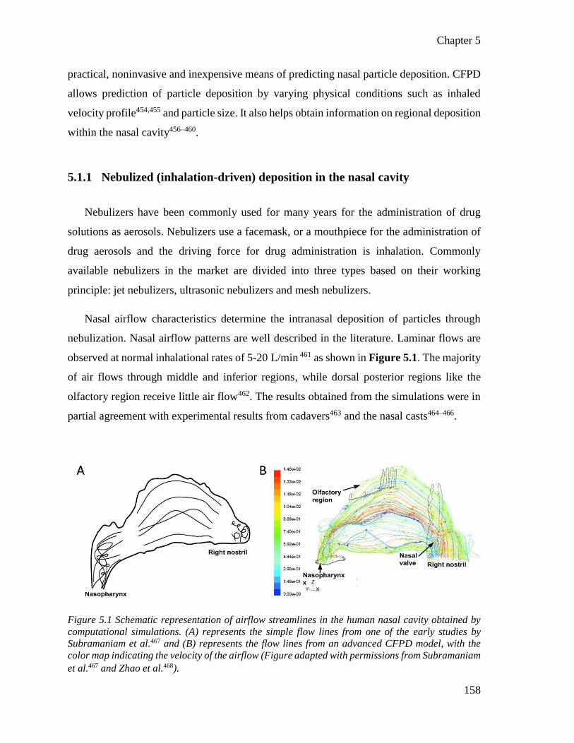
Chapter 5
158
practical, noninvasive and inexpensive means of predicting nasal particle deposition. CFPD
allows prediction of particle deposition by varying physical conditions such as inhaled
velocity profile454,455 and particle size. It also helps obtain information on regional deposition
within the nasal cavity456–460.
5.1.1 Nebulized (inhalation-driven) deposition in the nasal cavity
Nebulizers have been commonly used for many years for the administration of drug
solutions as aerosols. Nebulizers use a facemask, or a mouthpiece for the administration of
drug aerosols and the driving force for drug administration is inhalation. Commonly
available nebulizers in the market are divided into three types based on their working
principle: jet nebulizers, ultrasonic nebulizers and mesh nebulizers.
Nasal airflow characteristics determine the intranasal deposition of particles through
nebulization. Nasal airflow patterns are well described in the literature. Laminar flows are
observed at normal inhalational rates of 5-20 L/min 461 as shown in Figure 5.1. The majority
of air flows through middle and inferior regions, while dorsal posterior regions like the
olfactory region receive little air flow462. The results obtained from the simulations were in
partial agreement with experimental results from cadavers463 and the nasal casts464–466.
Figure 5.1 Schematic representation of airflow streamlines in the human nasal cavity obtained by
computational simulations. (A) represents the simple flow lines from one of the early studies by
Subramaniam et al.467 and (B) represents the flow lines from an advanced CFPD model, with the
color map indicating the velocity of the airflow (Figure adapted with permissions from Subramaniam
et al.467 and Zhao et al.468).

Chapter 5
159
Alterations in nasal airflow patterns within human population have also been
successfully characterized469–471. These results identified that 5 to 20 L min-1 are the
physiological inhalation flow rates215,472. Zhu et al.473 identified the difference in airflow
patterns between two nasal models based on different ethnic origins. The study reported a
lower flow in the olfactory region for the Indian model (about 1 %) compared to the
Caucasian (about 6 %) and Chinese (about 5 %) models. The Caucasian nasal model showed
high flow (35 %) through the middle passage and high flow (about 34 %) in the inferior
portion of the Indian nasal model.
5.1.2 Particle deposition in the nasal cavity
5.1.2.1 Experimental measurements
Many studies have reported particle deposition in adult human nasal replica casts474,475
at various air flow rates (4 to 50 L min-1) and particle sizes (4 nm to 12 µm)476–479. The
results from these studies have observed a common trend defining deposition in the nasal
cavity. Total deposition fraction of particles in the nasal passages is a function of inertial
impaction. The inertial particle deposition increases with the particle mean diameter and air
flow rate and is expressed in terms of impaction parameter (IP)480–482.
𝐼𝑃 = 𝑑𝑎2. 𝑄 𝐸𝑞𝑢𝑎𝑡𝑖𝑜𝑛 6
Where da is the particle aerodynamic diameter and Q is the volumetric airflow rate.
Hence, inertial particle deposition increases with the particle mean diameter and air flow
rate.
5.1.2.2 Computational simulations
Computational simulations couple airflow and particle trajectories in order to compute
deposition of particles in human nasal cavities. Airflow in the nasal cavities is typically
solved using a set of coupled partial differential equations called the Navier-Stokes equations
which yields air flow velocity and pressure. Microparticle movement is usually tracked using

Chapter 5
160
the Lagrangian approach by taking into account various forces acting on a particle. For
example, Li et al.483 investigated microparticle deposition during normal inhalation for
health-risk assessments associated with particle inhalation. Shang et al.484 compared the
deposition of microparticles between human and rat nasal cavities with an aim to improve
extrapolation of particle deposition data between inter-subject species. The total nasal
deposition under the effect of dynamic air flow was studied by Karakosta et al.458 and they
identified that dynamic flow has a significant effect on the microparticle deposition. It has
been applied successfully to examine deposition of particles from nasal sprays485–488 and the
effects of ethnic groups489, spray orientation459 etc.
Shanely et al.490 investigated region specific deposition of particles at inhalational air
flow rates of 2 to 7 L min-1 in one healthy adult human subject. The nasal deposition was
reported as a function of Stokes number in the vestibule, valves, and main airways. Liu et
al.491 investigated deposition of 1.71 to 10.00 μm sized microparticles at higher inhalation
flow rates of 30, 60 and 90 L min-1 in one healthy adult human subject. This study divided
the nasal cavity into an anterior, middle and posterior region. Particle deposition was
reported to be dependent on particle size and flow rates. This study reported a preferential
deposition of 10 µm particles at approximately 60 % of the distance into the cavity (from
nostrils) at an airflow rate of 30 L min-1. The particles between 5.77 to 10.00 µm were
reported to significantly deposit in the first 30 % of the length of the nasal cavity at flow
rates of 30, 60, and 90 L min-1. Ghalati et al.492 studied the deposition of microparticles
(1 to 30 µm) at flow rates of 5, 10 and 15 L min-1 in one healthy adult human subject. This
study predicted deposition in the vestibule, nasal valve, main airway, and nasopharynx
regions of the human nasal cavity. Although these studies have reported critical data on
region-specific deposition, the olfactory region was not identified.
Shi et al.215 reported deposition of microparticles in the olfactory region. This study
divided the nasal cavity into the anterior part, olfactory, middle meatus and inferior meatus
and tested the deposition of microparticles at 7.5 and 20 L min-1. The results of this study
reported that relatively more particles pass through the olfactory and lower middle meatus
regions, compared to the inferior meatus region with an increase in particle size (from 2 µm
to 20 µm) due to the centrifugal force effect. The effect of gravity is negligible for flow rate

Chapter 5
161
≥ 20 L min-1 due to inertia dominating, and the effect of gravity is higher for lower flow rates
≤ 7.5 L min-1 and particle size > 10 µm. This study identified that there is an optimal IP to
enhance deposition in the olfactory region (Figure 5.2).
Figure 5.2 The region-specific deposition of microparticles as a function of IP reported by Shi et al.
Circle represents the identified optimal IP to enhance the deposition in different regions of the nasal
cavity. (Figure adapted with permission from Shi et al.215).
Xi et al.493 compared drug deposition in human nasal replica casts with different nasal
devices (sprays and nebulizers). This study reported that only 4.6 % of the particles released
from spray pumps had reached the olfactory region whereas up to 9 % release was observed
near the olfactory region with the pointed release administration by nebulizers.

Chapter 5
162
Schroeter et al214 reported detailed local deposition patterns in the human nasal cavity.
Deposition of particles with size 2.6 to 14.3 µm at an inhalation flow rate of 7.5, 15.0, and
30.0 L min-1 in one healthy adult human subject were examined. Both the computational
simulations and nasal replica cast deposition studies were reported. This study divided the
nasal cavity into six regions; 1) nasal vestibule containing the anterior portion of the nose,
2) nasal valve region containing the anterior part of the nasal cavity with minimum
cross-sectional area, 3) anterior turbinates encompassing the anterior sections of the middle
and inferior turbinates, 4) olfactory region, 5) turbinates containing the middle and posterior
parts of the inferior and middle turbinates, and 6) nasopharynx. Deposition efficiency as a
function of particle size was reported in this study. The results from this study identified the
optimal particle size for enhancing the deposition in specific regions of the nasal cavity. As
stated in Chapter 1, this study identified that a particle size around 10 µm is optimal for
maximizing the deposition in the olfactory region as shown in Figure 5.3.
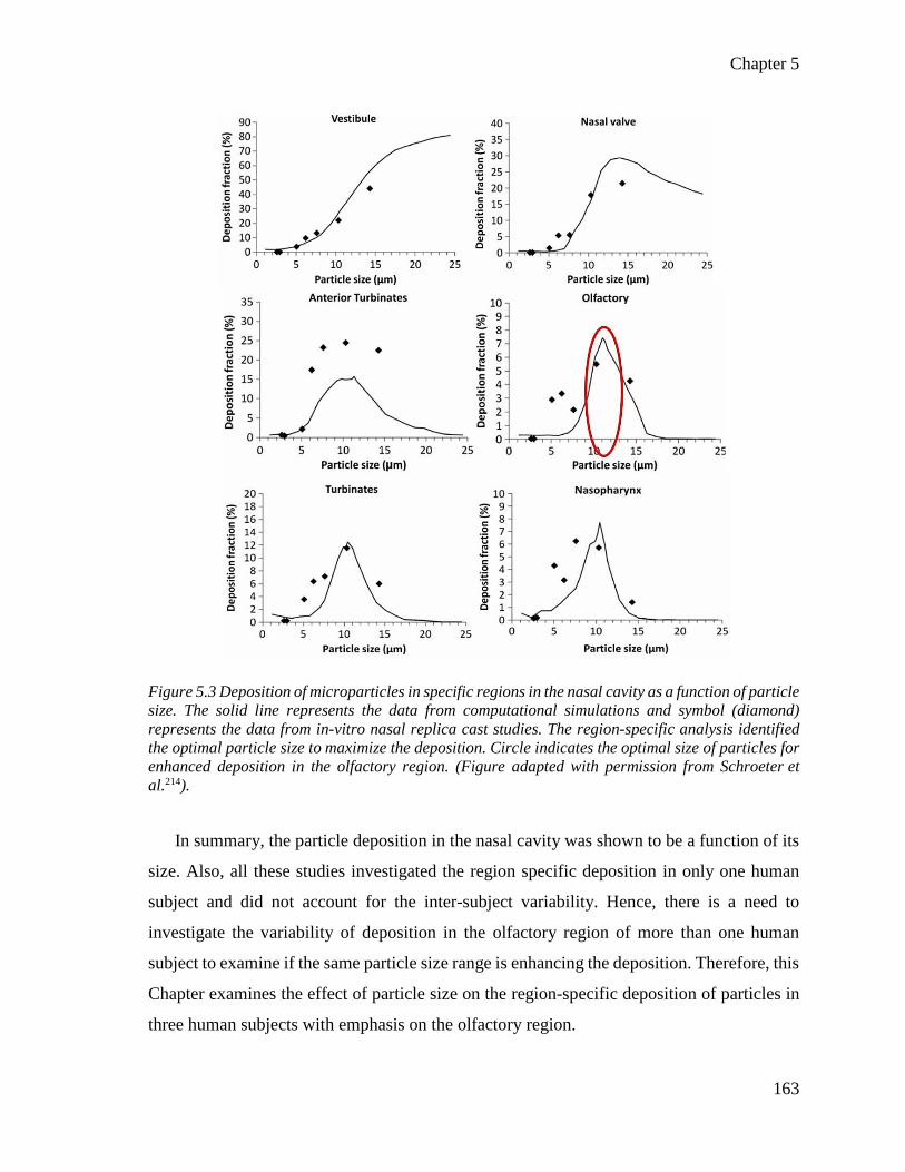
Chapter 5
163
Figure 5.3 Deposition of microparticles in specific regions in the nasal cavity as a function of particle
size. The solid line represents the data from computational simulations and symbol (diamond)
represents the data from in-vitro nasal replica cast studies. The region-specific analysis identified
the optimal particle size to maximize the deposition. Circle indicates the optimal size of particles for
enhanced deposition in the olfactory region. (Figure adapted with permission from Schroeter et
al.214).
In summary, the particle deposition in the nasal cavity was shown to be a function of its
size. Also, all these studies investigated the region specific deposition in only one human
subject and did not account for the inter-subject variability. Hence, there is a need to
investigate the variability of deposition in the olfactory region of more than one human
subject to examine if the same particle size range is enhancing the deposition. Therefore, this
Chapter examines the effect of particle size on the region-specific deposition of particles in
three human subjects with emphasis on the olfactory region.

Chapter 5
164
5.1.3 Modern targeting devices
As mentioned in Chapter 1, POD technology, Kurve technology, and the OPTINOSE
bi-directional delivery technique can improve the deposition in the nasal airways particularly
near the olfactory region. Nebulisation often leads to a significant loss of the administered
drug dose to the lungs resulting in low drug deposition in the olfactory region. Hence, the
use of novel drug delivery device technologies for targeting formulations to the olfactory
region is gaining traction. Among the targeting devices, the OPTINSOE ‘Bi-directional
delivery technique’ shown in Figure 5.4 has been popular and the recently FDA approved
drug sumatriptan powder (Onzetra Xsail®) for nose-to-brain delivery is based on this
delivery technique.
Figure 5.4 Illustration of breath powered bi-directional delivery technique. (A) OPTINOSE® device
(B) with air flow into nasal cavity from the mouth through the device. (C) Steps involved in the
delivery technique and (D) direction of air flow entering from one nostril and exiting from other.
(Figure adapted with permission from Tepper et al.494).

Chapter 5
165
Optinose Inc. (Oslo, Norway) developed the breath powered bi-directional delivery
technique. The device consists of two parts: a sealing nosepiece and an adjustable
mouthpiece. The nosepiece is inserted into one nostril, and the subject blows into the
mouthpiece. Due to the positive pressure during exhalation, the soft palate closes and forms
a communication pathway between two nostrils near the nasopharynx region. Under these
circumstances, airflow enters via one nostril and leaves by the other, hence the name
‘bi-directional delivery technique’ as shown in Figure 5.4. The closure of the nasopharynx
also prevents the loss of drug molecules into the lungs495.
The bi-directional delivery technique has shown the potential to improve drug delivery
to the olfactory region and consequently enhance nose-to-brain drug delivery. The
bi-directional delivery technique has been tested with successful results in the treatment of
migrane494,496–498, nasal polyps499,500, rhinosinusitis501. It has also shown evidence for direct
nose-to-brain transfer of oxytocin by decreasing the dose required in a human study to
improve social-cognitive behaviour218 and might have implications in the treatment of
complex CNS disorders502.
Skretting et al.503 identified the deposition of radiolabelled particles in the human nose
administered by bi-directional delivery technique and showed that the bi-directional delivery
could improve deposition in deep regions of the nasal cavity. Djupesland et al.1 demonstrated
an improved deposition in the upper nasal cavities and less deposition in the nasal valve and
vestibule region using the bi-directional delivery technique. Djupesland et al.504,505 compared
the depositions of traditional spray pump in the human nasal cavity with bi-directional
delivery technique and concluded that the bi-directional delivery could significantly improve
the deposition of particles in the upper posterior segment of the nasal cavity near to the
olfactory region.
Chapter 5
166
5.1.4 Computational simulations in literature
Kleven et al.506 were first to simulate bi-directional delivery at 6 L min-1 and uniform
particle size of 3.5 µm as a part of the development process of this delivery technique. The
particles were released near the nasal valve in the form of a spray. Under these conditions,
the bi-directional delivery leads to 55.4 % total nasal deposition. The air flow velocity was
observed to be greatest at the nasal valve regions of exit nostrils. The velocity at the inlet
nasal valve was not high due to the placement of the delivery device. This study also tested
the deposition of 10 µm sized particles and reported that the whole nasal deposition
improved to 69.2 % due to the increase in particle size.
Xi et al.507 compared the deposition of particles from two nebulizers vibrating mesh and
jet nebulizer in one healthy human subject by inhalational and bi-directional delivery
techniques. The particles released at the nostrils with size 1.5-3.2 µm and the air flow rates
tested were 6, 12, 18 L min-1. The nasal cavity was separated into the olfactory region and
vestibule-turbinate region to estimate the deposition. The results of this study reported up to
6 % of olfactory deposition using the bi-directional delivery technique.
Recently, Dong et al.508 simulated and compared the deposition pattern of particles (1 nm
to 10 µm) released near the nasal valve in one healthy human subject by inhalational and
bi-directional delivery techniques. The airflow rates tested in this study were 7.5 and
15 L min-1. The deposition of particles near the olfactory region and the effects of exhalation
flow rate were investigated. The results of this study were also in agreement with increased
deposition of particles in the olfactory region by bi-directional delivery technique.
In summary, although bi-directional delivery was shown to improve the drug delivery in
the olfactory region, no report has attempted to study the effect of particle size on the
olfactory deposition by bi-directional delivery technique. Also, the detailed region-specific
deposition of the bi-directional delivery that might have implications in toxicity and drug
delivery studies has not yet been reported. Moreover, all these studies investigated the
deposition in only one subject and did not assess variability between subjects.
Chapter 5
167
5.2 Objectives
The computational simulations in this Chapter aim to study the effect of particle size on
the regional deposition of particles in three human subjects and consequently the
nose-to-brain delivery of the drugs. To this effect, the following are the two objectives of
this chapter:
1. To study the effect of particle size combined with inhalation-driven (nebulized)
delivery on the region-specific deposition of microparticles in the human nasal cavity with
emphasis on the olfactory targeting of microparticles and study the variability between
subjects in this study.
2. To study the effect of particle size combined with bi-directional delivery technique
on the region-specific deposition of microparticles in the human nasal cavity with emphasis
on the olfactory targeting of microparticles.
5.2.1 Chapter aims
The overall aim of this Chapter is to investigate the interaction between particle size
and two different intranasal delivery mechanisms.
1. To analyze olfactory particle deposition in three human subjects under nebulization.
a. By reconstructing the human nasal cavities in-silico using CT-scan images.
b. By simulating the inhalational airflow and dynamics of particle deposition
within the nasal passage.
c. By analyzing and comparing the regional deposition data to identify the
similarity in the deposition of the particles between three human subjects.
2. To identify the ideal particle size to improve the olfactory targeting under
bi-directional delivery technique.
a. By simulating the bi-directional airflow and dynamics of particle deposition
at various particle sizes within the nasal passages.
b. By analyzing the deposition data to identify the suitable particle size for
enhancing the olfactory deposition.

Chapter 5
168
5.3 Methods
5.3.1 Subjects used in this study
Axial head and neck computed tomographic (CT) scans of three adult normal healthy
human subjects were used to reconstruct the anatomically accurate nasal airways. A Philips
CT scanner was employed to obtain the scans. Scans were comprised of stacks of horizontal
slices with 6.0000 pixels per mm resolution, and the voxel size was 0.1667x0.1667x0.5 mm3.
Subject 1 was a 40-year-old healthy male, Subject 2 was an 88-year-old male, and Subject 3
was a 60-year-old healthy male. Patients were imaged awake, in a supine position. CT scans
were obtained following approval from New Zealand Health and Disability Ethics
committee protocol NTX/08/12/26. All the raw image files of CT scans were provided by
the Department of Surgery at the University of Auckland. Further processing of raw image
files to generate in-silico nasal airways and simulation studies were performed at the
University of Otago and Auckland Bioengineering Institute.
5.3.2 Image-based reconstruction of human nasal airways and mesh generation
Using ITK‑SNAP 3.5.0, CT‑scan images were segmented, and the airways in the nasal
passage were identified. The segmented images were then converted to a 3D surface and
smoothed using MESHLAB®. The resulting 3D nasal airways meshed with high-quality
computational elements, surface and volume mesh were generated by ICEM‑CFD®
(ANSYS, Inc., Canonsburg, PA) for numerical analysis. The final 3D geometry was divided
into six anatomical regions similar to Schroeter et al.214 as shown in Figure 5.5 to identify
the region-specific deposition of particles.

Chapter 5
169
Figure 5.5 A representative 3D nasal cavity identifying the regions; nasal vestibule and valve (NV),
anterior turbinates (AT), turbinates (T), olfactory region (OR), and nasopharynx (NP).
5.3.1 Governing equations
Assuming steady, laminar, incompressible and isothermal flow, the governing equations
for air flow can be given as
Continuity
𝜕𝑢𝑖
𝜕𝑥𝑖= 0 |𝐸𝑞𝑢𝑎𝑡𝑖𝑜𝑛 8
Momentum
𝜕𝑢𝑖
𝜕𝑡+ 𝑢𝑗
𝜕𝑢𝑖
𝜕𝑥𝑗= −
1
𝜌
𝜕𝑝
𝜕𝑥𝑖+
𝜕
𝜕𝑥𝑗[𝑣 (
𝜕𝑢𝑖
𝜕𝑥𝑗+
𝜕𝑢𝑗
𝜕𝑥𝑖)] |𝐸𝑞𝑢𝑎𝑡𝑖𝑜𝑛 9
Where ui is the air velocity, ρ is the fluid density, 𝑝 is the pressure and 𝑣 is the air
kinematic viscosity.
One-way coupling between fluid and particle phase was assumed. This assumption
implies that the fluid phase affects particle phase. However, the particle does not influence
the fluid flow. This assumption is valid for dilute suspensions. The governing equations for
the microparticle transfer are given by

Chapter 5
170
ⅆx⃗ p
ⅆt= u⃗ p |𝐸𝑞𝑢𝑎𝑡𝑖𝑜𝑛 10
and
𝑚p
ⅆu⃗ p
ⅆt=
1
8𝜋𝜌𝑔𝑑𝑝
2𝐶𝐷(U⃗⃗ g − u⃗ p)|U⃗⃗ g − u⃗ p| + 𝐺 |𝐸𝑞𝑢𝑎𝑡𝑖𝑜𝑛11
Where, x⃗ p is the particle position; u⃗ P and U⃗⃗ g≡ ui is the particle and airflow velocity
vectors, mp is the particle mass; ρg is the gas density; 𝐺 is the gravitational force; CD is the
drag coefficient; and dp is the particle diameter, each particle represents the group of particles
possessing the same characteristics215.
The air flow and particle deposition simulations were conducted with an air density of
1.225 kg/m3, dynamic air viscosity of 1.8 X 10⁻⁵ kg/m.sec, absolute air temperature of 293 K
and a particle density of 1350 kg/m3.
5.3.2 Simulation of airflow and particle deposition
Steady incompressible, laminar and isothermal airflow was simulated in the three-
dimensional (3D) nasal airways. The inhalational airflow rates of 5, 10, and 20 Lmin-1 and a
bi‑directional airflow rate of 6 Lmin‑1 were simulated in this study. For the inhalation
simulations boundary conditions were defined as, rigid airway walls with no-slip and perfect
absorption, zero pressure at the nostrils and a negative pressure at the outlet. Outlet pressure
was specified to attain the required flow rate.
Boundary conditions for bi-directional delivery technique were defined as follows:
nostrils were defined as inlet and outlet. Each subject was simulated twice with one nostril
serving as inlet once. Rigid airway walls with no-slip and perfect absorption, zero pressure
at the outlet were specified. Airflow was simulated at 6 Lmin‑1 at an upward angle of 60°
through the inlet. All the simulations were performed by numerically solving the
incompressible Navier-stokes equations using the finite volume method with commercial
software ANSYS-CFX V16.0® (Canonsburg, PA).
Chapter 5
171
Particle sizes of 2, 4, 6, 8, 10, 12, 14, 16, 18 and 20 µm were simulated in this thesis.
Particles of 2-20 µm were selected to represent ideal size range to target olfactory region.
The numerical computations of particle dynamics were performed with the low Reynolds
number k-ω model; this model has demonstrated accurate solutions for laminar airflow509.
Particles were released at both the nostrils when simulating nebulization. Particles were
released at one of the noses for bi-directional delivery. In both cases, particles were released
from uniformly distributed points across a surface. Total nasal deposition fraction was
computed as the ratio of some particles deposited in the nose to the number of particles
released. Region-specific deposition fractions were computed as the ratio of the number of
particles deposited in each region by the total number of particles released. All the
simulations were performed with commercial software ANSYS‑CFX V16.0® (Canonsburg,
PA). The deposition efficiency of particles according to the nasal regions was calculated,
analyzed and reported.
𝐷𝐸( %) = 𝑁𝑢𝑚𝑏𝑒𝑟 𝑜𝑓 𝑝𝑎𝑟𝑡𝑖𝑐𝑙𝑒𝑠 𝑑𝑒𝑝𝑜𝑠𝑖𝑡𝑒𝑑 𝑖𝑛 𝑡ℎ𝑒 𝑠𝑝𝑒𝑐𝑖𝑓𝑖𝑐 𝑛𝑎𝑠𝑎𝑙 𝑟𝑒𝑔𝑖𝑜𝑛
𝑇𝑜𝑡𝑎𝑙 𝑛𝑢𝑚𝑏𝑒𝑟 𝑜𝑓 𝑝𝑎𝑟𝑡𝑖𝑐𝑙𝑒𝑠 𝑟𝑒𝑙𝑒𝑎𝑠𝑒𝑑 𝑖𝑛𝑡𝑜 𝑡ℎ𝑒 𝑛𝑎𝑠𝑎𝑙 𝑐𝑎𝑣𝑖𝑡𝑦 |𝐸𝑞𝑢𝑎𝑡𝑖𝑜𝑛 12
5.3.3 Statistical analysis
Where applicable, statistical analyses were conducted using an unpaired t-test or
ANOVA on Prism 6 (GraphPad, USA). A P-value of < 0.05 was taken to be significant.
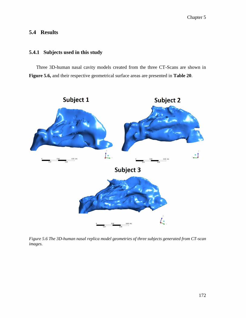
Chapter 5
172
5.4 Results
5.4.1 Subjects used in this study
Three 3D-human nasal cavity models created from the three CT-Scans are shown in
Figure 5.6, and their respective geometrical surface areas are presented in Table 20.
Figure 5.6 The 3D-human nasal replica model geometries of three subjects generated from CT-scan
images.
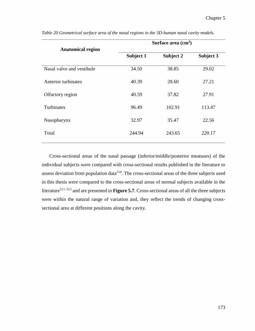
Chapter 5
173
Table 20 Geometrical surface area of the nasal regions in the 3D-human nasal cavity models.
Anatomical region
Surface area (cm2)
Subject 1 Subject 2 Subject 3
Nasal valve and vestibule 34.50 38.85 29.02
Anterior turbinates 40.39 28.60 27.21
Olfactory region 40.59 37.82 27.91
Turbinates 96.49 102.91 113.47
Nasopharynx 32.97 35.47 22.56
Total 244.94 243.65 220.17
Cross-sectional areas of the nasal passage (inferior/middle/posterior meatuses) of the
individual subjects were compared with cross-sectional results published in the literature to
assess deviation from population data510. The cross-sectional areas of the three subjects used
in this thesis were compared to the cross-sectional areas of normal subjects available in the
literature511–513 and are presented in Figure 5.7. Cross-sectional areas of all the three subjects
were within the natural range of variation and, they reflect the trends of changing cross-
sectional area at different positions along the cavity.

Chapter 5
174
Figure 5.7 Comparison of the average cross-sectional area of the three individual subjects with
published data from the literature. The horizontal axis is the distance from the anterior tip of the
nostril.
The nasal airways were divided into tetrahedrons (Volume mesh) by ICEM-CFD. This
mesh generation helps solve fluid flow and particle motion. Also, near to the nasal wall, a
prism mesh consisting of three-layer tetrahedral or pentahedral elements was generated as
shown in Figure 5.8 to accurately calculate deposition and wall turbulence effects. The final
mesh features about 89668 nodes, and 179086 elements for subject 1, 90000 nodes, and
179726 elements for Subject 2, and 86528 nodes, and 172771 elements for Subject 3.

Chapter 5
175
Figure 5.8 Reconstruction of human nasal cavities in-silico. (A) Segmentation of nasal airways in a
CT-scan image (B) reconstruction of surface and generation of surface mesh (C) reconstructed nasal
cavity model showing complex airways and (D) volume mesh generated in the airways and a three-
layer prism mesh at the borders.
5.4.2 Model validations
Our predictions were overall in agreement with reported values from the literature.
Particle deposition data from Kelly et al.481 and Schroeter et al.214 were selected to validate
the data as they feature similar inlet conditions and anatomical descriptions of the regions in
the nasal cavity. The whole nose deposition data as a function of IP from the current
simulations were compared with the experimental data from nebulization simulations in a
human nasal cast by Kelly et al.481 and shown in Figure 5.9.

Chapter 5
176
Figure 5.9 Whole nose deposition efficiency prediction of the three subjects for particle size between
2 to 20 µm (inhalation flow rates ranging from 5 to 20 L min-1) as a function of IP compared to the
published data from Kelly et al.481.
Four inhalational flow rates (5, 7.5, 10, 15, and 20 L min-1) and particle sizes from 2 to
20 µm were investigated in this study in each subject. The whole nasal deposition data of
three subjects in this study were compared to the two nasal cast deposition data from Kelly
et al. 481 as a function of IP. The total nasal deposition curve of all the three subjects was
sigmoidal in shape and was in good agreement with experimental data. The minimum
deposition of 4 % was observed with 2 µm particles at a lower airflow rate of 5 L min-1. The
deposition fraction of 2 µm particles increased to 8 % at a higher airflow rate of 20 L min-1.
The maximum deposition rate was 78 % for 20 µm particles at an airflow rate of 5 L min-1.
Near 100 % deposition was achieved for 20 µm, 14 µm, 12 µm particles at 10 L min-1,
15 L min-1 and 20 L min-1 airflow rates respectively. The preferential deposition in the
anterior parts of the nasal cavity was observed with the increase in particle size as shown in
Figure 5.10.
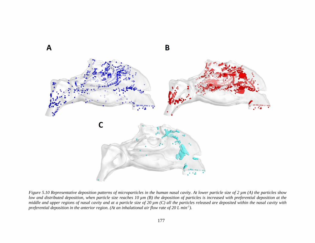
177
Figure 5.10 Representative deposition patterns of microparticles in the human nasal cavity. At lower particle size of 2 µm (A) the particles show
low and distributed deposition, when particle size reaches 10 µm (B) the deposition of particles is increased with preferential deposition at the
middle and upper regions of nasal cavity and at a particle size of 20 µm (C) all the particles released are deposited within the nasal cavity with
preferential deposition in the anterior region. (At an inhalational air flow rate of 20 L min-1).

Chapter 5
178
5.4.2.1 Agreement with published literature
5.4.2.1.1 Results from nebulization
The region-specific particle deposition data were compared with the experimental data
from Schroeter214 to validate the computational predictions and presented in Figure 5.11.
Results from the flow rate case of 15 L min-1 were used. The overall deposition in all the
regions was in good qualitative agreement with the experimental data. For example, in 10 µm
particles inhaled at 15 L min-1, the olfactory deposition fraction matched closely with
Subject 3.
5.4.2.2 Results from bi-directional delivery technique
The data for whole nasal deposition from Kleven et al.506 was used to validate the
deposition efficiency which had also used 6 L min-1. The whole nose deposition data from
the current simulations were in reasonable agreement with the experimental data
(62 vs 73.96 ± 21 %).

179
Figure 5.11 Comparision of computational predictions of microparticle deposition in three subjects (nebulization delivery technique) with the
experimental data from Schroeter et al.214 at a steady air flow rate of 15 L min-1.
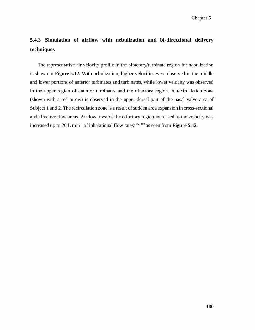
Chapter 5
180
5.4.3 Simulation of airflow with nebulization and bi-directional delivery
techniques
The representative air velocity profile in the olfactory/turbinate region for nebulization
is shown in Figure 5.12. With nebulization, higher velocities were observed in the middle
and lower portions of anterior turbinates and turbinates, while lower velocity was observed
in the upper region of anterior turbinates and the olfactory region. A recirculation zone
(shown with a red arrow) is observed in the upper dorsal part of the nasal valve area of
Subject 1 and 2. The recirculation zone is a result of sudden area expansion in cross-sectional
and effective flow areas. Airflow towards the olfactory region increased as the velocity was
increased up to 20 L min-1 of inhalational flow rates215,509 as seen from Figure 5.12.

Chapter 5
181
Figure 5.12 Representative velocity profile of the air in the olfactory/turbinate region with
nebulization at (A) 5 L min-1, (B) 10 L min-1, (C) 15 L min-1 and (D) 20 L min-1 inhalational air flow
rates. Increased air flow in the olfactory region (Inset) can be observed with the increase in
inhalational air flow rate.
The representative air velocity profile in the olfactory/turbinate region for bi-directional
delivery technique is shown in Figure 5.13. With the bi-directional method, the nasopharynx
is closed. Hence airflow enters through one nostril and exits from another nostril. The
velocity was maximum at the nasal valve region of the exit nostril. In this study, air flow
was simulated at an upward angle of 60° which led to higher velocity in regions of superior
turbinates and the olfactory region as shown in Figure 5.13.

Chapter 5
182
Figure 5.13 Representative velocity profile of the airflow in the olfactory/turbinate region with bi-
directional delivery technique. Increased airflow in the olfactory regions (Inset) were observed.
Fluid streamlines for an inhalation air flow rate of 20 L min-1 and bi-directional flow rate
of 6 L min-1 is shown in Figure 5.14. Airflow was at the highest speed in the nasal valve
area due to the minimum cross-sectional area.

183
Figure 5.14 Representative airflow streamlines of nebulization and bi-directional delivery methods in three subjects. Black arrows indicate the
direction of airflow. (A),(B) & (C) represent the nebulization airflow streamlines at 20 L min-1. (D),(E) & (F) represent the bi-directional air flow
streamlines at 6 L min-1. (D) represents the top view of airflow streamlines in bi-directional simulations. (E) & (F) represent a side view of airflow
streamlines in bi-directional simulations. Red arrows indicate the regions of recirculation in the nasal cavity. The colour map indicates the velocity
of air flow. Streamlines for other inhalational airflow rates (5, 10 and 15 L min-1) are presented in Appendix F.
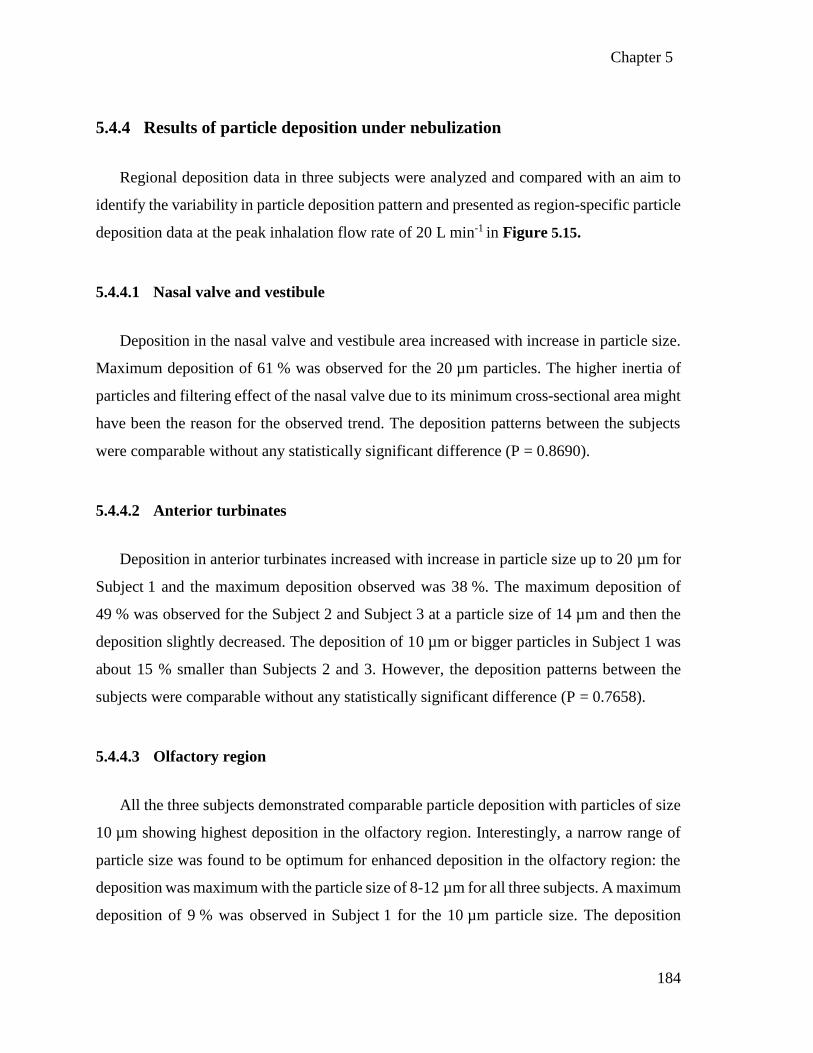
Chapter 5
184
5.4.4 Results of particle deposition under nebulization
Regional deposition data in three subjects were analyzed and compared with an aim to
identify the variability in particle deposition pattern and presented as region-specific particle
deposition data at the peak inhalation flow rate of 20 L min-1 in Figure 5.15.
5.4.4.1 Nasal valve and vestibule
Deposition in the nasal valve and vestibule area increased with increase in particle size.
Maximum deposition of 61 % was observed for the 20 µm particles. The higher inertia of
particles and filtering effect of the nasal valve due to its minimum cross-sectional area might
have been the reason for the observed trend. The deposition patterns between the subjects
were comparable without any statistically significant difference (P = 0.8690).
5.4.4.2 Anterior turbinates
Deposition in anterior turbinates increased with increase in particle size up to 20 µm for
Subject 1 and the maximum deposition observed was 38 %. The maximum deposition of
49 % was observed for the Subject 2 and Subject 3 at a particle size of 14 µm and then the
deposition slightly decreased. The deposition of 10 µm or bigger particles in Subject 1 was
about 15 % smaller than Subjects 2 and 3. However, the deposition patterns between the
subjects were comparable without any statistically significant difference (P = 0.7658).
5.4.4.3 Olfactory region
All the three subjects demonstrated comparable particle deposition with particles of size
10 µm showing highest deposition in the olfactory region. Interestingly, a narrow range of
particle size was found to be optimum for enhanced deposition in the olfactory region: the
deposition was maximum with the particle size of 8-12 µm for all three subjects. A maximum
deposition of 9 % was observed in Subject 1 for the 10 µm particle size. The deposition

Chapter 5
185
patterns between the subjects were comparable without any statistically significant
difference (P =0.9795).
5.4.4.4 Turbinates
The turbinate region included all parts of the nasal passage past the anterior nasal passage
to the nasal choana where the two nasal airways coalesce. This region presents the largest
mucosal surface for drug distribution. The deposition pattern was comparable between the
three subjects. Maximum deposition in turbinates of 14 % and 22 % was observed with
14 µm particles for Subject 1 and Subject 2. For Subject 3, maximum deposition of 20 %
was with 10 µm particles. For Subject 2 and 3, deposition fraction in the turbinates decreased
for particles above 10 µm size. For Subject 1 the deposition fraction in the turbinates
decreased for particles above 12 µm. The deposition patterns between the subjects were
comparable without any statistically significant difference (P =0.1465).
5.4.4.5 Nasopharynx
Particles around 8 µm consistently showed maximum deposition in the nasopharynx
region for all the subjects. The maximum deposition was 6.7 %, 16.95 % and 12.56 % with
Subjects 1, 2 and 3 respectively. The deposition pattern was comparable between the three
subjects without any statistically significant difference (P =0.8309).
186
Figure 5.15 Regional specific deposition in nasal cavities of three human subjects at an inhalation rate of 20 L min-1. No significant difference in
deposition patterns in olfactory regions between the human subjects was observed.

Chapter 5
187
5.4.4.6 Other airflow rate simulations
Results from computational simulations for lower inhalational rates of 5 L min-1 and
10 L min-1 for all three subjects are presented in Figure 5.16. The deposition fraction
increased with increase in the particle size, and flow rate in the nasal valve and vestibule
region. The maximum deposition observed in this region was 51.93 % for 20 µm particles at
10 L min-1 in Subject 2. The deposition pattern in the anterior turbinates region was similar
and was a function of particle inertia. For air flow rates of 5 L min-1 and 10 L min-1, the
deposition increased with increase in particle size until 20 µm. For the higher flow rates, a
trend of decrease in particle deposition was observed in all the subjects at a particle size of
14-18 µm. Deposition patterns were significantly different between subjects at 5 L min-1
(P = 0.0080). Turbinates received increased deposition with an increase in particle size at
5 L min-1 air flow rate and for other flow rates deposition was maximum with particle sizes
up to 14 µm and then decreased. A significant difference of deposition pattern in turbinates
between the subjects was observed at 10 L min-1 (P = 0.0361). The nasopharynx received
comparatively low deposition in all the subjects despite large surface area; the deposition
decreased with the increase in flow rates and a sharp increase in particle deposition was
observed at 8-10 µm for all the subjects observed at all the flow rates.
5.4.4.7 Particles escaped from the nasal passages
The particles that escaped from the nasal passages were also determined. The particle
size and airflow rate had a significant effect on the deposition of the particles. About 75 %
to 90 % of the smaller particles (less than 6 µm) escaped the nasal cavity without depositing.
The lower the airflow rate, the higher was the percentage of particles that escaped. Particles
between 8 to 12 µm showed huge variations depending on the airflow rate. Around 76 % to
85 % of particles escaped at the low 5 L min-1 airflow rate, and the number decreased to 50
to 15 % at the high 20 L min-1 air flow rate. Particles greater than 14 µm in size had low
escape percentage, with about 1 % of particles escaping at the higher flow rates, and between
22 to 50 % at the lower airflow rates.
188
Figure 5.16 Region-specific deposition of particles with different flow rates, the data presented are means of three individual subjects.

Chapter 5
189
5.4.5 Identification of suitable particle size to maximize olfactory deposition
with the bi-directional delivery technique
Region-specific deposition in nasal cavities was assessed with a bi-directional delivery
technique at an air flow rate of 6 L min-1 and presented in Figure 5.17 and Figure 5.18. For
each subject, two protocols of simulations were run with a different nostril serving as an
outlet each time and were referred as left to right and right to left indicating the direction of
airflow. Results were analyzed to identify a suitable particle size to maximize the deposition
in the olfactory region. Also, the results were compared to identify any variability in the
deposition data between the subjects. Due to the particle release position located near the
nasal valve, deposition fractions were not calculated in the vestibule and nasal valve region.
5.4.5.1 Anterior turbinates
Particle deposition followed a similar trend to the inhalational flow with an increase in
deposition observed with increase in particle size. A maximum deposition of 47 % was
observed with 20 µm particles for Subject 3. The deposition pattern between the subjects
and the left and right protocols was comparable (P > 0.2). A significant difference in
deposition pattern between the subjects (P = 0.0187) was observed.
5.4.5.2 Olfactory region
Deposition in the olfactory region was higher than by the nebulization delivery technique
in all the subjects. Deposition increased with increase in particle size up to 18 µm and then
decreased about 2-4 % in all the subjects with 20 µm particles. The greatest deposition of
13.16 % was observed with the 16 µm particle in Subject 2 with the right to left protocol.
The deposition pattern between the subjects and the left and right protocols was comparable
(P > 0.4).
Chapter 5
190
5.4.5.3 Turbinates
With increase in particle size, the deposition in the turbinates region increased. The
maximum deposition for all the subjects was ~30 % with particle size around 16-20 µm.
There was no significant difference observed between left to right and right to left protocols
of all the subjects or between the individual subjects (P > 0.2).
5.4.5.4 Nasopharynx
Bi-directional delivery technique produced higher deposition in the nasopharynx
compared to nebulization. The increase in particle deposition was observed with increase in
size. The maximum deposition of ~25 % was observed with 16 µm particles in Subject 1.
There was a significant difference between left to right and right to left protocols and
deposition data for Subject 3 (P = 0.0350) and in the deposition pattern between subjects
(P = 0.0014).

Chapter 5
191
Figure 5.17 Effect of particle size on region-specific deposition in the nasal cavities of three human
subjects with bi-directional delivery technique at an air flow rate of 6 L min-1 when bi-directional
airflow was simulated from left to right. (**P < 0.01).

Chapter 5
192
Figure 5.18 Effect of particle size on region-specific deposition in the nasal cavities of three human
subjects with bi-directional delivery technique at an air flow rate of 6 L min-1 when bi-directional
airflow was simulated from right to left. (*P < 0.05).

Chapter 5
193
5.4.5.5 Total nasal deposition
The total nasal deposition with the bi-directional delivery technique was dependent upon
the particle size. For smaller particles (less than 6 µm) the total deposition was around
11 to 26 %. For particles between 8–12 µm the total nasal deposition was around 43-91 %.
Almost 99 % total nasal deposition was observed after particle size reached 14 µm or higher.
5.5 Discussion
To exploit advantages of the nasal route to improve nose-to-brain drug delivery, targeted
drug delivery to the olfactory region is very important214,215,509. Previous Chapters of this
thesis have experimentally demonstrated the preferential deposition of particles in the
olfactory region as a function of particle size. This Chapter utilizes computational
simulations to investigate the effect of particle size on olfactory deposition by nebulization
and bi-directional delivery techniques in human subjects.
Anatomically accurate 3D-human nasal cavity models of three subjects were
constructed, and steady laminar airflow was simulated at airflow rates of 5, 10, and 20 Lmin-1
and a bi‑directional airflow rate of 6 Lmin‑1. For nebulization, the flow patterns did not
change very much with an increase in the inhalational airflow rates. At the lower airflow rate
(5 L min-1), the majority of the flow passes through the middle and lower portion of the nasal
cavity resulting in low airflow towards the upper narrow olfactory region. With an increase
in the inhalational airflow rate (20 L min-1) airflow bypasses the meatuses due to stronger
momentum increasing the airflow towards the olfactory region. Increased airflow towards
the olfactory region was evident for bi-directional delivery and could have been due to the
simulation of airflow at an upward angle of 60º. Although the airflow was simulated at a
particular angle, the characteristic geometry of the nasal cavity might have offered some
resistance for alteration of the downstream flow pattern resulting in the majority of airflow
through the middle and lower regions of the nasal cavity similar to the nebulization514.
For the nebulization technique, whole nasal deposition was a function of particle size
and flow rate for all the regions in the nasal cavity. The smaller particles (less than 6 µm)

Chapter 5
194
have shorter relaxation times and lack momentum to deviate from the airflow; thereby a low
portion was deposited in the nasal cavity. Larger particles (> 8 µm) have high inertia, and
consequently, high deposition rates; the whole nasal deposition rate increased with an
increase in the particle size. The deposition rate increased with the increase in inhalational
flow rate. This may have been due to the higher air momentum and increased secondary
flows at the higher flow rates which contributed to the increased particle deposition rates in
the nasal cavity215. At a particle size of 18 µm and flowrate of 20 L min-1 99 % of particles
were deposited.
For bi-directional delivery technique, the whole nasal deposition was a function of
particle size. Although the smaller particles (less than 6 µm) escaped the nasal cavity due to
lower inertia, the total deposition compared to the respective particle size by nebulization
was higher with bi-directional delivery. The increased transit time of a particle in the nasal
passages due to bi-directional airflow by the closed nasopharynx contributed to this
improved deposition rates. Similarly, the larger particles showed higher deposition rates and
when the particle size reached 14 µm 99 % percent of the particles were deposited.
The region-specific deposition of particles with nebulization was analyzed and compared
with the experimental results in a nasal cast (airflow rate of 15 L min-1) from Schroeter et al.
214. The region-specific deposition predictions were in good agreement with the experimental
data except for the valve and vestibule region. The trend of deposition was similar between
the studies. The particle size resulting in maximum deposition in each region was
comparable between simulation studies and the experimental data. The underprediction of
the deposition in the nasal valve region and vestibule area could have been due to the
geometrical complexity and narrow width of this region. Similar anatomical partitioning of
the nasal cavity and the density of the particles used in both the studies were the reasons for
the comparison.
One of the primary objectives of this Chapter was to study the effect of the particle size
on region-specific deposition with nebulization delivery. For the nebulization studies, the
deposition patterns were dependent on particle size and flow rate in all of the regions. Inertia
played an essential role in the region-specific particle deposition. The overall deposition was
comparable between all the three subjects with two exceptions. Firstly, the deposition in

Chapter 5
195
anterior turbinates at the lower flow rate (5 L min-1) between subjects was significantly
different. This variability could have been due to the gravitational sedimentation effect of
particles at lower inhalation rates coupled with the complex anterior turbinate geometry. The
anterior turbinates have a very narrow width (~1 cm), and the geometry expands suddenly
due to the formation of turbinates. A recirculation was observed in this region due to the
geometrical complexity and might have augmented the different particle deposition patterns
in this region between the subjects214. Secondly, a notable difference in a deposition in the
turbinate area at a flow rate of 10 L min-1 occurred.
Olfactory deposition in the nebulization delivery method was dependent on particle size
and flow rate. At lower flow rates (5 and 10 L min-1) the particles sized 14-18 µm were
shown to improve the olfactory deposition. The increased deposition was observed with the
higher flow rate. At the higher flow rates (15 L min-1) and peak inhalational flow rate
(20 L min-1) the optimum size of particles with enhanced olfactory deposition was found to
be 10-11 µm. Shi et al.215 and Liu et al.487 have reported increased deposition of particles in
the middle region of nasal cavity at a particle size of 10 µm. Schroeter et al.214 reported
preferential deposition of 10 µm particles in the olfactory region. The geometric differences
between human nasal cavities can cause variation in the deposition of microparticles481,482.
Hence, there is a need to investigate the inter-subject variability in olfactory deposition to
examine if the same particle size range identified in this study is suitable for enhancing the
deposition. The results from the computational studies in this chapter showed no
inter-subject variability between the olfatory deposition of the microparticles and are in
agreement with the reported studies, which reinforce that particles with a size around 10 µm
can have enhanced deposition in the olfactory region when nebulized.
The second objective of this Chapter was to study the combined effect of particle size
with bi-directional delivery on region-specific deposition in the nasal cavity. To the best of
the authors knowledge, this was the first study to investigate region-specific deposition with
bi-directional delivery. Therefore, the data from this thesis cannot be compared with any
other experimental data. However, whole nasal deposition data in a human nasal cast has
been reported in the literature by Kleven et al.506.The inlet conditions used in this study are
similar to the current simulations. The whole nasal deposition data were comparable between

Chapter 5
196
the simulation and experimental studies. In this thesis, the region-specific deposition with
two protocols, where a different nostril served as an outlet each time (referred as left to right
and right to left, indicating the direction of airflow) has been analyzed.
For bi-directional delivery, the particle deposition in the anterior turbinates was a
function of particle size. However, due to the complex nasal geometry and gravitational
sedimentation, significant variations in deposition between the subjects was observed. For
Subject 1 the difference was notable between the left to right and right to left protocols. The
deposition in the turbinates was a function of particle size. However, a decrease in the
deposition was observed after particle size reaching 14-16 µm. The filtering effect of the
nasal valve with larger particles could be a reason for this decrease. Bi-directional flow
enhanced the deposition in the turbinate region compared to the nebulization due to the
enhanced air penetration into these regions515.The deposition patterns between the two
protocols and subjects were comparable. The deposition in the nasopharynx was dependent
on the particle size. However, the deposition reached a plateau phase when the particle size
was 12-14 µm and then there was a slight decrease in deposition with 20 µm particles. Also,
significant differences in the deposition of particles at the nasopharynx between subjects
were observed, which could have been due to the combined effect of airflow, inertia, and
interception. In addition to the inertia, the interception of particles at the nasopharynx due to
the change in airflow (i.e. changes of about 180° near the nasopharynx) can increase the
number of particles depositing in this region. Due to the substantial change in the airflow
direction, the chance of variation in particles depositing in this region can be high. Also, the
notable difference observed between left to right and right to left protocols of Subject 3 can
be attributed to the significant change in airflow direction and increased chances of
interception. The bi-directional delivery significantly increases the deposition in the
nasopharynx.
Results of this study indicate that deposition in the olfactory region is a function of
particle size. A suitable size of particles for targeting the olfactory region with the bi-
directional delivery was found to be 14-18 µm (Figure 5.19). The bi-directional delivery
significantly enhanced the deposition of particles in the olfactory region compared to the
nebulization delivery (*P = 0.0183). Enhanced deposition in the olfactory region can be due
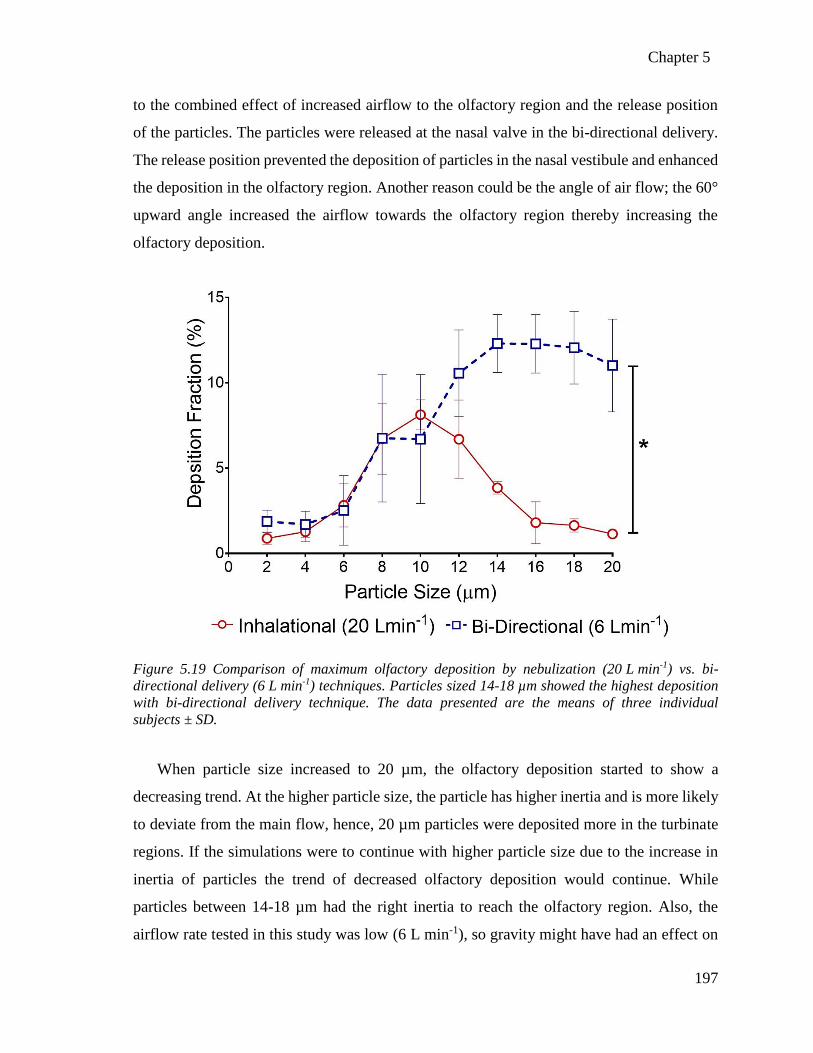
Chapter 5
197
to the combined effect of increased airflow to the olfactory region and the release position
of the particles. The particles were released at the nasal valve in the bi-directional delivery.
The release position prevented the deposition of particles in the nasal vestibule and enhanced
the deposition in the olfactory region. Another reason could be the angle of air flow; the 60°
upward angle increased the airflow towards the olfactory region thereby increasing the
olfactory deposition.
Figure 5.19 Comparison of maximum olfactory deposition by nebulization (20 L min-1) vs. bi-
directional delivery (6 L min-1) techniques. Particles sized 14-18 µm showed the highest deposition
with bi-directional delivery technique. The data presented are the means of three individual
subjects ± SD.
When particle size increased to 20 µm, the olfactory deposition started to show a
decreasing trend. At the higher particle size, the particle has higher inertia and is more likely
to deviate from the main flow, hence, 20 µm particles were deposited more in the turbinate
regions. If the simulations were to continue with higher particle size due to the increase in
inertia of particles the trend of decreased olfactory deposition would continue. While
particles between 14-18 µm had the right inertia to reach the olfactory region. Also, the
airflow rate tested in this study was low (6 L min-1), so gravity might have had an effect on

Chapter 5
198
larger particles and reduced their penetration to the upper dorsal regions of nasal cavity like
the olfactory region.
Results of this study corroborate with the results from by Djupesland et al.214,504,516 who
demonstrated higher deposition efficiency in the upper nasal cavity with the bi-directional
delivery technique. The high deposition can be attributed to increased airflow velocities and
penetration in the upper nasal cavity with the bi-directional delivery technique. The region-
specific deposition patterns reported in this study identified the suitable particle size to
maximize the deposition.The results from this study can help in optimizing the particle size
of the powder formulations to target the region of interest in the nasal cavity.
The computational simulations in this chapter assessed the variability in deposition
pattern of the particle s between the subjects with nebulization and bi-directional delivery
techniques. Anterior turbinates have shown a significant difference in particle deposition
pattern with the nebulization technique. The anterior turbinates and nasopharynx region
received the various deposition patterns in three individuals with the bi-directional delivery
technique. Gravitational sedimentation and the effect of geometry were speculated to cause
the variability in anterior turbinate deposition, and the bi-directional air flow was speculated
to be the reason for the variability in the nasopharynx.
Computational predictions of region-specific particle deposition patterns in this study
can provide detailed insights into the effect of particle size and inter-individual variability
which is otherwise difficult to observe experimentally. Geometry and particle size were
shown to impact the region-specific deposition. Also, particle release position and airflow
angle also might affect the deposition of particles in the olfactory region.
Chapter 5
199
5.6 Key findings
The computational simulations in this chapter suggests particles around 10 µm size show
high deposition in the olfactory region of all three subjects studied under nebulization with
no significant variability observed in the deposition pattern between subjects. Microparticles
with 8 µm were found to improve deposition in the nasopharynx with the nebulization
delivery technique. Microparticles sized between 14-18 µm are found to be suitable for
enhanced drug delivery in the olfactory region with the bi-directional delivery technique.
This study identified that the bi-directional delivery technique can enhance the olfactory
deposition of microparticles compared to nebulization without any inter-subject variability
and might be a desirable method to enhance nose-to-brain delivery of drugs. Anterior
turbinates and nasopharynx were found to have significant variation in the particle deposition
of particles.
5.7 Limitations and future directions
This study assumes particles are of uniform size; future computational simulations shall
consider the effect of polydisperse particles on region-specific deposition in the nasal cavity
especially the olfactory region, to further exploit the probability of enhancing delivery to this
region. The olfactory region in this study contains some part of the meatuses due to the
geometrical definition of regions. A better resolution of the 3-D structure can provide further
details of olfactory deposition. Three human subjects were used in this study, but analyzing
large population data might provide more insight into the inter-individual variability. In
addition, future studies need to explore the potential of bi-directional delivery by changing
the parameters such as particle release position and airflow angle which might have an
impact on the particle deposition in the olfactory region.

Chapter 5
200
Chapter 6
Summary and future outlook


Chapter 6
203
6 Summary and future outlook
The inability of most of the drugs administered orally or IV to reach the brain at
therapeutic levels due to the BBB has moved the focus to using the intranasal route of
delivery to deliver drugs to the brain. The olfactory pathway in the olfactory region and
trigeminal neural pathway in the nasal cavity can transport drugs directly to the brain as
discussed in Chapter 1. Therefore, targeting drugs to the olfactory region has the potential
to enhance nose-to-brain delivery517,518. However, targeted delivery to the olfactory region
is difficult due to its location in the posterior uppermost part of the nasal cavity. Convoluted
and narrow air passages limit drug delivery to the olfactory region, and most of the
administered drugs are either filtered by the nasal valve region or lost to the lungs1,507.
Furthermore, retaining drugs in the nasal cavity is a challenging task due to mucociliary
clearance48. The clinical feasibility of nose-to-brain delivery of drugs is still challenging due
to a very low dose of drugs reaching the olfactory region and thereby the brain. Given the
increasing number of drugs being discovered for the treatment of complex CNS disorders,
developing efficient delivery systems to enhance nose-to-brain drug delivery by targeting
the olfactory region has become very topical 215,493,509.
Various formulation strategies to enhance nose-to-brain drug delivery are under
investigation. One promising strategy is formulating drugs as particulate delivery systems.
Formulating drugs as particulate delivery systems can reduce side effects and enhance brain
absorption of polar and large drug molecules which otherwise have minimal absorption into
the brain230. Another strategy is the use of mucoadhesive polymers to improve their residence
time in the nasal cavity197. In addition to these strategies, recent modeling studies suggest
that particles around 10 µm size can target the olfactory region214,215, which can be beneficial
to the nose-to-brain delivery of drugs. Therefore, the objective of this thesis was to examine
the potential of combining these formulation strategies for enhancing nose-to-brain delivery
of drugs. Features of microparticles that make them an attractive nose-to-brain drug delivery
system were described extensively in Chapter 1 of this thesis and included the ability to
target the olfactory region due to their size.
Chapter 6
204
Chapter 3 of this thesis examined the potential of size tailored mucoadhesive
microparticles to target the olfactory region in-vitro and the potential of TSP as a
mucoadhesive carrier of drugs. Designing appropriate in-vitro methods to test the objectives
of this thesis was in itself challenging. Mucoadhesion of TSP-microparticles in the nasal
cavity is vital for facilitating nose-to-brain drug delivery. Previously reported in-vitro
methods were modified to mimic as closely as possible the human nasal environment and
respiration. In this thesis, a combination of in-vitro and ex-vivo methods were employed to
evaluate the mucoadhesion. This enabled one of the key identifications of the work presented
in this thesis; the mechanism of mucoadhesion and visualization of the two critical stages
involved in the mucoadhesion process; contact and consolidation221. TSP-microparticles
were found to sustain drug release compared to their solution forms. The rate-limiting step
of drug release from microparticles was found to be diffusion of the drug from the polymer
matrix. The release studies served as a guide to their potential kinetics in-vivo. However, any
in-vitro data has to be interpreted with caution due to the artificial nature of in-vitro
experiments compared to the in-vivo environment.
As discussed in Section 3.9 use of hydrophilic drugs in this study may be a limitation for
the extrapolation of the data reported to different classes of drugs. The release behaviour of
model drug from TSP-microparticles changed from diffusion controlled (Higuchi) to
diffusion/erosion controlled (Korsmeyer-peppas) when the molecular weight reached
40 kDa. Whether this is an effect of TSP-microparticles or the cellulose acetate membrane
used for the in-vitro studies needs to be examined and poses the question of reliability of
data extrapolation to release behaviour of large molecules (>40 kDa) from TSP-
microparticles. Mucociliary clearance and effect of other stimulations such as respiration
and sneezing, which cannot be simulated in-vitro can affect drug release in-vivo resulting in
a different release profile.
Though selective deposition of the particles in different regions of the human nasal cavity
as a function of particle size has been reported previously491, formulating particles to a
specific size to enhance the deposition in the olfactory region, however, has not been
previously examined. This thesis demonstrated preferential deposition of 10 µm particles in
the olfactory region of a 3D-human nasal replica at normal breathing rates. Therefore, the

Chapter 6
205
results of this study are only applicable for the peak normal inhalational airflow rate and
when particles are released from the nostrils. Particle density can affect the deposition of
particles in human nasal cavities519. The microparticles used in this thesis had a density of
1.3 kg/m3 which is similar to other published report214 that demonstrated high deposition in
the olfactory region with 10 µm particles, therefore, the effect of density on the deposition
was not evaluated in this thesis. However, to translate the proposed delivery system to a
clinical stage, future work will be required to examine the effect of particle density, effect of
inhalational airflow rate individually and in combination with the particle size on the
olfactory deposition of particles. Although this thesis provided evidence for enhanced
deposition in the olfactory region, the critical question ‘whether this enhancement can
produce any therapeutic effect?’ remains. Data correlating this enhancement with the
biological relevance is the key for translation of such a microparticle system from lab to
market and needs to be generated.
Data from the animal studies are essential to understand the behavior of delivery systems
in the dynamic in-vivo environment. Chapter 4 of this thesis examined the potential of
mucoadhesive TSP-microparticles loaded with an AED phenytoin to enhance its
nose-to-brain delivery in-vivo.
The interesting observation with TSP-microparticles when phenytoin was loaded in to
them was their drug release profiles. Though the release of phenytoin was sustained when
incorporated into TSP-microparticles, the release of phenytoin within the first 5 min from
the microparticles was significantly higher than phenytoin released from the unformulated
powder due to the change in crystallinity. The change from a crystalline into an amorphous
phase can aid nose-to-brain delivery of phenytoin by increasing its solubility520. This change
in physical form is speculated to play an essential role in the shelf life of the microparticles.
Therefore, a six-month stability study of TSP-microparticles containing phenytoin was
conducted according to the ICH guidelines. The transformation of amorphous phase to
crystalline nature was observed by six months. Since polymers with similar functional
groups may prevent the recrystallization for more prolonged periods, it would be interesting
to examine the effect of chemical modifications to TSP with an aim to control the
recrystallization of the phenytoin in TSP-microparticles. Also, studies to investigate the

Chapter 6
206
correlation of such a change in physical state to the extent and rate of drug absorption and
therapeutic efficacy are needed to further exploit the potential of TSP formulations.
The concentrations of phenytoin observed in-vivo in the olfactory bulb and the brain can
be correlated to the burst release of phenytoin from TSP-microparticles observed in-vitro,
and the availability of phenytoin at the site of absorption (olfactory region). While the
intranasal solution failed to achieve high concentrations at the olfactory region after 30 min,
TSP-microparticles were hypothesized to withstand mucociliary clearance and show high
levels of phenytoin at the olfactory region over 60 min and the concentrations of phenytoin
were expected to increase beyond this period. Correlating the in-vitro and in-vivo data it can
be hypothesized that, 50-60 % of the dose of phenytoin administered as TSP-microparticles
was sufficient to produce equivalent or higher brain concentrations of phenytoin than
phenytoin administered as intranasal solution. This reduction in dose can be correlated to the
decreased systemic absorption and increased direct nose-to-brain delivery. The objective of
this work presented in this thesis was to examine the potential of formulation strategies to
enhance nose-to-brain delivery. The time points selected for the in-vivo studies demonstrated
the potential of TSP-microparticles to increase drug delivery to the brain. The time points
chosen in this study and the olfactory bulb phenytoin concentrations measured have provided
insights into the initial stages of the drug release from TSP-microparticles.
Nevertheless, to progress into a clinical trial stage, any future work will require data from
more extended time periods to thoroughly understand the pharmacokinetic profile of the drug
administered when formulated into TSP-microparticles. Furthermore, it will allow safety to
be assessed further and the overall absolute bioavailability of the drug to be calculated. Also,
future work will require the development of more sensitive assays so drug concentrations in
specific regions of the brain can be quantified, which can provide valuable data for treatment
of CNS disorders that are specific to an area in the brain. In addition, studies to investigate
the biological efficacy in disease animal models are critical for the translation to a clinical
stage and need to be included in the future work.
In-silico methods have been shown complementary results from in-vitro and in-vivo
studies521. They can provide insight and answer to experimentally tricky observations. The
recent trend in incorporating in-silico methods in the drug development process has
Chapter 6
207
established a branch linking engineering and medical sciences. With continuous
development both the fields complement each other and facilitate the complicated and
tedious process of drug development.
Chapter 5 of this thesis utilized in-silico techniques like CFPD to examine the
experimentally challenging studies like the effect of inhalational airflow and never reported
studies like the effect of inter-subject variability on the deposition of particles in the nasal
cavity. The in-silico studies in this thesis suggested that the 10 µm sized particles were able
to enhance the olfactory deposition rates in all the three human subjects examined without
significant inter-subject variability. The bi-directional delivery technique has demonstrated
higher olfactory deposition with no inter-subject variability. However, this is a small sample
size including only male subjects, and future work will be required to assess the data from a
larger population including different sex, race and geographical locations. Also, further
resolution of anatomical models to clearly identify the olfactory region is required in the
future work. Since in-silico studies can generate data on various experimentally difficult
facets like particle release position, variable air velocity, geometrical complexity, etc. a
comprehensive database needs to be developed to identify, compare and comprehend the
results from different subjects which might help advance the liaison of two fields for faster
drug development in nose-to-brain delivery.
The formulative approach of administering size specific 10 µm particles increased
deposition in the olfactory region. The observations from Chapter 3 and Chapter 4 posed
an exciting research hypothesis that such a formulative approach used in combination with
an intranasal targeting device can further augment olfactory targeting. Therefore in
Chapter 5, a CFPD modeling technique was employed to answer this question with an aim
to study the effect of particle size combined with bi-directional delivery technique on
olfactory deposition. The results of this study provided an essential identification;
microparticles sized between 14-18 µm showed significantly enhanced drug deposition in
the olfactory region combined with the bi-directional delivery technique. The airflow
direction in the bi-directional delivery technique was anticipated to be reason for increase in
the particle size showing preferential deposition in the olfactory region.
Chapter 6
208
Many other targeting devices are being developed to improve olfactory deposition; it
would be interesting to examine the particle size effect on these targeting devices and to
identify if such a combined approach can further enhance olfactory targeting. Future studies
will be required to acquire deposition data from a large population to determine any
differences in deposition patterns between the subjects. Effect of surface modification of
size-specific particles with olfactory neuron-specific proteins like lectins on nose-to-brain
delivery of drugs could be an exciting research question for the future studies. While this
thesis provides evidence to support that the size controlled particles increase deposition in
the olfactory region, the biological relevance of this increased deposition needs to be
investigated in future studies.
In summary, the in-vitro, in-vivo and in-silico studies complement and refine each other
and are essential to optimize a drug delivery system. This thesis utilized a formulation
strategy, the development of a microparticle delivery system and integrated data from
in-vitro, in-vivo and in-silico studies to refine and optimize the delivery system. Together,
the data presented in this thesis provides evidence for the potential of a size tailored
mucoadhesive microparticle delivery system to enhance nose-to-brain drug delivery.

References


References
211
7 References
1. Djupesland, P. G. Nasal drug delivery devices: Characteristics and performance in a clinical
perspective-a review. Drug Deliv. Transl. Res. 3, 42–62 (2013).
2. Kapoor, M., Cloyd, J. C. & Siegel, R. A. A review of intranasal formulations for the treatment
of seizure emergencies. J. Control. Release 237, 147–159 (2016).
3. U.S.FDA. Drugs@FDA: FDA Approved Drug Products. (2018). Available at:
https://www.accessdata.fda.gov/scripts/cder/daf/. (Accessed: 10th March 2018)
4. Harkema, J. R., Carey, S. A. & Wagner, J. G. The Nose Revisited: A Brief Review of the
Comparative Structure, Function, and Toxicologic Pathology of the Nasal Epithelium.
Toxicol. Pathol. 34, 252–269 (2006).
5. Nicolaas G.M. Schipper, J. C. V. & Merkus, F. W. H. M. The Nasal Mucociliary Clearance:
Relevance to Nasal Drug Delivery. Pharm. Res. 8, 807–14 (1991).
6. Chien, Y.W., Su, K.S.E., and Chang, S.-F. Nasal systemic drug delivery. (Marcel Dekker,
Inc, 1989).
7. Tiozzo Fasiolo, L. et al. Opportunity and challenges of nasal powders: Drug formulation and
delivery. Eur. J. Pharm. Sci. 113, 2–17 (2018).
8. Gizurarson, S. The relevance of nasal physiology to the design of drug absorption studies.
Advanced Drug Delivery Reviews 11, 329–347 (1993).
9. Thorne, J. J. L. and R. G. Intranasal Drug Delivery to the Brain. in Drug Delivery to the Brain:
Physiological Concepts, Methodologies and Approaches (ed. Margareta Hammarlund-
Udenaes, Elizabeth C.M. de Lange, R. G. T.) 401–30 (Springer Science & Business Media,
2013).
10. Field, P. M., Li, Y. & Raisman, G. Ensheathment of the olfactory nerves in the adult rat. J.
Neurocytol. 32, 317–324 (2003).
11. Carmichael, S. T., Clugnet, M. ‐C & Price, J. L. Central olfactory connections in the macaque
monkey. J. Comp. Neurol. 346, 403–434 (1994).
12. Hegg, C. C., Irwin, M. & Lucero, M. T. Calcium store-mediated signaling in sustentacular
cells of the mouse olfactory epithelium. Glia 57, 634–644 (2009).
13. Caggiano, M., Kauer, J. S. & Hunter, D. D. Globose basal cells are neuronal progenitors in
the olfactory epithelium: A lineage analysis using a replication-incompetent retrovirus.
Neuron 13, 339–352 (1994).
14. Iwai, N., Zhou, Z., Roop, D. R. & Behringer, R. R. Horizontal Basal Cells Are Multipotent
Progenitors in Normal and Injured Adult Olfactory Epithelium. Stem Cells 26, 1298–1306
(2008).
15. Elsaesser, R. & Paysan, J. The sense of smell, its signalling pathways, and the dichotomy of
cilia and microvilli in olfactory sensory cells. BMC Neuroscience 8, (2007).

References
212
16. Ward, A. THIEME Atlas of Anatomy general anatomy and musculoskeletal system.
Acupunct. Med. 26, 253–253 (2008).
17. Finger, T. E., Jeor, V. L. S., Kinnamon, J. C. & Silver, W. L. Ultrastructure of substance P‐
and CGRP‐immunoreactive nerve fibers in the nasal epithelium of rodents. J. Comp. Neurol.
294, 293–305 (1990).
18. Finger, T. E., Böttger, B., Schaefer, M. L. & Silver, W. L. Trigeminal collaterals in the nasal
epithelium and olfactory bulb: A potential route for direct modulation of olfactory information
by trigeminal stimuli. J. Comp. Neurol. 444, 221–226 (2002).
19. Broadwell, R. D. & Balin, B. J. Endocytic and exocytic pathways of the neuronal secretory
process and trans synaptic transfer of wheat germ agglutinin‐horseradish peroxidase in vivo.
J. Comp. Neurol. 242, 632–650 (1985).
20. Deatly AM, Haase AT, Fewster PH, Lewis E, B. M. Human herpes virus infections and
Alzheimer’s disease. Neuropathol Appl Neurobiol 16, 213–23 (1990).
21. Anton, F. & Peppel, P. Central projections of trigeminal primary afferents innervating the
nasal mucosa: A horseradish peroxidase study in the rat. Neuroscience 41, 617–628 (1991).
22. Doty, R. L. The olfactory vector hypothesis of neurodegenerative disease: Is it viable? Annals
of Neurology 63, 7–15 (2008).
23. Kristensson, K. & Olsson, Y. Uptake of exogenous proteins in mouse olfactory cells. Acta
Neuropathol. 19, 145–154 (1971).
24. Thorne, R. G., Emory, C. R., Ala, T. A., Frey, W. H. & Frey Ii, W. H. Quantitative analysis
of the olfactory pathway for drug delivery to the brain. Brain Res. 692, 278–282 (1995).
25. Baker, H. & Spencer, R. F. Transneuronal transport of peroxidase-conjugated wheat germ
agglutinin (WGA-HRP) from the olfactory epithelium to the brain of the adult rat. Exp. brain
Res. 63, 461–73 (1986).
26. Kristensson, K. Microbes’ roadmap to neurons. Nature Reviews Neuroscience 12, 345–357
(2011).
27. Thorne, R. G., Pronk, G. J., Padmanabhan, V. & Frey, W. H. Delivery of insulin-like growth
factor-I to the rat brain and spinal cord along olfactory and trigeminal pathways following
intranasal administration. Neuroscience 127, 481–496 (2004).
28. Khan, A. R., Liu, M., Khan, M. W. & Zhai, G. Progress in brain targeting drug delivery
system by nasal route. Journal of Controlled Release 268, 364–389 (2017).
29. Thorne, R. G., Hrabetová, S. & Nicholson, C. Diffusion of epidermal growth factor in rat
brain extracellular space measured by integrative optical imaging. J. Neurophysiol. 92, 3471–
81 (2004).
30. Illum, L. Is nose-to-brain transport of drugs in man a reality? J. Pharm. Pharmacol. 56, 3–17
(2004).
31. Dhuria, S. V., Hanson, L. R. & Frey, W. H. Intranasal delivery to the central nervous system:
Mechanisms and experimental considerations. Journal of Pharmaceutical Sciences 99, 1654–
1673 (2010).
References
213
32. Lochhead, J. J. & Thorne, R. G. Intranasal delivery of biologics to the central nervous system.
Adv. Drug Deliv. Rev. 64, 614–628 (2012).
33. Edeling, M. A., Smith, C. & Owen, D. Life of a clathrin coat: Insights from clathrin and AP
structures. Nat. Rev. Mol. Cell Biol. 7, 32–44 (2006).
34. Rejman, J., Oberle, V., Zuhron, I. S. & HoekstraA, D. Size-dependent internalization of
particles via the pathways of clathrin- and caveolae-mediated endocytosis. Biochem. J. 377,
159–169 (2004).
35. Jones, A. T. Gateways and tools for drug delivery: Endocytic pathways and the cellular
dynamics of cell penetrating peptides. Int. J. Pharm. 354, 34–38 (2008).
36. Altner, H. & Altner-Kolnberger, I. Freeze-fracture and tracer experiments on the permeability
of the zonulae occludentes in the olfactory mucosa of vertebrates. Cell Tissue Res. 154, 51–
59 (1974).
37. Miyamoto, M. et al. Effect of poly-L-arginine on the nasal absorption of FITC-dextran of
different molecular weights and recombinant human granulocyte colony-stimulating factor
(rhG-CSF) in rats. Int. J. Pharm. 226, 127–138 (2001).
38. Faber, W. M. The nasal mucosa and the subarachnoid space. Am. J. Anat. 62, 121–48 (1937).
39. Jansson, B. & Björk, E. Visualization of in vivo olfactory uptake and transfer using
fluorescein dextran. J. Drug Target. 10, 379–386 (2002).
40. Thorne, R. G. & Frey, W. H. Delivery of neurotrophic factors to the central nervous system:
pharmacokinetic considerations. Clin. Pharmacokinet. 40, 907–946 (2001).
41. Hadaczek, P. et al. The ‘Perivascular Pump’ Driven by Arterial Pulsation Is a Powerful
Mechanism for the Distribution of Therapeutic Molecules within the Brain. Mol. Ther. 14,
69–78 (2006).
42. Iliff, J. J. et al. A paravascular pathway facilitates CSF flow through the brain parenchyma
and the clearance of interstitial solutes, including amyloid β. Sci. Transl. Med. 4, (2012).
43. Rennels, M. L., Gregory, T. F., Blaumanis, O. R., Fujimoto, K. & Grady, P. A. Evidence for
a ‘Paravascular’ fluid circulation in the mammalian central nervous system, provided by the
rapid distribution of tracer protein throughout the brain from the subarachnoid space. Brain
Res. 326, 47–63 (1985).
44. I, Carlstedt, JK Sheehan, AP Corfield, J. G. Mucous glycoproteins: a gel of a problem. Essays
Biochem. 20, 10–76 (1985).
45. Sanderson, M. J. & Sleigh, M. A. Ciliary Activity of Cultured Rabbit Tracheal Epithelium:
Beat Pattern and Metachrony. J. Cell Sci 47, 331–347 (1981).
46. Mygind, N. & Dahl, R. Anatomy, physiology and function of the nasal cavities in health and
disease. Advanced Drug Delivery Reviews 29, 3–12 (1998).
47. Chien, Y. W. & Chang, S. F. Intranasal drug delivery for systemic medications. Crit. Rev.
Ther. Drug Carrier Syst. 4, 67–194 (1987).
48. Marttin, E., Schipper, N. G. M., Coos Verhoef, J. & Merkus, F. W. H. M. Nasal mucociliary

References
214
clearance as a factor in nasal drug delivery. Adv. Drug Deliv. Rev. 29, 13–38 (1998).
49. Petruson, B., Hansson, H. A. & Karlsson, G. Structural and Functional Aspects of Cells in the
Nasal Mucociliary System. Arch. Otolaryngol. 110, 576–581 (1984).
50. Satir, P. & Sleigh, M. A. the Physiology of Cilia and Mucociliary Interactions. Annu. Rev.
Physiol 52, 137–55 (1990).
51. Lucas AM, D. L. Principles Underlying Ciliary Activity in the Respiratory Tractii. A
Comparison of Nasal Clearance in Man, Monkey and Other Mammals. Arch Otolaryngol 20,
518–41 (1934).
52. Andersen I, P. D. Measurement of nasal mucociliary clearance. Eur J Respir Dis Suppl. 127,
37–40 (1983).
53. Duchateau, G. S., Graamans, K., Zuidema, J. & Merkus, F. W. Correlation between nasal
ciliary beat frequency and mucus transport rate in volunteers. Laryngoscope 95, 854–859
(1985).
54. Soane, R. J. et al. Evaluation of the clearance characteristics of bioadhesive systems in
humans. Int. J. Pharm. 178, 55–65 (1999).
55. Shipley, M. T. Transport of molecules from nose to brain: Transneuronal anterograde and
retrograde labeling in the rat olfactory system by wheat germ agglutinin-horseradish
peroxidase applied to the nasal epithelium. Brain Res. Bull. 15, 129–142 (1985).
56. Smolnik, R., Molle, M., Fehm, H. L. & Born, J. Brain potentials and attention after acute and
subchronic intranasal administration of ACTH 4-10 and desacetyl-alpha-MSH in humans.
Neuroendocrinology 70, 63–72 (1999).
57. Chapman, C. D. et al. Intranasal treatment of central nervous system dysfunction in humans.
Pharm. Res. 30, 2475–2484 (2013).
58. Matthias Reitz, Maria Demestre, Jan Sedlacik, Hildegard Meissner, Jens Fiehler, B. & Seung
U. Kim, Manfredwestphal, N. O. S. Intranasal Delivery of Neural Stem/Progenitor Cells: A
Noninvasive Passage to Target Intracerebral Glioma. Stem Cells Transl. Med. 866–873
(2012).
59. McGowan, J. W. D., Shao, Q., Vig, P. J. S. & Bidwell, G. L. Intranasal administration of
elastin-like polypeptide for therapeutic delivery to the central nervous system. Drug Des.
Devel. Ther. 10, 2803–2813 (2016).
60. Rapoport, A. & Winner, P. Nasal delivery of antimigraine drugs: Clinical rationale and
evidence base. Headache 46, (2006).
61. Febbraro, F., Andersen, K. J., Sanchez-Guajardo, V., Tentillier, N. & Romero-Ramos, M.
Chronic intranasal deferoxamine ameliorates motor defects and pathology in the α-synuclein
rAAV Parkinson’s model. Exp. Neurol. 247, 45–58 (2013).
62. Guo, C. et al. Intranasal deferoxamine reverses iron-induced memory deficits and inhibits
amyloidogenic APP processing in a transgenic mouse model of Alzheimer’s disease.
Neurobiol. Aging 34, 562–575 (2013).
63. Hanson, L. R. et al. Intranasal deferoxamine provides increased brain exposure and significant

References
215
protection in rat ischemic stroke. J. Pharmacol. Exp. Ther. 330, 679–86 (2009).
64. Danielyan, L. et al. Intranasal delivery of cells to the brain. Eur. J. Cell Biol. 88, 315–324
(2009).
65. Charlton, S. T. et al. Evaluation of direct transport pathways of glycine receptor antagonists
and an angiotensin antagonist from the nasal cavity to the central nervous system in the rat
model. Pharm. Res. 25, 1531–1543 (2008).
66. Stevens, J., Ploeger, B. A., Van Der Graaf, P. H., Danhof, M. & De Lange, E. C. M. Systemic
and direct nose-to-brain transport pharmacokinetic model for remoxipride after intravenous
and intranasal administration. Drug Metab. Dispos. 39, 2275–2282 (2011).
67. Munjal, S. et al. A multicenter, open-label, long-term safety and tolerability study of DFN-
02, an intranasal spray of sumatriptan 10 mg plus permeation enhancer DDM, for the acute
treatment of episodic migraine. J. Headache Pain 18, (2017).
68. Munjal, S. et al. A Randomized Trial Comparing the Pharmacokinetics, Safety, and
Tolerability of DFN-02, an Intranasal Sumatriptan Spray Containing a Permeation Enhancer,
With Intranasal and Subcutaneous Sumatriptan in Healthy Adults. Headache 56, 1455–1465
(2016).
69. Avcu, N. et al. Intranasal Lidocaine in Acute Treatment of Migraine: A Randomized
Controlled Trial. Ann. Emerg. Med. (2016). doi:10.1016/j.annemergmed.2016.09.031
70. Winner, P. et al. Efficacy and tolerability of zolmitriptan nasal spray for the treatment of acute
migraine in adolescents: Results of a randomized, double-blind, multi-center, parallel-group
study (TEENZ). Headache 56, 1107–1119 (2016).
71. Mischley, L. K. et al. A randomized, double-blind phase I/IIa study of intranasal glutathione
in Parkinson’s disease. Mov. Disord. 30, 1696–1701 (2015).
72. Born, J. et al. Sniffing neuropeptides: A transnasal approach to the human brain. Nat.
Neurosci. 5, 514–516 (2002).
73. Vaka, S. R. K., Sammeta, S. M., Day, L. B. & Murthy, S. N. Delivery of nerve growth factor
to brain via intranasal administration and enhancement of brain uptake. J. Pharm. Sci. 98,
3640–3646 (2009).
74. Lv, Q. et al. Intranasal delivery of nerve growth factor attenuates aquaporins-4-induced
edema following traumatic brain injury in rats. Brain Res. 1493, 80–89 (2013).
75. Zhao, Y. Z. et al. Gelatin nanostructured lipid carriers-mediated intranasal delivery of basic
fibroblast growth factor enhances functional recovery in hemiparkinsonian rats.
Nanomedicine Nanotechnology, Biol. Med. 10, 755–764 (2014).
76. Zhao, Y. Z. et al. Intranasal delivery of bFGF with nanoliposomes enhances in vivo
neuroprotection and neural injury recovery in a rodent stroke model. J. Control. Release 224,
165–175 (2016).
77. Serova, L. I., Laukova, M., Alaluf, L. G., Pucillo, L. & Sabban, E. L. Intranasal neuropeptide
Y reverses anxiety and depressive-like behavior impaired by single prolonged stress PTSD
model. Eur. Neuropsychopharmacol. 24, 142–147 (2014).
References
216
78. Dine, J. et al. Intranasally applied neuropeptide S shifts a high-anxiety electrophysiological
endophenotype in the ventral hippocampus towards a ‘normal’-anxiety onee0120272. PLoS
One 10, 1–10 (2015).
79. Vaka, S. R. K., Murthy, S. N., Balaji, A. & Repka, M. A. Delivery of brain-derived
neurotrophic factor via nose-to-brain pathway. Pharm. Res. 29, 441–447 (2012).
80. Alcalá-Barraza, S. R. et al. Intranasal delivery of neurotrophic factors BDNF, CNTF, EPO,
and NT-4 to the CNS. J. Drug Target. 18, 179–190 (2010).
81. Chen, H. et al. Focused ultrasound-enhanced intranasal brain delivery of brain-derived
neurotrophic factor. Sci. Rep. 6, 1–8 (2016).
82. Samaridou, E. & Alonso, M. J. Nose-to-brain peptide delivery - The potential of
nanotechnology. Bioorganic Med. Chem. 26, 2888–2905 (2018).
83. Francis, G. J. et al. Intranasal insulin prevents cognitive decline, cerebral atrophy and white
matter changes in murine type I diabetic encephalopathy. Brain 131, 3311–3334 (2008).
84. Craft, S. et al. Intranasal insulin therapy for Alzheimer disease and amnestic mild cognitive
impairment: A pilot clinical trial. Arch. Neurol. 69, 29–38 (2012).
85. Claxton, A. et al. Long-acting intranasal insulin detemir improves cognition for adults with
mild cognitive impairment or early-stage Alzheimer’s Disease dementia. J. Alzheimer’s Dis.
44, 897–906 (2015).
86. Rosenbloom, M. H. et al. A single-dose pilot trial of intranasal rapid-acting insulin in
apolipoprotein E4 carriers with mild-moderate Alzheimer’s disease. CNS Drugs 28, 1185–
1189 (2014).
87. Jauch-Chara, K. et al. Intranasal insulin suppresses food intake via enhancement of brain
energy levels in humans. Diabetes 61, 2261–2268 (2012).
88. Baier, P. C. et al. Effects of intranasal hypocretin-1 (orexin A) on sleep in narcolepsy with
cataplexy. Sleep Med. 12, 941–946 (2011).
89. Baier, P. C. et al. Olfactory dysfunction in patients with narcolepsy with cataplexy is restored
by intranasal Orexin A (Hypocretin-1). Brain 131, 2734–2741 (2008).
90. Deadwyler, S. A., Porrino, L., Siegel, J. M. & Hampson, R. E. Systemic and Nasal Delivery
of Orexin-A (Hypocretin-1) Reduces the Effects of Sleep Deprivation on Cognitive
Performance in Nonhuman Primates. J. Neurosci. 27, 14239–14247 (2007).
91. Fan, X. et al. No effect of adjunctive, repeated dose intranasal insulin treatment on
psychopathology and cognition in patients with schizophrenia. J. Clin. Psychopharmacol. 33,
226–230 (2013).
92. Freiherr, J. et al. Intranasal insulin as a treatment for alzheimer’s disease: A review of basic
research and clinical evidence. CNS Drugs 27, 505–514 (2013).
93. Reger, M. A. et al. Effects of intranasal insulin on cognition in memory-impaired older adults:
Modulation by APOE genotype. Neurobiol. Aging 27, 451–458 (2006).
94. Craft S, A. P. The Study of Nasal Insulin in the Fight Against Forgetfulness (SNIFF). (2014).

References
217
Available at: https://clinicaltrials.gov/ct2/show/NCT01767909.
95. Zhao, Y. et al. Pharmacokinetic Evaluation of Intranasally Administered Vinyl Polymer-
Coated Lorazepam Microparticles in Rabbits. AAPS J. 14, 218–224 (2012).
96. Hallschmid, M., Benedict, C., Born, J., Fehm, H. L. & Kern, W. Manipulating central nervous
mechanisms of food intake and body weight regulation by intranasal administration of
neuropeptides in man. Physiol. Behav. 83, 55–64 (2004).
97. Bohringer, A., Schwabe, L., Richter, S. & Schachinger, H. Intranasal insulin attenuates the
hypothalamic-pituitary-adrenal axis response to psychosocial stress.
Psychoneuroendocrinology 33, 1394–1400 (2008).
98. Williams, D. R. & Bürkner, P. C. Effects of intranasal oxytocin on symptoms of
schizophrenia: A multivariate Bayesian meta-analysis. Psychoneuroendocrinology 75, 141–
151 (2017).
99. Pedersen, C. A. et al. Intranasal oxytocin reduces psychotic symptoms and improves Theory
of Mind and social perception in schizophrenia. Schizophr. Res. 132, 50–53 (2011).
100. Jarskog, L. F. et al. A 12-week randomized controlled trial of twice-daily intranasal oxytocin
for social cognitive deficits in people with schizophrenia. Schizophr. Res. 185, 88–95 (2017).
101. Tachibana, M. et al. Long-Term Administration of Intranasal Oxytocin Is a Safe and
Promising Therapy for Early Adolescent Boys with Autism Spectrum Disorders. J. Child
Adolesc. Psychopharmacol. 23, 123–127 (2013).
102. Guastella, A. J. et al. The effects of a course of intranasal oxytocin on social behaviors in
youth diagnosed with autism spectrum disorders: A randomized controlled trial. J. Child
Psychol. Psychiatry Allied Discip. 56, 444–452 (2015).
103. Gordon, I. et al. Intranasal Oxytocin Enhances Connectivity in the Neural Circuitry
Supporting Social Motivation and Social Perception in Children with Autism. Sci. Rep. 6, 1–
14 (2016).
104. Sack, M. et al. Intranasal oxytocin reduces provoked symptoms in female patients with
posttraumatic stress disorder despite exerting sympathomimetic and positive chronotropic
effects in a randomized controlled trial. BMC Med. 15, 1–11 (2017).
105. Guastella, A. J. et al. Intranasal Oxytocin Improves Emotion Recognition for Youth with
Autism Spectrum Disorders. Biol. Psychiatry 67, 692–694 (2010).
106. van Zuiden, M. et al. Intranasal Oxytocin to Prevent Posttraumatic Stress Disorder
Symptoms: A Randomized Controlled Trial in Emergency Department Patients. Biol.
Psychiatry 81, 1030–1040 (2017).
107. Murgatroyd, C. A. et al. Effects of chronic social stress and maternal intranasal oxytocin and
vasopressin on offspring interferon-γ and behavior. Front. Endocrinol. (Lausanne). 7, 1–11
(2016).
108. Guastella, A. J., Kenyon, A. R., Alvares, G. A., Carson, D. S. & Hickie, I. B. Intranasal
Arginine Vasopressin Enhances the Encoding of Happy and Angry Faces in Humans. Biol.
Psychiatry 67, 1220–1222 (2010).
References
218
109. Henkin, R. I. Intranasal insulin: From nose to brain. Nutrition 26, 624–633 (2010).
110. Danielyan, L. et al. Therapeutic Efficacy of Intranasally Delivered Mesenchymal Stem Cells
in a Rat Model of Parkinson Disease. Rejuvenation Res. 14, 3–16 (2011).
111. Wei, N. et al. Delayed intranasal delivery of hypoxic-preconditioned bone marrow
mesenchymal stem cells enhanced cell homing and therapeutic benefits after ischemic stroke
in mice. Cell Transplant. 22, 977–991 (2013).
112. Donega, V. et al. Intranasal Mesenchymal Stem Cell Treatment for Neonatal Brain Damage:
Long-Term Cognitive and Sensorimotor Improvement. PLoS One 8, (2013).
113. van Velthoven, C. T. J. et al. Mesenchymal stem cell transplantation attenuates brain injury
after neonatal stroke. Stroke. 44, 1426–32 (2013).
114. Fransson, M. et al. CAR/FoxP3-engineered T regulatory cells target the CNS and suppress
EAE upon intranasal delivery. J. Neuroinflammation 9, (2012).
115. Casettari, L. & Illum, L. Chitosan in nasal delivery systems for therapeutic drugs. Journal of
Controlled Release 190, 189–200 (2014).
116. Liu, L. B., Xue, Y. X. & Liu, Y. H. Bradykinin increases the permeability of the blood-tumor
barrier by the caveolae-mediated transcellular pathway. J. Neurooncol. 99, 187–194 (2010).
117. Zhang, H., Ting Gu, Y. & Xue, Y. X. Bradykinin-induced blood-brain tumor barrier
permeability increase is mediated by adenosine 5’-triphosphate-sensitive potassium channel.
Brain Res. 1144, 33–41 (2007).
118. O’Reilly, M. A., Huang, Y. & Hynynen, K. The impact of standing wave effects on
transcranial focused ultrasound disruption of the blood-brain barrier in a rat model. Phys.
Med. Biol. 55, 5251–5267 (2010).
119. Brem, H. et al. Interstitial chemotherapy with drug polymer implants for the treatment of
recurrent gliomas. J. Neurosurg. 74, 441–446 (1991).
120. Gallia GL, Brem S, B. H. Local treatment of malignant brain tumors using implantable
chemotherapeutic polymers. J Natl Compr Canc Netw. 3, 721–8 (2005).
121. Sutradhar, K. B. & Sumi, C. D. Implantable microchip: The futuristic controlled drug delivery
system. Drug Deliv. 23, 1–11 (2016).
122. Pavan B, D. A. Prodrugs and endogenous transporters: are they suitable tools for drug
targeting into the central nervous system? Curr Pharm Des 17, 3560–76 (2011).
123. Gynther, M. et al. Large Neutral Amino Acid Transporter Enables Brain Drug Delivery via
Prodrugs Large Neutral Amino Acid Transporter Enables Brain Drug Delivery via Prodrugs.
J. Med. Chem. 51, 932–936 (2008).
124. Sezaki, H. Mucosal penetration enhancement. Journal of Drug Targeting 3, 175–177 (1995).
125. Merkus, F. W. H. M., Schipper, N. G. M., Hermens, W. A. J. J., Romeijn, S. G. & Verhoef,
J. C. Absorption enhancers in nasal drug delivery: efficacy and safety. J. Control. Release 24,
201–208 (1993).

References
219
126. Pavan, B. & Dalpiaz, A. Prodrugs and Endogenous Transporters: Are They Suitable Tools for
Drug Targeting into the Central Nervous System? Curr. Pharm. Des. 17, 3560–3576 (2011).
127. Gynther, M. et al. Large neutral amino acid transporter enables brain drug delivery via
prodrugs. J. Med. Chem. 51, 932–936 (2008).
128. Chen, P., Bodor, N., Wu, W. M. & Prokai, L. Strategies to target kyotorphin analogues to the
brain. J. Med. Chem. 41, 3773–3781 (1998).
129. Singh, R. K., Prasad, D. N. & Bhardwaj, T. R. Design, synthesis, chemical and biological
evaluation of brain targeted alkylating agent using reversible redox prodrug approach. Arab.
J. Chem. 10, 420–429 (2017).
130. Shadab, M. D. et al. Bromocriptine loaded chitosan nanoparticles intended for direct nose to
brain delivery: Pharmacodynamic, Pharmacokinetic and Scintigraphy study in mice model.
Eur. J. Pharm. Sci. 48, 393–405 (2013).
131. Gavini, E. et al. Influence of chitosan glutamate on the in vivo intranasal absorption of
rokitamycin from microspheres. J. Pharm. Sci. 100, 1488–1502 (2011).
132. Nagpal, K., Singh, S. K. & Mishra, D. N. Optimization of brain targeted chitosan
nanoparticles of Rivastigmine for improved efficacy and safety. Int. J. Biol. Macromol. 59,
72–83 (2013).
133. Haque, S., Md, S., Sahni, J. K., Ali, J. & Baboota, S. Development and evaluation of brain
targeted intranasal alginate nanoparticles for treatment of depression. J. Psychiatr. Res. 48,
1–12 (2014).
134. Haque, S. et al. Venlafaxine loaded chitosan NPs for brain targeting: Pharmacokinetic and
pharmacodynamic evaluation. Carbohydr. Polym. 89, 72–79 (2012).
135. Patel, D., Naik, S. & Misra, A. Improved transnasal transport and brain uptake of tizanidine
HCl-loaded thiolated chitosan nanoparticles for alleviation of pain. J. Pharm. Sci. 101, 690–
706 (2012).
136. Sharma, D. et al. Nose to Brain Delivery of Midazolam Loaded PLGA Nanoparticles: In Vitro
and In Vivo Investigations. Curr. Drug Deliv. 13, 557–64 (2016).
137. Jose, S. et al. Thermo-sensitive gels containing lorazepam microspheres for intranasal brain
targeting. Int. J. Pharm. 441, 516–526 (2013).
138. Wang, X., Chi, N. & Tang, X. Preparation of estradiol chitosan nanoparticles for improving
nasal absorption and brain targeting. Eur. J. Pharm. Biopharm. 70, 735–740 (2008).
139. Lungare, S., Hallam, K. & Badhan, R. K. S. Phytochemical-loaded mesoporous silica
nanoparticles for nose-to-brain olfactory drug delivery. Int. J. Pharm. 513, 280–293 (2016).
140. Abdelbary, G. A. & Tadros, M. I. Brain targeting of olanzapine via intranasal delivery of
core-shell difunctional block copolymer mixed nanomicellar carriers: In vitro
characterization, ex vivo estimation of nasal toxicity and in vivo biodistribution studies. Int.
J. Pharm. 452, 300–310 (2013).
141. Illum, L. Nasal drug delivery - Possibilities, problems and solutions. J. Control. Release 87,
187–198 (2003).

References
220
142. Pires, P. C. & Santos, A. O. Nanosystems in nose-to-brain drug delivery: A review of non-
clinical brain targeting studies. J. Control. Release 270, 89–100 (2017).
143. Mittal, D. et al. Brain targeted nanoparticulate drug delivery system of rasagiline via
intranasal route. Drug Deliv. 23, 130–139 (2016).
144. Wen, Z. et al. Odorranalectin-conjugated nanoparticles: Preparation, brain delivery and
pharmacodynamic study on Parkinson’s disease following intranasal administration. J.
Control. Release 151, 131–138 (2011).
145. Veronesi, M. C., Aldouby, Y., Domb, A. J. & Kubek, M. J. Thyrotropin-releasing hormone
d,l polylactide nanoparticles (TRH-NPs) protect against glutamate toxicity in vitro and
kindling development in vivo. Brain Res. 1303, 151–160 (2009).
146. Kubek, M. J., Domb, A. J. & Veronesi, M. C. Attenuation of Kindled Seizures by Intranasal
Delivery of Neuropeptide-Loaded Nanoparticles. Neurotherapeutics 6, 359–371 (2009).
147. Kumar, M. et al. Evaluation of neuropeptide loaded trimethyl chitosan nanoparticles for nose
to brain delivery. Int. J. Biol. Macromol. 61, 189–195 (2013).
148. Joachim, E. et al. Gelatin nanoparticles enhance the neuroprotective effects of intranasally
administered osteopontin in rat ischemic stroke model. Drug Deliv. Transl. Res. 4, 395–399
(2014).
149. Gao, X. et al. Brain delivery of vasoactive intestinal peptide enhanced with the nanoparticles
conjugated with wheat germ agglutinin following intranasal administration. J. Control.
Release 121, 156–167 (2007).
150. Yadav, S., Gattacceca, F., Panicucci, R. & Amiji, M. M. Comparative biodistribution and
pharmacokinetic analysis of cyclosporine-A in the brain upon intranasal or intravenous
administration in an oil-in-water nanoemulsion formulation. Mol. Pharm. 12, 1523–1533
(2015).
151. Kanazawa, T., Taki, H., Tanaka, K., Takashima, Y. & Okada, H. Cell-penetrating peptide-
modified block copolymer micelles promote direct brain delivery via intranasal
administration. Pharm. Res. 28, 2130–2139 (2011).
152. Taki, H., Kanazawa, T., Akiyama, F., Takashima, Y. & Okada, H. Intranasal delivery of
camptothecin-loaded tat-modified nanomicells for treatment of intracranial brain tumors.
Pharmaceuticals 5, 1092–1102 (2012).
153. Narayan, R. et al. Development of risperidone liposomes for brain targeting through
intranasal route. Life Sci. 163, 38–45 (2016).
154. Subodhkant Samudre, Avinash Tekade, Kanchan Thorve, Anup Jamodkar, Gauri Parashar,
N. C. Xanthan Gum Coated Mucoadhesive Liposomes for Efficient Nose to Brain Delivery
of Curcumin. Drug Deliv. Lett. 5, 201–207 (2015).
155. Khan, S., Patil, K., Bobade, N., Yeole, P. & Gaikwad, R. Formulation of intranasal
mucoadhesive temperature-mediated in situ gel containing ropinirole and evaluation of brain
targeting efficiency in rats. J. Drug Target. 18, 223–234 (2010).
156. Mistry, A., Stolnik, S. & Illum, L. Nose-to-Brain Delivery: Investigation of the Transport of
Nanoparticles with Different Surface Characteristics and Sizes in Excised Porcine Olfactory
References
221
Epithelium. Mol. Pharm. 12, 2755–2766 (2015).
157. Kohane, D. S. Microparticles and nanoparticles for drug delivery. Biotechnology and
Bioengineering 96, 203–209 (2007).
158. Varde, N. K. & Pack, D. W. Microspheres for controlled release drug delivery. Expert Opin.
Biol. Ther. 4, 35–51 (2004).
159. Chattaraj, S. C., Rathinavelu, A. & Das, S. K. Biodegradable microparticles of influenza viral
vaccine: Comparison of the effects of routes of administration on the in vivo immune response
in mice. J. Control. Release 58, 223–232 (1999).
160. Verma, R. K., Kaur, J., Kumar, K., Yadav, A. B. & Misra, A. Intracellular time course,
pharmacokinetics, and biodistribution of isoniazid and rifabutin following pulmonary
delivery of inhalable microparticles to mice. Antimicrob. Agents Chemother. 52, 3195–3201
(2008).
161. Cambronero-Rojas, A. et al. Capreomycin oleate microparticles for intramuscular
administration: Preparation, in vitro release and preliminary in vivo evaluation. J. Control.
Release 209, 229–237 (2015).
162. Vasa, D. M., O’Donnell, L. A. & Wildfong, P. L. D. Influence of Dosage Form, Formulation,
and Delivery Device on Olfactory Deposition and Clearance: Enhancement of Nose-to-CNS
Uptake. J. Pharm. Innov. 10, 200–210 (2015).
163. Soane, R. J., Hinchcliffe, M., Davis, S. S. & Illum, L. Clearance characteristics of chitosan
based formulations in the sheep nasal cavity. Int. J. Pharm. 217, 183–191 (2001).
164. Vasa, D. M., Buckner, I. S., Cavanaugh, J. E. & Wildfong, P. L. D. Improved Flux of
Levodopa via Direct Deposition of Solid Microparticles on Nasal Tissue. AAPS
PharmSciTech 18, 904–912 (2016).
165. Patil, S., Babbar, A., Mathur, R., Mishra, A. & Sawant, K. Mucoadhesive chitosan
microspheres of carvedilol for nasal administration. J. Drug Target. 18, 321–331 (2010).
166. Elmowafy, E., Osman, R., El-Shamy, A. E. H. A. & Awad, G. A. S. Nasal polysaccharides-
glucose regulator microparticles: Optimization, tolerability and antidiabetic activity in rats.
Carbohydr. Polym. 108, 257–265 (2014).
167. Lim, S. T., Forbes, B., Berry, D. J., Martin, G. P. & Brown, M. B. In vivo evaluation of novel
hyaluronan/chitosan microparticulate delivery systems for the nasal delivery of gentamicin in
rabbits. Int. J. Pharm. 231, 73–82 (2002).
168. Krauland, A. H., Leitner, V. M., Grabovac, V. & Bernkop-Schnürch, A. In vivo evaluation of
a nasal insulin delivery system based on thiolated chitosan. J. Pharm. Sci. 95, 2463–2472
(2006).
169. Abdel Mouez, M., Zaki, N. M., Mansour, S. & Geneidi, A. S. Bioavailability enhancement of
verapamil HCl via intranasal chitosan microspheres. Eur. J. Pharm. Sci. 51, 59–66 (2014).
170. Kozlovskaya, L. & Stepensky, D. Quantitative analysis of the brain-targeted delivery of drugs
and model compounds using nano-delivery systems. J. Control. Release 171, 17–23 (2013).
171. Kozlovskaya, L., Abou-Kaoud, M. & Stepensky, D. Quantitative analysis of drug delivery to
References
222
the brain via nasal route. Journal of Controlled Release 189, 133–140 (2014).
172. Sun, Y., Shi, K., Wan, F. & Cui, F. De. Methotrexate-loaded microspheres for nose to brain
delivery: In vitro/in vivo evaluation. J. Drug Deliv. Sci. Technol. 22, 167–174 (2012).
173. Dalpiaz, A. et al. Brain uptake of an anti-ischemic agent by nasal administration of
microparticles. J. Pharm. Sci. 97, 4889–4903 (2008).
174. Dalpiaz, A. et al. Brain uptake of a zidovudine prodrug after nasal administration of solid
lipid microparticles. Mol. Pharm. 11, 1550–1561 (2014).
175. Rassu, G. et al. Solid microparticles based on chitosan or methyl-??-cyclodextrin: A first
formulative approach to increase the nose-to-brain transport of deferoxamine mesylate. J.
Control. Release 201, 68–77 (2015).
176. Khan, A. et al. Brain Targeting of Temozolomide via the Intranasal Route Using Lipid-Based
Nanoparticles: Brain Pharmacokinetic and Scintigraphic Analyses. Mol. Pharm. 13, 3773–
3782 (2016).
177. Salama, H. A., Mahmoud, A. A., Kamel, A. O., Abdel Hady, M. & Awad, G. A. S. Brain
delivery of olanzapine by intranasal administration of transfersomal vesicles. J. Liposome
Res. 22, 336–345 (2012).
178. Devkar, T. B., Tekade, A. R. & Khandelwal, K. R. Surface engineered nanostructured lipid
carriers for efficient nose to brain delivery of ondansetron HCl using Delonix regia gum as a
natural mucoadhesive polymer. Colloids Surfaces B Biointerfaces 122, 143–150 (2014).
179. Singh, R. & Lillard, J. W. Nanoparticle-based targeted drug delivery. Experimental and
Molecular Pathology 86, 215–223 (2009).
180. Li, Y., Jiang, H. L., Zhu, K. J., Liu, J. H. & Hao, Y. L. Preparation, characterization and nasal
delivery of α-cobrotoxin- loaded poly(lactide-co-glycolide)/polyanhydride microspheres. J.
Control. Release 108, 10–20 (2005).
181. Dalpiaz, A. et al. Nasal chitosan microparticles target a zidovudine prodrug to brain HIV
sanctuaries. Antiviral Res. 123, 146–157 (2015).
182. Gavini, E. et al. Influence of polymeric microcarriers on the in vivo intranasal uptake of an
anti-migraine drug for brain targeting. Eur. J. Pharm. Biopharm. 83, 174–183 (2013).
183. Tafaghodi, M., Tabassi, S. A. S., Jaafari, M. R., Zakavi, S. R. & Momen-Nejad, M. Evaluation
of the clearance characteristics of various microspheres in the human nose by gamma-
scintigraphy. Int. J. Pharm. 280, 125–135 (2004).
184. Sharma, N., Kulkarni, G. T., Sharma, A., Bhatnagar, A. & Kumar, N. Natural mucoadhesive
microspheres of Abelmoschus esculentus polysaccharide as a new carrier for nasal drug
delivery. J. Microencapsul. 30, 589–598 (2013).
185. Mao, S., Chen, J., Wei, Z., Liu, H. & Bi, D. Intranasal administration of melatonin starch
microspheres. Int. J. Pharm. 272, 37–43 (2004).
186. Belgamwar, V. S. et al. Design and development of nasal mucoadhesive microspheres
containing tramadol HCl for CNS targeting. Drug Deliv. 18, 353–360 (2011).

References
223
187. Gungor, S., Okyar, A., Erturk-Toker, S., Baktir, G. & Ozsoy, Y. Ondansetron-loaded
biodegradable microspheres as a nasal sustained delivery system: In vitro/in vivo studies.
Pharm. Dev. Technol. 15, 258–265 (2010).
188. Gungor, S., Okyar, A., Erturk-Toker, S., Baktir, G. & Ozsoy, Y. Ondansetron-loaded chitosan
microspheres for nasal antiemetic drug delivery: An alternative approach to oral and
parenteral routes. Drug Dev. Ind. Pharm. 36, 806–813 (2010).
189. Gavini, E. et al. Nasal administration of Carbamazepine using chitosan microspheres: In
vitro/in vivo studies. Int. J. Pharm. 307, 9–15 (2006).
190. Ikechukwu Ugwoke, M., Kaufmann, G., Verbeke, N. & Kinget, R. Intranasal bioavailability
of apomorphine from carboxymethylcellulose-based drug delivery systems. Int. J. Pharm.
202, 125–131 (2000).
191. Krauland, A. H., Guggi, D. & Bernkop-Schnürch, A. Thiolated chitosan microparticles: A
vehicle for nasal peptide drug delivery. Int. J. Pharm. 307, 270–277 (2006).
192. Sadrai, J. V. H. & Alinagari, R. Nasal delivery of insulin using chitosan microspheres. 21,
761–774 (2004).
193. Brime B, Ballesteros MP, F. P. Preparation and in vitro characterization of gelatin
microspheres containing Levodopa for nasal administration. J Microencapsul. 17, 777–84
(2000).
194. Hafner, A., Filipovic-Grcic, J., Voinovich, D. & Jalsenjak, I. Development and in vitro
characterization of chitosan-based microspheres for nasal delivery of promethazine. Drug
Dev. Ind. Pharm. 33, 427–436 (2007).
195. Gavini, E., Rassu, G., Muzzarelli, C., Cossu, M. & Giunchedi, P. Spray-dried microspheres
based on methylpyrrolidinone chitosan as new carrier for nasal administration of
metoclopramide. Eur. J. Pharm. Biopharm. 68, 245–252 (2008).
196. Jug, M. & Becirevic-Lacan, M. Development of a cyclodextrin-based nasal delivery system
for Lorazepam. Drug Dev. Ind. Pharm. 34, 817–826 (2008).
197. Rassu, G., Gavini, E., Mattana, A. & Giunchedi, P. Improvement of Antiamoebic Activity of
Rokitamycin Loaded in Chitosan Microspheres. Open Drug Deliv. J. 2, 38–43 (2008).
198. Alhalaweh, A., Andersson, S. & Velaga, S. P. Preparation of zolmitriptan-chitosan
microparticles by spray drying for nasal delivery. Eur. J. Pharm. Sci. 38, 206–214 (2009).
199. Rassu, G. et al. New chitosan derivatives for the preparation of rokitamycin loaded
microspheres designed for ocular or nasal administration. J. Pharm. Sci. 98, 4852–4865
(2009).
200. Jain, S. A., Chauk, D. S., Mahajan, H. S., Tekade, A. R. & Gattani, S. G. Formulation and
evaluation of nasal mucoadhesive microspheres of Sumatriptan succinate. J. Microencapsul.
26, 711–721 (2009).
201. Mahajan, H. S. & Gattani, S. G. Nasal administration of ondansetron using a novel
microspheres delivery system Part II: Ex vivo and in vivo studies. Pharm. Dev. Technol. 15,
653–657 (2010).

References
224
202. Gavini, E. et al. Mucoadhesive microspheres for nasal administration of cyclodextrins. J.
Drug Target. 17, 168–179 (2009).
203. Sun, Y. et al. The effect of chitosan molecular weight on the characteristics of spray-dried
methotrexate-loaded chitosan microspheres for nasal administration. Drug Dev. Ind. Pharm.
35, 379–386 (2009).
204. Nagda, C. D., Chotai, N. P., Nagda, D. C., Patel, S. B. & Patel, U. L. Development and
characterization of mucoadhesive microspheres for nasal delivery of ketorolac. Pharmazie
66, 249–257 (2011).
205. Vetter, A. & Bernkop-Schnürch, A. Nasal delivery of antisense oligonucleotides: In vitro
evaluation of a thiomer/glutathione microparticulate delivery system. J. Drug Target. 18,
303–312 (2010).
206. Cho, H. J. et al. Characterization and in vitro evaluation of freeze-dried microparticles
composed of granisetron-cyclodextrin complex and carboxymethylcellulose for intranasal
delivery. Int. J. Pharm. 400, 59–65 (2010).
207. Cho, H. J., Balakrishnan, P., Chung, S. J., Shim, C. K. & Kim, D. D. Evaluation of protein
stability and in vitro permeation of lyophilized polysaccharides-based microparticles for
intranasal protein delivery. Int. J. Pharm. 416, 77–84 (2011).
208. Patil, S. B. & Sawant, K. K. Chitosan microspheres as a delivery system for nasal insufflation.
Colloids Surfaces B Biointerfaces 84, 384–389 (2011).
209. Gavini, E., Rassu, G., Sanna, V., Cossu, M. & Giunchedi, P. Mucoadhesive microspheres for
nasal administration of an antiemetic drug, metoclopramide: in-vitro/ex-vivo studies. J.
Pharm. Pharmacol. 57, 287–294 (2005).
210. Karavasili, C. et al. PLGA/DPPC/trimethylchitosan spray-dried microparticles for the nasal
delivery of ropinirole hydrochloride: In vitro, ex vivo and cytocompatibility assessment.
Mater. Sci. Eng. C 59, 1053–1062 (2016).
211. Mahajan, H. S. & Gattani, S. G. Gellan gum based microparticles of metoclopromide
hydrochloride for intranasal delivery: development and evaluation. Chem. Pharm. Bull.
(Tokyo). 57, 388–392 (2009).
212. Saladini, B., Bigucci, F., Cerchiara, T., Gallucci, M. C. & Luppi, B. Microparticles based on
chitosan/pectin polyelectrolyte complexes for nasal delivery of tacrine hydrochloride. Drug
Deliv. Transl. Res. 3, 33–41 (2013).
213. El-Sherbiny, I. M., El-baz, N. M. & Yacoub, M. H. Inhaled nano- and microparticles for drug
delivery. Glob. Cardiol. Sci. Pract. 2, 1–14 (2015).
214. Schroeter, J. D., Tewksbury, E. W., Wong, B. A. & Kimbell, J. S. Experimental
Measurements and Computational Predictions of Regional Particle Deposition in a Sectional
Nasal Model. J. Aerosol Med. Pulm. Drug Deliv. 28, 20–29 (2015).
215. Shi, H., Kleinstreuer, C. & Zhang, Z. Modeling of inertial particle transport and deposition in
human nasal cavities with wall roughness. J. Aerosol Sci. 38, 398–419 (2007).
216. Hoekman, J. D. & Ho, R. J. Y. Enhanced analgesic responses after preferential delivery of
morphine and fentanyl to the olfactory epithelium in rats. Anesth. Analg. 113, 641–651

References
225
(2011).
217. KurveTechnology. A New Spin on Nasal Drug Delivery. (2014). Available at:
http://www.kurvetech.com/pdf/DepositionComp_ScintigraphicStudy.pdf.
218. Quintana, D. S. et al. Low-dose oxytocin delivered intranasally with Breath Powered device
affects social-cognitive behavior: A randomized fourway crossover trial with nasal cavity
dimension assessment. Transl. Psychiatry 5, (2015).
219. Brown, V. & Liu, F. Intranasal delivery of a peptide with antidepressant-like effect.
Neuropsychopharmacology 39, 2131–2141 (2014).
220. Hoekman, J. D. & Ho, R. J. Y. Effects of Localized Hydrophilic Mannitol and Hydrophobic
Nelfinavir Administration Targeted to Olfactory Epithelium on Brain Distribution. AAPS
PharmSciTech 12, 534–543 (2011).
221. Smart, J. D. The basics and underlying mechanisms of mucoadhesion. Advanced Drug
Delivery Reviews 57, 1556–1568 (2005).
222. Leitner, V. M., Walker, G. F. & Bernkop-Schnürch, A. Thiolated polymers: Evidence for the
formation of disulphide bonds with mucus glycoproteins. Eur. J. Pharm. Biopharm. 56, 207–
214 (2003).
223. Mortazavi, S. A. & Smart, J. D. An investigation into the role of water movement and mucus
gel dehydration in mucoadhesion. J. Control. Release 25, 197–203 (1993).
224. Mathiowitz, E. and Chickering, D. . Definitions, mechanisms, and theories of bioadhesion. in
Bioadhesive drug delivery systems: Fundamentals, novel approaches, and development (ed.
Mathiowitz, E., D.E. Chickering, and C. M. L.) 1–10 (Marcel Dekker, Inc, 1999).
225. Illum, L. Nasal drug delivery - recent developments and future prospects. J Control Release
161, 254–263 (2012).
226. Illum, L., Jørgensen, H., Bisgaard, H., Krogsgaard, O. & Rossing, N. Bioadhesive
microspheres as a potential nasal drug delivery system. Int. J. Pharm. 39, 189–199 (1987).
227. Björk, E. Degradable starch microspheres as a nasal delivery system for insulin. 47, 233–238
(1988).
228. Björk, E. & Edman, P. Characterization of degradable starch microspheres as a nasal delivery
system for drugs. Int. J. Pharm. 62, 187–192 (1990).
229. Wang, J., Tabata, Y. & Morimoto, K. Aminated gelatin microspheres as a nasal delivery
system for peptide drugs: Evaluation of in vitro release and in vivo insulin absorption in rats.
J. Control. Release 113, 31–37 (2006).
230. Björk, E., Isaksson, U., Edman, P. & Artursson, P. Starch microspheres induce pulsatile
delivery of drugs and peptides across the epithelial barrier by reversible separation of the tight
junctions. J. Drug Target. 2, 501–507 (1995).
231. Yeh, T. H. et al. Mechanism and consequence of chitosan-mediated reversible epithelial tight
junction opening. Biomaterials 32, 6164–6173 (2011).
232. Kriwet, B. Interactions between bioadhesive poly(acrylic acid) and calciumions. Int. J.
References
226
Pharm. 127, 135–145 (1996).
233. Rao PS, K. S. Tamarind seed polysaccharide. Curr Sci 16, 256 (1947).
234. Gidley, M. J. et al. Structure and solution properties of tamarind-seed polysaccharide.
Carbohydr. Res. 214, 299–314 (1991).
235. Picout, D. R., Ross-Murphy, S. B., Errington, N. & Harding, S. E. Pressure cell assisted
solubilization of xyloglucans: tamarind seed polysaccharide and detarium gum.
Biomacromolecules 4, 799–807 (2003).
236. Pardeshi, C. V., Rajput, P. V., Belgamwar, V. S. & Tekade, A. R. Formulation, optimization
and evaluation of spray-dried mucoadhesive microspheres as intranasal carriers for Valsartan.
J. Microencapsul. 29, 103–114 (2012).
237. Bhalekar, M., Madgulkar, A., Gunjal, S. & Bagal, A. Formulation and optimisation of
sustained release spray-dried microspheres of glipizide using natural polysaccharide. PDA J.
Pharm. Sci. Technol. 67, 146–154 (2013).
238. Aslan, N. & Cebeci, Y. Application of Box-Behnken design and response surface
methodology for modeling of some Turkish coals. Fuel 86, 90–97 (2007).
239. Ragonese, R., Macka, M., Hughes, J. & Petocz, P. The use of the Box-Behnken experimental
design in the optimisation and robustness testing of a capillary electrophoresis method for the
analysis of ethambutol hydrochloride in a pharmaceutical formulation. J. Pharm. Biomed.
Anal. 27, 995–1007 (2002).
240. Amin, M., Hussain, M. A., Shahwar, D. & Hussain, M. Thermal Analysis and Degradation
Kinetics of Dextran and Highly Substituted Dextran Acetates. J.Chem.Soc.Pak 37, (2015).
241. Katsikas, L., Jeremic, K., Jovanovic, S., Velickovic, J. S. & Popovic, I. G. The thermal
degradation kinetics of dextran and pullulan. J. Therm. Anal. 40, 511–517 (1993).
242. Khedr, A., Moustafa, M., Abdel-Naim, A. B., Alahdal, A. & Mosli, H. High-performance
liquid chromatographic method for determination of phenytoin in rabbits receiving sildenafil.
Anal. Chem. Insights 2008, 61–67 (2008).
243. Sears, P. R., Yin, W.-N. & Ostrowski, L. E. Continuous mucociliary transport by primary
human airway epithelial cells in vitro. Am. J. Physiol. - Lung Cell. Mol. Physiol. 309, L99–
L108 (2015).
244. Huang, Y. et al. Markers of inflammation in the breath in paediatric inflammatory bowel
disease. J. Pediatr. Gastroenterol. Nutr. 59, 505–510 (2014).
245. Kockisch, S., Rees, G. D., Young, S. A., Tsibouklis, J. & Smart, J. D. Polymeric microspheres
for drug delivery to the oral cavity: An in vitro evaluation of mucoadhesive potential. J.
Pharm. Sci. 92, 1614–1623 (2003).
246. Spitzlei, M. Choosing a Method for Measuring your Material’s Moisture Content. Powder
and Bulk Engineering 1–9 (2000).
247. Willits, C. O. Methods for Determination of Moisture-Oven Drying. Anal. Chem. 23, 1058–
1062 (1951).

References
227
248. WHO. Stability testing of active pharmaceutical ingredients and fi nished pharmaceutical
products. WHO Technical Report Series, No. 953 87–130 (2009). Available at:
http://www.ich.org/fileadmin/Public_Web_Site/ICH_Products/Guidelines/Quality/Q1F/Stab
ility_Guideline_WHO.pdf. (Accessed: 3rd March 2018)
249. Rubino, J. T. Solubilities and solid state properties of the sodium salts of drugs. J. Pharm.
Sci. 78, 485–489 (1989).
250. Samson, G. et al. Ex vivo study of bevacizumab transport through porcine nasal mucosa. in
European Journal of Pharmaceutics and Biopharmaceutics 80, 465–469 (2012).
251. Leopold, D. A. et al. Anterior distribution of human olfactory epithelium. Laryngoscope 110,
417–421 (2000).
252. Ungaro, F., De Rosa, G., Miro, A., Quaglia, F. & La Rotonda, M. I. Cyclodextrins in the
production of large porous particles: Development of dry powders for the sustained release of
insulin to the lungs. Eur. J. Pharm. Sci. 28, 423–432 (2006).
253. Molina, R. M. et al. Pulmonary distribution of nanoceria: comparison of intratracheal,
microspray instillation and dry powder insufflation. Inhal. Toxicol. 28, 550–560 (2016).
254. Hartman, N. R., Johns, D. G. & Mitsuya, H. Pharmacokinetic analysis of dextran sulfate in
rats as pertains to its clinical usefulness for therapy of HIV infection. AIDS Res Hum
Retroviruses 6, 805–812 (1990).
255. Gerber, N. et al. Study of dose-dependent metabolism of 5,5-diphenyl-hydantoin in the rat
using new methodology for isolation and quantitation of metabolites in vivo and in vitro. J.
Pharmacol. Exp. Ther. 178, 567–79 (1971).
256. Löscher, W. The pharmacokinetics of antiepileptic drugs in rats: Consequences for
maintaining effective drug levels during prolonged drug administration in rat models of
epilepsy. Epilepsia 48, 1245–1258 (2007).
257. Potschka, H. & Löscher, W. In vivo evidence for P-glycoprotein-mediated transport of
phenytoin at the blood-brain barrier of rats. Epilepsia 42, 1231–1240 (2001).
258. Walker, M. C., Alavijeh, M. S., Shorvon, S. D. & Patsalos, P. N. Microdialysis study of the
neuropharmacokinetics of phenytoin in rat hippocampus and frontal cortex. Epilepsia 37,
421–427 (1996).
259. Marchi, N. et al. Blood-brain barrier damage and brain penetration of antiepileptic drugs:
Role of serum proteins and brain edema. Epilepsia 50, 664–677 (2009).
260. Zhang, J., Zhu, X., Jin, Y., Shan, W. & Huang, Y. Mechanism study of cellular uptake and
tight junction opening mediated by goblet cell-specific trimethyl chitosan nanoparticles. Mol.
Pharm. 11, 1520–1532 (2014).
261. Croisier, F. & Jérôme, C. Chitosan-based biomaterials for tissue engineering. Eur. Polym. J.
49, 780–792 (2013).
262. Kulkarni, A. D. et al. Xyloglucan: A functional biomacromolecule for drug delivery
applications. Int. J. Biol. Macromol. 104, 799–812 (2017).
263. Sumathi, S. & Ray, A. R. Release behaviour of drugs from tamarind seed polysaccharide
References
228
tablets. J. Pharm. Pharm. Sci. 5, 12–18 (2002).
264. Gupta, S., Jain, S., Rao, G., Gupta, V. & Puri, R. Tamarind kernel gum: An upcoming natural
polysaccharide. Syst. Rev. Pharm. 1, 50 (2010).
265. FDA. GRAS Notices. (2018). Available at:
https://www.accessdata.fda.gov/scripts/fdcc/?set=GRASNotices&sort=GRN_No&order=D
ESC&startrow=1&type=basic&search=tamarind.
266. Jacobi, C., Kruse, F. E. & Cursiefen, C. Prospective, Randomized, Controlled Comparison of
SYSTANE UD ® Eye Drops Versus VISINE INTENSIV 1% EDO ® Eye Drops for the
Treatment of Moderate Dry Eye. J. Ocul. Pharmacol. Ther. 28, 598–603 (2012).
267. Savur, G. R. & Sreenivasan, A. Isolation and characterization of tamarind seed (Tamarindus
indica L.) polysaccharide. J. Biol. Chem. 172, 501–509 (1948).
268. Kaur, H., Yadav, S., Ahuja, M. & Dilbaghi, N. Synthesis, characterization and evaluation of
thiolated tamarind seed polysaccharide as a mucoadhesive polymer. Carbohydr. Polym. 90,
1543–1549 (2012).
269. Edwards, M., Dea, I. C. M., Bulpin, P. V. & Reid, J. S. G. Purification and properties of a
novel xyloglucan-specific endo-(1→4)-β-D-glucanase from germinated nasturtium seeds
(Tropaeolum majus L.). J. Biol. Chem. 261, 9489–9494 (1986).
270. York, W. S., van Halbeek, H., Darvill, A. G. & Albersheim, P. Structural analysis of
xyloglucan oligosaccharides by1H-n.m.r. spectroscopy and fast-atom-bombardment mass
spectrometry. Carbohydr. Res. 200, 9–31 (1990).
271. Fry, S. C. The structure and functions of xyloglucan. Journal of Experimental Botany 40, 1–
11 (1989).
272. Kochumalayil, J. J. & Berglund, L. A. Water-soluble hemicelluloses for high humidity
applications – enzymatic modification of xyloglucan for mechanical and oxygen barrier
properties. Green Chem. 16, 1904–1910 (2014).
273. Buckeridge, M. S., Rocha, D. C., Reid, J. S. G. & Dietrich, S. M. C. Xyloglucan structure and
post‐germinative metabolism in seeds of Copaifera langsdorfii from savanna and forest
populations. Physiol. Plant. 86, 145–151 (1992).
274. Morris, E. R., Cutler, A. N., Ross-Murphy, S. B., Rees, D. A. & Price, J. Concentration and
shear rate dependence of viscosity in random coil polysaccharide solutions. Carbohydr.
Polym. 1, 5–21 (1981).
275. Kumar, A., Garg, T., Sarma, G. S., Rath, G. & Goyal, A. K. Optimization of combinational
intranasal drug delivery system for the management of migraine by using statistical design.
Eur. J. Pharm. Sci. 70, 140–151 (2015).
276. Madgulkar, A. R., Bhalekar, M. R., Asgaonkar, K. D. & Dikpati, A. A. Synthesis and
characterization of a novel mucoadhesive derivative of xyloglucan. Carbohydr. Polym. 135,
356–362 (2016).
277. Ghelardi, E. et al. Effect of a novel mucoadhesive polysaccharide obtained from tamarind
seeds on the intraocular penetration of gentamicin and ofloxacin in rabbits. J. Antimicrob.
Chemother. 46, 831–834 (2000).
References
229
278. Burgalassi, S., Chetoni, P., Panichi, L., Boldrini, E. & Saettone, M. F. Xyloglucan as a novel
vehicle for timolol: pharmacokinetics and pressure lowering activity in rabbits. J. Ocul.
Pharmacol. Ther. 16, 497–509 (2000).
279. Di Colo, G., Zambito, Y., Zaino, C. & Sanso, M. Selected polysaccharides at comparison for
their mucoadhesiveness and effect on precorneal residence of different drugs in the rabbit
model. Drug Dev. Ind. Pharm. 35, 941–949 (2009).
280. Uccello-Barretta, G. et al. Synergistic interaction between TS-polysaccharide and hyaluronic
acid: implications in the formulation of eye drops. Int. J. Pharm. 395, 122–131 (2010).
281. Ghelardi, E. et al. A mucoadhesive polymer extracted from tamarind seed improves the
intraocular penetration and efficacy of rufloxacin in topical treatment of experimental
bacterial keratitis. Antimicrob. Agents Chemother. 48, 3396–3401 (2004).
282. Burgalassi, S. et al. Rufloxacin eyedrops: effect of different formulations on ocular
pharmacokinetics in rabbits. Eur. J. Ophthalmol. 16, 311–317 (2006).
283. Miyazaki, S. et al. Thermally reversible xyloglucan gels as vehicles for rectal drug delivery.
J. Control. Release 56, 75–83 (1998).
284. Miyazaki, S. et al. In situ gelling xyloglucan formulations for sustained release ocular delivery
of pilocarpine hydrochloride. Int. J. Pharm. 229, 29–36 (2001).
285. Takahashi, A. et al. Percutaneous absorption of non-steroidal anti-inflammatory drugs from
in situ gelling xyloglucan formulations in rats. Int. J. Pharm. 246, 179–186 (2002).
286. Suisha, F. et al. Xyloglucan gels as sustained release vehicles for the intraperitoneal
administration of mitomycin C. Int. J. Pharm. 172, 27–32 (1998).
287. Kawasaki, N. et al. Thermally reversible xyloglucan gels as vehicles for oral drug delivery.
Int. J. Pharm. 181, 227–234 (1999).
288. Miyazaki, S., Kawasaki, N., Kubo, W., Endo, K. & Attwood, D. Comparison of in situ gelling
formulations for the oral delivery of cimetidine. Int. J. Pharm. 220, 161–168 (2001).
289. Pandit, A. P., Pol, V. V & Kulkarni, V. S. Xyloglucan Based In Situ Gel of Lidocaine HCl
for the Treatment of Periodontosis. J. Pharm. 2016, 3054321 (2016).
290. Ghosh, S. & Pal, S. Modified tamarind kernel polysaccharide: a novel matrix for control
release of aspirin. Int. J. Biol. Macromol. 58, 296–300 (2013).
291. Jana, S., Sen, K. K. & Basu, S. K. In vitro aceclofenac release from IPN matrix tablets
composed of chitosan-tamarind seed polysaccharide. Int. J. Biol. Macromol. 65, 241–245
(2014).
292. Razavi, M., Nyamathulla, S., Karimian, H., Moghadamtousi, S. Z. & Noordin, M. I. Hydrogel
polysaccharides of tamarind and xanthan to formulate hydrodynamically balanced matrix
tablets of famotidine. Molecules 19, 13909–13931 (2014).
293. Razavi, M. et al. Gamma scintigraphic study of the hydrodynamically balanced matrix tablets
of Metformin HCl in rabbits. Drug Des. Devel. Ther. 9, 3125–3139 (2015).
294. Bhalekar, M. R., Bargaje, R. V, Upadhaya, P. G., Madgulkar, A. R. & Kshirsagar, S. J.

References
230
Formulation of mucoadhesive gastric retentive drug delivery using thiolated xyloglucan.
Carbohydr. Polym. 136, 537–542 (2016).
295. Priyadarshini, R. et al. Gastroretentive extended release of metformin from methacrylamide-
g-gellan and tamarind seed gum composite matrix. Carbohydr. Polym. 137, 100–110 (2016).
296. Rajab, M., Tounsi, A., Jouma, M., Neubert, R. H. H. & Dittgen, M. Influence of tamarind
seed gum derivatives on the in vitro performance of gastro-retentive tablets based on
hydroxypropylmethylcellulose. Pharmazie 67, 956–957 (2012).
297. Badillo, N. & Ghaly, E. S. In vitro evaluation of theophylline matrices using xyloglucan.
Pharm. Dev. Technol. 13, 481–486 (2008).
298. Kulkarni, G. T., Gowthamarajan, K., Dhobe, R. R., Yohanan, F. & Suresh, B. Development
of controlled release spheroids using natural polysaccharide as release modifier. Drug Deliv.
12, 201–206 (2005).
299. Nayak, A. K. & Pal, D. Development of pH-sensitive tamarind seed polysaccharide-alginate
composite beads for controlled diclofenac sodium delivery using response surface
methodology. Int. J. Biol. Macromol. 49, 784–793 (2011).
300. Pal, D. & Nayak, A. K. Novel tamarind seed polysaccharide-alginate mucoadhesive
microspheres for oral gliclazide delivery: in vitro-in vivo evaluation. Drug Deliv. 19, 123–
131 (2012).
301. Kulkarni, R. V, Mutalik, S., Mangond, B. S. & Nayak, U. Y. Novel interpenetrated polymer
network microbeads of natural polysaccharides for modified release of water soluble drug:
in-vitro and in-vivo evaluation. J. Pharm. Pharmacol. 64, 530–540 (2012).
302. Nayak, A. K., Pal, D. & Santra, K. Development of calcium pectinate-tamarind seed
polysaccharide mucoadhesive beads containing metformin HCl. Carbohydr. Polym. 101,
220–230 (2014).
303. Nayak, A. K., Pal, D. & Santra, K. Swelling and drug release behavior of metformin HCl-
loaded tamarind seed polysaccharide-alginate beads. Int. J. Biol. Macromol. 82, 1023–1027
(2016).
304. Nayak, A. K., Pal, D. & Santra, K. Tamarind seed polysaccharide-gellan mucoadhesive beads
for controlled release of metformin HCl. Carbohydr. Polym. 103, 154–163 (2014).
305. Jana, S., Saha, A., Nayak, A. K., Sen, K. K. & Basu, S. K. Aceclofenac-loaded chitosan-
tamarind seed polysaccharide interpenetrating polymeric network microparticles. Colloids
Surf. B. Biointerfaces 105, 303–309 (2013).
306. Mahajan, H. S. & Gundare, S. A. Preparation, characterization and pulmonary
pharmacokinetics of xyloglucan microspheres as dry powder inhalation. Carbohydr. Polym.
102, 529–536 (2014).
307. Avachat, A. M., Gujar, K. N. & Wagh, K. V. Development and evaluation of tamarind seed
xyloglucan-based mucoadhesive buccal films of rizatriptan benzoate. Carbohydr. Polym. 91,
537–542 (2013).
308. Bassi, P. & Kaur, G. Polymeric films as a promising carrier for bioadhesive drug delivery:
Development, characterization and optimization. Saudi Pharm. J. SPJ Off. Publ. Saudi

References
231
Pharm. Soc. 25, 32–43 (2017).
309. Kulkarni, G. T., Sethi, N., Awasthi, R., Pawar, V. K. & Pahuja, V. Development of Ocular
Delivery System for Glaucoma Therapy Using Natural Hydrogel as Film Forming Agent and
Release Modifier. Polim. Med. 46, 25–33 (2016).
310. Mahajan, H. S. & Deshmukh, S. R. Development and evaluation of gel-forming ocular films
based on xyloglucan. Carbohydr. Polym. 122, 243–247 (2015).
311. Dilbaghi, N., Kaur, H., Ahuja, M. & Kumar, S. Evaluation of tropicamide-loaded tamarind
seed xyloglucan nanoaggregates for ophthalmic delivery. Carbohydr. Polym. 94, 286–291
(2013).
312. Joseph, M. M., G, A., T K, M. & T T, S. Galactoxyloglucan-doxorubicin nanoparticles exerts
superior cytotoxic effects on cancer cells-A mechanistic and in silico approach. Int. J. Biol.
Macromol. 92, 20–29 (2016).
313. Chen, D. et al. Properties of xyloglucan hydrogel as the biomedical sustained-release carriers.
J. Mater. Sci. Mater. Med. 23, 955–962 (2012).
314. Hirun, N., Tantishaiyakul, V. & Pichayakorn, W. Effect of Eriochrome Black T on the
gelatinization of xyloglucan investigated using rheological measurement and release behavior
of Eriochrome Black T from xyloglucan gel matrices. Int. J. Pharm. 388, 196–201 (2010).
315. Ghosh, S., Sen, G., Jha, U. & Pal, S. Novel biodegradable polymeric flocculant based on
polyacrylamide-grafted tamarind kernel polysaccharide. Bioresour. Technol. 101, 9638–9644
(2010).
316. Shailaja, T., Latha, K., Sasibhushan, P., Alkabab, A. M. & Uhumwangho, M. U. A novel
bioadhesive polymer: Grafting of tamarind seed polysaccharide and evaluation of its use in
buccal delivery of metoprolol succinate. Der Pharm. Lett. 4, 487–508 (2012).
317. Mazzarino, L. et al. Xyloglucan-block-poly(ε-caprolactone) copolymer nanoparticles coated
with chitosan as biocompatible mucoadhesive drug delivery system. Macromol. Biosci. 14,
709–719 (2014).
318. Mazzarino, L. et al. Nanoparticles made from xyloglucan-block-polycaprolactone
copolymers: Safety assessment for drug delivery. Toxicol. Sci. 147, 104–115 (2015).
319. Mahajan, H. S. & Mahajan, P. R. Development of grafted xyloglucan micelles for pulmonary
delivery of curcumin: In vitro and in vivo studies. Int. J. Biol. Macromol. 82, 621–627 (2016).
320. Madgulkar, A., Bhalekar, M. R. & Dikpati, A. A. Improving oral bioavailability of acyclovir
using nanoparticulates of thiolated xyloglucan. Int. J. Biol. Macromol. 89, 689–699 (2016).
321. Kaur, H., Ahuja, M., Kumar, S. & Dilbaghi, N. Carboxymethyl tamarind kernel
polysaccharide nanoparticles for ophthalmic drug delivery. Int. J. Biol. Macromol. 50, 833–
839 (2012).
322. PATENT, Del, P. A. & Sanso, M. Ophthalmic compositions based on tamarind seed
polysaccharide and hyaluronic acid. (2011).
323. Rana, V., Sharma, R., Kamboj, S., Singh, K. & Suresh, S. A lubricant formulation of
carbamoylethyl katira and a process for preparation thereof. (2017).

References
232
324. PATENT, Giori, A., Arpini, S. & Togni, S. Tamarind seed polysaccharide for use in the
treatment of inflammatory diseases. (2011).
325. PATENT & Pauly, G. Use of at least one protein extract of the moringa genus plant seeds and
corresponding cosmetic and/or pharmacological composition. (2002).
326. Burgalassi, S. et al. Effect of xyloglucan (tamarind seed polysaccharide) on conjunctival cell
adhesion to laminin and on corneal epithelium wound healing. Eur. J. Ophthalmol. 10, 71–
76 (2000).
327. Joseph, M. M. et al. Antitumor activity of galactoxyloglucan-gold nanoparticles against
murine ascites and solid carcinoma. Colloids Surf. B. Biointerfaces 116, 219–227 (2014).
328. Joseph, M. M., Aravind, S. R., Varghese, S., Mini, S. & Sreelekha, T. T. PST-Gold
nanoparticle as an effective anticancer agent with immunomodulatory properties. Colloids
Surf. B. Biointerfaces 104, 32–39 (2013).
329. Aravind, S. R., Joseph, M. M., Varghese, S., Balaram, P. & Sreelekha, T. T. Antitumor and
immunopotentiating activity of polysaccharide PST001 isolated from the seed kernel of
Tamarindus indica: an in vivo study in mice. ScientificWorldJournal. 2012, 361382 (2012).
330. Lim, S. H. & Lee, J. Xyloglucan intake attenuates myocardial injury by inhibiting apoptosis
and improving energy metabolism in a rat model of myocardial infarction. Nutr. Res. 45, 19–
29 (2017).
331. do Rosario, M. M. T., Noleto, G. R. & de Oliveira Petkowicz, C. L. Degalactosylation of
xyloglucans modify their pro-inflammatory properties on murine peritoneal macrophages. Int.
J. Biol. Macromol. 105, 533–540 (2017).
332. PATENT, Melloni, E. & Sardina, M. Polysaccharide polymer from the seeds of the tamarind
tree for use in treating dry cough. (2015).
333. Rolando, M. & Valente, C. Establishing the tolerability and performance of tamarind seed
polysaccharide (TSP) in treating dry eye syndrome: results of a clinical study. BMC
Ophthalmol. 7, 5 (2007).
334. Mahajan, H. S., Tyagi, V., Lohiya, G. & Nerkar, P. Thermally reversible xyloglucan gels as
vehicles for nasal drug delivery. Drug Deliv. 19, 270–276 (2012).
335. Davis, M. & Walker, G. Recent strategies in spray drying for the enhanced bioavailability of
poorly water-soluble drugs. J. Control. Release 269, 110–127 (2018).
336. PATENT & Percy, S. R. Improvement in drying and concentrating liquid substances by
atomizing. (1872).
337. Cal, K. & Sollohub, K. Spray drying technique. I: Hardware and process parameters. Journal
of Pharmaceutical Sciences 99, 575–586 (2010).
338. Schafroth, D. C. A. & Nina, D. Spray dried biodegradable polymers as target material for
controlled drug deliver. best@buchi information bulletin 46 / 2007 (2007).
339. Vehring, R. Pharmaceutical particle engineering via spray drying. Pharmaceutical Research
25, 999–1022 (2008).
References
233
340. Gharsallaoui, A., Roudaut, G., Chambin, O., Voilley, A. & Saurel, R. Applications of spray-
drying in microencapsulation of food ingredients : an overview . Food Res Int Applications
of spray-drying in microencapsulation of food ingredients : An overview. Food Res. Int. 40,
1107–1121 (2007).
341. Tewa-Tagne, P., Briançon, S. & Fessi, H. Spray-dried microparticles containing polymeric
nanocapsules: Formulation aspects, liquid phase interactions and particles characteristics. Int.
J. Pharm. 325, 63–74 (2006).
342. Gong, P. et al. Injury Mechanisms of Lactic Acid Bacteria Starter Cultures During Spray
Drying: A Review. Dry. Technol. 32, 793–800 (2014).
343. Tshweu, L., Katata, L., Kalombo, L. & Swai, H. Nanoencapsulation of water-soluble drug,
lamivudine, using a double emulsion spray-drying technique for improving HIV treatment. J.
Nanoparticle Res. 15, (2013).
344. Lane, M. E., Brennan, F. S. & Corrigan, O. I. Comparison of post-emulsification freeze drying
or spray drying processes for the microencapsulation of plasmid DNA. J. Pharm. Pharmacol.
57, 831–8 (2005).
345. Sosnik, A. & Seremeta, K. P. Advantages and challenges of the spray-drying technology for
the production of pure drug particles and drug-loaded polymeric carriers. Adv. Colloid
Interface Sci. 223, 40–54 (2015).
346. Maury, M., Murphy, K., Kumar, S., Shi, L. & Lee, G. Effects of process variables on the
powder yield of spray-dried trehalose on a laboratory spray-dryer. Eur. J. Pharm. Biopharm.
59, 565–573 (2005).
347. Elmowafy, E., Osman, R., El-Shamy, A. H. ameed & Awad, G. A. s. Nanocomplexes of an
insulinotropic drug: optimization, microparticle formation, and antidiabetic activity in rats.
Int. J. Nanomedicine 9, 4449–4465 (2014).
348. Andrade, F. et al. Nanotechnology and pulmonary delivery to overcome resistance in
infectious diseases. Advanced Drug Delivery Reviews 65, 1816–1827 (2013).
349. Ståhl, K., Claesson, M., Lilliehorn, P., Lindén, H. & Bäckström, K. The effect of process
variables on the degradation and physical properties of spray dried insulin intended for
inhalation. Int. J. Pharm. 233, 227–237 (2002).
350. Kincl, M., Turk, S. & Vrečer, F. Application of experimental design methodology in
development and optimization of drug release method. in International Journal of
Pharmaceutics 291, 39–49 (2005).
351. Brandl, F. et al. Hydrogel-based drug delivery systems: Comparison of drug diffusivity and
release kinetics. J. Control. Release 142, 221–228 (2010).
352. Li, J. et al. Self-assembled supramolecular hydrogels formed by biodegradable PEO-PHB-
PEO triblock copolymers and α-cyclodextrin for controlled drug delivery. Biomaterials 27,
4132–4140 (2006).
353. Junginger, H. E., Hoogstraate, J. A. & Verhoef, J. C. Recent advances in buccal drug delivery
and absorption--in vitro and in vivo studies. J. Control. Release 62, 149–159 (1999).
354. Dimler, R. J., Wolff, I. A., Sloan, J. W. & Rist, C. E. Interpretation of Periodate Oxidation

References
234
Data on Degraded Dextran. J. Am. Chem. Soc. 77, 6568–6573 (1955).
355. Chawananorasest, K., Saengtongdee, P. & Kaemchantuek, P. Extraction and characterization
of Tamarind (Tamarind indica L.) Seed Polysaccharides (TSP) from three difference sources.
Molecules 21, (2016).
356. Nayak, A. kumar & Pal, D. Functionalization of Tamarind Gum for Drug Delivery. in
Functional Biopolymers (eds. Thakur, V. K. & Thakur, M. K.) 44–45 (Springer International
Publishing, 2018). doi:10.1007/978-3-319-66417-0
357. Wadell, C., Björk, E. & Camber, O. Permeability of porcine nasal mucosa correlated with
human nasal absorption. Eur. J. Pharm. Sci. 18, 47–53 (2003).
358. Asai, K. et al. The effects of water-soluble cyclodextrins on the histological integrity of the
rat nasal mucosa. Int. J. Pharm. 246, 25–35 (2002).
359. Tas, C. et al. Nasal absorption of metoclopramide from different Carbopoll® 981 based
formulations: In vitro, ex vivo and in vivo evaluation. Eur. J. Pharm. Biopharm. 64, 246–254
(2006).
360. Ellis, P. R. & Morris, E. R. Importance of the Rate of Hydration of Pharmaceutical
Preparations of Guar Gum; a New In Vitro Monitoring Method. Diabet. Med. 8, 378–381
(1991).
361. Ellis, P. R., Dawoud, F. M. & Morris, E. R. Blood glucose, plasma insulin and sensory
responses to guar-containing wheat breads: effects of molecular weight and particle size of
guar gum. Br. J. Nutr. 66, 363–379 (1991).
362. I Ré, M. Microencapsulation by spray drying. Dry. Technol. 16, 1195–1236 (1998).
363. Chew, N. Y. K., Tang, P., Chan, H. K. & Raper, J. A. How much particle surface corrugation
is sufficient to improve aerosol performance of powders? Pharm. Res. 22, 148–152 (2005).
364. Chew, N. Y. K. & Chan, H. K. Use of solid corrugated particles to enhance powder aerosol
performance. Pharm. Res. 18, 1570–1577 (2001).
365. Kulkarni, A. D., Bari, D. B., Surana, S. J. & Pardeshi, C. V. In vitro, ex vivo and in vivo
performance of chitosan-based spray-dried nasal mucoadhesive microspheres of diltiazem
hydrochloride. J. Drug Deliv. Sci. Technol. 31, 108–117 (2016).
366. Mortazavi, S. A. & Smart, J. D. An investigation of some factors influencing the in vitro
assessment of mucoadhesion. Int. J. Pharm. 116, 223–230 (1995).
367. Agarwal, S. & Murthy, R. S. R. Effect of Different Polymer Concentration on Drug Release
Rate and Physicochemical Properties of Mucoadhesive Gastroretentive Tablets. Indian J.
Pharm. Sci. 77, 705–14 (2015).
368. Kim, S. W., Bae, Y. H. & Okano, T. Hydrogels: Swelling, Drug Loading, and Release. Pharm.
Res. 9, 283–290 (1992).
369. Chaturvedi, M., Kumar, M. & Pathak, K. A review on mucoadhesive polymer used in nasal
drug delivery system. J. Adv. Pharm. Technol. Res. 2, 215 (2011).
370. Matsukawa, Y., Lee, V. H., Crandall, E. D. & Kim, K. J. Size-dependent dextran transport

References
235
across rat alveolar epithelial cell monolayers. Journal of pharmaceutical sciences 86, 305–
309 (1997).
371. Sakane, T. et al. Direct Drug Transport from the Rat Nasal Cavity to the Cerebrospinal Fluid:
the Relation to the Molecular Weight of Drugs. J. Pharm. Pharmacol. 47, 379–381 (1995).
372. Ngugi, A. K., Bottomley, C., Kleinschmidt, I., Sander, J. W. & Newton, C. R. Estimation of
the burden of active and life-time epilepsy: A meta-analytic approach. Epilepsia 51, 883–890
(2010).
373. Ngugi, A. K. et al. Incidence of epilepsy: A systematic review and meta-analysis. Neurology
77, 1005–1012 (2011).
374. Mohanraj, R. & Brodie, M. J. Early predictors of outcome in newly diagnosed epilepsy.
Seizure 22, 333–344 (2013).
375. Tang, F., Hartz, A. M. S. & Bauer, B. Drug-resistant epilepsy: Multiple hypotheses, few
answers. Frontiers in Neurology 8, (2017).
376. Sisodiya, S. M., Lin, W. R., Harding, B. N., Squier, M. V & Thom, M. Drug resistance in
epilepsy: expression of drug resistance proteins in common causes of refractory epilepsy.
Brain 125, 22–31 (2002).
377. Aronica, E. et al. Expression and cellular distribution of multidrug transporter proteins in two
major causes of medically intractable epilepsy: Focal cortical dysplasia and glioneuronal
tumors. Neuroscience 118, 417–429 (2003).
378. Van Dycke, A., Raedt, R., Vonck, K. & Boon, P. Local delivery strategies in epilepsy; A
focus on adenosine. Seizure 20, 376–382 (2011).
379. Kwan, P., Schachter, S. C. & Brodie, M. J. Drug-Resistant Epilepsy. N. Engl. J. Med. 365,
919–926 (2011).
380. Luna-Tortós, C., Fedrowitz, M. & Löscher, W. Several major antiepileptic drugs are
substrates for human P-glycoprotein. Neuropharmacology 55, 1364–1375 (2008).
381. Löscher, W. & Potschka, H. Drug resistance in brain diseases and the role of drug efflux
transporters. Nat. Rev. Neurosci. 6, 591–602 (2005).
382. Van Vliet, E. A. et al. Inhibition of the multidrug transporter P-glycoprotein improves seizure
control in phenytoin-treated chronic epileptic rats. Epilepsia 47, 672–680 (2006).
383. Callaghan, R., Luk, F. & Bebawy, M. Inhibition of the multidrug resistance P-glycoprotein:
Time for a change of strategy? Drug Metabolism and Disposition 42, 623–631 (2014).
384. DiMeco, F. et al. Local delivery of mitoxantrone for the treatment of malignant brain tumors
in rats. J. Neurosurg. 97, 1173–8 (2002).
385. Lu, C.-T. et al. Current approaches to enhance CNS delivery of drugs across the brain barriers.
Int. J. Nanomedicine 9, 2241–57 (2014).
386. Bialer, M. et al. Progress report on new antiepileptic drugs: A summary of the Twelfth Eilat
Conference (EILAT XII). Epilepsy Res. 111, 85–141 (2015).
References
236
387. Klein, P. et al. A randomized, double-blind, placebo-controlled, multicenter, parallel-group
study to evaluate the efficacy and safety of adjunctive brivaracetam in adult patients with
uncontrolled partial-onset seizures. Epilepsia 56, 1890–1898 (2015).
388. Thakker, A. & Shanbag, P. A randomized controlled trial of intranasal-midazolam versus
intravenous-diazepam for acute childhood seizures. J. Neurol. 260, 470–474 (2013).
389. Mahmoudian, T. & Mohammad, M. Z. Comparison of intranasal midazolam with intravenous
diazepam for treating acute seizures in children. Epilepsy Behav. 5, 253–255 (2004).
390. Serralheiro, A., Alves, G., Fortuna, A. & Falcão, A. Direct nose-to-brain delivery of
lamotrigine following intranasal administration to mice. Int. J. Pharm. 490, 39–46 (2015).
391. Florence, K. et al. Intranasal clobazam delivery in the treatment of status epilepticus. J.
Pharm. Sci. 100, 692–703 (2011).
392. Wermeling, D. P. H., Miller, J. L., Archer, S. M., Manaligod, J. M. & Rudy, A. C.
Bioavailability and pharmacokinetics of lorazepam after intranasal, intravenous, and
intramuscular administration. J. Clin. Pharmacol. 41, 1225–1231 (2001).
393. Jain, A. S., Date, A. A., Pissurlenkar, R. R. S., Coutinho, E. C. & Nagarsenker, M. S.
Sulfobutyl Ether7 β-Cyclodextrin (SBE7 β-CD) Carbamazepine Complex: Preparation,
Characterization, Molecular Modeling, and Evaluation of In Vivo Anti-epileptic Activity.
AAPS PharmSciTech 12, 1163–1175 (2011).
394. Tsuruoka, M. et al. Enhanced bioavailability of phenytoin by beta-cyclodextrin complexation.
J. Pharm. Soc. Japan 101, 360–367 (1981).
395. Widanapathirana, L., Tale, S. & Reineke, T. M. Dissolution and Solubility Enhancement of
the Highly Lipophilic Drug Phenytoin via Interaction with Poly(N-isopropylacrylamide-co-
vinylpyrrolidone) Excipients. Mol. Pharm. 12, 2537–2543 (2015).
396. Yakou, S., Yamazaki, S., Sugihara, M., Sonobe, T. & Nagai, T. Dissolution and
Bioavailability of Phenytoin in Phenytoin-Polyvinylpyrrolidone-Sodium Deoxycholate
Coprecipitate. Chem. Pharm. Bull. 34, (1986).
397. Afifi, S. Solid dispersion approach improving dissolution rate of Stiripentol: A novel
antiepileptic drug. Iran. J. Pharm. Res. 14, 1001–1014 (2015).
398. Sethia, S. & Squillante, E. Solid dispersion of carbamazepine in PVP K30 by conventional
solvent evaporation and supercritical methods. Int. J. Pharm. 272, 1–10 (2004).
399. Sethia, S. & Squillante, E. Physicochemical characterization of solid dispersions of
carbamazepine formulated by supercritical carbon dioxide and conventional solvent
evaporation method. J. Pharm. Sci. 91, 1948–1957 (2002).
400. Moneghini, M., Kikic, I., Voinovich, D., Perissutti, B. & Filipović-Grcić, J. Processing of
carbamazepine-PEG 4000 solid dispersions with supercritical carbon dioxide: preparation,
characterisation, and in vitro dissolution. Int. J. Pharm. 222, 129–138 (2001).
401. Douroumis, D., Bouropoulos, N. & Fahr, A. Physicochemical characterization of solid
dispersions of three antiepileptic drugs prepared by solvent evaporation method. J. Pharm.
Pharmacol. 59, 645–653 (2007).
References
237
402. Franco, M. et al. Dissolution properties and anticonvulsant activity of phenytoin-polyethylene
glycol 6000 and -polyvinylpyrrolidone K-30 solid dispersions. Int. J. Pharm. 225, 63–73
(2001).
403. Jabir, N. et al. A Synopsis of Nano-Technological Approaches Toward Anti-Epilepsy
Therapy: Present and Future Research Implications. Curr. Drug Metab. 16, 336–345 (2015).
404. Thakur, R. & Gupta, R. B. Formation of phenytoin nanoparticles using rapid expansion of
supercritical solution with solid cosolvent (RESS-SC) process. Int. J. Pharm. 308, 190–199
(2006).
405. Wilson, B., Lavanya, Y., Priyadarshini, S. R. B., Ramasamy, M. & Jenita, J. L. Albumin
nanoparticles for the delivery of gabapentin: Preparation, characterization and
pharmacodynamic studies. Int. J. Pharm. 473, 73–79 (2014).
406. Leyva-Gõmez, G. et al. Nanoparticle formulation improves the anticonvulsant effect of
clonazepam on the pentylenetetrazole-induced seizures: Behavior and electroencephalogram.
J. Pharm. Sci. 103, 2509–2519 (2014).
407. Ying, X. et al. Angiopep-conjugated electro-responsive hydrogel nanoparticles: Therapeutic
potential for epilepsy. Angew. Chemie - Int. Ed. 53, 12436–12440 (2014).
408. Wang, Y. et al. Electroresponsive Nanoparticles Improve Antiseizure Effect of Phenytoin in
Generalized Tonic-Clonic Seizures. Neurotherapeutics 13, 603–613 (2016).
409. Eskandari, S., Varshosaz, J., Minaiyan, M. & Tabbakhian, M. Brain delivery of valproic acid
via intranasal administration of nanostructured lipid carriers: in vivo pharmacodynamic
studies using rat electroshock model. Int. J. Nanomedicine 6, 363–371 (2011).
410. Sharma, D. et al. Formulation and optimization of polymeric nanoparticles for intranasal
delivery of lorazepam using Box-Behnken design: In vitro and in vivo evaluation. Biomed
Res. Int. 2014, (2014).
411. Vyas, T. K., Babbar, A. K., Sharma, R. K., Singh, S. & Misra, A. Intranasal mucoadhesive
microemulsions of clonazepam: Preliminary studies on brain targeting. J. Pharm. Sci. 95,
570–580 (2006).
412. Li, Z. et al. Formulation of spray-dried phenytoin loaded poly(ε-caprolactone) microcarrier
intended for brain delivery to treat epilepsy. J. Pharm. Sci. 96, 1018–1030 (2007).
413. Cekić, N. D. et al. Preparation and characterisation of phenytoin-loaded alginate and alginate-
chitosan microparticles. Drug Deliv. 14, 483–490 (2007).
414. Schmidt, D. & Löscher, W. Drug resistance in epilepsy: Putative neurobiologic and clinical
mechanisms. Epilepsia 46, 858–877 (2005).
415. Keppel Hesselink, J. M. & Kopsky, D. J. Phenytoin: neuroprotection or neurotoxicity?
Neurological Sciences 38, 1137–1141 (2017).
416. Dyla̧g, T. et al. Synthesis and evaluation of in vivo activity of diphenylhydantoin basic
derivatives. Eur. J. Med. Chem. 39, 1013–1027 (2004).
417. Raftopoulos, R. et al. Phenytoin for neuroprotection in patients with acute optic neuritis: a
randomised, placebo-controlled, phase 2 trial. Lancet Neurol. 15, 259–269 (2016).

References
238
418. Zeng, Z. et al. Effect of phenytoin on sodium conductances in rat hippocampal CA1
pyramidal neurons. J. Neurophysiol. 116, 1924–1936 (2016).
419. Rogawski, M. A. & Löscher, W. The neurobiology of antiepileptic drugs. Nat. Rev. Neurosci.
5, 553–564 (2004).
420. Stella, V. J. A case for prodrugs: Fosphenytoin. Advanced Drug Delivery Reviews 19, 311–
330 (1996).
421. Stella, V. J., Martodihardjo, S. & Rao, V. M. Aqueous solubility and dissolution rate does not
adequately predict in vivo performance: A probe utilizing some N-acyloxymethyl phenytoin
prodrugs. J. Pharm. Sci. 88, 775–779 (1999).
422. Antunes Viegas, D. et al. Development and application of an ex vivo fosphenytoin nasal
bioconversion/permeability evaluation method. Eur. J. Pharm. Sci. 89, 61–72 (2016).
423. Chollet, D. F. Determination of antiepileptic drugs in biological material. Journal of
Chromatography B: Analytical Technology in the Biomedical and Life Sciences 767, 191–
233 (2002).
424. Sekikawa, H., Fujiwara, J., Naganuma, T., Nakano, M. & Arita, T. Dissolution Behaviors and
Gastrointestinal Absorption of Phenytoin in Phenytoin-Polyvinylpyrrolidone Coprecipitate.
Chem. Pharm. Bull. (Tokyo). 26, 3033–3039 (1978).
425. Elversson, J. & Millqvist-Fureby, A. Particle size and density in spray drying-effects of
carbohydrate properties. J. Pharm. Sci. 94, 2049–60 (2005).
426. Corbo, G. M. et al. Measurement of nasal mucociliary clearance. Arch. Dis. Child. 64, 546–
550 (1989).
427. FDA. Guidance for Industry Bioanalytical Method Validation. 1–27 (2013). Available at:
https://www.fda.gov/downloads/Drugs/GuidanceComplianceRegulatoryInformation/Guidan
ces/UCM368107.pdf. (Accessed: 3rd April 2018)
428. Reichel, A. The Role of Blood-Brain Barrier Studies in the Pharmaceutical Industry. Curr.
Drug Metab. 7, 183–203 (2006).
429. Feng, M. R. Assessment of blood-brain barrier penetration: in silico, in vitro and in vivo.
Curr. Drug Metab. 3, 647–57 (2002).
430. Dagenais, C., Rousselle, C., Pollack, G. M. & Scherrmann, J. M. Development of an in situ
mouse brain perfusion model and its application to mdr1a P-glycoprotein-deficient mice. J.
Cereb. Blood Flow Metab. 20, 381–386 (2000).
431. Schafroth, N., Arpagaus, C., Jadhav, U. Y., Makne, S. & Douroumis, D. Nano and
microparticle engineering of water insoluble drugs using a novel spray-drying process.
Colloids Surfaces B Biointerfaces 90, 8–15 (2012).
432. Kauppinen, A. et al. Efficient production of solid dispersions by spray drying solutions of
high solid content using a 3-fluid nozzle. Eur. J. Pharm. Biopharm. 123, 50–58 (2018).
433. Siepmann, J. & Peppas, N. A. Higuchi equation: Derivation, applications, use and misuse.
International Journal of Pharmaceutics 418, 6–12 (2011).

References
239
434. Michinori, S., Teruko, I., Hiroshi, O., Hidekazu, A. & Masaki, O. Effects of absorption
enhancers on the transport of model compounds in Caco‐2 cell monolayers: Assessment by
confocal laser scanning microscopy. J. Pharm. Sci. 86, 779–785 (2000).
435. Zipp, G. L. & Rodrfguez-Hornedo, N. Growth mechanism and morphology of phenytoin and
their relationship with crystallographic structure. J. Phys. D. Appl. Phys. 26, B48–B55 (1993).
436. Denayer, T., Stöhrn, T. & Van Roy, M. Animal models in translational medicine: Validation
and prediction. New Horizons Transl. Med. 2, 5–11 (2014).
437. Church, W. H. Column chromatography analysis of brain tissue: an advanced laboratory
exercise for neuroscience majors. J. Undergrad. Neurosci. Educ. 3, A36-41 (2005).
438. Fridén, M., Ljungqvist, H., Middleton, B., Bredberg, U. & Hammarlund-Udenaes, M.
Improved measurement of drug exposure in the brain using drug-specific correction for
residual blood. J. Cereb. Blood Flow Metab. 30, 150–161 (2010).
439. Fenyk-Melody, J. E. et al. Comparison of the effects of perfusion in determining brain
penetration (brain-to-plasma ratios) of small molecules in rats. Comp. Med. 54, 378–381
(2004).
440. Andrews, G. P., Laverty, T. P. & Jones, D. S. Mucoadhesive polymeric platforms for
controlled drug delivery. European Journal of Pharmaceutics and Biopharmaceutics 71,
505–518 (2009).
441. Colburn, W. A. & Gibaldi, M. Plasma protein binding and metabolic clearance of phenytoin
in the rat. J Pharmacol Exp Ther 203, 500–506 (1977).
442. Gottlieb, S. How FDA Plans to Help Consumers Capitalize on Advances in Science.
https://blogs.fda.gov Available at: https://blogs.fda.gov/fdavoice/index.php/2017/07/how-
fda-plans-to-help-consumers-capitalize-on-advances-in-science/. (Accessed: 13th March
2018)
443. Lopez-Perez, A., Sebastian, R. & Ferrero, J. M. Three-dimensional cardiac computational
modelling: methods, features and applications. BioMedical Engineering Online 14, (2015).
444. Roberts, P. A., Gaffney, E. A., Luthert, P. J., Foss, A. J. E. & Byrne, H. M. Mathematical and
computational models of the retina in health, development and disease. Prog. Retin. Eye Res.
53, 48–69 (2016).
445. Raunio, H. In silico toxicology non-testing methods. Front. Pharmacol. 2, (2011).
446. Natsch, A., Emter, R. & Ellis, G. Filling the concept with data: Integrating data from different
in vitro and in silico assays on skin sensitizers to explore the battery approach for animal-free
skin sensitization testing. Toxicol. Sci. 107, 106–121 (2009).
447. Barbolosi, D., Ciccolini, J., Lacarelle, B., Barlési, F. & André, N. Computational oncology-
mathematical modelling of drug regimens for precision medicine. Nature Reviews Clinical
Oncology 13, 242–254 (2016).
448. Sanga, S. et al. Predictive oncology: A review of multidisciplinary, multiscale in silico
modeling linking phenotype, morphology and growth. Neuroimage 37, (2007).
449. Hayashi, M. Three Rs in mutation research --From in vivo to in silico evaluation. AATEX 14,

References
240
9–13 (2007).
450. Di Ventura, B., Lemerle, C., Michalodimitrakis, K. & Serrano, L. From in vivo to in silico
biology and back. Nature 443, 527–533 (2006).
451. Gerhard, F. A., Webster, D. J., van Lenthe, G. H. & Muller, R. In silico biology of bone
modelling and remodelling: adaptation. Philos. Trans. R. Soc. A Math. Phys. Eng. Sci. 367,
2011–2030 (2009).
452. An, G. In silico experiments of existing and hypothetical cytokine-directed clinical trials using
agent-based modeling. Critical Care Medicine 32, 2050–2060 (2004).
453. Peirce, S. M., Gabhann, F. Mac & Bautch, V. L. Integration of experimental and
computational approaches to sprouting angiogenesis. Current Opinion in Hematology 19,
184–191 (2012).
454. Weinhold, I. & Mlynski, G. Numerical simulation of airflow in the human nose. Eur. Arch.
Oto-Rhino-Laryngology 261, 452–455 (2004).
455. Hörschler, I., Meinke, M. & Schröder, W. Numerical simulation of the flow field in a model
of the nasal cavity. Comput. Fluids 32, 39–45 (2003).
456. Zamankhan, P. et al. Airflow and deposition of nano-particles in a human nasal cavity.
Aerosol Sci. Technol. 40, 463–476 (2006).
457. Shang, Y. D., Inthavong, K. & Tu, J. Y. Detailed micro-particle deposition patterns in the
human nasal cavity influenced by the breathing zone. Comput. Fluids 114, 141–150 (2015).
458. Karakosta, P., Alexopoulos, A. H. & Kiparissides, C. Computational model of particle
deposition in the nasal cavity under steady and dynamic flow. Comput. Methods Biomech.
Biomed. Engin. 18, 514–526 (2015).
459. Tong, X., Dong, J., Shang, Y., Inthavong, K. & Tu, J. Effects of nasal drug delivery device
and its orientation on sprayed particle deposition in a realistic human nasal cavity. Comput.
Biol. Med. 77, 40–48 (2016).
460. Bahmanzadeh, H., Abouali, O. & Ahmadi, G. Unsteady particle tracking of micro-particle
deposition in the human nasal cavity under cyclic inspiratory flow. J. Aerosol Sci. 101, 86–
103 (2016).
461. Quadrio, M. et al. Review of computational fluid dynamics in the assessment of nasal air flow
and analysis of its limitations. European Archives of Oto-Rhino-Laryngology 271, 2349–2354
(2014).
462. Wen, J. et al. Airflow Patterns in Both Sides of a Realistic Human Nasal Cavity for Laminar
and Turbulent Conditions. in 16th Australasian Fluid Mechanics Conference (AFMC) (eds.
Jacobs, P. et al.) 68–74 (School of Engineering, The University of Queensland, 2007).
463. Swift, D. L. & Proctor, D. F. Access of air to the respiratory tract. in Respiratory Defence
Mechanisms (ed. Brain, J. D., Proctor, D. F. & Reid, L. M.) (Marcel Dekker, Inc, 1977).
464. Kim, S. K. & Chung, S. K. An investigation on airflow in disordered nasal cavity and its
corrected models by tomographic PIV. Meas. Sci. Technol. 15, 1090–1096 (2004).

References
241
465. Kelly, J. T., Prasad, a K. & Wexler, a S. Detailed flow patterns in the nasal cavity. J. Appl.
Physiol. 89, 323–337 (2000).
466. Churchill, S. E., Shackelford, L. L., Georgi, J. N. & Black, M. T. Morphological variation
and airflow dynamics in the human nose. Am. J. Hum. Biol. 16, 625–638 (2004).
467. Subramaniam, R. P., Richardson, R. B., Morgan, K. T., Kimbell, J. S. & Guilmette, R. A.
Computational fluid dynamics simulations of inspiratory airflow in the human nose and
nasopharynx. Inhal. Toxicol. 10, 91–120 (1998).
468. Zhao, K., Scherer, P. W., Hajiloo, S. A. & Dalton, P. Effect of anatomy on human nasal air
flow and odorant transport patterns: Implications for olfaction. Chem. Senses 29, 365–379
(2004).
469. Chen, X. B., Lee, H. P., Chong, V. F. H. & De Wang, Y. Assessment of septal deviation
effects on nasal air flow: A computational fluid dynamics model. Laryngoscope 119, 1730–
1736 (2009).
470. Lee, H. P., Poh, H. J., Chong, F. H. & Wang, D. Y. Changes of airflow pattern in inferior
turbinate hypertrophy: A computational fluid dynamics model. Am. J. Rhinol. Allergy 23,
153–158 (2009).
471. Garcia, G. J. M., Bailie, N., Martins, D. a & Kimbell, J. S. Atrophic rhinitis: a CFD study of
air conditioning in the nasal cavity. J. Appl. Physiol. 103, 1082–1092 (2007).
472. Hahn, I., Scherer, P. W. & Mozell, M. M. Velocity profiles measured for airflow through a
large-scale model of the human nasal cavity. J. Appl. Physiol. 75, 2273–2287 (1993).
473. Zhu, J. H., Lee, H. P., Lim, K. M., Lee, S. J. & Wang, D. Y. Evaluation and comparison of
nasal airway flow patterns among three subjects from Caucasian, Chinese and Indian ethnic
groups using computational fluid dynamics simulation. Respir. Physiol. Neurobiol. 175, 62–
69 (2011).
474. Cheng, K. H., Swift, D. L. & Yeh, H. C. Deposition of ultrafine aerosols and thoron progeny
in replicas of nasal airways of young children. Aerosol Sci. Technol. 23, 541–552 (1995).
475. Cheng, K. H., Cheng, Y. S., Yeh, H. C. & Swift, D. L. Deposition of ultrafine aerosols in the
head airways during natural breathing and during simulated breath holding using replicate
human upper airway casts. Aerosol Sci. Technol. 23, 465–474 (1995).
476. Cheng, Y.-S., Yamada, Y., Yeh, H.-C. & Swift, D. L. Diffusional deposition of ultrafine
aerosols in a human nasal cast. J. Aerosol Sci. 19, 741–751 (1988).
477. Swift, D. L. Inspiratory Inertial Deposition of Aerosols in Human Nasal Airway Replicate
Casts: Implication for the Proposed NCRP Lung Model. Radiat. Prot. Dosimetry 38, 29–34
(1991).
478. Swift, D. L. et al. Inspiratory deposition of ultrafine particles in human nasal replicate cast. J.
Aerosol Sci. 23, 65–72 (1992).
479. Zwartz, G. J. & Guilmette, R. A. Effect of flow rate on particle deposition in a replica of a
human nasal airway. Inhal. Toxicol. 13, 109–127 (2001).
480. Cheng, Y. S. Aerosol deposition in the extrathoracic region. Aerosol Sci. Technol. 37, 659–

References
242
671 (2003).
481. Kelly, J. T., Asgharian, B., Kimbell, J. S. & Wong, B. A. Particle deposition in human nasal
airway replicas manufactured by different methods. Part I: Inertial regime particles. Aerosol
Sci. Technol. 38, 1063–1071 (2004).
482. Kelly, J. T., Asgharian, B., Kimbell, J. S. & Wong, B. A. Particle deposition in human nasal
airway replicas manufactured by different methods. Part II: Ultrafine particles. Aerosol Sci.
Technol. 38, 1072–1079 (2004).
483. Li, X., Inthavong, K. & Tu, J. Particle inhalation and deposition in a human nasal cavity from
the external surrounding environment. Build. Environ. 47, 32–39 (2012).
484. Shang, Y., Dong, J., Inthavong, K. & Tu, J. Comparative numerical modeling of inhaled
micron-sized particle deposition in human and rat nasal cavities. Inhal. Toxicol. 27, 694–705
(2015).
485. Inthavong, K., Ge, Q., Se, C. M. K., Yang, W. & Tu, J. Y. Simulation of sprayed particle
deposition in a human nasal cavity including a nasal spray device. J. Aerosol Sci. 42, 100–
113 (2011).
486. Inthavong, K. et al. A Numerical Study of Spray Particle Deposition in a Human Nasal
Cavity. Aerosol Sci. Technol. 40, 1034–1045 (2006).
487. Liu, Y., Matida, E. A., Gu, J. & Johnson, M. R. Numerical simulation of aerosol deposition
in a 3-D human nasal cavity using RANS, RANS/EIM, and LES. J. Aerosol Sci. 38, 683–700
(2007).
488. Inthavong, K., Tian, Z. F., Tu, J. Y., Yang, W. & Xue, C. Optimising nasal spray parameters
for efficient drug delivery using computational fluid dynamics. Comput. Biol. Med. 38, 713–
726 (2008).
489. Keeler, J. A., Patki, A., Woodard, C. R. & Frank-Ito, D. O. A Computational Study of Nasal
Spray Deposition Pattern in Four Ethnic Groups. J. Aerosol Med. Pulm. Drug Deliv. 28, 1–
14 (2015).
490. Shanley, K. T., Zamankhan, P., Ahmadi, G., Hopke, P. K. & Cheng, Y. S. Numerical
simulations investigating the regional and overall deposition efficiency of the human nasal
cavity. Inhal. Toxicol. 20, 1093–1100 (2008).
491. Liu, Y., Matida, E. A. & Johnson, M. R. Experimental measurements and computational
modeling of aerosol deposition in the Carleton-Civic standardized human nasal cavity. J.
Aerosol Sci. 41, 569–586 (2010).
492. Farhadi Ghalati, P. et al. Numerical analysis of micro- and nano-particle deposition in a
realistic human upper airway. Comput. Biol. Med. 42, 39–49 (2012).
493. Xi, J. et al. Visualization and Quantification of Nasal and Olfactory Deposition in a Sectional
Adult Nasal Airway Cast. Pharm. Res. 33, 1527–1541 (2016).
494. Tepper, S. J. et al. AVP-825 breath-powered intranasal delivery system containing 22 mg
sumatriptan powder vs 100 mg oral sumatriptan in the acute treatment of migraines (The
COMPASS Study): A comparative randomized clinical trial across multiple attacks.
Headache 55, 621–635 (2015).

References
243
495. Djupesland, P. G., Skretting, A., Winderen, M. & Holand, T. Bi-directional nasal delivery of
aerosols can prevent lung deposition. J. Aerosol Med. 17, 249–59 (2004).
496. Luthringer, R. et al. Rapid absorption of sumatriptan powder and effects on glyceryl trinitrate
model of headache following intranasal delivery using a novel bi-directional device. J.
Pharm. Pharmacol. 61, 1219–1228 (2009).
497. Djupesland, P. G. & Dočekal, P. Intranasal sumatriptan powder delivered by a novel breath-
actuated bi-directional device for the acute treatment of migraine: A randomised, placebo-
controlled study. Cephalalgia 30, 933–942 (2010).
498. Obaidi, M. et al. Improved pharmacokinetics of sumatriptan with breath poweredTM nasal
delivery of sumatriptan powder. Headache 53, 1323–1333 (2013).
499. Vlckova, I. et al. Effective treatment of mild-to-moderate nasal polyposis with fluticasone
delivered by a novel device. Rhinology 47, 419–426 (2009).
500. Djupesland, P. G., Vlckova, I. & Hewson, G. Impact of baseline nasal polyp size and previous
surgery on efficacy of fluticasone delivered with a novel device: A subgroup analysis. Am. J.
Rhinol. Allergy 24, 291–295 (2010).
501. Hansen, F. S., Djupesland, P. G. & Fokkens, W. J. Preliminary efficacy of fluticasone
delivered by a novel device in recalcitrant chronic rhinosinusitis. Rhinology 48, 292–299
(2010).
502. Djupesland, P. G., Messina, J. C. & Mahmoud, R. A. The nasal approach to delivering
treatment for brain diseases: an anatomic, physiologic, and delivery technology overview.
Ther. Deliv. 5, 709–733 (2014).
503. Skretting, A. & Djupesland, P. G. A new method for scintigraphic quantification of deposition
and clearance in anatomical regions of the human nose. Nucl. Med. Commun. 30, (2009).
504. Djupesland, P. G., Skretting, A., Winderen, M. & Holand, T. Breath actuated device improves
delivery to target sites beyond the nasal valve. Laryngoscope 116, 466–72 (2006).
505. Djupesland, P. G. & Skretting, A. Nasal Deposition and Clearance in Man: Comparison of a
Bidirectional Powder Device and a Traditional Liquid Spray Pump. J. Aerosol Med. Pulm.
Drug Deliv. 25, 280–289 (2012).
506. Kleven, M. et al. Using Computational Fluid Dynamics (CFD) to Improve the Bi-Directional
Nasal Drug Delivery Concept. Food Bioprod. Process. 83, 107–117 (2005).
507. Xi, J., Wang, Z., Nevorski, D., White, T. & Zhou, Y. Nasal and Olfactory Deposition with
Normal and Bidirectional Intranasal Delivery Techniques: In Vitro Tests and Numerical
Simulations. J. Aerosol Med. Pulm. Drug Deliv. 29, jamp.2016.1295 (2016).
508. Dong, J., Shang, Y., Inthavong, K., Chan, H. K. & Tu, J. Numerical Comparison of Nasal
Aerosol Administration Systems for Efficient Nose-to-Brain Drug Delivery. Pharm. Res. 35,
(2018).
509. Si, X. A., Xi, J., Kim, J. W., Zhou, Y. & Zhong, H. Modeling of release position and
ventilation effects on olfactory aerosol drug delivery. Respir. Physiol. Neurobiol. 186, 22–32
(2013).

References
244
510. Nejati, A., Kabaliuk, N., Jermy, M. C. & Cater, J. E. A deformable template method for
describing and averaging the anatomical variation of the human nasal cavity. BMC Med.
Imaging 16, 55 (2016).
511. Terheyden, H., Maune, S., Mertens, J. & Hilberg, O. Acoustic rhinometry: validation by
three-dimensionally reconstructed computer tomographic scans. J. Appl. Physiol. 89, 1013–
1021 (2000).
512. Corey, J. P., Gungor, A., Nelson, R., Fredberg, J. & Lai, V. A comparison of the nasal cross-
sectional areas and volumes obtained with acoustic rhinometry and magnetic resonance
imaging. Otolaryngol. - Head Neck Surg. 117, 349–354 (1997).
513. Cheng, Y. S. et al. Nasal Deposition of Ultrafine Particles in Human Volunteers and Its
Relationship to Airway Geometry. Aerosol Sci. Technol. 25, 274–291 (1996).
514. Keyhani, K., Scherer, P. W. & Mozell, M. M. Numerical Simulation of Airflow in the Human
Nasal Cavity. J. Biomech. Eng. 117, 429 (1995).
515. Möller, W. et al. Topical drug delivery in chronic rhinosinusitis patients before and after sinus
surgery using pulsating aerosols. Pulsating Aerosols Top. Up. Airw. Ther. 8, e74991 (2013).
516. Djupesland, P. G., Messina, J. C. & Mahmoud, R. A. Breath powered nasal delivery: A new
route to rapid headache relief. Headache 53, 72–84 (2013).
517. Brasnjevic, I., Steinbusch, H. W. M., Schmitz, C. & Martinez-Martinez, P. Delivery of
peptide and protein drugs over the blood-brain barrier. Prog. Neurobiol. 87, 212–251 (2009).
518. Hanson, L. R. & Frey, W. H. Strategies for intranasal delivery of therapeutics for the
prevention and treatment of neuroAIDS. J. Neuroimmune Pharmacol. 2, 81–86 (2007).
519. Lipmann, M. & Albert, R. E. The Effect of Particle Size on the Regional Deposition of Inhaled
Aerosols in the Human Respiratory Tract. Am. Ind. Hyg. Assoc. J. 30, 257–275 (1969).
520. Hancock, B. C. & Parks, M. What is the true solubility advantage for amorphous
pharmaceuticals? Pharm. Res. 17, 397–404 (2000).
521. Brodland, G. W. How computational models can help unlock biological systems. Seminars in
Cell and Developmental Biology 47–48, 62–73 (2015).

Appendices

Appendix
247
8 Appendices
A. Clinical trials on nose-to-brain drug delivery
The PubMed database (http://www.ncbi.nlm.nih.gov/pubmed/) was searched with the
specific keyword string for intranasal drug delivery to the brain. Clinical trials intended for
the drug delivery to the brain via nasal route were identified after reading the titles and/or
abstracts. While few of the important clinical trials are references in the Chapter 1 a full list
is uploaded to the following web link.
https://files.acrobat.com/a/preview/c4afac0e-4406-492b-bd01-0897060ecef6
Search criteria: ("nose"[MeSH Terms] OR "nose"[All Fields] OR "nasal"[All Fields]
OR "transnasal"[All Fields] OR "intranasal"[All Fields] OR "olfactory"[All Fields]) AND
("brain"[MeSH Terms] OR "brain"[All Fields] OR "central nervous system"[MeSH Terms]
OR "central nervous system"[All Fields] OR "cerebrospinal"[All Fields]) AND (“targeting”
[All Fields] OR "delivery"[All Fields] OR "transport"[All Fields]) AND (“clinical trial”
(Filter)).
B. Calculation of %DTP values for microparticle formulations
The PubMed database (http://www.ncbi.nlm.nih.gov/pubmed/) was searched with
following keyword string to identify the suitable literature. The %DTP values for the
individual studies identified are given in Table 21.
Search criteria: ("nose"[MeSH Terms] OR "nose"[All Fields] OR "nasal"[All Fields]
OR "transnasal"[All Fields] OR "intranasal"[All Fields] OR "olfactory"[All Fields]) AND
("brain"[MeSH Terms] OR "brain"[All Fields] OR "central nervous system"[MeSH Terms]
OR "central nervous system"[All Fields] OR "cerebrospinal"[All Fields]) AND (“targeting”
[All Fields] OR "delivery"[All Fields] OR "transport"[All Fields]) AND ("Microparticles
[All Fields] OR Micro [All Fields] or particles [All Fields]).

248
Table 21 %DTP values calculated for the microparticle formulations from the reported literature.
Reference Intravenous route Intranasal route F value % DTP Drug
AUC
blood
AUC
brain
AUC
blood
AUC
brain
Dilpiaz
et al.2008
4210 0 583 3480 0 100 N6-
cyclopentyladenosine 4210 0 807 33495 6420.53 80.83
Gavini
et al. 2010 144 0 47 55 17.95 67.36 Rokitamycin
Sun
et al. 2012 480.37 0 129.16 25.18 6.77 73.11 Methotrexate
Rassu
et al. 2015
145.10 NR 8.92 198.20 12.18 93.85 deferoxamine
mesylate 145.10 NR 14.89 815.70 83.70 89.73
Dalpiaz
et al. 2014
NR NR NR NR NA 100 (claimed)
Zidovudine Prodrug NR NR NR NR NA 100 (claimed)
Dilpiaz
et al. 2015 NR NR NR 354 NA 100 (claimed)
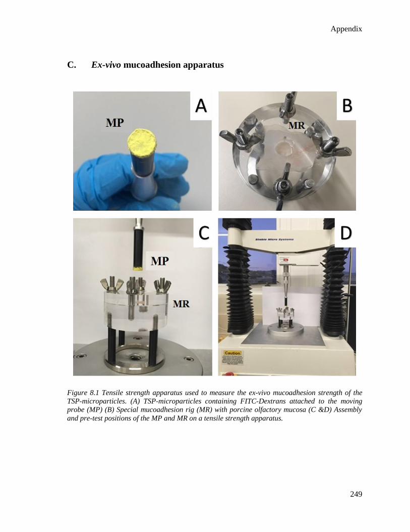
Appendix
249
C. Ex-vivo mucoadhesion apparatus
Figure 8.1 Tensile strength apparatus used to measure the ex-vivo mucoadhesion strength of the
TSP-microparticles. (A) TSP-microparticles containing FITC-Dextrans attached to the moving
probe (MP) (B) Special mucoadhesion rig (MR) with porcine olfactory mucosa (C &D) Assembly
and pre-test positions of the MP and MR on a tensile strength apparatus.

Appendix
250
D. Microparticle insufflation studies
Average weight of microparticles being expelled from the insufflator device was
established using a series of experiments (n = 5) where a constant volume of air (10mL) was
sucked into the syringe and mass of the expelled microparticles is determined using an
electronic balance.
Table 22. Microparticle expulsion from insufflator device. Weight of empty insufflator (W1), weight
of insufflator loaded with microparticles (W2) and weight of insufflator after expulsion of
microparticles (W3) were used to calculate the weight of loaded microparticles and expelled
microparticles.
W1*
(g)
W2*
(g)
W3*
(g)
Loaded
microparticles
[(W2-W1)*1000]
(mg)
Expelled
microparticles
[(W3-W1)*1000]
(mg)
Average ±
SD (mg)
19.4342 19.4451 19.4392 10.9000 5.9000
5.8360 ± 0.3
19.4342 19.4454 19.4397 11.2000 5.7000
19.4342 19.4460 19.4404 11.8000 5.6000
19.4342 19.4455 19.4400 11.3000 5.5000
19.4342 19.4466 19.4401 12.4000 6.4800
* Weights include mass of 10 mL of sucked air.
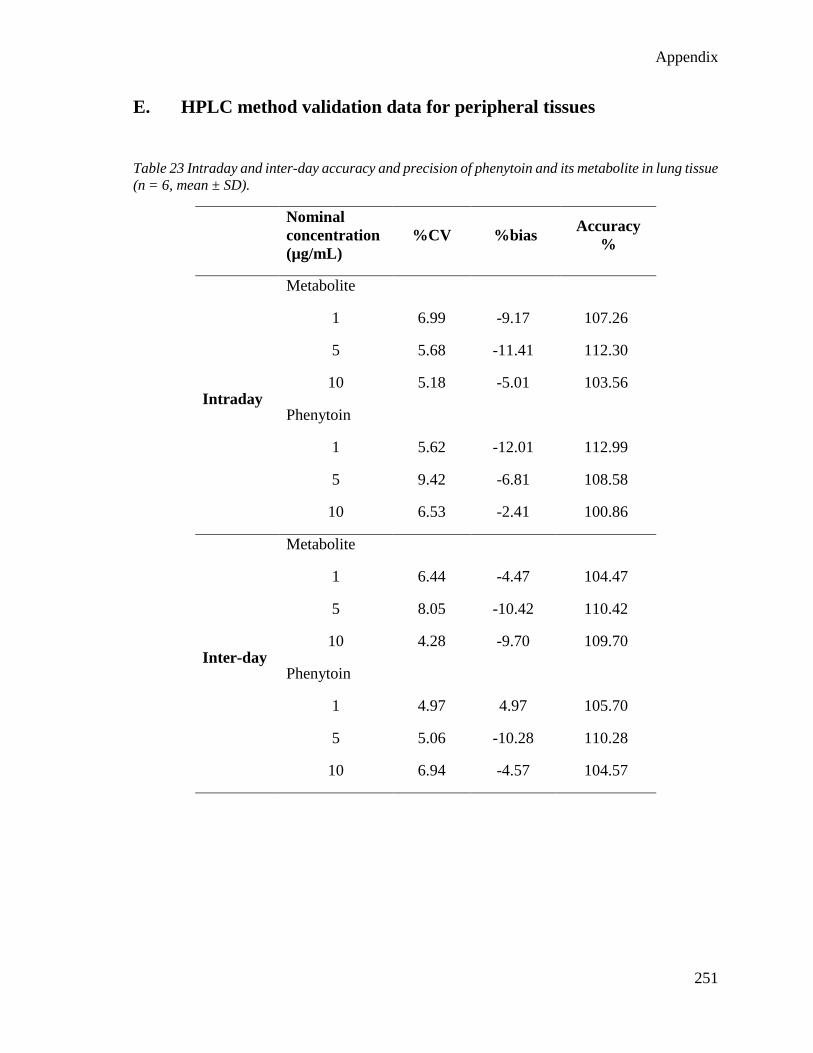
Appendix
251
E. HPLC method validation data for peripheral tissues
Table 23 Intraday and inter-day accuracy and precision of phenytoin and its metabolite in lung tissue
(n = 6, mean ± SD).
Nominal
concentration
(µg/mL)
%CV %bias Accuracy
%
Intraday
Metabolite
1 6.99 -9.17 107.26
5 5.68 -11.41 112.30
10 5.18 -5.01 103.56
Phenytoin
1 5.62 -12.01 112.99
5 9.42 -6.81 108.58
10 6.53 -2.41 100.86
Inter-day
Metabolite
1 6.44 -4.47 104.47
5 8.05 -10.42 110.42
10 4.28 -9.70 109.70
Phenytoin
1 4.97 4.97 105.70
5 5.06 -10.28 110.28
10 6.94 -4.57 104.57

Appendix
252
Table 24 Intraday and inter-day accuracy and precision of phenytoin and its metabolite in liver tissue
(n = 6, mean ± SD).
Nominal
concentration
(µg/mL)
%CV %bias Accuracy
%
Intraday
Metabolite
1 6.02 -9.25 109.25
5 6.48 -0.78 100.78
10 2.98 4.21 95.79
Phenytoin
1 3.79 -13.34 113.34
5 4.45 -2.66 102.66
10 2.15 5.03 94.97
Inter-day
Metabolite
1 6.05 -4.57 104.57
5 6.63 -7.55 107.55
10 3.32 -4.81 104.81
Phenytoin
1 3.48 3.48 104.58
5 5.70 -2.97 102.97
10 3.87 -3.87 103.87

Appendix
253
Table 25 Intraday and inter-day accuracy and precision of phenytoin and its metabolite in kidney
tissue (n = 6, mean ± SD).
Nominal
concentration
(µg/mL)
%CV %bias Accuracy
%
Intraday
Metabolite
1 1.67 -10.19 110.19
5 6.29 0.93 99.07
10 7.49 5.61 94.39
Phenytoin
1 4.49 5.03 94.97
5 8.92 -4.82 104.82
10 7.42 12.65 87.35
Inter-day
Metabolite
1 4.54 2.86 97.14
5 8.25 -2.92 102.92
10 8.07 -0.58 99.79
Phenytoin
1 4.94 4.94 98.99
5 6.35 3.77 96.23
10 7.40 8.59 90.93
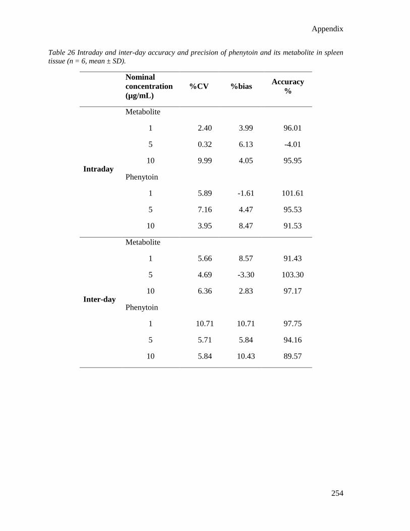
Appendix
254
Table 26 Intraday and inter-day accuracy and precision of phenytoin and its metabolite in spleen
tissue (n = 6, mean ± SD).
Nominal
concentration
(µg/mL)
%CV %bias Accuracy
%
Intraday
Metabolite
1 2.40 3.99 96.01
5 0.32 6.13 -4.01
10 9.99 4.05 95.95
Phenytoin
1 5.89 -1.61 101.61
5 7.16 4.47 95.53
10 3.95 8.47 91.53
Inter-day
Metabolite
1 5.66 8.57 91.43
5 4.69 -3.30 103.30
10 6.36 2.83 97.17
Phenytoin
1 10.71 10.71 97.75
5 5.71 5.84 94.16
10 5.84 10.43 89.57

Appendix
255
Table 27 Intraday and inter-day accuracy and precision of phenytoin and its metabolite in heart
tissue (n = 6, mean ± SD).
Nominal
concentration
(µg/mL)
%CV %bias Accuracy
%
Intraday
Metabolite
1 4.19 -5.61 105.61
5 1.93 1.63 98.37
10 5.82 8.11 91.89
Phenytoin
1 7.30 -13.60 113.60
5 1.74 6.08 93.92
10 3.78 -0.92 100.92
Inter-day
Metabolite
1 9.81 6.23 93.77
5 4.03 0.54 99.46
10 4.56 3.57 96.01
Phenytoin
1 5.30 5.30 107.94
5 3.15 5.07 94.93
10 5.43 7.09 92.99
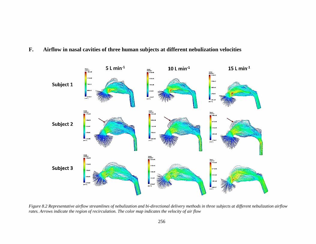
256
F. Airflow in nasal cavities of three human subjects at different nebulization velocities
Figure 8.2 Representative airflow streamlines of nebulization and bi-directional delivery methods in three subjects at different nebulization airflow
rates. Arrows indicate the region of recirculation. The color map indicates the velocity of air flow