A snake toxin as a theranostic agent for the type 2 vasopressin ...
Upload
independentCategory
view
6download
0
http://informahealthcare.com/drdISSN: 1071-7544 (print), 1521-0464 (electronic)
Drug Deliv, Early Online: 1–13! 2014 Informa Healthcare USA, Inc. DOI: 10.3109/10717544.2014.923068
RESEARCH ARTICLE
Formulation development of a novel targeted theranostic nanoemulsionof docetaxel to overcome multidrug resistance in ovarian cancer
Srinivas Ganta1, Amit Singh2, Yashesh Rawal3, Joseph Cacaccio1,3, Niravkumar R. Patel1, Praveen Kulkarni4,5,Craig F. Ferris4,5, Mansoor M. Amiji2,5, and Timothy P. Coleman1,3,5,6
1Nemucore Medical Innovations, Inc., Worcester, MA, USA, 2Department of Pharmaceutical Sciences, School of Pharmacy, Northeastern University,
Boston, MA, USA, 3Blue Ocean Biomanufacturing, Inc., Worcester, MA, USA, 4Center for Translational Imaging, Northeastern University, Boston, MA,
USA, 5Center for Translational Cancer Nanomedicine Northeastern University, Boston, MA, USA, and 6Foundation for the Advancement of
Personalized Medicine Manufacturing, Phoenix, AZ, USA
Abstract
Objective: Ovarian cancer is a highly lethal disease in which the majority of patients eventuallydemonstrate multidrug resistance. Develop a novel active targeted theranostic nanomedicinedesigned to overcome drug efflux mechanisms, using a Generally Regarded As Safe (GRAS)grade nanoemulsion (NE) as a clinically relevant platform.Materials and methods: The NEs surface-functionalized with folate and gadolinium, were madeusing GRAS grade excipients and a high-shear microfluidization process. Efficacy was evaluatedin ovarian cancer cells, SKOV3 and SKOV3TR. The NE accumulation in tumors was evaluated inSKOV3 tumor-bearing mice by magnetic resonance imaging (MRI).Results and discussion: The NE with particle size5150 nm were stable in plasma and parenteralfluids for 24 h. Ovarian cancer cells in vitro efficiently took up the non-targeted and folate-targeted NEs; improved cytotoxicity was observed for the folate-targeted NEs showing a 270-fold drop in the IC50 in SKOV3TR cells as compared to docetaxel alone. The addition ofgadolinium did not affect cell viability in vitro, but showed relaxation times comparable toMagnevist�. Folate-targeted NEs accumulated in tumors for prolonged period of timecompared to Magnevist� and showed enhanced contrast compared to non-targeted NEs withMRI in SKOV3 tumor-bearing mice suggesting active targeting of NEs due to folatemodification.Conclusions: A folate-targeted, theranostic NE delivers docetaxel by receptor mediatedendocytosis that shows enhanced cytotoxicity capable of overcoming ABC transportermediated taxane resistance. The diagnostic capability of the targeted nanomedicine showedenhanced contrast in tumors compared to clinically relevant MRI contrast agent Magnevist� .
Keywords
Docetaxel, folate, gadolinium, MRI
History
Received 18 April 2014Revised 7 May 2014Accepted 7 May 2014
Introduction
Taxane chemotherapies are important primary (e.g. pacli-
taxel) and secondary (e.g. docetaxel) line of treatment for
ovarian cancer patients. Even with these powerful che-
motherapies eventually all patients become resistant to
treatment, clinically defined as multidrug resistance (MDR).
Factors leading to MDR can include enzymatic inactivation,
ATP binding cassette (ABC) transporters (i.e. P-glycoprotein
(P-gp) and breast cancer resistant protein (BRCP-1)) respon-
sible for drug efflux mechanisms, or dysregulation of DNA
repair mechanisms. Drug efflux inhibitors like PSC 833,
GF120918, VX-710, LY335979, OC144093 and XR9576
have been clinically investigated to mitigate drug efflux
mechanisms but have shown limited benefits and none have
yet been approved by the U.S. Food and Drug Administration
(FDA) for clinical use (Seiden et al., 2002; Pusztai et al.,
2005; Ruff et al., 2009). These compounds were designed as
inhibitors of ABC transporters to physically block drug
efflux. Unlike these pharmacological means the development
of nanomedicines can integrate design features which take
advantage of simultaneous delivery of drug efflux counter
measures while transporting a clinically relevant chemother-
apeutic payload. Instead of pharmacologically blocking ABC
transporters (Kelly et al., 2011), a folate-targeted nanoemul-
sion (NE) was designed to utilize receptor-mediated endo-
cytosis to deliver docetaxel (DTX). Receptor-mediated
endocytosis has been proposed as a means to bypass ABC
transporter mediated drug efflux (Goren et al., 2000). Folate
was chosen as the ligand because folate receptor (FR) is
poorly expressed or absent on most normal tissues (Leamon,
2008). However, it is over-expressed in many cancers and is
highly relevant in epithelial ovarian cancer as �80% express
FR (Toffoli et al., 1997; Sudimack & Lee, 2000; Li et al.,
2009; Spannuth et al., 2010).
Address for correspondence: Timothy P. Coleman, PhD, CEO &President, Nemucore Medical Innovations, Inc., Worcester, MA 01608,USA. Email: [email protected]
Dru
g D
eliv
ery
Dow
nloa
ded
from
info
rmah
ealth
care
.com
by
71.9
.158
.242
on
06/0
5/14
For
pers
onal
use
onl
y.
In this study, we have investigated the development of a
folate-targeted theranostic NE, annotated with the clinically
relevant MRI contrast agent Gd and carrying DTX for the
enhancement of cytotoxicity in SKOV3 and SKOV3TR, taxane
sensitive and resistant respectively. NEs have proven to be a
versatile platform for the development of a variety of drug
delivery vehicles (Ganta et al., 2014a,b). For heavily treated
patients, in the later stages of a diagnosis, time to ascertain if a
therapy is working is of up most importance. Therefore, drug
distribution is an important parameter to understand, so the
NE designed were optimized to carry the MRI imaging agent
Gd to act as a surrogate marker for drug distribution. In
addition to formulation optimization, we examined intracellu-
lar delivery, ability to change apoptotic potential, reversal of
taxane resistance, and capacity to be an MRI agent.
Materials and methods
Materials
DTX was obtained from LC Laboratories (Woburn, MA).
L-a-phosphatidylethanolamine transphosphatidylated (PE)
and C6-NBD-ceramide were purchased from Avanti Polar
Lipids (Alabaster, AL). Egg phosphotidylcholine (Lipoid�
E80) was received from Lipoid GmbH (Ludwigshaffen,
Germany). 1,2- distearoyl-sn-glycero-3-phosphoethanola-
mine-N-[methoxy-(polyethyleneglycol)-2000] (DSPE-
PEG2000) and 1,2-distearoyl-sn-glycero- 3-phosphoethanola-
mine-N-[maleimide(polyethylene glycol)-2000] (ammonium
salt) (DSPE-PEG2000-MAL) was purchased from Laysan
Bio Inc., (Arab, AL) Poly unsaturated fatty acid (PUFA)
rich flaxseed oil was obtained from Puritan’s Pride Inc.
(Oakdale, NY). Cysteine, folic acid (FA), diethylenetriami-
nepentaacetic dianhydride (DTPA), 2,7-bis(o-arsenopheny-
lazo)-1,8-dihydroxynaphthalene-3,6-disulfonic acid (arsenazo
III), 3-[4,5-dimethyl thiazolyl]-2,5-diphenyl tetrazolium
bromide (MTT reagent), gadolinium (III) chloride (GdCl3)
hexahydrate, pyridine, ninhydrine, and N,N0-dicyclohexylca-
bodiimide (DCC) were obtained from Sigma Chemicals (St.
Louis, MO). Apo-ONE� Homogeneous Caspase-3/7 assay
was obtained from Promega (Madison, WI). Slowfade� Gold
Antifade mounting media supplemented with DAPI, lyso-
tracker red, Novex� 4-12% Tri-glycine mini gels and PVDF
membrane were obtained from Life Technologies (Grand
Island, NY). RIPA cell lysis buffer, Halt� protease inhibitor
cocktail, Pierce� Lane Marker Sample Buffers, Running
buffer (10X), NuPAGE� transfer buffer and SuperSignal
West Pico Chemiluminescent Substrate were obtained from
Fisher Scientific (Pittsburgh, PA). Anti-P-gp mouse 1 � anti-
body (ab80594), anti b-actin mouse 1 � antibody (ab8224), and
HRP-linked 2 � antibody (Goat anti mouse IgG, ab97023) were
obtained from Abcam (Cambridge, MA). All other chemicals
and solvents used were of the highest available grade.
SKOV3 human ovarian carcinoma cells were purchased
from ATCC (Manassas, VA). Multi-drug resistant SKOV3TR
cells were generously provided by Dr. Duan Zhenfeng (Mass
General Hospital, Boston, MA).
Synthesis of folate-PEG-DSPE
DSPE-PEG2000-MAL (200 mg) was added to cysteine (Cys)
(16.5 mg) dissolved in 1 M HEPES buffer (pH 7.4) at 1:2
molar ratio and mixed under nitrogen environment at 4 �C for
overnight to facilitate to conjugation. The DSPE-PEG2000-Cys
conjugate was purified by dialysis against water using dialysis
bag with molecular weight cut-off of 3500 Da (Spectrapore,
Spectrum Laboratories, CA) and lyophilized. DSPE-
PEG2000-Cys (120 mg) was dissolved in 12 ml dry DMSO
and 13 mg of FA was added to it. A 7 ml of pyridine and
16 mg of DCC was added subsequently and the mixture
stirred for 4 h at room temperature to allow amide coupling of
FA to the amine group of Cys. The final product was dialyzed
in water for 24 h, lyophilized and analyzed by NMR to
confirm conjugation.
Synthesis of Gd-DTPA-PE
Gd-DTPA-PE was synthesized as reported earlier with
modifications (Levchenko et al., 2002). L-a-phosphatidy-
lethanolamine transphosphatidylated (PE) (100 mg) dissolved
in chloroform (4 ml) was treated with triethylamine (30ml).
This solution was added drop-wise to DTPA (400 mg)
dissolved in DMSO (20 ml), and the mixture was stirred for
3 h under nitrogen atmosphere at room temperature.
Chloroform was removed by blowing nitrogen, and reaction
was incubated overnight at RT. The resulting DTPA-PE
conjugate was purified by dialysis (6-8000 Da, Spectrapore,
Spectrum Laboratories, CA) against 5% DMSO in water at
room temperature for 24 h followed by further 24 h of
dialysis in water. The purified sample was lyophilized and
the purity of DTPA-PE complex was determined by thin
layer chromatography (TLC) using a mobile phase of
chloroform, methanol and water at 65:25:4 (v/v) ratio, and
ninhydrin as visualizing reagent. In the next step, Gd was
chelated to DTPA by adding 18.5 mg of GdCl3 to DTPA-PE
complex in 20 ml of DMSO and the reaction mixture stirred
for 1 h. The resulting Gd-DTPA-PE conjugate was purified
by dialysis against water at room temperature for 24 h.
The purified sample was lyophilized and stored at �20 �Cuntil use.
Preparation of the NE formulations
Oil-in-water NE formulation encapsulating DTX was pre-
pared by high shear homogenization method using LV1
Microfluidizer (Microfluidics Corporation, Newton, MA).
DTX (10 mg) dissolved in chloroform was added to flaxseed
oil (1 g) and chloroform evaporated using nitrogen gas,
resulting in formation of oil phase. The aqueous phase of the
formulation was prepared by dissolving egg lecithin
(120 mg) and DSPE-PEG2000 (15 mg) in 4 ml of glycerol
(2.21% w/v)-water. To this initial solution, Gd-DTPA-PE
(100 mg) and folate conjugate (8 mg) were added and stirred
for 2 h to achieve complete dissolution of all components.
After this, aqueous and oil phases were heated (60 �C,
2 min), mixed and homogenized at 25 000 psi for 10 cycles
to obtain the NE formulation. Non-targeted formulation
(DTX-NE NT) without a folate conjugate and corresponding
blank NEs without the DTX were prepared in a similar
manner. Additionally, non-targeted and folate targeted NEs
for fluorescent microscopy were prepared in a similar
manner by replacing DTX with a fluorescent labeled
ceramide, C6-NBD-ceramide.
2 S. Ganta et al. Drug Deliv, Early Online: 1–13
Dru
g D
eliv
ery
Dow
nloa
ded
from
info
rmah
ealth
care
.com
by
71.9
.158
.242
on
06/0
5/14
For
pers
onal
use
onl
y.
Formulation characterization
Particle size and zeta potential analysis
Size distribution and zeta potential were measured by diluting
10 ml of the different NE formulations in 10 ml of DI water
and subjected to analysis using Zetas Sizer ZS (Malvern,
Worcestershire, UK). The average droplet size was measured
for each nanoemulsion formulation by dynamic light scatter-
ing at room temperature and a 90 � fixed angle. The
hydrodynamic radius and poly-dispersity index (PDI) of the
droplets were determined for a count rate between 100–
500 kcps. For zeta potential measurements, diluted sample
was placed in electrophoretic cell and charge was recorded.
Transmission electron microscopy (TEM)
Samples for TEM analysis were prepared by dispersing 2 ml of
the NE in 10 ml of DI water. From these diluted samples,
10 ll was drop-coated on the Formvar-coated copper grids
(Electron Microscopy Science, Hatfield, PA) and allowed to
stand for 1 min following which, excess was drained using
Whatman filter paper. The NE were then stained with 10 lL
of 1% (w/v) uranyl acetate for 1 min, excess stain was
removed using Whatman filter paper. The grids were allowed
to air-dry before performing observation under a JEOL 100X
transmission electron microscope (Peabody, MA).
Analysis of DTX loading and encapsulation
A high-performance liquid chromatography (HPLC) assay
was performed to measure DTX loading and encapsulation
efficiency of the NEs. A Waters LC system (model 2487,
Waters Corporation, Milford) comprising of a quaternary
pump, an autosampler, and UV-detector was used for the
analysis. The LC system was interfaced with Empower 3
software for remote controlling, data acquisition and process-
ing. The mobile phase consisted of acetonitrile:water
(50:50 v/v) and was pumped to a reverse phase C18 column
(4.6 mm� 15 mm, 5 mm, Hypersil Gold, US) with a C18 pre-
column (4 mm� 20 mm, 5 mm, Phenomenex, US) at a flow
rate of 1 ml/min. Samples at 10 ml were injected and
monitored for DTX elution at 230 nm.
Both non-targeted and folate-targeted NEs were assayed for
DTX loading prior to all studies. A standard curve of DTX was
generated with a linear range of detection between 0.1–50 mg/
ml. To determine loading efficiency, a 10 ml formulation was
diluted in 1 ml of acetonitrile, vortexed and centrifuged at
10 000 rpm for 15 min. The supernatant was subjected to HPLC
analysis to determine concentration of DTX.
DTX encapsulation efficiency in the NE was determined by
an ultracentrifugation method using centrifugal filter devices
(MWCO 3,000 Da; centricorn, Millipore, MA). one ml of the
nanoemulsion was placed in the upper donor chamber,
centrifuged at 10 000 rpm for 30 min to separate the aqueous
phase in to the recovery chamber and the sample containing the
drug was collected from the donor chamber. The DTX
concentration in the recovery and donor chambers was
measured by HPLC and% encapsulation was calculated.
Gadolinium assay
Arsenazo III was employed to determine free Gd content
during Gd-DTPA-PE synthesis as described previously (8,9).
Samples assayed for free Gd were prepared by mixing of 50 ml
sample with 100 ml of 0.2 mM arsenazo III solution and
diluted to 1 ml with water. Then 200 ml aliquots of these
samples were added to 96-well plate and read at 652 nm
on Synergy HT plate reader (BioTex). A standard curve of
Gd was prepared using GdCl3 with the linearity range of 2–
50 mg/ml and used for calculating Gd amount in the samples.
T1 relaxivities
Gd-annotated NE samples were filled in tubes and ran
through a phantom Brucker 500 MHz MRI machine (Bruker
Biospec 20/70, Bruker Biospin MRI, Inc) in a 4.7 Tesla
magnetic field, giving MRI scans showing NE generated
contrast as well as T1 time measurements. Magnevist�, a
clinically used Gd-DTPA chelate also tested for T1 relaxation
time at same Gd concentration as Gd-annotated NEs.
DTX release from NEs
The DTX release assays were carried out by loading NE
formulations equivalent to 4 mg of DTX into dialysis bag
(3500 Da) and placing it in 100 ml release media (0.5% Tween
80/ PBS, pH 7.4) maintained at 37 �C and stirring rate of
400 rpm. Sample aliquots (1 ml) were collected at different
time intervals up to 72 h and replaced with fresh media. The
docetaxel concentration in the samples was analyzed using
HPLC as described above. At the end of study, unreleased
drug remained in the dialysis bag was measured and
compared with the release data. The cumulative amount of
DTX released and release kinetics were calculated to assess
the release profile.
Stability of formulations in plasma and intravenous infusion
solutions
NEs were diluted in dog plasma and electrolyte solutions, and
monitored for particle size over 24 h at 37 �C. The change in
particle size was used as indicator of stability upon dilution.
For this, NEs were diluted 90% with fresh dog plasma,
sodium chloride (0.9%), dextrose (5%) and phosphate buf-
fered saline (PBS pH 7.4) and incubated at 37 �C. Aliquots of
10 ml of samples were taken for analysis at 1, 2, 4, 6, 8 and
24 h, diluted 1000-fold with distilled water and analyzed
particle size using Zetasizer ZS as described above.
In vitro studies
Cell culture conditions
SKOV3 and SKOV3TR cells were cultured in RPMI 1640
media supplemented with 10% fetal bovine serum and 1%
Penicillin/streptomycin and maintained in a humidified 95%
O2/5% CO2 atmosphere at 37 �C. Cells were grown until 60-
70% confluent in the flasks and trypsinized with a solution
0.25% Trypsin with 2.25mM EDTA in HBSS. Trypan blue
exclusion method was employed to determine the viable cells.
Cellular uptake
SKOV3 cells were seeded in 6-well plate over cover slip at
density of 100 000 cells/well and incubated for 24 h before
treatment. Cells were washed with media and incubated with
either non-targeted or folate-targeted C6-NBD-ceramide
DOI: 10.3109/10717544.2014.923068 Folate targeted nanoemulsion to overcome MDR 3
Dru
g D
eliv
ery
Dow
nloa
ded
from
info
rmah
ealth
care
.com
by
71.9
.158
.242
on
06/0
5/14
For
pers
onal
use
onl
y.
labeled NEs for 5, and 15 minutes, followed by washing with
PBS. Cells were incubated with lysotracker red according to
manufacturer’s protocol for 20 minutes followed by treatment
with 4% formaldehyde for 30 minand mounted on to a
glass slide with a Slowfade� Gold Antifade mounting
media supplemented with DAPI. Glass slides were rested
for 30 minutes on flat surface in dark before imaging
localization of fluorescence signal using Zeiss confocal
microscope (LSM-700).
P-gp expression by western blot analysis
SKOV3 and SKOV3TR cells were grown in T75 flask until
80% confluent. Cells were mechanically scraped and
centrifuged to get cell pellet. Cells were lysed in 200 ml of
radioimmunoprecipitation assay (RIPA) cell lysis buffer with
Halt� protease inhibitor cocktail for 30 minutes on ice. Cell
lysate was centrifuged at 13,000 rpm for 15 minutes and
supernatant was analyzed using bicinchoninic acid (BCA)
assay for protein concentration. Loading buffer with reducing
agent was added to cell lysate containing 20 mg of protein and
final volume of 25 ml was achieved by diluting using PBS.
These samples were heated at 85 �C for 5 minutes and loaded
in gel locked in XCell SureLock� system with running buffer
in place as described by manufacturer. Samples were
electrophoresed on a 4–12% SDS-PAGE. Proteins were
transferred on to PVDF membrane using transfer assembly
in XCell SureLock� system. Transfer sandwich was prepared
by placing from anode sponge, sponge, filter paper, gel,
PVDF, filter paper, sponge and sponge to cathode. PVDF
membrane was then incubated in blocking buffer in 1X tris
buffered saline with Tween 20 (TBST) (1% BSA) for 1 h
followed by incubation with 1 mg/ml of primary antibody
(mouse anti P-gp primary antibody, AB80594) overnight at
4 �C. Membrane was washed three times with TBST for 5
minwith rocking followed by incubation with secondary
antibody (HRP-linked Goat anti mouse IgG, AB97023) for 1 h
in 1%BSA in TBST at room temperature. Membrane was
washed again three times as described before followed by
incubation with 10 ml of chemiluminescence substrate
(SuperSignal West Pico Kit, Pittsburgh, PA) for 5 min.
Substrate was discarded and membrane was placed in plastic
bag before detecting luminescence using KODAK 2000R
(Carestream, Rochester, NY) imaging system using cooled
CCD camera. b-actin was used as loading control for each
sample.
Cytotoxicity assay
SKOV3 and SKOV3TR cells were seeded in 96-well plate
at 3000 cells per well. After 24 h seeding, cells were treated
with concentrations of DTX over eight orders of magnitude
ranging from 0.001 nM to 100 000 nM. For this, DTX solution
in DMSO, non-targeted and folate-targeted NEs were diluted
in RPMI media and added to cells. After the 72 h treatment,
cells were washed with media and incubated with MTT
reagent (50 mg/well) for 3 h. RPMI growth media was used as
a negative control (0% cell death) and poly (ethyleneimine), a
cationic cytotoxic polymer at a concentration of 250 mg/ml
(molecular weight 10 kDa) was used as a positive control
(100% cell death). Vehicle controls without any drug were
also employed at corresponding volumes. Dose-response
curves and IC50 values were obtained by fitting data
Sigmoidal-dose response curves using Graphpad Prism 5.
Caspase-3/7 activity
SKOV3 and SKOV3TR cells were seeded in 96-well plate at
2000 cells/well and incubated for 24 h before treatment. Cells
were then treated with either DTX solution in DMSO, non-
targeted and folate-targeted NEs at 1 nM DTX for SKOV3
cells and 100 nM DTX for SKOV3TR cells for 1, 2, 4, and 8 h.
Media with caspase reagent without cells and untreated cells
were kept as control. Additionally, vehicle controls were
employed to account for any caspase activity. After desired
incubation, cells were washed with media followed by
incubation with 50 ml of caspase 3/7 activity assay reagent
mixed with 50 ml of complete media according to manufac-
turer’s protocol. Cells were incubated for 2 h and fluorescence
was measured at 499 nm excitation and 521 nm emission. Fold
change in caspase 3/7 activation by DTX when delivered
using DMSO, non-targeted or folate-targeted NEs was
calculated after subtracting background fluorescence
(media + assay reagent without cells) from all reading.
In vivo studies
T1 weighted MRI imaging
SKOV3, approximately 1� 106 cells suspended in PBS:
Matrigel (1:1), were injected subcutaneously in female nu/nu
mice. Tumors were allowed to grow until 250 mm3. Mice
were pretreated with 400 mg of Pemetrexed 1 h prior to
imaging agent administration. Three mice per cohort were
then injected with either magnevist, NT-unloaded NEs, or
folate-targeted unloaded NEs at final concentration of Gd at
0.072 mmoles/kg. The mice were anesthetized with isoflurate
during scans and were kept in MRI between scans. T1
weighted images were acquired using a phantom Brucker
500 MHz MRI machine (Bruker Biospec 20/70, Bruker
Biospin MRI, Inc, Billerica, MA) in a 4.7 Tesla magnetic
field up to 24 h.
Data analysis
Data are reported as the average and standard deviation.
Comparisons between the groups were made using student’s
t-test and with more than two groups, the ANOVA test was
used. Values of p50.05 were considered statistically
significant.
Results
Characterization of the conjugates.
NMR spectroscopy was used to characterize all the individual
components and the final purified folate-PEG-DSPE conju-
gate to confirm the final product (Figure 1). The proton
signature from pure DSPE-PEG-Mal and folic acid
(Figures 1A and B, respectively) could be successfully
assigned in the NMR spectra obtained and these spectra
from standards were used to confirm successful formation of
the folic acid conjugate. The NMR spectrum of folate-PEG-
DSPE shows the signature peaks of PEG protons between
4 S. Ganta et al. Drug Deliv, Early Online: 1–13
Dru
g D
eliv
ery
Dow
nloa
ded
from
info
rmah
ealth
care
.com
by
71.9
.158
.242
on
06/0
5/14
For
pers
onal
use
onl
y.
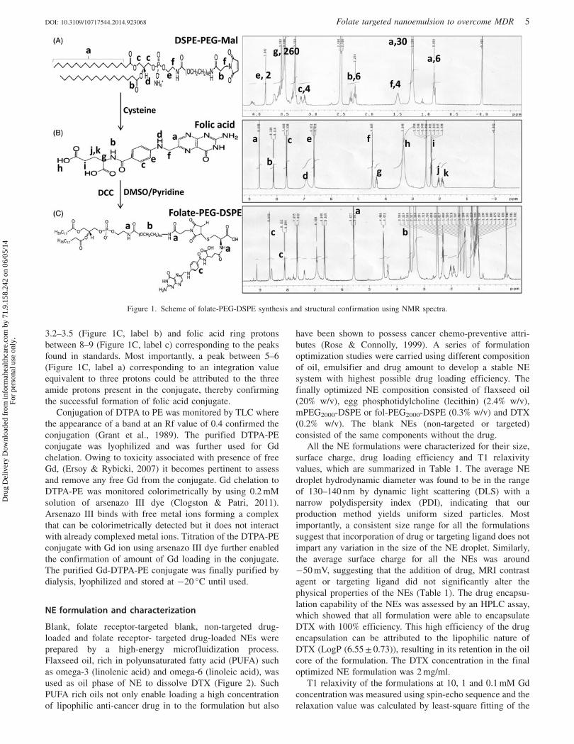
3.2–3.5 (Figure 1C, label b) and folic acid ring protons
between 8–9 (Figure 1C, label c) corresponding to the peaks
found in standards. Most importantly, a peak between 5–6
(Figure 1C, label a) corresponding to an integration value
equivalent to three protons could be attributed to the three
amide protons present in the conjugate, thereby confirming
the successful formation of folic acid conjugate.
Conjugation of DTPA to PE was monitored by TLC where
the appearance of a band at an Rf value of 0.4 confirmed the
conjugation (Grant et al., 1989). The purified DTPA-PE
conjugate was lyophilized and was further used for Gd
chelation. Owing to toxicity associated with presence of free
Gd, (Ersoy & Rybicki, 2007) it becomes pertinent to assess
and remove any free Gd from the conjugate. Gd chelation to
DTPA-PE was monitored colorimetrically by using 0.2 mM
solution of arsenazo III dye (Clogston & Patri, 2011).
Arsenazo III binds with free metal ions forming a complex
that can be colorimetrically detected but it does not interact
with already complexed metal ions. Titration of the DTPA-PE
conjugate with Gd ion using arsenazo III dye further enabled
the confirmation of amount of Gd loading in the conjugate.
The purified Gd-DTPA-PE conjugate was finally purified by
dialysis, lyophilized and stored at �20 �C until used.
NE formulation and characterization
Blank, folate receptor-targeted blank, non-targeted drug-
loaded and folate receptor- targeted drug-loaded NEs were
prepared by a high-energy microfluidization process.
Flaxseed oil, rich in polyunsaturated fatty acid (PUFA) such
as omega-3 (linolenic acid) and omega-6 (linoleic acid), was
used as oil phase of NE to dissolve DTX (Figure 2). Such
PUFA rich oils not only enable loading a high concentration
of lipophilic anti-cancer drug in to the formulation but also
have been shown to possess cancer chemo-preventive attri-
butes (Rose & Connolly, 1999). A series of formulation
optimization studies were carried using different composition
of oil, emulsifier and drug amount to develop a stable NE
system with highest possible drug loading efficiency. The
finally optimized NE composition consisted of flaxseed oil
(20% w/v), egg phosphotidylcholine (lecithin) (2.4% w/v),
mPEG2000-DSPE or fol-PEG2000-DSPE (0.3% w/v) and DTX
(0.2% w/v). The blank NEs (non-targeted or targeted)
consisted of the same components without the drug.
All the NE formulations were characterized for their size,
surface charge, drug loading efficiency and T1 relaxivity
values, which are summarized in Table 1. The average NE
droplet hydrodynamic diameter was found to be in the range
of 130–140 nm by dynamic light scattering (DLS) with a
narrow polydispersity index (PDI), indicating that our
production method yields uniform sized particles. Most
importantly, a consistent size range for all the formulations
suggest that incorporation of drug or targeting ligand does not
impart any variation in the size of the NE droplet. Similarly,
the average surface charge for all the NEs was around
�50 mV, suggesting that the addition of drug, MRI contrast
agent or targeting ligand did not significantly alter the
physical properties of the NEs (Table 1). The drug encapsu-
lation capability of the NEs was assessed by an HPLC assay,
which showed that all formulation were able to encapsulate
DTX with 100% efficiency. This high efficiency of the drug
encapsulation can be attributed to the lipophilic nature of
DTX (LogP (6.55 ± 0.73)), resulting in its retention in the oil
core of the formulation. The DTX concentration in the final
optimized NE formulation was 2 mg/ml.
T1 relaxivity of the formulations at 10, 1 and 0.1 mM Gd
concentration was measured using spin-echo sequence and the
relaxation value was calculated by least-square fitting of the
Figure 1. Scheme of folate-PEG-DSPE synthesis and structural confirmation using NMR spectra.
DOI: 10.3109/10717544.2014.923068 Folate targeted nanoemulsion to overcome MDR 5
Dru
g D
eliv
ery
Dow
nloa
ded
from
info
rmah
ealth
care
.com
by
71.9
.158
.242
on
06/0
5/14
For
pers
onal
use
onl
y.

exponentially varying signal intensity as a function of
recovery time. As shown in Table 1, the T1 relaxation time
of the non-targeted nanoemulsion was found to be
35.51 ± 13.5 and folate-targeted nanoemulsion was found to
be 79.46 ± 2.63, and was comparable to that of Magnevist�
(22 ± 0.27 ms) at similar Gd concentrations. The physico-
chemical characterizations of all the formulations indicate
that we could successfully prepare NE of identical size,
charge, drug loading efficiency and T1 relaxivity with little
inter-formulation variation.
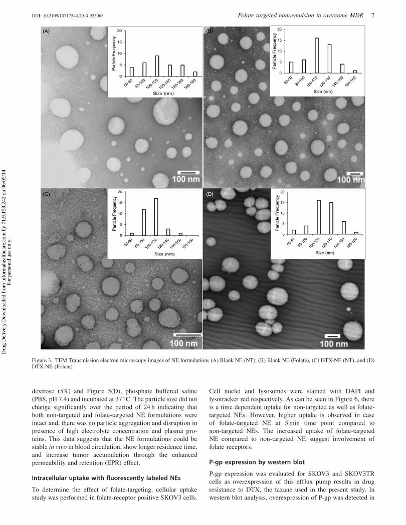
Size and morphology of the blank, folate-targeted NE,
non-targeted NE with DTX, and folate-targeted NE with
DTX were analyzed by TEM show a spherical morphology
with droplet size in the range of 100–120 nm, which is
consistent with the size range obtained by DLS measure-
ment (Figure 3). It is important to note that a decrease in
the average particle size calculated by TEM compared to
that obtained by DLS measurement is due to the fact that
DLS analysis gives the hydrodynamic size of the oil
droplets while TEM images show the actual size. Inset in
each image in Figure 3 shows a particle size distribution
plot calculated from a minimum of 30 NE droplets for the
respective sample.
In vitro drug release analysis
The release profile of DTX from non-targeted and folate-
targeted NE is shown in Figure 4. Both formulations release
approximately 10% DTX at the end of 72 h.The DTX release
data obtained were fitted into various kinetic models to
describe the release kinetics as shown in Table 2.
Predominant DTX release mechanism for non-targeted and
folate-targeted NE formulations was zero order kinetics
(Lachman et al., 1986).
Stability assessment of the NEs
Upon IV injection or mixing in parental infusion solutions, the
physical stability of NE formulation can be affected ranging
from particle aggregation and disruption. Aggregation leads to
rapid in vivo clearance whereas particle disruption causes
burst release kinetics of payload (dose dumping). To test the
NE formulations for stability, samples were diluted in plasma
and parental infusion solutions and monitored for particle size
over 24 h at 37 �C. The change in particle size was analyzed by
DLS and used as indicator of stability upon dilution. For this,
NE formulations were diluted Figure 5(A), 90% with fresh dog
plasma, Figure 5(B), sodium chloride (0.9%), Figure 5(C),
Figure 2. Schematic showing encapsulation of DTX into lipid core of NE and its surface is modified with Gd and folate for MRI functionality andtargeting, respectively.
Table 1. Characterization of formulations.
Hydrodynamic diameter of formulationsZeta DTX T1 values
Formulations Size (nm) PDI potential (mV) encapsulation (%) (msec)
Blank NE 142 ± 7 0.1 �50 ± 10 –Blank NE (folate-targeted) 123 ± 22 0.1 �52 ± 10 –DTX-NE (non-targeted) 130 ± 3 0.1 �50 ± 12 100 34.51 ± 13.5DTX-NE (folate targeted) 135 ± 6 0.06 �54 ± 10 100 79.46 ± 2.63
Magnevist had T1 values of 22.2 ± 0.27. The values are shown as Avg ± SD, n¼ 3.
6 S. Ganta et al. Drug Deliv, Early Online: 1–13
Dru
g D
eliv
ery
Dow
nloa
ded
from
info
rmah
ealth
care
.com
by
71.9
.158
.242
on
06/0
5/14
For
pers
onal
use
onl
y.
dextrose (5%) and Figure 5(D), phosphate buffered saline
(PBS, pH 7.4) and incubated at 37 �C. The particle size did not
change significantly over the period of 24 h indicating that
both non-targeted and folate-targeted NE formulations were
intact and, there was no particle aggregation and disruption in
presence of high electrolyte concentration and plasma pro-
teins. This data suggests that the NE formulations could be
stable in vivo in blood circulation, show longer residence time,
and increase tumor accumulation through the enhanced
permeability and retention (EPR) effect.
Intracellular uptake with fluorescently labeled NEs
To determine the effect of folate-targeting, cellular uptake
study was performed in folate-receptor positive SKOV3 cells.
Cell nuclei and lysosomes were stained with DAPI and
lysotracker red respectively. As can be seen in Figure 6, there
is a time dependent uptake for non-targeted as well as folate-
targeted NEs. However, higher uptake is observed in case
of folate-targeted NE at 5 min time point compared to
non-targeted NEs. The increased uptake of folate-targeted
NE compared to non-targeted NE suggest involvement of
folate receptors.
P-gp expression by western blot
P-gp expression was evaluated for SKOV3 and SKOV3TR
cells as overexpression of this efflux pump results in drug
resistance to DTX, the taxane used in the present study. In
western blot analysis, overexpression of P-gp was detected in
Figure 3. TEM Transmission electron microscopy images of NE formulations (A) Blank NE (NT), (B) Blank NE (Folate), (C) DTX-NE (NT), and (D)DTX-NE (Folate).
DOI: 10.3109/10717544.2014.923068 Folate targeted nanoemulsion to overcome MDR 7
Dru
g D
eliv
ery
Dow
nloa
ded
from
info
rmah
ealth
care
.com
by
71.9
.158
.242
on
06/0
5/14
For
pers
onal
use
onl
y.

SKOV3TR, taxane resistant variant at 170 000 dalton whereas
no P-gp expression was observed for SKOV3, taxanes
sensitive cell line (Figure 7). b-actin was used as loading
control and was observed for all three cell lines. These results
demonstrate that the cell lines used in this study are ideal to
investigate reversal of the multidrug resistance.
Cytotoxicity studies in sensitive and resistant ovariancancer cells
In order to determine if the NEs produce a cytotoxic effect,
the following experiments were done on SKOV3 and
SKOV3TR cells. The data was obtained using a tetrazolium
(MTT) assay, which measures the activity of cellular enzymes
that reduces the MTT dye to its insoluble formazan.
SKOV3TR cells express the ABC transporter, P-gp, which
produces DTX efflux out of the cell and is associated with
multidrug resistant cancer cells. The concentration required to
inhibit growth of cells by 50% (IC50) was calculated for each
formulation and is shown in Table 3. As shown in Table 3,
the IC50 of SKOV3 cells decreased 3.33-fold when DTX is
encapsulated in folate-targeted NE formulation compared
to DTX in solution. In taxane resistant SKOV3TR cells a 270-
fold decrease in IC50 was observed when they were treated
folate-targeted NE formulations when compared to DTX in
solution. This significant improvement in cytotoxicity indi-
cates that there is merit to the hypothesis that folate receptor
mediated endocytosis is capable of bypassing multidrug
resistant mechanisms present in SKOV3TR. Nanoemulsion
formulation containing no DTX did not affect cell viability.
Quantitative and qualitative apoptotic studies
In order to confirm that the anti-cancer activity of the
nanoemulsion formulation is due to induction and
Figure 5. Physical stability of DTX containing non-targeted and folate targeted NE formulations upon 90% dilution in dog plasma, parenteral infusionfluids (5% dextrose and 0.9% sodium chloride) and phosphate buffered saline. The data are shown as mean ± SD (n¼ 3).
0.00 12 24 36 48 60 72
2.0
4.0
6.0
8.0
10.0
12.0
14.0
16.0
18.0
20.0
% R
elea
sed
Time (h)
Non-targeted Nanoemulsion
Folate Targeted Nanoemulsion
Figure 4. DTX release from the non-targeted and folate targeted NEformulations in PBS (pH 7.4) containing 0.5% Tween 80 at 37 �C. Thedata are shown as mean ± SD (n¼ 3).
Table 2. Drug release kinetics.
Zero
order
First
order
Higuchi
equation
Formulation R2 K0 R2 K1 R2 K2
Non-targeted NE 0.9929 0.341765 0.9128 0.035466 0.9439 2.805284
Folate-targeted NE 0.9716 0.301923 0.9053 0.036157 0.8862 2.427362
8 S. Ganta et al. Drug Deliv, Early Online: 1–13
Dru
g D
eliv
ery
Dow
nloa
ded
from
info
rmah
ealth
care
.com
by
71.9
.158
.242
on
06/0
5/14
For
pers
onal
use
onl
y.

upregulation of apoptotic pathways, the quantitative and
qualitative apoptotic analysis was performed in SKOV3 cells.
Figure 8 shows the caspase 3/7 activities in cells incubated
with docetaxel in DMSO, non-targeted and folate targeted
formulation as a function of time of incubation and the
apoptotic activity was calculated relative to that obtained
from untreated control cells. Figure 8(A) shows the apoptotic
activity of the SKOV3 cells treated with DTX in different
formulations for 1, 2 4 and 8 h. No significant change in
apoptotic activity was observed post-incubation in the sam-
ples after 1 and 2 h but the apoptotic activity of the cells
incubated with targeted NEs increased after 4 and 8 h
suggesting that higher accumulation of the drug via receptor
mediated uptake results in induction of apoptosis resulting in
increased cell death compared to DTX in DMSO and non-
targeted NE. This trend correlates well with in vitro
cytotoxicity experiments where the targeted NE shows
maximum cell-killing capability owing to an increased
apoptosis induction.
The caspase 3/7 activities were similarly monitored in
SKOV3TR cells treated with DTX in DMSO, non-targeted
NE and folate-targeted NE for 2, 4 and 8 h (Figure 8B). Cells
treated with DTX in DMSO, non-targeted nanoemulsion, and
folate targeted nanoemulsion formulations for 2 h show
essentially similar enzymatic activity with no significant
difference. However, 4 h post-treatment with DTX containing
formulations, cells start to show an enhanced caspase 3/7
activity with folate-targeted NE showing much higher
increase (8-fold compared to untreated control) compared to
non-targeted NE (6-fold) and taxotere (3.5-fold). The enzym-
atic activity for all the treatment groups decreased at 8 h
incubation period with folate targeted nanoemulsion-treated
cells still showing a significantly higher activity. This
suggests that the enzymatic activity peaks around 4 h post-
incubation in formulations. The significantly increased
enzymatic activity in folate-targeted NE treated cells corrob-
orate well with the cytotoxicity data where folate-targeted NE
shows very-high cell-killing efficiency, which could be
attributed to capability to induce apoptosis in the cells. It is
also key to note that the levels of enzymatic activity is
significantly higher in all the SKOV3TR cells (Figure 8B)
compared to SKOV3 cells (Figure 8A) suggesting that even
though these cells show taxane resistance, they are extremely
prone to the drug if their drug-detoxification strategy is
compensated by designing an efficient delivery vehicle.
Magnetic resonance imaging study
Gadolinium, an MRI contrast agent, alters relaxation times
of water molecules in tissues where Gd is concentrated after
oral or intravenous administration. Theranostic application of
Figure 6. Fluorescent microscopy imagesshowing uptake of NBD-ceramide (green)containing non-targeted and folate targetedNEs in SKOV3 cells. Lyso Tracker (red) andDAPI (blue) were used to stain lysosomes andnucleus respectively and to monitor theco-localization of NE in SKOV3 cells.
Non-t
arg
ete
d
15 min
Fola
teT
arg
ete
d
5 min
Figure 7. P-glycoprotein expression in SKOV3, SKOV3TR cells usingwestern blot analysis. Lane 1: SKOV3 cells (20 mg protein lysate), Lane2: SKOV3TR cells (20 mg protein lysate). b-actin was used as a loadingcontrol.
DOI: 10.3109/10717544.2014.923068 Folate targeted nanoemulsion to overcome MDR 9
Dru
g D
eliv
ery
Dow
nloa
ded
from
info
rmah
ealth
care
.com
by
71.9
.158
.242
on
06/0
5/14
For
pers
onal
use
onl
y.

non-targeted and folate-targeted NEs were evaluated by
injecting 0.072 mmoles/kg of Gd and evaluating tumor
accumulation at various time points. Mice were pretreated
with pemetrexed to reduce off-target kidney accumulation
(Muller et al., 2008). As can be seen in Figure 9(A),
folate-targeted NEs showed enhanced contrast compared
to non-targeted NEs suggesting active targeting of NEs
due to folate modification. Using the tumor region of interest,
the relative T1 signal generated by Gd was used to
determine uptake. Folate-targeted NEs and non-targeted
NEs showed tumor tissue accumulation over the period of
24 h Figure 9(B). However, clinically used magnevist
accumulated in tumor tissues very rapidly and cleared
within 5 h Figure 9(B). These results strongly support
potential of folate-targeted NEs as a theranostic drug delivery
system. Use of such a strategy will provide numerous
advantages such as localization of the tumor tissues, deter-
mination of drug distribution as well as potential quantifica-
tion of disease progression.
Discussion
With the clinical advancement and FDA approval of targeted
therapies, antibody therapies, antibody-drug conjugate thera-
pies and small-molecule drug conjugates like Endocytes
folate-targeted therapies/imaging agents (e.g. vintifolide,
EC145), targeting the molecular differences of cancer is no
longer fantasy. Targeted nanomedicine is the next evolution of
targeted therapies as they can be molecularly ‘‘designed’’ to
carry multiple drugs, target disease relevant receptors, and
carry diagnostic capabilities suitable of tracking accumulation
in diseased tissues. DTX is used to treat ovarian cancer
patients as a second line of therapy if patients show resistance
to Taxol� or Abraxane� (Paclitaxel formulated in human
serum albumin). Often recurrent ovarian cancer patients have
been so heavily treated previously that their cancer might be
non-responsive to DTX as a second line treatment due to
mutations that affects its ability to stabilize tubulin polymer-
ization or over expression of ABC transporters. High
expression of one ABC transporter, MDR-1, has been
correlated as an independent prognosticator for poor survival
for ovarian and renal cancer (Penson et al., 2004; Mignogna
et al., 2006). However it is generally believed that either
resistance mechanism has the potential to minimize DTX
clinical effectiveness, thus a theranostic approach might
provide relevant chemical value.
Even with potential for resistance, DTX is a compelling
molecule for ovarian cancer patients, in vitro DTX is a
stronger promoter of tubulin polymerization than paclitaxel
partially because of its greater affinity to b-Tublin showing
1.2–2.6 times more cytotoxicity than paclitaxel. However this
enhancement in cytotoxicity may also be due to the
interaction of docetaxel with BCL-2. BCL-2 is a multidomain
antiapoptotic protein that sequesters BAX and BAK proteins,
two pro-apoptotic proteins that play a major role in
mitochondrial outer membrane permeabilization, a pivotal
event in the intrinsic apoptosis pathway (Thanos et al., 2003)
DTX and paclitaxel both induce phosphorylation of BCL-2
leading to the release of BAX and BAK, however DTX does
so at a concentration 100-fold less than paclitaxel (Sadurni
et al., 2005). Clinically there is incomplete cross-resistance,
specifically with paclitaxel-resistant breast, lung and ovarian
tumors potentially due to different mechanisms of uptake,
efflux, or stimulation of apoptosis. Therefore if DTX could be
delivered in nanomedicine specifically designed to mitigate
reduced uptake, enhanced efflux and apoptosis escape
mechanisms associated with multidrug resistance heavily
treated recurrent ovarian cancer patients could potentially be
treated more effectively than current standard of care second
line treatment protocols.
Disease specific targeting is important to deliver higher
concentrations of chemotherapy to the tumor and should have
0.0
0.1
0.2
0.3
0.4
0.5
0.6
0.7
0.8
0.9
1.0
2 4 8
Caspase3/7
activity in S
KO
V3
cells
(fo
ld incre
ase fro
m c
ontr
ol)
Caspase 3
/7 a
ctivity in S
KO
V3T
R
cells
(fo
ld incre
ase fro
m c
ontr
ol)
Duration of treatment (h)
DTX Sol
DTX-NE (NT)
DTX-NE (Folate)
DTX-NE (Folate)
(A)
(B)
0.0
1.0
2.0
3.0
4.0
5.0
6.0
7.0
8.0
9.0
10.0
2 4 8
Duration of treatment (h)
DTX Sol
DTX-NE (NT)
Figure 8. Caspase 3/7 activity assay in (A) SKOV3 and (B) SKOVTRcells treated with DTX in non-targeted NE (DTX-NE (NT)) and folatetargeted NE (DTX-NE (Folate)), relative to DTX solution (DTX Sol).The data are shown as mean ± SD (n¼ 2).
Table 3. IC50 values.
Formulation SKOV3 Fold improvement SKOV3TR Fold improvement
DTX solution 1.0 ± 1.3 nM 1.00 27 000 ± 4050 nM 1.00DTX non-targeted NE 0.5 ± 1.2 nM 2.00 6800 ± 1400 nM 3.97DTX folate-targeted NE 0.3 ± 1.2 nM 3.33 100 ± 12 nM 270
10 S. Ganta et al. Drug Deliv, Early Online: 1–13
Dru
g D
eliv
ery
Dow
nloa
ded
from
info
rmah
ealth
care
.com
by
71.9
.158
.242
on
06/0
5/14
For
pers
onal
use
onl
y.

the added benefit of limiting side effects by minimizing
systemic distribution. Folate receptor alpha (FR-a) is one of
three folate receptor isoforms:, a,b & g. FR- a is the most
widely studied isoform and is a 38 kD glycosyl-phosphatidy-
linositol-anchored glycoprotein that binds folic acid with a
Kd51 nM and is highly expressed in a number of human
tumors including ovarian (480%), lung (475%), breast
cancer(460%) renal cell(465%) and brain, head & neck
(Fisher et al., 2008). In normal tissue expression is much
lower and limited to kidney tubuli, lung epithelium in the
apical cell, the choroid plexus, and placenta (Parker et al.,
2005). FR-a over expression is negatively associated with
overall survival in ovarian and other cancers; however with
480% of ovarian tumors expressing FR-a it is difficult to
power a study appropriately to correlate expression with
mortality. As a predictor of response rate to chemotherapy,
either complete or partial remission, patients with FR-agreater then median level had a 15-fold higher likelihood of
negative response (Toffoli et al., 1997). In the light of
correlation with poor clinical outcome we investigated
whether targeting FR-a with folate-targeted NE could
enhance in vitro potency of DTX in taxane sensitive and
resistant ovarian cancer cells. FR-a targeting was achieved
through the use of a lipidated-version of a folate, a config-
uration that has demonstrated efficient binding and preferen-
tial internalization by folate receptor expressing tumor cells
in vitro, and tumor xenografts in vivo (Goren et al., 2000,
Leamon et al., 2003).
In the current study, non-targeted and folate-targeted,
DTX-loaded NEs show improved cytotoxicity in both taxane-
sensitive and taxane-resistant cells. The folate-targeted DTX
NE showed a 270-fold increase in cytotoxicity in taxane-
resistant cells as compared to DTX alone, a finding that
suggests the amount of folate-targeted DTX NE could be
reduced significantly and be more effective than current
clinically available taxane therapies. Although uptake mech-
anisms of these NE formulations have yet to be studied in
detail, in general NEs fuse with cellular membrane and most
likely undergo non-specific transport via phagocytosis. This
passive mechanism improved the potency of DTX when
compared to DMSO solubilized DTX in vitro. However
greater potency was observed when the NE was
0%
10%
20%
30%
40%
50%
60%
9630 12 15 18 21 24
% C
hang
e T1
sig
nal i
n RO
I
Hours
Change in ROI T1-Signal in SKOV3 Tumor vsTime
Folate Targeted NE Non-Targeted NE Magnevist
(A)
(B)
Figure 9. (A)T1 weighted images and (B) quantitative analysis of % tumor signal versus time of mice bearing subcutaneous SKOV3 tumor xenograftpost i.v. injection of magnevist, non-targeted NEs and folate-targeted NEs with pemetrexed treatment at final concentration of Gd at 0.072 mmoles/kg,the data are shown as mean ± SD (n¼ 3).
DOI: 10.3109/10717544.2014.923068 Folate targeted nanoemulsion to overcome MDR 11
Dru
g D
eliv
ery
Dow
nloa
ded
from
info
rmah
ealth
care
.com
by
71.9
.158
.242
on
06/0
5/14
For
pers
onal
use
onl
y.
functionalized with folate to facilitate specific uptake via
folate receptor-mediated endocytosis. Both these mechan-
isms, i.e. passive lipid membrane fusion or receptor-mediated
endocytosis, mitigated taxane-resistance associated with
taxane efflux out of the cell. Delivery via endocytosis appears
to protect DTX from ABC transporter mediated drug efflux
mechanisms and enhance apoptosis. The remarkable increase
in potency of DTX in folate-targeted NEs should be a benefit
to ovarian cancer therapy and also potentially limit toxic side
effects. For example, less DTX could be used to overcome
taxane resistance for patients that have shown resistance
to Taxol� or Abraxane�.
Cancer stem cells (CSCs) have recently been shown to
preferentially proliferate when treated with platinum and
taxane therapies and are implicated in recurrent ovarian
cancer which is multidrug resistant (Joo et al., 2013). Our
investigation appears to be especially important for recurrent
ovarian cancers which might be derived from CSCs which
express ABC transporters. Our results indicate that using the
folate receptor to deliver DTX could mitigate drug efflux and
mitigated some apoptotic escape mechanisms CSCs initiate
to survive second line therapies. Perhaps this is through
improved phosphorylation of BCL-2 and the release of the
proapoptotic proteins BAX and BAK (Sadurni et al., 2005).
If this is potentially the case then using a folate-targeted DTX
nanoemulsions as a second line therapy for folate positive
recurrent ovarian cancer could improve outcomes and poten-
tially reduce burden of using a toxic drug that provides little
efficacy. Additionally, folate-targeted DTX-NEs could be
dosed less frequently while still achieving efficacy and
reducing the long-term chemotherapeutic burden to the
body. Another reason to encapsulate DTX in a folate-targeted
NE is that this nanomedicine design might limit systemic
toxicities associated with current DTX formulations particu-
larly bone marrow suppression, neutropenia, renal and
hypersensitivity. Our studies showed that DTX could be
sequestered at high concentrations in the lipid core of a stable
folate-targeted NE with a size between 130–150 nm in
diameter. This size range is still capable of taking advantage
of the EPR effect and active targeting but avoids clearance
by the kidney which clears nanomedicines with a diameter
55 nm.
Normally clinical diagnosis of therapeutic efficacy takes a
significant amount of time to ascertain, but for patients
suffering with recurrent ovarian cancer time is critically
important. A delivery vehicle that can inform the physician
of drug uptake by a tumor in a relatively short time period
post administration and in a non-invasive manner might be a
diagnostic attribute that is advantageous for improving care.
The NE created for this study was designed as a theranostic
capable of simultaneously imaging and targeting drug deliv-
ery to folate receptor-positive ovarian tumors. Our NEs were
functionalized with Gd-DTPA-PE chelate to track particle
uptake by MRI detection (Ganta et al., 2014a). This design
with the Gd-chelate residing on the outer surface of the NE
provides a suitable environment for Gd longitudinal relaxivity
and generates contrast for suitable for clinical MRI. Thus,
potentially providing the physician with a visualization
method of therapy uptake and potentially improved monitor-
ing of disease progression in recurrent ovarian cancer
patients. The NE formulations had comparable magnetic
relaxation times in vitro comparable to clinically relevant
Magnevist�. Moreover, folate-targeted NEs showed enhanced
contrast in tumors in vivo compared to non-targeted NEs or
Magnevist�. Such a theranostic NE allows visualization of the
DTX pharmacodynamics with the potential to monitor tumor
progression. If the MRI contrast enhancement in the tumor is
not consistent with enhanced T1 weighted images consistent
with Gd-accumulation, clinicians will have the opportunity
to adjust the dosing regimens or change therapies if the
theranostic NE appears not to be efficacious.
Overall, this study demonstrates that the theranostic
properties of a folate-targeted DTX nanomedicine should be
advantageous for the treatment of folate receptor-positive
ovarian cancer. As most ovarian cancers will eventually
become multidrug resistant we show that targeted delivery of
DTX using the folate receptor is capable of overcoming
taxane-resistance. The diagnostic potential of the NE is
designed for direct monitoring of nanomedicine uptake and
has potential for monitoring disease progression. While
further preclinical efficacy, imaging and toxicology investi-
gations are required to confirm and translate this theranostic
nanomedicine, the potential of its medical utility based on the
initial results is promising for folate receptor positive,
multidrug-resistant ovarian cancer.
Conclusions
We developed a novel theranostic that delivers DTX by
receptor mediated endocytosis that shows enhanced cytotox-
icity capable of overcoming ABC transporter mediated taxane
resistance. The diagnostic capability of the targeted nanome-
dicine shows enhanced contrast for prolonged period of time
compared to clinically relevant MRI contrast agent
Magnevist� in SKOV3 tumor-bearing mice. Our studies
indicate that this novel nanomedicine demonstrates signifi-
cant potential to treat MDR cancers that eventually develop in
most ovarian cancer patients. This potentially clinically
relevant nanomedicine for MDR cancer was designed to
carry DTX in the lipid core of the NE composed of GRAS
grade excipients suitable for parenteral administration. DTX
encapsulated in a non-targeted NE was more potent than DTX
dissolved in DMSO in in vitro cytotoxicity assays. However,
encapsulation of DTX in a folate-targeted NE enhanced the
potency in SKOV3 ovarian cancer cells but significantly
increased the potency in taxane resistant SKOV3TR ovarian
cancer cells.
The multifunctionality of this novel folate-targeted, Gd
annotated, DTX loaded NE formulation demonstrate that
clinically relevant capabilities to track drug distribution,
evade multidrug resistant mechanisms and potentially miti-
gate free drug induced systemic toxicity can be designed into
nanomedicines. With regards to multidrug resistance mech-
anisms, e.g. reduced drug uptake, enhanced drug efflux, and
escape from apoptosis, the design of this nanomedicine
mitigates each of them as shown, by increased caspase 3/7
activity and cytotoxicity. The results indicate the potential for
a new class of DTX therapies to treat not only DTX MDR
ovarian cancer, but other cancers where taxane therapy is
standard of care.
12 S. Ganta et al. Drug Deliv, Early Online: 1–13
Dru
g D
eliv
ery
Dow
nloa
ded
from
info
rmah
ealth
care
.com
by
71.9
.158
.242
on
06/0
5/14
For
pers
onal
use
onl
y.

Acknowledgements
We would like to thank Mary Elizabeth Schwartz, Susan
Riley Keyes, Allison Morse, Rupa Sawant, Philip Heisler, and
Keri Forbringer for assistance with the manuscripts
preparation.
Declaration of interest
Research reported in this publication was supported by the
National Cancer Institute of the National Institutes of Health
under Awards Number R01CA158881 and Number U54
CA151881. The content is solely the responsibility of the
authors and does not necessarily represent the official views
of the National Institutes of Health. The authors report no
conflicts of interest.
References
Clogston JD, Patri AK. (2011). Detecting and measuring free gadoliniumin nanoparticles for MRI imaging. Methods Mol Biol 697:101–8.
Ersoy H, Rybicki FJ. (2007). Biochemical safety profiles of gadolinium-based extracellular contrast agents and nephrogenic systemic fibrosis.Journal of Magnetic Resonance Imaging: JMRI 26:1190–7.
Fisher RE, Siegel BA, Edell SL, et al. (2008). Exploratory study of99mTc-EC20 imaging for identifying patients with folate receptor-positive solid tumors. J Nuclear Med 49:899–906.
Ganta S, Singh A, Patel NR, et al. (2014a). Development of EGFR-targeted nanoemulsion for imaging and novel platinum therapy ofovarian cancer. Pharm Res.
Ganta S, Talekar M, Singh A, et al. (2014b). Nanoemulsions intranslational research-opportunities and challenges in targeted cancertherapy. AAPS PharmSci Tech.
Goren D, Horowitz AT, Tzemach D, et al. (2000). Nuclear delivery ofdoxorubicin via folate-targeted liposomes with bypass of multidrug-resistance efflux pump. Clin Cancer Res 6:1949–57.
Grant CW, Karlik S, Florio E. (1989). A liposomal MRI contrast agent:phosphatidylethanolamine-DTPA. Magn Reson Med 11:236–43.
Joo WD, Visintin I, Mor G. (2013). Targeted cancer therapy – are thedays of systemic chemotherapy numbered? Maturitas 76:308–14.
Kelly RJ, Draper D, Chen CC, et al. (2011). A pharmacodynamic studyof docetaxel in combination with the P-glycoprotein antagonisttariquidar (XR9576) in patients with lung, ovarian, and cervicalcancer. Clin Cancer Res 17:569–80.
Lachman L, Liebermann HA, Kanig JL. (1986). The Theory and practiceof industrial pharmacy, Philadelphia: Lea & Febiger.
Leamon CP. (2008). Folate-targeted drug strategies for the treatment ofcancer. Curr Opin Investig Drugs 9:1277–86.
Leamon CP, Cooper SR, Hardee GE. (2003). Folate-liposome-mediatedantisense oligodeoxynucleotide targeting to cancer cells: evaluation invitro and in vivo. Bioconjug Chem 14:738–47.
Levchenko TS, Rammohan R, Lukyanov AN, et al. (2002). Liposomeclearance in mice: the effect of a separate and combined presence ofsurface charge and polymer coating. Int J Pharm 240:95–102.
Li J, Sausville EA, Klein PJ, et al. (2009). Clinical pharmacokinetics andexposure-toxicity relationship of a folate-Vinca alkaloid conjugateEC145 in cancer patients. J Clin Pharmacol 49:1467–76.
Mignogna C, Staibano S, Altieri V, et al. (2006). Prognostic significanceof multidrug-resistance protein (MDR-1) in renal clear cell carcin-omas: a five year follow-up analysis. BMC Cancer 6:293.
Muller C, Schibli R, Krenning EP, De Jong M. (2008). Pemetrexedimproves tumor selectivity of 111In-DTPA-folate in mice with folatereceptor-positive ovarian cancer. J Nucl Med 49:623–9.
Parker N, Turk MJ, Westrick E, et al. (2005). Folate receptor expressionin carcinomas and normal tissues determined by a quantitativeradioligand binding assay. Anal Biochem 338:284–93.
Penson RT, Oliva E, Skates SJ, et al. (2004). Expression of multidrugresistance-1 protein inversely correlates with paclitaxel response andsurvival in ovarian cancer patients: a study in serial samples. GynecolOncol 93:98–106.
Pusztai L, Wagner P, Ibrahim N, et al. (2005). Phase II study oftariquidar, a selective P-glycoprotein inhibitor, in patients withchemotherapy-resistant, advanced breast carcinoma. Cancer 104:682–91.
Rose DP, Connolly JM. (1999). Omega-3 fatty acids as cancerchemopreventive agents. Pharmacol Ther 83:217–44.
Ruff P, Vorobiof DA, Jordaan JP, et al. (2009). A randomized,placebo-controlled, double-blind phase 2 study of docetaxelcompared to docetaxel plus zosuquidar (LY335979) in womenwith metastatic or locally recurrent breast cancer who have receivedone prior chemotherapy regimen. Cancer Chemother Pharmacol 64:763–8.
Sadurni N, Solans C, Azemar N, et al. (2005). Studies on theformation of O/W nano-emulsions, by low-energy emulsificationmethods, suitable for pharmaceutical applications. Eur J Pharm Sci26:438–45.
Seiden MV, Swenerton KD, Matulonis U, et al. (2002). A phase II studyof the MDR inhibitor biricodar (INCEL, VX-710) and paclitaxel inwomen with advanced ovarian cancer refractory to paclitaxel therapy.Gynecol Oncol 86:302–10.
Spannuth WA, Sood AK, Coleman RL. (2010). Farletuzumab inepithelial ovarian carcinoma. Expert Opin Biol Ther 10:431–7.
Sudimack J, Lee RJ. (2000). Targeted drug delivery via the folatereceptor. Adv Drug Deliv Rev 41:147–62.
Thanos CG, Liu Z, Goddard M, et al. (2003). Enhancing the oralbioavailability of the poorly soluble drug dicumarol with abioadhesive polymer. J Pharm Sci 92:1677–89.
Toffoli G, Cernigoi C, Russo A, et al. (1997). Overexpression of folatebinding protein in ovarian cancers. Int J Cancer 74:193–8.
DOI: 10.3109/10717544.2014.923068 Folate targeted nanoemulsion to overcome MDR 13
Dru
g D
eliv
ery
Dow
nloa
ded
from
info
rmah
ealth
care
.com
by
71.9
.158
.242
on
06/0
5/14
For
pers
onal
use
onl
y.
Copyright © 2022 FDOKUMEN