Development and applications of photo-triggered theranostic agents
32
This article appeared in a journal published by Elsevier. The attached copy is furnished to the author for internal non-commercial research and education use, including for instruction at the authors institution and sharing with colleagues. Other uses, including reproduction and distribution, or selling or licensing copies, or posting to personal, institutional or third party websites are prohibited. In most cases authors are permitted to post their version of the article (e.g. in Word or Tex form) to their personal website or institutional repository. Authors requiring further information regarding Elsevier’s archiving and manuscript policies are encouraged to visit: http://www.elsevier.com/copyright
-
Upload
independent -
Category
Documents
-
view
9 -
download
0
Transcript of Development and applications of photo-triggered theranostic agents
This article appeared in a journal published by Elsevier. The attachedcopy is furnished to the author for internal non-commercial researchand education use, including for instruction at the authors institution
and sharing with colleagues.
Other uses, including reproduction and distribution, or selling orlicensing copies, or posting to personal, institutional or third party
websites are prohibited.
In most cases authors are permitted to post their version of thearticle (e.g. in Word or Tex form) to their personal website orinstitutional repository. Authors requiring further information
regarding Elsevier’s archiving and manuscript policies areencouraged to visit:
http://www.elsevier.com/copyright
Author's personal copy
Development and applications of photo-triggered theranostic agents☆
Prakash Rai a, Srivalleesha Mallidi a, Xiang Zheng a, Ramtin Rahmanzadeh a, Youssef Mir a, Stefan Elrington a,Ahmat Khurshid a,c, Tayyaba Hasan a,b,⁎a Wellman Center for Photomedicine, Massachusetts General Hospital, Harvard Medical School, Boston, MA, 02114, United Statesb Harvard Science and Technology, Harvard MIT, Boston MA, 02114, United Statesc Department of Physics and Applied Mathematics, Pakistan Institute of Engineering and Applied Sciences, PO Box 45650, Nilore, Islamabad, Pakistan
a b s t r a c ta r t i c l e i n f o
Article history:Received 23 July 2010Accepted 1 September 2010Available online 19 September 2010
Keywords:Photodynamic therapyNanotechnologyPhotothermal therapyCancerInfectionsImagingDiagnosticsTargetingMultifunctionalDrug delivery
Theranostics, the fusion of therapy and diagnostics for optimizing efficacy and safety of therapeutic regimes, isa growing field that is paving the way towards the goal of personalized medicine for the benefit of patients.The use of light as a remote-activation mechanism for drug delivery has received increased attention due to itsadvantages in highly specific spatial and temporal control of compound release. Photo-triggered theranosticconstructs could facilitate an entirely new category of clinical solutions which permit early recognition of thedisease by enhancing contrast in various imaging modalities followed by the tailored guidance of therapy.Finally, such theranostic agents could aid imaging modalities in monitoring response to therapy. This articlereviews recent developments in the use of light-triggered theranostic agents for simultaneous imaging andphotoactivation of therapeutic agents. Specifically, we discuss recent developments in the use of theranosticagents for photodynamic-, photothermal- or photo-triggered chemotherapy for several diseases.
Published by Elsevier B.V.
Advanced Drug Delivery Reviews 62 (2010) 1094–1124
Abbreviations: 3-(PhS)4-PcAlOH, hydroxyaluminium tetra-3-phenylthiophthalocyanine; Ag, silver; ALA, 5-aminolevulinic acid; AlPcS4, phthalocyanine tetrasulfonate; Au, gold;BDP-MA, benzoporphyrin derivative monoacid ring A; BLM, bleomycin; C11Pc, Zn(II)-phthalocyanine disulphide; C60, fullerene; CdSe, cadmium selenide; CE, contrast-enhanced;CNT, carbon nanotubes; CPT, camptothecin; CR, complete regression; CT, computed tomography; CVD, cardiovascular disease; Dac, daclizumab; DDS, drug delivery system; DMNB,dimethoxy-2-nitrobenzyl; DO3A, 1,4,7,10-tetraazacyclododecane-1,4,7-triacetic acid; DOX, doxorubicin; DPBF, 1,3-diphenylisobenzofuran; DTPA, diethylenetriaminepentaaceticacid; E. coli, Escherichia coli; EGFR, epidermal growth factor receptor; EPR, enhanced permeability and retention; Er3+, erbium; Eu, europium; FDA, food and drug administration;Fe3O4, iron oxide; FITC, (fluorescein 5(6)-isothiocyanate); FR, folate receptor; FRET, fluorescence resonance energy transfer; EtNBS, carboxybutylamino diethylaminobenzophenothiazinium; GLUT, glucose transporter; Gd, gadolinium; GNP, gold nanoparticle; GNT, gold coated carbon nanotube; GNR, gold nanorod; HA, hyaluronic acid; HAuNS, hollowgold nanosphere; HGN, hollow gold nanoshell; HFF-1, human foreskin fibroblasts; HP, hematoporphyrin; HPPH, pyropheophorbide-alpha-hexyl-ether; HSA, human serum albumin;i.c., intracranial; ICG, indocyanine green; i.p., intra peritoneal; IR, infra red; i.v., intra venous; LAMS, light-activated mesostructured silica; LbL, layer-by-layer; L-BPD, liposomalbenzoporphyrin derivative monoacid ring A; LCST, lower critical solution temperature; β-LEAP, β-lactamase enzyme-activated photosensitizer; LDL, low-density lipoprotein; LDLR,LDL receptors; Lip-NP, liposome-nanoparticle assembly; LMB, leuko methylene blue; Ln, lanthanides; MB, methylene blue; Mce6, mesochlorin e6; MDR, multi drug resistance;MFNP, magnetofluorescent nano particle; MRI, magnetic resonance imaging; MRSA, methicillin-resistant Staphylococcus aureus; MSNP, mesoporous silica nanoparticle; MTCNPs,magnetic targeting chitosan NPs; MTCP, meso-tetra(4-carboxyphenyl) porphine; mTHPC, meso-tetra(hydroxyphenyl) chlorine; MTT, 3-(4,5-dimethyltiazol-2-yl)-2,5-diphenyltetrazolium bromide; MWNT, multi wall carbon nanotubes; NaYF4, sodium yttrium fluoride; Nc, naphthalocyanine; NIPAAm-co-AAm, N-isopropylacrylamide-co-acrylamide; NIR, near-infrared; NP, nanoparticle; OCT, optical coherence tomography; ORMOSIL, organically modified silica; PA, photoacoustic; PAH, poly(allylaminehydrochloride); Pan, panitumumab; Pc4, phthalocyanine 4; PCI, photochemical internalization; PDD, photodynamic diagnosis; pDNA, plasmid DNA; PDT, photodynamic therapy;PEG, polyethylene glycol; PEI, poly(ethylene imine); PET, positron emission tomography; Pheo, pheophorbide; PHPP, 2,7,12,18-tetramethyl-3,8-di-(1-propoxyethyl)-13,17-bis-(3-hydroxypropyl) porphyrin; PI, propidium iodide; PIC, photoimmunoconjugate; PICEL, photoimmunoconjugate encapsulating liposome; PLGA, poly-L-co-glycolic-acid; PS,photosensitizer; PSiNPs, phosphonate-terminated silica nanoparticles; PSS, poly(styrene sulfonate); PT, photothermal; PTT, photothermal therapy; PTX, paclitaxel; pz,porphyrazine; QD, quantum dots; rGel, gelonin toxin; RA, rheumatoid arthritis; ROS, reactive oxygen species; SDS, sodium dodecyl sulfate; SDT, sonodynamic therapy; SiNcBOA,silicon naphthalocyanine bisoleate; SiO2, silica; siRNA, small interfering RNA; SLN, solid lipid nanoparticles; S. aureus, Staphylococcus aureus; SWNTS, single wall carbon nanotubes;TEOS, tetraEthOxy silane; Tf-Lip, transferring-conjugated liposomes; THPMP, tri-hydroxyl silyl propyl methyl phosphonate; TPC, 5-(4-carboxyphenyl)-10,15,20-triphenyl-2,3-dihydroxychlorin; TPPS2A, disulfonated meso-tetraphenylporphine; Tra, trastuzumab; UV, ultraviolet; VIS, visible light; Yb3+, ytterbium; ZnPC, zinc phthalocyanine.☆ This review is part of the Advanced Drug Delivery Reviews theme issue on “Development of Theranostic Agents that Co-Deliver Therapeutic and Imaging Agents”.⁎ Corresponding author. Wellman Center for Photomedicine, Massachusetts General Hospital, Harvard Medical School, Boston, MA, 02114, United States. Tel.: +1 617 726 6856;
fax: +1 617 726 8566.E-mail address: [email protected] (T. Hasan).
0169-409X/$ – see front matter. Published by Elsevier B.V.doi:10.1016/j.addr.2010.09.002
Contents lists available at ScienceDirect
Advanced Drug Delivery Reviews
j ourna l homepage: www.e lsev ie r.com/ locate /addr

Author's personal copy
Contents
1. Introduction . . . . . . . . . . . . . . . . . . . . . . . . . . . . . . . . . . . . . . . . . . . . . . . . . . . . . . . . . . . . . . 10952. Photodynamic therapy and imaging for cancer . . . . . . . . . . . . . . . . . . . . . . . . . . . . . . . . . . . . . . . . . . . . . . 1096
2.1. Photosensitizers for imaging and PDT. . . . . . . . . . . . . . . . . . . . . . . . . . . . . . . . . . . . . . . . . . . . . . . 10972.1.1. Optical imaging . . . . . . . . . . . . . . . . . . . . . . . . . . . . . . . . . . . . . . . . . . . . . . . . . . . . . 10972.1.2. Multimodal imaging (MRI and PET) . . . . . . . . . . . . . . . . . . . . . . . . . . . . . . . . . . . . . . . . . . . 1098
2.2. Nanoparticles for imaging and PDT . . . . . . . . . . . . . . . . . . . . . . . . . . . . . . . . . . . . . . . . . . . . . . . . 11002.2.1. Optical imaging . . . . . . . . . . . . . . . . . . . . . . . . . . . . . . . . . . . . . . . . . . . . . . . . . . . . . 11002.2.2. Magnetic resonance imaging (MRI) . . . . . . . . . . . . . . . . . . . . . . . . . . . . . . . . . . . . . . . . . . . 1103
3. Photothermal therapy and imaging for cancer . . . . . . . . . . . . . . . . . . . . . . . . . . . . . . . . . . . . . . . . . . . . . . 11043.1. Optical imaging . . . . . . . . . . . . . . . . . . . . . . . . . . . . . . . . . . . . . . . . . . . . . . . . . . . . . . . . . 11063.2. Ultrasound based imaging . . . . . . . . . . . . . . . . . . . . . . . . . . . . . . . . . . . . . . . . . . . . . . . . . . . . 11063.3. Magnetic resonance imaging (MRI) . . . . . . . . . . . . . . . . . . . . . . . . . . . . . . . . . . . . . . . . . . . . . . . . 11073.4. Ionizing imaging modalities . . . . . . . . . . . . . . . . . . . . . . . . . . . . . . . . . . . . . . . . . . . . . . . . . . . 1108
4. Photo-triggered drug release and imaging for cancer . . . . . . . . . . . . . . . . . . . . . . . . . . . . . . . . . . . . . . . . . . . 11094.1. Photochemically triggered drug release and imaging. . . . . . . . . . . . . . . . . . . . . . . . . . . . . . . . . . . . . . . . 11104.2. Photothermally triggered drug release and imaging . . . . . . . . . . . . . . . . . . . . . . . . . . . . . . . . . . . . . . . . 11124.3. Combined optical imaging and therapy . . . . . . . . . . . . . . . . . . . . . . . . . . . . . . . . . . . . . . . . . . . . . . 1115
5. Photo-triggered theranostic agents for non-cancer pathologies . . . . . . . . . . . . . . . . . . . . . . . . . . . . . . . . . . . . . . 11165.1. Infectious diseases . . . . . . . . . . . . . . . . . . . . . . . . . . . . . . . . . . . . . . . . . . . . . . . . . . . . . . . . 1116
5.1.1. Photodynamic therapy . . . . . . . . . . . . . . . . . . . . . . . . . . . . . . . . . . . . . . . . . . . . . . . . . 11165.1.2. Photothermal therapy . . . . . . . . . . . . . . . . . . . . . . . . . . . . . . . . . . . . . . . . . . . . . . . . . . 1117
5.2. Other diseases. . . . . . . . . . . . . . . . . . . . . . . . . . . . . . . . . . . . . . . . . . . . . . . . . . . . . . . . . . 11186. Future directions and discussion . . . . . . . . . . . . . . . . . . . . . . . . . . . . . . . . . . . . . . . . . . . . . . . . . . . . 11197. Conclusions . . . . . . . . . . . . . . . . . . . . . . . . . . . . . . . . . . . . . . . . . . . . . . . . . . . . . . . . . . . . . . 1120Acknowledgments. . . . . . . . . . . . . . . . . . . . . . . . . . . . . . . . . . . . . . . . . . . . . . . . . . . . . . . . . . . . . . 1120References . . . . . . . . . . . . . . . . . . . . . . . . . . . . . . . . . . . . . . . . . . . . . . . . . . . . . . . . . . . . . . . . . 1120
1. Introduction
Global healthcare costs have been rising steeply over the lastdecade [1]. However there hasn't been a dramatic reduction in diseaserelated deaths to warrant such a drastic rise in costs [2]. During thistime there has been a paradigm shift in disease management andclinicians are gradually moving from the traditional “one drug fits all”approach towards the idea of personalized medicine— ‘the right drugfor the right person administered at the right time’ [3,4]. Althoughsignificant awareness has been created about personalized medicine,its full potential has yet to be tapped [5,6]. The field of theranostics hassprung from the recognition that heterogeneous diseases requiremore personalized solutions [7]. Theranostics refers to the fusion oftherapy and diagnostics, with the purpose of optimizing efficacy andsafety, as well as streamlining the process of drug development and asa field is still in its infancy. The convergences of a number of scientificbreakthroughs have made the development of theranostics possible[8]. In the field of biology, the human genome project and thedevelopment of biomarker initiatives, among others, have enhancedthe understanding of disease progression. Technologies such asgenotyping or gene expression profiling make it possible to transferthis newly acquired biological knowledge into the development ofdiagnostic tests [8]. Theranostics empower physicians with high-medical value testing for science-driven treatment decisions; improvepatient outcomes and patient safety by identifying patients whowon'trespond to a drug or who are likely to experience an adverse event;increase the efficiency of drug development, helping pharmaceuticalcompanies by pinpointing those patients most likely to benefit fromthe new drug; and positively impact health economics, thus helpingphysicians select optimal and cost effective therapy. Although there isbroad agreement that this nascent field has much potential inimproving healthcare, there are a number of challenges that need tobe overcome before it translates into routine use in the clinic [8]. Chiefamong these hurdles is the availability and use of a single platform fordiagnosis and therapy.
A tool that may help in overcoming this hurdle by successfullyintegrating therapeutic and diagnostic agents is nanotechnology [9–12].
Application of nanotechnology tomedical science has been emerging asa new field of interdisciplinary research among medicine, biology,toxicology, pharmacology, chemistry,material science, engineering, andmathematics, and is expected to bring a major breakthrough to addressseveral unsolved medical issues [9–12]. Nanomedicine — the use ofnanotechnology for medicine is starting tomake an impact in areas likedisease imaging and diagnosis, drug delivery and as reporters oftherapeutic efficacy and of disease pathogenesis [9–12]. Many multi-functional nanoparticle (NP) technologies, capable of performing one ormore of the above duties, are now in various stages of preclinical andclinical development [9–12]. Theranostic nanomedicine refers to suchan integrated nano-platform which can diagnose, deliver targetedtherapy and monitor response to therapy. A scheme illustrating thepotential role of theranostic agents at various stages of diseasemanagement is shown in Fig. 1.
The selectivity and specificity for disease destruction can beenhanced by using externally activatable theranostic agents toproduce localized cytotoxicity with little collateral damage. Theability to control drug dosing in terms of quantity, location, andtime is a key goal for drug delivery science, as improved controlmaximizes therapeutic effect while minimizing side effects. Systemsresponsive to a stimulus such as temperature, pH, applied magnetic orelectrical field, ultrasound, light, or enzymatic action have beenproposed as triggered delivery systems [13]. Light-triggered ther-anostics are attracting increasing attention over the past few yearsdue to its advantages in spatial and temporal control of compoundrelease [14]. Recently, light has been used to release therapeuticagents from delivery systems or to activate agents that producecytotoxic species. In fact, among the approved nanoconstructs listedby the food and drug administration (FDA), is a light-activatable agent(Visudyne), widely used for the treatment of age-related maculardegeneration (AMD) which is the major cause of blindness among theelderly in the developed world. Technological advances in fiber-opticfluorescence imaging, a modality which allows investigators to reachinto body cavities via minimally invasive endoscopes, have consider-ably broadened the applications of in vivo optical imaging [15]. Thisarticle reviews recent developments in the use of light-triggered
1095P. Rai et al. / Advanced Drug Delivery Reviews 62 (2010) 1094–1124

Author's personal copy
theranostic agents for simultaneous imaging and photoactivation oftherapeutic agents. The use of lasers and minimally invasive fiber-optic tools, along with the development of new agents that respond toNIR wavelengths for better tissue penetration, make direct targetingof deep tissues possible and thus enabling treatment of severalpathologies.
Cancer is one of the most pressing public health concerns of the21st century. The statistics are daunting; it was projected that 550,000people would die of cancer and that another 1.4 million would bediagnosed with the disease in 2009 in the United States alone [16].Another major cause of death, especially in the developing world, areinfectious diseases which are making a come-back owing to theproblems of drug resistance and lack of sensitive diagnostic tests [10].Infectious diseases, caused by bacteria, viruses, fungi and otherparasites are major causes of death, disability, and social andeconomic disruption for millions of people. Over 9.5 million peopledie each year due to infectious diseases— nearly all live in developingcountries [10]. Despite the existence of safe and effective interven-tions, many people lack access to needed preventive and treatmentcare [10]. Cardiovascular diseases (e.g. atherosclerosis) continue to bethe biggest cause of death in the developed world [9]. Taken together,there is a vital un-met need for agents that can be used forsimultaneous detection, diagnosis and remotely triggered therapyfor selective destruction of diseases tissue.
Photo-triggered theranostic constructs could enable an entirelynew category of clinical solutions, which permit early recognition ofthe disease through the use of contrast agents combinedwith existingimaging modalities (MRI, optical imaging, ultrasound) followed bythe tailored release of the therapeutic agent. Here, we will discussrecent developments in the use of theranostic agents for photody-namic-, photothermal- or photo-triggered chemotherapy for severaldiseases including cancer and infectious diseases. Sections 2–4 of thisreview are focused on the application of light-triggered theranostic
agents for cancer while Section 5 discusses their use for non-cancerpathologies. Generally, this kind of multifunctional agents willprovide information on location of disease; targeted and on demanddrug release that will lead to more effective therapies, eliminating thepotential for both under and overdosing; the need for feweradministrations; optimal use of the drug in question; and increasedpatient compliance.
2. Photodynamic therapy and imaging for cancer
Photodynamic therapy (PDT) is an emerging, externally activa-table, treatment modality for various diseases [17]. PDT can bedefined as the administration of a non-toxic drug or dye known as aphotosensitizer (PS) either systemically, locally, or topically to apatient bearing a lesion, which is frequently, but not always cancer[17]. After a sufficient incubation period with the PS, this lesion isthen selectively illuminated with light of appropriate wavelength,which, in the presence of oxygen, leads to the generation of cytotoxicspecies and consequently to cell death and tissue destruction. PDT isclinically approved for treatment of several diseases including cancerand offers several advantages over conventional chemotherapy byproviding additional selectivity through the spatial confinement oflight used for PS activation [17]. A wide range of PSs have beenevaluated so far and only a few of them have successfully transitionedfrom bench to bedside applications [17]. PS molecules are inherentlyfluorescent and this can be used for imaging and locating disease,photodiagnosis, often referred to, somewhat incorrectly, as photo-dynamic diagnosis (PDD). This approach is becoming of increasinginterest for oncological applications. It is based on a higheraccumulation of PS in tumors compared to normal tissue and isnow being routinely used for diagnosis in bladder cancer [17] andfluorescence-guided resection in surgical procedures [17]. The use ofPDT as a cancer therapy is particularly attractive because of its
Fig. 1. Role of photo-triggered theranostic agents at various stages of disease management. Following administration of a single, integrated theranostic agent, a clinician can diagnosedisease, detect location of disease, deliver light at the disease locations for activating targeted therapy and following treatment, monitor response to therapy. At this stage, bymonitoring the patients response to the therapy, the clinician can decide to either re-initiate treatment or if sufficient regression or cure of disease is observed, call the patient for afollow up visit.
1096 P. Rai et al. / Advanced Drug Delivery Reviews 62 (2010) 1094–1124

Author's personal copy
fundamental specificity and selectivity [17]. This is due to the factthat the PS concentrates specifically within the malignant tissue sowhen the light is directly focused on the lesion, it causes PDT reactiveoxygen species (ROS) to be generated resulting in cellular destructionat the region of interest. For this very reason, in recent years, PDT hasbecome the subject of intense investigation as a possible treatmentmodality for various forms of cancer. Similar to chemotherapy, PDTstill requires agents which exhibit selectivity for the target cells.Similar to radiotherapy, the mode of action with PDT involves the useof electromagnetic radiation in order to generate radical species insitu. However, PDT is a much milder approach for cancer treatmentthan either. The reason for this lies in the combination of the mode ofaction of the PSs employed and their activation in situ by relativelylong wavelength, visible light. Ideal PSs are non-toxic in the absenceof activating light. The targeting of the cancer in PDT has a dualnature: the selectivity of the photosensitizing drugs employed andthe confinement of the activating light to the tumor site alone. Due tothe dual selectivity in PDT the non-tumor tissue largely remainsunaffected [17].
Despite the regulatory approvals and the clinical success of PDT inoncology, a limitation of all existing PSs is the lack of high selectivityfor target tissue at complex anatomical sites. PSs fluoresce upon lightactivation, thus enabling online imaging of drug for both imageguided drug delivery and for image guided, active, light dosimetry.Simultaneously combining therapy with imaging would help guidetreatments and thus enhance treatment response. PS conjugates andsupramolecular delivery platforms can improve PDT selectivity byexploiting cellular and physiological markers of targeted tissue [17].Overexpression of receptors in cancer and angiogenic endothelial cellsallows their targeting by affinity-based moieties for the selectiveuptake of PS conjugates and encapsulating delivery carriers, while theabnormal tumor neovascularisation induces a specific accumulationof PS nanocarriers by the EPR effect [14]. In addition, polymericprodrug delivery platforms triggered by the acidic nature of the tumorenvironment or the expression of proteases can be designed [14].Promising results obtained with recent systemic theranostic carrierplatforms are discussed in the next section. These agents will, in duecourse, be translated into the clinic for highly efficient and selectivePDT protocols.
2.1. Photosensitizers for imaging and PDT
2.1.1. Optical imagingOptical imaging is a non-ionizing, noninvasive technique whose
contrast mechanism is based on the optical properties of the tissueconstituents such as absorption, scattering and reflectance. Differentmicroscopic to whole body optical imaging techniques based onabsorption, scattering, fluorescence, transmission and reflectionproperties of tissue constituents are available for various biomedicalapplications. However the primary limitation of various opticalimaging techniques is penetration depth due to strong opticalscattering properties of tissue. The use of contrast agents in theoptical transparent window of 600–900 nm has alleviated thislimitation. A recent review on various biomedical optical imagingtechniques illustrates the schematics and principles of the techniques[18].
A summary of the PSs currently being used for clinical orpreclinical research is shown in Table 1. Most approved PSs areporphyrins, consisting of four pyrrole subunits linked together by fourmethine bridges. Most commonly used photosensitizing agentsamong them are a photosensitizer precursor ALA (5-aminolevulinicacid) and derivatives and the sensitizers Verteporfin (benzoporphyrinderivative) and Photofrin (hematoporphyrin derivatives), all of whichare effective, FDA-approved PS and are often used in clinicalapplications for imaging and therapy [19]. ALA or its derivatives areadministered either locally or systemically and endogenously con-verted to protoporphyrin IX, the actual PS. This conversion takes placeas part of themitochondrial heme biosynthetic pathway and in case ofcancer cells, the higher activity of enzymes involved in this synthesispathway may contribute to the observed tumor specificity with thisPS [19]. ALA has been extensively explored for image guided PDT and5-ALA hexylester (Hexvix®) is approved for the diagnosis of bladdercancer [20]. Bogaards et al. showed the use of ALA for image guidedbrain tumor resection with adjuvant PDT [21]. Another PS which isapproved for a broad range of applications is Photofrin, which consistsof a mixture of four hematoporphyrin derivatives. Photofrin isapproved for the therapy of advanced and early lung cancer,superficial gastric cancer, esophageal adenocarcinoma, cervical cancerand dysplasia, superficial bladder cancer and Barrett's esophagus [22].
Table 1Current approvals and clinical trials with a selection of photosensitizers. The data on clinical trials was obtained from http://www.clinicaltrials.gov. For clinical approvals seereferences [17,22].
Photosensitizer Clinical trials Approvals
Foscan, meta-tetra(hydroxyphenyl) chlorin) Nasopharyngeal carcinoma, bile duct carcinoma, headand neck cancer
Palliative head and neck cancer
Hexvix, 5-aminolevulinic acid hexyl ester(converted to protoporphyrin IX)
Colorectal cancer, bladder cancer, cervical intraepithelialneoplasma
Diagnosis of bladder cancer
Hypericin and hypericin derivatives Actinic keratosis, basal cell carcinoma, bladder cancerLevulan, 5-aminolevulinic acid (convertedto protoporphyrin IX)
Bladder cancer, skin cancer, penile cancer, glioma Actinic keratosis, basal cell carcinoma.
Lu-Tex, lutetium texaphyrin Prostate cancer, non-small cell lung cancerMetvix, 5-aminolevulinic acid methyl ester(converted to protoporphyrin IX)
Basal cell carcinoma, nonmelanoma skin cancer Actinic keratosis, basal cell carcinoma.
NPe6, mono-L-aspartyl chlorine-e6 Hepatocellular carcinoma, colorectal cancer patients withrecurrent liver metastases, glioma
Early lung cancer.
Pc4, silicon phthalocyanine Cutaneous T-cell lymphoma, skin cancers, pancreatic cancerPhotochlor, Hexyl ether pyropheophorbide-aderivative
Lung carcinoma, basal cell carcinomas, Barrett's esophagus.
Photofrin, hematoporphyrin derivatives Intraperitoneal cancer, cholangiocarcinoma, refractory braintumors, non-small cell lung cancer.
Advanced and early lung cancer, superficial gastric cancer,esophageal adenocarcinoma, cervical cancer and dysplasia,superficial bladder cancer, Barrett's esophagus.
Photolon, chlorin-e6-polyvinylpyrrolidone Malignant skin and mucosa tumors, myopic maculopathyPurlytin, tin ethyl etiopurpurin Skin adenocarcinoma, prostate cancer, breast cancerTookad, palladium-bacteriopheophorbide-a Prostate cancerVisudyne, benzoporphyrin derivative monoacidring A
Pancreatic cancer, brain cancer, basal cell carcinoma, brainand central nervous system tumors, melanoma
Age-related macular degeneration
1097P. Rai et al. / Advanced Drug Delivery Reviews 62 (2010) 1094–1124

Author's personal copy
Malignant and premalignant lesions in the lung have been detectedusing Photofrin fluorescence [23]. Kohno et al. also showed the use ofhematoporphyrins for early cancer diagnosis in peripheral bloodlymphocytes [24]. ALA and Photofrin, often referred to as firstgeneration PSs, are not ideally suited for the imaging or treatmentof deeper tissues because of their low absorption capability at longerwavelengths. For an effective PS it is crucial that the absorption peakmatches the so called optical window of the tissue for deeperpenetration of the light beam. This window describes a wavelengthrange from 600 to 900 nm where the light absorption and scatteringof the tissue is lower than at other wavelengths. The absorption ofhemoglobin andmelanin restrict the lower end of this optical windowfor PDT. The upper end of the optical window is around 900 nm, dueto the energy requirement of the light beam for singlet oxygengeneration [19].
New PSs have been designed that have higher absorption coeffi-cients in the longer wavelength range. Some of these PS like Tookad®,the palladium complex of bacteriochlorophyll, have expanded the rangeof usable wavelength to over 800 nm and possess excellent tissuepenetration [25]. Tookad has a very high singlet oxygen quantum yieldof 0.99 but a very low fluorescence quantum yield, thus limiting thetheranostic use of Tookad. An ideal theranostic PS is an agent that hashigh singlet oxygen quantum yield for therapy and also a reasonablyhigh fluorescence quantum yield for fluorescence detection. Theserequirements are moderately fulfilled by two PSs Verteporfin (BPD;benzoporphyrin derivative monoacid ring A) and Photochlor (HPPH;pyropheophorbide-alpha-hexyl-ether).With a singlet oxygen quantumyield of 0.76 and a fluorescent quantum yield of 0.05 for the monomer,BPD can be an effective theranostic agent. Another PS which has beenreceiving increasing attention in the last few years is Hypericin. It is oneof the most potent naturally occurring PS and was originally extractedfrom Hypericum (Saint John's wort). Various synthetic hypericinderivatives have been synthesized with improved physicochemicalproperties which can be used for imaging and PDT [26]. Clinical studieshave demonstrated the potential of hypericin for diagnosis of bladdercancer [27] as well as oral cancers [28]. Hypericin was successfullytested in clinical trials for actinic keratosis, nonmelanoma skin cancer[29]. It has also been evaluated in combination therapy withbevacizumab in a mouse model for bladder cancer and the treatmentresponsewas imaged using confocal fluorescence endomicroscopy [30].Recently, Trivedi et al. reported the preparation of chiral porphyrazine(pz), H2[pz(trans-A2B2)] (247), and its potential for imaging andtherapy [31,32]. Pz-247 exhibits NIR-emission and shows preferentialuptake into tumor cells. The authors demonstrated the association of Pz-247 with low-density lipoproteins (LDL) and it's receptor-mediatedcellular uptake with localization in lysosomes. NIR optical imagingof mice with subcutaneous breast cancer tumors showed a strongcontrast between tumor and surrounding normal tissue 48 h afterintravenous (i.v.) injection of Pz-247.
Most of the clinically used PSs show some inherent selectivity forthe diseased tissue probably due to the enhanced permeability andretention (EPR) effect. While some additional selectivity of PDT forlocalized tumors can be achieved by site-specific administration oflight using optical fibers, the nonspecific uptake of PS by normal tissueis a major problem for PDT of highly disseminated tumors (e.g.ovarian cancers), as this can cause severe collateral damage. PS tumorselectivity can be improved by conjugation of the PS with molecularmoieties that are known to target cellular receptors, intracellularorganelles, or vasculature of diseased tissue. One of the earliesttargeting strategy was to use antibodies by covalent conjugation withthe PSs to form the phootimmunoconjugates (PIC). Recently,Savellano et al. conjugated BPD to PEGylated cetuximab, a clinicallyapproved monoclonal antibody which binds to epidermal growthfactor receptor (EGFR), that is often over-expressed on the surface ofepithelial cancers [33]. At an optimal labeling ratio (BPD:cetuxi-mab=7 or 10 :1), PIC was found to accumulate at a significantly
higher level on EGFR-overexpressing cancer cells (A431 and OVCAR-5) as compared to the low EGFR expressing fibroblast cells (NR6).Although the phototoxicity was less compared to BPD at theequivalent dose, the authors showed that they can still effectivelydestroy cancer cells by increasing the light dose.
In addition to antibody-based PS conjugates, small molecules andsynthetic biomolecules such as RNA-aptamers [34] and peptides havebeen developed to enhance the delivery to cancer cells. For example,conjugation of pyropheophobide to 2-deoxyglucose resulted indelivery and trapping of PS in cancer cells via the glucose transporter(GLUT)/hexokinase pathway, and therefore is useful both as a near-infrared fluorescence imaging probe and as a PDT agent for thedestruction of cancers which have higher levels of GLUT andhexokinase activity than normal tissues [35]. Although, most of thebiomolecules were chemically modified to overcome potentialdegradation by proteases and RNases in vivo, the protease suscepti-bility of peptides has been explored to design target activatableprodrugs. This approach was initially developed as an imagingtechnique to differentiate between target and background [36]. Atypical construct or “molecular beacon” as it has been referred to,consists of a fluorophore attached to an appropriate fluorescencequencher by a short linker. Cleavage of this linker by some stimulusspecific to the target can activate the fluorophore for imaging. Thisapproach has been demonstrated to be well suited for monitoring thetarget activity [37]. In addition, it has the advantage that one target(e.g., an enzyme) can activate several individual beacon moleculesleading to amplification of the fluorescence intensity. This strategy hasbeen shown to elicit a 10 to 1000-fold amplification of thefluorescence signal compared to simple tagging. This activatableimaging strategy was first introduced for PDT by Zheng et al. in 2004by replacing the fluorophore with a PS and has been explored for itstheranostic potential [38]. Since then, several groups have publishedpromising results and the number of activatable PSs has increaseddramatically [39–41]. An in depth review of these activatable PSs wasrecently published by Lovell et al. [22].
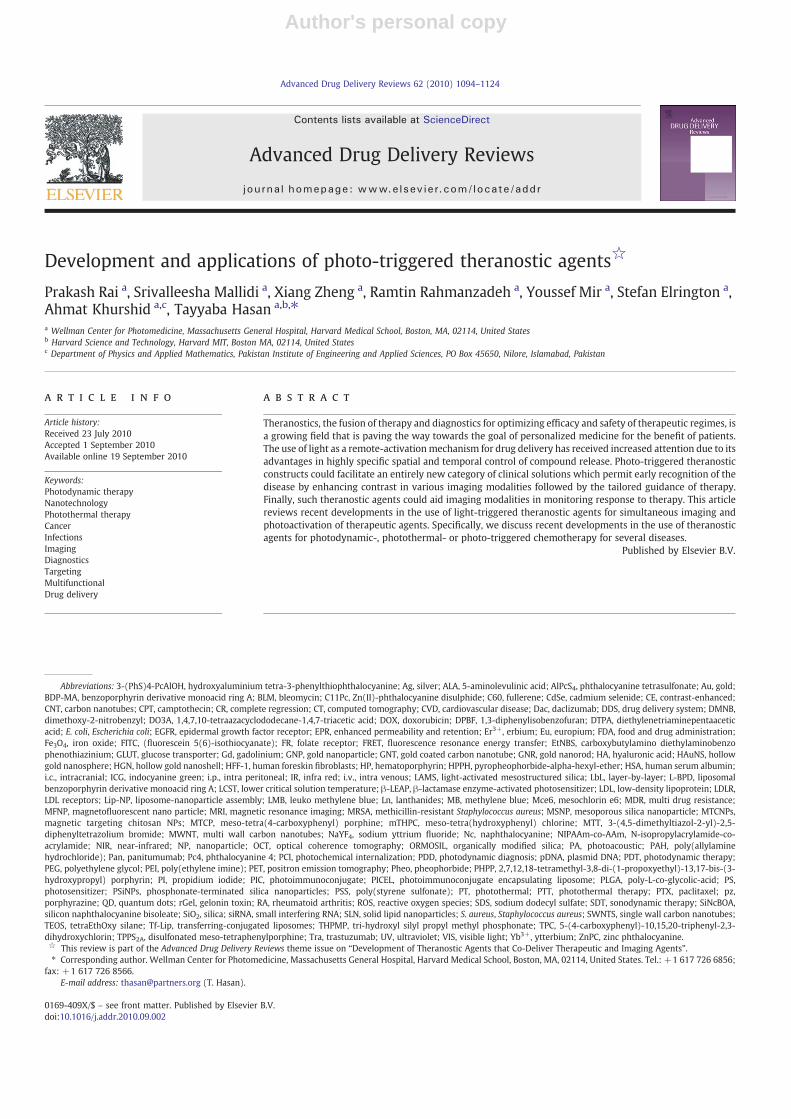
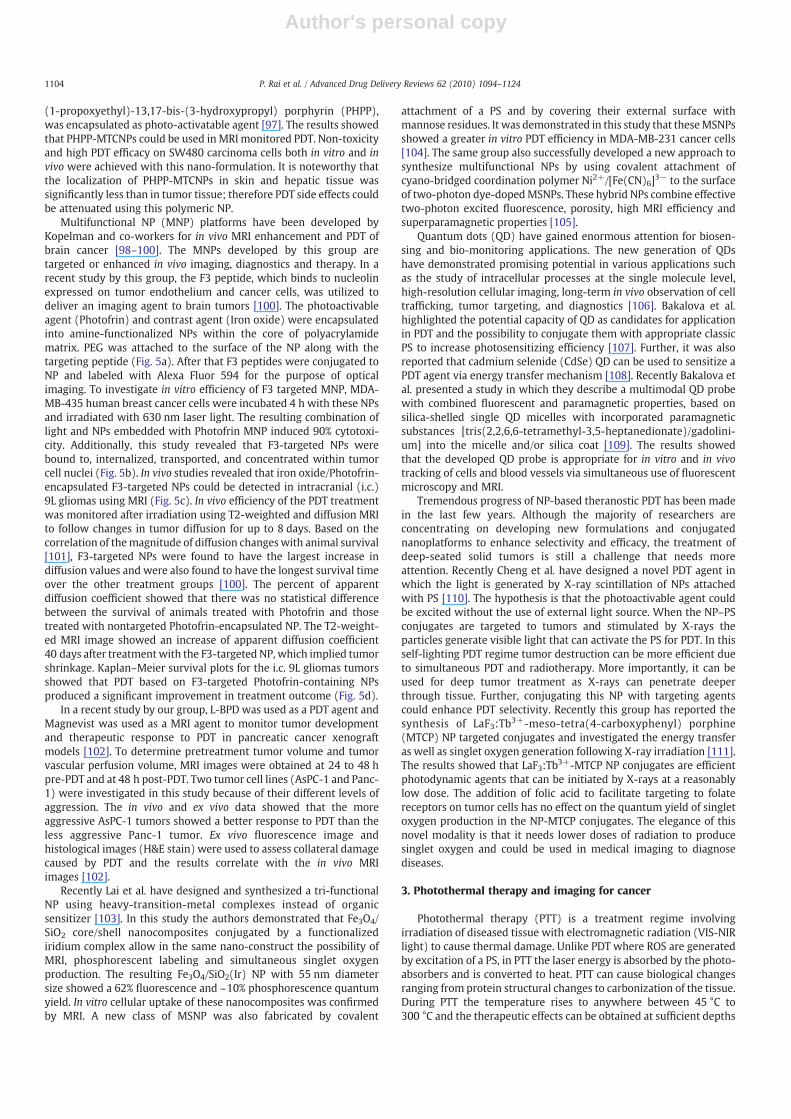
A further development in this field of activatable molecular beaconsis the use of a targetingmoiety. This targetedmolecular beacon strategywas demonstrated by Stefflova et al. [42]. The authors designed amultifunctional, membrane-permeable, and cancer-specific constructthat triggers and images apoptosis in cancer cells (Fig. 2). This constructcontains a fluorescent PS (pyropheophobide) and a cancer-associatedfolate receptor targeting molecule connected to a caspase-3 cleavablepeptide linker that has a fluorescence quencher (BHQ-3) on theopposite side. The double-tumor mouse bearing a folate receptornegative tumor (derived from HT 1080 cells) on one side and a folatereceptor positive tumor (derived fromKBcells) on the contralateral sidewas injected intravenously with the construct, followed by PDT. Adistinctly higher post-PDT increase in fluorescence was observed in thefolate receptor positive tumor compared to the folate receptor negativetumor, confirming the targeting and apoptosis-reporting functions ofthe construct (Fig. 2). The use of a single molecular agent for targetedPDT and monitoring response to the treatment demonstrated thetheranostic potential of such an approach.
2.1.2. Multimodal imaging (MRI and PET)Optical fiber-based fluorescence imaging techniques combined
with targeting agents have been extensively studied for diagnosis,PDT and treatment response monitoring [17]. However, poor lightpenetration limits the applicability of light-based imaging andtherapies to superficial tumors with depths of 1–2 mm into thetissue. Thus, an emerging trend in the development of theranostic PDTagents is the coupling of optical imaging with other imagingmodalities such as positron emission tomography (PET), magneticresonance imaging (MRI) and ultrasound.
PET imaging agents are most commonly labeled with radio-isotopes such as 11C (t1/2=20.4 min) and 18F (t1/2=110 min).
1098 P. Rai et al. / Advanced Drug Delivery Reviews 62 (2010) 1094–1124
Author's personal copy
However, it is very challenging as synthesis, purification and analysisof these short-lived isotopes have to be done within the order of afew minutes. Large isotopes such as 86Y (t1/2=14.7 h), 64Cu (t1/2=12.7 h), and 124I (t1/2=4.2 days) are more suitable candidates.Radiolabeling with 124I for PET studies involving PDT is mostappropriate because PSs needs relatively long time to accumulate intumors. A simple method to prepare the 124I-labeled PS is the directelectrophilic aromatic iodination of the trimethylstannyl substitutedanalogues with Na124I in the presence of commercial iodogen beads.Using this strategy, Pandey et al. prepared 124I-labeled pyropheo-phorbide and purpurinimide analogues with N95% radioactivespecificity [43,44]. It was proposed that this radioactive constructcould be used for PET and fluorescence imaging as well as PDT.
MRI is a widely used tool in pharmaceutical research due to itsexcellent soft tissue contrast property that provides three-dimensionalanatomic images with high spatial resolution. Unlike nuclear scanning,conventional radiography or computed tomography,MRI often relies oncontrast enhancers to improve inherent contrast between normal anddiseased tissue by altering longitudinal (1/T1) and transverse relaxationrates (1/T2) of tissueprotons. Agents containingparamagnetic transitionmetal ions such as gadolinium (Gd3+) and manganese (Mn2+) havebeen shown to effectively alter 1/T1 and/or 1/T2. Gd3+ in particular, hasseven unpaired electrons within the inner orbital shells and providesa high degree of paramagnetism which causes an increase in the T1relaxation rates of nearby water molecules. Gd3+ is too large to beaccommodated in the macrocyclic center of ordinary porphyrins.
Fig. 2. Theranostic molecular beacons for targeted PDT and monitoring treatment response. The top panel is the schematic diagram of structure and function of a targeted PDT agentwith a built-in apoptosis sensor: (1) this construct consists of PS, caspase 3 cleavable sequence, fluorescence quencher, and delivery vehicle; (2) the construct accumulatespreferentially in cells overexpressing folate receptor, and once activated by light, the PS produces singlet oxygen that destroys the mitochondrial membrane and triggers apoptosis;(3) this leads to activation of caspase 3, which cleaves the peptide linker between the PS and the quencher, thus restoring the PS's fluorescence and identifying those cells dying byapoptosis by NIR fluorescence imaging. The bottom left panel demonstrated in vivo induction and detection of apoptosis in a mouse bearing folate receptor positive (FR+, KB) cells)and folate receptor negative (FR-, HT 1080 cells) tumors after light treatment (Photodynamic Therapy=PDT, 90 J/cm2) using intravenously administered photoactivatable drugPyro-K(folate)GDEVDGSGK (BHQ-3) (PFPB, 25 nmol) cleavable by Caspase-3. (a–c): Xenogen images of a mouse bearing FR−(left) and FR+(right) tumors: a. before i.v. injection ofPFPB or PDT; b. 0.5 h after PDT (4 h after drug injection); c. 3 h after PDT (6.5 h after drug injection). These images are showing a gradual increase in fluorescence in the FR+compared to FR−tumor. Bottom right: confocal images of the histology tissue slides of the corresponding FR+and FR−tumors stained with Apoptag confirmed increased light-induced apoptosis in the FR+tumor. Courtesy of Dr. Gang Zheng and Dr. Klara Stefflova.
1099P. Rai et al. / Advanced Drug Delivery Reviews 62 (2010) 1094–1124
Author's personal copy
Incorporation of Gd3+ with a PS can be achieved by two methods: firstmethod is to insert the ion into an expanded porphyrin which containsfive (instead of four) nitrogen atoms in the ring and forms a centralchelating cavity 20% larger than that of ordinary porphyrins toaccommodate Gd3+ [45]. This metal complex, namely Motexafingadolinium has been proposed for the treatment of brain cancer butwas rejected by FDA in 2007. While the lutetium complex of Motexafinwas undergoing clinical trials as a PS agent [46], interestingly, Gd3+
analog had only been investigated as a potential radiation sensitizer bycausing redox stress to cancer cells. The secondmethod of incorporatingGd3+ with a PS is to stabilize Gd3+ by attaching PS with a side-chainmoiety such as diethylenetriaminepentaacetic acid (DTPA). Thisstrategy is more favorable because it can be applied to virtually anyPS. In an early study, two Gd-DTPA moieties were covalently linked tomesoporphyrin (Gadophrin-2) and later to the copper complex(Gadophrin-3) for improved stability and safety. Although suchmetalloporphyrins may be useful for tumor imaging, they were foundto preferentially localize in the periphery of necrotic areas rather thanthe viable cancer tissue [47].
Pandey and co-workers [48,49] investigated the possibility ofdelivering the contrast agents to living tumor cells by conjugatinggadolinium complexes to HPPH, a tumor-avid chlorophyll derivativeat Phase II clinical trials. In this study, up to six Gd3+ aminobenzylDTPA complexes were coupled to HPPH. Most of them had enhancedtumor-imaging potential (Fig. 3), which increased with a largernumber of Gd3+ units. To achieve a comparable signal intensity of theclinical MRI agent Gd-DTPA, only a 10 to 20-fold lower dose of theconjugate was required. The three Gd+3aminobenzyl DTPA conju-gates, which showed the best PDT activity in vitro was evaluated invivo. At 24 h post injection, the accumulation of the conjugate in theWard tumor was higher than in blood, muscle and most organs. Atimaging concentration, the required light dose for the conjugate waslower than the one required for HPPH alone, to achieve comparabletumor response in both radiation induced fibroblast (RIF) and Colon26 tumor models.
Another promising theranostic system has been developed by Liuet al. to enhance the monitoring of PDT efficacy [50]. In this study, theauthors investigated a novel PS, prepared from fullerene (C60), whichcombines the property to produce singlet oxygen and can beconjugated to an MRI agent. Gd3+ was selected as the MRI contrastagent and introduced to the PEG terminal of C60-PEG through metalchelation. Following intravenous injection in the tumor-bearing mice,C60-PEG-Gd maintained an enhanced MRI signal at the tumor tissuefor a longer time period in comparison with the commercial contrastagent (Magnevist®). The PEG-conjugated fullerene system showedsignificant tumor PDT effect although the effect depended on thetiming of light irradiation.
Vaidya et al. have synthesized PEGylated poly-(L-glutamic acid)conjugates containing mesochlorin e6, a PS, and Gd(III)-DO3A [51,52].MRI images showed that pegylated conjugate had longer bloodcirculation, lower liver uptake and higher tumor accumulation thanthe non-pegylated conjugate. Laser irradiation of tumors resulted inhigher therapeutic efficacy for the pegylated conjugate. The PDTtreated animals showed a reduced vascular permeability withdynamic contrast-enhanced-MRI and reduced microvessel densityon histopathological analysis. They concluded that PEGylation of thebifunctional polymer conjugates reduced nonspecific liver uptake andincreased tumor uptake, resulting in significant tumor contrastenhancement and higher therapeutic efficacy [52].
Ultrasound as a label-free technique has been used in vascular andinterventional imaging. Its therapeutic effects in treatment of solidtumors and its efficacy and safety were confirmed in clinicalinvestigations [53]. There have been several reports on the ability ofcertain porphyrins (sonosensitizers) to enhance the low-intensityultrasound-induced cytotoxicity, both in cell culture and in tumormodel. This treatment modality is named Sonodynamic therapy
(SDT). Although the mechanism of this enhancement effect has notyet established, the experimental evidence suggests that sonody-namic effect may due to the chemical activation of sonosensitizersinside or in the close vicinity of hot collapsing cavitation bubbles toform sensitizer-derived free radicals, and/or due to mechanical stressof physical disruption of cellular membrane by sensitizers [54]. It wasalso reported that the combination of SDT with PDT can induce tumornecrosismore extensively than inmice receiving only SDT or PDT [55].
The use of PSs as imaging agents for diagnosis or fluorescence-guided tumor resection is emerging in the last years. PSs werecombined with imaging techniques including fluorescence, MRI, PET,and ultrasound. Some of these approaches have already enteredclinical trials or are approved like 5-ALA hexyl ester for the diagnosisof bladder cancer in Sweden. Hexvix was recently approved in the USfor bladder cancer detection and fluorescence-guided resection. Newtechnologies for molecular targeting will increase the tumor specific-ity thereby enabling sensitive and specific detection and site-specifictreatments.
2.2. Nanoparticles for imaging and PDT
Over the last fewyears, nanoparticle (NP) based PDThas emerged asan alternative to conventional PDT to efficiently target cancer cells. Thedual selectivity provided by the target localizing ability of NP and thespatial control of illumination could significantly reduce the systemictoxicity associated with classical PDT therapy. Besides the systemictoxicity, most PSs used in PDT, have other limitations. Mainly, they arehydrophobic or have limited water solubility and therefore couldaggregate in biological media which leads to the modification of theiroptical properties and the decrease of singlet oxygen production [56].Although recently a lot of work has focused on developing several newstrategies to improve the performance of PDT agents, includingconjugation with oligonucleotides, monoclonal antibodies, carrierproteins, lipids, carbohydrates, or hydrophilic polymers for selectivedelivery of the agents into tumor tissues [57], the lack of specific targetsand the dark cytotoxicity is still a principal challenge for PDT. Manyefforts are ongoing to develop new conjugated PS with a covalentlylinked vector to target receptors over-expressed in cancer cells,however very few have been evaluated in the clinic mainly because oftheir lower in vivo selectivity [58].
Nanotechnology provides a platform for integration of multiplefunctionalities in a single construct [59]. Here we provide an update ofsimultaneous tumor targeting and imaging with a number of differentnanosystems that have the potential for theranostic PDT. Variousnanoprobes have been developed for in vivo magnetic resonance andoptical imaging, which include quantum dots, up-converting nano-phosphors, gold and Silica NPs, and PS containing nanoparticulatecarriers such as liposomes, ceramic, polymeric. The in vitro and in vivofate of these systems after administration is discussed. Althoughseveral challenges remain before this modality can be adopted in theclinic, multifunctional NPs offer a good tool to treat deep tumorsefficiently with PDT.
2.2.1. Optical imagingRecent breakthroughs in the synthesis of mesoporous silica NP
(MSNP) with high surface area and tunable pore diameter (2–10 nm)have led to the design of new delivery systems, where differentmolecules, such as pharmaceutical drugs or fluorescent imagingagents, could be absorbed into the mesopores and released later intovarious solutions [60]. Furthermore, some reports on the design of PSbased MSNP have been published, among them, Roy et al. havestudied a ceramic system based NP with an average diameter of30 nm. The particularity of this NP is its capacity to optically protect 2-devinyl-2-(1-hexyloxyethyl) pyropheophorbide (HPPH) PS resultingin a drug-doped, highly monodispersed, and stable NP in an aqueoussystem. In vitro imaging study of this system demonstrated active
1100 P. Rai et al. / Advanced Drug Delivery Reviews 62 (2010) 1094–1124

Author's personal copy
uptake of HPPH-doped NPs into the cytosol of tumor cells and efficientcytotoxicity upon irradiation [61].
To circumvent the problem of the mesoporosity of MSNP and therelease of the PS during systemic circulation, Prasad's group hassucceeded to covalently incorporate iodobenzylpyropheophorbide PSinto a novel nano-formulation named Organicaly Modified Silica NPs(ORMOSIL) [62]. These NPs were taken up by tumor cells in vitro anddemonstrated phototoxic action. Kim et al. reported a promisingmodality using the ORMOSIL system where the photosensitizing unit(energy acceptor) is indirectly excited through fluorescence reso-nance energy transfer (FRET) from the two-photon absorbing dye unit(energy donor [63]. In this study, the authors showed use ofnanophotonic tools to produce singlet oxygen and to induce tumorcell death following two-photon PDT. They also demonstrated thepotential for co-encapsulation of two drugs to provide contrast influorescence images of live tumor cells under two-photon excitationand under near-infrared (NIR) light.
More recently Prasad et al. investigated the biological issue raisedover the use and safety of the ORMOSIL system for diagnosis andtherapeutic purposes using different bio-imaging modalities includ-ing PET, MRI and optical imaging [64]. A NIR PS DY776 wasencapsulated in ORMOSIL NP, resulting in a ~20 nm diameter sizeNIR optical probe. The PET imaging probe iodine-124 was conjugatedwith the NPs, which allow imaging of deep tissue. To study the in vivoclearance of the NPs, animals injected with DY776 conjugated
ORMOSIL were imaged daily over a period of 15 days. The resultsshowed accumulation of NPs in the liver and spleen over a period of24 h, whereas the skin showed amaximum concentration at 72 h postinjection of NPs. Furthermore the clearance studies confirmed that allof the injected ORMOSIL was excreted out of the animal via thehepatobiliary excretion without any sign of organ toxicity [64]. Thesein vivo bio-imaging, bio-distribution, clearance, and toxicity studiesshowed that combining multimodal nano-formulation with PDT,makes ORMOSIL NPs an exciting modality for treatment andmonitoring.
In a recent report He et al. explored the use of methylene blue-encapsulated silica NPs (MB-PSiNPs) for simultaneous in vivo imagingand PDT [65]. MB-PSiNPs were synthesized with an average diameterof 105 nm. Using a chemical trap, the authors confirmed singletoxygen production after irradiation. To investigate the biologicalenvironment effects on the encapsulated MB, the decreased absorp-tion of leukomethylene blue (LMB) which is the reduced form of MBafter contact with enzymeswas probed at 660 nm. The results showedthat encapsulated MB stayed intact in the biological systemand PSiNPs prevented the MB from being reduced by enzymes [65].In vivo monitoring of PDT post irradiation was also performed onsubcutaneous-Hela-tumor-xenografted mice. Twelve hours after theinjection of MB-PSiNPs, the induced fluorescence was used to guidetreatment with 635 nm laser light (500 mW/cm2, 5 min). Followingtreatment, in vivo imaging was performed using a hyperspectral
Fig. 3. Tumor-Avid PS-Gd(III)DTPA for imaging and therapy. Conjugate shows potential for in vivo imaging (MR, Fluorescence) and PDT. a. Structure of Gd3+ aminobenzyl DTPAconjugates of HPPH; b. In vivo PDT efficacy of HPPH-3Gd in C3H mice bearing RIF tumors and BALB/c mice bearing Colon 26 tumors. At an imaging/therapeutic dose of 10 umol/kg;c. increase in signal intensity was seen in rat Ward Colon tumors (arrow) from preinjection (left) to postinjection (right, 24 h postinjection) of HPPH-3Gd; d. fluorescent images ofHPPH-3Gd in BALB/c mice at 24, 48, and 72 h. Adapted from Spernyak et al. [49].
1101P. Rai et al. / Advanced Drug Delivery Reviews 62 (2010) 1094–1124
Author's personal copy
imaging system to assess treatment response. The tumors treatedwith NPs and light irradiation were found to shrink gradually whilethe control tumors did not show any significant effect [65].
MSNPs have also been designed by Zhang et al. [66]. Thismultifunctional core–shell NP contains a nonporous dye-doped silicacore with an average diameter of ~37 nm and a ~57 nm ofmesoporous silica shell containing PS molecules, hematoporphyrin(HP). These nanocomposites are stable and can be stored for over1 month at room temperature. The elegance of the bi-functionality ofthis system is its capacity to act not only as a carrier for thephotoactivable drug which is covalently linked to the mesoporoussilica shell but also as a nanoreactor to facilitate the photo-oxidationreaction. The author demonstrated that doping of fluorescence dyesinto the nonporous core allows for simultaneous PDT and fluores-cence imaging in vitro.
Low-density lipoprotein (LDL) provides a highly polyvalentnatural nanoplatform for delivery of various imaging and therapeuticagents to neoplastic cells that over express LDL receptors (LDLR) [67].Covalent attachment of other ligands to the lysine side-chain aminogroups could be used to target other receptors [68]. The incorporationof NIR fluorescent probes into LDL showed promising results usingoptical imaging [69,70]. In addition, Gadolinium based agents couldalso be attached to LDL to improve tumor detection using magneticresonance imaging (MRI) [71]. More recently Song et al. designed anovel naphthalocyanine (Nc)-based PS as a PDT agent to be deliveredby a LDL NP [72]. This Nc-LDL NP was prepared by reconstituting thetetra-t-butyl silicon naphthalocyanine bisoleate (SiNcBOA) into LDLlipid core. In vitro results indicated that Nc-LDL NPs are internalizedinto Hep G2 cells specifically via the LDLR mediated pathway. Thispreferential uptake of Nc-LDL NPs by tumor tissue was confirmed invivo by noninvasive optical imaging technique. Human serumalbumin (HSA), the most abundant protein in human blood plasmahas been used recently as a platform to deliver a photoactivable drugPheophorbide (Pheo) into tumor for PDT [73]. In vitro studies onJurkat cells using fluorescence lifetime imaging showed that Pheo-HSA NPs efficiently decomposed in the cellular lysosomes resulting inhigher phototoxicity.
Solid lipid NPs (SLN) represent a novel carrier system that hasvarious advantages compared to liposomes and polymeric NPs [74].Stevens et al. have used this platform to synthesize a folate receptor(FR)-targeted SLN in which hematoporphyrin PS has been encapsu-lated to target FR-overexpressing tumor cells. In vitro cytotoxicitystudy of these NPs showed an IC50 of 1.57 μM in human oral epidermalcarcinoma cells and nontargeted SLN gave an IC50 of 5.17 μM. Theselectivity of FR-targeted NP was confirmed by fluorescence micros-copy [75].
Up-converting Phosphor Technology, based on lanthanide-con-taining, submicrometer-sized, ceramic particles that can absorbinfrared light and emit visible light, has been used by Chatterjee etal. to design NPs as transducers for PDT [76,77]. The up-convertingNPs investigated in this study are composed of sodium yttriumfluoride (NaYF4) nanocrystals co-doped with the rare earth ionsytterbium (Yb3+) and erbium (Er3+) with a polymeric coat of poly(ethylene imine) (PEI). The PS zinc phthalocyanine (ZnPC) wassuperficially noncovalently adsorbed to the NPs. The researchers havesucceeded in this study to image cellular uptake by fluorescenceimaging microscopy of PEI/NaYF4:YB3+, Er3+ NPs, and they foundsignificant cell death following irradiation with NIR laser light. Apotential clinical use of these NPs is to image and photodynamicallytreat cancers situated in deep tissue.
Liposomes represent a valuable carrier and delivery system due totheir high loading capacity and their flexibility to accommodatedifferent PS with variable physicochemical properties [78]. The PDTagents could be targeted by modifying the design and the surfacechemistry of liposomes. However conventional liposome could givepromising results when administered topically. Bendsoe et al. have
reported a clinical study using liposomal Meso-tetra(hydroxyphenyl)chlorin (mTHPC) gel formulation for topical application in connectionwith PDT of non-pigmented skin malignancies [79]. In this study theauthors reported that the treated area did not show any swelling orreddening, as is often seen in PDT using topical ALA. Further, no painduring or after treatment were reported. One week after treatment,healing progress was observed in several patients and no complica-tions were registered. Beside the clinical efficiency of this formulation,liposomal NPs could be used as a probe to monitor the sensitizerdistribution within tumor and surrounding normal skin usingfluorescence imaging before, during, and after PDT.
In our group, Zhong et al. have used a liposomally encapsulatedbenzoporphyrin derivative monoacid ring A (L-BPD) to image, treatand monitor PDT response in vivo in a mouse model of disseminatedovarian cancer [80]. L-BPD (Visudyne)was originally developed for itsapplication in ophthalmology and is currently approved for thetreatment of age-related macular degeneration (AMD). Additionaldetails about its use in AMD are discussed in Section 5.2 of this review.Visudyne is also currently in clinical trials for treatment of ovarian[81,82] and pancreatic cancer [83]. In the study of Zhong and Celli etal. high-resolution fiber-optic fluorescence imaging was used for thedetection of microscopic ovarian cancer and for monitoring PDTtreatment response. After administration, L-BPD serves as both animaging agent and a light-activated therapeutic agent. By comparisonwith a histopathology basedmethod, Zhong et al. showed a sensitivityof 86% for in vivo tumor detection using the microendoscope. Theresults showed that PDT treated mice exhibit an average decrease of59% in tumor volumes. The author concluded the potential of theapproach used to treat and monitor the treatment outcome [80].Additional studies are necessary to compare feedback from imagingwith long-term outcomes to evaluate the potential of this approachfor early reporting following treatment and, by extension, as a tool toaid in rational treatment planning.
Using the same platform Derycke et al. have studied the tumorselective behavior of phthalocyanine tetrasulfonate (AlPcS4) when itsapplied intravesically in transferrin-conjugated liposomes (Tf-Lip-AlPcS4) [84]. The results reported show an efficacy of the PDTtreatment using Tf-Lip-ALPCS4 on AY-27 rat bladder carcinoma cells.The authors concluded that transferrin-mediated liposomal targetingof ALPcS4 drugs is a promising tool for PDT of superficial bladdertumors. Thus the selective accumulation of Tf-Lip-AlPcS4 in bladdertransitional-cell carcinoma cells would allow photodiagnosis andfluorescence-guided transurethral resection of lesions with a highsensitivity and specificity. Another liposome-based formulation hasbeen developed by Meerovich et al. using hydroxyaluminium tetra-3-phenylthiophthalocyanine (3-(PhS)4-PcAlOH) as a NIR PS. Experi-ments on mice with solid Ehrlich tumor and subcutaneouslytransplanted P-388 leukemia revealed highly selective accumulationof 3-(PhS)4-PcAlOH in tumors in comparison with normal tissues andhigh PDT activity. The authors concluded that the high selectiveaccumulation of 3-(PhS)4-PcAlOH in a tumor could be used fordiagnosis [85].
Recently, our group has demonstrated a new approach to targetand photoinactivate the nuclear proliferationmarker pKi-67 [86]. pKi-67 is a marker that is strongly expressed in all cells that have theability to divide and proliferate [87]. In many cancers pKi-67expression is correlated with poor prognosis for disease freeprogression and overall survival. Therefore, antibodies against pKi-67 are widely used in diagnostics to access the growth fraction oftumors from patient biopsies [88]. Despite the interest of using adiagnostic valuable marker as target for therapy, the inactivation of anuclear protein has been challenging so far. To target the Ki-67protein we used dye-labeled antibodies which are encapsulated intoPEGylated liposomes (photoimmunoconjugate encapsulated lipo-somes, PICELS) [86]. The liposomes deliver the photoimmunoconju-gate intracellularly where a fraction is released into the cytoplasm.
1102 P. Rai et al. / Advanced Drug Delivery Reviews 62 (2010) 1094–1124

Author's personal copy
From the cytoplasm the conjugates localize into the nucleus to theactual pKi-67 site. The efficacy of pKi-67 inactivation was demon-strated in a 3D in vitromodel for ovarian cancer. In this model ovariancancer cells form multicellular acini (Fig. 4a), which mimic the smalltumor nodules that are found in vivo all over the peritoneal cavity [86].Treatment of these cells with PICELS and subsequent light irradiationled to destruction of the acinar structure and to more than 70% deadcells 72 h following treatment (Fig. 4b). As a control, pKi-67 negativeconfluent lung fibroblasts showed no significant effect on cell viabilityafter pKi-67 PDT [86]. In this approach the target protein is not onlyutilized for the selective delivery of the PS but the antibody itself isinhibiting the protein. Only after light irradiation and the generationof ROS, the photoimmunoconjugate becomes an inhibitor for thetarget protein. Besides its important role in diagnostics, the studydemonstrated a potential role of pKi-67 as a molecular target forcancer therapy and demonstrated the nuclear delivery of an antibodywith non-cationic liposomes [86]. The fact that pKi-67 is a wellestablishedmarker in tumor diagnosticsmakes this approach not onlyvaluable for eliminating aggressive cancer cells, but could in the futurealso be applied to access the growth fraction of the tumor in real-timein vivo (Fig. 4a). One or two days after drug administration the fractionof pKi-67 positive cells could be imaged endoscopically in the patient.Different regions in the tissue could be accessed in a short time aftereach other and the Ki-67 labeling index could be estimated based onimage fluorescence data. In this first study anti-pKi-67 antibodieswere conjugated to FITC (fluorescein 5(6)-isothiocyanate). FITC caneasily be conjugated to antibodies and it has been widely used forspecific protein inactivation in living cells [89]. FITC has an excitationmaximum of 490 nm where light penetration into tissue is fairlylimited. For in vivo application a PS with absorption maximum in thelonger wavelength range and with higher singlet oxygen quantumyield seems to be more applicable.
Numerous reviews have described and provided results on the useof gold NP (GNP) in many biomedical applications including imagingand therapy of cancer [90]. Beside the availability of rich chemistryregarding GNP, currently it is possible to modify the surface of this NPeither covalently or noncovalently with PSs. Recently, Wieder et al.reported the development of a new delivery system based on GNPs,whereby the PS is attached to the surface of the NP [91]. Their resultsshowed that GNP conjugates are an excellent carrier for the deliveryof hydrophobic PS for high PDT efficacy toward tumor cells. The
uptake of these NPs and their phototoxicity toward Hela cells wasconfirmed using confocal imaging microscope. Though the results areencouraging using these NPs, the PDT efficiency of this systemremains to be evaluated in vivo.
Zaruba et al. recently studied the efficacy of PDT using GNP onwhich two porphyrin–brucine conjugates were immobilized [92]. Theintracellular distribution and tumor cell uptake of these NPs werestudied using fluorescence microscopy and the results showed thatthese NPs localize in lysosomes. The in vivo results showed that thebrucine–porphyrin derivatives bound to modified GNPs mediate acomplete regression of PE/CA-PJ34 carcinoma after PDT. Morerecently Russell and Jori groups have investigated the in vivo efficacyof Zn(II)-phthalocyanine disulphide (C11Pc) bound to GNPs for thePDT of amelanotic melanoma [93]. In this paper the authors showedan enhanced accumulation of this NP on subcutaneously implantedamelanotic melanoma. Further, electron microscopy observations oftumor specimens obtained at different times after PDT, showed anextensive damage of the blood capillaries and endothelial cells.
Among the various delivery system of GNP, Cheng et al.investigated the efficiency of PEGylated GNP attached to phthalocy-anine 4 (Pc4) for in vivo PDT of cancer [94]. A 35% singlet oxygenquantum yield was obtained for Pc4 on PEGylated GNPswhile free Pc4had 50% singlet oxygen quantum yield. Fluorescence images of atumor-bearing mouse were also taken at 1, 30 and 120 min after i.v.injection of drug conjugated NPs. The results showed that the drugsaccumulated at the tumor site through a passive targeting process.After illumination the effect of treatment appeared within one weekwithout any noticeable toxicity or side effects to the animals [94].
2.2.2. Magnetic resonance imaging (MRI)Considerable research efforts have been directed towards devel-
oping efficient chitosan-based NP drug delivery systems. In compar-ison to other biological polymers, positive charges target the chitosancarriers to the negatively charged cell membrane and have mucoad-hesive properties to prolong the retention time of chitosan in thetargeted locations [95]. Magnetic chitosan NPs can provide excellentbiocompatibility, biodegradability, non-toxicity and water solubilitywithout compromising their magnetic targeting ability [96]. Basedon these findings Sun et al. have studied magnetic targeting chitosanNPs (MTCNPs) which have been prepared and tailored asMRI imaging agents and in which PS-2,7,12,18-tetramethyl-3,8-di-
Fig. 4. Photoimmunoconjugate encapsulating liposomes (PICELS) for targeting and inactivation of the nuclear proliferation marker pKi-67. a. PICELS deliver pKi-67-FITC antibodiesintracellular and can be imagedwith confocal microscopy in ovarian cancer 3D-culture ascini. b. Imaging of monolayer cultures shows the nucleolar localization on the PICELS. c. 72 hafter laser irradiation with 5 J/cm2 at 488 nm the 3D-acini have lost their spherical morphology and d. following treatment with PICELS and light irradiation the 3D-acini show asignificant decrease in the number of viable cells as measured by a live-dead assay. Based on work by Rahmanzadeh et al. [86].
1103P. Rai et al. / Advanced Drug Delivery Reviews 62 (2010) 1094–1124
Author's personal copy
(1-propoxyethyl)-13,17-bis-(3-hydroxypropyl) porphyrin (PHPP),was encapsulated as photo-activatable agent [97]. The results showedthat PHPP-MTCNPs could be used in MRI monitored PDT. Non-toxicityand high PDT efficacy on SW480 carcinoma cells both in vitro and invivo were achieved with this nano-formulation. It is noteworthy thatthe localization of PHPP-MTCNPs in skin and hepatic tissue wassignificantly less than in tumor tissue; therefore PDT side effects couldbe attenuated using this polymeric NP.
Multifunctional NP (MNP) platforms have been developed byKopelman and co-workers for in vivo MRI enhancement and PDT ofbrain cancer [98–100]. The MNPs developed by this group aretargeted or enhanced in vivo imaging, diagnostics and therapy. In arecent study by this group, the F3 peptide, which binds to nucleolinexpressed on tumor endothelium and cancer cells, was utilized todeliver an imaging agent to brain tumors [100]. The photoactivableagent (Photofrin) and contrast agent (Iron oxide) were encapsulatedinto amine-functionalized NPs within the core of polyacrylamidematrix. PEG was attached to the surface of the NP along with thetargeting peptide (Fig. 5a). After that F3 peptides were conjugated toNP and labeled with Alexa Fluor 594 for the purpose of opticalimaging. To investigate in vitro efficiency of F3 targeted MNP, MDA-MB-435 human breast cancer cells were incubated 4 h with these NPsand irradiated with 630 nm laser light. The resulting combination oflight and NPs embedded with Photofrin MNP induced 90% cytotoxi-city. Additionally, this study revealed that F3-targeted NPs werebound to, internalized, transported, and concentrated within tumorcell nuclei (Fig. 5b). In vivo studies revealed that iron oxide/Photofrin-encapsulated F3-targeted NPs could be detected in intracranial (i.c.)9L gliomas using MRI (Fig. 5c). In vivo efficiency of the PDT treatmentwas monitored after irradiation using T2-weighted and diffusion MRIto follow changes in tumor diffusion for up to 8 days. Based on thecorrelation of themagnitude of diffusion changeswith animal survival[101], F3-targeted NPs were found to have the largest increase indiffusion values and were also found to have the longest survival timeover the other treatment groups [100]. The percent of apparentdiffusion coefficient showed that there was no statistical differencebetween the survival of animals treated with Photofrin and thosetreated with nontargeted Photofrin-encapsulated NP. The T2-weight-ed MRI image showed an increase of apparent diffusion coefficient40 days after treatmentwith the F3-targeted NP, which implied tumorshrinkage. Kaplan–Meier survival plots for the i.c. 9L gliomas tumorsshowed that PDT based on F3-targeted Photofrin-containing NPsproduced a significant improvement in treatment outcome (Fig. 5d).
In a recent study by our group, L-BPD was used as a PDT agent andMagnevist was used as a MRI agent to monitor tumor developmentand therapeutic response to PDT in pancreatic cancer xenograftmodels [102]. To determine pretreatment tumor volume and tumorvascular perfusion volume, MRI images were obtained at 24 to 48 hpre-PDT and at 48 h post-PDT. Two tumor cell lines (AsPC-1 and Panc-1) were investigated in this study because of their different levels ofaggression. The in vivo and ex vivo data showed that the moreaggressive AsPC-1 tumors showed a better response to PDT than theless aggressive Panc-1 tumor. Ex vivo fluorescence image andhistological images (H&E stain) were used to assess collateral damagecaused by PDT and the results correlate with the in vivo MRIimages [102].
Recently Lai et al. have designed and synthesized a tri-functionalNP using heavy-transition-metal complexes instead of organicsensitizer [103]. In this study the authors demonstrated that Fe3O4/SiO2 core/shell nanocomposites conjugated by a functionalizediridium complex allow in the same nano-construct the possibility ofMRI, phosphorescent labeling and simultaneous singlet oxygenproduction. The resulting Fe3O4/SiO2(Ir) NP with 55 nm diametersize showed a 62% fluorescence and ~10% phosphorescence quantumyield. In vitro cellular uptake of these nanocomposites was confirmedby MRI. A new class of MSNP was also fabricated by covalent
attachment of a PS and by covering their external surface withmannose residues. It was demonstrated in this study that theseMSNPsshowed a greater in vitro PDT efficiency in MDA-MB-231 cancer cells[104]. The same group also successfully developed a new approach tosynthesize multifunctional NPs by using covalent attachment ofcyano-bridged coordination polymer Ni2+/[Fe(CN)6]3− to the surfaceof two-photon dye-dopedMSNPs. These hybrid NPs combine effectivetwo-photon excited fluorescence, porosity, high MRI efficiency andsuperparamagnetic properties [105].
Quantum dots (QD) have gained enormous attention for biosen-sing and bio-monitoring applications. The new generation of QDshave demonstrated promising potential in various applications suchas the study of intracellular processes at the single molecule level,high-resolution cellular imaging, long-term in vivo observation of celltrafficking, tumor targeting, and diagnostics [106]. Bakalova et al.highlighted the potential capacity of QD as candidates for applicationin PDT and the possibility to conjugate them with appropriate classicPS to increase photosensitizing efficiency [107]. Further, it was alsoreported that cadmium selenide (CdSe) QD can be used to sensitize aPDT agent via energy transfer mechanism [108]. Recently Bakalova etal. presented a study in which they describe a multimodal QD probewith combined fluorescent and paramagnetic properties, based onsilica-shelled single QD micelles with incorporated paramagneticsubstances [tris(2,2,6,6-tetramethyl-3,5-heptanedionate)/gadolini-um] into the micelle and/or silica coat [109]. The results showedthat the developed QD probe is appropriate for in vitro and in vivotracking of cells and blood vessels via simultaneous use of fluorescentmicroscopy and MRI.
Tremendous progress of NP-based theranostic PDT has been madein the last few years. Although the majority of researchers areconcentrating on developing new formulations and conjugatednanoplatforms to enhance selectivity and efficacy, the treatment ofdeep-seated solid tumors is still a challenge that needs moreattention. Recently Cheng et al. have designed a novel PDT agent inwhich the light is generated by X-ray scintillation of NPs attachedwith PS [110]. The hypothesis is that the photoactivable agent couldbe excited without the use of external light source. When the NP–PSconjugates are targeted to tumors and stimulated by X-rays theparticles generate visible light that can activate the PS for PDT. In thisself-lighting PDT regime tumor destruction can be more efficient dueto simultaneous PDT and radiotherapy. More importantly, it can beused for deep tumor treatment as X-rays can penetrate deeperthrough tissue. Further, conjugating this NP with targeting agentscould enhance PDT selectivity. Recently this group has reported thesynthesis of LaF3:Tb3+-meso-tetra(4-carboxyphenyl) porphine(MTCP) NP targeted conjugates and investigated the energy transferas well as singlet oxygen generation following X-ray irradiation [111].The results showed that LaF3:Tb3+-MTCP NP conjugates are efficientphotodynamic agents that can be initiated by X-rays at a reasonablylow dose. The addition of folic acid to facilitate targeting to folatereceptors on tumor cells has no effect on the quantum yield of singletoxygen production in the NP-MTCP conjugates. The elegance of thisnovel modality is that it needs lower doses of radiation to producesinglet oxygen and could be used in medical imaging to diagnosediseases.
3. Photothermal therapy and imaging for cancer
Photothermal therapy (PTT) is a treatment regime involvingirradiation of diseased tissue with electromagnetic radiation (VIS-NIRlight) to cause thermal damage. Unlike PDT where ROS are generatedby excitation of a PS, in PTT the laser energy is absorbed by the photo-absorbers and is converted to heat. PTT can cause biological changesranging from protein structural changes to carbonization of the tissue.During PTT the temperature rises to anywhere between 45 °C to300 °C and the therapeutic effects can be obtained at sufficient depths
1104 P. Rai et al. / Advanced Drug Delivery Reviews 62 (2010) 1094–1124

Author's personal copy
using NIR radiation. PTT like PDT brings additional specificity to thetherapeutic technique as only the diseased tissue is irradiated withlight while the surrounding benign tissue is minimally damaged. Thisspatial specificity and the minimal-invasiveness make PTT anattractive therapeutic modality as compared to open surgery orother invasive therapeutic procedures. In PTT either continuous waveor pulsed lasers are used for tissue irradiation. In case of continuouswave lasers, sufficient laser energy needs to be deposited in the targetarea before heat loss occurs in the tissue due to blood perfusion. With
pulsed lasers, intense heat is built up during PTT as the pulse widthused is shorter than the thermal relaxation time of the tissue (thermalconfinement condition) [112]. In either case, the laser parametersneed to be chosen appropriately to obtain effective thermaltherapeutic response. In addition, the laser illumination needs to bechosen at a wavelength where the diseased tissue has higherabsorbance than the surrounding tissue i.e., presence of moreendogenous chromophores such as hemoglobin and melanin orspecific accumulation of photo-absorbers such as NIR dyes in the
Fig. 5. Vascular targeted PDT with theranostic agents improves brain cancer therapy as confirmed by MRI. a. Schematic representation of the multifunctional nanoparticles. The coreof the nanoparticle was synthesized from polyacrylamide, which was embedded with PDT dyes (Photofrin) and/or imaging agents (magnetite/fluorochrome). Polyethylene glycollinker and a molecular address tag (F3 peptide) were attached to target these nanoparticles to cancer cells. b. Cytotoxicity induced by F3-tagged Photofrin-embedded nanoparticlesand laser irradiation. MDA-435 cells were incubated 4 h with nanoparticles with or without F3 tag and irradiated with 1500 mW of laser for y. The Photofrin-mediated cytotoxicitywas then monitored by labeling cells with calcein-AM (green, live cells) and propidium iodide (dead, red cells). Bar, 20 μm. c. T2-weighted magnetic resonance images at day 8 aftertreatment from (C) a representative control i.c. 9L tumor and tumors treated with (D) laser light only, (E) i.v. administration of Photofrin plus laser light, and (F) nontargetednanoparticles containing Photofrin plus laser light and (G) targeted nanoparticles containing Photofrin plus laser light. The image shown in (H) is from the same tumor shown in (G),which was treated with the F3-targeted nanoparticle preparation but at day 40 after treatment. The color diffusion maps overlaid on top of T2-weighted images represent theapparent diffusion coefficient (ADC) distribution in each tumor slice shown. d. Kaplan–Meier survival plot for the i.c. 9L tumor groups. Survival curves for brain tumor animals:untreated, laser only, i.v. Photofrin+laser treated, nontargeted nanoparticles containing Photofrin + laser, and F3-targeted Photofrin-containing nanoparticles+laser treated.Adapted from Reddy et al. [100].
1105P. Rai et al. / Advanced Drug Delivery Reviews 62 (2010) 1094–1124

Author's personal copy
diseased tissue. Several non-photosensitizing dyes have been intro-duced to increase the spatial specificity of PTT [113]. Some of thesedyes have absorption greater than 600 nm enabling the treatment ofdeeply situated pathologies. Moreover, as in the case of PDT, the PTTagents that are widely used have a higher optical absorbance in the“photothermal-therapeutic” window between 600 and 1000 nm, arange in which the absorption of endogenous chromophores is low.For example, indocyanine green (ICG) based PTT was used to treatacne vulgaris and the treatment showed significant improvement in80% of the patients [114]. Similar to PDT photosensitizers, photo-bleaching of dye molecules is a major limitation in PTT. The advent ofnon-photobleaching plasmonic metal NPs has enhanced the photo-diagnostic and phototherapeutic strategies used for detection andtreatment of tumors and infections due to their unique photophysicalproperties. Especially the development of gold nanoshells by theHalas group has further enhanced the efficacy of PTT due to the NIRabsorption properties of nanoshells [115]. El-sayed et al. have shownthe use of gold nanorods for effective treatment of cancer cells [116].Plasmonic GNPs are excellent PTT agents as they have three to fiveorders of magnitude higher absorbance than endogenous chromo-phores and NIR dyes. Moreover, the optical properties of GNPs can bemodified by varying their shape, size, coating etc. The rapid heating ofnon-photobleaching plasmonic NPs has also lead to reduction intreatment time of PTT.
Image guided PTT procedure will enhance therapeutic outcomeespecially in cancer diagnosis and therapy because 1. Imaging will aidin identifying the precise location of the tumor, 2. Guide and monitorspatial and temporal changes in temperature and tissue morphologyduring therapeutic procedures and 3. Evaluate the response of thetumor to therapy immediately after therapy procedure and 4.Evaluate the patients for resurgence of the tumors after thetherapeutic procedures. A theranostic agent has the potential to beused in one or more of the steps listed above in image guided PTT.Specifically in this section we will review PTT agents that enhancecontrast in various imaging modalities such as optical, ultrasound,magnetic resonance imaging and ionizing imaging modalities such asX-ray.
3.1. Optical imaging
PTT agents such as NPs and dyes exhibit intense and narrowoptical extinction bands making them ideal contrast agents for opticalimaging. Indeed the combination of PTT with various optical imagingmodalities is seamless. For example ICG is used for fluorescenceimaging as well as PTT. However ICG has short lifetime and is rapidlycleared from the body. To increase the uptake of ICG in tumor locationYu et al. encapsulated ICG in 120-nm polyallylamine capsules [117]. Invitro studies with confocal fluorescence imaging were performed toevaluate the phototherapeutic response of the ICG nanocapsules. Arecent review by Jiang et al. featured various fluorescent NPs thatwere used simultaneously for optical imaging and cancer therapy[118].
In vitro demonstration of the PTT efficacy using various gold basedtheranostic agents such as nanospheres [119–121], nanoshells [122],nanorods [116], nanocages [123] and nanocubes [124] was performedusing traditional optical microscopy techniques such as darkfieldimaging, confocal fluorescence imaging etc. Hybrid nanosystems suchas silver–gold dendrites [125] and supramolecularly assembled goldnanospheres [126] also show NIR theranostic properties. Novelimaging techniques such as two-photon imaging [127] and photo-thermal imaging [119] have also been used to image cells inconjunction with PTT. One of the first successful in vivo demonstra-tions for combined optical imaging and PTT using plasmonic NPs wasshown by Gobin et al. [128]. Specifically NIR absorbing goldnanoshells were used as dual function theranostic agent for bothimaging and cancer therapy. Optical coherence tomorgraphy (OCT), a
methodology based on optical backscattering of tissue constituents,was used to monitor uptake of nanoshells in the tumor. Goldnanoshells enhanced the scattering signal for OCT imaging whileretaining their photothermal properties i.e., the GNPs can be moldedto have high absorption and scattering properties. The results of theirtherapeutic study showed approximately 40% increase in survival ratein mice that underwent PTT therapy using gold nanoshells ascompared to the control study groups. In another in vivo mousestudy by Dickerson et al. the potential curative and adjunctiveapplications of NIR plasmonic gold were showcased [129]. Subcuta-neous squamous cell carcinoma xenografts were grown in nude (nu/nu) mice and particles were selectively delivered to tumors by bothdirect and intravenous injection. In vivo imaging of PEGylated goldnanorod accumulation was monitored by attenuation of NIR trans-mission at 808 nm using a custom-built CCD device array. PTT wasperformed with continuous wave laser (0.9–1.1 W/cm2, 6 mm dia.,10 min). The results of the study showed approximately a 5-folddecrease in tumor volume as compared to control mice injected withsaline solution.
In addition to plasmonic metal NPs and NIR dyes, nanosystemssuch as carbon nanotubes (CNT) have also been used as theranosticagents. Recently Zhou et al. reported an in vivo photothermal studyusing single-walled carbon nanotubes (SWNTs) tagged with fluores-cently labeled folate antibody as theranostic agents [130]. SWNTshave a high optical absorbance in the NIR region (optical absorptionpeak at 980 nm) and the results of the study showed the potential ofSWNTs combined with suitable tumor markers for selective photo-thermal cancer treatment. Torti et al. showed the feasibility of usingmulti-walled carbon nanotubes on cancer cells [131]. Overall, asexisting optical probes and new optical in vivo imaging techniquesmake their way to the clinic in the next few years after throughcharacterization, it will not be uncommon to see routine clinicalprocedures that incorporate such theranostic probes for cancerdiagnosis and therapy.
3.2. Ultrasound based imaging
Ultrasound imaging is an appealing modality for temperaturemonitoring during PTT as it is a relatively inexpensive, noninvasiveand portable imaging technique. Ultrasound has been used byEmelianov et al. to guide and monitor PTT with GNPs as it has theability to track spatial and temporal changes in temperature increasethroughout the region of interest [132]. During these processes, theelasticity of the tissue is also affected which can be evaluated withultrasound based elasticity imaging [133]. Recently another studyfrom the same group showed the progression of photothermaltreatment by quantifying the mechanical properties of tissue using anovel ultrasound based technique namely magneto-motive ultra-sound [134]. Therefore, a comprehensive guidance and assessment ofthe PTT may be feasible through various ultrasound based imagingtechniques [135].
Photoacoustic imaging (optoacoustic or thermoacoustic imaging)is an ultrasound based imaging modality with inherent advantage ofhigh contrast optical imaging techniques. More specifically, itprovides information on the optical absorption properties of tissueat spatial resolution on par with ultrasound imaging [136–138].Photoacoustic transients are generated when nanosecond durationlaser pulses interact with the tissue causing thermoelastic expansion.The generated pressure wave is detected by an ultrasound transducerand is digitally processed to obtain a photoacoustic image. Thus, byanalyzing photoacoustic images captured at multiple wavelengths,the distribution of optical absorption properties of the tissues can bevisualized. Optical backscattering from the tissue limits the penetra-tion depth in optical imaging techniques unlike photoacousticimaging that is only limited by the penetration of light into tissue.Therefore, photoacoustic technique can image deeper since sound is
1106 P. Rai et al. / Advanced Drug Delivery Reviews 62 (2010) 1094–1124
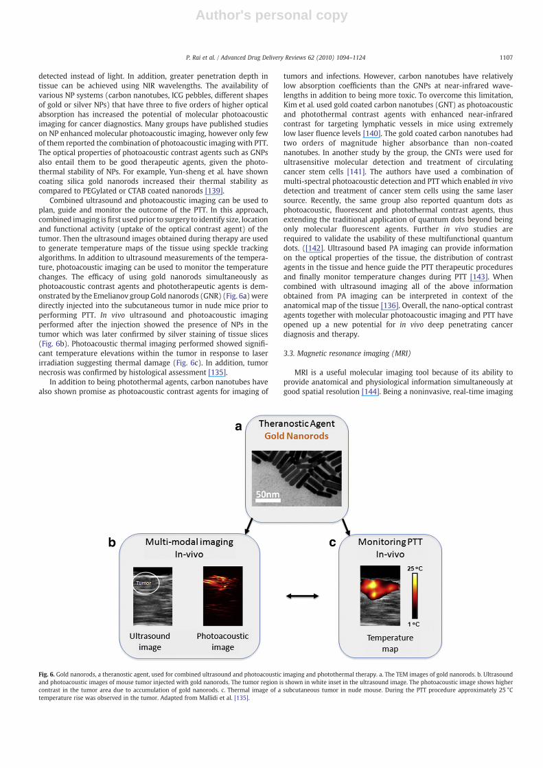
Author's personal copy
detected instead of light. In addition, greater penetration depth intissue can be achieved using NIR wavelengths. The availability ofvarious NP systems (carbon nanotubes, ICG pebbles, different shapesof gold or silver NPs) that have three to five orders of higher opticalabsorption has increased the potential of molecular photoacousticimaging for cancer diagnostics. Many groups have published studieson NP enhanced molecular photoacoustic imaging, however only fewof them reported the combination of photoacoustic imaging with PTT.The optical properties of photoacoustic contrast agents such as GNPsalso entail them to be good therapeutic agents, given the photo-thermal stability of NPs. For example, Yun-sheng et al. have showncoating silica gold nanorods increased their thermal stability ascompared to PEGylated or CTAB coated nanorods [139].
Combined ultrasound and photoacoustic imaging can be used toplan, guide and monitor the outcome of the PTT. In this approach,combined imaging isfirst used prior to surgery to identify size, locationand functional activity (uptake of the optical contrast agent) of thetumor. Then the ultrasound images obtained during therapy are usedto generate temperature maps of the tissue using speckle trackingalgorithms. In addition to ultrasound measurements of the tempera-ture, photoacoustic imaging can be used to monitor the temperaturechanges. The efficacy of using gold nanorods simultaneously asphotoacoustic contrast agents and phototherapeutic agents is dem-onstrated by the Emelianov group Gold nanorods (GNR) (Fig. 6a)weredirectly injected into the subcutaneous tumor in nude mice prior toperforming PTT. In vivo ultrasound and photoacoustic imagingperformed after the injection showed the presence of NPs in thetumor which was later confirmed by silver staining of tissue slices(Fig. 6b). Photoacoustic thermal imaging performed showed signifi-cant temperature elevations within the tumor in response to laserirradiation suggesting thermal damage (Fig. 6c). In addition, tumornecrosis was confirmed by histological assessment [135].
In addition to being photothermal agents, carbon nanotubes havealso shown promise as photoacoustic contrast agents for imaging of
tumors and infections. However, carbon nanotubes have relativelylow absorption coefficients than the GNPs at near-infrared wave-lengths in addition to being more toxic. To overcome this limitation,Kim et al. used gold coated carbon nanotubes (GNT) as photoacousticand photothermal contrast agents with enhanced near-infraredcontrast for targeting lymphatic vessels in mice using extremelylow laser fluence levels [140]. The gold coated carbon nanotubes hadtwo orders of magnitude higher absorbance than non-coatednanotubes. In another study by the group, the GNTs were used forultrasensitive molecular detection and treatment of circulatingcancer stem cells [141]. The authors have used a combination ofmulti-spectral photoacoustic detection and PTT which enabled in vivodetection and treatment of cancer stem cells using the same lasersource. Recently, the same group also reported quantum dots asphotoacoustic, fluorescent and photothermal contrast agents, thusextending the traditional application of quantum dots beyond beingonly molecular fluorescent agents. Further in vivo studies arerequired to validate the usability of these multifunctional quantumdots. ([142]. Ultrasound based PA imaging can provide informationon the optical properties of the tissue, the distribution of contrastagents in the tissue and hence guide the PTT therapeutic proceduresand finally monitor temperature changes during PTT [143]. Whencombined with ultrasound imaging all of the above informationobtained from PA imaging can be interpreted in context of theanatomical map of the tissue [136]. Overall, the nano-optical contrastagents together with molecular photoacoustic imaging and PTT haveopened up a new potential for in vivo deep penetrating cancerdiagnosis and therapy.
3.3. Magnetic resonance imaging (MRI)
MRI is a useful molecular imaging tool because of its ability toprovide anatomical and physiological information simultaneously atgood spatial resolution [144]. Being a noninvasive, real-time imaging
Fig. 6. Gold nanorods, a theranostic agent, used for combined ultrasound and photoacoustic imaging and photothermal therapy. a. The TEM images of gold nanorods. b. Ultrasoundand photoacoustic images of mouse tumor injected with gold nanorods. The tumor region is shown in white inset in the ultrasound image. The photoacoustic image shows highercontrast in the tumor area due to accumulation of gold nanorods. c. Thermal image of a subcutaneous tumor in nude mouse. During the PTT procedure approximately 25 °Ctemperature rise was observed in the tumor. Adapted from Mallidi et al. [135].
1107P. Rai et al. / Advanced Drug Delivery Reviews 62 (2010) 1094–1124
Author's personal copy
methodology, MRI was chosen for guiding and monitoring varioustypes of thermal therapies [144,145]. In particular, Hirsch et al.employed MRI to measure temperature during PTT of tumors injectedwith NIR absorbing nanoshells [115]. The real-time MR temperatureimaging (MRTI) provided depth dependence of thermal profiles inirradiated regions. Tumors injected with gold nanoshells andirradiated with NIR light (820 nm, 4 W/cm2, 5-mm spot diameter,b6 min irradiation time) showed a relative temperature increase ofapproximately 35 °C as compared to 15 °C in tumors injected withsaline. Furthermore, histological examination revealed that MRTIestimation of tissue damage was in good agreement with experimen-tal findings, demonstrating its potential utility in determining tissuedamage during therapy. In another study with gold nanoshells byDiagaradjane et al. MRTI was used to monitor mild hyperthermiacreated in tumors loaded with gold nanoshells using low laser power[146]. The photosensitzation by the mild temperature hyperthermiacaused increased tumor perfusion. Using dynamic contrast-enhancedMRI, vascular perfusion in the tumors was also estimated. The DCE-MRI images clearly showed increase in perfusion at the tumor posthyperthermia. MR temperature monitoring during PTT was also usedin the study by Bruke et al. In this study multi-walled carbonnanotubes were used as therapy agents [147]. Though carbonnanotubes are good light-heat energy convertors, they do not possessmagnetic properties. Only carbon nanotubes for MRI and PT-MRthermometry enables monitoring of heat induction in near real-timeand can thus be used to minimize incomplete treatment of tumormargins, a major limitation of current thermal therapies.
Gadolinium forms the basis for many MRI contrast agents despitehaving low retention time in the body. More recently, superparamagnetic iron oxide particles (SPIOs) have been developed thathave high magnetic moment compared to Gd based contrast agents.By controlling the size of the magnetic cores in these NPs, theirmagnetic properties can be manipulated. SPIO are FDA-approved in1996 and they have been used as MRI contrast enhancers in detectingtumors. Indeed, the efficacy of SPIO NPs as photothermal agents hasalso explored [148]. However, SPIO NPs have very low opticalabsorption at NIR wavelength compared to gold or silver NPs. Toenhance the photo-theranostic abilities of magnetic NPs, hybridnanosystems with gold have been proposed. The complexity of thesehybrid nanosystems ranged from coatingmagnetic iron oxide NPwithgold [149] to wonton shaped nanosystems containing gold and cobalt[150]. Gold covered cobalt NPs had less toxicity compared to highlytoxic cobalt NPs. Apart from gold, magnetic NPs coated with graphitecarbon were also used for PTT enhancement [159]. The magnetic andoptical properties of various hybrid theranostic agents are listed inTable 2. In vitro cell studies were performed using these hybridmultifunctional nanosystems showcasing the photothermal andmagnetic properties. The stability of the hybrid nanosystems interms of storage, biostability, thermal stability still need to beexamined and further in vivo studies are required for the NPs tocreate significant impact in cancer diagnosis and therapy.
3.4. Ionizing imaging modalities
Apart from non-ionizing imaging modalities such as MRI andultrasound, ionizing imaging techniques such as X-ray computedtomography are also widely used for screening and diagnosis ofvarious pathologies including cancer. The ability to visualize deepstructures in the body is the main advantage of ionizing imagingmodalities. Moreover, a recent review by Gupta et al. highlighted thesignificant use of PET/CT in planning and guiding radiotherapy in lungcancer, head and neck cancer and cervical cancer [160]. Recently thestudy performed by Hainfeld et al. showcased gold nanospheres asexcellent X-ray contrast agent, in addition to enhancement ofradiotherapy in vivo [161,162]. Gold has a higher atomic numberand a higher absorption coefficient than standard iodinized contrast
agents and hence provides a 2–2.7-fold greater contrast per unitweight. Therefore a lower concentration of the contrast agent can beused leading to higher sensitivity of the imaging technique. On theother hand, Melancon et al. developed radiolabeled GNPs to observethe accumulation of targeted particles in the tumor [163]. Specificallyimmuno-hollow gold nanospheres targeted to EGFR have been shownto selectively bind to EGFR-positive cells and destroy these cells via aphotothermal effect in vitro. Melancon et al. predict the usage of theseEGFR targeted NPs to increase efficacy of PTT as their initial mouseexperiments showed approximately 7% of the injected dose reachedthe tumor site. The hollow gold nanospheres used in the study did notdisplay their “theranostic” capabilities (i.e., no imaging on tissues orwhole body was performed for diagnosis or treatment monitoring),however by utilizing a γ counter, in vivo tissue distribution of thehollow nanospheres was determined.
The theranostic nature of GNPs was used in a study done byMaltzahn et al. where the gold nano-antennas (gold nanorods ofdimension 13 nm×47 nm) were employed as X-ray contrast agentand photothermal-therapeutic agent [164]. Briefly, the study involveddevelopment of PEGylated gold nanorods for longer half-life in blood(approximately 17 h). The nanorods acted as antennas for acceptingthe externally applied photo energy. The PEGylated nanorodsexhibited greater photothermal heat generation than gold nanoshellsdue to enhanced optical absorption. An integrated approach wasdeveloped for laser irradiation protocol using the bio-distribution ofNPs obtained from X-ray CT images. These quantitative protocolsenabled estimation of temperature rise during PTT. Minimal damageto the surrounding benign tissue was seen due to the specificity of thetherapy procedure that is obtained on two levels — 1. higheraccumulation of nanorods in tumor due to enhanced permeationand retention effect and 2. laser illumination at the tumor site insteadof whole body illumination. Indeed, the study showcased highlyselective ablation of the subcutaneous MDA-MB-435 tumors (breastadenocarcinoma) inmice that were injectedwith 20 mg/kg PEGylatedgold nanorods and laser irradiation (2 W/cm2 at 810 nmwavelength).The results of the study are summarized in Fig. 7. Maltzahn et al.anticipate using gold nano-antennas together with PTT and wholesubject X-ray CT as a route towards personalized diagnosis, radiationplanning, therapy optimization and monitoring response [164]. Inanother study by Lu et al. melanoma-targeted hollow gold nano-spheres (HAuNS) were used for selective photothermal ablation(PTA) [165]. The surface of HAuNs had a chelation agent, S-2-(4-[5-dithiolane-3-pentanamide]benzyl)diethylenetriamine pentaaceticacid (DTPA-TA) which facilitated PET imaging. PTA effect of the NPs
Table 2Hybrid theranostic NPs with potential for combined MRI and PTT. The empty spaces inthe table indicate unavailability of the data.
Nanoparticles Magnetic properties Opticalabsorption(nm)
References
r1 mM−1 s−1 r2 mM−1 s−1 emu/g
Iron oxide core–goldshell
23.5 540 [149]
Magnetic-goldnanoshell
251 700–800 [151]
Magnetic core-silicaand gold coating
0.65 152 825–910 [152]
Magnetic-goldcomposite
465 2 820 [153]
Nano dumbbell 80 520–538 [154,155]Nano roses 219 34 730 [156]Nano wantons 400–800 [150]Au nanoshell —ICG-FeNPs
520–600 [157]
PLGA/Mn/Aucomposite
800–810 [158]
FeCo-graphitecarbon coating
70 644 215 700–1100 [159]
1108 P. Rai et al. / Advanced Drug Delivery Reviews 62 (2010) 1094–1124
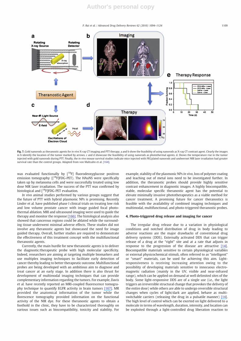
Author's personal copy
was evaluated functionally by [18F] fluorodeoxyglucose positronemission tomography ([18F]FDG-PET). The HAuNS were specificallytaken up by melanoma cells and were successfully treated using lowdose NIR laser irradiation. The success of the PTT was confirmed byhistological and [18F]FDG-PET evaluation.
In vivo animal studies performed by various groups suggest thatthe future of PTT with hybrid plasmonic NPs is promising. RecentlyLinder et al. have published phase I clinical trials on treating low riskand low volume prostate cancer with image guided focal photo-thermal ablation. MRI and ultrasound imaging were used to guide thetherapy and monitor the response [166]. The histological analysis alsoshowed that cancerous regions could be ablated while the surround-ing tissue underwent minimal adverse effects. These studies did notinvolve any theranostic agents but showcased the need for imageguided therapy. Overall, further studies are required to demonstratethe effectiveness of this treatment concept with the multifunctionaltheranostic agents.
Currently, the main hurdle for new theranostic agents is to deliverthe diagnostic/therapeutic probe with high molecular specificity.Indeed, researchers are aiming at targeting multiple biomarkers anduse multiplex imaging techniques to facilitate early detection ofcancer thereby leading to better therapeutic outcome. Multifunctionalprobes are being developed with an ambitious aim to diagnose andtreat cancer at an early stage. In addition there is also thrust fordevelopment of multimodal imaging techniques that can providecomplementary information regarding the tumors. For example, Daviset al. have recently reported an MRI-coupled fluorescence tomogra-phy technique to quantify EGFR activity in brain tumors [167]. MRprovided the anatomical information of the tumor while theflorescence tomography provided information on the functionalactivity of the NIR dye. For these theranostic agents to obtain afoothold in the clinic, they have to be characterized thoroughly onvarious issues such as biocompatibility, toxicity and stability. For
example, stability of the plasmonic NPs in vivo, loss of polymer coatingand leaching out of metal ions need to be investigated further. Inaddition, the theranostic probes should provide highly sensitivecontrast enhancement in diagnostic images. A highly biocompatible,stable, molecular specific theranostic agent has the potential toelevate minimally invasive phototherapeutics as a viable method forcancer treatment. A promising future for cancer theranostics isfeasible with the availability of combined imaging techniques andmultimodal, multifunctional, and photo-triggered theranostic probes.
4. Photo-triggered drug release and imaging for cancer
The irregular drug release due to a variation in physiologicalconditions and notched distribution of drug in body leading toadverse reactions are the major drawbacks of conventional drugdelivery systems (DDS). Externally activated DDS that can triggerrelease of a drug at the “right” site and at a rate that adjusts inresponse to the progression of the disease are attractive [14].Biocompatible materials sensitive to certain physiological variablesor external physicochemical stimuli, often referred to as “intelligent”or “smart” materials, can be used for achieving this aim. Light-responsiveness is receiving increasing attention owing to thepossibility of developing materials sensitive to innocuous electro-magnetic radiation (mainly in the UV, visible and near-infraredrange), which can be applied on demand at well delimited sites of thebody. Some light-responsive DDS are of a single use (i.e., the lighttriggers an irreversible structural change that provokes the delivery ofthe entire dose) while others are able to undergo reversible structuralchanges when cycles of light/dark are applied, behave as multi-switchable carriers (releasing the drug in a pulsatile manner) [14].The high level of control which can be exerted on light delivered to amolecule in terms of wavelength, duration, intensity, and location canbe exploited through a light-controlled drug liberation reaction to
Fig. 7. Gold nanorods as theranostic agents for in vivo X-ray CT imaging and PTT therapy. a and b show the feasibility of using nanorods as X-ray CT contrast agent. Clearly the imagesin b identify the location of the tumor marked by arrows. c and d showcase the feasibility of using nanorods as photothermal agents. d. Shows the temperature rise in the tumorinjected with gold nanorods during PTT. Finally, the in vivomouse survival studies indicate mice injected with PEGylated nanorods and undwernet NIR laser irradiation had greatersurvival rate than the control groups. Adapted from von Maltzahn et al. [164].
1109P. Rai et al. / Advanced Drug Delivery Reviews 62 (2010) 1094–1124
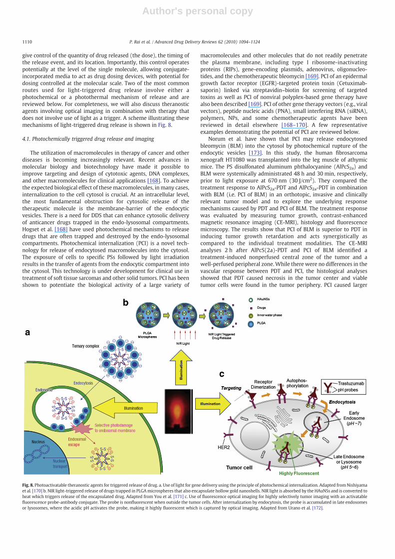
Author's personal copy
give control of the quantity of drug released (the dose), the timing ofthe release event, and its location. Importantly, this control operatespotentially at the level of the single molecule, allowing conjugate-incorporated media to act as drug dosing devices, with potential fordosing controlled at the molecular scale. Two of the most commonroutes used for light-triggered drug release involve either aphotochemical or a photothermal mechanism of release and arereviewed below. For completeness, we will also discuss theranosticagents involving optical imaging in combination with therapy thatdoes not involve use of light as a trigger. A scheme illustrating thesemechanisms of light-triggered drug release is shown in Fig. 8.
4.1. Photochemically triggered drug release and imaging
The utilization of macromolecules in therapy of cancer and otherdiseases is becoming increasingly relevant. Recent advances inmolecular biology and biotechnology have made it possible toimprove targeting and design of cytotoxic agents, DNA complexes,and other macromolecules for clinical applications [168]. To achievethe expected biological effect of thesemacromolecules, in many cases,internalization to the cell cytosol is crucial. At an intracellular level,the most fundamental obstruction for cytosolic release of thetherapeutic molecule is the membrane-barrier of the endocyticvesicles. There is a need for DDS that can enhance cytosolic deliveryof anticancer drugs trapped in the endo-lysosomal compartments.Hogset et al. [168] have used photochemical mechanisms to releasedrugs that are often trapped and destroyed by the endo-lysosomalcompartments. Photochemical internalization (PCI) is a novel tech-nology for release of endocytosed macromolecules into the cytosol.The exposure of cells to specific PSs followed by light irradiationresults in the transfer of agents from the endocytic compartment intothe cytosol. This technology is under development for clinical use intreatment of soft tissue sarcomas and other solid tumors. PCI has beenshown to potentiate the biological activity of a large variety of
macromolecules and other molecules that do not readily penetratethe plasma membrane, including type I ribosome-inactivatingproteins (RIPs), gene-encoding plasmids, adenovirus, oligonucleo-tides, and the chemotherapeutic bleomycin [169]. PCI of an epidermalgrowth factor receptor (EGFR)-targeted protein toxin (Cetuximab-saporin) linked via streptavidin–biotin for screening of targetedtoxins as well as PCI of nonviral polyplex-based gene therapy havealso been described [169]. PCI of other gene therapy vectors (e.g., viralvectors), peptide nucleic acids (PNA), small interfering RNA (siRNA),polymers, NPs, and some chemotherapeutic agents have beenreviewed in detail elsewhere [168–170]. A few representativeexamples demonstrating the potential of PCI are reviewed below.
Norum et al. have shown that PCI may release endocytosedbleomycin (BLM) into the cytosol by photochemical rupture of theendocytic vesicles [173]. In this study, the human fibrosarcomaxenograft HT1080 was transplanted into the leg muscle of athymicmice. The PS disulfonated aluminum phthalocyanine (AlPcS2a) andBLM were systemically administrated 48 h and 30 min, respectively,prior to light exposure at 670 nm (30 J/cm2). They compared thetreatment response to AlPcS2a-PDT and AlPcS2a-PDT in combinationwith BLM (i.e. PCI of BLM) in an orthotopic, invasive and clinicallyrelevant tumor model and to explore the underlying responsemechanisms caused by PDT and PCI of BLM. The treatment responsewas evaluated by measuring tumor growth, contrast-enhancedmagnetic resonance imaging (CE-MRI), histology and fluorescencemicroscopy. The results show that PCI of BLM is superior to PDT ininducing tumor growth retardation and acts synergistically ascompared to the individual treatment modalities. The CE-MRIanalyses 2 h after AlPcS(2a)-PDT and PCI of BLM identified atreatment-induced nonperfused central zone of the tumor and awell-perfused peripheral zone. While there were no differences in thevascular response between PDT and PCI, the histological analysesshowed that PDT caused necrosis in the tumor center and viabletumor cells were found in the tumor periphery. PCI caused larger
Fig. 8. Photoactivatable theranostic agents for triggered release of drug. a. Use of light for gene delivery using the principle of photochemical internalization. Adapted fromNishiyamaet al. [170] b. NIR light-triggered release of drugs trapped in PLGAmicrospheres that also encapsulate hollow gold nanoshells. NIR light is absorbed by the HAuNSs and is converted toheat which triggers release of the encapsulated drug. Adapted from You et al. [171] c. Use of fluorescence optical imaging for highly selectively tumor imaging with an activatablefluorescence probe-antibody conjugate. The probe is nonfluorescent when outside the tumor cells. After internalization by endocytosis, the probe is accumulated in late endosomesor lysosomes, where the acidic pH activates the probe, making it highly fluorescent which is captured by optical imaging. Adapted from Urano et al. [172].
1110 P. Rai et al. / Advanced Drug Delivery Reviews 62 (2010) 1094–1124

Author's personal copy
necrotic areas and the regrowth in the peripheral zone was almostcompletely inhibited after PCI. The results indicate that PDT is lessefficient in the tumor periphery than in the tumor center and that thetreatment effect of PCI is superior to PDT in the tumor periphery [173].
PCI has also been shown to enhance the treatment effect oftargeted therapeutic macromolecules. Selbo et al. have combined PCIwith a recombinant and targeted agent and demonstrated that thisnoninvasive, multimodal approach enhances the in vivo efficacywithout sacrificing selectivity or enhancing systemic toxicity [174].They used a recombinant single-chain fusion construct scFvMEL/rGel,composed of an antibody targeting the progenitor marker HMW-MAA/NG2/MGP/gp240 and the highly effective toxin gelonin (rGel).They have demonstrated enhanced tumor cell selectivity, cytosolicdelivery and antitumor activity by applying PCI of scFvMEL/rGel. PCIperformed by light activation of cells co-incubated with scFvMEL/rGeland the endo-lysosomal targeting PSs AlPcS2a or TPPS2a resulted inenhanced cytotoxic effects against antigen-positive cell lines, while nodifferences in cytotoxicity between the scFvMEL/rGel and rGel wereobserved in antigen-negative cells. Mice bearing well-developedmelanoma (A-375) xenografts (50–100 mm3) were treated with PCIof scFvMEL/rGel. By 30 days after injection, approximately 100% ofmice in the control groups had tumors N800 mm3. In contrast, by day40, 50% of mice in the PCI of scFvMEL/rGel combination group hadtumors b800 mm3 with no increase in tumor size up to 110 days. PCIof scFvMEL/rGel resulted in a synergistic effect (pb0.05) andcomplete regression (CR) in 33% of tumor-bearing mice (n=12).Such an approach warrants further evaluation of its clinical potential[174].
Multiple drug resistance (MDR) is a problem that seriouslyreduces the efficacy of many chemotherapy agents. One mechanismfor MDR is increased acidification of endocytic vesicles and increasedcytosol pH, so weak base chemotherapeutic agents, includingdoxorubicin, are trapped in endocytic vesicles and exhibit a drug-resistant phenotype [175]. Treatments that selectively reverse thisaccumulation may therefore reverse the MDR phenotype. Lou PJ et al.evaluated the potential of PCI for release of doxorubicin fromendocytic vesicles in MDR cells [175]. Two breast cancer cell lines,MCF-7 and MCF-7/ADR (the latter resistant to doxorubicin), wereselected. They were found equally sensitive to photochemicaltreatment with the photosensitiser TPPS2a (disulfonated meso-tetraphenylporphine) and light. On exposure to doxorubicin alone,the IC50 (drug concentration for 50% reduction in colony formation)was 0.1 μM for MCF-7 and 1 μM for MCF-7/ADR. After PCI (photo-chemical treatment followed by doxorubicin), the IC50 concentrationwas 0.1 μM for both cell lines. Comparable changes were seen withassay of cell viability using 3-(4,5-dimethyltiazol-2-yl)-2,5-diphenyl-tetrazolium bromide (MTT). On fluorescence microscopy in MCF-7/ADR cells, doxorubicin localized in granules identified as lysosomes.After PCI, doxorubicin was released into the cytosol and entered cellnuclei, as was seen in MCF-7 cells without PCI. PCI reversed the MDRphenotype of doxorubicin resistant breast cancer cells by endo-lysosomal release of the drug [175]. The technique is a promising newapproach to tackling the problem of MDR.
Febvay et al. have presented a method for precise spatial andtemporal control over cytosolic delivery of compounds that wouldotherwise be cell-impermeable by using PCI mechanism within a celltargetable mesoporous silica nano-carrier [176]. Size-tunable (30–200 nm), highly monodispersed mesoporous silica NPs that can bebiofunctionalized and targeted to specific cell surface proteins werefirst developed. As an example, they delivered a cell-impermeablefluorescent compound exclusively to the cytosol of multidrug-resistant cancer cells in a mixed population that was verified byconfocal microscopy. These NPs can be loaded with a wide variety ofcompounds and can mediate cytosolic release of cell-impermeablemolecules in P-gp-expressing cells via light-mediated endosomalbreakage. Such an approach is promising in expanding the pharma-
cological arsenal for cell-impermeable compounds to overcomemultidrug resistance. In addition, these novel NPs may be usefulvectors for highly specific protein and nucleic acid delivery [176].
Recently, light has also been used to release therapeutic agentsfrom delivery systems or to activate cytotoxic drugs. Pashkovskaya etal. have studied photosensitized damage to liposome membranes byusing different dye-leakage assays based on fluorescence dequench-ing of a series of dyes upon their release from liposomes [177]. Theirwork demonstrated the participation of oxidative damage tomembrane lipids in the photosensitized membrane destructionwhich could play a role in many of the toxic as well as therapeuticeffects of photodynamic action [177]. In particular, photosensitizeddamage to lipids in liposomemembranes was shown tomanifest itselfin the formation of pores with rather high selectivity. These poresproved to be permeable not only for fluorescent markers but also forPSs, e.g., AlPcS3. Thus, the basis of targeted delivery of PSs viaencapsulation and subsequent light-induced release from liposomesas an approach for PDTwas experimentally confirmed in a very simplemodel system. Moreover, the present data provide the rationale forselective liposome-based drug delivery [177].
Further, liposomes made with light-sensitive lipids have beendeveloped to release their contents when exposed to near-UV lightdue to changes in membrane permeability [178]. Photosensitiveliposomes have been studied for a few decades and various photo-sensitive triggers have been developed so far. A recent review byWang et al. offers an overview of the different photo-triggeringmechanisms for controlled pulsatile content release from liposomes,which have the potential of finding clinical applications as intelligentDDS [178]. Lu et al. have reported the use of nanoimpeller-controlledmesostructured silica NPs to deliver and release anticancer drugs intoliving cells upon external command [179]. Using light-activatedmesostructured silica (LAMS) NPs, luminescent dyes and anticancerdrugs are only released inside of cancer cells that are illuminated atthe specific wavelengths that activate the impellers. The quantity ofmolecules released is governed by the light intensity and theirradiation time. Human cancer cells (a pancreatic cancer cell line,PANC-1 and a colon cancer cell line, SW480) were exposed tosuspensions of the particles and the particles were taken up by thecells. Confocal microscopy imaging of cells containing the particlesloaded with the membrane impermeable dye, propidium iodide (PI),shows that the PI is released from the particles only when theimpellers are photoexcited, resulting in staining of the nuclei. Theanticancer drug camptothecin (CPT)was also loaded into and releasedfrom the particles inside the cells under light excitation, and apoptosiswas induced. Intracellular release of molecules is sensitively con-trolled by the light intensity, irradiation time, and wavelength, andthe anticancer drug delivery inside of cells is regulated under externalcontrol. The delivery and release capability of light-activatedmesostructured silica particles containing molecular impellers is thefirst step towards a novel platform for the next generation ofnanotherapeutics with both spatial and temporal external control[179].
Dvir et al. have recently described the use of light to target NPbinding (as compared to single release events) in specific illuminatedareas [180]. The basic design is a drug-loaded NP whose surface iscovalently modified with a targeting moiety consisting of an avid butnonspecific ligand that is rendered biologically nonfunctional(“caged”) and prevented from binding by chemical modificationwith a photoremovable protecting group. The caging group isremoved at the desired site by illumination. For proof of concept,the authors used commercially available carboxylated polystyreneNPs with diameters of 328±2 nm as model NPs. The nonspecificligand was a small peptide YIGSR, an amino acid sequence in lamininthat is crucial for adhesion to integrin β1 on the cell membrane of abroad range of potential target cell types including stromal andendothelial cells, which are present in all tissues. The biological
1111P. Rai et al. / Advanced Drug Delivery Reviews 62 (2010) 1094–1124
Author's personal copy
activity of YIGSR can be greatly attenuated by mutation or deletion oftyrosine. Therefore caging the tyrosine with 4,5-dimethoxy-2-nitrobenzyl (DMNB) would inactivate the peptide until the cagewas removed by illumination. This work by Kohane et al. is the firstexample of a targeting system capable of binding NPs to cellsselectively upon illumination [180]. In contrast to previous workwhere NPs have been triggered to produce a single drug release eventby light, this approach results in the deposition of a sustained releasesystem at the desired site. Another important point is that this systemallows tissue targeting without specific markers (since the receptorfor the ligand used in these experiments is ubiquitous throughout thebody), provided the tissue can be illuminated. Furthermore, thisapproach could be used with specific ligands to perhaps furtherenhance specificity. The potential applicability of caging to a spectrumof potential ligands is seen in the fact that it has been used toinactivate a wide range of biomolecules including peptides, enzymes,nucleotides, mRNA and DNA [180]. In vivowork will require the use ofNPs with direct applicability in drug delivery, such as liposomes andbiodegradable polymeric nanospheres. Other modifications may alsobe necessary, such as surface optimization to minimize uptake by thereticuloendothelial system, for example, by PEGylation. The wave-lengths at which DMNB can be made to uncage (350–400 nm) limitsthis particular application of this technology to areas of the body thatcan be illuminated directly, like for skin cancers. However, the use oflasers and minimally invasive fiber-optic tools, and the developmentof new caging groups that respond to wavelengths with better tissuepenetration such as near-infrared, may make direct targeting of deeptissues possible.
Drug release triggered by two-photon excitation in NIR usingFRET has not yet been developed despite progress in photo-triggereddrug release. The approach of two-photon induced intraparticle FRETfor drug release, based on the use of two-photon fluorescent nano-assembly as a donor and a photo-sensitive linker as an acceptor,offers a novel design for developing formulations of smart drug-carrier nanoassemblies for more superior control over the locationand the onset of drug release [181]. Banerjee et al. have developed amultifunctional NP that can efficiently up-convert the energy of NIRlight for triggering drug release by cleavage of a photosensitivelinker [181]. The NP and the underlying nanophotonics approachdescribed in this work represents a significant breakthrough indeveloping a two-photon-triggered drug delivery vehicle thatcombines imaging, sensing and therapy and drug targeting at thesame time [181].
Light-triggered theranostic nanocarriers could revolutionize can-cer chemotherapy. Most chemotherapeutic compounds are nonspe-cific and are taken up by all cell types — and this nonselective natureof the agents usually causes severe toxicity [182]. The drugnanocarriers reviewed here have the potential to dramaticallyimprove the treatment of cancer by selectively providing theoptimum dosage of the drug at the tumor site, ultimately even tothe individual tumor cell. However, most of these stimuli-responsiveDDSs are indirectly triggered, as they induce a macroscopic change inthe matrix into which the drug is incorporated [13,14]. To date, nomethod to externally and directly trigger precise drug doses to atargeted area has been demonstrated. McCoy et al. show a molecularmethod for light-triggered drug delivery of various drug classes usinglow energy, long wavelength radiation, with the drug dose beingprecisely controlled by the duration of applied light [183]. Togetherwith an appropriate polymer matrix, the molecular unit acts as amolecule-scale drug dosing device, with control potentially at thelevel of a single drug molecule [13].
4.2. Photothermally triggered drug release and imaging
Combination therapies are commonly employed in a wide range ofcancer treatments as they have a better therapeutic response
compared to monotherapies. In particular, hyperthermia can increasethe concentration of an administered therapeutic NP in a tumoralregion by increasing blood flow and vessel permeability. Cavitationbubbles produced following irradiation of GNPs can also be used forintracellular drug delivery of cell-impermeable biomolecules likeantibodies. For e.g., Yao et al. recently demonstrated intracellulardelivery of an antibody following light irradiation using GNPs [184]. Inthis work lymphoma cells were targeted with GNPs that wereconjugated to anti-CD-30 antibody. Pulsed laser irradiation led totransient permeabilization of cell membranes and this lead tosubsequent internalization of a secondary dye-labeled antibody.Delivery of the secondary dye-labeled antibody was confirmed byflow cytometry. The membrane permeabilization only occurred incells expressing the CD30 receptor. No effect was seen with CD30negative cells.
Hyperthermia can also enhance drug toxicity in cancer cells thatare otherwise resistant to chemotherapeutics [185]. Furthermore,local hyperthermia can improve the accumulation of a drug, which isencapsulated in a thermosensitive carrier [185]. The combination ofhyperthermia and chemotherapeutics can, therefore, be employedsynergistically to treat high-risk tumors with a goal of total tumoreradication. From a clinical perspective, precise and site-specific heattransfer to a diseased site would improve the safety and efficacy ofthermal cancer therapies [185]. Park et al. have demonstrated that apair of synthetic NPs can work together to detect a diseased site andmore effectively deliver chemotherapeutics to the site than individualNP treatments [186]. This system relies on GNR transduction of anexternal optical signal into a tumor-specific thermal signal thatenhances recruitment of circulating drug carriers into the tumor andtriggers drug release from the carriers. GNRs localized in tumors canbe identified in vivo by their intense SERS signals, making it possible touse these nanoagents in both diagnostic and therapeutic applicationsand illustrates the potential advantage of dual therapeutic nanoma-terials in the precision treatment of more drug-resistant cancers[186].
Photosensitive caged compounds have enhanced our ability toaddress the complexity of biological systems by generating effectorswith remarkable spatial/temporal resolutions. The caging effect istypically removed by photolysis with ultraviolet light to liberate thebioactive species. Although this technique has been successfullyapplied to many biological problems, it suffers from a number ofintrinsic drawbacks [187]. For example, it requires dedicated efforts todesign and synthesize a precursor compound for each effector. Theultraviolet light may cause damage to biological samples and issuitable only for in vitro studies because of its rapid attenuation intissue. Yavuz et al. have addressed these issues by developing aplatform based on the photothermal effect of gold nanocages [187].Gold (Au) nanocages represent a class of nanostructures with hollowinteriors and porous walls. They can have strong absorption (for thephotothermal effect) in the near-infrared while maintaining acompact size. When the surface of the Au nanocage is covered witha smart polymer, the pre-loaded effector can be released in acontrollable fashion using NIR irradation. This system works wellwith various effectors without involving sophisticated syntheses, andis well suited for in vivo studies owing to the high transparency of softtissue in the near-infrared region. They have demonstrated a platformbased on Au nanocages covered with smart polymers for controlledrelease with NIR light [187]. When combined with optical manipula-tion, this platform offers many extra advantages such as high spatial/temporal resolution. In addition, Au nanocages are bio-inert and thesurface can be readily functionalized with targeting ligands such asantibodies using the gold-thiolate chemistry.
Silica–gold (SiO2–Au) nanoshells are a new class of NPs thatconsist of a silica dielectric core that is surrounded by a gold shell.These nanoshells are unique because their peak extinctions are veryeasily tunable over a wide range of wavelengths particularly in the
1112 P. Rai et al. / Advanced Drug Delivery Reviews 62 (2010) 1094–1124
Author's personal copy
NIR region of the spectrum. Light in this region is transmitted throughtissuewith relatively little attenuation due to absorption. As discussedin the PTT section, irradiation of SiO2–Au nanoshells at their peakextinction coefficient results in the conversion of light to heat energythat produces a local rise in temperature. Thus, to develop aphotothermal modulated drug delivery system, Bikram et al. havefabricated nanoshell-composite hydrogels in which SiO2–Au nano-shells of varying concentrations have been embedded withintemperature-sensitive hydrogels, for the purpose of initiating atemperature changewith light. N-isopropylacrylamide-co-acrylamide(NIPAAm-co-AAm) hydrogels are temperature-sensitive hydrogelsthat were fabricated to exhibit a lower critical solution temperature(LCST) slightly above body temperature [188]. The resulting compos-ite hydrogels had the extinction spectrum of the SiO2–Au nanoshellsin which the hydrogels collapsed reversibly in response to temper-ature (50 °C) and laser irradiation. The degree of collapse of thehydrogels was controlled by the laser fluence as well as theconcentration of SiO2–Au nanoshells. Modulated drug deliveryprofiles for methylene blue, insulin, and lysozyme were achieved byirradiation of the drug-loaded nanoshell-composite hydrogels, whichshowed that drug release was dependent upon the molecular weightof the therapeutic molecule [188].
As discussed, the combination of chemotherapeutics and hyper-thermia has been an emerging approach for cancer therapy. However,this combinatorial therapy is highly requisite to deliver drugs andlocalized heating to the cancerous area. Recently, Cheng et al. havedeveloped stabilizer-free taxol-loaded poly(lactic-co-glycolic acid)(PLGA) NPs [189]. These can be directly surface conjugated to otherNPs, like gold nanorods (GNR), iron oxide NP (Fe3O4), quantum dots(QD) and the inner core can be used to encapsulate drugs topotentially serve as multifunctional probes. In this work, they havedemonstrated efficacy of a nanoplatform that combined chemother-apy and PTT in vitro, in mammalian cells and in vivo, in an animalmodel of lung cancer. A significant enhancement of the anticancereffect was observed when chemotherapy and photothermal destruc-tion were combined, with GNR/QD/Fe3O4/Taxol-loaded PLGA NPsinjection being followed by laser irradiation [189]. Since the ironoxide NPs are decorated on PLGA NPs, they can potentially serve as acontrast agent for MRI. The GNR/QD/Fe3O4/Taxol-loaded PLGA NPswere further administered to A549 (lung cancer cells)-induced SCIDmice and the tumor was imaged using MRI. In mice that receivedbinary treatment, the tumor growth was suppressed and the tumorstended to shrink as the test period went on. The mice treated withchemotherapy and photothermal destruction remained alive aftertwo months, and the tumors of treated mice either decreased 100% orshowed no sign of regrowth after therapy [189].
Liposomes show great promise as intravenous drug deliveryvehicles, but it is often difficult to combine stability in the circulationwith rapid, targeted release at the site of interest. Targeting tospecific tissues requires developing highly specific ligands withstrong affinities to receptors over-expressed on diseased cells; a newcellular target requires developing new ligands and identifying newreceptors. A novel proof of principle demonstration for contentsrelease from liposomes that can be selectively activated by lightirradiation is presented by Paasonen et al. [190]. The content releasetemperature was adjusted to slightly above body temperature, andhydrophobic or hydrophilic GNPs were incorporated into the lipidbilayer or the core of the liposomes, respectively. The release of afluorescent marker was monitored upon exposure of the liposomesto UV light. GNP-containing liposomes remained intact at 37 °C butcontents release was triggered by UV light-induced heating of theGNPs [190].
Drug release has also been triggered by irradiating liposomesconjugated to hollow gold nanoshells with a near-infrared (NIR)pulsed laser [191]. Novel photoactivated, hollow, gold nanoshell(HGN)/liposome composites provide a new approach to both
controlled release and specific targeting. HGN are extremely efficientNIR light absorbers, and are not susceptible to photobleaching likeconventional dyes. Near-complete liposome contents release can beinitiated within seconds by irradiating HGNs with an NIR pulsed laser.Targeting the drug is limited only by the dimensions of the laserbeam; no specific ligands or antibodies are required, so differenttissues and cells can be targeted with the same HGN/liposomes. HGNscan be encapsulated within liposomes or tethered to the outer surfaceof liposomes for the most efficient drug release. HGNs in liposomesolutions can also trigger release, but with lower efficiency. Drugrelease is induced by adsorbing femto- to nanosecond NIR light pulsesthat cause the HGNs to rapidly increase in temperature. The resultinglarge temperature gradients lead to the formation of vapor micro-bubbles in aqueous solutions, similar to the cavitation bubblesinduced by sonication. The collapse of the unstable vapor bubblescauses liposome-membrane rupture and contents release, withminimal damage to the surroundings, and little overall heating ofthe solution. Wu et al. have reviewed the use of such hybrids forphotothermally-triggered drug release and imaging [191]. NIR lightcan penetrate up to 10 cm into tissue, which should allow theseliposome/HGN complexes to be addressed noninvasively within areasonable fraction of the human body. Any liposome carrier could bemodified by tethering or encapsulating HGN to produce a system forrapid release on demand via NIR irradiation. This should eventuallyallow for better control of drug delivery to selected disease sites whileminimizing systemic toxicity [191].
The hypothesis that the photothermal effect mediated by a NIRlaser and hollow gold nanospheres (HAuNSs) could modulate therelease of anticancer agents was tested by You Jian et al. usingbiodegradable and biocompatible microspheres (1–15 μm) containingthe antitumor drug paclitaxel (PTX) and HAuNSs (~35 nm indiameter), which display surface plasmon absorbance in the NIRregion [171]. HAuNS-containing microspheres exhibit NIR-inducedthermal effect similar to that of plain HAuNSs. Rapid, repetitive PTXrelease from the PTX/HAuNS-containing microspheres is observedupon irradiation with NIR light (808 nm), whereas PTX release isinsignificant when the NIR light is switched off. The release of PTXfrom the microspheres is readily controlled by the output power ofthe NIR laser, duration of irradiation, treatment frequency, andconcentration of HAuNSs embedded inside the microspheres. Invitro, cancer cells incubated with PTX/HAuNS-loaded microspheresand irradiated with NIR light display significantly greater cytotoxiceffects than cells incubated with the microspheres alone or cellsirradiated with NIR light alone, owing to NIR light-triggered drugrelease. Treatment of human U87 gliomas and MDA-MB-231mammary tumor xenografts in nude mice with intratumoral injec-tions of PTX/HAuNS-loaded microspheres followed by NIR irradiationresults in significant tumor growth delay compared to tumors treatedwith HAuNS-loaded microspheres (no PTX) and NIR irradiation orwith PTX/HAuNS-loaded microspheres alone. The data support thefeasibility of a therapeutic approach in which NIR light is used forsimultaneous modulation of drug release and induction of photo-thermal cell killing [171]. Dual-functional hollow gold nanospheres(HAuNS, ~40-nm diameter) capable of mediating both photothermalablation of cancer cells and drug release upon near-infrared (NIR)light irradiation has been reported by You Jian et al. [192]. As high as63% DOX by weight could be loaded to polyethylene glycol (PEG)-coated HAuNS since DOX was coated to both the outer and the innersurfaces of HAuNS. Irradiation with NIR laser induced photothermalconversion, which triggered rapid DOX release from DOX-loadedHAuNS. The release of DOX was also pH-dependent, with more DOXreleased in aqueous solution at lower pH. Significantly greater cellkilling was observed when MDA-MB-231 cells incubated with DOX-loaded HAuNS were irradiated with NIR light, attributable to bothHAuNS-mediated photothermal ablation and cytotoxicity of releasedfree DOX [192]. This approach is advantageous in several aspects.
1113P. Rai et al. / Advanced Drug Delivery Reviews 62 (2010) 1094–1124
Author's personal copy
HAuNS displayed exceptionally high drug loading capacity andstability as exemplified by DOX owing to the unique physicochemicalcharacteristics of HAuNS. In particular, the hollow interior of the NPsallowed significant increase in effective surface area for DOXattachment, resulting in a 3.5-fold increase in DOX payload comparedwith solid GNP of the same size, surface charge, and weight. HAuNSmediated a strong photothermal effect owing to their strong surfaceplasmon absorption in the NIR region. This property was exploited forthe controlled release of DOX fromDOX-loaded HAuNS using NIR lightas the external stimulus to trigger drug release. Dual modality of cellkilling were integrated into a single nanodevice, that is, photothermalablation mediated by HAuNS and antitumor activity of DOX releasedfrom HAuNS upon NIR laser irradiation. This “two-punch” approach isexpected to significantly increase the likelihood of cell killing andpotentially overcome resistance to chemotherapeutic agents, makingit a promising approach to cancer therapy [192].
Hollow layer-by-layer (LbL) capsules can be refilled with variousmolecules for drug delivery. Drug release can be activated on demandby several remote physical stimuli. For example, Skirtach et al.demonstrated the selective addressing of intra cellular LbL micro-capsules with laser light. The release of encapsulated material frompolyelectrolyte-multilayer capsules has been demonstrated insideliving cells. Metal NPs were incorporated inside the walls of thecapsules, and served as energy absorbing centers for illumination bylaser light. AF-488 dextran was successfully incorporated into thecapsules using a novel heat-shrinking method. The capsules obtainedby such a method exhibit improved mechanical stability — propertiesimportant for the delivery of encapsulated material. Upon illumina-tion by laser light, the encapsulated dextran leaves the interior of acapsule inside a living cancer cell. This study serves as a significantstep toward the use of polyelectrolyte-multilayer capsules for thedelivery of medicine into biological cells. The presented method isdifferent from previous, albeit also important, studies in that it isconducted on an individual capsule level and offers an improveddegree of control and monitoring. Release from polyelectrolytemicrocapsules functionalized with metal NPs, by burst opening anddeformation, has been demonstrated by Volodkin et al. [193]. Theyhave also demonstrated temperature-triggered release of a liposomecargo from surface-supported vesicles embedded inside biocompat-ible polyelectrolyte multilayers. In an effort to enlarge the scope ofapplication of remote release and to extend it further to other surface-supported drug delivery vesicles, Volodkin et al. [194] have appliedremote release to liposome-GNPs, referred to as assemblies orcomplexes (Lip-NP). The goals of this work were to show that Lip-NP assemblies could be prepared in a controlled manner in terms ofsize and NP state and then to use near-IR light to selectively releaseencapsulated dye from the assemblies. Functionalized liposome-NPassemblies can be used for transdermal applications in which anactive compound is delivered through the skin, which is easilyaccessible by light. Due to quite deep IR light tissue penetration, thelight-responsive liposome assemblies could serve as active constitu-ents of implanted devices. In a recent study, Volodkin et al. havereported on the functionalization of layer-by-layer films with GNPs,microcapsules, and DNAmolecules by spontaneous incorporation intothe film [195]. Exponentially growing films from biopolymers,namely, hyaluronic acid (HA) and poly-L-lysine (PLL), and linearlygrowing films fromthe synthetic polymers, namely, poly(styrenesulfonate) (PSS) and poly(allylamine hydrochloride) (PAH), wereexamined for the embedding. The HA/PLL film studied here possesseshigh loading capacity as a result of polymer doping onto the filmsurface that results in the accumulation of a large amount of adsorbingmaterial, which is many times less for the PSS/PAH film that has lowpolymer mobility. Microcapsules, GNPs, and DNA can be embedded inthe HA/PLL film and located on the film surface. The diffusion ofembedded DNA into the film can be triggered by heating. The HA/PLLfilm with adsorbed GNPs and DNA possesses remote release features
by stimulation with “biofriendly” IR light. DNA release from the filmmodified with GNPs is supposed to be caused by the local destructionof the polymer network in the film followed by the blocking of PLL-DNA bonding and, as a result, the release of DNA molecules from thefilm. Laser activation of film-supported capsules shows the remoterelease of encapsulated dextran. This study can serve futurebiomedical applications in tissue engineering and biocoatings wherehigh loading capacity together with remote release functionalities isdemanded. Light-triggered DNA transfection to a single cell can alsobe achieved by this approach. The mechanism of the release isdependent on the disturbance in bonding between “doping” PLL andDNA, which is induced by local thermal decomposition of the HA/PLLnetwork in the film when the film is exposed to IR light. Remote IRlight activation of dextran-filled microcapsules modified by GNPs andintegrated into the HA/PLL film is also demonstrated, revealing analternative release pathway using immobilized light-sensitive carriers(microcapsules) [195].
Carbon nanotubes are unique materials that absorb infrared (IR)radiation, especially between 700 and 1100 nm, where body tissuesare most transparent. Absorbed IR promotes molecular oscillationleading to efficient heating of the surrounding environment. Amethod to enhance drug localization for peritoneal malignancies isperfusion of warm (40–42 °C) chemotherapeutic agents in theabdomen. However, all tissues in the peritoneal cavity are subjectedto enhanced drug delivery due to increased cell membrane perme-ability at hyperthermic temperatures. Levi et al. have shown thatrapid heating (within ten seconds) of colorectal cancer cells to 42 °C,using infrared stimulation of nanotubes as a heat source, in thepresence of the drugs oxaliplatin or mitomycin C, is as effective as 2 hof radiative heating at 42 °C for the treatment of peritonealdissemination of colorectal cancer [196]. This approach has thepotential to be used as a rapid bench to bedside clinical therapeuticagent with significant impact for localizing chemotherapy agentsduring the surgical management of peritoneal dissemination ofcolorectal cancer. The method is quite simple since no attachmentof the NP to the drug is required. Yet, the effect is significant increasein the amount of agent that is retained in the cells. The evaluations ofnanotubes and other NPs reported to date have focused ontherapeutic delivery or thermal ablation. However, most cancertreatments involve a combination of surgery, chemotherapy, andradiation. Intraperitoneal hyperthermic chemoperfusion using carbonnanotubes could be utilized as a rapid bench to bedside techniquesince it can be applied during surgical procedures, all the nanomater-ial can be removed from the body following delivery, and hyperther-mia can be localized. MWNT induced chemotherapy for metastaticperitoneal cancer can be clinically applied during open abdominalprocedures by filling the abdomen with a solution of nanotubes andchemotherapeutic agent and applying infrared light only to the tumornodules. MWNT would be directly introduced into the treatmentregion and not intravenously introduced due to the lack of systemicdelivery of therapeutic agents from the bloodstream to the peritone-um. It is beneficial to minimize the surgical time to reduce the timethat a patient is anesthetized, to maximize recovery and minimizecomplications due to prolonged anesthesia. For example, to surgicallyremove a tumor nodule may take 5 min but laser application wouldtake only a few seconds. This would significantly reduce the overalltime of the surgery. Furthermore, the nanotubes could be easilyremoved from the abdomen following hyperthermic chemotherapydelivery by flushing the abdomen with saline after the procedure.Future evolutions of nanotubes used for dissemination of metastaticperitoneal cancers include (1) targeting nanotubes to specific tumortypes using antibodies, bacteriophages, or other moieties such as folicacid; or (2) using nanotubes to induce hyperthermic chemotherapy ina closed abdominal procedure using laproscopic techniques and afiber-optic infrared source. IPHC using carbon nanotubes has theclinical potential to reduce treatment times for hyperthermic
1114 P. Rai et al. / Advanced Drug Delivery Reviews 62 (2010) 1094–1124

Author's personal copy
chemotherapy by localizing heat, thus aiding in the penetration ofchemotherapeutics into malignant tumor cells, and hence reducingthe overall treatment time and increasing the effectiveness oftreatment and patient survival [196].
The development of systems for releasing guest molecules frommesoporous silica using molecular and supramolecular concepts iscurrently taking chemistry to the forefront of nanoscience. Aznar et al.have shown that it is possible to obtain a simple and very effectiveguest release control using polyalcohol entities anchored ontomesoporous materials and boronic acid functionalized GNPs aseffective caps [197]. Both pH-controlled and NIR light-controlleddelivery effects have been observed to occur in pure water. In relationto the pH-controlled delivery, the release of the cargo is inhibited atpH 5, whereas there is rapid release of the guest molecule from themesoporous silica scaffolding at pH 3. The pH-controlled release isreversible, and the entrapped guests can be delivered in installmentsby simple changes in the pH. The pH-controlled “open-close”mechanism is associated with the reversible formation of boroestersbetween alcohol groups and boronic acid functionalized NPs (closed-gate) and their quick and easy hydrolysis (open-gate). At the sametime the use of GNPs opens the way to employing light as a suitablestimulus for release procedures. This is related to the ability of GNPs toraise their temperature locally by absorbing laser light. Plasmonicheating results in cleavage of the boronic ester linkage that anchorsthe NPs to the surface of the mesoporous silica-based material,allowing the release of the cargo. A fine-tune of the amount of cargodelivered by simply controlling the laser irradiation is also possible.Both pH and light are easy-to-use external stimuli and appealingmethods of releasing entrapped guests that could be used to developnew controlled delivery systems for a wide range of applications. Thepossibility of using these stimuli for delivering cargo in small portionsalso opens the possibility of designing stimuli-induced pulsatilerelease supports. Plasmonic heating results in cleavage of the boronicester linkage that anchors the NPs to the surface of the mesoporoussilica-based material, allowing the release of the cargo. A fine-tune ofthe amount of cargo delivered by simply controlling the laserirradiation is also possible. Both pH and light are easy-to-use externalstimuli and appealing methods of releasing entrapped guests thatcould be used to develop new controlled delivery systems for a widerange of different applications. The possibility of using these stimulifor delivering cargo in small portions also opens the possibility ofdesigning multi-stimuli-induced pulsatile release supports [197].
4.3. Combined optical imaging and therapy
Several agents that can be used for therapy in combination withoptical imaging are also being developed for cancer applications and afew of these agents are reviewed here. NIR fluorophores have severaladvantages over visible fluorophores, including improved tissuepenetration and lower autofluorescence [172,198–200]; however,only indocyanine green (ICG) is clinically approved. Its use inmolecular imaging probes is limited because it loses its fluorescenceafter protein binding. This property can be harnessed to create anactivatable NIR probe. After cell binding and internalization, ICGdissociates from the targeting antibody, thus activating fluorescence.In work done by Ogawa et al. ICG was conjugated to the therapeuticantibodies daclizumab (Dac), trastuzumab (Tra), or panitumumab(Pan) [201].The conjugates had almost no fluorescence in PBS butbecame fluorescent after SDS and 2-mercaptoethanol, with aquenching capacity of 10-fold for 1:1 conjugates and 40- to 50-foldfor 1:5 conjugates. In vitro microscopy showed activation within theendolysosomes in target cells. In vivo fluorescent imaging in miceshowed that CD25-expressing tumors were specifically visualizedwith Dac-ICG. Furthermore, tumors overexpressing HER1 and HER2were successfully characterized in vivo by using Pan-ICG(1:5) and Tra-ICG(1:5), respectively. Thus, they have developed an activatable NIR
optical probe that “switches on” only in target cells. Because both theantibody and the fluorophore are FDA approved, the likelihood ofclinical translation is improved [201].
A long-term goal of cancer diagnosis is to develop tumor imagingtechniques that have sufficient specificity and sensitivity. To achievethis goal, minimizing the background signal originating from non-target tissues is crucial. Urano et al. have achieved highly specific invivo cancer visualization by using a newly designed targeted‘activatable’ fluorescent imaging probe [172]. This agent is activatedafter cellular internalization by sensing the pH change in thelysosome. Novel acidic pH-activatable probes based on the boron–dipyrromethene fluorophore were synthesized and then conjugatedto a cancer-targeting monoclonal antibody. As proof of concept, exvivo and in vivo imaging of human EGFR type 2-positive lung cancercells in mice was performed. The probe was highly specific for tumorswith minimal background signal. Furthermore, because the acidic pHin lysosomes is maintained by the energy-consuming proton pump,only viable cancer cells were successfully visualized. The designconcept can be widely adapted to cancer-specific, cell surface-targeting molecules that result in cellular internalization. They havedeveloped small-molecule, pH-activatable fluorescence probes andhave targeted them to viable cancer cells using macromoleculeconjugates. These probe conjugates can potentially be used as in vitrotools for evaluating intracellular receptor kinetics, cell viability andreal-time monitoring of cell death, although their main potentialapplication will be as a clinical tool for cancer detection and real-timemonitoring of therapy [172].
Luminescent semiconductor nanocrystals, also known as quantumdots (QDs), have advanced the fields of molecular diagnostics andnanotherapeutics [202]. Much of the initial progress for QDs in biologyand medicine has focused on developing new biosensing formats topush the limit of detection sensitivity. Nevertheless, QDs can be morethan passive bio-probes or labels for biological imaging and cellularstudies. The high surface-to-volume ratio of QDs enables theconstruction of a smart multifunctional nanoplatform, where theQDs serve not only as an imaging agent but also a nano-scaffoldcatering for therapeutic and diagnostic (theranostic) modalities. PingHo et al. have recently highlighted the emerging applications offunctionalized QDs as fluorescence contrast agents for imaging or asnanoscale vehicles for delivery of therapeutics, with special attentionpaid to the promise and challenges towards QD-based theranostics[202].
Ma et al. [203] have developed a two-step method for synthesis ofmultifunctional core–shell NPs with an improved structure ascompared with those prepared by traditional methods used indepen-dently. The NPs comprise a superparamagnetic core, an innerinsulating dye-free silica shell, an outer luminescent silica shellencapsulating thousands of dye molecules and a functionalizeablesurface. The innovative insertion of the isolating silica shell benefitsthe NPs' architecture in two ways. Firstly, by keeping the dyemolecules away from the magnetic core, the silica shell preventsdye luminescence quenching. Secondly, the non-magnetic shelldecreases magnetic interparticle coupling, which, by reducingaggregation and preventing agglomeration, facilitates the formationof the high-quality luminescent shell in the second step of thesynthesis procedure. The final NPs being both superparamagnetic andluminescent have a great potential for theranostic applications such asultrasensitive detection, and in vitro and in vivo imaging [203].
The delivery of therapeutic nucleic acids such as siRNA, antisenseagents, transcription factor decoys, and plasmid DNA (pDNA) offers anunprecedented opportunity for developing highly specific treatmentsfor many devastating diseases including cancer. The parallel develop-ment of novel nucleic acid drugs and theranostic vehicles that offerdisease diagnosis, treatment, and the ability to understand the deliverymechanisms/kinetics on a range of biological scales will advance thisfield toward the discovery of personalized treatment strategies. Bryson
1115P. Rai et al. / Advanced Drug Delivery Reviews 62 (2010) 1094–1124
Author's personal copy
et al. have developed polymer beacons that allow the delivery of nucleicacids to be visualized at different biological scales [204]. The poly-cations have been designed to contain repeated oligoethyleneamines,for binding and compacting nucleic acids into NPs, and lanthanide (Ln)chelates [either luminescent europium (Eu3+) or paramagnetic gado-linium (Gd3+)]. The chelated Lns allow the visualization of the deliveryvehicle both on the nanometer scale viamicroscopy and on the sub-mmscale via MRI. Bryson et al. demonstrate that these delivery beaconseffectively bind and compact plasmidpDNA intoNPs andprotect nucleicacids from nuclease damage. These delivery beacons efficiently deliverpDNA into cultured cells and do not exhibit toxicity. Micrographs ofcultured human cervix adenocarcinoma (HeLa) cells exposed to the NPcomplexes formed with fluorescein-labeled pDNA and the europium-chelated polymers reveal effective intracellular imaging of the deliveryprocess. MRI of bulk cells exposed to the complexes formulated withpDNA and the gadolinium-chelated structures show bright imagecontrast, allowing visualization of effective intracellular delivery on thetissue scale. Because of their versatility, these delivery beacons possesremarkable potential for tracking and understanding nucleic acidtransfer in vitro, and have promise as in vivo theranostic agents [204].
Thus to summarize, light-triggered DDS has been introduced as anovel scientific approach leading to macroscopic changes in thesystem for controlled release of drug in terms of quantity, location andtime thereby overcoming the shortcomings of conventional DDS.Nanotechnology offers novel insights and concepts for drug deliveryand diagnostics. The remote release of encapsulated materials isdesired in drug delivery for minimizing drug toxicity, controlling theproperties of surfaces and interfaces, and studying intracellularprocesses. Light-stimulated remote release is of special interestbecause of the possibility for external control of the light intensity.Low energy radiation and accurate control on applied light bysophisticated equipment are the unique features of the system.Photochemical internalization (PCI) is a novel technology developedfor site-specific enhancement of the therapeutic efficacy and hasconsiderable translational potential. Other potential applications ofsuch a light-triggered approach are expected in PTT, in which thedynamic photothermal effect can be augmented by the delivery cargo.Optical imaging-based diagnosis can be used to guide therapy andmonitor post-treatment response. The synthesis of novel photore-sponsive conjugates and incorporation of more drugs in these light-triggered theranostics may enhance patient compliance by reductionin side effect and promote the scientific research towards sensitiveand specific diagnosis and precisely controlled drug delivery forpersonalized theranostics.
5. Photo-triggered theranostic agents for non-cancer pathologies
5.1. Infectious diseases
Theranostic agents have a well known impact in oncology but italso set a treatment ground for drug-resistant infectious diseases.Infectious diseases are caused by pathogenic microorganisms; whichcan be viruses, bacteria, fungi or protozoa. Today the major challengesfor the treatment of infectious diseases are the increase in therapy-resistant pathogen strains together with the need for fast diagnosismethods to specify the infecting organism and enable better initialtreatment of patients and more efficient use of antimicrobials [205].Bacterial identification and antibacterial susceptibility testing meth-ods, currently used in clinical microbiology laboratories, require atleast two days because they rely on the growth and isolation ofmicroorganisms. However, the delay of initiation of adequatetreatment is a major determinant of success in the therapy ofinfectious diseases, underlying the urgent need for rapid and accuratediagnostic tests. In recent years, a number of different molecularmethods for the rapid detection of Methicillin-resistant Staphylococ-cus aureus (MRSA) which is a major cause of nosocomial infection
have been described. The Infection Diagnostic Inc-MRSA test(Cepheid, Sunnyvale, CA) is highly specific for detecting MRSA innasal swabs [206]. Polymerase chain reaction screening for MRSAwith this test at admission to critical care units has been demonstrat-ed to be feasible in routine clinical practice, and to provide quickerresults than culture-based screening [207]. Disadvantages include theneed for specific DNA primers and advanced equipment. In contrast,NP-based theranostic methods have the potential to be rapid, easy-to-use and inexpensive, while maintaining a high level of accuracy [208].
5.1.1. Photodynamic therapyPDT has been successfully applied for elimination of pathogens.
The dual selectivity of PDT (i.e., the selective PS and the localizedselective illumination) is an advantage in the treatment of infectiousdiseases [209]. The ROS produced during PDT have multiple cellulartargets [17]. Zeina et al. demonstrated the elimination of severalcutaneous microbial species with methylene blue and visible light[210]. As control, the human keratinocyte cell line (H103) resistedkilling under the same treatment conditions and showed noimmediate or delayed genotoxic damage. Recently, Dai et al. [211]showed the efficacy of PDT for the treatment of MRSA infection in skinabrasion wounds in a mouse model. PDT with polyethylenimine(PEI)-ce6 as PS and red light accelerated the wound healing onaverage by 8.6 days in comparison to the untreated infected wounds.Our group has recently demonstrated a new way to target ampicillinresistant pathogens in taking advantage of their resistance mecha-nism [212]. A target-activated drug (β-LEAP) was developed, forwhich two phenothiazinium PS (EtNBS) were attached to a cephalo-sporin linker. These PSs are quenched in the uncleaved construct, butactivated by cleavage of the lactam ring by β-lactamase, which issynthesized only by resistant bacteria (Fig. 9a). The selectivity of β-LEAP was shown in co-culture experiments with human foreskinfibroblasts (HFF-1) and S. aureus strain 8179. Fluorescence intensity inS. aureuswasmuch higher than in fibroblasts (Fig. 9b) and eliminationafter light illumination was more successful than with Penicillin G(Fig. 9c and d). This novel targeting strategy of the resistancemechanism itself has, besides the specificity for resistant bacteria,the potential advantage to distinguish between human and microbialcells. We anticipate that this strategy will be able to be used incombination with standard antibiotic treatment to eliminate resistantand nonresistant bacteria. Because of the increase in fluorescenceintensity upon cleavage of the β-LEAP construct, it can also be used indiagnostics, to identify resistant pathogens. Engelhardt et al. showedthe efficacy of the PSs hypericin and fospeg against S. aureus [213].100 nM of water-soluble formulations of hypericin (PVP-hypericin)and m-tetrahydroxyphenylchlorin (Fospeg) were incubated for 5 minwith S. aureus and illuminated for 30 min at a power density of75 mW/cm2. Both PSs led to an impressive 4–5 log reduction inbacterial burden [213].
PDT has also been successfully applied for the treatment ofLeishmaniasis, which is caused by parasites of the species Leishmaniaand can lead to ulcerate lesions on exposed skin, scar formation, bloodvessel and nerve damage, and secondary infection. Akilov et al.demonstrated effective PDT in vitro and in a mouse model ofLeishmaniasis with the phenothiazinium PS EtNBS and PPA904 invitro [214]. Multiple treatments where required for optimal outcomeof parasite elimination. A clinical trial with PDT against CutaneousLeshmaniasis was reported by Asilian et al. [215]. Topical PDT,repeated weekly over 4 weeks resulted in significant better treatmentoutcome than standard paromomycin treatment. Patients whoreceived PDT showed a 100% parasitological cure compared to 63%in the paromomycin treatment group. Successful PDT was alsodemonstrated for the treatment of fungal or virus infections. Smijset al. showed photodynamic killing of Trichophyton rubrum, ananthropophilic dermatophyte with the porphine. Sylsens B [216].Calzavara-Pinton et al. showed a modest efficacy of 20% ALA in
1116 P. Rai et al. / Advanced Drug Delivery Reviews 62 (2010) 1094–1124

Author's personal copy
Eucerin cream and red light illumination for treatment of interdigitalmycosis of the feet [217]. A clinical trial carried out by Stender et al.showed the efficacy of ALA-PDT against warts and proposed futurerandomized clinical trials for efficient cure of warts, mosaic warts andcondylomata [218]. And recently Marotti et al. demonstrated PDTefficacy in patients withmethylene blue (MB) against the herpes virus[219].
It has also been demonstrated that coupling of GNPs to PSsenhanced their PDT capability for cell elimination. Perni et al.embedded MB and GNPs in polysiloxane polymers. 5 min lightirradiation at low power at 660 nm led to significant elimination ofS. aureus and Escherichia coli [220]. While the bacterial death wasrelated to singlet oxygen production from MB, GNP significantlyenhanced the PDT efficacy of MB. The reason for this synergistic effectis not clear. Due to thewavelength and laser power, a thermal effect ofGNP has been excluded by the authors as a mechanism of bactericidaleffect. Due to their optical properties, GNPs have a strong potential fordiagnosis of pathogenic microorganisms. An optical absorptionchange in aggregating GNPs, has been utilized to detect bacterialDNA after hybridization. For this, GNP where functionalized with thiolmodified oligonucleotides, which bind complementary DNA. This
binding is followed by clustering of the GNP [221]. Also asdemonstrated by Huang et al. in their work, the aggregation ofbacteria bound to magnetic particles in an external magnetic field canbe used for diagnostics following the same principle of detectingclustering of GNPs [222].
5.1.2. Photothermal therapyPhotothermal therapy (PTT) offers innovative new technologies for
the treatment of infectious diseases, but as compared to PDT, it is in theexperimental- and in vitro-stage. During the last decade the use of GNPsfor the application of localized heat in biological tissue and bacteria hasbeen greatly emerging. GNPs have attracted interest because of theiroptical properties resulting from their plasmon resonance absorption.Gold nanospheres, nanorods or nanotubes have been developed forshifting the absorption band into NIR wavelength range and for furtherfunctionalization. Another advantage is the localization of the damagingeffects in the nanometer range around GNP. One of the first reports ofbacterial inactivation with GNPs was given by Zharov et al. [223], whodemonstrated the elimination of S. aureus after pulsed irradiation of40 nm spherical GNPs. The GNP were functionalized with antibodiesagainst protein A on the bacterial cell surface. Cavitation bubbles, which
Fig. 9. Enzyme activated theranostic prodrug for overcoming bacterial resistance. a. Mechanism for the activation of β-LEAP. In resistant bacteria β-LEAP is cleaved by β-lactamase,releasing the quenched PS (blue balls) into an unquenched state (red balls). b. The released PS is activated by light irradiation, as shown in co-culture of HFF-1 cells and strain 8179(left panel) and HFF-1 cells alone (right panel). c. Inhibition profiles for selected strains of S. aureus with penicillin G for comparison are shown in. d. β-LEAP hydrolysis by selectedstrains of S. aureus leads to loss of viability in bacterial cells. Key for c and d: purple 29213, green 9307, gold 8150, red 8179, blue 8140 (d only). Based on work by Zheng et al. [212].
1117P. Rai et al. / Advanced Drug Delivery Reviews 62 (2010) 1094–1124

Author's personal copy
emerged upon irradiation around clustered GNP led to elimination ofthe bacteria. Recently Huang et al. [222] used multifunctional Fe3O4/Aunanoparticles (NPs) with magnetic properties. Vancomycin wasimmobilized on the surface of these NPs to provide selective bindingto bacteria. 3 min irradiation with NIR light led to a temperatureincrease up to 55 °C and elimination of different bacterial strains. Asecond characteristic of these NPs, their magnetism, was used to clusterbacteria in an external magnetic field before light irradiation. Thisclustering led to a more pronounced effect in killing after NIR lightexposure. Wang et al. [224] showed a similar approach with Fe3O4/Auhybrid NPs, and demonstrated their tunable magnetic and plasmonicfunctionality.
Similar approaches have been successfully explored with carbonnanotubes (CNT) to achieve antimicrobial diagnostics and therapy asNIR light photothermal contrast agents for pathogens. CNT have highbinding affinity to bacteria and also bacterial internalization has beenobserved and utilized to transport peptides, DNA and RNA into cells[225,226]. Kim et al. [227] have shown the ability of SWNTs andMWNTs as PT contrast agents for diagnosis, and their potential incausing irreversible damage to pathogens upon NIR laser irradiation.Upon incubation of E. coli with CNTs and CNTs self-assemble toclusters, which led to an increase in PT response. After a nanosecond-pulsed laser irradiation at 532 nm and 1064 nm a decrease in bacteriaviability was observed, demonstrating the therapeutic potential ofthis approach. The authors point out, that this technology may also beused for purification of drinking water, food processing, anddisinfection of medical instrumentation and transplants.
Light-based theranostics have the potential to overcome resistanceagainst antibiotics — one of the major problems for the treatment ofinfectious diseases. A great potential of these technologies indiagnosis and treatment with the same agent has been demonstrated.Rapid and sensitive diagnosis for infectious diseases is especiallyimportant for the success of the treatment. PDT has already proven tobe successful for treatment of several infectious diseases. Thedevelopment of so called “smart” drugs that are activated by resistantbacteria and differentiate between human and microbial cells willincrease target specificity and efficacy. PTT has proven to be wellsuited for selective antimicrobial treatment.
5.2. Other diseases
Cardiovascular diseases (CVD) are the leading causes of deathworldwide, a fact that eclipses the mortalities due to any type of canceror infectious disease in the United States, Europe and much of Asia[228,229]. This highmortality reflects the importanceof developingusefulapplications of light-based theranostic agents for CVD and for other non-cancerous and non-infectious diseases. Though pathogenesis can occurlatently over decades, theranostic agents will be able to address thisproblem in symptomatic as well as the asymptomatic high-risk popula-tions. Theranostic regimens are finding potential applications for otherdiseases such as atherosclerosis, arthritic diseases, AMD and psoriasis[228–233].
AMD is the leading cause of vision loss in elderly populations of thedeveloped world [233]. The disease can be either characterized as non-neovascular or neovascular with the latter being routinely treated witheither bevacizucimab or visudyne-based PDT or a combination of both[234]. In neovascular AMD the proliferation of endothelial cells to formchoroidal neovascularization behind the retina is implicated in the causefor vision loss [235]. PDT as a means to occlude CNV avoids collateraldamage to the retinal tissue because the PS, Visudyne, is preferentiallyretained in CNV [236]. After photoactivation, the generation of ROS andother reactive species damage endothelial cells which lead to theocclusion of CNV. Visudyne is a thus useful therapeutic for managementof AMD. Following treatments fluorescein angiography is used to observechoroidal closure.
Atherosclerosis is an inflammatory disease which results in theemergence of lesions mostly in medium to large sized elastic andmuscular arteries. These lesions can be present for decades or eventhroughout a person's life, presenting as early as infanthood where theearly stage lesions consist exclusively ofmacrophages andT lymphocytes[229]. Instable, dangerous plaques are difficult to diagnose by angiogra-phy and plaque rupture accounts for approximately 75% of acutecoronary events and 60% of symptomatic carotid artery disease[229,232]. In atherosclerogenesis, macrophages drive the inflammatoryresponse by secreting cytokines to recruit other cells to lesions as well asproduce metalloproteinase which promotes plaque instability [229].Given that macrophages also tend to accumulate in the atheroscleroticplaques, theymakepromising targets for imaging, diagnosis and targetedcytotoxic therapy of atherosclerogenesis. It has been previously reportedthatmacrophages have dextran receptors on the cell surface and this hasbeen exploited to selectively deliver dextranated nanoconstructs tomacrophages because the dextran coat promotes phagocytosis of the NPby the cell. Using dextranated nanoconstructs McCarthy et al. and Lim etal. have been able to target macrophages in vitro and also promote deathusing two different light-activated cytotoxic therapies, PDT and PTTrespectively [228,237]. McCarthy et al. synthesized multimodal magne-tofluorescent NP (MFNP) to aid in the diagnosis and therapy ofatherosclerotic plaques. The dextranated particle, produced by cross-linking amine-dextran to iron oxide was conjugated to Alexa Fluor 750(AF750) and also a PS, 5-(4-carboxyphenyl)-10,15,20-triphenyl-2,3-dihydroxychlorin (TPC). When the nanoconstructs were excited at thetherapeutic wavelength (650 nm) there was minimal intramolecularenergy transfer between the therapeutic agent, TPC, and the fluorescentdiagnostic agent (AF750). At the imaging wavelength (750 nm) notherapeutic effect was observed. Uptake of the nanoconstructs wasobserved in vitro in human macrophage monolayer cultures andcytotoxicity was observed upon irradiation with 650 nm light. Giventhat this moiety consists of an MRI contrast agent in addition to thefluorescent probe, it acts as a multi-modality agent. In addition to PDTagent, PTT agents have been dextranated to selectively target macro-phages [237]. Dextranated hollow-type GNPs were synthesized to beresponsive to near-infrared light and shown to be internalized byRAW264.7 cells in vitro. Light scatteringproperties offeredaway to imagethe internalization of the nanoconstructs while their light absorbingproperties enabled photothermal therapy.
Rheumatoid arthritis (RA), like atherosclerosis, is an inflammatorydisease andmost of the cellular agents implicated in lesion formation inatherosclerosis are also present in RA. The inflammation of the joint isdriven by activated macrophages that secrete cytokines and recruiteimmune cells [230]. As a result there is an increase in local angiogenesisand hyperplasia along the synovium. PDT based reduction of hyper-plastic synovium is a conservative approach to treating RA. Selectivity ofany treatment is of importance because of potential to damage othertissues. Gabriel et al. synthesized a novel polymeric PS (Pheophorbide a)prodrug that is cleavable by the protease thrombin, which isupregulated in a synovial tissue of RApatients (Fig. 10a). PSfluorescencewas directly observable within arthritic joints which had levels 4-foldmore than in non diseased joints (Fig. 10b) and also the increase in thefluorescence caused by the peptide cleavage correlates to the clinicalgrade of arthritis which will be useful in diagnosis. The group alsodemonstrated the cytotoxicity of the activated PS to in vitro primaryhuman synoviocytes [230]. This could be a valuable tool to diagnose thethrombin status or aggressiveness of the inflammation and also treatarthritis, perhaps at an earlier stage.
Psoriasis is a dermatological autoimmune disease. Fluorescencediagnosis with ALA-induced porphyrins (FDAP) has been used in theclinic to visualize psoriatic plaques and lesions. After topical administra-tion,ALA is takenupby thediseased tissue and thenconverted toPPIXdueto metabolic activity. The fluorescence of PPIX is detectable when it isexcited with blue light. Fluorescence signal is negatively correlated tocorneum thickness so heterogeneity in lesion thickness can be observed
1118 P. Rai et al. / Advanced Drug Delivery Reviews 62 (2010) 1094–1124
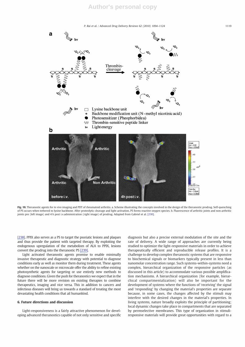
Author's personal copy
[238]. PPIX also serves as a PS to target the psoriatic lesions and plaquesand thus provide the patient with targeted therapy. By exploiting theendogenous upregulation of the metabolism of ALA to PPIX, lesionsconvert the prodrug into the theranostic PS [239].
Light activated theranostic agents promise to enable minimallyinvasive therapeutic and diagnostic strategy with potential to diagnoseconditions early as well as monitor them during treatment. These agentswhether on the nanoscale or microscale offer the ability to refine existingphotosynthetic agents for targeting or use entirely new methods todiagnose conditions. Given the push for theranosticswe expect that in thefuture there will be more revision on existing therapies to combinetherapeutics, imaging and vice versa. This in addition to cancers andinfectious diseases will bring us towards a standard of treating the mostdevastating health conditions that ail humankind.
6. Future directions and discussion
Light-responsiveness is a fairly attractive phenomenon for devel-oping advanced theranostics capable of not only sensitive and specific
diagnosis but also a precise external modulation of the site and therate of delivery. A wide range of approaches are currently beingstudied to optimize the light-responsive materials in order to achievetherapeutically efficient and reproducible release profiles. It is achallenge to develop complex theranostic systems that are responsiveto biochemical signals or biomarkers typically present in less thannanomolar concentration range. Such systems-within-systems need acomplex, hierarchical organization of the responsive particles (asdiscussed in this article) to accommodate various possible amplifica-tion mechanisms. A hierarchical organization (for example, hierar-chical compartmentalization) will also be important for thedevelopment of systems where the functions of ‘receiving’ the signaland ‘responding’ by changing the material's properties are separatebecause, in some cases, the changes affected by the stimuli mayinterfere with the desired changes in the material's properties. Inliving systems, nature broadly exploits the principle of partitioning;local dynamic changes take place in compartments that are separatedby permselective membranes. This type of organization in stimuli-responsive materials will provide great opportunities with regard to a
Fig. 10. Theranostic agents for in vivo imaging and PDT of rheumatoid arthritis. a. Scheme illustrating the concepts involved in the design of the theranostic prodrug. Self-quenchingof PS occurs when tethered to lysine backbone. After proteolytic cleavage and light activation, PS forms reactive oxygen species. b. Fluorescence of arthritic joints and non-arthriticjoints pre (left image) and 4 h post i.v.administration (right image) of prodrug. Adapted from Gabriel et al. [230].
1119P. Rai et al. / Advanced Drug Delivery Reviews 62 (2010) 1094–1124
Author's personal copy
programmable, complex response of thematerials. Another challenge inthe design of theranostic agents is to develop systems that can respondto several external stimuli in an intelligentway. Much research remainsto be done before practical applications become viable. The clinical useof the light-sensitive theranostics is still in its infancy and requiresconsiderable efforts on several aspects listed below.
(1) Design and synthesis of new biocompatible materials in orderto increase the range of light-sensitive compounds that fulfillthe requirements of generally recognized safe products.
(2) Specialized equipment capable of providing the adequateirradiation intensity to the target place without alteringsurrounding tissues. The relative impermeability of thehuman body to the lightmakes direct irradiation at a significantdepth of the body difficult and confines the applicability of theUV⁄visible light-sensitive DDS to treatments of the surfacelayers of the skin or a few millimeters beyond. NIR lasers andNIR-sensitive light materials appear to be feasible alternativesto their UV⁄visible counterparts.
(3) In vivo evaluation of the performance of the new deliverysystems needs to be assessed. Currently, most of the reviewedsystems have been tested in vitro under limited experimentalconditions or no toxicity studies were performed in vivo. Thesestudies must be supplemented by extensive studies in vivo asthe DDS make progress toward clinical use.
(4) Imaging systems for extremely sensitive and specific detectionof microscopic disease.
7. Conclusions
Medicine, as we move into the third millennium, still targetstherapy to the broadest patient population that might possibly benefitfrom it, and it relies on statistical analysis of this population's responsefor predicting therapeutic outcome in individual patients. This “onedrug fits all” approach could, with the use of theranostics, evolve intoan individualized approach to therapy where optimally effectivedrugs are matched to a patient's unique molecular profile and theiractivity is triggered using light to localize the therapeutic effects onlyin and around the diseased tissue with limited collateral damage.Monitoring the effects of light-triggered therapy and initiating asecond treatment if required would also be possible. Light-triggeredtheranostics in combination with multimodal imaging techniques canthus help provide personalized medicine; that is, unique, individual-ized for each patient. Pharmaceutical, diagnostics and biotechnolog-ical companies are all interested in this rapidly emerging field. Alreadythe U.S. Department of Health and Human Services has publiclyrecognized personalizedmedicine as one of its top priorities. Althoughsignificant awareness has been created about personalized medicine,its full potential is yet to be tapped. Nanotechnology could provide aplatform for the successful integration of diagnostics and therapy,which is vital for the future of customized treatments. Factors such ascost and regulatory timelines are among the major hurdles that needto be addressed at the moment. The concepts presented in this reviewwill allow the introduction of new possibilities in the field ofbiomedical theranostics.
Acknowledgments
We would like to thank Prof. Gang Zheng and his postdoctoralfellow Dr. Klara Stefflova for their help with making Fig. 2 of thismanuscript. We would also like to thank Dr. Bryan Spring for hishelpful suggestions. This work was supported by National Institutes ofHealth Grant Numbers P01 CA084203 and R44CA128364 NationalCancer Institute/National Institutes of Health Grant Numbers R01CA119388 and RC1 CA146337, the Department of Defense Air ForceOffice of Scientific Research FA9550-04-1-0079, and the Wellman
Center for Photomedicine core funds. Y.M. acknowledges fundingfrom Fonds québécois de la recherche sur la nature et les technologies(FQRNT), Québec, Canada. A.K. would like to acknowledge fundingfrom the Higher Education Commission (HEC) of Pakistan.
References
[1] Rising Health Costs a Global Problem, in, 2010.[2] World Health Statistics, World Health Organization France 2010, 2010, pp. 1–177.[3] A.J. Rai, J. Yee, M. Fleisher, Biomarkers in the era of personalized medicine — a
multiplexed SNP assay using capillary electrophoresis for assessing drugmetabolism capacity, Scand. J. Clin. Lab. Invest. Suppl. 242 (2010) 15–18.
[4] K.D. Blanchet, Redefining personalized medicine in the postgenomic era:developing bladder cancer therapeutics with proteomics, BJU Int. 105 (2010) i–iii.
[5] U. Francke, On the bumpy road towards ‘personalized medicine’, EMBO Mol.Med. 2 (2010) 1–2.
[6] S. Bates, Progress towards personalized medicine, Drug Discov. Today 15 (2010)115–120.
[7] V. Espina, L.A. Liotta, E.F. Petricoin III, Reverse-phase protein microarrays fortheranostics and patient tailored therapy, Methods Mol. Biol. 520 (2009) 89–105.
[8] J. Falk, Epigenetics & sequencing — gene expression systems' second interna-tional meeting. chromatin methylation to disease biology & theranostics, IDrugs11 (2008) 650–652.
[9] D.B. Buxton, Current status of nanotechnology approaches for cardiovasculardisease: a personal perspective, Wiley Interdiscip. Rev. Nanomed. Nanobiotech-nol. 1 (2009) 149–155.
[10] K. Morris, Nanotechnology crucial in fighting infectious disease, Lancet Infect.Dis. 9 (2009) 215.
[11] A. Nazem, G.A. Mansoori, Nanotechnology solutions for Alzheimer's disease:advances in research tools, diagnostic methods and therapeutic agents, J.Alzheimers Dis. 13 (2008) 199–223.
[12] P. Couvreur, C. Vauthier, Nanotechnology: intelligent design to treat complexdisease, Pharm. Res. 23 (2006) 1417–1450.
[13] C.P. McCoy, C. Brady, J.F. Cowley, S.M. McGlinchey, N. McGoldrick, D.J. Kinnear,G.P. Andrews, D.S. Jones, Triggered drug delivery from biomaterials, ExpertOpin. Drug Deliv. 7 (2010) 605–616.
[14] C. Alvarez-Lorenzo, L. Bromberg, A. Concheiro, Light-sensitive intelligent drugdelivery systems, Photochem. Photobiol. 85 (2009) 848–860.
[15] J.W. McBride, A. Balestrero, L. Ghezzi, G. Tribulato, K.J. Cross, Optical fiberimaging for high speed plasmamotion diagnostics: applied to low voltage circuitbreakers, Rev. Sci. Instrum. 81 (2010) 055109.
[16] A. Jemal, R. Siegel, E. Ward, Y. Hao, J. Xu, M.J. Thun, Cancer statistics, 2009, CACancer J. Clin. 59 (2009) 225–249.
[17] J.P. Celli, B.Q. Spring, I. Rizvi, C.L. Evans, K.S. Samkoe, S. Verma, B.W. Pogue, T.Hasan, Imaging and photodynamic therapy: mechanisms, monitoring, andoptimization, Chem. Rev. 110 (2010) 2795–2838.
[18] C. Balas, Review of biomedical optical imaging—a powerful, non-invasive, non-ionizing technology for improving in vivo diagnosis, Meas. Sci. Technol. 20(2009) 104020.
[19] K. Berg, P.K. Selbo, A. Weyergang, A. Dietze, L. Prasmickaite, A. Bonsted, B.O.Engesaeter, E. Angell-Petersen, T. Warloe, N. Frandsen, A. Hogset, Porphyrin-related photosensitizers for cancer imaging and therapeutic applications, J.Microsc.-Oxf. 218 (2005) 133–147.
[20] E.R. Ray, K. Chatterton, M.S. Khan, K. Thomas, A. Chandra, T.S. O'Brien,Hexylaminolaevulinate ‘blue light’ fluorescence cystoscopy in the investigation ofclinically unconfirmed positive urine cytology, BJU Int. 103 (2009) 1363–1367.
[21] A. Bogaards, A. Varma, K. Zhang, D. Zach, S.K. Bisland, E.H. Moriyama, L. Lilge, P.J.Muller, B.C. Wilson, Fluorescence image-guided brain tumour resection withadjuvant metronomic photodynamic therapy: pre-clinical model and technol-ogy development, Photochem. Photobiol. Sci. 4 (2005) 438–442.
[22] J.F. Lovell, T.W. Liu, J. Chen, G. Zheng, Activatable photosensitizers for imagingand therapy, Chem. Rev. 110 (2010) 2839–2857.
[23] Y. Ohsaki, K. Takeyama, S. Nakao, S. Tanno, E. Toyoshima, K. Nakanishi, Y. Nishigaki,T. Ogasa, S. Osanai, K. Kikuchi, S.Nakajima,Detection of Photofrinfluorescence frommalignant and premalignant lesions in the bronchus using a full-color endoscopicfluorescence imaging system, Diagn. Ther. Endosc. 7 (2001) 187–195.
[24] M. Kohno, M. Takeda, Y. Niwano, R. Saito, N. Emoto, M. Tada, T. Kanazawa, N.Ohuchi, R. Yamada, Early diagnosis of cancer by detecting the chemilumines-cence of hematoporphyrins in peripheral blood lymphocytes, Tohoku J. Exp.Med. 216 (2008) 47–52.
[25] Q. Chen, Z. Huang, D. Luck, J. Beckers, P.H. Brun, B.C. Wilson, A. Scherz, Y.Salomon, F.W. Hetzel, Preclinical studies in normal canine prostate of a novelpalladium-bacteriopheophorbide (WST09) photosensitizer for photodynamictherapy of prostate cancer, Photochem. Photobiol. 76 (2002) 438–445.
[26] A. Karioti, A.R. Bilia, Hypericins as potential leads for new therapeutics, Int. J. Mol.Sci. 11 (2010) 562–594.
[27] J.C. Kah, W.K. Lau, P.H. Tan, C.J. Sheppard, M. Olivo, Endoscopic image analysis ofphotosensitizer fluorescence as a promising noninvasive approach for patholo-gical grading of bladder cancer in situ, J. Biomed. Opt. 13 (2008) 054022.
[28] P.S. Thong, M. Olivo, W.W. Chin, R. Bhuvaneswari, K. Mancer, K.C. Soo, Clinicalapplication of fluorescence endoscopic imaging using hypericin for the diagnosisof human oral cavity lesions, Br. J. Cancer 101 (2009) 1580–1584.
1120 P. Rai et al. / Advanced Drug Delivery Reviews 62 (2010) 1094–1124

Author's personal copy
[29] D. Kacerovska, K. Pizinger, F. Majer, F. Smid, Photodynamic therapy ofnonmelanoma skin cancer with topical hypericum perforatum extract — apilot study, Photochem. Photobiol. 84 (2008) 779–785.
[30] R. Bhuvaneswari, P.S. Thong, Y.Y. Gan, K.C. Soo, M. Olivo, Evaluation of hypericin-mediated photodynamic therapy in combination with angiogenesis inhibitorbevacizumab using in vivo fluorescence confocal endomicroscopy, J. Biomed.Opt. 15 (2010) 011114.
[31] E.R. Trivedi, A.S. Harney, M.B. Olive, I. Podgorski, K. Moin, B.F. Sloane, A.G. Barrett,T.J. Meade, B.M. Hoffman, Chiral porphyrazine near-IR optical imaging agentexhibiting preferential tumor accumulation, Proc. Natl. Acad. Sci. U. S. A. 107(2010) 1284–1288.
[32] E.R. Trivedi, B.J. Vesper, H.Weitman, B. Ehrenberg, A.G. Barrett, J.A. Radosevich, B.M.Hoffman, Chiral bis-acetal porphyrazines as near-infrared optical agents fordetection and treatment of cancer, Photochem. Photobiol. 86 (2010) 410–417.
[33] M.D. Savellano, T. Hasan, Photochemical targeting of epidermal growth factorreceptor: a mechanistic study, Clin. Cancer Res. 11 (2005) 1658–1668.
[34] P. Mallikaratchy, Z.W. Tang, W.H. Tan, Cell specific aptamer-photosensitizerconjugates as a molecular tool in photodynamic therapy, ChemMedChem 3(2008) 425–428.
[35] M. Zhang, Z.H. Zhang,D. Blessington, H. Li, T.M. Busch, V.Madrak, J.Miles, B. Chance,J.D. Glickson, G. Zheng, Pyropheophorbide 2-deoxyglucosamide: a new photosen-sitizer targeting glucose transporters, Bioconjug. Chem. 14 (2003) 709–714.
[36] R. Weissleder, C.H. Tung, U. Mahmood, A. Bogdanov, In vivo imaging of tumorswith protease-activated near-infrared fluorescent probes, Nat. Biotechnol. 17(1999) 375–378.
[37] G. Zlokarnik, P.A. Negulescu, T.E. Knapp, L. Mere, N. Burres, L. Feng,M.Whitney, K.Roemer, R.Y. Tsien, Quantitation of transcription and clonal selection of singleliving cells with beta-lactamase as reporter, Science (New York, N.Y.) 279 (1998)84–88.
[38] J. Chen, K. Stefflova, M.J. Niedre, B.C. Wilson, B. Chance, J.D. Glickson, G. Zheng,Protease-triggered photosensitizing beacon based on singlet oxygen quenchingand activation, J. Am. Chem. Soc. 126 (2004) 11450–11451.
[39] Y. Choi, R. Weissleder, C.H. Tung, Selective antitumor effect of novel protease-mediated photodynamic agent, Cancer Res. 66 (2006) 7225–7229.
[40] G. Zheng, J. Chen, K. Stefflova, M. Jarvi, H. Li, B.C. Wilson, Photodynamicmolecular beacon as an activatable photosensitizer based on protease-controlledsinglet oxygen quenching and activation, Proc. Natl. Acad. Sci. U. S. A. 104 (2007)8989–8994.
[41] I.V. Nesterova, S.S. Erdem, S. Pakhomov, R.P. Hammer, S.A. Soper, Phthalocyaninedimerization-based molecular beacons using near-IR fluorescence, J. Am. Chem.Soc. 131 (2009) 2432-+.
[42] K. Stefflova, J. Chen, H. Li, G. Zheng, Targeted photodynamic therapy agent with abuilt-in apoptosis sensor for in vivo near-infrared imaging of tumor apoptosistriggered by its photosensitization in situ, Mol. Imaging 5 (2006) 520–532.
[43] S.K. Pandey, A.L. Gryshuk, M. Sajjad, X. Zheng, Y. Chen, M.M. Abouzeid, J. Morgan,I. Charamisinau, H.A. Nabi, A. Oseroff, R.K. Pandey, Multimodality agents fortumor imaging (PET, fluorescence) and photodynamic therapy. A possible “seeand treat” approach, J. Med. Chem. 48 (2005) 6286–6295.
[44] S.K. Pandey, M. Sajjad, Y.H. Chen, A. Pandey, J.R. Missert, C. Batt, R.T. Yao, H.A.Nabi, A.R. Oseroff, R.K. Pandey, Compared to purpurinimides, the pyropheo-phorbide containing an iodobenzyl group showed enhanced PDT efficacy andtumor imaging (I-124-PET) ability, Bioconjug. Chem. 20 (2009) 274–282.
[45] S.W. Young, F.Qing,A.Harriman, J.L. Sessler,W.C.Dow, T.D.Mody,G.W.Hemmi, Y.P.Hao, R.A. Miller, Gadolinium(III) texaphyrin: a tumor selective radiation sensitizerthat is detectable by MRI, Proc. Natl. Acad. Sci. U. S. A. 93 (1996) 6610–6615.
[46] S.G. Rockson, P. Kramer, M. Razavi, A. Szuba, S. Filardo, P. Fitzgerald, J.P. Cooke, S.Yousuf, A.R. DeVault, M.F. Renschler, D.C. Adelman, Photoangioplasty for humanperipheral atherosclerosis — results of a phase I trial of photodynamic therapywith motexafin lutetium (Antrin), Circulation 102 (2000) 2322–2324.
[47] B. Hofmann, A. Bogdanov, E. Marecos, W. Ebert, W. Semmler, R. Weissleder,Mechanism of gadophrin-2 accumulation in tumor necrosis, JMRI-J. Magn.Reson. Imaging 9 (1999) 336–341.
[48] G.L. Li, A. Slansky, M.P. Dobhal, L.N. Goswami, A. Graham, Y.H. Chen, P. Kanter, R.A.Alberico, J. Spernyak, R. Mazurchuk, A. Oseroff, Z. Grossman, R.K. Pandey,Chlorophyll-a analogues conjugated with aminobenzyl-DTPA as potential bifunc-tional agents for magnetic resonance imaging and photodynamic therapy,Bioconjug. Chem. 16 (2005) 32–42.
[49] J.A. Spernyak, W.H. White, M. Ethirajan, N.J. Patel, L. Goswami, Y.H. Chen, S.Turowski, J.R. Missert, C. Batt, R. Mazurchuk, R.K. Pandey, Hexylether derivativeof pyropheophorbide-a (HPPH) on conjugating with 3gadolinium(III) amino-benzyldiethylenetriaminepentaacetic acid shows potential for in vivo tumorimaging (MR, fluorescence) and photodynamic therapy, Bioconjug. Chem. 21(2010) 828–835.
[50] J. Liu, S. Ohta, A. Sonoda, M. Yamada, M. Yamamoto, N. Nitta, K. Murata, Y. Tabata,Preparation of PEG-conjugated fullerene containing Gd3+ ions for photody-namic therapy, J. Control. Release 117 (2007) 104–110.
[51] A. Vaidya, Y. Sun, T. Ke, E.K. Jeong, Z.R. Lu, Contrast enhanced MRI-guidedphotodynamic therapy for site-specific cancer treatment, Magn. Reson. Med. 56(2006) 761–767.
[52] A. Vaidya, Y. Sun, Y. Feng, L. Emerson, E.K. Jeong, Z.R. Lu, Contrast-enhancedMRI-guided photodynamic cancer therapy with a pegylated bifunctional polymerconjugate, Pharm. Res. 25 (2008) 2002–2011.
[53] F. Wu, Z.B. Wang, W.Z. Chen, J.Z. Zou, Extracorporeal high intensity focusedultrasound for treatment of solid carcinomas: four-year Chinese clinicalexperience, 2nd International Symposium on Therapeutic Ultrasound, Seattle,WA, USA, 2002, p. 11.
[54] W. Hiraoka, H. Honda, L.B. Feril, N. Kudo, T. Kondo, Comparison betweensonodynamic effect and photodynamic effect with photo sensitizers on freeradical formation and cell killing, Ultrason. Sonochem. 13 (2006) 535–542.
[55] Z.H. Jin, N. Miyoshi, K. Ishiguro, S. Umemura, K. Kawabata, N. Yumita, I. Sakata, K.Takaoka, T. Udagawa, S. Nakajima, H. Tajiri, K. Ueda, M. Fukuda, M. Kumakiri,Combination effect of photodynamic and sonodynamic therapy on experimentalskin squamous cell carcinoma in C3H/HeN mice, J. Dermatol. 27 (2000) 294–306.
[56] D.K. Chatterjee, L.S. Fong, Y. Zhang, Nanoparticles in photodynamic therapy: anemerging paradigm, Adv. Drug Deliv. Rev. 60 (2008) 1627–1637.
[57] J. Gravier, R. Schneider, C. Frochot, T. Bastogne, F. Schmitt, J. Didelon, F. Guillemin,M. Barberi-Heyob, Improvement of meta-tetra(hydroxyphenyl)chlorin-likephotosensitizer selectivity with folate-based targeted delivery. synthesis andin vivo delivery studies, J. Med. Chem. 51 (2008) 3867–3877.
[58] J.P. Taquet, C. Frochot, V. Manneville, M. Barberi-Heyob, Phthalocyaninescovalently bound to biomolecules for a targeted photodynamic therapy, Curr.Med. Chem. 14 (2007) 1673–1687.
[59] C. Fang, M. Zhang, Nanoparticle-based theragnostics: integrating diagnostic andtherapeutic potentials in nanomedicine, J. Control. Release (2010).
[60] I.I. Slowing, B.G. Trewyn, V.S. Lin, Mesoporous silica nanoparticles forintracellular delivery of membrane-impermeable proteins, J. Am. Chem. Soc.129 (2007) 8845–8849.
[61] I. Roy, T.Y. Ohulchanskyy, H.E. Pudavar, E.J. Bergey, A.R. Oseroff, J. Morgan, T.J.Dougherty, P.N. Prasad, Ceramic-based nanoparticles entrappingwater-insolublephotosensitizing anticancer drugs: a novel drug-carrier system for photodynamictherapy, J. Am. Chem. Soc. 125 (2003) 7860–7865.
[62] T.Y. Ohulchanskyy, I. Roy, L.N. Goswami, Y. Chen, E.J. Bergey, R.K. Pandey, A.R.Oseroff, P.N. Prasad, Organically modified silica nanoparticles with covalentlyincorporated photosensitizer for photodynamic therapy of cancer, Nano Lett. 7(2007) 2835–2842.
[63] S. Kim, T.Y. Ohulchanskyy, H.E. Pudavar, R.K. Pandey, P.N. Prasad, Organicallymodified silica nanoparticles co-encapsulating photosensitizing drug andaggregation-enhanced two-photon absorbing fluorescent dye aggregates fortwo-photon photodynamic therapy, J. Am. Chem. Soc. 129 (2007) 2669–2675.
[64] R. Kumar, I. Roy, T.Y. Ohulchanskky, L.A. Vathy, E.J. Bergey, M. Sajjad, P.N. Prasad,In vivo biodistribution and clearance studies using multimodal organicallymodified silica nanoparticles, ACS Nano 4 (2010) 699–708.
[65] X. He, X. Wu, K. Wang, B. Shi, L. Hai, Methylene blue-encapsulated phosphonate-terminated silica nanoparticles for simultaneous in vivo imaging and photody-namic therapy, Biomaterials 30 (2009) 5601–5609.
[66] R. Zhang, C. Wu, L. Tong, B. Tang, Q.H. Xu, Multifunctional core–shellnanoparticles as highly efficient imaging and photosensitizing agents, Langmuir25 (2009) 10153–10158.
[67] J.D. Glickson, S. Lund-Katz, R. Zhou, H. Choi, I.W. Chen, H. Li, I. Corbin, A.V. Popov,W. Cao, L. Song, C. Qi, D. Marotta, D.S. Nelson, J. Chen, B. Chance, G. Zheng,Lipoprotein nanoplatform for targeted delivery of diagnostic and therapeuticagents, Mol. Imaging 7 (2008) 101–110.
[68] J.D. Glickson, S. Lund-Katz, R. Zhou, H. Choi, I.W. Chen, H. Li, I. Corbin, A.V. Popov,W. Cao, L. Song, C. Qi, D. Marotta, D.S. Nelson, J. Chen, B. Chance, G. Zheng,Lipoprotein nanoplatform for targeted delivery of diagnostic and therapeuticagents, Adv. Exp. Med. Biol. 645 (2009) 227–239.
[69] G. Zheng, H. Li, K. Yang, D. Blessington, K. Licha, S. Lund-Katz, B. Chance, J.D.Glickson, Tricarbocyanine cholesteryl laurates labeled LDL: new near infraredfluorescent probes (NIRFs) for monitoring tumors and gene therapy of familialhypercholesterolemia, Bioorg. Med. Chem. Lett. 12 (2002) 1485–1488.
[70] H. Li, Z. Zhang, D. Blessington, D.S. Nelson, R. Zhou, S. Lund-Katz, B. Chance, J.D.Glickson, G. Zheng, Carbocyanine labeled LDL for optical imaging of tumors,Acad. Radiol. 11 (2004) 669–677.
[71] I.R. Corbin, H. Li, J. Chen, S. Lund-Katz, R. Zhou, J.D. Glickson, G. Zheng, Low-density lipoprotein nanoparticles as magnetic resonance imaging contrastagents, Neoplasia 8 (2006) 488–498.
[72] L. Song, H. Li, U. Sunar, J. Chen, I. Corbin, A.G. Yodh, G. Zheng, Naphthalocyanine-reconstituted LDL nanoparticles for in vivo cancer imaging and treatment, Int. J.Nanomedicine 2 (2007) 767–774.
[73] K. Chen, A. Preuss, S. Hackbarth, M. Wacker, K. Langer, B. Roder, Novelphotosensitizer-protein nanoparticles for photodynamic therapy: photophysicalcharacterization and in vitro investigations, J. Photochem. Photobiol. B 96 (2009)66–74.
[74] A. Saupe, S.A. Wissing, A. Lenk, C. Schmidt, R.H. Muller, Solid lipid nanoparticles(SLN) and nanostructured lipid carriers (NLC)— structural investigations on twodifferent carrier systems, Biomed. Mater. Eng. 15 (2005) 393–402.
[75] P.J. Stevens, M. Sekido, R.J. Lee, Synthesis and evaluation of a hematoporphyrinderivative in a folate receptor-targeted solid-lipid nanoparticle formulation,Anticancer Res. 24 (2004) 161–165.
[76] D.K. Chatterjee, Z. Yong, Upconverting nanoparticles as nanotransducersfor photodynamic therapy in cancer cells, Nanomedicine (Lond.) 3 (2008)73–82.
[77] D.K. Chatterjee, A.J. Rufaihah, Y. Zhang, Upconversion fluorescence imaging ofcells and small animals using lanthanide doped nanocrystals, Biomaterials 29(2008) 937–943.
[78] A.S. Derycke, P.A. de Witte, Liposomes for photodynamic therapy, Adv. DrugDeliv. Rev. 56 (2004) 17–30.
[79] N. Bendsoe, L. Persson, A. Johansson, J. Axelsson, J. Svensson, S. Grafe, T. Trebst, S.Andersson-Engels, S. Svanberg, K. Svanberg, Fluorescence monitoring of atopically applied liposomal Temoporfin formulation and photodynamic therapyof nonpigmented skin malignancies, J. Environ. Pathol. Toxicol. Oncol. 26 (2007)117–126.
1121P. Rai et al. / Advanced Drug Delivery Reviews 62 (2010) 1094–1124
Author's personal copy
[80] W. Zhong, J.P. Celli, I. Rizvi, Z. Mai, B.Q. Spring, S.H. Yun, T. Hasan, In vivo high-resolution fluorescence microendoscopy for ovarian cancer detection andtreatment monitoring, Br. J. Cancer 101 (2009) 2015–2022.
[81] S.K. Hendren, S.M. Hahn, F.R. Spitz, T.W. Bauer, S.C. Rubin, T. Zhu, E. Glatstein, D.L.Fraker, Phase II trial of debulking surgery and photodynamic therapy fordisseminated intraperitoneal tumors, Ann. Surg. Oncol. 8 (2001) 65–71.
[82] S.M. Hahn, D.L. Fraker, R. Mick, J. Metz, T.M. Busch, D. Smith, T. Zhu, C. Rodriguez,A. Dimofte, F. Spitz, M. Putt, S.C. Rubin, C. Menon, H.W.Wang, D. Shin, A. Yodh, E.Glatstein, A phase II trial of intraperitoneal photodynamic therapy for patientswith peritoneal carcinomatosis and sarcomatosis, Clin. Cancer Res. 12 (2006)2517–2525.
[83] N.S. Sandanayake, M.T. Huggett, S.G. Bown, B.W. Pogue, T. Hasan, S.P. Pereira, in:D.H. Kessel (Ed.), Photodynamic Therapy for Locally Advanced PancreaticCancer: Early Clinical Results, SPIE, San Francisco, California, USA, 2010,pp. 75510L–75518.
[84] A.S. Derycke, A. Kamuhabwa, A. Gijsens, T. Roskams, D. De Vos, A. Kasran, J.Huwyler, L. Missiaen, P.A. deWitte, Transferrin-conjugated liposome targeting ofphotosensitizer AlPcS4 to rat bladder carcinoma cells, J. Natl. Cancer Inst. 96(2004) 1620–1630.
[85] I.G.Meerovich, Z.S. Smirnova, N.A. Oborotova, E.A. Luk'yanets, G.A.Meerovich, V.M.Derkacheva, A.P. Polozkova, I.Y. Kubasova, A.Y. Baryshnikov, Hydroxyalumi-nium tetra-3-phenylthiophthalocyanine is a new effective photosensitizer forphotodynamic therapy and fluorescent diagnosis, Bull. Exp. Biol. Med. 139(2005) 427–430.
[86] R. Rahmanzadeh, P. Rai, J.P. Celli, I. Rizvi, B. Baron-Luhr, J. Gerdes, T. Hasan, Ki-67as a molecular target for therapy in an in vitro 3D model for ovarian cancer,Cancer Res, Epublication ahead of print, doi:10.1158/0008-5472.CAN-10-1190.
[87] J. Gerdes, U. Schwab, H. Lemke, H. Stein, Production of a mouse monoclonalantibody reactive with a human nuclear antigen associated with cell prolifer-ation, Int. J. Cancer 31 (1983) 13–20.
[88] T. Scholzen, J. Gerdes, The Ki-67 protein: from the known and the unknown,J. Cell. Physiol. 182 (2000) 311–322.
[89] T. Surrey, M.B. Elowitz, P.E. Wolf, F. Yang, F. Nédélec, K. Shokat, S. Leibler,Chromophore-assisted light inactivation and self-organization of microtubulesand motors, Proc. Natl. Acad. Sci. U. S. A. 95 (1998) 4293–4298.
[90] E. Boisselier, D. Astruc, Gold nanoparticles in nanomedicine: preparations, imaging,diagnostics, therapies and toxicity, Chem. Soc. Rev. 38 (2009) 1759–1782.
[91] M.E. Wieder, D.C. Hone, M.J. Cook, M.M. Handsley, J. Gavrilovic, D.A. Russell,Intracellular photodynamic therapy with photosensitizer-nanoparticle conju-gates: cancer therapy using a ‘Trojan horse’, Photochem. Photobiol. Sci. 5 (2006)727–734.
[92] K. Zaruba, J. Kralova, P. Rezanka, P. Pouckova, L. Veverkova, V. Kral, Modifiedporphyrin-brucine conjugated to gold nanoparticles and their application inphotodynamic therapy, Org. Biomol. Chem. 8 (2010).
[93] M. Camerin, M. Magaraggia, M. Soncin, G. Jori, M. Moreno, I. Chambrier, M.J.Cook, D.A. Russell, The in vivo efficacy of phthalocyanine-nanoparticleconjugates for the photodynamic therapy of amelanotic melanoma, Eur. J.Cancer 46 (2010) 1910–1918.
[94] Y. Cheng, C.S. A., J.D. Meyers, I. Panagopoulos, B. Fei, C. Burda, Highly efficientdrug delivery with gold nanoparticle vectors for in vivo photodynamic therapyof cancer, J. Am. Chem. Soc. 130 (2008) 10643–10647.
[95] S.A. Agnihotri, N.N. Mallikarjuna, T.M. Aminabhavi, Recent advances on chitosan-based micro- and nanoparticles in drug delivery, J. Control. Release 100 (2004)5–28.
[96] D. Yan, L. Li, D.Y. Chen, Y.H. Zhang, C.H. Hu, Z.H. Deng, Detection of fungi in liquorworkers with tinea corporis and tinea cruris using arbitrarily primed polymerasechain reaction, Zhonghua Lao Dong Wei Sheng Zhi Ye Bing Za Zhi 25 (2007)133–135.
[97] Y. Sun, Z.L. Chen, X.X. Yang, P. Huang, X.P. Zhou, X.X. Du, Magnetic chitosannanoparticles as a drug delivery system for targeting photodynamic therapy,Nanotechnology 20 (2009) 135102.
[98] Y.E. Koo, W. Fan, H. Hah, H. Xu, D. Orringer, B. Ross, A. Rehemtulla, M.A. Philbert,R. Kopelman, Photonic explorers based on multifunctional nanoplatforms forbiosensing and photodynamic therapy, Appl. Opt. 46 (2007) 1924–1930.
[99] W. Tang, H. Xu, R. Kopelman, M.A. Philbert, Photodynamic characterization andin vitro application of methylene blue-containing nanoparticle platforms,Photochem. Photobiol. 81 (2005) 242–249.
[100] G.R. Reddy, M.S. Bhojani, P. McConville, J. Moody, B.A. Moffat, D.E. Hall, G. Kim, Y.E.Koo, M.J. Woolliscroft, J.V. Sugai, T.D. Johnson, M.A. Philbert, R. Kopelman, A.Rehemtulla, B.D.Ross, Vascular targetednanoparticles for imagingand treatmentofbrain tumors, Clin. Cancer Res. 12 (2006) 6677–6686.
[101] B.A. Moffat, T.L. Chenevert, C.R. Meyer, P.E. McKeever, D.E. Hall, B.A. Hoff, T.D.Johnson, A. Rehemtulla, B.D. Ross, The functional diffusion map: an imagingbiomarker for the early prediction of cancer treatment outcome, Neoplasia8 (2006) 259–267.
[102] K.S. Samkoe, A. Chen, I. Rizvi, J.A. O'Hara, P.J. Hoopes, S.P. Pereira, T. Hasan, B.W.Pogue, Imaging tumor variation in response to photodynamic therapy inpancreaticcancer xenograft models, Int. J. Radiat. Oncol. Biol. Phys. 76 (2010) 251–259.
[103] C.W. Lai, Y.H. Wang, C.H. Lai, M.J. Yang, C.Y. Chen, P.T. Chou, C.S. Chan, Y. Chi, Y.C.Chen, J.K. Hsiao, Iridium-complex-functionalized Fe3O4/SiO2 core/shell nano-particles: a facile three-in-one system in magnetic resonance imaging,luminescence imaging, and photodynamic therapy, Small 4 (2008) 218–224.
[104] D. Brevet, M. Gary-Bobo, L. Raehm, S. Richeter, O. Hocine, K. Amro, B. Loock, P.Couleaud, C. Frochot, A. Morere, P. Maillard, M. Garcia, J.O. Durand, Mannose-targeted mesoporous silica nanoparticles for photodynamic therapy, Chem.Commun. (Camb.) (2009) 1475–1477.
[105] E. Chelebaeva, L. Raehm, J.O. Durand, Y. Guari, J. Larionova, C. Guerin, A.Trifonov, M. Willinger, K. Thangavel, A. Lascialfari, O. Mongin, Y. Mir, M.Blanchard-Desce, Mesoporous silica nanoparticles combining two-photonexcited fluorescence and magnetic properties, J. Mater. Chem. 20 (2010)1877–1884.
[106] X. Michalet, F.F. Pinaud, L.A. Bentolila, J.M. Tsay, S. Doose, J.J. Li, G. Sundaresan, A.M.Wu, S.S. Gambhir, S. Weiss, Quantum dots for live cells, in vivo imaging, anddiagnostics, Science 307 (2005) 538–544.
[107] R. Bakalova, H. Ohba, Z. Zhelev, M. Ishikawa, Y. Baba, Quantum dots asphotosensitizers? Nat. Biotechnol. 22 (2004) 1360–1361.
[108] A.C. Samia, X. Chen, C. Burda, Semiconductor quantum dots for photodynamictherapy, J. Am. Chem. Soc. 125 (2003) 15736–15737.
[109] R. Bakalova, Z. Zhelev, I. Aoki, K. Masamoto, M. Mileva, T. Obata, M. Higuchi, V.Gadjeva, I. Kanno, Multimodal silica-shelled quantum dots: direct intracellulardelivery, photosensitization, toxic, and microcirculation effects, Bioconjug.Chem. 19 (2008) 1135–1142.
[110] W. Chen, J. Zhang, Using nanoparticles to enable simultaneous radiation andphotodynamic therapies for cancer treatment, J. Nanosci. Nanotechnol. 6 (2006)1159–1166.
[111] Y.F. Liu, W. Chen, S.P. Wang, A.G. Joly, Investigation of water-soluble x-rayluminescence nanoparticles for photodynamic activation, Appl. Phys. Lett. 92(2008).
[112] S. Jacques, Laser-tissue interactions. Photochemical, photothermal, and photo-mechanical, Surg. Clin. North Am. 72 (1992) 531.
[113] G. Jori, J.D. Spikes, Photothermal sensitizers: possible use in tumor therapy,J. Photochem. Photobiol., B 6 (1990) 93–101.
[114] E.A. Genina, A.N. Bashkatov, G.V. Simonenko, O.D. Odoevskaya, V. Tuchin, G.B.Altshuler, Low-intensity indocyanine-green laser phototherapy of acne vulgaris:pilot study, J. Biomed. Opt. 9 (2004) 828–834.
[115] L.R. Hirsch, R.J. Stafford, J.A. Bankson, S.R. Sershen, B. Rivera, R.E. Price, J.D. Hazle,N.J. Halas, J.L. West, Nanoshell-mediated near-infrared thermal therapy oftumors under magnetic resonance guidance, Proc. Natl. Acad. Sci. U. S. A. 100(2003) 13549–13554.
[116] I. El-Sayed, X. Huang, M. El-Sayed, Selective laser photo-thermal therapy ofepithelial carcinoma using anti-EGFR antibody conjugated gold nanoparticles,Cancer Lett. 239 (2006) 129–135.
[117] J. Yu, D. Javier, M.A. Yaseen, N. Nitin, Self-assembly synthesis, tumor celltargeting, and photothermal capabilities of antibody-coated indocyanine greennanocapsules, J. Am. Chem. Soc. 132 (6) (2010) 1929–1938.
[118] S. Jiang, M. Gnanasammandhan, Y. Zhang, Optical imaging-guided cancertherapy with fluorescent nanoparticles, J. R. Soc. Interface 7 (2009) 3.
[119] V. Zharov, E. Galitovskaya, C. Johnson, T. Kelly, Synergistic enhancement ofselective nanophotothermolysis with gold nanoclusters: potential for cancertherapy, Lasers Surg. Med. 37 (2005) 219–226.
[120] M. Skala, M. Crow, A. Wax, J. Izatt, Photothermal optical coherence tomographyof epidermal growth factor receptor in live cells using immunotargeted goldnanospheres, Nano Lett. 8 (2008) 3461–3467.
[121] X. Liu, M. Lloyd, I. Fedorenko, P. Bapat, T. Zhukov, Q. Huo, Enhanced imaging andaccelerated photothermalysis of A549 human lung cancer cells by goldnanospheres, Nanomedicine (London, England) 3 (2008) 617–626.
[122] S. Lal, S. Clare, N. Halas, Nanoshell-enabled photothermal cancer therapy:impending clinical impact, Acc. Chem. Res. 41 (2008) 1842–1851.
[123] S.E. Skrabalak, J. Chen, L. Au, X. Lu, X. Li, Y. Xia, Gold nanocages for biomedicalapplications, Adv. Mater. (Deerfield Beach, Fla) 19 (2007) 3177–3184.
[124] X. Wu, T. Ming, X. Wang, P. Wang, J. Wang, J. Chen, High-photoluminescence-yield gold nanocubes: for cell imaging and photothermal therapy, ACS Nano 4(2010) 113–120.
[125] K.W. Hu, C.C. Huang, J.R. Hwu, W.C. Su, D.B. Shieh, C. Yeh, A new photothermaltherapeutic agent: core-free nanostructured Au x Ag1-x dendrites, Chemistry(Weinheim an der Bergstrasse, Germany) 14 (2008) 2956–2964.
[126] S. Wang, K. Chen, T.H. Wu, H. Wang, Photothermal effects of supramolecularlyassembled gold nanoparticles for the targeted treatment of cancer cells, Angew.Chem. Int. Ed. Engl. 49 (2010).
[127] T. Huff, M. Hansen, L. Tong, Y. Zhao, H. Wang, D. Zweifel, J. Cheng, A. Wei,Plasmon-resonant nanorods as multimodal agents for two-photon luminescentimaging and photothermal therapy, Society of Photo-Optical InstrumentationEngineers (SPIE) Conference Series, 6448, 2007, p. 5.
[128] A.M. Gobin, M.H. Lee, N. Halas, W.D. James, R.A. Drezek, J.L. West, Near-infraredresonant nanoshells for combined optical imaging and photothermal cancertherapy, Nano Lett. 7 (2007) 1929–1934.
[129] E.B. Dickerson, E.C. Dreaden, X. Huang, I. El-Sayed, H. Chu, S. Pushpanketh, J.F.McDonald, M. El-Sayed, Gold nanorod assisted near-infrared plasmonic photo-thermal therapy (PPTT) of squamous cell carcinoma in mice, Cancer Lett. 269(2008) 57–66.
[130] F. Zhou, D. Xing, Z. Ou, B. Wu, D.E. Resasco, W.R. Chen, Cancer photothermaltherapy in the near-infrared region by using single-walled carbon nanotubes,J. Biomed. Opt. 14 (2009) 021009.
[131] S. Torti, F. Byrne, O. Whelan, N. Levi, B. Ucer, M. Schmid, F. Torti, S. Akman, J. Liu,P. Ajayan, Thermal ablation therapeutics based on CNx multi-walled nanotubes,Int. J. Nanomedicine 2 (2007) 707.
[132] J. Shah, S.R. Aglyamov, K. Sokolov, T.E. Milner, S.Y. Emelianov, Ultrasoundimaging to monitor photothermal therapy — feasibility study, Opt. Express 16(2008) 3776–3785.
[133] S. Aglyamov, S. Emelianov, J. Shah, K. Sokolov, T. Milner, Ultrasound-basedthermal and elasticity imaging to assist photothermal cancer therapy —
preliminary study, IEEE Ultrasonics Symposium, (2006) 1029–1032.
1122 P. Rai et al. / Advanced Drug Delivery Reviews 62 (2010) 1094–1124

Author's personal copy
[134] M. Mehrmohammadi, L.L. Ma, Y.S. Chen, M. Qu, Combined photothermal therapyand magneto-motive ultrasound imaging using multifunctional nanoparticles,Proc. SPIE 7574 (2010) 1–8.
[135] S. Mallidi, B. Wang, M. Mehrmohammadi, M. Qu, Y. Chen, P. Joshi, S. Kim, K.A.Homan, A.B. Karpiouk, R.W. Smalling, K. Sokolov, S. Emelianov, Ultrasound-based imaging of nanoparticles: from molecular and cellular imaging to therapyguidance, IEEE Ultrasonics Symposium, (2009) 27–36.
[136] S.Y. Emelianov, P.C. Li, M. O'Donnell, Photoacoustics for molecular imaging andtherapy, Phys. Today 62 (2009) 34–39.
[137] L. Wang, Multiscale photoacoustic microscopy and computed tomography, Nat.Photonics 3 (2009) 503–509.
[138] M. Xu, L. Wang, Photoacoustic imaging in biomedicine, Rev. Sci. Instrum. 77(2006) 041101.
[139] Y. Chen, W. Frey, S. Kim, K. Homan, P. Kruizinga, Enhanced thermal stability ofsilica-coated gold nanorods for photoacoustic imaging and image-guidedtherapy, Opt. Express. 18 (9) (2010) 8867–8878.
[140] J. Kim, E. Galanzha, E. Shashkov, H. Moon, V. Zharov, Golden carbon nanotubes asmultimodal photoacoustic and photothermal high-contrast molecular agents,Nat. Nanotechnol. 4 (2009) 688–694.
[141] E. Galanzha, J. Kim, V. Zharov, Nanotechnology-based molecular photoacousticand photothermal flow cytometry platform forin-vivodetection and killing ofcirculating cancer stem cells, J. Biophotonics 2 (2009) 725–735.
[142] E.V. Shashkov, M. Everts, E.I. Galanzha, V.P. Zharov, Quantum dots as multimodalphotoacoustic and photothermal contrast agents, Nano Lett. 8 (2008)3953–3958.
[143] J. Shah, S. Park, S. Aglyamov, T. Larson, L. Ma, K. Sokolov, K. Johnston, T. Milner, S.Y.Emelianov, Photoacoustic imaging and temperature measurement for photother-mal cancer therapy, J. Biomed. Opt. 13 (2008) 034024.
[144] S. Caruthers, P. Winter, S. Wickline, G. Lanza, Targeted magnetic resonanceimaging contrast agents, Meth. Mol. Med. 124 (2006) 387.
[145] A.M. Morawski, G.A. Lanza, S.A. Wickline, Targeted contrast agents for magneticresonance imaging and ultrasound, Curr. Opin. Biotechnol. 16 (2005) 89–92.
[146] P. Diagaradjane, A. Shetty, J. Wang, A. Elliott, J. Schwartz, S. Shentu, H. Park, A.Deorukhkar, R. Stafford, S. Cho, Modulation of in vivo tumor radiation responsevia gold nanoshell-mediated vascular-focused hyperthermia: characterizing anintegrated antihypoxic and localized vascular disrupting targeting strategy,Nano Lett. 8 (2008) 1492.
[147] A. Burke, X. Ding, R. Singh, R.A. Kraft, N. Levi-Polyachenko, M.N. Rylander, C. Szot,C. Buchanan, J. Whitney, J. Fisher, H.C. Hatcher, R. D'Agostino, N.D. Kock, P.M.Ajayan, D.L. Carroll, S. Akman, F.M. Torti, S.V. Torti, Long-term survival followinga single treatment of kidney tumors with multiwalled carbon nanotubes andnear-infrared radiation, Proc. Natl. Acad. Sci. U. S. A. 106 (2009) 12897–12902.
[148] J. Kim, J. Oh, H. Kang, M. Feldman, T. Milner, Photothermal response ofsuperparamagnetic iron oxide nanoparticles, Lasers Surg.Med. 40 (2008) 415–421.
[149] T. Larson, J. Bankson, J. Aaron, K. Sokolov, Hybrid plasmonic magneticnanoparticles as molecular specific agents for MRI/optical imaging andphotothermal therapy of cancer cells, Nanotechnology 18 (2007) 325101–???.
[150] L.-S. Bouchard, M.S. Anwar, G.L. Liu, B. Hann, Z.H. Xie, J.W. Gray, X. Wang, A.Pines, F.F. Chen, Picomolar sensitivity MRI and photoacoustic imaging of cobaltnanoparticles, Proc. Natl. Acad. Sci. U. S. A. 106 (2009) 4085–4089.
[151] J. Kim, S. Park, J.E. Lee, S.M. Jin, J.H. Lee, I.S. Lee, I. Yang, J.S. Kim, S.K. Kim, M.H.Cho, T. Hyeon, Designed fabrication of multifunctional magnetic gold nanoshellsand their application to magnetic resonance imaging and photothermal therapy,Angew. Chem. Int. Ed. Engl. 45 (2006) 7754–7758.
[152] X. Ji, R. Shao, A.M. Elliott, R.J. Stafford, E. Esparza-Coss, G. Liang, Z.P. Luo, K.Park, J.T. Markert, C. Li, Bifunctional gold nanoshells with a superparamagneticiron oxide-silica core suitable for both mr imaging and photothermal therapy,J. Phys. Chem. C Nanomaterials Interfaces 111 (2007) 6245.
[153] J. Lee, J. Yang, H. Ko, S. Oh, J. Kang, J. Son, K. Lee, S. Lee, H. Yoon, J. Suh,Multifunctional magnetic gold nanocomposites: human epithelial cancerdetection via magnetic resonance imaging and localized synchronous therapy,Adv. Funct. Mater. 18 (2008) 258.
[154] D.K. Kirui, D.A. Rey, C.A. Batt, Gold hybrid nanoparticles for targetedphototherapy and cancer imaging, Nanotechnology 21 (2010) 105105.
[155] H. Yu, M. Chen, P. Rice, S. Wang, R. White, S. Sun, Dumbbell-like bifunctional Auâˆ′Fe3O4 nanoparticles, Nano Lett. 5 (2005) 379–382.
[156] L. Ma, M. Feldman, J. Tam, A. Paranjape, K. Cheruku, T. Larson, D. Ingram, V.Paramita, J. Villard, Small multifunctional nanoclusters (nanoroses) for targetedcellular imaging and therapy, ACS Nano 3 (2009) 2686–2696.
[157] W. Chen, N. Xu, L. Xu, L. Wang, Z. Li, W. Ma, Y. Zhu, C. Xu, N. Kotov,Multifunctional magnetoplasmonic nanoparticle assemblies for cancertherapy and diagnostics (theranostics), Macromol. Rapid Commun. 31(2010) 228–236.
[158] H. Park, J. Yang, S. Seo, K. Kim, J. Suh, D. Kim, S. Haam, K.H. Yoo, Multifunctionalnanoparticles for photothermally controlled drug delivery and magneticresonance imaging enhancement, Small 4 (2008) 192–196.
[159] W.S. Seo, J.H. Lee, X. Sun, Y. Suzuki, D. Mann, Z. Liu, M. Terashima, P.C. Yang, M.V.McConnell, D.G. Nishimura, H. Dai, FeCo/graphitic-shell nanocrystals asadvanced magnetic-resonance-imaging and near-infrared agents, Nat. Mater. 5(2006) 971–976.
[160] T. Gupta, S. Beriwal, PET/CT-guided radiation therapy planning: from present tothe future, Indian J. Cancer 47 (2010) 126–133.
[161] J. Hainfeld, D. Slatkin, T. Focella, H. Smilowitz, Gold nanoparticles: a new X-raycontrast agent, Br. J. Radiol. 79 (2006) 248.
[162] J.F. Hainfeld, D.N. Slatkin, H.M. Smilowitz, The use of gold nanoparticles toenhance radiotherapy in mice, Phys. Med. Biol. 49 (2004) N309–N315.
[163] M. Melancon, W. Lu, Z. Yang, R. Zhang, Z. Cheng, A. Elliot, J. Stafford, T. Olson, J.Zhang, C. Li, In vitro and in vivo targeting of hollow gold nanoshells directed atepidermal growth factor receptor for photothermal ablation therapy, Mol.Cancer Ther. 7 (2008) 1730–1739.
[164] G. von Maltzahn, J.H. Park, A. Agrawal, N.K. Bandaru, S.K. Das, M.J. Sailor, S.N.Bhatia, Computationally guided photothermal tumor therapy using long-circulating gold nanorod antennas, Cancer Res. 69 (2009) 3892–3900.
[165] W.Lu,C.Xiong,G.Zhang,Q.Huang, R. Zhang, J.Z. Zhang, C. Li, Targetedphotothermalablation of murine melanomas with melanocyte-stimulating hormone analog-conjugated hollow gold nanospheres, Clin. Cancer Res. 15 (2009) 876–886.
[166] U. Lindner, R.A. Weersink, M.A. Haider, M.R. Gertner, S.R. Davidson, M. Atri, B.C.Wilson, A. Fenster, J. Trachtenberg, Image guided photothermal focal therapy forlocalized prostate cancer: phase I trial, J. Urol. 182 (2009) 1371–1377.
[167] S.C. Davis, K.S. Samkoe, J.A. O'Hara, S.L. Gibbs-Strauss, MRI-coupled fluorescencetomography quantifies EGFR activity in brain tumors, Acad. Radiol. 17 (2010)271–276.
[168] A. Hogset, L. Prasmickaite, P.K. Selbo, M. Hellum, B.O. Engesaeter, A. Bonsted, K.Berg, Photochemical internalisation in drug and gene delivery, Adv. Drug Deliv.Rev. 56 (2004) 95–115.
[169] K. Berg, P.K. Selbo, L. Prasmickaite, A. Hogset, Photochemical drug and genedelivery, Curr. Opin. Mol. Ther. 6 (2004) 279–287.
[170] N. Nishiyama, A. Iriyama, W.D. Jang, K. Miyata, K. Itaka, Y. Inoue, H. Takahashi, Y.Yanagi, Y. Tamaki, H. Koyama, K. Kataoka, Light-induced gene transfer frompackaged DNA enveloped in a dendrimeric photosensitizer, Nat. Mater. 4 (2005)934–941.
[171] J. You, R. Shao, X. Wei, S. Gupta, C. Li, Near-infrared light triggers release ofPaclitaxel from biodegradable microspheres: photothermal effect and enhancedantitumor activity, Small 6 (2010) 1022–1031.
[172] Y. Urano, D. Asanuma, Y. Hama, Y. Koyama, T. Barrett, M. Kamiya, T. Nagano, T.Watanabe, A. Hasegawa, P.L. Choyke, H. Kobayashi, Selective molecular imagingof viable cancer cells with pH-activatable fluorescence probes, Nat. Med. 15(2009) 104–109.
[173] O.J. Norum, J.V. Gaustad, E. Angell-Petersen, E.K. Rofstad, Q. Peng, K.E. Giercksky,K. Berg, Photochemical internalization of bleomycin is superior to photodynamictherapy due to the therapeutic effect in the tumor periphery, Photochem.Photobiol. 85 (2009) 740–749.
[174] P.K. Selbo, M.G. Rosenblum, L.H. Cheung, W. Zhang, K. Berg, Multi-modalitytherapeutics with potent anti-tumor effects: photochemical internalizationenhances delivery of the fusion toxin scFvMEL/rGel, PLoS ONE 4 (2009) e6691.
[175] P.J. Lou, P.S. Lai, M.J. Shieh, A.J. Macrobert, K. Berg, S.G. Bown, Reversal ofdoxorubicin resistance in breast cancer cells by photochemical internalization,Int. J. Cancer 119 (2006) 2692–2698.
[176] S. Febvay, D.M. Marini, A.M. Belcher, D.E. Clapham, Targeted cytosolic delivery ofcell-impermeable compounds by nanoparticle-mediated, light-triggered endo-some disruption, Nano Lett. 10 (2010) 2211–2219.
[177] A. Pashkovskaya, E. Kotova, Y. Zorlu, F. Dumoulin, V. Ahsen, I. Agapov, Y.Antonenko, Light-triggered liposomal release: membrane permeabilization byphotodynamic action, Langmuir 26 (2010) 5726–5733.
[178] J.Y. Wang, Q.F. Wu, J.P. Li, Q.S. Ren, Y.L. Wang, X.M. Liu, Photo-sensitiveliposomes: chemistry and application in drug delivery, Mini. Rev. Med. Chem. 10(2010) 172–181.
[179] J. Lu, E. Choi, F. Tamanoi, J.I. Zink, Light-activated nanoimpeller-controlled drugrelease in cancer cells, Small 4 (2008) 421–426.
[180] T. Dvir, M.R. Banghart, B.P. Timko, R. Langer, D.S. Kohane, Photo-targetednanoparticles, Nano Lett. 10 (2010) 250–254.
[181] S.S. Banerjee, D.H. Chen, A multifunctional magnetic nanocarrier bearingfluorescent dye for targeted drug delivery by enhanced two-photon triggeredrelease, Nanotechnology 20 (2009) 185103.
[182] C.P. McCoy, C. Rooney, D.S. Jones, S.P. Gorman, M. Nieuwenhuyzen, Rational designof a dual-mode optical and chemical prodrug, Pharm. Res. 24 (2007) 194–200.
[183] C.P. McCoy, C. Rooney, C.R. Edwards, D.S. Jones, S.P. Gorman, Light-triggeredmolecule-scale drug dosing devices, J. Am. Chem. Soc. 129 (2007) 9572–9573.
[184] C. Yao, X. Qu, Z. Zhang, G. Huttmann, R. Rahmanzadeh, Influence of laserparameters on nanoparticle-induced membrane permeabilization, J. Biomed.Opt. 14 (2009) 054034.
[185] J.G. Morton, E.S. Day, N.J. Halas, J.L. West, Nanoshells for photothermal cancertherapy, Methods Mol. Biol. 624 (2010) 101–117.
[186] H. Park, J. Yang, J. Lee, S. Haam, I.H. Choi, K.H. Yoo, Multifunctional nanoparticlesfor combined doxorubicin and photothermal treatments, ACS Nano 3 (2009)2919–2926.
[187] M.S. Yavuz, Y. Cheng, J. Chen, C.M. Cobley, Q. Zhang, M. Rycenga, J. Xie, C. Kim, K.H.Song, A.G. Schwartz, L.V. Wang, Y. Xia, Gold nanocages covered by smart polymersfor controlled release with near-infrared light, Nat. Mater. 8 (2009) 935–939.
[188] M. Bikram, A.M. Gobin, R.E. Whitmire, J.L. West, Temperature-sensitivehydrogels with SiO2–Au nanoshells for controlled drug delivery, J. Control.Release 123 (2007) 219–227.
[189] F.Y. Cheng, C.H. Su, P.C. Wu, C.S. Yeh, Multifunctional polymeric nanoparticles forcombined chemotherapeutic and near-infrared photothermal cancer therapy invitro and in vivo, Chem. Commun. (Camb.) 46 (2010) 3167–3169.
[190] L. Paasonen, B. Romberg, G. Storm, M. Yliperttula, A. Urtti, W.E. Hennink,Temperature-sensitive poly(N-(2-hydroxypropyl)methacrylamide mono/dilac-tate)-coated liposomes for triggered contents release, Bioconjug. Chem. 18(2007) 2131–2136.
[191] G. Wu, A. Mikhailovsky, H.A. Khant, J.A. Zasadzinski, Chapter 14 — synthesis,characterization, and optical response of gold nanoshells used to trigger releasefrom liposomes, Methods Enzymol. 464 (2009) 279–307.
1123P. Rai et al. / Advanced Drug Delivery Reviews 62 (2010) 1094–1124

Author's personal copy
[192] J. You, G. Zhang, C. Li, Exceptionally high payload of doxorubicin in hollow goldnanospheres for near-infrared light-triggered drug release, ACS Nano 4 (2010)1033–1041.
[193] D.V. Volodkin, M. Delcea, H. Mohwald, A.G. Skirtach, Remote near-IR lightactivation of a hyaluronic acid/poly(l-lysine) multilayered film and film-entrapped microcapsules, ACS Appl. Mater. Interfaces 1 (2009) 1705–1710.
[194] D.V. Volodkin, A.G. Skirtach, H. Mohwald, Near-IR remote release fromassemblies of liposomes and nanoparticles, Angew. Chem. Int. Ed. Engl. 48(2009) 1807–1809.
[195] D.V. Volodkin, N. Madaboosi, J. Blacklock, A.G. Skirtach, H. Mohwald, Surface-supported multilayers decorated with bio-active material aimed at light-triggered drug delivery, Langmuir 25 (2009) 14037–14043.
[196] N.H. Levi-Polyachenko, E.J. Merkel, B.T. Jones, D.L. Carroll, J.H.T. Stewart, Rapidphotothermal intracellular drug delivery using multiwalled carbon nanotubes,Mol. Pharm. 6 (2009) 1092–1099.
[197] E. Aznar, M.D. Marcos, R. Martinez-Manez, F. Sancenon, J. Soto, P. Amoros, C.Guillem, pH- and photo-switched release of guest molecules from mesoporoussilica supports, J. Am. Chem. Soc. 131 (2009) 6833–6843.
[198] M. Ogawa, N. Kosaka, M.R. Longmire, Y. Urano, P.L. Choyke, H. Kobayashi,Fluorophore-quencher based activatable targeted optical probes for detecting invivo cancer metastases, Mol. Pharm. 6 (2009) 386–395.
[199] D. Asanuma, H. Kobayashi, T. Nagano, Y. Urano, Fluorescence imaging of tumorswith “smart” pH-activatable targeted probes, Methods Mol. Biol. 574 (2009)47–62.
[200] H. Kobayashi, M. Ogawa, N. Kosaka, P.L. Choyke, Y. Urano, Multicolor imaging oflymphatic function with two nanomaterials: quantum dot-labeled cancer cellsand dendrimer-based optical agents, Nanomedicine (Lond.) 4 (2009) 411–419.
[201] M. Ogawa, N. Kosaka, P.L. Choyke, H. Kobayashi, In vivo molecular imaging ofcancer with a quenching near-infrared fluorescent probe using conjugates ofmonoclonal antibodies and indocyanine green, Cancer Res. 69 (2009)1268–1272.
[202] Y.-P. Ho, K.W. Leong, Quantum dot-based theranostics, Nanoscale 2 (2010)60–68.
[203] D. Ma, Z.J. Jakubek, B. Simard, A new approach towards controlled synthesis ofmultifunctional core–shell nano-architectures: luminescent and superparamag-netic, J. Nanosci. Nanotechnol. 6 (2006) 3677–3684.
[204] J.M. Bryson, K.M. Fichter, W.J. Chu, J.H. Lee, J. Li, L.A. Madsen, P.M. McLendon, T.M.Reineke, Polymer beacons for luminescence and magnetic resonance imaging ofDNA delivery, Proc. Natl. Acad. Sci. U. S. A. 106 (2009) 16913–16918.
[205] F.J. Picard, M.G. Bergeron, Rapid molecular theranostics in infectious diseases,Drug Discov. Today 7 (2002) 1092–1101.
[206] A. Huletsky, P. Lebel, F.J. Picard, M. Bernier, M. Gagnon, N. Boucher, M.G.Bergeron, Identification of methicillin-resistant Staphylococcus aureus carriage inless than 1 hour during a hospital surveillance program, Clin. Infect. Dis. 40(2005) 976–981.
[207] R. Cunningham, P. Jenks, J. Northwood, M. Wallis, S. Ferguson, S. Hunt, Effect onMRSA transmission of rapid PCR testing of patients admitted to critical care, J.Hosp. Infect. 65 (2007) 24–28.
[208] D. Pissuwan, C.H. Cortie, S.M. Valenzuela, M.B. Cortie, Functionalised goldnanoparticles for controlling pathogenic bacteria, Trends Biotechnol. 28 (2010)207–213.
[209] M.R. Hamblin, T. Hasan, Photodynamic therapy: a new antimicrobial approach toinfectious disease? Photochem. Photobiol. Sci. 3 (2004) 436–450.
[210] B. Zeina, J. Greenman, D. Corry, W.M. Purcell, Antimicrobial photodynamictherapy: assessment of genotoxic effects on keratinocytes in vitro, Br. J.Dermatol. 148 (2003) 229–232.
[211] T. Dai, G.P. Tegos, T. Zhiyentayev, E. Mylonakis, M.R. Hamblin, Photodynamictherapy for methicillin-resistant Staphylococcus aureus infection in a mouse skinabrasion model, Lasers Surg. Med. 42 (2010) 38–44.
[212] X. Zheng, U.W. Sallum, S. Verma, H. Athar, C.L. Evans, T. Hasan, Exploiting abacterial drug-resistance mechanism: a light-activated construct for thedestruction of MRSA, Angew. Chem. Int. Ed Engl. 48 (2009) 2148–2151.
[213] V. Engelhardt, B. Krammer, K. Plaetzer, Antibacterial photodynamic therapyusing water-soluble formulations of hypericin or mTHPC is effective ininactivation of Staphylococcus aureus, Photochem. Photobiol. Sci. 9 (2010)365–369.
[214] O.E. Akilov, S. Kosaka, K. O'Riordan, T. Hasan, Photodynamic therapy forcutaneous leishmaniasis: the effectiveness of topical phenothiaziniums inparasite eradication and Th1 immune response stimulation, Photochem.Photobiol. Sci. 6 (2007) 1067–1075.
[215] A. Asilian, M. Davami, Comparison between the efficacy of photodynamictherapy and topical paromomycin in the treatment of Old World cutaneousleishmaniasis: a placebo-controlled, randomized clinical trial, Clin. Exp.Dermatol. 31 (2006) 634–637.
[216] T.G. Smijs, H.J. Schuitmaker, Photodynamic inactivation of the dermatophyteTrichophyton rubrum, Photochem. Photobiol. 77 (2003) 556–560.
[217] P.G. Calzavara-Pinton, M. Venturini, R. Capezzera, R. Sala, C. Zane, Photodynamictherapy of interdigital mycoses of the feet with topical application of 5-aminolevulinic acid, Photodermatol. Photoimmunol. Photomed. 20 (2004)144–147.
[218] I.M. Stender, R. Na, H. Fogh, C. Gluud, H.C. Wulf, Photodynamic therapy with 5-aminolaevulinic acid or placebo for recalcitrant foot and hand warts:randomised double-blind trial, Lancet 355 (2000) 963–966.
[219] J. Marotti, A.C. Aranha, P. Eduardo Cde, M.S. Ribeiro, Photodynamic therapy canbe effective as a treatment for herpes simplex labialis, Photomed. Laser Surg. 27(2009) 357–363.
[220] S. Perni, C. Piccirillo, J. Pratten, P. Prokopovich, W. Chrzanowski, I.P. Parkin, M.Wilson, The antimicrobial properties of light-activated polymers containingmethylene blue and gold nanoparticles, Biomaterials 30 (2009) 89–93.
[221] J.J. Storhoff, A.D. Lucas, V. Garimella, Y.P. Bao, U.R. Muller, Homogeneousdetection of unamplified genomic DNA sequences based on colorimetric scatterof gold nanoparticle probes, Nat. Biotechnol. 22 (2004) 883–887.
[222] W.C. Huang, P.J. Tsai, Y.C. Chen, Multifunctional Fe3O4@Au nanoeggs asphotothermal agents for selective killing of nosocomial and antibiotic-resistantbacteria, Small 5 (2009) 51–56.
[223] V.P. Zharov, K.E. Mercer, E.N. Galitovskaya, M.S. Smeltzer, Photothermalnanotherapeutics and nanodiagnostics for selective killing of bacteria targetedwith gold nanoparticles, Biophys. J. 90 (2006) 619–627.
[224] C. Wang, J. Irudayaraj, Multifunctional magnetic-optical nanoparticle probes forsimultaneous detection, separation, and thermal ablation of multiple pathogens,Small 6 (2010) 283–289.
[225] N.W. Kam, Z. Liu, H. Dai, Carbon nanotubes as intracellular transporters forproteins and DNA: an investigation of the uptake mechanism and pathway,Angew. Chem. Int. Ed. Engl. 45 (2006) 577–581.
[226] A. Bianco, K. Kostarelos, C.D. Partidos, M. Prato, Biomedical applications offunctionalised carbon nanotubes, Chem. Commun. (Camb.) (2005) 571–577.
[227] J.W. Kim, E.V. Shashkov, E.I. Galanzha, N. Kotagiri, V.P. Zharov, Photothermalantimicrobial nanotherapy and nanodiagnostics with self-assembling carbonnanotube clusters, Lasers Surg. Med. 39 (2007) 622–634.
[228] J.R. McCarthy, F.A. Jaffer, R. Weissleder, A macrophage-targeted theranosticnanoparticle for biomedical applications, Small 2 (2006) 983–987.
[229] R. Ross, Mechanisms of disease — atherosclerosis — an inflammatory disease, N.Engl. J. Med. 340 (1999) 115–126.
[230] D. Gabriel, N. Busso, A. So, H. van den Bergh, R. Gurny, N. Lange, Thrombin-sensitive photodynamic agents: a novel strategy for selective synovectomy inrheumatoid arthritis, J. Control. Release 138 (2009) 225–234.
[231] K. Park, Selective synovectomy using thrombin-sensitive photodynamic agents,J. Control. Release 138 (2009) 224.
[232] M. Slevin, L. Badimon, M. Grau-Olivares, M. Ramis, J. Sendra, M. Morrison, J.Krupinski, Combining nanotechnology with current biomedical knowledge forthe vascular imaging and treatment of atherosclerosis, Mol. Biosyst. 6 (2010)444–450.
[233] U. Schmidt-Erfurth, T. Hasan, Mechanisms of action of photodynamic therapywith verteporfin for the treatment of age-related macular degeneration, Surv.Ophthalmol. 45 (2000) 195–214.
[234] P.K. Kaiser, D.S. Boyer, R. Garcia, Y. Hao, M.S. Hughes, N.M. Jabbour, W. Mieler, J.S.Slakter, M. Samuel, M.J. Tolentino, D. Roth, T. Sheidow, H.A. Strong, Verteporfinphotodynamic therapy combined with intravitreal bevacizumab for neovascularage-related macular degeneration, Ophthalmology, 116 (2009) 747–755, 755e741.
[235] S.B. Bressler, N.M. Bressler, S.L. Fine, A. Hillis, R.P. Murphy, R.J. Olk, A. Patz,Natural course of choroidal neovascular membranes within the foveal avascularzone in senile macular degeneration, Am. J. Ophthalmol. 93 (1982) 157–163.
[236] L. Gryziewicz, Regulatory aspects of drug approval for macular degeneration,Adv. Drug Deliv. Rev. 57 (2005) 2092–2098.
[237] Y.T. Lim, et al., Diagnosis and therapy of macrophage cells using dextran-coatednear-infrared responsive hollow-type gold nanoparticles, Nanotechnology 19(2008) 375105.
[238] M.M. Kleinpenning, T. Smits, E. Ewalds, P.E.J.v. Erp, P.C.M.v.d. Kerkhof, M.J.P.Gerritsen, Heterogeneity of fluorescence in psoriasis after application of 5-aminolaevulinic acid: an immunohistochemical study, Br. J. Dermatol. 155(2006) 539–545.
[239] M.M. Kleinpenning, J.H. Kanis, T. Smits, P.E.J. van Erp, P. van de Kerkhof, R.M.J.P.Gerritsen, The effects of keratolytic pretreatment prior to fluorescence diagnosisand photodynamic therapy with aminolevulinic acid-induced porphyrins inpsoriasis, J. Dermatol. Treat. 21 (2010) 245–251.
1124 P. Rai et al. / Advanced Drug Delivery Reviews 62 (2010) 1094–1124