For imaging and radiotherapy professionals
75
For imaging and radiotherapy professionals 2021
-
Upload
khangminh22 -
Category
Documents
-
view
3 -
download
0
Transcript of For imaging and radiotherapy professionals

For imaging and radiotherapy professionals
2021
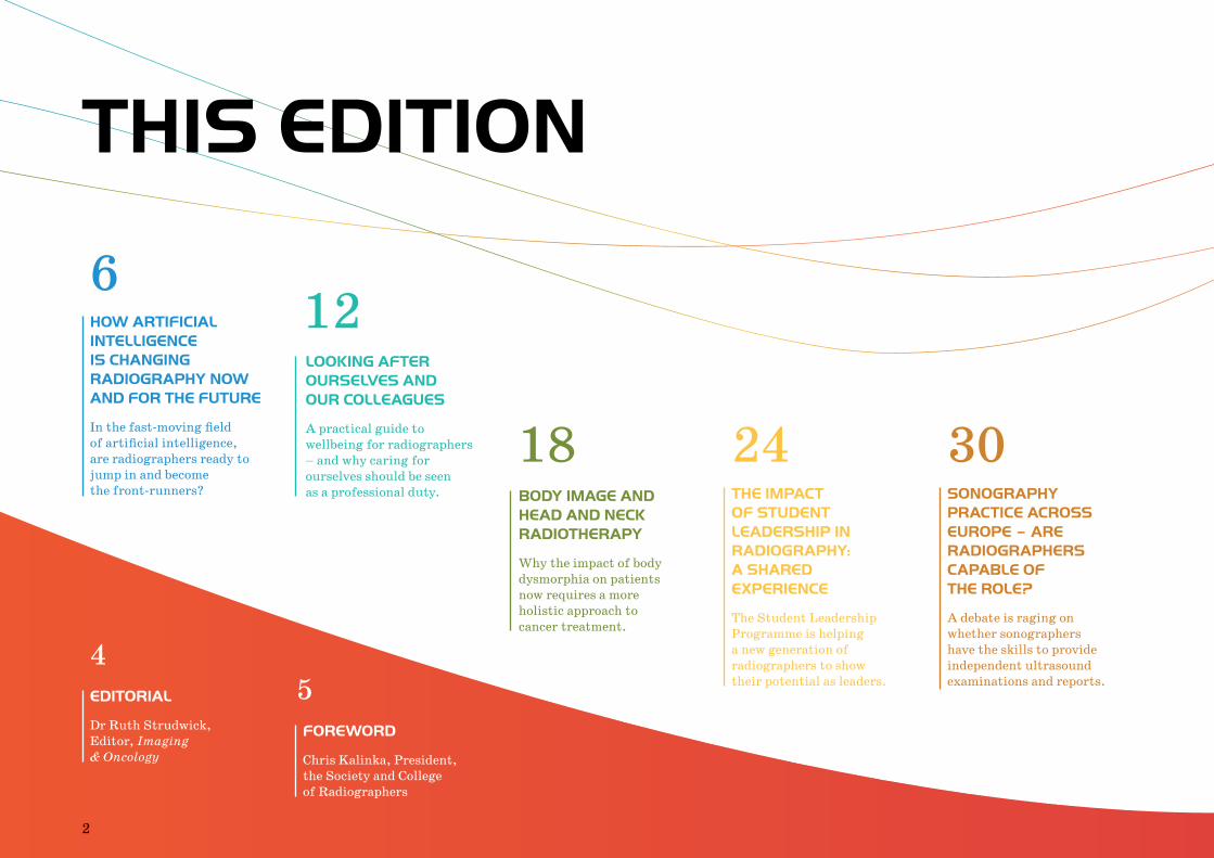
HOW ARTIFICIAL INTELLIGENCE IS CHANGING RADIOGRAPHY NOW AND FOR THE FUTURE
In the fast-moving field of artificial intelligence, are radiographers ready to jump in and become the front-runners?
FOREWORD
Chris Kalinka, President, the Society and College of Radiographers
6
5
12
18 24 30BODY IMAGE AND HEAD AND NECK RADIOTHERAPY
Why the impact of body dysmorphia on patients now requires a more holistic approach to cancer treatment.
LOOKING AFTER OURSELVES AND OUR COLLEAGUES
A practical guide to wellbeing for radiographers – and why caring for ourselves should be seen as a professional duty. THE IMPACT
OF STUDENT LEADERSHIP IN RADIOGRAPHY: A SHARED EXPERIENCE
The Student Leadership Programme is helping a new generation of radiographers to show their potential as leaders.
SONOGRAPHY PRACTICE ACROSS EUROPE – ARE RADIOGRAPHERS CAPABLE OF THE ROLE?
A debate is raging on whether sonographers have the skills to provide independent ultrasound examinations and reports.
EDITORIAL
Dr Ruth Strudwick, Editor, Imaging & Oncology
4
THIS EDITION
2

3856 64
72
MOTIVATIONS, PROCESS AND ASPIRATIONS FOR THE FUTURE: A DISCUSSION TO INFORM THE ECF DEVELOPMENT
Fast-changing technology and complex caseloads mean radiographers need a new Education and Career Framework.
PROMOTING THE ROLE OF THERAPEUTIC RADIOGRAPHERS: THE VALUE OF MACMILLAN CLINICAL FELLOWS
The number of therapeutic radiographers needs to increase by 45% but expansion requires a wider understanding of the role.
RADIOGRAPHER RESEARCH: FUNDING OPPORTUNITIES
Research is not just for the elite – but radiographers need to give themselves permission to become the clinicians they aspire to be.
NEW GUIDELINES ON CONTACT SHIELDING AND RADIATION PROTECTION: TEACHING AND IMPLEMENTATION
Why contact shielding for patients is no longer needed during most X-ray examinations.
48DXA IN THE DIAGNOSIS OF OSTEOPOROSIS AND THE ROLE OF THE RADIOGRAPHER
Why osteoporosis and fragility fractures are not receiving the attention and investment that they deserve.
3

Dr Ruth Strudwick
EDITORIAL
What a year we have had, facing a global pandemic and not really knowing how things would turn out. I have just reviewed
my editorial from last year’s Imaging & Oncology, which was written in April 2020, close to the start of the first lockdown. Such a lot has happened since then and I am so proud of our profession and all that it has contributed to the health and wellbeing of the population during the pandemic.
We have a new publisher this year and I would like to thank Colin Cooper for his support and understanding as we learnt together and Charlotte Beardmore for her continuing support and guidance. Thank you all the authors who have contributed to this edition.
This publication begins with a discussion paper about artificial intelligence from Christina Malamateniou, and the theme of the ever-changing world of radiography is echoed in our final paper from Philip Cosson about changes to the guidelines on the use of contact shielding.
The future direction of the profession is outlined in the paper from the steering group for the new
Education and Career Framework, which is due for publication later this year. And the future direction for sonography practice in Europe is the theme of Gill Harrison’s article.
Jill Griffin highlights the importance of the role of the radiographer in dual energy X-ray absorptiometry (DXA) and the diagnosis of osteoporosis, while Jo McNamara and Hazel Pennington outline the importance of the role of the therapeutic radiographer and encourage us to do all we can to promote our profession.
Nichola Jamison and Sarah Bradder summarise their experiences of the Council of Deans of Health Student Leadership Programme and the impact it has made on their own career aspirations.
It is always important to consider our service users and we have a thought-provoking article from Sabina Khan, who considers the experiences of patients with head and neck cancer and the impact of treatment on body image.
Carole Burnett encourages us all to be research-active and explores the funding opportunities that are available to radiographers.
Finally, Linda Hindle, the deputy chief allied health professions officer for England, encourages us all to look after ourselves and our colleagues. This is an important message for us all during these continuing times of uncertainty.
I hope that you will all find something to take into your own practice – happy reading.
Best wishes
Dr Ruth StrudwickEditor
Editor Dr Ruth Strudwick Managing editor Colin Cooper Production editor Maria Ainley-Taylor Designer Ana Acosta Display advertising Daniel Greenaway [email protected] tel 07540 371347
Published by Haymarket Business Media Bridge House, 69 London Road Twickenham TW1 3SP Email: [email protected] Tel +44 (0)20 8267 5000
Imaging & Oncology is a publication of The Society and College of Radiographers 207 Providence Square Mill Street London SE1 2EW Tel 020 7740 7200 All correspondence relating to Imaging & Oncology should be addressed to [email protected]
Disclaimer © The Society of Radiographers 2021. Unless otherwise indicated, views expressed are those of the editorial staff, contributors and correspondents. They are not necessarily the views of The Society of Radiographers (SoR), its officers or council. The publication of an advertisement does not imply that a product is recommended by the Society. Material may only be reproduced by prior arrangement and with due acknowledgement to Imaging & Oncology.
Haymarket is certified by BSI to environmental standard ISO14001 and energy management standard ISO5001.
PEFC/16-33-1007
PEFC Certified
This product is from sustainably managed forests and controlled sources
www.pefc.org
PEFC Certified
This product is from sustainably managed forests and controlled sources
www.pefc.org
PEFC Certified
This product is from sustainably managed forests and controlled sources
www.pefc.org
PEFC Certified
This product is from sustainably managed forests and controlled sources
www.pefc.orgPEFC/16-33-1007 PEFC/16-33-1007
PEFC/16-33-1007
controlled sources
PEFC Certified
This product is from sustainably managed forests and controlled sources
www.pefc.orgPEFC/16-33-1007
PEFC/16-33-197
4

Chris Kalinka
FOREWORD
A very warm welcome to the 2021 edition of Imaging & Oncology, it is a real privilege as President to contribute the foreword to such
an eminent professional publication. The past year has been unprecedented and extremely challenging for all our imaging and therapy professionals. I am humbled by the bravery, professionalism, stoicism and selfless service you all provide to our patients.
This edition of the journal addresses some very topical issues as we emerge and, hopefully, begin to recover from the Covid-19 pandemic. I do hope our CPD activity can return to more face-to-face events, given all the associated personal networking opportunities we have missed during a year dominated by video meetings.
Research and evidence is vitally important to underpin the development of our workforce and clinical techniques to improve patient-centred care. All the contributions contained in this edition add to our knowledge base and work towards quality improvement. I have real admiration for all those who author articles and are our knowledge pioneers, stimulating reaction, discussion and development.
The breadth of articles is inspiring, from the wellbeing of all our colleagues, which is so important at this difficult time, to the development of artificial intelligence that will, hopefully, help with the capacity and quality of patient care in the future. Our student leaders inspire me and their contribution must be valued, especially as our future colleagues will be providing and leading clinical services. Ensuring sonography quality has sometimes proved contentious as we seek better regulation of ultrasound in the future. We must really value all our non-medical contributions to this essential modality. Patient care and safety is certainly well-represented across this edition of the journal.
Diagnostics and therapy are essential elements of patient pathways – the expansion of capacity and resources, both in terms of workforce and equipment, will be vital to support the recovery and even expansion of services, which will ensure the improved survival of our patients. We must provide highly technical care with an empathetic, human face – fundamentally, we all look after people. There are difficult times ahead, no one is sure what the ‘new normal’ post-Covid future holds for the NHS, with its ‘free at the point of delivery’ ethos, an example to the world and held so dear.
I heartily recommend Imaging & Oncology 2021 to you all. I hope it helps your personal development and enhances professional practice across all our varied services. I hope it encourages cooperation, professional development and improvement across patient care and may encourage you to contribute to the journal one day.
Aspire to be inspirational.
Chris KalinkaPresident, the Society and College of Radiographers
5

HOW ARTIFICIAL INTELLIGENCE IS CHANGING RADIOGRAPHY NOW AND FOR THE FUTURE
Dr Christina Malamateniou, Dr Ruth Strudwick
6

While many AI technologies are impressive, they are often called in to solve problems that do not really exist. So, AI innovation will need to be designed around patient suggestions and clinical practice challenges – always with practitioner input – to maximise its potential and bring solutions to real-life problems’
7

A rtificial intelligence (AI), the ability of a computer to perform human-like tasks, is neither a new concept nor a new technology. It has been solving problems and helping with decision making since 1950, when Alan Turing
published his seminal theoretical paper “Computing machinery and intelligence”1.
Computer scientists in the field of AI have been quietly working in the background for decades to improve algorithms and refine mathematical processes. The latest additions to the field of AI are machine learning and deep learning, which allow machines to learn better and faster2. Despite developments in the mathematical theories of AI, for many years it has not been integrated into clinical practice. This has been due to many reasons, including: a) the slow speed and limited capacity of processors; b) our limited and often incomplete understanding of the human brain and intelligence and the ways by which humans learn; and c) the lack of online availability of big databases with well-curated clinical data. As these challenges have been gradually overcome in the past two decades, AI has started to revolutionise healthcare at an unprecedented rate2,3.
This is particularly evident and potent in technology-enabled professions, such as radiography and radiology. In radiology, AI and deep learning have been changing workflows and image interpretation pathways, proposing to make the processes more efficient and more consistent. Similarly, radiography has seen the introduction of new AI applications across all the fields of radiography practice: clinical, research and education4.
Impact of AI on radiography clinical practice The uptake of AI in radiography clinical practice is being inadvertently accelerated by staffing shortages, long waiting lists for medical imaging appointments and the Covid-19 pandemic, which has stretched NHS resources more than any other crisis in the past century. Every aspect of the clinical practice of radiography has started to be impacted by AI applications: patient appointments, protocol planning and optimisation, data acquisition and slice positioning, patient safety checks (radiation and MRI safety), data postprocessing (segmentation, radiomics), and image interpretation2,5. Image postprocessing holds the lion’s share at the moment on AI radiography applications but other fields of radiography practice are starting to accelerate, such as data acquisition and protocol optimisation.
AI tools have permeated the different modalities of radiography in different ways and at varying degrees. Cross-sectional imaging (magnetic resonance imaging, computed tomography, positron
Despite the abundance of AI algorithms available for clinical applications in radiography and radiology, very few of these have been externally validated and thoroughly tested to be suitable for clinical deployment’8

been externally validated and thoroughly tested to be suitable for clinical deployment. Furthermore, even if an AI algorithm has been thoroughly tested for a specific condition or patient sample, there is little generalisability in a different set up (different scanner, different imaging protocols or even different patient populations and demographics). Therefore, AI validation has to become an integral part of quality assurance and quality control in radiography and medical imaging in general7.
Impact of AI on radiography education The more that AI infiltrates clinical practice in radiography, the more pressing the need to proportionately update undergraduate and postgraduate curricula for safe and efficient radiography practice. While at the beginning many radiographers will learn ‘on the job’, to maximise the benefits of AI tools, the radiography workforce will need to be trained on these new technologies systematically to increase their understanding of them, exactly as outlined in the recent Topol review8.
Education about AI at any level could involve familiarisation with terminology, clinical examples of AI applications in radiography to appreciate impact on practice, teaching technical aspects of AI such as statistics, AI design principles (but not necessarily programming), validation techniques, ethical implications of AI, patient-centred care principles and applications, and entrepreneurship to propel innovation. In other words, a similar approach to other educational interventions designed to introduce new technologies to healthcare professionals9. This is by no means an exhaustive list of the AI topics one could study, because AI techniques develop very quickly and all higher education institutions offering AI for radiography courses will need to be ready to update their curricula regularly to keep up with developments and customise to local needs.
Teaching will not only be about AI but with AI. There have been early applications for AI-based adaptive learning in radiography with some good results and this is a promising field for the future. Using AI in teaching can drastically change the static ways in which we think about radiography education and education in general, and allows us to adopt more flexible formats, tailored to each candidate’s strengths and weaknesses. Teaching on and with AI can be delivered online or through hands-on workshops, on undergraduate or postgraduate curricula, and in different modalities and aspects of radiography, depending on local needs and expertise.
Internships in AI facilitated by academic-industry partnerships or intercalated radiography degrees will help the educational transition of radiographers into this accelerated digitalisation of healthcare
emission tomography), chest radiography and mammography, are leading this list, each for different reasons: the natural affinity with advanced technological input as part of the clinical examination, the abundance of well-curated big datasets and national cancer screening programmes. Similar to diagnostic radiography, therapeutic radiography has witnessed the emergence of new AI tools, changing the methods of treatment planning and therapeutic techniques6.
Covid-19 has only accelerated this trend as the pressures on healthcare and diagnostics have increased many times over during the pandemic and medical imaging has a central role in diagnosis, understanding of disease progression, follow up and treatment monitoring, as well as in decision making for different treatment pathways.
Despite the abundance of AI algorithms available for clinical applications in radiography and radiology, very few of these have
Using AI in teaching can drastically change the static ways in which we think about radiography education and education in general. It allows us to adopt more flexible formats that are tailored to each candidate’s strengths and weaknesses’
9
F

and, for a time during this transition, there will be some old-school instructional design with some new AI tools working side by side.
It is vital to appreciate that AI training should be integral to healthcare professions and not an option. Generous funding and robust incentives have to be set aside on a national level to accelerate the education of radiographers and other healthcare professionals on AI techniques and applications. Without these, AI training will remain the benefit of those who can afford it or those who have the insight to see it as a way towards self-improvement or a career advancement.
Research and innovationResearch is the main tool to build the evidence base for AI in radiography. It is also our only chance of making sense of all the topics that matter to us as a profession: what AI is for radiography, what it means for our practice, how it can better help our patients, how to avoid mistakes and take full advantage of its capabilities, how to answer pressing clinical questions and give solutions to long-standing clinical problems in radiography. Research is even the answer to how best to train the radiography workforce on AI techniques through carefully designed educational research projects. Research in radiography ‘is a requirement and not an option’, as Professor Peter Hogg and his team highlighted many years ago10.
Research should be grounded in clinical practice and help make AI less of a ‘black box’. This is also the only way to build trust in AI and facilitate its safe and efficient implementation, both for patients and practitioners. AI is quite diverse and complex, meaning we need to prioritise the areas of research that would have more impact on patients and those that can deliver the biggest improvements in healthcare outcomes. In addition, AI research should be multidisciplinary, bringing together all the skill sets required, including radiologists, computer scientists, statisticians, health psychologists and other healthcare practitioners, depending on the research focus. That said, in order to solve radiography practice problems it needs to be radiographer-led.
It is also vital for each AI research project, like all research projects in radiography, to actively involve patients and the public in its design and implementation, to ensure a patient-centred and person-centred focus and the user-friendliness of these technologies11.
For radiography, a young profession with limited research capacity, this might sound like a rude awakening. However, the de facto aptitude with technology required to become a radiographer and the excellent skill mix of patient-centred care and technological optimisation might offer the most fertile ground to propel AI-based radiography-led research. It remains to be seen whether radiographers can jump on this opportunity and become front-runners in the field.
While many AI technologies are impressive, they are often called in to solve problems that do not really exist. So, AI innovation will need to be designed around patient suggestions and clinical practice challenges – always with practitioner input – in order to maximise its potential and bring solutions to real-life problems.
PrioritiesClinical practice A robust AI validation framework needs to be designed for the clinical application of AI in radiography to ensure that high-quality, thoroughly tested, reliable AI tools are available to patients and staff, to ensure beneficence and no maleficence. Deployment and testing of AI tools will be vital for areas such as screening but also for high-frequency examinations like chest radiography, to help with backlogs of reporting but also to help with perfecting the AI tools on large training datasets.
Education Radiographers must be trained to the latest technologies and their methodological, clinical and ethical implications, because training the workforce is the only way to ensure AI tools are used efficiently and safely. The way each country will choose to do this
Research should be grounded in clinical practice and help make AI less of a ‘black box’. This is also the only way to build trust in AI and facilitate its safe and efficient implementation both for patients and practitioners’
10

will depend on a complex set of factors, including but not limited to: radiography clinical needs, local expertise, current educational structures and workforce skill set (for example, very few countries have radiographers reporting or using ultrasound), the legal framework, workforce education budgets and the partnerships that each country will prioritise (industry-academic, clinical-academic, and patient-academic).
Research More radiography-led research on AI is required. This could include the fields of explainable AI, AI for patient-centred care, quality assurance of AI, and AI for patient safety, and should be prioritised each time, based on local contexts. Funding has to be invested in this field in the form of project grants and fellowships, ensuring there are opportunities and conditions for all healthcare professionals to participate and be successful.
Conclusion Appropriate and relevant training and focused research are needed to maximise the benefits and minimise the risks of AI for clinical practice, all done with the patients in mind. Radiographers have a central role in AI implementation and, therefore, will need to embrace AI education and research initiatives for safely and effectively delivering high-quality medical imaging services. ■
It remains to be seen whether radiographers can jump on this opportunity and become front-runners in the field’
Dr Christina Malamateniou, Director of Postgraduate and Doctorate Programme in Radiography, Division of Radiography and Midwifery, City, University of London
Dr Ruth Strudwick, Professional Lead: Radiography, University of Suffolk
References
1. Turing AM (1950) ‘Computing machinery and intelligence’, Mind 49 pp433-460.
2. Meijering E (2020) ‘A bird’s-eye view of deep learning in bioimage analysis’ Computational and Structural Biotechnology Journal 18 pp2312-2325.
3. Lewis SJ, Gandomkar Z, Brennan PC PhD (2019) ‘Artificial intelligence in medical imaging practice: looking to the future’, Journal of Medical Radiation Sciences 00 pp1-4.
4. Hardy M, Harvey H (2020) ‘Artificial intelligence in diagnostic imaging: impact on the radiography profession’, British Journal of Radiology 93 p1108.
5. Lakhani P, Prater AB, Hutson RK, Andriole KP, Dreyer KJ, Morey J, Prevedello LM, Clark TJ, Geis JR, Itri JN, Hawkins CM (2020) ‘Machine learning in radiology: applications beyond image interpretation’, Journal of the American College of Radiology 15(2) pp350-359.
6. Boon IS, Lim JS, Yap MH, Au Yong TPT, Boon CS (2020) ‘Artificial intelligence and soft skills in radiation oncology: data versus wisdom’, Journal of Medical Imaging and Radiation Sciences 01 pp1-2.
7. Kelly CJ, Karthikesalingam A, Suleyman AM, Corrado G, King D (2020) ‘Key challenges for delivering clinical impact with artificial intelligence’, BMC Medicine 29 17(1) p195.
8. Topol E (2019) ‘Preparing the healthcare workforce to deliver the digital future’, The Topol Review. An independent report on behalf of the Secretary of State for Health and Social Care (https://topol.hee.nhs.uk/ accessed October 2020).
9. Edirippulige S, Armfield NR (2017) ‘Education and training to support the use of clinical telehealth: a review of the literature’, Journal of Telemedicine and Telecare 23(2) pp273-282.
10. Gambling T, Brown P, Hogg P (2003) ‘Research in our practice – a requirement not an option’, discussion paper, Radiography 9 (1) pp71-76.
11. SCoR (2018) Patient Public and Practitioner Partnerships within Imaging and Radiotherapy: Guiding Principles (www.sor.org/sites/default/files/document-versions/guiding_principles_final_proofed_0.pdf accessed January 2021).
11

LOOKING AFTER OURSELVES AND OUR COLLEAGUES
Linda Hindle
12

It is normal to feel worried, scared or helpless about the current situation, especially if you are over-tired. Share your concerns with others you trust; doing so may help them too. Notice how your colleagues are feeling and take time to talk’
13

What is resilience?This article highlights the potential impacts of the pandemic on physical and mental wellbeing and offers effective strategies to maintain wellbeing and resilience.
Resilience is our ability to cope with the normal stress of life and to bounce back from crises2. It includes the physiology, attitudes, knowledge, skills, resources and circumstances that can withstand stress and adapt to change.
Across the population, resilience has been relatively high during the pandemic3. However, as the longevity of the pandemic increases and fatigue sets in, the ability to adopt and maintain coping strategies may become more challenging.
The causes of poor mental and physical wellbeing during the pandemic are multiple and will be specific to individual circumstances. Healthcare professionals may potentially be affected in one or several of the following ways:
• Changes to physical activity levels because of altered commuting arrangements, working from home, lack of access to sporting and activity venues or limited time. • Alterations to diet, leading to weight gain. • Increased alcohol consumption, smoking or drug use. • Fatigue, poor sleep. • Anxiety, depression, irritability or stress. • Reduced social connections. • Dealing with trauma, including bereavement. • Financial worries. • Caring responsibilities. • Work pressures.
The impact and experience of the Covid-19 outbreak has been different for everyone, as has how we have reacted to it, but there is no doubt it has been an extremely difficult time for us all. That is why it is so important to do what we can to look after our mental and physical health and wellbeing – now more than ever – and to reach out if you need support.
Evidence-based approach to wellbeingMany of us will now be reflecting on our personal strategies to maintain resilience and wellbeing. In this article I have collated evidence-based approaches to maintaining health and wellbeing during the pandemic. It is not an exhaustive list, and you could take some time with your colleagues to think about what else might help you as individuals or as a team.
T he impact of Covid-19 has taken its toll on all of us in some way. Many healthcare professionals have been working extra hours, either responding to Covid-19 directly or backfilling to enable others to do so. Some will have been unable to
respond because of health conditions, place of work or other personal circumstances and this has also been stressful, often leading to feelings of guilt related to not being able to help.
On top of this are the extra challenges linked to balancing caring for family members with work, homeschooling or coping with health conditions when care may have been stopped or delayed. This is in addition to the natural fears about the virus itself, particularly for those who have been shielding or are from higher-risk demographics such as black and minority ethnic communities1.
Many people are tired and in need of rest and respite. Evidence tells us that those in caring roles often wait until they are very unwell before seeking help. We can only continue to help others if we look after ourselves, so it is important to consider this as part of our professional duty and not just something that is ‘nice to do’. We must all encourage each other to seek help and to seek it as soon as it is needed. Leaders, teams and employers need to keep offering people support to stay well at work – and they must keep offering it consistently across teams, organisations and sectors.
14

F
• Think about when you will take time to rest. Plan your annual leave early so you have time to take a break and recharge your batteries. Schedule breaks during the day when you can. • Healthcare professionals are offered and encouraged to have
vaccinations for flu and Covid-19. This year it is more important than ever to protect ourselves, our families and our patients: don’t forget to book your vaccinations. • Look after your mental wellbeing and that of your colleagues.
It is normal to feel worried, scared or helpless about the current situation, especially if you are over-tired. Share your concerns with others you trust; doing so may help them too. Notice how your colleagues are feeling and take time to talk. • In a crisis, we do our best with the information and resources we
have available. The pace of work combined with virtual working environments can impact on working relationships, so it is important to remember everyone is doing their best and avoid putting unnecessary pressure on yourself and your colleagues. Don’t be afraid to accept help if offered and ask for help if needed. • Our physical health has a big impact on how we feel. At times like
these, it can be easy to fall into unhealthy patterns of behaviour that end up making you feel worse. Try to eat healthy, well-balanced meals, drink enough water and exercise regularly. • Physical activity is known to boost mood, while physical fitness
is a protective factor for good mental health. There is now strong evidence to demonstrate the protective effect of physical activity on a range of many chronic conditions, including coronary heart disease, obesity, type-2 diabetes, mental health problems and social isolation. Even relatively small increases in physical activity can contribute to improved health and quality of life4. • Sleep is a protective factor for mental health. Sleep deprivation can
affect emotional regulation, and studies suggest that lack of sleep may affect the ability to respond to negative situations5, therefore sleep is even more important during times of crisis. The NHS Every Mind Matters website6 provides useful information about managing stress, improving sleep and protecting your mental wellbeing. Special access has been arranged for healthcare workers to certain wellbeing apps, such as Silvercloud7, Sleepio8 and Daylight9, which give advice on how to cope with stress, improve sleeping patterns and offer suggestions for practising mindfulness. • Check out the services your employer is providing to support you.
Specific mental health support is available on the National NHS Helpline, including a dedicated support line: text FRONTLINE to 85258 to start a conversation or call 0300 131 7000. This service is free on all major mobile networks and provides direct support.
Resilience is our ability to cope with the normal stress of life and to bounce back from crises. It includes skills, knowledge and resources that can withstand stress and adapt to change’
15

References
1. Public Health England (2020) Disparities in the Risk and Outcomes of Covid-19.
2. Mental Health Foundation/The Faculty of Public Health (2016) Better Mental Health for All: a Public Health Approach to Mental Health Improvement. London: Mental Health Foundation.
3. Mental Health Foundation (nd) ‘Resilience across the UK during the coronavirus pandemic’, (www.mentalhealth.org.uk/coronavirus/resilience-across-uk-coronavirus-pandemic accessed 23 December 2020).
4. Chief Medical Officers (2019) Physical Activity Guidelines (https://assets.publishing.service.gov.uk/government/uploads/system/uploads/ attachment_data/file/832868/uk-chief-medical-officers-physical-activity-guidelines.pdf accessed 23 December 2020).
5. Anderson C and Platten CR (2011) ‘Sleep deprivation lowers inhibition and enhances impulsivity to negative stimuli’, Behavioural Brain Research 217(2) pp463-466.
6. Public Health England (nd) Every Mind Matters (www.nhs.uk/oneyou/every-mind-matters/ accessed 23 December 2020).
7. www.silvercloudhealth.com/uk (accessed 23 December 2020).
8. www.sleepio.com (accessed 23 December 2020).
9. www.bighealth.com/daylight (accessed 23 December 2020).
10. www.samaritans.org/how-we-can-help/health-and-care/here-listen-support-line-nhs-people (accessed 23 December 2020).
11. Campion J, Bhui K and Bhugra D (2012) ‘European Psychiatric Association (EPA) Guidance on Prevention of Mental Disorders’, European Psychiatry 27:67-80.16/j.eurpsy.2011.10.004.
12. Mental Health Foundation (nd) Thriving in Nature: a Guide for Everyone (www.mentalhealth.org.uk/campaigns/thriving-with-nature/guide).
Linda Hindle, Deputy Chief AHP Officer for England, PHE Lead Allied Health Professional and National Engagement Lead for Public health in Police, Fire and Ambulance Services, Public Health England
Leaders, teams and employers must keep offering people the support they need to stay well at work – and they must keep offering it consistently across organisations and sectors’
• Samaritans also has a specialist Wellbeing Support phoneline10 for health and social care workers, which is free to access and available from 7am to 11pm, seven days a week. You can receive support, signposting and confidential listening from trained professionals in a number of areas by calling 0800 069 6222. • The Covid-19 outbreak has brought a great deal of uncertainty to
our lives, and many families are facing job loss or financial difficulties. Feeling stressed about money can impact on mental wellbeing, so it is important to act as soon as possible. The NHS Every Mind Matters6
website also offers practical financial advice and support. • Connecting with others with whom we have a positive relationship
is an effective way of supporting our mental health and wellbeing and may prevent mental health problems11. Healthcare professionals have generally managed to retain social connections and a sense of purpose during the pandemic because of the roles we play; however, connections with family members and friends will have been reduced and the social side of work has been affected. Make time to connect with colleagues, family and friends and notice if colleagues need to talk. • Spending time in green space is known to be beneficial for mental
health and overall wellbeing12.
Thank you for everything you are doing for others. Your role has been – and will continue to be – invaluable. However, you can only continue to help others if you look after yourself, so consider self-care to be part of your professional duty. ■
16

BODY IMAGE AND HEAD AND NECK RADIOTHERAPY
Sabina Khan
18

Our pathways from diagnosis to treatment are becoming shorter to treat cancers more quickly but, in a bid to cure, are we struggling to prepare patients psychologically for the impact of their diagnosis and treatment in such a small space of time?’
19

The patient’s hoarse voice and oral mucositis left him frustrated due to difficulties in communicating verbally and assuming people would be focusing on the amount of saliva being produced by his mouth. This affected his relationship with his partner physically and he found it difficult to accept her as a carer’
B ody image is a key issue for patients with cancer. It can make a significant impact on an individual’s wellbeing and can affect the patient’s perception of themselves. The psychological impact of cancer has been reported widely
across all cancer groups1, 2, 3. Head and neck cancer (HNC) accounts for more than 12,200 cases annually and is the eighth most common cancer in the UK4.
Patients with HNC have particular visible changes caused by the diagnosis or treatment, such as radiotherapy and surgery. Patients who undergo radiotherapy are required to cope with a broad range of side effects, such as pain, fatigue, oral mucositis, skin reactions and swallowing difficulties and require a close multidisciplinary team approach for treatment5. Interventions such as nasogastric (NG) and percutaneous endoscopy gastric (PEG) tubes are commonly used, requiring physical changes to patients’ bodies.
Post-operative scars and permanent alterations to the face can also have a profound effect because of their visibility. Many articles have been written exploring cancer and its influence on body image6,7,8. For some patients, the psychological effect of body dysmorphia is just as difficult to manage as the cancer treatment and its side effects. It can lead to anxiety, depression and the feeling of being unattractive and can affect patients socially, leaving them feeling isolated.
The extent to which body image is affected during treatment is highly subjective and can be influenced by the patient’s environment, relationships, life experience and understanding of the diagnosis7. For some, it can be influenced by culture, shaped by social identities and self-acceptance9. The effects of this can have an impact on patients long after treatment has finished, affect their loved ones and serve as a reminder of their experience.
The impact of skin reactionsSkin reactions are an inevitable side effect of head and neck radiotherapy, although the extent of this has significantly reduced with the use of intensity modulated radiotherapy10. A common theme for patients is the difficulty in hiding their skin reactions and disfigurements with clothes and accessories.
An example of this is a 53-year-old patient, who did not want to stop wearing make-up in the irradiated region of her skin. This was particularly difficult as she was being treated for a superficial parotid tumour, which required a dose to the skin, causing erythema and dry desquamation. Even though the application of these products were painful and aggravating for her skin reaction, she felt it allowed her to live a ‘normal life’. For her, the ability to disguise this reaction was just as important as the treatment itself. Once her
20

Fundamental changes to lifestyleWith the increased incidence of human papillomavirus infection (HPV) positive tumours in HNC, younger patients are being diagnosed11. With a younger demographic, challenges such as fatigue, the struggle to keep up with previous routines and the impact on social situations can prove difficult for some patients.
An example is a 44-year-old patient being treated for a HPV-positive tonsillar tumour with radiotherapy, who at first appeared very confident and social. Fitness and healthy eating were important to his lifestyle. The patient tried to maintain his daily running regime and struggled with changing his diet to increase his calorie intake. This is particularly important for head and neck radiotherapy patients to help maintain their contour/separation to maximise the efficacy of their treatment12.
Mucositis and pain caused a reduction in his oral intake, thus requiring the patient to fortify his food and alter his diet by eating things he would not normally consume, such as dairy products. As treatment progressed, the patient struggled with changes in taste, fatigue and the inability to maintain his fitness regime. He struggled with the physical changes to his body and felt disappointed for not being able to continue with exercise. When the time came to use his PEG, he deemed it a sign of failure that he could not manage to be fed orally. In effect, he became deflated and needed reassurance that this type of fatigue was short term.
The patient became more withdrawn during the six weeks of radiotherapy. When asked how he was, his primary concern was how he felt over any other physical side effects. The body disfigurement and treatment reactions made him self-conscious. The patient’s hoarse voice and oral mucositis left him frustrated because of the difficulties in communicating verbally and he assumed people would be focusing on the amount of saliva being produced by his mouth. This affected his relationship with his partner physically and he found it difficult to accept her as a carer, feeling that he was a burden to her. The patient required close support during and post radiotherapy from our psychology colleagues for many weeks until he could reach a point where he felt more confident.
Distressing tooth extractionsDental work is a necessary part of radiotherapy treatment for HNC. It is essential to complete extractions for teeth that are unrestorable or require periodontal treatment to minimise the risk of osteoradionecrosis13, 14. For many of our patients, assessment and several extractions are required within days of their diagnosis to be able to start their treatment as soon as possible. Some of these
skin had started to break down and she was no longer able to carry on using the products because she needed to minimise the risk of infection. This had a detrimental effect on the patient because it meant she would need to face the reality of her diagnosis. As she said: ‘This became real.’
The patient was unhappy with the way she looked and felt people would stare at her and ask questions. The skin reaction embarrassed her because it was a reminder of what her body was undergoing, and she felt happier when she could ‘pretend’ she was not having any treatment. By hiding her reaction, she was able to carry on a normal life. The lack of control around her diagnosis and treatment and the way she looked now affected her confidence and personality.
As the patient’s skin reaction improved in the months post radiotherapy, she was able to wear make-up again and some of her self-confidence returned. However, the discolouration of her skin reminded the patient of the treatment she had undertaken in her cancer journey.
Dentures could not be supported until the patient had healed from the treatment, which made her feel more anxious. This affected the way she smiled, spoke, ate and interacted with her family. The removal of her teeth made more of an impact on her lifestyle than the treatment itself’
21
F

With increased success rates of cure and long-term survival, it is even more imperative that we look at the quality of life for those patients who experience a very visual impact’
extractions are clearly visible and can affect their verbal skills. Dentures cannot be worn until recovery due to the impact on the treatment area, which can cause pain and discomfort. Patients’ pathways from diagnosis to treatment can be fast. They require urgent dental interventions such as extractions and, in rare cases, can become edentulous, leaving a significant psychological impact.
A 64-year-old woman having radiotherapy to treat an oropharyngeal squamous cell carcinoma had several extractions within seven days of her diagnosis and commenced treatment within 20 days. The patient initially presented to the radiotherapy department unable to talk without covering her mouth. She felt distressed that we were unable to supply her with dentures so early on in her treatment. Her experience of having to have extractions while awake under mild sedation, so soon after her diagnosis, was particularly distressing. She consented because she knew it was an essential part of the treatment but did not have time to mentally prepare herself for the procedure. The pain and lack of teeth meant she had to alter her diet significantly and was unable to eat the foods she had previously enjoyed.
22

References
1. Costa DSJ, Mercieca R, Rutherford C, Gabb King MT (2016) ‘The impact of cancer on psychological and social outcomes’, Australian Psychologist 51 (2) pp89-99.
2. Ruane-McAteer E, Porter S, O’Sullivan J, Dempster M, Prue G (2019) ‘Investigating the psychological impact of active surveillance or active treatment in newly diagnosed favourable-risk prostate cancer patients: a nine-month longitudinal study’, Psycho-Oncology 29 (8) pp1743-1752.
3. Mcbride CM, Clipp E, Peterson BL, Lipkus IM, Demark-Wahnwfried W (2000) ‘Psychological impact of diagnosis and risk reduction among cancer survivors’, Psycho-Oncology 9 (5) pp418-427.
4. Cancer Research UK (nd) Head and neck cancer incidence statistics (www.cancerresearchuk.org/health-professional/cancer-statistics/statistics-by-cancer-type/head-and-neck-cancers/incidence accessed December 2020)
5. Verdonck-de Leeuw IM, Buffart LM, Heymans MW, Rietveld DH, Doornaert P, de Bree R, Buter J, Aaronson NK, Slotman BJ, Leemans CR, Langendijk JA (2014) ‘The course of health-related quality of life in head and neck cancer patients treated with chemoradiation: a prospective cohort study’, Radiotherapy & Oncology 110 (3) pp422-428.
6. Hopwood P (1993) ‘The assessment of body image in cancer patients’, European Journal of Cancer 29 (2) pp276-281.
7. White CA (2000) ‘Body image dimensions and cancer: a heuristic cognitive behavioural model’, Psycho-Oncology 9 (3) pp183-192.
8. Fingeret MC, Teo I, Epner DE (2013) ‘Managing body image difficulties of adult cancer patients: lessons from available research’, Cancer 120 (5) pp633-641.
9. Tylka TL, Wood-Barcalow NL (2015) ‘What is and what is not positive body image? Conceptual foundations and construct definition’, Body Image 14 (1) pp118-129.
10. Lee N, Chuang C, Quivey JM, Phillips TL, Akazawa P, Verhey LJ, Xia P (2002) ‘Skin toxicity due to intensity-modulated radiotherapy for head-and-neck carcinoma’, International Journal of Radiation Oncology, Biology, Physics (IJROBP) 53 (3) pp630-637.
11. Young D, Xiao CC, Murphy B, Moore M, Fakhry C, Day TA (2015) ‘Increase in head and neck cancer in younger patients due to human papillomavirus (HPV)’, Oral Oncology 51 (8) pp727-730.
12. Hansen EK, Bucci MK, Quivey MD, Weinberg V, Xia P (2004) ‘Repeat CT imaging and replanning during the course of IMRT for head and neck cancer’, IJROBP 64 (2) pp355-362.
13. Koga DH, Salvajoli JV, Alves FA (2008) ‘Dental extractions and radiotherapy in head and neck oncology: review of the literature’, Oral Diseases 14 (1) pp40-44.
14. Devi S, Singh N (2014) ‘Dental care during and after radiotherapy in head and neck cancer’, National Journal of Maxillofacial Surgery 5 (2) pp117-125.
Sabina Khan, Head and Neck Specialist Radiographer, University College London Hospitals
In every clinic appointment she mentioned the extractions and the need to have dentures made urgently. Unfortunately, this could not be supported until the patient had healed from the treatment, which led to her feeling more anxious. This affected the way she smiled, spoke, ate and interacted with her family. The removal of her teeth made more of an impact on her lifestyle than the treatment itself.
The patient did not attend subsequent dental follow-up appointments for fear of further extractions. She felt deflated that her need for dentures could not be resolved until a few months post radiotherapy treatment, which meant the psychological effect made an impact on her life well after her treatment finished. For this patient, even when the treatment was deemed successful, there was a constant visual reminder that normality had not returned.
A more holistic way forwardThe impact of body dysmorphia in HNC is an area scarcely looked at in the available literature. The onus on the seriousness of HNC and its pathology means there is limited time to assist with body image concerns pre-treatment. Our pathways from diagnosis to treatment are becoming shorter to treat cancers more quickly but, in a bid to cure, are we struggling to prepare patients psychologically for the impact of their diagnosis and treatment in such a small space of time? With increased success rates of cure and long-term survival, it is even more imperative we look at the quality of life of these patients who experience a very visual impact of the cancer and treatment, even after the five-year survival mark.
Interventions to improve quality of life as well as survival time are required in order to treat patients more holistically. Some centres in the UK are moving forward with prehabilitation services, where informed sessions enable patients to prepare for such side effects and know what services are available to them if needed.
This would require a multidisciplinary team approach with our clinicians, surgeons, clinical nurse specialists, allied health professions, dentistry and psychology colleagues. Support services may need to be offered throughout the patient’s cancer journey to give them the chance to use these services, if and when they are needed.
More research needs to be carried out into the quality of life of HNC patients to highlight the common themes and allow the correct provisions. Depression, anxiety and social isolation can become chronic post treatment and may require long-term support. In effect, further support for body dysmorphia needs to be made available to patients. The need for timely referrals is critical to enable us, as healthcare professionals, to assist patients at a time when they need the most support. ■
23

THE IMPACT OF STUDENT LEADERSHIP IN RADIOGRAPHY: A SHARED EXPERIENCE
Nichola Jamison, Sarah Bradder
24

The Student Leadership Programme finds these students, acknowledges their potential and gives them the perfect tools to go forward and make a difference. The programme really embodies what it means to be a healthcare professional in the NHS today – we work interprofessionally, advocate for those we care for, and are all leaders’Sarah Bradder
25

T he NHS Long Term Plan states that the ability of the NHS to deliver high-quality care and transform services that continually meet the needs of its populations depends on ‘great leadership’ at all levels1. This requires a significant
shift in leadership culture from the historic ‘top-down’ style to acknowledging that everyone who can improve a service is a leader2. By taking this grassroots approach to leadership, it ensures that the future workforce is well equipped to deliver safe and effective care in an ever-changing setting3, thus highlighting that building these skills at pre-registration level is paramount.
The level of leadership education given to pre-registration healthcare students varies across institutions, if it is delivered at all. Leadership can often be viewed as important only to those in managerial positions; however, this is not the ‘leadership at every level’ ethos.
Established in 2017, the Council of Deans of Health (CoDH) Student Leadership Programme (SLP) recognised the value of leadership components within The Health and Care Professions Council (HCPC) standards for the allied health professions (AHP) workforce, and has been completed by 200 nursing, midwifery and AHP students since its inception4.
Programme scholars – affectionately known as #150leaders – complete a one-year programme of residential events, undertaking leadership opportunities and individual coaching from visionary leaders who are frontrunners in their own healthcare fields. Students are encouraged to put new skills into practice by leading a project during this time, and many step into leadership roles within their professions upon completing the programme and graduating.
Here, Sarah Bradder and Nichola Jamison share their personal experiences of the programme and describe the impact it has made on their own journeys and those of the people around them.
No two beginnings are the sameSarah began her therapeutic radiography career at Birmingham City University at the age of 18 but, unfortunately, ill health forced her to withdraw from the course before she could complete her studies. Once recovered, she spent the next few years enjoying work at a secondary school as part of a small team that met the needs of students with physical disabilities. Sarah knew, however, that her true passion still lay elsewhere so she re-enrolled in her studies. She applied for the SLP during the second year of her undergraduate studies (a BSc in radiotherapy and oncology) at Sheffield Hallam University.
Sarah says: ‘To be completely honest it was because someone else, my course leader at the time, saw something in me that I didn’t see in myself. I knew that I wanted to “make a difference” but, before the programme, that felt like such an abstract concept. I was trying to get involved with as many opportunities for development as possible, participating in research projects, becoming department representative for AHPs in my university and volunteering at STEMnet evenings. As this was my “second time around” and with more life experience behind me, I wanted to get the most out of my time as a student, but it wasn’t until the SLP that I knew what that meant and how to act on that in a meaningful way.’
Nichola began her radiography journey at the age of 32, having spent her adult life raising her children and working in various healthcare settings as a physiotherapy assistant and dental nurse. Alongside these roles, she had enjoyed a career as a semi-professional musician since the age of 16 and spent much of her time teaching music and performance to children and adults. Despite having a varied professional background, Nichola never felt she had found her place and knew it was time to pursue a formal education.
‘I never really thought about what I wanted out of life as I always felt my priorities lay with my family,’ says Nichola. ‘To these ends, I worked only to fulfil these responsibilities, while knowing that I
I had all of these goals and ambitions, and no idea how to pursue them. I knew that no amount of drive and determination could get me there without the skills and training to focus my aspirations into impactful actions’ Nichola Jamison
26

mentoring enabled me to move forward in a productive manner, while maintaining my awareness of self care.’
She continues: ‘Gill’s guidance was invaluable when it came to creating my leadership project and she always knew when I was pushing myself too hard! More impactful, however, was her awareness of and interest in my long-term goals. Gill helped me express and process my hopes and fears, and that external guidance meant that we could look at challenges with a “risk vs benefit” approach and break each leap into smaller steps. I made significant professional choices during the programme and would never have had the courage to do so without Gill’s input.’
Thriving community of peersBeyond the formal element of the programme, the limited cohort size provides students with a thriving community of peers, who all share a passion for upholding excellent standards of care for service users. This community, along with the support of programme facilitators, was an important factor for Sarah.
‘I found the structure to be really helpful and support from the
had so much more to give. In 2014, changes in my circumstances offered me the chance to reset my life and so I chose to go back into education. I completed an access course at college and, when I discovered therapeutic radiography, I knew immediately that this was my path.’
Fully embracing new experiences, Nichola was in her second year of studies at Ulster University when she discovered SLP. At this point, she had been exploring leadership through various roles as an academic representative, chair of the Society of Radiographers (SoR) UK Student Representative Forum, and by sitting on sub-groups of the Northern Ireland Cancer Strategy. With the support of academic staff, she applied for the programme and was successfully enrolled in the 2018 cohort.
‘I had all of these goals and ambitions, and no idea how to pursue them,’ Nichola says. ‘I knew that no amount of drive and determination could get me there without the skills and training to focus my aspirations into impactful actions. When I received notification of my successful application to the programme, I knew I had been offered an invaluable resource.’
The path to leadershipEach year, the programme launches with a two-day conference. The event enables members of the cohort to meet, often for the first time, and to workshop ideas and concepts throughout the busy event programme. Like so many, Sarah was apprehensive about what to expect. ‘It’s hard to put into words to really explain my experience of the SLP,’ she says. ‘I did not know what to expect and was very nervous that it would be something I wasn’t qualified for; I was feeling like an imposter before I even really knew about imposter syndrome. However, my concerns were wiped away within minutes of being at the first event. Never have I been in a room full of complete strangers and felt so at home. It was, without a doubt, the best thing I have ever been a part of. The doors it has opened for me, and allowed me to open for myself, are invaluable.’
Following the launch event and feeling inspired by the energy and encouragement from speakers and fellow students, Nichola was excited to meet her personal coach and to begin work on developing her skills into meaningful aspirations.
‘As I left the conference, I felt empowered to take the next steps toward my goals,’ Nichola explains. ‘I was passionate about improving support for patients and staff in Northern Ireland but, while I had many ideas, I knew I needed to take smaller steps toward achieving these. I was quickly introduced to my coach, Gill Harrison, an officer at the SoR, whose structured and objective approach to
Never have I been in a room full of complete strangers and felt so at home. It was, without a doubt, the best thing I have ever been a part of. The doors it has opened for me, and allowed me to open for myself, are invaluable’ Sarah Bradder
27
F

organisers was always available. It was great to have a designated mentor to muddle through my thoughts and ambitions with, as well as the support of peers within the programme. I was stunned by the camaraderie that developed quickly between students – each of us had different experiences, professions and exposure to leadership but our common goal was the same.
‘I think that’s what makes the experience so fulfilling and inspiring because the programme finds these students, acknowledges their potential and gives them the perfect tools to go forward and make a difference. SLP really embodies what it means to be a healthcare professional within the NHS today – we work interprofessionally, advocate for those we care for and are all leaders.’
Students were encouraged to interpret key concepts in a way that was relevant and significant to them individually. Many of these could be implemented easily, such as the use of reflection and self-evaluation. Spending time exploring these with her peers inspired Nichola to look closer at her long-term aspirations.
‘The realisation that had the most impact on my career during this time was the importance of each and every member of healthcare staff. Sitting in a room full of students – all hugely driven to effect change, each from different beginnings and each living in different circumstances – I realised that my passion for improving psychological support had developed further into the need to provide support for the individual needs of our AHP workforce,’ she says.
‘SLP taught me that leadership is about so much more than one person. Through the people I met along the way, I learned to celebrate the input of others and the value of “finding your tribe”. After years of juggling life’s responsibilities on my own, I was finally learning to identify and collaborate with those who shared my goals.’
Lessons in leadershipThrough a series of talks and workshops, experienced leaders and programme alumni shared their lessons in leadership. Each experience was different but all were relatable. Some poignant messages have remained with Sarah and Nichola as they move forward in their careers:
Be authentic. Authentic leadership relies on self-awareness, transparency, balanced processing and the upholding of morals in order to inspire trust and loyalty in those around you. It teaches us that displaying vulnerability can be a strength, and that we should allow others to see us as we are.
This was an important realisation for Sarah: ‘Having those in leadership roles share both their successes and failures encourages
us to be bold and accept that it’s OK when things don’t go to plan. By being open and honest, you could be inspiring people who might have given up at the first hurdle. I always worried about telling people that I had done the course before because I viewed this as a failure, but I realised that this was an important part of my story that needed to be acknowledged.’
Where am I and what more can I do? Leadership is about constantly evaluating one’s environment. Beyond this, it is being aware of your role within this environment and knowing what more you can contribute. This outlook empowers those at every level to recognise their value and act upon their goals.
It is a question that Nichola has carried with her every day and speaks about often: ‘Ask yourself now. There is something you would like to change? That’s the leader in you. I impress this regularly upon the students I support. I truly believe there is a leader within us all, and the moment you start believing it, others will too.’
You’ve earned your seat at the table – use it! The power that you have is that no one else is you. Your opinions are valid and deserve
Leadership is about so much more than one person. Through the people I met along the way, I learned to celebrate the input of others and the value of finding your tribe’ Nichola Jamison
28

to be heard. SLP taught Sarah and Nichola to take risks, say yes to opportunities and know that their experiences provide a unique view. You are not ‘just a student’. Student insight can be invaluable to a department and the same can be said for a newly qualified practitioner. Sarah says: ‘When you do have that seat at the table, believe in what you can bring because, in all likelihood, someone who believes in you has advocated for your seat. Ask yourself, “If not me, who? If not now, when?” and remember that having a place at the table is a waste unless you use your voice.’
It’s OK to say no. The self-care component of leadership is often overlooked by those who perceive that leadership can only be achieved through hard work and an unrelenting drive to succeed. In fact, this approach can be harmful if not maintained by self-care, awareness and regulation. This was a difficult, but important concept for Nichola to implement: ‘I had this tendency to be afraid of turning opportunities down. I was always trying to prove myself. My coach broke through this cycle with me and helped me understand that if I continued to say yes to new things, it meant I was saying no to something else – often myself or my family. Over time I learned to balance priorities, and this way I have maintained discipline and focus on what is important to me.’
No one is a leader on their own. Surround yourself with people who inspire, encourage and motivate you. Great leaders are those who share their passion and vision with those around them and welcome collaboration. Sarah takes pleasure in practising this: ‘I have seen what a positive impact the right people can make on someone’s potential. You should always give back what you took from your networks. Help those who are starting out in their careers, be a mentor and advocate, and pass on what you have learned.’
Beyond the programmeSarah now works as a band 5 radiographer at Queen Elizabeth Hospital, where she continues to explore leadership through professional development and promotion of her profession. She currently leads the @WeAHPs twitter platform and speaks regularly at conferences about her experiences.
Sarah continues to work within professional groups, helping shape policy, and is the SoR industrial relations representative in her department. This year, she was accepted on to the Healthcare Leadership Academy Scholarship Programme, where she is creating a project that aims to improve the awareness of the AHP professions among the general public.
‘SLP was instrumental in how I went forward as a student and how I have continued to practice as a qualified professional,’ says Sarah. I believe that leadership is for everyone, not just something that should only be fostered by a few.’
Having graduated in 2020, Nichola worked with the SCoR as its interim student support officer. She is passionate about improving engagement, experience, wellbeing and leadership within the pre-registration workforce and has since taken on the new role of students and new professionals officer at the Society.
She continues to work within the Northern Ireland Cancer Strategy, advocating for improved psychological support for frontline cancer staff. To further develop her support skills, Nichola is undertaking an MSc in psychological sciences at Queens University, Belfast, and hopes to continue building on her training as she moves forward on her radiography journey.
‘I fell in love with radiography from day one,’ she says. ‘The staff and students are the most inspiring and driven people I have ever met. I am more passionate about supporting them now than I ever have been and believe this support should begin from the first day of their radiography studies. Radiographers are natural leaders and SLP has taught me the importance of identifying and nurturing leadership qualities in others. I would not be where I am today without the support and guidance of the programme.’ ■
Nichola Jamison, SCoR Students and New Professionals Officer
Sarah Bradder, Therapeutic Radiographer, Queen Elizabeth Hospital, Birmingham
References
1. NHS (2019) The NHS Long Term Plan (www.longtermplan.nhs.uk/publication/nhs-long-term-plan accessed 12 May 2019).
2. NHS Improving Quality (2014) The New Era of Thinking and Practice in Change and Transformation: a Call to Action for Leaders of Health and Care.
3. Health Education England (2018) Maximising Leadership Learning in the Pre-Registration Healthcare Curricula – Model and Guidelines for Healthcare Education Providers 2018.
4. Council of Deans of Health (2021) About the Student Leadership Programme, (https://councilofdeans.org.uk/studentleadership/about-the-programme/programme-structure accessed 3 January 2021).
29

SONOGRAPHY PRACTICE ACROSS EUROPE – ARE RADIOGRAPHERS CAPABLE OF THE ROLE?
Gill Harrison
30

The UK seems to be unique in that the expectation of any sonographer is to “independently undertake, interpret, analyse and report” ultrasound examinations’
31

A s part of the European Congress of Radiology in 2017, a debate that focused on providing an effective ultrasound service generated much discussion in the European and international community. Opinions were divided on
whether sonographers had the necessary skills, competence and ability to provide independent ultrasound examinations and reports.
Presentations by UK sonographers provided evidence of the successful implementation of sonographer practice going back some 40 years and the introduction of independent reporting practice by radiographers more than 30 years ago.
Following the conference presentations, further publications caused some controversy in the wider ultrasound community1,2. Questions were asked about how radiographers/sonographers could provide interpretative reports without medical training. Evidence was provided to emphasise the efficacy of sonographer practice in the UK and how effective team working can improve patient care2.
The European Federation of Radiographer Societies (EFRS) is an organisation representing the radiography profession. In 2019, a working group was convened by the EFRS to explore current ultrasound practice across member organisations’ countries. An ultrasound survey working group of five radiographers from different European countries came together to develop the surveys, in consultation with the EFRS executive board.
Three surveys were undertaken, covering wide-ranging issues including education, ultrasound practice, report writing, practice development and opinions about sonography from the perspectives of the national societies, educationalists and ultrasound practitioners. Articles based on the findings are being drafted for publication, therefore this article will provide a brief overview of the headline themes and consider some of the issues surrounding radiographer ultrasound practice in Europe (Figure 1). The term ‘sonographer’ will be used to represent non-medical ultrasound practitioners, including radiographers.
EducationOne key finding from the EFRS surveys was a lack of appropriate education being a barrier to sonographer practice in some European countries (Figure 1). In the UK, sonographer education has undergone rapid change in response to the work led by Health Education England (HEE) on the sonographer career framework3. Traditionally, ultrasound education has been at postgraduate academic level 7, often attracting radiographers, nurses, midwives and other healthcare professionals.
Most ultrasound courses are accredited by the Consortium for the
Funding for ultrasound education is seen as a challenge across Europe and was also highlighted in publications by the Society and College of Radiographers’32

Colleagues from many European countries would like a model of education similar to that of the UK, but they also need financial input and support from multi-professional teams to be able to develop and support radiographers in the pursuit of sonography careers. There are Standards for Sonographic Education in the UK6 and in Europe7, which require clinical competency assessment in addition to theoretical and clinical education components. Standards are helpful but will not, in themselves, deliver the education required to ensure safe, competent sonographers.
Funding for ultrasound education is seen as a challenge across Europe and was also highlighted in publications by the Society and College of Radiographers (SCoR)8,9. Funding issues are not limited solely to sonographer education but also impact on ultrasound integration within medical students’ education10. Ultrasound clinical skills require a great deal of time and support to develop to a level of competence for safe practice.
Accreditation of Sonographic Education (CASE), which oversees the quality and standards of education for higher education institutions. In the CASE annual performance monitoring reports, it is clear that the majority of new sonographers are from a radiography background but an increasing number of sonographers are exiting CASE-accredited courses who have no statutory regulated healthcare background and so could be classed as ‘direct-entry’ sonographers.
Direct-entry students have previously had to secure their own clinical placements. In the current climate of staff shortages and increasing workloads4, coupled with clinical departments selecting their own students from the radiography or wider healthcare pool, it can be extremely challenging for direct-entry students to obtain a placement.
Direct-entry programmes at both postgraduate and undergraduate levels have evolved and begun to open up alternative routes into sonography. Education providers work with local clinical departments to offer placements for direct-entry students, as they would for radiography programmes.
Ensuring ongoing placement capacity has been difficult. The pandemic, with associated deferrals in clinical practice for some current students, has added to the demands on placements. An ultrasound apprenticeship standard has been developed at academic level 6, BSc (Hons), but no programmes have commenced due to the low tariff and the need for financially viable programmes of study.
The newly announced changes to the tariff for diagnostic and therapeutic radiography apprenticeships give some hope that an increase in the tariff may be possible for ultrasound5. Further work is being undertaken to develop preceptorship and capability development frameworks for sonographers, to assist enhancement of skills and competencies of academic level 6 sonographers to progress in the workplace.
The frameworks will also provide opportunities for existing sonographers to progress in their career to advanced and consultant levels3. There may be lessons to learn from European and international colleagues, particularly those who offer ultrasound education at undergraduate or postgraduate direct-entry level.
There is also a plethora of focused courses in the UK, some CASE accredited, others not. Focused courses provide a level of education that leads to skills and competency in a narrowly defined scope of practice, such as ultrasound of the hand and wrist for rheumatology or the foot and ankle for podiatrists. Examinations carried out by someone with a CASE-accredited focused course qualification should be at the same level as any other practitioner undertaking that scan to ensure patient safety and an equitable service.
Lack of appropriate education
Resistance from medical
staff
No legal framework
F
33
Figure1: Key themes from the EFRS survey of sonographer practice

In the 2019 Society of Radiographers’ census9, respondents discussed self-funding trainees. It can be challenging for self-funding learners to balance academic studies, clinical experience and the financial need to support themselves and often their family for the duration of the course. In the UK, HEE has invested heavily in Clinical Ultrasound Training Academies (CLUSTAs) to increase capacity for clinical ultrasound education. While the pandemic has stalled the rollout of these CLUSTAs, work is slowly progressing in many centres to implement these facilities.
During discussions with European colleagues, the difficulty in comparing educational standards and equivalence of non-UK CASE-accredited ultrasound programmes became evident. Some sonographers undertake very short, focused courses, others undertake lengthy dedicated ultrasound programmes or have an element of ultrasound included within the radiography degree, with or without clinical competency assessment.
In the UK, the Health and Care Professions Council (HCPC) requires new registrants to meet specific competencies set out in the standards of proficiency before admission on to the register11. It is unclear how the already demanding radiography curriculum could be developed to include comprehensive theory and clinical competency in ultrasound within a three-year BSc (Hons) degree, while continuing to meet the standards of proficiency for radiography.
ResistanceA key finding from discussions across Europe was resistance from the medical establishment to the sonographer role. In some cases, it was negative attitudes to sonographer practice in general, in others
it was particularly focused on independent ultrasound and reporting practice whereby sonographers take responsibility for the ultrasound examination.
Recent literature has focused on protectionism as a factor making an impact on the development of sonography as a profession12,13. Sevens and Reeves13 suggest that opinions are ‘entrenched’ within groups of healthcare staff, based on custom and practice. In 2016, it was reported that radiologists were constraining consultant radiographer development, highlighting a lack of medical knowledge as a major concern14. Despite this, consultant radiographer practice is flourishing in the UK, with HEE developing a multi-professional consultant-level practice capability and impact framework15 and professional bodies such as the College of Radiographers accrediting consultant practitioners16.
Edwards and Sidhu1 suggest that protectionism might also be influencing the slow uptake of sonography in some European communities, where there is enthusiasm among radiographers to develop their skills. Mitchell and Nightingale17 discuss participants’ views on graduate-entry sonography and highlight concerns relating to professional values and existing staff feeling ‘threatened’ that new entry levels might lead to devaluing the sonographer role. They also hypothesise that sonographers have a sense of power, stemming from their delegated role from radiologists17. Power imbalance might also be a factor for the medical professions’ reluctance to empower sonographer development in a number of European countries and this resonates with work in Australia18.
Arguments opposing the need for sonographers in some European countries relate to payment structures. Unlike the UK’s NHS, payment often comes from the medical doctor providing the report2,19. In a letter to the editor, responding to the editorial by Edwards and Sidhu1, Seitz suggests that ultrasound is not simply a technical skill to be delegated but an extension of the clinical examination, which should be performed by the specialist clinician. The author also mentions the consequences, presumably both to the patient and medico-legally, of making management decisions based on an ultrasound report by a sonographer.
A key point made in the letter is the potential loss of skill and expertise if sonographers assumed more of the ultrasound workload. This is a possible consequence in the UK if radiologists do not take a specialist interest in ultrasound, either through preference for other modalities or because their clinical skills are better suited to some of the more complex imaging cases, particularly considering the current shortage of radiologists4.
A contemporary statement from the European Society of
A key finding from discussions across Europe was resistance from the medical establishment to the sonographer role’
34

Radiologists (ESR)19 seems to suggest that ultrasound examinations should be the domain of medical professionals. This is contrary to the model of sonography in the UK and a number of other countries within Europe and further afield. As sonographers take on additional workloads and expand their scope of practice, would there be an impact on patient care? If the appropriate education, training, support, ongoing audit and team working is in place, it is likely to enhance patient care and outcomes. Sonographers will be enabled to specialise in advanced and consultant-level roles to further develop ultrasound services and build the research evidence base. The combined skills, experiences and different perspectives of medical and non-medical ultrasound practitioners could be extremely beneficial to future progression.
Legal frameworkWithin the UK, work is ongoing to try to achieve statutory regulation for sonographers. This is becoming increasingly important as new models of education are developed to encourage direct-entry pathways into ultrasound in response to national shortages of radiographers and radiologists and ever-increasing workloads within medical imaging4. Without statutory regulation, a sonographer without an existing regulated professional background, such as radiography, would be unable to act as a referrer for ionising radiation examinations, train as an independent or supplementary prescriber or use Patient Group Directions. Neither could they advance their career by completing an advanced clinical practice apprenticeship20.
Independent or supplementary prescribing enables sonographers to undertake interventional procedures that impact on patient care pathways and, in some cases, reduce the need for additional hospital visits, invasive procedures or investigations that require ionising radiation. Examples include contrast-enhanced ultrasound for the assessment of liver lesions or cortisone injections for musculoskeletal pain relief20.
In the 2019 SCoR census, 65% of respondents highlighted that statutory regulation was essential for sonographers to gain employment within their organisation9. This is a potential barrier to direct-entry sonographers and overseas recruitment, which is seen as essential to grow the sonographer workforce to meet current demands.
In Europe, there are very few countries that have fully independent reporting sonographers. The UK seems to be unique in that the expectation of any sonographer is to ‘independently undertake, interpret, analyse and report’ ultrasound examinations6. A number
of European countries have pockets of independent sonographer practice, but many view the UK as the model example to follow. Countries that do have independent reporting are often limited in scope and are unable to suggest further management options, such as refer to a specialist, recommend additional investigations or undertake an interventional procedure at the time of the examination, all of which can hinder the patient pathway.
Sonographers in the UK, who have undertaken further certified or in-house training, have expanded their role into many areas of interventional practice, including fine needle aspirations, biopsy, drainage and contrast enhanced ultrasound9,20. These roles will be essential as ultrasound services move to a more community-based method of delivery in community diagnostic hubs4. The expertise of advanced and consultant practitioners will be key factors in the success of these hubs, enabling streamlined care to be delivered by sonographers with appropriate skills, knowledge and experience to extend examinations and make onward referrals where necessary. F
35
Publishing audits and research demonstrating sonographer ability, competence and outcomes is important to provide the evidence base for career development’

References
1. Edwards H and Sidhu P ‘Who’s doing your scan? A European perspective on ultrasound services,’ Ultraschall der Medizin. 2017;38(5) 479-482. doi:10.1055/s-0043-117449.
2. Gibbs V, Edwards H and Harrison G ‘Independent reporting sonographers: could other countries follow the UK’s lead?’ Imaging Ther Pract. 2017 25-29.
3. Health Education England Sonography (www.hee.nhs.uk/our-work/sonography accessed 12 November 2020).
4. NHS England Diagnostics Recovery and Renewal 2020 (www.england.nhs.uk/wp-content/uploads/2020/10/BM2025Pu-item-5-diagnostics-recovery-and-renewal.pdf. accessed 4 November 2020).
5. Society of Radiographers ‘England funding increased for level 6 BSc Hons therapeutic and diagnostic radiographer apprenticeships’ (www.sor.org/news/england-funding-increased-level-6-bsc-hons-therapeutic-and-diagnostic-radiographer-apprenticeships accessed 1 December 2020).
6. Consortium for the Accreditation of Sonographic Education (2019) Standards for Sonographic Education version 2.0 (www.case-uk.org/information/publications/ accessed 4 November 2020).
7. European Federation of Societies for Ultrasound in Medicine and Biology (2006) ‘Minimum training recommendations for the practice of medical ultrasound’ Ultraschall der Medizin Eur J Ultrasound. 2006;27(01):79-95. doi:10.1055/s-2006-933605.
8. Harrison G and Beardmore C (2020) ‘Ultrasound clinical teaching capacity in England: a scoping exercise’, Radiography 2020;26(1) 3-8. doi:10.1016/j.radi.2019.09.005
9. Society and College of Radiographers (2019) Ultrasound Workforce UK Census 2019 (www.sor.org/sites/default/files/document-versions/2019.7.5_final_scor_ultrasound_workforce_uk_survey_2019_report_v3.pdf accessed 12 November 122020).
10. Prosch H, Radzina M, Dietrich C et al (2020) ‘Ultrasound curricula of student education in Europe: summary of the experience’, Ultrasound Int open. 2020;6(1):E25-E33. doi:10.1055/a-1183-3009.
11. Health and Care Professions Council (2013) The Standards of Proficiency for Radiographers (www.hcpc-uk.org/standards/standards-of-proficiency/radiographers/ accessed 12 November 12 2020).
12. Mitchell P and Nightingale J (2019) ‘Sonography culture: power and protectionism’, Radiography 2019;25(3): 227-234. doi:10.1016/j.radi.2019.02.004.
13. Sevens TJ and Reeves PJ (2019) ‘Professional protectionism; a barrier to employing a sonographer graduate?’ Radiography 2019;25(1):77-82. doi:10.1016/j.radi.2018.11.001
14. Henwood S, Booth L and Miller PK (2016) ‘Reflections on the role of consultant radiographers in the UK: The perceived impact on practice and factors that support and hinder the role’, Radiography 2016; 22:44-49. doi:10.1016/j.radi.2015.06.001.
15. Health Education England (2020) Multi-Professional Consultant-Level Practice Capability and Impact Framework 2020 (www2.health.vic.gov.au/Api/downloadmedia/%7BB3105094-8CB4-4C15-AD28-A4682E5CC34D%7D accessed 12 November 2020).
16. College of Radiographers (2020) Consultant Practitioner Accreditation (www.sor.org/career-progression/consultants/consultant-practitioner-accreditation accessed 12 November 2020).
17. Mitchell P and Nightingale J (2019) ‘Sonography culture: power and protectionism’, Radiography 2019;25(3):227-234. doi:10.1016/j.radi.2019.02.004.
18. McGregor R, O’Loughlin K, Cox J, Clarke J and Snowden A (2009) ‘Sonographer practitioner development in Australia: qualitative analysis of an Australian sonographers’ survey’, Radiography 2009;15(4):313-319. doi:10.1016/j.radi.2009.07.006.
19. European Society of Radiology, Clevert D, Nyhsen C et al (2020) ‘Position statement and best practice recommendations on the imaging use of ultrasound from the European Society of Radiology ultrasound subcommittee’, Insights Imaging 2020;11(1):115. doi:10.1186/s13244-020-00919-x.
20. Professional Standards Authority (2019) Report to Health Education England Right-Touch Assurance for Sonographers Based on Risk of Harm Arising from Practice 2019 (www.professionalstandards.org.uk/docs/default-source/publications/policy-advice/right-touch-assurance-for-sonographers-a-report-for-hee.pdf?sfvrsn=9cfd7420_13. accessed 28 August 2020).
Gill Harrison, Professional Officer for Ultrasound, the Society and College of Radiographers
ConclusionSonography practice across Europe varies widely from the fully independent practice in the UK – where sonographers are able to interpret and report their findings, instigate referrals and, in some cases, undertake interventional procedures – to technician-style practice in countries where sonographers take images and plan provisional reports or complete a checklist proforma for a radiologist or medical practitioner to complete.
Some countries have barriers to the implementation of the sonographer role, thus limiting career opportunities for radiographers and other healthcare professionals to develop ultrasound skills. These barriers are predominantly linked to protectionism by medical colleagues, lack of legislation or regulation and the need for better educational programmes and funding.
UK sonographers have published work that supported their development in the early stages of sonographer career development. Working within radiology teams, sonographers and radiologists were able to evidence high-quality patient care and comparable outcomes, which demonstrated their capabilities to perform ultrasound examinations. The publication of audits and research demonstrating sonographer ability, competence and outcomes is extremely important to provide the evidence base for ongoing and wider career development.
In the UK, publication of evidence demonstrating the outcomes from new educational models for sonographers or extended roles is essential to ensure that standards are equivalent to the current provision and to support ongoing developments. Other European countries that want to introduce or expand the sonographer role need to engage with evidencing and publishing their findings to challenge negative opinions about the role of the independent reporting sonographer and the impact it can make on patient care pathways. ■
36

THE NEW COLLEGE OF RADIOGRAPHERS’ EDUCATION AND CAREER FRAMEWORK (ECF) FOR RADIOGRAPHY
MOTIVATIONS, PROCESS AND ASPIRATIONS FOR THE FUTURE: A DISCUSSION TO INFORM THE ECF DEVELOPMENT
Karen M Knapp, Helen White, Maureen McPake, Jonathan McConnell, Heidi Probst, Linda Samuels,
Rachel Nolan, Christina Malamateniou, Charles Sloane, Jacquie Vallis, Charlotte Beardmore
38

Today’s radiographers need to be adaptive and flexible in practice. They need to ensure innovation, leadership and the evaluation of current practice is of a high standard, such that practice is always state of the art, patients always receive the best possible care and the profession is always future facing’
39

T he current College of Radiographers’ (CoR) education career framework for the radiography workforce (third version) (ECF) provides guidance for the education and development of the whole diagnostic imaging and
radiotherapy workforce in the UK but is also intended to have a wider international reach1.
The ECF standards inform the CoR pre-registration programme approval process and the individual accreditation of practitioners within the CoR accreditation scheme. The framework is core to the work of the College and the professional practice of the radiography profession. Since it was published in 2013, there have been considerable changes in the professional practice of the radiography workforce, both in the requirement for imaging and therapeutic services but also in the technological advances and the scope of practice of radiographers2,3. The original ECF was developed by building on the framework within policy documents from the Department of Health’s four-tier career structure4, and subsequent CoR publications have built on this work.
A group of experienced diagnostic and therapeutic radiographers, including managers, academics and consultant radiographers
With fast-moving technological advances in both diagnostic and therapeutic radiography, and the increasing number of patients with comorbidities requiring complex care, radiographers now and in the future need to be equipped with the knowledge and skills to become an agile, flexible and digitally enabled workforce’
along with a lay member, were drawn together by the CoR to work on developing version four of the ECF. Recognition was made that radiography (both diagnostic and therapeutic) has significantly developed and expanded over the past five years in response to changes in service and technology, government policy and the growing evidence base.
Radiography remains a rapidly changing profession and therefore a visionary new document, reflecting the future trajectory of careers within radiography, is needed. The new version of the ECF will remain inclusive of all roles, considering assistant practitioners through to consultant radiographers as in the previous versions, but will also be inclusive of the wider professional roles in radiography, including research, leadership and academia. This paper presents thoughts from the expert group as the work on the ECF review starts, identifying key areas for development and consideration within the research that will underpin the update of the ECF.
The workforce of the futureToday’s radiographers need to be adaptive and flexible in practice. They need to ensure innovation, leadership and the evaluation of current practice is of a high standard, such that practice is always state of the art, patients always receive the best possible care and the profession is always future facing5.
The current evidence base demonstrates the need for a radical development in the ECF to ensure radiographers being educated for the future can meet the requirements of the role they will enter, as well as supporting continuing professional development as radiographers extend their careers into advanced and consultant practice, leadership, academic or research roles2,6-11.
There is a need for increased numbers of radiographers in the future, both to address the current shortages and also to deal with the increasing demands for higher levels of practice across the multi-professional workforce. NHS Scotland12, the Department of Health in Northern Ireland13 and Health Education and Improvement Wales14 have already implemented strategies that aim to increase their health and care workforces by transforming traditional roles and ways of working, by increasing workforce skills to improve flexibility and retention, and by increasing workforce size.
Similarly, Health Education England’s new draft health and care workforce strategy sets out some ambitious targets to attract more staff into health and care, maximising the self-supply of the workforce, developing the scope for more blending of
40

41
Technological advancesWith fast-moving technological advances in both diagnostic and therapeutic radiography and the increasing number of patients with comorbidities requiring complex care, radiographers now and in the future need to be equipped with the knowledge and skills to become an agile, flexible and digitally enabled workforce18.
The Richards’ report aims to transform imaging services and radiographers need to be equipped with the skills to develop these services and to ensure they provide efficient services with the highest quality of patient care3.
The Topol review has highlighted the need for further training of the healthcare workforce in a world where artificial intelligence is being increasingly used19. It is an opportunity to rethink the delivery of diagnostic imaging services embedded within imaging networks to create a patient-centred approach20,21, underpinned by a robust evidence base and education to create the evolving practice, which will keep diagnostic imaging advancing and offer opportunities for new roles within the profession22.
Increased use of MRI linear accelerators23 within the radiotherapy service requires therapeutic radiographers to have MRI interpretative and technological knowledge, alongside developments in image guided and adaptive radiotherapy. Radiotherapy patients will increasingly be able to access advanced radiotherapy treatments, such as proton beam radiotherapy24 and stereotactic body radiotherapy (SBRT) radiotherapy25, as these become more commonly available within the UK.
Against this increasingly technological backdrop, ensuring our radiographers – both therapeutic and diagnostic – are supporting their patients in the knowledge and awareness of personalised care26 is an important requirement. From a Scottish perspective, the reports from the chief medical officer around ‘realistic medicine’, when applied to radiology, and the ‘clinical plan’ identify how personalised care can be aligned within imaging and cancer treatment27 and such principles are similarly espoused by the Framework for Advanced Nursing, Midwifery and Allied Health Professional Practice in Wales28.
Increasingly, radiography support workers are going to be important in realising high-quality imaging and radiotherapy services by freeing up radiographer time to extend their scope of practice29. However, it is important that career progression is available to assistant practitioners and the new degree apprenticeship for radiography provides the perfect skills escalator to enable career progression and development through to the practitioner level and beyond30,31.
clinical responsibilities between professions and developing more structured career opportunities to enhance retention and provide a more resilient service.
Widening participation into NHS jobs is also a key part of the strategy, along with the ability to retain dedicated staff while protecting against burnout15,16. The Richards’ report demonstrates the need to revolutionise imaging services with the implementation of community imaging centres, one-stop shops and plans to increase the radiography workforce by 4,000 diagnostic radiographers, including 500 advanced practitioners3, over the next five years.
The NHS Cancer Plan highlights intentions to increase the breast screening programme (requiring more assistant practitioners and advanced practitioners to work at higher levels of practice), the need for more therapeutic radiographers and more magnetic resonance imaging (MRI) and computed tomography (CT) scanners to increase diagnostic capacity, as well as 45 new linear accelerators17. Investment in the education of the teams set to deliver these services, taking account of leading care across pathways and not solely linked to technology and task, is critical to achieving this.
There is wide variability across the country in the scope of practice, the level of qualification held by radiographers and the implementation of advanced and consultant roles’
F

Operating across pathwaysHaving high-level healthcare professionals, who are able to operate across a range of pathways commensurate with their area of expertise, is essential to modern health services. Radiographers need to feel empowered and supported to progress in their careers; they need to be equipped with the relevant education, qualifications and skills to enter the arena of advanced and consultant practice in increasing numbers if the Richards’ report vision is to be realised.
There are many examples of other allied health professions, nurses and midwives incorporating imaging as part of their advanced practice32-35. As a leading profession within the original four-tier model, radiographers must equally be able work within this flexible model in the future. Many radiographers are already working at advanced and consultant level and we hope the ECF will support and enable numbers to increase in response to growing service needs, aligned with the national frameworks for these higher levels of practice across each of the devolved nations36.
This vision has to be seen and embraced at all levels. It could enable increased recruitment and retention of those in the profession who want to progress services that have been retarded by resource limitation and the inability to support a change in patient pathways, while recognising the target-based operating system in which management teams are required to operate.
There is wide variability across the country in the scope of practice, the level of qualification held by radiographers and the
implementation of advanced and consultant roles. The outcomes from the review of the ECF will inform the ongoing development of the CoR accreditation scheme for higher levels of practice.
At the time of writing, the College is working to align its accreditation systems with the government’s emerging Centre for Advancing Practice standards. This will ensure the radiography profession is positioned to respond to, and capitalise on, opportunities to support workforce transformation and career development aligned to professional body and government standards for these levels of practice.
A more strategic approachAdvanced and consultant practice radiographer posts need to be built into business and strategic development plans for departments and wider imaging or cancer networks. The four pillars of practice underline a highly enabling model of working that can define roles and is flexible enough to allow changes to requirements as services demand. However, the interpretation of immediate service needs has resulted in many advanced practitioner and consultant roles being focused on clinical demand37.
A failure to recognise the positive impact that research, leadership and education can have on service redevelopment most likely stems from target operating demands coupled with many years of austerity, as opposed to taking a strategic perspective to build for the future.
By allowing the wider potential to be achieved through increased leadership input, leadership skills will be embedded at every level of practice within the ECF to enable this to happen. Leadership, education and research in combination will underpin the new ways of working to be developed and led by radiographers. With consequent service evaluation and enactment of evidence-based adjustments, the current position in the longer term could be improved with associated service resilience built on advanced practice developments.
Entrepreneurship is seen as a key outcome from many pre-registration programmes of study that can contribute to enhancing leadership profiles within radiography more widely, across all stages of the career framework. Clinical skills are intertwined with clinical leadership, research and education at the higher levels of practice and they cannot sit alone or be taken apart and removed from roles because they need to underpin the service changes required. This approach to working, in combination with the four pillars, represents a positive move forward in how services may develop, change their focus and enable wider staff contributions beyond purely medical leadership modelling to work as more cohesive, multidisciplinary teams38.
Entrepreneurship is seen as a key outcome from many pre-registration programmes of study that can contribute to enhancing leadership profiles within radiography’
42

ResearchResearch is essential for informing professional practice and, while some radiographers lead research, many more are involved in studies but frequently they are not recognised for the know-how they bring into this arena39.
Research roles in both therapeutic and diagnostic radiography are increasing and radiographers need to also be equipped to move into more generic research roles. They can then experience research across a range of trials to underpin their own future research trajectory with greater experience and expertise. Therapeutic radiography is excelling in research compared with diagnostic, although diagnostic radiography is improving rapidly.
More radiographers educated in research to a masters and doctoral level will improve our evidence base as a profession. Academic and advanced and consultant radiographers routinely need to be leading research as part of their activity40, and more so in areas such as artificial intelligence, public health, changing patient pathways and prehabilitation in radiotherapy, where radiography-led research is currently limited.
The future evidence base upon which the radiography profession is built must have radiography input but it must also be recognised that radiographers work in multi-professional teams and research should also reflect this41,42.
Ultimately, research improves radiography practice, patient care and outcomes, and this needs to be the driving force behind radiographers undertaking research42. The clinical-academic career pathway also requires attention – current barriers relate to the lack of clear entry points, the lack of a clear model for career progression and the lack of flexibility relating to the ratio of practice to research. These all needing addressing43.
Education Education is an enabler at all levels of the radiography profession, from assistant practitioners demonstrating a technique to a student radiographer or apprentice, through to academic radiographers who have moved into education as a career. The development of skills and knowledge in education, as well as in current and emerging technologies and person-centred care, is vital for all radiographers in order to support the development of clinical services as well as the development of the workforce.
The development of teaching skills in clinical departments is arguably increasing in importance through the development of apprenticeships. Employers need to support knowledge and skill development across those undertaking education programmes of
The development of skills and knowledge in education, as well as in current and emerging technologies and in person-centred care, is vital for all radiographers’
study while maintaining the currency of the existing staff base as their knowledge is passed on to the apprentices31. This is also true for pre-registration degree students and in postgraduate advanced or specialist teaching. Ensuring the ECF represents the needs of both clinical and university-based educator development is, therefore, critical.
The role of the practice educator in radiography is rightfully expanding across the UK and the recognition that well-supported and educated students make better graduate radiographers is clear44. Furthermore, practice placements have a major impact on student experiences and even on their attrition rates, with negative placement experiences cited highly as a reason for students considering withdrawing or leaving their programmes and the profession45.
Key positive characteristics of practice educators are identified by students as: being able to make the student feel part of the team; being enthusiastic and knowledgeable about their job or specialty; and giving positive feedback and suggestions for improvement in a constructive manner44. Practice educators are also integral to wider staff development, including helping to facilitate continuing professional development.
Enabling individuals with a keen interest in education to become educators, who can teach and inspire new recruits to become the professionals of the future, is core to ensuring our professions remain current and exciting. It is also essential to promoting best practice and care of the patient through the use of the most recent research46.
However, the role of the radiographer at all levels includes education and radiographers are fundamental to the development of the future workforce. The new ECF will support radiographers to understand how to develop their own careers, including into such roles as practice educator, clinical academic and academic.
43
F

Leadership Leadership in radiography is integral to the development of staff and the profession as well as ensuring safe and efficient services for patients. However, leadership happens at all levels and it is important for radiographers to recognise themselves as leaders and be equipped with the skills to lead in a compassionate way47.
Enabling all diagnostic and therapeutic radiographers to recognise the expertise they bring to discussions about patients, about the service and about the profession will enable the status quo to be challenged and others to be supported in their development, which is integral to the professional role. It has been recognised that radiographers, alongside many allied health professionals (AHPs), build their leadership skills through their expert clinical knowledge by using their experiences to support decision making and problem solving to the benefit of the patient48.
The development of advanced clinical practice requires the development of leadership skills so that sound judgments in clinical practice are made, individuals role-model the values of the profession and use their clinical expertise to develop new practice and service design.
This could, of course, be achieved through the undertaking and use of research or, equally, through adding to a culture of learning49. In common with their clinical counterparts, academic radiographers take on a leadership role at every level, with more junior academics still holding this responsibility through developing students and inspiring them for their future50. The new ECF will embed leadership at all levels, which is essential to underpin the leadership needs of the future.
Advanced and consultant clinical practiceAdvanced practice has, and continues to, open doors for career development within both branches of the profession. Particular examples include ‘one-stop-shop’ principles for areas such as breast services, where advanced practitioner radiographers perform the full triple assessment process and contribute significantly to multidisciplinary team discussions around cases.
There are developments of a similar nature for chest abnormality detection, resulting in rapid CT assessment following projection radiography examinations. This is supported at earlier career stages through longstanding developments, such as preliminary clinical evaluation/image commenting alongside radiographer reporting.
Indeed, the preliminary clinical evaluation is embedded at pre-registration level and within the Health and Care Professions Council’s standards of proficiency. This has enabled radiographers
across stages of career development to ease the burdens on emergency departments through radiographer discharge, highlighting the relative ease with which radiographers can identify and participate in pathway change that underpins advanced working51,52.
The signposting of help, such as seen in the falls services following DXA reporting or, more frequently, in GP referrals of frail older people, demonstrate the public health aspects of activity that in the past, perhaps, was not perceived part of the radiographic role53.
Therapeutic radiographer colleagues have been enabled, through the furtherance of the prescribing rights of non-medical practitioners, to review and enhance service delivery. By developing their clinical know-how and through focused leadership, education and research, therapeutic radiographers have shown how they can enhance activity in planning, delivery and consequent broader long-term interaction with patients and other professionals during radiotherapy treatment and follow-up.
The broadened potential for therapeutic radiographers enabled by non-medical prescribing rights is a strong example of how radiographers generally can be mediated by the recognition that services can progress when the staff base has the appropriate education to enable patient pathway change54.
A change in medical perspectives of personalised care will support the diagnostic radiographer’s role in decision making within the patient pathway, including image justification and vetting, more surely defining the practice of the diagnostic radiographer. Extension of regulation to enable independent prescribing54 clearly enables therapeutic radiographers to better support their patients and should be part of the expectations of diagnostic radiographers
The patient is at the centre of everything we do and this will be reflected in the new ECF, along with consideration of the increasing evidence base regarding patient experiences, compassionate care and patient-centred care’
44

to better support their patients and to break through the glass ceiling created by the disjoint in prescribing entitlement within the radiography profession. Advanced and consultant practitioners in radiotherapy have identified the need for improved career and pathway guidance with educational routes that support clear standardised roles55.
The patient is at the centre of everything we do and this will be reflected in the new ECF, along with consideration of the increasing evidence base regarding patient experiences, compassionate care and patient-centred care, which will be used alongside patient and public involvement to ensure this is reflected20,21.
To achieve this, patients and the public will be involved in both the development of the ECF and also the future vision that they will be involved in all policies and processes.
Engaging patient participation may be perceived as a challenge but there are growing numbers of patient advocacy groups, which are best placed to contribute to service improvement at all levels. They can support the radiography profession in its commitment to the guiding principle that the patient voice must be heard at all levels and in all areas. In this way, the profession will not only fulfil its undertaking to deliver patient-centred care but will enhance public confidence in the service.
In conclusion, the new ECF will be an ambitious document, based on expert consensus gained via a Delphi study, which will provide guidance for radiographers and radiography support workers as they progress within their roles and will enable them to develop their careers to achieve their full potential. The publication is planned for this year and will be made available on the CoR website. ■
Karen M Knapp, University of Exeter
Helen White, Birmingham City University
Maureen McPake, Glasgow Caledonian University
Jonathan McConnell, Queen Elizabeth University Hospital, NHS Greater Glasgow and Clyde
Heidi Probst, Sheffield Hallam University
Linda Samuels, Patient Advisory Group, Society and College of Radiographers
Rachel Nolan, North West Anglia NHS Foundation Trust
Christina Malamateniou, City, University of London
Charles Sloane, University of Cumbria
Jacquie Vallis, Society and College of Radiographers
Charlotte Beardmore, Society and College of Radiographers
Corresponding author Karen Knapp, South Cloisters, St Luke’s Campus, Heavitree Road, Exeter, EX1 2LU [email protected]
References
1. Radiographers TCo. (2013) Education and Career Framework for the Radiograpy Workforce (www.sor.org/learning/document-library/education-and-career-framework-radiography-workforce/1-introduction. Accessed 18 November 2020).
2. Sloane C, Miller PK (2017) ‘Informing radiography curriculum development: The views of UK radiology service managers concerning the ‘fitness for purpose’ of recent diagnostic radiography graduates’, Radiography 2017; 23 Suppl 1: S16-s22.
3. Health Education England (2020) M R Diagnostics: Recovery and Renewal.
4. Health Do. (2003) Radiography Skills Mix: a Report on the Four-Tier Service Delivery Model.
5. White N, White H (2019) ‘Advanced practice in the radiography professions’, Advanced Practice in Healthcare: Dynamic Developments in Nursing and Allied Health Professions 2019: 101-13.
6. Knapp KM, Wright C, Clarke H, McAnulla SJ, Nightingale JM (2017) ‘The academic radiography workforce: age profile, succession planning and academic development’, Radiography 2017; 23 Suppl 1: S48-s52.
7. Harris R, Paterson A (2016) ‘Exploring the research domain of consultant practice: perceptions and opinions of consultant radiographers’, Radiography 2016; 22(1): 12-20.
8. Snaith B, Harris MA, Harris R (2016) ‘Radiographers as doctors: a profile of UK doctoral achievement’, Radiography 2016; 22(4): 282-6.
9. Harris R, Paterson A (2016) ‘Exploring the research domain of consultant practice: experiences of consultant radiographers’, Radiography 2016; 22(1): e25-e33.
10. Nightingale J (2016) ‘Establishing a radiography research culture – are we making progress?’ Radiography 2016; 22(4): 265-6.
11. Awwad DA, Lewis SJ, Mackay S, Robinson J (2020) ‘Examining the relationship between emotional intelligence, leadership attributes and workplace experience of Australian chief radiographers’, Journal of Medical Imaging and Radiation Sciences 2020.
12. Health Workforce LaSRD (2019) An Integrated Health and Social Care Workforce Plan for Scotland. Scottish Government 2019.
13. Health Do. (2018) Health and Social Care Workforce Strategy 2026: Department of Health, NI, 2018.
14. Vaughan G (2020) A Healthier Wales: Our Workforce Strategy for Health and Social Care 2020.
45
F

15. England HE (2020) Facing the Facts, Shaping the Future. A draft health and care workforce strategy for England to 2027. In England HE, editor 2020.
16. Government S. (2019) An Integrated Health and Social Care Workforce Plan for Scotland.
17. NHS. NHS Cancer Plan 2000 (www.thh.nhs.uk/documents/_Departments/Cancer/NHSCancerPlan.pdf.)
18. England HE (2020) System Planning – Phase 2-4. 10 High Impact Workforce Requirements Plus Availability and Education Capacity Questions 2020 (www.hee.nhs.uk/our-work/workforce-planning-intelligence. Accessed 10 November 2020).
19. E T. (2019) The Topol Review: Preparing the healthcare workforce to deliver the digital future: NHS England 2019.
20. Bleiker J, Knapp K, Morgan-Trimmer S, Hopkins S (2018) ‘“It’s what’s behind the mask”: Psychological diversity in compassionate patient care’, Radiography 2018; 24: S28-S32.
21. Hyde E, Hardy M (2020) ‘Patient-centred care in diagnostic radiography (Part 2): a qualitative study of the perceptions of service users and service deliverers’, Radiography 2020.
22. McNulty JP, Knapp K, Brown P (2017) ‘Radiography education in the spotlight’, Radiography 2017; 23: S1-S2.
23. Lagendijk JJ, Raaymakers BW, Van Vulpen M (2014) ‘The magnetic resonance imaging-linac system’, Seminars in Radiation Oncology 2014: Elsevier pp207-9.
24. Levin W, Kooy H, Loeffler J, DeLaney T (2005) ‘Proton beam therapy’, British journal of Cancer 2005; 93(8): 849-54.
25. Tree AC, Khoo VS, Eeles RA, et al (2013) ‘Stereotactic body radiotherapy for oligometastases’, The Lancet Oncology 2013; 14(1): e28-e37.
26. Improvement NEaN. (2019) Diagnostic Imaging Dataset Annual Statistical Review 2019.
27. Scotlans CMOf (2016) Realising Realistic Medicine: The Chief Medical Officer for Scotland Annual Report 2015-16 (www.gov.scot/Publications/2017/02/3336/downloads. Accessed 24 November 2020).
28. Ryley N, Middleton C (2016) ‘Framework for advanced nursing, midwifery and allied health professional practice in Wales: the implementation process’, Journal of Nursing Management 2016; 24(1): E70-E6.
29. Miller L, Williams J, Marvell R, Tassinari A (2015) ‘Assistant practitioners in the NHS in England’, British Journal of Healthcare Assistants 2015; 9(4): 194-5.
30. Baker D (2019) ‘Potential implications of degree apprenticeships for healthcare education’, Higher Education, Skills and Work-Based Learning 2019.
31. Sevens T, Nightingale J (2020) ‘Trailblazers: stakeholder motivations for developing degree apprenticeships for the radiography profession’, Radiography 2020; 26(1): 15-21.
32. Potter CL, Cairns MC, Stokes M (2012) ‘Use of ultrasound imaging by physiotherapists: a pilot study to survey use, skills and training’, Manual Therapy 2012; 17(1): 39-46.
33. Edwards H (2019) ‘Midwife sonographer activity in the UK’, Evidence-Based Midwifery 2009; 7(1): 8-16.
34. Adebajo A, Campbell L, Hitchcock J (2006) ‘Evaluating a nurse-led DXA scanning service’, Nursing Standard (through 2013) 2006; 20(51): 35.
35. Meek S, Kendall J, Porter J, Freij R (1998) ‘Can accident and emergency nurse practitioners interpret radiographs? A multicentre study’, Emergency Medicine Journal 1998; 15(2): 105-7.
36. SoR (2009) Role of the Community Liaison Expert Radiographer Practitioner: guidance for radiotherapy and imaging service managers and commissioners (www.sor.org/learning/document-library/role-community-liaison-expert-radiographer-practitioner-guidance-radiotherapy-and-imaging-service. Accessed 12 December 2020).
37. Tsang Y WS, Nisbet H, Skermer L, Wickers S and McConnell J (2020) ‘Consultant radiographers in the UK. The Society and College of Radiographers (SCoR) and Consultant Radiographers Advisory Group (CRAG) position paper’, Imaging and Oncology, Deeson, London. SoR 2020.
38. Itri JN, Ballard DH, Kantartzis S, et al (2015) ‘Entrepreneurship in the academic radiology environment’, Academic radiology 2015; 22(1): 14-24.
39. Radiographers SaCo. (2017) Research Strategy 2016-2021. SCoR 2017.
40. Malamateniou C (2009) ‘Radiography and research: a United Kingdom perspective’, European Journal of Radiography 2009; 1(1): 2-6.
41. Tracy MF, Chlan L (2014) ‘Interdisciplinary research teams’, Clinical Nurse Specialist 2014; 28(1): 12-4.
42. Hanney SR, González-Block MA (2015) Health research Improves Healthcare: Now we Have the Evidence and the Chance to Help the WHO Spread such Benefits Globally. Springer 2015.
43. Baltruks D, Callaghan P (2018) ‘Nursing, midwifery and allied health clinical academic research careers in the UK’,
London: Council of Deans of Health 2018.44. Francis A, Hills C, MacDonald-Wicks L, et al (2016)
‘Characteristics of an ideal practice educator: perspectives from practice educators in diagnostic radiography, nuclear medicine, nutrition and dietetics, occupational therapy and physiotherapy and radiation therapy’, Radiography 2016; 22(4): 287-94.
45. McAnulla S, Ball S, Knapp K (2020) ‘Understanding student radiographer attrition: risk factors and strategies’, Radiography 2020; 26(3): 198-204.
46. Knapp K, Wright C, Clarke H, McAnulla S, Nightingale J (2017) ‘The academic radiography workforce: age profile, succession planning and academic development’, Radiography 2017; 23: S48-S52.
47. de Zulueta PC (2016) ‘Developing compassionate leadership in health care: an integrative review,’ Journal of Healthcare Leadership 2016; 8: 1.
48. Improvement NEN (2019) Developing allied health professional leaders: a guide for trust boards and clinicians 1 November 2019 (https://improvement.nhs.uk/resources/developing-allied-health-professional-leaders-guide/ Accessed November 2020).
49. England N (2017) Multi-professional framework for advanced clinical practice in England 2017 (www.hee.nhs.uk/our-work/advanced-clinical-practice/multi-professional-framework).
50. Hendry JA (2013) ‘Are radiography lecturers, leaders?’ Radiography 2013; 19(3): 251-8.
51. Radiographers SaCo (2013) Preliminary Clinical Evaluation and Clinical Reporting by Radiographers: Policy and Practice Guidance. London: Society and College of Radiographers 2013.
52. Rachuba S KK, Ashton L and Pitt M (2018) ‘Streamlining pathways for minor injuries in emergency departments through radiographer-led discharge’, Johrc 2018; 19: 44-56.
53. Society RO. Fracture Liaison Service (https://theros.org.uk/healthcare-professionals/fracture-liaison-services/ accessed November 2020).
54. NHS Independent prescribing for therapeutic radiographers (www.england.nhs.uk/ahp/med-project/radiographers/ accessed November 2020).
55. Khine R S-LA, Clements H and Goodman S (2020) ‘An exploration of advanced clinical practice (ACP) in therapeutic radiography: a qualitative enquiry,’ Imaging and Oncology 2020.
References (continued)
46

DXA IN THE DIAGNOSIS OF OSTEOPOROSIS AND THE ROLE OF THE RADIOGRAPHER
Jill G L Griffin
48

Radiographers are excellent inquisitors and are rigorous at following protocol, so they are expertly placed to define protocol for safe and effective healthcare and pathways and to lead teams in the delivery of these’
49

Dual energy X-ray absorptiometry (DXA) is a technique using X-radiation that measures the absorption of X-ray photons in tissues. The resulting measurements are used clinically in the form of bone mineral density
(BMD) for the diagnosis of osteoporosis, monitoring changes in bone mass over time and assessing fracture risk in adults over the age of 40.
DXA evolved from a technique known as dual photon absorptiometry, which used a fixed radiation source (gadolinium), meaning many DXA scanners started their utility in nuclear medicine or medical physics services as research tools. The scanners only became more widely used clinically in the early 1990s when the World Health Organization (WHO) defined the technique as the diagnostic test for osteoporosis.
This led to DXA scanners being installed not only in diagnostic imaging services but also in metabolic bone, rheumatology and fracture prevention services. It also meant a transition for scanners sited in medical physics settings from research to clinical applications.
It is for these reasons that DXA today sits across different clinical settings and is managed by healthcare professionals from differing backgrounds. This is also why DXA services nationally may vary greatly in their operation and clinical application.
What makes DXA different?While DXA generates an image derived from X-rays and is subject to regulation under the Ionising Radiation (Medical Exposure) Regulations (IR(ME)R) 2017 and IR(ME)R (NI) 2018 1,2, the image is not the primary output of the scan that holds the diagnostic information. It is the BMD measurements that are compared with standardised reference data, interpreted and applied to an individual’s clinical context by an expert clinician in DXA that forms the diagnostic opinion.
Since DXA BMD is a measurement, there are greater requirements for accuracy, precision and minimisation of precision error to ensure the measurements and their application result in appropriate care for the patient. It is for these reasons that DXA cannot be regarded as ‘just another imaging test’ but rather a specialist technique that relies wholly upon the competency of the operator and reporting practitioner.
The role of the IR(ME)R practitioner and operator in the optimisation of DXA exposures in DXA is particularly important because of this responsibility for accurate and precise measurements.
Because osteoporosis does not sit in any specialist medical area or in any one specialist team, the Royal Osteoporosis Society’s role is filling this gap. It is needed so patients get the best standard of care they deserve’ Nicola Peel, metabolic bone specialist consultant, Sheffield, and chair of the clinical committee, ROS
50

Osteoporosis as an entity could be in danger of disappearing if the Royal Osteoporosis Society does not support healthcare professionals’ best practice. The charity keeps healthcare professionals and specialties together to manage the condition and support patients’ Neil Gittoes, endocrinologist, Birmingham, and chair of the board of trustees, ROS
fracture being similar to the lifetime risk of a stroke, and fragility fracture impact outweighs chronic obstructive pulmonary disease, stroke and osteoarthritis in terms of total disability-adjusted life years in Europe (DALYs). Fragility fractures are the fourth leading cause of chronic disease morbidity, accounting for more than 2.6 million reported DALYs annually across six European nations6.
Osteoporosis and its only symptom – fragility fracture – simply does not receive the attention and investment to support the best patient outcomes it deserves. The reasons for this are multifactorial but because osteoporosis is not managed by any one specialist medical area or team, the responsibility for good-quality osteoporosis care, understanding and clinical ownership varies from hospital to hospital. Also there is no one single college engaged in defining best practice, awareness raising, lobbying and driving policy or public awareness.
Charitable organisations, such as the Royal Osteoporosis Society (ROS), are the one place where best-practice guidance and support in the field are considered and published by convening experts from across specialisms. In a sense, the ROS is the specialist-condition professional body for osteoporosis as described by respected clinicians in the field.
DXA in fracture risk assessmentDXA BMD is, however, just one facet of the fragility fracture picture. There are multiple independent clinical risk factors for fragility fracture, including a low BMD, which may be combined via fracture risk assessment tools to estimate an individual risk of fragility fracture and support subsequent treatment decisions based on absolute fracture risk.
Two such tools are widely used in the UK: FRAX®7 and QFracture®8. Both of these tools estimate fracture risk and are validated techniques for supporting patient management. However, only FRAX® includes DXA BMD. FRAX® also links directly to the National Osteoporosis Guidance Group (NOGG)9,which supports the interpretation of fracture risk assessment with the application of clinically evidenced treatment thresholds, which direct therapy and appropriate lifestyle measures. For these reasons FRAX® may be the most commonly used tool in secondary care and within secondary fracture prevention services, such as a Fracture Liaison Service (FLS).
However, there are limitations to FRAX® since BMD input into this tool relies on the neck of femur BMD. This means fracture risk can be underestimated in patients where the spinal BMD is measured to be lower than the neck of femur BMD and this
DXA in diagnosisDXA is the current preferred method for the diagnosis of osteoporosis and, in 1994, WHO3 defined this condition as ‘…a disease characterised by low bone mass and a microarchitectural deterioration of bone tissue, leading to enhanced bone fragility and consequent increase in fracture risk’.
The organisation set the benchmark for the diagnosis of osteoporosis as where a DXA BMD measurement, expressed as a T-score, is 2.5 standard deviations (SD) below the mean of the peak bone mass from a white female reference data set. This complex definition has done little to support clear understanding of osteoporosis, its diagnosis and the DXA technique among patients and clinicians who are not experts in the specialism.
It is estimated that more than three million people in the UK have osteoporosis and the resulting fractures associated with having a low bone mass affect around 500,000 people every year4. That is one in two women and one in five men over the age of 50 having a broken bone due to osteoporosis5.
Fragility fractures resulting from osteoporosis are clearly a public health issue, with the lifetime risk of sustaining a hip
51
F

needs to be recognised by the reporting clinician. BMD input into FRAX® also relies on the user being aware of the manufacturer of the DXA equipment that the measurement was derived from, and whether it is the BMD as a measurement or as a standard deviation following analysis. There are easy steps to entering the incorrect values, which will have adverse effects on the output fracture risk, in turn affecting the patient outcome and effectiveness of any interventions arising from the assessment.
FRAX/NOGG may also be used within a secondary fracture prevention service to support the clinical indication for DXA referral. For example, in a 50-year-old female with a recent wrist fracture, her fracture risk without BMD assessment might approach an intervention threshold but would be better understood with a BMD measurement, which may down or upgrade her risk depending on the measurement. Alternatively, in older patients, this may support the avoidance of adding to polypharmacy where bone mass is normal in the presence of other clinical risk factors. When used as a tool in combination, FRAX® and BMD improves fracture prediction in individuals.
Monitoring changes in bone massDXA BMD has a role to play in monitoring changes in BMD over time. This is particularly useful for directing prophylactic bone sparing therapy in patients being treated with drugs known to affect bone mass in a negative way. Aromatase inhibitor therapy in breast cancer is one such example, with pathways defined to measure bone mass at the start of therapy10 and every two years thereafter to monitor bone loss and direct intervention at the appropriate thresholds. This aims to assess the rate of bone loss, diagnose accelerated bone loss and the risk of osteoporosis early and to intervene appropriately to reduce the risk of fragility fractures.
Pathways for monitoring response to bone protective medication used to treat osteoporosis and lower the risk of fragility fractures are evolving all the time with new evidence and understanding of the long-term side effects of some of the therapies11,12 and the changing healthcare environment. The effects of Covid-19 reduced the capacity of DXA services and, therefore, forced changes to practice13 and the decision to not accept referrals for interim treatment monitoring as their only indication, for example.
In clinical practice, for measurements to be reliable enough to inform evidence-based clinical decisions in an individual patient over time, it is vital to minimise precision error. Having a deep understanding of causes and factors that might affect this, both in principle and for that individual patient, is vital for the operator and the reporting clinician. The operator, the person analysing the scan and the clinician reporting the scan must all understand their role in minimising precision error and interpreting changes in bone mass in individual patients’ contexts. This is because the outcomes may have consequences that deeply affect the patient’s treatment, non-treatment or they may even harm in cases of over-treatment. For this reason, expert clinical knowledge specifically in DXA measurements is needed14.
Radiographer roles within DXAIt is well recognised that radiographers are expertly placed to support, lead and develop DXA services at every step of the clinical pathway in DXA, from referral to report4.
Since Radiographer and Diagnostic Radiographer are protected titles and are registered with the Health and Care Professions Council, this provides assurance to employers, commissioners and patients that high standards of knowledge and skills are met and the practising healthcare professional is upholding a professional code of conduct to provide safe and effective practice15.
Radiographers not only understand first-hand the suffering of
There are easy steps to entering the incorrect values, which will have adverse effects on the output fracture risk, in turn affecting the patient outcome and the effectiveness of any interventions arising from the assessment’
52

patients with acute fractures or a delayed diagnosis of osteoporosis, particularly in those with multiple vertebral fractures, they are expert at providing quality-assured and high-quality diagnosis and care4. That said, because DXA is unique among imaging tests due to its singular function to measure bone mass and the fact that its measurement informs diagnosis, there must be specific training and education undertaken in scan acquisition, analysis and reporting to optimise the test and ensure the best patient outcomes.
There is not only a clinical requirement for this but under the IR(ME)R requirements, operators and practitioners must be adequately trained to optimise the exposure. Because of the specialist nature of the technique, there are limited opportunities to receive formal training and education in the UK for DXA scanning and reporting.
Practical training should include DXA applications specific to the manufacturer of the equipment the operator will be using. Unlike general X-ray equipment, there are manufacturer-specific differences in the way the X-rays are filtered and detected, and discrete variance to anatomical sites used to calculate measurements.
In clinical practice, for measurements to be reliable enough to inform evidence-based clinical decisions in an individual patient over time, it is vital to minimise precision error’
In addition to practical training and conforming to national occupational standards for DXA16, appropriate accredited clinical training should be undertaken, such as: • The Royal Osteoporosis Society’s Bone Densitometry Foundation
Course17. • The Royal Osteoporosis Society’s National Training Scheme for
Bone Densitometry18. • The International Society for Clinical Densitometry’s Certified
Densitometry Technician Training19.
These courses provide a deeper level of clinical knowledge and understanding as well as the practical aspects for delivering a quality DXA scan. Importantly, the courses are examined and tested with a portfolio that evidences both the theoretical understanding and the operator’s practical clinical skills in scan acquisition, analysis and interpretation.
Those reporting DXA may undertake specific DXA reporting training as part of an advanced practice MSc or a standalone PgC currently only offered by the University of Derby.
The depth of knowledge required in osteoporosis, fracture risk and the DXA measurement itself means that to optimise the examination under IR(ME)R and to be sure that the patient receives best-practice evidence-based care, it is not sufficient to simply report the output from the scanner in terms of a measurement alone14,20. The reporter must be sufficiently expert to offer a diagnostic opinion in an individual, which must take into account the scan technique, the reference databases used, limitations of the scan acquisition, basic statistics and the link between BMD, clinical risk factors and fracture risk – in addition to the secondary causes of osteoporosis.
In view of their very profound understanding of these factors as operators, specialist DXA radiographers are ideally suited to extend their role into the reporting of DXA. Often the reporting radiographer will have been the operator acquiring the scan, talking with the patient and understanding the unique clinical context for that patient. This insight allows for a nuance that remote reporting cannot achieve and, within an examination that relies on precision, this supports and delivers quality.
Role extension within the DXA service is possible, appropriate and should be supported. Extended roles already in existence and proven to be valuable include appointment to IR(ME)R practitioner, clinical lead and consultant to support effective use of a service and ensure clinically appropriate scans are performed. Radiographers are excellent inquisitors and are rigorous at following protocol
53
F

It is well recognised that radiographers are expertly placed to support, lead and develop DXA services at every step of the clinical pathway in DXA, from referral to report’
References1. Department of Health and Social Care
(2017) The Ionising Radiation (Medical Exposure) Regulations 2017, Queens Printer of Acts of Parliament.
2. Department of Health and Social Care (2018) The Ionising Radiation (Medical Exposure) Regulations (Northern Ireland) 2018, Queens Printer of Acts of Parliament.
3. World Health Organisation Study Group (1994) Assessment of fracture risk and its application to screening for postmenopausal osteoporosis.
4. The Society and College of Radiographers (2018) The role of the radiographer in DXA and osteoporosis services.
5. Van Staa TP et al (2001) ‘Epidemiology of fractures in England and Wales’, Bone 2001; 29(6): 517-22.
6. International Osteoporosis Foundation (2018) Broken bones, broken lives: A roadmap to solve the fragility fracture crisis in Europe.
7. University of Sheffield Centre for Metabolic Bone Diseases (2008) FRAX fracture risk assessment tool (www.sheffield.ac.uk/FRAX/index.aspx).
8. Collins GS (2011) Predicting Risk of Osteoporotic and Hip Fracture in the United Kingdom: prospective independent and external validation of QFracture Scores, BMJ; 2011;342:d3651; (https://qfracture.org/)
9. University of Sheffield Centre for Metabolic Bone Diseases (2019) National Osteoporosis Guidance Group (www.sheffield.ac.uk/NOGG/about.html)
10. NICE (2018) Early and locally advanced breast cancer: diagnosis and management, NG101.
11. Royal Osteoporosis Society (2018) Duration of osteoporosis treatment flowchart.
12. National Osteoporosis Guidance Group (2017) Clinical guideline for the prevention and treatment of osteoporosis, University of Sheffield Centre for Metabolic Bone Diseases.
13. Knell J et al (2020) ‘Covid-19 learning: a telephone triage system for treatment/discharge in a DXA scanning service’, Therapeutic Advances in Musculoskeletal Disease.
14. Royal Osteoporosis Society (2019) Reporting dual energy X-ray absorptiometry scans in adult fracture risk assessment: standards for quality.
15. Health and Care Professions Council (2016) Standards of conduct, performance and ethics (www.hcpc-uk.org/assets/documents/10004EDFStandardsof conduct,performanceandethics.pdf).
16. Skills for Health (2019) National occupational standards produce dual-energy X-ray absorptiometry (DXA) scans for diagnostic purposes.
17. Royal Osteoporosis Society (nd) Bone densitometry foundation course (https://theros.org.uk/healthcare-professionals/courses-and-cpd/bone-densitometry-foundation-course/).
18. Royal Osteoporosis Society (nd) National training scheme for bone bensitometry (https://theros.org.uk/healthcare-professionals/courses-and-cpd/national-training-scheme-for-bone-densitometry/).
19. International Society for Clinical Densitometry (nd) Certified bone densitometry technologist (https://iscd.org/certify/certification/certified-bone-densitometry-technologist-cbdt/).
20. Royal Osteoporosis Society (2014) Guidelines for the provision of a clinical bone densitometry service: standards for quality.
21. NHS England (2021) About AHPs (https://www.england.nhs.uk/ahp/about/ accessed January 2021).
Jill G L Griffin DCR (R), Consultant Practitioner, Healthy Bones Service, University Hospitals Plymouth; Education and Professional Development Lead and Clinical Lead for Vertebral Fractures, Royal Osteoporosis Society, Bath
and so are expertly placed to define protocol for safe and effective healthcare and pathways and to lead teams in the delivery of these.
Allied health professionals are being increasingly recognised as having skills and experience that are eminently transferable across healthcare services and settings21 and the role of the radiographer cannot be limited in a truly multi-disciplinary healthcare system. These roles are already developing into such areas as leadership and delivery of the FLS and prescribing skills. They are also contributing nationally and internationally as experts in their field and leading the academic and third sectors in research, pre-registration and specialist training and education.
SummaryDXA BMD is the preferred diagnostic test for osteoporosis. DXA is also used clinically to monitor changes in bone mass over time and to support fragility fracture risk assessments. As a precise measurement, DXA relays on highly skilled and expert operators, analysis and reporting. Radiographers are expertly positioned to support quality DXA services across all roles and beyond. ■
54

PROMOTING THE ROLE OF THERAPEUTIC RADIOGRAPHERS: THE VALUE OF MACMILLAN CLINICAL FELLOWS
Jo McNamara, Hazel Pennington
Artwork: Paul Ryding
56

The fellowship offers the opportunity to develop skills in leadership, management, strategy, project management and health policy outside of clinical practice and to gain insight into how they can be agents of change in the NHS’
57

T he Health and Care Professions Council reported 4,616 registered therapeutic radiographers in 20181, with Cancer Research UK predicting that, by 2029, the number of therapeutic radiographers needs to increase by 45% to meet
the demand for radiotherapy services2. The challenges with the recruitment and retention of therapeutic
radiographers have been well documented for years3,4,5,6 and it has been well recognised that by using therapeutic radiographers across the cancer pathway, and increasing their scope of practice, services and patient experience will improve7,8,9.
The National Macmillan Radiotherapy Clinical Fellow role has been developed to increase recruitment into the profession, raise the profile of the profession, review the scope of practice for therapeutic radiographers along the cancer pathway, identify ways in which the therapeutic radiographers can impact patient outcomes and influence the profession to different ways of working within oncology. If successful, this will go some way to support the commitment of Health Education England (HEE) to meeting the cancer workforce plan10,11.
What is a Macmillan Clinical Fellow?Macmillan first participated in the National Medical Director’s Clinical Fellow Scheme in 2015, managed initially by the Faculty of Medical Leadership, and since then it has hosted seven clinical fellows. Following the success of the junior doctor clinical fellows, Macmillan worked with the Society and College of Radiographers (SCoR) to develop a therapeutic radiographer clinical fellow role within Macmillan, funded by HEE.
The fellowship offers the opportunity to develop skills in leadership, management, strategy, project management and health policy outside of clinical practice and to gain insight into how they can be agents of change within the NHS.
Dr Garry Davenport, a National Medical Director’s Clinical Fellow alumnus, anaesthetic ST7, says: ‘My clinical fellowship with Macmillan Cancer Support was transformational, both personally and professionally. I have returned to my clinical role with an expanded toolkit of knowledge and skills, which will influence the care I provide going forward. Also, as a result of the fellowship, I have taken on a non-clinical role working with Mid and South Essex Health and Care Partnership to design and develop clinical pathways within its Rapid Diagnosis Centre Programme.’
Dr Ashling Lillis, a consultant in acute medicine, says: ‘My clinical fellowship was an invaluable experience. I learned how to influence and make change from a national policy point of view and also how best to engage and influence from the ground up with the healthcare
My clinical fellowship with Macmillan Cancer Support was transformational, both personally and professionally. I have returned to my clinical role with an expanded toolkit of knowledge and skills, which will influence the care I provide going forward’Dr Garry Davenport, National Medical Director’s Clinical Fellow alumnus, anaesthetic ST7
58

links to Macmillan as employees, volunteers and alumni. We hope that exposure to the many ways in which Macmillan supports people affected by cancer will inform their consideration of the way in which tomorrow’s healthcare leaders can enable change.’
We were both delighted to be appointed the first therapeutic radiography clinical fellows, as well as a little apprehensive that we were trailblazers and all eyes would be on us. Of course, the challenges of being on secondment during the global Covid-19 pandemic have been unique, but the virtual welcome from the Macmillan CMO team has been amazing and we feel very privileged to be part of this professional network.
The fellowship has already provided us with valuable exposure and experience while learning under the guidance and mentorship of experienced professionals within the charity sector, acute medicine, primary care as well as oncology. We have been able to gain new insights and perspectives, while also getting the opportunity to meet other experts in the field.
What is the problem?As we have found through our experience to date, therapeutic radiographers are significantly underrepresented within oncology, with a distinct lack of appreciation from healthcare professionals outside of radiotherapy for the knowledge, skills and attributes therapeutic radiographers possess. Talking with oncology colleagues has highlighted their lack of insight into the role and, indeed, the knowledge and clinical skills taught as part of the undergraduate degree. It seems there is a misconception that therapeutic radiographers only have knowledge of radiotherapy and we all know how much we have to offer if we are provided with the time, training, opportunity and support.
Radiotherapy promotionThere has been some great work in promoting therapeutic radiography, along with other allied health profession (AHP) careers, by HEE, under the direction of Paul Chapman, AHP programme manager. To aid with promotion of radiographers (both diagnostic and therapeutic), the SCoR recently appointed a professional outreach officer, Michelle Tyler.
However, the promotion of therapeutic radiography should not just be the responsibility of the professional body or HEE. We would like to encourage all therapeutic radiographers to become ambassadors for the profession and to raise awareness of therapeutic radiography. By doing this, we will ensure that we are able to make a more far-reaching impact.
practitioners who meet people living with cancer.’ During our secondment, we have joined the Chief Medical Officer
(CMO) team. However, we also have the opportunity to work with other teams in Macmillan, including members of the Senior Leadership team and its Media, Policy and Strategy Development teams, providing an atypical experience to our other roles.
We have also been able to see how Macmillan influences national and local policies; how it helps build and integrate services around an understanding of the needs of people affected by cancer; and how it delivers a programme of evidence and insight by working with academics, institutions and think tanks.
Crucially, we are currently working on specific radiotherapy projects, while also supporting Macmillan with developing and maintaining its Covid-19 hub for healthcare professionals; talking at best-practice webinars; developing and reviewing content for prehabilitation, rehabilitation and personalised care projects; presenting at conferences; and engaging audiences through blogs, social media and podcasts.
Explaining the two-way relationship, Dr Rosie Loftus, Macmillan’s chief medical officer, comments on the impact the fellows have and continue to make on Macmillan: ‘We’ve learned so much from our clinical fellows. They have made a significant contribution to Macmillan and the work we do. We are very keen to retain their
F
59

Being an ambassador for the profession could take various forms, depending on your role, time commitment and motivation. You could target schools, colleges and further education providers to give talks, provide posters and offer departmental visits to spread the word. Those of you who present work at conferences, publish papers and write articles, should always ensure you tell the audience you are a therapeutic radiographer or qualified as a therapeutic radiographer.
You do not need to be in a senior role to promote therapeutic radiography, you can simply build it into your everyday practice, for example, by making sure all service users know that you are a therapeutic radiographer and do not mistake you for a nurse or a doctor. When in a multidisciplinary meeting, do you always introduce yourself as a therapeutic radiographer or add your protected title to your email signature? We have both reflected on our practice and have recognised that we have certainly missed many simple opportunities to promote the profession so have identified 10 simple tips for promoting the profession (see box, left).
We have set up a national recruitment steering group of therapeutic radiographers, with representation from clinical, higher education institutions, the Society of Radiographers (SoR), Operational Delivery Network colleagues and students. Having a diverse range of professionals allows us to develop truly representative materials and events to compliment the promotional resources and events already taking place across the country. Watch this space for events and resources generated by this group.
1. Multidisciplinary team introductions – always introduce yourself as a therapeutic radiographer.
2. Email signature – try to include therapeutic radiographer in your job title signature or use a generic email banner advertising that you are a therapeutic radiographer.
3. Name badges – include therapeutic radiographer. Make sure human resources advertises jobs using the title therapeutic radiographer.
4. Use our protected title at all times – ‘Hello, my name is Jo and I am a therapeutic radiographer’ (rather than a radiation therapist, radiotherapist or therapy radiographer).
5. First contact with patients – always introduce yourself as a therapeutic radiographer.
6. Conference presentations – include that you are a therapeutic radiographer. Even if your departmental title omits it, for
example, radiotherapy review radiographer
7. Any media, including blogs, professional social media, publications, news articles, TV and radio interviews – try to include that you are a therapeutic radiographer and explain what that means. People will not always ask the question or may have misconceptions of what a therapeutic radiographer does.
8. Patient literature – ensure it always refers to therapeutic radiographer. If it does not, try to have it changed for republication.
9. Ensure therapeutic radiography is featured on your organisation website. Consider case studies featuring the profession.
10. Use every available opportunity to celebrate and educate others on the diversity of roles we have and have the potential to do. We are not just ‘button pushers’!
Ten simple tips for promoting the profession You do not need to be in a senior role to promote therapeutic radiography, you can simply build it into your everyday practice’
60

Prehabilitation and rehabilitationAs part of our secondment, we are working with Macmillan, in collaboration with HEE and other AHP representatives, on developing accessible education on prehabilitation and rehabilitation for all different levels of knowledge within the cancer workforce. This project has, once again, given us cause for reflection because there is a notable absence of any mention of the therapeutic radiographer in any prehabilitation guidance – despite 50% of all cancer patients receiving radiotherapy treatment delivered by therapeutic radiographers 12.
Prehabilitation prepares people for cancer treatment by optimising their physical and mental health through a needs-based prescription of exercise, nutrition and psychological interventions. The British Medical Journal notes that, although this is a great idea in theory, it is somewhat trickier in practice13. This can be seen in the literature, where it is well documented that rehabilitation/prehabilitation should be delivered by the multidisciplinary team14,15. However, as mentioned above, there is a notable absence of the therapeutic radiographer and we believe that this is a significant missed opportunity.
We need to realise the potential of the therapeutic radiographer because, in order to meet the increased demand for cancer services, new ways of working are essential16 and therapeutic radiographers should be integral to workforce redesign that will include the prehabilitation/rehabilitation agenda.
Therefore, over the next year, we will work with our AHP
To meet the increased demand for cancer services, new ways of working are essential and therapeutic radiographers should be integral to workforce redesign’
colleagues and radiotherapy operational delivery network programme managers to highlight how therapeutic radiographers could potentially support prehabilitation/rehabilitation and optimise patient care. Prehabilitation/rehabilitation should be integral to the role of all of the multidisciplinary team and, although emphasis has historically been on surgery, focusing on radiotherapy is key to ensure patients living with cancer have the appropriate support.
Scope of practiceWithin the fellowship we are also undertaking a large-scale project to look at the current roles and scope of therapeutic radiographers’ practice. This will indicate areas where therapeutic radiographers could provide improved patient experience and clinical outcomes and ways in which undergraduate education could provide the necessary skills and knowledge for workforce development.
There is now a much greater emphasis on the multidisciplinary approach to managing and optimising patient care and treatment pathways. Where and how therapeutic radiographers can contribute more will be investigated.
Having undertaken a small pilot questionnaire via Twitter, we have already been able to establish that the appetite from therapeutic F
61

References
1. Health and Care Professions Council (2018) (www.hcpc-uk.org/resources/freedom-of-information-requests/2018/number-of-therapeutic-radiographers-on-the-hcpc-register---may-2018/)
2. Cancer Research UK (2020) Estimating the Cost of Growing the NHS Cancer Workforce in England by 2029 – full report (www.cancerresearchuk.org)
3. Health Education England (2018) Reducing Pre-registration Attrition and Improving Retention (RePAIR) (www.hee.nhs.uk)
4. Colyer H (2013) Improving Retention of the Radiotherapy Workforce – the Role of Practice Placements in Student Attrition from Pre-registration Programmes in England. Full report. Society and College of Radiographers, London.
5. Nightingale J (2016) ‘Radiography education funding – crisis or opportunity?’ Radiography 22(2): 105-106.
6. Nightingale J, McNamara J and Posnett J (2019) ‘Challenges in recruitment and retention: securing the therapeutic radiography workforce of the future’, Radiography 25 (1), 1-3.
7. Cancer Research (2017) Full Team Ahead: Understanding the Non-Surgical Cancer Treatments Workforce (www.cancerresearchuk.org/sites/default/files/full_team_ahead-full_report.pdf).
8. Department of Health Learning and Personal Development Division (2003) Radiography Skills Mix: a Report on the Four-Tier Service Delivery Model. Department of Health, London.
9. Health Education England Star: Accelerating Workforce Redesign.
10. Health Education England (2017) Cancer Workforce Plan (www.hee.nhs.uk/our-work/cancer-workforce-plan).
11. NHS England (2020) We are the NHS: People Plan for 2020/2021 – Action for us All (www.england.nhs.uk/wp-content/uploads/2020/07/We_Are_The_NHS_Action_For_All_Of_Us_FINAL_24_08_20.pdf).
12. Cancer Research UK (2020) What is radiotherapy? (www.cancerresearchuk.org/about-cancer/cancer-in-general/treatment/radiotherapy/about).
13. Giles C, Cummins S (2019) ‘Prehabilitation before cancer treatment’, British Medical Journal Vol. 366. doi: (https://doi.org/10.1136/bmj.l5120).
14. Macmillan Cancer Support (2020)Principles and guidance for prehabilitation within the management and support of people with cancer (www.macmillan.org.uk/healthcare-professionals/news-and-resources/guides/principles-and-guidance-for-prehabilitation)
15. Macmillan Cancer Support (2018) Cancer Rehabilitation Pathways (www.macmillan.org.uk/assets/macmillan-cancer-rehabilitation-pathways.pdf)
16. Macmillan Cancer Support and NHS Improvement (2013). Living with and Beyond Cancer: Taking Action to Improve Outcomes – an update to The National Cancer Survivorship Initiative Vision 2010 (https://assets.publishing.service.gov.uk/government/uploads/system/uploads/attachment_data/file/181054/9333-TSO-2900664-NCSI_Report_FINAL.pdf)
Jo McNamara, National Macmillan Therapeutic Radiographer, Clinical Fellow and Senior Lecturer, Sheffield Hallam University. [email protected] Twitter: @SHURadiotherapy
Hazel Pennington, National Macmillan Radiotherapy Clinical Fellow and Principal Therapeutic Radiographer, The Christie NHS Foundation Trust. [email protected] Twitter: @Pennington_haze Blog: https://sites.google.com/my.shu.ac.uk/radiotherapyclinicalfellow/home
radiographers is there to expand and develop their scope of practice and be given the opportunity to apply for roles outside of the radiotherapy department, usually only reserved for clinical nurse specialists.
ConclusionWe are truly privileged to be able to use this clinical fellowship to further our profession and help shape the future of the workforce. We are passionate about ensuring that all those working in oncology recognise the amazing skills therapeutic radiographers possess and the potential they have to support patients and their families across the entire patient pathway. To keep abreast of all our projects and the work we are involved in, please follow our monthly blog (see below). And please feel free to contact us if you would like to be involved in any of our projects. ■
We are truly privileged to be able to use this clinical fellowship to help shape the future of the workforce’
62

I&TP is changing...Imaging & Therapy Practice is to relaunch in
September under the new name
We want to hear from authors at all career stages, and in all specialties of radiography, to ensure we have the
best possible mix of reflection, CPD and research from across the profession in this exciting new quarterly magazine
from the Society of Radiographers.
(Insight)Supporting Imaging and Radiotherapy Practice
(Insight)Supporting Imaging and Radiotherapy Practice
Emai l [email protected] to share your work

RADIOGRAPHER RESEARCH: FUNDING OPPORTUNITIES
Dr Carole Burnett
64

We need to give ourselves permission to seek out opportunities in order for us to become the research-engaged clinicians we aspire to be’
65

Continuing advances in technology, drug and treatment regimens mean that now, more than ever, we need to keep abreast with medical research. Being involved with research allows us, as clinicians, to keep up to date with developments
to provide our patients with the best care possible. In addition, outcomes of research influence policy and practice providing evidence to allow us to advance our clinical practices.
There are more opportunities than ever for radiographers to become involved with research. Often there are hurdles that must be overcome1. We need to give ourselves permission to seek out opportunities in order for us to become the research-engaged clinicians we aspire to be.
Research in the NHSResearch is considered to be the core business of the NHS and is key to improving patient outcomes and delivering the NHS Long Term Plan2. It has been demonstrated that research-active hospitals deliver
Sadly, the term ‘research’ comes with preconceived connotations. It can be intimidating and enforce beliefs that research is for the elite, is only for the very clever people, can only be performed in world-leading academic institutions and is certainly not for clinical staff within the NHS’
better outcomes for all their patients regardless of whether a patient is enrolled in a research study or not3. Our patients have become more aware of potential research opportunities. This awareness has been heightened recently as a result of the Covid-19 pandemic, with publicity surrounding vaccine clinical trials4.
Sadly, the term ‘research’ comes with preconceived connotations. It can be intimidating and enforce beliefs that research is for the elite, is only for the very clever people, can only be performed in world-leading academic institutions and is certainly not for clinical staff within the NHS.
We and our managers are comfortable with the terms ‘audit’ and ‘service development’, and embrace these in our everyday clinical practice. What we tend to forget is that research is a part of the service development continuum, allowing for the development of evidence-based practice that can be incorporated into everyday clinical practice to improve clinical pathways and outcomes of our patients5.
Research is now acknowledged to be the normal rather than the abnormal. This has been ratified by the Care Quality Commission (CQC) inspections and, since September 2019, CQC hospital inspections can enquire about staff and patients’ knowledge of ongoing institutional research as part of the ‘well led’ metric6.
Radiographers and research Everyone can be – and should be – involved with research. Our professional body, the Society and College of Radiographers (SCoR), and regulatory body, the Health and Care Professions Council (HCPC), expect and require us to engage with research to fulfil our professional code of practice.
The SCoR has been explicit in its aspirations and in 2016 published its fourth research strategy7 to improve the outcomes of patients through engaging with research. Three aims were identified:1. Embed research at all levels of radiography practice and education. 2. Raise the impact and profile of radiography through high-quality
research focused on improving patient care and/or service delivery. 3. Expand UK radiography research capacity through development of
skilled and motivated research-active members of the profession7. The four pillars of practice (including research) are used to guide us from practitioner to consultant practice. We need to keep abreast of current evidence, through the outcomes of research studies, to allow us to provide the most effective and appropriate service available.
Leading clinical research has historically been seen to be the bastion of others. Both diagnostic and therapeutic radiographers
66

There are two awards available, one for students and one for qualified staff, with funding calls in April and October. The CoRIPS awards are for novice researchers to undertake projects related to the science or practice of radiography. Further information regarding eligibility criteria can be found at www.collegeofradiographers.ac.uk/research-grants-and-funding/corips.
Health Education England (HEE) and National Institute of Health Research (NIHR) fellowshipsThe NIHR, formed in 2006, is the research arm of the NHS. It supports research that improves the health and wealth of the nation and can provide patient benefit within five years after the end of the research project.
The NIHR provides funding for research undertaken in the NHS. In addition to providing research infrastructure, the NIHR in partnership with HEE supports a range of career pathway fellowship awards. These are for registered healthcare professionals who wish to
have been instrumental in the delivery of research studies. In order for radiographers to fulfil their potential and become integral to instigating and leading clinical research, a change in culture is required: to give permission to the masses, including radiographers, to encourage discussions to take place and to ensure that research is an ‘ever’ event rather than a ‘never’ event.
The ability to become involved with research in the clinical diagnostic and therapeutic radiography departments remains limited. Radiographers may lack the time and confidence to pursue an interest in engaging with research. A range of perceived barriers is often blamed for the lack of research engagement, including the paucity of role models, peer support and mentorship. In addition, ‘lack of support from my manager’ and ‘research is for the elite’ are often cited as reasons why individuals do not get involved with research (Figure 1).
These barriers are not insurmountable and solutions are often quickly identified with appropriate support and signposting. The biggest barrier that individuals perceive is ‘there is no funding available’ and is where most people stumble.
Funding schemes available to radiographers As radiographers, there are national and locally funded schemes we can apply to that provide financial support to fund a research study or qualification. All are competitive and require a well-thought-out project plan. Successful applicants are supported by academic and clinical supervisors, providing knowledge and skills to allow the applicant to conduct a piece of research with the appropriate methodology and resources. The number of awards are limited but success provides financial support to the individual to achieve a research goal.
The College of Radiographers (CoR)The CoR offers funding opportunities that are specifically for radiographers. These opportunities offer funding to study for a PhD or to fund a research or educational project.
The CoR’s doctoral fellowship grant is an annual call – full members of the Society of Radiographers, who are registered with the HCPC, can apply to fund a doctoral qualification. The CoR awards up to £25,000 annually with a deadline of 5pm on the first Monday of April. Further information can be found at www.collegeofradiographers.ac.uk/research-grants-and-funding.
The Society and College of Radiographers, in partnership with the College of Radiographers Industry Partnership Scheme (CoRIPS) also provides funding to facilitate research and educational projects.
67
Figure 1: Perceived barriers to undertaking research
STOP
Research is elitist
There is no funding
available
There are no opportunities
unless you want to do a PhD
My line manager will
not support me
Research is only for those in universities
Research is for young graduates
F

develop their careers to include clinical research with their clinical roles. The fellowships range from novice (pre-doctoral) through to independent researcher (professorship) (Figure 2).
The prestigious fellowships provide the successful individuals with associated salary costs and entitlements (at the same level as their existing NHS contract), funding for a bespoke training package and the resources required to undertake the research project. In addition, the fellow has access to members of the wider NIHR family, providing mentorship and network opportunities.
The fellowship schemes are: • Fellowships for all: anyone can apply for these, regardless of
profession or employer. • HEE/NIHR integrated clinical academic programme: a ringfenced
fellowship scheme to which only non-medics and dentists can apply. • Doctors and dentists: a fellowship scheme that only doctors and
dentists can apply for.
The HEE/NIHR integrated clinical academic (ICA) fellowship programme provides ringfenced monies for non-medics (nurses, midwives, allied health professionals, healthcare scientists and pharmacists) to apply for. These are national competitive fellowships that provide an award dependent on the level of research experience. It is not expected that an individual starts at the first level fellowship and works their way through the different awards. It is acceptable to apply for the appropriate award regardless of whether you have been awarded a previous fellowship.
HEE/NIHR research internships (not shown in Figure 2). These are locally managed taster fellowships that have been developed to enable novice researchers to ‘dip their toes’ into research. There is no expectation for the applicants to have any prior research experience. The programme provides funding for 30 days’ backfill for the applicant, a small training budget and funding for their supervisor. This internship is perfect for those who wish to gain some experience in a research team, without committing the rest of their careers to it.
HEE/NIHR pre-doctoral clinical academic fellowship (PCAF). The PCAF provides protected funded time for an individual to prepare and write an application for their doctoral fellowship. This includes salary, training and supervision costs. The PCAF does not provide funding to complete a standalone research project. The metric of success of the PCAF is a completed application for a doctoral fellowship.There are two PCAF fellowships available to apply to:
For radiographers to fulfil their potential and become integral in instigating and leading clinical research, a change in culture is required... to ensure that research is an ‘ever’ event rather than a ‘never’ event’
1. Full PCAF funding to support the submission of a doctoral fellowship application and to undertake a programme of academic training at masters level.
2. PCAF bridge funding to support submission of a doctoral fellowship application and to undertake a small amount of academic training at masters level.
The PCAF competition normally launches in January.
HEE/NIHR clinical doctoral research fellowship (CDRF). The CDRF scheme funds registered healthcare professionals to undertake a PhD by research in conjunction with developing their clinical skills while continuing in their clinical role. These fellowships are awarded to individuals who show potential leadership in the research arena and they cover the costs of their salary, PhD fees, bespoke training, development plan and research costs. The CDRF competition normally launches in March.
HEE/NIHR clinical lectureship (CL). The CL scheme supports early career researchers who have recently completed their PhD. This scheme supports them to establish themselves as independent
68
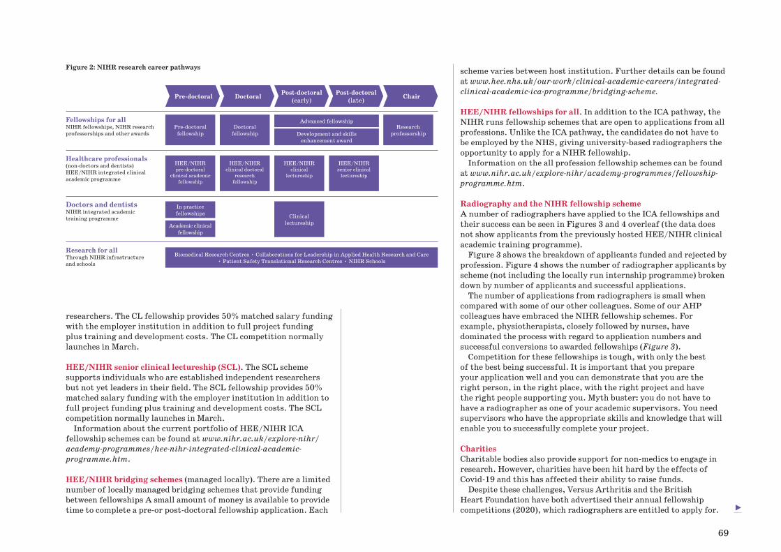
researchers. The CL fellowship provides 50% matched salary funding with the employer institution in addition to full project funding plus training and development costs. The CL competition normally launches in March.
HEE/NIHR senior clinical lectureship (SCL). The SCL scheme supports individuals who are established independent researchers but not yet leaders in their field. The SCL fellowship provides 50% matched salary funding with the employer institution in addition to full project funding plus training and development costs. The SCL competition normally launches in March.
Information about the current portfolio of HEE/NIHR ICA fellowship schemes can be found at www.nihr.ac.uk/explore-nihr/academy-programmes/hee-nihr-integrated-clinical-academic-programme.htm.
HEE/NIHR bridging schemes (managed locally). There are a limited number of locally managed bridging schemes that provide funding between fellowships A small amount of money is available to provide time to complete a pre-or post-doctoral fellowship application. Each
scheme varies between host institution. Further details can be found at www.hee.nhs.uk/our-work/clinical-academic-careers/integrated-clinical-academic-ica-programme/bridging-scheme.
HEE/NIHR fellowships for all. In addition to the ICA pathway, the NIHR runs fellowship schemes that are open to applications from all professions. Unlike the ICA pathway, the candidates do not have to be employed by the NHS, giving university-based radiographers the opportunity to apply for a NIHR fellowship.
Information on the all profession fellowship schemes can be found at www.nihr.ac.uk/explore-nihr/academy-programmes/fellowship-programme.htm.
Radiography and the NIHR fellowship schemeA number of radiographers have applied to the ICA fellowships and their success can be seen in Figures 3 and 4 overleaf (the data does not show applicants from the previously hosted HEE/NIHR clinical academic training programme).
Figure 3 shows the breakdown of applicants funded and rejected by profession. Figure 4 shows the number of radiographer applicants by scheme (not including the locally run internship programme) broken down by number of applicants and successful applications.
The number of applications from radiographers is small when compared with some of our other colleagues. Some of our AHP colleagues have embraced the NIHR fellowship schemes. For example, physiotherapists, closely followed by nurses, have dominated the process with regard to application numbers and successful conversions to awarded fellowships (Figure 3).
Competition for these fellowships is tough, with only the best of the best being successful. It is important that you prepare your application well and you can demonstrate that you are the right person, in the right place, with the right project and have the right people supporting you. Myth buster: you do not have to have a radiographer as one of your academic supervisors. You need supervisors who have the appropriate skills and knowledge that will enable you to successfully complete your project.
CharitiesCharitable bodies also provide support for non-medics to engage in research. However, charities have been hit hard by the effects of Covid-19 and this has affected their ability to raise funds.
Despite these challenges, Versus Arthritis and the British Heart Foundation have both advertised their annual fellowship competitions (2020), which radiographers are entitled to apply for.
Figure 2: NIHR research career pathways
Fellowships for all NIHR fellowships, NIHR research professorships and other awards
Healthcare professionals (non-doctors and dentists) HEE/NIHR integrated clinical academic programme
Doctors and dentists NIHR integrated academic training programme
Research for all Through NIHR infrastructure and schools
Pre-doctoral Doctoral Post-doctoral(early)
Post-doctoral(late) Chair
Biomedical Research Centres • Collaborations for Leadership in Applied Health Research and Care • Patient Safety Translational Research Centres • NIHR Schools
Pre-doctoral fellowship
In practice fellowships
Academic clinical fellowship
HEE/NIHR pre-doctoral
clinical academic fellowship
Doctoral fellowship
Clinical lectureship
HEE/NIHR clinical doctoral
research fellowship
Research professorship
HEE/NIHR clinical
lectureship
HEE/NIHR senior clinical
lectureship
Advanced fellowship
Development and skills enhancement award
69
F

220
200
180
160
140
120
100
80
60
40
20
0
• Start your application as early as possible. It takes about a year to complete an NIHR application form. So don’t leave it too late to complete it for a submission date.
• Find an academic team to sit within. Successful fellowship applicants have a relationship with an existing academic team that can provide support and expertise.
• Find a lead academic supervisor who has credibility and the appropriate experience. They do not have to be a radiographer. You need someone who has a skill set that will help you to complete your proposed project.
• Talk to current and past NIHR applicants. There maybe some successful NIHR fellows who are not radiographers at your trust. It always helps to get advice from people who have been through the process.
• Contact a NIHR advocate. There are a number of NIHR advocates who can advise and signpost you. Details of the advocates can be found at www.nihr.ac.uk/researchers.
• Think outside of the box when it comes to your training plan. The NIHR is investing in you. These are clinical academic roles; don’t just focus on the research element of your training plan.
• Don’t give up Sometimes it takes more than one attempt to be successful.
Top tips for applying for an NIHR fellowship
Figure 4: Radiographer applications to HEE/NIHR ICA fellowship schemes
Figure 3: Applicants by profession to HEE/NIHR ICA fellowship schemes
16
14
12
10
8
6
4
2
0
HEE/NIHR ICA Programme Clinical Doctoral Research
Fellowship
HEE/NIHR ICA Programme Clinical Lectureship
HEE/NIHR ICA Programme Pre-doctoral Clinical Academic
Fellowship
HEE/NIHR ICA Programme Senior Clinical Lectureship
● Funded ● Rejected ● Total applications
● Funded ● Rejected ● Withdrawn ● Total applications
Art T
herapist
Chiropra
ctor
Clinica
l Psy
cholo
gist
Denta
l Ther
apist
Dietiti
an
Dram
a Ther
apist
Health
Care
Scie
ntist
Health
care
Scie
ntist
Mid
wife
Non-med
ical P
ublic H
ealth
Nurse
Occupatio
nal Ther
apist
Operatin
g Dep
artm
ent P
ract
itioner
Optom
etris
t
Orthoptis
t
Other
Param
edic
Pharmacis
t
Pharmacy
Technici
an
Physioth
erapist
Podiatrist
Pract
itioner
Psy
cholo
gist
Prost
hetist
/Orthotis
t
Radiogra
pher (D
iagnost
ic)
Radiogra
pher (T
herapeu
tic)
Social W
orker
Speech
and Language T
herapist
70

For details, visit www.versusarthritis.org/research and www.bhf.org.uk/for-professionals/information-for-researchers.
One hopes that the pandemic is over soon and charities can recommence their fundraising to allow for future funding calls.
The Council for Allied Health Professions Research (CAHPR) The CAHPR was founded in 2014 and supports AHPs to become engaged in research. While hosted on the Chartered Society for Physiotherapy website, CAHPR is available to all AHPs. CAHPR hosts small research grant calls, which are run annually and are managed locally by their hubs. The funding amounts are small, with a maximum of £1,000 available, but are invaluable to those wishing to get on the first step of the research ladder. Further information about CAHPR can be found at https://cahpr.csp.org.uk.
Impact of radiographers engaging in researchResearch carried out by radiographers on the frontline can positively impact our patients. Those working in clinical roles can identify research questions that are clinically relevant and need answering. Investing time and resources on a small research question could translate to a big clinical impact. This could provide the NHS with best practice, which improves the care of our patients and, at the same time, empowers and retains our staff to ask and lead on pertinent clinical research questions.
ConclusionNow is the time for radiographers to become involved in clinical research. Our patients are demanding it, professional and regulatory bodies require it and there are more financial opportunities than ever to support radiographers to fund a research degree or project. ■
References
1. Pager S, Holden, Golenko (2012) ‘Motivators, enablers, and barriers to building allied health research capacity’, Journal of Multidisciplinary Healthcare 2012:53.
2. National Health Service (2019) The NHS Long Term Plan (www.longtermplan.nhs.uk/wp-content/uploads/2019/01/nhs-long-term-plan.pdf accessed 13 November 2020).
3. Ozdemir BA, Karthikesalingam A, Sinha S, et al (2015) ‘Research activity and the association with mortality’, PLoS One 2015;10(2):e0118253.
4. National Health Service (2020) Coronavirus vaccine research 2020 (www.nhs.uk/conditions/coronavirus-covid-19/research/coronavirus-vaccine-research/ accessed 2 January 2021).
5. Reid K, Edwards H (2011) ‘Evaluating the role of the diagnostic research radiographer’, Radiography 2011;17(3):207-11.
6. Care Quality Commission (2020) What we will inspect: NHS trusts. Vol. 2021 (www.cqc.org.uk/guidance-providers/nhs-trusts/what-we-will-inspect-nhs-trusts accessed 3 December 2020).
7. Society and College of Radiographers (2016) 2016-2021 Society and College of Radiographers research strategy (www.sor.org/learning/document-library/research-strategy-2016-2021 accessed 15 December 2020).
Acknowledgement. This article presents work supported by the National Institute for Health Research (NIHR) and Leeds Biomedical Research Centre. The views expressed are those of the author and not necessarily those of the NIHR or the Department of Health and Social Care.
Dr Carole Burnett, Lead Research and Innovation Radiographer, Leeds Teaching Hospitals NHS Trust; Academic Research Capacity Development Lead, Leeds NIHR Biomedical Research Centre; Visiting Fellow, Leeds Institute of Medical Research, University of Leeds; Lead Training Advocate for Radiographers, National Institute of Health Research
Myth buster: you do not have to have a radiographer as one of your academic supervisors’
71

NEW GUIDELINES ON CONTACT SHIELDING AND RADIATION PROTECTION: TEACHING AND IMPLEMENTATION
Philip Cosson
72

The research findings suggest that contact shielding provides minimal or no benefit and practitioners should concentrate on other areas of radiation protection that are more effective in optimising the patient radiation exposure’
73

Such changes usually disseminate slowly into practice. However, this abrupt change could be considered somewhat revolutionary’
In 2020, the British Institute of Radiology (BIR) published evidence-based guidance on why contact shielding for patients is no longer needed during most X-ray examinations, computed tomography (CT) scans and interventional radiology1. This was a joint report of
the BIR, Institute of Physics and Engineering in Medicine (IPEM), Public Health England (PHE), Royal College of Radiologists (RCR), Society and College of Radiographers (SCoR) and the Society for Radiological Protection (SRP). Other radiation protection bodies have since followed suit2. The guidance can be downloaded from the BIR website and there is a helpful patient leaflet.
Over the past 70 years or so, it has been a common practice among radiographers to place attenuating material (mostly lead rubber sheeting) directly on the surface of a patient during radiography examinations. The aim has been to help reduce the dose to what were considered ‘critical organs’. However, concern over the use of patient contact shielding had been building in the literature for some time3-7. The most common form used was aimed at shielding the gonads but there were also practices around use for breast shielding in CT8-9.
The guidance published in March 2020 challenges the historical perspective that using contact shielding only provides a benefit for the patient. Rather, it suggests that contact shielding can adversely interfere with automatic settings of radiation exposure and, if misplaced or allowed to move during an examination, can actually
lead to a repeat of the examination or misdiagnosis. Both of these possibilities would lead to increased radiation exposure and poorer patient outcomes.
Research has also provided evidence of inconsistent use5, which is confusing for the patient and their family, has a possible negative effect on image quality of the resultant radiograph10 or CT scan, and brings potential issues with infection control, which is a very important consideration.
Another point made in the guidance was that contact shielding was too often relied upon as the only radiation protection tool. Local rules often only mentioned gonad shielding and collimation, forgoing any of the more recent evidence-based methods to reduce the patient’s exposure across the entire beam.
Overall, the research findings suggest that contact shielding provides minimal or no benefit and practitioners should concentrate on other areas of radiation protection that are more effective in optimising the patient’s radiation exposure.
Changing a long-held belief in shielding may not be easy. For many practitioners, the decision by the BIR in favour of an almost blanket removal of contact shielding, particularly for pregnant women, has come as a shock. Such changes usually disseminate slowly into practice. However, this abrupt change could be considered somewhat revolutionary.
The arguments against ‘shielding’ are partly based on human factors. It is common for individual practitioners to look at their own performance independent of the system in which they find themselves working. In this case, it is easy to imagine that you, the practitioner, will always remember to use the shield, will always disable auto exposure settings (and choose the correct manual ones), will always have time to apply the shield, will always be able to discern its correct location and keep the patient from moving. It is easy to see yourself in the role of the perfect practitioner. However, policy should take a view of the whole system in which all practitioners work. In systems, there are pressure points, resource constraints, distractions, competing priorities, hesitant trainees, difficult patient encounters, less-than-optimal control panels and odd-sized shields that need disinfecting. It can be accepted that ideal practice is not always possible.
Clinically, in reality, the use of gonad shielding has been falling for many years: practitioners were becoming deskilled in optimal positioning, problems with maintaining shielding position in paediatric images – coupled with a growing degree of unease about the physical application of shielding to such personal areas – have seen them fall from general use. This difference in the espoused
74

A coordinated effort is required to update the curriculum. At university, we need to amend slides, lab manuals and classroom materials to remove the mention of patient contact shielding. This is preferred to leaving the content as is and using it as a debating point because new students have enough to assimilate without adding outdated practices. Textbook suggestions need to be reviewed to choose those that have updated content to reflect modern practice. Where this is not yet possible, tutors need to direct students on how to interpret the text in light of current guidance and direct them to seek out supporting research.
Assessments should be rewritten to ensure no credit is given to answers espousing patient contact shielding as a radiation protection consideration. Evidence-based methods of reducing radiation risk should be better emphasised in the curriculum, such as use of copper filtration, longer source-to-image distances, higher tube voltages and lower tube charge (consistent with required image quality).
Within the clinical department, site co-ordinators and student mentors should be on-board with the changed curriculum. Hopefully, the radiation protection supervisor and radiation protection advisor will be aware of the guidance and will work to bring the local rules and IR(ME)R procedures and protocols into compliance. However, this process will take time and during the transition period students will be expected to work with all qualified practitioners
belief of the practitioner that shielding was inherently a good thing, but practically failing to apply it, has been causing discomfort and unease for many. Hopefully, this alignment of guidance to practice will be welcome.
In the case of gonad shielding, even ideal practice with perfect placement and ideal exposure factors offers little benefit to the patient. First, it has been long demonstrated that, in females, the location of the ovaries can vary so substantially that, even with perfect shielding placement, the gonads were not guaranteed to be afforded full protection.
Second, and most importantly, the evidence base shows that genetic effects of radiation are largely absent in the human population. The fear of genetic effects was largely responsible for the introduction of patient contact shielding in the 1950s but continued research into the epidemiology of populations exposed to radiation through treatments, accidents and atrocities over the years has shown no cause for concern of hereditary effects. However, more than 50 years of fear has left its mark not only on local rules, textbooks and presentation slides, but also on healthcare staff and the public at large.
Finally, the guidance does not just advocate for abandoning some practices but also for adopting new evidence-based practices of protection that reduce the risk of radiographic procedures as a whole.
Scenario Recommendation Comments
Patient contact shielding for protection of breast.
Not recommended. Consider Postero-Anterior (PA) positioning rather than shielding for spinal and chest examinations where possible.If using Antero-Posterior (AP) projection then a Scoliosis shawl may be considered
Patient contact shielding for protection of thyroid.
Not recommended. Recommended where thyroid is less than 5cm from the primary beam, projection is AP and will not obscure anatomy of interest or interfere with Automatic Exposure Control (AEC) device.
Patient contact shielding for protection of gonads.
Not recommended. Consider PA positioning, no recommendations for shielding.
Patient contact shielding for protection of eye lens.
Not recommended. Use PA Facial Bones positioning, no recommendations for shielding.
Pregnant patients. Not recommended. Not required for examinations outside the pelvic region (diaphragm to knee).For examinations within pelvic region, consider non-ionising imaging alternatives. If ionising radiation must be used, carry out a thorough justification and risk assessment process.
Summary of the guidance for general radiography (from Table 7.1, BIR Guidance, p481)
75
F

Summary of the guidance for CT (from Table 9.1, BIR Guidance, p721)
Scenario Recommendation Comments
In-plane organ contact shielding (adult and children).
Not recommended. Ensure optimisation by alternative means wherever practicable.
Adverse effects on image quality. Unpredictable AEC performance – rendering it ineffective, or even resulting in higher dose.
In-plane contact shields to protect the lens for patients where frequent follow up head CT likely.
Not recommended. Ensure optimisation by alternative means wherever practicable.
Often low-dose protocols can achieve the required diagnostic outcome e.g. when assessing shunt patency or ventricle size.(If there is a strong basis for protection on an individual basis, an air gap should be considered.)If considering, a careful review of likely image quality in line with the diagnostic purpose needs to be performed.
Out-of-plane organ contact shielding (adult and children).
Not recommended. Actual dose savings will be low.Not possible to limit internal scatter. Highest savings when shield close to edge of scanning volume but more likely to interfere with AEC if close.An adequate distance to rule out misplacement or movement of protection then renders the reduction in external scatter minimal.
Out-of-plane contact shieldingto protect fetus in pregnancy.
Not recommended. See comments above.
In-plane contact shielding of the gonads.
Not recommended. Little evidence, careful thought required to positioning.Testes are not listed as an International Commission on Radiological Protection (ICRP) radiosensitive organ (for cancer incidence).Adverse effects on image quality for pelvis examinations (especially if protecting the ovaries).Hereditable effects associated with typical dose range are likely to be negligible.
Out-of-plane contact shielding of the gonads.
Not recommended. Little evidence.Not possible to limit internal scatter (especially for ovaries).Testes are not listed as an ICRP radiosensitive organ (for cancer incidence).Hereditable effects associated with typical dose range are likely to be negligible.
76

and take instruction from them. Indeed, armed with knowledge and understanding of recent changes to shielding guidance, students may help embed the changes within clinical practice through their questioning of professionals who have yet to embrace updated practices.
It is encouraging to note that many practitioners have been seeking out the evidence underpinning the updated guidance to understand the reasoning behind it, reinforcing the ideal that evidenced-based practice is at the heart of modern radiography.
Arguably, the hospital trust could change its patient-facing signage and make it explicit that patient contact shielding is no longer routinely used at the trust. However, the fact that shielding is not now undertaken might not need to be advertised. Many younger patients would have no awareness of shielding use since its decline anyway.
There are advantages and disadvantages to both approaches and patient information leaflets have been produced for trusts to use if they choose. Advantages of advertising the change include empowering radiographers to action the guidance, providing them with explicit support should a patient challenge them, and reminding new or agency staff of the departmental policy.
There will be instances where other healthcare workers and patients are unconvinced by the argument concerning the removal of what appears to be protective. If a local department decides to maintain its use, then written procedures should reflect equality and diversity and meet the needs of local populations. This may include the use of shielding for patients, for example, who may find it difficult to cooperate. Careful consideration should be given to the location of reproductive organs in transgender and gender
A coordinated effort is required to update the curriculum. At university, we need to amend slides, lab manuals and classroom materials to remove the mention of patient contact shielding’
1. Why am I considering the use of contact shielding? While not generally advised, any use of contact shielding should be considered carefully by a multidisciplinary team and written into examination protocols ahead of use. Its selection simply to reassure the apprehensive patient should be discouraged as this promotes mixed messages and an exaggeration of radiation risk to the patient and wider community. Instead, efforts should concentrate on explaining the risks from the use of contact shields to the patient.
2. What is the likelihood and consequence of the contact shielding interfering with automatic exposure selection? If optimised for the given procedure, the AEC is arguably the best tool to optimise the patient exposure for the intended diagnostic purpose. If the contact shield is scanned (either deliberately or inadvertently) during the CT localiser series then the patient exposure is likely to be significantly higher than intended, undoing any small intended benefit.
3. What is the effect on image quality of the introduction of contact shielding? In beam shielding in general radiography, such as gonad shielding, relevant anatomical and pathological detail can be obscured, leading to missed diagnosis or repeat of the exam. The presence of the shield can also create distraction to the reader – the Mach effect.
In-plane contact shielding in CT leads to photon starvation and beam hardening artefacts in the image (especially where stand-off material is not employed between the patient–shield interface).
Out-of-beam shielding aims to limit external scatter and extra focal radiation from the tube/collimator assembly to the patient. Any potential dose saving is a small fraction of the overall scattered dose (external and within the patient). There is a risk of this shielding causing artefacts when placed too close to, or slipping into, the scanned volume.
The following questions should be asked prior to any consideration of using patient contact shields:
77
F

Philip Cosson, Senior Lecturer, Teesside University
This article has relied heavily on the BIR guidance documentation1 and the author wishes to thank Peter Hiles, the editor of the guidance, and the other contributing authors: Elizabeth Benson (BIR), Helen Hughes (BIR), Rob Loader (IPEM), Dan Shaw (IPEM), Sue Edyvean (PHE), Yvonne Sullivan (PHE), Guy Hickson (RCR), Phil Cosson (SCoR), Lynda Johnson (SCoR), David Dommett (SRP)
Also thanks to Joanne MacRae, Senior Radiographer, County Durham and Darlington Foundation Trust
References
1. British Institute of Radiology (2020) Guidance on using shielding on patients for diagnostic radiology applications, (www.bir.org.uk/media/416143/final_patient_shielding_guidance.r1.pdf).
2. National Council on Radiation Protection and Measurements (2021) Recommendations for Ending Routine Gonadal Shielding During Abdominal and Pelvic Radiography: statement 13 January 2021 (https://ncrponline.org/wp-content/themes/ncrp/PDFs/Statement13.pdf).
3. Frantzen MJ, Robben S, Postma AA, Zoetelief J, Wildberger JE and Kemerink GJ (2012) ‘Gonad shielding in paediatric pelvic radiography: disadvantages prevail over benefit’, Insights into Imaging 2012; 3(1): 23-32.
4. Fawcett SL, Gomez AC, Barter SJ, Ditchfield M and Set P (2012) ‘More harm than good? The anatomy of misguided shielding of the ovaries’, Br. J. Radiol. 2012; 85: e442-e447.
5. Warlow T, Walker-Birch P and Cosson P (2014) ‘Gonad shielding in paediatric pelvic radiography: effectiveness and practice’, Radiography 2014; 20(3): 178-182.
6. Burns KM, Shoag JM, Kahlon SS, Parsons PJ, Bijur PE, Taragin BH and Markowitz M (2017) ‘Lead aprons are a lead exposure hazard’, J. Am. Coll. Radiol. 2017; 14: 641-647.
7. Marsh RM and Silosky M (2019) ‘Patient shielding in diagnostic imaging: discontinuing a legacy practice’, Am. J. Roentgenol 2019; 212(4): 755-757.
8. Huggett J, Mukonoweshuro W and Loader R (2013) ‘A phantom-based evaluation of three commercially available patient organ shields for computed tomography X-ray examinations in diagnostic radiology’, Radiation protection dosimetry 2013; 155(2): 161-8.
9. Lawrence S and Seeram E (2017) ‘The current use and effectiveness of bismuth shielding in computed tomography: a systematic review’, Radiol Open J. 2017; 2: 7-16.
10. Panikkath R and Panikkath D (2014) ‘Mach band sign: an optical illusion’, Proc. (Bayl. Univ. Med. Cent.) 2014; 27: 364-5.
11. HPA, RCR, SCoR (2009) Protection of Pregnant Patients During Diagnostic Medical Exposures to Ionising Radiation. Documents of the Health Protection Agency, RCE-9.
12. ICRP (1982) Protection of the Patient in Diagnostic Radiology. ICRP Publication 34. Ann. ICRP 9(2/3).
13. Hayre CM, Blackman S, Carlton K and Eyden A (2018) ‘Attitudes and perceptions of radiographers applying lead (Pb) protection in general radiography: an ethnographic study’, Radiography 2018; 24: e13–e18.
non-conforming individuals. Where required, there should be a suitable range of devices available to meet the age and size requirements of the individual. Some providers may include a size guide for reference. Regular assessment of practice and review of local policies and procedures are recommended.
Examination of pregnant patients is a special case with heightened sensitivities of more than one meaning. The new guidance is that radiography of areas remote from the fetus may be carried out at any point during pregnancy with no additional patient contact shielding, provided that accurate collimation is used and that the equipment itself is adequately shielded. Guidance indicates that ‘remote from the fetus’ refers to any examination outside the area between the diaphragm and knees11.
In the extremely rare occasion where ionising radiation exposure to the fetus is justified, ICRP report 3412 recommends that consideration should be given to the techniques used to ensure dose to the fetus is kept as low as reasonably practicable, for example, minimisation of the number of projections taken, strict collimation and partial shielding of the fetus. However, care must be taken to ensure that the images remain of suitable diagnostic quality and that exposure settings are not prone to inaccuracies due to the presence of any contact shield. Shielding should never be used where it is possible that an over-exposure of another body part would occur.
These recommendations do not necessarily consider the psychological effect of an exposure to ionising radiation on an expectant mother. It has been documented that pregnant patients undergoing diagnostic radiology examinations may request patient contact shielding13. In these cases, whether or not to provide extra shielding, usually in the form of lead/lead-equivalent material draped over the abdomen, is in accordance with written procedures and at the discretion of the radiographer. This is another case where advertising the change may reassure the patient.
Leadership plays an important role in setting standards of practice in the radiography department. The formation of multidisciplinary radiation protection champions within Image Optimisation Teams should support, drive and provide training in all areas of radiation protection. The local rules should be reviewed regularly to ensure they are based on the most recent evidence.
Practitioners should also be trained to be able to answer patient (or representative) questions relating to the benefits and limitations of contact shielding. It is highly likely that similar or even larger dose savings can be achieved by carefully considering alternative optimisation strategies that will not introduce the problems associated with contact shields. ■
78

Download a PDF version at www.sor.org/imaging-oncology