
Focusing in on structural genomics: The University of Queensland structural biology pipeline
9
Review Focusing in on structural genomics: The University of Queensland structural biology pipeline Munish Puri a,1, * , Gautier Robin a , Nathan Cowieson a,e , Jade K. Forwood b , Pawel Listwan b,c , Shu-Hong Hu a , Gregor Guncar b,2 , Thomas Huber b,d , Stuart Kellie a,b,c , David A. Hume a,b,c,e , Bostjan Kobe a,b,e , Jennifer L. Martin a,b,e a Institute for Molecular Bioscience, University of Queensland, Brisbane, Queensland, Australia b School of Molecular and Microbial Sciences, University of Queensland, Brisbane, Queensland, Australia c Cooperative Research Centre for Chronic Inflammatory Diseases, University of Queensland, Brisbane, Queensland, Australia d Department of Mathematics, University of Queensland, Brisbane, Queensland, Australia e ARC Special Research Centre for Functional and Applied Genomics, University of Queensland, Brisbane, Queensland, Australia Received 14 July 2006; received in revised form 22 September 2006; accepted 25 September 2006 Abstract The flood of new genomic sequence information together with technological innovations in protein structure determination have led to worldwide structural genomics (SG) initiatives. The goals of SG initiatives are to accelerate the process of protein structure determination, to fill in protein fold space and to provide information about the function of uncharacterized proteins. In the long-term, these outcomes are likely to impact on medical biotechnology and drug discovery, leading to a better understanding of disease as well as the development of new therapeutics. Here we describe the high throughput pipeline established at the University of Queensland in Australia. In this focused pipeline, the targets for structure determination are proteins that are expressed in mouse macrophage cells and that are inferred to have a role in innate immunity. The aim is to characterize the molecular structure and the biochemical and cellular function of these targets by using a parallel processing pipeline. The pipeline is designed to work with tens to hundreds of target gene products and comprises target selection, cloning, expression, purification, crystallization and structure determination. The structures from this pipeline will provide insights into the function of previously uncharacterized macrophage proteins and could lead to the validation of new drug targets for chronic obstructive pulmonary disease and arthritis. # 2006 Elsevier B.V. All rights reserved. Keywords: High throughput crystallography; Protein expression; Macrophage proteins; Crystallization; Structural genomics Contents 1. Introduction ................................................................................. 282 2. High throughput crystallography ................................................................... 284 2.1. Target selection .......................................................................... 284 2.2. Cloning ................................................................................ 284 2.3. Expression and purification .................................................................. 285 2.4. Crystallization ........................................................................... 286 2.5. Crystal to structure ........................................................................ 286 2.6. Protein function in macrophages and other outcomes of the pipeline ..................................... 286 3. Impact of SG ................................................................................ 287 3.1. Functional assignment ...................................................................... 287 www.elsevier.com/locate/geneanabioeng Biomolecular Engineering 23 (2006) 281–289 * Corresponding author at: Institute for Molecular Bioscience, Queensland Bioscience Precinct, University of Queensland, 306 Carmody Road, St. Lucia, Brisbane, Queensland 4072, Australia. E-mail address: [email protected] (M. Puri). 1 Present address: Fermentation and Protein Biotechnology Laboratory, Department of Biotechnology, Punjabi University, Patiala, India. 2 On leave from the Department of Biochemistry and Molecular Biology, Josef Stefan Institute, Ljubljana, Slovenia. 1389-0344/$ – see front matter # 2006 Elsevier B.V. All rights reserved. doi:10.1016/j.bioeng.2006.09.002
Transcript of Focusing in on structural genomics: The University of Queensland structural biology pipeline
Review
Focusing in on structural genomics: The University of
Queensland structural biology pipeline
Munish Puri a,1,*, Gautier Robin a, Nathan Cowieson a,e, Jade K. Forwood b, Pawel Listwan b,c,Shu-Hong Hu a, Gregor Guncar b,2, Thomas Huber b,d, Stuart Kellie a,b,c,
David A. Hume a,b,c,e, Bostjan Kobe a,b,e, Jennifer L. Martin a,b,e
a Institute for Molecular Bioscience, University of Queensland, Brisbane, Queensland, Australiab School of Molecular and Microbial Sciences, University of Queensland, Brisbane, Queensland, Australia
c Cooperative Research Centre for Chronic Inflammatory Diseases, University of Queensland, Brisbane, Queensland, Australiad Department of Mathematics, University of Queensland, Brisbane, Queensland, Australia
e ARC Special Research Centre for Functional and Applied Genomics, University of Queensland, Brisbane, Queensland, Australia
Received 14 July 2006; received in revised form 22 September 2006; accepted 25 September 2006
www.elsevier.com/locate/geneanabioeng
Biomolecular Engineering 23 (2006) 281–289
Abstract
The flood of new genomic sequence information together with technological innovations in protein structure determination have led to
worldwide structural genomics (SG) initiatives. The goals of SG initiatives are to accelerate the process of protein structure determination, to fill in
protein fold space and to provide information about the function of uncharacterized proteins. In the long-term, these outcomes are likely to impact
on medical biotechnology and drug discovery, leading to a better understanding of disease as well as the development of new therapeutics. Here we
describe the high throughput pipeline established at the University of Queensland in Australia. In this focused pipeline, the targets for structure
determination are proteins that are expressed in mouse macrophage cells and that are inferred to have a role in innate immunity. The aim is to
characterize the molecular structure and the biochemical and cellular function of these targets by using a parallel processing pipeline. The pipeline
is designed to work with tens to hundreds of target gene products and comprises target selection, cloning, expression, purification, crystallization
and structure determination. The structures from this pipeline will provide insights into the function of previously uncharacterized macrophage
proteins and could lead to the validation of new drug targets for chronic obstructive pulmonary disease and arthritis.
# 2006 Elsevier B.V. All rights reserved.
Keywords: High throughput crystallography; Protein expression; Macrophage proteins; Crystallization; Structural genomics
Contents
1. Introduction . . . . . . . . . . . . . . . . . . . . . . . . . . . . . . . . . . . . . . . . . . . . . . . . . . . . . . . . . . . . . . . . . . . . . . . . . . . . . . . . . 282
2. High throughput crystallography . . . . . . . . . . . . . . . . . . . . . . . . . . . . . . . . . . . . . . . . . . . . . . . . . . . . . . . . . . . . . . . . . . . 284
2.1. Target selection . . . . . . . . . . . . . . . . . . . . . . . . . . . . . . . . . . . . . . . . . . . . . . . . . . . . . . . . . . . . . . . . . . . . . . . . . . 284
2.2. Cloning . . . . . . . . . . . . . . . . . . . . . . . . . . . . . . . . . . . . . . . . . . . . . . . . . . . . . . . . . . . . . . . . . . . . . . . . . . . . . . . . 284
2.3. Expression and purification . . . . . . . . . . . . . . . . . . . . . . . . . . . . . . . . . . . . . . . . . . . . . . . . . . . . . . . . . . . . . . . . . . 285
2.4. Crystallization . . . . . . . . . . . . . . . . . . . . . . . . . . . . . . . . . . . . . . . . . . . . . . . . . . . . . . . . . . . . . . . . . . . . . . . . . . . 286
2.5. Crystal to structure . . . . . . . . . . . . . . . . . . . . . . . . . . . . . . . . . . . . . . . . . . . . . . . . . . . . . . . . . . . . . . . . . . . . . . . . 286
2.6. Protein function in macrophages and other outcomes of the pipeline . . . . . . . . . . . . . . . . . . . . . . . . . . . . . . . . . . . . . 286
3. Impact of SG . . . . . . . . . . . . . . . . . . . . . . . . . . . . . . . . . . . . . . . . . . . . . . . . . . . . . . . . . . . . . . . . . . . . . . . . . . . . . . . . 287
3.1. Functional assignment. . . . . . . . . . . . . . . . . . . . . . . . . . . . . . . . . . . . . . . . . . . . . . . . . . . . . . . . . . . . . . . . . . . . . . 287
* Corresponding author at: Institute for Molecular Bioscience, Queensland Bioscience Precinct, University of Queensland, 306 Carmody Road, St. Lucia, Brisbane,
Queensland 4072, Australia.
E-mail address: [email protected] (M. Puri).1 Present address: Fermentation and Protein Biotechnology Laboratory, Department of Biotechnology, Punjabi University, Patiala, India.2 On leave from the Department of Biochemistry and Molecular Biology, Josef Stefan Institute, Ljubljana, Slovenia.
1389-0344/$ – see front matter # 2006 Elsevier B.V. All rights reserved.
doi:10.1016/j.bioeng.2006.09.002
M. Puri et al. / Biomolecular Engineering 23 (2006) 281–289282
3.2. Other implications of SG outcomes . . . . . . . . . . . . . . . . . . . . . . . . . . . . . . . . . . . . . . . . . . . . . . . . . . . . . . . . . . . . 287
3.3. Drug and drug-lead discovery . . . . . . . . . . . . . . . . . . . . . . . . . . . . . . . . . . . . . . . . . . . . . . . . . . . . . . . . . . . . . . . . 287
4. Outputs from UQ SG pipeline . . . . . . . . . . . . . . . . . . . . . . . . . . . . . . . . . . . . . . . . . . . . . . . . . . . . . . . . . . . . . . . . . . . . . 287
5. Conclusion . . . . . . . . . . . . . . . . . . . . . . . . . . . . . . . . . . . . . . . . . . . . . . . . . . . . . . . . . . . . . . . . . . . . . . . . . . . . . . . . . . 287
Acknowledgement . . . . . . . . . . . . . . . . . . . . . . . . . . . . . . . . . . . . . . . . . . . . . . . . . . . . . . . . . . . . . . . . . . . . . . . . . . . . . 288
References . . . . . . . . . . . . . . . . . . . . . . . . . . . . . . . . . . . . . . . . . . . . . . . . . . . . . . . . . . . . . . . . . . . . . . . . . . . . . . . . . . 288
1. Introduction
Structural biology has emerged as one of the most powerful
approaches for defining the functions of proteins; this capacity
is based on the observation that the evolutionary constraints for
three-dimensional structures of proteins are higher than for
sequences (Thornton et al., 2000; Yakunin et al., 2004). The
strong predictive power of structure in functional annotation
has resulted in the rapid growth of the new field of structural
genomics (SG) (or structural proteomics) (Burley, 2000) and to
the rapid development of novel high throughput technologies
(Stevens and Wilson, 2001). In addition to expediting
functional characterization of gene products, SG initiatives
will also provide a comprehensive view of the protein structure
universe, by determining the structures of representative
proteins from every protein fold family and thereby filling in
Table 1
List of structural genomics consortia from TargetDB (http://targetdb.pdb.org)
Centre/consortium Location
Berkeley Structural Genomics Center (BSGC) USA
Midwest Center for Structural Genomics (MCSG) USA
Northeast Structural Genomics Consortium (NEGS) USA
NewYork Structural Genomics Research Consortium (NYSGXRC) USA
Southeast Collaboratory for Structural Genomics (SECSG) USA
TB Structural Genomics Consortium (TB) USA
Joint Center for Structural Genomics (JCSG) USA
Center for Eukaryotic Structural Genomics (CESG) USA
Structure 2 Function Project (S2F) University of California Berkeley USA
Structural Genomics of Pathogenic Protozoa consortium (SGPP) USA
Montreal-Kingston Bacterial Structural Genomics Initiative (BSGI) Canada
Bacterial Targets at IGS-CNRS (BIGS) France
Oxford protein production facility (OPPF) UK
Structural Proteomics in Europe (SPINE) UK
The Israel Structural Proteomics Center (ISPC) Israel
Marseilles Structural Genomics Programe (MSGP) France
Mycobacterium Tuberculosis Structural Proteomics Project (XMTB) Germany
Protein Structure Factory (PSF) Germany
Paris-Sud Yeast Structural Genomics (YSG) France
RIKEN Structural Genomics Initiative (RSGI) Japan
Integrated Center for Structure and Function Innovation (ISFI) USA
protein fold space (Todd et al., 2005). SG outcomes will also
identify novel drug targets (Buchanan, 2002) and advance our
understanding of protein evolution. Overall, SG research
promises to have a major impact on the life sciences,
biotechnology and medicine (Hol, 2000).
To meet these goals, high throughput (HT) or parallel
processing approaches have been developed for producing
protein samples for structural biology and functional studies
(Lesley et al., 2002). X-ray crystallography is the most widely
used approach for protein structure determination, accounting
for �85% of structures in the Protein Data Bank (http://
www.rcsb.org/pdb/) (Berman et al., 2000) though most SG
initiatives use both crystallography and NMR approaches.
SG consortia have been established around the world
(Table 1) for the systematic high throughput determination of
protein structures on a genome-wide scale (Brenner, 2001).
Organism/focus
Structural representation of the genomes of two pathogens,
Mycobacterium genitalium and Mycobacterium pneumoniae
Proteins from all three kingdoms of life
Small proteins from eukaryotic model organisms such as Saccharomyces
cerevisiae, Caenorbaditis elegans and Drosophila melanogaster
Biologically interesting proteins from model organisms and humans
C. elegans and Pyrococcus furiosus and selected human proteins
Mycobacterium tuberculosis proteins, particularly potential drug
targets and novel folds
Thermotoga maritima, novel folds from C. elegans, and human
proteins implicated in cell signaling
Arabidopsis thaliana proteins; technology development for
eukaryotic proteins
Functional characterization of hypothetical proteins from
Haemophilus influenzae
Proteins from major global pathogenic protozoa, Leishmania major,
Trypanosoma brucei and Plasmodium falciparum
Structures of potential virulence factors from pathogenic bacteria
Discovery of new antibacterial gene targets among evolutionary
conserved genes of uncharacterized function
Biomedical relevance of human pathogens, in particular Herpes viruses
Targets include human proteins implicated in cancer and
neurodegenerative diseases
Increase the efficiency of protein structure determination
Focus on bacterial, viral, and human ORFs
Identify lead compounds against XMTB, using a structure based approach
Human proteins to understand health and disease
Non-membrane proteins of unknown structure
Proteins of biological and medical interest from mouse,
A. thaliana and Thermus thermophilus
Developing and applying technologies to overcome bottlenecks of
production of soluble protein and protein crystallization
M. Puri et al. / Biomolecular Engineering 23 (2006) 281–289 283
Some consortia target the proteomes of thermophilic organ-
isms, which offer the advantage of working on small stable
proteins, on which high-throughput methodologies can be
tested (Yee et al., 2003). Although specialist membrane protein
SG projects have recently emerged (Walian et al., 2004) the
focus of SG consortia has been, for the most part, on soluble
proteins. TargetDB (http://targetdb.pdb.org/statistics/Target-
Statistics.html) reveals that 21 SG consortia have contributed
over 2500 crystal structures since September 2000. The quality
of the structures and size of the proteins are comparable to those
solved by traditional structural biology approaches (Todd et al.,
2005). In terms of contributing to fold space, SG consortia have
been estimated to contribute around half of all novel structures
reported over a recent 1-year period (Chandonia and Brenner,
2006). Furthermore, TargetDB reports that 50% of structures
determined by SG consortia have less than 30% identity to
known protein structures, though only around 16% of structures
solved by SG were deemed to represent a new fold or
superfamily (Chandonia and Brenner, 2006). The average cost
of determining a protein structure by SG consortia in the US
was estimated at US$138,000; this figure increases with the
novelty of the protein to $1M for a representative of a
structurally uncharacterized family and US$2.2M for a
representative structure of a new superfamily or fold
(Chandonia and Brenner, 2006). These statistics show that
the costs of novel protein structure determination within
structural genomics consortia are still relatively high. These
cost estimates make a strong case for the development of
smaller, more cost-efficient research programs to tackle
medically significant proteins.
At the University of Queensland (UQ) in Australia such a
pipeline has been established. The UQ pipeline takes
advantage of developments in high throughput processing
and applies these to the parallel processing of hundreds rather
than thousands of protein targets. Most importantly, the UQ
pipeline has a clear focus on protein targets that are
biologically or biomedically significant. Specifically, the
initiative explores the structural biology of proteins that are
highly expressed in macrophages and potentially involved in
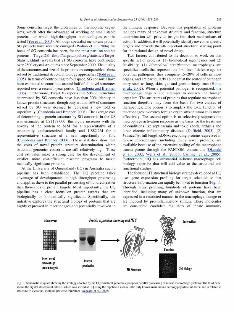
Fig. 1. Schematic diagram showing the strategy adopted by the UQ structural genom
shows the crystal structure of latexin, which was solved at UQ using the pipeline. La
structure to cystatins, cysteine protease inhibitors (Aagaard et al., 2005).
the immune response. Because this population of proteins
includes many of unknown structure and function, structure
determination will provide insight into their mechanisms of
action. In addition, it will potentially identify novel therapeutic
targets and provide the all-important structural starting point
for the rational design of novel drugs.
Two factors contributed to the decision to work on this
specific set of proteins: (1) biomedical significance and (2)
feasibility. (1) Biomedical significance: macrophages are
specialized cells that represent the first line of defense against
potential pathogens; they comprise 15–20% of cells in most
organs, and are particularly abundant at the routes of pathogen
entry such as lung, skin, gut and genitourinary tract (Hume
et al., 2002). When a potential pathogen is recognized, the
macrophage engulfs and attempts to destroy the foreign
organism. The structures of proteins that regulate macrophage
function therefore may form the basis for two classes of
therapeutics. One option is to amplify the toxic function of
macrophages to destroy foreign organisms or tumor cells more
effectively. The second option is to selectively suppress the
macrophage activation response as the basis for the treatment
of conditions like septicaemia and toxic shock, arthritis and
other chronic inflammatory diseases (Duffield, 2003). (2)
Feasibility: full length cDNAs encoding proteins expressed in
mouse macrophages, including many novel proteins, are
available because of the extensive polling of the macrophage
transcriptome through the FANTOM consortium (Okazaki
et al., 2002; Wells et al., 2003b; Carninci et al., 2005).
Furthermore, UQ has substantial in-house macrophage cell
biology expertise that will add value to the structural and
functional studies.
The focused HT structural biology strategy developed at UQ
uses gene expression profiling for target selection so that
structural information can rapidly be linked to function (Fig. 1).
Through array profiling, hundreds of proteins have been
identified, including many of unknown function, that are
expressed in a restricted manner in the macrophage lineage or
are induced by pro-inflammatory stimuli. These molecules
are considered candidate regulators of innate immunity
ics group for parallel processing of mouse macrophage proteins. The third panel
texin is the only known mammalian carboxypeptidase inhibitor, and is related in
M. Puri et al. / Biomolecular Engineering 23 (2006) 281–289284
(Wells et al., 2003b). In this article, we describe the parallel
processing pipeline developed at UQ and aimed at determining
the biological (cellular) and biochemical (molecular) functions
of important uncharacterized or novel macrophage proteins that
are likely to play a role in the immune response and in
inflammation (Aagaard et al., 2005; Cowieson et al., 2005).
2. High throughput crystallography
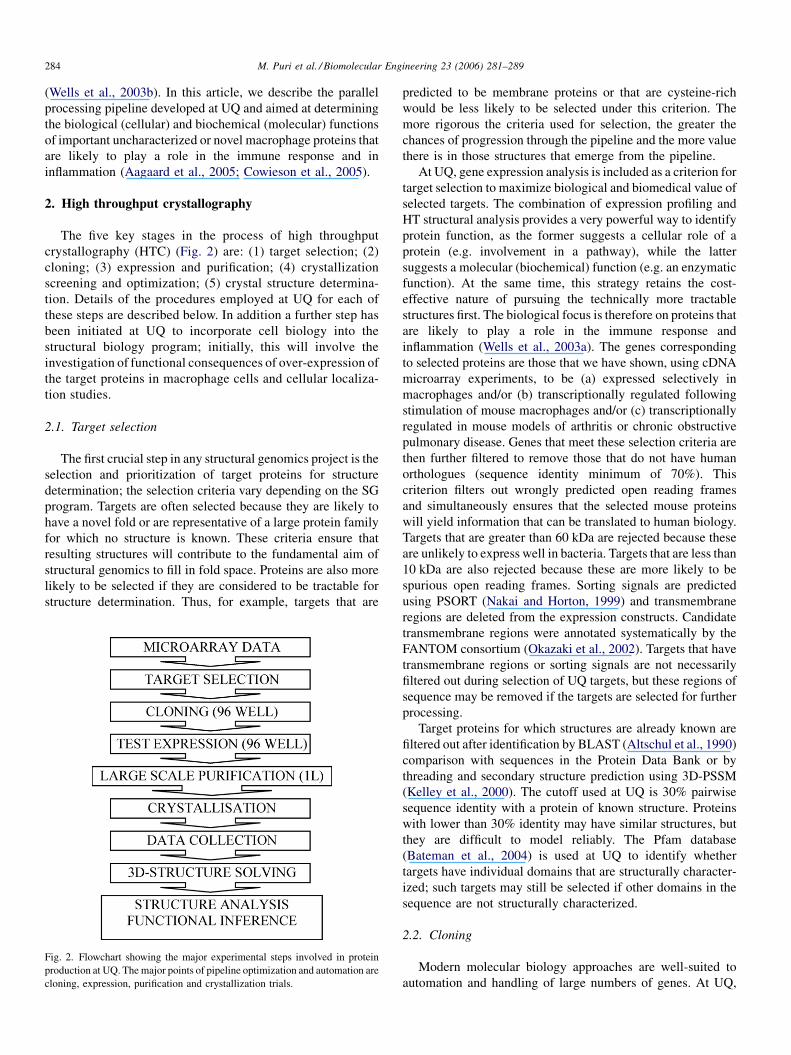
The five key stages in the process of high throughput
crystallography (HTC) (Fig. 2) are: (1) target selection; (2)
cloning; (3) expression and purification; (4) crystallization
screening and optimization; (5) crystal structure determina-
tion. Details of the procedures employed at UQ for each of
these steps are described below. In addition a further step has
been initiated at UQ to incorporate cell biology into the
structural biology program; initially, this will involve the
investigation of functional consequences of over-expression of
the target proteins in macrophage cells and cellular localiza-
tion studies.
2.1. Target selection
The first crucial step in any structural genomics project is the
selection and prioritization of target proteins for structure
determination; the selection criteria vary depending on the SG
program. Targets are often selected because they are likely to
have a novel fold or are representative of a large protein family
for which no structure is known. These criteria ensure that
resulting structures will contribute to the fundamental aim of
structural genomics to fill in fold space. Proteins are also more
likely to be selected if they are considered to be tractable for
structure determination. Thus, for example, targets that are
Fig. 2. Flowchart showing the major experimental steps involved in protein
production at UQ. The major points of pipeline optimization and automation are
cloning, expression, purification and crystallization trials.
predicted to be membrane proteins or that are cysteine-rich
would be less likely to be selected under this criterion. The
more rigorous the criteria used for selection, the greater the
chances of progression through the pipeline and the more value
there is in those structures that emerge from the pipeline.
At UQ, gene expression analysis is included as a criterion for
target selection to maximize biological and biomedical value of
selected targets. The combination of expression profiling and
HT structural analysis provides a very powerful way to identify
protein function, as the former suggests a cellular role of a
protein (e.g. involvement in a pathway), while the latter
suggests a molecular (biochemical) function (e.g. an enzymatic
function). At the same time, this strategy retains the cost-
effective nature of pursuing the technically more tractable
structures first. The biological focus is therefore on proteins that
are likely to play a role in the immune response and
inflammation (Wells et al., 2003a). The genes corresponding
to selected proteins are those that we have shown, using cDNA
microarray experiments, to be (a) expressed selectively in
macrophages and/or (b) transcriptionally regulated following
stimulation of mouse macrophages and/or (c) transcriptionally
regulated in mouse models of arthritis or chronic obstructive
pulmonary disease. Genes that meet these selection criteria are
then further filtered to remove those that do not have human
orthologues (sequence identity minimum of 70%). This
criterion filters out wrongly predicted open reading frames
and simultaneously ensures that the selected mouse proteins
will yield information that can be translated to human biology.
Targets that are greater than 60 kDa are rejected because these
are unlikely to express well in bacteria. Targets that are less than
10 kDa are also rejected because these are more likely to be
spurious open reading frames. Sorting signals are predicted
using PSORT (Nakai and Horton, 1999) and transmembrane
regions are deleted from the expression constructs. Candidate
transmembrane regions were annotated systematically by the
FANTOM consortium (Okazaki et al., 2002). Targets that have
transmembrane regions or sorting signals are not necessarily
filtered out during selection of UQ targets, but these regions of
sequence may be removed if the targets are selected for further
processing.
Target proteins for which structures are already known are
filtered out after identification by BLAST (Altschul et al., 1990)
comparison with sequences in the Protein Data Bank or by
threading and secondary structure prediction using 3D-PSSM
(Kelley et al., 2000). The cutoff used at UQ is 30% pairwise
sequence identity with a protein of known structure. Proteins
with lower than 30% identity may have similar structures, but
they are difficult to model reliably. The Pfam database
(Bateman et al., 2004) is used at UQ to identify whether
targets have individual domains that are structurally character-
ized; such targets may still be selected if other domains in the
sequence are not structurally characterized.
2.2. Cloning
Modern molecular biology approaches are well-suited to
automation and handling of large numbers of genes. At UQ,
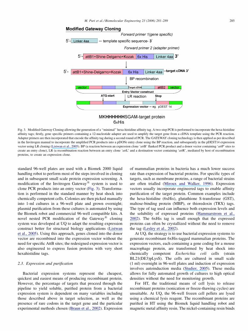
Fig. 3. Modified Gateway Cloning allowing the generation of a ‘‘minimal’’ hexa-histidine affinity tag. A two-step PCR is performed to incorporate the hexa-histidine
affinity tags; firstly, gene specific primers containing a 12-nucleotide adapter are used to amplify the target gene from a cDNA template using the PCR reaction.
Adapter primers are then incorporated that encode the affinity tag during a second round of PCR. The GATEWAY cloning technology is then applied as per described
in the Invitrogen manual to incorporate the amplified PCR products into a pDONr entry clone using the BP reaction, and subsequently in the pDEST14 expression
vector using LR cloning (Listwan et al., 2005). BP (a reaction between an expression clone ‘attB’ flanked PCR product and a donor vector containing ‘attP’ sites to
create an entry clone), LR (a recombination reaction between an entry clone ‘attL’ and a destination vector containing ‘attR’, mediated by host of recombination
proteins, to create an expression clone.
M. Puri et al. / Biomolecular Engineering 23 (2006) 281–289 285
standard 96-well plates are used with a Biomek 2000 liquid
handling robot to perform most of the steps involved in cloning
and in subsequent small scale protein expression screening. A
modification of the Invitrogen Gateway1 system is used to
clone PCR products into an entry vector (Fig. 3). Transforma-
tion is performed in the standard manner by heat shock into
chemically competent cells. Colonies are then picked manually
into 1-ml cultures in a 96-well plate and grown overnight;
plasmid purification from these cultures is automated by using
the Biomek robot and commercial 96-well compatible kits. A
novel nested PCR modification of the Gateway1 cloning
system was developed at UQ to adapt the resulting expression
construct better for structural biology applications (Listwan
et al., 2005). Using this approach, genes cloned into the donor
vector are recombined into the expression vector without the
need for specific AttB sites; the redesigned expression vector is
also engineered to express fusion proteins with very short
hexahistidine tags.
2.3. Expression and purification
Bacterial expression systems represent the cheapest,
quickest and easiest means of producing recombinant protein.
However, the percentage of targets that proceed through the
pipeline to yield soluble, purified protein from a bacterial
expression system is dependent on many variables including
those described above in target selection, as well as the
presence of rare codons in the target gene and the particular
experimental methods chosen (Braun et al., 2002). Expression
of mammalian proteins in bacteria has a much lower success
rate than expression of bacterial proteins. For specific types of
targets, such as membrane proteins, a range of bacterial strains
are often trialled (Miroux and Walker, 1996). Expression
vectors usually incorporate engineered tags to enable affinity
purification of the target protein. Common examples include
the hexa-histidine (6xHis), glutathione S-transferase (GST),
maltose-binding protein (MBP), or thioredoxin (TRX) tags.
The type of tag used can influence both expression levels and
the solubility of expressed proteins (Hammarstrom et al.,
2002). The 6xHis tag is small enough that the expressed
proteins can often be crystallized without the need to remove
the tag (Lesley et al., 2002).
At UQ, the strategy is to use bacterial expression systems to
generate recombinant 6xHis-tagged mammalian proteins. The
expression vectors, each containing a gene coding for a mouse
macrophage protein, are transformed by heat shock into
chemically competent Escherichia coli cells (strain
BL21(DE3)pLysS). The cells are cultured in small scale
(1 ml) overnight in 96-well plates and induction of expression
involves autoinduction media (Studier, 2005). These media
allows for fully automated growth of cultures to high optical
densities without the need for monitoring growth.
For HT, the traditional means of cell lysis to release
recombinant proteins (sonication or freeze-thawing cycles) are
unsuitable. At UQ, the 96-well frozen cell pellets are lysed
using a chemical lysis reagent. The recombinant proteins are
purified in HT using the Biomek liquid handling robot and
magnetic metal affinity resin. The nickel-containing resin binds
M. Puri et al. / Biomolecular Engineering 23 (2006) 281–289286
to the 6xHis-tagged proteins; unbound cellular debris is
removed by washing and the 6xHis-tagged fusion proteins are
eluted with a gradient of imidazole (20–250 mM). The
purification of 96 ml � 1 ml cultures in a 96-well plate format,
from cell culture to eluted protein, is completed within 90 min
using the automated liquid-handling workstation. To analyse
expression levels and solubility, we make use of the LabChip90
automated electrophoresis system for DNA and proteins
(www.caliperls.com). Samples of whole-cell extracts, soluble
fractions and elutions from metal affinity resin are analyzed on
a protein chip in 96-well format. The resulting information
from these small-scale expressions allows identification of
targets that are produced in soluble form in bacteria. These
protein targets are then expressed in large scale (1–2 l) for
crystallization trials and the proteins purified by metal affinity
and size exclusion chromatography. Protein purity and
homogeneity is further checked by MALDI-TOF mass
spectrometry (Leushner, 2001), by gel filtration chromatogra-
phy, circular dichroism (Kelly et al., 2005) and by dynamic
light scattering (Wilson, 2003) prior to progression through to
crystallization trials.
The solubility of expressed proteins is a major bottleneck,
particularly when expressing mammalian proteins in bacteria.
We have also developed a matrix-assisted refolding approach in
which correctly folded proteins are distinguished from
misfolded proteins by their elution from affinity resin under
non-denaturing conditions. The assay can be applied to
insoluble proteins on an individual basis but is also suited to
automation (Cowieson et al., 2006).
2.4. Crystallization
The production of diffraction-quality crystals by screening
variables such as precipitants, pH and temperature often
represents another bottleneck in SG pipelines. The number of
parameters to be evaluated results in a large multi-dimensional
sampling space, but in most cases the amount of protein
available is small, so that the number of crystallization
experiments needs to be minimised. A recent advance has been
the introduction of methodology that allows the set-up of
crystallization trials using nanolitre amounts of protein
(Stevens and Wilson, 2001). The most common method used
for crystallization screening is vapor diffusion, either by
hanging drop or sitting drop; both are amenable to HT
application in 96-well plate format. Free interface diffusion
methods have also been developed (Hansen et al., 2002) that are
suited to HT. Robots are now commonly used for all three steps
in crystallization (Bard et al., 2004), namely preparation of
crystallization formulations, small volume (50–200 nl) crystal-
lization experiment set-up and monitoring of the crystallization
experiments. Furthermore, many crystallization screens are
available commercially (e.g. Hampton Research, Emerald
Biostructures, Jena Bioscience, Molecular Dimensions) or can
be prepared based on previously identified successful
formulations (Page et al., 2003). At UQ, the crystallization
pipeline involves first establishing the optimal protein
concentration, by using a pre-screen from Hampton Research.
Then for each purified protein, a panel of five commercial or
lab-prepared screens are set-up at two different temperatures
(20 8C and 4 8C). The 100 nl + 100 nl sitting drops in the 96-
well tray format are set-up using a TTP LabTech Mosquito
robot and experiments are monitored using a DeCode Genetics
Crystal Monitor. Hits are then optimized by setting up focused
screens (Senger and Mueser, 2005).
2.5. Crystal to structure
Over the past 15 years, the process of protein structure
determination by crystallography has been revolutionized by
developments in methodology, hardware and software. Indeed,
it is now possible to determine the structure of a novel protein in
a matter of hours from the time data measurement begins. The
improvements are due to faster and simpler phasing methods
such as multiple (MAD) and single wavelength anomalous
dispersion (SAD) (Hendrickson, 1999), more intense X-ray
sources (lab-based and at synchrotron beamlines) allowing
quicker data collection and measurement of higher resolution
data, improvements in computing hardware and more robust
and automated software packages for processing and solving
structures (Lamzin and Perrakis, 2000).
At UQ, the structure determination process uses the standard
approaches of cryocrystallography and MAD phasing from
selenomethionine (SeMet)-substituted protein crystals. Crys-
tals of native protein are flash-cooled in a nitrogen gas stream at
�100 K after soaking in a suitable cryoprotectant. X-ray
diffraction quality of protein crystals is assessed using the
laboratory equipment. When diffraction-quality crystals are
obtained and a suitable molecular replacement model is not
available, the protein is expressed in minimal media in the
presence of SeMet to produce SeMet-labelled protein crystals
for use in MAD phasing at a synchrotron. Standard crystal-
lographic packages are used to process and phase the data and
to visualize and refine structures (Aagaard et al., 2005).
2.6. Protein function in macrophages and other outcomes
of the pipeline
In such a comparatively small program, proteins that are
cloned into expression plasmids and which fail to express in
bacteria, or which are not soluble, or which fail to generate
crystals, represent a significant loss of productivity. The
advantage of using the Gateway system is that the cDNAs can
be cloned from the entry vector into mammalian expression
vectors to enable us to investigate function in the absence of
structural outcomes. We have now begun to systematically
clone candidate targets into commercial mammalian destina-
tion vectors, and into an inducible expression system. These
vectors can be transiently or stably transfected into the mouse
macrophage cell line, RAW264. Epitope or His-tags are used to
localize the expressed protein within the cell, and if expression
is successful, to extract and purify the protein and aid in the
identification of binding partners. Over-expression of the target
protein may modify the biological function of the cells; we can
routinely assay cell proliferation, adhesion and spreading, and
M. Puri et al. / Biomolecular Engineering 23 (2006) 281–289 287
inducible production of inflammatory cytokines. Clearly, for
proteins that do generate crystal structures, such information,
combined with information about mRNA expression and
regulation in macrophages, can reinforce and strengthen
structure-based functional inferences.
In the event that a soluble protein is produced but fails to
crystallize, we have initiated an alternative approach to gain
structural information using chemical cross-linking. The
assignment of proximity relationships, combined with mole-
cular modeling, provides the structural information in this case.
We used this method to identify the site of interaction between
latexin, the first crystal structure from the UQ structural
genomics program, and its partner protein carboxypeptidase A
(Mouradov et al., 2006).
3. Impact of SG
Each new protein structure, whether it originates from an
international consortium or a small focused program like that at
UQ, provides a wealth of information for fundamental, applied
and strategic research. For example, when a new protein fold is
revealed, the database of known protein folds is enriched, and
when the function of a new protein is determined, novel
structure–function relationships are established (Zhang and
Kim, 2003). In this section we comment briefly on the impact of
SG protein structure outcomes.
3.1. Functional assignment
A major goal of SG initiatives is the identification of
function for uncharacterized gene products. It is estimated that
over 2/3 of structures of proteins of unknown function can be
used to directly infer a molecular function (Christendat et al.,
2000; Kim et al., 2005), most often because the structures are
remote homologues of proteins of known function (evaluated
for example using DALI (Holm and Sander, 1994) and SCOP
(Murzin et al., 1995)) or because ligands fortuitously co-
crystallize bound to the protein. For uncharacterized proteins
with a new fold, in silico approaches are being developed to use
structure to locate binding sites and to help assign function
(Laskowski et al., 2005; Pal and Eisenberg, 2005).
3.2. Other implications of SG outcomes
As described in Section 1, one of the major goals of SG is to
provide representative structures for every protein fold family.
These representative structures can then be used as templates to
generate protein models for all other members within that
protein family (Chance et al., 2004), using automated methods
such as MODELLER (John and Sali, 2003). In addition, the
wealth of structural information will underpin fundamental
improvements in knowledge including protein folding, protein
structure prediction, and protein evolution. In terms of
biomedical impact, structural data will facilitate the design
of improved therapeutic agents by allowing comparison of
functionally similar protein structures from pathogens and
hosts, or proteins from diseased and normal tissues.
3.3. Drug and drug-lead discovery
It is now widely accepted that protein structure is critical for
the design of new drugs. However, the impact of SG and HT
technology is now extending beyond structure determination and
into new approaches for drug and drug target discovery (Claverie
et al., 2002). In this context, X-ray crystallography has a major
advantage of defining ligand-binding sites in intricate detail
(Kuhn et al., 2002). Drug-like fragment libraries have been
developed that sample chemical space more efficiently than
ligand-based screens (Hann et al., 2001), and these are screened
with a single target protein to identify fragments that bind and to
define precisely the binding sites (Blundell and Patel, 2004).
These approaches rely on robots for soaking the crystals with the
fragment cocktails and to collect the X-ray diffraction data.
Automated software is also being developed to identify the
fragments that bind. Those drug-like fragments that are found to
bind to a protein target (the hits) become the starting point for
medicinal chemistry to develop potent and selective leads that
may then further evolve into drugs.
4. Outputs from UQ SG pipeline
A total of 318 macrophage genes have been processed in
three rounds of expression screening at UQ. The Gateway1
approach was used to successfully clone 220 of these mouse
protein targets into bacterial expression vector. These generated
52 soluble mouse macrophage proteins that have entered
crystallization trials. Crystals have been produced for 6 of the
52 proteins and two crystal structures have been solved. One of
these two structures is that of latexin, also referred to as tissue
carboxypeptidase inhibitor, which is the only known mamma-
lian carboxypeptidase inhibitor. The structure reveals that
latexin comprises two cystatin folds in tandem (Aagaard et al.,
2005) (Fig. 1). The cystatin fold is commonly found in
inhibitors of cysteine proteases, but has not before been
observed in tandem as a pair. The second structure solved is that
of long chain acyl-CoA thioesterase (Serek et al., 2006). The
success rates from the UQ pipeline to date for the 318 mouse
macrophage proteins (16% soluble per selected target; 12%
crystals per soluble protein; 33% structures per crystal) is
comparable to the success rates reported in TargetDB for
eukaryotic proteins (8% purified per selected target; 24%
crystallized per purified protein; 33% structures per crystal, for
57,153 eukaryotic targets, 12 September 2006).
5. Conclusion
Parallel processing of thousands of protein targets is now a
reality for SG consortia. However, these approaches are not
limited to genome-wide studies of protein structure. On the
contrary, the established protocols and automated infrastruc-
ture open up enormous possibilities for other protein science
programs, because it provides the speed and throughput
necessary to rapidly identify, from hundreds or thousands of
starting constructs, those proteins, expression systems,
crystallization conditions or crystals, which structural studies
M. Puri et al. / Biomolecular Engineering 23 (2006) 281–289288
should focus on. We have employed exactly that strategy at
UQ, by using techniques and infrastructure suitable for an
academic laboratory and focusing on proteins with strong
biological importance. We note that several other academic
labs have developed structural genomics technology in what
has been termed ‘‘medium-throughput’’ structural biology
projects (Claverie et al., 2002; Vincentelli et al., 2003; Segelke
et al., 2004; Busso et al., 2005; Moreland et al., 2005). The next
goal for academia and consortia alike will be to progress from a
high-throughput to a higher-output mode, and to proceed from
analyzing ‘‘low-hanging fruit’’ to addressing technically
challenging targets such as membrane proteins and macro-
molecular assemblies.
Acknowledgements
MP thanks DEST for the award of an Australia-Asia
Fellowship. The work at UQ is supported by an Australian
Research Council (ARC) grant to JLM and BK. BK is an ARC
Federation Fellow and National Health and Medical Research
Council (NHMRC) Honorary Research Fellow. JKF is an
NHMRC CJ Martin Research Fellow. NC is an Australian
Synchrotron Research Program Fellow.
References
Aagaard, A., Listwan, P., Cowieson, N., Huber, T., Ravasi, T., Wells, C.A.,
Flanagan, J.U., Kellie, S., Hume, D.A., Kobe, B., Martin, J.L., 2005. An
inflammatory role for the mammalian carboxypeptidase inhibitor latexin:
relationship to cystatins and the tumor suppressor TIG1. Structure 13, 309–
317.
Altschul, S.F., Gish, W., Miller, W., Myers, E.W., Lipman, D.J., 1990. Basic
local alignment search tool. J. Mol. Biol. 215, 403–410.
Bard, J., Ercolani, K., Svenson, K., Olland, A., Somers, W., 2004. Automated
systems for protein crystallization. Methods 34, 329–347.
Bateman, A., Coin, L., Durbin, R., Finn, R.D., Hollich, V., Griffiths-Jones, S.,
Khanna, A., Marshall, M., Moxon, S., Sonnhammer, E.L., Studholme, D.J.,
Yeats, C., Eddy, S.R., 2004. The Pfam protein families database. Nucl.
Acids Res. 32, D138–D141.
Berman, H.M., Westbrook, J., Feng, Z., Gilliland, G., Bhat, T.N., Weissig, H.,
Shindyalov, I.N., Bourne, P.E., 2000. The Protein Data Bank. Nucl. Acids
Res. 28, 235–242.
Blundell, T.L., Patel, S., 2004. High-throughput X-ray crystallography for drug
discovery. Curr. Opin. Pharmacol. 4, 490–496.
Braun, P., Hu, Y., Shen, B., Halleck, A., Koundinya, M., Harlow, E., LaBaer, J.,
2002. Proteome-scale purification of human proteins from bacteria. Proc.
Natl. Acad. Sci. U.S.A. 99, 2654–2659.
Brenner, S.E., 2001. A tour of structural genomics. Nat. Rev. Genet. 2, 801–809.
Buchanan, S.G., 2002. Structural genomics: bridging functional genomics and
structure-based drug design. Curr. Opin. Drug Discov. Devel. 5, 367–381.
Burley, S.K., 2000. An overview of structural genomics. Nat. Struct. Biol. 7
(Suppl.), 932–934.
Busso, D., Poussin-Courmontagne, P., Rose, D., Ripp, R., Litt, A., Thierry, J.C.,
Moras, D., 2005. Structural genomics of eukaryotic targets at a laboratory
scale. J. Struct. Funct. Genom. 6, 81–88.
Carninci, P., Kasukawa, T., Katayama, S., Gough, J., Frith, M.C., Maeda, N.,
Oyama, R., Ravasi, T., Lenhard, B., Wells, C., Kodzius, R., Shimokawa, K.,
Bajic, V.B., Brenner, S.E., Batalov, S., Forrest, A.R., Zavolan, M., Davis,
M.J., Wilming, L.G., Aidinis, V., Allen, J.E., Ambesi-Impiombato, A.,
Apweiler, R., Aturaliya, R.N., Bailey, T.L., Bansal, M., Baxter, L., Beisel,
K.W., Bersano, T., Bono, H., Chalk, A.M., Chiu, K.P., Choudhary, V.,
Christoffels, A., Clutterbuck, D.R., Crowe, M.L., Dalla, E., Dalrymple, B.P.,
de Bono, B., Della Gatta, G., di Bernardo, D., Down, T., Engstrom, P.,
Fagiolini, M., Faulkner, G., Fletcher, C.F., Fukushima, T., Furuno, M.,
Futaki, S., Gariboldi, M., Georgii-Hemming, P., Gingeras, T.R., Gojobori,
T., Green, R.E., Gustincich, S., Harbers, M., Hayashi, Y., Hensch, T.K.,
Hirokawa, N., Hill, D., Huminiecki, L., Iacono, M., Ikeo, K., Iwama, A.,
Ishikawa, T., Jakt, M., Kanapin, A., Katoh, M., Kawasawa, Y., Kelso, J.,
Kitamura, H., Kitano, H., Kollias, G., Krishnan, S.P., Kruger, A., Kum-
merfeld, S.K., Kurochkin, I.V., Lareau, L.F., Lazarevic, D., Lipovich, L.,
Liu, J., Liuni, S., McWilliam, S., Madan Babu, M., Madera, M., March-
ionni, L., Matsuda, H., Matsuzawa, S., Miki, H., Mignone, F., Miyake, S.,
Morris, K., Mottagui-Tabar, S., Mulder, N., Nakano, N., Nakauchi, H., Ng,
P., Nilsson, R., Nishiguchi, S., Nishikawa, S., Nori, F., Ohara, O., Okazaki,
Y., Orlando, V., Pang, K.C., Pavan, W.J., Pavesi, G., Pesole, G., Petrovsky,
N., Piazza, S., Reed, J., Reid, J.F., Ring, B.Z., Ringwald, M., Rost, B., Ruan,
Y., Salzberg, S.L., Sandelin, A., Schneider, C., Schonbach, C., Sekiguchi,
K., Semple, C.A., Seno, S., Sessa, L., Sheng, Y., Shibata, Y., Shimada, H.,
Shimada, K., Silva, D., Sinclair, B., Sperling, S., Stupka, E., Sugiura, K.,
Sultana, R., Takenaka, Y., Taki, K., Tammoja, K., Tan, S.L., Tang, S., Taylor,
M.S., Tegner, J., Teichmann, S.A., Ueda, H.R., van Nimwegen, E., Verardo,
R., Wei, C.L., Yagi, K., Yamanishi, H., Zabarovsky, E., Zhu, S., Zimmer, A.,
Hide, W., Bult, C., Grimmond, S.M., Teasdale, R.D., Liu, E.T., Brusic, V.,
Quackenbush, J., Wahlestedt, C., Mattick, J.S., Hume, D.A., Kai, C., Sasaki,
D., Tomaru, Y., Fukuda, S., Kanamori-Katayama, M., Suzuki, M., Aoki, J.,
Arakawa, T., Iida, J., Imamura, K., Itoh, M., Kato, T., Kawaji, H., Kawaga-
shira, N., Kawashima, T., Kojima, M., Kondo, S., Konno, H., Nakano, K.,
Ninomiya, N., Nishio, T., Okada, M., Plessy, C., Shibata, K., Shiraki, T.,
Suzuki, S., Tagami, M., Waki, K., Watahiki, A., Okamura-Oho, Y., Suzuki,
H., Kawai, J., Hayashizaki, Y., 2005. The transcriptional landscape of the
mammalian genome. Science 309, 1559–1563.
Chance, M.R., Fiser, A., Sali, A., Pieper, U., Eswar, N., Xu, G., Fajardo, J.E.,
Radhakannan, T., Marinkovic, N., 2004. High-throughput computational
and experimental techniques in structural genomics. Genome Res. 14,
2145–2154.
Chandonia, J.M., Brenner, S.E., 2006. The impact of structural genomics:
expectations and outcomes. Science 311, 347–351.
Christendat, D., Yee, A., Dharamsi, A., Kluger, Y., Savchenko, A., Cort, J.R.,
Booth, V., Mackereth, C.D., Saridakis, V., Ekiel, I., Kozlov, G., Maxwell,
K.L., Wu, N., McIntosh, L.P., Gehring, K., Kennedy, M.A., Davidson, A.R.,
Pai, E.F., Gerstein, M., Edwards, A.M., Arrowsmith, C.H., 2000. Structural
proteomics of an archaeon. Nat. Struct. Biol. 7, 903–909.
Claverie, J.M., Monchois, V., Audic, S., Poirot, O., Abergel, C., 2002. In search
of new anti-bacterial target genes: a comparative/structural genomics
approach. Comb. Chem. High Throughput Screen. 5, 511–522.
Cowieson, N.P., Listwan, P., Kurz, M., Aagaard, A., Ravasi, T., Wells, C.,
Huber, T., Hume, D.A., Kobe, B., Martin, J.L., 2005. Pilot studies on the
parallel production of soluble mouse proteins in a bacterial expression
system. J. Struct. Funct. Genom. 6, 13–20.
Cowieson, N.P., Wensley, B., Listwan, P., Hume, D.A., Kobe, B., Martin, J.L.,
2006. An automatable screen for the rapid identification of proteins amen-
able to refolding. Proteomics 6, 1750–1757.
Duffield, J.S., 2003. The inflammatory macrophage: a story of Jekyll and Hyde.
Clin. Sci. (Lond.) 104, 27–38.
Hammarstrom, M., Hellgren, N., van Den Berg, S., Berglund, H., Hard, T.,
2002. Rapid screening for improved solubility of small human proteins
produced as fusion proteins in Escherichia coli. Protein Sci. 11, 313–
321.
Hann, M.M., Leach, A.R., Harper, G., 2001. Molecular complexity and its
impact on the probability of finding leads for drug discovery. J. Chem. Inf.
Comput. Sci. 41, 856–864.
Hansen, C.L., Skordalakes, E., Berger, J.M., Quake, S.R., 2002. A robust and
scalable microfluidic metering method that allows protein crystal growth by
free interface diffusion. Proc. Natl. Acad. Sci. U.S.A. 99, 16531–16536.
Hendrickson, W., 1999. Maturation of MAD phasing for the determination of
macromolecular structures. J. Synchrotron Radiat. 6, 845–851.
Hol, W.G., 2000. Structural genomics for science and society. Nat. Struct. Biol.
7 (Suppl.), 964–966.
Holm, L., Sander, C., 1994. The FSSP database of structurally aligned protein
fold families. Nucl. Acids Res. 22, 3600–3609.
M. Puri et al. / Biomolecular Engineering 23 (2006) 281–289 289
Hume, D.A., Ross, I.L., Himes, S.R., Sasmono, R.T., Wells, C.A., Ravasi, T.,
2002. The mononuclear phagocyte system revisited. J. Leukoc. Biol. 72,
621–627.
John, B., Sali, A., 2003. Comparative protein structure modeling by iterative
alignment, model building and model assessment. Nucl. Acids Res. 31,
3982–3992.
Kelley, L.A., MacCallum, R.M., Sternberg, M.J., 2000. Enhanced genome
annotation using structural profiles in the program 3D-PSSM. J. Mol. Biol.
299, 499–520.
Kelly, S.M., Jess, T.J., Price, N.C., 2005. How to study proteins by circular
dichroism. Biochim. Biophys. Acta 1751, 119–139.
Kim, S.H., Shin, D.H., Liu, J., Oganesyan, V., Chen, S., Xu, Q.S., Kim, J.S.,
Das, D., Schulze-Gahmen, U., Holbrook, S.R., Holbrook, E.L., Martinez,
B.A., Oganesyan, N., DeGiovanni, A., Lou, Y., Henriquez, M., Huang, C.,
Jancarik, J., Pufan, R., Choi, I.G., Chandonia, J.M., Hou, J., Gold, B.,
Yokota, H., Brenner, S.E., Adams, P.D., Kim, R., 2005. Structural genomics
of minimal organisms and protein fold space. J. Struct. Funct. Genom. 6,
63–70.
Kuhn, P., Wilson, K., Patch, M.G., Stevens, R.C., 2002. The genesis of high-
throughput structure-based drug discovery using protein crystallography.
Curr. Opin. Chem. Biol. 6, 704–710.
Lamzin, V.S., Perrakis, A., 2000. Current state of automated crystallographic
data analysis. Nat. Struct. Biol. 7 (Suppl.), 978–981.
Laskowski, R.A., Watson, J.D., Thornton, J.M., 2005. ProFunc: a server for
predicting protein function from 3D structure. Nucl. Acids Res. 33, W89–
W93.
Lesley, S.A., Kuhn, P., Godzik, A., Deacon, A.M., Mathews, I., Kreusch, A.,
Spraggon, G., Klock, H.E., McMullan, D., Shin, T., Vincent, J., Robb, A.,
Brinen, L.S., Miller, M.D., McPhillips, T.M., Miller, M.A., Scheibe, D.,
Canaves, J.M., Guda, C., Jaroszewski, L., Selby, T.L., Elsliger, M.A.,
Wooley, J., Taylor, S.S., Hodgson, K.O., Wilson, I.A., Schultz, P.G.,
Stevens, R.C., 2002. Structural genomics of the Thermotoga maritima
proteome implemented in a high-throughput structure determination pipe-
line. Proc. Natl. Acad. Sci. U.S.A. 99, 11664–11669.
Leushner, J., 2001. MALDI TOF mass spectrometry: an emerging platform for
genomics and diagnostics. Expert Rev. Mol. Diagn. 1, 11–18.
Listwan, P., Cowieson, N., Kurz, M., Hume, D.A., Martin, J.L., Kobe, B., 2005.
Modification of recombinatorial cloning for small affinity tag fusion protein
construct generation. Anal. Biochem. 346, 327–329.
Miroux, B., Walker, J.E., 1996. Over-production of proteins in Escherichia coli:
mutant hosts that allow synthesis of some membrane proteins and globular
proteins at high levels. J. Mol. Biol. 260, 289–298.
Moreland, N., Ashton, R., Baker, H.M., Ivanovic, I., Patterson, S., Arcus, V.L.,
Baker, E.N., Lott, J.S., 2005. A flexible and economical medium-throughput
strategy for protein production and crystallization. Acta Crystallogr. D:
Biol. Crystallogr. 61, 1378–1385.
Mouradov, D., Craven, A., Forwood, J.K., Flanagan, J.U., Garcia-Castellanos,
R., Gomis-Ruth, F.X., Hume, D.A., Martin, J.L., Kobe, B., Huber, T., 2006.
Modelling the structure of latexin-carboxypeptidase A complex based on
chemical cross-linking and molecular docking. Protein Eng. Des. Sel. 19, 9–
16.
Murzin, A.G., Brenner, S.E., Hubbard, T., Chothia, C., 1995. SCOP: a structural
classification of proteins database for the investigation of sequences and
structures. J. Mol. Biol. 247, 536–540.
Nakai, K., Horton, P., 1999. PSORT: a program for detecting sorting signals in
proteins and predicting their subcellular localization. Trends Biochem. Sci.
24, 34–36.
Okazaki, Y., Furuno, M., Kasukawa, T., Adachi, J., Bono, H., Kondo, S.,
Nikaido, I., Osato, N., Saito, R., Suzuki, H., Yamanaka, I., Kiyosawa, H.,
Yagi, K., Tomaru, Y., Hasegawa, Y., Nogami, A., Schonbach, C., Gojobori,
T., Baldarelli, R., Hill, D.P., Bult, C., Hume, D.A., Quackenbush, J.,
Schriml, L.M., Kanapin, A., Matsuda, H., Batalov, S., Beisel, K.W., Blake,
J.A., Bradt, D., Brusic, V., Chothia, C., Corbani, L.E., Cousins, S., Dalla, E.,
Dragani, T.A., Fletcher, C.F., Forrest, A., Frazer, K.S., Gaasterland, T.,
Gariboldi, M., Gissi, C., Godzik, A., Gough, J., Grimmond, S., Gustincich,
S., Hirokawa, N., Jackson, I.J., Jarvis, E.D., Kanai, A., Kawaji, H.,
Kawasawa, Y., Kedzierski, R.M., King, B.L., Konagaya, A., Kurochkin,
I.V., Lee, Y., Lenhard, B., Lyons, P.A., Maglott, D.R., Maltais, L., March-
ionni, L., McKenzie, L., Miki, H., Nagashima, T., Numata, K., Okido, T.,
Pavan, W.J., Pertea, G., Pesole, G., Petrovsky, N., Pillai, R., Pontius, J.U.,
Qi, D., Ramachandran, S., Ravasi, T., Reed, J.C., Reed, D.J., Reid, J., Ring,
B.Z., Ringwald, M., Sandelin, A., Schneider, C., Semple, C.A., Setou, M.,
Shimada, K., Sultana, R., Takenaka, Y., Taylor, M.S., Teasdale, R.D.,
Tomita, M., Verardo, R., Wagner, L., Wahlestedt, C., Wang, Y., Watanabe,
Y., Wells, C., Wilming, L.G., Wynshaw-Boris, A., Yanagisawa, M., Yang, I.,
Yang, L., Yuan, Z., Zavolan, M., Zhu, Y., Zimmer, A., Carninci, P., Hayatsu,
N., Hirozane-Kishikawa, T., Konno, H., Nakamura, M., Sakazume, N., Sato,
K., Shiraki, T., Waki, K., Kawai, J., Aizawa, K., Arakawa, T., Fukuda, S.,
Hara, A., Hashizume, W., Imotani, K., Ishii, Y., Itoh, M., Kagawa, I.,
Miyazaki, A., Sakai, K., Sasaki, D., Shibata, K., Shinagawa, A., Yasunishi,
A., Yoshino, M., Waterston, R., Lander, E.S., Rogers, J., Birney, E.,
Hayashizaki, Y., 2002. Analysis of the mouse transcriptome based on
functional annotation of 60,770 full-length cDNAs. Nature 420, 563–573.
Page, R., Grzechnik, S.K., Canaves, J.M., Spraggon, G., Kreusch, A., Kuhn, P.,
Stevens, R.C., Lesley, S.A., 2003. Shotgun crystallization strategy for
structural genomics: an optimized two-tiered crystallization screen against
the Thermotoga maritima proteome. Acta Crystallogr. D: Biol. Crystallogr.
59, 1028–1037.
Pal, D., Eisenberg, D., 2005. Inference of protein function from protein
structure. Structure 13, 121–130.
Segelke, B.W., Schafer, J., Coleman, M.A., Lekin, T.P., Toppani, D., Skow-
ronek, K.J., Kantardjieff, K.A., Rupp, B., 2004. Laboratory scale structural
genomics. J. Struct. Funct. Genom. 5, 147–157.
Senger, A., Mueser, T., 2005. Rapid preparation of custom grid screens for
crystal growth optimization. J. Appl. Cryst. 38, 847–850.
Serek, R., Forwood, J.K., Hume, D.A., Martin, J.L., Kobe, B., 2006. Crystal-
lization of the C-terminal domain of the mouse brain cytosolic long-chain
acyl-CoA thioesterase. Acta Crystallogr. Sect. F: Struct. Biol. Cryst. Com-
mun. 62, 133–135.
Stevens, R.C., Wilson, I.A., 2001. Tech.Sight. Industrializing structural biology.
Science 293, 519–520.
Studier, F.W., 2005. Protein production by auto-induction in high density
shaking cultures. Protein Expr. Purif. 41, 207–234.
Thornton, J.M., Todd, A.E., Milburn, D., Borkakoti, N., Orengo, C.A., 2000.
From structure to function: approaches and limitations. Nat. Struct. Biol. 7
(Suppl.), 991–994.
Todd, A.E., Marsden, R.L., Thornton, J.M., Orengo, C.A., 2005. Progress of
structural genomics initiatives: an analysis of solved target structures. J.
Mol. Biol. 348, 1235–1260.
Vincentelli, R., Bignon, C., Gruez, A., Canaan, S., Sulzenbacher, G., Tegoni,
M., Campanacci, V., Cambillau, C., 2003. Medium-scale structural geno-
mics: strategies for protein expression and crystallization. Acc. Chem. Res.
36, 165–172.
Walian, P., Cross, T.A., Jap, B.K., 2004. Structural genomics of membrane
proteins. Genome Biol. 5, 215.
Wells, C.A., Ravasi, T., Faulkner, G.J., Carninci, P., Okazaki, Y., Hayashizaki,
Y., Sweet, M., Wainwright, B.J., Hume, D.A., 2003a. Genetic control of the
innate immune response. BMC Immunol. 4, 5.
Wells, C.A., Ravasi, T., Sultana, R., Yagi, K., Carninci, P., Bono, H., Faulkner,
G., Okazaki, Y., Quackenbush, J., Hume, D.A., Lyons, P.A., 2003b. Con-
tinued discovery of transcriptional units expressed in cells of the mouse
mononuclear phagocyte lineage. Genome Res. 13, 1360–1365.
Wilson, W.W., 2003. Light scattering as a diagnostic for protein crystal
growth—a practical approach. J. Struct. Biol. 142, 56–65.
Yakunin, A.F., Yee, A.A., Savchenko, A., Edwards, A.M., Arrowsmith, C.H.,
2004. Structural proteomics: a tool for genome annotation. Curr. Opin.
Chem. Biol. 8, 42–48.
Yee, A., Pardee, K., Christendat, D., Savchenko, A., Edwards, A.M., Arrow-
smith, C.H., 2003. Structural proteomics: toward high-throughput structural
biology as a tool in functional genomics. Acc. Chem. Res. 36, 183–189.
Zhang, C., Kim, S.H., 2003. Overview of structural genomics: from structure to
function. Curr. Opin. Chem. Biol. 7, 28–32.