Faecal case architecture in the gibbosus species group of Neochlamisus Karren, 1972 (Coleoptera:...
37
Faecal case architecture in the gibbosus species group of Neochlamisus Karren, 1972 (Coleoptera: Chrysomelidae: Cryptocephalinae: Chlamisini) CAROLINE S. CHABOO 1 *†, CHRISTOPHER G. BROWN 2 and DANIEL J. FUNK 2 1 Cullman Molecular Laboratory, American Museum of Natural History, Central Park West @79th St, New York, NY, 10024-5192, USA 2 Department of Biological Sciences, Vanderbilt University, VU Station B, Box 35-1634, Nashville, TN, 37235-1634, USA Received 1 April 2006; accepted for publication 22 May 2007 Constructions composed of faeces are rare in insects, but occur in certain leaf-beetle clades. Members of the subfamily Cryptocephalinae share a complex behavioural and morphological synapomorphy, involving portable faecal cases that house the immature stages. A maternally initiated egg case is expanded and enlarged through four larval stadia, then sealed to provide a pupal chamber from which adults eventually emerge. We analyse and compare faecal-case architecture in ten taxa of the cryptocephaline genus Neochlamisus, and assess structural variation within a life cycle, between different ‘host forms’ of Neochlamisus bebbianae, and among species. These cases proved to be comprised primarily of faeces, with plant trichomes representing the only common secondary component. General architectural trends reflected variation in shape, faecal texture, and the incorporation and density of trichomes. Deviations of the Neochlamisus case from a simple geometrical gnomon reflect the changing body size of the animal, differential application of faeces, and shifts in the orientation of the carriage of the case. Neochlamisus cases are presumably energetically costly to produce, carry, and maintain, and some adaptive hypotheses of case evolution are proposed. Additionally, literature on case morphology in other camptosomates is reviewed. © 2008 The Linnean Society of London, Zoological Journal of the Linnean Society, 2008, 152, 315–351. ADDITIONAL KEYWORDS: defence – faeces – host plant – larvae – leaf beetles – trichomes. INTRODUCTION Faecal construction is one of the oddest behaviours among certain clades of Chrysomelidae. Adults and immature stages of species in several subfamilies retain faeces as camouflage, clubs, and protective covers. The subfamilies Cryptocephalinae (135 genera; c. 3587 species) and Lamprosomatinae (13 genera; c. 190 species) (Seeno & Wilcox, 1982; Reid, 1995, 2000; Farrell, 1998) are commonly referred to as the Camptosomata or ‘case-bearers’, because their species build hard cases of faeces, mixed with anal and oral fluids, around the immature stages (Cockerell, 1891; Lécaillon, 1898; Sharp, 1899; Donisthorpe, 1902; Fiebrig, 1910; Fiori, 1950; Erber, 1968, 1969, 1988; Schmitt, 1988; Reid, 1990, 1991; Olmstead, 1994; Jolivet, 1997; Jolivet & Verma, 2002; Schöller, 2004; Brown & Funk, 2005) (Fig. 1). [We note that the term ‘case-bearers’ is also commonly applied to members of the lepidopteran family Coleophoridae; e.g. Bucheli, Landry & Wenzel (2002) and Falkovitch (2003)]. All case-bearing chrysomelids build their cases in a similar way: cryptocephaline females enclose single eggs in a hardened faecal case. Upon hatching, the *Corresponding author. E-mail: [email protected] †Current address: Division of Entomology, The University of Kansas Natural History Museum, 1501 Crestline Drive, Suite 140, University of Kansas, Lawrence, KS 66049-2711, USA. Zoological Journal of the Linnean Society, 2008, 152, 315–351. With 19 figures © 2008 The Linnean Society of London, Zoological Journal of the Linnean Society, 2008, 152, 315–351 315
-
Upload
un-lincoln -
Category
Documents
-
view
1 -
download
0
Transcript of Faecal case architecture in the gibbosus species group of Neochlamisus Karren, 1972 (Coleoptera:...

Faecal case architecture in the gibbosusspecies group of Neochlamisus Karren, 1972(Coleoptera: Chrysomelidae: Cryptocephalinae:Chlamisini)
CAROLINE S. CHABOO1*†, CHRISTOPHER G. BROWN2 and DANIEL J. FUNK2
1Cullman Molecular Laboratory, American Museum of Natural History, Central Park West @79th St,New York, NY, 10024-5192, USA2Department of Biological Sciences, Vanderbilt University, VU Station B, Box 35-1634, Nashville, TN,37235-1634, USA
Received 1 April 2006; accepted for publication 22 May 2007
Constructions composed of faeces are rare in insects, but occur in certain leaf-beetle clades. Members of thesubfamily Cryptocephalinae share a complex behavioural and morphological synapomorphy, involving portablefaecal cases that house the immature stages. A maternally initiated egg case is expanded and enlarged throughfour larval stadia, then sealed to provide a pupal chamber from which adults eventually emerge. We analyse andcompare faecal-case architecture in ten taxa of the cryptocephaline genus Neochlamisus, and assess structuralvariation within a life cycle, between different ‘host forms’ of Neochlamisus bebbianae, and among species. Thesecases proved to be comprised primarily of faeces, with plant trichomes representing the only common secondarycomponent. General architectural trends reflected variation in shape, faecal texture, and the incorporation anddensity of trichomes. Deviations of the Neochlamisus case from a simple geometrical gnomon reflect the changingbody size of the animal, differential application of faeces, and shifts in the orientation of the carriage of the case.Neochlamisus cases are presumably energetically costly to produce, carry, and maintain, and some adaptivehypotheses of case evolution are proposed. Additionally, literature on case morphology in other camptosomates isreviewed. © 2008 The Linnean Society of London, Zoological Journal of the Linnean Society, 2008, 152, 315–351.
ADDITIONAL KEYWORDS: defence – faeces – host plant – larvae – leaf beetles – trichomes.
INTRODUCTION
Faecal construction is one of the oddest behavioursamong certain clades of Chrysomelidae. Adults andimmature stages of species in several subfamiliesretain faeces as camouflage, clubs, and protectivecovers. The subfamilies Cryptocephalinae (135genera; c. 3587 species) and Lamprosomatinae (13genera; c. 190 species) (Seeno & Wilcox, 1982; Reid,1995, 2000; Farrell, 1998) are commonly referred to
as the Camptosomata or ‘case-bearers’, because theirspecies build hard cases of faeces, mixed withanal and oral fluids, around the immature stages(Cockerell, 1891; Lécaillon, 1898; Sharp, 1899;Donisthorpe, 1902; Fiebrig, 1910; Fiori, 1950; Erber,1968, 1969, 1988; Schmitt, 1988; Reid, 1990, 1991;Olmstead, 1994; Jolivet, 1997; Jolivet & Verma, 2002;Schöller, 2004; Brown & Funk, 2005) (Fig. 1). [Wenote that the term ‘case-bearers’ is also commonlyapplied to members of the lepidopteran familyColeophoridae; e.g. Bucheli, Landry & Wenzel (2002)and Falkovitch (2003)].
All case-bearing chrysomelids build their cases in asimilar way: cryptocephaline females enclose singleeggs in a hardened faecal case. Upon hatching, the
*Corresponding author. E-mail: [email protected]†Current address: Division of Entomology, The University ofKansas Natural History Museum, 1501 Crestline Drive,Suite 140, University of Kansas, Lawrence, KS 66049-2711,USA.
Zoological Journal of the Linnean Society, 2008, 152, 315–351. With 19 figures
© 2008 The Linnean Society of London, Zoological Journal of the Linnean Society, 2008, 152, 315–351 315

first larval instar destroys the sealed flattened ‘roof ’,turns the egg case upside down, and retains it as aprotective home that covers most of the body, withonly the legs and head projecting from the baseopening, enabling movement and feeding. As thelarva grows, it expands the case longitudinally byapplying its faeces to extend the basal margins, andlaterally by making a longitudinal slit that is forcedopen and filled in, thus adding to the faecal wall(Erber, 1988; Brown & Funk, 2005). This case isretained, carried, and expanded by all four instars,and is used as a ‘moveable house’ (e.g. Scudder, 1891).The prepupa (late instar IV) fixes the case to thesubstrate, closes the base opening with its faeces, andso makes a pupal chamber. Finally, the adult exitsby cutting away a circular ‘cap’ from the case apexand escaping (e.g. Hislop, 1872). The material com-prising these cases appears to be primarily faeces,mixed with salivary and anal secretions, along withinclusions like soil, debris, leaf fragments, wood chips,and leaf trichomes (Fiebrig, 1910; Linsenmaier,1972; Erber, 1988; Lawson, 1991; Jolivet & Verma,2002). Cases of the cryptocephaline egg, larva, andpupa are the product of complex and highly inte-grated morphological and behavioural traits in theadult female and her offspring. In this paper wepresent the most complete study of case architecture
in the tribe Chlamisini, an unusual example of insectarchitecture.
The Chlamisini comprise 11 genera and c. 400species, and occur worldwide with 81% of the knownspecies in the Neotropics (Monrós, 1951; Karren,1972; Seeno & Wilcox, 1982; Erber, 1988; Reid, 1991).Biological information on immature stages is avail-able for six genera: Chlamisus Rafinesque, 1815(Packard, 1869, 1889; Riley, 1874a, b; Dimmock, 1885;Popenoe & Marlatt, 1889; Scudder, 1891; Wickham,1896; Briggs, 1905; Fiebrig, 1910; Girault, 1911;Frost, 1942; Brown, 1943; Monrós, 1948, 1951; Peter-son, 1951; Morgan & Maxwell, 1952; Wood, 1966;Karren, 1972; Medvedev & Zaitsev, 1979; Zaitsev,1992; Reu & Del-Claro, 2005), Exema Lacordaire,1848 (Dugès, 1881; Pierce, 1940; Brown, 1943;Karren, 1964; LeSage, 1982; Root & Messina, 1983;Boldt & White, 1992), Diplacaspis Jacobson, 1824(Emden, 1932; Monrós, 1951; Karren, 1972), FulcidaxVoet, 1806 (Flinte et al., 2003; Flinte & Macêdo,2004a, b; Flinte, 2006), Melitochlamys Monrós, 1948(Fiebrig, 1910; Monrós, 1951), and NeochlamisusKarren, 1972 (Karren, 1972; LeSage, 1984; Neal,1989; Brown & Funk, 2005). Arthrochlamys Ihering,1904, Chlamys (Knoch, 1801), and some species ofChlamisus were synonymized under the gibbosusspecies group of Neochlamisus (Karren, 1972), which
Figure. 1. A, Neochlamisus bebbianae IV: Acer (maple) host form coating her faeces on her eggs. B, egg case ofNeochlamisus platani. C, third instar of Neochlamisus chamaedaphnes walking with its portable case. D, second instarof Neochlamisus bebbianae, Salix (willow) host form. All photos by CGB.
316 C. S. CHABOO ET AL.
© 2008 The Linnean Society of London, Zoological Journal of the Linnean Society, 2008, 152, 315–351
is the subject of the present study. Erber (1988)reviewed case structure across the Camptosomates.
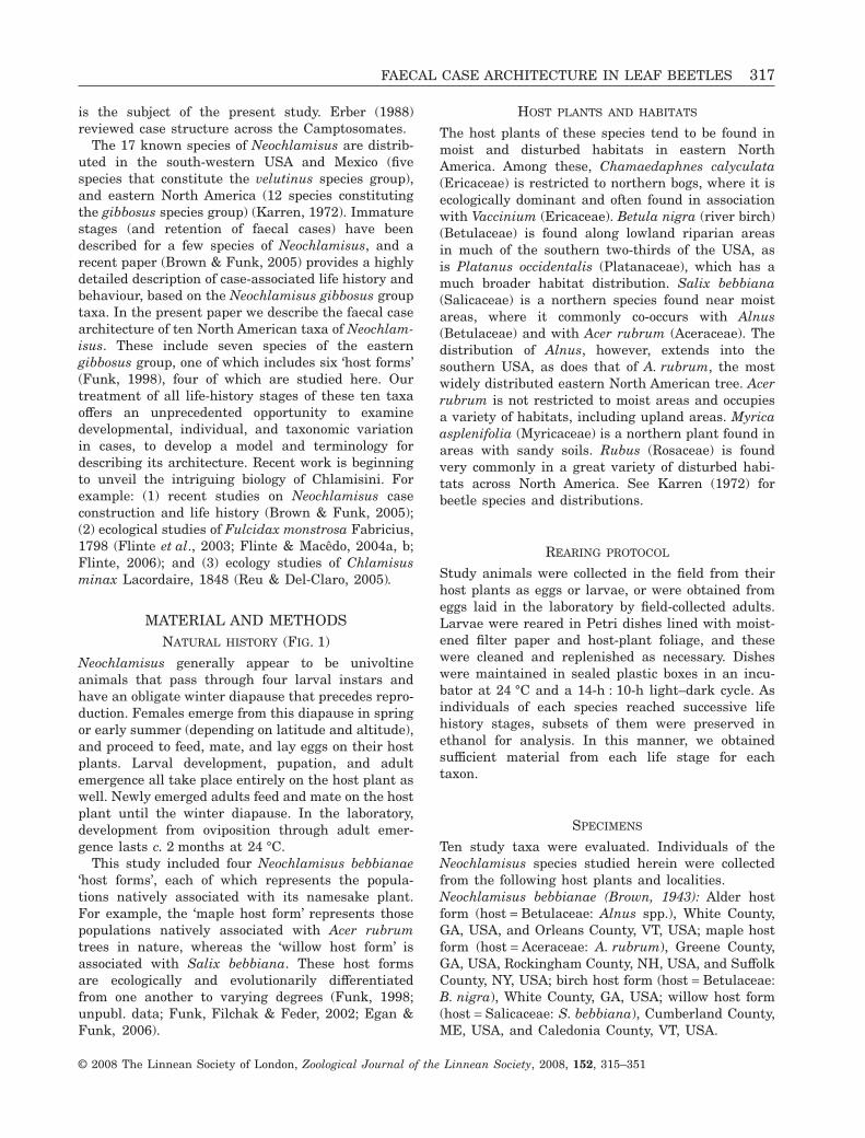
The 17 known species of Neochlamisus are distrib-uted in the south-western USA and Mexico (fivespecies that constitute the velutinus species group),and eastern North America (12 species constitutingthe gibbosus species group) (Karren, 1972). Immaturestages (and retention of faecal cases) have beendescribed for a few species of Neochlamisus, and arecent paper (Brown & Funk, 2005) provides a highlydetailed description of case-associated life history andbehaviour, based on the Neochlamisus gibbosus grouptaxa. In the present paper we describe the faecal casearchitecture of ten North American taxa of Neochlam-isus. These include seven species of the easterngibbosus group, one of which includes six ‘host forms’(Funk, 1998), four of which are studied here. Ourtreatment of all life-history stages of these ten taxaoffers an unprecedented opportunity to examinedevelopmental, individual, and taxonomic variationin cases, to develop a model and terminology fordescribing its architecture. Recent work is beginningto unveil the intriguing biology of Chlamisini. Forexample: (1) recent studies on Neochlamisus caseconstruction and life history (Brown & Funk, 2005);(2) ecological studies of Fulcidax monstrosa Fabricius,1798 (Flinte et al., 2003; Flinte & Macêdo, 2004a, b;Flinte, 2006); and (3) ecology studies of Chlamisusminax Lacordaire, 1848 (Reu & Del-Claro, 2005).
MATERIAL AND METHODSNATURAL HISTORY (FIG. 1)
Neochlamisus generally appear to be univoltineanimals that pass through four larval instars andhave an obligate winter diapause that precedes repro-duction. Females emerge from this diapause in springor early summer (depending on latitude and altitude),and proceed to feed, mate, and lay eggs on their hostplants. Larval development, pupation, and adultemergence all take place entirely on the host plant aswell. Newly emerged adults feed and mate on the hostplant until the winter diapause. In the laboratory,development from oviposition through adult emer-gence lasts c. 2 months at 24 °C.
This study included four Neochlamisus bebbianae‘host forms’, each of which represents the popula-tions natively associated with its namesake plant.For example, the ‘maple host form’ represents thosepopulations natively associated with Acer rubrumtrees in nature, whereas the ‘willow host form’ isassociated with Salix bebbiana. These host formsare ecologically and evolutionarily differentiatedfrom one another to varying degrees (Funk, 1998;unpubl. data; Funk, Filchak & Feder, 2002; Egan &Funk, 2006).
HOST PLANTS AND HABITATS
The host plants of these species tend to be found inmoist and disturbed habitats in eastern NorthAmerica. Among these, Chamaedaphnes calyculata(Ericaceae) is restricted to northern bogs, where it isecologically dominant and often found in associationwith Vaccinium (Ericaceae). Betula nigra (river birch)(Betulaceae) is found along lowland riparian areasin much of the southern two-thirds of the USA, asis Platanus occidentalis (Platanaceae), which has amuch broader habitat distribution. Salix bebbiana(Salicaceae) is a northern species found near moistareas, where it commonly co-occurs with Alnus(Betulaceae) and with Acer rubrum (Aceraceae). Thedistribution of Alnus, however, extends into thesouthern USA, as does that of A. rubrum, the mostwidely distributed eastern North American tree. Acerrubrum is not restricted to moist areas and occupiesa variety of habitats, including upland areas. Myricaasplenifolia (Myricaceae) is a northern plant found inareas with sandy soils. Rubus (Rosaceae) is foundvery commonly in a great variety of disturbed habi-tats across North America. See Karren (1972) forbeetle species and distributions.
REARING PROTOCOL
Study animals were collected in the field from theirhost plants as eggs or larvae, or were obtained fromeggs laid in the laboratory by field-collected adults.Larvae were reared in Petri dishes lined with moist-ened filter paper and host-plant foliage, and thesewere cleaned and replenished as necessary. Disheswere maintained in sealed plastic boxes in an incu-bator at 24 °C and a 14-h : 10-h light–dark cycle. Asindividuals of each species reached successive lifehistory stages, subsets of them were preserved inethanol for analysis. In this manner, we obtainedsufficient material from each life stage for eachtaxon.
SPECIMENS
Ten study taxa were evaluated. Individuals of theNeochlamisus species studied herein were collectedfrom the following host plants and localities.Neochlamisus bebbianae (Brown, 1943): Alder hostform (host = Betulaceae: Alnus spp.), White County,GA, USA, and Orleans County, VT, USA; maple hostform (host = Aceraceae: A. rubrum), Greene County,GA, USA, Rockingham County, NH, USA, and SuffolkCounty, NY, USA; birch host form (host = Betulaceae:B. nigra), White County, GA, USA; willow host form(host = Salicaceae: S. bebbiana), Cumberland County,ME, USA, and Caledonia County, VT, USA.
FAECAL CASE ARCHITECTURE IN LEAF BEETLES 317
© 2008 The Linnean Society of London, Zoological Journal of the Linnean Society, 2008, 152, 315–351

Neochlamisus bimaculatus Karren, 1972(host = Rosaceae: Rubus spp.): White County, GA,USA and Davidson County, TN, USA.Neochlamisus chamaedaphnes (Brown, 1943) (host= Ericaceae: Chamaedaphne calyculata L): BurnettCounty, WI, USA.Neochlamisus comptoniae (Brown, 1943) (host =Myricaceae: Myrica asplenifolia L): RockinghamCounty, NH, USA.Neochlamisus cribripennis (LeConte, 1878) (host =Ericaceae: Vaccinium spp.): Suffolk County, NY, USA.Neochlamisus eubati (Brown, 1943) (host = Rosaceae:Rubus spp.): Rockingham County, NH, USA.Neochlamisus platani (Brown, 1952) (host =Platanaceae: Platanus occidentalis L): White County,GA, USA, and Davidson and Williamson Counties,TN, USA.
PRESERVATION
Specimens were collected into and stored in 75%ethanol for photographic analysis, and in 90% ethanolfor SEM work. Individuals were removed from theircases and stored in 75% ethanol, whereas their caseswere air-dried before imaging.
VOUCHERS
Vouchers of all insect stages and cases have beendeposited in four American collections: AmericanMuseum of Natural History, D. J. Funk collection(DJFC), University of Kansas Insect Collection, andthe US National Museum. Beetle identificationswere based on the keys and species descriptions ofKarren (1972). Plant taxonomy was checked with theMissouri Botanical Garden W3Tropicos database(Solomon 2006).
EXAMINATION AND PHOTOGRAPHY
Small balls of plasticine were used to stabilize thedried cases in shallow wells of ceramic trays. Caseswere stabilized and positioned on a bed of glass beadsin a glass dish filled with 75% ethanol for initialexaminations, and were then air-dried and examined.They were then cleaved by holding them with fineforceps and gently pressing against the ventralsuture. This exposed the internal surface for exami-nation. Exuvial inclusions were determined bytearing apart cases.
Cases were examined with a Zeiss compound micro-scope with a Type 256576 camera lucida attachment.Measurements were taken with a Lasico digitalocular micrometer. Cases and individuals were pho-tographed with a Nikon D1 Digital camera, Infinity
K2 lenses, and a Microptics ML1000 fibre optic flashunit. Photos were all taken by the first author, exceptwhere noted.
SCANNING ELECTRON MICROSCOPY (SEM)
Air-dried cases were mounted on standard aluminiumSEM stubs (diameter, 12 mm; height, 7 mm; ElectronMicroscopy Sciences) and sputter coated with gold/palladium in a Denton Vacuum Desktop II model.Cases were broken longitudinally and mounted onSEM stubs. Cases were then examined with a HitachiS4700 Field Emission Scanning Electron Microscope,at the American Museum of Natural History.
IMAGING AND FIGURE PREPARATION
Illustrations were scanned, digitized, and minimallyedited in MS-Paint 2000. Images were minimallyedited (background removed, some contrast manipu-lation) in Adobe Photoshop 6.0 and Canvas 9.0.1, andarranged as figures with Adobe Photoshop 6.0. Allscanning electron micrographs are indicated; all otherimages are photomicrographs.
TERMINOLOGY
Cryptocephaline cases have been called ‘sac’ (Packard,1869, 1889), ‘bag’ (Beeson, 1941), ‘scatoshells’ (Hinton,1981; Erber, 1988), ‘case’ (LeSage, 1982), ‘mantle’(Erber, 1988), and ‘sheath’ (Zaitsev, 1992). To avoidinferences about construction materials we elimi-nated scatoshell, as there may be situations whereother materials may be incorporated, as suggested inthe literature. Sac and bag suggest a structure that issuspended, so we selected case as the more inclusivedescriptor.
Major landmarks and terminology for the descrip-tion of cryptocephalinae cases are illustrated inFigure 2. The egg case is completely sealed and isshaped like an upturned bell, with a stalk at one endand a flattened sealed roof at the other end. The stalkis short and terminates in a flared rounded attach-ment disc that adheres to the substrate. The sealedroof may have the margin slightly flared, forming aflange (Karren, 1964). It is important to note that theinstar-I larva turns the egg case upside down, andthis has significant implications for terminology, ori-entation, and homology concepts. The case is orien-tated vertically relative to the substrate in younglarvae, but is carried almost horizontally in olderlarvae. The original sealed roof of the egg case isorientated basally and is open in the larvae. We referto the side walls as dorsal, lateral and ventral sur-faces. The ventral surface is easily distinguished inall larval instars (starting with late instar I) and in
318 C. S. CHABOO ET AL.
© 2008 The Linnean Society of London, Zoological Journal of the Linnean Society, 2008, 152, 315–351
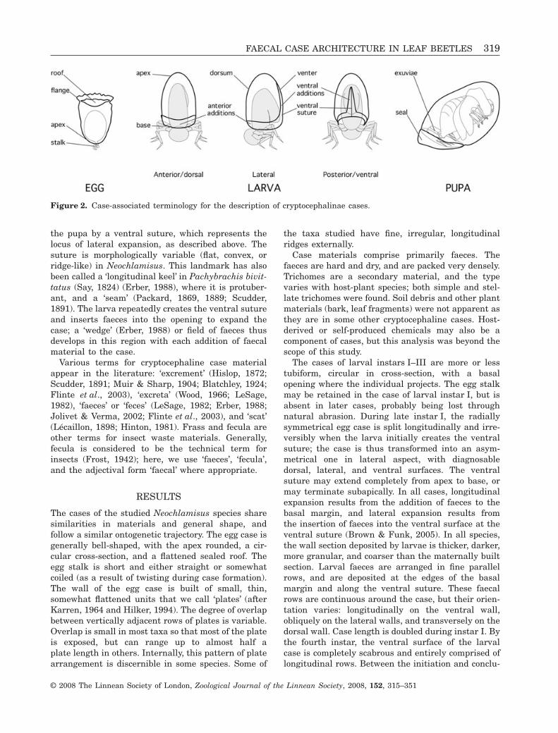
the pupa by a ventral suture, which represents thelocus of lateral expansion, as described above. Thesuture is morphologically variable (flat, convex, orridge-like) in Neochlamisus. This landmark has alsobeen called a ‘longitudinal keel’ in Pachybrachis bivit-tatus (Say, 1824) (Erber, 1988), where it is protuber-ant, and a ‘seam’ (Packard, 1869, 1889; Scudder,1891). The larva repeatedly creates the ventral sutureand inserts faeces into the opening to expand thecase; a ‘wedge’ (Erber, 1988) or field of faeces thusdevelops in this region with each addition of faecalmaterial to the case.
Various terms for cryptocephaline case materialappear in the literature: ‘excrement’ (Hislop, 1872;Scudder, 1891; Muir & Sharp, 1904; Blatchley, 1924;Flinte et al., 2003), ‘excreta’ (Wood, 1966; LeSage,1982), ‘faeces’ or ‘feces’ (LeSage, 1982; Erber, 1988;Jolivet & Verma, 2002; Flinte et al., 2003), and ‘scat’(Lécaillon, 1898; Hinton, 1981). Frass and fecula areother terms for insect waste materials. Generally,fecula is considered to be the technical term forinsects (Frost, 1942); here, we use ‘faeces’, ‘fecula’,and the adjectival form ‘faecal’ where appropriate.
RESULTS
The cases of the studied Neochlamisus species sharesimilarities in materials and general shape, andfollow a similar ontogenetic trajectory. The egg case isgenerally bell-shaped, with the apex rounded, a cir-cular cross-section, and a flattened sealed roof. Theegg stalk is short and either straight or somewhatcoiled (as a result of twisting during case formation).The wall of the egg case is built of small, thin,somewhat flattened units that we call ‘plates’ (afterKarren, 1964 and Hilker, 1994). The degree of overlapbetween vertically adjacent rows of plates is variable.Overlap is small in most taxa so that most of the plateis exposed, but can range up to almost half aplate length in others. Internally, this pattern of platearrangement is discernible in some species. Some of
the taxa studied have fine, irregular, longitudinalridges externally.
Case materials comprise primarily faeces. Thefaeces are hard and dry, and are packed very densely.Trichomes are a secondary material, and the typevaries with host-plant species; both simple and stel-late trichomes were found. Soil debris and other plantmaterials (bark, leaf fragments) were not apparent asthey are in some other cryptocephaline cases. Host-derived or self-produced chemicals may also be acomponent of cases, but this analysis was beyond thescope of this study.
The cases of larval instars I–III are more or lesstubiform, circular in cross-section, with a basalopening where the individual projects. The egg stalkmay be retained in the case of larval instar I, but isabsent in later cases, probably being lost throughnatural abrasion. During late instar I, the radiallysymmetrical egg case is split longitudinally and irre-versibly when the larva initially creates the ventralsuture; the case is thus transformed into an asym-metrical one in lateral aspect, with diagnosabledorsal, lateral, and ventral surfaces. The ventralsuture may extend completely from apex to base, ormay terminate subapically. In all cases, longitudinalexpansion results from the addition of faeces to thebasal margin, and lateral expansion results fromthe insertion of faeces into the ventral surface at theventral suture (Brown & Funk, 2005). In all species,the wall section deposited by larvae is thicker, darker,more granular, and coarser than the maternally builtsection. Larval faeces are arranged in fine parallelrows, and are deposited at the edges of the basalmargin and along the ventral suture. These faecalrows are continuous around the case, but their orien-tation varies: longitudinally on the ventral wall,obliquely on the lateral walls, and transversely on thedorsal wall. Case length is doubled during instar I. Bythe fourth instar, the ventral surface of the larvalcase is completely scabrous and entirely comprised oflongitudinal rows. Between the initiation and conclu-
Figure 2. Case-associated terminology for the description of cryptocephalinae cases.
FAECAL CASE ARCHITECTURE IN LEAF BEETLES 319
© 2008 The Linnean Society of London, Zoological Journal of the Linnean Society, 2008, 152, 315–351

sion of stadium IV, there is a striking change in caseshape. During this period it is transformed fromtubular to ovoid and barrel-like, as a result of unevenfaecal deposition, with most material added mediallyon the ventral wall (rather than the basal margin)during the latter half of the instar (Brown & Funk,2005). The late instar-IV larva attaches the caseopening to the host surface, and seals it completelywith faeces and additional secretions to form thepupal case; the seal is very coarse and thick. In thefollowing section, the egg case, larval cases frominstars I–III, early and late instar-IV cases, and thepupal cases are described for each taxon studied here.
DESCRIPTIONS OF NEOCHLAMISUS CASES
Neochlamisus bebbianae (Brown, 1943) I:Salix bebbiana host form (Figs 3–5)
Egg case (Fig. 3): Size (N = 8): length (L), 1.87–2.68 mm, width (W) at roof, 0.95–1.05 mm. Colour:dark brown with longitudinal ridges lighter brown.
Shape: bell-like, symmetrical in lateral aspect. Apex:conical, transverse in lateral aspect; flange uneven.Egg stalk: present (Fig. 3B), coiled and narrowed,colour translucent white. Roof: slightly convex inlateral view; texture coarse. External surface: textureof walls coarse; faeces arranged as overlappingplates (Fig. 3C). Plates: quadriform, slightly domedmedially, with basal edges exposed; longitudinalridges present, arising from multiple plates of vari-able lengths, with heights also variable but generallyshort. Trichomes: present, simple, incorporateddeeply into plate matrix; decumbent, some slightlyprotuberant at surface (Fig. 3C). Internal surface:texture rough, but smoother than external surface,lacking apparent plates and ridges; slight spacebetween case wall and egg.
Larva-I case (Figs 3A, 4A): Size (N = 9): L, 1.98–2.12 mm; W at base opening, 0.91–1.09 mm. Colour:unevenly dark-brown–black. Egg case: mostly intact,distinguishable from new larval section by width,
Figure 3. Neochlamisus bebbianae I: Salix bebbianae (willow) host form. A, case series from egg case (left) to pupal case(right). B, egg case, lateral aspect, scanning electron micrograph. C, egg case, scanning electron micrograph.
320 C. S. CHABOO ET AL.
© 2008 The Linnean Society of London, Zoological Journal of the Linnean Society, 2008, 152, 315–351
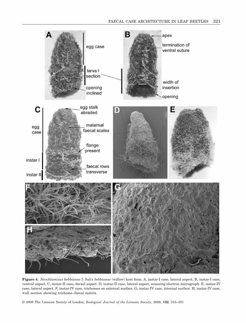
Figure 4. Neochlamisus bebbianae I: Salix bebbianae (willow) host form. A, instar-I case, lateral aspect. B, instar-I case,ventral aspect. C, instar-II case, dorsal aspect. D, instar-II case, lateral aspect, scanning electron micrograph. E, instar-IVcase, lateral aspect. F, instar-IV case, trichomes on external surface. G, instar-IV case, internal surface. H, instar-IV case,wall section showing trichome–faecal matrix.
FAECAL CASE ARCHITECTURE IN LEAF BEETLES 321
© 2008 The Linnean Society of London, Zoological Journal of the Linnean Society, 2008, 152, 315–351

colour, wall thickness texture, trichome placement,and patterning of faecal deposit; egg stalk present orabsent. Walls of larval section: slightly wider andthicker than those of egg case. Larval faeces granular:deposited in rows, not in plates. Ventral wall: withsuture continuous, extending subapically from eggcase through larval section to opening; faecal rowsfew in number, positioned obliquely; dark triangularwedge spanning ventral suture. Lateral and dorsalwalls: with transverse faecal rows. Base opening:slightly oblique in lateral view; basal margin simple.External surface of larval section with trichomesunevenly distributed, protruding as dense fur-likepatches in some sections. Internal surface: smooth,lacking apparent plates and ridges.
Larva-II case (Figs 3, 4): Size (N = 8): L, 2.41–3.48 mm; W at base opening, 1.32–1.58 mm. Shape:tubular, slightly asymmetrical in lateral aspect withdorsal wall longer than ventral wall. Apex: rounded;egg stalk absent. Egg case: generally intact, except forventral suture; position, shape, colour and other char-acteristics as above. Larval case additions duringphases I and II: not distinguishable; similar in colourand surface texture characteristics. Ventral suture:
extended from apex through egg case to base opening.Trichomes present, as in case of instar I. Internalsurface: smooth.
Larva-III case (Fig. 3): Size (N = 10): L, 3.66–3.91 mm; W at base opening, 1.82–1.91 mm. Colour oflarval sections I, II, and III: generally dark, unevenlystriate. Shape: tubular; egg case apparent, positioninclined ventrad. Trichomes of sections I, II, and III ofmedium density.
Larva-IV younger case (Figs 3, 4E–G, 5): Size (N = 9):L, 4.06–6.07 mm; W at base opening, 3.53–3.95 mm.Colour: medium to dark brown, striated appearance,sections deposited by each larval instar not distin-guishable. Shape: tubular; apex elongate and cone-like; shape asymmetrical in lateral aspect, ventralwall shorter than dorsal wall and egg case inclinedventrad. External surface texture: coarse, withfine rows of faecal deposits and dense trichomes(Fig. 4D–F). Ventral wall: with large triangular inser-tion around ventral suture, flattened in lateral view;wall thickened, slightly protuberant from adjacentwall; suture not particularly protuberant; faecaldeposit at suture lighter brown than in wall. Lateral
Figure 5. Larval case IV of Neochlamisus bebbianae I: Salix bebbiana (willow) host form. A, ventral view. B, dorsal view.C, lateral view.
322 C. S. CHABOO ET AL.
© 2008 The Linnean Society of London, Zoological Journal of the Linnean Society, 2008, 152, 315–351

wall: expanded to twice original width as a result offaecal insertion into ventral wall. Dorsal wall: withevenly tapered lateral margins, rounded at apex;surface generally rounded in lateral view, with trans-verse, even faecal rows. Base opening: oblique,margin simple. Internal surface texture (Fig. 4G):smooth, trichomes not apparent. Larva denselycovered in trichomes, evenly distributed.
Larva-IV older case (Fig. 5): Size (N = 2): L, 4.60–7.04 mm; W at base opening, 3.59–4.00 mm. Caseslightly longer than younger instar-IV case, but twotimes wider, greatly expanded below egg and instar-Isections. Shape: barrel-like; egg case reduced to asmall apical nipple. Trichomes: increasingly densewith age in instar-IV section, eventually with furryappearance; dense trichomes span dorsal and lateralwalls, but not ventral wall.
Pupal case (Fig. 3A): Size (N = 8): L, 6.07–6.54 mm;W at base opening, 3.53–3.95 mm. Colour: medium todark brown, striated; seal, lighter brown. Shape:barrel-like; base opening obliquely angled, sealed byflattened circular disc. External surface and basalseal surface: rough, trichomes sparse. Ventral wall:flattened in lateral view, striated longitudinally.Lateral walls: symmetrical, evenly widened belowapex and slightly narrowed at basal margins. Dorsalwall: striated transversely. Internal surface: smooth.
Neochlamisus bebbianae (Brown, 1943) II:Alnus host form (Fig. 6)
Egg case (Fig. 6B–D): Size (N = 9): L, 1.37–1.68 mm;W at roof, 0.89–0.94 mm. Colour: brownish black.Shape: bell-like; symmetrical in lateral aspect;ventral, lateral, and dorsal surfaces not apparent.Apex: dome-like; attachment stalk, present, creamybrown, translucent, not coiled. Roof: thin-walled,shallowly convex; internal and external surface tex-tures similarly rough; flange, thin and ragged. Exter-nal wall: coarse, with faeces arranged as overlappingplates; plates with apical margins exposed andarcuate; shallow uneven longitudinal ridges present;ridges evenly spaced, edges ragged. Trichomes: appar-ent within matrix, simple, arranged randomly andhorizontally. Internal surface: texture lacking appar-ent plates and ridges; narrow space between case walland egg.
Larva-I case (Fig. 6E): Size (N = 5): L, 1.45–1.70 mm;W at base opening, 1.09–1.7 mm. Colour: unevenlydark brown, with larval addition in basal section andin ventral wall darker, almost blackish. Egg case:generally intact; egg stalk present or absent. Externalsurface of instar-I section: with faeces deposited in
rows. Ventral wall: distinct in late instar I with inser-tion of ventral suture and faeces; suture extendingsubapically through egg case to basal margin. Lateraland dorsal walls: as in egg case, basally, with narrowaddition of faecal rows; rows arranged transversely.Base opening: transverse, margin simple. Internalsurface: relatively smooth; plates and rows not appar-ent. Wall thickness: evenly thin; apex approximatelytwo times thicker than wall.
Larva-II case (Fig. 6F): Size (N = 9): L, 2.75–2.77 mm;W at base opening, 1.23–1.60 mm. Colour: unevenlydark brown. Shape: tubular; apex, dome-like; eggstalk, absent. Egg case: apparent, accounting for halfof the total case length; shape, position, and externaltexture of plates and weak longitudinal ridges, gen-erally intact; flange of original egg case somewhatabraded. Larval-built case section: distinct frommaternal section in colour (blackish), surface texture(coarser, in rows), and trichomes (more protuberant).External surface of larval sections: coarse with fineand even faecal rows. Trichomes: present, sparse,deeply embedded into faecal matrix, some more pro-tuberant in larval section. Ventral wall: with sutureextending from apex through egg case to baseopening; suture and adjacent faecal insertion protu-berant. Walls: evenly thick, slightly thicker at base.Base opening: slightly oblique, margin simple. Inter-nal surface: smooth; plates, ridges, and rows notapparent.
Larva-III case (Fig. 6B): Size (N = 8): L, 3.03–3.06 mm; W at base opening, 1.62–1.87 mm. Case IIIresembles case II, with larval section II similar tolarval section I.
Larva-IV case (Fig. 6G): Size (N = 8): L, 3.27–3.73 mm; W at base opening, 2.06–3.40 mm. Colour:light brown in egg section, dark brown/black in larvalsections. Shape: tubular, asymmetrical in lateralaspect, ventral wall longer than dorsal wall; apexforming elongate dome. Egg and larval sections: dis-tinguishable; external plates of egg case not apparent,longitudinal ridges vague; larval section with finerows. Ventral wall with suture extended from apex tobasal margin; ventral field quadriform, not triangularas in earlier instars. Base opening: transverse,margin simple. Internal surface texture: relativelysmooth, instar sections not distinct. Wall thickness ofegg case and sections I–III evenly narrow; wall ofinstar IV approximately twice as thick. Trichomes:present, sparse, deeply embedded in faecal matrix.
Pupal case (Fig. 6H–J): Size (N = 7): L, 5.42–5.56 mm; W at base opening, 4.05–4.15 mm. Colour:
FAECAL CASE ARCHITECTURE IN LEAF BEETLES 323
© 2008 The Linnean Society of London, Zoological Journal of the Linnean Society, 2008, 152, 315–351
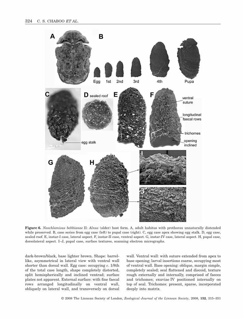
dark-brown/black, base lighter brown. Shape: barrel-like, asymmetrical in lateral view with ventral wallshorter than dorsal wall. Egg case: occupying c. 1/8thof the total case length, shape completely distorted,split hemispherically and inclined ventrad; surfaceplates not apparent. External surface: with fine faecalrows arranged longitudinally on ventral wall,obliquely on lateral wall, and transversely on dorsal
wall. Ventral wall: with suture extended from apex tobase opening; larval insertions coarse, occupying mostof ventral wall. Base opening: oblique, margin simple,completely sealed; seal flattened and discoid, texturerough externally and internally, comprised of faecesand trichomes; exuviae IV positioned internally ontop of seal. Trichomes: present, sparse, incorporateddeeply into matrix.
Figure 6. Neochlamisus bebbianae II: Alnus (alder) host form. A, adult habitus with prothorax unnaturally distendedwhile preserved. B, case series from egg case (left) to pupal case (right). C, egg case apex showing egg stalk. D, egg case,sealed roof. E, instar-I case, lateral aspect. F, instar-II case, ventral aspect. G, instar-IV case, lateral aspect. H, pupal case,dorsolateral aspect. I–J, pupal case, surface textures, scanning electron micrographs.
324 C. S. CHABOO ET AL.
© 2008 The Linnean Society of London, Zoological Journal of the Linnean Society, 2008, 152, 315–351

Neochlamisus bebbianae (Brown, 1943) III:Betula nigra host form (Figs 7, 8)
Egg case (Fig. 7B–F): Size (N = 10): L, 0.89–1.28 mm; W at roof, 0.79–0.84 mm. Colour: unevenlydark-brown/black. Shape: bell-like, symmetrical inlateral aspect; apex conical. Egg stalk: presentbasally, short, translucent, slender. Roof: transverse
(Fig. 7C) in lateral aspect, discoid, flattened, notconvex; margin simple, not flanged. Externalsurface: coarse with overlapping faecal plates(Fig. 7E, F); plates most apparent apically, theirmargins exposed apically; short, discontinuous lon-gitudinal ridges present, spanning two or threescales (Fig. 7D). Internal surface: smoother thanexternal surface; slight space between case wall and
Figure 7. Neochlamisus bebbianae III: Betula (river birch) host form. A, adult habitus. B, case series from egg case (left)to pupal case (right). C–F, egg case, scanning electron micrographs. C, lateral aspect. E–F, external surface.
FAECAL CASE ARCHITECTURE IN LEAF BEETLES 325
© 2008 The Linnean Society of London, Zoological Journal of the Linnean Society, 2008, 152, 315–351

egg. Trichomes: present, simple, dense, deeplyembedded in faecal matrix.
Larva-I case (Fig. 8A): Size (N = 4): L, 1.53–1.89 mm;W at base opening, 1.51–2.89 mm. Colour: unevenbrownish-black. Shape: asymmetrical in lateral view;egg case distinct, occupying two-thirds of the totalcase length; apex conical; egg stalk present or absent;base opening slightly transverse. Ventral surface:with suture extending subapically through egg caseto basal margin; faecal rows, inserted adjacent toventral suture, occupy triangular wedge; youngerrows of medium brown colour. External surface:rough, faecal rows orientated longitudinally ventrad,transversely lateral, and dorsad. Internal surface:without plates or ridges; texture smoother and darkerin larval section than egg section. Case walls: evenlythin, apex slightly thicker. Trichomes: present inlarval section, sparse.
Larva-II case (Fig. 8B, C): Size (N = 7): L, 3.84–4.02 mm; W at base opening, 1.65–1.87 mm. Colour:uneven dark brown, larval sections appearing striatebecause of colour tone variation. Shape: tubular;asymmetrical in lateral view; ventral, lateral, anddorsal walls distinguishable by varying orientation
of faecal rows; apex rounded. Egg case: apparent,shape distorted by ventral surface expansion. Exter-nal surface: coarse, with larval faeces in rows.Ventral surface: with suture extending subapically tobasal margin; adjacent triangular field occupyinghalf of the ventral surface area. Base opening:oblique; ventral wall shorter than dorsal wall;margin simple. Internal surface: smooth, boundarybetween larval instars indistinct. Walls: slightlythicker apically than laterally. Trichomes: at mediumdensity, some protuberant.
Larva-III case (Fig. 8D): Size (N = 10): L, 5.12–5.45 mm; W at base opening, 2.5–3.19 mm. Colour:unevenly brownish black with lighter brown stria-tions. Shape: tubular, asymmetrical in lateral viewwith dorsal wall slightly longer than ventral wall,and egg case inclined ventrad. Egg case: distinguish-able by position, surface texture, and colour; generalshape distorted, split hemispherically by ventralsuture and large ventral insertion of faeces. Ventralsurface: with ventral suture extending from apex tobase; adjacent longitudinal faecal rows slightly pro-tuberant, occupying three-quarters of the ventralsurface. Walls of larval sections: thicker than inegg case. Base opening: transverse, margin simple.
Figure 8. Neochlamisus bebbianae III: Betula host form. A, instar-I case, lateral aspect. B, instar-II case, lateral aspect.C, instar-II case, ventral aspect. D, instar-III case, ventral aspect. E, instar-IV case, lateral aspect. F, pupal case, ventralaspect.
326 C. S. CHABOO ET AL.
© 2008 The Linnean Society of London, Zoological Journal of the Linnean Society, 2008, 152, 315–351
Internal surface: smooth; plates, ridges, and rows,not apparent; larval sections not distinguishable.Trichomes: present, deeply embedded into matrix,with some protuberant.
Larva-IV case (Fig. 8E): Size (N = 4): L, 4.85–6.51 mm; W at base opening, 3.60–3.88 mm. Colour:uneven dark brownish black. Shape: barrel-like,asymmetrical with distinct ventral, lateral, anddorsal surfaces; egg case hemispherical, inclinedventrad, dissected by ventral suture; apex rounded.External surface: variable; densely covered with tri-chomes; trichome coat thick dorsally, thinner ven-trally. Ventral suture: extending from apex to base;larval faecal insertion occupying most of ventralsurface. Base opening: transverse, margin simple.Internal surface texture: smoothened; walls evenlythin, apex wall slightly thicker.
Pupal case (Fig. 8F, G): Size (N = 4): L, 6.27–7.54 mm; W at base opening, 5.25–6.02 mm. Shape:texture and colour as in late larval case IV; basal seallacking trichome coat.
Neochlamisus bebbianae (Brown, 1943) IV:Acer rubrum host form (Figs 9, 10)
Egg case (Fig. 9B–E): Size (N = 6): L, 1.68–1.87 mm;W at roof, 0.95–1.05 mm. Colour: unevenly brownish-black. Shape: bell-like; symmetrical in lateral aspect;apex rounded; egg stalk present, slender, translucent,one-third of the length of case, lacking coiling. Exter-nal surface: coarse, with pine cone-like appearance;faeces arranged as overlapping plates, dorsal marginof plates exposed, surface protuberant, producingpine cone-like appearance; plates at apex of caseproject vertically; longitudinal ridges absent. Roof:transverse, coarse, slightly concave; flange: narrow.Internal surface: smoother than external surface;plates and ridges not apparent. Walls: evenly narrow.Trichomes: present, simple, sparse, deeply embeddedinto faecal matrix.
Larva-I case (Fig. 9B, F–I): Size (N = 11): L, 1.47–1.79 mm; W at base opening, 0.94–0.99 mm. Colour:uneven brownish black, larval section grey-black.Shape: bell-like, symmetrical to asymmetricalaccording to age; egg case generally intact; egg stalkpresent or absent; base opening transverse. Ex-ternal surface: coarse, larval section with dark finetransverse faecal rows. Internal surface: lackingapparent plates and ridges (Fig. 9G). Walls: evenlynarrow (Fig. 9H, I). Trichomes: apparent, deeply
embedded into matrix, not protuberant, lackingfurry appearance.
Larva-II case (Figs 9B, 10A, B): Size (N = 8): L, 2.05–2.21 mm; W at base opening, 1.34–1.36 mm. Colour:uneven dark brownish-black, larval sections greyish-brown, younger sections grey. Shape: tubular,asymmetrical; ventral, lateral, and dorsal surfacesapparent; ventral wall shorter than dorsal wall; apexdome-like. Egg case: apparent; shape, position, andtexture generally intact; egg stalk absent. Ventralsurface: with ventral suture and triangular insertionextending subapically to margin; insertion protuber-ant, occupying half of the total ventral surface. Exter-nal surface: rough; larval sections with fine parallelrows of faecal deposit. Base opening: transverse,margin simple. Walls and apex: generally narrow,slightly thicker in larval section. Internal surfacetexture of larval section: smoother than externalsurface; egg section partially papered over with faecallayer. Trichomes: apparent, sparse, deeply embeddedinto matrix, decumbent.
Larva-III case (Figs 9B, 10C–F): Size (N = 4): L, 4.45–5.44 mm, W at base opening, 2.6–3.25 mm. Shape:tubular; ventral, lateral, and dorsal walls apparent.Egg case: apparent, position inclined ventrad, shapedistorted by ventral insertion of faecal field. Externalsurface: rough, faeces in fine rows (Fig. 10C, D);plates and ridges absent. Anterior surface with trans-verse faecal rows; lateral surface with obliquelytransverse faecal rows; posterior surface with longi-tudinal faecal rows and medial ventral suture;ventral suture extending from apex through egg caseto basal margin. Base opening: transverse, dorsalwall longer than ventral wall; margin simple. Inter-nal surface: smooth; rows, projections, and plates notapparent; walls evenly thick, broader than in eggcase. Trichomes: present, sparse, deeply embedded inmatrix, decumbent.
Larva-IV case (Fig. 9B): Size (N = 4): L, 6.49–6.81 mm; W at base opening, 4.40–4.64 mm. Colour:brownish-black. Shape: tubular, asymmetrical inlateral view, ventral surface somewhat flattened. Eggcase: apparent, distorted with ventral insertion offaeces, position inclined ventrad. External surface:rough, striated appearance with faecal rows orien-tated longitudinally on ventral surface, obliquelytransverse laterally and transverse dorsally. Baseopening: transverse, margin simple. Internal surface:smooth, walls evenly thick; rows, ridges, and platesnot apparent. Trichomes: present, deeply embedded infaecal matrix.
FAECAL CASE ARCHITECTURE IN LEAF BEETLES 327
© 2008 The Linnean Society of London, Zoological Journal of the Linnean Society, 2008, 152, 315–351
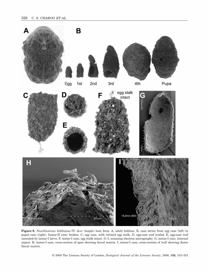
Figure 9. Neochlamisus bebbianae IV: Acer (maple) host form. A, adult habitus. B, case series from egg case (left) topupal case (right). Instar-II case: broken. C, egg case, with twisted egg stalk. D, egg-case roof sealed. E, egg-case roofunsealed by instar-I larva. F, instar-I case, egg stalk intact. G–I, scanning electron micrographs. G, instar-I case, internalaspect. H, instar-I case, cross-section of apex showing faecal matrix. I, instar-I case, cross-section of wall showing densefaecal matrix.
328 C. S. CHABOO ET AL.
© 2008 The Linnean Society of London, Zoological Journal of the Linnean Society, 2008, 152, 315–351

Pupal case (Figs 9B, 10H): Size (N = 9): L, 6.98–7.23 mm; W at base opening, 4.74–5.20 mm. Colour:brownish-black. Shape: bean-shaped, asymmetrical inlateral view; ventral, lateral, and dorsal walls appar-ent. Egg case: not easily discernible, split hemispheri-cally by ventral suture and faecal insertions, eachhemisphere shifted laterad and ventrad. Ventral wall:somewhat flattened, texture coarse with fine faecalrows; faecal rows orientated longitudinally, faecalinsertion occupying most of wall; ventral sutureextending medially from apex to base. Lateral walls:bulging medially, tapering centrad apically andbasally. Base: transverse, margin simple. Internalsurface: smooth; plates, rows, and ridges absent.
Trichomes: present, deeply embedded in faecalmatrix, not forming furry surface.
Neochlamisus bimaculatus Karren, 1972:host plant Rubus spp. (Fig. 11)
Egg case (Fig. 11B–E): Size (N = 10): L, 1.27–1.61; Wat roof, 0.97–1.18 mm. Colour: unevenly brownishblack. Shape: bell-like; symmetrical in lateral aspect;flange narrow. Egg stalk: present, short, not coiled.External surface: coarse; faeces arranged as flattenedplates; plates rounded to quadriform, some slightlyprotuberant medially, overlapping basally with apicalmargin exposed; longitudinal ridges absent. Roof:
Figure 10. Neochlamisus bebbianae IV: Acer (maple) host form. A, instar-II case, ventral aspect. B, instar-II case, lateralaspect, orientation of faecal rows, scanning electron micrograph. C, Instar-III case, lateral aspect. D, instar-III case, baseopening. E, instar-III case, ventral faecal wedge at base, scanning electron micrograph. F, instar-III case, internal surface,scanning electron micrograph. G, larval instar-IV, lateral aspect. H, Pupal case, lateral aspect.
FAECAL CASE ARCHITECTURE IN LEAF BEETLES 329
© 2008 The Linnean Society of London, Zoological Journal of the Linnean Society, 2008, 152, 315–351

transverse; surface shallowly concave and texturecoarse. Internal surface: smooth, scale outline some-times apparent. Trichomes: present, sparse, deeplyembedded in faecal matrix.
Larva-I case (Fig. 11B): Size (N = 8): L, 2.37–3.37 mm;W at base opening, 1.46–1.62 mm. Young cases: shape,colour, and texture the same as in egg case. Oldercases: asymmetrical in lateral view; dorsal, ventral,and lateral surfaces distinct; egg section occupyinghalf of the total case length; apex rounded; egg stalkpresent or absent. Larval section: with fine faecal rowsextending longitudinally on ventral surface and trans-versely on other surfaces. Ventral suture: slightly
protuberant, extending subapically to base. Baseopening: transverse, basal margin continuous withlateral walls; flange of egg case slightly discernible.Internal surface: smooth, internal surface of egg caseas original, larval surface lacking discernible patternsof plates, rows, or ridges. Wall thickness: evenlynarrow. Trichomes: present, very sparse.
Larva-II case (Fig. 11B, F): Size (N = 8): L, 3.47–5.06 mm; W at base opening, 1.7–2.40 mm. Colour:unevenly dark brownish-black. Shape: asymmetricalin lateral view; egg case apparent, inclined ventrad;apex rounded; attachment stalk absent. Externalsurface: coarse; dorsal surface with faeces in
Figure 11. Neochlamisus bimaculatus. A, adult habitus. B, case series from egg case (left) to pupal case (right). C–E, H–I,scanning electron micrographs. C, egg case, lateral aspect, broken medially. D, egg case, egg stalk. E, egg case, flange.F, instar-II case, lateral aspect. G–I, instar-IV case. G, ventral aspect. H, internal aspect with forewall removed. I, internalaspect, faecal layers of wall.
330 C. S. CHABOO ET AL.
© 2008 The Linnean Society of London, Zoological Journal of the Linnean Society, 2008, 152, 315–351

transverse rows; ventral surface with larval sectionroughly triangular, occupying half of the total surfacearea, and comprising longitudinal rows; ventralsuture extending subapically to base. Base opening:slightly transverse. Internal surface: smooth; rows,plates, or ridges absent. Walls: evenly thin. Tri-chomes: present, sparse, deeply embedded in faecalmatrix.
Larva-III case (Fig. 11B): Size (N = 9): L, 5.12–5.99 mm; W at base opening, 2.43–3.62 mm. Colour:uneven brownish-black, with striated appearance.Shape: asymmetrical; dorsal, lateral, and ventral sur-faces apparent; apex rounded, apical margin continu-ous with lateral walls; diameter widest in basal third.Egg case: apparent, shape distorted, split into twohemispheres by larval insertion; inclined ventrad,appearing as nipple-like dome. External surface:coarse; longitudinal ridges absent. Ventral surface:with larval insertion occupying more than half of thetotal surface area; ventral suture extending from apexto base, dissecting egg case. Base opening: transverse.Internal surface: smoothened. Wall: thickness,surface, and trichome density as in case II.
Larva-IV case (Fig. 11B, G–I): Size (N = 9): L, 7.27–7.78 mm; W at base opening, 4.96–5.12 mm. Shape,colour, surface texture, and trichome density and dis-tribution, as in case III. Older cases: barrel-shaped,being widest in basal third; egg case distorted,inclined ventrad; base opening transverse. Internalsurface (Fig. 11H): smoothened. Walls: with layers offaeces (Fig. 11I).
Pupal case (Fig. 11B): Size (N = 7): L, 7.98–785 mm;W at base, 4.96–5.12 mm. Shape, colour, texture, tri-chome pattern as in case IV; ventral wall shorter thandorsal wall.
Neochlamisus chamaedaphnes (Brown, 1943)(Figs 12, 13): host plant Chamaedaphnae calyculata
Egg case (Fig. 12B–D): Size (N = 18): L, 1.55–2.35 mm; W at roof, 0.84–1.24 mm. Colour: uneven,medium to dark brown. Shape: elongate bell-shape;symmetrical in lateral aspect; apex rounded; flangenarrow. Egg stalk: slender, translucent, broadly coiled.External surface: coarse, wall comprised of faecalplates; plates somewhat flattened, triangular toquadriform, overlapping basally, apical marginexposed; apical plates project vertically; plate surfaceslightly protuberant medially, projections formingoverall effect of slight, irregular longitudinal rows.Roof: surface rough, shallowly concave; flange narrow,thin, unevenly flared. Trichomes: present, sparse.
Internal surface: smooth; plate outline slightly dis-cernible; space between egg and wall narrow.
Larva-I case (Fig. 12E–G): Size (N = 9): L, 1.46–2.00 mm; W at base opening, 0.81–0.87 mm. Colour:unevenly dark brown, larval section darker. Shape:bell-like, similar to egg case; apex rounded; apicalfaecal plates projecting slightly; egg stalk absent.External surface of larval section: rough, faecesarranged as fine horizontal rows, not pellets. Baseopening: transverse; margin slightly flared. Ventralsurface: apparent in older cases; ventral suture ter-minating subapically. Internal surface: smooth; wallof larval section slightly thicker than wall of egg case.Trichomes: present, sparse.
Larva-II case (Fig. 13A–C): Size (N = 4): L, 2.34–2.91 mm; W at base opening, 1.39–1.72 mm. Shape:elongate tube, twice as long as base width; slightlyasymmetrical in lateral view with anterior marginslightly longer than posterior margin. Apex: rounded;egg stalk absent. Egg case: generally intact, distortedventrally by insertion of ventral suture and faecalwedge. Larval sections: darker brown than egg case;boundary between instar-I section and instar-IIsection marked by light-brown colour; surface texturescabrous. Ventral surface: with triangular faecalwedge occupying half the surface; surface slightlyprojecting, lateral margins with irregular projections.Ventral suture: terminating subapically. Baseopening: slightly oblique in lateral view; marginsimple, not flared. Internal surface: smooth, egg casesurface intact, larval sections with rows not apparent.Wall of larval section: slightly thicker than in eggcase.
Larva-III case: Size (N = 4): L, 3.84–4.09 mm; W atbase opening, 1.84–1.87 mm. Shape: elongate tube,widest medially and slightly narrowed basally;asymmetrical in lateral view with dorsal wall longerthan ventral wall; apex rounded. Egg case: splithemispherically by ventral suture, hemispheres con-nected apically; egg stalk absent. Larval section-IIIsurface texture: coarser and lighter brown than pre-vious sections. Base opening and margin: as in caseII. Internal surface: smooth, external pattern ofplates and rows not apparent; wall, evenly narrow.Trichomes: present, sparse, unevenly distributed,decumbent.
Larva-IV case (Fig. 13D): Size (N = 5): younger cases,L, 4.20–4.68 mm, W at base opening, 2.26–2.74 mm;older cases, L, 4.65–4.91 mm, W, 3.39 mm. Colour:light to dark brown, striated appearance, thickcoloured bands marking some instar sections. Shape:
FAECAL CASE ARCHITECTURE IN LEAF BEETLES 331
© 2008 The Linnean Society of London, Zoological Journal of the Linnean Society, 2008, 152, 315–351
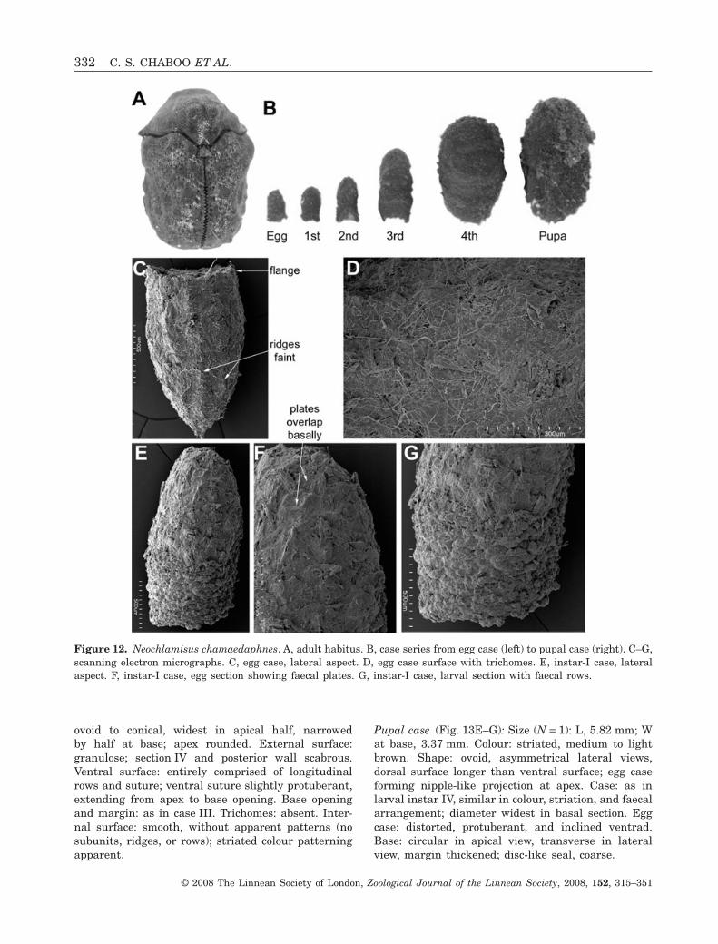
ovoid to conical, widest in apical half, narrowedby half at base; apex rounded. External surface:granulose; section IV and posterior wall scabrous.Ventral surface: entirely comprised of longitudinalrows and suture; ventral suture slightly protuberant,extending from apex to base opening. Base openingand margin: as in case III. Trichomes: absent. Inter-nal surface: smooth, without apparent patterns (nosubunits, ridges, or rows); striated colour patterningapparent.
Pupal case (Fig. 13E–G): Size (N = 1): L, 5.82 mm; Wat base, 3.37 mm. Colour: striated, medium to lightbrown. Shape: ovoid, asymmetrical lateral views,dorsal surface longer than ventral surface; egg caseforming nipple-like projection at apex. Case: as inlarval instar IV, similar in colour, striation, and faecalarrangement; diameter widest in basal section. Eggcase: distorted, protuberant, and inclined ventrad.Base: circular in apical view, transverse in lateralview, margin thickened; disc-like seal, coarse.
Figure 12. Neochlamisus chamaedaphnes. A, adult habitus. B, case series from egg case (left) to pupal case (right). C–G,scanning electron micrographs. C, egg case, lateral aspect. D, egg case surface with trichomes. E, instar-I case, lateralaspect. F, instar-I case, egg section showing faecal plates. G, instar-I case, larval section with faecal rows.
332 C. S. CHABOO ET AL.
© 2008 The Linnean Society of London, Zoological Journal of the Linnean Society, 2008, 152, 315–351
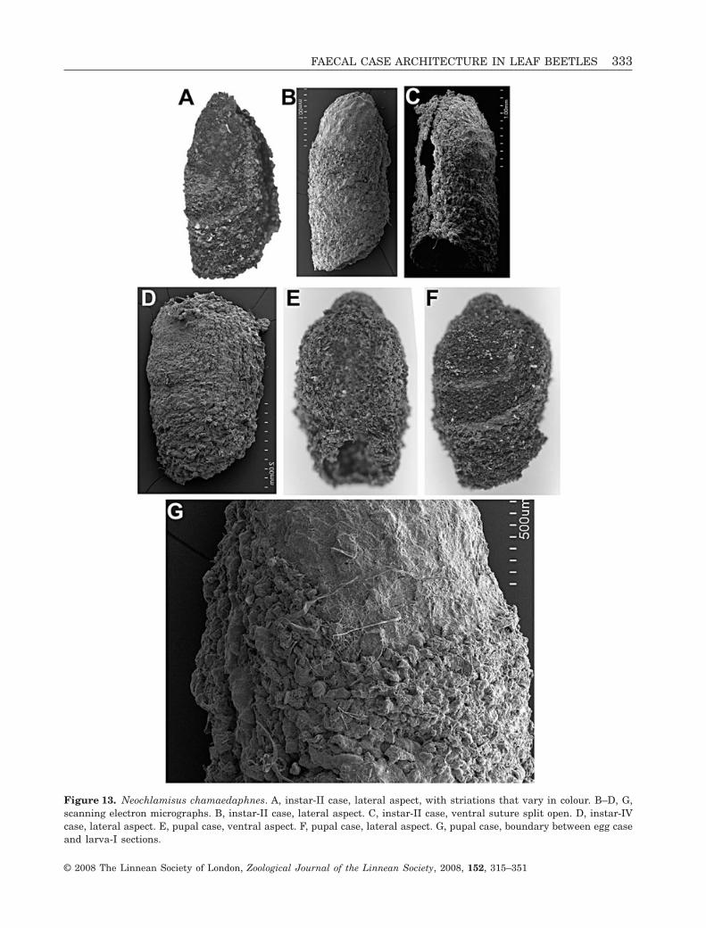
Figure 13. Neochlamisus chamaedaphnes. A, instar-II case, lateral aspect, with striations that vary in colour. B–D, G,scanning electron micrographs. B, instar-II case, lateral aspect. C, instar-II case, ventral suture split open. D, instar-IVcase, lateral aspect. E, pupal case, ventral aspect. F, pupal case, lateral aspect. G, pupal case, boundary between egg caseand larva-I sections.
FAECAL CASE ARCHITECTURE IN LEAF BEETLES 333
© 2008 The Linnean Society of London, Zoological Journal of the Linnean Society, 2008, 152, 315–351
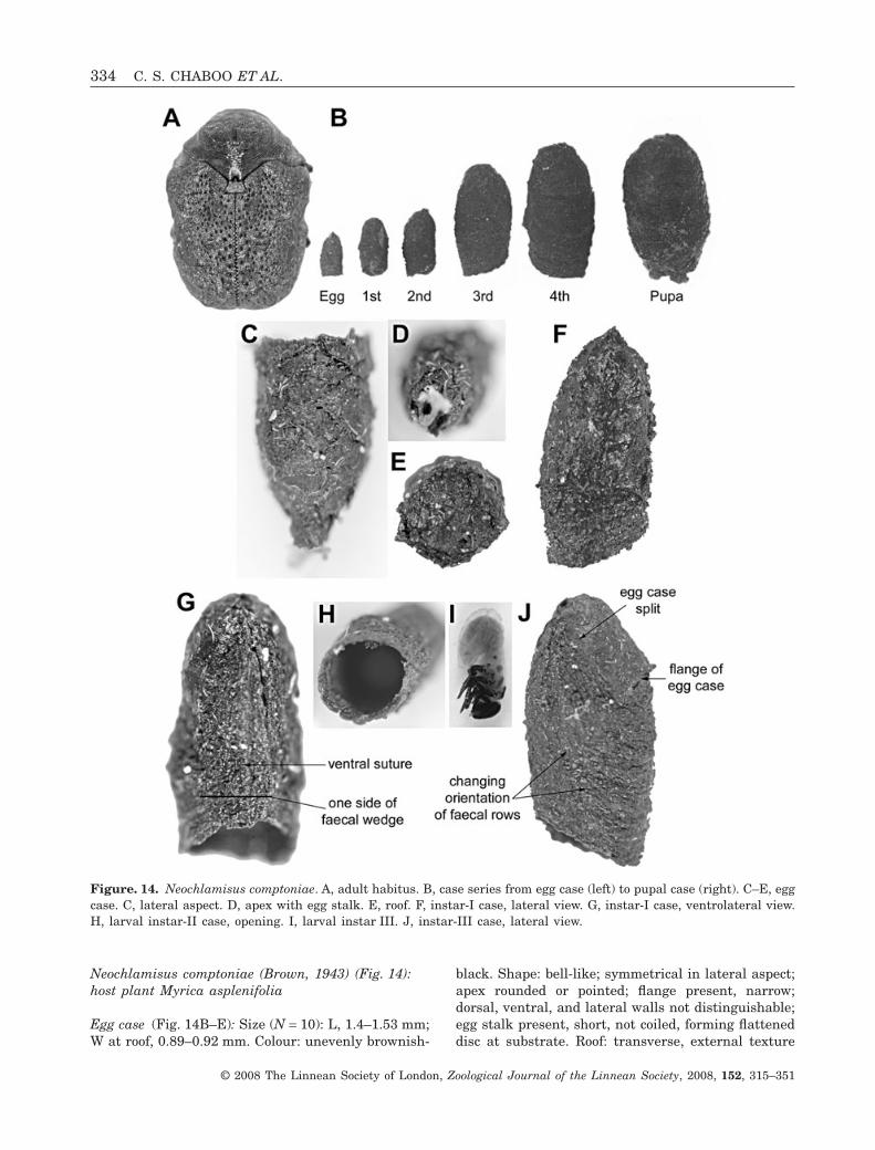
Neochlamisus comptoniae (Brown, 1943) (Fig. 14):host plant Myrica asplenifolia
Egg case (Fig. 14B–E): Size (N = 10): L, 1.4–1.53 mm;W at roof, 0.89–0.92 mm. Colour: unevenly brownish-
black. Shape: bell-like; symmetrical in lateral aspect;apex rounded or pointed; flange present, narrow;dorsal, ventral, and lateral walls not distinguishable;egg stalk present, short, not coiled, forming flatteneddisc at substrate. Roof: transverse, external texture
Figure. 14. Neochlamisus comptoniae. A, adult habitus. B, case series from egg case (left) to pupal case (right). C–E, eggcase. C, lateral aspect. D, apex with egg stalk. E, roof. F, instar-I case, lateral view. G, instar-I case, ventrolateral view.H, larval instar-II case, opening. I, larval instar III. J, instar-III case, lateral view.
334 C. S. CHABOO ET AL.
© 2008 The Linnean Society of London, Zoological Journal of the Linnean Society, 2008, 152, 315–351

coarse. External surface of walls: rough, faecesarranged in plates; plates rounded to quadriform,fitted together apically, overlapping with basalmargin exposed; longitudinal ridges very weaklydeveloped. Internal surface: smoothened; case wallsevenly thin, external plate pattern not distinct. Tri-chomes: not apparent.
Larva-I case (Fig. 14F, G): Size (N = 7): L, 0.99–1.35 mm; W at base opening, 1.65–2.46 mm. Youngphase of case with shape, colour, texture, and sym-metry as in egg case. Apex: rounded; lateral marginssubparallel; base opening transverse. Flange of eggcase: apparent. Larval case section: darker than eggcase, with fine faecal rows. Older case: with insertionof ventral suture and development of dorsal, lateral,and ventral walls; suture terminating subapically.Internal surface: as in egg case, larval section darkerand wall thicker. Trichomes: present, very sparse,deeply embedded in faecal matrix, evenly distributed.
Larva-II case (Fig. 14H, I): Size (N = 7): L, 2.54–2.57 mm; W at base opening, 1.39–1.50 mm. Colour:uneven, egg case dark brown; sections of instars I–IIdarker, boundary between I and II marked by finelight-brown line. Shape: tubular, asymmetrical inlateral aspect, margins subparallel. Apex: rounded;egg case split ventrally by ventral suture and faecalinsertion; flange of egg case apparent in some cases;egg stalk absent. External surface: coarse; larva I–IIsections with faeces arranged in fine rows. Ventralsurface: with protuberant triangular wedge, longitu-dinal faecal rows occupying half surface; ventralsuture slightly protuberant, terminating subapically.Base opening: transverse, margin simple. Internalsurface: rough, lacking any plates, ridges, or rowpatterns; colour and texture of egg case intact; larvalsection evenly darker; wall thickness evenly narrow.Trichomes: sparse, evenly distributed, and deeplyembedded in faecal matrix.
Larva-III case (Fig. 14B, J): Size (N = 10): L, 4.15–4.58 mm; W at base opening, 1.88–2.00 mm. Colour oflarval sections: dark-brown/black. Shape: tubular,asymmetrical in lateral aspect. Egg case: occupying1/8th of case length, split into two hemispheres,forming basal cap, position not markedly inclinedventrad. Ventral wall: with triangular wedge occupy-ing two-thirds of the surface; ventral suture termi-nating subapically; walls with faeces arranged inrows. Internal surface: smooth, external patterningnot apparent. Trichomes: apparent, sparse, evenlydistributed, deeply embedded into faecal matrix.
Larva-IV case (Fig. 14B): Size (N = 9): L, 5.17–5.18 mm; W at base opening, 2.86–3.00 mm. Shape:tubular, ovoid, widest in basal half. Egg case: distin-guishable by lighter brown coloration, granulartexture, and lack of colour striations; larval sectiondarker, blackish, striated in appearance; surfacetexture granular in early stages, coarser in laterphases; faecal rows unevenly ridged and coarse.Boundaries between subsequent larval phases distin-guishable by texture changes, indentations, andsometimes coloration; indentation especially apparenton anterior wall between the instar I–II boundary.Base opening: transverse in lateral aspect, ventralwall shorter than dorsal wall; basal margin simple.Ventral surface: with broad faecal insertion occupyingmost of wall; ventral suture protuberant, terminatingsubapically. Internal surface: smooth; plates, ridges,and rows not apparent; wall evenly narrow. Trichome:density and distribution as in younger cases.
Pupal case (Fig. 14B): Size (N = 12): L, 5.40–5.42 mm;W at base opening, 3.26–3.32 mm. Colour, texture,and trichomes: as in larval case IV. Shape: somewhatbarrel-shaped, apex and base inclined ventrad;ventral wall flattened. Egg case: split into two hemi-spheres, inclined ventrad. Base: obliquely transversein lateral aspect; coarse seal texture, margin some-times with irregular projections.
Neochlamisus cribripennis (LeConte, 1878)(Figs 15, 16): host plant Vaccinum spp.
Egg case (Fig. 15B–D): Size (N = 9): L, 1.48–1.56 mm;W at roof, 0.99–1.04 mm. Colour: unevenly darkbrown. Shape: bell-like, symmetrical in lateral aspect;apex arcuate; egg stalk present, short, not coiled;flange slight. External surface: with faeces arrangedin rounded plates; plates: protuberant medially,giving uneven surface texture, contiguous, not over-lapping; longitudinal ridges absent. Trichomes: notapparent.
Larva-I case (Fig. 15B): Size (N = 5): L, 1.57–1.73 mm;W at base opening, 1.08–1.36 mm. Colour: backgrounddark brown. Shape: conical, symmetrical in lateralview; apex arcuate; egg stalk present; flange of eggcase distinct. External surface: flattened and rough;egg case with faecal plates fitted together, not overlap-ping; section I with faeces in horizontal rows, not inplates. Faecal rows arranged horizontally on ventral,lateral, and dorsal surfaces; ventral suture apparent inolder cases. Base opening: slightly oblique; marginsimple, not flared or thickened. Trichomes: notapparent.
FAECAL CASE ARCHITECTURE IN LEAF BEETLES 335
© 2008 The Linnean Society of London, Zoological Journal of the Linnean Society, 2008, 152, 315–351

Larva-II case (Fig. 15B): Size (N = 5): L, 2.53–3.78 mm; W at base opening, 1.36–2.00 mm. Colour:unevenly dark brown; lighter brown marking bound-aries between egg case and section I, and betweensections I and II. Shape: generally conical, symmetri-cal in lateral view; apex rounded; egg stalk absent.Diameter of sections I and II similar, slightly widerthan diameter of egg case at apical margin; remnantsof flange of egg case apparent. External surfacetexture: scabrous with faecal rows. Ventral suture:present, terminating subapically, dissecting egg case.
Base opening: oblique, basal margin simple, notthickened. Trichomes: present, sparse, irregularlyarranged, deeply incorporated into wall matrix, someemergent and decumbent.
Larva-III case (Figs 15E–G, 16A): Size (N = 4): L,4.31–4.36 mm; W at base opening, 1.63–2.23 mm.Colour: uneven dark brown, boundaries between eggcase and section I, sections I–II and sections II–IIImarked by slightly lighter-brown coloration. Shape:conical, symmetrical in lateral view; apex rounded.
Figure 15. Neochlamisus cribipennis. A, adult habitus. B, case series from egg case (left) to pupal case (right); instar-IIIand pupal cases are broken, exposing stage inside. C, egg case, scanning electron micrograph. D, egg case, lateral wall,scanning electron micrograph. E–G, instar-III case. E, lateral aspect. F, ventral aspect. G, base opening.
336 C. S. CHABOO ET AL.
© 2008 The Linnean Society of London, Zoological Journal of the Linnean Society, 2008, 152, 315–351

External surface: rough. Ventral suture: extendingfrom apex to base, dissecting egg case. Base opening:oblique; margin simple, uneven, not flared or thick-ened. Trichomes: as in case II.
Larva-IV case (Figs 15B, 16C, D): Size (N = 3): L,4.31–4.86 mm; W at base opening, 2.61–2.8 mm.Colour: uneven dark brown; boundaries of sectionsdistinct. Shape: barrel-like, symmetrical in lateralview; apex rounded. External surface: rough. Ventralsuture: extended from apex to base, dissecting eggcase. Base opening and margin: as in case III. Tri-chomes: as in cases IIand III.
Pupal case (Figs 15B, 16B): Size (N = 1): L, 5.47 mm;W at base opening, 3.28 mm. Shape, colour, texture,and trichomes as in instar IV. Apex: somewhat flat-tened; egg case discernible, inclined ventrad. Baseopening: inclined at 45° to ventral wall, marginsflared.
Neochlamisus eubati (Brown, 1943) (Fig. 17):host plant Rubus spp.
Egg case (Fig. 17B, C): Size (N = 10): L, 1.62–1.76 mm; W at roof, 1.00–1.12 mm. Colour: dark
brown. Shape: bell-like, approximately twice as longas wide; symmetrical in lateral aspect; flange veryslight. Apex: rounded; egg stalk present, short,narrow, tan-coloured. Faeces shaped into flattenedtriangular plates; plates overlap basally, apicalmargins free; scale pattern indistinct near apex.External surface: uneven with inclusions of tri-chomes; longitudinal ridges not apparent; wall thick-ness very narrow; internal surface smooth. Baseopening: rounded; disc seal concave, texture uneven,rough. Trichomes: sparse, evenly distributed, anddeeply incorporated into faecal material, some tri-chomes occasionally protuberant.
Larva-I case (Fig. 17B): Size (N = 14): L, 1.58–2.29 mm; W at base opening, 0.94–1.02 mm. Colour oflarval addition: brownish-black. Shape, symmetry,texture, and colour of young cases: as in egg case.Older cases: double the length of egg case; ventralsuture terminating subapically; lateral margins sub-parallel, evenly tapered. Egg case: intact except forventral suture; egg stalk present or absent; flange ofegg case sometimes apparent. Base opening: trans-verse in lateral aspect. Internal surface: smooth,thicker in larval section, external patterns of platesand rows not apparent internally.
Figure 16. Neochlamisus cribipennis. A, larval instar III, live feeding on host plant leaf (photo by CGB). B, pupaattached to host leaf petiole (photo by CGB). C, instar-IV case, ventral suture split open. D, instar-IV case, wallcross-section showing trichome in faecal matrix, scanning electron micrograph.
FAECAL CASE ARCHITECTURE IN LEAF BEETLES 337
© 2008 The Linnean Society of London, Zoological Journal of the Linnean Society, 2008, 152, 315–351
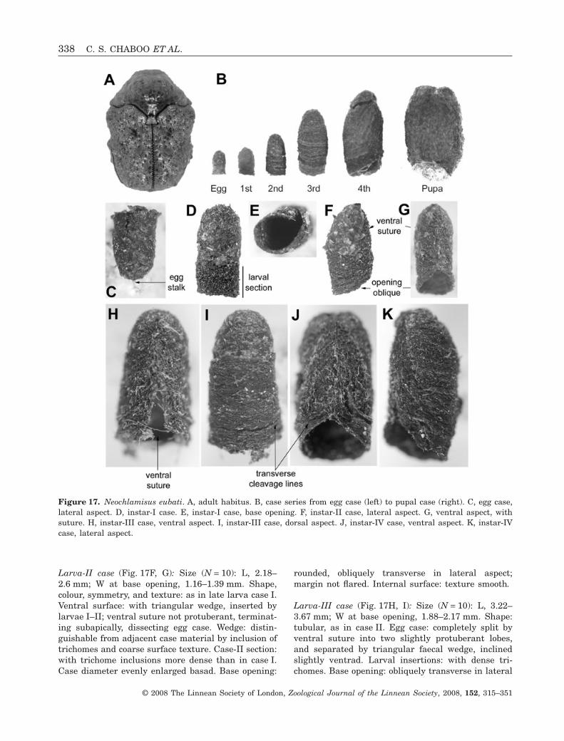
Larva-II case (Fig. 17F, G): Size (N = 10): L, 2.18–2.6 mm; W at base opening, 1.16–1.39 mm. Shape,colour, symmetry, and texture: as in late larva case I.Ventral surface: with triangular wedge, inserted bylarvae I–II; ventral suture not protuberant, terminat-ing subapically, dissecting egg case. Wedge: distin-guishable from adjacent case material by inclusion oftrichomes and coarse surface texture. Case-II section:with trichome inclusions more dense than in case I.Case diameter evenly enlarged basad. Base opening:
rounded, obliquely transverse in lateral aspect;margin not flared. Internal surface: texture smooth.
Larva-III case (Fig. 17H, I): Size (N = 10): L, 3.22–3.67 mm; W at base opening, 1.88–2.17 mm. Shape:tubular, as in case II. Egg case: completely split byventral suture into two slightly protuberant lobes,and separated by triangular faecal wedge, inclinedslightly ventrad. Larval insertions: with dense tri-chomes. Base opening: obliquely transverse in lateral
Figure 17. Neochlamisus eubati. A, adult habitus. B, case series from egg case (left) to pupal case (right). C, egg case,lateral aspect. D, instar-I case. E, instar-I case, base opening. F, instar-II case, lateral aspect. G, ventral aspect, withsuture. H, instar-III case, ventral aspect. I, instar-III case, dorsal aspect. J, instar-IV case, ventral aspect. K, instar-IVcase, lateral aspect.
338 C. S. CHABOO ET AL.
© 2008 The Linnean Society of London, Zoological Journal of the Linnean Society, 2008, 152, 315–351

view; lateral walls evenly tapered, widening basad.Internal surface: as in case II.
Larva-IV case (Fig. 17J, K): Size (N = 8): L, 5.37–6.52 mm; W at base opening, 2.80–3.89 mm. Colour,texture, and orientation of faecal rows: as in case III.Shape of young cases: tubular, as in case III. Oldercases: ovoid or barrel-shaped, widest near baseopening; egg case distorted, as in case III. Ventralwall: slightly protuberant near base. Base opening:strongly obliquely transverse in lateral view, marginsimple.
Pupal case (Fig. 17B): Size (N = 5): L, 5.76–6.82 mm;W at base, 4.13–4.67 mm. Shape: barrel-shaped,dorsal wall longer than ventral wall; apex somewhatflattened. Egg case: not easily discernible. Base:obliquely transverse; coarse seal texture. Internalsurface: smooth, evenly thickened, multi-layered,layers parchment-like; apex thick, comprising manylayers.
Neochlamisus platani (Brown, 1952) (Figs 18, 19):host plant Platanus occidentalis
Egg case (Fig. 18B–E): Size (N = 11): L, 1.32–1.56 mm; W at roof, 0.91–0.95 mm. Colour: unevenlydark brown. Shape: bell-like, symmetrical in lateralaspect; apex rounded; roof margin simple, flangeabsent; egg stalk present. Walls composed of faecalplates; plates faintly pentagonal, fitted togetherbasally, and overlapping apically, some projecting ver-tically at case apex; plate surface protuberant cen-trally. Wall with fine longitudinal ridges, extendingfrom apex to base, spanning multiple plates; ridgesevenly spaced and slightly radially arranged. Tri-chomes: present, filamentous type, medium density,evenly distributed, deeply embedded in matrix. Inter-nal surface: rough, external plates and ridges notapparent; wall evenly thin. Roof: transverse, sealunevenly flattened.
Larva-I case (Fig. 18G, H): Size (N = 9): L, 2.13–2.30 mm; W at base opening, 1.04–1.14 mm. Shape,colour, and symmetry of young case: as in egg case;larval section brownish-black; egg stalk sometimespresent. Older cases: conical, asymmetrical in lateralaspect; dorsal, ventral, and lateral surfaces apparent.Ventral suture: terminating subapically; triangularfaecal insertion not protuberant. Trichomes: present,stellate type, light to medium density. Internalsurface: smooth. Wall of larval section: slightlythicker than in egg case. Base opening: obliquelytransverse.
Larva-II case (Figs 18B, F, 19A): Size (N = 9): L, 3.94–4.27 mm; W at base opening, 1.46–1.72 mm. Colour:uneven brown-black. Shape: tubular; egg case shape,position, and texture intact; ventral suture presentventrally, terminating subapically; apex evenlyrounded; egg stalk lacking. Larval section with faecesas in instar I. Boundaries between egg case andlarval-I case well demarcated; boundary betweenlarval sections I and II obscured by trichomes. Inter-nal surface: smooth. Base opening: obliquely trans-verse, anterior wall slightly longer than ventral wall.Trichomes: more dense than in case I, obscuringsurface.
Larva-III case (Figs 18B, 19B): Size (N = 12): L, 4.97–5.08 mm; W at base opening, 2.38–2.85 mm. Shape,colour, texture, and trichome arrangement: as incase II. Egg case: apparent, position inclined ventrad.
Larva-IV case (Figs 18B, 19G, H): Size (N = 8): L,7.26–8.86 mm; W at base opening, 3.95–4.46 mm.Shape: tubular in young phase; barrel-like in olderstage, widest in apical section. Asymmetry, colour,and trichome pattern: as in case III. Egg case: some-what distorted, split into two hemispheres, inclinedventrad, diagnosable by lack of stellate trichomes.
Pupal case (Figs 18B, 19I, J): Size (N = 15): L, 7.33–8.75 mm; W at base, 4.24–4.42 mm. Shape, colour,asymmetry, and trichome type and density: as in latecase IV. Egg case: inclined ventrad, protuberant, over-hanging ventral suture. Base: rough seal surface;trichomes present, medium density, incorporated intofaecal matrix, not forming fuzz.
DISCUSSION
In this study of Neochlamisus cases, we found thatthe underlying patterns in form and ontogeny indi-cate highly repetitive construction behaviour. Theconservative nature of the architecture and attach-ment of the egg case suggests a stereotypical behav-iour in case construction. The patterns of architecturedescribed here are consistent with the tempo andmode of case construction presented by Brown &Funk (2005), and these patterns may hold generallyfor most camptosomates (Cryptocephalinae and Lam-prosomatinae). Interestingly, the Neochlamisus casesshare certain traits with caddisfly cases (Trichoptera)(e.g. Hansell, 1974), for example, wall asymmetry, thepupal seal, and smaller subunit composition. Perhapsthat better-studied system can provide models forfuture research on camptosomate cases.
The similarity of the basic architecture acrossNeochlamisus cases suggests that construction doesnot vary in significant ways. In fact, case growth does
FAECAL CASE ARCHITECTURE IN LEAF BEETLES 339
© 2008 The Linnean Society of London, Zoological Journal of the Linnean Society, 2008, 152, 315–351

seem to follow a logarithmic growth pattern (A.E.Aldridge, pers. comm.). However, unlike other naturalgnomons (e.g. claws, sunflowers, and seashells) thatgrow in mathematically predictable self-repeating
patterns, the chlamisine case shape is more ontoge-netically dynamic, and several different shapes maybe exhibited within a single life history (see Aldridge,1999). The addition of material to the anterior
Figure 18. Neochlamisus platani. A, adult habitus, with prothorax distended as a result of preservation. B, case seriesfrom egg case (left) to pupal case (right). C–H, scanning electron micrographs. C, egg case, lateral aspect. D, egg stalk.E, egg case surface. F, instar-II case, showing external texture variation between maternal and larval sections. G, instar-Icase, forewall removed, internal aspect. H, instar-I case, exterior trichome coat of larval section.
340 C. S. CHABOO ET AL.
© 2008 The Linnean Society of London, Zoological Journal of the Linnean Society, 2008, 152, 315–351

Figure 19. Neochlamisus platani. A, instar-II case, dorsal aspect. B, instar-III case, lateral aspect. C, instar-IV case,lateral aspect. D, larva, instar IV, lateral aspect. E–J, Scanning electron micrographs. E, instar-III case, forewall removed,internal aspect. F, instar-III case, forewall removed, internal surface with trichome. G, instar-IV case, external surfacewith stellate trichomes. H, instar-IV case, cross-section of wall showing trichome–faecal matrix. I, pupal case, cross-section of wall. J, Pupal case, cross-section near apex.
FAECAL CASE ARCHITECTURE IN LEAF BEETLES 341
© 2008 The Linnean Society of London, Zoological Journal of the Linnean Society, 2008, 152, 315–351

dorsum and the ventral seam in different orientationsand at different rates (see Brown & Funk, 2005)contributes to these patterns. The case is initiallymaintained as a tube by larval additions only to theapical opening. It is then widened and turned into abarrel-shape by also adding material to the ventralsuture. Case growth then follows an interesting tra-jectory, with the dorsal wall curving such that thecase is held ever more closely to the substrate, whilethe case is expanded by increased faecal additions tothe ventral suture.
MODEL DESCRIPTION AND TERMINOLOGY FOR
CASE ARCHITECTURE
The case can be described by the following characters:length-to-width ratio, symmetry, apex shape (roundedor arcuate), colour (striated or banded; black-brown tolight brown), regions (apex, base, or opening), wall(internal versus external), and inclusions (e.g. tri-chomes, soil, etc.). Ultimately, our description of thestates of these characters should facilitate phyloge-netic analyses that will elucidate the evolution ofthese unusual examples of insect architecture. Below,we discuss the nature and hypothetical functions ofvarious case stages and characters that were thesubject of our study.
Egg stalk: Neochlamisus eggs are attached to thehost plant via an initially pliable string of unknownsubstance, known as the ‘egg stalk’. The substance ofthe stalk and morphological site of its production hasnot been determined. It is probably a glandularproduct in all species (Hilker, 1994). The stalk mayprovide leverage that helps the female pull the eggfrom her abdomen (Karren, 1964), or serve to elevatethe egg from the substrate away from predators. Inlarval cases, the stalk may be chewed away, abraded,or obscured. The egg stalk is not present in all camp-tosomates, and its presence/absence may reflectthe differing requirements of strictly phytophagousspecies versus the omnivorous, detritivorous, andmyrmecophilic taxa that generally lack them.
Plates: The egg cases of all Neochlamisus speciesappear to be comprised of smaller subunits. Scudder(1891) first described the egg case surface appearanceas resembling ‘papier mâché’, an effect arising fromthe layers of individual plates. Plates have also beentermed ‘scales’ (Karren, 1964; Lawson, 1976) or‘platelets’ (Lawson, 1976; Neal, 1989; LeSage &Stiefel, 1996). Maternal faeces are flattened andshaped as discrete plates, which form the basic build-ing blocks. A new row of plates is added when thepreceding one is complete, yet in the completed eggcase there is no indication of initiation or termination
points. Across the taxa examined, plates are similarlysized, but vary in being triangular, quadrate, or pen-tagonal in shape, and have adjoined or overlappingarrangements. The use of plates as basic buildingunits is common in cryptocephaline cases, but this isnot universal.
Egg case ridges: Often, a longitudinal ridge existsalong the median of each plate. In Neochlamisus eggcases, these ridges were generally fine, and did notproject greatly from the surface. They extended fromthe roof margin across multiple plates, but termi-nated subapically, never extending fully to the attach-ment stalk. Plate convexity, shape, and externallongitudinal ridges may reflect the individual pecu-liarities in the female rectal morphology of eachspecies (see Erber, 1988). For example, they may beformed by a depression in the rectal sclerites used bythe mother to construct the plates (see Erber, 1988).
Flange: Karren (1964) coined this term for the flaredmargin of the roof of the egg case. The flange, alsocalled ‘shoulder’ in Pachybrachis bivitattus (Lawson,1976), is characteristic to the overall bell shape con-sistently observed in Neochlamisus egg cases. Thisshape has also been described as a ‘bell-flower’(Karren, 1964) or ‘flask-shape’ (Packard, 1869, 1889),and appears to be common throughout Cryptocepha-linae (Erber, 1968, 1988). It is very broad and curvingin cases of Fulcidax (Flinte et al., 2003). The flangemay be an inadvertent consequence of the female’sanatomy pressing down on the longitudinallyextended case wall when she seals the roof of the eggcase. No specific function has been suggested.
Anterior additions: Case surface texture variesgreatly between the egg section and the larval sectionas a result of differences in faecal arrangement, build-ing units (plates versus rows), and trichome density.Unlike the maternal production of plates, larvae addrows of moistened faecal material to the case. Thelarva starts applying faeces horizontally along theedge of the flared margin of the case. In all casescompared, the larval sections comprised fine faecalrows arranged radially, but with changing orienta-tion. This is similar to patterns described forPachybrachis (LeSage, 1985) and Anomoea (LeSage &Stiefel, 1996) cases, and suggests a taxonomicallywidespread approach to larval faecal application. Bythe end of stadium I, the larva has applied numeroushorizontal rows and the bell-shaped egg case has beentransformed into a tube.
Ventral suture: This is common to all instars ofNeochlamisus and provides a useful descriptive land-mark. This cleavage line is initiated during instar I,
342 C. S. CHABOO ET AL.
© 2008 The Linnean Society of London, Zoological Journal of the Linnean Society, 2008, 152, 315–351
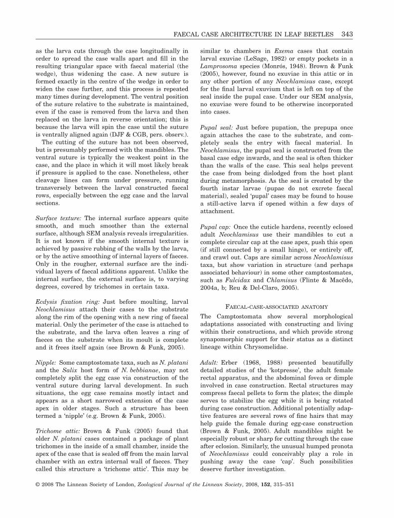
as the larva cuts through the case longitudinally inorder to spread the case walls apart and fill in theresulting triangular space with faecal material (thewedge), thus widening the case. A new suture isformed exactly in the centre of the wedge in order towiden the case further, and this process is repeatedmany times during development. The ventral positionof the suture relative to the substrate is maintained,even if the case is removed from the larva and thenreplaced on the larva in reverse orientation; this isbecause the larva will spin the case until the sutureis ventrally aligned again (DJF & CGB, pers. observ.).
The cutting of the suture has not been observed,but is presumably performed with the mandibles. Theventral suture is typically the weakest point in thecase, and the place in which it will most likely breakif pressure is applied to the case. Nonetheless, othercleavage lines can form under pressure, runningtransversely between the larval constructed faecalrows, especially between the egg case and the larvalsections.
Surface texture: The internal surface appears quitesmooth, and much smoother than the externalsurface, although SEM analysis reveals irregularities.It is not known if the smooth internal texture isachieved by passive rubbing of the walls by the larva,or by the active smoothing of internal layers of faeces.Only in the rougher, external surface are the indi-vidual layers of faecal additions apparent. Unlike theinternal surface, the external surface is, to varyingdegrees, covered by trichomes in certain taxa.
Ecdysis fixation ring: Just before moulting, larvalNeochlamisus attach their cases to the substratealong the rim of the opening with a new ring of faecalmaterial. Only the perimeter of the case is attached tothe substrate, and the larva often leaves a ring offaeces on the substrate when its moult is completeand it frees itself again (see Brown & Funk, 2005).
Nipple: Some camptostomate taxa, such as N. plataniand the Salix host form of N. bebbianae, may notcompletely split the egg case via construction of theventral suture during larval development. In suchsituations, the egg case remains mostly intact andappears as a short narrowed extension of the caseapex in older stages. Such a structure has beentermed a ‘nipple’ (e.g. Brown & Funk, 2005).
Trichome attic: Brown & Funk (2005) found thatolder N. platani cases contained a package of planttrichomes in the inside of a small chamber, inside theapex of the case that is sealed off from the main larvalchamber with an extra internal wall of faeces. Theycalled this structure a ‘trichome attic’. This may be
similar to chambers in Exema cases that containlarval exuviae (LeSage, 1982) or empty pockets in aLamprosoma species (Monrós, 1948). Brown & Funk(2005), however, found no exuviae in this attic or inany other portion of any Neochlamisus case, exceptfor the final larval exuvium that is left on top of theseal inside the pupal case. Under our SEM analysis,no exuviae were found to be otherwise incorporatedinto cases.
Pupal seal: Just before pupation, the prepupa onceagain attaches the case to the substrate, and com-pletely seals the entry with faecal material. InNeochlamisus, the pupal seal is constructed from thebasal case edge inwards, and the seal is often thickerthan the walls of the case. This seal helps preventthe case from being dislodged from the host plantduring metamorphosis. As the seal is created by thefourth instar larvae (pupae do not excrete faecalmaterial), sealed ‘pupal’ cases may be found to housea still-active larva if opened within a few days ofattachment.
Pupal cap: Once the cuticle hardens, recently eclosedadult Neochlamisus use their mandibles to cut acomplete circular cap at the case apex, push this open(if still connected by a small hinge), or entirely off,and crawl out. Caps are similar across Neochlamisustaxa, but show variation in structure (and perhapsassociated behaviour) in some other camptostomates,such as Fulcidax and Chlamisus (Flinte & Macêdo,2004a, b; Reu & Del-Claro, 2005).
FAECAL-CASE-ASSOCIATED ANATOMY
The Camptostomata show several morphologicaladaptations associated with constructing and livingwithin their constructions, and which provide strongsynapomorphic support for their status as a distinctlineage within Chrysomelidae.
Adult: Erber (1968, 1988) presented beautifullydetailed studies of the ‘kotpresse’, the adult femalerectal apparatus, and the abdominal fovea or dimpleinvolved in case construction. Rectal structures maycompress faecal pellets to form the plates; the dimpleserves to stabilize the egg while it is being rotatedduring case construction. Additional potentially adap-tive features are several rows of fine hairs that mayhelp guide the female during egg-case construction(Brown & Funk, 2005). Adult mandibles might beespecially robust or sharp for cutting through the caseafter eclosion. Similarly, the unusual humped pronotaof Neochlamisus could conceivably play a role inpushing away the case ‘cap’. Such possibilitiesdeserve further investigation.
FAECAL CASE ARCHITECTURE IN LEAF BEETLES 343
© 2008 The Linnean Society of London, Zoological Journal of the Linnean Society, 2008, 152, 315–351
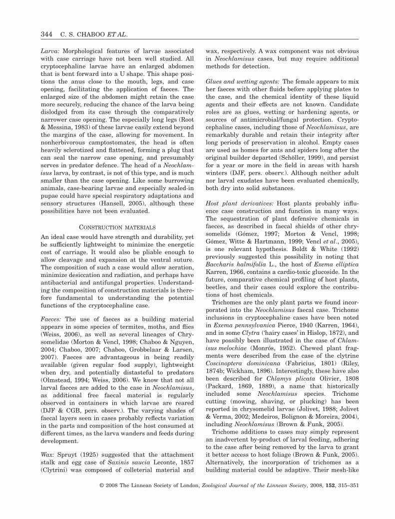
Larva: Morphological features of larvae associatedwith case carriage have not been well studied. Allcryptocephaline larvae have an enlarged abdomenthat is bent forward into a U shape. This shape posi-tions the anus close to the mouth, legs, and caseopening, facilitating the application of faeces. Theenlarged size of the abdomen might retain the casemore securely, reducing the chance of the larva beingdislodged from its case through the comparativelynarrower case opening. The especially long legs (Root& Messina, 1983) of these larvae easily extend beyondthe margins of the case, allowing for movement. Innonherbivorous camptostomates, the head is oftenheavily sclerotized and flattened, forming a plug thatcan seal the narrow case opening, and presumablyserves in predator defence. The head of a Neochlam-isus larva, by contrast, is not of this type, and is muchsmaller than the case opening. Like some burrowinganimals, case-bearing larvae and especially sealed-inpupae could have special respiratory adaptations andsensory structures (Hansell, 2005), although thesepossibilities have not been evaluated.
CONSTRUCTION MATERIALS
An ideal case would have strength and durability, yetbe sufficiently lightweight to minimize the energeticcost of carriage. It would also be pliable enough toallow cleavage and expansion at the ventral suture.The composition of such a case would allow aeration,minimize desiccation and radiation, and perhaps haveantibacterial and antifungal properties. Understand-ing the composition of construction materials is there-fore fundamental to understanding the potentialfunctions of the cryptocephaline case.
Faeces: The use of faeces as a building materialappears in some species of termites, moths, and flies(Weiss, 2006), as well as several lineages of Chry-somelidae (Morton & Vencl, 1998; Chaboo & Nguyen,2004; Chaboo, 2007; Chaboo, Grobbelaar & Larsen,2007). Faeces are advantageous in being readilyavailable (given regular food supply), lightweightwhen dry, and potentially distasteful to predators(Olmstead, 1994; Weiss, 2006). We know that not alllarval faeces are added to the case in Neochlamisus,as additional free faecal material is regularlyobserved in containers in which larvae are reared(DJF & CGB, pers. observ.). The varying shades offaecal layers seen in cases probably reflects variationin the parts and composition of the host consumed atdifferent times, as the larva wanders and feeds duringdevelopment.
Wax: Spruyt (1925) suggested that the attachmentstalk and egg case of Saxinis saucia Leconte, 1857(Clytrini) was composed of colleterial material and
wax, respectively. A wax component was not obviousin Neochlamisus cases, but may require additionalmethods for detection.
Glues and wetting agents: The female appears to mixher faeces with other fluids before applying plates tothe case, and the chemical identity of these liquidagents and their effects are not known. Candidateroles are as glues, wetting or hardening agents, orsources of antimicrobial/fungal protection. Crypto-cephaline cases, including those of Neochlamisus, areremarkably durable and retain their integrity afterlong periods of preservation in alcohol. Empty casesare used as homes for ants and spiders long after theoriginal builder departed (Schöller, 1999), and persistfor a year or more in the field in areas with harshwinters (DJF, pers. observ.). Although neither adultnor larval exudates have been evaluated chemically,both dry into solid substances.
Host plant derivatives: Host plants probably influ-ence case construction and function in many ways.The sequestration of plant defensive chemicals infaeces, as described in faecal shields of other chry-somelids (Gómez, 1997; Morton & Vencl, 1998;Gómez, Witte & Hartmann, 1999; Vencl et al., 2005),is one relevant hypothesis. Boldt & White (1992)previously suggested this possibility in noting thatBaccharis halmifolia L., the host of Exema ellipticaKarren, 1966, contains a cardio-toxic glucoside. In thefuture, comparative chemical profiling of host plants,beetles, and their cases could explore the contribu-tions of host chemicals.
Trichomes are the only plant parts we found incor-porated into the Neochlamisus faecal case. Trichomeinclusions in cryptocephaline cases have been notedin Exema pennsylvanica Pierce, 1940 (Karren, 1964),and in some Clytra (‘hairy cases’ in Hislop, 1872), andhave possibly been illustrated in the case of Chlam-isus melochiae (Monrós, 1952). Chewed plant frag-ments were described from the case of the clytrineCoscinoptera dominicana (Fabricius, 1801) (Riley,1874b; Wickham, 1896). Interestingly, these have alsobeen described for Chlamys plicata Olivier, 1808(Packard, 1869, 1889), a name that historicallyincluded some Neochlamisus species. Trichomecutting (mowing, shaving, or plucking) has beenreported in chrysomelid larvae (Jolivet, 1988; Jolivet& Verma, 2002; Medeiros, Bolignon & Moreira, 2004),including Neochlamisus (Brown & Funk, 2005).
Trichome additions to cases may simply representan inadvertent by-product of larval feeding, adheringto the case after being removed by the larva to grantit better access to host foliage (Brown & Funk, 2005).Alternatively, the incorporation of trichomes as abuilding material could be adaptive. Their mesh-like
344 C. S. CHABOO ET AL.
© 2008 The Linnean Society of London, Zoological Journal of the Linnean Society, 2008, 152, 315–351

arrangement within the faecal matrix and externallymay increase case durability. Smith (1922) indicatedthat plant fibers, along with spider silk, are used tohold trash packets together in chrysopid larvae. Asimilar structural role may be conceived for planttrichomes in Neochlamisus cases, providing anetwork or mesh that holds wet faeces together untildried into a sturdy case. Additionally, toxic or stickyglandular exudates from trichomes could enhancepredator deterrence or increase handling time.Trichome-covered cases may also more closelyresemble plant structures such as buds, thus increas-ing cryptic concealment from potential predators(Hansell, 1996). Or they might provide insulation,moderating temperature and humidity within cases.The potential benefits of trichome inclusions versustheir possible disadvantages (e.g. increasing caseweight; time and energy spent in active incorporation)require further study.
LIFE HISTORY AND TAXONOMIC VARIATION
Instar variation within Neochlamisus species: As theindividual grows, the shape of the case changes com-pletely from an initial bell-shaped maternal case, to aconical case in early instars, and finally to theincreasingly ovoid shape of the late fourth instar andpupa. Early instar cases are cylindrical to conical,with a fairly longitudinally consistent diameter,except at the narrowed apex, and with the basalmargin slightly angular in lateral view. In lateinstar IV and in the pupal case, the case is widestmedially, acquiring a barrel-shaped or ovoid formsimilar to that noted for E. canadensis (LeSage,1982). This pattern may anticipate the space neededfor the broadly block-shaped adult (Brown & Funk,2005). Cases of late-instar larvae also tended toexhibit the highest density of trichomes.
The case widens and lengthens in each subsequentlarval instar. In later instars, the egg case has usuallybeen completely, or mostly, dissected by the ventralsuture into two hemispherical portions, which haveby then become incorporated into the rounded caseapex. The progressive shift in the position of the eggcase and its eventual ventral inclination appears inother published illustrations of cryptocephaline cases(Dugès, 1881; Hinton, 1981; Erber, 1988). In allstudied taxa, the angle of carriage of the case, relativeto the substrate, declines from close to 90° in the firstinstar larvae to approximately 45° in pupae. This is aresult of the uneven deposition of faeces along the rimof the opening, in a manner that lengthens the dorsalwall at a greater rate than the ventral wall. It is notobvious what, if any, function this change in caseangle serves, although it may be promoted by theconstraints of carrying an increasingly heavy case.
The differential shape changes (viewed in lateralaspect) observed between the dorsal and ventral wallssuggest asymmetrical growth. In the ventral wall, thepattern of incorporation of faeces results in a largetriangular field of longitudinal rows. This field issomewhat flattened in some taxa, so that the ventralcase margin is in one plane in lateral view. Thisflattened surface may ease carriage over the sub-strate or represent a means of better balancing theincreased case weight. By contrast, the dorsal wallremains rounded and hemispherical.
Variation among host forms of N. bebbianae: The eggcases of all four N. bebbianae host forms studied herehave overlapping plates and simple trichomes. Differ-ences in elongation and width were minute, and thedegree of flaring of the basal margins varied slightlyamong individuals. Egg cases of the Alnus host formvaried in the depth of the convex roof. The Betula hostform had a flattened roof and simple margin, whereasother forms had a concave roof and a slightly flaredmargin. The Betula host form also had external ridgesthat varied in length, spanning the entire case orterminating subapically, whereas the Salix host formshad more developed longitudinal ridges. The Salixhost form case had the lightest brown coloration (andsometimes appears reddish in nature; DJF, pers.observ.), in contrast to the blackish colour of variousother host forms. The Acer host form egg cases hadfaecal plates that were slightly protuberant medially,producing a resemblance to pinecones.
The larval case of the Salix host form differedsomewhat from those of the other three host forms,which were quite similar to each other. Salix host formcases had more trichome inclusions and were moreelongate. Cases of the Betula host form showed thewidest variation in trichome density, ranging fromnaked to densely pubescent. Instar IV of the Salix hostform tended to have trichomes arranged in a verydense row, forming an incomplete collar at the base inlarval sections III–IV. Within an instar, considerablevariation in case size was observed. Trichome densityvaried most, from a complete lack of trichomes tocomplete coverage with trichomes for case IV.
Pupae in all forms of N. bebbianae are somewhatsimilar: flattened ventrally, with the opening deeplyoblique in lateral aspect. Pupae in the Salix andcertain individuals of the Acer host forms exhibited aslightly protuberant apical nipple.
Interspecific variation within Neochlamisus: All studytaxa had generally similar egg cases, with some excep-tions: the egg case of N. platani was notably moreelongate, with the length almost twice the width.Individual faecal plates were less discernible in N. bi-maculatus, and appeared to be absent in N. eubati. In
FAECAL CASE ARCHITECTURE IN LEAF BEETLES 345
© 2008 The Linnean Society of London, Zoological Journal of the Linnean Society, 2008, 152, 315–351

N. cribripennis, plates overlapped by half their length.The shapes of individual plates ranged from flattened,as in N. cribripennis, to protuberant, as describedabove for the Acer host form of N. bebbianae.
Although the vertical ridges on the plates of sometaxa were laid parallel to the longitudinal axis of theegg case, in others they were orientated diagonally soas to appear to spiral around the case from the apex tothe roof. The egg cases of N. cribripennis showed thewidest variation in length, some being at least a thirdlonger than others. Trichomes were incorporated intothe faecal wall of all egg cases, but were most dense inthat of the willow host form of N. bebbianae. Theflange on the edge of the case roof also varied acrossNeochlamisus taxa. In N. eubati, the edge curvedoutwards yielding a very flared flange. In N. platani,by contrast, the flange was completely vertical, so thatthe roof of the case was recessed by comparison. Thegreatest degree of interspecific variation in theobserved Neochlamisus larval cases was in colorationand trichome incorporation and density. Larval casesof all species included trichomes in the case-wallmatrix, but N. platani and the Salix host form ofN. bebbianae had the most externally pubescent pupalcases, with the Betula and Acer host forms also includ-ing some highly pubescent individuals.
Among the taxa we studied, the pupal cases ofN. bimaculatus and N. platani were the largest,whereas those of N. comptoniae were the smallest.Neochlamisus chamaedaphnes had the most obviouscolour variations, varying from light brown to black-ish within and between cases. Neochlamisus bimacu-latus appears to have the most barrel-shaped form,and has a somewhat flattened apex. The shape of thecase in N. chamaedaphnes is peculiar, being widestapically, rather than medially as in other taxa.
Generic variation in cases of Chlamisini: Insightsfrom the comparison of Neochlamisus cases with pub-lished descriptions and illustrations of cases fromother camptostomates falls short of those allowed bycomparing actual specimens. Nonetheless, these com-parisons are useful for initiating discussion on genericand tribal-level variations in case architecture.
The cases of Neochlamisus appear to be quitesimilar in structure and form to those of Exema, butdiffer from particular species of two other genera ofChlamisini that have been studied, i.e. Fulcidax(Flinte & Macêdo 2004b) and Chlamisus (Reu &Del-Claro, 2005). Fulcidax monstrosa differs fromthese other taxa in producing egg cases that are notcomposed of individual plates, but instead cover eggsin ‘a long strip of accumulated feces’ (Flinte &Macêdo, 2004b: 460). The ventral suture receives nomention in the description of cases of some otherchlamisines (Erber, 1988; Flinte & Macêdo, 2004b;
Reu & Del-Claro, 2005). Rather, C. minax is said toadd new layers in a ring around the opening for mostof the larval stadia (Reu & Del-Claro, 2005), whereasFulcidax constructs two anterior lobes as part of itscase (Erber, 1988).
The final position of material from the original eggcase within the pupal case varies among chlamisinegenera. As described here, in Neochlamisus taxa, theegg case is split, and tends to become gradually flat-tened by faecal insertion and shifting, with its outlinebecoming less clear (Brown & Funk, 2005). Monrós(1951) illustrated a very tapered case for Chlamisusmelochiae Monrós, 1952, in which the maternal sectionis distinguishable only by texture and position. On theother hand, the egg case remains distinctive in a fewNeochlamisus taxa, as described above, and also insome Exema cases (LeSage, 1982). The egg case inC. minax is obvious as a round ball atop the larvalcase, and is lost before pupation (Reu & Del-Claro,2005). Flinte & Macêdo (2004a) show that the egg caseremains intact until pupation in F. monstrosa. Ellis &LeRoux (1964) were the first to point out variationsbetween N. cribripennis and E. canadensis in theshape and protrusion of the egg case section of thepupal case. This variation could be explored further fordiagnostic and phylogenetic utility.
Brown & Funk (2005) found exuvial skins inside thepupal case. We also found the last larval exuviae insome pupal cases, where it was crumpled and sittingon the inside wall of the pupal case seal. LeSage (1982)found exuviae I–II, and possibly III, packed into theapex of the case, and exuviae IV packed next to thesealed pupal base opening in E. canadensis. We did notfind exuviae of other instars inside the larval or pupalcases examined here.
Tribal variation in cases of Cryptocephalini: Longitu-dinal ridges appear in many cryptocephaline eggcases, but vary widely in form. In Neochlamisus theseare slender and short, and are orientated longitudi-nally. In the egg cases of the cryptocephalinesPachybrachis pecans Suffrian, 1852 and P. bivittatus(LeSage, 1985), and Cryptocephalus moraei (Lin-neaus, 1758), these ridges have a radial arrangementand are broken into regular short thick sections(Schöller, 1999). In an unidentified Cryptocephalusspecies illustrated in Emden (1932), completely par-allel ridges extend from apex to base. In contrast,such ridges are absent altogether in the egg cases ofthe chlamisine E. canadensis (LeSage, 1982). Thecase of S. saucia (Clytrini) was described as havingthe arrangement of a double faecal envelope, with aninternal wall of small plates and an external wall oflong strands (Erber, 1988).
The various tribes of Camptostomata may differmost in the presence of an egg stalk. Many Chlamisini
346 C. S. CHABOO ET AL.
© 2008 The Linnean Society of London, Zoological Journal of the Linnean Society, 2008, 152, 315–351

are completely phytophagous and require fresh leavesto live. In this tribe, eggs are attached to the host plantsubstrate with an egg stalk. Egg stalks are alsopresent in some species of Lamprosomatinae (Erber,1988). Although they are phytophagous as adults,many clytrine larvae are detritivores and myrmeco-philes. Their eggs have the egg stalk, and rather areoften dropped to the ground to await discovery by ants,which take them to live in their nests.
Older instar cases of Neochlamisus are generallysimilar in texture to earlier instars. This does notappear to be the same in some other cryptocephalines.The last instar larval cases of Anomoea laticlava(Forster, 1771) (Clytrini) have a band of short, parallel,horizontally orientated, crescent-shaped ridges thatare not present in earlier instars (LeSage & Stiefel,1996). The radial arrangement of faecal rows aroundthe case, with their changing orientation, is discerniblein many illustrations of cryptocephaline cases (e.g.Dugès, 1881; Hinton, 1981), and suggests a universalpattern in the way larvae add faecal material.
COSTS AND BENEFITS OF CASES
It seems implausible that cryptocephaline specieshave invested so many morphological features, time,and energetic costs towards the construction, car-riage, and maintenance of their faecal cases by chancealone. There may indeed be costs involved in produc-ing and carrying these cases, such as the timeinvolved in construction and repair, and the energyrequired to carry a structure that may weigh consid-erably more than the larva itself. Additionally, mostanimals avoid faeces generally as a source of disease,and to avoid attracting enemies through chemicalcues, as in the example of faeces-orienting parasitoidwasps (Weiss, 2006). Yet the evolutionary persistenceof these cases despite these costs argues that they arelikely to present some functional utility, and repre-sent evolutionary adaptations.
Several explanations have been proposed for theutility of cryptocephaline cases. These includemimicry of buds and other host-plant structures(Briggs, 1905; Karren, 1964; LeSage, 1984), and ofplant debris (LeSage, 1982) or insect faeces (espe-cially caterpillar frass) (Popenoe & Marlatt, 1889;Karren, 1964; LeSage, 1982; Verma & Vyas, 1987;Jolivet & Verma, 2002), hypotheses that remainuntested. The case may also provide a physical orchemical barrier against various predators and para-sitoids (Cockerell, 1891; Karren, 1964; Wallace, 1970;Root & Messina, 1983; Hilker, 1994).
The interactions between larval chlamisines andtheir predator and parasitoid enemies have not beenstudied in great detail (but see observations below).Predation of immatures has not been observed in the
field (except for a handful of observations by DJF),and it is unclear what the range of natural predatorsmight be (Cox, 1996). Cases may increase the preda-tor search and handling times or be distasteful. Cock-erell (1891) speculated that case manufacture couldhave originated as protection from ants, birds,lizards, and other predator enemies. Protection fromants is a well-supported hypothesis for certain taxa(Wallace, 1970; Jolivet & Verma, 2002; Flinte et al.,2003; Flinte & Macêdo, 2004a; Vencl et al., 2005),although ants have never been observed to attackNeochlamisus specifically (despite abundant fieldobservation by DJF). Predator deterrence throughincorporation of host-plant chemicals into faeces ishighly possible, as there is growing evidence demon-strating this in other Chrysomelidae (Pasteels et al.,1984; Olmstead & Denno, 1992; Olmstead, 1994,1996; Gómez, 1997, 2004; Morton & Vencl, 1998;Gómez et al., 1999; Müller & Hilker, 1999, 2001, 2003,2004; Nogueira-de-Sá & Trigo, 2002).
Chalcid parasitoids have been reared from imma-tures of Neochlamisus (Packard, 1869, 1889) andN. chamaedaphnes (Haardstad 2001). Eulophid andeupelmid parasites were also reared from larvae andpupae of E. elliptica (Boldt & White, 1992), N. platani(Neal, 1989), and N. cribripennis (Ellis & LeRoux,1964; Wood, 1966). A considerable variety of parasi-toid taxa have been reared from eggs, larvae, andpupae of Neochlamisus (D. Funk, unpubl. data). A fewinstances of parasitoids circumventing the case, bydrilling through it and by successfully inserting theovipositor through the case opening, have beenobserved in the field (DJF, pers. observ.). A need toavoid parasitism may thus influence when the case isto be sealed to the substrate.
Cases could also play some role in humidity control(minimizing water loss) (Karren, 1964) or thermoregu-lation (minimizing heat loss or gain). Cryptocephalineimmatures have a light-coloured, soft abdomen that islargely lacking in sclerotization. The reduction insclerotization may be an evolutionary consequence ofcase carriage – either as a result of relaxed selection onthe maintenance of heavily sclerotized bodies, orperhaps via selection against the maintenance of ener-getically costly structures. In turn, however, suchreduced sclerotization may result in increased vulner-ability to ambient changes. It has been observed thathumidity is important for hatching (Karren, 1972),that larvae quickly die once removed from cases(Karren, 1964), and that adult emergence is reducedunder low humidity (Ellis & LeRoux, 1964).
Each of these hypotheses about the potential abioticand biotic functional roles of cryptocephaline cases,and their relative importance, remain to be testedand evaluated. The role of these cases in governingthe dynamics of interactions between Neochlamisus
FAECAL CASE ARCHITECTURE IN LEAF BEETLES 347
© 2008 The Linnean Society of London, Zoological Journal of the Linnean Society, 2008, 152, 315–351
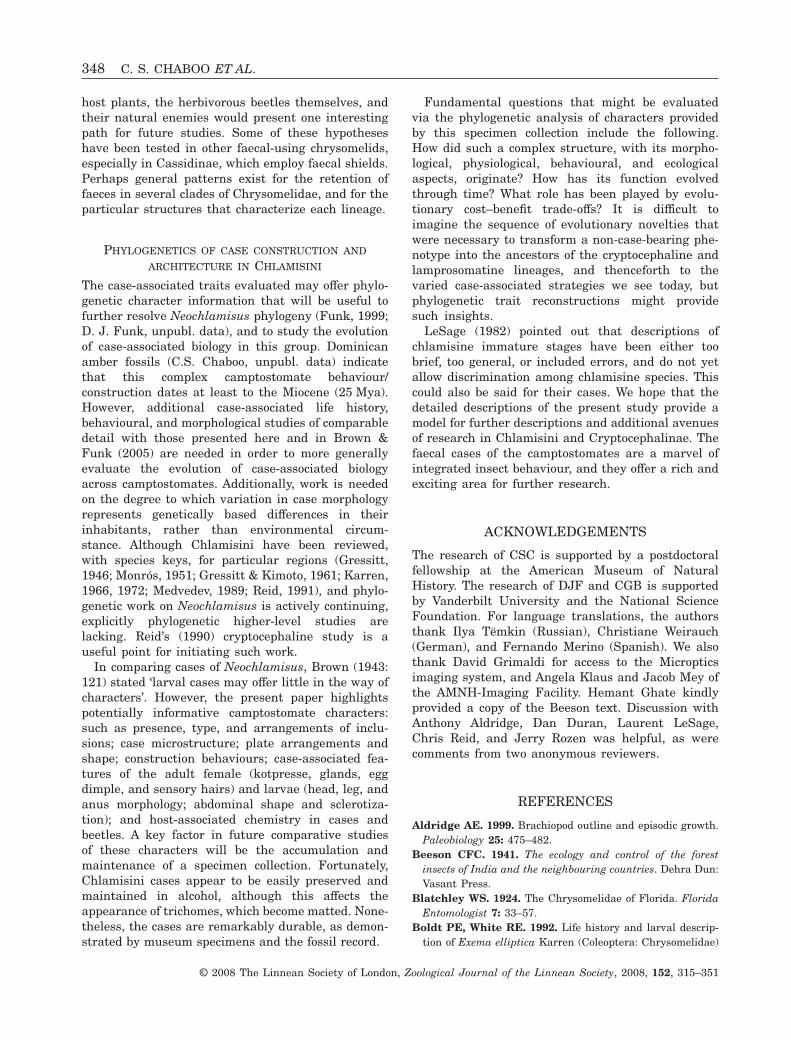
host plants, the herbivorous beetles themselves, andtheir natural enemies would present one interestingpath for future studies. Some of these hypotheseshave been tested in other faecal-using chrysomelids,especially in Cassidinae, which employ faecal shields.Perhaps general patterns exist for the retention offaeces in several clades of Chrysomelidae, and for theparticular structures that characterize each lineage.
PHYLOGENETICS OF CASE CONSTRUCTION AND
ARCHITECTURE IN CHLAMISINI
The case-associated traits evaluated may offer phylo-genetic character information that will be useful tofurther resolve Neochlamisus phylogeny (Funk, 1999;D. J. Funk, unpubl. data), and to study the evolutionof case-associated biology in this group. Dominicanamber fossils (C.S. Chaboo, unpubl. data) indicatethat this complex camptostomate behaviour/construction dates at least to the Miocene (25 Mya).However, additional case-associated life history,behavioural, and morphological studies of comparabledetail with those presented here and in Brown &Funk (2005) are needed in order to more generallyevaluate the evolution of case-associated biologyacross camptostomates. Additionally, work is neededon the degree to which variation in case morphologyrepresents genetically based differences in theirinhabitants, rather than environmental circum-stance. Although Chlamisini have been reviewed,with species keys, for particular regions (Gressitt,1946; Monrós, 1951; Gressitt & Kimoto, 1961; Karren,1966, 1972; Medvedev, 1989; Reid, 1991), and phylo-genetic work on Neochlamisus is actively continuing,explicitly phylogenetic higher-level studies arelacking. Reid’s (1990) cryptocephaline study is auseful point for initiating such work.
In comparing cases of Neochlamisus, Brown (1943:121) stated ‘larval cases may offer little in the way ofcharacters’. However, the present paper highlightspotentially informative camptostomate characters:such as presence, type, and arrangements of inclu-sions; case microstructure; plate arrangements andshape; construction behaviours; case-associated fea-tures of the adult female (kotpresse, glands, eggdimple, and sensory hairs) and larvae (head, leg, andanus morphology; abdominal shape and sclerotiza-tion); and host-associated chemistry in cases andbeetles. A key factor in future comparative studiesof these characters will be the accumulation andmaintenance of a specimen collection. Fortunately,Chlamisini cases appear to be easily preserved andmaintained in alcohol, although this affects theappearance of trichomes, which become matted. None-theless, the cases are remarkably durable, as demon-strated by museum specimens and the fossil record.
Fundamental questions that might be evaluatedvia the phylogenetic analysis of characters providedby this specimen collection include the following.How did such a complex structure, with its morpho-logical, physiological, behavioural, and ecologicalaspects, originate? How has its function evolvedthrough time? What role has been played by evolu-tionary cost–benefit trade-offs? It is difficult toimagine the sequence of evolutionary novelties thatwere necessary to transform a non-case-bearing phe-notype into the ancestors of the cryptocephaline andlamprosomatine lineages, and thenceforth to thevaried case-associated strategies we see today, butphylogenetic trait reconstructions might providesuch insights.
LeSage (1982) pointed out that descriptions ofchlamisine immature stages have been either toobrief, too general, or included errors, and do not yetallow discrimination among chlamisine species. Thiscould also be said for their cases. We hope that thedetailed descriptions of the present study provide amodel for further descriptions and additional avenuesof research in Chlamisini and Cryptocephalinae. Thefaecal cases of the camptostomates are a marvel ofintegrated insect behaviour, and they offer a rich andexciting area for further research.
ACKNOWLEDGEMENTS
The research of CSC is supported by a postdoctoralfellowship at the American Museum of NaturalHistory. The research of DJF and CGB is supportedby Vanderbilt University and the National ScienceFoundation. For language translations, the authorsthank Ilya Tëmkin (Russian), Christiane Weirauch(German), and Fernando Merino (Spanish). We alsothank David Grimaldi for access to the Micropticsimaging system, and Angela Klaus and Jacob Mey ofthe AMNH-Imaging Facility. Hemant Ghate kindlyprovided a copy of the Beeson text. Discussion withAnthony Aldridge, Dan Duran, Laurent LeSage,Chris Reid, and Jerry Rozen was helpful, as werecomments from two anonymous reviewers.
REFERENCES
Aldridge AE. 1999. Brachiopod outline and episodic growth.Paleobiology 25: 475–482.
Beeson CFC. 1941. The ecology and control of the forestinsects of India and the neighbouring countries. Dehra Dun:Vasant Press.
Blatchley WS. 1924. The Chrysomelidae of Florida. FloridaEntomologist 7: 33–57.
Boldt PE, White RE. 1992. Life history and larval descrip-tion of Exema elliptica Karren (Coleoptera: Chrysomelidae)
348 C. S. CHABOO ET AL.
© 2008 The Linnean Society of London, Zoological Journal of the Linnean Society, 2008, 152, 315–351

on Baccharis halimifolia L. (Asteraceae) in Texas. Proceed-ings of the Entomological Society of Washington 94: 83–90.
Briggs EM. 1905. The life history of case bearers: 1. Chlamysplicata. Cold Spring Harbor Monographs IV.
Brown WJ. 1943. The Canadian species of Exema and Arth-rochlamys (Coleoptera: Chrysomelidae). Canadian Ento-mologist 75: 119–131.
Brown CG, Funk DJ. 2005. Aspects of the natural history ofNeochlamisus (Coleoptera: Chrysomelidae): fecal-case asso-ciated life history and behavior, with a method for studyinginsect constructions. Annals of the Entomological Society ofAmerica 98: 711–725.
Bucheli S, Landry JF, Wenzel J. 2002. Larval case archi-tecture and implications of host–plant associations forNorth American Coleophora (Lepidoptera: Coleophoridae).Cladistics 18: 71–93.
Chaboo CS. 2007. Biology, morphology and systematics of theCassidinae Gyllenhal (tortoise and leaf-mining beetles).Bulletin of the American Museum of Natural History 305:1–250.
Chaboo CS, Grobbelaar E, Larsen A. 2007. Fecal ecologyin leaf beetles: novel records in the African arrow-poisonbeetles, Diamphidia Gerstaecker and Polyclada Chevrolat(Chrysomelidae: Galerucinae). Coleopterists Bulletin 61:297–309.
Chaboo CS, Nguyen T. 2004. Immatures of Hemisphaerotapalmarum (Boheman), with a discussion of the caudalprocess and shield architecture in the tribe Hemisphaerotini(Chrysomelidae: Cassidinae). In: Jolivet P, Santiago-Blay J,Schmitt M, eds. New contributions to the biology of thechrysomelidae. The Hague: Kluwer Academic Press, 171–184.
Cockerell TDA. 1891. Case-making coleopterous larvae.Entomologist’s Monthly Magazine, 2nd Series II: 190–191.
Cox ML. 1996. Insect predators of Chrysomelidae. In: JolivetPHA, Cox ML, eds. Chrysomelidae biology: ecological studies.Vol. 2. Amsterdam: SPB Academic Publishing, 23–92.
Dimmock AK. 1885. The insects of Betula in North America.Psyche 4: 283.
Donisthorpe HJK. 1902. The life history of Clythra quad-ripunctata, L. Transactions of the Entomological Society ofLondon 50: 11–24.
Dugès E. 1881. Métamorphoses de ‘Exema dispar’. Annalesde la Société Entomologique de Belgique 25: 5–7.
Egan SP, Funk DJ. 2006. Individual advantages to ecologi-cal specialization: insights on cognitive constraints fromthree conspecific taxa. Proceedings of the Royal Society ofLondon. Series B. Biological Sciences 273: 843–848.
Ellis CR, LeRoux EJ. 1964. Chlamisus cribripennis (Lec.)(Col. Chrys.), a new pest of blueberries in Nova Scotia.Canadian Entomologist 96: 809–810.
Emden F. 1932. Die larven von Discoloma cassideum Reitt.(Col. Colyd.) und Skwarraia paradoxa Lac. (Col. Chrysom.).Zoologische Anzeiger 101: 1–17.
Erber D. 1968. Bau, funktion und bildung der kotpressemittleuropäischer Clytrinen und Cryptocephalinen(Coleoptera: Chrysomelidae). Zeitschrift für Morphologie derTiere 62: 245–306.
Erber D. 1969. Beitrag zur Entwicklungs-Biologie mit-tleeuropäischer Clytrinae und Cryptocephalinen (Coleop-tera, Chrysomelidae). Zoologische Jahrbücher Abteilung fürSystematik, Ökologie, und Geographie der Tiere 96: 453–477.
Erber D. 1988. Biology of Camptostomata Clytrinae-Cryptocephalinae-Chlamisinae-Lamprostomatinae. In:Jolivet P, Petitpierre E, Hsiao TH, eds. Biology of theChrysomelidae. Dordrecht: Kluwer Academic Publisher,513–552.
Falkovitch MI. 2003. On the classification of the casebearers(Lepidoptera: Coleophoridae), with descriptions of the newtaxa. Entomological Review 83: 827–847.
Farrell BD. 1998. ‘Inordinate Fondness’ explained: why arethere so many beetles? Science 281: 555–559.
Fiebrig K. 1910. Cassiden und Cryptocephaliden Paraguays.Ihre Entwicklungsstadien und Schutzvorrichtungen. Zoolo-gisches Jahrbuch Supplement 12, Heft 2: 161–264.
Fiori G. 1950. Contributi alla conoscenza morfologica edetoligica dei Coleotteri 5. Bolletino del Laboratorio di Ento-moliga di Bologna 18: 182–196.
Flinte V. 2006. Chrysomelid fauna in Brazilian restingahabitats and the chlamisine Fulcidax. Chrysomela Newslet-ter 45: 4–5.
Flinte V, Macêdo MV. 2004a. Population ecology of Fulcidaxmonstrosa (Chlamisinae). In: Jolivet P, Santiago-Blay J,Schmitt M, eds. New contributions in biology of the Chry-somelidae. The Hague: Kugler Publications, 623–631.
Flinte V, Macêdo MV. 2004b. Biology and seasonality ofFulcidax monstrosa (F.) (Chrysomelidae: Chlamisinae).Coleopterists Bulletin 58: 457–465.
Flinte V, Macêdo MV, Viera RC, Karren JB. 2003.Feeding behavior of Fulcidax monstrosa (Chlamisinae) onits host plant Byrsonima sericea (Malpighiaceae). In: FurthDG, ed. Special topics in leaf beetle biology proceedings ofthe 5th International Symposium on the Chrysomelidae.Sofia: Pensoft Publishers, 155–159.
Frost SW. 1942. Insect life and insect natural history. NewYork: Dover Publications.
Funk DJ. 1998. Isolating a role for natural selection inspeciation: host adaptation and sexual isolation in Neochlam-isus bebbianae leaf beetles. Evolution 52: 1744–1759.
Funk DJ. 1999. Molecular systematics of CytochromeOxidase I and 16S in Neochlamisus leaf beetles and theimportance of sampling. Molecular Biology and Evolution16: 67–82.
Funk DJ, Filchak KE, Feder JL. 2002. Herbivorousinsects: model systems for the comparative study of specia-tion ecology. Genetica 116: 251–267.
Girault AA. 1911. A note on Chlamys picata Fabricius. Ento-mological News 22: 114.
Gómez NE. 1997. The fecal shields of larva of tortoisebeetles (Cassidinae: Chrysomelidae): a role in chemicaldefense using plant-derived secondary compounds. PhD Dis-sertation, Technischen Universität Carolo-Wilhemina zuBraunschweig.
Gómez NE. 2004. Survivorship of immature stages of Eurype-dus nigrosignatus Bohema (Chrysomelidae: Cassidinae:
FAECAL CASE ARCHITECTURE IN LEAF BEETLES 349
© 2008 The Linnean Society of London, Zoological Journal of the Linnean Society, 2008, 152, 315–351
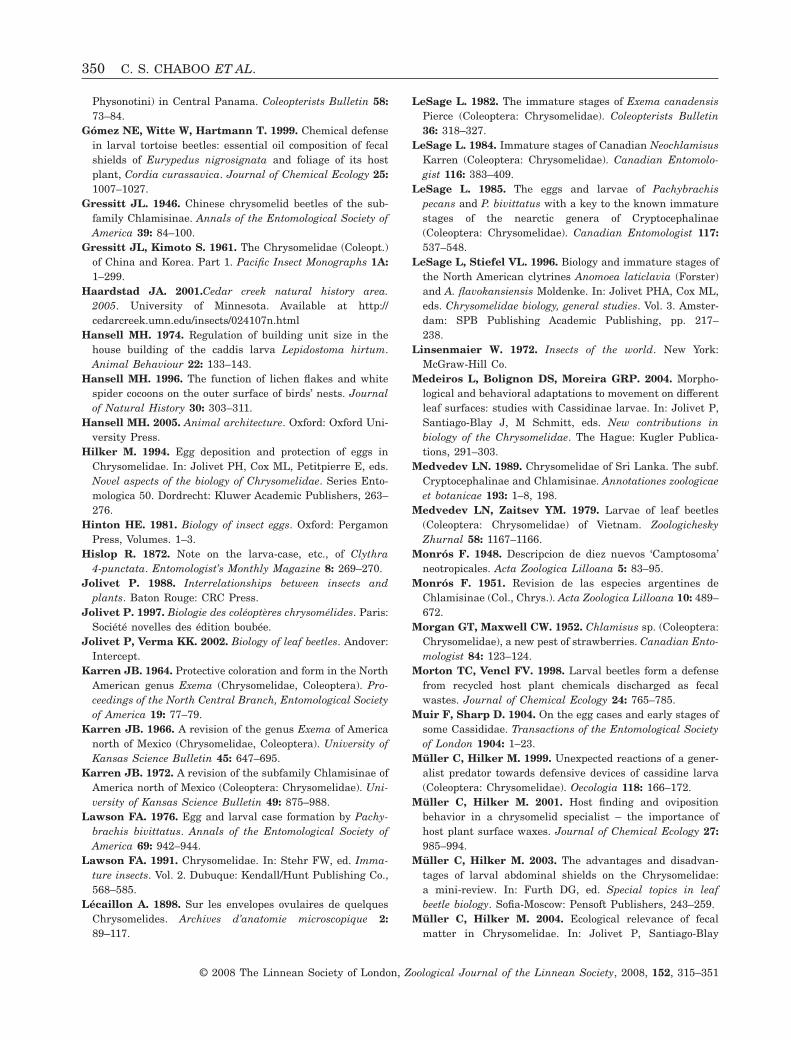
Physonotini) in Central Panama. Coleopterists Bulletin 58:73–84.
Gómez NE, Witte W, Hartmann T. 1999. Chemical defensein larval tortoise beetles: essential oil composition of fecalshields of Eurypedus nigrosignata and foliage of its hostplant, Cordia curassavica. Journal of Chemical Ecology 25:1007–1027.
Gressitt JL. 1946. Chinese chrysomelid beetles of the sub-family Chlamisinae. Annals of the Entomological Society ofAmerica 39: 84–100.
Gressitt JL, Kimoto S. 1961. The Chrysomelidae (Coleopt.)of China and Korea. Part 1. Pacific Insect Monographs 1A:1–299.
Haardstad JA. 2001.Cedar creek natural history area.2005. University of Minnesota. Available at http://cedarcreek.umn.edu/insects/024107n.html
Hansell MH. 1974. Regulation of building unit size in thehouse building of the caddis larva Lepidostoma hirtum.Animal Behaviour 22: 133–143.
Hansell MH. 1996. The function of lichen flakes and whitespider cocoons on the outer surface of birds’ nests. Journalof Natural History 30: 303–311.
Hansell MH. 2005. Animal architecture. Oxford: Oxford Uni-versity Press.
Hilker M. 1994. Egg deposition and protection of eggs inChrysomelidae. In: Jolivet PH, Cox ML, Petitpierre E, eds.Novel aspects of the biology of Chrysomelidae. Series Ento-mologica 50. Dordrecht: Kluwer Academic Publishers, 263–276.
Hinton HE. 1981. Biology of insect eggs. Oxford: PergamonPress, Volumes. 1–3.
Hislop R. 1872. Note on the larva-case, etc., of Clythra4-punctata. Entomologist’s Monthly Magazine 8: 269–270.
Jolivet P. 1988. Interrelationships between insects andplants. Baton Rouge: CRC Press.
Jolivet P. 1997. Biologie des coléoptères chrysomélides. Paris:Société novelles des édition boubée.
Jolivet P, Verma KK. 2002. Biology of leaf beetles. Andover:Intercept.
Karren JB. 1964. Protective coloration and form in the NorthAmerican genus Exema (Chrysomelidae, Coleoptera). Pro-ceedings of the North Central Branch, Entomological Societyof America 19: 77–79.
Karren JB. 1966. A revision of the genus Exema of Americanorth of Mexico (Chrysomelidae, Coleoptera). University ofKansas Science Bulletin 45: 647–695.
Karren JB. 1972. A revision of the subfamily Chlamisinae ofAmerica north of Mexico (Coleoptera: Chrysomelidae). Uni-versity of Kansas Science Bulletin 49: 875–988.
Lawson FA. 1976. Egg and larval case formation by Pachy-brachis bivittatus. Annals of the Entomological Society ofAmerica 69: 942–944.
Lawson FA. 1991. Chrysomelidae. In: Stehr FW, ed. Imma-ture insects. Vol. 2. Dubuque: Kendall/Hunt Publishing Co.,568–585.
Lécaillon A. 1898. Sur les envelopes ovulaires de quelquesChrysomelides. Archives d’anatomie microscopique 2:89–117.
LeSage L. 1982. The immature stages of Exema canadensisPierce (Coleoptera: Chrysomelidae). Coleopterists Bulletin36: 318–327.
LeSage L. 1984. Immature stages of Canadian NeochlamisusKarren (Coleoptera: Chrysomelidae). Canadian Entomolo-gist 116: 383–409.
LeSage L. 1985. The eggs and larvae of Pachybrachispecans and P. bivittatus with a key to the known immaturestages of the nearctic genera of Cryptocephalinae(Coleoptera: Chrysomelidae). Canadian Entomologist 117:537–548.
LeSage L, Stiefel VL. 1996. Biology and immature stages ofthe North American clytrines Anomoea laticlavia (Forster)and A. flavokansiensis Moldenke. In: Jolivet PHA, Cox ML,eds. Chrysomelidae biology, general studies. Vol. 3. Amster-dam: SPB Publishing Academic Publishing, pp. 217–238.
Linsenmaier W. 1972. Insects of the world. New York:McGraw-Hill Co.
Medeiros L, Bolignon DS, Moreira GRP. 2004. Morpho-logical and behavioral adaptations to movement on differentleaf surfaces: studies with Cassidinae larvae. In: Jolivet P,Santiago-Blay J, M Schmitt, eds. New contributions inbiology of the Chrysomelidae. The Hague: Kugler Publica-tions, 291–303.
Medvedev LN. 1989. Chrysomelidae of Sri Lanka. The subf.Cryptocephalinae and Chlamisinae. Annotationes zoologicaeet botanicae 193: 1–8, 198.
Medvedev LN, Zaitsev YM. 1979. Larvae of leaf beetles(Coleoptera: Chrysomelidae) of Vietnam. ZoologicheskyZhurnal 58: 1167–1166.
Monrós F. 1948. Descripcion de diez nuevos ‘Camptosoma’neotropicales. Acta Zoologica Lilloana 5: 83–95.
Monrós F. 1951. Revision de las especies argentines deChlamisinae (Col., Chrys.). Acta Zoologica Lilloana 10: 489–672.
Morgan GT, Maxwell CW. 1952. Chlamisus sp. (Coleoptera:Chrysomelidae), a new pest of strawberries. Canadian Ento-mologist 84: 123–124.
Morton TC, Vencl FV. 1998. Larval beetles form a defensefrom recycled host plant chemicals discharged as fecalwastes. Journal of Chemical Ecology 24: 765–785.
Muir F, Sharp D. 1904. On the egg cases and early stages ofsome Cassididae. Transactions of the Entomological Societyof London 1904: 1–23.
Müller C, Hilker M. 1999. Unexpected reactions of a gener-alist predator towards defensive devices of cassidine larva(Coleoptera: Chrysomelidae). Oecologia 118: 166–172.
Müller C, Hilker M. 2001. Host finding and ovipositionbehavior in a chrysomelid specialist – the importance ofhost plant surface waxes. Journal of Chemical Ecology 27:985–994.
Müller C, Hilker M. 2003. The advantages and disadvan-tages of larval abdominal shields on the Chrysomelidae:a mini-review. In: Furth DG, ed. Special topics in leafbeetle biology. Sofia-Moscow: Pensoft Publishers, 243–259.
Müller C, Hilker M. 2004. Ecological relevance of fecalmatter in Chrysomelidae. In: Jolivet P, Santiago-Blay
350 C. S. CHABOO ET AL.
© 2008 The Linnean Society of London, Zoological Journal of the Linnean Society, 2008, 152, 315–351

J, Schmitt M, eds. New contributions in biology of theChrysomelidae. The Hague: Kugler Publications, 693–705.
Neal JW Jr. 1989. Bionomics of immature stages and ethol-ogy of Neochlamisus platani (Coleoptera, Chrysomelidae) onAmerican sycamore. Annals of the Entomological Society ofAmerica 82: 64–72.
Nogueira-de-Sá F, Trigo JR. 2002. Do fecal shields providephysical protection to larvae of the tortoise beetles Plagi-ometriona flavescens and Stolas chalybea against naturalenemies. Entomologia Experimentalis et Applicata 104:203–206.
Olmstead KL. 1994. Waste products as chrysomelid defenses.In: Jolivet PHA, Cox ML, Petitpierre E, eds. Novel aspectsof the biology of Chrysomelidae. Series Entomologica 50.Dordrecht: Kluwer Academic Publishers, 311–318.
Olmstead KL. 1996. Cassidine defences and naturalenemies. In: Jolivet PHA, Cox ML, eds. Chrysomelidaebiology: ecological studies. Vol. 2. Amsterdam: SPB Aca-demic Publishers, 1–25.
Olmstead KL, Denno RF. 1992. Cost of shield defence fortortoise beetles (Coleoptera: Chrysomelidae). EcologicalEntomology 17: 237–243.
Packard AS. 1869. Guide to the study of insects. 1st ed. NewYork: Henry Holt.
Packard AS. 1889. Guide to the Study of Insects. 9th ed. NewYork: Henry Holt.
Pasteels JM, Rowell-Rahier M, Braekman JC, Daloze D.1984. Chemical defenses in leaf beetles and their larvae: theecological, evolutionary and taxonomic significance. Bio-chemical Systematics and Ecology 12: 395–406.
Peterson A. 1951. Larvae of insects. Part II. Coleoptera,Diptera, Neuroptera, Siphonaptera, Mecoptera, Trichoptera.Columbus, OH: Ohio State University.
Pierce WD. 1940. The dune case bearer beetles of the genusExema, with a review of the genus. Bulletin Southern Cali-fornia Academy of Sciences 39: 6–27.
Popenoe AM, Marlatt CL. 1889. The sycamore leafbeetle, Chlamys plicata Fab. Report of the Departmentof Horticulture and Entomology extracted from Frist.Annual Report of the Kansas Experimental Station; 40–41.
Reid CAM. 1990. Systematics of the Australian Cryptocepha-linae (Coleoptera: Chrysomelidae). PhD Thesis, AustralianNational University.
Reid CAM. 1991. The Australian species of Chlamisini(Coleoptera: Chrysomelidae). Journal of the AustralianEntomological Society 30: 315–323.
Reid CAM. 1995. A cladistic analysis of subfamilial relation-ships of the Chrysomelidae sensu lato (Chrysomeloidea). In:Pakaluk J, Slipinski SA, eds. Biology, Phylogeny and Clas-sification of Coleoptera: papers celebrating the 80th Birthdayof Roy A. Crowson. Vol. 2. Warszawa: Muzeum I InstytutZoologii PAN, 559–631.
Reid CAM. 2000. Spilopyrinae Chapuis: a new subfamilyin the Chrysomelidae and its systematic placement(Coleoptera). Invertebrate Taxonomy CSIRO 14: 837–862.
Reu WFJr, Del-Claro K. 2005. Natural history and biology ofChlamisus minax Lacordaire (Chrysomelidae: Chlamisi-nae). Neotropical Entomology 34: 357–362.
Riley CV. 1874a. Chlamys plicata. Sixth Annual Report,State Entomology Monthly, 128–129.
Riley CV. 1874b. Innoxious insects: the Dominican case-bearer – Coscinoptera dominicana (Fabr.). Sixth annualreport on the noxious, beneficial and other insects of theState of Missouri. Jefferson City, 128–131.
Root RB, Messina FJ. 1983. Defensive adaptations andnatural enemies of a case-bearing beetle, Exema canadensis(Coleoptera: Chrysomelidae). Psyche 90: 67–80.
Schmitt M. 1988. The Criocerinae: biology, phylogeny andevolution. In: Jolivet P, Petitpierre E, Hsiao TH, eds.Biology of Chrysomelidae. Dordrecht: Kluwer AcademicPublishers, 475–495.
Schöller M. 1999. Field studies of Cryptocephalinae biology.In: Cox ML, ed. Advances in Chrysomelidae biology. Vol. 1.Leiden: Backhuys Publishers: 421–436.
Schöller M. 2004. A new species in the formerly monotypicgenus Heptartrius Suffrian (Chrysomelidae: Cryptocephali-nae). Amazoniana 18 1/2: 145–149.
Scudder SH. 1891. The early stages of three Coleoptera.Psyche 6: 173–175.
Seeno TN, Wilcox JA. 1982. Leaf beetle genera. Entomog-raphy 1: 1–221.
Sharp D. 1899. Hymenoptera continued (Tubulifera andAculeata), Coleoptera, Strepsiptera, Lepidoptera, Diptera,Aphaniptera, Thysanoptera, Hemiptera, Anoplura. Part II.New York: MacMillan.
Smith RC. 1922. The biology of the Chrysopidae. CornellUniversity Agricultural Experiment Station Memoir 58:1286–1376.
Solomon J. 2006. Missouri Botanical Garden. 2005.W3Tropicos, rev. 1.5. http://mobot.mobot.org/W3T/Search/vast.html.
Spruyt FJ. 1925. Observations on the egg-laying habits ofSaxinis saucia LeC. (Coleoptera-Chrysomelidae). Pan-Pacific Entomologist 1: 176–178.
Vencl FV, Nogueira-de-Sa F, Allen BJ, Windsor DM,Futuyma DJ. 2005. Dietary specialization influences theefficacy of larval tortoise beetle shield defenses. Oecologia145: 404–414.
Verma KK, Vyas M. 1987. Protective mimicry shown by somechrysomelids living on ‘ber’ shrubs. Chrysomela Newsletter16: 5–6.
Wallace JB. 1970. The defensive function of a case on achrysomelid larva. Journal of the Georgia EntomologicalSociety 5: 19–24.
Weiss M. 2006. Defecation behavior and ecology of insects.Annual Review of Entomology 51: 635–661.
Wickham HF. 1896. The Coleoptera of Canada. XVI. TheChrysomelidae of Ontario and Quebec. Canadian Entomolo-gist 28: 151–157.
Wood GW. 1966. Life history and control of a casebearer,Chlamisus cribripennis (Coleoptera, Chrysomelidae), onblueberry. Journal of Economic Entomology 59: 823–825.
Zaitsev YM. 1992. Ranie Neizuestnye Lichinki Zhukov-Listoyedov (Coleoptera, Chyrsomelidae) [Previouslyunknown larvae of leaf-beetles from Vietnam]. Systematicsand ecology of Insects of Vietnam. Moscow: Nauka, 156–168.
FAECAL CASE ARCHITECTURE IN LEAF BEETLES 351
© 2008 The Linnean Society of London, Zoological Journal of the Linnean Society, 2008, 152, 315–351