
Expression profile of the entire family of Adhesion G protein-coupled receptors in mouse and rat
14
BioMed Central Page 1 of 14 (page number not for citation purposes) BMC Neuroscience Open Access Research article Expression profile of the entire family of Adhesion G protein-coupled receptors in mouse and rat Tatjana Haitina 1 , Fredrik Olsson 1 , Olga Stephansson 1 , Johan Alsiö 1 , Erika Roman 2 , Ted Ebendal 3 , Helgi B Schiöth 1 and Robert Fredriksson* 1 Address: 1 Department of Neuroscience, Unit of Functional Pharmacology, Uppsala University, BMC, 75124 Uppsala, Sweden, 2 Department of Pharmaceutical Biosciences, Division of Pharmacology, Uppsala University, BMC, 75124 Uppsala, Sweden and 3 Department of Neuroscience, Unit of Developmental Neuroscience, Uppsala University, BMC, 75124 Uppsala, Sweden Email: Tatjana Haitina - [email protected]; Fredrik Olsson - [email protected]; Olga Stephansson - [email protected]; Johan Alsiö - [email protected]; Erika Roman - [email protected]; Ted Ebendal - [email protected]; Helgi B Schiöth - [email protected]; Robert Fredriksson* - [email protected] * Corresponding author Abstract Background: The Adhesion G protein-coupled receptors (GPCRs) are membrane-bound receptors with long N termini. This family has 33 members in humans. Several Adhesion GPCRs are known to have important physiological functions in CNS development and immune system response mediated by large cell surface ligands. However, the majority of Adhesion GPCRs are still poorly studied orphans with unknown functions. Results: In this study we performed the extensive tissue localization analysis of the entire Adhesion GPCR family in rat and mouse. By applying the quantitative real-time PCR technique we have produced comparable expression profile for each of the members in the Adhesion family. The results are compared with literature data and data from the Allen Brain Atlas project. Our results suggest that the majority of the Adhesion GPCRs are either expressed in the CNS or ubiquitously. In addition the Adhesion GPCRs from the same phylogenetic group have either predominant CNS or peripheral expression, although each of their expression profile is unique. Conclusion: Our findings indicate that many of Adhesion GPCRs are expressed, and most probably, have function in CNS. The related Adhesion GPCRs are well conserved in their structure and interestingly have considerable overlap in their expression profiles, suggesting similarities among the physiological roles for members within many of the phylogenetically related clusters. Background G protein-coupled receptors (GPCRs) can be divided into five subfamilies according to the GRAFS classification sys- tem: Glutamate, Rhodopsin, Adhesion, Frizzled/Taste2 and Secretin [1]. The Adhesion GPCRs, also known as long N- terminal seven transmembrane receptors related to family B (LNB-TM7) and epidermal growth factor seven trans- membrane (EGF-TM7) receptors. This family is the sec- ond largest GPCR family with 33 members in humans and 30 members in mice and rats. The endogenous lig- ands are discovered for BAI1, GPR56, CD97 and EMR2, while the rest of the Adhesion GPCRs remain orphans. Published: 29 April 2008 BMC Neuroscience 2008, 9:43 doi:10.1186/1471-2202-9-43 Received: 6 November 2007 Accepted: 29 April 2008 This article is available from: http://www.biomedcentral.com/1471-2202/9/43 © 2008 Haitina et al; licensee BioMed Central Ltd. This is an Open Access article distributed under the terms of the Creative Commons Attribution License (http://creativecommons.org/licenses/by/2.0 ), which permits unrestricted use, distribution, and reproduction in any medium, provided the original work is properly cited.
-
Upload
independent -
Category
Documents
-
view
1 -
download
0
Transcript of Expression profile of the entire family of Adhesion G protein-coupled receptors in mouse and rat
BioMed CentralBMC Neuroscience
ss
Open AcceResearch articleExpression profile of the entire family of Adhesion G protein-coupled receptors in mouse and ratTatjana Haitina1, Fredrik Olsson1, Olga Stephansson1, Johan Alsiö1, Erika Roman2, Ted Ebendal3, Helgi B Schiöth1 and Robert Fredriksson*1Address: 1Department of Neuroscience, Unit of Functional Pharmacology, Uppsala University, BMC, 75124 Uppsala, Sweden, 2Department of Pharmaceutical Biosciences, Division of Pharmacology, Uppsala University, BMC, 75124 Uppsala, Sweden and 3Department of Neuroscience, Unit of Developmental Neuroscience, Uppsala University, BMC, 75124 Uppsala, Sweden
Email: Tatjana Haitina - [email protected]; Fredrik Olsson - [email protected]; Olga Stephansson - [email protected]; Johan Alsiö - [email protected]; Erika Roman - [email protected]; Ted Ebendal - [email protected]; Helgi B Schiöth - [email protected]; Robert Fredriksson* - [email protected]
* Corresponding author
AbstractBackground: The Adhesion G protein-coupled receptors (GPCRs) are membrane-boundreceptors with long N termini. This family has 33 members in humans. Several Adhesion GPCRs areknown to have important physiological functions in CNS development and immune systemresponse mediated by large cell surface ligands. However, the majority of Adhesion GPCRs are stillpoorly studied orphans with unknown functions.
Results: In this study we performed the extensive tissue localization analysis of the entire AdhesionGPCR family in rat and mouse. By applying the quantitative real-time PCR technique we haveproduced comparable expression profile for each of the members in the Adhesion family. Theresults are compared with literature data and data from the Allen Brain Atlas project. Our resultssuggest that the majority of the Adhesion GPCRs are either expressed in the CNS or ubiquitously.In addition the Adhesion GPCRs from the same phylogenetic group have either predominant CNSor peripheral expression, although each of their expression profile is unique.
Conclusion: Our findings indicate that many of Adhesion GPCRs are expressed, and mostprobably, have function in CNS. The related Adhesion GPCRs are well conserved in their structureand interestingly have considerable overlap in their expression profiles, suggesting similaritiesamong the physiological roles for members within many of the phylogenetically related clusters.
BackgroundG protein-coupled receptors (GPCRs) can be divided intofive subfamilies according to the GRAFS classification sys-tem: Glutamate, Rhodopsin, Adhesion, Frizzled/Taste2 andSecretin [1]. The Adhesion GPCRs, also known as long N-terminal seven transmembrane receptors related to familyB (LNB-TM7) and epidermal growth factor seven trans-
membrane (EGF-TM7) receptors. This family is the sec-ond largest GPCR family with 33 members in humansand 30 members in mice and rats. The endogenous lig-ands are discovered for BAI1, GPR56, CD97 and EMR2,while the rest of the Adhesion GPCRs remain orphans.
Published: 29 April 2008
BMC Neuroscience 2008, 9:43 doi:10.1186/1471-2202-9-43
Received: 6 November 2007Accepted: 29 April 2008
This article is available from: http://www.biomedcentral.com/1471-2202/9/43
© 2008 Haitina et al; licensee BioMed Central Ltd. This is an Open Access article distributed under the terms of the Creative Commons Attribution License (http://creativecommons.org/licenses/by/2.0), which permits unrestricted use, distribution, and reproduction in any medium, provided the original work is properly cited.
Page 1 of 14(page number not for citation purposes)
BMC Neuroscience 2008, 9:43 http://www.biomedcentral.com/1471-2202/9/43
The Adhesion receptors are characterized by long N-ter-mini with complex domain architecture including GPCRproteolytic site (GPS), epidermal growth factor, throm-bospondin, pentraxin, immunoglobulin, olfactomedin,cadherin domains [2]. Several of the domains in the N-ter-mini have been shown to have cell adhesion functionsand to be responsible for cell-to-cell and cell-to-matrixinteractions [3,4]. The GPS domain is thought to be anintracellular cleavage motif, which is crucial for transportof the receptor from ER to the membrane [5]. The Adhe-sion GPCRs are cleaved into two parts: N-terminal and7TM part with the C-terminal. Nevertheless it has beenreported that the N-terminal is responsible for ligandbinding and is able to re-associate with the rest of thereceptor and in this way initiate intracellular signallingcascade [6]. The genes coding for Adhesion GPCRs are dif-ficult to study due to their complex genomic structure anda large number of exons.
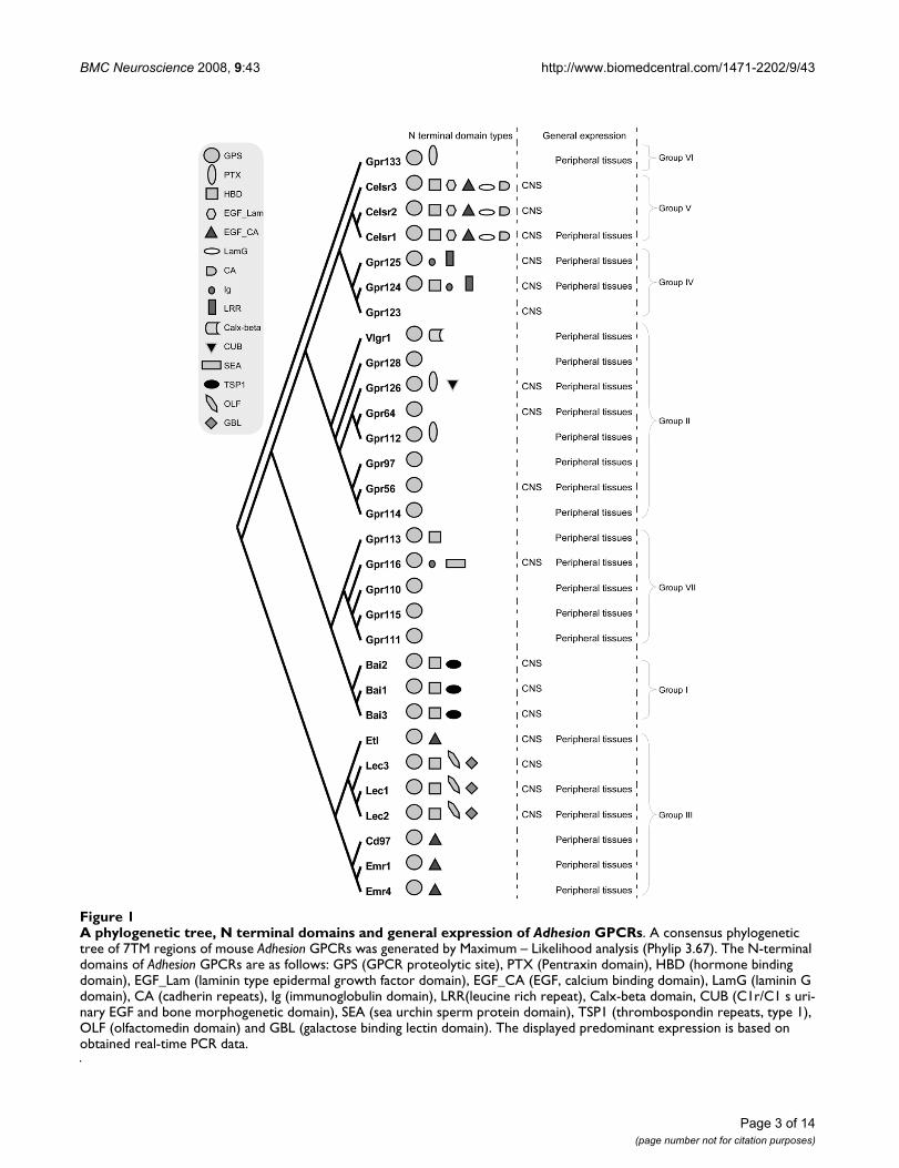
The Adhesion GPCRs can be divided into seven groupsaccording to phylogenetic analysis (Figure 1) [2]. Thebrain angiogenesis inhibitors (BAIs) 1–3 form group I andare known to be mainly expressed in the brain. The N-ter-minal of BAI receptors contains GPS, hormone bindingdomain and thrombospondin repeats which are likely tobe involved in the angiogenesis inhibitor function ofBAIs. The role of BAIs in tumor suppression has beenclosely investigated as angiogenesis is an essential part oftumor formation [7]. BAI1 has been recently reported as areceptor for phospatidylserine involved in recognitionand internalization of apoptotic cells [8]. GPR56 (groupII) is another Adhesion GPCR with a role in tumor devel-opment [9]. Mutations in GPR56 have also been reportedto cause human brain malformations [10]. It is describedthat GPR56 is able to build complex with cell-surface pro-tein tetraspanin CD81 and Gαq/11 and is able to bind tis-sue transglutaminase, TG2 [11,12].
GPR64 also called HE6 (group II), unlike GPR56, has itsonly known function in the periphery and disruption ofthis gene results in mouse male infertility [13]. The verylarge G-protein-coupled receptor (VLGR1) is importantfor normal development of auditory hair bundles in theinner ear [14]. The functions of other five members ofgroup II are not known.
The Adhesion receptors forming group III can be dividedinto two groups according to the architecture of their N-termini. EMR1, EMR2, EMR3, EMR4, CD97 and ETLreceptors have all a variable number of epidermal growthfactor (EGF) and calcium binding domains and arereported to be important components of the immune sys-tem [2]. EMR2 and EMR3 are missing in the mouse andrat genomes. CD55 and chondroitin sulphate have beendescribed as ligands for CD97, while chondroitin sul-
phate is also a ligand for EMR2. It is also suggested thatEGF domains in the N-termini of CD97 are essential forCD55 binding. [15,16]. LEC1, LEC2 and LEC3 on theother hand are proposed to play more central roles likesynaptic cell adhesion [17] and have a different repertoireof domains in the N-termini (hormone binding, olfacto-medin and galactose binding lectin domains). LEC2 hasbeen reported to bind α-latrotoxin from black widow spi-der venom [5].
GPR123, GPR124 and GPR125 belong to group IV ofAdhesion GPCRs. GPR123 has been reported to have spe-cific expression in the brain [18]. GPR125 was recentlypresented as a stem cell marker with a possible therapeuticuse [19]. Both these receptors have protein-protein inter-action domains in the form of leucine rich repeats, whileGPR124 and GPR125 have immunoglobulin domains inN-termini as well.
CELSR1–3 receptors (group V) have the most complexdomain structure with cadherin repeats, two types of EGFdomains, laminin G and hormone binding domains.CELSR receptors are thought to have crucial functions inCNS development. Mutations in CELSR1 cause abnormalneural tube development in mice [20]. CELSR2 andCELSR3 seem to have opposite effects in neurite growthregulation; CELSR2 is enhancing neurite growth whileCELSR3 is suppressing it. [21]. Unlike CELSR receptors,the members of group VI, GPR133 and GPR144 as well asgroup VII, GPR110, GPR111, GPR113, GPR115 andGPR116 do not have any discovered function.
The majority of Adhesion GPCRs are poorly studied, withspecific expression patterns and physiological functionsyet to be discovered. Until today, the published expres-sion data on many Adhesion GPCR members is limited toa few tissues and is performed by different methods withvarying sensitivity. Therefore, an overall expression analy-sis of Adhesion GPCRs is useful for comparison of expres-sion and identifying specific patterns. It is also animportant step towards finding the functions of thepoorly studied receptors.
In this study we report an extensive tissue localizationanalysis of the entire Adhesion GPCR family in rat andmouse. By applying the quantitative real-time PCR tech-nique we have produced comparable expression patternsfor each Adhesion family member. We have comparedexpression patterns between rat and mouse and recent insitu hybridization data for mouse from the large AllenBrain Atlas project [22] as well as human and mouse ESTs[23]. We describe similarities in Adhesion GPCR expres-sion patterns between rat and mouse as well as withingroups of Adhesion GPCRs.
Page 2 of 14(page number not for citation purposes)
BMC Neuroscience 2008, 9:43 http://www.biomedcentral.com/1471-2202/9/43
Page 3 of 14(page number not for citation purposes)
A phylogenetic tree, N terminal domains and general expression of Adhesion GPCRsFigure 1A phylogenetic tree, N terminal domains and general expression of Adhesion GPCRs. A consensus phylogenetic tree of 7TM regions of mouse Adhesion GPCRs was generated by Maximum – Likelihood analysis (Phylip 3.67). The N-terminal domains of Adhesion GPCRs are as follows: GPS (GPCR proteolytic site), PTX (Pentraxin domain), HBD (hormone binding domain), EGF_Lam (laminin type epidermal growth factor domain), EGF_CA (EGF, calcium binding domain), LamG (laminin G domain), CA (cadherin repeats), Ig (immunoglobulin domain), LRR(leucine rich repeat), Calx-beta domain, CUB (C1r/C1 s uri-nary EGF and bone morphogenetic domain), SEA (sea urchin sperm protein domain), TSP1 (thrombospondin repeats, type 1), OLF (olfactomedin domain) and GBL (galactose binding lectin domain). The displayed predominant expression is based on obtained real-time PCR data.
BMC Neuroscience 2008, 9:43 http://www.biomedcentral.com/1471-2202/9/43
ResultsThe mouse Adhesion GPCR gene sequences were down-loaded from GenBank (Additional File 2) and 7TMregions were identified with Conserved Domain Database[24]. The tree calculations were carried out using ClustalWalignment of TM regions, which was bootstrapped 100times and distances were calculated with PROML fromPhylip 3.67 package. The Maximum-Likelihood consen-sus tree is presented in Figure 1.
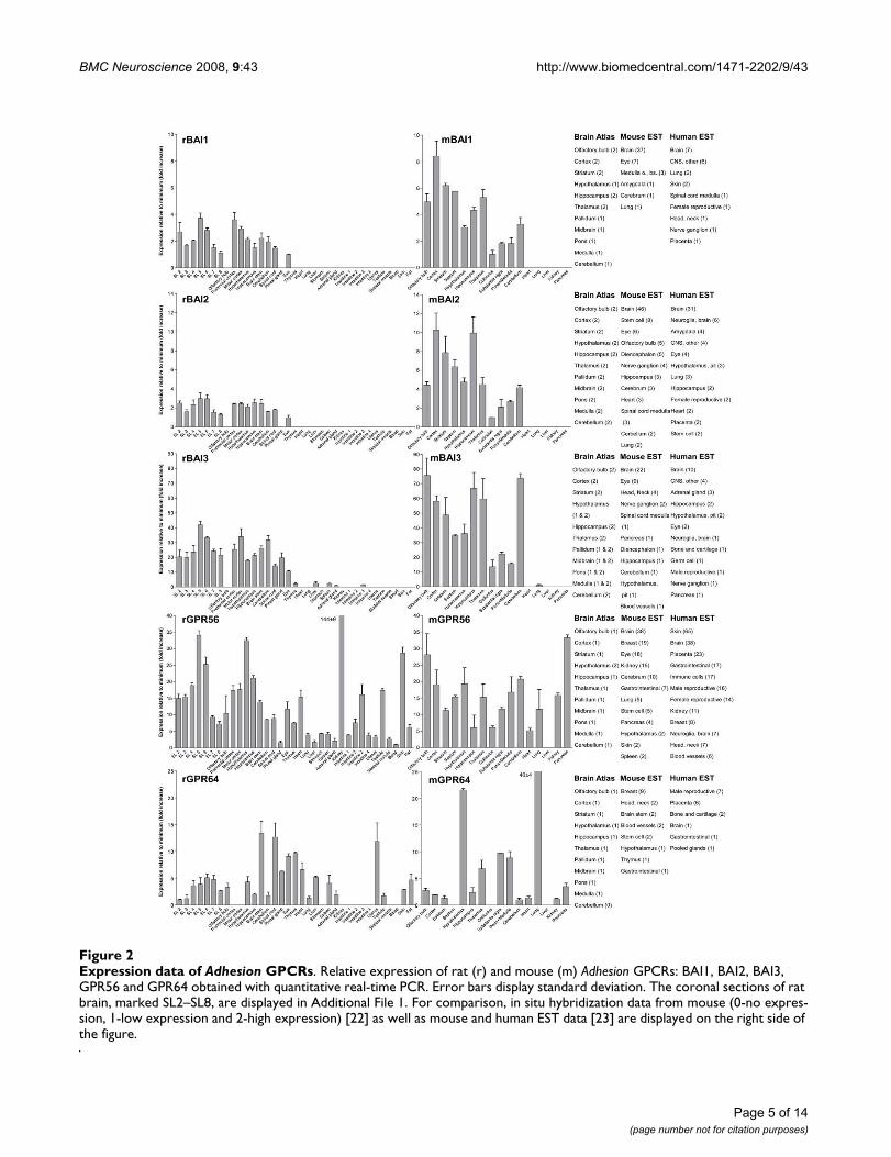
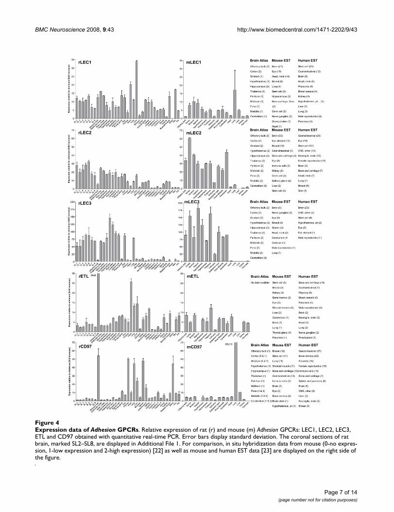
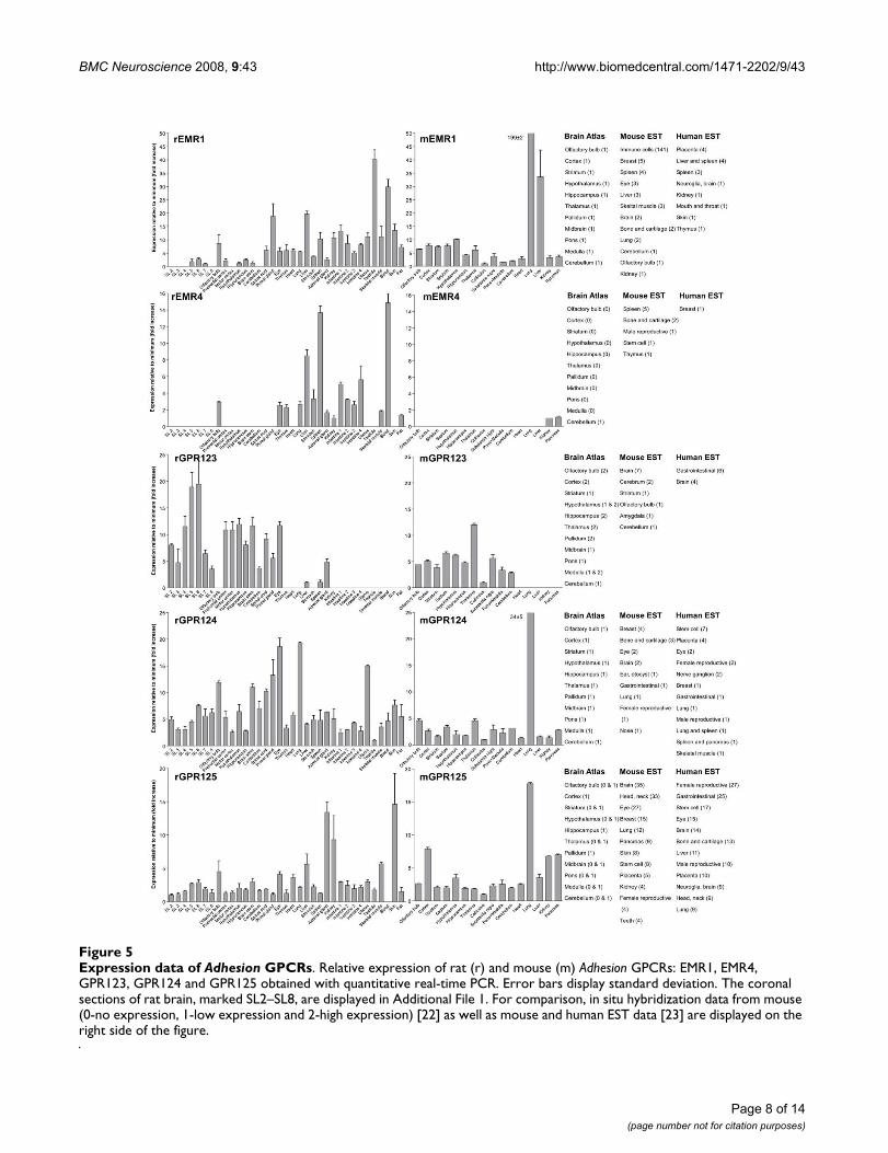
We performed an expression analysis of 30 rat and mouseAdhesion GPCR family members in a range of central andperipheral tissues. Seven coronal sections of rat brain(Additional file 1), a number of brain regions and periph-eral tissues were isolated from mice and rats and used forRNA isolation and cDNA synthesis. The expression valuesof four housekeeping genes were used to calculate nor-malization factors for mouse and rat cDNA. Relativeexpression values displayed as a fold increase from thedetected minimum expression for rat and mouse AdhesionGPCRs are presented in Figures 2, 3, 4, 5, 6, 7. Expressionof BAI1 and BAI2 from group I was detected only in brainregions of rat and mouse. BAI3 transcripts were detectedmostly in brain, with very low levels in some peripheraltissues (Figure 2). The expression data of BAI receptors isin agreement with previously published results [25,26].GPR56, GPR64, GPR126 and VLGR1 from group II weredetected in central and peripheral tissues, whereas GPR97,GPR112, GPR114 and GPR128 were detected in periph-eral tissues (Figures 2, 3 and 6). The members of group IIILEC1, LEC2, LEC3 and ETL were expressed ubiquitouslyin both rat and mouse, whereas LEC2 and LEC3 hadhigher levels in CNS compared to levels in the periphery(Figure 4). The members of group III CD97, EMR1 andEMR4 were detected mostly in peripheral tissues and atvery low levels in the brain (Figures 4, 5). The expressionprofile of receptors from Adhesion group III is in agree-ment with previous reports [27-31]. The member of groupIV GPR123 displayed central expression, while the othermembers GPR124 and GPR125 showed ubiquitousexpression (Figure 5). The members of group V CELSR2and CELSR3 were detected mainly in CNS, while CELSR1was found both in central and peripheral tissues (Figure6). These results confirmed previously published expres-sion data of CELSR receptors [32,33]. Group VI memberGPR133 was detected in both CNS and periphery (Figure6). GPR144, which is a pseudogene in rat, was notdetected in any of the examined mouse tissues (results notshown). Group VII members GPR110, GPR111, GPR113and GPR115 were detected in only few peripheral tissues(Figure 7). GPR116 was detected in all analyzed tissues,except blood, but mainly at low levels (Figure 7).
DiscussionIn this paper we present for the first time the expressionchart of 30 members of Adhesion GPCRs in mouse and rat.Together with EST data from human and mouse [23] andpreviously published Allen Brain Atlas in situ hybridiza-tion data [22] this provides the most comprehensiveinformation about the expression pattern of the Adhesionfamily of GPCRs (Figures 1, 2, 3, 4, 5, 6, 7).
Adhesion receptors is an evolutionary conserved group, forexample an Adhesion like genes have been described infruit fly (Drosophila melanogaster) [34] and in the choano-flagellate (Monosiga brevicollis) [35]. According to our phy-logenetic analysis based on TM regions of Adhesionreceptors they formed seven groups (Figure 1), which is inagreement with previously published studies [2]. Despitethat phylogeny was based only on TM regions the N-ter-mini of the receptors in each group had similar domaintypes and organization. Surprisingly, our results showedthat the members within each group also share consider-able similarities in their expression profiles (Figure 1). Soaccording to our results group I with BAI1, BAI2, BAI3;group IV with GPR123, GPR124, GPR125; group V withCELSR1, CELSR2, CELSR3 and even LEC1–3 and ETL1which form a cluster within group III showed predomi-nant expression in the brain of rat and mouse (Figures 2,4, 5 and 6).
The Adhesion GPCR group containing GPR123, GPR124and GPR125 is one of the least studied groups. Thedetailed CNS profile of GPR123 was recently published byLagerström et al. with some indications of important neu-ronal functions [18]. It is therefore very exciting that ourresults display wide expression in the rat and mouse brainfor all 3 members of this group. In contrast to GPR123,which displayed specific central expression, GPR124 andGPR125 displayed also very wide peripheral expression(Figure 5). It is very interesting that both the human andmouse ESTs for GPR125 are detected in stem cells and itwas recently reported that GPR125 could be used thera-peutically as a stem cell marker for generation of vessels[19]. The N-terminals of GPR124 and GPR125 containGPS, leucine-rich repeats, Ig domain and hormone bind-ing domain in contrast to GPR123, which is lacking all ofthese domains. It is therefore tempting to speculate aboutif any of these domains have a role in peripheral actionsof GPR124 and GPR125. Other proteins with leucine-richrepeats like LINGO-1 and LRRK2 have been shown to playa role in structural and functional integrity of neuronsinvolved in Parkinson's disease [36]. The members of thisgroup are present in teleosts [18], amphioxus and fruit fly(unpublished data). As this group is present within mostspecies with a nervous system and have a wide CNSexpression, members from this family could have theirmain function within the CNS.
Page 4 of 14(page number not for citation purposes)
BMC Neuroscience 2008, 9:43 http://www.biomedcentral.com/1471-2202/9/43
Page 5 of 14(page number not for citation purposes)
Expression data of Adhesion GPCRsFigure 2Expression data of Adhesion GPCRs. Relative expression of rat (r) and mouse (m) Adhesion GPCRs: BAI1, BAI2, BAI3, GPR56 and GPR64 obtained with quantitative real-time PCR. Error bars display standard deviation. The coronal sections of rat brain, marked SL2–SL8, are displayed in Additional File 1. For comparison, in situ hybridization data from mouse (0-no expres-sion, 1-low expression and 2-high expression) [22] as well as mouse and human EST data [23] are displayed on the right side of the figure.
BMC Neuroscience 2008, 9:43 http://www.biomedcentral.com/1471-2202/9/43
Page 6 of 14(page number not for citation purposes)
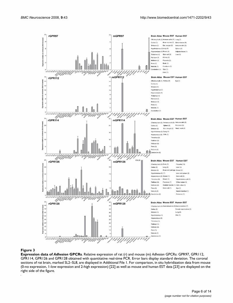
Expression data of Adhesion GPCRsFigure 3Expression data of Adhesion GPCRs. Relative expression of rat (r) and mouse (m) Adhesion GPCRs: GPR97, GPR112, GPR114, GPR126 and GPR128 obtained with quantitative real-time PCR. Error bars display standard deviation. The coronal sections of rat brain, marked SL2–SL8, are displayed in Additional File 1. For comparison, in situ hybridization data from mouse (0-no expression, 1-low expression and 2-high expression) [22] as well as mouse and human EST data [23] are displayed on the right side of the figure.
BMC Neuroscience 2008, 9:43 http://www.biomedcentral.com/1471-2202/9/43
Page 7 of 14(page number not for citation purposes)
Expression data of Adhesion GPCRsFigure 4Expression data of Adhesion GPCRs. Relative expression of rat (r) and mouse (m) Adhesion GPCRs: LEC1, LEC2, LEC3, ETL and CD97 obtained with quantitative real-time PCR. Error bars display standard deviation. The coronal sections of rat brain, marked SL2–SL8, are displayed in Additional File 1. For comparison, in situ hybridization data from mouse (0-no expres-sion, 1-low expression and 2-high expression) [22] as well as mouse and human EST data [23] are displayed on the right side of the figure.
BMC Neuroscience 2008, 9:43 http://www.biomedcentral.com/1471-2202/9/43
Page 8 of 14(page number not for citation purposes)
Expression data of Adhesion GPCRsFigure 5Expression data of Adhesion GPCRs. Relative expression of rat (r) and mouse (m) Adhesion GPCRs: EMR1, EMR4, GPR123, GPR124 and GPR125 obtained with quantitative real-time PCR. Error bars display standard deviation. The coronal sections of rat brain, marked SL2–SL8, are displayed in Additional File 1. For comparison, in situ hybridization data from mouse (0-no expression, 1-low expression and 2-high expression) [22] as well as mouse and human EST data [23] are displayed on the right side of the figure.
BMC Neuroscience 2008, 9:43 http://www.biomedcentral.com/1471-2202/9/43
Page 9 of 14(page number not for citation purposes)
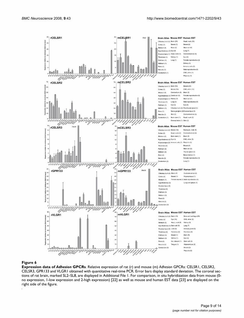
Expression data of Adhesion GPCRsFigure 6Expression data of Adhesion GPCRs. Relative expression of rat (r) and mouse (m) Adhesion GPCRs: CELSR1, CELSR2, CELSR3, GPR133 and VLGR1 obtained with quantitative real-time PCR. Error bars display standard deviation. The coronal sec-tions of rat brain, marked SL2–SL8, are displayed in Additional File 1. For comparison, in situ hybridization data from mouse (0-no expression, 1-low expression and 2-high expression) [22] as well as mouse and human EST data [23] are displayed on the right side of the figure.
BMC Neuroscience 2008, 9:43 http://www.biomedcentral.com/1471-2202/9/43
Page 10 of 14(page number not for citation purposes)
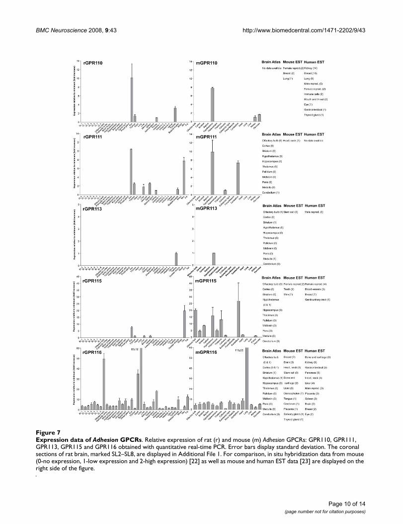
Expression data of Adhesion GPCRsFigure 7Expression data of Adhesion GPCRs. Relative expression of rat (r) and mouse (m) Adhesion GPCRs: GPR110, GPR111, GPR113, GPR115 and GPR116 obtained with quantitative real-time PCR. Error bars display standard deviation. The coronal sections of rat brain, marked SL2–SL8, are displayed in Additional File 1. For comparison, in situ hybridization data from mouse (0-no expression, 1-low expression and 2-high expression) [22] as well as mouse and human EST data [23] are displayed on the right side of the figure.
BMC Neuroscience 2008, 9:43 http://www.biomedcentral.com/1471-2202/9/43
Similarly to the centrally expressed Adhesion GPCR genes,the genes expressed predominantly in peripheral tissuesare also found in phylogenetic clusters. High levels ofGPR128 transcripts were detected in stomach, and allparts of intestine. This is in agreement with the observa-tion that the highest number of ESTs for GPR128 fromhuman and mouse are found in the gastrointestinal (GI)tract (Figure 3). The GPR128 is unlikely to play a role inimmune system regulation as it is not detected in high lev-els in immune system specific organs. The GI specificexpression profile of GPR128 could possibly indicate afunction in cell migration during development of GI tractendothelium. Two other genes from group II, GPR97 andGPR114, were also expressed in the GI tract but they werealso detected at high levels in thymus, spleen and blood(Figure 3). In addition, the highest number of ESTs forthese genes was detected in immune cells, spleen andbone marrow. Altogether this indicates that GPR97 andGPR114 most probably are involved in functions ofimmune system in humans and rodents.
GPR56, GPR64 and GPR126 from Adhesion GPCR groupII displayed ubiquitous expression (Figures 2, 3). GPR56is most studied of these receptors. It is reported to play arole in development of human cerebral cortex [10] as wellas tumor development [12]. According to our results, theexpression of GPR56 is highest in mouse pancreas and ratkidney. GPR64 has been described to regulate the func-tion of the male reproductive system [13]. Our resultstogether with data from Allen Brain Atlas [22] displayexpression of GPR64 in hypothalamus, brain stem, sub-stantia nigra and spinal cord. This was surprising to us asseveral publications have described GPR64 as an epididy-mal specific receptor. Therefore it is likely that GPR64could also have an additional function in CNS.
Mutations in VLGR1 are known to cause audiogenic sei-zures in mice [37]. Hence VLGR1 expression in embryonicCNS is widely described not only in mammals but also inother vertebrates like zebrafish [38]. Interestingly, ourresults indicate that there are common expression sites ofVLGR1 in zebrafish embryo and adult rat such as hypoth-alamus and eye (Figure 6). Such similarity in expressionpatterns between mammals and teleosts points at remark-able conservation of function of the VLGR1 receptor dur-ing vertebrate evolution.
GPR133 and GPR144 are relatively recently discovered[39] and poorly studied Adhesion GPCRs. The presence ofGPR133 like sequence (XM_001197663) in purple seaurchin (Strongylocentrotus purpuratus) indicates a long evo-lutionary history and an important physiological func-tion. The expression of GPR133 was highest in intestine,skin, adipose tissue and olfactory bulb (Figure 6), whichpoints toward a function in periphery. The inability to
detect GPR144 transcripts in mouse tissues verifies thatGPR144 is a murine pseudogene.
GPR116 is the only member of the Adhesion GPCR groupVII with ubiquitous expression (Figure 7). The expressionprofile of GPR116 with highest transcript levels in lung,olfactory bulb and heart is most similar to the ETL andCD97 receptors. This could be a sign of functional similar-ities between these receptors. GPR110, GPR111 andGPR115 have common expression site in the rat eye. Twoof these receptors, GPR111 and GPR115 also share expres-sion in the rat skin. These receptors have similar structurein their N-termini and similar protein length. It is possiblethat these receptors play a role in similar peripheral func-tions like epithelial cell adhesion in eye epithelium andskin.
ConclusionIt is remarkable that phylogenetic clusters of AdhesionGPCRs seem to have either predominant central orperipheral expression in rat and mouse tissues. Though, itis also clear that within the phylogenetic clusters theexpression profiles are not identical as Adhesion GPCRs areexpressed in a large number of tissues. If we analyze thefunctions of the well studied Adhesion GPCRs, it is evidentthat even if the receptors from the same phylogenetic clus-ter do play roles in similar physiological processes, theydo not have the same function and sometimes can evenhave opposite functions. One possible explanation forthis is that Adhesion receptors are very well conserved dur-ing evolution and sub-functionalization was a commonprocess among phylogenetic clusters of Adhesion GPCRs.Adhesion receptors participate in highly complex processeslike development of nervous system and immune systemresponse. These processes require often a strict sequenceof steps with different types of adhesion molecules partic-ipating at the different stages. Altogether the AdhesionGPCRs are a unique and fascinating family of membranereceptors with conserved evolutionary history and anumber of vital physiological functions.
MethodsAnimal handling and tissue isolationA number of adult male and female 129sv mice andSprague-Dawley rats (Scanbur, BK AB) were kept in sepa-rate air-conditioned rooms (12 h dark/light cycle) at 22–23°C in an air humidity of 55%. The animals had freeaccess to water and food pellets (Labfor, Lactamin, Swe-den). These conditions were maintained for 7 days, and atthe end of the period the animals were sacrificed bydecapitation (rats) or cervical dislocation (mice) between3 and 6 hours into the light period. All animal procedureswere approved by the Uppsala Ethics Committee and fol-lowed the guidelines of European Communities CouncilDirective (86/609/EEC). Different brain regions and
Page 11 of 14(page number not for citation purposes)
BMC Neuroscience 2008, 9:43 http://www.biomedcentral.com/1471-2202/9/43
peripheral tissues were isolated. With regard to the ratbrains, they were taken out immediately after decapita-tion. Two whole rat brains were sliced into coronal crosssections (approximately 3 mm thick) using a rat brainmatrix (Pelco International, Canada) as presented in Fig.1. The remaining rat brains were dissected in more detail.The entire hypothalamus was carefully removed by liftingit from the brain with a slightly curved forceps. Thereafterthe following regions were manually dissected with theguidance of a rat brain atlas (Paxinos G., Watson C., 1997.The Rat Brain in Stereotaxic Coordinates. Academic Press,San Diego, CA): prefrontal cortex, motor cortex and hip-pocampus. The first section (approximately bregma 5.20–4.20 mm) was denoted prefrontal cortex. Motor cortexwas collected from sections approximately betweenbregma 4.20–2.70 mm and included primary and second-ary motor cortex. The hippocampus was collected fromseveral sections and therefore comprised the hippocam-pus CA1–3 and the dentate gyrus. The tissues wereimmersed into RNA-later solution (Ambion, USA), kept atroom temperature for 1 hour and thereafter stored at -80°C until further processed.
RNA isolation and cDNA synthesisIndividual tissue samples were homogenized by sonica-tion in TRIzol reagent (Invitrogen, Sweden) using a Bran-son sonifier. Chloroform was added to the homogenate,which was then centrifuged at 10 000 g at 4°C for 15 min-utes. The water phase was collected and RNA was precipi-tated with isopropanol. The pellets were washed with75% ethanol, air dried and dissolved in RNAse free water.DNA contamination was removed by treatment withDNAse I (Roche Diagnostics, Sweden) at 37°C for 4 hwhere after the DNAse I was inactivated by heating thesamples to 75°C for 15 minutes. Absence of DNA con-tamination in the RNA was confirmed by PCR and RNAconcentration was determined using a Nanodrop® ND-1000 Spectrophotometer (NanoDrop Technologies, Dela-ware, USA). cDNA was synthesized with M-MLV reversetranscriptase (Invitrogen, Sweden) using random hexam-ers as primers according to the manufacturers' instruc-tions. The quality of the cDNA was confirmed by PCR.
Primer design and evaluationSequences for rat and mouse house keeping genes and allknown Adhesion GPCR gene sequences were downloadedfrom GenBank. GenBank accession numbers are pre-sented in Additional File 2. All primers were designedwith Beacon Designer v4.0 (Premier Biosoft, USA) andpositioned within TM regions of the Adhesion GPCRs. Theprimers sequences for rat and mouse Adhesion GPCRs andhouse keeping genes are displayed in Additional File 2.
Quantitative Real-Time PCRThe cDNA was analyzed in quantitative real-time PCRwith a MyiQ thermal cycler (Bio-Rad Laboratories, Swe-den). Each real-time PCR reaction with a total volume of20 µl contained cDNA synthesized from 25 ng of totalRNA; 0.25 µM of each primer, 20 mM Tris/HCl (pH 8.4),50 mM KCl, 4 mM MgCl2, 0.2 mM dNTP, SYBR Green(1:50 000). Real-time PCR was performed with 0.02 u/µlTaq DNA polymerase (Invitrogen, Sweden) under the fol-lowing conditions: initial denaturation at 95°C for 4 min,followed by 50 cycles at 95°C for 15 s, 55–62°C for 30 s(optimal annealing temperature) and 72°C for 30 s. Thiswas followed by 84 cycles at 55°C for 10 s (increased by0.5°C per cycle). All real-time PCR experiments were per-formed in duplicate. A negative control for each primerpair and a positive control with 25 ng of rat and mousegenomic DNA, respectively, was included on each plate.
Data analysis and relative expression calculationsMyiQ software v 1.04 (Bio-Rad Laboratories, Sweden) wasused to analyse real-time PCR data and derive thresholdcycle (Ct) values. Melting curves were analysed to confirmthat only one product was amplified and that it was differ-ent from the negative control. LinRegPCR [40] was used tocalculate PCR efficiencies for each sample. After thatGrubbs' test (GraphPad, USA) was applied to exclude theoutliers and calculate average PCR efficiency for eachprimer pair. The delta Ct method [41] was used to trans-form Ct values into relative quantities with standard devi-ations and the highest expression was normalized to 1.The GeNorm software [42] was used with results fromfour housekeeping genes in order to calculate normaliza-tion factors for every tissue to compensate for differencesin cDNA amount. Thereafter the normalized quantitieswere calculated and minimum expression was set to 1. Allrelative expression values were displayed as fold increasefrom the detected minimum expression.
Authors' contributionsTH designed the study, analyzed the data and drafted themanuscript. FO and OS performed the real-time PCR onrat, respectively mouse tissues and made the calculations.TE and JA carried out the dissection of mouse tissues. ERand RF performed the dissection of rat tissues. HBS con-ceived of the study and helped to draft the manuscript. RFparticipated in the design of the study and helped to draftthe manuscript. All authors read and approved the finalmanuscript.
Page 12 of 14(page number not for citation purposes)
BMC Neuroscience 2008, 9:43 http://www.biomedcentral.com/1471-2202/9/43
Additional material
AcknowledgementsRF was supported by the Swedish Brain Foundation (Hjärnfonden). The studies were supported by the Swedish Research Council, AFA, Svenska Läkaresällskapet, Åke Wiberg Foundation, Lars Hiertas foundation, Thur-ings foundation, The Royal Swedish Academy of Sciences, the Magnus Bergvall Foundation and Åhlens foundation.
References1. Fredriksson R, Lagerstrom MC, Lundin LG, Schioth HB: The G-pro-
tein-coupled receptors in the human genome form five mainfamilies. Phylogenetic analysis, paralogon groups, and finger-prints. Mol Pharmacol 2003, 63(6):1256-1272.
2. Bjarnadottir TK, Fredriksson R, Schioth HB: The adhesion GPCRs:a unique family of G protein-coupled receptors with impor-tant roles in both central and peripheral tissues. Cell Mol LifeSci 2007, 64(16):2104-2119.
3. Krasnoperov V, Bittner MA, Holz RW, Chepurny O, Petrenko AG:Structural requirements for alpha-latrotoxin binding andalpha-latrotoxin-stimulated secretion. A study with calcium-independent receptor of alpha-latrotoxin (CIRL) deletionmutants. J Biol Chem 1999, 274(6):3590-3596.
4. Obermann H, Samalecos A, Osterhoff C, Schroder B, Heller R, Kirch-hoff C: HE6, a two-subunit heptahelical receptor associatedwith apical membranes of efferent and epididymal duct epi-thelia. Molecular reproduction and development 2003, 64(1):13-26.
5. Krasnoperov V, Lu Y, Buryanovsky L, Neubert TA, Ichtchenko K,Petrenko AG: Post-translational proteolytic processing of thecalcium-independent receptor of alpha-latrotoxin (CIRL), anatural chimera of the cell adhesion protein and the G pro-tein-coupled receptor. Role of the G protein-coupled recep-tor proteolysis site (GPS) motif. J Biol Chem 2002,277(48):46518-46526.
6. Volynski KE, Silva JP, Lelianova VG, Atiqur Rahman M, Hopkins C,Ushkaryov YA: Latrophilin fragments behave as independentproteins that associate and signal on binding of LTX(N4C).The EMBO journal 2004, 23(22):4423-4433.
7. Kudo S, Konda R, Obara W, Kudo D, Tani K, Nakamura Y, Fujioka T:Inhibition of tumor growth through suppression of angiogen-esis by brain-specific angiogenesis inhibitor 1 gene transfer inmurine renal cell carcinoma. Oncology reports 2007,18(4):785-791.
8. Park D, Tosello-Trampont AC, Elliott MR, Lu M, Haney LB, Ma Z, Kli-banov AL, Mandell JW, Ravichandran KS: BAI1 is an engulfmentreceptor for apoptotic cells upstream of the ELMO/Dock180/Rac module. Nature 2007, 450(7168):430-434.
9. Ke N, Sundaram R, Liu G, Chionis J, Fan W, Rogers C, Awad T, Grif-man M, Yu D, Wong-Staal F, Li QX: Orphan G protein-coupledreceptor GPR56 plays a role in cell transformation and tum-
origenesis involving the cell adhesion pathway. Molecular can-cer therapeutics 2007, 6(6):1840-1850.
10. Piao X, Hill RS, Bodell A, Chang BS, Basel-Vanagaite L, Straussberg R,Dobyns WB, Qasrawi B, Winter RM, Innes AM, Voit T, Ross ME,Michaud JL, Descarie JC, Barkovich AJ, Walsh CA: G protein-cou-pled receptor-dependent development of human frontalcortex. Science 2004, 303(5666):2033-2036.
11. Little KD, Hemler ME, Stipp CS: Dynamic regulation of a GPCR-tetraspanin-G protein complex on intact cells: central role ofCD81 in facilitating GPR56-Galpha q/11 association. Molecularbiology of the cell 2004, 15(5):2375-2387.
12. Xu L, Begum S, Hearn JD, Hynes RO: GPR56, an atypical G pro-tein-coupled receptor, binds tissue transglutaminase, TG2,and inhibits melanoma tumor growth and metastasis. Pro-ceedings of the National Academy of Sciences of the United States of Amer-ica 2006, 103(24):9023-9028.
13. Davies B, Baumann C, Kirchhoff C, Ivell R, Nubbemeyer R, HabenichtUF, Theuring F, Gottwald U: Targeted deletion of the epididy-mal receptor HE6 results in fluid dysregulation and maleinfertility. Molecular and cellular biology 2004, 24(19):8642-8648.
14. McGee J, Goodyear RJ, McMillan DR, Stauffer EA, Holt JR, Locke KG,Birch DG, Legan PK, White PC, Walsh EJ, Richardson GP: The verylarge G-protein-coupled receptor VLGR1: a component ofthe ankle link complex required for the normal developmentof auditory hair bundles. J Neurosci 2006, 26(24):6543-6553.
15. Hamann J, Stortelers C, Kiss-Toth E, Vogel B, Eichler W, van Lier RA:Characterization of the CD55 (DAF)-binding site on theseven-span transmembrane receptor CD97. European journalof immunology 1998, 28(5):1701-1707.
16. Stacey M, Chang GW, Davies JQ, Kwakkenbos MJ, Sanderson RD,Hamann J, Gordon S, Lin HH: The epidermal growth factor-likedomains of the human EMR2 receptor mediate cell attach-ment through chondroitin sulfate glycosaminoglycans. Blood2003, 102(8):2916-2924.
17. Sudhof TC: alpha-Latrotoxin and its receptors: neurexins andCIRL/latrophilins. Annu Rev Neurosci 2001, 24:933-962.
18. Lagerstrom MC, Rabe N, Haitina T, Kalnina I, Hellstrom AR, KlovinsJ, Kullander K, Schioth HB: The evolutionary history and tissuemapping of GPR123: specific CNS expression pattern pre-dominantly in thalamic nuclei and regions containing largepyramidal cells. J Neurochem 2007, 100(4):1129-1142.
19. Seandel M, James D, Shmelkov SV, Falciatori I, Kim J, Chavala S, ScherrDS, Zhang F, Torres R, Gale NW, Yancopoulos GD, Murphy A,Valenzuela DM, Hobbs RM, Pandolfi PP, Rafii S: Generation of func-tional multipotent adult stem cells from GPR125+ germlineprogenitors. Nature 2007, 449(7160):346-350.
20. Curtin JA, Quint E, Tsipouri V, Arkell RM, Cattanach B, Copp AJ,Henderson DJ, Spurr N, Stanier P, Fisher EM, Nolan PM, Steel KP,Brown SD, Gray IC, Murdoch JN: Mutation of Celsr1 disruptsplanar polarity of inner ear hair cells and causes severe neu-ral tube defects in the mouse. Curr Biol 2003, 13(13):1129-1133.
21. Shima Y, Kawaguchi SY, Kosaka K, Nakayama M, Hoshino M,Nabeshima Y, Hirano T, Uemura T: Opposing roles in neuritegrowth control by two seven-pass transmembrane cadher-ins. Nature neuroscience 2007, 10(8):963-969.
22. Lein ES, Hawrylycz MJ, Ao N, Ayres M, Bensinger A, Bernard A, BoeAF, Boguski MS, Brockway KS, Byrnes EJ, Chen L, Chen L, Chen TM,Chin MC, Chong J, Crook BE, Czaplinska A, Dang CN, Datta S, DeeNR, Desaki AL, Desta T, Diep E, Dolbeare TA, Donelan MJ, DongHW, Dougherty JG, Duncan BJ, Ebbert AJ, Eichele G, Estin LK, FaberC, Facer BA, Fields R, Fischer SR, Fliss TP, Frensley C, Gates SN, Glat-tfelder KJ, Halverson KR, Hart MR, Hohmann JG, Howell MP, JeungDP, Johnson RA, Karr PT, Kawal R, Kidney JM, Knapik RH, Kuan CL,Lake JH, Laramee AR, Larsen KD, Lau C, Lemon TA, Liang AJ, Liu Y,Luong LT, Michaels J, Morgan JJ, Morgan RJ, Mortrud MT, MosquedaNF, Ng LL, Ng R, Orta GJ, Overly CC, Pak TH, Parry SE, Pathak SD,Pearson OC, Puchalski RB, Riley ZL, Rockett HR, Rowland SA, RoyallJJ, Ruiz MJ, Sarno NR, Schaffnit K, Shapovalova NV, Sivisay T,Slaughterbeck CR, Smith SC, Smith KA, Smith BI, Sodt AJ, StewartNN, Stumpf KR, Sunkin SM, Sutram M, Tam A, Teemer CD, ThallerC, Thompson CL, Varnam LR, Visel A, Whitlock RM, Wohnoutka PE,Wolkey CK, Wong VY, Wood M, Yaylaoglu MB, Young RC, Young-strom BL, Yuan XF, Zhang B, Zwingman TA, Jones AR: Genome-wide atlas of gene expression in the adult mouse brain.Nature 2007, 445(7124):168-176.
Additional file 1Schematic presentation of coronal sections of rat brain. The sections used in this study are marked SL2–SL8.Click here for file[http://www.biomedcentral.com/content/supplementary/1471-2202-9-43-S1.pdf]
Additional file 2Primers used for real-time PCR analysis. Table includes gene names for mouse (m) and rat (r) Adhesion GPCRs and house-keeping genes (*), GenBank accession numbers and primer sequences. NA – not available.Click here for file[http://www.biomedcentral.com/content/supplementary/1471-2202-9-43-S2.pdf]
Page 13 of 14(page number not for citation purposes)
http://www.ncbi.nlm.nih.gov/entrez/query.fcgi?cmd=Retrieve&db=PubMed&dopt=Abstract&list_uids=9920906
http://www.ncbi.nlm.nih.gov/entrez/query.fcgi?cmd=Retrieve&db=PubMed&dopt=Abstract&list_uids=9920906
http://www.ncbi.nlm.nih.gov/entrez/query.fcgi?cmd=Retrieve&db=PubMed&dopt=Abstract&list_uids=9920906
http://www.ncbi.nlm.nih.gov/entrez/query.fcgi?cmd=Retrieve&db=PubMed&dopt=Abstract&list_uids=9603477
http://www.ncbi.nlm.nih.gov/entrez/query.fcgi?cmd=Retrieve&db=PubMed&dopt=Abstract&list_uids=9603477
BMC Neuroscience 2008, 9:43 http://www.biomedcentral.com/1471-2202/9/43
Publish with BioMed Central and every scientist can read your work free of charge
"BioMed Central will be the most significant development for disseminating the results of biomedical research in our lifetime."
Sir Paul Nurse, Cancer Research UK
Your research papers will be:
available free of charge to the entire biomedical community
peer reviewed and published immediately upon acceptance
cited in PubMed and archived on PubMed Central
yours — you keep the copyright
Submit your manuscript here:http://www.biomedcentral.com/info/publishing_adv.asp
BioMedcentral
23. Boguski MS, Lowe TM, Tolstoshev CM: dbEST--database for"expressed sequence tags". Nat Genet 1993, 4(4):332-333.
24. Marchler-Bauer A, Panchenko AR, Shoemaker BA, Thiessen PA, GeerLY, Bryant SH: CDD: a database of conserved domain align-ments with links to domain three-dimensional structure.Nucleic Acids Res 2002, 30(1):281-283.
25. Nishimori H, Shiratsuchi T, Urano T, Kimura Y, Kiyono K, Tatsumi K,Yoshida S, Ono M, Kuwano M, Nakamura Y, Tokino T: A novelbrain-specific p53-target gene, BAI1, containing throm-bospondin type 1 repeats inhibits experimental angiogen-esis. Oncogene 1997, 15(18):2145-2150.
26. Shiratsuchi T, Nishimori H, Ichise H, Nakamura Y, Tokino T: Cloningand characterization of BAI2 and BAI3, novel genes homol-ogous to brain-specific angiogenesis inhibitor 1 (BAI1).Cytogenetics and cell genetics 1997, 79(1-2):103-108.
27. Sugita S, Ichtchenko K, Khvotchev M, Sudhof TC: alpha-Latrotoxinreceptor CIRL/latrophilin 1 (CL1) defines an unusual familyof ubiquitous G-protein-linked receptors. G-protein couplingnot required for triggering exocytosis. J Biol Chem 1998,273(49):32715-32724.
28. Nechiporuk T, Urness LD, Keating MT: ETL, a novel seven-trans-membrane receptor that is developmentally regulated inthe heart. ETL is a member of the secretin family andbelongs to the epidermal growth factor-seven-transmem-brane subfamily. J Biol Chem 2001, 276(6):4150-4157.
29. Jaspars LH, Vos W, Aust G, Van Lier RA, Hamann J: Tissue distri-bution of the human CD97 EGF-TM7 receptor. Tissue antigens2001, 57(4):325-331.
30. McKnight AJ, Gordon S: The EGF-TM7 family: unusual struc-tures at the leukocyte surface. Journal of leukocyte biology 1998,63(3):271-280.
31. Stacey M, Chang GW, Sanos SL, Chittenden LR, Stubbs L, Gordon S,Lin HH: EMR4, a novel epidermal growth factor (EGF)-TM7molecule up-regulated in activated mouse macrophages,binds to a putative cellular ligand on B lymphoma cell lineA20. J Biol Chem 2002, 277(32):29283-29293.
32. Hadjantonakis AK, Sheward WJ, Harmar AJ, de Galan L, Hoovers JM,Little PF: Celsr1, a neural-specific gene encoding an unusualseven-pass transmembrane receptor, maps to mouse chro-mosome 15 and human chromosome 22qter. Genomics 1997,45(1):97-104.
33. Formstone CJ, Barclay J, Rees M, Little PF: Chromosomal localiza-tion of Celsr2 and Celsr3 in the mouse; Celsr3 is a candidatefor the tippy (tip) lethal mutant on chromosome 9. MammGenome 2000, 11(5):392-394.
34. Usui T, Shima Y, Shimada Y, Hirano S, Burgess RW, Schwarz TL,Takeichi M, Uemura T: Flamingo, a seven-pass transmembranecadherin, regulates planar cell polarity under the control ofFrizzled. Cell 1999, 98(5):585-595.
35. King N, Hittinger CT, Carroll SB: Evolution of key cell signalingand adhesion protein families predates animal origins. Sci-ence 2003, 301(5631):361-363.
36. Inoue H, Lin L, Lee X, Shao Z, Mendes S, Snodgrass-Belt P, SweigardH, Engber T, Pepinsky B, Yang L, Beal MF, Mi S, Isacson O: Inhibitionof the leucine-rich repeat protein LINGO-1 enhances sur-vival, structure, and function of dopaminergic neurons inParkinson's disease models. Proceedings of the National Academyof Sciences of the United States of America 2007, 104(36):14430-14435.
37. Skradski SL, Clark AM, Jiang H, White HS, Fu YH, Ptacek LJ: A novelgene causing a mendelian audiogenic mouse epilepsy. Neuron2001, 31(4):537-544.
38. Gibert Y, McMillan DR, Kayes-Wandover K, Meyer A, Begemann G,White PC: Analysis of the very large G-protein coupled recep-tor gene (Vlgr1/Mass1/USH2C) in zebrafish. Gene 2005,353(2):200-206.
39. Bjarnadottir TK, Fredriksson R, Hoglund PJ, Gloriam DE, LagerstromMC, Schioth HB: The human and mouse repertoire of theadhesion family of G-protein-coupled receptors. Genomics2004, 84(1):23-33.
40. Ramakers C, Ruijter JM, Deprez RH, Moorman AF: Assumption-free analysis of quantitative real-time polymerase chainreaction (PCR) data. Neurosci Lett 2003, 339(1):62-66.
41. Livak KJ, Schmittgen TD: Analysis of relative gene expressiondata using real-time quantitative PCR and the 2(-Delta DeltaC(T)) Method. Methods 2001, 25(4):402-408.
42. Vandesompele J, De Preter K, Pattyn F, Poppe B, Van Roy N, DePaepe A, Speleman F: Accurate normalization of real-timequantitative RT-PCR data by geometric averaging of multi-ple internal control genes. Genome Biol 2002,3(7):RESEARCH0034.
Page 14 of 14(page number not for citation purposes)
http://www.ncbi.nlm.nih.gov/entrez/query.fcgi?cmd=Retrieve&db=PubMed&dopt=Abstract&list_uids=8401577
http://www.ncbi.nlm.nih.gov/entrez/query.fcgi?cmd=Retrieve&db=PubMed&dopt=Abstract&list_uids=8401577
http://www.ncbi.nlm.nih.gov/entrez/query.fcgi?cmd=Retrieve&db=PubMed&dopt=Abstract&list_uids=9393972
http://www.ncbi.nlm.nih.gov/entrez/query.fcgi?cmd=Retrieve&db=PubMed&dopt=Abstract&list_uids=9393972
http://www.ncbi.nlm.nih.gov/entrez/query.fcgi?cmd=Retrieve&db=PubMed&dopt=Abstract&list_uids=9393972
http://www.ncbi.nlm.nih.gov/entrez/query.fcgi?cmd=Retrieve&db=PubMed&dopt=Abstract&list_uids=9533023
http://www.ncbi.nlm.nih.gov/entrez/query.fcgi?cmd=Retrieve&db=PubMed&dopt=Abstract&list_uids=9533023
http://www.ncbi.nlm.nih.gov/entrez/query.fcgi?cmd=Retrieve&db=PubMed&dopt=Abstract&list_uids=9830014
http://www.ncbi.nlm.nih.gov/entrez/query.fcgi?cmd=Retrieve&db=PubMed&dopt=Abstract&list_uids=9830014
http://www.ncbi.nlm.nih.gov/entrez/query.fcgi?cmd=Retrieve&db=PubMed&dopt=Abstract&list_uids=9830014
http://www.ncbi.nlm.nih.gov/entrez/query.fcgi?cmd=Retrieve&db=PubMed&dopt=Abstract&list_uids=9500513
http://www.ncbi.nlm.nih.gov/entrez/query.fcgi?cmd=Retrieve&db=PubMed&dopt=Abstract&list_uids=9500513
http://www.ncbi.nlm.nih.gov/entrez/query.fcgi?cmd=Retrieve&db=PubMed&dopt=Abstract&list_uids=9339365
http://www.ncbi.nlm.nih.gov/entrez/query.fcgi?cmd=Retrieve&db=PubMed&dopt=Abstract&list_uids=9339365