Expression and localization of proteins of the complement system in human skin
13
Expression and Localization of Proteins of the Complement System in Human Skin Nebojsa Dovezenski, Rosario Billetta, and Irma Gigli Department ofMedicine, Division ofDermatology, University of California, San Diego School ofMedicine, San Diego, California 92103 Abstract The complement system participates in the immune recognition of foreign antigens, many of which may penetrate the skin by physical injury or transcutaneous adsorption. In this study, we examined the presence of complement components and comple- ment regulatory proteins in the human skin and cultured human keratinocytes. Immunofluorescence studies showed C3, Factor B, decay accelerating factor, the C3b receptor (CR1), and C3d receptor (CR2), distributed among cells of the epidermis as well as on cultured keratinocytes. Immunoblot analysis of kera- tinocytes supernatants showed the presence of C3 with a molec- ular weight of 180 kD. The decay accelerating factor was localized as previously reported on elastic fibers; additionally it was observed in the basement membrane zone. In situ hybridiza- tion studies suggest the expression of CR1 and CR2 mRNA in human epidermis. These results show the presence in the hu- man epidermis of complement components that are capable of generating the initial C3 convertase of the alternative pathway. The presence of complement regulatory proteins could endow keratinocytes with immune functions such as the regulation of complement activation and endocytosis of C3 opsonized parti- cles. (J. Clin. Invest. 1992. 90:2000-2012.) Key words: CR1- CR2 - decay accelerating factor * C3 * Factor B Introduction The complement system is a complex group of proteins that mediates biologically important reactions ranging from lysis of cells, bacteria, and viruses to initiation of the humoral portion of the inflammatory response, and stimulation of immuno- cytes through receptor-ligand interactions (reviewed in 1). Complement activation is mediated by two distinct pathways, the classical (antibody dependent) and alternative (antibody independent). Both eventuate in the activation of C5 leading to the formation of the membrane attack complex (2). The sys- tem is controlled by serum proteins such as Factor H (3), Fac- tor I (4), C4b binding protein (C4bp) (5), and the membrane proteins C3b receptor (CR 1) (6), C3d receptor (CR2) (7), mem- brane cofactor protein (MCP) (8), decay accelerating factor (DAF)' (9), and the homologous restriction factors (10). These Address reprint requests to Dr. Irma Gigli, Division of Dermatology (8420), University of California, San Diego, Medical Center, 225 Dick- inson Street, San Diego, CA 92103-8420. Receivedfor publication 29 May 1991 and in revisedform 30 Oc- tober 1991. proteins are widely distributed on the surface of blood and tissue cells. In addition to cytolysis, the role of complement is effected by three kinds of products generated during complement acti- vation, namely antigen bound components, inactive fluid phase fragments and enzymatically released peptides, all of which may serve as ligands for specific receptors on a variety of cells. Although plasma complement proteins are primarily syn- thesized in the liver, a wide variety of extrahepatic cells have been reported to be capable of synthesizing complement com- ponents and regulatory proteins. C3, for example, has been found to be secreted by monocytes, lung macrophages, and skin fibroblasts. Such secretion of complement proteins by a variety of cell types is thought to contribute to the local concen- tration of these proteins in specific tissues (reviewed in refer- ence 11). The production of complement by keratinocytes would allow the epidermis to form a first line of immunologic defense. The presence of complement receptors and regulatory proteins could protect these cells from the random deposition of activated complement components, and endow keratino- cytes with the capacity to generate biologically active frag- ments, primarily those derived from C3. Because human epi- dermis is in continuous contact with foreign antigens, we stud- ied the presence of complement proteins and receptors in epidermal cells. Methods Human skin. Normal human skin was obtained from healthy volun- teers by shave or punch biopsy following the local injection of 1% lidocaine. Human foreskin was obtained from newborn circumcisions. Antibodies. Anti-CR I monoclonal antibody 543 was previously re- ported (12), 44F anti-CR1 as well as HB5 anti-CR2 were purchased from Becton Dickinson (Mountain View, CA). Anti-CR2, OKB7 was purchased from Ortho Pharmaceuticals (Raritan, NJ). Monoclonal an- tibodies to biological cleavage products of C3 and C4, as well as Factor B, Factor H, and Factor I, were purchased from Quidell (San Diego, CA). pBluescript was purchased from Stratagene Inc. (La Jolla, CA). Polyclonal goat anti-C3 and monoclonal anti-C5 were the gift of Dr. H. J. Mfiller-Eberhard (Hamburg, Germany). Nonimmune isotypic mouse IgGI and IgG2, used as controls were purchased from Coulter Immunology (Hialeah, FL). Monoclonal anti-DAF IgG, was a gift of Dr. M. Davitz (New York University). FITC-goat anti-mouse affinity- purified Fab'2 were purchased from Caltag Laboratories (South San Francisco, CA). Cultured keratinocytes. Keratinocytes were generously provided by Dr. J. Hansbrough, University of California, San Diego. These were obtained free of fibroblasts from human skin according to the method of Boyce and Ham (13) and grown to semiconfluency in serum-free keratinocyte growth medium (KGMh; Clonetics Corp., San Diego, CA), in plastic chamber slides (Nunc Inc., Naperville, IL) or 150-ml 1. Abbreviations used in this paper: CR1, C3breceptor, CR2, C3d re- ceptor, DAF, decay accelerating factor; MCP, membrane cofactor pro- tein. 2000 N. Dovezenski, R. Billetta, and I. Gigli J. Clin. Invest. ©) The American Society for Clinical Investigation, Inc. 0021-9738/92/11/2000/13 $2.00 Volume 90, November 1992, 2000-2012
-
Upload
independent -
Category
Documents
-
view
0 -
download
0
Transcript of Expression and localization of proteins of the complement system in human skin

Expression and Localization of Proteins of the Complement System in Human SkinNebojsa Dovezenski, Rosario Billetta, and Irma GigliDepartment ofMedicine, Division ofDermatology, University ofCalifornia, San Diego School ofMedicine, San Diego, California 92103
Abstract
The complement system participates in the immune recognitionof foreign antigens, many of which may penetrate the skin byphysical injury or transcutaneous adsorption. In this study, weexamined the presence ofcomplement components and comple-ment regulatory proteins in the human skin and cultured humankeratinocytes. Immunofluorescence studies showed C3, FactorB, decay accelerating factor, the C3b receptor (CR1), and C3dreceptor (CR2), distributed among cells of the epidermis aswell as on cultured keratinocytes. Immunoblot analysis of kera-tinocytes supernatants showed the presence ofC3 with a molec-ular weight of 180 kD. The decay accelerating factor waslocalized as previously reported on elastic fibers; additionally itwas observed in the basement membrane zone. In situ hybridiza-tion studies suggest the expression of CR1 and CR2 mRNA inhuman epidermis. These results show the presence in the hu-man epidermis of complement components that are capable ofgenerating the initial C3 convertase of the alternative pathway.The presence of complement regulatory proteins could endowkeratinocytes with immune functions such as the regulation ofcomplement activation and endocytosis of C3 opsonized parti-cles. (J. Clin. Invest. 1992. 90:2000-2012.) Key words: CR1-CR2 - decay accelerating factor * C3 * Factor B
Introduction
The complement system is a complex group of proteins thatmediates biologically important reactions ranging from lysis ofcells, bacteria, and viruses to initiation ofthe humoral portionof the inflammatory response, and stimulation of immuno-cytes through receptor-ligand interactions (reviewed in 1).Complement activation is mediated by two distinct pathways,the classical (antibody dependent) and alternative (antibodyindependent). Both eventuate in the activation ofC5 leading tothe formation of the membrane attack complex (2). The sys-tem is controlled by serum proteins such as Factor H (3), Fac-tor I (4), C4b binding protein (C4bp) (5), and the membraneproteins C3b receptor (CR 1) (6), C3d receptor (CR2) (7), mem-brane cofactor protein (MCP) (8), decay accelerating factor(DAF)' (9), and the homologous restriction factors (10). These
Address reprint requests to Dr. Irma Gigli, Division of Dermatology(8420), University ofCalifornia, San Diego, Medical Center, 225 Dick-inson Street, San Diego, CA 92103-8420.
Receivedfor publication 29 May 1991 and in revisedform 30 Oc-tober 1991.
proteins are widely distributed on the surface of blood andtissue cells.
In addition to cytolysis, the role of complement is effectedby three kinds of products generated during complement acti-vation, namely antigen bound components, inactive fluidphase fragments and enzymatically released peptides, all ofwhich may serve as ligands for specific receptors on a variety ofcells. Although plasma complement proteins are primarily syn-thesized in the liver, a wide variety of extrahepatic cells havebeen reported to be capable of synthesizing complement com-ponents and regulatory proteins. C3, for example, has beenfound to be secreted by monocytes, lung macrophages, andskin fibroblasts. Such secretion of complement proteins by avariety ofcell types is thought to contribute to the local concen-tration of these proteins in specific tissues (reviewed in refer-ence 11). The production of complement by keratinocyteswould allow the epidermis to form a first line of immunologicdefense. The presence ofcomplement receptors and regulatoryproteins could protect these cells from the random depositionof activated complement components, and endow keratino-cytes with the capacity to generate biologically active frag-ments, primarily those derived from C3. Because human epi-dermis is in continuous contact with foreign antigens, we stud-ied the presence of complement proteins and receptors inepidermal cells.
Methods
Human skin. Normal human skin was obtained from healthy volun-teers by shave or punch biopsy following the local injection of 1%lidocaine. Human foreskin was obtained from newborn circumcisions.
Antibodies. Anti-CR I monoclonal antibody 543 was previously re-ported (12), 44F anti-CR1 as well as HB5 anti-CR2 were purchasedfrom Becton Dickinson (Mountain View, CA). Anti-CR2, OKB7 waspurchased from Ortho Pharmaceuticals (Raritan, NJ). Monoclonal an-tibodies to biological cleavage products ofC3 and C4, as well as FactorB, Factor H, and Factor I, were purchased from Quidell (San Diego,CA). pBluescript was purchased from Stratagene Inc. (La Jolla, CA).Polyclonal goat anti-C3 and monoclonal anti-C5 were the gift of Dr.H. J. Mfiller-Eberhard (Hamburg, Germany). Nonimmune isotypicmouse IgGI and IgG2, used as controls were purchased from CoulterImmunology (Hialeah, FL). Monoclonal anti-DAF IgG, was a gift ofDr. M. Davitz (New York University). FITC-goat anti-mouse affinity-purified Fab'2 were purchased from Caltag Laboratories (South SanFrancisco, CA).
Cultured keratinocytes. Keratinocytes were generously provided byDr. J. Hansbrough, University of California, San Diego. These wereobtained free of fibroblasts from human skin according to the methodof Boyce and Ham (13) and grown to semiconfluency in serum-freekeratinocyte growth medium (KGMh; Clonetics Corp., San Diego,CA), in plastic chamber slides (Nunc Inc., Naperville, IL) or 150-ml
1. Abbreviations used in this paper: CR1, C3breceptor, CR2, C3d re-ceptor, DAF, decay accelerating factor; MCP, membrane cofactor pro-tein.
2000 N. Dovezenski, R. Billetta, and I. Gigli
J. Clin. Invest.©) The American Society for Clinical Investigation, Inc.0021-9738/92/11/2000/13 $2.00Volume 90, November 1992, 2000-2012
plastic flasks (Becton Dickinson). The keratinocyte culture superna-tants were collected and mixed with the following protease inhibitors atthe following final concentrations: 1 mM epsilon-amino-n-caproicacid, 1 mM benzamidine hydrochloride, 5 mM EDTA, 1 mM PMSF,and 0.02% sodium azide (Sigma Chemical Co., St. Louis, MO). A totalof450 ml ofsupernatant was collected and stored at -80'C until used.Upon thawing of the supernatant (overnight on ice), the final concen-tration of EDTA was brought to 10 mM and the material was filteredthrough a 0.22-,gm filter to remove any particulate material present.
Isolation ofC3 from keratinocyte culture supernatant. An immu-noabsorbent gel was prepared by coupling to 2 ml of Affi-Gel 10 (Bio-Rad Laboratories, Richmond, CA) 50 mg ofanti-C3 IgG isolated fromgoat anti-human C3 by precipitation with Rivanol (Sigma ChemicalCo.) followed by ion exchange chromatography. A small glass columnwas packed with washed immunoabsorbent gel and the 450 ml ofkera-tinocyte culture fluid was applied in a close-loop at a flow rate of 0.5ml/min at 4VC for 24 h.
SDS-PAGE and immunoblot. After the affinity chromatographywas completed, 200 Ml of wet immunoabsorbent gel was dried by cen-trifugation. The dried gel was boiled in 200 Ad ofsample buffer contain-ing 8% SDS and subjected to a 7.5% SDS-PAGE according to Laemmli( 14). After electrophoresis, the proteins were transferred to an Immobi-Ion membrane (Millipore Corp., Bedford, MA) using a semi-dry sys-tem (Bio-Rad Laboratories). The membrane was reacted with a mousemonoclonal anti-human C3c (Quidell) at 1:12,000 dilution for 1 h atroom temperature. As a secondary antiserum, an immunoblot gradegoat anti-mouse IgG alkaline phosphatase conjugate (Bio-Rad Labora-tories) was used at 1:3,000 dilution for 1 h at room temperature. Themembrane was subsequently reacted with 5-bromo-4-chloro-3-imdolylphosphate and nitroblue tetrazolium (Bio-Rad Laboratories).
Because the preparation of keratinocytes from human skin for cul-ture requires the use of trypsin and subsequent addition of smallamounts of fetal calf serum as trypsin inhibitor, control experimentswere performed to test whether the monoclonal anti-human C3c anti-body cross-reacts with bovine C3 which might have been present inculture supernatants. To maximize the sensitivity of the reaction adot-blot immunoassay was used with the same monoclonal anti-hu-man C3c antibody as used in immunoblot of supernatants and twoadditional polyclonal anti-C3 sera. Fetal calf serum undiluted or di-luted to 5% in medium, undiluted KGM' medium, supernatant ofcultures of keratinocytes or highly diluted normal human plasma aspositive control were slowly suctioned through the nitrocellulose mem-brane using low household vacuum and a dot-blot apparatus (Bio-RadLaboratories). No reactivity was observed between the samples and anyof the anti-C3 antisera tested.
Immunofluorescent studies. Human skin was snap frozen in TissueTek O.C.T. (Miles Scientific Div., Miles Laboratories Inc., Naperville,IL) over dry ice. 4-Mm thick sections were cut and fixed in cold acetonefor 10 min, washed in PBS, and blocked for 30 min in PBS containing2% BSA (Sigma Chemical Co.). The samples were then incubated over-night with 50 Ml ofPBS-1% BSA, containing 1 gg, respectively, ofmono-clonal antibodies to: C3c, C3d, C4c, C4d, C5, Factor B, Factor I, FactorH, CR1, CR2, DAF, or with identical amounts of the correspondingisotypic nonimmune IgG as control. After 16 h at 4°C, the slides werewashed five times in PBS, and incubated for 1 h at 22°C with 50 M1 ofFITC goat Fab'2 anti-mouse IgG at a concentration of 20 Mg/ml. Thetissue was then washed, mounted, and examined with a fluorescentmicroscope.
Cultured keratinocytes, when used, were fixed on the chamberslides, and treated in identical fashion as the skin sections.
Detection ofCRI andCR2mRNA in the epidermis by in situ hybrid-ization. Plasmids containing CR1 (15) (American Type Culture Col-lection, Rockville, MD) and CR2 (16) cDNA inserts (generous gift ofDr. G. Nemerow, Scripps Clinic, La Jolla, CA) were labeled by nicktranslation (17). Briefly, 0.5 Mg ofCR1 cDNA was incubated for 45 minat room temperature with 100 MM of deoxynucleoside triphosphates(dNTP) containing digoxigenin coupled-dUTP (Dig-dUTP) (Boeh-ringer Mannheim Biochemicals, Indianapolis, IN), 10 U ofDNA poly-
merase (Bethesda Research Laboratories [BRL], Bethesda, MD), andan optimal concentration ofDNase (gift of Dr. C. Zuker, University ofCalifornia, San Diego, San Diego, CA) which was previously calibratedto result in DNA fragments of - 100 bp (3 Ag/ml). UnincorporateddNTP was removed by a Sephadex G-50 spin column and the DNAprecipitated by ethanol/sodium acetate. After drying, the probes wereresuspended in 300 Mg of prehybridization solution (0.6 M NaCl, 10mM Tris; I mM EDTA, lx Denhardt's solution, 10% dextran sulfate,50% deionized high purity formamide [Fisher Scientific Co., Fairlawn,NJ), 0.5 mg/ml transfer RNA (Sigma Chemical Co.), 0.1 mg/mlsheared denatured salmon sperm DNA, pH 7.5 (Sigma Chemical Co.).
Alternatively, weprepared digoxigenin-labeledCR2cDNA oligonu-cleotides by "run offsynthesis" using a combination ofpublished pro-tocols (18, 19). Briefly, 100 ng of a 1.6-kb insert CR2 cDNA in pBlue-script were digested with either 1 U of the restriction enzymes Bgl I orNdeI. These enzymes digest sites in the insert 50 bp from the 5' end and3' ends, respectively. Run offpolymerization was carried out using 0.1Mg ofthe digested DNA templates, 2 MM ofT3 or T7 primers, 140,gMofeach dNTP, 100MM digoxigenin- I l-dUTP, and 1.5 U Taq polymer-ase (Gibco BRL) in a total vol of 50 Ml of 10 mM Tris, pH 8.3, 50 mMKC1, 1.5 mM MgCl2, 0.01% gelatin. These reagents were preparedunder sterile conditions. The solution was overlaid with paraffin oil,and the polymerization reaction performed in a DNA thermal cycler(Perkin-Elmer Cetus Instruments, Norwalk, CT) for 40-80 cycles asfollows: 1 min at 940C, 2 min at 450C, and 3 min at 720C. Transcrip-tion from the T7 promoter gave rise to the antisense strand, whereas theT3 promoter generated the sense strand used as negative control forhybridization studies. Unincorporated dNTPs were separated byNH4AC/ETOH precipitation and the probes were finally resuspendedin 200 Ml of hybridization solution.
6-Mm thick sections were cut form frozen human skin and layeredover acid cleaned glass slides pretreated with 50 Mg/ml poly-L-lysine(Sigma Chemical Co.) for 30 min. The tissue was allowed to air dry for20 min, fixed in 4% paraformaldehyde-PBS (Polysciences Inc.,Warrington, PA), for 20 min, digested for 2 min with 5 ,g/ml of pro-teinase K, and washed in PBS-glycine (Sigma Chemical Co.) (2 mg/ml).After refixing with paraformaldehyde for an additional 20 min, thetissue was ready for prehybridization.
Hybridization was performed using the Genius kit (BoehringerMannheim), following the manufacturer's instructions with minor vari-ations. Briefly, tissue slides were incubated with 50 Ml heat denaturedhybridization solution for 60-min at 370C, afterwhich they were hybrid-ized at 37OC with 30 Ml ofboiled hybridization solution containing 10%1vol/vol digoxigenin labeled probes. Negative controls consisted ofiden-tically labeled vectors lacking DNA inserts. After 16 h, the slides werewashed for 4 h in eight changes of50% formamide, 0.6M NaCl, 10mMTris (pH 7.5), 1 mM EDTA at 37°C. The tissue was incubated for I h atroom temperature with PBS containing 0.1% Saponin, 2% normalsheep serum, and 0.024% levamisole, and then reacted with the samesolution lacking levamisole and containing a 1:500 dilution ofthe com-mercial antidigoxigenin Fab fragments coupled to alkaline phospha-tase for 2 h at room temperature. This was followed by washing with 10changes of PBS-0.1% Saponin. The presence ofmRNA was revealed bythe nitroblue tetrazolium reaction, following manufacturer's instruc-tions.
Results
Cell membrane regulatory proteins ofcomplement in humanepidermis and cultured human keratinocytes. The expressionof the receptors CR1 and CR2 was examined using two ap-proaches: immunofluorescence of human skin and culturedkeratinocytes, and in situ hybridization in human skin. Themonoclonal anti-CR 1 antibodies used (44F and 543) reactedwith keratinocytes localized in the basal layer ofthe epidermis.CR1 appeared to be distributed primarily on the cell mem-brane, with faint fluorescence being present in the cytoplasm
Complement Proteins in Human Skin 2001
Figure 1. Expression ofCR1 in human epidermis and cultured keratinocytes. Immunofluorescence of human skin (A) and cultured keratinocytes(B). In situ hybridization using digoxigenin labeled CR1 cDNA (C). Nonimmune IgGI (D).
2002 N. Dovezenski, R. Billetta, and I. Gigli
I: ::Y.;^. :_ RClSsi.rv?:_i'.,-. ,_ ,,--ww w s-r rr - In- -| w fB.>e :o Ats l -i:zes .: - l.rob l _|. _. _.a S 11 my. : ' ..............! An: He S^s ::@S{ FSessaseSs --- . . . : g!A_ _ 3._ A_ i- _._ qFA.
..
.__F :S;_ .*:* .iw--s
Jo_exF 'vewe
I'.s.
.. .,
A:
X;Dei|i' ^ s4 F :=
. :,7 r ffi
I'
I'¢.:C
A...C
Figure I (Continued)
Complement Proteins in Human Skin 2003
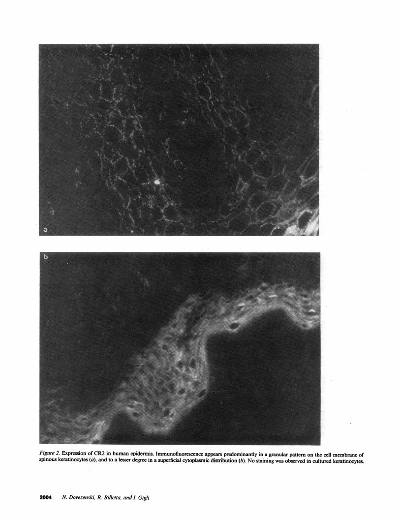
Figure 2. Expression of CR2 in human epidermis. Immunofluorescence appears predominantly in a granular pattern on the cell membrane ofspinous keratinocytes (a), and to a lesser degree in a superficial cytoplasmic distribution (b). No staining was observed in cultured keratinocytes.
2004 N. Dovezenski, R. Billetta, and I. Gigli
A
B
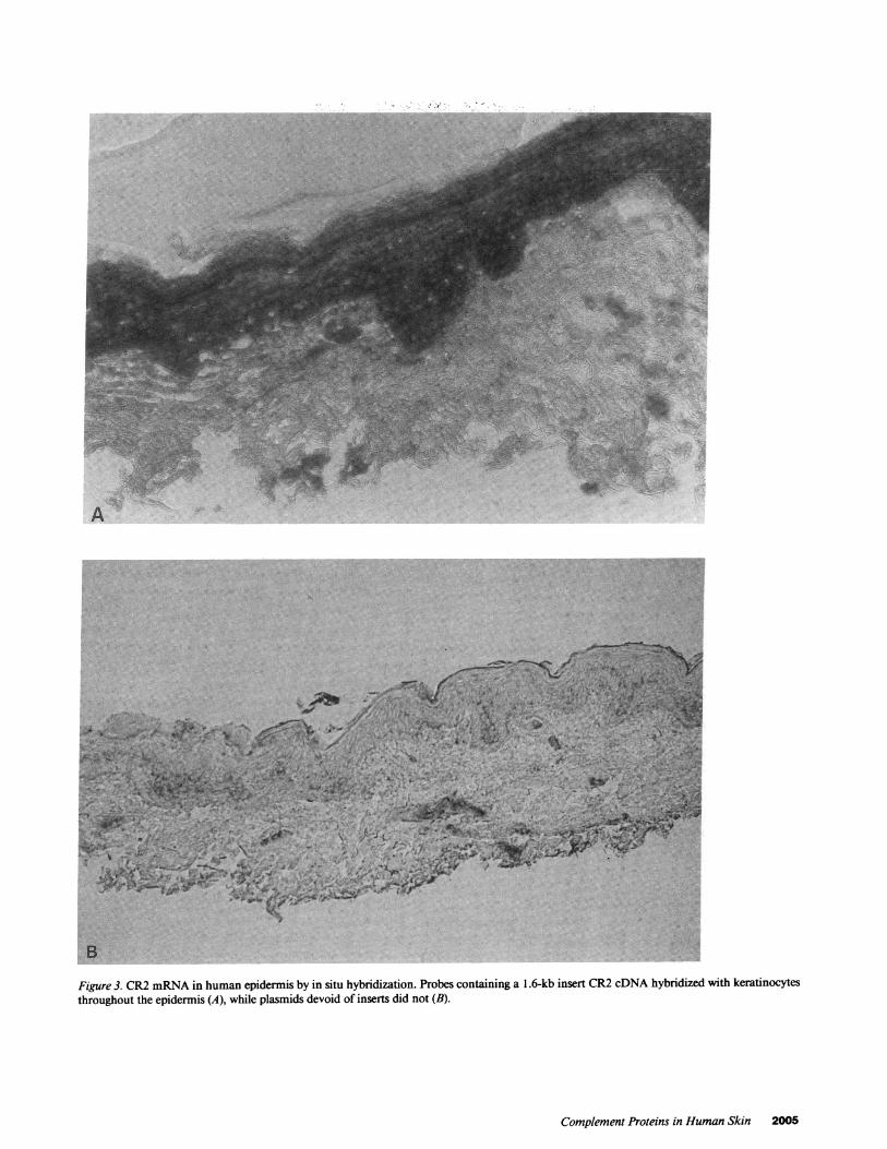
Figure 3. CR2 mRNA in human epidermis by in situ hybridization. Probes containing a 1.6-kb insert CR2 cDNA hybridized with keratinocytesthroughout the epidermis (A), while plasmids devoid of inserts did not (B).
Complement Proteins in Human Skin 2005
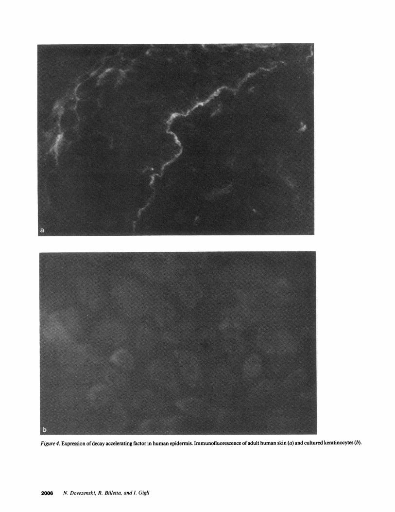
Figure 4. Expression ofdecay accelerating factor in human epidermis. Immunofluorescence ofadult human skin (a) and cultured keratinocytes (b).
2006 N. Dovezenski, R. Billetta, and I. Gigli
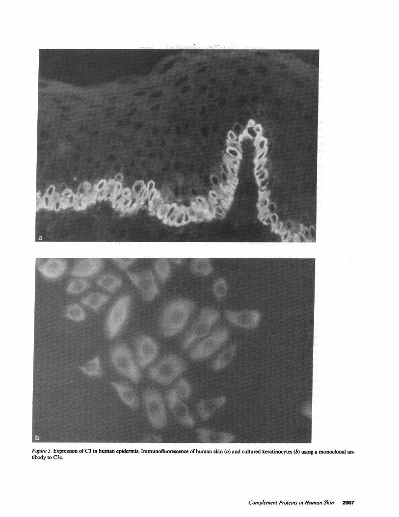
Figure 5. Expression ofC3 in human epidermis. Immunofluorescence ofhuman skin (a) and cultured keratinocytes (b) using a monoclonal an-tibody to C3c.
Complement Proteins in Human Skin 2007
(Fig. 1 A). This receptor was also detected by immunofluores-cence in cultured keratinocytes where it could be observedthroughout the cells (Fig. 1 B). In situ hybridization using di-goxigenin labeled CR1 cDNA showed the expression of thismRNA distributed primarily in the keratinocytes of the lowerhalfofthe epidermis (Fig. 1 C). The negative control consistingof isotypic IgG is shown in Fig. 1 D.
CR2 was visualized using the monoclonal antibody HB5. Itwas present on keratinocytes ofthe superficial two-thirds oftheepidermis, and no fluorescence was observed in the basal layer.It appeared predominantly in a granular pattern on the cellmembrane of spinous keratinocytes (Fig. 2 a), and to a lesserdegree in a superficial cytoplasmic distribution (Fig. 2 b). Nostaining could be observed in skin reacted with another anti-CR2 monoclonal antibody, OKB7. Nick-translated probes ob-tained from the 1.6-kb insert CR2 cDNA, hybridized with ker-atinocytes throughout the epidermis, whereas plasmids devoidof inserts did not (Fig. 3, A and B). Furthermore, labeled Dig-DUTP antisense ssDNA probes were obtained by "run offsyn-thesis" and hybridized to the cytoplasm of keratinocytes withthe same distribution. Sense strands were negative (not shown).
Another membrane associated regulatory protein, the de-cay accelerating factor, previously demonstrated in the elasticfibers in the dermis (20), was also investigated. Anti-DAFmonoclonal antibodies reacted with elastic fibers in the dermis,but in addition, we found a linear continuous deposit ofDAFin the basement membrane zone (Fig. 4 a). Contrary to find-ings with CR1 and CR2, DAF was differentially expressed innewborn foreskin and adult skin. DAF expression predomi-nated in the elastic fibers of adult skin while the basal mem-brane deposits were more striking in newborn foreskin (Fig. 4a). Cultured keratinocytes from adult skin showed a faint, butrecognizable, fluorescence, which in most cells was concen-trated at one pole (Fig. 4 b).
Complement proteins in human epidermis and culturedkeratinocytes. The presence ofC3 was investigated using mono-clonal antibodies to the C3c portion of the molecule. Thesereacted with the cytoplasm ofthe keratinocytes localized in thebasal layer of the epidermis (Fig. 5 a). Keratinocytes in culturealso showed strong cytoplasmic staining (Fig. 5 b). In agree-ment with a previous report (21), we also observed the presenceof the C3d fragment of C3 localized in the area of the basalmembrane in a linear pattern (not shown). Identical distribu-tion of fluorescence was visualized when a monoclonal anti-body raised against the C4d fragment of C4 was used in onesample (not shown), but no staining was noted with anti-C4 oranti-C4c antibodies.
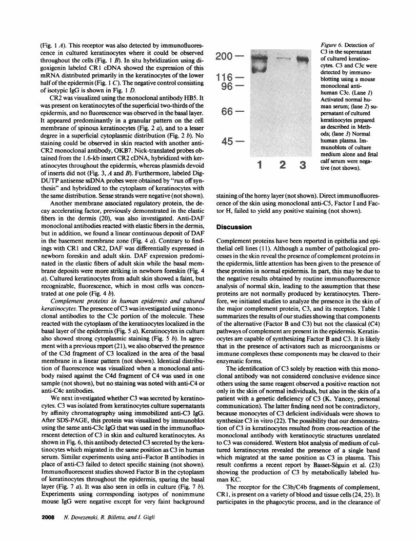
We next investigated whether C3 was secreted by keratino-cytes. C3 was isolated from keratinocytes culture supernatantsby affinity chromatography using immobilized anti-C3 IgG.After SDS-PAGE, this protein was visualized by immunoblotusing the same anti-C3c IgG that was used in the immunofluo-rescent detection ofC3 in skin and cultured keratinocytes. Asshown in Fig. 6, this antibody detected C3 secreted by the kera-tinocytes which migrated in the same position as C3 in humanserum. Similar experiments using anti-Factor B antibodies inplace of anti-C3 failed to detect specific staining (not shown).Immunofluorescent studies showed Factor B in the cytoplasmof keratinocytes throughout the epidermis, sparing the basallayer (Fig. 7 a). It was also seen in cells in culture (Fig. 7 b).Experiments using corresponding isotypes of nonimmunemouse IgG were negative except for very faint background
Figure 6. Detection ofC3 in the supernatant
200 of cultured keratino-cytes. C3 and C3c weredetected by immuno-
1 1 6 blotting using a mouse
96 monoclonal anti-human C3c. (Lane 1)Activated normal hu-man serum; (lane 2) su-
66- pernatant of culturedkeratinocytes preparedas described in Meth-ods; (lane 3) Normal
45 human plasma. Im-munoblots of culturemedium alone and fetal
12e calf serum were nega-2 3 tive (not shown).
staining ofthe horny layer (not shown). Direct immunofluores-cence of the skin using monoclonal anti-C5, Factor I and Fac-tor H, failed to yield any positive staining (not shown).
Discussion
Complement proteins have been reported in epithelia and epi-thelial cell lines (1 1). Although a number of pathological pro-cesses in the skin reveal the presence ofcomplement proteins inthe epidermis, little attention has been given to the presence ofthese proteins in normal epidermis. In part, this may be due tothe negative results obtained by routine immunofluorescenceanalysis of normal skin, leading to the assumption that theseproteins are not normally produced by keratinocytes. There-fore, we initiated studies to analyze the presence in the skin ofthe major complement protein, C3, and its receptors. Table Isummarizes the results ofour studies showing that componentsof the alternative (Factor B and C3) but not the classical (C4)pathways ofcomplement are present in the epidermis. Keratin-ocytes are capable of synthesizing Factor B and C3. It is likelythat in the presence of activators such as microorganisms orimmune complexes these components may be cleaved to theirenzymatic forms.
The identification ofC3 solely by reaction with this mono-clonal antibody was not considered conclusive evidence sinceothers using the same reagent observed a positive reaction notonly in the skin ofnormal individuals, but also in the skin ofapatient with a genetic deficiency of C3 (K. Yancey, personalcommunication). The latter finding need not be contradictory,because monocytes of C3 deficient individuals were shown tosynthesize C3 in vitro (22). The possibility that our demonstra-tion ofC3 in keratinocytes resulted from cross-reaction of themonoclonal antibody with keratinocytic structures unrelatedto C3 was considered. Western blot analysis ofmedium ofcul-tured keratinocytes revealed the presence of a single bandwhich migrated at the same position as C3 in plasma. Thisresult confirms a recent report by Basset-Seguin et al. (23)showing the production of C3 by metabolically labeled hu-man KC.
The receptor for the C3b/C4b fragments of complement,CR1, is present on a variety ofblood and tissue cells (24, 25). Itparticipates in the phagocytic process, and in the clearance of
2008 N. Dovezenski, R. Billetta, and I. Gigli
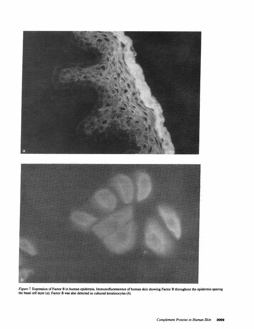
Figure 7. Expression of Factor B in human epidermis. Immunofluorescence ofhuman skin showing Factor B throughout the epidermis sparingthe basal cell layer (a). Factor B was also detected in cultured keratinocytes (b).
Complement Proteins in Human Skin 2009

Table I. Complement Proteins in the Skin and KC*
Human skinKC* SKCt
In situI1 hybridization IF Western blot
ComponentC3 + (K)II nd! + + (180-kd protein)FB +(K) nd +C4 nd - ndC3d + (DEJ)** nd - ndCR1 + (Basal K) + + ndCR2 + (Spinous K) + - ndDAF + (Dermis, DEJ, K) nd + nd
ControlsIsotypic IgGLabeled plasmid
* KC, Cultured keratinocytes. $SKC, Supernatants of cultured keratinocytes. IF, Immunofluorescence. 1K, Tissue keratinocytes. 'nd, Notdone. ** DEJ, Dermoepidermal junction.
immune complexes as well as regulating complement activa-tion (6, 26, 27). We detected CR1 on keratinocytes located inthe basal layer ofthe epidermis. Previous studies have reportedthat another cell surface complement regulatory protein withFactor I cofactor activity, MCP, is present on human epithelialcells (28). These investigators immunoprecipitated MCP fromnoncutaneous epithelial cell lines, but were unable to detectCR1 or CR2 (28). The latter is of interest, and may indicate adistinct regulatory process for CR1 and CR2 expression in tis-sues. CR1 has been reported to be absent in the epidermis (29,30) using C3b as the ligand. This difference may be explainedby the use of monoclonal antibodies in our experiments whichprovide higher sensitivity. The demonstration ofCR1 mRNAin epidermal keratinocytes by in situ hybridization, furthersupports the presence of CR1. The expression of this receptorwould enable these cells to bind C3b molecules which, in con-junction with Factor I, will cleave C3b to C3bi (31). C3bi willremain bound to CR1 and undergo further degradation toC3dg and C3c, or will bind to cells bearing C3bi receptors(CR3) (32). Furthermore, because CR1 functions by accelerat-ing the natural decay ofthe C3 and C5 convertases ofboth theclassical and alternative pathway (6, 33), the further generationof activated complement components in the skin will be re-stricted. CR1, and in particular, its soluble form, has recentlybeen shown to be a very potent inhibitor ofcomplement activa-tion (34), thus its presence on keratinocytes may provide pro-tection from random deposition ofC3b in a milieu where com-plement activation is proceeding. Whether CR 1, as it has beenshown with other cell types (35), endows keratinocytes with thecapability of internalizing particles bearing C3b is not known.Although further research is required to confirm this notion,keratinocytes have been shown to phagocytize erythrocytesunder special conditions (36).
The receptor for the C3d fragment of C3 (CR2) has beenshown to be the Epstein-Barr virus receptor (16, 37). A numberofreports have provided evidence that EBV can infect the skin;this virus has been detected in keratinocytes of the skin (38)and mucous membranes of immunosuppressed patients (39).Its receptors (CR2/EBVR) are present, on B lymphocytes, on
the surface ofcultured cervical epithelial cells, squamous carci-noma cell lines, nasopharyngeal (40), and oral mucosal epithe-lium (41). In this study, we revealed by immunofluorescencethe binding ofanti-CR2 MoAb HB5, to the membrane ofkera-tinocytes. Young et al. (40) have recently reported the bindingofthree out ofnine anti-CR2 MoAb to epithelial cells. Immun-oprecipitation of metabolically labeled epithelial cells withHB5 antibody has been reported to reveal a molecule of 200kD (40) which differs from the 145-kD CR2 precipitated fromB cells (42). It was concluded that this size difference may repre-sent posttranslational modifications of the protein, or alterna-tively, that both are the product of two distinct but relatedgenes. The hybridization of CR2 cDNA to keratinocytes sug-gests the presence ofCR2 mRNA; however, sequencing of theputative epithelial CR2 will be required to confirm the natureofthis molecule and definitely rule out cross-hybridization to arelated gene. The presence ofCR2 on keratinocytes could pro-vide a port of entry to EBV infection, manifesting itself in dis-orders of the skin and mucous membranes as reported earlier(38, 39). Whether EBV plays a role in other dermatologic dis-eases remains to be studied. Additionally, CR2 functions as thereceptor for the C3d fragment ofC3 (7). As such, on B cells, thisreceptor regulates B cell responses, particularly to T cell prod-ucts. Differentiation and proliferation of B cells have been re-ported using cross-linked C3d or anti-CR2 antibodies (43-48).These studies suggest that cross-linking CR2 on cell surfaces isinvolved in regulating B cell activation. Therefore, it may bespeculated that it could have regulatory effects in epidermalcell proliferation through its interaction with its ligand, C3d.
DAF is a glycoprotein present on the surface ofneutrophils,monocytes, lymphocytes, platelets (49, 50) and a variety ofhuman epithelial cells as well as in a soluble form in body fluids(51). The only known function ofDAF consists in acceleratingthe decay of the classical and alternative pathway C3 and CSconvertases (9, 52). This is believed to control the functionalpresence and deleterious effects ofthese enzyme complexes onthe surface of autologous cells during complement activation,where C3b may be randomly deposited on their membranes.DAF has been reported to be present on the periphery ofelastic
2010 N. Dovezenski, R. Billetta, and L Gigli
fibers in the skin (20), as well as on the surface of keratinocytes(53). In our study, a similar distribution ofDAF was observed,but we were also able to identify it along the basal membrane ofthe epidermis. A possible function for DAF at this locationcould consist in preventing the deposition of the products ofcomplement activation in this location. Whether DAF at thatsite is the result of local production or entrapment ofthe circu-lating soluble form of this glycoprotein is not known. Overall,these studies indicate that since the epidermis is continuouslyexposed to foreign antigens, the local synthesis ofcomplementproteins may contribute to the effective processing of theseantigens. In the presence of activators of complement, locallyproduced C3 and epidermal proteases may generate C3b andcontribute to the formation of the initial C3 convertase of thealternative pathway. Deposited C3b may be further cleavedgenerating fragments that may interact with complement re-ceptors on keratinocytes and immunocompetent cells. It is pos-tulated that stimulation ofcomplement receptors on keratino-cytes may exert similar differentiation effects as seen on B cells.
Acknowledgments
The authors wish to thank Susan Krayzel and Teresa Torbett for theirsecretarial support. The authors also wish to thank Ms. Angie Grimesfrom Clonetics Co., San Diego, CA, for providing isolated bovine pitu-itary extract present in KGMh.
This research was supported by grants AI20067 and A120476 fromthe National Institutes of Health. Dr. Dovezenski is a recipient of agrant from the Scientific Foundation of the Republic of Serbia, Yugo-slavia.
References
1. Gigli, I., and F. A. Tausk. 1987. Human complement system. In Dermatol-ogy in General Medicine. T. B. Fitzpatrick, A. Z. Eisen, K. Wolff, I. M. Freed-berg, and K. F. Austen, editors. McGraw-Hill Inc., New York. 442-456.
2. Muller-Eberhard, H. J. 1984. The membrane attack complex. SpringerSemin. Immunopathol. 7:93-141.
3. Nilsson, U. R., and H. J. Mililer-Eberhard. 1965. Isolation ofbetaIF-globu-lin from human serum and its characterization as the fifth component ofcomple-ment. J. Exp. Med. 122:277-298.
4. Nelson, R. A., J. Jensen, I. Gigli, and N. Tamura. 1966. Methods for theseparation, purification and measurement of nine components of hemolyticcomplement in guinea pig serum. Immunochemistry. 3:111-135.
5. Ferreira, A., M. Takahashi, and V. Nussenzweig. 1977. Purification andcharacterization ofmouse serum protein with specific binding affinity for C4 (SSprotein). J. Exp. Med. 146:1001-1018.
6. Fearon, D. B. 1979. Regulation of the amplification C3 convertase of hu-man complement by an inhibitory protein isolated from human erythrocytemembrane. Proc. Nail. Acad. Sci. USA. 76:5867-5871.
7. Ross, G. D., M. J. Polley, E. M. Rabellino, and R. M. Grey. 1973. Twodifferent complement receptors on human lymphocytes, one specific forC3d andone specific for C3b inactivator-cleaved C3b. J. Exp. Med. 138:798-81 1.
8. Seya, T., J. Turner, and J. P. Atkinson. 1986. Purification and characteriza-tion of a membrane protein (gp 45-70) that is a cofactor for cleavage ofC3b andC4b. J. Exp. Med. 163:837-855.
9. Nicholson-Weller, A., J. Burge, D. T. Fearon, P. F. Weller, and K. F.Austen. 1982. Isolation of a human erythrocyte membrane glycoprotein withdecay-accelerating activity for C3 convertases of the complement system. J. Im-munol. 129:184-189.
10. Zalman, L. S., L. M. Wood, and H. J. Muller-Eberhard. 1986. Isolation ofa human erythrocyte membrane protein capable of inhibiting expression of ho-mologous complement transmembrane channel. Proc. Natl. Acad. Sci. USA.83:6975-6979.
11. Cole, F. S., and H. R. Colten. 1988. In The Complement System. K.Rother and G. 0. Till, editors. Springer-Verlag, Heidelberg.
12. Tausk, F. A., J. A. McCutchan, P. Spechko, R. D. Schreiber, and I. Gigli.
1986. Altered erythrocyte C3b receptor expression, immune complexes, andcomplement activation in homosexual men in varying risk groups for acquiredimmune deficiency syndrome. J. Clin. Invest. 78:977-982.
13. Boyce, S. T., and R. B. Ham. 1983. Calcium-regulated differentiation ofnormal human epidermal keratinocytes in chemically defined clonal culture andserum-free social culture. J. Invest. Dermatol. 81:35s-40s.
14. Laemmli, U. K. 1970. Cleavage ofstructural proteins during the assemblyof the head of bacteriophage T4. Nature (Lond.). 227:680-685.
15. Klickstein, L. B., T. J. Bartow, V. Miletic, L. D. Rabson, J. A. Smith, andD. T. Fearon. 1988. Identification ofdistinct C3b and C4b recognition sites in thehuman C3b/C4b receptor (CR1, CD35) by deletion mutagenesis. J. Exp. Med.168:1699-1717.
16. Moore, M. D., N. R. Cooper, B. F. Tack, and G. R. Nemerow. 1987.Molecular cloning of the cDNA encoding the Epstein-Barr virus/C3d receptor(complement receptor type 2) of human B lymphocytes. Proc. Natl. Acad. Sci.USA. 84:9194-9198.
17. Maniatis, T., E. F. Fritsch, and J. Sambrook. 1989. Molecular Cloning: ALaboratory Manual. Cold Spring Harbor Laboratory, Cold Spring Harbor, NY.
18. Sturzl, M., and W. K. Roth. 1990. "Run-off" synthesis and application ofdefined single-stranded DNA hybridization probes. Anal. Biochem. 185:164-169.
19. Lanzillo, J. J. 1990. Preparation of digoxigenin-labeled probes by thepolymerase chain reaction. Biotechniques. 8:620-622.
20. Werth, V. P., I. E. Ivanov, and V. Nussenzweig. 1988. Decay-acceleratingfactor in human skin is associated with elastic fibers. J. Invest. Dermatol. 91:51 1-516.
21. Basset-Seguin, N., M. Dersookian, K. Cehrs, and K. Yancey. 1988. C3d,gis present in normal human epidermal basement membrane. J. Immunol.141:1273-1280.
22. Einstein, L. P., P. J. Hansen, M. Ballow, A. E. Davis, J. S. Davis, C. A.Alper, F. S. Rosen, and H. R. Colten. 1977. Biosynthesis ofthe third componentof complement (C3) in vitro by monocytes from both normal and homozygousC3 deficient humans. J. Clin. Invest. 60:963-969.
23. Basset-Seguin, N., S. W. Caughman, and K. B. Yancey. 1990. A-431 cellsand human keratinocytes synthesize and secrete the third component ofcomple-ment. J. Invest. Dermatol. 95:621-625.
24. Berman, B., and I. Gigli. 1980. Complement receptors on guinea pigepidermal Langerhans cells. J. Immunol. 124:685-690.
25. Fearon, D. T. 1980. Identification of the membrane glycoprotein that isthe C3b receptor of the human erythrocyte, polymorphonuclear leukocyte, Blymphocyte and monocyte. J. Exp. Med. 152:20-30.
26. Cornacoff, J. D., L. A. Herbert, W. L. Smead, M. E. van Aman, D. J.Birmingham, and F. J. Waxman. 1983. Primate erythrocyte immune complexclearing mechanism. J. Exp. Med. 7 1:236-247.
27. Medof, E., and V. Nussenzweig. 1984. Control of the function of sub-strate-bound C4b-C3b by the complement receptorCR 1. J. Exp. Med. 159:1669-1685.
28. NcNearney, T., L. Ballard, T. Seya, and J. P. Atkinson. 1989. Membranecofactor protein of complement is present on human fibroblast, epithelial, andendothelial cells. J. Clin. Invest. 84:538-545.
29. Schreiber, L., and R. Penny. 1979. Tissue C3b receptors. Clin. Exp. Im-munol. 38:316-322.
30. Thyresson, H. N. M., F. C. McDuffie, and A. L. Schroeter. 1981. C3breceptor in normal human skin. J. Invest. Dermatol. 77:353-357.
31. Medof, M. E., K. lida, C. Mold, and V. Nussenzweig. 1982. Unique role ofthe complement receptorCR I in the degradation ofC3b associated with immunecomplexes. J. Exp. Med. 156:1739-1754.
32. Ross, G. D., J. D. Lambris, J. A. Cain, and S. L. Newman. 1982. Genera-tion of three different fragments of bound C3 with purified factor I or serum. I.Requirements for factor H vs. CR1 cofactor activity. J. Immunol. 129:2051-2060.
33. Wilson, J. G., W. W. Wong, P. H. Schur, and D. T. Fearon. Mode ofinheritance of decreased C3b receptors on erythrocytes of patients with systemiclupus erythematosus. N. Engl. J. Med. 307:981-986.
34. Weisman, H. F., T. Bartow, M. K. Leppo, H. C. Marsh, Jr., G. R. Carson,M. F. Concino, M. P. Boyle, K. H. Roux, M. L. Weisfeldt, and D. T. Fearon.1990. Soluble human complement receptor type 1: in vivo inhibitor of comple-ment suppressing post-ischemic myocardial inflammation and necrosis. Science(Wash. DC). 249:146-15 1.
35. Gigli, I., and R. A. Nelson. 1968. Complement dependent immune phago-cytosis. I. Requirements for C1, C4, C2, C3. Exp. Cell Res. 51:45-67.
36. Schenk, P. 1986. Erythrophagocytosis in Kaposi's sarcoma in acquiredimmune deficiency syndrome. ORL (Oto-Rhino-Laryngol) (Israel). 48:167-173.
37. Fingeroth, J. D., J. J. Weis, T. F. Tedder, J. L. Strominger, P. A. Biro, andD. T. Fearon. 1984. Epstein-Barr virus receptor of human B lymphocytes is theC3d receptor C2. Proc. Nall. Acad. Sci. USA. 81:4510-4514.
38. Fermand, J.-P., J. Gozlan, A. Bendelac, M.-C. Delauche-Cavallier, J.-C.
Complement Proteins in Human Skin 2011
Brouet, and F. Morinet. 1990. Detection of Epstein-Barr virus in epidermal skinlesions of an immunocompromised patient. Ann. Intern. Med. 112:511-515.
39. Greenspan, J. S., D. Greenspan, and E. T. Lennette. 1985. Replication ofEpstein-Barr virus within the epithelial cells oforal "hairy" leukoplakia, an AIDSassociated lesion. N. Engl. J. Med. 313:1564-1571.
40. Young, L. S., C. W. Dawson, K. W. Brown, and A. B. Rickinson. 1989.Identification of a human epithelial cell surface protein sharing an epitope withthe C3d/Epstein-Barr virus receptor molecule of B lymphocytes. Int. J. Cancer.43:786-794.
41. Corso, B., L. F. Eversole, and L. Hutt-Fletcher. 1989. Hairy leukoplakia:Epstein-Barr virus receptors on oral keratinocyte plasma membranes. Oral Surg.Oral Med. Pathol. 67:416-421.
42. Deleted in proof.43. Tedder, T. F., J. J. Weis, L. T. Clement, D. T. Fearon, and M. D. Cooper.
1986. The role of receptors to complement in induction of polyclonal B-cellproliferation and differentiation. J. Clin. Immunol. 6:65-73.
44. Perri, R. T., B. S. Wilson, and N. E. Kay. 1986. Inhibition ofB cell growthfactor (BCGF) by monoclonal antibodies directed against the C3d receptor(CR2). Eur. J. Immunol. 16:350-355.
45. Carter, R. H., M. 0. Spycher, Y. C. Ng, R. Hoffman, and D. T. Fearon.1988. Synergistic interaction between complement receptor type 2 and mem-brane IgM and B lymphocytes. J. Immunol. 141:457-463.
46. Wilson, B. S., J. L. Platt, and N. E. Kay. 1985. Monoclonal antibodies to
the 140,000 molecular weight glycoprotein of B lymphocyte membranes (CR2receptor) initiates proliferation of B cells in vitro. Blood. 66:824-829.
47. Frade, R. 1986. Structure and functions ofgp 140, the C3d/EBV receptor(CR2) of human B lymphocytes. Mol. Immunol. 23:1249-1253.
48. Melchers, F., A. Erdei, T. Schult, and M. P. Dierich. 1985. Growth controlof activated synchronized murine B cells by the C3 fragment of human comple-ment. Nature (Lond.). 317:264-267.
49. Nicholson-Weller, A., J. P. March, C. E. Rosen, D. B. Spicer, and K. F.Austen. 1985. Surface membrane expression by human blood leukocytes andplatelets of decay-accelerating factor, a regulatory protein, of the complementsystem. Blood. 65:1237-1244.
50. Kinoshita, T., M. E. Medof, R. Silber, and V. Nussenzweig. 1985. Distri-bution of decay-accelerating factor in the peripheral blood of normal individualsand patients with paroxysmal nocturnal hemogobinuria. J. Exp. Med. 162:75-92.
51. Medof, M. E., E. I. Walter, J. L. Rutgers, D. M. Knowles, and V. Nussen-zweig. 1987. Identification of the complement decay-accelerating factor (DAF)on epithelium and glandular cells and in body fluids. J. Exp. Med. 165:848-864.
52. Fujita, T., T. Inoue, K. Ogawa, K. lida, and N. Tamura. 1987. The mecha-nism of action of decay-accelerating factor (DAF). DAF inhibits the assembly ofC3 convertases by dissociating C2a and Bb. J. Exp. Med. 166:1221-1228.
53. Sayama, K., S. Shiraishi, Y. Shyirakata, Y. Kobayashi, and Y. Miki. 1991.Characterization of decay-accelerating factor (DAF) in human skin. J. Invest.Dermatol. 96:61-64.
2012 N. Dovezenski, R. Billetta, and I. Gigli