Exposure to Multiple Parasites Is Associated with the Prevalence of Active Convulsive Epilepsy in...
10
Exposure to Multiple Parasites Is Associated with the Prevalence of Active Convulsive Epilepsy in Sub-Saharan Africa Gathoni Kamuyu 1,2 *, Christian Bottomley 3,4,5 , James Mageto 1,6 , Brett Lowe 1,2,7 , Patricia P. Wilkins 8 , John C. Noh 8 , Thomas B. Nutman 9 , Anthony K. Ngugi 1,2,10 , Rachael Odhiambo 1 , Ryan G. Wagner 11,12 , Angelina Kakooza-Mwesige 2,13,14 , Seth Owusu-Agyei 15 , Kenneth Ae-Ngibise 15 , Honorati Masanja 16 , Faith H. A. Osier 1 , Peter Odermatt 17,18 , Charles R. Newton 1,2,19,20,21 , on behalf of the Study of Epidemiology of Epilepsy in Demographic Sites (SEEDS) group " 1 KEMRI/Wellcome Trust Research Programme, The Centre of Geographical Medicine Research – Coast, Kilifi, Kenya, 2 Studies of the Epidemiology of Epilepsy in Demographic Surveillance Systems (SEEDS)-INDEPTH Network, Accra, Ghana, 3 Department of Infectious Disease Epidemiology, Faculty of Epidemiology and Population Health, London School of Hygiene and Tropical Medicine, London, United Kingdom, 4 MRC Tropical Epidemiology Group, Faculty of Epidemiology and Population Health, London School of Hygiene and Tropical Medicine, London, United Kingdom, 5 Faculty of Infectious and Tropical Diseases, London School of Hygiene and Tropical Medicine, London, United Kingdom, 6 Egerton University, Nakuru, Kenya, 7 Nuffield Department of Clinical Medicine, University of Oxford, Oxford, United Kingdom, 8 Division of Parasitic Diseases and Malaria, Centers for Disease Control and Prevention (CDC), Atlanta, Georgia, United States of America, 9 Laboratory of Parasitic Diseases. National Institute of Allergy and Infectious Diseases, Bethesda, Maryland, United States of America, 10 Research Support Unit, Faculty of Health Sciences, Aga Khan University (East Africa), Nairobi, Kenya, 11 MRC/Wits Rural Public Health & Health Transitions Research Unit (Agincourt), School of Public Health, Faculty of Health Sciences, University of the Witwatersrand, Johannesburg, South Africa, 12 Epidemiology and Public Health, Department of Public Health and Clinical Medicine, Umea ˚ University, Umea ˚, Sweden, 13 Iganga-Mayuge Health and Demographic Surveillance System, Iganga, Uganda, 14 Department of Paediatrics and Child Health, Makerere University College of Health Sciences, Kampala, Uganda, 15 Kintampo Health Research Centre, Kintampo, Ghana, 16 Ifakara Health Institute, Ifakara, Tanzania, 17 Swiss Tropical and Public Health Institute, Basel, Switzerland, 18 Unversity of Basel, Basel, Switzerland, 19 Neurosciences Unit, UCL Institute of Child Health, London, United Kingdom, 20 Clinical Research Unit, London School of Hygiene and Tropical Medicine, London, United Kingdom, 21 Department of Psychiatry, University of Oxford, Oxford, United Kingdom Abstract Background: Epilepsy is common in developing countries, and it is often associated with parasitic infections. We investigated the relationship between exposure to parasitic infections, particularly multiple infections and active convulsive epilepsy (ACE), in five sites across sub-Saharan Africa. Methods and Findings: A case-control design that matched on age and location was used. Blood samples were collected from 986 prevalent cases and 1,313 age-matched community controls and tested for presence of antibodies to Onchocerca volvulus, Toxocara canis, Toxoplasma gondii, Plasmodium falciparum, Taenia solium and HIV. Exposure (seropositivity) to Onchocerca volvulus (OR = 1.98; 95%CI: 1.52–2.58, p,0.001), Toxocara canis (OR = 1.52; 95%CI: 1.23–1.87, p,0.001), Toxoplasma gondii (OR = 1.28; 95%CI: 1.04–1.56, p = 0.018) and higher antibody levels (top tertile) to Toxocara canis (OR = 1.70; 95%CI: 1.30–2.24, p,0.001) were associated with an increased prevalence of ACE. Exposure to multiple infections was common (73.8% of cases and 65.5% of controls had been exposed to two or more infections), and for T. gondii and O. volvulus co-infection, their combined effect on the prevalence of ACE, as determined by the relative excess risk due to interaction (RERI), was more than additive (T. gondii and O. volvulus, RERI = 1.19). The prevalence of T. solium antibodies was low (2.8% of cases and 2.2% of controls) and was not associated with ACE in the study areas. Conclusion: This study investigates how the degree of exposure to parasites and multiple parasitic infections are associated with ACE and may explain conflicting results obtained when only seropositivity is considered. The findings from this study should be further validated. Citation: Kamuyu G, Bottomley C, Mageto J, Lowe B, Wilkins PP, et al. (2014) Exposure to Multiple Parasites Is Associated with the Prevalence of Active Convulsive Epilepsy in Sub-Saharan Africa. PLoS Negl Trop Dis 8(5): e2908. doi:10.1371/journal.pntd.0002908 Editor: Hector H. Garcia, Universidad Peruana Cayetano Heredia, Peru Received June 10, 2013; Accepted April 16, 2014; Published May 29, 2014 This is an open-access article, free of all copyright, and may be freely reproduced, distributed, transmitted, modified, built upon, or otherwise used by anyone for any lawful purpose. The work is made available under the Creative Commons CC0 public domain dedication. Funding: The Wellcome Trust funded this research through a Wellcome Trust Senior Fellowship in Clinical Tropical Medicine (No. 083744 to CRN), and supported GK to write up this work through a strategic training award (No. 084538) and a Wellcome Trust Master’s Training Fellowship (No. 096392) to KEMRI-Wellcome Trust Research Programme. The funders had no role in study design, data collection and analysis, decision to publish, or preparation of the manuscript. Competing Interests: The authors have declared that no competing interests exist. * E-mail: [email protected] " Membership of the Study of Epidemiology of Epilepsy in Demographic Sites (SEEDS) group is provided in the Acknowledgments. PLOS Neglected Tropical Diseases | www.plosntds.org 1 May 2014 | Volume 8 | Issue 5 | e2908
-
Upload
independent -
Category
Documents
-
view
3 -
download
0
Transcript of Exposure to Multiple Parasites Is Associated with the Prevalence of Active Convulsive Epilepsy in...
Exposure to Multiple Parasites Is Associated with thePrevalence of Active Convulsive Epilepsy in Sub-SaharanAfricaGathoni Kamuyu1,2*, Christian Bottomley3,4,5, James Mageto1,6, Brett Lowe1,2,7, Patricia P. Wilkins8,
John C. Noh8, Thomas B. Nutman9, Anthony K. Ngugi1,2,10, Rachael Odhiambo1, Ryan G. Wagner11,12,
Angelina Kakooza-Mwesige2,13,14, Seth Owusu-Agyei15, Kenneth Ae-Ngibise15, Honorati Masanja16,
Faith H. A. Osier1, Peter Odermatt17,18, Charles R. Newton1,2,19,20,21, on behalf of the Study of
Epidemiology of Epilepsy in Demographic Sites (SEEDS) group"
1 KEMRI/Wellcome Trust Research Programme, The Centre of Geographical Medicine Research – Coast, Kilifi, Kenya, 2 Studies of the Epidemiology of Epilepsy in
Demographic Surveillance Systems (SEEDS)-INDEPTH Network, Accra, Ghana, 3 Department of Infectious Disease Epidemiology, Faculty of Epidemiology and Population
Health, London School of Hygiene and Tropical Medicine, London, United Kingdom, 4 MRC Tropical Epidemiology Group, Faculty of Epidemiology and Population Health,
London School of Hygiene and Tropical Medicine, London, United Kingdom, 5 Faculty of Infectious and Tropical Diseases, London School of Hygiene and Tropical
Medicine, London, United Kingdom, 6 Egerton University, Nakuru, Kenya, 7 Nuffield Department of Clinical Medicine, University of Oxford, Oxford, United Kingdom,
8 Division of Parasitic Diseases and Malaria, Centers for Disease Control and Prevention (CDC), Atlanta, Georgia, United States of America, 9 Laboratory of Parasitic
Diseases. National Institute of Allergy and Infectious Diseases, Bethesda, Maryland, United States of America, 10 Research Support Unit, Faculty of Health Sciences, Aga
Khan University (East Africa), Nairobi, Kenya, 11 MRC/Wits Rural Public Health & Health Transitions Research Unit (Agincourt), School of Public Health, Faculty of Health
Sciences, University of the Witwatersrand, Johannesburg, South Africa, 12 Epidemiology and Public Health, Department of Public Health and Clinical Medicine, Umea
University, Umea, Sweden, 13 Iganga-Mayuge Health and Demographic Surveillance System, Iganga, Uganda, 14 Department of Paediatrics and Child Health, Makerere
University College of Health Sciences, Kampala, Uganda, 15 Kintampo Health Research Centre, Kintampo, Ghana, 16 Ifakara Health Institute, Ifakara, Tanzania, 17 Swiss
Tropical and Public Health Institute, Basel, Switzerland, 18 Unversity of Basel, Basel, Switzerland, 19 Neurosciences Unit, UCL Institute of Child Health, London, United
Kingdom, 20 Clinical Research Unit, London School of Hygiene and Tropical Medicine, London, United Kingdom, 21 Department of Psychiatry, University of Oxford,
Oxford, United Kingdom
Abstract
Background: Epilepsy is common in developing countries, and it is often associated with parasitic infections. Weinvestigated the relationship between exposure to parasitic infections, particularly multiple infections and active convulsiveepilepsy (ACE), in five sites across sub-Saharan Africa.
Methods and Findings: A case-control design that matched on age and location was used. Blood samples were collectedfrom 986 prevalent cases and 1,313 age-matched community controls and tested for presence of antibodies to Onchocercavolvulus, Toxocara canis, Toxoplasma gondii, Plasmodium falciparum, Taenia solium and HIV. Exposure (seropositivity) toOnchocerca volvulus (OR = 1.98; 95%CI: 1.52–2.58, p,0.001), Toxocara canis (OR = 1.52; 95%CI: 1.23–1.87, p,0.001),Toxoplasma gondii (OR = 1.28; 95%CI: 1.04–1.56, p = 0.018) and higher antibody levels (top tertile) to Toxocara canis(OR = 1.70; 95%CI: 1.30–2.24, p,0.001) were associated with an increased prevalence of ACE. Exposure to multiple infectionswas common (73.8% of cases and 65.5% of controls had been exposed to two or more infections), and for T. gondii and O.volvulus co-infection, their combined effect on the prevalence of ACE, as determined by the relative excess risk due tointeraction (RERI), was more than additive (T. gondii and O. volvulus, RERI = 1.19). The prevalence of T. solium antibodies waslow (2.8% of cases and 2.2% of controls) and was not associated with ACE in the study areas.
Conclusion: This study investigates how the degree of exposure to parasites and multiple parasitic infections are associatedwith ACE and may explain conflicting results obtained when only seropositivity is considered. The findings from this studyshould be further validated.
Citation: Kamuyu G, Bottomley C, Mageto J, Lowe B, Wilkins PP, et al. (2014) Exposure to Multiple Parasites Is Associated with the Prevalence of Active ConvulsiveEpilepsy in Sub-Saharan Africa. PLoS Negl Trop Dis 8(5): e2908. doi:10.1371/journal.pntd.0002908
Editor: Hector H. Garcia, Universidad Peruana Cayetano Heredia, Peru
Received June 10, 2013; Accepted April 16, 2014; Published May 29, 2014
This is an open-access article, free of all copyright, and may be freely reproduced, distributed, transmitted, modified, built upon, or otherwise used by anyone forany lawful purpose. The work is made available under the Creative Commons CC0 public domain dedication.
Funding: The Wellcome Trust funded this research through a Wellcome Trust Senior Fellowship in Clinical Tropical Medicine (No. 083744 to CRN), and supportedGK to write up this work through a strategic training award (No. 084538) and a Wellcome Trust Master’s Training Fellowship (No. 096392) to KEMRI-WellcomeTrust Research Programme. The funders had no role in study design, data collection and analysis, decision to publish, or preparation of the manuscript.
Competing Interests: The authors have declared that no competing interests exist.
* E-mail: [email protected]
" Membership of the Study of Epidemiology of Epilepsy in Demographic Sites (SEEDS) group is provided in the Acknowledgments.
PLOS Neglected Tropical Diseases | www.plosntds.org 1 May 2014 | Volume 8 | Issue 5 | e2908
Introduction
The prevalence of epilepsy in low and middle-income countries
is higher than in high-income countries, especially in the rural
areas[1,2]. The prevalence is particularly high in sub-Saharan
Africa (SSA)[3] and South America[4], where parasitic infestations
are thought to contribute to the increased burden[5]. Within these
regions, there are areas in which most of the population are
exposed to endemic parasites, and it is not clear why some people
develop epilepsy, whilst others do not.
Many factors are associated with epilepsy in SSA[3,6] with
infections that involve the central nervous system (CNS) represent-
ing common and preventable causes of epilepsy[5]. Some parasitic
infestations manifest in the human CNS, with the clinical
presentation of seizures and are thought to be associated with the
development of epilepsy[5,7]. A small number of studies conducted
in SSA have shown that exposure to helminths, e.g., Toxocara
canis[8,9,10], Onchocerca volvulus [10,11,12,13] and Taenia solium
[10,14], as well as following severe Plasmodium falciparum malar-
ia[15,16] are associated with epilepsy. The relationship between
Toxoplasma gondii and epilepsy has only been explored in one study in
SSA[10], and a review suggests a possible association[17], though
co-infection with human immunodeficiency virus may confound
this relationship. Seizures are observed in HIV-infected individuals
and are mainly associated with opportunistic infections although
HIV infection can independently cause seizures at seroconversion
or at advanced stages[18]. A comprehensive analysis of exposure to
parasitic infestations as well as HIV using the same methodology
across different geographical locations in SSA would help elucidate
the relationship between parasitic infections and epilepsy, and
provide data to guide public health measures.
The objective of the current study was to investigate the
association between active convulsive epilepsy (ACE) and i) the
degree of exposure to parasitic infections (measured by antibody
levels) and ii) exposure to multiple co-incidental parasitic infections.
We used data from a case-control study conducted in five health and
demographic surveillance systems (HDSS) in SSA in which
exposure to the six infections namely: O. volvulus, T. solium, T. canis,
T. gondii, P. falciparum and HIV, was determined by serology.
Methods
Ethics statementAll aspects of the study were approved by the ethics committees
of University College London and the London School of Hygiene
and Tropical Medicine, and by the ethics review boards in each of
the participating countries. All participants or guardians gave
written informed consent. Since some study participants were
minors, parents/guardians provided consent on behalf of all child
participants and all adults provided consent for themselves.
Participants and study designA case control design was used in which prevalent cases of ACE
were compared to community controls. ACE was defined as two
or more unprovoked convulsions (seizures with tonic and/or clonic
movements) occurring at least 24 hours apart with at least one
seizure in the preceding 12 months[6]. The diagnosis for ACE was
initially made by a clinician with special training in epilepsy, and
was confirmed by a panel of neurologists.
Cases and controls were identified from five HDSS in SSA
(Agincourt in South Africa, Kilifi in Kenya, Kintampo in Ghana,
Ifakara in Tanzania and Iganga-Mayuge in Uganda). Exposure to
parasitic infections was measured using plasma samples collected
from a random subsample of all cases and controls. A sample size
of 300 cases per site with an equal number of controls was chosen
to give 80% power to detect an odds ratio (OR) .2.8 (5%
significance level) given a frequency of at least 5% in the controls.
For each case, an age-matched control was selected at random
from a database of individuals in the HDSS. The controls were
frequency matched using the age bands 0–5, 6–12, 13–18, 19–28,
29–49, 50+ years, to account for increasing exposure with age.
Laboratory proceduresExposure to infections was determined by detection of IgG
antibodies to the parasitic antigens as well as HIV. Exposure to O.
volvulus and T. solium was determined in three study sites: Iganga,
Ifakara and Kintampo, where these parasites are endemic.
Exposure to Onchocerca volvulus. Exposure to O. volvulus
was determined using an in-house modification of an anti-OV-
16GST IgG4 ELISA described previously[19]. This assay is highly
sensitive (90.0%) and specific (98.0%) when tested using sera from
other filarial infections[19]. A sample with an optical density (OD)
value greater than the cut-off (mean +3 standard deviation of 30
plasma samples from Agincourt, South Africa, in which oncho-
cerciasis is absent) was classified as being anti-OV16gst IgG4
positive.
Exposure to larval and adult stages of Taenia
solium. Exposure to larval (cysticercosis) and adults (taeniasis)
stages of T. solium was determined using a Western blotting
technique that has been described previously[20]. Nitrocellulose
strips that contained the recombinant rT24H and rES33 antigens,
which detect antibodies to cysticercosis and taeniasis respectively,
were used. Samples were considered positive for cysticercosis or
taeniasis if a brown band was observed in regions of the strip
corresponding to rT24H or rES33 antigen respectively[21,22]. A
similar technique (MAPIA), is reported to be 97.0% sensitive and
99.4% specific for detection of cases with two or more viable cysts
in the brain and 99.4% sensitive and 93.9% specific for detecting
cases of taeniasis[20].
Exposure to Toxocara canis. Anti-T. canis total IgG
antibodies were detected using a commercial kit (Toxocara IgG-
ELISA, Cypress Diagnostics, Belgium) that is based on the
Toxocara excretory secretory antigen (TES) and used according to
manufacturer’s instructions. The cut-off value was calculated by
dividing the sample test OD by the average OD of negative
controls plus 0.150 OD units according to manufacturer’s
instructions. A sample with a cut-off ratio .1.1 was interpreted
as positive and tested for anti-Toxocara IgG4 antibodies, to increase
the specificity of the assay. Anti-Toxocara IgG4 antibodies were
Author Summary
The prevalence of epilepsy is greater in developingcountries compared to developed countries, and parasiticinfestations are thought to contribute to this increasedburden. We conducted a case-control study across fivesites in sub-Saharan Africa to investigate the relationshipbetween epilepsy and exposure to parasitic infections, andthe association between epilepsy and multiple co-inciden-tal infections. Exposure to Onchocerca volvulus, Toxocaracanis and Toxoplasma gondii as well as high antibodylevels (top tertile) to Toxocara canis was positivelyassociated with the prevalence of active convulsiveepilepsy (ACE). Multiple co-incidental parasitic infectionswere common, and the combined effect of T. gondii and O.volvulus co-infection on ACE was greater than the sum ofthe individual effects. The contribution of each of theseparasitic infections on the burden of epilepsy in sub-Saharan Africa should be explored.
Parasites and Risk of Active Convulsive Epilepsy
PLOS Neglected Tropical Diseases | www.plosntds.org 2 May 2014 | Volume 8 | Issue 5 | e2908
detected using the same TES-precoated plates with the
following modifications. Mouse anti-human IgG4 conjugated
to alkaline phosphatase (9190–04, Southern Biotech, USA) as
the secondary antibody and p-nitrophenyl phospate (pNPP)
(N2765, Sigma Aldrich) as the substrate. Serum dilution (1:50)
and secondary antibody dilution (1:500) was optimized by
checkerboard titration. A sample with an OD value greater than
the cut-off (mean +3 standard deviation of 30 Toxocara canis IgG
negative serum samples) was interpreted as being anti-Toxocara
IgG4 positive. The T. canis assay is reported to be 97.0%
sensitive and 78.6% specific[23] and cross-reactive responses
from other soil-transmitted helminths may have been detected
using this assay.
Exposure to Toxoplasma gondii. Anti-T. gondii IgG anti-
bodies were detected using a commercial kit (Toxoplasma IgG-
ELISA, Genesis Diagnostics, United Kingdom) that is based on T.
gondii purified antigens enriched for P30 (SAG1) and used
according to manufacturer’s instructions.
Exposure to Plasmodium falciparum. Exposure to P.
falciparum was determined using an in-house ELISA described
previously[24] that tests for IgG antibodies to crude schizont
extract from P. falciparum A4 clone line derived from ITO parent
strain. A sample with an OD value greater than the cut-off (mean
+3 standard deviation of 30 serum samples from unexposed adults
from United Kingdom) was interpreted as being anti-P. falciparum
IgG positive.
Exposure to HIV. Anti- HIV-1 type and/or anti-HIV-2 type
IgG antibodies and P24 antigen were detected by the 4th
generation screening test, Vironostika HIV Uniform II Ag/Ab
(BioMerieux, France) and was performed according to manufac-
turer’s instructions. The HIV assay was selected as it detects
antibodies to both HIV-1 and HIV-2 type strains whose
geographic distribution varies across SSA.
Statistical analysisLogistic regression was used to model the association between
antibody titre and ACE.
Antibody titre was categorised into tertiles, which were
calculated separately for each study site. The model also included,
as potential confounders: age (0–5, 6–12, 13–18, 19–28, 29–49,
50+ years), sex, study-site, education (none, primary, or secondary
and above), employment and marital status.
Logistic regression was also used to model exposure to multiple
infections. We used adjusted odds ratios obtained from logistic
regression to test for an additive interaction between parasites by
calculating the relative excess risk due to interaction (RERI). The
analysis of interactions was restricted to those parasites where the
association with epilepsy was statistically significant (p,0.05). We
briefly outline the relation between RERI and interaction on an
additive scale.
Under an additive model the following relation holds
p11{p00~(p10{p00)z(p01{p00)
where p11 and p00 correspond to the prevalence when both or
neither parasite risk factor is present, and p10 and p01 represent
the prevalence when one of the risk factors is present. An
interaction on an additive scale can be quantified as the difference
between p11{p00 and (p10{p00)z(p01{p00). If this differ-
ence is positive then the combined effect of the two parasites is
more than the sum of their individual effects. Conversely, if the
difference is negative then the combined effect is less than the sum
of the individual effects. The RERI is obtained by dividing the
difference by p00
RERI~RR11{RR10{RR01z1
Again a positive RERI represents a combined effect that is
greater than that predicted by the additive model and a negative
RERI occurs when the combined effect that is less than additive. We
use odds ratios obtained from logistic regression to approximate
prevalence ratios in the above formula; this approximation is
expected to work well since epilepsy is rare in the study
population[25]. No adjustment for multiple comparisons was made
because this adjustment can lead to errors of interpretation when
strong previous evidence of association is available for several risk
factors [26]. All analyses were conducted using STATA version 12
(StataCorp.College Station, TX, USA).
Results
Antibody prevalence and prevalence of ACE2,032 controls and 1,711 cases were recruited. The sensitivity of
the three-stage survey method was 48.6%[27], and among
controls the rate of refusal ranged between 49.8% in Agincourt
to 57.4% in Kilifi[10]. Blood samples were collected from 986
cases of ACE and 1,313 controls. The number of blood samples
tested for each parasite were as follows: O. volvulus, 535 cases and
836 controls; T. canis, 862 cases and 1,121 controls; T. gondii 971
cases and 1,291 controls; P. falciparum, 986 cases and 1,313
controls; HIV, 977 cases and 1,304 controls; T.solium, 530 cases
and 833 controls. The demographic details of the cases and
controls from each study site are shown in Table S1.
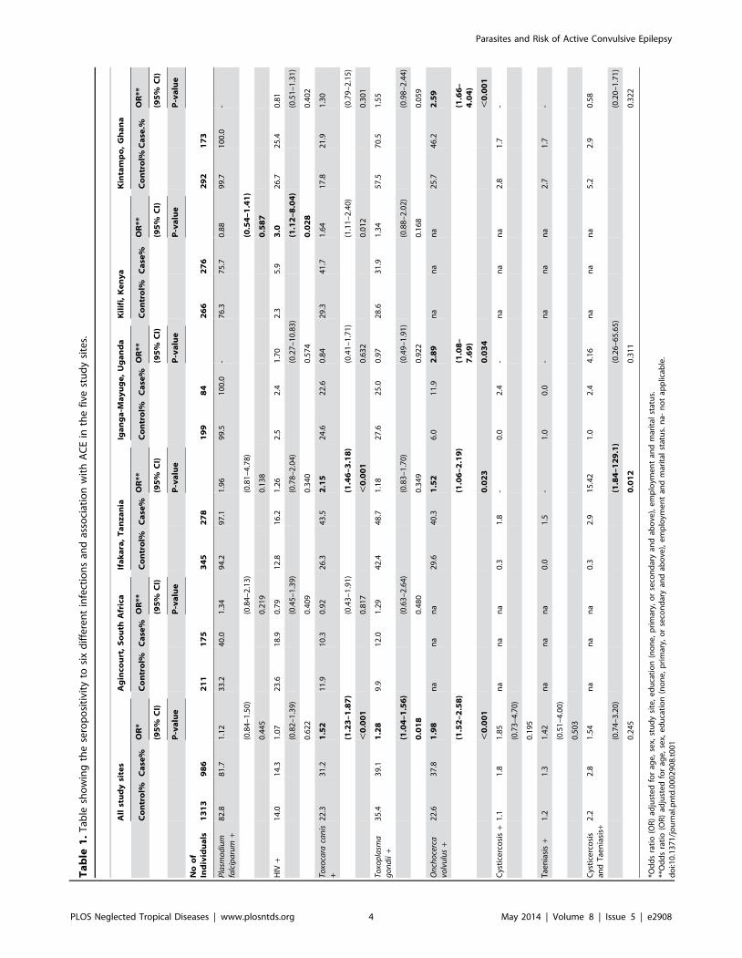
The prevalence of antibodies to the six infections studied varied
between the five study sites (Table 1). The prevalence of antibodies
increased with age in both cases and controls for O. volvulus, T. canis,
T. gondii and P. falciparum (Figure S1–S3, respectively) and these
trends were consistent in all study sites. Almost all study participants
were P. falciparum positive in three sites (Figure S4). Antibody
prevalence to T. solium was low in the three study sites analysed
(Table 1) and showed no trend with age (Figure S5). Similarly,
antibody prevalence to HIV showed no trend with age (Figure S6).
The association between seropositivity and ACE, varied between
different exposures and study sites (Table 1). Significant associations
with ACE include exposure to O. volvulus in all study sites (OR = 1.98;
95%CI: 1.52–2.58, p,0.001), Ifakara (OR = 1.52; 95%CI: 1.06–
2.19, p = 0.023), Iganga-Mayuge (OR = 2.89; 95%CI: 1.08–7.69,
p = 0.034) and Kintampo (OR = 2.59; 95%CI: 1.66–4.04, p,0.001),
exposure to either larval or adult stages of T. solium in Ifakara
(OR = 15.42; 95%CI: 1.84–129.10, p = 0.012), exposure to T. canis in
all study site (OR = 1.52; 95%CI: 1.23–1.87, p,0.001), Ifakara
(OR = 2.15; 95%CI: 1.46–3.18, p,0.001) and Kilifi (OR = 1.64;
95%CI: 1.11–2.40, p = 0.012), T. gondii in all study sites (OR = 1.28;
95%CI: 1.04–1.56, p = 0.018) and exposure to HIV in Kilifi
(OR = 3.00; 95%CI: 1.12–8.04,p = 0.028), all of which were
associated with increased prevalence of ACE [10]. The association
between seropositivity and ACE on analysis of HIV negative
individuals (exclusion of 182 controls and 140 cases who were HIV
positive) had similar results with the exception of T. gondii, in which
the positive association was not statistically significant (Table S6).
Magnitude of antibody response and prevalence of ACEThere was an increase in antibody levels with age in both cases
and controls for O. volvulus, T. canis, T. gondii and P. falciparum
Parasites and Risk of Active Convulsive Epilepsy
PLOS Neglected Tropical Diseases | www.plosntds.org 3 May 2014 | Volume 8 | Issue 5 | e2908
Ta
ble
1.
Tab
lesh
ow
ing
the
sero
po
siti
vity
tosi
xd
iffe
ren
tin
fect
ion
san
das
soci
atio
nw
ith
AC
Ein
the
five
stu
dy
site
s.
All
stu
dy
site
sA
gin
cou
rt,
So
uth
Afr
ica
Ifa
ka
ra,
Ta
nz
an
iaIg
an
ga
-Ma
yu
ge
,U
ga
nd
aK
ilif
i,K
en
ya
Kin
tam
po
,G
ha
na
Co
ntr
ol%
Ca
se%
OR
*C
on
tro
l%C
ase
%O
R**
Co
ntr
ol%
Ca
se%
OR
**C
on
tro
l%C
ase
%O
R**
Co
ntr
ol%
Ca
se%
OR
**C
on
tro
l%C
ase
.%O
R**
(95
%C
I)(9
5%
CI)
(95
%C
I)(9
5%
CI)
(95
%C
I)(9
5%
CI)
P-v
alu
eP
-va
lue
P-v
alu
eP
-va
lue
P-v
alu
eP
-va
lue
No
of
Ind
ivid
ua
ls1
31
39
86
21
11
75
34
52
78
19
98
42
66
27
62
92
17
3
Pla
smo
diu
mfa
lcip
aru
m+
82
.88
1.7
1.1
23
3.2
40
.01
.34
94
.29
7.1
1.9
69
9.5
10
0.0
-7
6.3
75
.70
.88
99
.71
00
.0-
(0.8
4–
1.5
0)
(0.8
4–
2.1
3)
(0.8
1–
4.7
8)
(0.5
4–
1.4
1)
0.4
45
0.2
19
0.1
38
0.5
87
HIV
+1
4.0
14
.31
.07
23
.61
8.9
0.7
91
2.8
16
.21
.26
2.5
2.4
1.7
02
.35
.93
.02
6.7
25
.40
.81
(0.8
2–
1.3
9)
(0.4
5–
1.3
9)
(0.7
8–
2.0
4)
(0.2
7–
10
.83
)(1
.12
–8
.04
)(0
.51
–1
.31
)
0.6
22
0.4
09
0.3
40
0.5
74
0.0
28
0.4
02
Toxo
cara
can
is+
22
.33
1.2
1.5
21
1.9
10
.30
.92
26
.34
3.5
2.1
52
4.6
22
.60
.84
29
.34
1.7
1.6
41
7.8
21
.91
.30
(1.2
3–
1.8
7)
(0.4
3–
1.9
1)
(1.4
6–
3.1
8)
(0.4
1–
1.7
1)
(1.1
1–
2.4
0)
(0.7
9–
2.1
5)
,0
.00
10
.81
7,
0.0
01
0.6
32
0.0
12
0.3
01
Toxo
pla
sma
go
nd
ii+
35
.43
9.1
1.2
89
.91
2.0
1.2
94
2.4
48
.71
.18
27
.62
5.0
0.9
72
8.6
31
.91
.34
57
.57
0.5
1.5
5
(1.0
4–
1.5
6)
(0.6
3–
2.6
4)
(0.8
3–
1.7
0)
(0.4
9–
1.9
1)
(0.8
8–
2.0
2)
(0.9
8–
2.4
4)
0.0
18
0.4
80
0.3
49
0.9
22
0.1
68
0.0
59
On
cho
cerc
avo
lvu
lus
+2
2.6
37
.81
.98
na
na
na
29
.64
0.3
1.5
26
.01
1.9
2.8
9n
an
an
a2
5.7
46
.22
.59
(1.5
2–
2.5
8)
(1.0
6–
2.1
9)
(1.0
8–
7.6
9)
(1.6
6–
4.0
4)
,0
.00
10
.02
30
.03
4,
0.0
01
Cys
tice
rco
sis
+1
.11
.81
.85
na
na
na
0.3
1.8
-0
.02
.4-
na
na
na
2.8
1.7
-
(0.7
3–
4.7
0)
0.1
95
Tae
nia
sis
+1
.21
.31
.42
na
na
na
0.0
1.5
-1
.00
.0-
na
na
na
2.7
1.7
-
(0.5
1–
4.0
0)
0.5
03
Cys
tice
rco
sis
and
Tae
nia
sis+
2.2
2.8
1.5
4n
an
an
a0
.32
.91
5.4
21
.02
.44
.16
na
na
na
5.2
2.9
0.5
8
(0.7
4–
3.2
0)
(1.8
4–
12
9.1
)(0
.26
–6
5.6
5)
(0.2
0–
1.7
1)
0.2
45
0.0
12
0.3
11
0.3
22
*Od
ds
rati
o(O
R)
adju
ste
dfo
rag
e,
sex,
stu
dy
site
,e
du
cati
on
(no
ne
,p
rim
ary,
or
seco
nd
ary
and
abo
ve),
em
plo
yme
nt
and
mar
ital
stat
us.
**O
dd
sra
tio
(OR
)ad
just
ed
for
age
,se
x,e
du
cati
on
(no
ne
,p
rim
ary,
or
seco
nd
ary
and
abo
ve),
em
plo
yme
nt
and
mar
ital
stat
us.
na-
no
tap
plic
able
.d
oi:1
0.1
37
1/j
ou
rnal
.pn
td.0
00
29
08
.t0
01
Parasites and Risk of Active Convulsive Epilepsy
PLOS Neglected Tropical Diseases | www.plosntds.org 4 May 2014 | Volume 8 | Issue 5 | e2908
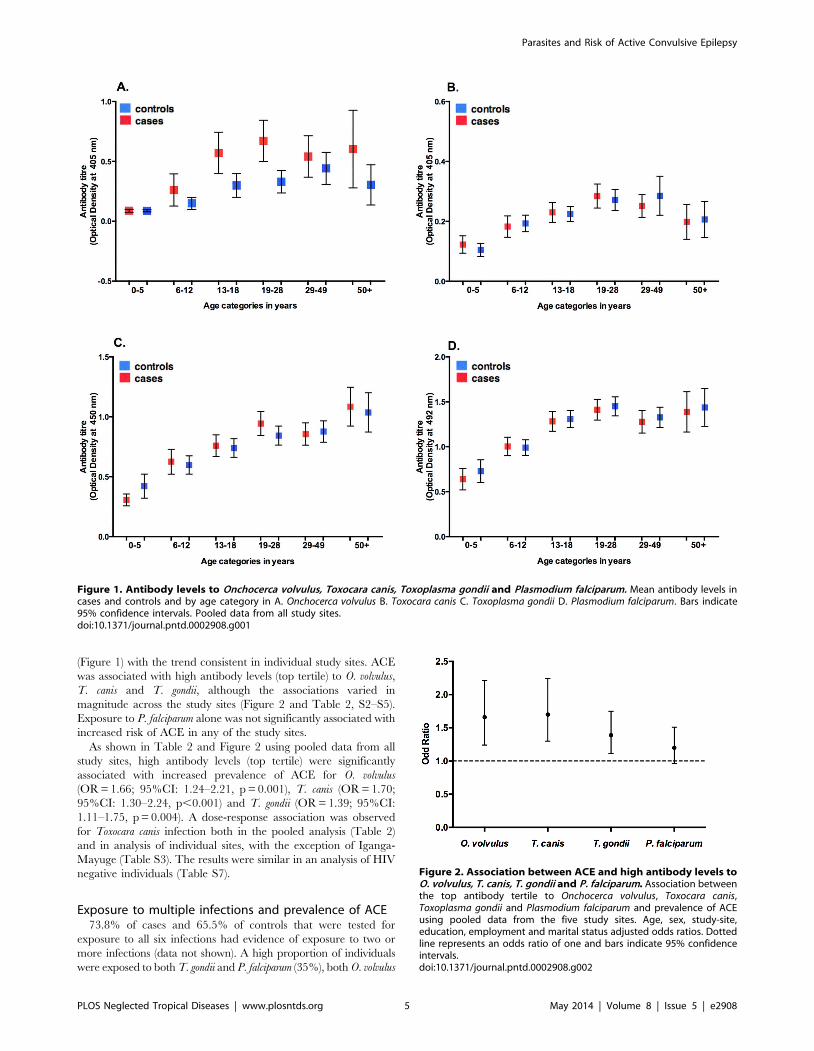
(Figure 1) with the trend consistent in individual study sites. ACE
was associated with high antibody levels (top tertile) to O. volvulus,
T. canis and T. gondii, although the associations varied in
magnitude across the study sites (Figure 2 and Table 2, S2–S5).
Exposure to P. falciparum alone was not significantly associated with
increased risk of ACE in any of the study sites.
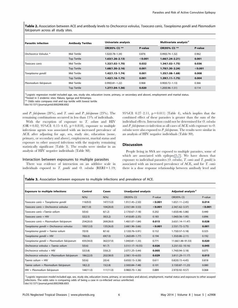
As shown in Table 2 and Figure 2 using pooled data from all
study sites, high antibody levels (top tertile) were significantly
associated with increased prevalence of ACE for O. volvulus
(OR = 1.66; 95%CI: 1.24–2.21, p = 0.001), T. canis (OR = 1.70;
95%CI: 1.30–2.24, p,0.001) and T. gondii (OR = 1.39; 95%CI:
1.11–1.75, p = 0.004). A dose-response association was observed
for Toxocara canis infection both in the pooled analysis (Table 2)
and in analysis of individual sites, with the exception of Iganga-
Mayuge (Table S3). The results were similar in an analysis of HIV
negative individuals (Table S7).
Exposure to multiple infections and prevalence of ACE73.8% of cases and 65.5% of controls that were tested for
exposure to all six infections had evidence of exposure to two or
more infections (data not shown). A high proportion of individuals
were exposed to both T. gondii and P. falciparum (35%), both O. volvulus
Figure 1. Antibody levels to Onchocerca volvulus, Toxocara canis, Toxoplasma gondii and Plasmodium falciparum. Mean antibody levels incases and controls and by age category in A. Onchocerca volvulus B. Toxocara canis C. Toxoplasma gondii D. Plasmodium falciparum. Bars indicate95% confidence intervals. Pooled data from all study sites.doi:10.1371/journal.pntd.0002908.g001
Figure 2. Association between ACE and high antibody levels toO. volvulus, T. canis, T. gondii and P. falciparum. Association betweenthe top antibody tertile to Onchocerca volvulus, Toxocara canis,Toxoplasma gondii and Plasmodium falciparum and prevalence of ACEusing pooled data from the five study sites. Age, sex, study-site,education, employment and marital status adjusted odds ratios. Dottedline represents an odds ratio of one and bars indicate 95% confidenceintervals.doi:10.1371/journal.pntd.0002908.g002
Parasites and Risk of Active Convulsive Epilepsy
PLOS Neglected Tropical Diseases | www.plosntds.org 5 May 2014 | Volume 8 | Issue 5 | e2908
and P. falciparum (28%), and T. canis and P. falciparum (23%). The
remaining combinations occurred in less than 15% of individuals.
With the exception of exposure to T. solium and HIV
(OR = 0.82; 95%CI: 0.15–4.45, p = 0.818), exposure to multiple
infectious agents was associated with an increased prevalence of
ACE after adjusting for age, sex, study site, education (none,
primary, or secondary and above), employment, marital status and
exposure to other assayed infections with the majority remaining
statistically significant (Table 3). The results were similar in an
analysis of HIV negative individuals (Table S8).
Interaction between exposures to multiple parasitesThere was evidence of interaction on an additive scale in
individuals exposed to T. gondii and O. volvulus (RERI = 1.19;
95%CI: 0.27–2.11, p = 0.011) (Table 4), which implies that the
combined effect of these parasites is greater than the sum of the
individual effects. Interaction could not be determined for O. volvulus
and P. falciparum co-infection as all cases of ACE with exposure to O.
volvulus were also exposed to P. falciparum. The results were similar in
an analysis of HIV negative individuals (Table S9).
Discussion
People living in SSA are exposed to multiple parasites, some of
which are associated with epilepsy[5,7]. We have shown that
exposure to individual parasites (O. volvulus, T. canis and T. gondii) is
associated with an increased prevalence of ACE, and for T. canis
there is a dose response relationship between antibody level and
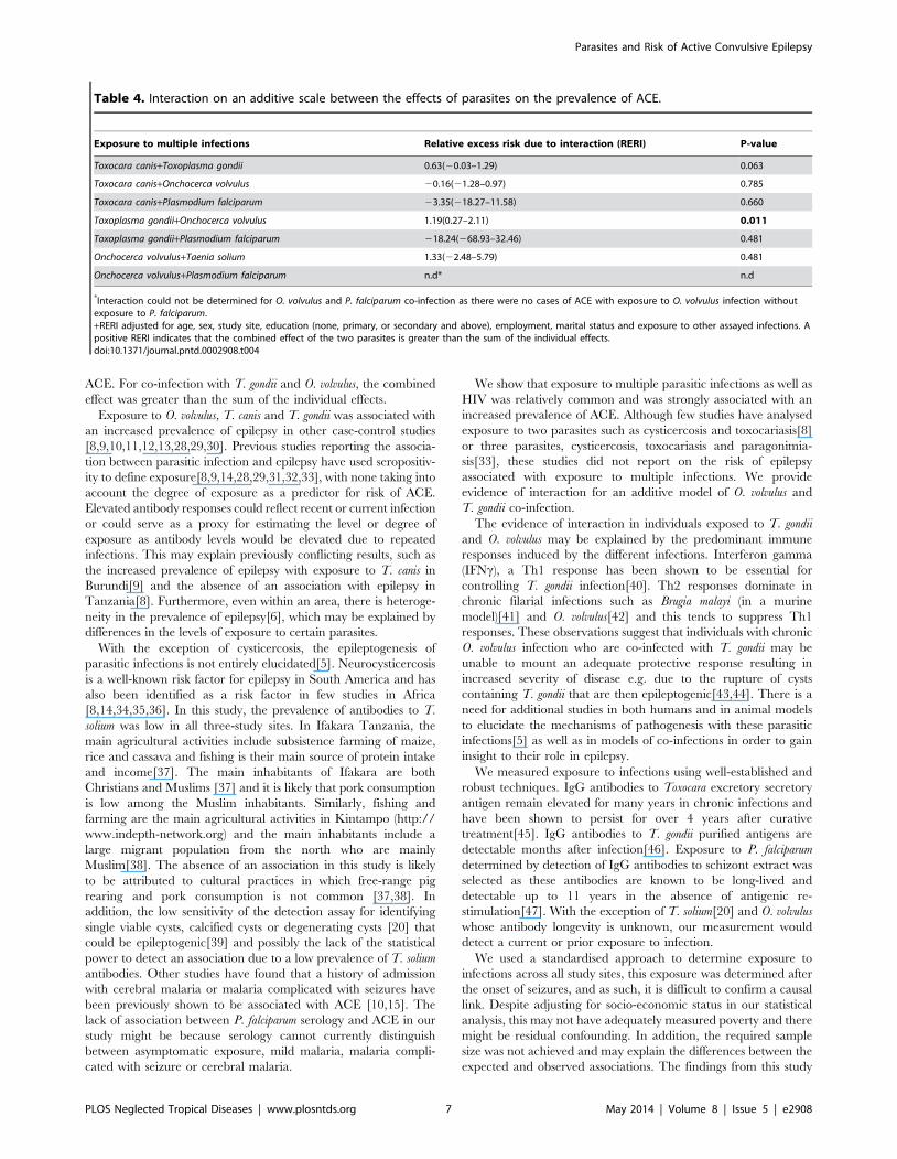
Table 2. Association between ACE and antibody levels to Onchocerca volvulus, Toxocara canis, Toxoplasma gondii and Plasmodiumfalciparum across all study sites.
Parasitic infection Antibody Tertiles Univariate analysis Multivariate analysis#
OR(95% CI) ** P-value OR(95% CI) ** P-value
Onchocerca Volvulus * Mid Tertile 1.02(0.78–1.34) 0.876 0.99(0.74–1.32) 0.962
Top Tertile 1.63(1.26–2.13) ,0.001 1.66(1.24–2.21) 0.001
Toxocara canis Mid Tertile 1.32(1.02–1.70) 0.032 1.34(1.02–1.75) 0.036
Top Tertile 1.68(1.30–2.16) 0.001 1.70(1.30–2.24) ,0.001
Toxoplasma gondii Mid Tertile 1.42(1.15–1.74) 0.001 1.35(1.08–1.68) 0.008
Top Tertile 1.42(1.16–1.75) 0.001 1.39(1.11–1.75) 0.004
Plasmodium falciparum Mid Tertile 0.99(0.81–1.22) 0.968 0.90(0.72–1.13) 0.366
Top Tertile 1.27(1.04–1.56) 0.020 1.20(0.96–1.51) 0.114
#Logistic regression model included age, sex, study site, education (none, primary, or secondary and above), employment and marital status.*Tested in 3 endemic sites: Ifakara, Iganga and Kintampo.** Odds ratio compares mid and top tertile with lowest tertiledoi:10.1371/journal.pntd.0002908.t002
Table 3. Association between exposure to multiple infections and prevalence of ACE.
Exposure to multiple infections Control Cases Unadjusted analysis Adjusted analysis#
N(%) N(%) OR(95% CI) P-value OR(95% CI) P-value
Toxocara canis + Toxoplasma gondii 116(9.0) 147(15.0) 1.91(1.45–2.50) ,0.001 1.65(1.11–2.45) 0.014
Toxocara canis + Onchocerca volvulus 84(11.0) 104(20.0) 2.55(1.84–3.53) ,0.001 2.34(1.62–3.37) ,0.001
Toxocara canis +Taenia solium 5(0.6) 6(1.2) 2.17(0.67–7.18) 0.202 1.65(0.46–5.86) 0.440
Toxocara canis + HIV 32(2.5) 30(3.2) 1.41(0.85–2.35) 0.183 1.04(0.56–1.95) 0.896
Toxocara canis + Plasmodium falciparum 254(20.0) 269(28.0) 1.40(1.07–1.84) 0.015 3.63(1.14–11.47) 0.028
Toxoplasma gondii + Onchocerca volvulus 100(12.0) 135(26.0) 2.68(1.96–3.66) ,0.001 2.53(1.72–3.75) 0.001
Toxoplasma gondii + Taenia solium 7(0.9) 8(1.6) 2.12(0.76–5.91) 0.152 1.73(0.57–5.18) 0.325
Toxoplasma gondii + HIV 78(6.0) 69(7.0) 1.26(0.89–1.77) 0.194 1.35(0.86–2.11) 0.192
Toxoplasma gondii + Plasmodium falciparum 435(34.0) 362(37.0) 1.04(0.81–1.33) 0.771 11.66(1.48–91.53) 0.020
Onchocerca volvulus + Taenia solium 5(0.6) 9(1.7) 3.51(1.17–10.55) 0.026 3.23(1.02–10.16) 0.045
Onchocerca volvulus + HIV 32(3.8) 33(6.2) 2.07(1.25–3.44) 0.005 1.74(0.94–3.18) 0.075
Onchocerca volvulus + Plasmodium falciparum 186(22.0) 202(38.0) 2.58(1.10–6.03) 0.029 3.81(1.29–11.17) 0.015
Taenia solium + HIV 5(0.6) 2(0.4) 0.65(0.13–3.38) 0.611 0.82(0.15–4.45) 0.818
Taenia solium + Plasmodium falciparum 18(2.2) 15(2.8) 2.50(0.84–7.48) 0.101 3.13(0.87–11.23) 0.080
HIV + Plasmodium falciparum 144(11.0) 111(11.0) 0.98(0.70–1.36) 0.889 2.97(0.92–9.57) 0.068
#Logistic regression model included age, sex, study site, education (none, primary, or secondary and above), employment, marital status and exposure to other assayedinfections. The odds ratio is comparing odds of being a case in co-infected versus uninfected.doi:10.1371/journal.pntd.0002908.t003
Parasites and Risk of Active Convulsive Epilepsy
PLOS Neglected Tropical Diseases | www.plosntds.org 6 May 2014 | Volume 8 | Issue 5 | e2908
ACE. For co-infection with T. gondii and O. volvulus, the combined
effect was greater than the sum of the individual effects.
Exposure to O. volvulus, T. canis and T. gondii was associated with
an increased prevalence of epilepsy in other case-control studies
[8,9,10,11,12,13,28,29,30]. Previous studies reporting the associa-
tion between parasitic infection and epilepsy have used seropositiv-
ity to define exposure[8,9,14,28,29,31,32,33], with none taking into
account the degree of exposure as a predictor for risk of ACE.
Elevated antibody responses could reflect recent or current infection
or could serve as a proxy for estimating the level or degree of
exposure as antibody levels would be elevated due to repeated
infections. This may explain previously conflicting results, such as
the increased prevalence of epilepsy with exposure to T. canis in
Burundi[9] and the absence of an association with epilepsy in
Tanzania[8]. Furthermore, even within an area, there is heteroge-
neity in the prevalence of epilepsy[6], which may be explained by
differences in the levels of exposure to certain parasites.
With the exception of cysticercosis, the epileptogenesis of
parasitic infections is not entirely elucidated[5]. Neurocysticercosis
is a well-known risk factor for epilepsy in South America and has
also been identified as a risk factor in few studies in Africa
[8,14,34,35,36]. In this study, the prevalence of antibodies to T.
solium was low in all three-study sites. In Ifakara Tanzania, the
main agricultural activities include subsistence farming of maize,
rice and cassava and fishing is their main source of protein intake
and income[37]. The main inhabitants of Ifakara are both
Christians and Muslims [37] and it is likely that pork consumption
is low among the Muslim inhabitants. Similarly, fishing and
farming are the main agricultural activities in Kintampo (http://
www.indepth-network.org) and the main inhabitants include a
large migrant population from the north who are mainly
Muslim[38]. The absence of an association in this study is likely
to be attributed to cultural practices in which free-range pig
rearing and pork consumption is not common [37,38]. In
addition, the low sensitivity of the detection assay for identifying
single viable cysts, calcified cysts or degenerating cysts [20] that
could be epileptogenic[39] and possibly the lack of the statistical
power to detect an association due to a low prevalence of T. solium
antibodies. Other studies have found that a history of admission
with cerebral malaria or malaria complicated with seizures have
been previously shown to be associated with ACE [10,15]. The
lack of association between P. falciparum serology and ACE in our
study might be because serology cannot currently distinguish
between asymptomatic exposure, mild malaria, malaria compli-
cated with seizure or cerebral malaria.
We show that exposure to multiple parasitic infections as well as
HIV was relatively common and was strongly associated with an
increased prevalence of ACE. Although few studies have analysed
exposure to two parasites such as cysticercosis and toxocariasis[8]
or three parasites, cysticercosis, toxocariasis and paragonimia-
sis[33], these studies did not report on the risk of epilepsy
associated with exposure to multiple infections. We provide
evidence of interaction for an additive model of O. volvulus and
T. gondii co-infection.
The evidence of interaction in individuals exposed to T. gondii
and O. volvulus may be explained by the predominant immune
responses induced by the different infections. Interferon gamma
(IFNc), a Th1 response has been shown to be essential for
controlling T. gondii infection[40]. Th2 responses dominate in
chronic filarial infections such as Brugia malayi (in a murine
model)[41] and O. volvulus[42] and this tends to suppress Th1
responses. These observations suggest that individuals with chronic
O. volvulus infection who are co-infected with T. gondii may be
unable to mount an adequate protective response resulting in
increased severity of disease e.g. due to the rupture of cysts
containing T. gondii that are then epileptogenic[43,44]. There is a
need for additional studies in both humans and in animal models
to elucidate the mechanisms of pathogenesis with these parasitic
infections[5] as well as in models of co-infections in order to gain
insight to their role in epilepsy.
We measured exposure to infections using well-established and
robust techniques. IgG antibodies to Toxocara excretory secretory
antigen remain elevated for many years in chronic infections and
have been shown to persist for over 4 years after curative
treatment[45]. IgG antibodies to T. gondii purified antigens are
detectable months after infection[46]. Exposure to P. falciparum
determined by detection of IgG antibodies to schizont extract was
selected as these antibodies are known to be long-lived and
detectable up to 11 years in the absence of antigenic re-
stimulation[47]. With the exception of T. solium[20] and O. volvulus
whose antibody longevity is unknown, our measurement would
detect a current or prior exposure to infection.
We used a standardised approach to determine exposure to
infections across all study sites, this exposure was determined after
the onset of seizures, and as such, it is difficult to confirm a causal
link. Despite adjusting for socio-economic status in our statistical
analysis, this may not have adequately measured poverty and there
might be residual confounding. In addition, the required sample
size was not achieved and may explain the differences between the
expected and observed associations. The findings from this study
Table 4. Interaction on an additive scale between the effects of parasites on the prevalence of ACE.
Exposure to multiple infections Relative excess risk due to interaction (RERI) P-value
Toxocara canis+Toxoplasma gondii 0.63(20.03–1.29) 0.063
Toxocara canis+Onchocerca volvulus 20.16(21.28–0.97) 0.785
Toxocara canis+Plasmodium falciparum 23.35(218.27–11.58) 0.660
Toxoplasma gondii+Onchocerca volvulus 1.19(0.27–2.11) 0.011
Toxoplasma gondii+Plasmodium falciparum 218.24(268.93–32.46) 0.481
Onchocerca volvulus+Taenia solium 1.33(22.48–5.79) 0.481
Onchocerca volvulus+Plasmodium falciparum n.d* n.d
*Interaction could not be determined for O. volvulus and P. falciparum co-infection as there were no cases of ACE with exposure to O. volvulus infection withoutexposure to P. falciparum.+RERI adjusted for age, sex, study site, education (none, primary, or secondary and above), employment, marital status and exposure to other assayed infections. Apositive RERI indicates that the combined effect of the two parasites is greater than the sum of the individual effects.doi:10.1371/journal.pntd.0002908.t004
Parasites and Risk of Active Convulsive Epilepsy
PLOS Neglected Tropical Diseases | www.plosntds.org 7 May 2014 | Volume 8 | Issue 5 | e2908
should be validated using longitudinal studies that monitor
exposure to infections, which may help establish a causal link
between parasitic infections and epilepsy. While it is not clear
whether it is the presence of the parasite in the CNS or the
immunological response to the infection that is epileptogenic,
efforts to control these infections are likely to reduce the burden of
epilepsy in SSA and should be explored using randomized
intervention studies.
Control is possible with ivermectin for individual and mass-
treatment of onchocerciasis[48],[49] niclosamide or praziquantel
for treatment of taeniasis as well as albendazole or praziquantel for
treatment of parasitic cysts such as in T. solium[50] and T. canis
infection[51]. In addition, efforts to improve sanitation and
personal hygiene practices, including safe food consumption
practices, will reduce transmission of T. canis, T. gondii and T.
solium infections. Safe pig rearing (i.e., separation from human
waste contact) will further impact on T. solium transmission. Vector
control measures as well as bed net usage, intermittent preven-
tative treatment and effective chemotherapy are available for
control P. falciparum infection[52]. These control measures should
be explored and their contribution to the burden of ACE
evaluated. While feasible control measures are known, their use
depends largely on a wider commitment to improving public
health.
We have shown that the intensity of exposure to certain
infections and multiple parasitic infections is associated with
increased prevalence of ACE and may explain conflicting results
obtained when only seropositivity is considered. A recent study
indicated that approximately 35% of ACE cases in adults in SSA
are attributed to parasitic infections[10]. The findings from this
study should be further validated using longitudinal studies to
confirm a causal link between parasitic infection and epilepsy.
Thereafter, randomized intervention studies targeting each
parasitic infection should be explored and their contribution to
the burden of ACE evaluated.
Supporting Information
Checklist S1 STROBE checklist for case-control studies.
(DOC)
Figure S1 Prevalence of antibodies to Onchocercavolvulus. Prevalence of IgG antibodies to Onchocerca volvulus in
A. Ifakara B. Iganga and C. Kintampo in cases and controls and
by age category.
(TIFF)
Figure S2 Prevalence of antibodies to Toxocara canis.Prevalence of IgG4 antibodies to Toxocara canis in A. Agincourt B.
Ifakara C. Iganga D. Kilifi and E. Kintampo in cases and controls
and by age category.
(TIFF)
Figure S3 Prevalence of antibodies to Toxoplasmagondii. Prevalence of IgG antibodies to Toxoplasma gondii in A.
Agincourt B. Ifakara C. Iganga D. Kilifi and E. Kintampo in cases
and controls and by age category.
(TIFF)
Figure S4 Prevalence of antibodies to Plasmodiumfalciparum. Prevalence of IgG antibodies to Plasmodium
falciparum in A. Agincourt B. Ifakara C. Iganga D. Kilifi and E.
Kintampo in cases and controls and by age category.
(TIFF)
Figure S5 Prevalence of antibodies to Taenia solium.Prevalence of IgG antibodies to Taenia solium in A. Ifakara B.
Iganga and C. Kintampo in cases and controls and by age
category.
(TIFF)
Figure S6 Prevalence of antibodies to HIV. Prevalence of
IgG antibodies to HIV in A. Agincourt B. Ifakara C. Iganga D.
Kilifi and E. Kintampo in cases and controls and by age category.
(TIFF)
Table S1 Demographic characteristics of cases andcontrols from each study site.(DOC)
Table S2 Association between IgG4 antibody titers toOnchocerca volvulus and prevalence of ACE.(DOC)
Table S3 Association between IgG4 antibody titers toToxocara canis and prevalence of ACE.(DOC)
Table S4 Association between IgG antibody titers toToxoplasma gondii and prevalence of ACE.(DOC)
Table S5 Association between IgG antibody titers toPlasmodium falciparum and prevalence of ACE.(DOC)
Table S6 Table showing the seropositivity to sixdifferent infections and association with ACE in HIVnegative individuals using pooled data from all studysites.(DOC)
Table S7 Association between ACE and antibody levelsto Onchocerca volvulus, Toxocara canis, Toxoplasmagondii and Plasmodium falciparum in HIV negativeindividuals across all study sites.(DOC)
Table S8 Association between exposure to multipleinfections and prevalence of ACE in HIV negativeindividuals across all study sites.(DOC)
Table S9 Interaction on an additive scale between theeffects of parasites on the prevalence of ACE in HIVnegative individuals.(DOC)
Acknowledgments
This work is published with the permission of the Director of KEMRI.
Study of Epidemiology of Epilepsy in Demographic Sites (SEEDS)
group:
Agincourt HDSS, South Africa: Ryan Wagner, Rhian Twine, Myles
Connor, F. Xavier Gomez-Olive, Mark Collinson (and INDEPTH
Network, Accra, Ghana), Kathleen Kahn (and INDEPTH Network,
Accra, Ghana), Stephen Tollman (and INDEPTH Network, Accra,
Ghana)
Ifakara HDSS, Tanzania: Honorati Masanja (and INDEPTH Network,
Accra, Ghana), Alexander Mathew{
Iganga/Mayuge HDSS, Uganda: Angelina Kakooza-Mwesige, George
Pariyo, Stefan Peterson (and Uppsala University, Dept of Women’s and
Children’s Health, IMCH; Karolinska Institutet, Div of Global Health,
IHCAR; Makerere University School of Public Health), Donald Ndyo-
mughenyi
Kilifi HDSS, Kenya: Anthony K Ngugi, Rachael Odhiambo, Eddie
Chengo, Martin Chabi, Evasius Bauni, Gathoni Kamuyu, Victor
Mung’ala Odera{, James O Mageto, Charles R Newton
Parasites and Risk of Active Convulsive Epilepsy
PLOS Neglected Tropical Diseases | www.plosntds.org 8 May 2014 | Volume 8 | Issue 5 | e2908
Kintampo HDSS, Ghana: Kenneth Ae-Ngibise, Bright Akpalu, Albert
Akpalu, Francis Agbokey, Patrick Adjei, Seth Owusu-Agyei (and
INDEPTH Network, Accra, Ghana)
London School of Hygiene and Tropical Medicine: Christian Bottom-
ley, Immo Kleinschmidt
Institute of Psychiatry, King’s College London, UK: Victor CK Doku
Swiss Tropical and Public Health Institute: Peter Odermatt
University College London, London, UK: Brian Neville, Josemir W
Sander, Steve White
National Institute of Health, USA: Thomas Nutman
Centre for Disease Control and Prevention, Atlanta, USA: Patricia P.
Wilkins, John C. Noh
{ - Deceased.
Underline-members who are listed as authors.
Author Contributions
Conceived and designed the experiments: GK CB BL CRN. Performed
the experiments: GK JM FHAO BL. Analyzed the data: GK CB PO
CRN. Contributed reagents/materials/analysis tools: PPW JCN TBN.
Wrote the paper: GK CB CRN. Coordinated and monitored sample
collection in the case control studies: AKN RGW AKM SOA KAN HM.
Created the database and monitored data from all centres: RO.
References
1. Ngugi AK, Bottomley C, Kleinschmidt I, Sander JW, Newton CR (2010)
Estimation of the burden of active and life-time epilepsy: a meta-analyticapproach. Epilepsia 51: 883–890.
2. Newton CR, Garcia HH (2012) Epilepsy in poor regions of the world. Lancet
380: 1193–1201.
3. Preux PM, Druet-Cabanac M (2005) Epidemiology and aetiology of epilepsy in
sub-Saharan Africa. Lancet Neurol 4: 21–31.
4. Burneo JG, Tellez-Zenteno J, Wiebe S (2005) Understanding the burden ofepilepsy in Latin America: a systematic review of its prevalence and incidence.
Epilepsy Res 66: 63–74.
5. Wagner RG, Newton CR (2009) Do helminths cause epilepsy? ParasiteImmunol 31: 697–705.
6. Edwards T, Scott AG, Munyoki G, Odera VM, Chengo E, et al. (2008) Activeconvulsive epilepsy in a rural district of Kenya: a study of prevalence and
possible risk factors. Lancet Neurol 7: 50–56.
7. Garcia HH, Modi M (2008) Helminthic parasites and seizures. Epilepsia 49Suppl 6: 25–32.
8. Winkler AS, Blocher J, Auer H, Gotwald T, Matuja W, et al. (2008)
Anticysticercal and antitoxocaral antibodies in people with epilepsy in ruralTanzania. Trans R Soc Trop Med Hyg 102: 1032–1038.
9. Nicoletti A, Bartoloni A, Sofia V, Mantella A, Nsengiyumva G, et al. (2007)Epilepsy and toxocariasis: a case-control study in Burundi. Epilepsia 48: 894–
899.
10. Ngugi AK, Bottomley C, Kleinschmidt I, Wagner RG, Kakooza-Mwesige A, etal. (2013) Prevalence of active convulsive epilepsy in sub-Saharan Africa and
associated risk factors: cross-sectional and case-control studies. Lancet Neurol12: 253–263.
11. Konig R, Nassri A, Meindl M, Matuja W, Kidunda AR, et al. (2010) The role of
Onchocerca volvulus in the development of epilepsy in a rural area of Tanzania.Parasitology 137: 1559–1568.
12. Boussinesq M, Pion SD, Demanga N, Kamgno J (2002) Relationship between
onchocerciasis and epilepsy: a matched case-control study in the Mbam Valley,Republic of Cameroon. Trans R Soc Trop Med Hyg 96: 537–541.
13. Kaiser C, Pion SD, Boussinesq M (2013) Case-control Studies on theRelationship between Onchocerciasis and Epilepsy: Systematic Review and
Meta-analysis. PLoS Negl Trop Dis 7: e2147.
14. Nsengiyumva G, Druet-Cabanac M, Ramanankandrasana B, Bouteille B,Nsizabira L, et al. (2003) Cysticercosis as a major risk factor for epilepsy in
Burundi, east Africa. Epilepsia 44: 950–955.
15. Carter JA, Neville BG, White S, Ross AJ, Otieno G, et al. (2004) Increasedprevalence of epilepsy associated with severe falciparum malaria in children.
Epilepsia 45: 978–981.
16. Ngoungou EB, Dulac O, Poudiougou B, Druet-Cabanac M, Dicko A, et al.
(2006) Epilepsy as a consequence of cerebral malaria in area in which malaria is
endemic in Mali, West Africa. Epilepsia 47: 873–879.
17. Palmer BS (2007) Meta-analysis of three case controlled studies and an
ecological study into the link between cryptogenic epilepsy and chronictoxoplasmosis infection. Seizure 16: 657–663.
18. Bhigjee AI (2005) Seizures in HIV/AIDS: a southern African perspective. Acta
Neurol Scand Suppl 181: 4–7.
19. Lobos E, Weiss N, Karam M, Taylor HR, Ottesen EA, et al. (1991) An
immunogenic Onchocerca volvulus antigen: a specific and early marker of infection.
Science 251: 1603–1605.
20. Handali S, Klarman M, Gaspard AN, Noh J, Lee YM, et al. (2010) Multiantigen
print immunoassay for comparison of diagnostic antigens for Taenia solium
cysticercosis and taeniasis. Clin Vaccine Immunol 17: 68–72.
21. Hancock K, Pattabhi S, Whitfield FW, Yushak ML, Lane WS, et al. (2006)
Characterization and cloning of T24, a Taenia solium antigen diagnostic forcysticercosis. Mol Biochem Parasitol 147: 109–117.
22. Levine MZ, Calderon JC, Wilkins PP, Lane WS, Asara JM, et al. (2004)
Characterization, cloning, and expression of two diagnostic antigens for Taenia
solium tapeworm infection. J Parasitol 90: 631–638.
23. Noordin R, Smith HV, Mohamad S, Maizels RM, Fong MY (2005)
Comparison of IgG-ELISA and IgG4-ELISA for Toxocara serodiagnosis. ActaTrop 93: 57–62.
24. Osier FH, Fegan G, Polley SD, Murungi L, Verra F, et al. (2008) Breadth andmagnitude of antibody responses to multiple Plasmodium falciparum merozoite
antigens are associated with protection from clinical malaria. Infect Immun 76:
2240–2248.
25. Knol MJ, van der Tweel I, Grobbee DE, Numans ME, Geerlings MI (2007)Estimating interaction on an additive scale between continuous determinants in
a logistic regression model. Int J Epidemiol 36: 1111–1118.
26. Rothman KJ (1990) No adjustments are needed for multiple comparisons.Epidemiology 1: 43–46.
27. Ngugi AK, Bottomley C, Chengo E, Kombe MZ, Kazungu M, et al. (2012) Thevalidation of a three-stage screening methodology for detecting active convulsive
epilepsy in population-based studies in health and demographic surveillance
systems. Emerg Themes Epidemiol 9: 8.
28. Nicoletti A, Bartoloni A, Reggio A, Bartalesi F, Roselli M, et al. (2002) Epilepsy,
cysticercosis, and toxocariasis: a population-based case-control study in rural
Bolivia. Neurology 58: 1256–1261.
29. Nicoletti A, Sofia V, Mantella A, Vitale G, Contrafatto D, et al. (2008) Epilepsy
and toxocariasis: a case-control study in Italy. Epilepsia 49: 594–599.
30. Yazar S, Arman F, Yalcin S, Demirtas F, Yaman O, et al. (2003) Investigation ofprobable relationship between Toxoplasma gondii and cryptogenic epilepsy.
Seizure 12: 107–109.
31. Garcia HH, Gilman RH, Tsang VC, Gonzalez AE (1997) Clinical significance
of neurocysticercosis in endemic villages. The Cysticercosis Working Group in
Peru. Trans R Soc Trop Med Hyg 91: 176–178.
32. Akyol A, Bicerol B, Ertug S, Ertabaklar H, Kiylioglu N (2007) Epilepsy and
seropositivity rates of Toxocara canis and Toxoplasma gondii. Seizure 16: 233–237.
33. Nkouawa A, Sako Y, Itoh S, Kouojip-Mabou A, Nganou CN, et al. (2010)Serological studies of neurologic helminthic infections in rural areas of southwest
cameroon: toxocariasis, cysticercosis and paragonimiasis. PLoS Negl Trop Dis 4:e732.
34. Winkler AS, Blocher J, Auer H, Gotwald T, Matuja W, et al. (2009) Epilepsy
and neurocysticercosis in rural Tanzania-An imaging study. Epilepsia 50: 987–993.
35. Blocher J, Schmutzhard E, Wilkins PP, Gupton PN, Schaffert M, et al. (2011) A
cross-sectional study of people with epilepsy and neurocysticercosis in Tanzania:clinical characteristics and diagnostic approaches. PLoS Negl Trop Dis 5: e1185.
36. Quet F, Guerchet M, Pion SD, Ngoungou EB, Nicoletti A, et al. (2010) Meta-
analysis of the association between cysticercosis and epilepsy in Africa. Epilepsia51: 830–837.
37. Mwanyangala MA, Mayombana C, Urassa H, Charles J, Mahutanga C, et al.
(2010) Health status and quality of life among older adults in rural Tanzania.Glob Health Action 3: 10.3402/gha.v3i0.2142.
38. Read UM, Adiibokah E, Nyame S (2009) Local suffering and the globaldiscourse of mental health and human rights: an ethnographic study of responses
to mental illness in rural Ghana. Global Health 5: 13.
39. Nash TE, Del Brutto OH, Butman JA, Corona T, Delgado-Escueta A, et al.(2004) Calcific neurocysticercosis and epileptogenesis. Neurology 62: 1934–
1938.
40. Suzuki Y, Sa Q, Gehman M, Ochiai E (2011) Interferon-gamma- and perforin-mediated immune responses for resistance against Toxoplasma gondii in the brain.
Expert Rev Mol Med 13: e31.
41. Pearlman E, Kroeze WK, Hazlett FE, Jr., Chen SS, Mawhorter SD, et al. (1993)Brugia malayi: acquired resistance to microfilariae in BALB/c mice correlates with
local Th2 responses. Exp Parasitol 76: 200–208.
42. Nmorsi OP, Nkot BP, Che J (2012) Relationship between pro-and anti-inflammatory cytokines profiles and some haematological parameters in some
Cameroonians infected with Onchocerca volvulus. Asian Pac J Trop Med 5: 713–717.
43. Dubey JP, Lindsay DS, Speer CA (1998) Structures of Toxoplasma gondii
tachyzoites, bradyzoites, and sporozoites and biology and development of tissuecysts. Clin Microbiol Rev 11: 267–299.
44. Frenkel JK, Escajadillo A (1987) Cyst rupture as a pathogenic mechanism of
toxoplasmic encephalitis. Am J Trop Med Hyg 36: 517–522.
45. Elefant GR, Shimizu SH, Sanchez MC, Jacob CM, Ferreira AW (2006) A
serological follow-up of toxocariasis patients after chemotherapy based on thedetection of IgG, IgA, and IgE antibodies by enzyme-linked immunosorbent
assay. J Clin Lab Anal 20: 164–172.
46. Montoya JG, Remington JS (2008) Management of Toxoplasma gondii infectionduring pregnancy. Clin Infect Dis 47: 554–566.
Parasites and Risk of Active Convulsive Epilepsy
PLOS Neglected Tropical Diseases | www.plosntds.org 9 May 2014 | Volume 8 | Issue 5 | e2908
47. Druilhe P, Pradier O, Marc JP, Miltgen F, Mazier D, et al. (1986) Levels of
antibodies to Plasmodium falciparum sporozoite surface antigens reflect malariatransmission rates and are persistent in the absence of reinfection. Infect Immun
53: 393–397.
48. Burnham G (1998) Onchocerciasis. Lancet 351: 1341–1346.49. Hoerauf A, Buttner DW, Adjei O, Pearlman E (2003) Onchocerciasis. BMJ 326:
207–210.
50. Garcia HH, Gilman RH, Catacora M, Verastegui M, Gonzalez AE, et al. (1997)
Serologic evolution of neurocysticercosis patients after antiparasitic therapy.Cysticercosis Working Group in Peru. J Infect Dis 175: 486–489.
51. Magnaval JF, Glickman LT, Dorchies P, Morassin B (2001) Highlights of human
toxocariasis. Korean J Parasitol 39: 1–11.52. WHO (2012) World Malaria Report 2012: Surveillance,monitoring and
evaluation. Malaria Fact Sheet No 94.
Parasites and Risk of Active Convulsive Epilepsy
PLOS Neglected Tropical Diseases | www.plosntds.org 10 May 2014 | Volume 8 | Issue 5 | e2908