Exploring the detection of associatively novel events using fMRI
12
r Human Brain Mapping 32:370–381 (2011) r Exploring the Detection of Associatively Novel Events Using fMRI Andreja Bubic, 1,2 * D. Yves von Cramon, 1,3 and Ricarda I. Schubotz 1,3 1 Max Planck Institute for Human Cognitive and Brain Sciences, Leipzig, Germany 2 University of Leipzig, Leipzig, Germany 3 Max Planck Institute for Neurological Research, Cologne, Germany r r Abstract: Identifying and evaluating events which are novel in a particular environment is crucially impor- tant for adaptive behavior. These events are often not just novel, as they typically violate expectations which may be formulated based on numerous features of our surroundings, one of which includes the ordinal structure (temporal order) of relevant stimuli. Events which violate such expectations, namely sequential deviants, constitute one category of associatively novel stimuli. The present event-related fMRI study investi- gated the detection of sequential deviants presented within three types of equivalently organized, attended visual sequences which differed in stimulus dimensions relevant for defining the sequential structure (posi- tion, rhythm, and object identity). Presenting deviants within perceptual sequences defined by position and rhythm stimulus properties triggered comparable patterns of activations within the lateral parietal, premotor, and prefrontal regions. However, the activations identified in the context of position sequences showed a more dorsal distribution when compared to those in rhythm sequences. In contrast, detection of deviants within object sequences was supported by right-lateralized parietal and temporal cortices. Thus, although the obtained results indicate similarities and partial overlap in activations triggered by specific pairs of devi- ants, differences in their processing were also revealed. This suggests that the general task context and spe- cific stimulus features which define the deviant itself influence which brain regions within a widespread network incorporating lateral prefrontal, anterior premotor, and posterior (mainly lateral parietal) areas will become engaged in its processing. Hum Brain Mapp 32:370–381, 2011. V C 2010 Wiley-Liss, Inc. Key words: associative novelty; deviant detection; fMRI; forward models; prediction; sequence processing r r INTRODUCTION Although crucially relevant for adaptive behavior, detec- tion of associative novelty characterized by the presenta- tion of familiar items in novel spatial or temporal configurations [Kumaran and Maguire, 2007a] has not been consistently investigated. This can be attributed to a wide array of situations in which this phenomenon is encountered and to the nonuniform terminology used to describe it. For instance, it can be equally well described as ‘‘relational’’ or ‘‘associative,’’ while additional terms such as ‘‘sequential’’ may be used for describing one (tem- poral) type of such novelty. Furthermore, the terms ‘‘nov- elty’’ and ‘‘deviance’’ can be interchanged, as such events are not simply novel, but also violate expectations which can be formulated based on the learned associations between stimuli. Accepting this variable terminology, a comprehensive understanding of regular and violated associative processing can be found in the field of motor sequence processing [Huettel et al., 2002; Keele et al., 2003; Ru ¨ sseler and Ro ¨ sler, 2000] which has recently been Contract grant sponsor: German Research Foundation (DFG). *Correspondence to: Andreja Bubic, Department of Cognitive Neurology, Functional Anatomy of the Frontal Lobes, Max Planck Institute for Human Cognitive and Brain Sciences, Stephanstrasse 1a, 04103 Leipzig, Germany. E-mail: [email protected] Received for publication 27 August 2009; Revised 12 January 2010; Accepted 16 January 2010 DOI: 10.1002/hbm.21027 Published online 3 May 2010 in Wiley Online Library (wileyonlinelibrary.com). V C 2010 Wiley-Liss, Inc.
Transcript of Exploring the detection of associatively novel events using fMRI
r Human Brain Mapping 32:370–381 (2011) r
Exploring the Detection of Associatively NovelEvents Using fMRI
Andreja Bubic,1,2* D. Yves von Cramon,1,3 and Ricarda I. Schubotz1,3
1Max Planck Institute for Human Cognitive and Brain Sciences, Leipzig, Germany2University of Leipzig, Leipzig, Germany
3Max Planck Institute for Neurological Research, Cologne, Germany
r r
Abstract: Identifying and evaluating events which are novel in a particular environment is crucially impor-tant for adaptive behavior. These events are often not just novel, as they typically violate expectations whichmay be formulated based on numerous features of our surroundings, one of which includes the ordinalstructure (temporal order) of relevant stimuli. Events which violate such expectations, namely sequentialdeviants, constitute one category of associatively novel stimuli. The present event-related fMRI study investi-gated the detection of sequential deviants presented within three types of equivalently organized, attendedvisual sequences which differed in stimulus dimensions relevant for defining the sequential structure (posi-tion, rhythm, and object identity). Presenting deviants within perceptual sequences defined by position andrhythm stimulus properties triggered comparable patterns of activations within the lateral parietal, premotor,and prefrontal regions. However, the activations identified in the context of position sequences showed amore dorsal distribution when compared to those in rhythm sequences. In contrast, detection of deviantswithin object sequences was supported by right-lateralized parietal and temporal cortices. Thus, althoughthe obtained results indicate similarities and partial overlap in activations triggered by specific pairs of devi-ants, differences in their processing were also revealed. This suggests that the general task context and spe-cific stimulus features which define the deviant itself influence which brain regions within a widespreadnetwork incorporating lateral prefrontal, anterior premotor, and posterior (mainly lateral parietal) areas willbecome engaged in its processing. Hum Brain Mapp 32:370–381, 2011. VC 2010Wiley-Liss, Inc.
Keywords: associative novelty; deviant detection; fMRI; forward models; prediction; sequence processing
r r
INTRODUCTION
Although crucially relevant for adaptive behavior, detec-tion of associative novelty characterized by the presenta-tion of familiar items in novel spatial or temporal
configurations [Kumaran and Maguire, 2007a] has notbeen consistently investigated. This can be attributed to awide array of situations in which this phenomenon isencountered and to the nonuniform terminology used todescribe it. For instance, it can be equally well describedas ‘‘relational’’ or ‘‘associative,’’ while additional termssuch as ‘‘sequential’’ may be used for describing one (tem-poral) type of such novelty. Furthermore, the terms ‘‘nov-elty’’ and ‘‘deviance’’ can be interchanged, as such eventsare not simply novel, but also violate expectations whichcan be formulated based on the learned associationsbetween stimuli. Accepting this variable terminology, acomprehensive understanding of regular and violatedassociative processing can be found in the field of motorsequence processing [Huettel et al., 2002; Keele et al., 2003;Russeler and Rosler, 2000] which has recently been
Contract grant sponsor: German Research Foundation (DFG).
*Correspondence to: Andreja Bubic, Department of CognitiveNeurology, Functional Anatomy of the Frontal Lobes, Max PlanckInstitute for Human Cognitive and Brain Sciences, Stephanstrasse1a, 04103 Leipzig, Germany. E-mail: [email protected]
Received for publication 27 August 2009; Revised 12 January2010; Accepted 16 January 2010
DOI: 10.1002/hbm.21027Published online 3 May 2010 in Wiley Online Library(wileyonlinelibrary.com).
VC 2010 Wiley-Liss, Inc.

complemented by comparable findings from the percep-tual domain [Schubotz and von Cramon, 2001, 2002c].Studies investigating the detection of violations withinboth motor and perceptual sequences have shown a ratherspecial status of processing sequential when compared tononsequential deviants which is supported by the pro-nounced involvement of lateral prefrontal cortices [Bubicet al., 2009; Huettel et al., 2002].
Although it seems plausible to suggest that the level ofprefrontal engagement in this context depends on specifictask requirements, an alternative emphasizing an impera-tive role of prefrontal cortex in detecting sequential oreven all associative deviants could also be proposed. Simi-larly, Kumaran and Maguire [2007a] have recently arguedfor the existence of a generative mechanism underlyingthe detection of all forms of associative novelty. Specifi-cally, they suggested that this detection relies on a match-mismatch comparison process supported by the hippocam-pus which is also in line with findings showing theinvolvement of this region and adjacent cortices in bothregular and violated associative, sequence and generalcontextual processing [Bar et al., 2008; Kumaran andMaguire, 2009; Lisman and Redish, 2009; Schendan et al.,2003]. However, absence of such engagement in someforms of regular sequencing within the motor [cf., Keeleet al., 2003] and other, e.g., perceptual, linguistic, or cogni-tive [Dominey, 2005; Schubotz, 2007] domains, as well asin processing sequential deviants [Bubic et al., 2009; Huet-tel et al., 2002] has also been previously reported.Although plausible given the differences in tasks andparadigms employed across different studies, such diver-gence in understanding regular and novel associativeprocessing is still somewhat surprising. Aimed at address-ing potential similarities and differences in processing dif-ferent types within the same class of associatively novelevents, the present event-related fMRI study investigatedthe detection of sequential deviants introduced into per-ceptual sequences previously suggested to rely on internalmodels as implemented within the motor system [Schu-botz, 2007]. By exploring the detection of sequential devi-ants introduced into three types of equivalently organizedperceptual sequences, we investigated whether and howdetecting such deviants depends on the stimulus features(position, rhythm, and object identity) relevant for definingthe sequential structure.
MATERIALS AND METHODS
Participants
Thirty right-handed, healthy male volunteers (mean age26.7) participated in the study. All participants reportedhaving normal or corrected-to-normal vision. Four partici-pants were excluded from further analysis due to below-chance level performance in the sequencing task and onedue to movement during the experiment. All subsequentanalysis was performed on the data from 25 participants.
All participants gave informed consent for participating af-ter being informed about potential risks and screened bythe physician of the institution. The experimental stand-ards were approved by the local ethics committee of theUniversity of Leipzig. Collected data were handledanonymously.
Procedure
Participants were instructed and underwent a behavioraltraining session several days before the fMRI measurementin which they were trained to perform the three tasks untilthey reached a learning criterion of 75%. Prior to the mainexperiment on the day of the measurement, they wereadditionally presented with the instructions and a 5-minbehavioral training session which included all tasks. Dur-ing the main experiment, participants were supine on thescanner bed with their index and middle fingers of theright hand positioned on the response buttons. To preventpostural adjustments, the participants’ arms and handswere carefully stabilized by tape. In addition, arm, hand,and head motion was prevented by using form-fittingcushions. To attenuate scanner noise, participants wereprovided with earplugs and headphones.
Stimuli and Task
The stimulus material used in this study included 12different objects, each composed of a 25-mm circle (0.14�
of visual angle) and a slightly smaller geometrical form, ei-ther a square or a circle, placed in its centre (see Fig. 1).The colors of both geometrical forms could be red, yellow,or blue and they always differed between the two forms.Each stimulus display consisted of two identical objectspresented on opposite locations of a virtual circle with aradius of 6 cm. A fixation cross was presented at thescreen centre to facilitate constant visual fixation. Eachstimulus was presented for either 300, 600, 900, 1,200,1,500, or 1,800 ms. Responses were made by pressing theleft or right key of a standard response button box withthe index and middle finger of the right hand.
Three different versions of the sequencing (serial predic-tion task; SPT) and a control (target detection task; control)task of equal trial organization were presented in a mixedtrial design. Each trial included the successive presentationof 12 stimuli without temporal gaps, preceded by a taskcue with the duration of 400 ms and followed by a 1,500-ms response period and performance feedback lasting for400 ms. During all other periods in the experiment a fixa-tion cross was presented at the center of the screen. Over-all trial duration was 14 s and, to improve temporalresolution, each trial occurred at four different offsetpoints (0, 500, 1,000, and 1,500 ms) in relation to fMRIdata acquisition [Josephs et al., 1997]. During the course ofthe experiment the stimulus trials were interspersed withempty trials during which only a fixation cross was
r Detection of Associatively Novel Events Using fMRI r
r 371 r
presented and no task was to be performed by the partici-pants. Stimuli were presented using Presentation 11.7(Neurobehavioral systems, San Francisco, CA).
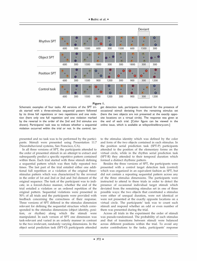
In all three versions of SPT, the participants attended tothe order of presented stimuli in an attempt to extract andsubsequently predict a specific repetitive pattern containedwithin them. Each trial started with three stimuli defininga sequential pattern which was then fully repeated twotimes. The last part of the trial entailed either one addi-tional full repetition or a violation of the original three-stimulus pattern which was characterized by the reversalin the order of 1st and 2nd or 2nd and 3rd element of theoriginal sequence. The task of the participant was to indi-cate, in a forced-choice manner, whether the end of thetrial entailed a violation or an ordered repetition of theoriginal pattern. Sequential violations were presented in50% of all trials and the participants were provided withfeedback concerning the correctness of their response.Three versions of SPT differed in the stimulus dimensionrelevant for defining the sequential structure which corre-sponded to the stimulus dimensions (object identity, posi-tion, or rhythm) along which the stimuli weremanipulated. In each version of SPT one dimension wastask-relevant and varied in an orderly manner in contrastto the two irrelevant, randomly varying dimensions. In theobject serial prediction task (SPT-O) participants attended
to the stimulus identity which was defined by the colorand form of the two objects contained in each stimulus. Inthe position serial prediction task (SPT-P) participantsattended to the position of the elementary forms on thevirtual circle, while in the rhythm serial prediction task(SPT-R) they attended to their temporal duration whichformed a distinct rhythmic pattern.
Besides the three versions of SPT, the participants werepresented with a control target detection task (control)which was organized in an equivalent fashion as SPT, butdid not contain a repeating sequential pattern across anyof the three stimulus dimensions. The participants wereinstructed to attend to these trials in order to detect thepresence of occasional individual target stimuli whichdeviated from the remaining stimulus set in one of threepossible ways: the two objects that constituted a stimuluswere either of unequal duration, were not identical orwere not presented at the exactly opposite locations on avirtual circle. The participants’ task was to count suchstimuli and respond whether an odd or even number ofthem was presented during the trial.
Across all trials in the experiment the order of stimuliwas pseudo-randomized. The probability of each stimulusand that of transitions between stimuli were balancedacross different positions within the trial. To avoid anymotor contributions to the tasks, participants’ response
Figure 1.
Schematic examples of four tasks. All versions of the SPT tri-
als started with a three-stimulus sequential pattern followed
by its three full repetitions or two repetitions and one viola-
tion (here only one full repetition and one violation marked
by the reversal in the order of the 2nd and 3rd stimulus are
shown). Participants’ task was to indicate whether a sequential
violation occurred within the trial or not. In the control, tar-
get detection task, participants monitored for the presence of
occasional stimuli deviating from the remaining stimulus set
(here the two objects are not presented at the exactly oppo-
site locations on a virtual circle). The response was given at
the end of each trial. [Color figure can be viewed in the
online issue, which is available at wileyonlinelibrary.com.]
r Bubic et al. r
r 372 r

was always required after the end of each sequence. Theexperiment included eight types of trials: ordered and vio-lated object SPT trials, ordered and violated position SPTtrials, ordered and violated rhythm SPT trials, control tri-als with a deviant and control trials without a deviant (seeFig. 1). Twenty one trials of each type were used which,together with the 15 empty trials, amounted to the total of183 trials presented in the course of the experiment.
Data Acquisition
The experiment was carried out on a 3T scanner (Med-spec S300, Bruker, Ettlingen) equipped with a standardbird cage coil. Immediately prior to the functional experi-ment, a set of two-dimensional anatomical images wasacquired for each participant using a MDEFT sequence(256 � 256 pixel matrix) [Norris, 2000; Ugurbil et al.,1993]. Additionally, to improve the localization of activa-tion foci, high resolution whole-brain images using a T1-weighted three-dimensional segmented MDEFT sequencewere acquired for each participant in a separate session.This volume dataset with 160 slices and 1-mm slice thick-ness was standardized to the Talairach stereotactic space[Talairach and Tournoux, 1988]. Functional images in-plane with the anatomical images were acquired using agradient-echo echo planar imaging (EPI) sequence with anecho time (TE) of 30 ms, a flip angle of 90�, and a repeti-tion time (TR) of 2,000 ms. Twenty two functional sliceswere acquired parallel to the bicommissural plane (AC-PC) (thickness 4 mm, interslice gap 1 mm) covering thewhole brain. The matrix acquired was 64 � 64 with a fieldof view of 192 mm, resulting in an in-plane resolution of 3mm � 3 mm. A total of 1,290 volumes were acquired.
Data Analysis
MR data processing was performed using the softwarepackage LIPSIA [Lohmann et al., 2001] which containstools for preprocessing, coregistration, statistical evalua-tion, and visualization of fMRI data. To correct for thetemporal offset between the slices acquired in one scan, acubic-spline-interpolation was applied. A temporal highpass filter with a cut-off frequency of 1/130 Hz was usedfor baseline correction, removing low-frequency drifts inan fMRI time series (frequencies due to global signalchanges). Spatial Gaussian smoothing was applied using aGaussian filter with 5.65-mm full width at half maximum(FWHM). To align the functional data slices with a 3D ste-reotactic coordinate system, a rigid linear registration withsix degrees of freedom (three translational and three rota-tional parameters) was performed. The parameters wereacquired on the basis on MDEFT and EPI-T1 slices toachieve an optimal match between these slices and theindividual 3D reference dataset. Each transformation ma-trix was subsequently transformed to a standard Talairachbrain size [x ¼ 135, y ¼ 175, z ¼ 120 mm; Talairach and
Tournoux, 1988] by applying linear scaling. Finally, thenormalized transformation matrices were applied to theacquired functional slices to align them with the stereotac-tic coordinate system. Transformation was performedusing trilinear interpolation, thus generating data with aspatial resolution of 3 mm3.
The statistical evaluation was based on a least-squaresestimation using the general linear model for serially auto-correlated observations (random effects model). In the firststage, autocorrelation parameters were estimated from theleast squares residuals using the Yule-Walker equationsand used to ‘‘whiten’’ the data and the design matrix. Inthe second stage, the linear model was reestimated usingleast-squares on the whitened data to produce estimates ofeffects and their standard errors [Worsley et al., 2002].Data were modeled using two design matrices. To explorethe neural correlates of deviance detection, a design matrixwas used which consisted of onset vectors with eventstime-locked to the violations within conditions containingthem and comparable positions within nonviolated trials,with one additional vector for responses and one for theremaining stimulation periods of no interest, including thetrials that were incorrectly responded to. To explore regu-lar sequence processing, a design matrix was used withevents time-locked to the presentation of the first stimuluswithin each sequence and two additional vectors identicalto the ones in the previously described matrix. Withinboth types of matrices the events related to each sequencetype were modeled with the same duration. The designmatrices were generated using a synthetic hemodynamicresponse function [Friston et al., 1998; Josephs et al., 1997]and, in case of the one used for modeling sequential viola-tions, its first derivative. Contrast images, namely esti-mates of the raw-score differences between specifiedconditions, were generated for each participant. Contrastimages which simultaneously compared one type of a trialwith two other trial types (e.g., position deviants con-trasted with object and rhythm deviants) were calculatedusing contrast vectors of a form c ¼ [2 �1 �1]. Specifically,the values in the contrast vector were set to ‘‘2’’ for onetarget trial (e.g., position deviant) and to ‘‘�1’’ for the twotrials against which the target trial was contrasted (e.g.,both object deviant and rhythm deviant). Single-partici-pant contrast images were entered into a second level ran-dom effects analysis for each of the contrasts. The groupanalysis consisted of one-sample t-tests across the contrastimages of all participants that indicated whether observeddifferences between conditions were significantly differentfrom zero (z > 3.09, P < 0.001, uncorrected) [Holmes andFriston, 1998]. To correct for false-positive activations, theresults were corrected using cluster-size and cluster-valuethresholds obtained by Monte Carlo simulations (P <0.005, corrected). For this purpose, all activated clusterswere first identified using the threshold of z ¼ 2.56. Next,the significantly activated clusters were selected at the pre-defined significance level of P ¼ 0.005, corrected for multi-ple comparisons. In addition, effect sizes as indexed by
r Detection of Associatively Novel Events Using fMRI r
r 373 r

the Cohen’s d index [Cohen, 1992] were calculated andreported for each of the identified activations. Next, con-junction analyses of the calculated contrasts were per-formed in order to identify common regions supportingthe conditions of interest. This was performed by identify-ing all voxels which exceeded the prespecified significantlevels within each of the reported contrast maps enteringthe analysis. Therefore, the output maps from the conjunc-tion analysis show the overlap between the contrasts of in-terest which corresponds to a logical ‘‘and’’ of thementioned contrasts [Nichols et al., 2005]. Finally, aregion-of-interest (ROI) analysis consisting of one-samplet-tests on ROIs selected from independent analysis acrossa group of contrast images was performed.
RESULTS
Behavioral Performance
Behavioral performance was assessed by calculating par-ticipants’ sensitivity index d-prime according to the signaldetection theory [Green and Swets, 1966]. The obtained d-prime indices were 2.53 � 0.72 for SPT-O, 3.10 � 0.70 forSPT-P, 2.57 � 0.68 for SPT-R, and 3.11 � 0.85 for control.A repeated-measures ANOVA with a four-level factor task(SPT-O, SPT-P, SPT-R, control) showed that the sensitivitysignificantly differed between the four tasks (F(3, 72) ¼
7.58, P < 0.001) such that, as revealed by additional pair-wise comparisons, d-prime was higher in SPT-P in com-parison with SPT-O (P ¼ 0.003) and SPT-R (P ¼ 0.027).Similarly, the sensitivity was higher in the control task incomparison with SPT-O (P ¼ 0.001) and SPT-R (P ¼0.0049). Participants’ sensitivity was equivalent in SPT-Pand control (P ¼ 1.0) as well as SPT-O and SPT-R (P ¼1.0) tasks. Analysis of participants’ response criteriarevealed that in SPT-R they maintained a generally con-servative response criterion (c ¼ 0.33 � 0.30, t(24) ¼ 5.55,P < 0.001) while in the other tasks no response bias wasidentified (SPT-O: c ¼ 0.08 � 0.22, t(24) ¼ 1.82, P ¼ 0.081;SPT-P: c ¼ �0.01 � 0.21, t(24) ¼ 0.27, P ¼ 0.789; control: c¼ �0.02 � 0.18, t(24) ¼ 0.63, P ¼ 0.534). Given that theparticipants’ responses were delayed and occurred at dif-ferent time periods following the critical event in the trial,response times were not used as an additional measure ofbehavioral performance.
MRI DATA
Neural Correlates of Detecting Different Types
of Sequential Deviants
Brain areas which were activated by the presentation ofsequential deviants in the three types of SPT wererevealed through the comparison of violated and orderedsequence trials (contrasts: violated object sequence vs. or-dered object sequence; violated position sequence vs. or-dered position sequence; violated rhythm sequence vs.ordered rhythm sequence; Table I, Fig. 2). As results, pre-sentation of deviants in SPT-O triggered only posterioractivations in the right hemisphere encompassing the rightinferior parietal lobule (IPL) and middle temporal gyrus(MTG). Bilateral IPL was activated in processing deviantswithin SPT-R and SPT-P where the precuneus was alsoactivated. Additionally, in SPT-P bilateral dorsal and supe-rior ventral premotor cortex and middle frontal gyrus(MFG) activations were triggered by the presence of se-quential deviants, while in SPT-R more ventral activationsalong the right inferior frontal gyrus (IFG) were revealed.Given that no frontal activations were revealed by the con-trast violated object sequence vs. ordered object sequence,it was hypothesized that this could be attributed to a morepronounced frontal contribution to processing all (both or-dered and violated) SPT-O when compared with SPT-Pand SPT-R trials. Therefore, a direct comparison betweenordered object sequence vs. ordered position and rhythmsequences was calculated and masked with the contrastcomparing ordered SPT-O sequences vs. control. This anal-ysis indeed showed preferential engagement of frontal cor-tices (x ¼ �41, y ¼ 4, z ¼ 30, max z ¼ 5.81, d ¼ 3.63; x ¼37, y ¼ 28, z ¼ 33, max z ¼ 4.36, d ¼ 2.36; x ¼ 28, y ¼ 46,z ¼ 12, max z ¼ 3.81, d ¼ 2.30), among other regions (x ¼28, y ¼ �62, z ¼ 42, max z ¼ 5.02, d ¼ 3.13; x ¼ �29, y ¼�65, z ¼ 42, max z ¼ 6.39, d ¼ 4.18; x ¼ �2, y ¼ 13, z ¼45, max z ¼ 4.60, d ¼ 2.74; x ¼ 4, y ¼ �62, z ¼ 42, max z
TABLE I. Anatomical brain area, hemisphere location,
Talairach coordinates (x,y,z), maximal z-score, size of
significant activations and effect size (Cohen’s d)
Anatomy Hem
Talairachcoordinates
z mm3 dx y z
Violated vs. ordered object sequenceIPL (39/40) R 55 �50 33 4.30 2,862 2.26
R 58 �50 21 4.16 2.09MTG (21) R 58 �38 �3 4.27 2.13Violated vs. ordered position sequencePMC (6) R 25 13 54 4.16 2,970 2.08MFG (9) R 52 16 30 3.48 1.64PMC (6) L �35 4 39 3.79 1,350 1.84MFG (6/8) L �44 10 45 3.97 1.96PCU (7) R 1 �65 60 3.96 2,862 1.95IPL (39/40) R 43 �41 51 3.90 1,458 1.91
L �44 �50 51 4.39 2,673 2.25Violated vs. ordered rhythm sequenceIFG (45/47) R 52 22 6 4.35 3,888 2.29PrCG/IFG (6/44) R 40 13 24 3.81 1.85IPL (39/40) R 55 �38 39 4.26 1,944 2.15
L �53 �50 39 3.81 1,404 1.85
Note: BA: Brodmann area; IFG: inferior frontal gyrus; IPL: inferiorparietal lobule; MFG: middle frontal gyrus; MTG: middle tempo-ral gyrus; PCU: precuneus; PMC: premotor cortex; PrCG: precen-tral gyrus.
r Bubic et al. r
r 374 r

¼ 3.82, d ¼ 1.82; x ¼ �8, y ¼ �11, z ¼ 6, max z ¼ 4.25, d¼ 2.13; x ¼ 37, y ¼ �65, z ¼ �15, max z ¼ 5.34, d ¼ 2.90; x¼ �38, y ¼ �62, z ¼ �9, max z ¼ 6.05, d ¼ 3.89; x ¼ �20,y ¼ �74, z ¼ 6, max z ¼ 4.41, d ¼ 2.02; x ¼ 7, y ¼ �71, z¼ �21, max z ¼ 5.06, d ¼ 2.93) in SPT-O. In summary, theobtained results indicate that deviants embedded intosequences defined by position and rhythm stimulus prop-
erties mainly activated posterior prefrontal, premotor, andparietal cortices such that the activations elicited by posi-tion deviants were distributed more dorsally when com-pared to those elicited by rhythm deviants. In contrast,violations of object sequences elicited only a parieto-tem-poral activation which might partially reflect the fact thatordered SPT-O sequences generally engaged the prefrontal
Figure 2.
A: Brain correlates of detecting sequential deviants in SPT-O
(violated vs. ordered object sequence). Shown is the right hemi-
sphere from parasaggital section (x ¼ 60) and a set of axial
images (in the first row: z ¼ �10, 0, 10, 20; in the second row:
z ¼ 30, 40, 50, 60). B: Brain correlates of detecting sequential
deviants in SPT-P (violated vs. ordered position sequence). From
left to right: left hemisphere from parasaggital section (x ¼�44), right hemisphere from parasaggital section (x ¼ 38) and a
set of axial images (in the first row: z ¼ �10, 0, 10, 20; in the
second row: z ¼ 30, 40, 50, 60). C: Brain correlates of detect-
ing sequential deviants in SPT-R (violated vs. ordered rhythm
sequence). From left to right: left hemisphere from parasaggital
section (x ¼ �55), right hemisphere from parasaggital section (x
¼ 53) and a set of axial images (in the first row: z ¼ �10, 0,
10, 20; in the second row: z ¼ 30, 40, 50, 60). Group-averaged
statistical maps are superimposed onto a brain of one partici-
pant from the experiment which was scaled to the standard
Talairach brain size [Talairach and Tournoux, 1988]. [Color fig-
ure can be viewed in the online issue, which is available at
wileyonlinelibrary.com.]
r Detection of Associatively Novel Events Using fMRI r
r 375 r

cortex to a higher degree than SPT-P and SPT-R orderedtrials, making them very similar to violated SPT-O trials inthis respect.
Similarities in Processing Different Types of
Sequential Deviants
To identify similarities in processing different types of se-quential deviants, a conjunction analysis was performed onthe three previously described contrasts (violated objectsequence vs. ordered object sequence, violated positionsequence vs. ordered position sequence, and violated rhythmsequence vs. ordered rhythm sequence). This analysis did notreveal any common activation. Thus, additional conjunctionsof three pairs of individual contrasts were performed. First, aconjunction between pairs of contrasts violated objectsequence vs. ordered object sequence and violated rhythmsequence vs. ordered rhythm sequence revealed a commonactivation in the right IPL (x ¼ 55, y ¼ �44, z ¼ 33, max z ¼3.72). Given that this portion of the right IPL activation wasnot revealed in the contrast violated position sequence vs. or-dered position sequence, a post-hoc region-of-interest (ROI)analysis independent from the contrast of interest was per-formed on this contrast. For this purpose, the right IPL coordi-nates identified in the conjunction of contrasts violated objectsequence vs. ordered object sequence and violated rhythmsequence vs. ordered rhythm sequence were used to limit thevolume of search. The obtained results indicated that thisregion was also activated for position deviants (t ¼ 2.18, P ¼0.02), but not to a degree high enough to be revealed usingwhole-brain analysis. Second, a conjunction analysis betweencontrasts violated position sequence vs. ordered positionsequence and violated rhythm sequence vs. ordered rhythmsequence showed an overlap in the left IPL (x ¼ �53, y ¼ �44,z ¼ 39, max z ¼ 3.43). Given that this region was not revealedin the contrast violated object sequence vs. ordered objectsequence, a post-hoc ROI analysis independent from the con-trast of interest was also performed on this contrast. For thispurpose, the coordinates of the left IPL identified in the con-junction of contrasts violated position sequence vs. orderedposition sequence and violated rhythm sequence vs. orderedrhythm sequence were used to limit the volume of search.This analysis indicated a trend towards an activation in case ofobject deviants which, however, failed to reach statistical sig-nificance (t ¼ 1.66, P ¼ 0.06). Third, a conjunction betweenpairs of contrasts violated object sequence vs. ordered objectsequence and violated position sequence vs. ordered positionsequence revealed no common activations. To summarize,right and, to a smaller degree, left IPL were engaged in pro-cessing all three deviant types.
Differences in Processing Different Types of
Sequential Deviants
In addition to comparing violated sequences with theirrespective ordered counterparts, direct contrasts of each
type of violated sequences with the other two types of vio-lations were calculated (violated object sequence vs. vio-lated position and rhythm sequences; violated positionsequence vs. violated object and rhythm sequences; vio-lated rhythm sequence vs. violated object and positionsequences). The obtained results indicated that deviantsintroduced into SPT-O triggered stronger activations in theoccipital and inferior temporal cortex including the bilat-eral fusiform gyrus than the two other deviant types. Onthe other hand, deviants introduced into SPT-P were asso-ciated with activations within bilateral posterior temporalcortices while those presented in SPT-R with activationswithin the IFG. In addition, all three types of sequentialdeviants preferentially activated different portions of theparietal lobe (Table II).
Next, to identify regions which can be considered bothas a correlate of deviance detection (as indicated by theirstronger involvement in violated when compared to or-dered sequences) and as showing preferential involvementin detecting one specific deviant type (as indicated by theirstronger activation for detecting one when compared toother deviant types), conjunctions between the abovereported contrasts and the respective contrasts comparingviolated and ordered sequences were calculated. First, con-junction between contrasts violated position sequence vs.ordered position sequence and violated position sequencevs. violated object and rhythm sequences revealed a com-mon activation in the precuneus (x ¼ 7, y ¼ �59, z ¼ 48,max z ¼ 3.69). Second, in the conjunction between con-trasts violated rhythm sequence vs. ordered rhythmsequence and violated rhythm sequence vs. violated objectand position sequences, activations within the right IPL (x¼ 55, y ¼ �38, z ¼ 39, max z ¼ 4.09) and IFG (x ¼ 52, y ¼22, z ¼ 6, max z ¼ 4.41) were identified. Finally, no activa-tions were revealed in the conjunction of violated objectsequence vs. ordered object sequence and violated objectsequence vs. violated position and rhythm sequences. Insummary, with respect to differences in processing differ-ent types of violations, precuneus was preferentiallyengaged in processing position in contrast to the right IPLand IFG which were specifically engaged in detectingrhythm deviants. Additionally, preferential engagement ofbrain regions involved in processing particular feature ofrelevance (posterior occipital and temporal cortex forobject, middle temporal gyrus for position and inferior pa-rietal and frontal cortices for rhythm properties) was alsoidentified when directly contrasting different devianttypes.
DISCUSSION
The present study investigated the process of detectingsequential deviants within three types of perceptualsequences differing in the stimulus property relevant fordefining the repeating sequential pattern. Such events rep-resent one form of associative novelty characterized by a
r Bubic et al. r
r 376 r

novel temporal arrangement (stimulus order) within thesequence. Although all deviant types within the experi-ment were highly similar and presented within perceptualsequences of equivalent organization, they neverthelessevoked somewhat distinct patterns of activation. Theseresults suggest that processing associative novelty doesnot always rely on the same brain structures, but dependson the properties of such events and the context in whichthey are encountered.
Neural Correlates of Detecting Different Types
of Sequential Deviants
To interpret the obtained results, the characteristics ofstimuli employed within the serial prediction task (SPT)need to be considered in more detail. As previouslydescribed, these were abstract and composed of two ele-mentary figures whose position, identity or presentationduration defined the stimulus dimension relevant fordetermining the sequential structure. They were shownwithin sequences whose processing typically engages thepremotor and connecting parietal regions [Schubotz andvon Cramon,2002a, 2002b, 2002c, 2003] and is suggested torely on forward models defined by styles of transforma-tions of the object or body part they describe [Schubotzet al., 2008]. It has also been previously argued that thetype of processing as evoked in this context is by naturepredictive [Schubotz, 2007] and includes constant compari-
sons between the expected and realized stimuli (match-mismatch comparison process).
Presenting deviants within sequences defined by posi-tion and rhythm stimulus properties (position and rhythmdeviants) triggered comparable increases of activationswithin the lateral parietal, premotor, and prefrontalregions when trials containing deviants were compared totheir ordered counterparts. Although these activationsshowed a restricted overlap within the left inferior parietallobule (IPL), dissociation between them was also found:activations within the position SPT (SPT-P) were distrib-uted more dorsally and posteriorly in contrast to thosefrom the rhythm SPT (SPT-R) which were located moreventrally and anteriorly within both the parietal and fron-tal cortex. In contrast, detecting object deviants, namelyviolations within the object SPT (SPT-O), elicited a wide-spread parieto-temporal activation which was not accom-panied by additional engagement of frontal areas. To acertain degree, the involvement of brain regions in detect-ing sequential deviants within SPT-P and SPT-R corre-sponds to the mapping which was previously identified inprocessing ordered sequences defined by spatial or rhyth-mical stimulus properties. Specifically, it has previouslybeen shown that predictions based on spatial stimulusproperties activate the dorsal part of the premotor cortex(PMC) in contrast to those based on rhythm propertieswhich activate inferiormost portion of the ventral PMC[Schubotz and von Cramon, 2001; Schubotz et al., 2003,2008]. This distribution of activations can be roughly
TABLE II. Anatomical brain area, hemisphere location, Talairach coordinates (x,y,z), maximal z-score, size of
significant activations and effect size (Cohen’s d)
Anatomy Hem
Talairach coordinates
z mm3 dx y z
Violated object vs. violated position and rhythm sequencesSPL (7) R 31 �62 45 4.60 2,457 2.41
L �26 �71 42 4.18 1,782 2.09PCU (7) L �5 �53 39 4.43 1,485 2.28OGm (18) R 22 �95 9 4.54 1,593 2.36FG (36/37) R 25 �47 �15 4.41 1,161 2.27
L �29 �47 �15 4.70 1,674 2.50FG/OGm (18/19) L �26 �80 �6 4.13 1,323 2.06CE R 7 �68 �18 3.95 1,755 1.94Violated position vs. violated object and rhythm sequencesSPL/PCU (7) R 19 �47 63 5.27 14,121 3.00
L �26 �44 63 5.19 8,478 2.92MTG (21) R 43 �62 3 4.71 4,401 2.51
L �47 �65 3 4.56 1,593 2.38Violated rhythm vs. violated object and position sequencesIFG/INS (44/45/47) R 46 22 0 5.56 16,335 3.29
L �41 19 6 5.13 9,666 2.87IPL (39/40) R 55 �38 42 4.46 1,539 2.30
Note: CE: cerebellum; FG: fusiform gyrus; INS: insula; OGm: middle occipital gyrus, SPL: superior parietal lobule. For other abbrevia-tions, see Table I.
r Detection of Associatively Novel Events Using fMRI r
r 377 r

related to findings showing that the PMC contains a move-ment or body representation comparable to the one con-tained in the primary motor cortex [Buccino et al., 2001;Corfield et al., 1999; Hamzei et al., 2002; O’Driscoll et al.,1995]. Specifically, the dorsal PMC which is more engagedin processing spatial sequences is also associated with pre-paring reaching movements in contrast to inferiormostportions of the ventral PMC which show a preference forrhythmic properties and are involved in preparing actionsrelated to vocal and articulatory control [cf., Schubotz,2004]. Interestingly, activations triggered by the presenta-tion of sequential deviants within SPT-P and SPT-R didnot only show a comparable differentiation across the dor-sal-ventral dimension, but also a shift toward the more an-terior prefrontal regions in comparison to more posteriorand premotor regions typically engaged in orderedsequencing.
When discussing the obtained results, more generalfindings showing the engagement of the dorsal PMC(especially its more anterior parts together with the frontaleye fields) in attentional processing [Bledowski et al.,2004a; Boussaoud, 2001; Chouinard and Paus, 2006] alsoneed to be taken into account. Although the participantswere constantly attending to spatial stimulus properties inSPT-P, it is plausible to assume that the presentation of adeviant triggered an increase in their attentional engage-ment and more careful monitoring of the final stimuliwhich could be informative for making their decisionregarding the compromised sequential order. In a compa-rable fashion, detection of violations pertaining to therhythmical structure of a sequence required more focusedreassessing within this stimulus property, triggering anincrease of activation within the posterior inferior frontalgyrus (IFG). The coinvolvement of IPL in this context isplausible taking into account findings showing that thisregion is, together with the inferiormost portion of theventral premotor cortex, involved in generating temporalexpectations [Coull and Nobre, 2008]. The overall patternof results pertaining to spatial and rhythm deviants is inline with the findings from Marois et al. [2000] who haveshown partial preferential activation of dorsal brainregions in detecting spatial oddball stimuli. In contrast,events which violated the rhythmical trial structure lead tothe involvement of Broca’s area which has previously beeninvolved in setting temporal expectations [Coull andNobre, 2008] as well as music processing [Maess et al.,2001; Patel, 2003]. Unlike the deviants within SPT-P andSPT-R, events which violated expectations related to theobject identity elicited only a right-lateralized activation inthe inferior parietal and temporal cortices, encompassingthe temporo-parietal junction (TPJ) which has, amongother contexts, previously been related to detection ofnovel events and attentional reorienting [Corbetta andShulman, 2002; Corbetta et al., 2008; Mitchell, 2008]. Kiehlet al. [2001] have previously suggested that a strong acti-vation of posterior brain regions in detecting rare eventsmay reflect a need for more visuo-spatial processing sup-
porting object recognition and spatial attention. Since par-ticipants in the present experiment had to attend todetailed stimulus features to verify the presence of anobject deviant, such an analysis was very likely to berequired in the present context. A lack of prefrontal activa-tion in detecting this deviant type may be related to thefact that the demands in ordered and violated objectsequences are more mutually similar than in othersequence types. Specifically, as shown through the directcomparison of object and the two other types of sequences,even ordered SPT-O trials required more prefrontalengagement when compared to SPT-P and SPT-R trials.This may reflect the fact that in this task, unlike in SPT-Pand SPT-R which afford continuous transformationsbetween stimuli, participants needed to remember theexact identity of each stimulus which promoted more pre-frontal-dependent strategies such as, e.g., linguistic ruledescriptions of the sequential structure. Interestingly,extended prefrontal involvement in detecting sequentialdeviants occurring in sequences defined by stimulus size,a dimension which also affords continuous transforma-tions [Bubic et al., 2009], may speak in favor of this hy-pothesis. In summary, detecting different types of deviantsembedded into perceptual sequences activated a wide-spread network incorporating different portions of theposterior, mainly parietal cortex coupled with, in case ofposition and rhythm deviants, lateral prefrontal and pre-motor cortices. The lack of more anterior activations incase of object deviants may be related to the fact that proc-essing all, both ordered and violated, sequences definedby object properties is generally more dependent on theprefrontal cortices, resulting in marginal relative differen-ces between the two trial types.
Comparing the Detection of Different Types of
Associatively Novel Events
A conjunction analysis which was calculated in order toexplore the similarities in processing different types of vio-lations revealed no common activations across all threedifferent deviant types. Instead, a portion of the left IPLwas identified as relevant for processing deviants intro-duced into SPT-P and SPT-R, while its right hemispherecounterpart supported the processing of deviants withinSPT-O and SPT-R sequences. Additional ROI analysis,however, also indicated significant contributions of theright IPL in processing position deviants, while the contri-bution of the left IPL in processing object deviants showeda trend which did not reach statistical significance. Takentogether, these results indicate that the right and, to alesser degree, left IPL supported the detection of differenttypes of sequential deviants. Although clearly localized inthe parietal lobule, these activations can be considered asbelonging to or closely neighboring the TPJ. As mentionedearlier, contributions of this region to novelty or devianceprocessing have previously been widely reported, mainly
r Bubic et al. r
r 378 r

in the context of the oddball paradigm [Bledowski et al.,2004b; Downar et al., 2000; Kiehl et al., 2001; Stevens et al.,2000]. Although its function is typically suggested toreflect behavioral relevance of such events [Corbetta et al.,2008], it has also been suggested that it is not behavioralrelevance, but events’ salience which determines the acti-vation of this region [Downar et al., 2002]. Although theresults of the present study may not distinguish betweenthese two hypotheses, they provide evidence of thisregion’s contribution in detecting not just oddball stimuli,but also sequential deviants which represent one categoryof associatively novel events.
With respect to the differences in processing the threetypes of sequential deviants, indirect evidence for a cleardifferentiation comes from nonoverlapping patterns ofactivations identified by comparing violated trials of thethree sequence types with their ordered counterparts. Theresults from these analyses indicate that, although thedetection of different types of sequential deviants mayprincipally be related to a network incorporating lateralprefrontal, anterior premotor, and posterior, mainly parie-tal areas, specific task requirements and relevant stimulusfeatures may strongly influence the relative contributionsof these regions. While in some contexts different compo-nents of the network may show a similar degree of activa-tion (e.g., SPT-P, SPT-R), in some others more posterior(SPT-O) or more anterior [Bubic et al., 2009] areas maydominate the activation pattern. However, as indicated bythe results of the present study, only a subset of regionswhich were identified as relevant for detecting sequentialdeviants by contrasting violated and ordered sequenceswere also significantly more activated when directly com-paring the sequences containing different deviant types.These included a portion of the precuneus in case of posi-tion and, in case of rhythm deviants, the right inferior pa-rietal lobule and the right inferior frontal gyrus. Thus,only these regions can indeed be considered as being pref-erentially involved in processing the mentioned types ofdeviants.
This does not, however, imply that the brain regionsinvolved in processing different types of violations do notmutually differ to a high degree, as is suggested by thedifferences identified through direct comparisons of trialscontaining different deviant types. Naturally, the activa-tions identified in this analysis differed from thoserevealed by contrasting violated and ordered sequenceswhich is not surprising given that the factor of interest,namely the violation effect, was cancelled out in thesedirect comparisons. Instead, this analysis was sensitive tothe specific stimulus features defining the violations whichare reflected in the identified brain activations. Specifically,occipital and inferior temporal regions typically involvedin visual recognition [Grill-Spector, 2003; Tyler et al., 2004]showed stronger activation for detecting deviants withinSPT-O, while detecting violations of the rhythmical struc-ture preferentially engaged inferior frontal regionsinvolved in rhythmic and music processing [Maess et al.,
2001; Patel, 2003]. Finally, detection of position deviantselicited stronger activation of posterior middle temporalcortices potentially overlapping with regions specializedfor motion processing [Tootell et al., 1996], an area whichhas previously been identified in detecting stimuli pre-sented in novel locations [Marois et al., 2000]. In addition,different portions of the parietal lobule were preferentiallyinvolved in processing different types of sequentialdeviants.
Taken together, the results of the present study indicateboth similarities and differences in detecting differenttypes of associatively novel events. With respect to similar-ities, lateral inferior parietal cortex, especially in the righthemisphere, showed involvement in processing all threetypes of deviants. Detecting different deviants was, how-ever, also marked by distinct patterns of activations.Among these, the precuneus was preferentially engaged inprocessing position in contrast to the right IPL and IFGwhich were more engaged in processing rhythm whencompared to other deviant types. Direct comparisonsbetween different deviant types additionally indicatedpreferential engagement of brain regions involved in proc-essing particular feature of relevance (posterior occipitaland temporal cortex for object, middle temporal gyrus forposition and inferior parietal and frontal cortices forrhythm properties) in detecting different deviant types.When these results are compared to the previouslyreported findings related to the detection of associativenovelty in other contexts, a rather divergent pictureemerges. For example, it has previously been convincinglyshown that hippocampus may be crucial in detecting asso-ciatively novel events and supporting match-mismatchcomparison process required for this detection [Kumaranand Maguire, 2006, 2007b, 2009]. Interestingly, however,although a similar process of constant comparison betweenpredicted and realized stimuli is also suggested to consti-tute the basis of deviant detection in the present study,hippocampus was not activated in detecting any type ofsequential deviants. Although the results obtained in thisstudy do not in any way undermine previous findingsshowing the relevance of this region in associative novelty,they raise the question about the generalizability of poten-tial novelty detection mechanisms across all domains.Obviously, cognitive domain as well as specific taskrequirements, especially the participants’ attentionalinvolvement, number of sequence repetitions (one-triallearning or repeated exposure to the pattern), type ofemployed stimuli or the timescale of stimulus presentationmight in this context be equally important as the associa-tive nature of presented deviants.
CONCLUSION
The present study investigated the process of detectingone category of associatively novel events, namely sequen-tial deviants presented within three types of perceptual
r Detection of Associatively Novel Events Using fMRI r
r 379 r

sequences differing in the stimulus property whichdefined the sequential pattern. Although highly similarand introduced into sequences of equivalent organization,the detection of deviants within different sequence typesengaged somewhat different brain regions. These resultsillustrate that processing associatively novel events doesnot always rely on the same brain structures, but dependson the properties of such events and the context in whichthey are encountered. Importantly, some of the regionsinvolved in detecting associatively novel events, e.g., theTPJ, probably subserve more general cognitive processeswhich are not specific to the detection of associativelynovel events. This suggests a need for caution when gener-alizing across all types of sequential, or especially thebroader class of relational deviants which makes thedemanding task of identifying the generative mechanismsof associative novelty even more challenging than previ-ously envisioned.
ACKNOWLEDGMENTS
This work was conducted within graduate programFunction of Attention in Cognition. The authors thankWessel van Dam for help with preparing and conductingthe experiment and Heike Schmidt-Duderstedt for thefigures.
REFERENCES
Bar M, Aminoff E, Schacter DL (2008): Scenes unseen: The para-hippocampal cortex intrinsically subserves contextual associa-tions, not scenes or places per se. J Neurosci 28:8539–8544.
Bledowski C, Prvulovic D, Goebel R, Zanella FE, Linden DE(2004a): Attentional systems in target and distractor process-ing: A combined ERP and fMRI study. Neuroimage 22:530–540.
Bledowski C, Prvulovic D, Hoechstetter K, Scherg M, Wibral M,Goebel R, Linden DE (2004b): Localizing P300 generators invisual target and distractor processing: A combined event-related potential and functional magnetic resonance imagingstudy. J Neurosci 24:9353–9360.
Boussaoud D (2001): Attention versus intention in the primatepremotor cortex. Neuroimage 14:S40–S45.
Bubic A, von Cramon DY, Jacobsen T, Schroger E, Schubotz RI(2009): Violation of expectation: Neural correlates reflect basesof prediction. J Cogn Neurosci 21:155–168.
Buccino G, Binkofski F, Fink GR, Fadiga L, Fogassi L, Gallese V,Seitz RJ, Zilles K, Rizzolatti G, Freund HJ (2001): Action obser-vation activates premotor and parietal areas in a somatotopicmanner: An fMRI study. Eur J Neurosci 13:400–404.
Chouinard PA, Paus T (2006): The primary motor and premotorareas of the human cerebral cortex. Neuroscientist 12:143–152.
Cohen J (1992): A power primer. Psych Bull 112:155–159.
Corbetta M, Shulman GL (2002): Control of goal-directed andstimulus-driven attention in the brain. Nat Rev Neurosci3:201–215.
Corbetta M, Patel G, Shulman GL (2008): The reorienting systemof the human brain: From environment to theory of mind.Neuron 58:306–324.
Corfield DR, Murphy K, Josephs O, Fink GR, Frackowiak RS, GuzA, Adams L, Turner R (1999): Cortical and subcortical controlof tongue movement in humans: A functional neuroimagingstudy using fMRI. J Appl Physiol 86:1468–1477.
Coull JT, Nobre AC (2008): Dissociating explicit timing from tem-poral expectation with fMRI. Curr Opin Neurobiol 18:137–144.
Dominey PF (2005): From sensorimotor sequence to grammaticalconstruction: Evidence from simulation and neurophysiology.Adapt Behav 13:347–361.
Downar J, Crawley AP, Mikulis DJ, Davis KD (2000): A multimo-dal cortical network for the detection of changes in the sensoryenvironment. Nat Neurosci 3:277–283.
Downar J, Crawley AP, Mikulis DJ, Davis KD (2002): A corticalnetwork sensitive to stimulus salience in a neutral behavioralcontext across multiple sensory modalities. J Neurophysiol87:615–620.
Friston KJ, Fletcher P, Josephs O, Holmes A, Rugg MD, Turner R(1998): Event-related fMRI: Characterizing differential responses.Neuroimage 7:30–40.
Green DM, Swets JA (1966): Signal Detection Theory and Psycho-physics. New York: Wiley.
Grill-Spector K (2003): The neural basis of object perception. CurrOpin Neurobiol 13:159–166.
Hamzei F, Dettmers C, Rijntjes M, Glauche V, Kiebel S, Weber B,Weiller C (2002): Visuomotor control within a distributed pari-eto-frontal network. Exp Brain Res 146:273–281.
Holmes AP, Friston KJ (1998): Generalisability, random effectsand population inference. NeuroImage 7:S754.
Huettel SA, Mack PB, McCarthy G (2002): Perceiving patterns inrandom series: Dynamic processing of sequence in prefrontalcortex. Nat Neurosci 5:485–490.
Josephs O, Turner R, Friston K (1997): Event-related fMRI. HumBrain Mapp 5:243–248.
Keele SW, Ivry R, Mayr U, Hazeltine E, Heuer H (2003): The cog-nitive and neural architecture of sequence representation. Psy-chol Rev 110:316–339.
Kiehl KA, Laurens KR, Duty TL, Forster BB, Liddle PF (2001):Neural sources involved in auditory target detection and nov-elty processing: An event-related fMRI study. Psychophysiol-ogy 38:133–142.
Kumaran D, Maguire EA (2006): An unexpected sequence ofevents: Mismatch detection in the human hippocampus. PLoSBiol 4:e424.
Kumaran D, Maguire EA (2007a): Match-mismatch processesunderlie human hippocampal responses to associative novelty.J Neurosci 27:8517–8524.
Kumaran D, Maguire EA (2007b): Which computational mecha-nisms operate in the hippocampus during novelty detection?Hippocampus 17:735–748.
Kumaran D, Maguire EA (2009): Novelty signals: A window intohippocampal information processing. Trends Cogn Sci 13:47–54.
Lisman J, Redish AD (2009): Prediction, sequences and the hippo-campus. Philos Trans R Soc Lond B Biol Sci 364:1193–1201.
Lohmann G, Muller K, Bosch V, Mentzel H, Hessler S, Chen L,Zysset S, von Cramon DY (2001): LIPSIA—A new softwaresystem for the evaluation of functional magnetic resonanceimages of the human brain. Comput Med Image Graph25:449–457.
Maess B, Koelsch S, Gunter TC, Friederici AD (2001): Musical syn-tax is processed in Broca’s area: An MEG study. Nat Neurosci4:540–545.
r Bubic et al. r
r 380 r

Marois R, Leung HC, Gore JC (2000): A stimulus-driven approachto object identity and location processing in the human brain.Neuron 25:717–728.
Mitchell JP (2008): Activity in right temporo-parietal junction isnot selective for theory-of-mind. Cereb Cortex 18:262–271.
Nichols T, Brett M, Andersson J, Wager T, Poline JB (2005): Validconjunction inference with the minimum statistic. Neuroimage25:653–660.
Norris DG (2000): Reduced power multislice MDEFT imaging.J Magn Reson Imaging 11:445–451.
O’Driscoll GA, Alpert NM, Matthysse SW, Levy DL, Rauch SL,Holzman PS (1995): Functional neuroanatomy of antisaccadeeye movements investigated with positron emission tomogra-phy. Proc Natl Acad Sci USA 92:925–929.
Patel AD (2003): Language, music, syntax and the brain. Nat Neu-rosci 6:674–681.
Russeler J, Rosler F (2000): Implicit and explicit learning of eventsequences: Evidence for distinct coding of perceptual andmotor representations. Acta Psychol (Amst) 104:45–67.
Schendan HE, Searl MM, Melrose RJ, Stern CE (2003): An fMRIstudy of the role of the medial temporal lobe in implicit andexplicit sequence learning. Neuron 37:1013–1025.
Schubotz RI (2004): Human Premotor Cortex: Beyond Motor Per-formance. Leipzig: Max Planck Institute for Human Cognitiveand Brain Sciences.
Schubotz RI (2007): Prediction of external events with our motorsystem: Towards a new framework. Trends Cogn Sci 11:211–218.
Schubotz RI, von Cramon DY (2001): Functional organization ofthe lateral premotor cortex: fMRI reveals different regions acti-vated by anticipation of object properties, location and speed.Brain Res Cogn Brain Res 11:97–112.
Schubotz RI, von Cramon DY (2002a): A blueprint for targetmotion: fMRI reveals perceived sequential complexity to mod-ulate premotor cortex. Neuroimage 16:920–935.
Schubotz RI, von Cramon DY (2002b): Dynamic patterns make thepremotor cortex interested in objects: Influence of stimulusand task revealed by fMRI. Brain Res Cogn Brain Res 14:357–369.
Schubotz RI, von Cramon DY (2002c): Predicting perceptualevents activates corresponding motor schemes in lateral pre-motor cortex: An fMRI study. Neuroimage 15:787–796.
Schubotz RI, von Cramon DY (2003): Functional-anatomical con-cepts of human premotor cortex: Evidence from fMRI and PETstudies. Neuroimage 20(Suppl 1):S120–S131.
Schubotz RI, von Cramon DY, Lohmann G (2003): Auditory what,where, and when: A sensory somatotopy in lateral premotorcortex. Neuroimage 20:173–185.
Schubotz RI, Kalinich C, von Cramon DY (2008): How anticipationrecruits our motor system: The habitual pragmatic event maprevisited. In: Haggard P, Rossetti Y, Kawato M, editors. Senso-rimotor Foundations of Higher Cognition: Attention and Per-formance, Vol.22. New York: Oxford University Press. pp 141–163.
Stevens AA, Skudlarski P, Gatenby JC, Gore JC (2000): Event-related fMRI of auditory and visual oddball tasks. Magn ResonImaging 18:495–502.
Talairach J, Tournoux P (1988): Coplanar Stereotaxic Atlas of theHuman Brain. New York: Thieme.
Tootell RB, Dale AM, Sereno MI, Malach R (1996): New imagesfrom human visual cortex. Trends Neurosci 19:481–489.
Tyler LK, Stamatakis EA, Bright P, Acres K, Abdallah S, Rodd JM,Moss HE (2004): Processing objects at different levels of speci-ficity. J Cogn Neurosci 16:351–362.
Ugurbil K, Garwood M, Ellermann J, Hendrich K, Hinke R, HuXP, Kim SG, Menon R, Merkle H, Ogawa S, Salmi R (1993):Imaging at high magnetic-fields—Initial experiences at 4-T.Magn Reson Quart 9:259–277.
Worsley KJ, Liao CH, Aston J, Petre V, Duncan GH, Morales F,Evans AC (2002): A general statistical analysis for fMRI data.Neuroimage 15:1–15.
r Detection of Associatively Novel Events Using fMRI r
r 381 r