experimental studies - physical working capacity
163
EXPERIMENTAL STUDIES OF PHYSICAL WORKING CAPACITY IN RELATION TO SEX AND AGE by PER-OLOF ÅSTRAND From the Department o f Physiologg Kungliga Gymnastiska Centralinstitutet, Stockholm EJNAR MUNKSGAARD • COPENHAGEN 1952
-
Upload
khangminh22 -
Category
Documents
-
view
0 -
download
0
Transcript of experimental studies - physical working capacity
EXPERIMENTAL STUDIESOFPHYSICAL WORKING CAPACITY
IN RELATION TOSEX AND AGEbyP E R - O L O F Å S T R A N D
From the Department o f Physiologg Kungliga Gymnastiska Centralinstitutet,
Stockholm
EJNAR MUNKSGAARD • COPENHAGEN 1952
EXPERIMENTAL STUDIESOFPHYSICAL WORKING CAPACITY
IN RELATION TOSEX AND AGEP E R - O L O F Å S T R A N D
From the Department o f Physiology Kungliga Gymnastiska Centralinstitutet,
Stockholm
EJ NAR M UNKSGAARD • COPENHAGEN 1952

Printed in Denmark
Vald. Pedersens Bogtrykkeri Kobenhavn
PREFACE
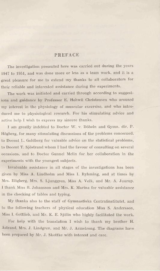
The investigation presented here was carried out during the years 1947 to 1951, and was done more or less as a team work, and it is a great pleasure for me to extend my thanks to all collaborators for their reliable and interested assistance during the experiments.
The work was initiated and carried through according to suggestions and guidance by Professor E. Hohwii Christensen who aroused my interest in the physiology of muscular excersise, and who introduced me to physiological research. For his stimulating advice and active help I wish to express my sincere thanks.
I am greatly indebted to Doctor W. v. Döbeln and Gymn. dir. P. Högberg, for many stimulating discussions of the problems concerned, to Docent L. Goldberg for valuable advice on the statistical problems, to Docent T. Sjöstrand whom I had the favour of consulting on several occasions, and to Doctor Gunnel Melin for her collaboration in the experiments with the youngest subjects.
Invaluable assistance in all stages of the investigations has been given by Miss A. Lindholm and Miss I. Ryhming, and at times by Mrs. Högberg, Mrs. S. Ljunggren, Miss A. Velk, and Mr. A. Juurup. I thank Miss B. Johansson and Mrs. K. Marina for valuable assistance in the checking of tables and typing.
My thanks also to the staff of Gymnastiska Centralinstitutet, and to the following teachers of physical education Miss S. Andersson, Miss I. Gottlieb, and Mr. K. E. Sjölin who highly facilitated the work.
For help with the translation I wish to thank my brother H. Åstrand, Mrs. J. Lindgren, and Mr. J. Armstrong. The diagrams have been prepared by Mr. J. Skottke with interest and care.

fiI offer my sincere thanks to all subjects who with greatest interest
and willingness took part in the really very streneous experiments.Generous contributions were received from Sveriges Riksidrotts-
forbund (The Swedish Sports Federation). Without such help it would hardly have been possible to carry out this investigation.
I do hope that the subjects’ strains and all help received were not in vain.
Stockholm. March 1952. P e r - O l OF ÂSTRAND

CONTENTS
Preface ............................................... 5 a. Total h em oglobin ..................... 44b. Blood hemoglobin concentra-
t i o n ............................................... 49Introduction ......................................... 9 c. Blood v o l u m e .......................... 49
4. D is c u s s io n ..................................... 51
C h a p t e r I 5. Sum m ary.......................................... 58
S u b je c t s ............................................... 11C h a p t e r VI
C h a p t e r II Lung v o l u m e s ..................................... 60M e t h o d s .............................................. 15 1. Introduction ..................................... 601. Statistics ......................................... 15 2. R e s u l t s .......................................... 602. Height and w e i g h t ..................... 16 3. D is c u s s io n ..................................... 683. Lung v o lu m e s ............................... 16 4. Sum m ary.......................................... 704. Hemoglobin and blood volume . 185. Experimental conditions and
w ork -p roced u res .......................... 186. Oxygen in ta k e ............................... 20 Lung v e n t i la t io n ............................... 727. Heart r a t e .................................... 21 1. Introduction .................................... 728. Blood lactate concentration . . 21 2. M e t h o d s .......................................... 72
3. Results and discussion . . . . 78a. Ventilation during maximal
C h a p t e r 111 w o r k .......................................... 78Criteria for estimation of maximal h. Ventilation during submax-
v a lu e s .............................................. imal w o r k ............................... 82c. Respiratory rate and tidal air
during maximal work . . . 84Ch a p t e r IV d. Comparison of the ventilation
Heart r a t e ......................................... 28 of adults during cycling and1. Introduction .................................... 28 r u n n i n g ..................................... 872. R e s u l t s ......................................... 29 4. The ventilation as a limiting3. D is c u s s io n .................................... 29 factor in ex erc ise .......................... 884. Summary.......................................... 37 5. Sum m ary.......................................... 90
C h a p t e r V C h a p t e r VIII
The total quantity of hemoglobin Blood lactate concentration . . . 92and blood vo lu m e .......................... 39 1. Introduction .................................... 92
1. Introduction.................................... 39 2. R e s u l t s .......................................... 942. M e th o d s ......................... 41 3. D is c u s s io n ............................... 963. Results . . . 44 4. Sum m ary.................................... 102
8
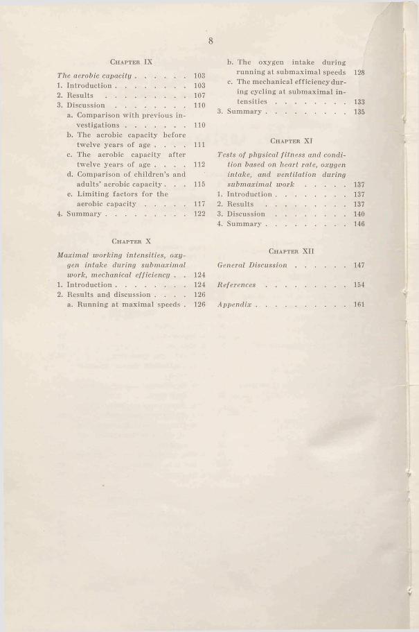
C h a p t e r IX
The aerobic capacity .......................... 1031. Introduction.................................1032. R e s u l t s ......................................1073. D is c u s s io n ................................... 110
a. Comparison with previous investigations .................................... 110
b. The aerobic capacity beforetwelve years of age . . . . I l l
c. The aerobic capacity aftertwelve years o f age . . . . 112
d. Comparison o f children’s andadults’ aerobic capacity . . . 115
e. Limiting factors for theaerobic c a p a c i t y ....................117
4. S u m m ary........................................ 122
C h a p t e r X
Maximal working intensities, oxygen intake during submaximalwork, mechanical efficiency . . 124
1. Introduction .................................... 1242. Results and discussion . . . . 126
a. Running at maximal speeds . 126
b. The oxygen intake during running at submaximal speeds 128
c. The mechanical efficiency during cycling at submaximal in-t e n s i t ie s .....................................133
3. Sum m ary........................................135
C h a p t e r XI
Tests o f physical fitness and condition based on heart rate, oxygen intake, and ventilation duringsubmaximal w o r k ..........................137
1. Introduction ......................................... 1372. R e s u l t s .............................................. 1373. D is c u s s io n ......................................... 1404. Sum m ary.............................................. 146
C h a p t e r XII
General D is c u s s io n ...............................147
R e f e r e n c e s .............................................. 154
A p p en d ix ....................................................161
INTRODUCTION
It is well known that the physical working capacity is less for females than for males, and for children as compared with adults. However, our present knowledge of the physiological basis of these differences, and of the limiting factors in working capacity is incomplete.
A problem, which is of increasing, importance, especially in wartime, is the utilization of womanpower by industry. Physiological research may contribute to solve the problem of women’s working capacity and fitness for a certain job.
In the military forces one of the main problem is to train men in as short a time as possible to a high standard of physical fitness, and to group them according to working capacity.
In the fields of sports, physiological bases are necessary for the planning of rational training and practice, and for a sound choice of events with regard to the participant’s sex and age.
In the clinic it must be of value to know the normal capacity of different bodily functions and of the body as a whole. None of the human organs are built for resting conditions, but for an activity level perhaps 10-20 times the basal metabolic rate.
The aim of the present investigation is to contribute to the physiological understanding of the great physical performances done for instance in sport, games, and during hard manuel labour. During heavy prolonged work “ first of all the supply of oxygen may be a limiting factor” {Dill, 1933). In this study special interest is consequently devoted to the determination of the maximal oxygen intake.

10In the laboratory working intensities from easy to maximal work
can be reproduced at standard conditions, and the reactions of the body to the stress can he followed and analysed. The results here reported are obtained from experiments dealing with male and female subjects from 4 to approx. 30 years of age, and the data recorded can be taken as “normal values” for healthy, well-trained individuals of the same age.
It is to be hoped that this investigation can be followed by a similar one with male and female subjects up to the age of for instance 65. This is of special interest from a social-economic viewpoint, since the average age of the population tends to increase.
It has been very stimulating to carry out this work due to a great interest shown by Sveriges Riksidrottsförbund (The Swedish Sports Federation) and several industries, especially Uddeholm Co.
The writer has tried to give each chapter in this hook a certain independence, which makes some repetitions inevitable. The review of previous works is limited to investigations of direct interest for the here discussed problems.
A preliminary report of some of the results was given at “VII Scandinavian Physiological Congress” , Aarhus. (Åstrand, 1951).

Chapter I
SUBJECTS
The number of subjects in this study was 112 females and 115 males between the ages of 4 and 33 (female subjects up to 25 years of age). The youngest children, between 4 and 6, came from Kindergartens'.
The subjects between 7 and 18 were pupils attending different schools in Stockholm. The selection was made by having the entire class draw lots, or by having the teacher select 3 subjects representing different standards, very good, rather good, and poor in respect to their working capacity.
The older subjects were students at the Gymnastiska Centralinstitutet, Stockholm (G.C.I., college of physical education), and all the students in three consecutive classes took part.
All subjects took medical examinations which gave normal results. Exceptionally fat children did not participate. All subjects have been rather well trained due to regular participation in gymnastics and games. This must he a necessary presumption if the results for the different groups are to he compared.
The students from G.C.I. are a selected group which is better equipped in regard to physical working capacity than men and women usually are. This must he remembered when comparing the'here obtained results with earlier values given in the literature. A comparison between the two sexes must be allowed, because the two groups belong to the same active category. This is of great advantage, and must give more reliable results than if well trained male subjects were compared with more or less untrained female subjects which apparently has been done often. Only a minority of the G.C.I. students belong to the élite in the field of sports and gymnastics. To what extent the school children represent the average is very difficult to settle. Very likely they lie somewhere above the average, because the poorest in a class will hardly undergo great bodily stress, the
' This part of the investigation was done in collaboration with dr Gunnel Melin (unpublished).
The figures denote X ±
e—
and
12
Q
— to to oera
erato■to
1+CO co b 1+
Oi to*■ 1+ 1+
to |+ 1+
1+ 1+
1+
O'^ 5°to O '
^ 1+ b
«> i+co 1-1ks |-f
1+4̂II
C ' » 2 ° 3 p -to2 .erator-t*
4 0 q .
H-i H-l
COto b - j b '
^ 1+ b l+i—i p
to O'o ' b c i bb !+ b i+
HJ. H-*O ' CO
1—l 4̂O O '
O ' ^ -J 4^b |-f « 1+
H-l p4^ to
V-i t - iO ' O'
p b P 4-c ,+
" 1+i- i poo to
,_ i p4̂ b co bo
b i+ b 1+I—i 4̂*>•
i- i
p C l4̂ C l «~ 1+ ^ 1+
i- i pbo CO
V-iC l
bo P ^1+° ! ip
bo b
TAB
LE 1
Num
ber of subjects in the different age groups and the average values of body height and weight.
13
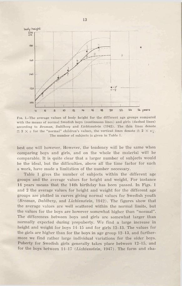
Fio. l.-T he average values of body height for the different age groups compared with the means of normal Swedish boys (continuous lines) and girls (dashed lines) according to Broman, Dahlberg and Lichtenstein (1942). The thin lines denote ± 2 X a for the “ normal” children’s values, the vertical lines denote ± 2 X
The number of subjects is given in Table 1.
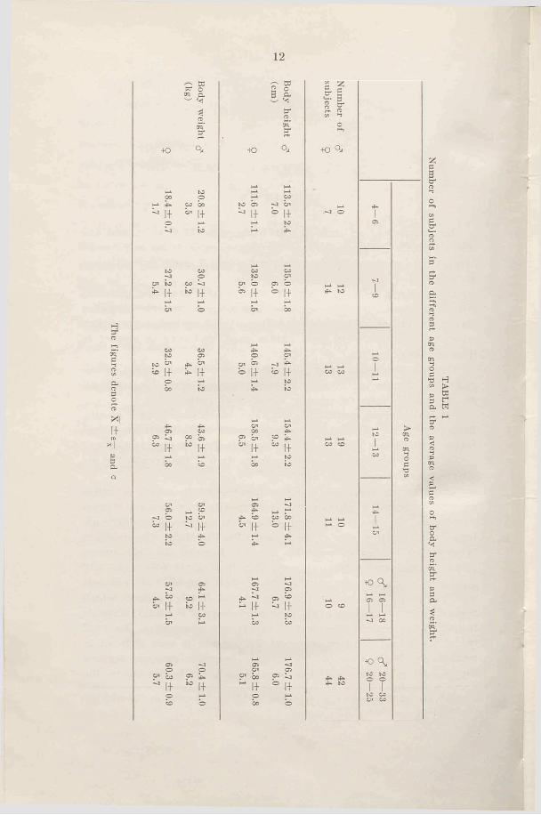
best one will however. However, the tendency will be the same when comparing boys and girls, and on the whole the material will be comparable. It is quite clear that a larger number of subjects would be the ideal, but the difficulties, above all the time factor for such a work, have made a limitation of the number necessary.
Table 1 gives the number of subjects within the different age groups and the average values for height and weight. For instance 14 years means that the 14th birthday has been passed. In Figs. 1 and 2 the average values for height and weight for the different age groups are plotted in curves giving normal values for Swedish youth (Broman, Dahlberg, and Lichtenstein, 1942). The figures show that the average values are well scattered within the normal limits, but the values for the boys are however somewhat higher than “normal” . The differences between boys and girls are somewhat larger than normally expected during prepuberty. We find a large increase in height and weight for hoys 14-15 and for girls 12-13. The values for the girls are higher than for the boys in age group 12-13, and furthermore we find rather large individual variations for the older boys. Puberty for Swedish girls generally takes place between 12—15, and for the boys between 14-17 (Lichtenstein, 1947). The form and cha-

14
F ig . 2.-The average values of body weight for the different age groups compared with the means of normal Swedish boys (continuous lines) and girls (dashed lines) according to Broman, Dahlberg and Lichtenstein (1942). The thin lines denote ± 2 X a for the “ normal” children’s values, the vertical lines denote ± 2 X
The number o f subjects is given in Table 1.
racteristic appearance of the curves is found later on in several connections where the ordinate represents entirely different hodily functions.
In the figures the values of 2 X a for “ normal” youth are given. Three boys fall outside these limits concerning both weight and height, and two hoys and two girls fall outside the limits concerning height. No values are outside the limit of 3 X a, and only two are outside 2.5 X a. For 141 subjects this distribution is normal.
For some of the determinations, for instance blood volume and respiratory functions the number of subjects wfas less. The following chapters will give detailed information concerning the composition of the material.
Chapter II
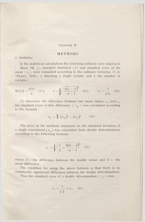
METHODS1. Stcitistics.
In the statistical calculations the following methods were employed. Mean (M, x), standard deviation (a) and standard error of the
mean ( e—) were computed according to the ordinary formulae (1-3) (Fisher, 1946), x denoting a single variate, and n the number of variates:
M (x )six)
n (1.); I / s(x — x )2 n — 1 (2.);
To determine the difference between two mean values, x t and~x 2, the standard error of this difference ( ed ) was calculated according to the formula
ea = V ( '7 . ) ! + (* ir ,)!
The error in the methods employed, i.e. the standard deviation of a single experiment (a ) was calculated from double determinations according to the following formula
(5.)
where d = the difference between the double values and d — the mean difference.
The condition for using the above formula is that there is no statistically significant difference between the double determinations.
Thus the standard error of a double determination ( Ee ) was
16
Regression equations were computed according to the ordinary formulae (Fisher).
Significance of differences between groups was tested by the t-test (Fisher).
Levels of significance:5 % level P = 0.05-0.01 ; almost significant1 % level P = 0.01-0.001; significant
0.1 % level ,P = < 0.001; highly significantIn the present study statistical calculations must be cautiously
used. As to the physical maximal performances it is difficult to obtain reproducable results. The maximal values for oxygen intake, ventilation, blood lactic acid etc. may be characterized as “ the highest attained values and possibly maximum” .
In the graphs which show average values of different functions for the various age groups the standard error of the mean is denoted by vertical lines limiting ± 2 X (twice the standard error of the mean; P = 0.05).
In the tables, the mean, the standard error of the mean, as well as the standard deviation are usually given. The most important functions and the highest individual values are to be found in the appendix.
2. Height and weight.
The height of the subjects was measured, and in each experiment the body weight with clothes was determined. The clothing was identical in all experiments, and its weight was known. All body weights given are net weights. Body surface area was calculated according to Du Bois’ formula.
3. Lung volumes.
The vital capacity was determined with a Krogh spirometer. The determinations were done in free standing position, and the highest value of 4 to 6 records was used.
In younger subjects it is difficult to obtain a maximal respiration. For that reason experiments have to he repeated until the subjects know the technique.
On the adult subjects, reserve and complemental air as well as the resperatory depth during normal breathing was determined in a standing position. Furthermore, the residual air was determined by the hydrogen method (according to Lindhard, 1925). The hydrogen-air-
17
<U33O'dCM 6
w 22 ^E- a
6cn<u'do£«soi-oSiEhW
ASTRAND
Oa-in
take
(lit
re/m
in.)
......
...
10
75
2.40
0 2.
407
0.00
7 ±0
.008
0.
077
0.05
5 0.
039
Lact
ic a
cid
(mgm
p.c
.) ...
....
30
30
78.6
7 78
.93
0.26
±0
.39
2.13
1.
51
1.07
Res
idua
l ai
r (li
tre)
...
......
.. 30
30
1.
416
1.39
4 0.
022
± 0
.014
0.
077
0.05
5 0.
039
Tota
l H
b (g
ram
) ...
......
......
.. 30
30
39
4.3
400.
9 6.
57
±3.5
2 19
.29
13.7
9.
7
18
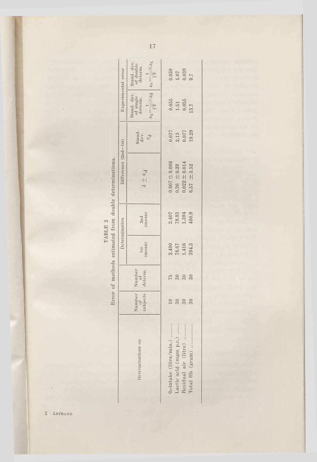
mixture was made up in a Krogh spirometer. After a maximal expiration the subject was connected to the spirometer, and he then made 3 almost maximal respirations. The spirometer was, as far as possible, emptied by each inspiration. The experiment was finished with a maximal expiration (total time about 10 seconds). At least 2 determinations were done on each subject. The error of the method can be judged from Table 2, showing the statistically treated results of 30 double determinations with the same number of subjects. The error of the method in a single determination is 0.055 litre, in a double one 0.039.
4. Hemoglobin and blood volume.
Later on, in the series of experiments, the total quantity of hemoglobin was determined by a CO-method modified by Sjöstrand (1948).
Altogether 51 female and 43 male subjects between the ages of 7 and 30 years were used. At least 2 determinations were done. Each value was often calculated from two samples, one after 15, and one after 30 minutes of rebreathing the CO-mixture.
According to Sjöstrand the 15-minute value shows the smallest individual variation and gives the best correlation with body weight. The writer’s results agree with this. No correction for the CO-ab- sorption to myoglobin has been done. The possible error according to Sjöstrand (1949 a) ought to be 2-3 per cent.
To estimate the total blood volume the relative hemoglobin values were determined with a Spekker-photometer, 100 per cent corresponding to 15.4 gm hemoglobin per 100 ml of blood. The blood sample was taken from a finger after warming it in water at 40-45 °C. for some minutes.
The error of the method has been calculated on the basis of double determinations on 30 subjects in different age groups (table 2). It amounts to 14 gm hemoglobin in a single determination and to 10 gm in a double one.
5. Experimental conditions and work-procedures.
As the aim of this work has been to determine the maximal oxygen intake, working procedures that bring large muscle groups into activity had to be chosen. Well suited working machines are the bicycle ergometer and the treadmill. All subjects, even the youngest, have run on the treadmill. In most cases the subjects over 20 years of age
19
have also been tested on Krogh’s bicycle ergometer. The frequency, set by a metronome, was 50 pedal revolutions per minute.
To obtain a high working intensity, the treadmill was set at an angel of 1° (1.75 per cent) for the subjects over 20. In all other experiments the treadmill was horizontal.
The first running was done at the low speed of 7-8 km/hr for the school children, and at 10-12 km/hr for the adult subjects. After a couple of days the experiment was repeated with a higher speed of 1-2 km etc. until the intensity was reached which exhausted the subject in 4 to 6 minutes. The determinations for each subject were spread over a period of 3 weeks or more.
Concerning the cycling experiments, the working intensities were for the male subjects 900, 1200, 1500, 1800 and 1950 kgm/min. or even higher according to the strength of the subjects. For the female subjects the working intensities were 600, 900, 1200 and 1350 kgm/min. etc.
At the highest working intensities the subjects have not always been able to follow the metronome. In those instances, they were asked to keep up the highest possible frequency. Consequently the exact intensity cannot be known, and the energy output will hardly have been constant throughout the experiment.
The experiments were done in the morning or afternoon, and no special care was taken to obtain basal conditions. Due to the great number of subjects this was not accomplishable. However, no experiments were done in the one or two hours directly after a meal. The school children and students came directly from their lessons. In experiments of this type there is no drawback with such an arrangement.
For several reasons it is desirable that experiments with a very high working intensity are not started directly from the resting state; the subject ought to have some “ conditioning” activity first. In determining the maximal working capacity this is of special importance. Whether this “warming up” procedure increases the oxygen intake is not quite clear, but according to Nielsen and Hansen (1937), the highest oxygen intake could be reached during a spurt following work of lower intensity, and they believed that a higher circulation rate was responsible for it. In the writer’s experiments the conditioning work had a low intensity compared to the work of Nielsen and Hansen.
Asmussen and Boje (1945); Högberg and Ljunggren (1947) have shown that the working capacity is greater and the feeling of stress is less if the subject is “warmed up” .
20As a rule, the first determinations made with a subject were not
used for the calculations of mean values. The subjects first had to get acquainted with the experimental procedures. Control experiments were done very liberally. The procedure applied here, with its many experiments and a successive increase in working intensity over a period of several weeks, is undoubtedly advantageous. Before the maximal working intensities are reached, the investigator gets to know the subject and has the possibility of judging his working capacity. This is very important in order to avoid overstrain, especially at the highest intensities. It can be mentioned, that in no case there was seen any damage or overstrain, in spite of the very high intensities.
6. Oxygen intake.
After 4-5 minutes of work the oxygen intake was determined by the Douglas bag method. In some of the maximal performance experiments the initial period of running had to be limited to 3 minutes. The initial period of mouthpiece breathing was 45-15 seconds. This time was dependent upon the intensity of the work. With few exceptions two bags were filled in close succession. The gas volume was measured in a gasometer and the gas analyses were made according to Haldane.
The method was effectively controlled by means of taking two samples of air. Especially with young subjects the possibility of leakage about the mouthpiece had to he considered. Furthermore the two air samples will show if the initial period of work before the collecting time was sufficiently long to obtain a steady state or “ apparent steady state” .
Amongst others Robinson (1938) has shown that after two minutes of maximal work 90-98 per cent of the maximal oxygen intake will be obtained, and after three minutes 95-98 per cent will have been reached. For the younger subjects acceleration in oxygen intake will be faster than for the older ones. As for the subjects in this study, results of the two bags show that the initial period was sufficiently long. This is also in agreement with the result of Nielsen and Hansen (1937). The mean values of the double determinations were used, and in order to determine the error of the method some results were treated statistically. The differences between the two determined values for a group of ten subjects, including seventy-five double determinations, gave an average difference of 7 ± 8 ml oxygen per minute. The error for a single determination wms 0.055 litres (Table 2). This error includes the physiological variations and the technical

21errors. The error of the method is relatively independent from the magnitude of the oxygen intake.
The accuracy of the determinations can be shown clearly by the results of a serie of 18 working experiments with a constant intensity (cycling). The subject, a w^ell-trained man, took a month to complete the series. During each experiment two air samples were taken. The oxygen intake which averaged 2,78 litres per minute had a standard error of ± 0.011 litres and a standard deviation of 0.046 litres or 1.7 per cent.
During the whole period of the collection of expired air the respirations were counted, and consequently the respiratory depth could be calculated.
Altogether 2000 experiments have been done on the treadmill and bicycle, and that means about 4000 determinations of oxygen intake.
7. Heart rate.
In the cycling experiments the heart rate was counted during work, in the running experiments, however, the time for the first 10 or 15 pulse beats was taken immediately after running, which practically gives the same values as if the determination was done during the work (Cotton and Dill, 1935; Lundgren, 1946). All determinations were done by palpation over the ictus or the arteria carotis.
S. Blood lactate concentration.
When the intensity of work was close to maximum, blood samples for determining lactic acid were taken (except from 33 subjects).
The analyses were done according to Edwards (1938), and control analyses have been done with known standards of lithium lactate in different concentrations. In most cases double analyses were done on the same blood sample, and the average value is given. The error of the method was calculated from the results of 30 double determinations, and was 1.5 mgm per cent for a single determination without definite variation with absolute values (between 30 and 125 mgm per cent). The error for a double determination was 1.1 mgm per cent (Table 2).
The blood samples were taken after the subject had had his hand in water of 40-45 °C. for a short time. Consequently the circulation through the finger was very lively, and blood almost identical with arterial blood could be obtained from a fingertip (comp. Bang, 1936).

22Due to the lively circulation, the necessary blood sample, 0.5 ml, could be obtained easily also from the younger subjects.
One sample was taken during the first minute after the end of work, and an additional one after 3-4 minutes. (Sometimes only the “ one minute” sample was taken from the youngest subjects). Consequently the concentration of blood lactic acid was determined 5-6 minutes and 8-10 minutes after the work had begun. According to Bang the highest values for lactic acid wall be obtained between 5 and 10 minutes after heavy work has begun. Due to the fact that the maximal working intensity was very high, the lactic acid values will maintain a rather high level for some time. The preliminary experiments have shown that the sampling procedure used will give sufficiently good information about the maximal values. The samples which gave the highest values have been used.

Ch apter III
CRITERIA FOR ESTIMATION OF MAXIMAL VALUES
Figs. 3 to 6 show the oxygen consumption in some series of experiments. Fig. 3 shows how the oxygen intake increases linearly with an increase in the speed, but only up to a certain speed, in this case 13-14 km/hr; from then on it levels off. It is obvious that the subject’s maximal oxygen intake value has been reached, at least with this type of work (“ apparent steady state” ). Since running will bring very large muscle groups into activity it is most likely that the value is very close to the absolute maximum for oxygen intake (see Chapter IX). The increase in energy output at higher speeds must be delivered anaerobically. This is also indicated by the increase in the blood lactic acid concentration. For these two subjects the curves level off at an intensity where the maximal values for lactic acid concentration are 60-70 mgm per cent.
In 70 of the 140 running experiments with school children such a level is found. Sometimes the oxygen intake will even drop at higher running speeds. Lactic acid determinations from the same experiments show rather large individual variations, especially in relation to age. The values in 53 experiments where lactic acid wras determined showed variations between 60 and 90 mgm per cent, when the oxygen intake curves levelled off, the average value was 70 to 75 mgm per cent. The older the subjects, the higher the values will be. Apparently the body tries as fast as possible to cover its need for energy aerobically, but at higher intensities the anaerobically delivered energy increases more and more. Consequently a higher acidity in the muscle will be the result, which undoubtedly contributes to a better utilization of the blood oxygen, because of Bohr effect and because of vasodilatation. At a certain working intensity an upper limit will be reached, and a further increase in anaerobic products will not influence the oxygen intake. This matter will be discussed later on.
It is quite clear, however, that a relationship exists between the
24
Ojrintqke Ojrinlakeper min. per min.
Fig. 3.-Oxygen intake at different working intensities.The figures give the corresponding values o f lactate concentration.
Fig. 4.-Text see Fig. 3.
Oi-intakep e r m in .
working intensity and the aerobic and anaerobic processes, so that the maximal oxygen intake will be attained when the lactic acid concentration has reached a certain value, different in different individuals.
The experiments have made clear that a small increase in intensity when the work is heavy gives a steep increase in the lactic acid values; compare values for U.C. in Fig. 3 (only 4 minutes’ running at 14.5 km/hr as compared to 5 minutes at 13 and 14 km /hr). This is in good agreement with the experiment of Asmussen (1946); Christensen

25
X •
-rf
F ig . 6.-0xygen intake (continuous line) and blood lactate concentration (dashed line) in relation to working intensity (cycling).
and Högberg (1950 a, b) who showed that the oxygen need at high working intensities increases more steeply than the oxygen intake, and an increasing amount of energy must be paid for by anaerobic processes. Consequently the efficiency will decrease.
In the experiments where no level for oxygen intake was seen (comp. Figs. 4 and 5), the experiments were continued at least until the before mentioned high lactic acid concentration was obtained. From then on the subject’s feeling of stress was taken into consideration. Fig. 5 shows two series of experiments were the oxygen intake increased in spite of very high values for lactic acid. Obviously one has to recognize great individual variations. No further experiments at higher intensities were done in those cases because the working intensity was absolutely maximal, according to the subjects, and the lactic acid values were extremely high.
Another example of the relationship between lactic acid, oxygen intake and working intensity is shown by Fig. 6. In this case the lactic acid and oxygen intake were determined during and after 7 minutes of work on a bicycle ergometer at 4 different intensities between 1620 and 2100 kgm/min. The oxygen intake had reached its maximum when the maximal value for lactic acid was 80 mgm per cent.
An objective control of the strain of work is absolutely necessary in experiments of this type. In many cases a subject already at a speed of for instance 12 km/hr explained that he was absolutely exhausted. If, however, the lactic acid value was low, say 40 mgm per cent, the subject was convinced that he could continue at a higher
26
P Ppr ?rre re3 s ©
2- cp3
CP
2 2 c c 2!o 3 re ’"'’3-a sf» g-gWli
ros'-n ap£3 ow S.?r 1~cp i
3- oQ CP CT>C/i ̂fb ►-. P ¡3o- rr - PCl * *
re re CL 3
S' S'
3»fc 3 2. re
*3 sire £. re « <n
£ 25. c o 9
<p p-i CO CO
M gffg- “ 3
• 3 re _ P-3^°P s5p
hi 3Cb C5E-2- rereo SL*-b i—- I“*} h—3 3w cp
o
jo coOI CO <3-
cp a r3 2.̂
TAB
LE 3
Maxim
al oxygen consumption per m
inute and per kg body weight and m
aximal lactate concentration for tw
o groups of subjects,
27
speed, which he really could do. The oxygen intake then increased significantly.
Concerning the experiments with the students from G.C.I. not quite enough determinations during running have been done to settle the oxygen diagram definitely, but the values for the oxygen intake at the highest speeds show the same tendency as in the other experiments. The lactic acid values and the subjective feeling of stress have helped to judge the intensity in relation to the working capacity of the subject.
To see how the values of maximal oxygen intake agree for the two groups, one where the oxygen levels off and the other where it does not, Table 3 has been made up. In the last group the capacity must be judged from the lactic acid values, and the subjective stress of the subject. The table contains values for all males and females between 14 and 18 years, and the reason why those age groups have been chosen is partly that the oxygen intake per kg body weight, which is the best way of comparison, is nearly constant in these age groups, and partly that the lactic acid values are known for all these subjects.
The table shows that half of the boys and half of the girls have reached a definite level for oxygen intake within the highest region of speed. The average maximal value of this group is, however, the same as for the other group where this level has not been reached. The lactic acid values are high for all groups, but apparently the girls not reaching a level for oxygen intake have been stressed the most.
Ch apter IV
HEART RATE
In this chapter the maximal values for heart rate will be chiefly discussed. The submaximal values will be reported later on (chapter XI).
7. Introduction.
Determinations of the heart rate, on male subjects during heavy work, have been done many times, and usually values of about 200 beats per minute for young, healthy subjects are given. Very few values for female subjects have been recorded.
Boas (1931) found during running (2 minutes) an average of 190 beats per minute with 27 boys between the ages of 9-13.
Dill and Brouha (1937) found with 14 men between 20-36 years and 20 boys between 12-19 years who ran to a state of exhaustion on a treadmill, maximal values of 195 for the men and 196 for the boys (running time 2 to 5 minutes).
Robinson (1938) using boys between 8-12 years old as subjects found 198 as an average value. With 13-19. year old subjects he found an insignificantly7 lower value, and for the 20-29 year old subjects the average value was 189. All ran on a treadmill. The highest individual value was 211. (The number of subjects was 39).
Morse, Schultz, and Cassels (1949) had as subjects 110 boys between 10—17 years old and found an average heart rate of 196 after maximal running. They did not find any variation with age.
Brouha and Heath (1943) tested college men 17 to 22 years of age in their capacity to run. Maximal heart rates ranged from 167 to 217, averaging 193 for 176 subjects.
Metheny, Brouha, Johnson, and Forbes (1942) had 17 women running to a state of exhaustion (average time 108 seconds), the average maximal heart rate was 197. When running until exhausted (216 seconds), 30 male subjects averaged 194.
Christensen and Hogberg (1950 c) report average values of 240
29
Max. heart rate
_-ft 1 1J u J D - j - J
1 _______1______ _______1
. - o ’1 ° - ?<+ e 6 10 12 14- 16 18 20 zz 2b 26 years
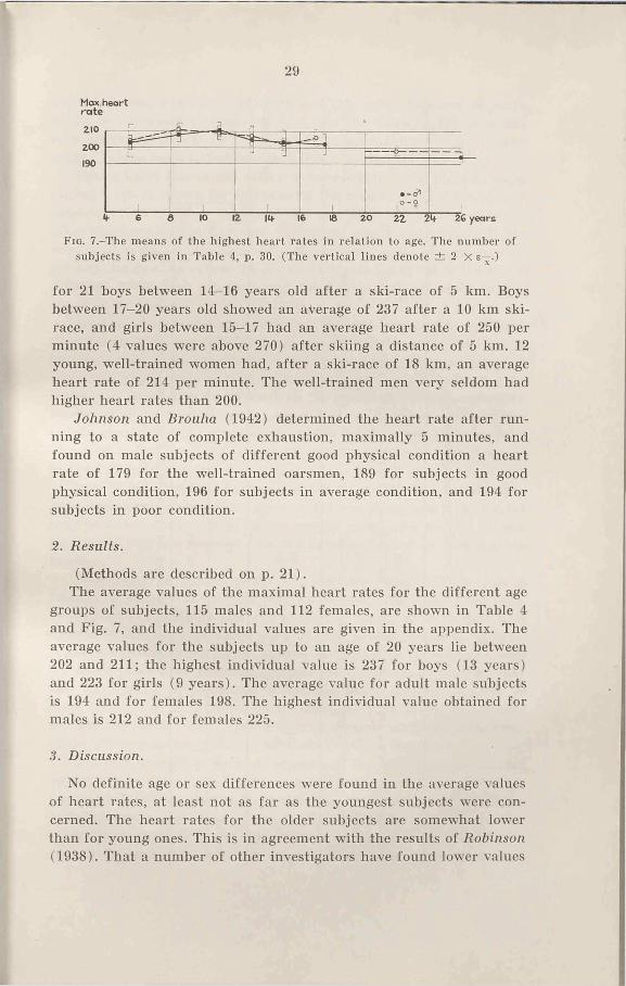
Fig. 7.-The means of the highest heart rates in relation to age. The number of subjects is given in Table 4, p. 30. (The vertical lines denote ± 2 X e—.)
for 21 boys between 14-16 years old after a ski-race of 5 km. Boys between 17-20 years old showed an average of 237 after a 10 km ski- race, and girls between 15—17 had an average heart rate of 250 per minute (4 values were above 270) after skiing a distance of 5 km. 12 young, well-trained women had, after a ski-race of 18 km, an average heart rate of 214 per minute. The well-trained men very seldom had higher heart rates than 200.
Johnson and Brouha (1942) determined the heart rate after running to a state of complete exhaustion, maximally 5 minutes, and found on male subjects of different good physical condition a heart rate of 179 for the well-trained oarsmen, 189 for subjects in good physical condition, 196 for subjects in average condition, and 194 for subjects in poor condition.
2. Results.
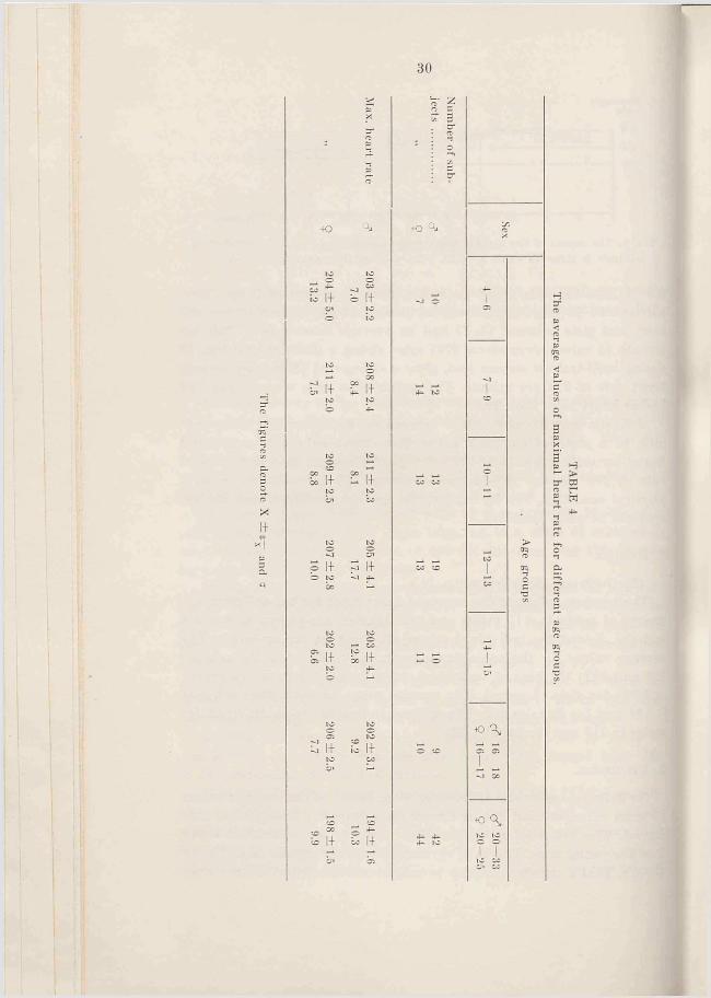
(Methods are described on p. 21).The average values of the maximal heart rates for the different age
groups of subjects, 115 males and 112 females, are shown in Table 4 and Fig. 7, and the individual values are given in the appendix. The average values for the subjects up to an age of 20 years lie between 202 and 211; the highest individual value is 237 for boys (13 years) and 223 for girls (9 years). The average value for adult male subjects is 194 and for females 198. The highest individual value obtained for males is 212 and for females 225.
3. Discussion.
No definite age or sex differences were found in the average values of heart rates, at least not as far as the youngest subjects were concerned. The heart rates for the older subjects are somewhat lower than for young ones. This is in agreement with the results of Robinson (1938). That a number of other investigators have found lower values
30
x1+
+o
io 1+ ti
ll 1+
1+
1+ S 1+
1+ 1+
1+ 1+
® 1+ » 1+
cnO
+0
o
C Q*1C 1CT T
TAB
LE 4
The average values of maxim
al heart rate for different age groups.
31
with the same working procedure and the same duration of work might be explained through the fact that the working intensity has been submaximal. Values for blood lactic acid, lung ventilation and oxygen intake seem to indicate that this has been the case.
That a heart rate of 200 does not represent the absolute maximum during work is shown for instance by the results of Christensen and Högberg (1950 c). They explained the very high values they obtained after ski-races by the fact that the skiing was always finished at a very high intensity. Consequently these high heart rates do not represent steady state values. In our experiments the speed of the treadmill had to be kept constant, and the heart rates are more or less steady state values.
Another possible explanation is that the very long duration of work in a ski-race (the best time for 5 km was 23 minutes and for 10 km 40 minutes) induces an overstrain of the circulatory system resulting in a lowered stroke volume and consequently an increasing heart rate. During the short duration of work on the treadmill such an overstrain would not come about. However, a very fast restitution could be shown to take place in the ski experiments, which speaks against an over-load (3 minutes after the finish, the heart rates had on an average dropped to 120-130 beats per minute).
It is evident that the heart rate of normal, young, and healthy subjects during work can reach values of 200 and more, and it is of great interest to see if these high values are the result of a normal regulation, or if they are more or less the consequence of an overstrained heart.
Wahlund (1948) came to the following conclusion: “ Pulse rate is roughly a linear function of oxygen consumption and work-load. Judged by the findings referred to in the survey of literature it is usually 170-200 at exhausting work in adult healthy subjects. At these values no further increase of cardiac output is to be expected with increased pulse rate. However, oxygen for the working muscles may still be supplied by increased utilization, but is is only possible to work for a short time under such conditions. The factor responsible for limiting cardiac output is the time of diastole which at pulse rates above 180 may be too short for adequate filling of the heart.
It is thus possible to make an estimate of the limit to which cardiac output can be increased, by studying the pulse curve during work in any individual case. When the pulse rate has reached a sufficiently high value at a certain load, this load is said to be maximal. In this study the maximum heart rate at which work can be performed adequately is put at 170 beats per minute. If the pulse rate at any load
32C^-pulse
28.0m l
24.0
2 0 .0
16.0
12.0
02,-pul9e
16.0ml. n
too 120 IUO ISO 180 200 220 heart rate
Fig. 8.-0xygen pulse for 15 male subjects in relation to heart rate (subjects and their ages are noted at the curves).
F ig . 9.-0xygen pulse for 15 female subjects in relation to heart rate (subjects and their ages are noted at the curves).
is above this value, the load is regarded as an overload. It must be admitted that some individuals can work with a pulse rate slightly above 170 beats per minute, but they are not expected to do this for any length of time as there is usually a prolonged increase of pulse rate. Consequently they may reach a value of 180 or more after some time and are thus at the limit of their capacity.”
Schneider and Karpovich (1948) stated that a heart rate of 170 “is close to the lower border of the zone of excessive rates.”
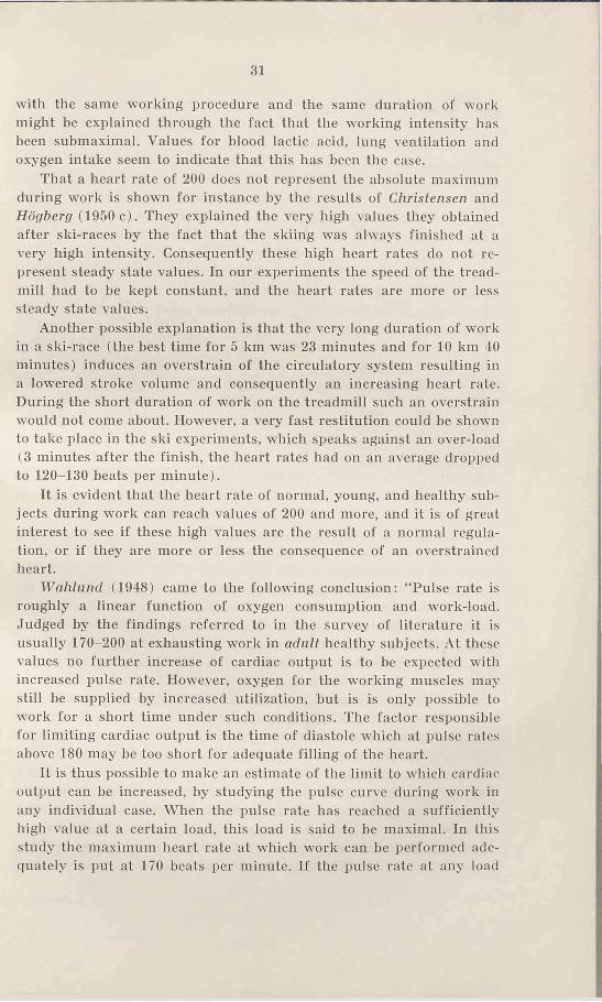
Only direct determinations can give a definite answer to the questions, but theoretical calculations might give some indications. The results shown in Figs. 8 and 9 indicate that the circulatory system must be regarded as “ effective” even if the heart rate values are 200 or more. The oxygen pulse, i.e. the gross oxygen intake divided by the corresponding heart rate, has been plotted in relation to the heart rates. Only 30 individual graphs, half from female and half from male subjects are given. They represent surprisingly well, however, the

33
results of all of the material. (The other graphs can be obtained from this institution upon application.)
The values for the oxygen pulse are somewhat uncertain, as both errors of pulse countings and oxygen intake determinations are included in this figure. However, it is of some interest to have the values for heart rate and oxygen intake combined in one figure. This figure represents two components, the stroke volume of the heart and the average value for the arterio-venous 0 =-difference. (a.-v. 0 =- difference).
Henderson and Prince (1914) found an almost constant oxygen pulse at heart rates of 80-100, at higher rates the oxygen pulse increased steeply to reach maximal values of 11-17 ml at a heart rate of 130-140 per minute. At still higher heart rates the oxygen pulse might decrease.
Benedict and Caihcart (1913) observed on a professional cyclist the highest oxygen pulse, 16.8 ml, at a working intensity during which the heart rate w7as 163 beats per minute.
From Figs. 8 and 9 it is quite clear that the oxygen pulse for the majority of the subjects increases with the heart rate, very markedly for adult subjects. This indicates that even at the maximal heart rates, i.e. about 200, the blood circulation is fully effective as far as oxygen transport is concerned. The continous increase in oxygen transport per pulse heat at increasing heart rate can be explained through an increase in the a.-v. O.-difference, or in the stroke volume, or an increase of both at the same time. Another possibility is a decrease of one factor which would be more than fully compensated by the other. The data related here can never prove anything definite, but it may be of some interest to discuss the problem.
With the acetylene method Christensen (1931 b) determined heart minute volume, stroke volume, and a.-v. 0 2-difference in 4 men and 3 women during heavy work. He found that the a.-v. Oa-difference as a rule increased with increasing working intensity, but at the maximal intensities it decreased (the duration of work was at least 20 minutes). The highest average value, independent of the working intensity, was 108 ml oxygen per litre blood for the women and 131 for the men. The stroke volume was either nearly constant or increased slightly with increased working intensity and heart rate. The heart rates, however, were never as high as in the writer’s experiments. The highest individual value of the oxygen pulse was 22.7 ml for men and 14.6 ml for women.
If we take 3 male subjects, whose oxygen pulse curves are very much alike, and represent the average for adult subjects (Fig. 8, see
3 ÅSTRAND
34
arrow), and if we presume an a.-v. 0 2-difference of 120 ml at a heart rate of 150 (corresponding to the average values given by Christensen), then the stroke volume at an oxygen pulse of 17.5 will be 146 ml.
Doing the calculation with a constant a.-v. 0 2-difference of 120 ml the stroke volume wall be 169 ml at a heart rate of 200 (the oxygen pulse 20.3 ml). If we presume instead a constant stroke volume, thea.-v. CVdifference would increase from the 120 ml at heart rate 150 to139 ml.
Using the same presumptions in the case of subject R.B. (Fig. 8) a constant a.-v. O.-difference of 120 ml gives a stroke volume of 115 ml at a heart rate of 155, 140 ml at a heart rate of 200, and 169 ml at a rate of 215 (oxygen pulse 13.8, 16.8, and 20.3 ml respectively). The decrease of the oxygen pulse at the highest heart rate of 222 (to 19.1 ml) corresponds to a decrease of the stroke volume to 159 ml.
If, however, the stroke volume should be kept constant at 115 and140 ml, respectively, a working intensity with a heart rate of 215 would give an a —v. 0,-difference of 177 and 145 ml respectively.
A smaller a.-v. (^-difference and consequently a higher stroke volume at the lower heart rate is of course possible, but the experimental data do not support this assumption. A higher a.-v. 0 2-difference than 120 may be possible at the highest frequencies, but it will hardly exceed 140-150 ml 0 2 per litre of blood, which would be the case if the stroke volume decreased as shown in the above mentioned calculations. Rather the reverse would be expected. As already mentioned, Christensen sometimes found a decrease in the a.-v. 0,-difference when the working intensity was close to maximum.
The purpose of these calculations has been to show that a decrease in stroke volume, as a rule, is very unlikely even if the heart rate rises above 200 per minute. An increase is much more likely at least for the older subjects. Of course the type of work and the working time can change the situation.
The younger subjects, below 12-13 years, often have an almost constant oxygen pulse at different heart rates. In 5 of the younger subjects there is even a decrease (see Fig. 8 G.M.). Galle (1926) determined thea.-v. CVdifference and stroke volume by a nitrous oxide method according to Krogh and Lindhard (1912) in four 12 year old boys during rest and in 2 subjects during work. He found during work, with an average heart rate of 113, an a —v. 0 2-difference of 106 ml, as compared to 68 during resting conditions. The stroke volume during work was 70 ml, in rest 41 ml. The small number of subjects makes the values somewhat uncertain, but the increase in stroke volume as well as ina.-v. CVdifference is so marked that they indicate a definite change.
35
The heart rates in the experiments of Galle are, however, at quite a different level compared to the writer’s values, and no certain conclusions can be drawn.
Due to the lack of experimental results as to stroke volume anda.-v. CVdifference in children during work, it is impossible to explain the constant oxygen pulse in the writer’s experiments. It is impossible to state if the a.-v. O.-difference and the stroke volume are constant, and consequently the increase in oxygen intake takes place only because of an increase in minute volume due to an increasing heart rate, or if a decrease in one component is compensated by an increase in the other one.
A decrease in a.-v. O¡.-difference during work with increasing intensity is very unlikely (except at maximum). Due to the changes in blood distribution with an increased vascular bed in the muscles during work, a higher a.—v. (h-difference in the working muscles is more likely. The increase in the blood lactic acid, which was also found in the young subjects at higher working intensities, must give a lower pH, and consequently the reduction of the oxyhemoglobin must have been more complete. If we accept a higher or perhaps an unchanged 0 2-difference the stroke volume must have been lowered or kept constant.
The younger subjects have, in relation to body weight, a smaller blood volume than the older ones, but they have a higher maximal oxygen intake per litre blood volume (see Chapter V). This makes likely a higher minute volume in relation to the blood volume. This in connection with a relatively low blood volume can bring about less favourable conditions for the filling of the heart, and a relatively lower stroke volume. As the peripheral vascular bed increases, this can be more and more pronounced. In this way the constant oxygen pulse in the children can be explained. (See Chapter V).
Concerning the heart rate of the younger subjects another problem has to be discussed. Robinson (1938) stated that “ the high heart rates of youthful subjects in work that is easy for them, is a characteristic of youth; presumably sympathetic stimulation raises the heart rate above the level necessary for accomplishment of the task.”
If the heart rate is unnecessarily high, this ought to be indicated by a low oxygen pulse which ought to increase when the working intensity makes the rate “necessary” . The writer’s experiments show, however, that the younger subjects during easy work have an oxygen pulse equal to that during heavy work. An “ unnecessarily” high heart rate is more likely to be characteristic for the younger group, when the working intensity approaches the upper limit.
3 ‘
36
Maximal values for stroke volume and minute volume can be calculated on the basis of theoretical values for a.-v. O.-differences. As a possible value we take 120 ml (comp. Christensen) and for adult subjects also 140 ml. Those values are very likely somewhat too high, and consequently the calculated values for stroke volume and minute volume will be minimal values. •
For the youngest (4 years), with an oxygen pulse of about 4 ml, an a.-v. Ck-difference of 120 ml would require a stroke volume of 33 ml. The minute volume with a heart rate of 215 would be somewhat more than 7 litres. Several boys 10 years of age had an oxygen pulse of about 10 ml. With a heart rate of 215 and an a.-v. 0 :-difference of 120 ml the stroke volume would be 80-90 ml and the minute volume of the heart 17-19 litres.
Two male subjects had an oxygen pulse of 28 ml at a heart rate of 180. With an a.-v. 0 2-difference of 120 cc the stroke volume would be somewhat higher than 230 ml and the minute volume 42 litres. With an a.-v. O.-difference of 140 ml the corresponding figures would be 200 ml for the stroke volume and 36 litres for the minute volume.
Several of the female subjects had an oxygen pulse of 16 ml with a heart rate of 200. With an a —v. O.-difference of 120 the stroke volume would be 136 ml and the minute volume 27 litres; with an a.-v 0,-difference of 140 ml the stroke volume would be 116 ml and the minute volume of the heart 23 litres.
These theoretically calculated values agree well with the experimentally determined values of Christensen. He reports for stroke volume a maximal value of 209 ml for the men and 161 for the women; the minute volume was 37 litres for the men and 25 for the women.
Differences in heart rate between male and female subjects, as far as rate at a given oxygen intake is concerned, are shown in Fig. 10. This figure shows the relation between heart rate and oxygen intake independent of working intensity and type of work (running or cycling).
The great scattering of the values is explained by the fact that the figure is made up of 86 subjects’ heart rate-oxygen intake curves, and their oxygen intake capacity varies to a great degree; extremes are 3.30 and 5.09 litres for the male subjects, 2.41 and 3.40 litres oxygen per minute for the female subjects.
The values for the males and females show only a slight overlapping, and male subjects’ higher oxygen transport at a certain heart rate during maximal and submaximal working intensities must be explained mainly by higher stroke volumes and possibly also by higher a.-v. O.-differences.

37
heart rate
l.o 2.0 3.0 40 50 0^-intakeht/min
F ig . lO.-Heart rates in relation to oxygen intake for adult male and female subjects (86 subjects). Maximal as well as submaximal values are represented.
TABLE 5The average values o f maximal heart rate and oxygen intake for adult males and
females during running and cycling.
Type of work SexNumbero fsubjects
Max.heart rate
Max. Os-intake litre per min.
Running ................... c? 40 189 ± 1 . 6 4.04 ± 0 .0 6Cycling ...................... 35 191 ± 1.9 4.03 ± 0.07Running and cycling 42 194 z t 1.6 4.11 ± 0 .0 6
Running ................... 9 43 198 ± 1 . 6 2.89 ± 0.04Cycling .................... 32 194 ± 1 . 6 2.76 ± 0 .0 5Running and cycling 44 198 ± 1 . 5 2.90 ± 0 .0 4
The figures denote x + e x
The type of work, cycling or running, has no definite influence on the maximal values for heart rate. The average values for heart rate and oxygen intake are given in Table 5. Fifteen of the thirty-three male subjects who were “ forced” to maximal work both in running and cycling showed the highest values during running. The corresponding number for 32 female subjects was 19.
i. Summary.
The heart rate was determined during maximal running and for adult subjects also during cycling (115 male subjects aged 4 to 33
38
years, 112 female subjects aged 4 to 25 years). For the subjects below 20 years the average values lay between 202 and 211, and were independent of age and sex. For the adults the mean values were 194 for men and 198 for women.
There was no difference between the maximal values obtained during cycling and running.
The experiments indicate that the circulation is fully effective as far as oxygen transport is concerned even at the highest heart rates if the oxygen transport per pulse beat is taken as indication of effec- tivity. Some exceptions exist among the youngest subjects. Excluding this age group, there are no findings indicating a decrease in stroke volume with increasing heart rate, not even at the highest rates.
Chapter V
THE TOTAL QUANTITY OF HEMOGLOBIN AND BLOOD VOLUME1
1. Introduction.
It is of the greatest interest to try to analyse the factors that determine the size of the maximal oxygen intake capacity. Since oxygen is transported via the blood and its hemoglobin, it is justifiable, in connection with an investigation of an individual’s aerobic capacity, to study at the same time his blood volume and the total quantity of hemoglobin.
This is of special interest since Kjellberg, Rudhe, and Sjöstrand (1949 a, c) have shown a close correlation (0.90), broadly speaking, irrespective of age, sex or training condition, between quantity of total hemoglobin, and to a still higher degree blood volume, and the heart volume as determined by X-ray in the horizontal resting position. Moreover they found a very marked correlation (0.82 and 0.90 respectively) between the amount of hemoglobin and the pulse rate during work (600 kgm/min), or the work at which the pulse reached a level of about 170 beats per minute. “ This indicates that there is a close correlation between the total blood volume and the stroke volume during work” .
Previous investigations.
Sjöstrand (1949 a) using a CO-method modified by him, has determined the total quantity of hemoglobin in 17 boys and 21 girls between the ages of 8 and 17, and in 174 men between 18 and 57 years, and 92 women between 17 and 70 years.
In the case of the men the total quantity of hemoglobin increased approximately parallel with the age and growth up to 22 years of age, whereas for the women the increase became considerably less after 12-13 years, and reached a constant level after 20 years.
During puberty, a definite increase was found for boys in relation
1 These investigations were made in collaboration with Astrid Lindholm. Preliminary reported at “ VII Scandinavian Physiological Congress” , Aarhus. (.Lindholm, 1951.)
40
o c; ^ to o *jI I I I I1C >-* l-l H-l h-km a' w m c
tO t-1O 05 ►£**: l I to O -si
C5 O' ^^ oo o b O' osvi vi si a> i- w Oi 05 M ^ p
b bsi c x
32 y O ’ 4* CO IOo p p to -sjb M C vl O’ Io-sj C i O ' 4 - CO coO p CD p p Ob b b b b
to to to to H i- 1
b b b co b bo >-* oo >—* o o
CO CC
p 00 io b
O' O' O’ 4^ CC toO CO s i 3 : O C s ]b b b bo b b
p p—1 oc ^ o0 5 ^ s]Cn
to to to to M _Mb si 35 W si bO ' CO 45* w W S j 4— p p tO tO ■—1io b b co b bto ce c: 3 : 4c v.
TAB
LE 6
Body height and w
eight and oxygen intake of the subjects for blood determinations
in comparison w
ith corresponding values of all subjects.
41
to their body weight, but for girls there was a relative reduction from 12 to 20 years.
The average quantity of hemoglobin in the adult men was 803 ± 8 gm or 1.16 per cent of body weight. In the adult women it was 555 ± 11 gm or 0.86 per cent. (The differences were 30 per cent and 25 per cent respectively, calculated from the values for the men).
Before puberty girls showed a lower hemoglobin quantity in relation to weight than the boys, but the same quantity in relation to age (8-12 year old boys had 1.17 per cent of body weight, girls 0.96 per cent).
The blood volumes showed about the same variations with the exception that the difference between the adult males and females was about 10 per cent less than that for the quantity of hemoglobin.
Brines, Gibson, and Kunkel (1941) determined the blood volume of 50 children between 2 and 17 years with “ the blue dye” . They found that the blood volume increased with age, and was the same for boys and girls up to puberty, when is was about 2.5 litres. Afterwards it increased more quickly for the males.
Morse, Cassels, and Schultz (1947), using the same method, examined 77 children from 1 to 17 years and found that their blood volumes were higher than those reported by Brines et al. The blood volumes were found to be related to body weight by a linear equation, to stature by an exponential equation and to surface area by a non-linear equation of the second degree. Less variation from the regression curve was found by using surfaces areas as the standard for reference. With one exception the greatest security of prediction came from multiple regression equations relating the blood and plasma volume to the height and chest girth of the child.
von Pornt (1951) has given in his work a review of the blood volume determinations done by the Evan’s blue dye method. In his own experiments he found in 20 men and 20 women, all normal, and with an average age of about 26 years, a total blood volume of 6.17 litres for the men and 4.48 for the women (respectively 83.3 and74.7 m l/kg).
2. Methods.
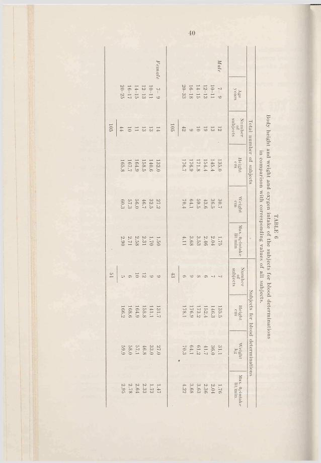
In these experiments, the total quantity of hemoglobin in 43 men (7-30 years) and 51 women (7-24 years) was determined. The subjects were classified into groups according to their ages (Table 6). When the average values for the height, weight, and maximal oxygen consumption of these groups are compared with the values of all subjects it is clear that the selections are representative.
The figures denote X ± e—
and
-J O X3 J ”3. o ¿.3 c it'“ 'B E .nj 3
tO ° *
o o o -1 o 01ti 1+ s; 1+
m + s i+
U 1+ b 1+ ° o 00 o
: i+ p i+. 0 ^ 0
pl+pl+
b 1+ b 1+
p .xa p cr V./■ *» 3*a p -1 ?rO Q3 ?
+o ° *
© |+ © |+
b |-f b |-frvt 1 1 i- 1 1
b 1+ b 1+
p |+p |+C: ? 5C ?
b || b ||
»1+ b 1+
o. g 'v o 3 .
— c" 3
t o
¡-» li *“ II
» 1+ o II
I 1+ 03 1+
; 1+ “ 1+K1 ® K*J
^ CO y i tfk^ 1+ H-
b 1+ co |+
*5 Cp
**3erac X- 2 - jo V* —e gtr °“ rf-i
t o QJ
> 1+ ~ 1+P P
o O O O'b II b 1+
‘ 1+ i* 1+
p 1 + 1 + «= *-
b || co !|
‘ 1+ b |+
42
3 o JJ o
S' ¡Po0 3*3 OQo
+0
b |+ ^ |+
COI CO COCO CO11+ ~ 1+
to H*if * CO
»1+ b 1+
^ CO£ 1+ p 1+ 00 _ ^
00 CO 4* C5 00b II b II
oo
. 1+ -1+to to
CO
-
t o ° *
o ob 1+ ~ 1+
b II U II
. i+ b i+
' l l 1-'i [4 b [+
»1+21+
o “ o ®I-* + ** 1+° P ° o
33
OtraO3 33
to
! ! + 3 1 +
t O ^
£l+gl+
S 1+ § 1+
! 1+ S 1+
t o a *
; i+ s i+
to o*
! l + g l +
43
F ig . l l . - T o t a l q u a n t i t y o f h e m o g lo b in in r e la t io n t o a ge .
.0
31
F ig . 12.-The average values of the total quantity of hemoglobin, blood volume, andblood hemoglobin concentration in relation to age. The number of subjects is given
in Table 7, p. 42. (The vertical lines denote ± 2 X s—).
44
A further account of the method used has been given in the chapter II (modified CO-method, according to Sjöstrand, 1948).
3. Results.
The individual values for total hemoglobin, relative hemoglobin and blood volumes are given in the appendix. The average values are shown in Table 7.
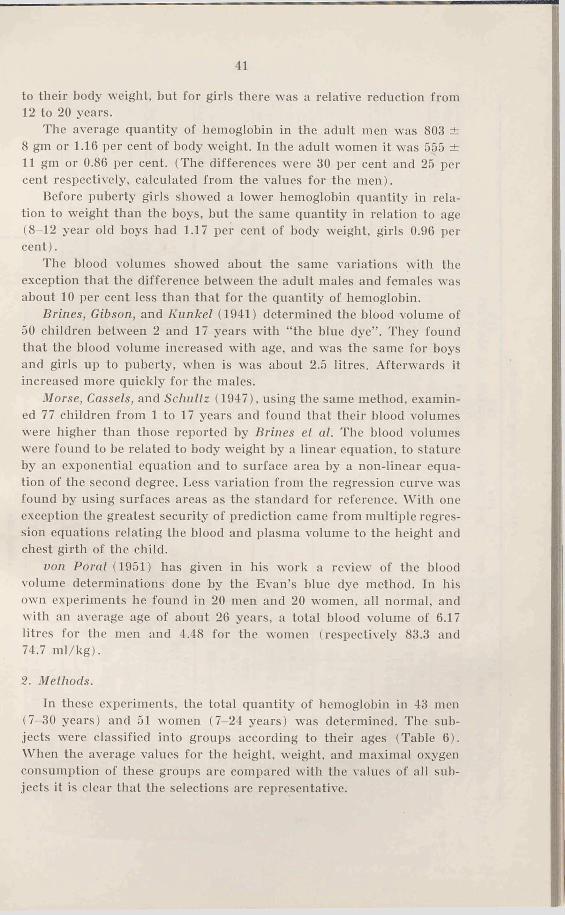
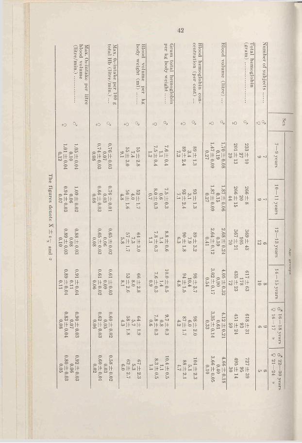
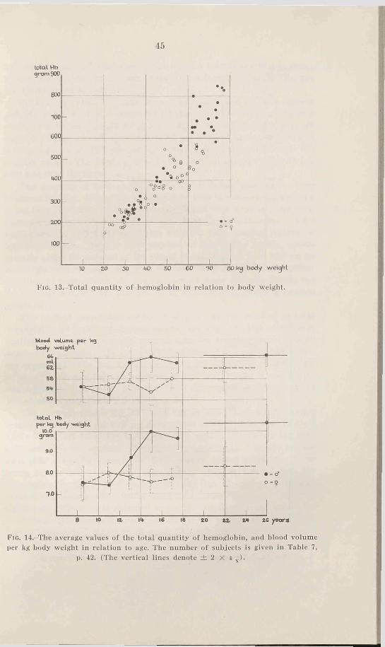
a. Total hemoglobin-The quantities of total hemoglobin for the various subjects at different ages are shown in Fig. 11 and the average values in Fig. 12. It is evident that the values for boys and girls are similar up to 12-13 years (7-9 years 233 and 201 gm, 12—13 years 369 and 367 gm, respectively), but after that age they separate and attain different “ levels” (for males from 617 to 727 gm, for females from 435 to 495 gm). As far as the girls are concerned the values for the 14-18 years old lie 28 per cent lower, and for the adult women 32 per cent lower than the corresponding male values.
The groups are so small that the average values merely show the trend and the difference which is shown between boys and girls of 7-9 years is, for instance, not statistically significant.
With regard to the average values, the quantity of hemoglobin in the girls increases fairly evenly with age up to 14-15 years, but afterwards the increase becomes considerably less, whereas in the boys there is a relatively higher increase during the puberty (an increase of 67 per cent). This should be seen against the background of an average increased body weight of 47 per cent (calculated from the lower values).
From Fig. 11 it is clear that some 13 year old girls and 15 year old hoys have already attained adult values.
If the value of total hemoglobin is related to body weight, the values are scattered as in Fig. 13. The hemoglobin increases with the body weight, but the variations are rather great. An analysis for hoys of about 40 and 55-60 kg cannot he made, because there are too few values available. Below 40 kg (before puberty) the values for boys and girls are of the same magnitude. The average values for the total hemoglobin within the weight limits 25-40 kg are 255 gm for boys and 251 gm for girls, and are consequently very similar.
Table 7 and Fig. 14 shows the total quantity of hemoglobin (in gm) per kilogram body weight for various age groups. It is evident that the development of boys during puberty brings about a great increase of the quantity of hemoglobin with relation to weight, and this increase
45
total Hbgram 900
800
TOO
600
-
•• •. • •
*» •• . •
500
MOO
-
•o
o% 9
• o
. \ o ° C
» , •o
5 o
300 -
o
, , 4 ‘
°o°°8 o °
o •
0D
100
______ i______
00 aP>
______ I______ ______ I______
O -
___I___10 20 30 W SO 6 0 TO 60 kg body weight
F ig . 13. Total quantity of hemoglobin in relation to body weight.
F ig . 14.-The average values of the total quantity of hemoglobin, and blood volumeper kg body weight in relation to age. The number of subjects is given in Table 7,
p. 42. (The vertical lines denote ± 2 X e~ ).
46total Hbgram 900• ••
700
600
••
• •
••
••.
500
1+00
300
o
•_ ° ••CD o °
88.o • ^ °
o
%>*° < * i
• 8 o *O • QA - '°
o oc
100
o°8o o , - a*
110 120 130 lifO 150 160 170 16 0 190 cm body height
F ig . 15.-Total quantity of hemoglobin in relation to body height.
total Hb
F ig . 16.—Total quantity of hemoglobin in relation to surface area.
47
is brought to an end after some years when a level is reached at about 10 gm hemoglobin per kg body weight (the average value is 10.0 gm for 14-30 year old subjects).
No corresponding increase is noted in the case of girls during puberty, but the average values for all groups under 20 years lie fairly constant between 7.5 and 8.0 gm per kg. For the 5 females over 20 years the value is somewhat higher, 8.3 gm (one of these subjects had a very high value, 10.1 gm per kg, which considerably influences the average value). The 14-24 year old girls have an average value 21 per cent less than the boys.
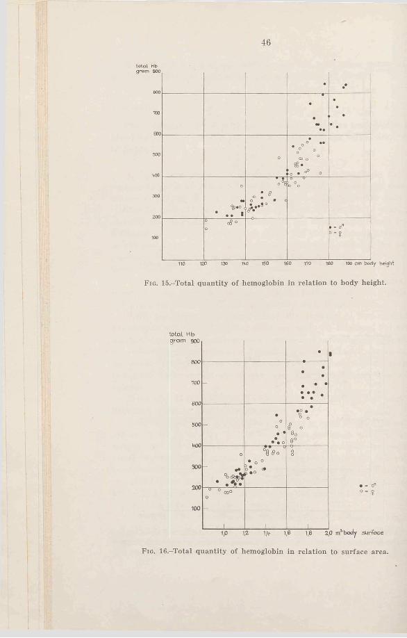
The quantity of hemoglobin in relation to height and surface area is shown by Figs. 15 and 16. (The surface area is calculated according to Du Bois’ formula). The values are scattered with a fairly large deviation along a curved line, and between the sexes no difference of total hemoglobin is noticeable for those subjects with smaller stature or less surface area. Neither is it marked at the higher values, consequently about 60 per cent of the values for girls taller than 165 cm lie inside the boys’ range, while the others have a somewhat smaller quantity of hemoglobin. As the number of male subjects inside the “ middle-age-groups” are too few, it is impossible to make a reliable comparison.
The highest correlation seems to be given by the relation of total hemoglobin to maximal oxygen intake, see Fig. 17. The correlation coefficient of 0.97 for males and of 0.94 for females shows a high degree of correlation. The two regression lines have a somewhat different mean and slope, but the difference is not statistically significant. (The correlation of total hemoglobin to maximal oxygen intake for all the subjects is 0.97). The deviation from the regression lines is about 10 per cent (statistics see Table 8, p. 50).
The average values for different age groups, of maximal oxygen intake (litres/min) per 100 gm hemoglobin are shown in Fig. 18 (and Table 7). Apart from the 10-11 year old groups the values for both boys and girls lie together. The relatively lower oxygen intake for the said girls could be a result of their earlier sexual maturity, if the lowTer oxygen intake per unit hemoglobin, which is noticed with the older girls, is understood as a secondary sex character. A greater number of subjects might throw some light on this question. (A highly significant difference is obtained by comparing the average values for 7-11 year old boys with those of the 14-18 group. The difference between the girls 7-9 and 10-11 years of age is almost significant).

48
total. Hb
Fig. 17,-Total quantity of hemoglobin in relation to maximal oxygen consumption (statistical data see Table 8, p. 50).
F ig . 18.-The means of the maximal oxygen intake per 100 gm total hemoglobin(dots) and litre blood volume (squares) in relation to age. The number of subjects
is given in Table 7, p. 42. (The vertical lines denote ± 2 X e~j4'
.
49b lood volum e lit. 6 .0
2,0
- ••
. •____
•
•
•
• -o -* —• ° 0 °° i * o°m
SP 0(* °0°*6 nC£
1
oCOo0
5 $o °
___________ 1 _ _________ ___________ l 1
• - d ”
O"?
30 years a g e
F ig . 19.-Blood volume in relation to age.
blood volume
F ig . 20.-Blood volume in relation to body weight.
b. Blood hemoglobin concentration.-The average values for hemoglobin concentration are shown by Fig. 12 (15.4 gm Hb/100 ml blood = 100 per cent). For the female subjects the values lie at about 90 per cent, and it is the same for the males up to and including 13 years. After this age the value increases to 97-98 per cent for 14-18 year old males and to an average of 104 per cent for the oldest.
c. Blood Do/ume.-The blood volume has been calculated from the values of total and relative hemoglobin, hut since the hemoglobin concentration varies in different parts of the vascular system, this calculation certainly does not give the true value. However, when the ex-
4 ÅSTRAND

50
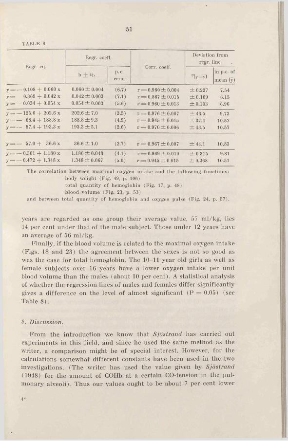
TABLE 8
Sex n X + © x y i ey
Max. 02-intake in relation to body weight d 115 52.2 ± 1 .7 7 3.01 ±0.11)» above 40 kg 9 76 57.5 ± 0.81 2.75 ±0.(1>> below 40 kg $ 36 28.1 ± 1.07 1.48 ±0.«
Total Hb in relation to max. 0 2 -intake d 43 2.98 ±0 .1 6 478.3 ±32.19 51 2.25 ± 0 .0 8 355.5 ±161
all subjects o ' + 9 94 2.58 ±0 .09 411.7 ±181
Total Hb in relation to max. 02-pulse, allsubjects .................................. d* + 9 92 12.7 ± 0 .4 8 407.3 ±18.
Blood volume in relation to max. 0 2-intake d 41 2.98 ±0 .1 7 3.21 ±0Jt»» 9 51 2.25 ± 0 .0 8 2.55 ±0.11
perimental conditions are identical, the results obtained can be used as a mean of comparison between the sexes and different age groups (Sjöstrand, 1949 a).
Since the average value of hemoglobin concentration is similar for boys and girls up to 13 years, but afterwards lower for the females, it can be stated:
1) that the blood volume in the younger subjects is approximately related to age, weight, height, and surface area like the total hemoglobin (see Figs. 19 to 22) and
2) in the case of the older subjects, the difference between male and female subjects is less. Thus, the difference in average blood volume values between boys and girls of 14-18 years is only 23 per cent and for adults 21 per cent. The corresponding figures for total hemoglobin were 28 and 32 per cent, respectively.
The average values of the blood volume per kg body weight are shown in Fig. 14. In the case of the boys, two different levels are seen, one before puberty (52-55 ml blood volume per kg body weight) and one after (64—67 ml). The average values for the female subjects lie more constant; however, they show a tendency towards a relative increase of blood volume after 16 years. If, in spite of this, all over 12
51
TABLE 8
Regr. eq.Regr. coeff.
Corr. coeff.
Deviation from regr. line
b + eh p.c.error °(y—y)
in p.c. of mean (y)
y = _ 0.108 + 0.060 x 0.060 ±0 .004 (6.7) r = 0.980 ±0.004 ± 0.227 7.54y — 0.369 + 0.042 x 0.042 ± 0.003 (7.1) r = 0.867 ±0.015 ±0.169 6.15j = - 0.034 + 0.054 x 0.054 ±0.003 (5.6) r = 0.960 ±0.013 ±0.103 6.96
y = — 125.6 + 202.6 x 202.6 ± 7.0 (3.5) r = 0.976 ±0.007 ±4 6 .5 9.73y=------ 68.4 + 188.8 x 188.8 ± 9 .3 (4.9) r = 0.945 ±0.015 ±3 7 .4 10.52y = — 87.4 + 193.3 x 193.3 ± 5 .1 (2.6) r = 0.970 ±0.006 ± 43.5 10.57
y = — 57.0 + 36.6 x 36.6 ± 1 .0 (2.7) r = 0.967 ±0.007 ±44.1 10.83
y = — 0.301 + 1.180 x 1.180 ±0 .048 (4.1) r = 0.969 ±0.010 ±0.315 9.81y=— 0.472 + 1.348 x 1.348 ±0.067 (5.0) r = 0.945 ±0.015 ± 0.268 10.51
The correlation between maximal oxygen intake and the following functions: body weight (Fig. 49, p. 106) total quantity of hemoglobin (Fig. 17, p. 48) blood volume (Fig. 23, p. 53)
and between total quantity of hemoglobin and oxygen pulse (Fig. 24, p. 57).
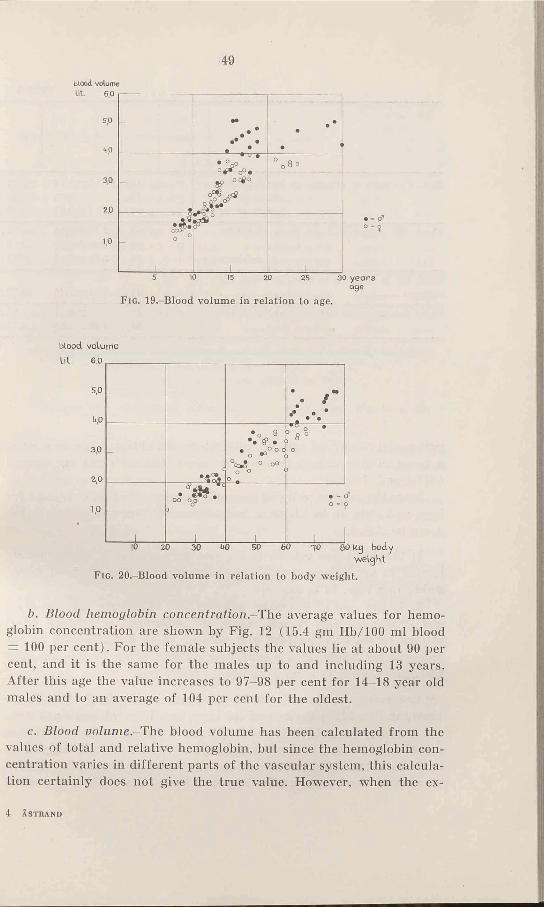
years are regarded as one group their average value, 57 ml/kg, lies 14 per cent under that of the male subject. Those under 12 years have an average of 56 ml/kg.
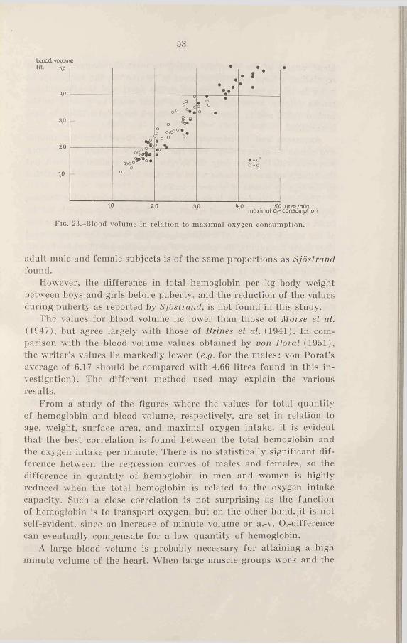
Finally, if the blood volume is related to the maximal oxygen intake (Figs. 18 and 23) the agreement between the sexes is not so good as was the case for total hemoglobin. The 10-11 year old girls as well as female subjects over 16 years have a lower oxygen intake per unit blood volume than the males (about 10 per cent). A statistical analysis of whether the regression lines of males and females differ significantly gives a difference on the level of almost significant (P = 0.05) (see Table 8).
i. Discussion.
From the introduction we know that Sjöstrand has carried out experiments in this field, and since he used the same method as the writer, a comparison might he of special interest. However, for the calculations somewhat different constants have been used in the two investigations. (The writer has used the value given by Sjöstrand (1948) for the amount of COHb at a certain CO-tension in the pulmonary alveoli). Thus our values ought to be about 7 per cent lower4’

52
blood volum e U t. 6.05.0
*tf>
3.02.0
1,0
F ig . 21.-Blood volume in relation to body h e ig h t .
V
---------------------- 1
*• •
°
. . » <
o o C£> ) O CO O
°
•"
^ ftp •
___Q_S_______
oo . - o *
ra j Ö 5 WÖ 150 Ï6Ô TM> ISO « 0 cmB o d y h e i g h t
blood volume tit 6.0
5,0
up
30
2,0
1.0
-
a s
V
_________!_________
•o * 0
« 0• o»
<fe * ° °. ° 0 ^ 0
3 * ____ 1 _________
80 £ o « > •OCDO
OO
o o o cr3 -* o o
[ -
1
• - o*o- 9
F ig . 22.-Blood volume in relation to surface area.
than those of Sjöstrand (1949 a). As the true value of the constants is not yet known, there is no reason for correcting the values obtained. Furthermore, there is no method that exactly determines the blood volume, so no “ normal values” can be chosen as basis for a comparison. The main interest of these investigations has been the variation of total quantity of hemoglobin and blood volume in relation to age and sex, and so the absolute values are of secondary importance. For such an aim Sjöstrand’s method is very useful.
When corrected with regard to the difference of 7 per cent due to the different constants the writer’s values will, broadly speaking, agree with those of Sjöstrand. The difference between the average values of
53
blood volume•
•V •
• •
-
o° ° * .
o0° O dP °*.f . ° ° °
8 °oO •
-
« O 0 * 0 'o
o
•• -<? 0 - 5
1,0 2,0 3,0 <f,0 . 5,0 litre/min.maximal o2-consumption
Fig. 23,-BIood volume in relation to maximal oxygen consumption.
adult male and female subjects is of the same proportions as Sjöstrand found.
However, the difference in total hemoglobin per kg body weight between boys and girls before puberty, and the reduction of the values during puberty as reported by Sjöstrand, is not found in this study.
The values for blood volume lie lower than those of Morse et at. (1947), but agree largely with those of Brines et at. (1941). In comparison with the blood volume values obtained by von Porat (1951), the writer’s values lie markedly lower (e.g. for the males: von Porat’s average of 6.17 should be compared with 4.66 litres found in this investigation). The different method used may explain the various results.
From a study of the figures where the values for total quantity of hemoglobin and blood volume, respectively, are set in relation to age, weight, surface area, and maximal oxygen intake, it is evident that the best correlation is found between the total hemoglobin and the oxygen intake per minute. There is no statistically significant difference between the regression curves of males and females, so the difference in quantity of hemoglobin in men and women is highly reduced when the total hemoglobin is related to the oxygen intake capacity. Such a close correlation is not surprising as the function of hemoglobin is to transport oxygen, but on the other hand, it is not self-evident, since an increase of minute volume or a.-v. Ch-difference can eventually compensate for a low quantity of hemoglobin.
A large blood volume is probably necessary for attaining a high minute volume of the heart. When large muscle groups work and the
54
blood vessels of the skin are dilated, to meet the demands of heat regulation, a large quantity of blood is “ stored” in the periphery. In order to secure the venous return to the heart and thereby a large stroke volume, a certain quantity of blood must be present in the central veins, and in order to achieve this a large total blood volume is essential.
Asmussen and Christensen (1939) have shown how the minute and stroke volumes change with the blood distribution. When work was done with the arms only, the oxygen intake remained constant, but the minute volume decreased when a certain blood volume was isolated in the legs by means of pressure cuffs.
The blood circulation is probably the most decisive factor limiting oxygen intake under normal conditions. The working capacity of the heart or the blood capacity of the working muscles determines the size of the minute volume. At working intensities where the oxygen need rises above the supply, the quantity of hemoglobin per litre blood ought to determine the size of the oxygen intake, independent of whether the limiting factor lies in the “ central” or “peripherial” capacity of the circulation. This presupposes that the a.-v. 0 2-difference is changed with the hemoglobin concentration, i.e. that the oxygen tension in the working muscles and in the venous blood leaving them is fairly constant during higher working intensities. (Unfortunately there is no reliable information concerning these oxygen tensions).
Consequently, if a large blood volume is primarily essential for a large minute volume, then the quantity of hemoglobin ought to be still more decisive with regard to the maximal oxygen intake. The better relation to oxygen consumption given by hemoglobin in these experiments as compared with blood volume, suggests this.
In the case of the female subjects, the relative hemoglobin, i.e. the oxygen capacity of the blood, is 12 per cent lower than for males after about 16 years of age. After this age the maximal oxygen intake per litre of blood and minute is also lower, 10 per cent. This agrees with the earlier statement, that the total quantity of hemoglobin to a higher degree determines the maximal oxygen intake.
However, there is no certain relationship between the hemoglobin concentration and the maximal oxygen intake, e.g. calculated per kg body weight, and the reason for this can be that the values of hemoglobin concentration lie within rather narrow limits.
Cullumbine { 1949) has studied the correlation between blood hemoglobin concentration and response to various exercise tests for subjects aged 10 to 20 years. It was concluded that the hemoglobin concentration was significantly correlated with speed of movement,
55
strength, and the ability to sustain prolonged moderate muscular effort. There was no evidence for a significant correlation between the hemoglobin level and either the response to moderate exercise or the response to severe exercise. Cullumbine believes that this severe type of exercise was too severe, that the oxygen debt was too large to be influenced significantly by the rather small range of hemoglobin levels present in his group of subjects.
The hemoglobin concentration is not increased by training (Robinson, Edwards, and Dill, 1937; Schneider and Karpovich, 1948). The increase of hemoglobin which follows physical training, takes place along with an increase of blood volume. The “normal” hemoglobin concentration (at a barometric pressure of about 760 mm Hg) obviously forms an optimum, even if the demands on the circulation vary considerably. It is difficult to explain why this optimal value is not the same for men, women, and children (see p. 115).
During acclimatization to a low oxygen pressure, a physiological increase of the quantity of hemoglobin takes place, but probably under these conditions other factors than the blood circulation determine the working capacity. (Christensen and Forbes, 1937).
The low hemoglobin concentration found in patients with anemia is compensated through an increase in the minute volume of the heart (Nielsen, 1934). It is, in this connection, of interest to see that individuals with chronic anemia, according to, among others, Sjöstrand (1949 b), have an increased blood volume as compared with normal people. This is probably one of the conditions necessary for their greater minute volumes. Sjöstrand has also pointed out that patients with chronic anemia are often in better physical condition than patients with hemorrhagic anemia in spite of the latter’s eventually higher relative hemoglobin, which can be explained by differing blood volumes.
Children before puberty have a relatively smaller quantity of hemoglobin and blood volume than their elders, but they have in relation to body weight a similar maximal oxygen intake per minute, for which reason the oxygen intake per unit of total hemoglobin or blood volume is greater than for their elders. This can depend upon:
1) a greater a.-v. 0 2-different which amply compensates for the lower oxygen capacity of the blood. According to Robinson (1938), the oxygen saturation of the arterial blood in a state of rest is about 0.5 per cent higher for 10 than for 20 years old. The difference can eventually he greater during heavy work, but not even under such conditions can it have such a great quantitative importance. If an increase of the a.-v. 0 2-difference takes place, this should consequently
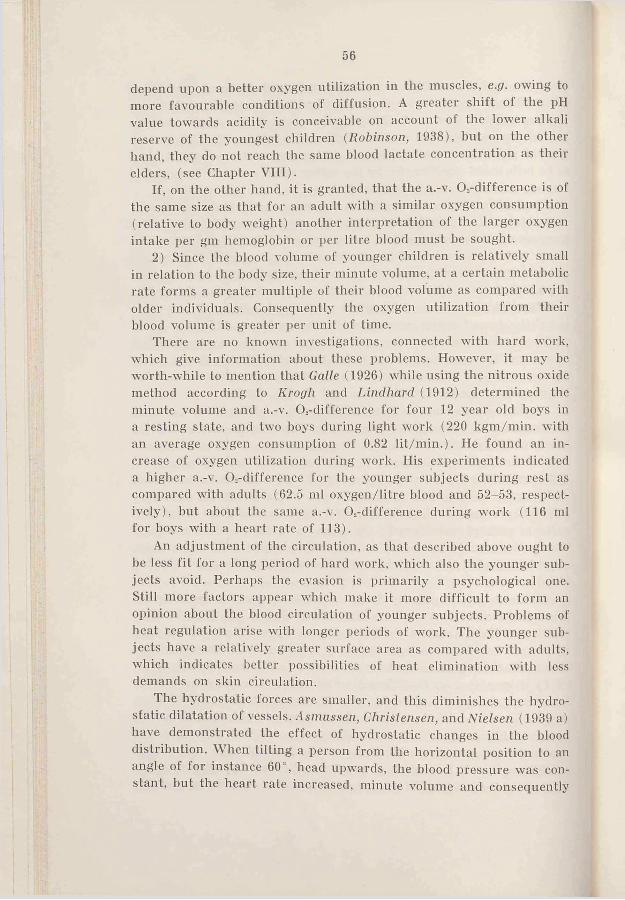
56
depend upon a better oxygen utilization in the muscles, e.g. owing to more favourable conditions ol diffusion. A greater shift of the pH value towards acidity is conceivable on account of the lower alkali reserve of the youngest children (Robinson, 1938), but on the other hand, they do not reach the same blood lactate concentration as their elders, (see Chapter VIII).
If, on the other hand, it is granted, that the a.-v. O.-difference is of the same size as that for an adult with a similar oxygen consumption (relative to body weight) another interpretation of the larger oxygen intake per gm hemoglobin or per litre blood must be sought.
2) Since the blood volume of younger children is relatively small in relation to the body size, their minute volume, at a certain metabolic rate forms a greater multiple of their blood volume as compared with older individuals. Consequently the oxygen utilization from their blood volume is greater per unit of time.
There are no known investigations, connected with hard work, which give information about these problems. However, it may be worth-while to mention that Galle (1926) while using the nitrous oxide method according to Krogh and Lindhard (1912) determined the minute volume and a.-v. Os-difference for four 12 year old boys in a resting state, and two boys during light work (220 kgm/min. with an average oxygen consumption of 0.82 lit/min.). He found an increase of oxygen utilization during work. His experiments indicated a higher a.-v. O.-difference for the younger subjects during rest as compared with adults (62.5 ml oxygen/litre blood and 52-53, respectively), but about the same a.-v. 0,-difference during work (116 ml for boys with a heart rate of 113).
An adjustment of the circulation, as that described above ought to be less fit for a long period of hard work, which also the younger subjects avoid. Perhaps the evasion is primarily a psychological one. Still more factors appear which make it more difficult to form an opinion about the blood circulation of younger subjects. Problems of beat regulation arise with longer periods of work. The younger subjects have a relatively greater surface area as compared with adults, which indicates better possibilities of heat elimination with less demands on skin circulation.
I he hydrostatic forces are smaller, and this diminishes the hydrostatic dilatation of vessels. Asmussen, Christensen, and Nielsen (1939 a) have demonstrated the effect of hydrostatic changes in the blood distribution. When tilting a person from the horizontal position to an angle of for instance 60°, head upwards, the blood pressure was constant, but the heart rate increased, minute volume and consequently
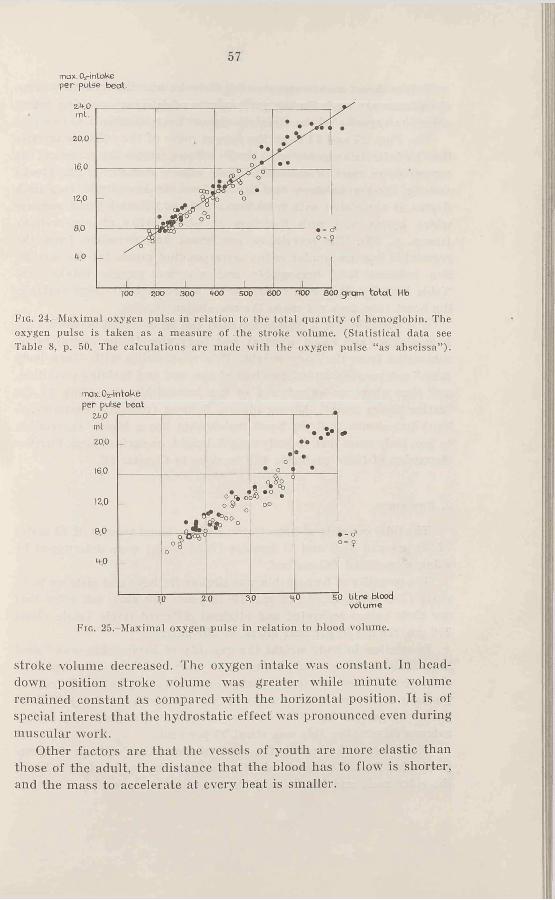
57max. Og-inlake per pulse beat.
F ig . 24.-Maximal oxygen pulse in relation to the total quantity of hemoglobin. The oxygen pulse is taken as a measure of the stroke volume. (Statistical data see Table 8, p. 50. The calculations are made with the oxygen pulse “ as abscissa” ).
max.Orintake
stroke volume decreased. The oxygen intake was constant. In head- down position stroke volume was greater while minute volume remained constant as compared with the horizontal position. It is of special interest that the hydrostatic effect was pronounced even during muscular work.
Other factors are that the vessels of youth are more elastic than those of the adult, the distance that the blood has to flow is shorter, and the mass to accelerate at every beat is smaller.

58
Before direct measurements of the stroke and the minute volume of children are made during performance of different working intensities the capacity of the circulation must he conjectured.
The Figs. 24 and 25 show the oxygen pulse of the subjects in relation to total hemoglobin and blood volume, respectively. Since the oxygen pulse must be considered to be a good measure of the stroke volume, the graphs show that the maximal stroke volume, to a high degree, is correlated with total hemoglobin (0.96) and blood volume, which agrees well with Kjellberg et. al.’s (1949 a, c) observations (comp. p. 39). The correlation coefficient and deviation from the regression line are similar to the corresponding values for the correlation between total hemoglobin and maximal oxygen intake (see Table 8, p. 50). It is of a certain value to see that the third variable, the heart rate, does not make the correlation smaller.
It is possible that a high correlation can be obtained between total hemoglobin (and blood volume) and body size, if the excess fat is subtracted from the body weight. This relationship with “ lean body mass” can possibly be independent of age, sex, and training condition, and thus show an adjustment of the hemoglobin according to the “ active tissue mass” . Among others, Behnke (personal communication) has shown that the basal metabolism has a higher correlation to lean body mass than to body weight, height, or surface area. Further discussion of these problems will he done in Chapter IX.
5. Summary.
The total quantity of hemoglobin and the blood volume of 43 males (7-30 years of age) and 51 females (7-24 years) were determined by using a modified CO-method.
The quantity of hemoglobin was similar for boys and girls up to 14 years (7-9 years about 220 gm, 12-13 years 370 gm), but after that age their values separated and attained different levels (male about 700 gm, female 30 per cent lower, or 500 gm).
In relation to body weight the quantity of hemoglobin was found to be similar for boys and girls up to 12 years (7.5-8.0 gm per kg). The older girls showed no greater change in their values, but in the case of the boys an increase took place, and they reached a new level at 14-15 years (10 gm per kg). The difference between the male and female average values after this was about 20 per cent.
Blood volume had nearly the same relation to sex and age as hemoglobin, but the difference in blood volume per kg body weight between the older male and female subjects was only 14 per cent.
59
A very high correlation (0.97) was obtained between total hemoglobin and maximal oxygen intake, where the male and female values followed to a large degree the same regression curve. This relationship was discussed.
The maximal oxygen pulse showed a similar correlation with total hemoglobin and blood volume, and if the oxygen pulse is taken as a measure of the stroke volume, it shows how close the relation between maximal stroke volume and blood volume is.
Ch a pt e r V I
LUNG VOLUMES11. Introduction.
Investigations on vital capacity, residual capacity and so on are numerous. A review of the main results as far as adults are concerned was given by Birath (1944) and for different age groups by Robinson (1938).
The average values given by Birath on the absolute and relative lung volumes in normal men and women, between the ages of 18 and 40, are given in Table 9. The determinations were done in an easy sitting position with a modified hydrogen method. Only a few of the subjects were physically well-trained. Birath states, “ as seems generally to be the case, in other materials also, the residual capacity for females was percentually higher than for males” , and that “ the demonstrated difference between males and females in the present material as regards the relative magnitude of the residual capacity may possibly be connected with the higher degree of training in the males” .
Robinson determined the vital capacity for 53 male subjects representing the age groups 6.0, 10.8, 14.1, 17.5, and 24.0 years, and he found the following average values: 1.26, 2.20, 3.71, 4.95, and 5.25 litres. For men 20—29 years old (11 subjects) the average value for total lung volume was 6.81 litres, for residual capacity 1.66 litres, and for residual and equilibrium capacity, expressed as a percentage of total capacity,23.7 and 43.9, respectively.
2. Results.
(The applied methods are given in Chapter II).With a few exceptions the vital capacity was determined for all
subjects from the age of 7 (95 males and 97 females). A more complete analysis of the lung volumes was carried out on 45 men and 51 women between 20-33 and 20-25 years, respectively, and gave the results shown in Table 9. Some students from the G.C.I., who did not take part
1 The lung volumes and ventilation are expressed in litres of gas saturated with water at 37° C, at the prevailing pressure (B.T.P.S.).
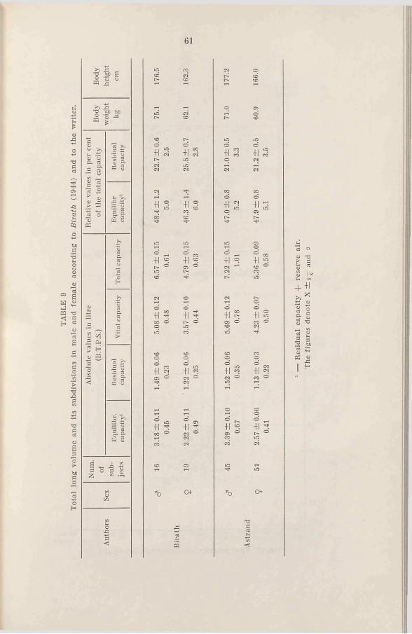
TAB
LE 9
Tota
l lu
ng v
olum
e an
d it
s su
bdiv
isio
ns i
n m
ale
and
fem
ale
acco
rdin
g to
Bir
ath
(194
4) a
nd t
o th
e w
rite
r.
61
■b ‘ 6E ££ 1 s
S .S> BP,3 Cj —•22 k
W >> u .ti cj cjC , C3a g-3 IS « 2 > O 0) J*
oc/3
— cj•3 ng. a
— CJ C3£ ab >.a £ s *33 K
-3 o£ .«
3«5
O+1
+1
+1
+1
+ 1
+1
+ 1
+1
+ 1 | 00 ° T—<CO
n ^31 ° OJoi
O f
£
+i
+i
o+1
+1
*b
©+1
+1
+1
+1 «to O
o+1
Of
'dfla
+ +i+ * cj « 1 & !
CO3 «3 SCO £ CJ
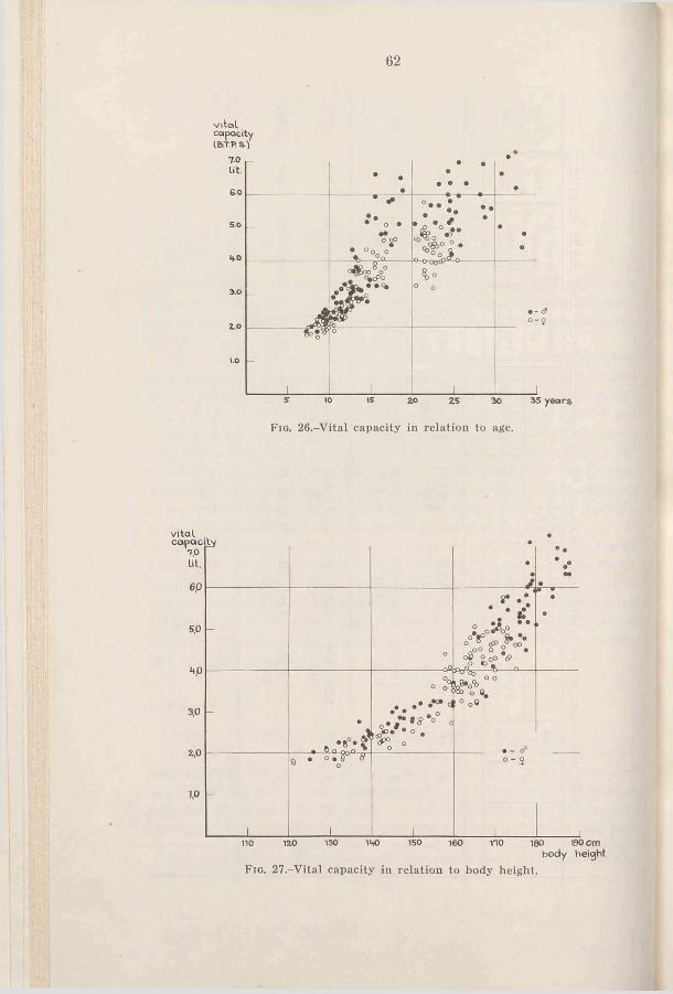
62
v ita lcapacity(B T .P s .}
F ig. 26.-Vital capacity in relation to age.
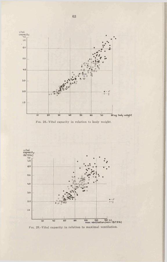
63
vital capacity
7.0 lit
6 0 _________________ _______
•
©e •• • .
•8
cP o 0O O
v - vT* «°«§ £
o « ° , ° . o \
. AA * *
J t * . .
°o / « « H, o # O o
v * « 1,9 * , s °°
CD̂ o
l
o -
-
1
o - J
________ 1________10 2 0 30 UO 50 60 7 0 8<
F ig . 28.-Vital capacity in relation to body weight.
vitalcapacity(fcT.P.S.)
TO lit.
6.0
4-.0
2 .0
••
• • • • •*
•7
e
<b«• °cP ___£
‘ ? " V 0 - V '
-is : °*
•
•* •
•
-
°o a
* - tD
1
• tf>
1_________ i
• - c fo - î
IOO 120max. ventilation/mm (BTPS.)
F ig . 29.-Vital capacity in relation to maximal ventilation.
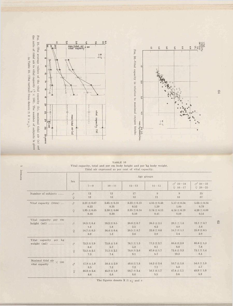
ÄS
TR
AN
D
T A B L E 10cji Vital capacity, total and per cm body height and per "kg body weight.
Tidal air expressed as per cent of vital capacity.
SexAge groups
7— 9 10-11 12— 13 14—15 <f 16—189 16— 17
<f 2 0 -3 3 9 20—25
Number of subjects ....... & 12 13 17 9 9 359 13 13 13 11 10 37
Vital capacity (litre) ... & 2.21 ±0 .07 2.65 ± 0 .1 0 3.22 ±0 .13 4.55 ±0 .4 6 5.17 ± 0.34 5.68 ± 0 .1 40.23 0.35 0.52 1.39 1.00 0.79
9 1.95 ±0 .0 5 2.30 ± 0 .0 6 3.25 ± 0 .1 4 3.74 ±0 .1 3 4.14 ±0 .1 9 4.28 ± 0.090.16 0.20 0.50 0.41 0.59 0.54
Vital capacity per cmheight (m l) ..................... a' 16.3 ± 0 .4 18.2 ± 0 .5 20.8 ± 0 .7 26.3 ± 2 .1 29.1 ± 1.6 32.1 ± 0.7
1.2 1.8 2.5 6.2 4.9 3.89 14.7 ± 0 .3 16.4 ± 0 .4 20.5 ± 0 .7 22.6 ± 0 .6 24.7 ± 1 .1 25.9 ± 0.5
0.9 1.3 2.6 2.0 3.4 2.9
Vital capacity per kgweight (m l) ................... & 72.3 ± 1 .9 72.8 ± 1 .6 76.1 ± 1 .3 77.3 ± 2 .7 80.4 ± 2 .9 80.8 ± 1.3
6.4 5.7 5.2 8.1 8.8 7.89 72.3 ± 2 .1 71.1 ± 2 .1 70.0 ± 2 .6 67.0 ± 1 .7 72.4 ± 3 .3 72.1 ± 1.4
7.3 7.4 9.1 5.7 10.3 8.4
Maximal tidal air s ,-------------------------- X 100vital capacity d" 47.9 ± 1.9 50.4 ± 2.0 49.4 ± 1 .8 54.2 ± 2 .4 53.7 ± 1 .8 54.3 ± 1 .0
6.5 7.2 7.2 7.1 5.3 5.7
9 46.8 ± 2 .4 45.9 ± 1 .9 50.7 ± 2.4 50.1 ± 1.7 47.4 ± 1 .1 48.9 ± 1 .08.6 6.6 8.6 5.5 3.6 5.8
The figures denote X ± g— and a
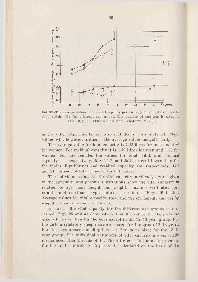
66
F ig . 32,-The average values of the vital capacity per cm body height (A) and per kg body weight (B) for different age groups. The number o f subjects is given in
Table 10, p. 65. (The vertical lines denote ± 2 X £—).
in the other experiments, are also included in this material. These values will, however, influence the average values insignificantly.
The average value for total capacity is 7.22 litres for men and 5.36 for women. For residual capacity it is 1.52 litres for men and 1.13 for women. For the females the values for total, vital, and residual capacity are, respectively, 25.8, 25.7, and 25.7 per cent lower than for the males. Equilibrium and residual capacity are, respectively, 47.5 and 21 per cent of total capacity for both sexes.
The individual values for the vital capacity in all subjects are given in the appendix, and graphic illustrations show the vital capacity in relation to age, body height and weight, maximal ventilation per minute, and maximal oxygen intake per minute (Figs. 26 to 30). Average values for vital capacity, total and per cm height, and per kg weight are summarized in Table 10.
As far as the vital capacity for the different age groups is concerned, Figs. 26 and 31 demonstrate that the values for the girls are generally lower than for the boys except in the 12-13 year group. For the girls a relatively steep increase is seen for the group 12-13 years. For the hoys a corresponding increase first takes place for the 13-14 year group. The individual variations of vital capacity are especially pronounced after the age of 14. The difference in the average values for the adult subjects is 25 per cent (calculated on the basis of the
67
male values). The absolute values for boys show an increase, from 2.2 litres for the age group 7-9 years to 5.7 litres for adult men. For the females the corresponding average values are 2.0 and 4.3 litres.
The vital capacity is often related to body size, and even with this material such a comparison seems justified, especially as the individual variations are so large. It is also of great interest to compare vital capacity with factors responsible for working capacity, i.e. lung ventilation and oxygen intake.
In relation to body height (Fig. 27) a relatively greater vital capacity is seen for both boys and girls above 155 cm i height (boys about 13 years, and girls about 12 years), and the values for both the sexes lie, broadly speaking, between the same limits. Calculated per cm body height (Fig. 32) the girls’ average values are lower than those for the boys, although the difference is very small for the 12-13 year group.
In the youngest group an average value of 16.3 ml per cm height for boys, and 14.7 ml for girls is found, and a steep increase takes place between 12 .and 15 years (appearing for the girls first). The average value for the adult men is 32.1 and for the women 25.9 ml per cm, i.e. 19 per cent lower for the women. The 7-9 year old subjects have only 51 and 57 per cent of the vital capacity of adults if the comparison is made with males and females, respectively.
Fig. 28 shows vital capacity in relation to body weight. The vital capacity increases with body weight, and the females have in most cases a somewhat lower capacity than the males.
Fig. 32 shows how the average values for the vital capacity per kg body weight for boys increase with age from 72 ml for the 7-9 age group to 81 ml for adults. The values for the females are more constant, and are similar to the values for boys in the 7-11 age group. The values for older females are 8-13 per cent lower than for males of the same ages.
If vital capacity is related to the maximal values for ventilation and oxygen intake the results are as Figs. 29 and 30 show. It should be emphasized that the values for the adults were obtained during running. In some cases higher values were recorded during cycling (comp, the average values in Table 13), but is was thought advisable to compare only the values obtained under identical working conditions.
The correlation is highest when the vital capacity is correlated with the maximal oxygen intake.
5*
68
3. Discussion.The absolute values for residual capacity and for the relative equili
brium capacity agree well with the values given by Birath, but the vital capacity is 11 per cent higher for the men and 16 per cent higher for the women in the writer’s experiments (the differences are highly significant). Consequently the total capacity is larger. Residual capacity in per cent of total capacity is lower, especially for the women (the difference between the women is highly significant and between the men almost significant).
In this material there is no difference in relative residual capacity between men and women (21.0 per cent and 21.2 per cent respectively) and this disagrees with earlier results. The explanation can be that the writer’s material includes men and women in very much the same physical condition. Real sex differences should consequently not exist as far as the relative residual capacity is concerned.
The higher value for vital and total capacity in the writer’s material as compared with the value given by Birath can probably be explained by a difference in physical development and training. That “ systematic physical exercise will increase the vital capacity of children and young people approaching maturity” is shown by the review given by Schneider and Karpovich (1948).
The difference between the vital capacity found in Birath’s and the writer’s experiments is perhaps due to the different physical development of the subjects, but the absolute values for residual capacity and the ratio of equilibrium capacity to total capacity are, in spite of this, similar in both studies. The increase in vital capacity would consequently be due to an increase in complemental air. This does not agree with the assumption of Lindhard (1926) who suggested that increased training also entailed a lower equilibrium capacity, and that the relatively lower residual capacity in males as compared with females was due to greater muscular strength, resulting in a more forced expiration.
Calculating with a similar classification in age groups as Robinson used, the writer’s average values for vital capacity are 8 to 12 per cent higher per cm body height, and 5 to 12 per cent higher per kg body weight compared with Robinson’s values. The differences are too large to be insignificant. The explanation may be that Robinson made his determinations in recumbent position in which the vital capacity is lower than in standing position (Bohr, 1907; Hasselbalch, 1908; A.s- mussen, Christensen, and Sjöstrand, 1939).
It is of great importance to make clear the extent to which differences in vital capacity between males and females (Fig. 32) are dependent on variations in body size.
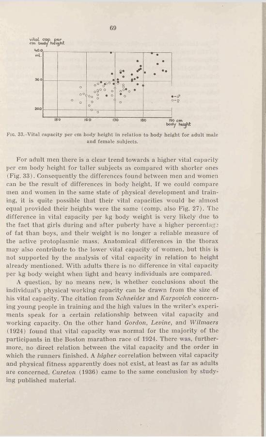
69
vital cap. per cm body height
ml.
3 0 0
------------------•—
•• * 4 » *
— n ----------••
• . ••
o'0
o o
o
o ° V > ° *o 9 ° ° o‘
>Oo ° . oo
o
0 8 * o°*o • • oo
•o
o170 l9o cm
body height
Fig. 33.-Vital capacity per cm body height in relation to body height for adult maleand female subjects.
For adult men there is a clear trend towards a higher vital capacity per cm body height for taller subjects as compared with shorter ones (Fig. 33). Consequently the differences found between men and women can be the result of differences in body height. If we could compare men and women in the same state of physical development and training, it is quite possible that their vital capacities would be almost equal provided their heights were the same (comp, also Fig. 27). The difference in vital capacity per kg body weight is very likely due to the fact that girls during and after puberty have a higher percentage of fat than boys, and their weight is no longer a reliable measure of the active protoplasmic mass. Anatomical differences in the thorax may also contribute to the lower vital capacity of women, but this is not supported by the analysis of vital capacity in relation to height already mentioned. With adults there is no difference in vital capacity per kg body weight when light and heavy individuals are compared.
A question, by no means new, is whether conclusions about the individual’s physical working capacity can be drawn from the size of his vital capacity. The citation from Schneider and Karpovich concerning young people in training and the high values in the writer’s experiments speak for a certain relationship between vital capacity and working capacity. On the other hand Gordon, Levine, and Wilmaers (1924) found that vital capacity was normal for the majority of the participants in the Boston marathon race of 1924. There was, furthermore, no direct relation between the vital capacity and the order in which the runners finished. A higher correlation between vital capacity and physical fitness apparently does not exist, at least as far as adults are concerned. Cureton (1936) came to the same conclusion by studying published material.
70
In this material important components of the working capacity, i.e. the maximal oxygen intake and ventilation were determined; these values will be analysed in relation to the vital capacity. Figs. 29 and 30 show that the correlation is highest between vital capacity and oxygen intake. Surely the explanation is that the “ maximal” ventilation is far more variable than the oxygen intake. Also of importance is the valve which may influence respiration so that hypo- or hyperventilation ensues. The magnitude of ventilation is influenced by psychological factors during work, especially when it is straining, where-as the oxygen intake is more constant.
From Fig. 30 it is evident that there is a rather constant increase of the oxygen intake capacity with increased vital capacity. However, a closer analysis is not possible. If we take, for instance, the girls with a vital capacity of about 4 litres, the maximal oxygen intake in this material varies between 2 and 3 litres per minute. For males the oxygen intake varies from 3.5 to 4.5 litres when vital capacity is about 6 litres. In spite of this it is interesting to see how the vital capacity, for the different age groups, increases linearly with an increase of the oxygen intake capacity. It is unfunctional to assume that vital capacity can be expressed in more or less complicated formulae, where body size in centimetres and kilograms is represented. The biological basis no doubt is more “ active” . A certain connection with the maximal oxygen intake is justifiable, as there is a great dependence between vital capacity and the maximal tidal air during exercise, and a great tidal air means an economic respiration. The vital capacity must be regarded as one of the many factors determining the working capacity, but the dependence on its magnitude is certainly' limited to “ at least normal value” . This applies also to élite athletes who are known for their endurance. The vital capacity is of small value, no doubt, as a test of the oxygen intake capacity and the capacity of the respiratory organs, in so far as no pathological conditions are prevailing.
4. Summary.
Determination of the total, vital, and residual capacity of 45 males (20-33 years of age) and 51 females (20-25 years of age) gave average values which for these volumes were 26 per cent lower for the females than for the males. Average values were 7.22, 5.69, and 1.52 litres, respectively, for males and 5.36, 4.23, and 1.13 litres, respectively, for females. Equilibrium and residual capacity was relatively the same for the two sexes, 47.5 and 21 per cent, respectively.
71
The vital capacity was determined for 95 male (7-33) and 97 female subjects (7-25). At different ages, the boys had greater vital capacities than the girls except for the group 12-13, where the average value was the same. Average values for subjects of 7-9 years were 2.21 litres for boys, and 1.95 litres for girls; for subjects of 12-13 it was 3.22 and 3.25 litres, respectively.
In relation to body weight, boys and girls had nearly the same vital capacity before puberty. For the male subjects it increased successively from 72 ml per kg for the subjects of 7-9 to values (81 ml) that were about 10 per cent above those of the females, whose average values were mainly constant at different ages.
In relation to body height the youngest groups (7-9) had an average vital capacity of 16.3 ml (boys) and 14.7 ml per cm (girls). These means were 51 and 57 per cent, respectively, of the adults’ ones.
The vital capacity in relation to maximal oxygen intake and maximal ventilation was discussed.
Ch a pt e r V II
LUNG VENTILATION1. Introduction.
As was shown by Christensen (1932) and others, the ventilation at submaximal working intensities increases almost linearly with oxygen intake. During maximal work it shows a relatively larger increase. The tidal air during work is within certain limits quite independent of the size of ventilation, and reaches 40-60 per cent of the vital capacity (the “ optimal” tidal air).
Robinson (1938) found average values for maximal ventilations of 33.3, 53.4, 92.2, 121.0, and 118.2 litres per minute for the following age groups: 6.1, 10.4, 14.1, 18.0, and 25.3. In relation to body weight, the ventilation was relatively constant for these groups (1.6-1.8 litres per minute and per kg). The maximal respiratory rate gradually declined from the average 62 for the youngest to 43 for the 25 year old subjects. The maximal tidal air was 44-56 per cent of vital capacity, whereof the lowest percentages refer to the younger subjects.
In their study of 110 boys, aged 10-17 years, Morse, Schlutz, and Cassels (1949) found that the maximal lung ventilation per minute was constant for the different age groups (1.6 litres per minute and per kg), but in the older boys it was attained with fewer respirations by increasing the ratio of tidal air to vital capacity. The respiratory rate for the youngest group was 60 per minute. Tidal air in relation to vital capacity was 47.5 per cent. Corresponding values for the oldest subjects were 53 per minute and 55 per cent, respectively. Broadly speaking, the results agree with those of Robinson.
2. Methods.
The experiments include determinations of lung ventilation, respiratory rate, and tidal air during submaximal and maximal work (running and for the older subjects also cycling). Chapter II and III give the methods used to determine these values but a further discussion is desireable.
Christensen and Hansen (1939 a) state in a work concerning the
73
determination of the respiratory quotient the necessity for having short and wide connections between the mouth of the subject and the Douglas bag at high working intensities. With narrow connections hypoventilation ensued. An initial period of 2 minutes with mouthpiece breathing is sufficient to give constant values provided the ventilation has reached a steady state. Especially at high intensities, when careful determinations are wanted, 10-15 minutes of work is often necessary in order to reach the steady state.
Robinson (1938) found in his experiments that during the second minute of work 90-98 per cent of the maximal value of the oxygen intake was attained, but that the ventilation varied during this period betweeen 75 and 90 per cent of the maximal value.
As earlier mentioned, the ventilation increases in relation to the oxygen intake more during exhausting work than during light work. An important cause is that the blood during heavy work contains specific substances which are produced under anaerobic conditions in the working muscles, and which have a pronounced stimulative effect on the ventilation, (Asmussen and Nielsen, 1950). It is also probable that the feeling of stress, while doing maximal work, gives an increase of ventilation.
It is fairly clear that a determination of ventilation after about 5 minutes, as in the writer’s experiments, during maximal work, where anaerobic processes go on all the time, and the stress becomes more and more perceptible, gives values difficult to reproduce.
The Douglas bag method has been used and the valve used has been a modified Enghoff (1930) valve, especially suited for large ventilations. Short and wide tubes and taps have likewise been constructed. According to determinations done in this laboratory the air resistence of this apparatus is 13 mm H,0 during expiration and 4 mm H.O during inspiration during a ventilation of about 60 litres per minute. During ventilation of 30 litres per minute the corresponding figures are 7 and 3 mm H20, respectively.
Changes in ventilation could be proved by measuring the expired air volume in two bags, collected one after the other. In most cases the ventilation was similar for both determinations, but in spite of a constant oxygen intake an increase or decrease was to be seen quite often during the collecting time.
In determining the ventilation per litre of oxygen intake the value of the second bag was used. Before taking the second determination, during a submaximal work (given below as “ 60-70 per cent” of the maximal aerobic capacity), the valve respiration was continued for 1%—2 minutes, which can be regarded as sufficient for adaptation. In
74
connection with the calculation of the ventilation per litre oxygen intake the second determination usually fits best into the individual diagrams which have been drawn up. Through preceding experiments the subjects have become well accustomed to the apparatus.
Variations in tidal air and respiratory rate have not as a rule been large, and so the usage of the mean of the two determinations has been well justified. The valve respiration can influence rate and depth, but as the respiratory resistance of the apparatus was made small, and the subjects were accustomed to it, possible errors are believed to be of little importance.
It was noticed, especially among the younger subjects, that the values of the ventilation, which were obtained during the first experiments, often fell outside the curves of the experimental series, and the values consequently were disregarded.
As for the adults, the work was done partly by cycling, partly by running. For the comparison between adults and younger subjects the running experiments were used. Different types of work do not alwmys give the same ventilation at a certain oxygen consumption (Collet and Liljeslrand, 1924), and the fixed rate of cycling can influence the respiratory rate, especially during lower working intensities. During maximal work the arms will influence the movements of the chest in a way quite different from that which is done by running. The “ optimal” tidal air may be different. In this study no significant difference ■was found between the means of maximal ventilation during running and cycling, but the individual variations from the means were least in the running experiments (comp. Table 13, p. 88).
For practical reasons the adults could he investigated in a series of submaximal intensities only while cycling, and so the calculation of the ventilation per litre oxygen intake concerns this type of work. For all subjects these estimations are made on values selected from experiments where the oxygen intake is 60-70 per cent (if possible) of the maximal individual values (extremes in the individual values are 56 to 87 per cent, 71 per cent of the values fall in the said region of 60-70 per cent). The reason for choosing these intensities is that reliable values were found within this region, and that the ventilation per litre oxygen intake is here equal to or very near the minimal values obtained (comp. p. 83). (Lowest working intensity usually corresponded to an energy output of 50-60 per cent of the maximal oxygen intake). The “ 60-70 per cent work” can with regard to its duration be judged as relatively easy. Moreover, for the analyses only those individual series of experiments were used that were complete as to different working intensities, and that within the analysed region
the
subm
axim
al w
ork.
(V
enti
lati
on v
olum
es a
t B
.T.P
.S.).
75
OA
Ö u <u a to a ^ a X o o „<y
h a
ö a O ¿5
CJJ M-S o« * 0) _ P< 03
•Ö se 'S
.3 æ O 3
C “.2 503 03a I > *« 2 C fcc•5 c « T « a
O
I ICO CO
"b o
ho<
+| CO +1 c
+1 « +1
+ 1 05 +1
+1 M +1 n
+1 2 +l
C'l r-<Il ^ +1
+1 S +1
•b c* *b O f
3
£
z
+1 o +1
o O o ^+1 d +1 d
+1 o +1
CO COo oO _* ® +1 2 +1
00 oc o o O cr. O+i 3 +'CO 00 C5 GO
+1 5 +1
G iß o+13+12
iß
*b CH-
flO >4 SS> 'S £o .5£ b£ \*-' *> o* . £3 £ ~
-b
+i 3 +lGO
+1 2 +1
+1 2 +1
+1 3 +1
+i 2 +1
+1 ° +11 •* « e m
+1 3 +i
O f
"u .§^ o _ _<
+i r? +i
+i 2 +i
+i
+i s +i
+i ^ +i
+i « +i
+1 5 +1
CH-
O o 22 G > a w v:
co io r— Oi
■*t ioGO I C O J .co coiß iß
cq t-*co t>. o r-co I Liß ’’"t* ÇC ^œ co co
—. CO . I> ® I CO I i o co r̂
*b
G■>- oJ, £ obl 'H Oc o tzi _ v ̂ t- 03G cC G -HS y a ^« ̂ H «o o .S o~ a x 5£ a aÜ .5 £ ?
The
four
upp
er c
olum
ns:
The
figur
es d
enot
e X
±
e— a
nd
The
low
est
colu
mn:
Th
e fig
ures
den
ote
mea
n an
d ra
n;
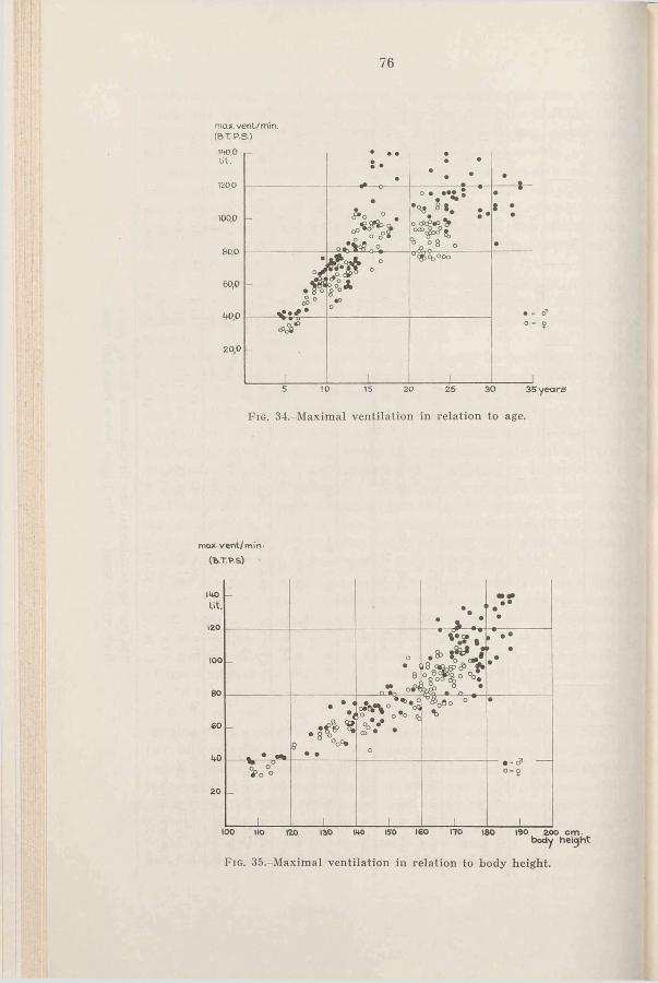
76
max. veni/min. (B .T P .S .)
140.0l i t .
100,0
80,0
60,0
1+0,0
20,0
• • • •• * * •
-•
° .- O ° ° *
° *
r P l °
•t • •••
o A
• 8 ° C P °o °
f o j r
8°°*•DO
^ c ÿ ^ O O O
• - cT0 - Î
__________________ 1__________________ 1___________________ 15 10 15 20 25 30 35 years
Fig. 34.-Maximal ventilation in relation to age.
max vent/min- (B .T . P .S )
-*•
• >
i• •»• ••
•
- o•
0n t .
.8 »•
••
V * 38 ° ° ° *
• •
° . ° * ° %
o
% ‘ c n° • %
•
o o *>o o• - d1o - ?
______ 1______ ______ i______ 1 i ______ i______100 1IO 120 130 »40 150 160 170 »80 190 200 c m .
b o d y h e ic jh t
F ig . 35.-Maxiraal ventilation in relation to body height.
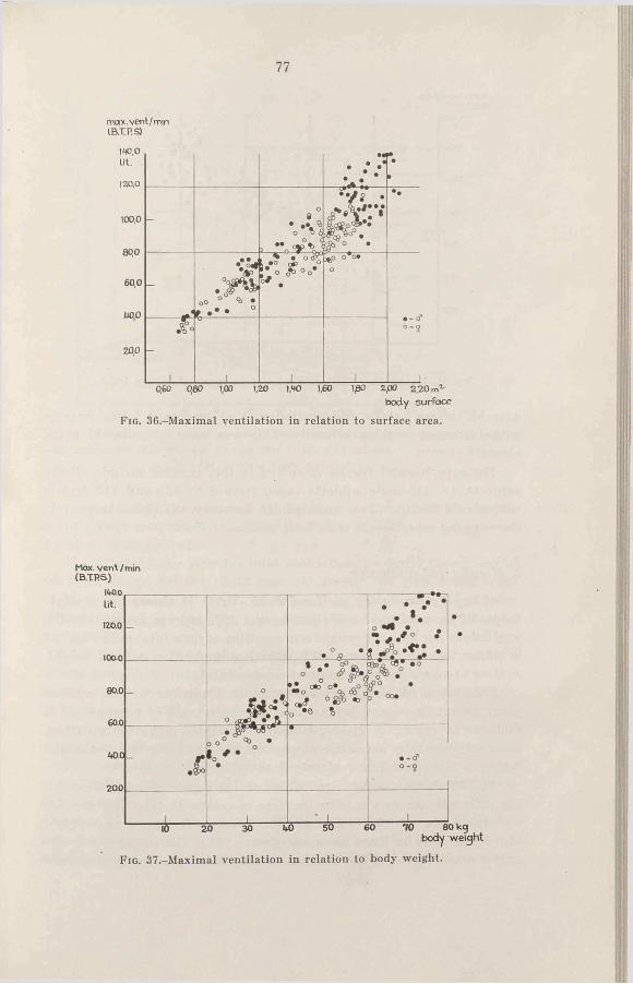
77
max.vent/mmIB.TP.S)140.0 Lit.
120.0
100,0
80,0
60,0
000
•« JÜ.•
•
-o
0• •% ^o T) i
L
r , / . ° •
S F^ ° . • .
••
OO ° • » °
, ^ • o & s »° c*> _° *• o ¿b°o*
0 / .
o%>° ° °* o
A ° 0 - 9
20,0
_____i_____ ______ 1______ ______ 1______0,60 080 1,00 1,20 1,MO 1,60 1,80 2,00 2,20 m2'
body surface
Fig. 36.-Maximal ventilation in relation to surface area.
Max. vent / min (B .TR S )
lit.
120.0
1000
-
o ©• -6 -
• " Z * *^ t "
a • * . * .«
80.0
60.0
- •8
fO
' •*. o a 9• * C*D O* ~ O G
0 J k & O
o •» %
fp° •f » 00.
•
o o •40.0 #• | * •
• V °• o - î
20.0
________ i_________ _ _ j ________ ______!__L _______ 120 30 60
body weight
Fig. 37.-Maximal ventilation in relation to body weight.
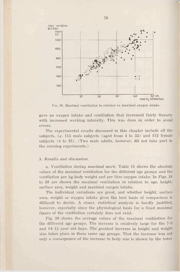
78
max. vent/min
F ig . 38.-Maximal ventilation in relation to maximal oxygen intake.
gave an oxygen intake and ventilation that increased fairly linearly with increased working intensity. This was done in order to avoid errors.
The experimental results discussed in this chapter include all the subjects, i.e. 115 male subjects (aged from 4 to 33) and 112 female subjects (4 to 25). (Two male adults, however, dit not take part in the running experiments.)
3. Results and discussion.
a. Ventilation during maximal worft.-Table 11 shows the absolute values of the maximal ventilation for the different age groups and the ventilation per kg body weight and per litre oxygen intake. In Figs. 34 to 38 are shown the maximal ventilation in relation to age, height, surface area, weight and maximal oxygen intake.
The individual variations are great, and whether height, surface area, weight or oxygen intake gives the best basis of comparison is difficult to decide. A closer, statistical analysis is hardly justified, however, especially since the physiological basis for a fixed maximal figure of the ventilation certainly does not exist.
Fig. 39 shows the average values of the maximal ventilation for the different age groups. The increase is relatively large for the 7-9 and 14-15 year old boys. The greatest increase in height and weight also takes place in these same age groups. That the increase was not only a consequence of the increase in body size is shown by the lower

F ig . 39.-The average values of maximal ventilation and maximal ventilation per kg body weight for different age groups. The number of subjects is given in Table 11,
p. 75. (The vertical lines denote± 2 X e — ) .
curves for maximal ventilation per kg body weight, where the means of the groups mentioned, especially the subjects 14-15, are higher than the surrounding groups.
The values of the girls for total ventilation are lower than those of the boys, except for the 12-13 year old group. The girls 4-6 average 34 litres per minute, the boys 40 litres. Boys and girls 12-13 average 75 and 80 litres, respectively, and adults 111 and 90 litres per minute, respectively.
The difference between male and female adults is 19 per cent. Of special interest is the fact that both boys and girls attain the same average value as the adults already at the age of 14-15 (girls as early as 13 as shown in Fig. 34).
Taking body weight in consideration, the difference in maximal ventilation between boys and girls under 14 years will he reduced so that differences in their average values is not significant. For the adults it is 6 per cent, just on the limit of being almost significant (P = 0.05).
From the age group 7-9 the ventilations per kg body weight decline with increasing age, in the case of males from 2.0 to 1.6 litres per minute and per kg, in females from 2.1 to 1.5 litres. This is in disagree-
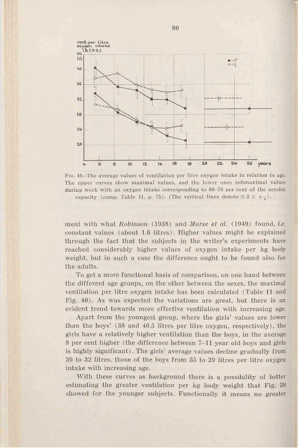
80
F ig . 40.-The average values of ventilation per litre oxygen intake in relation to age. The upper curves show maximal values, and the lower ones submaximal values during work with an oxygen intake corresponding to 60-70 per cent of the aerobic
capacity (comp. Table 11, p. 75). (The vertical lines denote ± 2 X e —).
ment with what Robinson (1938) and Morse et al. (1949) found, i.e. constant values (about 1.6 litres). Higher values might be explained through the fact that the subjects in the writer’s experiments have reached considerably higher values of oxygen intake per kg body weight, but in such a case the difference ought to be found also for the adults.
To get a more functional basis of comparison, on one hand between the different age groups, on the other between the sexes, the maximal ventilation per litre oxygen intake has been calculated (Table 11 and Fig. 40). As was expected the variations are great, but there is an evident trend towards more effective ventilation with increasing age.
Apart from the youngest group, where the girls’ values are lower than the boys’ (38 and 40.5 litres per litre oxygen, respectively), the girls have a relatively higher ventilation than the boys, in the average 8 per cent higher (the difference between 7-11 year old boys and girls is highly significant). The girls’ average values decline gradually from 39 to 32 litres, those of the boys from 35 to 29 litres per litre oxygen intake with increasing age.
With these curves as background there is a possibility of better estimating the greater ventilation per kg body weight that Fig. 39 showed for the younger subjects. Functionally it means no greater
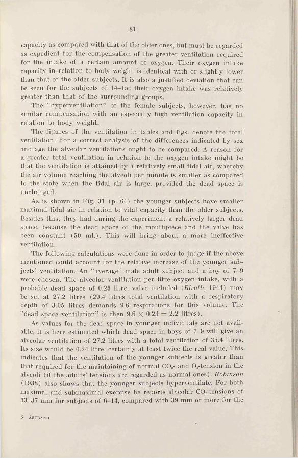
81
capacity as compared with that of the older ones, but must be regarded as expedient for the compensation of the greater ventilation required for the intake of a certain amount of oxygen. Their oxygen intake capacity in relation to body weight is identical with or slightly lower than that of the older subjects. It is also a justified deviation that can be seen for the subjects of 14-15; their oxygen intake was relatively greater than that of the surrounding groups.
The “ hyperventilation” of the female subjects, however, has no similar compensation with an especially high ventilation capacity in relation to body weight.
The figures of the ventilation in tables and figs, denote the total ventilation. For a correct analysis of the differences indicated by sex and age the alveolar ventilations ought to be compared. A reason for a greater total ventilation in relation to the oxygen intake might be that the ventilation is attained by a relatively small tidal air, whereby the air volume reaching the alveoli per minute is smaller as compared to the state when the tidal air is large, provided the dead space is unchanged.
As is shown in Fig. 31 (p. 64) the younger subjects have smaller maximal tidal air in relation to vital capacity than the older subjects. Besides this, they had during the experiment a relatively larger dead space, because the dead space of the mouthpiece and the valve has been constant (50 ml.). This will bring about a more ineffective ventilation.
The following calculations were done in order to judge if the above mentioned could account for the relative increase of the younger subjects’ ventilation. An “ average” male adult subject and a boy of 7-9 were chosen. The alveolar ventilation per litre oxygen intake, with a probable dead space of 0.23 litre, valve included (Birath, 1944) may be set at 27.2 litres (29.4 litres total ventilation with a respiratory depth of 3.05 litres demands 9.6 respirations for this volume. The “dead space ventilation” is then 9.6 X 0.23 = 2.2 litres).
As values for the dead space in younger individuals are not available, it is here estimated which dead space in boys of 7-9 will give an alveolar ventilation of 27.2 litres with a total ventilation of 35.4 litres. Its size would be 0.24 litre, certainly at least twice the real value. This indicates that the ventilation of the younger subjects is greater than that required for the maintaining of normal CO-- and 0,-tension in the alveoli (if the adults’ tensions are regarded as normal ones). Robinson (1938) also shows that the younger subjects hyperventilate. For both maximal and submaximal exercise he reports alveolar CCVtensions of 33—37 mm for subjects of 6—14, compared with 39 mm or more for the
6 ASTRAND

82
adults. The alveolar O.-tension was consequently highest in the younger subjects.
Using, in the same manner, the average value for a female adult who has an assumed dead space of 0.16 litre, the maximal alveolar ventilation can be calculated to 29.8 litres per litre oxygen intake, a value that is 10 per cent higher than that of the male. The difference in total ventilation was also 10 per cent.
The difference of ventilation in the different age groups and sexes must depend on a dissimilarity in respiratory regulation.
It can be mentioned that the younger subjects reached lower lactic acid values during maximal work when compared with the adults, and that males and females attained roughly identical values. It is found that younger individuals have a lower alkali reserve (Robinson, 1938), but it is not probable that a difference in pH or a difference in the concentration of anaerobic products can explain the difference.
b. Ventilation during submaximal w ork -(Methods see p. 74). Subjects were 88 males and 101 females.
The average values of the ventilation per litre oxygen intake are found in Table 11. Fig. 40 shows, that the ventilation per litre oxygen intake is considerably smaller during submaximal work than maximal, but the same age variations exist, i.e. the younger subjects have a higher ventilation at a certain oxygen consumption than the older, and this at a working intensity that should mean the same strain for the individuals (60-70 per cent of the aerobic capacity is utilized). The younger girls (4—6), have a smaller ventilation than the boys of the same age (31 and 34 litres per litre oxygen intake respectively). Such was also the case during maximal work. Then the boys and girls have nearly the same ventilation until they reach 14 (25 litres for both groups 12-13). The older females average 24-25 litres, but the males decline to 21 litres ventilation per litre oxygen intake. The alveolar ventilation per litre oxygen intake for the male and female students is calculated to be 19 and 22-23 litres respectively. The difference between the male and female adults (highly significant) is in litres about the same as during maximal work; for the youngest the material is too small for a reliable conclusion. That there should be a true difference between boys and girls in this respect is hardly probable.
Thus it is interesting to note, that the girls from 4 to 13 years have a ventilation as effective as that of the boys during a submaximal work, but less effective during maximal work. This difference might depend upon the fact that the girls are less accustomed to “ strain”
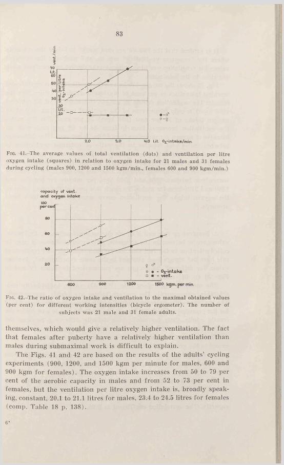
2.0 3 .0 4 .0
0-2
lit. 0L-intc*ke/min.
Fig. 41. The average values of total ventilation (dots) and ventilation per litre oxygen intake (squares) in relation to oxygen intake for 21 males and 31 females during cycling (males 900, 1200 and 1500 kgm/min., females 600 and 900 kgm/min.)
capacity of vent- and oxygen intake
F ig . 42.-The ratio o f oxygen intake and ventilation to the maximal obtained values (per cent) for different working intensities (bicycle ergometer). The number of
subjects was 21 male and 31 female adults.
themselves, which wmuld give a relatively higher ventilation. The fact that females after puberty have a relatively higher ventilation than males during submaximal wrork is difficult to explain.
The Figs. 41 and 42 are based on the results of the adults’ cycling experiments (900, 1200, and 1500 kgm per minute for males, 600 and 900 kgm for females). The oxygen intake increases from 50 to 79 per cent of the aerobic capacity in males and from 52 to 73 per cent in females, but the ventilation per litre oxygen intake is, broadly speaking, constant, 20.1 to 21.1 litres for males, 23.4 to 24.5 litres for females (comp. Table 18 p. 138).
6 '
84
It is evident that the “ 60-70 per cent work” is below the intensity where the “ excessive ventilation” sets in, and probably the reported values for ventilation per litre oxygen intake during submaximal work are close to the minimum.
Fig. 42 shows the oxygen intake and the ventilation during different working intensities in per cent of maximally attained values. For the females the ventilation during an oxygen consumption of 52 per cent (600 kgm) is only 39 per cent of the maximal ventilation, and at an oxygen intake of 73 per cent (900 kgm) 56 per cent. For the males the ventilation increases linearly from 34 to 58 per cent with an increase of oxygen consumption from 50 to 79 per cent. The difference between males and females in the ratio of obtained ventilation to maximal ventilation is a tendency towards a some per cent greater utilization of the respiratory capacity for the female subjects at a certain level of the aerobic capacity. The “ 100 percent level” is reached somewdiere between 1200 and 1500 kgm per minute for females and 1800 and 2100 kgm for males. To reach this level at the same point as the oxygen intake the ventilation curve must sooner or later bend upwards.
c. Respiratory rate and tidal air during maximal inor/c.-Individual values are to be seen in the appendix. (To avoid confusion it may be called attention to the fact that the maximal tidal air and respiratory rate have not always been attained in the same experiments). Average values for different age groups are summarized in Table 12 and in Figs. 31 (p. 64) and 43.
The maximal respiratory rate (Fig. 43) varies individually considerably, consequently the average values are highly approximate. The average values decrease gradually with increasing age, from 65-70 for the youngest subjects (4-6 years) to 40 for the male and 46 for the female adults. Only for adults is a definite difference between the means for males and females found (highly significant).
The maximal tidal air (Fig. 31) is greater for boys than for girls except in the group 12-13 (comp, ventilation), and the highest average values reached by the adults are 3.05 litres for males and 2.10 litres for females. The youngest children, 4-6 years, average 0.60 and 0.52 litres, respectively (the difference is not significant).
The increase of the maximal tidal air with increasing age runs almost parallel with the increase of the vital capacity. This is also apparent in Fig. 44 where the tidal air is correlated with the vital capacity. The correlation coefficient is 0.96.
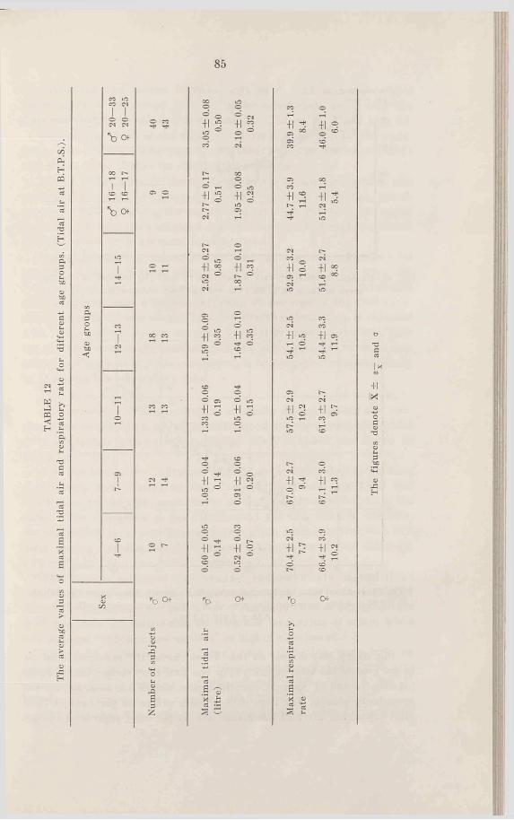
TAB
LE 1
2Th
e av
erag
e va
lues
of
max
imal
tid
al a
ir a
nd r
espi
rato
ry r
ate
for
diffe
rent
age
gro
ups.
(T
idal
air
at
B.T
.P.S
.).
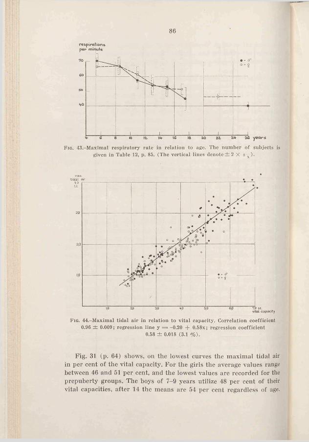
86
respirations per minute
F ig . 43.-Maximal respiratory rate in relation to age. The number of subjects is given in Table 12, p. 85. (The vertical lines denote ± 2 X s —).
max.
F i g . 44-M axim al tidal air in relation to vital capacity. Correlation coefficient 0.96 ± 0.009; regression line y = -0.20 + 0.58x; regression coefficient
0.58 ± 0.018 (3.1 % ).
Fig. 31 (p. 64) shows, on the lowest curves the maximal tidal air in per cent of the vital capacity. For the girls the average values range between 46 and 51 per cent, and the lowest values are recorded for the prepuberty groups. The hoys of 7-9 years utilize 48 per cent of their vital capacities, after 14 the means are 54 per cent regardless of age.
87
(A statistical comparison between boys aged 7-13 and males aged 14-30 gives a difference that is highly significant, as is also the difference between male and female adults.) Values, see Table 10 p. 65.
The values, absolute and relative, for the tidal air and for the respiratory rate are in good agreement with those found by Robinson (1938) and Morse et al. (1949) for different age groups.
In the writer’s study the maximal values of the respiratory rate were usually recorded during the maximal working intensities, simultaneously with obtaining the maximal ventilation. Inversely the maximal tidal air was often attained during a submaximal exercise, which entirely agrees with previous investigations.
It is striking how constant the tidal air is when related to the vital capacity of the individual when greater demands are made upon the respiratory system. This agrees with Christensen’s (1932) experiments and his opinion that a tidal air of about 50 per cent of the vital capacity is an optimal one where the most effective ventilation is obtained with the smallest energy output. The fairly great variations with age and sex found in the ventilation, and also for instance when oxygen intake and body weight are considered, is compensated almost entirely by changing the respiratory rate.
d. Comparison of the ventilation of adults during cycling and runn ing- The maximal values of ventilation, of oxygen consumption, and of blood lactate concentration for the adult subjects are shown in Table 13.
For the males the maximal ventilation is lower during running experiments than during cycling, the average values are 111.3 and116.0 litres per minute, respectively, although the oxygen intake is the same. (The difference is not significant.) For the females, however, the ventilation is largely the same, 88.0 litres during cycling and 89.8 litres during running, but the oxygen intake is somewhat different, 2.76 and 2.89 litres per minute, respectively.
The average values of the maximal ventilation are highest if all experiments are included, 122.0 litres per minute for males and 92.2 litres for females (24 per cent lower). The difference in oxygen intake is 29 per cent. The difference between the ventilation ot males while running and “ running and cycling” is highly significant.
Out of 34 males who, apparently, did exhausting work both on treadmill and bicycle ergometer, 16 attained the maximal values of ventilation during running and the other 18 during cycling. For 33 females the corresponding numbers are 18 and 15, respectively.
The fact that the highest values are attained when the subjects
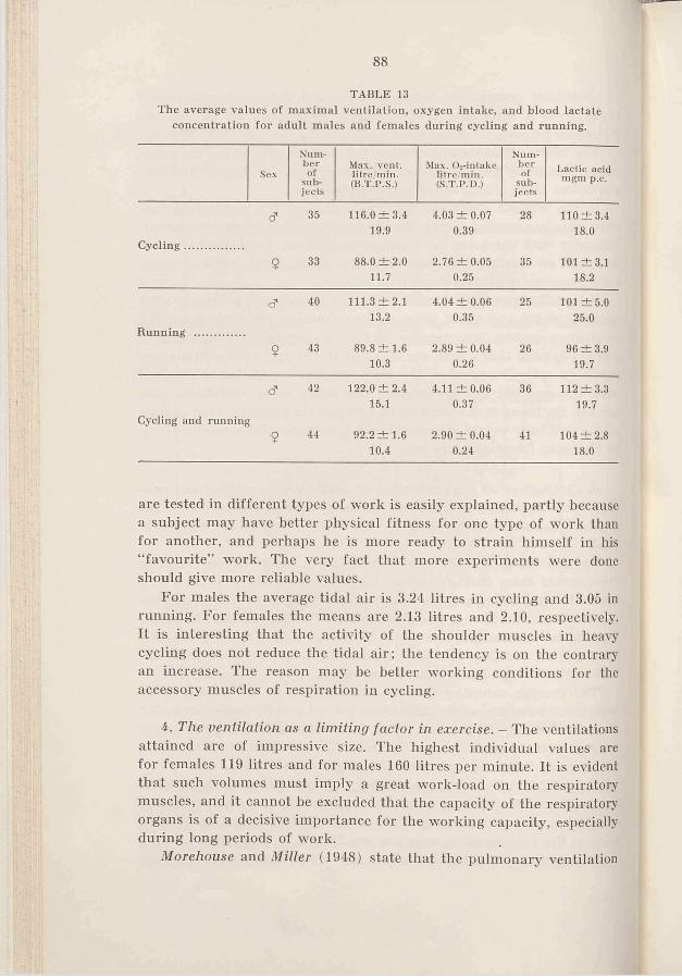
'It»»*88
TABLE 13The average values o f maximal ventilation, oxygen intake, and blood lactate
concentration for adult males and females during cycling and running.
Sex
N um b e ro f
subjects
Max. vent, litre/m in . (B .T.P.S.)
Max. ()»-intake litre m in . (S.T.P.D.)
N um b e ro f
subjects
L actic acid m gm p.c.
& 35 116.0 ± 3 .4 19.9
4.03 ±0 .0 7 0.39
28 110 ± 3.4 18.0
C ycling....................
9 33 88.0 ± 2 .0 11.7
2.76 ± 0 .0 5 0.25
35 101 ±3 .1 18.2
& 40 111.3 ± 2 .1 13.2
4.04 ± 0 .0 6 0.35
25 101 ± 5 .0 25.0
Running .................
9 43 89.8 ± 1 .6 10.3
2.89 ± 0.04 0.26
26 96 ± 3 .9 19.7
id"
Cycling and running
42 122.0 ± 2 .4 15.1
4.11 ± 0 .0 6 0.37
36 112 ± 3 .3 19.7
9 44 92.2 ± 1 .6 10.4
2.90 ± 0 .0 4 0.24
41 104 ± 2 .8 18.0
are tested in different types of work is easily explained, partly because a subject may have better physical fitness for one type of work than for another, and perhaps he is more ready to strain himself in his “ favourite” work. The very fact that more experiments wrere done should give more reliable values.
For males the average tidal air is 3.24 litres in cycling and 3.05 in running. For females the means are 2.13 litres and 2.10, respectively. It is interesting that the activity of the shoulder muscles in heavy cycling does not reduce the tidal air; the tendency is on the contrary an increase. The reason may be better working conditions for the accessory muscles of respiration in cycling.
4. The ventilation as a limiting factor in exercise. — The ventilations attained are of impressive size. The highest individual values are for females 119 litres and for males 160 litres per minute. It is evident that such volumes must imply a great work-load on the respiratory muscles, and it cannot be excluded that the capacity of the respiratory organs is of a decisive importance for the working capacity, especially during long periods of work.
Morehouse and Miller (1948) state that the pulmonary ventilation
89
at great working intensities “ becomes excessive and no longer bears a constant relation to oxygen consumption. This excessive ventilation serves no useful purpose, since the delivery of oxygen to the tissues is now limited by the maximal output of blood by the heart. It does indicate, however, that pulmonary ventilation is capable of further increase at a time when the limit of circulatory adjustment has been reached, emphasizing the fact that the minute volume of breathing is unlikely to he a limiting factor in exercise” .
It has been clearly stated, among others by Robinson (1938), that the alveolar 0 2-tension during maximal work is even higher than in the resting state, also that the CO:-tension is lower. Evidently, from the point of view of gas exchange the ventilation is sufficient, provided no pulmonary diseases are prevailing or that the 0 2-tension of the inspired air is not too low.
Christensen and Forbes (1937) have given strong arguments for the view that the ventilation and not the circulation during work at high altitudes can be the limiting factor for the maximal oxygen intake (acclimatized individuals). The ventilation attained almost the same maximal value, independent of altitude, despite the fact that the alveolar 0,-tension decreased strongly with higher altitudes. This may indicate that even the values reached at normal barometric pressure are true maximal values, even if the CO:- and 0,-alveolar tensions do not indicate insufficiency. During maximal works there is no doubt often a strong and unpleasant dyspnea, w'hich gives much of the subjective feeling of strain. This may at least be a contributing reason for “ giving up” a work.
Nielsen (1936) showed, how the cost of ventilation became an increasing percentage of the total metabolism when the work increased, and he estimated that its size can reach 9 per cent of the total oxygen intake during maximal exercise.
This shows that the respiratory work places a heavy load on the respiratory muscles. It may be emphasized that this concerns respiration with mouthpiece, valve etc. The respiratory work under such circumstances is surely somewhat greater than during “ free” breathing. The working capacity can certainly be decreased by giving a subject a valve system and tubes that have great resistance to air passage.
Nielsen and Hansen (1937) found that the breathing of room air or a gas mixture of 22.8 per cent oxygen during exhausting work gave ventilations of about 130 litres per minute. By having the subject breathe an air mixture of 45-47 per cent oxygen the ventilation decreased to 110-115 litres with an estimated decrease in the oxygen consumption of the respiratory muscles of 70-80 ml. This means
• 1
an increased oxygen supply to other muscles, and a feeling of less respiratory discomfort for the subject. The improved working capacity found in the experiments with 45-47 per cent oxygen is, according to the authors, partly due to this diminished lung ventilation.
With regard to younger subjects, the writer’s and others experiments have shown that the ventilation in relation to oxygen intake, and in this study also in relation to body weight, is greater for youth than for adults. It would be interesting to determine the “ costs” of this greater ventilation. It is not impossible at all that the more elastic chest and the shorter respiratory tracts, which means a smaller air resistance in the young subjects, will compensate for the relatively greater ventilation.
This discussion can be summarized as follows: During heavy work the ventilation gives a subjective feeling of strain that probably will be of great importance as a limiting factor, at least from a psychological point of view. The definite relief felt when an oxygen-rich mixture is breathed during severe work certainly increases the performance. Under certain conditions, e.g. high altitudes, the ventilation evidently will limit the working capacity. Normally, however, the circulation will probably be most decisive for the magnitude of the aerobic capacity, at least in trained subjects, and the discussion in the other chapters will deal mainly with the minute volume of the heart as the limiting factor.
90
5. Summary.
The maximal ventilation, respiratory rate, and tidal air have been determined during running for 113 male (4-33 years) and 112 female subjects (4-25 years).
The males attained higher average values than females for the maximal ventilation per minute except in the age group 12-13 years, but in relation to the body weight the ventilation capacity was almost the same up to the age of 13. Afterwards the boys reached higher values. The maximal ventilation per kg body weight of the children was greater than that of the older subjects (The subjects of 4-6 averaged 1.9 litre, adult males 1.6, and females 1.5 litre per minute and per kg).
After the age of 14 an almost constant level was attained for the maximal ventilation which was for the males about 110 litres per minute, for the females about 90 litres. The boys and girls aged 4-6 reached 40 and 34 litres per minute, respectively.
In experiments including both running and cycling the male and
/

91
female subjects averaged 122 and 92 litres, respectively, thus the difference in ventilation capacity of the 20-30 year old subjects was about 25 per cent (calculated from the male’s value).
Excluding the youngest group, the maximal ventilation per litre oxygen intake was for the females about 8 per cent higher than for the males, whose average values decreased gradually from 35 litres for the 7-9 year old hoys to 29 litres per litre oxygen intake for the men.
During submaximal work, where the individual oxygen intake was about 65 per cent of the aerobic capacity, the ventilation per litre oxygen consumption was almost the same for both sexes up to the age of 14. After that age a level of 23-24 litres was obtained for the females. The values of the males declined gradually from 28.5 litres for boys of 7-9 to about 21 litres for adults.
The maximal respiratory rates were similar for the two sexes up to adult age, when the average value of the males was 40 per minute, that of the females 46; the youngest subjects reached 65-70 per minute.
The maximal tidal air was about 50 per cent of the vital capacity. Before puberty the boys and girls were fairly equal, and had between 45 and 50 per cent. After the age of 14 the average values were for the males 4 to 5 per cent above 50 per cent, and for the females 1 or 2 per cent below.
The ventilation as a limiting factor in exercise was discussed.
Ch a pt e r V III
BLOOD LACTATE CONCENTRATION
1. Introduction.
As the principal aim in this investigation is placed upon the determination of the aerobic capacity, i.e. the maximal oxygen intake, the consideration of the anaerobic capacity has had to recede somewhat. In order to determine the maximal oxygen intake, the duration of work should certainly not be less than 3 to 4 minutes, while the “ optimal” time for obtaining a high concentration of lactic acid in the blood is probably shorter.
The blood lactate concentration has been an auxiliary means of determining the degree of stress in order to guarantee that the maximal work was obtained. Apparently, the subjects exhausted themselves (the youngest possibly excepted), and thus the values of lactate concentration can certainly be regarded as maximum for the type and duration of work used here.
After Fletcher and Hopkins (1906-07) and Iiyffel (1909-10) found an increase in blood lactate concentration after muscular activity, numerous experiments on blood lactate during and after work were carried out.
It has been stated that there is an increase only when a certain working intensity is reached, individually different and depending upon physical condition and type of work done (Owles, 1930; Margaria, Edwards, and Dill, 1933, and others).
The greater the working intensity, the higher rises the blood lactate concentration, and during very severe work the maximal value is attained after the end of work (Bang, 1936).
Training brings about a decrease of lactate concentration at a certain working intensity (Bang, 1936; Edwards, Brouha, and Johnson, 1940), furthermore a well-trained person can reach higher values as compared with an untrained (Robinson and Harmon, 1941 a ); Knehr, Dill, and Neufeld, 1942). The relationship between lactic acid and training condition has caused experiments on lactic acid concentration to he used as tests of physical fitness (Johnson and Brouha, 1942,

93
among others), but the results are ambiguous (Crescitelli and Taylor, 1944; Taylor, 1944, 1945).
Robinson (1938) determined the lactic acid concentration 5 minutes after maximal work (running 2-5 minutes), and found average values of 25 to 97 mgm per cent (male subjects between 5 and 76 years of age). The youngest boys, about 6 years old, had the lowest values, and subjects about 35 years old, reached the highest. The highest individual value was 140 mgm per cent.
Morse, Schultz, and Cassels (1949) found in their study of 110 boys between the ages of 10 and 17 years, that the older boys attained the highest maximal level of blood lactate (mean 68 mgm per cent above the resting level) and the hoys 12-13 years of age the lowest (32 mgm per cent).
Johnson and Brouha (1942) examined 40 subjects, (divided into 4 groups with regard to their physical fitness), during running to a state of exhaustion with a maximal duration of 5 minutes (extremes 31/2-5 minutes). He found for the best trained subjects (oarsmen) an average value of 97 mgm per cent, for men in good condition 121 mgm per cent, for those in average condition 123 and for those in poor condition 131' mgm per cent. The highest individual value was 178 mgm per cent.
Metheny, Brouha, Johnson, and Forbes (1942) had 17 female and 30 male subjects run until they reached a state of exhaustion. The females averaged 112 mgm per cent (108 seconds’ running time), and the males 119 mgm per cent (216 seconds’ running time). The highest individual values were 144 and 178 mgm per cent, respectively.
Bang (1936) found at maximal “ standing running” (1 minute) 14 millinormal (about 125 mgm per cent).
Asmussen, v. Döbeln, and Nielsen (1948) found by experiments with different oxygen percentages of the inspiratory air values reaching 140 mgm per cent at normal oxygen pressure and 212 mgm per cent at 12 per cent oxygen (maximal work on a bicycle ergometer for 5 and 6 minutes, respectively).
Robinson, Edwards, and Dill (1937) recorded lactic acid values of 116, 134, and 150 mgm per cent with three of the world’s best middle- distance runners.
In contrast to these values the experimental results of Crescitelli and Taylor (1944) should be noted. Their average values for maximal lactic acid were 218 mgm per cent, ranging from 117 to 328. They examined 31 students, between 19 and 33 years of age, running at 1
1 If corrected by means of the individual values, 113 mgm per cent.
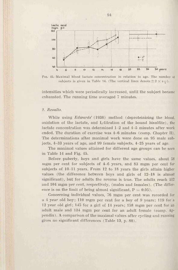
94
lactic acid
F ig . 45.-Maximal blood lactate concentration in relation to age. The number of subjects is given in Table 14. (The vertical lines denote ± 2 X s—).
intensities which were periodically increased, until the subject became exhausted. The running time averaged 7 minutes.
2. Results.
While using Edwards’ (1938) method (deproteinizing the blood, oxidation of the lactate, and I,-titration of the bound bisulfite), the lactate concentration was determined 1-2 and 4—5 minutes after work ended. The duration of exercise was 4-6 minutes (comp. Chapter II). The determinations after maximal work were done on 95 male subjects, 4-33 years of age, and 99 female subjects, 4—25 years of age.
The maximal values attained for different age groups can he seen in Table 14 and Fig. 45.
Before puberty, boys and girls have the same values, about 58 mgm per cent for subjects of 4-6 years, and 83 mgm per cent for subjects of 10-11 years. From 12 to 18 years the girls attain higher values (the difference between boys and girls of 12-18 is almost significant), but for adults the reverse is true. The adults reach 112 and 104 mgm per cent, respectively, (males and females). (The difference is on the limit of being almost significant, P = 0.05).
Concerning individual values, 76 mgm per cent was recorded for a 4 year old boy; 110 mgm per cent for a boy of 9 years; 119 for a 12 year old girl; 145 for a girl of 14 years; 158 mgm per cent for an adult male and 134 mgm per cent for an adult female (comp. Appendix). A comparison of the maximal values after cycling and running gives no significant differences (Table 13, p. 88).
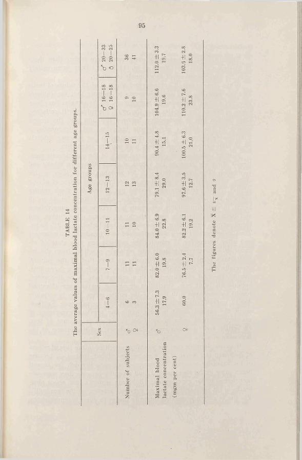
ftPo
WJCQ
I Io oSS CSO
i ¿
Of
ft3o
to<
95
*b 0f
£¡=3
3z.
+1 o»
+1
+1
o oo+1 2
+1 -
’b
T3OO
X *: d o1rH rt
+1 SC'I «
+1 «
+1
+1
O
+1
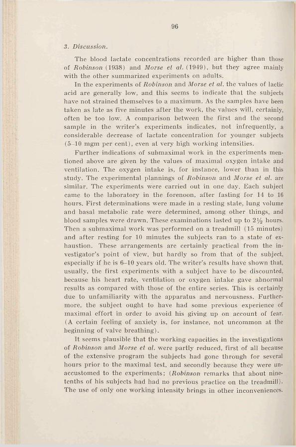
96
3. Discussion.
The blood lactate concentrations recorded are higher than those of Robinson (1938) and Morse el al. (1949), but they agree mainly with the other summarized experiments on adults.
In the experiments of Robinson and Morse et al. the values of lactic acid are generally low, and this seems to indicate that the subjects have not strained themselves to a maximum. As the samples have been taken as late as five minutes after the work, the values will, certainly, often be too low. A comparison between the first and the second sample in the writer’s experiments indicates, not infrequently, a considerable decrease of lactate concentration for younger subjects (5-10 mgm per cent), even at very high working intensities.
Further indications of submaximal work in the experiments mentioned above are given by the values of maximal oxygen intake and ventilation. The oxygen intake is, for instance, lower than in this study. The experimental plannings of Robinson and Morse et al. are similar. The experiments were carried out in one day. Each subject came to the laboratory in the forenoon, after fasting for 14 to 16 hours. First determinations were made in a resting state, lung volume and basal metabolic rate were determined, among other things, and blood samples were drawn. These examinations lasted up to 2 y2 hours. Then a submaximal work was performed on a treadmill (15 minutes) and after resting for 10 minutes the subjects ran to a state of exhaustion. These arrangements are certainly practical from the investigator’s point of view, hut hardly so from that of the subject, especially if he is 6-10 years old. The writer’s results have shown that, usually, the first experiments with a subject have to be discounted, because his heart rate, ventilation or oxygen intake gave abnormal results as compared with those of the entire series. This is certainly due to unfamiliarity with the apparatus and nervousness. Furthermore, the subject ought to have had some previous experience of maximal effort in order to avoid his giving up on account of fear. (A certain feeling of anxiety is, for instance, not uncommon at the beginning of valve breathing).
It seems plausible that the working capacities in the investigations of Robinson and Morse et al. were partly reduced, first of all because of the extensive program the subjects had gone through for several hours prior to the maximal test, and secondly because they were unaccustomed to the experiments; (Robinson remarks that about nine- tenths of his subjects had had no previous practice on the treadmill). The use of only one working intensity brings in other inconveniences.
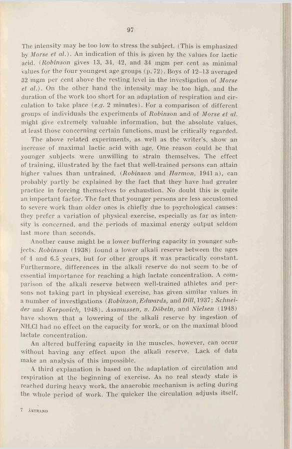
97
The intensity may be too low to stress the subject. (This is emphasized by Morse ei al.). An indication of this is given by the values for lactic acid. (Robinson gives 13, 34, 42, and 34 mgm per cent as minimal values for the four youngest age groups (p. 72). Boys of 12-13 averaged 32 mgm per cent above the resting level in the investigation of Morse et al.). On the other hand the intensity may be too high, and the duration of the Work too short for an adaptation of respiration and circulation to take place (e.g. 2 minutes). For a comparison of different groups of individuals the experiments of Robinson and of Morse et al. might give extremely valuable information, but the absolute values, at least those concerning certain functions, must be critically regarded.
The above related experiments, as well as the writer’s, show an increase of maximal lactic acid with age. One reason could be that younger subjects were unwilling to strain themselves. The effect of training, illustrated by the fact that well-trained persons can attain higher values than untrained. (Robinson and Harmon, 1941 a), can probably partly be explained by the fact that they have had greater practice in forcing themselves to exhaustion. No doubt this is quite an important factor. The fact that younger persons are less accustomed to severe work than older ones is chiefly due to psychological causes: they prefer a variation of physical exercise, especially as far as intensity is concerned, and the periods of maximal energy output seldom last more than seconds.
Another cause might be a lower buffering capacity in younger subjects. Robinson (1938) found a lower alkali reserve between the ages of 4 and 6.5 years, but for other groups it was practically constant. Furthermore, differences in the alkali reserve do not seem to be of essential importance for reaching a high lactate concentration. A comparison of the alkali reserve between well-trained athletes and persons not taking part in physical exercise, has given similar values in a number of investigations (Robinson, Edwards, and Dill, 1937; Schneider and Karpovich, 1948). Assmussen, v. Dobeln, and Nielsen (1948) have shown that a lowering of the alkali reserve by ingestion of NH,C1 had no effect on the capacity for work, or on the maximal blood lactate concentration.
An altered buffering capacity in the muscles, however, can occur without having any effect upon the alkali reserve. Lack oi data make an analysis of this impossible.
A third explanation is based on the adaptation of circulation and respiration at the beginning of exercise. As no real steady state is reached during heavy work, the anaerobic mechanism is acting during the whole period of work. The quicker the circulation adjusts itself,
1 ASTHAND
98
the smaller is the fraction of energy that must be supplied anaerobically, and the smaller is the accumulation of lactic acid. When doing similar work the trained individual has a greater acceleration of oxygen intake than the untrained. This partly explains the lower lactic acid values of the trained. Younger subjects also show a greater acceleration of oxygen intake than older ones. In Robinson’s investigations, for instance, the boys aged 5-12 years reached 55 to 41 per cent of their maximal oxygen consumption within the first half minute of running, while the adults only reached 29 to 35 per cent (average values). This must cause a relatively lower lactic acid accumulation in the younger subjects.
According to Margaria, Edwards, and Dill (1933) lactic acid accumulates in the blood to the extent of 7 gm for each litre of oxygen debt above a certain level. According to Asmussen (1946) and Christensen and Högberg (1950 a) the efficiency of anaerobic work is about 50 per cent of that of aerobic work. One litre of oxygen debt, i.e. 7 mg lactic acid, would in that case correspond to an oxygen deficit of0.5 litre.
According to Robinson’s determinations, an average boy of 6 years (maximal oxygen intake 0.98 litre per minute), reached during the first half minute of work an oxygen intake of 0.27 litre (55 per cent
of the maximum ^ '^ ). If, however, his circulation adapted itself as 2slowly as that of an adult, the boy would during the first half minute
have attained only 0.16 litre (32 per cent of ^‘^ ), and the deficit,20.11 litre oxygen, corresponds to 1.54 gm lactic acid. Distributed over 50 per cent (Hill, Long, and Lupton, 1925) of the body (weight 21 kg), it gives a concentration of 15 mgm per cent.
Correspondingly it can be reckoned that a subject of 25 years of age would have passed through the first half minute with 5.7 gm less lactic acid (16 mgm per cent), if he could have accelerated his oxygen intake as quickly as the 6 years old.
These calculations are, of course, only approximate, partly because they are based on average values of what “ took place” during one-half minute and partly because the quantitative relation between oxygen deficit and lactic acid production is not exactly known. Neither is the distribution of lactic acid in the organism. It appears, however, to be probable, that younger subjects’ ability to adapt themselves more quickly to work than their elders, brings about a significant decrease in the amount of anaerobic products.
Lundin and Ström (1947) found that the rise of blood lactic acid
99
from the rest value to the steady state value is significantly higher during exercise at low oxygen pressure than at a normal oxygen pressure. The most logical explanation is an inadequate oxygen supply to the tissues in the first case (comp, the effect of a slow adaption to work).
In relation to body weight, the subjects aged 7-9 years in the writer’s own material reached about the same oxygen intake per minute as the adults. There is reason to believe that the highest oxygen intake determined is very near to being the highest value possible. In which case there should be no greater differences between the children and the adults with regard to load on their organs and their feeling of strain. But the 7-9 year old subjects have shown an average lactate concentration of 82 mgm per cent as compared with the 112 mgm per cent of the adult subjects. Should this difference be regarded as a manifestation of the chrildren’s unwillingness to strain themselves? If so, then it must be realised that they can reach still higher values of oxygen intake than those noted. However, a more probable explanation is that younger subjects, by a more effective regulation of their circulation are able to mobilize their aerobic capacity more quickly.
The different values of lactic acid pose other questions, e.g. to what extent lactate concentration reflects the feeling of strain, and whether a certain concentration is possibly a limiting factor of performance capacity.
Asmussen et al. (1948) state that the limit for maximal work is set at a critical value of the lactic acid concentration in the muscles, which is of the same height in low' oxygen, in normal air, and in pure oxygen.
Crescitelli and Taylor (1944) consider that there is “no evidence of a “ ceiling” lactate level toward which the subjects approach when reaching the point of exhaustion” . This is due to the wide scattering of the individual lactic acid values at maximal work (117-328 mgm per cent).
It must be stressed here, that these values of Crescitelli and Taylor clearly disagree with those of other investigators. Values above 200 mgm per cent are rarely reported. At this laboratory, experiments were carried out on 5 élite Swedish athletes after they had run 400-800 meters while taking part in the Swedish trials for the 1948 Olympic Games in London (Åstrand and Högberg, not published). The average value of maximal lactate concentration was 179 mgm per cent, the highest individual value was 198 mgm per cent.
Among the subjects examined by Crescitelli and Taylor, only two were athletes in training, the rest were normally active college stu-
7*
100dents. It is also astonishing to find, in their experiments, that the subjects, after a 4 minutes’ walk on a 5 per cent grade at a rate of 108 m per minute, have a lactic acid concentration as high as 32 mgm per cent, two minutes after the work has ceased (range 13-61 mgm per cent), although oxygen intake was only 1.65 litres per minute, range 1.2-2.0 {Taylor, 1944). Owles (1930) states that the intensity at which hlood lactic acid increases over the resting value corresponds to an oxygen consumption of 1.8 1. per minute (adult males). Other authors give slightly different figures, though on about the same level. (This concerns types of work where large muscle groups are involved.) Robinson has determined the lactic acid during walking, where the oxygen intake, for eleven subjects between 20 and 30 years of age, reached an average value of 1.96 litres per minute (range 1.61-2.40 litres). After about 7y2 minutes’ work the average value of blood lactic acid was 15 mgm per cent (range 7-31 mgm per cent). These values agree with most experiments where the oxygen consumption is comparable (e.g. with unpublished experiments at this laboratory).
In the case of the lactic acid values during a resting state, as given by Crescitelli and Taylor, the average value for 10 subjects was 12.4 mgm per cent (post-absorptive, resting state), and this agrees well with other experiments (Robinson found 9 mgm per cent for the same age). Samples taken by Crescitelli and Taylor before work on the treadmill, however, gave an average value as high as 28.5 mgm per cent ( “ rest, not basal” ). Consequently this means, either that the “ resting” values were taken after an activity of the same intensity as the treadmill walk, or that the values in both cases are too high.
The results of the writer’s experiments, as well as others, lead to the conclusion that the values for maximal lactic acid after exhausting work as reported by Crescitelli and Taylor are too high, and that the probable values are about half of these. Crescitelli and Taylor in their lactic acid determinations used the Barker-Summerson (1941) method, while the other experiments related here, were carried out according to Edwards (1938), Friedemann, Cotonio, and Shaffer (1927) and Orskov (1930).
The values which at the present time are acceptable as normal values for the quantities of maximally possible blood lactate concentrations are:
1. For groups of subjects during maximal exercise in laboratory experiments 100-120 mgm per cent, with individual values above 140 mgm per cent (independent of sex).
2. For élite athletes after competitions average values up to
101200 mgm per cent, with individual values above 200 mgm. (No values are reported for female élite performers.)
3. For younger subjects (4-20 years) average values of 60-120 mgm per cent. The youngest have the lowest values. Individual values of 100 mgm per cent or higher are possible, at least for children of 9 years or more, independent of sex.
In the opinion of the writer the youngest subjects (4-6 years) did not strain themselves maximally, but the subjects of 7 years and older did their very best. If this has been the case, then the “ critical value of the lactic acid concentration” for the children is on another level than that of the adults. The interpretation of this can be that:
1) the lactic acid concentration as a limiting factor is of relatively secondary importance, and other mechanisms dominate the performance capacity. The lower level of lactic acid in the younger subjects can be explained as the result of a more effective regulation of their metabolism at the beginning of work.
Training improves, for instance, the neuromuscular coordination which allows harder work to be done, so increasing the amount of anaerobic products.
2) the lactic acid concentration primarily determines the performance capacity. “ The threshold” varies with age, and can be raised by training.
With our present knowledge, no definite opinion of the alternatives can be given. No matter how it is interpreted, however, the lactic acid concentration during exercise serves as a good indicator of intensity in comparison with the capacity of the subject with regard to metabolism if only age and physical condition of the subject are taken into consideration.
It is of special interest that males and females attained approximately the same maximal values. This clearly indicates that the female has the same possibilities as the male to make use of her physical resources, and that she can strain herself to the same- state of exhaustion. In the writer’s experiments it was often astonishing that 13-17 year old girls, as a rule, did maximal work “without complaint ’, while boys of the same age more often wanted to give up. With adult subjects, the women seemed to be more inclined to discontinue the work. The cause is probably a psychological one. The fact that the working capacity of women is lower than that of men will influence a number of functions quantitatively. When the subjects are tested with a certain work intensity, i. a. heart rate, lactic acid, ventilation of females will attain higher values as compared with males. Best
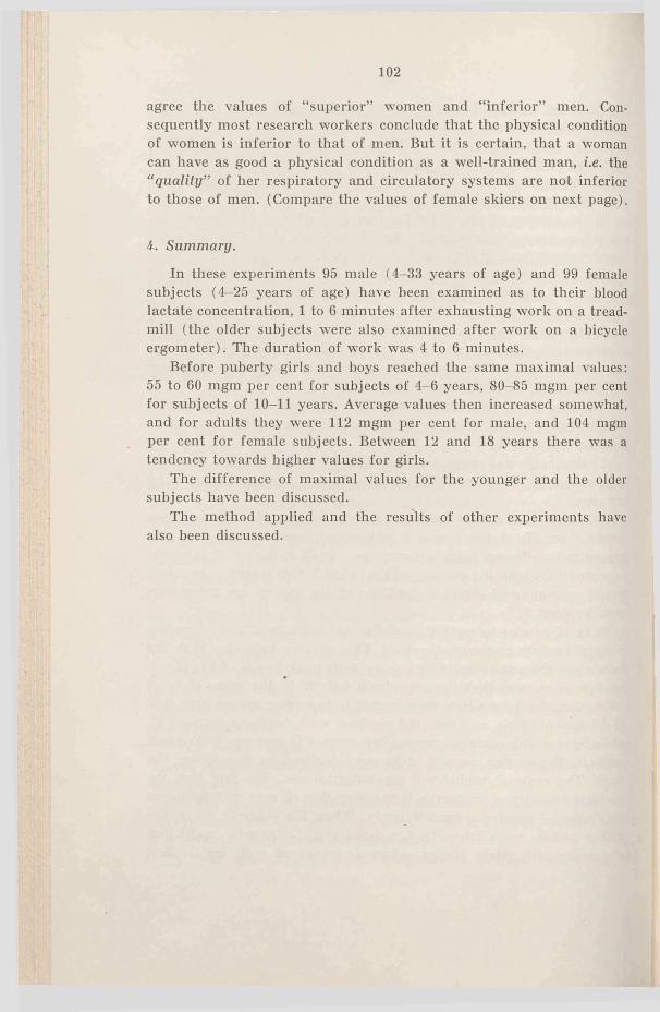
102agree the values of “ superior” women and “ inferior” men. Consequently most research workers conclude that the physical condition of women is inferior to that of men. But it is certain, that a woman can have as good a physical condition as a well-trained man, i.e. the “ quality” of her respiratory and circulatory systems are not inferior to those of men. (Compare the values of female skiers on next page).
i . Summary.
In these experiments 95 male (4-33 3rears of age) and 99 female subjects (4-25 years of age) have been examined as to their blood lactate concentration, 1 to 6 minutes after exhausting work on a treadmill (the older subjects were also examined after work on a bicycle ergometer). The duration of work was 4 to 6 minutes.
Before puberty girls and boys reached the same maximal values: 55 to 60 mgm per cent for subjects of 4-6 years, 80-85 mgm per cent for subjects of 10-11 years. Average values then increased somewhat, and for adults they were 112 mgm per cent for male, and 104 mgm per cent for female subjects. Between 12 and 18 years there was a tendency towards higher values for girls.
The difference of maximal values for the younger and the older subjects have been discussed.
The method applied and the results of other experiments have also been discussed.
Ch apter IX
THE AEROBIC CAPACITY/. Introduction.
In his studies of physical fitness in relation to age Robinson (1938) found for males between 5 and 29 average values of between 47 and 53 ml oxygen intake per minute and per kg body weight, without noticeable variations for age. For boys 10-12, Morse, Schlutz and Cossets (1949) found 48 ml. Boys of 13 attained 45 ml, and this value gradually increased until it reached 51 ml for subjects of 17 years. In both studies the treadmill was used.
Wright (1950) contributed results on 41 subjects, aged 25-45, who ran on the treadmill for 6 minutes at maximal speed. The oxygen intake averaged 1.425 ± 0.030 litres per minute and per nr surface area.
Metheny, Brouha, Johnson, and Forbes (1942) found, in experiments with 17 rather well-trained female subjects, 20-27 years old, average values of 40.9 ml oxygen intake per kg weight (highest individual value 47.5 ml). The work consisted of running on the treadmill until exhausted, for an average time of 108 seconds. In similar experiments 30 male subjects of the same age were found to have an average value of 51.3 ml per kg for oxygen intake (highest value 60.5 ml). The running time averaged 216 seconds.
Christensen and Hogberg (1950 c) report from their experiments on well-trained women skiers an oxygen intake of 4.17 litres per minute, corresponding to 58.2 ml per kg. A lighter subject reached 3.74 litres per minute or 64.5 ml per kg. Two male subjects belonging to the Swedish élite attained oxygen intakes of 5.0 and 5.1 litres per minute respectively.
The highest value observed is Lash’s oxygen intake of 5.35 litres per minute (Robinson, Edwards, and Dill, 1937).
With work on a bicycle ergometer an oxygen intake of 4.68 litres per minute has been recorded by Christensen (1932). Nielsen and Hansen (1937) found an oxygen intake of 5.05 litres in a subject breathing an air mixture containing a high percentage of oxygen.
Cureton (1951) carried out comprehensive experiments on élite
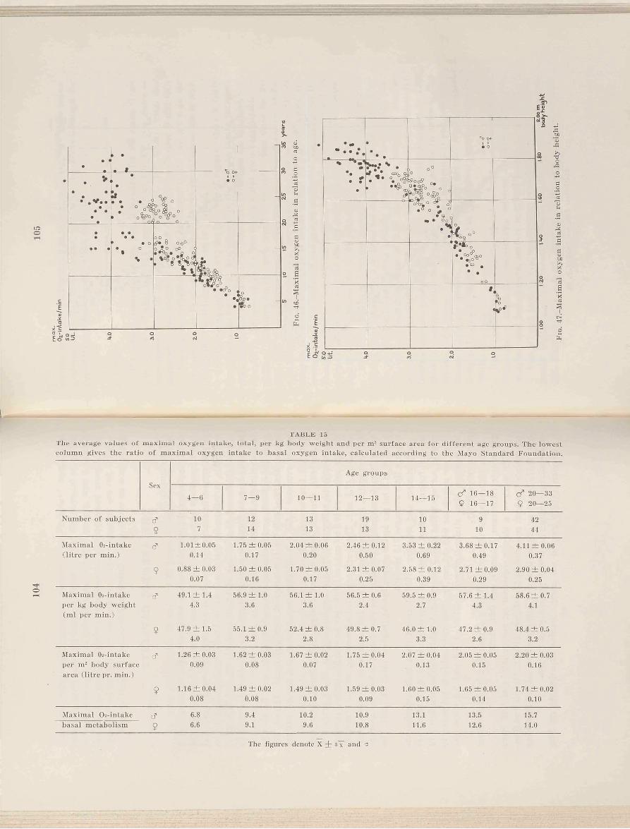
104
ia o
• <•
• •
• t
i
. >© •
•• . V . .
•• *
•
•
•
• o % ° °
°9 ? o to eg
o g i o , ° ° ° o °
• o
-
• • •
•
•
• •© •
• • o • o t
\ •
O o< £ • o O o ° o ? ° CP • * 4 d • O O
» 8
V *
•
-
o l . ° 0 ° 0• «O o
• ^
•
dCfjaoC5O02fla13cOJtr.>>o
E
CD
6£
T A B L E 15The average values of maximal oxygen intake, total, per kg body weight and per m2 surface area for different age groups. The lowest column gives the ratio of maximal oxygen intake to basal oxygen intake, calculated according to the Mayo Standard Foundation.
Age groups
4—6 7—9 10— 11 12— 13 14— 15 < f 16— 189 16— 17
cd 20— 33 9 20—25
Number o f subjects d 10 12 13 19 10 9 429 7 14 13 13 11 10 44
Maximal Oj-intake d 1.01 ±0.05 1.75 ± 0 .0 5 2.04 ± 0 .0 6 2.46 ±0 .1 2 3.53 ± 0 .2 2 3.68 ± 0 .1 7 4.11 ± 0 .0 6(litre per min.) 0.14 0.17 0.20 0.50 0.69 0.49 0.37
9 0.88 ±0 .0 3 0.07
1.50 ±0 .0 5 0.16
1.70 ±0 .0 5 0.17
2.31 ±0 .0 7 0.25
2.58 ± 0 .1 2 0.39
2.71 ±0 .0 9 0.29
2.90 ± 0.04 0.25
Maximal Oi-intake d 49.1 ± 1.4 56.9 ± 1 .0 56.1 ± 1.0 56.5 ± 0.6 59.5 ± 0.9 57.6 ± 1 .4 58.6 ± 0.7per kg body weight (m l per min.)
4.3 3.6 3.6 2.4 2.7 4.3 4.1
9 47.9 ± 1 .5 4.0
55.1 ± 0.9 3.2
52.4 ± 0 .8 2.8
49.8 ± 0 .7 2.5
46.0 ± 1.0 3.3
47.2 ± 0.9 2.6
48.4 ± 0.5 3.2
Maximal 02-intake d 1.26 ±0 .0 3 1.62 ±0 .0 3 1.67 ±0 .0 2 1.75 ±0 .0 4 2.07 ± 0 .0 4 2.05 ± 0.05 2.20 ± 0 .0 3per mu body surface area (litre pr. min.)
0.09 0.08 0.07 0.17 0.13 0.15 0.16
9 1.16 ±0 .0 4 0.08
1.49 ±0 .0 2 0.08
1.49 ± 0.03 0.10
1.59 ±0 .0 3 0.09
1.60 ±0 .0 5 0.15
1.65 ± 0.05 0.14
1.74 ±0 .0 2 0.10
Maximal 02-intake d 1 6.8 9.4 10.2 10.9 13.1 13.5 15.7basal metabolism 9 6.6 9.1 9.6 10.8 11.6 12.6 14.0
The figures denote X + s x and a
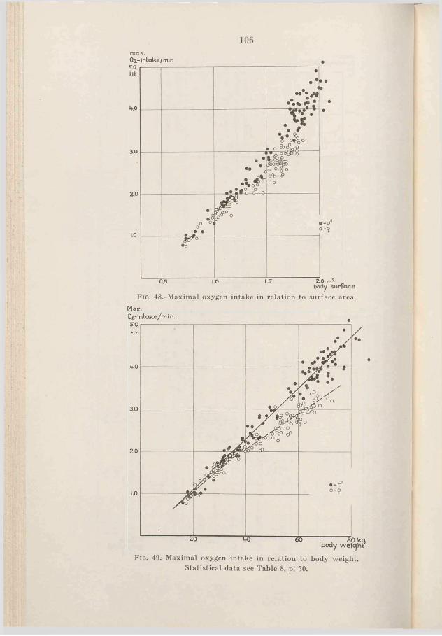
106m a x .
Oz-intaWe/min SO Lit.
1*0
3.0
2.0
•• ••
<*.*.
8*0^0 • o-È&R_____________,
.ci'*• ! f°o rfo n
-°o^
| CK
•o8 o °« o• e
JB*®O
•o
1.0 2 0 ,body surface
Fig. 48,-Maximal oxygen intake in relation to surface area.Max.
Fig. 49,-Maximal oxygen intake in relation to body weight. Statistical data see Table 8, p. 50.
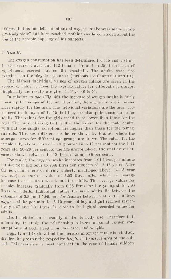
107
athletes, but as his determinations of oxygen intake were made before a “steady state” had been reached, nothing can be concluded about the size of the aerobic capacity of his subjects.
2. Results.
The oxygen consumption has been determined for 115 males (from 4 to 33 years of age) and 112 females (from 4 to 25) in a series of experiments carried out on the treadmill. The adults were also examined on the bicycle ergometer (methods see Chapter II and III).
The highest individual values of oxygen intake are given in the appendix. Table 15 gives the average values for different age groups. Graphically the results are given in Figs. 46 to 51.
In relation to age (Fig. 46) the increase of oxygen intake is fairly linear up to the age of 13, but after that, the oxygen intake increases more rapidly for the men. The individual variations are the most pronounced in the ages of 12-15, but they are also quite considerable for adults. The values for the girls trend to be lower than those for the boys. The most striking fact is that the values for the male adults, with but one single exception, are higher than those for the female subjects. This sex difference is better shown by Fig. 50, where the average curves for different age groups are drawn. The values for the female subjects are lower in all groups: 13 to 17 per cent for the 4-11 years old, 26-29 per cent for the age groups 14-25. The smallest difference is shown between the 12-13 year groups (6 per cent).
For males, the oxygen intake increases from 1.01 litres per minute for 4—6 year old boys to 2.46 litres for subjects of 12-13 years. After the powerful increase during puberty mentioned above, 14-15 year old subjects reach a value of 3.53 litres, after which an average increase to 4.11 litres was found for adults. The average values for females increase gradually from 0.(38 litres for the youngest to 2.90 litres for adults. Individual values for male adults lie between the extremes of 3.30 and 5.09, and for females between 2.41 and 3.40 litres oxygen intake per minute. A 15 year old boy and girl reached respectively 4.47 and 3.31 litres, i.e. close to the highest recorded values for adults.
Basal metabolism is usually related to body size. Therefore it is interesting to study the relationship between maximal oxygen consumption and body height, surface area, and weight.
Figs. 47 and 48 show that the increase in oxygen intake is relatively greater the greater the respective height and surface area of the subject. This tendency is least apparent in the case of female subjects
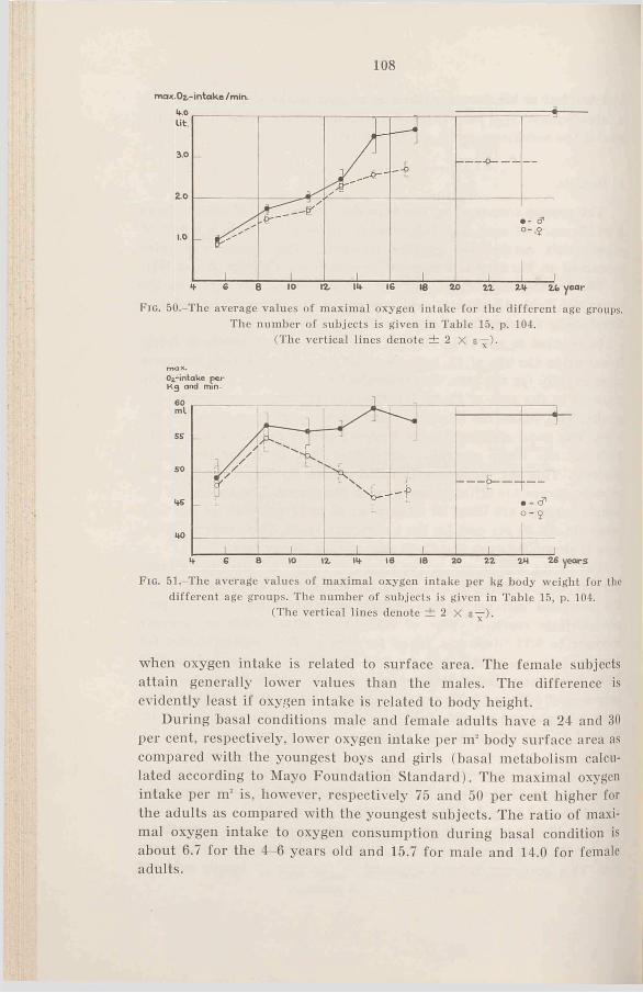
108
max.02.-intake /min
F ig . 50.-The average values of maximal oxygen intake for the different age groups. The number of subjects is given in Table 15, p. 104.
(The vertical lines denote ± 2 X e —).
max.O^-intake p e r K g and min-
F ig . 51.-The average values of maximal oxygen intake per kg body weight for the different age groups. The number of subjects is given in Table 15, p. 104.
(The vertical lines denote ± 2 X e-j-).
when oxygen intake is related to surface area. The female subjects attain generally lower values than the males. The difference is evidently least if oxygen intake is related to body height.
During basal conditions male and female adults have a 24 and 30 per cent, respectively, lower oxygen intake per nr body surface area as compared with the youngest hoys and girls (basal metabolism calculated according to Mayo Foundation Standard). The maximal oxygen intake per nr is, however, respectively 75 and 50 per cent higher for the adults as compared with the youngest subjects. The ratio of maximal oxygen intake to oxygen consumption during basal condition is about 6.7 for the 4-6 years old and 15.7 for male and 14.0 for female adults.

109
Fig. 49 shows the maximal oxygen intake in relation to body weight. It appears that the oxygen intake for male subjects increases roughly linearly with increasing body weight. It is worth pointing out that the determinations cover a weight range of 16 to 85 kg, and a range in oxygen intake from 0.7 to 5.1 litres per minute.
In the case of the female subjects, the values mainly follow those of the boys up to 40 kg (all under 40 kg are under 12 years, except two girls and seven boys), after which their values of oxygen intake lie definitely lower. Only a few of the older females’ values fall within the range of the males.
Table 8 (p. 50) shows some statistical calculations based on Fig. 49. The correlation between maximal oxygen intake and body weight is for males 0.98, for girls below 40 kg 0.96, and for females over 40 kg 0.86. The difference between the regression lines of males and girls is not significant, between males and females, however, it is highly significant. The deviations from the regression lines are 6 to 8 per cent.
The average values for maximal oxygen intake per minute and kg for different age groups are plotted in Fig. 51. For the groups of 4—9 years the girls’ values are somewhat lower (the difference is not significant). The youngest subjects only reach 48-49 ml per kg, whereas the means for the 7-9 year old subjects are 55-57 ml. After that the average values of the males are surprisingly constant, ranging between 56 and 59 ml. The aerobic capacity of the girls, however, declines to 46 ml for the 14—15 years old, but then it increases slightly to 48 ml for adults. (The difference between male and female subjects after the age of 12 is highly significant.) The difference between oxygen intake for male and female adults is reduced to 17 per cent by taking into consideration the differing body weights (the total figures gave a 29 per cent difference).
The extremes of the maximal oxygen intake per kg weight are 48.1 and 67.4 ml for male adults and 43.2 and 59.6 for female adults. The highest individual values for subjects under 12 years are 62.7 ml for hoys (one hoy of 7, and one of 9 years), for girls 58.9 ml (9 years), and for subjects of 12—16 years 63.7 ml (14 years) and 53.5 ml (12 years), respectively.
Table 13 (p. 88) shows the maximal oxygen intake obtained lor male and female adults in the running and cycling experiments, respectively.
For the males the oxygen intake is the same in both experiments (4.04 and 4.03 litres per minute, respectively). The females attain somewhat lower values during cycling as compared with running (2.76 and 2.89 litres, respectively, P = 0.05).

110Of 34 males who, apparently, did exhausting work both on tread
mill and bicycle ergometer, 21 reached the maximal values of oxygen intake during running. For 33 females the corresponding number is 23. In order to determine the aerobic capacity it evidently does not matter if cycling or running is chosen as working procedure.
As these experiments proceeded and more and more data from different subjects were brought together, it was astonishing to see how the values of maximal oxygen intake were grouped when related to the body weight of the subjects. Primarily, how regularly the oxygen intake increased with increasing weight, secondly how little the sex difference was reflected in the values up to a certain limit of weight, thirdly how evident the sex difference was for the heavier subjects. In examining other functions the difference between the males and females was not so pronounced (comp, for instance vital capacity, ventilation, blood volume).
3. Discussion.
a. Comparison with previous investigations.-A comparison with the experiments by Robinson (1938) and Morse et at. (1949) reported above show's that their average values for oxygen intake in corresponding age groups are considerably lower (8 to 20 per cent if subjects of 4-6 years are excluded and the calculations intend to show the oxygen intake per kg body weight). The methods used in the two series of experiments have been discussed on page 96. The lactic acid values indicated that the work in several cases must have been submaximal, which naturally results in lower “ maximal values” . In Robinson’s experiments the maximal lactic acid value recorded was 72 mgm per cent for subjects up to 15 years of age. According to the discussion on page 23 of the relationship between maximal oxygen intake and blood lactate concentration, the value of 72 mgm per cent is just “ sufficiently” high to indicate that the maximal oxygen intake has been reached. The minimal values given by Robinson (13-42 mgm per cent up to the age of 20) are certainly too low. Some of the differences in the results of the experiments, however, must be ascribed to a difference of the subjects’ physical development. Thus, the difference between 20-30 year old subjects is very great (the writer’s mean is 59 ml, that of Robinson 49 ml oxygen intake per kg), and the lactic acid values in Robinson’s experiments indicate a high working intensity. Attention may be called to the fact that the G.C.I. students are physically in good condition and are W'ell-trained.

I l l
The average value for female adults in the experiment carried out by Metheny et al. (1942) was 40.9 ml oxygen intake per kg. The reason for this low value can be entirely explained by the fact that the determinations were made before the respiration and circulation had been adapted to the requirements of the work. The average running time was only 108 seconds. The subjects attended a college of physical education, and consequently they ought to have been practically in the same physical category as the writer’s adult subjects. The male subjects in Metheny's et al. experiments also attained definitely lower values as compared with the writer’s. The reason for this may be partly the shorter running time, averaging 3y2 minutes, and partly different degrees of “ fitness” .
The highest maximal values in the writer’s experiments can be compared with the highest reported. In these cases the subjects were élite athletes in training. The male subject with an oxygen intake of 5.09 litres (A. W., 28 years) has been Swedish 400 m hurdle champion (best time 53.6), and the female subject with 59.6 ml per kg body weight (I. R., 23 years) is a first-class athlete, cross-country runner, and skier.
b. The aerobic capacity before twelve years of age.-A “ natural” starting of the relative decrease in the girls’ aerobic capacity would be the beginning of puberty. There is no information concerning the time for menarche for the females but as already mentioned puberty for Swedish girls as a rule takes place during the ages of 12 to 15 (boys 14 to 17). In Fig. 51 it was shown that the decrease of the maximal oxygen intake per kg weight already appeared at the age of 10-11 (there is no difference in the average values between girls of 10 and 11 years of age). Girls and boys attained the same lactic acid values, which indicates the same state of exhaustion when the maximal oxygen intake was recorded. As the oxygen intake at the age of 4 to 9 was almost the same for boys and girls, it seems plausible to regard the lower values of the girls aged 10-11 years, either as early secondary- sex character or as a consequence of not quite representative material. During puberty there is an increase of body weight, but in this study all the girls under 12 years weighed less than 40 kg, while 2 hoys weighed more than 40 kg (comp. Appendix). In order to get an explanation of the different oxygen intake additional experiments must be done with the involved age groups.
Before the age of 12 the average weights for the boys are 2-4 kg greater than those for the girls. There is no reason to suppose that boys have a greater percentage of fatty tissues, and they must be regarded
112as physically more developed than girls of the same age. The plays and games generally chosen by the boys must have a greater training effect than girls’ more quiet occupations. As was shown by Robinson and Hannon (1941h) training increases the aerobic capacity of the individual. The tendency to higher capacity of oxygen intake that was found in the boys as compared to the girls, might be a consequence of psychological factors as well as of a primarily greater physical working capacity.
The youngest subjects, aged 4-6, have a definitely lower oxygen intake capacity than the older subjects. Whether this depends on an inclination to “ give it up” too early or on a physiologically lower capacity, cannot be settled (average maximal lactic acid values were 56-60 mgm per cent as compared with 77-82 mgm per cent for the 7—9 year old groups).
It is of greatest interest to see that the boys of 7-11 years, girls 7-9, reach practically the same oxygen intake per kg body weight as the adult men. Boys of 7-9 years attain 56.9 ml, adults 58.6 ml, a difference of only 3 per cent. On the other hand, the younger subjects have a relatively higher basal metabolism. The maximal oxygen intake of the 7-9 year old boys means an increase of 9.4 times the basal value, while the male adults are able to increase their basal oxygen intake15.7 times. The younger subjects’ “ range of variations” is not as large. This at least partly explains why the younger subjects quickly accelerate their oxygen intake towards their maximal values in the beginning of exercise (comp. p. 98). It is difficult to conclude anything from this smaller “ range of variation” in the aerobic processes. Possibly the aerobic capacity of their muscular mass is less than the adult’s one. However, it is not out of question that a greater or smaller part of the oxygen corresponding to the basal metabolism will be available for the working muscles during heavy work.
c. The aerobic capacity after twelve years of age-F or the boys no decrease of the maximal oxygen intake per kg body weight is found during puberty. There is even a tendency towards a higher average value for the subjects of 14-15 years as compared with the next age group and younger subjects. This is in disagreement with the results obtained by Robinson and Morse et at. who found lower oxygen intake during puberty. Morse et at. cannot in other experimental results find any explanation of this lower aerobic capacity (concerning above all the subjects of 13 years). One reason might be that the intensity‘was too lowr. (For the 13 year olds the speed was 6 m.p.hr. while the subjects of 14 ran at the rate of 7 m.p.hr.). An indication of this is the
113
fact that the subjects of 13 had longer running times than for instance the subjects of 10-12 years. The lactic acid values were also low.
As for the girls, there is a decrease of the aerobic capacity per kg body weight from the age of about 10 years (comp. Fig. 51). The gradually increasing difference in oxygen intake between males and females must he regarded as a consequence of sexual maturity. The average values for the maximal lactic acid concentration of the girls were higher than those of the boys aged 12-18 so, evidently, it is no question of “ laziness” (comp. Fig. 45, p. 94).
The question arises as to the explanation of this lower oxygen intake capacity. A consequence of puberty is the relatively large increase of the fatty tissue in women. The body weight will be a relatively poor measure for the amount of active protoplasmic mass. A better comparison between the sexes would be the maximal oxygen intake per kg of muscular tissue. This mass, however, cannot be determined, but it ought to be related to the active protoplasmic mass or to the lean body mass (gross body weight minus excess fat, Behnke), and both can be determined indirectly. Dahlström (1950) has determined the extracellular fluid volume by the thiocyanate method. He proposes that the extracellular fluid volume is related to the active protoplasmic mass and its normal, basal activity, irrespective of its actual activity. Behnke (personal communication) shows a close relationship between the calculated basal metabolism and the lean body mass.
The basal caloric requirements must be directly dependent on the mass of active protoplasma. It is estimated that 20-25 per cent of the basal oxygen intake is consumed in the voluntary muscles (Asmussen, Christensen, and Nielsen, 1939 b). Physiological variations of muscular mass certainly cannot, with the present methods of determination, be detected at rest. According to Asmussen et al. the metabolism of the muscles can increase to more than 100 times the basal caloric output. During maximal work the oxygen consumption of the remaining active protoplasmic mass will only amount to a small per cent of the total oxygen intake (heart muscle excepted), and a physiological variation of the muscular mass will more or less he reflected in the metabolism (comp, training). The capacity of organs involved in energy output can be judged from the capacity of oxygen intake. The problem is whether the size of heart force, extra cellular fluid, blood volume, total quantity of hemoglobin etc. are specifically related to muscular mass, or more to active protoplasmic mass in general, or other factors. This question takes on importance when, as in this study, the relationship between the aerobic capacity and blood volume or quantity of hemo
8 ÅSTRAND
114
globin is discussed. It is probable that the more physically active an individual is, the more muscular mass will influence, for instance, the total amount of hemoglobin. For a sedentary person other factors will predominate.
Fig. 18 p. 48 shows that the maximal oxygen intake per unit of total hemoglobin is the same for males and females in all age groups except that of 10-11 years. Thus, sex differences found in oxygen intake per kg body weight (ranging from 3 to 23 per cent for means) are reflected by similar differences in total hemoglobin. If the total quantity of hemoglobin and the maximal oxygen intake variate with the muscular mass, the conclusion will be that the female adult ought to have 15-20 per cent less muscular tissue per kg body weight than the male adult. (Women’s oxygen intake and quantity of hemoglobin per kg are 17 and 20 per cent less, respectively). This implies that the distribution of muscles in the body is almost the same in man and woman, as the type of work was the same.
If this difference in muscular mass in relation to weight is not accepted, the relationship between quantity of hemoglobin (or aerobic capacity) and muscular mass must be different in man and woman. The woman’s muscular mass -would be “ excessively great” (or small) with regard to the oxygen transport. The last alternative seems least probable.
It is certain that the difference in aerobic capacity between males and females after puberty will be largely reduced if the difference in adiposity is taken into consideration. Further experiments have to show how' much.
A study of the literature available gave no consistent averages of muscular mass in males and females. The same holds true for fatty tissue. According to Schaeffer (1944) “ tela subcutanea forms nearly one-eighth of the body' weight at birth. Thereafter it declines in relative amount, forming about 10 per cent at maturity in men and over 20 per cent in average mature woman” . “ The musculature increases from about 25 per cent of the body at birth to 40 or 45 per cent in the adult”.
Edwards (1950) found that the amount of subcutaneous fat was the same in boys and girls before puberty, but it was 1.75 times greater in women of average weight than in men. “ If the subcutaneous fat in human beings mirrors the amount of total body fat, as it does in guinea pigs (Pitts, 1951), then women have on the average about 1.75 times more fat than men, ages corresponding” (Behnke, personal communication ).
It is possible that the high correlation between total quantity of hemoglobin and maximal oxy'gen consumption found in this study is
115
a consequence of the homogeneity of the material as to physical activity. In the opinion of the writer, however, also untrained persons will fit the curve. The close relationship between body weight and maximal oxygen intake certainly is due to the fact that none of the subjects were excessively fat. (The aerobic capacity and quantity of hemoglobin might possibly be correlated to body weight independently. Then, however, the variation from the regression line for the relationship of total hemoglobin to oxygen intake would be greater than for the direct correlation of oxygen intake with body weight, which is not the case.)
The adult female subjects have a maximal oxygen intake per litre blood volume that is about 10 per cent lower than in male subjects. The hemoglobin concentration too is about 10 per cent lower in the women. This could mean that the quantity of blood volume and of hemoglobin are regulated by different factors, e.g. circulatory demands would “ definitely” decide the volume of blood, not the metabolism (comp. p. 55).
The blood volume in the woman compared with that in the man. is (1) smaller per kg body weight, but (2) greater if related to the maximal oxygen intake. The fatty tissue demands less circulation than “ active” tissues, which possibly explains (1). On the other hand blood volume cannot be regulated only with regard to the active protoplasmic mass (and the muscular mass), but a certain volume is necessary in order to fill the vascular bed of the fatty tissue, which at least partly explains (2). Because of the low metabolism in this tissue there is no need for a greater quantity of hemoglobin, and the concentration will be somewhat lower.
d. Comparison of children's and adults’ aerobic capacity -For the boys the oxygen intake capacity in relation to blood volume is greater than for the older subjects (Fig. 18, p. 48). If we reverse this, saying that the older subjects have “ too great” a blood volume, this can be explained by greater demands on the circulation in older individuals,e.g. due to gravity (comp. p. 56). It is more difficult to find an explanation for the lower quantity of hemoglobin in relation to body weight in the younger subjects (about 25 per cent), and for their lower hemoglobin concentration (about 10 per cent). With the same ratio of hemoglobin to muscular mass for children as for adults, the muscular mass per kg body weight in the younger subjects should be about 25 per cent lower than in adults. As the maximal oxygen intake per kg body weight was about the same, the aerobic capacity of the muscles must be relatively higher in children, or the other active
8‘
116
protoplasma consumes a relatively greater percentage of the total oxygen intake. The truth probably lies somewhere between. In children, as compared with adults, favourable circulatory conditions make a smaller quantity of hemoglobin sufficient. The muscular mass per kg body weight is lower (comp. p. 114, Schaeffer). The basal metabolism is relatively higher.
The relationship between basal metabolism (active protoplasmic mass?) and maximal oxygen intake appears in Table 15, p. 104. The ratio of maximal oxygen intake to basal metabolism varies with age, as stated above, but is fairly similar when males are compared with female subjects of the same age. The oxygen consumption during exhausting exercise increases 9.4 and 9.1 times respectively for boys and girls of 7-9 years; 10.9 and 10.8 respectively for those of 12-13 years; 13.5 and 12.6 respectively for subjects of 16-18; and 15.7 and14.0 respectively for adults. The highest individual values for adults are in these experiments 18.1 times the basal level for male and 15.7 for female subjects. According to Dill (1936) the ratio of maximal oxygen consumption to basal oxygen consumption in men running at top speed varies from 10 to 20, depending on physical development and training. Lash’s maximal oxygen intake (5.35 litres per minute) was 21.4 times his basal level as compared to 14.5, the maximum of the best untrained man (Robinson, Edwards, and Dill, 1937). As an interesting comparison Brody (1945) concludes that the maximal amount of oxygen that a work horse in excellen physical condition can utilize is about 21 times the basal oxygen. He also concludes, that “ the ratio of work to resting oxygen consumption is apparently independent of size or even species” .
However, attention has to be paid to age, and in certain cases even to species. Thus both man and horse drop far behind a number of flying insects. Krogh and Weis-Fogh (1951) in excellent experiments over the respiratory exchange of the desert locust have found that the metabolic rate during flight had increased fifteen to fifty times compared with the value during rest. The oxygen consumption during flight varied between 10 and 30 litres per kg and hour. This might be compared with the maximal values for man, who in the writer’s experiments attained about 4 litres oxygen intake per kg and hour. Krogh and Weis-Fogh report even higher values for bees, flies, and butterflies found by other investigators (Jongbloed and Wiersma, 1935; Davis and Fraenkel, 1940). During flight the oxygen consumption has been estimated at about 100 litres oxygen intake per kg and hour. This means that the metabolic rate during flight in some insects in
117
creases 100 times or even more as compared with the metabolism during rest.
e. Limiting factors for the aerobic capacity .-In a discussion about factors influencing the oxygen supply during maximal work, Christensen, Krogh, and Lindhard (1934) analyse the following factors: 1) the pulmonary ventilation, 2) the rate of diffusion of oxygen from the alveolar air to the blood, 3) the minute volume of the heart, 4) the rate of blood flow through the muscles, and 5) the conditions for oxygen diffusion from the muscle capillaries to the tissue.
1) Concerning the ventilation as a limiting factor in exercise the reader is referred to Chapter VII, p. 88.
2) M. Krogh (1915) determined by the carbon monoxide method the diffusion constant during rest and moderate work, and the results were confirmed by Boje (1933), who also made determinations during heavy work. It was shown that there are large individual differences with regard to the diffusion constant during rest; that the diffusion constant increases with increasing rates or work; and that the maximal oxygen diffusion calculated for a normal pressure and composition of the inspired air is about 5 litres per minute, and that this would involve a definitely incomplete saturation of the arterial blood with oxygen.
Lilienthal, Riley, Proemmel, and Franke (1946) mainly confirm the results obtained by Krogh and Boje. They report interesting experiments concerning the oxygen pressure gradient from alveolar air to arterial blood, but the working intensities were too low to apply to this discussion.
As was earlier mentioned, the highest values of oxygen intake observed are about 5 litres. If the oxygen intake is limited by the rate of diffusion of oxygen in the lungs, breathing of oxygen-rich air should increase the oxygen intake.
Nielsen and Hansen (1937) report experiments, where the subjects during exercise breathed gas mixtures with normal oxygen concentration, with 23 per cent and 45-47 per cent oxygen. It was found that with heavier works the oxygen consumption increased by breathing the highest oxygen percentage, and a greater amount of work could be performed as compared with the “ room air experiments” . As there was no effect when breathing 23 per cent oxygen, they are of the opinion, that the higher oxygen intake capacity could not be due to better conditions of diffusion. Nor can the increased saturation of the blood explain the higher oxygen intake. They conclude that the
118
effect depends on a specific influence on the circulation or possibly on the central nervous system.
From these experiments, it seems obvious that the diffusion of oxygen from alveoli to blood is sufficient also at maximal work, provided normal conditions prevail.
3 and 4) It is common to consider the minute volume of the heart as the limiting factor for the oxygen intake during such types of exercise as cycling and running. The working capacity of the heart should determine that of the muscles. To analyse in detail the influencing factors, e.g. the venous filling pressure, the period of diastole, force of contraction, is here impossible. For that reason these and other components of direct importance for the function of the heart are summarized as “ central factors in circulation” as distinguished from the “ peripheral” ones. According to the division given above the periphery is represented in factor (4), “ the rate of blood flow' through the muscles” . The connection between (3) and (4) is perhaps too close to make a division physiologically correct, hut it can be justified and exemplified as follow's.
During maximal w'ork with his arms (bicycle ergonieter), a subject attained for instance a maximal oxygen intake of 3.0 litres per minute (Christensen, 1932). The same subject working with his legs (cycling) reached an oxygen intake of 4.7 litres. It is fairly clear, that the strain of the heart is moderate in arm work. The limiting factor may be found in the capacity of the vascular bed in the muscles. The central circulation is fully sufficient, the peripheral one fails. The question arises now' wiiether cycling with maximal intensity will overload the central organs, or w'hether it is still only a matter of failing peripheral circulation. An important problem is, if (a) the capacity of the heart permits only a certain percentage of the muscles in the body to w'ork maximally or if (b) the capacity of the heart to increase the minute volume is greater than the ability of the muscles to “ receive” it. The question is of vital importance; if alternative (b) holds true, then the heart cannot under normal conditions be overstrained. Naturally, the blood flow through the muscular vessels can increase, if only the arterial blood pressure increases. A very high blood pressure w'ould, however, probably cause disadvantages for other organs (e.g. the central nervous system), and would certainly be prevented by a peripheral regulation. The fact that the blood pressure during exercise seldom surpasses 200 to 250 mm Hg does not necessarily prove lack of capacity for the heart.
As mentioned above, during maximal work on the bicycle ergo- meter the aerobic capacity will increase by breathing oxygen-rich air.

119
TABLE 16Maximal ventilation and oxygen intake for subject B.L. ( t f , 25 years of age) during cycling, running, and skiing. The minute volume of the heart evidently
attains a submaximal value, at least during cycling and running.
Work max. vent, lit/min.
max. O» intake lit/min.
Bicycling 2100 kgm ./m in.......... 130.3 4.64Running 16 km p.h. 1° ........... 115.4 4.57Skiing1 14.7 km pr.h.................. 139.4 5.24
' = The values obtained from Christensen and Högberg (1950 c).
To make this increase in oxygen intake possible an increase of the minute volume above the “ normal maximum” is essential. However, this cannot simply be regarded as an evidence that the heart “normally” should work submaximally. The higher oxygen tension in the arterial blood may be sufficient to increase the force of the heart.
Some other experiments indicating that the heart in maximal cycling or running works submaximally shall, however, be mentioned.
A male subject, B.L., 25 years old, reached in these experiments an oxygen intake of 4.64 and 4.57 litres per minute in maximal cycling and running, respectively, but in skiing he attained 5.24 litres per minute (Christensen and Högberg, 1950 c) (see Table 16). The experiments were carried out during the same period, and the subject had no special training for any of the exercises. Methods and working time were the same in the experiments. Unfortunately, blood lactate concentration was determined only in one of the experiments (cycling, 94 mgm per cent), but the subject apparently worked to a state of exhaustion in the different experiments. No doubt, this subject did not attain his maximal minute volume either during maximal running or cycling. (If the a.-v. Ch-difference is estimated to 140 ml per litre blood a minute volume of 33 litres is needed for an oxygen consumption of 4.6 litres per minute. With an oxygen intake of 5.2 litres the minute volume would be somewhat more than 37 litres).
Another male subject reached during cycling a maximal oxygen intake of 4.3 litres per minute, see Fig. 6, p. 25. He was able to continue the work for about 7 minutes. On the other hand he could when skiing go on for 50-60 minutes with an oxygen intake of about 4.35 litres per minute (Christensen and Högberg, 1950 c). The oxygen intake (approximatively = minute volume) that the subject can maintain for only 7 minutes in cycling, he is able to uphold for a period about 8 times as long when skiing.
In the review in the beginning of this chapter, some very high
120values of oxygen intake in skiing were related. These values are probably due to the fact that the effort during skiing is spread to a large number of powerful muscles, and that the muscles will be working under relatively favourable conditions, due to alternation of work and relative rest during the glide (Christensen and Högberg). More muscles are working, and more capillaries are dilated. The minute volume and the oxygen intake can increase, provided the heart can manage.
On the basis of this, there are reasons to suppose that the maximal oxygen intake is limited by the local blood flow through the muscles (4) also in such types of work as running and cycling. Whether this is also the case in skiing remains to be investigated. In Chapter IV (p. 34) the discussion was summarized in the statement that there were no signs of a decrease in the stroke volume of the heart when work became maximum, at least not in the older subjects. This would have been the case if the central circulation had failed.
(5) Concerning the conditions for oxygen diffusion in the muscles little experimental data are available to settle the problem. According to Dahlström (1950): “ Individuals of the same height, weight, age and sex have been found to have the same extracellular fluid volume. The one with higher metabolism has a greater blood volume and consequently smaller interstitial fluid volume (since the extracellular fluid remains the same) and vice versa” . “ The diminished interstitial fluid shortens the diffusion distances. The one with the lower metabolism needs less circulatory supply but gains more interstitial fluid to take care of metabolites when the metabolism is increased. It would be of great interest to see if well-trained athletes with considerably increased blood volume have the corresponding reduction of their interstitial fluids” .
More favourable diffusion conditions mean perhaps less for the exchanges of oxygen and carbon dioxid than for the exchanges of anaerobic products and other metabolites. More experiments must be done before conclusions can be drawn. In this connection the question is of secondary importance, as a limited diffusion rate will bring about the same effect as a limited blood flow through the muscles (4).
The consequence of an inadequate oxygen supply to the muscles will be an increased delivery of anaerobic products. Too little is known about how these products will influence the transmission of impulses from nerves to muscle cells, the function of the muscle cells, and the reflexes from the muscles which partly adjust the respiratory and circulatory systems in muscular exercise (Asmussen, Nielsen, and With-Pedersen, 1943; Asmussen and Nielsen, 1950).
Beyond these physiological factors, the psychological aspects have
121great practical consequences, not least in experiments concerning the individual’s aerobic capacity. The feeling of discomfort and fatigue in muscular exercise is very differently tolerated by different individuals, and the same individual does not sustain the work similarly from day to day. The total energy output is of secondary importance for the sensation of strain. This is “ apparently more closely related to the maximal tension on the cross section of the working muscles than of the total tension developed and the total energy output” (Christensen and Högberg, 1950 c).
A probable order for the factors influencing the magnitude of the oxygen intake is:
(1) Psychological factors. This “safety-stop” is more or less easily forced.
(2) The peripheral circulation, where the capacity of the vascular bed in the muscles is decisive. In most types of physical exercise this will be “ the limiting factor” .
(3) The central circulation, i.e. the working capacity of the heart. On occasions when the physical condition is lowered (disease, untrained) or under unfavourable environmental conditions (e.g. heat-work) the heart can fail, especially in exercise where large muscle groups are involved. The order will possibly be (1), (3), (2), and a more or less irreparable damage can ensue an overstrain.
In heavy work of long duration even more factors must he involved among the limiting factors, e.g. the quantity of liver and muscle glycogen (Christensen and Hansen, 1939 b, c, d).
Now it can be remarked that the experiments related in this book have not examined the aerobic capacity, only part of it. It is probable that several subjects would have attained higher values of oxygen intake if the work had comprised skiing, for instance. For this type of work, howmver, a special technique is required and it would not be applicable to individuals in general, e.g. 4 year old children. Cycling and running give about the same maximal oxygen intake, the ways of work are functional, and are mastered by everybody. The working intensities of industrial work never, or sport and games seldom exceed the intensities that can be reproduced on bicycle ergometer or treadmill.
Concerning the effect of training on oxygen intake in muscular exercise, it is well known that a certain work can be carried out with less energy output, and that the aerobic capacity will increase. The vascularization of the muscles is improved, i.a. due to an increase
122in the number of capillaries (Petrén, Sjöstrand, and Sylvén, 1936). In order to “ fill” the increased vascular bed and to secure the venous flow to the heart the blood volume must increase (Kjellberg, Rudhe, and Sjöstrand, 1949 b). To meet the heavier demands on the circulation the heart force increases (Christensen, 1932).
Müller (1942) concludes that the slowing of the heart rate found during a period of training is peripherically conditioned, i.e. it is due to a better vascularization and oxygen utilization in the muscles. It is an interesting point of view, but according to the writer, these conclusions can hardly be drawn from Muller’s experiments.
4. Summary.
After a series of submaximal works with increasing intensities on a treadmill (for adults also on a bicycle ergometer) 115 male (4-33 years) and 112 female (4-25 years) subjects have worked to a state of exhaustion, working time about 5 minutes.
For males the average values of maximal oxygen intake increased from 1.0 litre per minute for the 4-6 year old boys to 2.0 litres for the boys of 10-11 years, and from 2.5 litres for the boys aged 12-13 up to 4.1 litres for the adults.
The average values of the girls up to the age of 12 were 13-17 per cent lower than those of the boys of the same age ( from 0.9 litres to 1.7 litres). For the girls of 14-15 years and older the means were 26-29 per cent lower (2.3 litres per minute for the girls of 12-13 years to 2.9 litres for the adults.
For males the maximal oxygen intake gave a high correlation with the body weight (0.98). The girls younger than 12 years fitted fairly well the same regression line, hut the older females attained definitely lower values for oxygen intake than males of the same weight.
For all the male age groups from 7 years the maximal oxygen intake per kg body weight was fairly constant (56-59 ml O- per minute and kg). The hoys of 4-6 years reached 49 ml. Girls aged 12-13 averaged 50 ml, and thereafter the average values were about 48 ml. Adult females thus had 17 per cent lower values than the males.
For subjects under 10 the highest individual values observed were62.7 ml oxygen intake per minute and kg for boys and 58.9 ml for girls. For adults the figures were 67.4 ml and 59.6 ml, respectively.
In relation to the total quantity of hemoglobin the female subjects had the same aerobic capacity as the male subjects. It is supposed that the quantity of hemoglobin in well-trained individuals is related to the muscular mass. Thus, the woman in relation to her body weight has
123
15-20 per cent less muscular tissue than the man, which explains her lower aerobic capacity.
Factors influencing the oxygen intake capacity have been discussed. It is assumed that in individuals of good physical condition the limiting factor lies in the capacity of the vascular bed in the muscles. During maximal running and cycling the heart probably works “submaximally” .
Ch a pt e r X
MAXIMAL WORKING INTENSITIES, OXYGEN INTAKE DURING SUBMAXIMAL WORK,
MECHANICAL EFFICIENCY7. Introduction.
A number of determinations on working capacity at different ages have been published. It is common to test speed and endurance by running different distances against time, endurance is also tested by a step-test, and the strength by putting the shot, weight lifting, and by gripping the dynamometer.
Schiotz (1927) reports an investigation on 4000 boys and girls, 11-20 years of age. Girls ran 60 m as fast as the boys up to 13 years. In running high jump, running broad jump, and ball throw, girls had a lower performance than boys. From the beginning of puberty the girls showed no further increase in performances, but those of the boys increased with age. Broadly speaking, taller and heavier subjects had better performances, regardless of age.
Espenschade (1940), from observations on Californian boys and girls during adolescence, found that the ability for sprinting and long jumping increased in boys during adolescence but reached a maximum in girls at the age of 14 years, and then tended to decline.
Jokl and Cluver (1941) tested South African children aged 5-20 (100 yards running, putting the 10 pound iron shot, 600 yards running). They noted that the power of endurance (600 yards) of boys increased steadily, but that puberty retarded the rate of progress in performance. The pow7er of endurance of girls before puberty was about equivalent to that of boys. It reached an absolute maximum at the age of 13 years after which it declined.
Jokl (1946) tested 40 white South African school girls of 13 years of age. 20 were in premenarcheal state (M -), while the other 20 had started to menstruate (M + ). There w7as a distinct and consistent trend for the M - group to surpass the M + group in the 600 yards running race. For the 100 yards sprint no differences of any kind were noted.
Culhimbine, Bibile, Wikramanaijake, and Watson (1950) examined 7000 Ceylonese subjects from the age of 10 years upwards and of both
125
sexes (different step-tests, weight lifting, running 100 yards). They found with severe exercise a decline in the value of the mean endurance index (pulse) of females and males with increasing age, and the index was at all ages consistently less for females than for males. The variation of endurance time with age and sex was similar to that noted for the index. The capacity for prolonged muscular effort increased with age (1000 boys examined). In running 100 yards the boys were faster than the girls, and speed of running increased with age for boys. In the case of the girls tested, speed increased with age up to 14 years, after which there was a deterioration in performance.
Johansen (1951) reports investigations on boys and girls 10-14 years of age (approx. 8000 subjects). They were tested in 60 m running, throwing a ball, and running high jump. The older the children, the better were the performances, and those of the boys were better than those of the girls of the same age. The greater the height within an age group, the better the performance, with the exception of girls aged 13-14.
In a test with 17 female and 30 male subjects Metheny, Brouha, Johnson, and Forbes (1942) found that the males could on the average run twice as long as the females when running at the same rate until exhausted.
Robinson (1938) determined the mechanical efficiency for males aged 6-91 in having them walk up an 8.6 per cent grade for 15 minutes at a rate of 5.6 km per hour. “ After maturity no significant change occurs with age, but the means indicate that hoys under 13 years of age have a lower mechanical efficiency than older people” .
Morse, Schultz, and Cassels (1949) let boys 10-17 years old walk up an 8.6 per cent grade for 15 minutes at the rate of 3.5 miles per hour. The mechanical efficiency showed no statistically significant variation with age.
The mechanical efficiency of cycling depends, among other things, upon the intensity and the rate of movement, but the optimum is stated quite differently by different investigators (comp. Henry and De Moor, 1950). The reason seems to he that the determinations often were not done in a steady state. A high “ efficiency” at a high working intensity can mean that a greater or smaller part of the energy is delivered anaerobically. The highest efficiency, though, seems to be between 20 and 25 per cent for adults.
Walilund (1948) in experiments, with subjects who had heart or respiratory troubles while working, with normal healthy subjects, and with athletes (all males, about 370 altogether) found no significant differences in oxygen consumption (net) between the group-means at
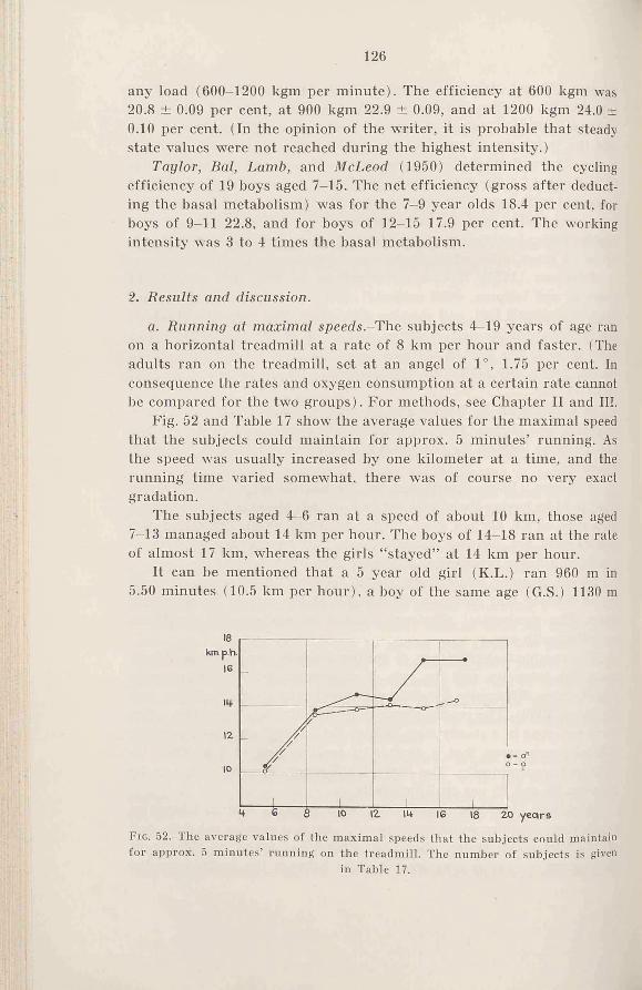
126
any load (600-1200 kgm per minute). The efficiency at 600 kgm was20.8 ± 0.09 per cent, at 900 kgm 22.9 ± 0.09, and at 1200 kgm 24.0 ± 0.10 per cent. (In the opinion of the writer, it is probable that steady state values were not reached during the highest intensity.)
Taylor, Bal, Lamb, and McLeod (1950) determined the cycling efficiency of 19 boys aged 7-15. The net efficiency (gross after deducting the basal metabolism) was for the 7-9 year olds 18.4 per cent, for boys of 9-11 22.8, and for boys of 12-15 17.9 per cent. The working intensity was 3 to 4 times the basal metabolism.
2. Results and discussion.
a. Running at maximal speeds-The subjects 4-19 years of age ran on a horizontal treadmill at a rate of 8 km per hour and faster. (The adults ran on the treadmill, set at an angel of 1°, 1.75 per cent. In consequence the rates and oxygen consumption at a certain rate cannot he compared for the two groups). For methods, see Chapter II and III.
Fig. 52 and Table 17 show the average values for the maximal speed that the subjects could maintain for approx. 5 minutes’ running. As the speed was usually increased by one kilometer at a time, and the running time varied somewhat, there tvas of course no very exact gradation.
The subjects aged 4-6 ran at a speed of about 10 km, those aged 7-13 managed about 14 km per hour. The boys of 14-18 ran at the rate of almost 17 km, whereas the girls “ stayed” at 14 km per hour.
It can be mentioned that a 5 year old girl (K.L.) ran 960 m in 5.50 minutes (10.5 km per hour), a boy of the same age (G.S.) 1130 m
tiG . 52.—The average values of the maximal speeds that the subjects could maintain for approx. 5 minutes’ running on the treadmill. The number of subjects is given
in Table 17.
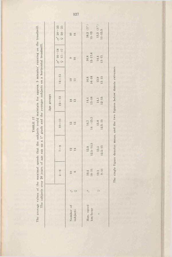
TAB
LE 1
7Th
e av
erag
e va
lues
of
the
max
imal
spe
eds
that
the
sub
ject
s co
uld
mai
ntai
n fo
r ap
prox
. 5
min
utes
’ ru
nnin
g on
the
tre
adm
ill.
The
subj
ects
ove
r 20
yea
rs o
f ag
e ra
n up
a 1
° gr
ade
and
the
youn
ger
subj
ects
on
a ho
rizo
ntal
tre
adm
ill.
127
CO CM T—< %| | v-' tO
o o o -cH CO ^co | CO I
"b o T-< r-‘
CC l'-T T COCO 1" 00 £ T* &1—1 T—1 9 10 co '| ''b Of
cc 5 -■ £«£> 1 go [
V5aou
CCco eo
14.4
3-16
14.1
2-15
1—< 1-10)-n
tOT co eo r- 2 00 2rH i-f i—( to
14
CM
tOcn tO £1t'» 1-1 tO 1-1 toCM CM
o co -+ i-<O 1
■b o ’b CM-
O ■at- t/5 a ss•3 S6 2 X >P S3
toJS £
Sox
oA
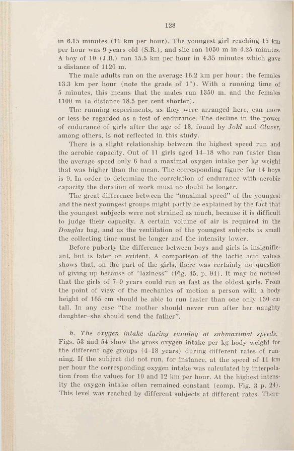
128
in 6.15 minutes (11 km per hour). The youngest girl reaching 15 km per hour was 9 years old (S.R.), and she ran 1050 m in 4.25 minutes. A boy of 10 (J.B.) ran 15.5 km per hour in 4.35 minutes which gave a distance of 1120 m.
The male adults ran on the average 16.2 km per hour; the females 13.3 km per hour (note the grade of 1°). With a running time of 5 minutes, this means that the males ran 1350 m, and the females 1100 m (a distance 18.5 per cent shorter).
The running experiments, as they were arranged here, can more or less be regarded as a test of endurance. The decline in the power of endurance of girls after the age of 13, found by Jokl and Chwer, among others, is not reflected in this study.
There is a slight relationship between the highest speed run and the aerobic capacity. Out of 11 girls aged 14-18 who ran faster than the average speed only 6 had a maximal oxygen intake per kg weight that was higher than the mean. The corresponding figure for 14 boys is 9. In order to determine the correlation of endurance with aerobic capacity the duration of work must no doubt be longer.
The great difference between the “ maximal speed” of the youngest and the next youngest groups might partly' be explained by the fact that the youngest subjects were not strained as much, because it is difficult to judge their capacity. A certain volume of air is required in the Douglas bag, and as the ventilation of the youngest subjects is small the collecting time must be longer and the intensity lower.
Before puberty the difference between boys and girls is insignificant, but is later on evident. A comparison of the lactic acid values shows that, on the part of the girls, there was certainly no question of giving up because of “ laziness” (Fig. 45, p. 94). It may be noticed that the girls of 7-9 years could run as fast as the oldest girls. From the point of view of the mechanics of motion a person with a body height of 165 cm should be able to run faster than one only 130 cm tall. In any case “ the mother should never run after her naughty daughter-she should send the father” .
b. The oxygen intake during running at submaximal speeds- Figs. 53 and 54 show the gross oxygen intake per kg body weight for the different age groups (4-18 years) during different rates of running. If the subject did not run, for instance, at the speed of 11 km per hour the corresponding oxygen intake was calculated by interpolation from the values for 10 and 12 km per hour. At the highest intensity the oxygen intake often remained constant (comp. Fig. 3 p. 24). This level was reached by different subjects at different rates. There-
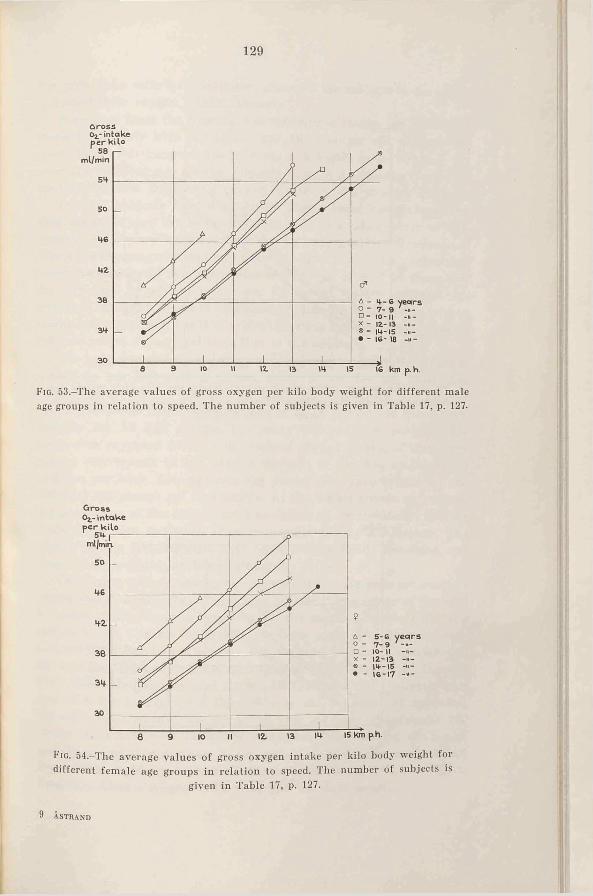
129
Gross
F ig. 53.-The average values of gross oxygen per kilo body weight for different male age groups in relation to speed. The number of subjects is given in Table 17, p. 127.
Gross 0 Z - intake
F ig . 54.-The average values of gross oxygen intake per kilo body weight for different female age groups in relation to speed. The number of subjects is
given in Table 17, p. 127.
9 Åstrand
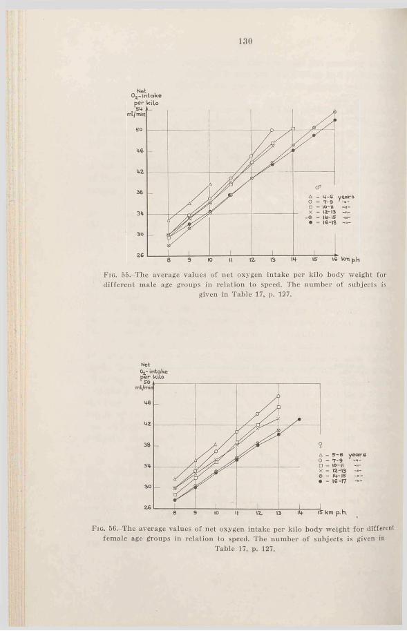
1800 ^ intake
F iq. 55.-The average values of net oxygen intake per kilo body weight for different male age groups in relation to speed. The number o f subjects is
given in Table 17, p. 127.
Net
F ig . 56,-The average values of net oxygen intake per kilo body weight for different female age groups in relation to speed. The number o f subjects is given in
Table 17, p. 127.

131
fore only those rates were included, where all the subjects in the group increased their oxygen intake “ linearly” .
It appears from the figures, that the oxygen intake per kg weight increases linearly with the speed, and the younger the subject, the greater it is. Furthermore, the increase at a higher speed is about the same for different age groups, possibly there is a tendency towards a relatively greater increase of oxygen intake for the younger subjects.
Younger subjects have a relatively higher basal metabolism, and their higher oxygen intake during work may be a consequence of this. Accordingly, the net oxygen intake per kg body weight was calculated (the gross oxygen consumption minus the basal metabolism, calculated according to Mayo Foundation Standard). Figs. 55 and 56 show' that the difference in oxygen intake per kg weight for the different age groups at a certain speed is now smaller than when the gross oxygen intake was compared, but it is evident that the running of the younger subjects is less economical than that of the adults.
The comparison between older and younger subjects is difficult, caused by the fact that a certain speed is low' for the older, but relatively high for the younger subjects (boys). However, 8-9 km per hour corresponds to an oxygen intake of 50-70 per cent of the aerobic capacity for all age groups. Excluding the youngest subjects 4-6 years, it is supposed that the determined oxygen intake, at least very nearly, corresponds to the oxygen demand of running at the speed of 8 km per hour. Consequently the values of oxygen intake for the different age groups are comparable. At the higher speeds an increasing percentage of the energy must be delivered anaerobically, and the “efficiency” will be lower than that suggested by the curves. The highest speeds that the girls 7—17 ran were practically the same, and their values are comparable at all rates.
The higher oxygen intake per kg w'eight for the younger subjects can be due to anatomical differences. Different leverage may have an influence. Furthermore a short leg usually means a short stride at a certain speed, and so the stride frequency must be higher in comparison with the taller person. The ensuing higher frequency can decrease the efficiency (comp, cycling at different pedal rates, Hansen, 1927; Grosse-Lordemann and Miiller, 1937). The stride frequency at a rate of 8 km averaged for the 7-8 year old children (12 subjects) -04 strides per minute, for the 17-18 year old boys (6 subjects) 160 strides. Whether these factors will influence the running efficiency, and if so, how much, cannot at present be settled. According to Hög- berg (1952) the oxygen intake has a minimum at a certain stride frequency when a subject is running at a constant speed.
9'
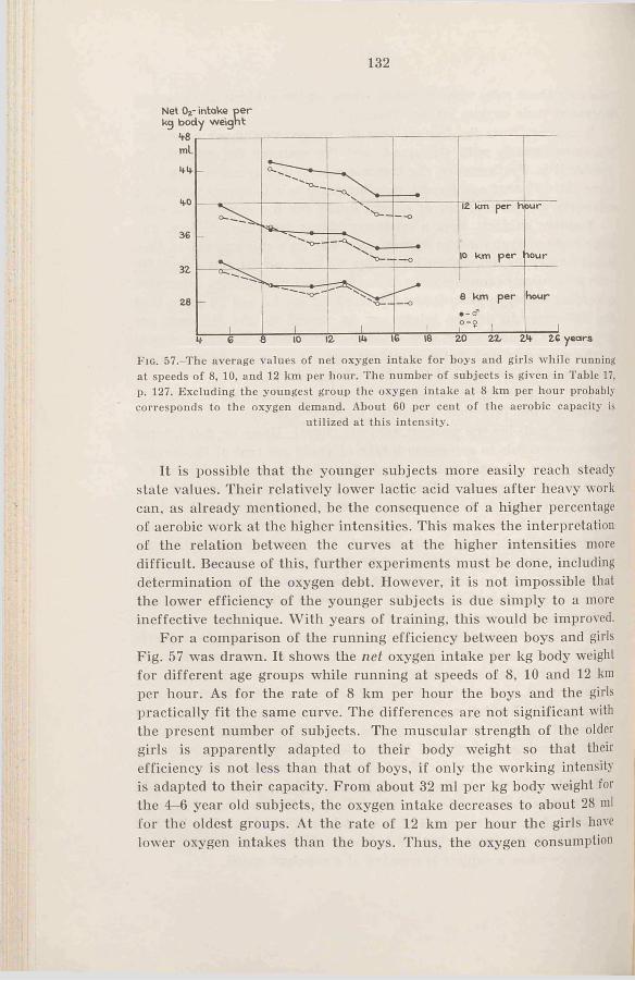
132
Net Oz- intake p e r kg body w eignt
F ig . 57.-The average values of net oxygen intake for boys and girls while running at speeds of 8, 10, and 12 km per hour. The number of subjects is given in Table 17, p. 127. Excluding the youngest group the oxygen intake at 8 km per hour probably corresponds to the oxygen demand. About 60 per cent o f the aerobic capacity is
utilized at this intensity.
It is possible that the younger subjects more easily reach steady state values. Their relatively lower lactic acid values after heavy work can, as already mentioned, be the consequence of a higher percentage of aerobic work at the higher intensities. This makes the interpretation of the relation between the curves at the higher intensities more difficult. Because of this, further experiments must be done, including determination of the oxygen debt. However, it is not impossible that the lower efficiency of the younger subjects is due simply to a more ineffective technique. With years of training, this wrould be improved.
For a comparison of the running efficiency between boys and girls Fig. 57 was drawn. It shows the net oxygen intake per kg body weight for different age groups while running at speeds of 8, 10 and 12 km per hour. As for the rate of 8 km per hour the boys and the girls practically fit the same curve. The differences are not significant with the present number of subjects. The muscular strength of the older girls is apparently adapted to their body weight so that their efficiency is not less than that of boys, if only the working intensity is adapted to their capacity. From about 32 ml per kg body weight for the 4-6 year old subjects, the oxygen intake decreases to about 28 ml for the oldest groups. At the rate of 12 km per hour the girls have lower oxygen intakes than the boys. Thus, the oxygen consumption
averages 45 ml for the boys 7-9, and 41 ml for the oldest, while the average values of the girls are 44 and 38 ml, respectively. The rate of 10 km per hour forms a transition to 8 km as far as the difference between boys and girls is concerned.
The reason for the girls’ lower oxygen intake and apparently higher efficiency certainly is that they have not covered the energy demand aerobically, at least not to the same extent as the boys. The fact that the oxygen consumption increases linearly with the rate is by no means an indication of a constant efficiency (comp. Christensen and Högberg, 1950 b). As the mechanical efficiency is lower during anaerobic work as compared with aerobic (Asmussen, 1946; Christensen and Högberg, 1950 b) the oxygen intake, including oxygen debt, of the older girls is greater than that of boys while running, for instance, 12 km per hour. Thus the figs, must be critically interpreted.
Girls 12-13 have almost the same maximal speed as the boys of the same age, but they have a lower aerobic capacity (the boys 56 ml per minute and per kg, the girls 50 ml). The running efficiency at 8 km per hour is about the same. A smaller oxygen demand than that of the boys at higher rates is not probable, e.g. 2.5 ml per minute at 12 km. At this rate the girls are closer to the upper limit of oxygen intake than the boys. The anaerobic processes must play a more important rôle, and their lactic acid values must be higher. It is also found that their maximal lactic acid concentrations are considerably higher than those of the boys, 18.5 mgm per cent. The same considerations apply to the older girls.
133
c. The mechanical efficiency during cycling at submaximal intensities- The mechanical efficiency during cycling of 21 male and 31 female subjects was determined. The determinations were made according to the formula:
Mechanical efficiency (net) —work performed X 100
(total energy) — (basal energy exchange)
Total energy was calculated from the values of oxygen intake during work. The basal metabolism was calculated according to Mayo Foundation Standard. The caloric coefficient of the oxygen was set at 4.90 Calories per litre.
The intensities were 900, 1200, and 1500 kgm per minute lor males and 600 and 900 kgm for females (50 pedal revolutions per minute). • The maximal work done ranged between 1800 and 2400 kgm per
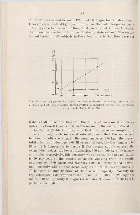
134
minute for males and between 1200 and 1650 kgm for females (comp. 1 horse power = 4500 kgm per minute). As the pedal frequency could not always be kept constant the actual work is not known. However, the intensities are too high to permit steady state values.) The reason for not including all subjects in the calculations is that they were not
F ig . 58.-Gross oxygen intake (dots) and net mechanical efficiency (squares) for 21 male and 31 female adults during cycling at different intensities. The values
are given in Table 18. p. 138.
tested at all intensities. However, the values of mechanical efficiency differ less than 0.1 per cent from the means of the entire material.
In Fig. 58 (Table 18) it appears that the oxygen consumption increases linearly with increased intensity, and that the males and females, broadly speaking, fit the same curve. At 900 kgm the oxygen intake for the males was 2.09 litres per minute, for the females 2.06 litres. It is impossible to decide if the oxygen supply covered the oxygen demand. At the lowest intensities, 600 and 900 kgm for females and males respectively, this certainly was the case (the oxygen intake is 50 per cent of the aerobic capacity’ ) . Judging from the results obtained by Christensen and Högberg (1950 b), well-trained individuals certainly’ will be able aerobically, to do work corresponding to 70 per cent or slightly more, of their aerobic capacity. Probably the true efficiency is determined at the intensities of 900 and 1200 kgm for males, 600 and possibly 900 kgm for females. The one at 1500 kgm is perhaps too high.
I
»
For the males the efficiency is very constant and varies between 23.3 and 23.7 per cent. For the females it is 22.5 per cent at 600 kgm and 23.1 per cent at 900 kgm. For 600 kgm the difference is almost significant in comparison with 900 kgm for males, and significant in comparison with 1200 kgm per minute. On the other hand the difference is hardly of practical importance. With a work-load corresponding to 50 per cent of the aerobic capacity, and at a pedalling rate of 50 per minute (Hansen, 1927; Grosse-Lordemann and Müller, 1937) optimal working conditions are prevailing. For the well-trained adult individuals the net efficiency then is about 23 per cent, independent of sex (comp. Wahlund 22.9 per cent at 900 kgm per minute for men).
135
3. Summary.
Among other things the oxygen consumption has been determined for 72 boys and 67 girls aged 4-18 during horizontal running at different speeds.
The maximal speeds that could be kept for about 5 minutes were almost the same for boys and girls up to the age of 12-13, 10 km per hour for the 4—6 year old subjects, 14 km for those aged 7-13. The older girls “ stayed” at the same maximal rate, whereas the boys reached 17 km per hour.
While running at different rates, 8 km per hour and faster, the net oxygen intake per kg body weight increased as a rule linearly up to at least 13 km per hour (except for the youngest). The oxygen intake per kg was generally greater at a certain rate, the younger the subject was. Possible reasons for this have been discussed.
The oxygen intake probably corresponds to the oxygen demand for subjects aged 7 years and over while running at the rate of 8 km per hour (the intensity corresponds to 50-70 per cent of the aerobic capacity). The running efficiency was here the same for boys and girls of the same age. At higher rates the average oxygen intake of the females 10—17 years was lower than that of the males, probably due to a lower aerobic capacity.
40 males (20-33 years) and 44 females (20-25 years) reached during running up a grade of 1°, a maximum of 16.2 and 13.3 km per hour, respectively.
The mechanical efficiency at different working intensities on a
136
bicycle ergometer has been determined for 21 male and 31 female subjects. At an intensity corresponding to about 50 per cent of the aerobic capacity (600 kgm per minute for females and 900 kgm per minute for males), and at a pedal frequency of 50 per minute the efficiency was 22.5 and 23.4 per cent, respectively. These values are considered to be close to the maximal values.
Ch apter XI
TESTS OF PHYSICAL FITNESS AND CONDITION BASED ON HEART RATE, OXYGEN
INTAKE, AND VENTILATION DURING SUBMAXIMAL WORK
1. Introduction.
The generally applied tests for physical fitness, physical condition, etc. can be separated in two main groups, one where the maximal capacity for sustained work (maximal oxygen intake) is determined and the other where, e.g., heart rate or respiratory rate are determined at a submaximal level of work. On the here presented experiments both principles can be applied and it might be of interest to compare the results obtained.
2 . Results.
The experiments were done on a bicycle ergometer at the intensities 900, 1200, and 1500 kgm per minute for the males, and 600 and 900 kgm per minute for the females. A total of 21 male (20-33 years) and 31 female subjects (20-25 years) were tested.
The results are found in Table 18.The average values of oxygen intake at the different intensities
follow an almost straight line (Fig. 58, p. 134), regardless of sex. Whether the oxygen intake at the highest intensities corresponds to the oxygen demand cannot be settled with certainty. At the lower intensities, the mechanical efficiency is the same size for males and females, about 23 per cent (see Chapter X).
During the heaviest manual labour, the working intensity can be estimated to correspond to about 50 per cent of the aerobic capacity (see Chapter XII), and so these values are of special interest. In Fig. 42 (p. 83) it is shown, that this “ 50 per cent load” is about 600 kgm per minute for a well-trained woman. The average value of the oxygen intake is 1.5 litres per minute, while the heart rate is 138. The ventilation averages 35 litres; calculated per litre oxygen intake 23 litres. This ventilation is only 39 per cent of the maximal ventilation. For males "the 50 per cent load” is about 900 kgm per minute. The average value
138
t
CU
cnOG
COcc
o4̂
toto
I-*- II
pCO
*1 1+ (0 X |so3
Q
oCOIItotn
b II
II
+ oGOG
IIO
O'to II
4-CO
Ci4=*
CO_ i 00
CO4—
G
toCO
Cn
CO
toGTO'to
>_. o' »yQ “ (I i
S I S
3“ ̂
D3 E
« 2 «
TAB
LE 18
Oxygen
intake, m
echanical efficiency,
heart rate,
anil ventilation for adults during work on the bicycle ergom
eter. The ratio of oxygen intake and ventilation to the m
aximally obtained values is calculated.
heart rate
139
F ig . 59,-The average of heart rate for 21 male and 31 female adults in relation to oxygen intake. The two curves to the right show the average heart rate in relation to the ratio of oxygen intake to aerobic capacity (per cent). Continuous line = male, dashed line = female. Related to the aerobic capacity the difference in heart rate between man and woman doing a certain work is reduced to less than 10 heats
per minute. All subjects are well-trained.
for the oxygen intake is 2.1 litres per minute; the heart rate is 128. The ventilation averages 42 litres; calculated per litre oxygen intake 20 litres. The ventilation is 34 per cent of the maximum.
For the women 900 kgm per minute corresponds to an oxygen intake of 73 per cent of the aerobic capacity. Corresponding percentage of 1200 and 1500 kgm per minute for the males are 64 and 79, respectively.
Fig. 41 (p. 83) shows how the ventilation increases linearly with the oxygen intake for males. The ventilation per litre oxygen intake is 20.1 litres when oxygen intake is 2.09 litres, and 21.1 litres when 3.33 litres. The small increase of ventilation indicates that also 1500 kgm per minute can probably be managed aerobically. The ventilation per litre oxygen intake for the females is 3-4 litres higher than that of the males when a similar percentage of the aerobic capacity is utilized.
At these moderate intensities a smaller percentage of the ventilation capacity is utilized than of the aerobic capacity (Fig. 42, p. 83). When the ventilation attains about 50 per cent of the maximal value (61 litres per minute for males, 45 litres for females), the oxygen intake for males corresponds to 70 per cent, and for females 65 per cent the aerobic capacity.
Fig. 59 shows the heart rate at different oxygen consumptions. For the males the average value increases linearly with the oxygen intake from 128 at 2.1 litres to 167 at 3.3 litres. At an oxygen intake of 2.1 litres, the females have a heart rate 40 beats higher than that of the males. This must primarily be due to their smaller stroke volume (comp. Chapter IV). In relation to the different aerobic capacity of males and females the difference in heart rate is, however, rather small. Thus, in these experiments the average heart rate of the female subjects is only 8-10 beats higher than that of the males, when the
140
comparison is done for a certain percentage of the aerobic capacity (Fig. 59, the two curves to the right).
Discussion.
At present there are divergent opinions about the heart rate as criterion of physical condition and of working capacity. One reason is probably the fact, that no clear distinction is made between the two terms, and in the opinion of the writer they denote different things: working capacity is a synthesis of aerobic and anaerobic capacity, mechanical efficiency, and physical condition; physical condition states how the circulation, respiration, muscles, etc., are fit to hard work of a long duration. The heaviness of the work must be related to the individual’s working capacity. Thus, working capacity refers more to quantity, physical condition to quality.
The graphs of heart rates in Fig. 59 might very well represent from the left a group of untrained individuals or individuals with cardiac insufficiency (the females), and a group of very well-trained individuals (the males). As a matter of fact both groups are well-trained. Well-trained men with a small aerobic capacity will fit the same line as well-trained women with a great oxygen intake capacity (comp. Fig. 10 p. 37). Certainly there is a considerable overlapping for the values of trained, untrained, and “ pathological” individuals, regardless of sex. As the resting value and maximal value of the heart rate are practically the same for individuals in different physical condition, the slope of “ the heart rate curve” will be influenced more by the aerobic capacity than by the physical conditions. This is also indicated by the wide scattering of the values for the heart rates in Fig. 10. It is evident that it is impossible to judge a person’s physical condition from the slope and mean of a curve showing the heart rate in relation to oxygen intake or working intensity, if not his aerobic or working capacity is taken into consideration. These circumstances can entirely explain why tests with submaximal, fixed working intensities give results so difficult to interprete. As an example, may be mentioned that Melheny et al. (1942) found that the oxygen intake and the ventilation were almost similar for males and females when doing a submaximal work on the treadmill, but that the heart rate and lactic acid concentration were higher for females (which very well agrees with the results of the writer’s experiments on the bicycle ergometer). As the values of the best women were comparable to those of the men with the poorest physical conditions the conclusion was drawn that the physical condition of women was poorer than that
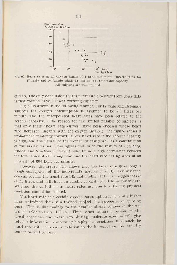
141Heart rate a t an o2- intake of 2 Ut/min
m ax. Ojr- in ta k e
Fig. 60-Heart rates at an oxygen intake of 2 litres per minut (interpolated) for 17 male and 16 female adults in relation to the aerobic capacity.
All subjects are well-trained.
of men. The only conclusion that is permissible to draw from these data is that women have a lower working capacity.
Fig. 60 is drawn in the following manner. For 17 male and 16 female subjects the oxygen consumption is assumed to be 2.0 litres per minute, and the interpolated heart rates have been related to the aerobic capacity. (The reason for the limited number of subjects is that only their “ heart rate curves” have been choosen whose heart rate increased linearly with the oxygen intake.) The figure shows a pronounced tendency towards a low heart rate if the aerobic capacity is high, and the values of the women fit fairly well as a continuation of the males’ values. This agrees well with the results of Kjellberg, Rudhe, and Sjöstrand (1949 c), who found a high correlation between the total amount of hemoglobin and the heart rate during work at an intensity of 600 kgm per minute.
However, the figure also shows that the heart rate gives only a rough conception of the individual’s aerobic capacity. For instance, one subject has the heart rate 142 and another 164 at an oxygen intake of 2,0 litres, and both have an aerobic capacity of 3.1 litres per minute. Whether the variations in heart rates are due to differing physical condition cannot be decided.
The heart rate at a certain oxygen consumption is generally higher in an untrained than in a trained subject, the aerobic capacity being equal. This is due mainly to the smaller stroke volume in the untrained (Christensen, 1931 a). Thus, when testing a person on different occasions the heart rate during moderate exercise will give valuable information concerning his physical condition. How much the heart rate will decrease in relation to the increased aerobic capacity cannot be settled here.
142
For a well-trained individual, the heart rate quickly attains a steady state after work is started, and this rate is maintained even for prolonged work. For untrained persons or persons with cardiac insufficiency the heart rate will not be stabilized, but will gradually increase (Christensen) , provided the work-load is relatively great. In order to test the physical condition or the subject’s performance with regard to the work done, the heart rate ought to be counted for, e.g., 10 minutes, and the end-value of the heart rate as well as the appearance of “ the heart rate-tim e diagram” must be regarded (Sjöstrand, 1950).
In this study a high correlation between the aerobic capacity and the body weight is found (Fig. 49 p. 106). A certain work-load per kg weight consequently will give about the same stress to the organism. The women’s work should he 15-20 per cent lower per kg weight than that of men, and the heart rate would practically attain the same level. On the other hand, take two subjects having the same aerobic capacity and training state, but one of them has 20 kg more excess fat than the other. At a certain work-load per kg weight, the heavier man will attain the highest heart rate, because he has done the heaviest work, and he has worked closer to the upper limit of his performance as compared with the thinner man.
When testing a subject’s fitness for manual labour, military service, physical performance on track and field, where the body weight has to be “ carried” , the working capacity per kg weight ought to be the basis for judgement. In general, the demand for aerobic capacity must he higher, the heavier the individual. The exception is found if he can utilize his weight in his job.
When testing a subjects’ fitness for a job in a sitting position the drawback of excess fat is not so great, and the demand for aerobic capacity may he estimated only with regard to the energy requirements of the job. The test-work can, e.g., be done on a bicycle ergo- meter and a working intensity be chosen, regardless of body weight.
As for the test, a general one is hardly possible to employ, but the man must be tested for the stress of the job, and for the factors that will limit his performance, e.g. capacity of energy output, ability to sustain heat etc. As for the metabolism, it is suggested that the oxygen intake during the job may not exceed 50 per cent of the aerobic capacity (see Chapter XII). A determination of the individual’s maximal oxygen intake is hardly possible as a method of routine, but probably the total amount of hemoglobin, and perhaps the lean body mass, will give good information about the aerobic capacity (comp. Fig. 17, p. 48). A “ control” of the calculated capacity may be obtained by giving the subject a certain amount of work, corresponding to a
143
certain percentage of the estimated aerobic capacity. In this material, a working intensity on the bicycle ergometer of 180 kgm per minute and per 100 gm hemoglobin is calculated to give an average oxygen intake corresponding to 70 per cent of the aerobic capacity (heart rate about 154 for males, 164 for females, see Table 21, p. 144). A varied intensity fits somewhat better, e.g. 200 kgm per 100 gm hemoglobin when the amount of hemoglobin is 300 g, and 170 kgm when the amount is 700 g. (It must be stressed that the absolute figures for total hemoglobin are lower in the writer’s experiments than in others). If it is a job where the body must be “ carried” , the weight of the tested individuals ought to be regarded when choosing the test-load.
To turn back to the physical condition, the central problem is whether this term shall be related to the active protoplasmic mass or to the total body weight. It goes against the grain to characterize a man weighing 150 kg as a man of good physical condition in spite of the fact that the capacity of his circulatory system may be very great in relation to the protoplasmic mass, but insufficient if related to the 150 kg that shall be carried.
The term good physical condition can apply to a man in a good state of health who has by rational training made “the best” of his physical development; his working capacity and physical fitness for heavy work may be high or low depending upon his natural endowment.
In the clinic it is this physical condition, less the working capacity, that shall be tested. It is very important to recognize this when choosing the test-method and comparing results of different age groups or of the two sexes.
It may be interesting to see the individual variations of the heart rate when the work-load is chosen according to different principles. From the point of view of physical condition, the scattering ought to be relatively small when working with this homogeneous material.
Table 19 shows the average values of the heart rate, the standard deviation, and the standard error of the mean for 17 male and 16 female subjects. Intensities, oxygen intakes, and heart rates have been determined or calculated by interpolation. If the working intensity is chosen regardless of the subject’s weight (600-1500 kgm per minute) the standard deviation varies between 11.4 and 12.4 heart heats, and is relatively independent of sex and intensity.
Table 20 shows results where the load was varied with regard to the body weights of the subjects, on one hand a certain intensity per kg body weight was chosen regardless of its size (one lighter and one heavier load); on the other hand the load per kg was varied with the
I
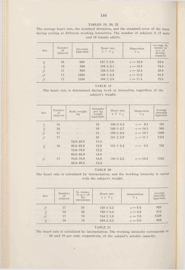
144
TABLES 19, 20, 21The average heart rate, the standard deviation, and the standard error o f the mean during cycling at different working intensities. The number of subjects is 17 male
and 16 female adults.
SexNumber
ofsubjects
Intensitykgm/min.
Heart rateT ± £ -
Dispersion a x .
Average O. -intake in
p. c. of aerobic capacity
5 16 600 137 ± 3 .0 a = 1 2 . 0 52.0$ 16 900 164 ± 3 .1 a = 1 2 . 2 72.5
d 17 900 128 ± 3 .0 a = 12.4 49.9d 17 1 2 0 0 148 ± 2 .8 3 = 1 1 . 6 63.9d 17 1500 166 ± 2 .8 3 = 11.4 79.5
TABLE 19The heart rate is determined during work at intensities regardless o f the
subject’s weight.
SexNumber
ofsubjects
Body weight" k g
Intensity per kg weight
kgm/min.
Heart rateY ± £ x
Dispersion a x
Averageintensitykgm/min.
9 16 _ 1 2 148 ± 2 .3 3 = 9.1 725
9 16 - 16 168 ± 2 .7 3 = 10.5 966d 17 - 15 139 ± 2 .6 3 = 10.7 1068d 17 - 20 161 ± 2 .9 3 = 11.7 1424
50.0-59.9 13.4
9 16 60.0-69.9 1 2 . 8 151 ± 2 .4 3 = 9.5 73670.0-79.9 1 2 . 2
60.0-69.9 16.6d 17 70.0-79.9 16.0 144 ± 2 .5 3 = 10.3 1153
80.0-89.9 15.2
TABLE 20The heart rate is calculated by interpolation, and the working intensity is varied
with the subject’s weight.
SexNumber
ofsubjects
0 2 -intake in p. c. of
themaximum
Heart rateX ± E -
DispersitionG X
Averageintensitykgm/min.
d 17 50 128 ± 2 .3 3 = 9.4 920
9 16 50 138 ± 2 .2 Q II 0° bo 612d 17 70 154 ± 1.9 3 = 7.8 1329
9 16 70 164 ± 2 .3 3 = 9 .0 908
TABLE 21The heart rate is calculated by interpolation. The working intensity corresponds to
50 and 70 per cent, respectively, o f the subject’s aerobic capacity.
145
body weight. The load was 20 per cent lower for the females than for the males of the same weight, (i.e. the difference of the maximal oxygen intake per kg body weight). The standard deviation in these experiments are somewhat smaller than in Table 19 (9.1 to 11.7).
The standard deviation is smallest when an intensity is chosen with regard to the aerobic capacity of the subject, Table 21. Working intensities corresponding to 50 and 70 per cent, respectively, of the maximal oxygen intake were “ performed” . The standard deviations vary between 7.8 and 9.4 heart beats.
The calculations involve intensities corresponding to 50-80 per cent of the aerobic capacity.
At a certain heart rate, 170 per minute (comp. Wahlund, 1948), the oxygen intake, and consequently the working intensity (and minute and stroke volume of the heart), will be individually rather different. For the males the heart rate of 170 is attained when the oxygen intakes average 82 per cent of the aerobic capacity (a = 6.3); for the females it averages 75 per cent (a = 7.6). The order of the “methods” as far as “precision” is concerned agrees fairly well with the preceeding arguments. However, the individual variations are considerable, regardless of working intensity.
A more comprehensive discussion of “physical fitness tests” is not justifiable here. The reader is referred to reviews given by Simonson ( 1944); Taylor (1945); Lundgren (1946); Karpovich (1947); Cureton (1947).
It shall be emphasized, however, that in testing the circulatory- respiratory fitness a type of work must be chosen which engages great groups of muscles, and that the working intensity must be relatively high. The duration of the work must be long enough to permit the adjustment of circulation and ventilation to the exercise, and the determinations have to be done particularly during the later phases of adaptation. Otherwise it will be quite different functions that are tested. Naturally, it is of interest to determine the anaerobic capacity, but it is extremely difficult, as regards methods, to get consistent results. As an example of results, difficult to interpret in the opinion of the writer, may be mentioned Cureton’s (1951) all-out treadmill tests of maximal physical efficiency, applied inter alia to a great number of champion athletes. Often the running time was only 1-2 minutes, 5 minutes at the most. Accordingly the duration was usually too short to permit the adjustment of respiration and circulation to the demands of the work. During the entire period of running the oxygen intake was determined, and its size was influenced by oxygen consumption during rest (the first seconds), by submaximal values (the
tO ASTRAND
146
time of adaptation), and possibly by maximal values. Thus, the determined oxygen intake gives no information about the one or the other. As running time varied, a comparison between the subjects will also be misleading. The values Cureton found for the gross oxygen intake of champion athletes are without exception lower than the average values of the male subjects in the writer’s study, and often they are lower than those of the female subjects with the lowest aerobic capacity. Moreover, Cureton determined the oxygen debt, and because of the methodical difficulties it is not astonishing that the gross total oxygen consumption (oxygen intake during running + oxygen debt) showed a low correlation with the subjects’ performances on the track and field.
i . Summary.
“Normal values“ of heart rate, oxygen intake, and ventilation were given for 21 male (20-33 years) and 31 female subjects (20-25 years) during cycling at different intensities. Oxygen intake and ventilation were related to the maximally obtained values.
In tests of physical condition by examinations of the circulatory and respiratory system during work the individuals aerobic capacity must be regarded. It is supposed that this capacity can be calculated from the total amount of hemoglobin or the lean body mass. In this way it is possible to choose a test-load fitting the subjects working capacity.
The difficulties to obtain consistent results were discussed.
Ch apter XII
GENERAL DISCUSSION
A decisive analysis of an individual’s working capacity is impossible. Different factors will, in varying degrees, influence the performances of different individuals, and for one and the same person the working capacity varies from day to day. The psychological state is probably most unstable. The environment of the individual, his state of mind, and his attitude to the job might highly influence his performance. From a physiological point of view working capacity includes the individual’s capacity of energy output and his mechanical efficiency. During heavy work of prolonged duration the aerobic capacity is, first of all, probably the limiting factor {Dill, 1933), during short spells of hard work the anaerobic capacity is decisive.
In this chapter the working capacity from the viewpoint of manual labour (1), and school and junior sports (2) will be discussed.
1. Manual labour.
Brody (1945) concludes the following: “The maximum long-range or life-time efficiency, however, involves liberal rest periods and moderate work rate. One of the problems of “ scientific management” or “work rationalization” , whether it be of man, cow, or horse, is to determine for animals of given productive capacity the work level foi- maximal long-range efficiency.
Machines are not usually run at more than 50 per cent of their capacity, and a similar safety margin should perhaps be allowed to man and animals so as to avoid injury or untimely death. The problem in work rationalization is to determine the maximum rate of work that can be carried on day in and day out and still retain vigour to an advanced age” .
Dill (1936) suggests as “arbitrary but useful lines of demarcation for hard work” a work metabolism between 3 and 8 times the rest metabolism.
Lundgren (1946) and Zotterman and Lundgren (1948) investigated the energy output during lumber work, one of the heaviest of manuel
10*
148
works. They found that most single work operations demanded a mean of about 9 to 11 Calories per minute.
Physiological investigations in hot steel mills carried out by this laboratory1 showed that the oxygen intake during many work operations was about 2 litres per minute. The blood lactate concentration in some cases attained values significantly above the resting level, indicating that the level of activity approached the capacity of the workman (comp. p. 134). Tests of physical condition in the beginning and at the end of the 8 hours’ work period gave in a few cases indications of overstrain of the circulatory system. (Significantly higher heart rates at 900 kgm per minute at the end of the working day).
With regard to their physical working capacity and condition these workers certainly can be compared with the male students in this experiment. This assumption is supported by the following data. The workers in the steel mill had a total amount of hemoglobin that averaged 0.98 per cent of the body weight, as compared with 1.00 per cent for the students. When cycling at 900 kgm per minute the average value of the heart rate was for the workers 127, for the students 128. The students were trained by sport and athletics, the workers by hard work. The maximal oxygen intake averaged 4.1 litres per minute for the students. Evidently, the workmen must often utilize up to 50 per cent of their aerobic capacity while on the job, but they avoid exceeding this percentage. This “ safety margin” lies on the level suggested by Brody. The condition precedent for so high an “ optimal” working intensity is that the work involves large groups of muscles (comp, p. 121). The “ 50 per cent load” must be related to the capacity of the involved muscles and not to that of the body as a whole. When increasing the metabolism over this 50 per cent, the feeling of discomfort and strain will probably be marked.
As the energy output approaches the capacity of the workman, he becomes more sensitive to changes in environmental conditions. An environment unfavourable to heat dissipation increases the stress on the circulatory system. An accelerated heart rate and an elevated body temperature will be observed, and a feeling of heat oppression and discomfort will be the consequence for the workman. If the water lost in the formation of sweat is not replaced the working capacity will be further reduced (Adolph, 1947; v. Dobeln, 1951). In unfavourable working positions the stress on the circulation can be higher than that indicated by the metabolism, e.g. due to the effect of gravity (Asmussen and Christensen, 1939 \ Asmussen, Christensen, and Nielsen,
1 “ Arbetsfysiologiska undersokningar i Nykroppa” , unpublished.
149
1939 a). A static work operation is relatively more of a strain than a dynamic one.
Due to similar adverse working conditions the “ safety margin” of the work level ought to involve a smaller percentage of the aerobic capacity. A maximum for heart rate, ventilation (at high altitudes), hodv temperature, have to be chosen. Applying the values for heart rate and ventilation obtained in this study, when the oxygen intake is 50 per cent of the maximum, the upper level for heart rate should be 130, and for ventilation 41 litres per minute (Tables 18 and 20. p. 138. 144).
A problem more and more pressing, especially in wartime, is how to employ women in industry. “ The first problem involved is whether they are physically able to perform industrial work as well and as efficiently as men, and, if not, what their limitations are and how these can be met, so that this large labour group can be utilized to the best advantage” (Baetjer, 1946). The discussion above concerning males probably applies to females (except during pregnancy). There are no proved reasons why 50 per cent of their capacity could not be utilized in manual work. As for the aerobic capacity, it is in this study, 25-30 per cent lower for females as compared with males. Consequently the heaviest work “permitted” for women should be a corresponding percentage lower. Her mechanical efficiency is of the same order as the man’s (in cycling and running), provided the work done is related to her working capacity, judged from the concentration of blood lactate. She can push herself to the same state of exhaustion as the man. For the G.C.I. students the “ 50 per cent load” corresponds to an oxygen intake of 1.5 litres per minute, a heart rate of 135, and a ventilation of 34 litres per minute.
The physical performances by women, for instance in agriculture, demonstrate fitness even for a hard job. It is unfortunate that the problem of “ utilization of womanpower by industry” is so tempered by traditions and prejudices. The utilization of women for hard physical work is probably more a psychological and social problem than a physiological one.
Among unfavourable working conditions, it must be taken into account that women usually have household duties and responsibilities which require many hours of work in addition to those spent in industry, but this could be compensated by part-time employment (Baetjer).
In order to select the right man or woman for the job demanding high working capacity the test method suggested in Chapter XI (p. 142) will probably give valuable information. The expenditure ol energy
150
necessary for doing the job can, in most cases, be determined by the Douglas-bag method, by counting the heart rate, or by measuring the body temperature (Lundgren, 1946; Berggren and Christensen, 1950).
As for the figures given above, they apply to well-trained individuals up to approx. 30 years. It is very important to investigate the psysical working capacity of older individuals, especially as the average age of the population capable of doing hard manuel labour is relatively high and tends to increase.
2. School and junior sports.
The writer’s experiments, partly confirming others, indicate that the aerobic capacity in relation to body weight is about the same for boys within all the age groups in school (from 7 years on). On the other hand the resting metabolism is relatively higher in younger boys, and so their net aerobic capacity is lower. At least in running their efficiency is lower, and the aerobic capacity will cover a relatively smaller intensity range. In these respects girls are comparable with boys of the same age, up to the beginning of puberty, but afterwards the aerobic capacity is 25-30 per cent lower than that of the males, in relation to body weight it is 15-20 per cent lower (comp. Figs. 50 and 51, p. 108). The mechanical efficiency during cycling and running is for both sexes the same, provided the intensity is adapted to the working capacity.
If the maximal blood lactate concentration attainable during exercise is regarded as a measure of the anaerobic capacity, then this is similar for both sexes (Fig. 45, p. 94). The lower maximum in younger subjects is not decidedly due to an “ insufficient” anaerobic capacity. For the ages before puberty there are some results that can be interpreted as “ contra-indications” against prolonged hard muscular exercise (relatively small blood volume and stroke volume, comp. p. 56). Further experiments are necessary.
A prediction of an individual’s working capacity from the values of maximal oxygen intake and lactic acid is not possible. A boy of 9 years may have the same maximum in relation to weight as one of 18, but he cannot run nearly as fast. The principal causes are factors of pure body mechanics. Nor is there, in the writer’s experiments, within an age group any definite relationship between the maximal running rate and the mentioned maximal values. In several cases the net energy output at the lower rates has been the same for two subjects with the same aerobic capacity, but the one has in the maximal experiment run at a rate, perhaps 2 km per hour higher.
151
On the other hand, it can be stated that an aerobic capacity equal to or higher than the average values was usually found in those subjects, within the group, who reached the highest intensities. A great oxygen intake capacity is certainly necessary for a runner in order for him to make good records in middle and long distance races. As an example it may be mentioned that the Olympic 1500 m champion in London 1948, H. Eriksson, -while running on a treadmill has reached an oxygen intake of 68.4 ml per minute and per kg body weight (5.24 litres per minute gross). The 4th man in the same race, G. Bergquist, has attained 73.7 ml (4.81 litres gross) (Högberg and Åstrand, results not published). These figures should be compared with the average values of 58.6 ml for G.C.I. students; highest individual value 67.4 ml (comp. Chapter IX).
A very important problem in physical education is what types of sport and competition should be practised by youths at different ages. The debate principally concerns the endurance events. As the aerobic capacity is relatively as great for younger children as for male adults, the children should also be capable of doing prolonged, strenuous, exercise. (A possible difference in supply of fuel may influence the working capacity. Robinson (1938) found in children, “ a lower R.Q. during moderate work which may reflect a less adequate carbohydrate supply when exercise is undertaken” ). However, prolonged stress is unfamiliar to children, but it is very difficult to judge whether then- aversion is of a psychological or physiological character. The children spontaneously choose varied exercises, they readily strain themselves maximally for some seconds but heartily dislike monotonous, heavy work. Their way of living is rather of an “ anaerobic” character. It must be stressed that the organism by rational and careful training must be adapted to the demands of strenuous work in order to avoid overstrain. A great aerobic capacity, due to natural endowment, is certainly one condition precedent, but nevertheless the involved organs must be trained. To reach the highest standard the middle or long distance runner trains for at least 6-8 months; such a training plan is impossible for children to undergo, it does not fit their mentality. Thus, the motive for the choice of events in school and junior sports is first of all a psychological and pedagogical one, not a physiological and medical.
Frequently young people, say of 16-17 years of age, turn up élite performances in athletics. In the writer’s experiments some gills and boys aged 15 have attained values of oxygen intake, ventilation etc. of the same size as the highest individual values for adults (means the absolute values). From a physiological point of view they have,
152
no doubt, “primarily” a physical working capacity comparable with the adults’ . If they prepare their competition season carefully, and have judicious coaching, there are probably no physiological reasons, why they should not compete in endurance events. However, it is dangerous to use the “ exceptions” as a proof that the events in question are well suited to their years. The consequence can be an overstrain of the competitor, and cases are described where young people in connection with some competition have developed, for example, cardiac insuffiency.
To give young people a certain endurance training, events should be chosen where stress and recovery alternate repeatedly, e.g. hall games, skiing and cross-country running (“ orientering” ) in varying, hilly, terrain. Durations and distances must be chosen above all, with regard to the training state of those taking part. During puberty there is probably no reason to be particularly cautious with physical exercises. In these experiments the results obtained for both hoys and girls during puberty fit well in between those for the younger and older age groups.
From a physiological point of view females can participate in the same sports as males. Before puberty, girls have the same physical performances as boys in some events, in others they are definitely inferior. This can very well depend on their mentality which “prefers playing with dolls to smashing window-panes” , thus, the difference can be explained as an effect of training.
As for women, it must be kept in mind that the results they can obtain must be of a different size as compared with those of men. They have not the same capacity for prolonged, strenuous muscular work, but this is no reason why, for instance, forms of athletics requiring endurance should be harmful to them. They have not the same emulation as the men, but if it is a pleasure for them, and if they have trained carefully, they may very well also compete over longer distances. However, they must put up with being rather critically regarded: an exhausted woman straining herself towards the finishing tape is a sight more repugnant than a man in the same situation.
The participation in muscular exercise during menstruation or pregnancy is a special problem which, how'ever, cannot be discussed here. (None experiments were done during the menstrual period.)
As for the grouping of children according to “physical age” (comp. Schiotz, 1927) in competitions the writer’s experimental results cannot afford reasons pro or contra. There is no tendency', however, to a higher oxygen intake capacity per kg body weight for the heavier children as compared with lighter children of the same age.

153
As emphasized above there is a gradual increase in the average age of the population. This brings on social problems on labour market, and suggests physiological investigations with older subjects. It is the writer’s sincere wish that this book may be only one in a series of reports on the physical working capacity in relation to sex and age.
REFERENCES
Adolph, E. F. (1947). Physiology o f Man in the desert. New York: Interscience Publishers, INC.
Asmussen, E. (1946). Aerobic recovery after anaerobiosis in rest and work. Acta physiol, scand. 11, 197.
Asmussen, E. & 0. Boje (1945). Body temperature and capacity for work. Acta physiol, scand. 10, 1.
Asmussen, E. & E. H. Christensen (1939). Einfluss der Blutverteilung auf den Kreislauf bei körperlicher Arbeit. Skand. Arch. Physiol. 82, 185.
Asmussen, E., E. H. Christensen & M. Nielsen (1939 a) : Pulsfrequenz und Körperstellung. Skand. Arch. Physiol. 81, 190.
Asmussen, E., E. H. Christensen & M. Nielsen (1939 b ). Die Ch-Aufnahme der ruhenden und der arbeitenden Skelettmuskeln. Skand. Arch. Physiol. 82, 212.
Asmussen, E., E. H. Christensen & T. Sjöstrand (1939). Über die Abhängigkeit der Lungenvolumen von der Blutverteilung. Skand. Arch. Physiol. 82, 314.
Asmussen, E., W. v. Döbeln & M. Nielsen (1948). Blood lactate and oxygen debt after exhaustive work at different oxygen tensions. Acta physiol, scand. 15, 57.
Asmussen, E. & M. Nielsen (1950). The effect of auto-transfusion of “ work-blood” on the pulmonary ventilation. Acta physiol, scand. 20, 79.
Asmussen, E., M. Nielsen & G. Wieth-Pedersen (1943). On the regulation of circulation during muscular work. Acfa physiol, scand. 6, 353.
Ästrand, P.-O. (1951). The maximal working capacity for the two sexes and for different age groups from 4 to 30 years. Acta physiol, scand. 25. Suppl. 89, 3.
Baetjer, A. (1946). Women in industry, p. 247. Philadelphia and London: W. B. Saunders Company.
Bang, 0. (1936). The lactate content of the blood during and after muscular exercise in man. Skand. Arch. Physiol. 74. Suppl. 10, 51.
Barker, S. B. & W. H. Summerson (1941). The colorimetric determination of lactic acid in biological material. J. Biol. Chem. 138, 535.
Benedict, F. G. & E. P. Cathart (1913). Muscular Work. Carnegie Inst, of Washington, Publication No. 187.
Berggren, G. & E. H. Christensen (1950). Heart rate and body temperature as indices o f metabolic rate during work. Arbeitsphysiologie. I t , 255.
Birath, G. (1944). Lung volume and ventilation efficiency. Acta med. scand. Suppl- 256.
Boas, E. P. (1931). The heart rate of boys during and after exhausting exercise. J. clin. Invest. 10, 145.
Bohr, Chr. (1907). Die funktionellen Änderungen in der Mittellage und Vitalkapazität der Lungen. Dtsch. Arch. klin. Med. 88, 385.
Boje, O. (1933). Über die Grösse der Lungendiffusen des Menschen während Ruhe und körperlicher Arbeit. Arbeitsphysiologie 7, 157.
155
Brines, J. K., J. G. Gibson, Jr. & P. Kunkel (1941). The blood volume in noi-mal infants and children. .7. Pediat. 18, 447.
Brody, S. (1945). Bioenergetics and Growth, pp. 906, 914. New York: Reinhold Publishing Corporation.
Broman, B., G. Dahiberg & A. Lichtenstein (1942). Height and weight during growth. Acta Paediatrica. XXX, 1.
Brouha, L. & C. W. Heath. (1943). Resting-pulse and blood-pressure values in relation to physical fitness in young men. New Engl. J. Medicine. 228, 473.
Christensen, E. H. (1931 a). Die Pulsfrequenz während und unmittelbar nach schwerer körperlicher Arbeit. Arbeitsphysiologie. 4, 453.
- (1931 b ). Minutenvolumen und Schlagvolumen des Herzens während schwerer Körperlicher Arbeit. Arbeitsphysiologie, 4, 470.
- (1932). Der Stoffwechsel und die respiratorischen Funktionen bei schwerer Körperlicher Arbeit. Arbeitsphysiologie. 5, 463.
Christensen, E. H. & W. H. Forbes (1937). Kreislauf in grossen Höhen. Skand. Arch. Physiol. 76, 75.
Christensen, E. H. & 0. Hansen (1939 a). Zur Methodik der Respiratorischen Quotient-Bestimmungen in Ruhe und bei Arbeit. Skand. Arch. Physiol. 81, 137.
Christensen, E. H. & O. Hansen (1939 b). Untersuchungen über die Verbrennungsvorgänge hei langdauernder, schwerer Muskelarbeit. Skand. Arch. Physiol. 81, 152.
Christensen, E. H. & O. Hansen (1939 c). Arbeitsfähigkeit und Ernährung. Skand. Arch. Physiol. 81, 160.
Christensen, E. H. & 0. Hansen (1939 d). Hypoglykämie, Arbeitsfähigkeit und Ermüdung. Skand. Arch. Physiol. 81, 172.
Christensen, E. H. & P. Högberg (1950 a). The efficiency of anaerobical work. Ar- beitsphysiologie. 74, 249.
Christensen, E. -H. & P. Högberg (1950b). Steady-state, CL-deficit and 0»-debt at severe work. Arbeitsphysiologie. 74, 251.
Christensen, E. H. & P. Högberg (1950c). Physiology of skiing. Arbeitsphysiologie. 74, 292.
Christensen, E. H., A. Krogh & J. Lindhard (1934). Investigations on heavy muscular work. Quarterly Bull, o f the Health Org. of the League of Rations. 3, 388.
Collet, M. E. & G. Liljestrand (1924). The minute volume of the heart in men during some different types of exercise. Skand. Arch. Physiol. 45, 29.
Cotton, F. S. & D. B. Dill (1935). On the relation between the heart rate during exercise and that of immediate post-exercise period. Am. J. Physiol. I l l , 554.
Crescitelli, F. & C. L. Taylor (1944). The lactate response to exercise and its relationship to physical fitness. Amer. J. Physiol. 747, 630.
Cullumbine, H. (1949). Hemoglobin concentration and physical fitness. J. appl. Physiol. 2, 274.
Cullumbine, H., S. W. Bibile, T. W. Wikramanayake & R. S. Watson (1950). Influence of age, sex, physique and muscular development on physical fitness. J. appl. Physiol. 2, 488.
Cureton, T. K. (1936). Analysis of vital capacity as a test of condition for high school boys. The Research Quarterly. VII, 80.
- (1947). Physical fitness appraisal and guidance. St. Louis: Ihe C. V. Mosb> Company.
156
Cureton, T. K. (1951). Physical fitness of champion athletes. Urbana: The University o f Illinois press.
Dahlström, H. (1950). Basal metabolism and extracellular fluid. Acta physiol, scand. 21. Suppl. 71.
Davis, R. A. & 0. Fraenkel (1940): J. exp. Biol. 17, 402. (Cited by Krogh & Weis- Fogh, 1951.)
Dill. D. B. (1933). The Nature of Fatigue. Personnel 9, 113.(1936). The economy of muscular exercise. Physiol. Ben. Id, 263.
Dili, D. B. & L. Brouha (1937). Étude sur le rythme cardiaüue pendant l'exercice.Ses rapports avec Tage et l ’entrainement. Travail Humain. V, 1.
Döbeln. W. von (1951). Dehydration and physical performance of human subjects. Acta physiol, scand. 25. Suppl. 89, 16.
Edwards, D. (1950). Differences in the distribution o f subcutaneous fat with sex and maturity. Clinical Science. 3, 10.
Edwards, H. T. (1938). A simplified estimation of lactate in normal human blood. J. biol. Chem. 125, 571.
Edwards, H. T., L. Brouha & R. E. Johnson (1940). Effets de l’entrainement sur le traux de l’acide lactique sanguin au cours du travail musculaire. Travail Humain. 8, 1. (Cited by Johnson & Brouha, 1942.)
Enghoff, H. (1930). Eine Modifikation des Lovén-Ventiles. Skand. Arch. Physiol. 58, 1.
Espenschade, A. (1940). Monogr. Soc. Res. Child Develop. 1, 5. (Cited by Cullumbine et al. 1950.)
Fisher, R. A. (1946). Statistical methods for research workers. 10th ed. Edinburgh: Oliver and Boyd.
Fletcher, W. M. & F. G. Hopkins (1906-07). Lactic acid in amphibian muscle. J. Physiol. 35, 247.
Friedemann, T. E., M. Cotonio & P. A. Shaffer (1927). The determination of lactic acid. J. biol. Chem. 73, 335.
Galle, P. (1926). The oxygen consumption per litre of blood in children. Skand. Arch. Pysiol. XLVI1, 174.
Gordon, B., S. A. Levine & A. Wilmaers (1924). Observations on a group of marathon runners. Arch. Int. Med. 33, 425.
Grosse-Lordemann, H. & E. A. Miiller (1937). Der Einfluss der Leistung und der Arbeitsgeschwindigkeit auf das Arbeitsmaximum und den Wirkungsgrad beim Radfahren. Arbeitsphysiologie. 9, 454.
Hansen, E. (1927). Skand. Arch. Physiol. 51, 1.Hasselbalch, K. A. (1908). Über die Totalkapazität der Lungen. Dtsch. Arch. klin.
Med. 93, 64.Henderson, Y. & A. L. Prince (1914). The oxygen pulse and the systolic discharge.
Amer. J. Physiol. 35, 106.Henry, F. M. & J. de Moor (1950). Metabolic efficiency of exercise in relation to
work load at constant speed. J. appl. Physiol. 2, 481.Hill, A. V., C. N. H. Long & H. Lupton (1925). Muscular exercise, lactic acid, and the
supply and utilization of oxygen. Proc. Roy. Soc., London, B. 97, 84, 155. Högberg, P. (1952). How do stride length and stride frequency influence the energy-
output during running. Arbeitsphysiologie (in the Press).Högberg, P. & O. Ljunggren (1947). Uppvärmningens inverkan på löpprestationerna.
Svensk idrott. 40.
157
Johansen, O. (1951). Barnas utvikling gjennom idrettsleik, p. 37. Oslo: Kirke- og undervisningsdepartementet Statens ungdoms- og idrettskontor.
Johnson, R. E. & L. Brouha (1942). Pulse rate, blood lactate, and duration of effort in relation to ability to perform strenuous exercise. Rev. canad. Biol. 1, 171.
Jokl, E. (1946). Menarche, growth and physical efficiency. Nature. 157, 195.Jokl, E. & E. H. Cluver (1941). Physical fitness. J. Amer. med. Ass. 116, 2383.Jongbloed, J. & C. A. G. Wiersma (1935). Z. vergl. Physiol. 21, 519. (Cited by Krogh
& Weis-Fogh, 1951.)Karpovich, P. V. (1947). Exercise. Annu. Rev. Physiol. 9, 149.Kjellberg, S. R., U. Rudhe & T. Sjöstrand (1949 a). The amount of hemoglobin and
the blood volume in relation to the pulse rate and cardiac volume during rest. Acta physiol, scand. 19, 136.
Kjellberg, S. R., U. Rudhe & T. Sjöstrand (1949 b). Increase of the amount of hemoglobin and blood volume in connection with physical training. Acta physiol, scand. 19, 146.
Kjellberg, S. R., U. Rudhe & T. Sjöstrand (1949 c). The amount of hemoglobin (blood volume) in relation to the pulse rate and heart volume during work. Acta physiol, scand. 19, 152.
Knehr, C. A., D. B. Dill & W. Neufeld (1942). Training and its effects on man at rest and work. Amer. J. Physiol. 136, 148.
Krogh, M. (1915). The diffusion of gases through the lungs of man. J. Physiol. 49, 271.
Krogh, A. & J. Lindhard (1912). Measurements of the blood flow through the lungs of man. Skand. Arch. Physiol. 27, 100.
Krogh, A. & T. Weis-Fogh (1951). The respiratory exchange of the desert locust (schistocerca Gregaria) before, during and after flight. J. exp. Biol. 28, 342.
Lichtenstein, A. (1947). Den fysiska utvecklingen under uppväxtåren och särskilt under skolåldern. In Skolhygien, p. 63. Stockholm: Svenska bokförlaget.
Liljenthal, J. L., Jr., R. L. Riley, D. D. Proemmel & R. E. Franke (1946). An experimental analysis in man of the oxygen pressure gradient from alveolar air to arterial blood during rest and exercise at sealevel and at altitude. Amer. J. Physiol. /47, 199.
Lindhard, J. (1925). Funktionsuntersuchungen an den Lungen des Menschen mittels gasanalytischer Methoden. Handb. d. biol. Arbeitsmeth., Abderhalden. Abt. 5, Teil 4, 2: 1581.(1926). Über den Einfluss einiger gymnastischer Stellungen auf den Brustkasten. Skand. Arch. Physiol. 47, 188.
Lindholm, A. (1951). The total quantity of hemoglobin for the two sexes and for different age groups from 8 to 30 years. Acta physiol, scand. 25. Suppl. 89, 54.
Lundgren, N. (1946). The physiological effects of time schedule work on lumber- workers. A da physiol, scand. 13. Suppl. 41.
Lundin, G. & G. Ström (1947). The concentration of blood lactic acid in man during muscular work in relation to the partial pressure of oxygen of the inspired air. Acta physiol, scand. 13, 253.
Margaria, R., H. T. Edwards & D. B. Dill (1933). The possible mechanism of contracting and paying the oxygen debt and the role of lactic acid in muscular contracting. Amer. J. Physiol. 106, 689.
Metheny, E. L., L. Brouha, R. E. Johnson & W. H. Forbes (1942). Some physiologic responses of women and men to moderate and strenuous exercise: a comparative study. Amer. J. Physiol. 137, 318.
158
Morehouse, L. & A. Miller (1948). Physiology of Exercise, p. 142. St. Louis: The C. V. Mosby Company.
Morse, M., D. Cassels & F. Schlutz (1947). Blood volumes of normal children. Amer. J. Physiol. 151, 448.
Morse, M., F. Schlutz & D. Cassels (1949). Relation of age to physiological responses of the older boy (10-17 years) to exercise. J. appl. Physiol. 1, 683.
Müller, E. A. (1942). Die Pulszahl als Kennzeichen für Stoffaustausch und Ermüdbarkeit des arbeitenden Muskels. Arbeitsphysiologie. 12, 92.
Nielsen, H. (1934). The circulation in anaemic conditions. Acta med. scand. LXXX1, 571.
Nielsen, M. (1936). Die Respirationsarbeit bei Körperruhe und bei Muskelarbeit. Skand. Archiv. Physiol. 74, 299.
Nielsen, M. & 0. Hansen (1937). Maximale körperliche Arbeit bei Atmung 0:- reicher Luft. Skand. Archiv. Physiol. 76, 37.
0rskov, S. L. (1930). Fortgesetzte Untersuchungen über ätherlösliche Säuren im Blut. Angabe einer Methode zur Bestimmung der Milchsäure in 1.5 ccm Blut. Eine kritische Untersuchung der verschiedenen Fällungsmethoden. Biochem. Z. 219, 409.
Owles, W. H. (1930). Alterations in the lactic acid content of the blood as a result of light exercise and associated changes in the CCL-combining power of the blood and in the alveolar COi-pressure. J. Physiol. 69, 214.
Petren, T., T. Sjöstrand & B. Sylven (1936). Der Einfluss der Trainings auf die Häufigkeit der Capillaren in Herz- und Skeletmuskulatur. Arbeitsphysiologie. 9, 376.
Pitts, J. C. (1951). Federation Proc. 40, 105. (Behnke, personal communication.) Porat, B. von (1951). Blood volume determinations with the Evans blue dye method.
Acta med. scand. HO. Suppl. 256.Robinson, S. (1938). Experimental studies o f physical fitness in relation to age.
Arbeitsphysiologie. 10, 251.Robinson, S., H. T. Edwards & D. B. Dill (1937). New records in human power.
Science. 85, 409.Robinson, S. & P. M. Harmon (1941 a). The lactic acid mechanism and certain pro
perties o f the blood in relation to training. Amer. J. Physiol. 132, 757. Robinson, S. & P. M. Harmon (1941 b ). The effect of training and gelatin upon
certain factors which lim it muscular work. Amer. J. Physiol. 133, 161. Ryffel, J. H. (1909-10). Experiments on lactic acid formation in man. J. Physiol.
39, XXIX.Schaeffer, J. P. (1944). Morris’ Human Anatomy. 10th ed. pp. 37, 49. Philadelphia:
The Blakiston Companj\Schiotz, €. (1927). Skolealderen, p. 270. Oslo: J. \V. Cappelens Forlag.Schneider, E. & P. Karpovich (1948). Physiology of muscular activity. 3rd ed.
Philadelphia & London: W. B. Saunders Company.Simonson, E. (1944). Industrial Physiology. Annu. Rev. Physiol. 6, 543.Sjöstrand, T. (1948). A method for the determination o f the total hemoglobin
content o f the body. Acta physiol, scand. 16, 211.- (1949 a). The total quantity of hemoglobin in man and its relation to age,
sex, bodyweight and height. Acta physiol, scand. 18, 324.- (1949 b ). Determination of the total amount o f hemoglobin in anemia and
polycythemia. The Scand. J. Clin. & Lab. Investigation. 1, 215.- (1950). Bcstämning av den fysiska arbetsförmägan säsom klinisk under-
sökningsmetod. Svenska Läkartidningen. 7, 349.
159
Taylor, C. L. (1944). Some properties of maximal and submaximal exercise with reference to physiological variation and the measurement of exercise tolerance. Amer. J. Physiol, lb2, 200.
- (1945). Exercise. Anna. Rev. Physiol. 7, 599.Taylor, C. M., M. Bal, M. Lamb & G. Mac Leod (1950). Mechanical efficiency in
cycling o f boys seven to fifteen years o f age. J. appl. Physiol. 2, 563. Wahlund, H. (1948). Determination of physical working capacity. Acta mecl. scand.
Suppl. 215.Whright, G. W. (1950). Maximum oxygen consumption for work periods o f six
minutes’ duration in normal and pathological subjects. Science. 112, 423. Zotterman, Y. & N. Lundgren (1948). Studier över tungt kroppsarbete. Stockholm:
Förlags AB Affärsekonomi.

APPENDIX
The tables give the individual values obtained for the subjects in
running or cycling, and some other data.
It may be called attention to the fact that the maximal tidal air
and respiratory rate have not always been attained in the same ex
periments. The body weight varied somewhat during the experimental
period, and consequently the value stated for maximal oxygen intake
and for maximal oxygen intake per kg weight does not always fit
the figure given for body weight.
11 ASTRAND
1. Wale.
Subject ageyears
bodyheight
cmbody
weightkg
vitalcapacitylitre
(BTPS)
residualcapacitylitre
(BTPS)heartrate
lactic acid m gm p.c.
B.A. 4 107 18.9 207B.P. 4 107.5 17.9 207 _H-U.B. 4 111.5 20.6 188 76A.K. 4 118 24.5 200 72L-O.S. 5 108.5 16.0 200 33M.O. 5 108 17.9 200 65G.S. 5 117 20.7 214 _K.K. 6 — 23.0 207 54K.R. 6 116 20.6 207 38U.E. 6 128 27.8 203 _T.H. 7 125 25.3 1.84 205 70C.N. 7 126 25.1 2.01 200 60u .c . 8 131 29.2 1.84 220 99U.E. 8 138.5 31.4 2.33 220 89H.O. 8 138.5 29.6 2.15 214 64L-A.J, 9 129 31.0 2.11 209 _S.H-fi. 9 138 30.5 2.38 202 110S.H. 9 143 31.6 2.51 209 107U.G. 9 140 31.9 2.43 214 65O.P. 9 133.5 36.5 2.27 207 103K.J. 9 138 34.2 2.20 207 73L.K. 9 140 31.8 2.44 191 62
Maximal values
vent, lit min. (BTPS)
resp.rate
tidalair
litre
max. 0 2 intake
lit/min. (STPD)
max. Oo intake per. kg
m l/min. (STPD)
totalMb
gram
bloodvolume
litre
Hbcon-
centr.percent
40.4 68 0.43 0.93 49.239.3 67 0.60 0.94 52.143.3 90 0.49 0.97 47.441.8 72 0.58 1.09 44.230.9 69 0.45 0.77 48.438.9 75 0.54 1.02 57.642.6 67 0.62 1.10 52.935.5 63 0.87 1.01 43.241.9 65 0.65 1.01 48.643.5 68 0.76 1.30 46.944.1 65 0.72 1.40 54.755.8 76 1.02 1.61 62.7 229 1.60 9359.3 55 1.19 1.63 55.4 212 1.57 8860.7 56 1.10 1.72 54.7 218 1.59 8962.2 64 0.88 1.72 58.0 225 1.76 8359.7 65 1.07 1.65 52.7 — — —
68.9 72 1.03 1.71 56.0 — — ___
75.2 62 1.25 1.84 59.2 250 1.75 9367.7 83 1.14 1.89 59.4 — — ___
59.5 57 1.13 1.89 51.8 212 1.53 9062.4 68 1.03 1.91 55.0 285 2.08 8966.2 81 1.09 2.01 62.7 — ___ ___
2. Male.
O.W. 10 132 31.1 2.25 208 — 72.5 77 1.14 1.78 56.8 — — —C.L. 10 139.5 37.0 2.45 227 125 74.6 65 1.14 1.88 51.2 — — —
B.S. 10 144 34.1 2.70 223 115 71.9 61 1.19 1.89 55.3 240 1.71 91
J.B. 10 142.5 33.3 2.27 214 76 70.9 58 1.28 2.05 61.5 265 1.76 98
H.A. 10 148 37.6 3.02 202 102 63.9 55 1.21 2.08 54.9 270 1.94 90
L.K. 10 154 44.2 2.90 216 63 69.1 55 1.60 2.28 51.1 287 2.03 92
A.N. 11 136 31.3 2.24 216 83 75.0 64 1.12 1.80 57.3 252 1.82 90
E.H. 11 145 33.8 2.49 209 77 70.4 64 1.20 1.88 55.8 254 1.76 94
K.L. 11 137 35.6 2.75 200 — 50.0 32 1.62 1.93 54.1 — — —B.B. 11 148 34.0 2.56 209 50 69.2 54 1.31 2.10 61.5 — — — i—*N-E.A. 11 154.5 38.7 3.16 205 90 76.5 57 1.38 2.26 58.6 297 — — 05COE.P. 11 157 44.7 3.25 207 62 75.3 54 1.44 2.31 51.6 — — —J.L. 11 152.5 38.8 2.44 204 81 77.5 52 1.60 2.32 59.7 — — —B.H. 12 139 31.8 2.52 192 54 58.1 43 1.36 1.79 57.2 282 2.23 82
D.C. 12 146 33.9 2.62 191 — 58.1 66 1.02 1.86 54.6 — — —K.H. 12 145 35.3 3.00 196 45 64.9 44 1.94 1.99 56.2 — — —N-E.A. 12 147 37.6 2.84 225 — 61.6 53 1.43 2.03 54.3 — — —
T.C. 12 152 41.0 3.19 175 ' — 58.7 42 1.67 2.19 53.0 — — —
u .s . 12 146 39.6 3.08 200 — 72.8 46 1.67 2.23 56.6 — — —
H.G. 12 168 42.2 3.38 196 82 80.9 60 1.41 2.39 56.4 — — —
V.S. 12 150 42.5 2.80 205 57 84.7 60 1.47 2.57 60.4 — — —B.N. 12 164 51.0 4.33 184 143 69.5 31 2.54 2.96 59.2 — — —
162
3. Male.
Subject ageyears
bodyheight
cmbody
weightkg
vitalcapacitylitre
(BTPS)
residualcapacitylitre
(BTPS)
Maximal values
totalMb
gramblood
volumelitre
Hbcon-
centr.percent
heartrate
lacticacidmgmp.c.
vent, lit/min. (BTPS)
resp.rate
tidalair
litre
max. Oo intake
lit/min. (STPD)
max. Os intake per. kg
ml min. (STPD)
G.G-h 13 146.5 33.9 2.68 213 104 73.0 57 1.31 1.84 54.0 259 2.21 76O.C. 13 148 37.1 2.84 200 61 70.0 67 1.07 2.04 54.9 324 2.26 93R.E. 13 159.5 43.0 3.15 234 — 72.1 43 1.74 2.33 54.6 — — —B.N-son 13 150.5 42.0 3.11 209 — 80.8 54 1.60 2.42 57.6 — — —G.G-r. 13 157.5 46.6 — 204 73 83.4 68 1.30 2.66 57.1 392 2.71 94B.G. 13 160 51.9 3.68 214 72 84.0 57 1.69 2.78 54.9 412 2.97 90H.B. 13 — 52.0 3.80 237 — 73.5 57 1.75 2.83 55.2 — — —H.M. 13 163 49.0 3.68 230 90 102.3 67 1.53 3.05 61.9 545 3.70 96K.A. 13 169.5 60.6 4.10 209 55 105.0 58 1.87 3.36 55.3 — — —B.N. 13 167 57.1 — 180 113 75.5 — — 3.40 59.6 — — —K-T.L. 14 150 40.6 2.78 202 81 84.5 61 1.62 2.59 63.7 — --- —A.O. 14 167 48.0 3.47 198 74 94.0 54 1.70 2.85 58.8 454 3.45 85E.D. 14 160 49.8 3.22 217 77 96.5 61 1.93 2.96 59.3 432 3.34 84K.A. 14 176 64.0 5.15 196 85 120.2 57 2.11 3.70 57.7 561 4.05 90G.J. 14 182 65.0 5.35 205 77 119.6 43 2.71 3.72 56.8 — — —L.J. 15 155 45.1 3.24 211 113 97.5 68 1.65 2.78 61.1 395 2.56 100B.N. 15 177 62.4 5.27 178 101 110.8 39 3.26 3.83 61.3 798 5.13 101B.B. 15 182.5 74.0 — 192 81 131.7 46 2.92 4.05 54.8 768 4.45 112R.B. 15 180 69.6 5.93 222 105 134.1 59 3.20 4.36 62.5 689 4.31 104R.J. 15 188 76.2 6.57 208 110 140.3 41 4.08 4.47 58.6 837 5.09 107J-O.G. 16 165.5 45.2 3.20 207 85 95.8 58 1.68 2.84 62.2 416 3.08 88S.K. 16 176 62.0 4.83 198 89 79.6 28 3.19 3.24 52.5 624 3.97 102L.S. 16 173 64.0 4.81 194 100 132.2 60 2.46 3.96 62.4 680 4.60 96
4. Male.
B-E.B. 17 177.5 56.5 4.47 212 110 94.3 38 2.58 3.48 61.3 563 3.35 109
B.H. 17 177.5 67.4 5.82 198 138 89.4 33 3.06 3.65 54.2 623 4.21 96
L-T.P. 17 184 72.9 5.78 194 133 139.3 54 2.93 4.35 59.2 733 4.72 101
G.R. 18 181 62.9 6.09 194 83 99.5 35 2.93 3.56 57.0 651 4.40 96
s.c. 18 171 73.0 5.09 220 104 123.7 48 2.67 3.71 51.0 582 3.90 97
Ä.J. 18 187 73.4 6.48 200 102 139.2 48 3.40 4.35 58.8 693 4.85 93
H.S. 20 180 73.5 5.11 1.24 208 80 127.4 56 2.90 4.29 58.4
L.B-r. 21 176 66.3 5.35 1.18 200 — 124.4 34 3.90 3.95 59.6
G.H-ii. 21 174 70.5 4.75 1.03 194 — 144.6 59 2.94 4.43 62.8
HI). 22 174.5 71.2 — — 188 158 129.2 43 2.95 3.78 53.0 649 4.18 101
S-A.A. 22 176 66.0 5.67 1.35 180 71 100.9 37 3.21 4.11 62.2
K-O.B. 22 169.5 70.1 5.13 1.27 200 106 153.0 53 3.49 4.36 62.1
B.J. 23 172 65.0 5.66 1.01 186 99 111.3 49 2.78 3.73 57.8
P.D. 23 179 71.2 6.32 1.54 196 130 113.7 31 3.63 4.24 59.3
P.L. 24 167 58.4 4.17 2.07 186 91 91.5 47 2.26 3.30 56.5
O.S. 24 177 65.8 4.77 1.23 196 113 106.4 40 3.02 3.77 57.2
I. A. 24 178 68.6 5.16 1.27 200 99 98.7 30 4.00 3.95 57.6
I.S. 24 171 69.2 5.22 1.40 212 95 115.9 46 2.85 4.11 59.4
I..P. 24 169 61.7 5.50 0.97 192 101 115.3 41 3.72 4.13 67.4
H.H-d. 24 165 67.3 4.89 1.02 194 100 124.9 58 2.78 4.21 63.0
S-J.E. 24 187 72.2 6.32 1.52 182 110 129.8 38 3.82 4.29 59.6
G.H-m. 24 173 68.9 5.77 1.31 171 114 135.5 33 3.90 4.34 62.9
Y.G. 24 184 77.6 5.97 1.24 194 99 139.7 45 3.42 4.49 57.9
B-L.L. 24 185 75.1 6.68 1.30 188 104 134.9 43 3.30 4.68 62.4
164 165

5. Male.
Subject ageyears
bodyheight
cmbody
weightkg
vitalcapacitylitre
(BTPS)
residual
capacitylitre
(BTPS)
Maximal values
totalHb
gramblood
volumelitre
Hbcon-
centr.per
centheartrate
lactic.acidmgmp.c.
vent, lit min. (BTPS)
resp.rate
tidalair
litre
max. O» intake lit min. (STPD)
max. 0» inta ke per. kg
ml min. (STPD)
G.O. 25 177 67.8 5.45 1.75 200 119 112.1 37 3.05 3.88 57.2B.D. 25 169.5 64.0 4.93 1.10 200 120 137.2 43 3.79 3.89 60.4G.B. 25 173 65.7 5.46 1.40 208 104 146.9 41 3.64 3.95 60.2Bi.L. 25 185 82.7 6.95 1.86 196 94 130.3 30 4.21 4.64 56.1Bo L. 26 188 77.7 6.32 1.47 190 — 116.9 39 3.74 3.96 51.1U.A. 26 171 65.6 — • --- 198 146 120.2 49 2.48 4.10 61.9 750 4.77 102G.H-d. 26 180.5 74.7 5.95 1.71 210 135 136.1 44 3.06 4.87 65.1B-O.K 27 184 71.4 — — 200 115 127.2 43 2.93 3.86 53.9 637 — —
L.B. 28 178.5 77.2 6.09 1.95 176 98 104.5 43 3.17 3.99 51.7K-E.S. 28 178 71.0 5.98 1.81 180 122 119.4 40 2.90 4.12 58.0J.E. 28 176 73.0 5.29 1.32 196 95 118.8 47 2.82 4.13 56.6P-O.Â. 28 178 74.0 5.01 1.57 204 155 160.3 54 3.30 4.61 62.3 846 4.95 I l lA.W. 28 187 79.0 6.90 1.96 184 119 136.0 43 4.09 5.09 64.1 828 5.07 106L.L. 29 178 63.5 5.55 1.81 200 134 103.1 34 3.18 3.49 54.3H.A. 29 185 86.6 6.08 1.35 192 120 127.6 35 3.91 4.17 48.1S.H. 30 171 58.5 5.02 1.80 180 — 105.7 45 2.95 3.61 61.7B.G. 30 174 71.2 — — 194 106 108.2 39 3.06 3.63 51.7O.B. 30 174 61.9 — — 174 101 114.8 50 2.94 3.88 62.5 650 4.31 98H.K. 30 178 70.6 6.61 1.79 203 — 107.5 30 4.01 4.14 58.6E.B. 31 178.5 81.6 7.11 1.97 188 116 125.9 36 4.72 4.66 57.1S.B. 32 179 71.2 6.18 2.07 192 — 107.9 34 3.16 4.02 56.9B.B. 32 183 74.0 7.26 2.12 209 129 117.8 27 4.35 4.18 56.9K.F. 33 172 66.0 4.41 1.15 207 139 121.4 57 2.57 3.71 56.1T.W . 33 166 66.2 4.80 1.52 182 94 119.0
< . - -
40 3.37 3.97 59.9
6. Female.
B.J. 4 109 17.4E.H. 4 — 17.9E-B.W. 5 108 17.5K.L-n. 5 111 17.5A-M.W. 5 114 19.2C.P. 6 113 17.5K.L-n. 6 114.5 21.9H.W. 7 121 20.6 1.76L.R. 7 121 22.7 1.80C.N. 7 133 24.2 2.01C.S. 8 132 27.9 1.69A.H. 8 135.5 29.8 2.04I-B.M. 8 131 29.0 2.04M.O. 8 142 33.0 2.24S.R. 9 133 25.0 1.84V.A. 9 133 28.7 1.89H.C. 9 131.5 27.7 —C.B. 9 129 27.8 1.88M.L. 9 133.5 27.6 2.18I.B. 9 138 28.2 1.96V.H. 9 134 28.3 1.97
214 69 32.5 58 0.55176 — 32.0 81 0.40211 60 35.0 76 0.46211 — 31.0 57 0.55202 51 32.3 56 0.58202 — 35.9 64 0.55211 — 38.9 73 0.55216 77 48.2 71 0.69207 84 49.2 78 0.64204 85 51.4 58 0.88194 64 54.8 67 0.84207 71 51.2 64 0.95205 •— 56.7 54 1.07212 80 67.6 55 1.22211 67 63.4 62 1.12219 84 62.0 94 0.66220 — 61.0 63 0.99210 — 55.8 61 1.09223 85 58.5 58 1.14212 74 62.6 78 0.84212 70 60.0 77 0.74
0.74 42.40.86 47.00.88 49.80.89 52.20.94 49.20.90 51.50.94 42.91.21 57.2 151 1.09 901.29 55.8 189 1.40 881.33 55.0 187 1.39 901.41 50.4 176 1.40 821.49 49.3 182 1.44 821.58 54.7 — — —1.79 54.7 245 1.52 1051.43 57.4 - —1.44 50.4 174 1.24 911.53 55.11.56 55.81.62 58.7 259 1.83 921.65 58.41.67 58.8 248 1.94 83
166 167
7. Female.
Subject ageyears
bodyheight
cmbody
weightkg
vitalcapacitylitre
(BTPS)
residualcapacitylitre
(BTPS)
Maximal values
totalHb
gramblood
volumelitre
Hbcon-
centr.percent
heartrate
lac tic- acid mgm p.c.
vent, lit min. (BTPS)
resp.rate
tidalair
litre
max. 0 2 intake
lit, min. (STPD)
max. Oü intake per. kg
ml min. (STPD)
A-M.W . 10 144.5 31.9 2.10 200 46.2 51 1.03 1.48 46.4K.H. 10 129 27.0 2.05 214 62 53.5 68 0.85 1.50 55.6 _ _ -_,B.H. 10 137.5 30.5 1.88 214 76 62.9 64 0.97 1.55 50.9 254 1.59 104S.Ä. 10 143.5 29.8 2.53 192 56 59.5 57 1.01 1.57 52.7 202 1.52 86M.L. 10 141.5 31.7 2.41 218 99 71.1 78 1.00 1.69 53.0 234 1.69 90E.B. 10 142 33.0 2.43 200 — 56.3 60 0.94 1.71 51.5 _ _ _B.H. 11 134.5 30.5 2.33 203 80 49.7 52 0.96 1.54 50.5 251 1.79 91U.W. 11 140 31.1 2.37 220 97 65.9 54 1.24 1.71 54.8 248 1.73 93I-B.H. 11 143 37.4 2.31 217 61 62.1 61 1.00 1.81 48.2 285 1.97 94I.W. 11 142.5 34.0 2.63 202 — 56.4 58 1.05 1.82 53.5 _ _ _E.S. 11 148 34.0 2.23 206 116 80.9 82 1.00 1.90 56.1 264 2.14 80K.D. 11 138 35.7 2.30 217 91 58.7 53 1.36 1.91 53.7 355 2.30 100I.N. 11 144 36.0 2.34 207 84 71.0 59 1.26 1.94 53.7 302 2.06 95B.W. 12 151.5 39.8 2.74 202 94 66.8 50 1.34 2.01 50.4 315 2.44 84I.B. 12 159.5 40.9 2.71 207 84 65.5 56 1.55 2.02 49.4 284 2.12 87A-M.W . 12 150 39.6 2.52 203 113 72.9 51 1.45 2.11 53.2 271 1.93 91K.P. 12 152 44.0 2.82 214 90 79.8 60 1.37 2.18 49.6 325 2.35 90I-B.L. 12 159 44.3 3.16 217 100 72.5 49 1.61 2.38 53.5 373 2.58 94M.L. 12 159 51.5 3.69 198 119 66.9 41 2.11 2.55 49.8 363 2.68 88G.S. 13 158 42.3 3.80 217 94 71.9 45 1.61 2.07 48.8 377 2.72 90L.K. 13 155 45.7 2.78 194 84 67.6 46 1.47 2.19 47.9 _ _ _M.F. 13 161 43.7 3.59 188 101 91.5 58 1.66 2.29 53.0 357 2.59 90M.J. 13 160 48.3 3.63 208 97 86.9 59 1.47 2.33 47.8 372 2.91 83G.S-g. 13 175 52.8 4.01 222 76 100.6 59 1.86 2.53 48.1 500 3.42 94
8. Female.
B.S. 13 156 53.2 3.22 214 104 102.6 88 1.28 2.67 50.3 490 3.00 105
G.S-h. 13 164 60.5 3.59 212 113 92.9 46 2.54 2.72 45.0 373 2.91 83
U.L. 14 156 46.2 2.94 205 145 82.9 71 1.34 2.05 44.4 365 2.55 93
B.S-n. 14 160 47.8 3.50 205 73 83.0 53 1.61 2.32 48.5 360 2.41 97
K.C. 14 165.5 60.3 3.65 202 82 100.7 55 1.90 2.69 44.4 355 2.45 94
B.F. 14 167 56.4 4.32 194 110 80.7 44 2.41 2.75 48.7 487 3.73 85
B.S-m. 14 165 53.5 3.65 205 122 97.1 58 1.68 2.79 52.5 463 3.46 87
K.L. 15 164.5 45.5 3.44 202 95 68.4 40 2.13 2.02 44.4 — — —U.AV. 15 162 55.8 3.47 200 97 79.9 54 1.60 2.39 42.6 393 2.60 98
K.H. 15 168 57.0 3.80 217 97 95.1 54 1.80 2.44 42.5 395 2.64 97
B.L. 15 165 62.6 3.90 192 75 88.9 41 2.15 2.69 43.4 450 3.11 94 H-A
R.S. 15 167.5 64.0 4.16 200 106 93.4 51 1.89 2.95 45.8 550 3.57 100 CO
F.L. 15 173 67.1 4.26 200 104 97.1 47 2.08 3.31 49.3 527 3.64 94
G.Ö. 16 165.5 52.5 3.24 212 77 73.6 55 1.43 2.25 42.8 — — —K.P. 16 163 50.5 4.04 207 93 82.7 46 1.95 2.32 46.2 — — ■—
G.S. 16 176 52.7 4.62 205 92 90.8 55 2.06 2.42 45.8 418 3.35 81
G.E. 16 170 59.4 3.79 205 143 88.1 46 1.85 2.68 44.8 430 3.11 90
S.S. 16 162 54.7 3.65 207 144 98.1 60 1.75 2.69 49.3 412 3.15 85
B.N. 16 170 61.5 4.28 214 104 119.1 56 2.12 2.92 47.0 — — —I.E. 16 167 60.9 3.49 200 84 95.9 52 1.85 2.95 48.8 — — —A-L.W . 16 165 60.2 5,04 206 121 102.9 52 2.03 3.08 51.2 456' 3.41 87
B.B. 17 169 57.0 4.59 214 119 95.3 44 2.22 2.84 50.1 422 3.12 88
M.W. 17 169.5 63.7 4.63 188 125 91.9 46 2.28 2.94 45.8 566 3.96 93
e a r s
202020
20
20212121212121212121212121212122222222
222222222222222323232323232323242424242425
bodyheight
cm
bodyweight
kg
vitalcapacitylitre
(BTPS)
residualcapacitylitre
(BTPS)
Maximal values
heartrate
lacticacidmgmp.c.
vent, lit /min. (BTPS)
resp.rate
tidalair
litre
max. 0» intake
lit/min. (STPD)
max. O« intake per. kg
ml min. (STPD)
162 53.5 3.26 1,00 192 i n 84.1 47 1.93 2.44 45.4163 55.6 4.64 1.13 207 93 86.8 42 2.85 2.89 51.7175 64.5 4.62 1.10 192 79 100.3 44 2.35 2.99 46.3170 63.0 3.99 0.89 188 87 94.3 50 2.31 3.02 48.3169 60.9 — — 186 101 101.8 40 2.73 3.08 50.6155 51.9 3.59 0.98 207 129 89.0 55 1.83 2.51 48.4166 53.4 4.69 0.98 204 99 93.2 48 2.13 2.59 48.6162 60.5 3.70 0.95 196 89 81.7 46 2.06 2.63 43.8167 57.4 3.92 0.92 200 79 74.4 46 1.74 2.65 44.7161 52.7 3.47 1.44 192 105 86.2 55 1.78 2.69 51.1164 59.4 4.35 1.05 194 79 78.7 45 2.13 2.83 47.6173.5 66.5 4.31 1.17 198 108 89.8 35 2.54 2.92 43.2160 61.0 3.98 1.09 184 127 97.7 46 2.18 3.08 50.6170 62.0 4.66 1.38 194 69 96.7 46 1.99 3.16 51.5172 66.0 5.76 1.36 200 131 107.2 45 2.48 3.22 49.0168 57.3 4.91 1.41 184 99 92.9 42 2.31 3.23 56.4166 66.8 — — 188 109 104.1 63 1.96 3.25 49.0173 71.3 4.82 0.95 225 129 114.8 49 2.70 3.26 45.9173 65.0 4.99 1.07 207 122 114.4 39 3.29 3.36 51.7164 50.0 3.15 1.33 200 119 77.2 52 1.64 2.41 48.4164 56.7 3.94 1.06 204 105 83.7 50 1.96 2.69 47.5169 55.0 4.47 1.57 192 69 78.1 28 2.92 2.70 49.6158 59.7 3.56 1.17 198 83 84.8 48 2.22 2.73 45.7
Hbtotal blood con-lib volume centr.
gram litre percent
534 3.81 01
169 59.5 4.23 1.28 205 128158 56.7 4.40 1.22 203 106169.5 64.2 — — 198 111164 63.5 4.39 1.01 196 118172 64.7 3.92 1.07 205 93173 66.2 4.68 1.22 208 121166.5 72.8 4.50 0.76 200 82158 53.2 3.99 0.98 207 110164 60.8 — — 190 118160 61.5 — — 192 —164 65.0 4.15 1.17 184 72165 51.2 4.47 1.10 220 116171 68.2 — — 207 —162 60.7 3.95 1.17 200 134171 66.0 5.00 1.14 205 —163 56.2 4.29 1.00 184 116166.5 56.5 — — 194 98164 60.4 4.78 1.18 222 99159 53.2 4.06 0.76 188 106172 64.0 4.54 1.29 188 94161 57.5 3.99 0.92 200 103
88.7 44 1.98 2.75 46.294.1 51 1.92 2.86 50.591.2 46 2.13 2.97 46.3 48298.6 53 2.58 2.99 46.591.2 55 1.79 3.05 47.497.6 40 2.46 3.07 46.4
104.1 56 2.10 3.20 46.691.8 49 2.27 2.70 50.983.7 42 2.08 2.76 45.0 46085.6 43 2.04 2.94 47.777.0 30 2.62 3.00 46.2
106.2 46 2.43 3.05 59.6 51593.8 51 1.94 3.09 45.3
104.7 56 2.30 3.11 51.2108.4 46 2.43 3.40 51.9
90.9 50 2.18 2.54 45.189.7 43 2.07 2.71 47.8 48376.2 34 2.89 2.74 45.384.4 56 1.92 2.78 52.2
105.1 49 2.21 3.02 47.283.1 44 2.44 2.83 49.2
3.56
3.69
3.60
3.65
88
81
93
86