Experimental reproduction of tectonic deformation lamellae in quartz and comparison to shock-induced...
9
Meteoritics & Planetary Science 40, Nr 9/10, 1353–1361 (2005) Abstract available online at http://meteoritics.org 1353 © The Meteoritical Society, 2005. Printed in USA. Experimental reproduction of tectonic deformation lamellae in quartz and comparison to shock-induced planar deformation features M. G. C. VERNOOIJ 1, 2 and F. LANGENHORST 2, 3* 1 Geologisches Institut, ETH Z¸rich, Sonneggstrasse 5, 8092 Z¸rich, Switzerland 2 Bayerisches Geoinstitut, Universit‰t Bayreuth, D-95440 Bayreuth, Germany 3 Institut f¸r Geowissenschaften, Friedrich-Schiller-Universit‰t Jena, Burgweg 11, D-07749 Jena, Germany * Corresponding author. E-mail: [email protected] (Received 25 April 2005; revision accepted 06 June 2005) Abstract–Planar features can develop in quartz during comparatively slow tectonic deformation and during very fast dynamic shock metamorphism. Despite their very different structural nature, tectonically induced deformation lamellae have sometimes been mistaken as shock-induced planar deformation features (PDFs). To understand the formation of deformation lamellae and to address the substantial differences between them and PDFs, we have conducted deformation experiments on single crystals of quartz in a Griggs-type apparatus, at a temperature of 800 °C, a confining pressure of 12 kbar, and a strain rate of 0.7–1.1 ⋅ 10 −6 . The deformed samples were analyzed with transmission electron microscopy (TEM) and compared to natural PDFs from the Ries Crater, Germany. TEM revealed that tectonic deformation lamellae are associated with numerous sub-parallel curved subgrain walls, across which the orientation of the crystal changes slightly. The formation of deformation lamellae is due to glide- and climb-controlled deformation in the exponential creep regime. In contrast, the PDFs in shocked quartz from the Ries are perfectly planar, crystallographically controlled features that originally represented amorphous lamellae. Due to post- shock annealing and hydrothermal activity they are recrystallized and decorated with fluid inclusions. INTRODUCTION The detection of shock metamorphism in minerals provides unequivocal proof for the recognition of impact structures and their related distal or global ejecta. Quartz is the prime mineral indicator of impact deformation, due to its large variety of shock effects and its widespread occurrence as a rock-forming mineral (Stˆffler and Langenhorst 1994; Grieve et al. 1996; French 1998). Shock effects in quartz range from the formation of mechanical Brazil twins, planar deformation features (PDFs), diaplectic glass, lechatelierite, to high-pressure polymorphs (coesite, stishovite, and post- stishovite phases) (see, e.g., Langenhorst et al. 1992; Leroux et al. 1994; Stˆffler and Langenhorst 1994; Langenhorst and Deutsch 1998; Sharp et al. 1999). PDFs in quartz occur as single or multiple sets of parallel, planar lamellae that are generated at shock pressures ≥10–15 GPa and are usually oriented parallel to rhombohedral planes (Doukhan 1995). They are the most robust shock indicators in many important rock-forming silicate minerals because they are often preserved as last evidence for an impact event despite long- standing annealing and alteration. Under the optical microscope, deformation lamellae in quartz from tectonically deformed crustal rocks sometimes seem to resemble PDFs, although their origin is completely different. Deformation lamellae are narrow planar features that result from slow tectonic deformation. They seem to be aligned along crystallographic planes (Bˆhm 1882; Becke 1893; Christie and Raleigh 1959) and can also be decorated with fluid inclusions. The apparent optical resemblance between deformation lamellae and PDFs has led to misidentifications of impact craters and distal ejecta layers such as the Susice, Sevetin, and Azuara structures, and the Permo-Triassic boundary layer (Cordier et al. 1994; Mossman et al. 1998; Langenhorst and Deutsch 1996; Langenhorst 2002; Langenhorst et al. 2005). Previous TEM studies have revealed a broad spectrum of microstructures that can be assigned to the optically visible deformation lamellae. In experimentally deformed quartz, lamellae are ascribed to walls of tangled dislocations (McLaren et al. 1970) and even zones of glass were reported (Christie and Ardell 1974). Natural deformation lamellae were attributed to elongated subgrains, subgrain walls, and zones of different dislocation density and water bubble
Transcript of Experimental reproduction of tectonic deformation lamellae in quartz and comparison to shock-induced...

Meteoritics & Planetary Science 40, Nr 9/10, 1353–1361 (2005)Abstract available online at http://meteoritics.org
1353 © The Meteoritical Society, 2005. Printed in USA.
Experimental reproduction of tectonic deformation lamellae in quartz and comparison to shock-induced planar deformation features
M. G. C. VERNOOIJ1, 2 and F. LANGENHORST2, 3*
1Geologisches Institut, ETH Z¸rich, Sonneggstrasse 5, 8092 Z¸rich, Switzerland2Bayerisches Geoinstitut, Universit‰t Bayreuth, D-95440 Bayreuth, Germany
3Institut f¸r Geowissenschaften, Friedrich-Schiller-Universit‰t Jena, Burgweg 11, D-07749 Jena, Germany*Corresponding author. E-mail: [email protected]
(Received 25 April 2005; revision accepted 06 June 2005)
Abstract–Planar features can develop in quartz during comparatively slow tectonic deformation andduring very fast dynamic shock metamorphism. Despite their very different structural nature,tectonically induced deformation lamellae have sometimes been mistaken as shock-induced planardeformation features (PDFs). To understand the formation of deformation lamellae and to address thesubstantial differences between them and PDFs, we have conducted deformation experiments onsingle crystals of quartz in a Griggs-type apparatus, at a temperature of 800 °C, a confining pressureof 12 kbar, and a strain rate of 0.7–1.1 ⋅ 10−6. The deformed samples were analyzed with transmissionelectron microscopy (TEM) and compared to natural PDFs from the Ries Crater, Germany. TEMrevealed that tectonic deformation lamellae are associated with numerous sub-parallel curvedsubgrain walls, across which the orientation of the crystal changes slightly. The formation ofdeformation lamellae is due to glide- and climb-controlled deformation in the exponential creepregime. In contrast, the PDFs in shocked quartz from the Ries are perfectly planar,crystallographically controlled features that originally represented amorphous lamellae. Due to post-shock annealing and hydrothermal activity they are recrystallized and decorated with fluid inclusions.
INTRODUCTION
The detection of shock metamorphism in mineralsprovides unequivocal proof for the recognition of impactstructures and their related distal or global ejecta. Quartz isthe prime mineral indicator of impact deformation, due to itslarge variety of shock effects and its widespread occurrenceas a rock-forming mineral (Stˆffler and Langenhorst 1994;Grieve et al. 1996; French 1998). Shock effects in quartzrange from the formation of mechanical Brazil twins, planardeformation features (PDFs), diaplectic glass, lechatelierite,to high-pressure polymorphs (coesite, stishovite, and post-stishovite phases) (see, e.g., Langenhorst et al. 1992; Lerouxet al. 1994; Stˆffler and Langenhorst 1994; Langenhorst andDeutsch 1998; Sharp et al. 1999). PDFs in quartz occur assingle or multiple sets of parallel, planar lamellae that aregenerated at shock pressures ≥10–15 GPa and are usuallyoriented parallel to rhombohedral planes (Doukhan 1995).They are the most robust shock indicators in many importantrock-forming silicate minerals because they are oftenpreserved as last evidence for an impact event despite long-standing annealing and alteration.
Under the optical microscope, deformation lamellae inquartz from tectonically deformed crustal rocks sometimesseem to resemble PDFs, although their origin is completelydifferent. Deformation lamellae are narrow planar featuresthat result from slow tectonic deformation. They seem to bealigned along crystallographic planes (Bˆhm 1882; Becke1893; Christie and Raleigh 1959) and can also be decoratedwith fluid inclusions. The apparent optical resemblancebetween deformation lamellae and PDFs has led tomisidentifications of impact craters and distal ejecta layerssuch as the Susice, Sevetin, and Azuara structures, and thePermo-Triassic boundary layer (Cordier et al. 1994; Mossmanet al. 1998; Langenhorst and Deutsch 1996; Langenhorst2002; Langenhorst et al. 2005).
Previous TEM studies have revealed a broad spectrum ofmicrostructures that can be assigned to the optically visibledeformation lamellae. In experimentally deformed quartz,lamellae are ascribed to walls of tangled dislocations(McLaren et al. 1970) and even zones of glass were reported(Christie and Ardell 1974). Natural deformation lamellaewere attributed to elongated subgrains, subgrain walls, andzones of different dislocation density and water bubble

1354 M. G. C. Vernooij and F. Langenhorst
content (McLaren and Hobbs 1972; White 1973; Christie andArdell 1976; Drury 1993).
In this paper, we demonstrate the experimentalreproduction of extensive deformation lamellae during slowductile deformation of single crystals of quartz in the presenceof water. The optically visible features are directly correlatedto the defect microstructure in order to better understand theformation mechanism and orientation of deformationlamellae and to address the fundamental optical andmicrostructural differences between the experimentallyproduced tectonic deformation lamellae and natural shock-induced PDFs. It is hoped that our observations will help toimprove the optical discrimination between deformationlamellae and PDFs in quartz.
SAMPLES AND METHODS
Griggs Rig Deformation Experiments
For deformation experiments, a natural single crystal ofquartz from Arizona with well-developed crystal faces and freeof inclusions was cut and cored in various orientations intosample cylinders (Table 1). The cylindrical samples, 11.5 ± 0.5mm long and 5.8 mm in diameter, were weld-sealed into goldjackets (400 μm wall thickness) with 1 vol% of distilled water.
The experiments were performed in a Tullis-modifiedGriggs apparatus (Tullis and Tullis 1986) with NaCl as a solidmedium. The sample assembly is shown in Fig. 1.Deformation experiments were carried out in the ductiledeformation regime with a confining pressure of 11–12 kbarand at a temperature of 800 °C in uniaxial compression at astrain rate of 0.7–1.1 ⋅ 10−6 s−1 (Table 1). The temperature wasapplied with a graphite resistance furnace and was measuredwith two Pt-Pt10%Rh thermocouples. The temperaturegradients were minimized by enclosing the sample in a Nitube (700 μm wall thickness).
The sample was brought to experimental conditions in 6–8 hr. Pressure and temperature were raised simultaneously,following the water isochore of 1 g ⋅ cm−3 as closely aspossible; this path is described by Den Brok (1992). Toequilibrate the experimental conditions, each sample was heldat experimental pressure and temperature for 16–19 hoursprior to deformation. After deformation, pressure andtemperature were lowered in ∼1 hour, again following thewater isochore of 1 g ⋅ cm−3.
The measured axial displacement and load werecorrected for apparatus distortion (at 800 °C and 12 kbar) andfor friction, respectively. Differential stresses were calculatedusing the initial cross-section of the samples. Finite strain wascalculated with respect to the sample length at roomtemperature and pressure.
PDF-Bearing Quartz Grains from Ries Suevite
For comparative study, shocked quartz grains from the
Ries crater in Germany were prepared. We particularlysearched for quartz grains containing PDFs that are decoratedwith water bubbles as do some of the experimentallyproduced deformation lamellae. Such grains were found inthe clastic matrix of suevites from the Aumühle andSeelbronn quarries and the Zipplingen outcrop. At theselocalities the so-called fall-out suevite occurs, which is mostlyderived from crystalline basement rocks (Stˆffler andOstertag 1983). The quartz grains reflect shock stage Ib (20–35 GPa) according to the classification of progressive shockmetamorphism (Stˆffler 1971).
Optical and Transmission Electron Microscopy
The optical microphotographs were taken from 30 μmpetrographic thin sections under crossed polarizers.Orientations of the lamellae were measured with a four-axisLeitz universal stage mounted on a Leitz Orthoplanmicroscope. For TEM examination, selected areas of the thinsections with either experimental deformation lamellae ornatural shock-induced PDFs in quartz were glued onto coppergrids. These samples were then thinned by ion milling in aGatan Duomill until electron transparency was reached(<0.1–0.2 μm). Specimens were finally coated with carbon toavoid charge problems. The TEM investigations wereperformed with a Philips field emission gun (FEG) CM20transmission electron microscope (TEM) at the BayerischesGeoinstitut, operating at 200 kV. Conventional bright-fieldand dark-field imaging techniques were used to observe andcharacterize the microstructures in quartz.
RESULTS
Experimentally Produced Deformation Lamellae inQuartz
Under the optical microscope, the experimentallyproduced deformation lamellae are recognizable as sub-parallel, >1 μm wide bands, across which the birefringencealternately changes (Figs. 2a and 2b), as described by Christieand Raleigh (1959). The bands are not strictly planar and theirspacing decreases with increasing differential stress from∼4 μm at ∼100 MPa to ∼2 μm at ∼400 MPa. Thicker lamellaeoften tend to branch into thinner lamellae (Fig. 2a). Whenthey are decorated with fluid inclusions, deformation lamellaeare of the Bˆhm type (Fig. 2c). Especially these kinds ofdeformation lamellae show some optical similarity to shock-induced decorated PDFs.
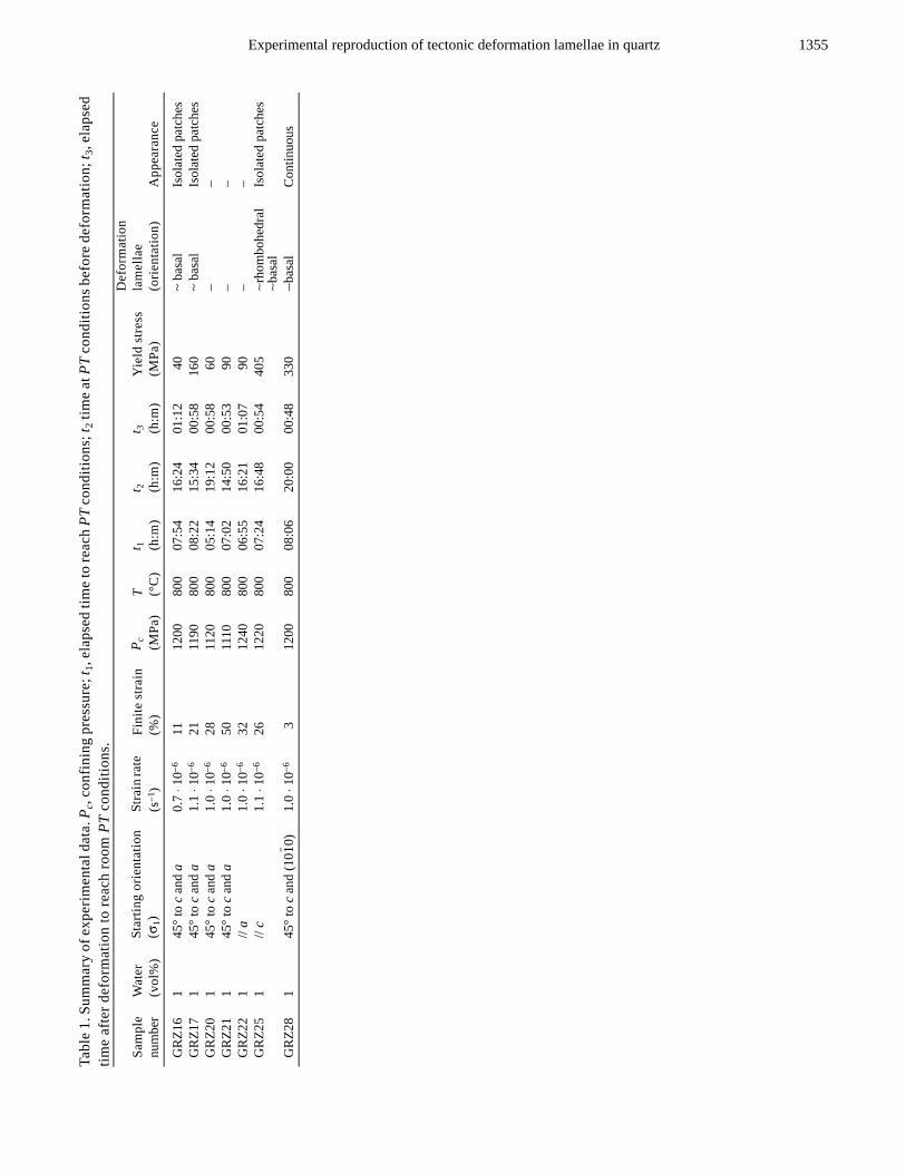
Overall, deformation lamellae were observed in samplesthat were deformed to low finite strains (≤26%) with thefollowing crystallographic orientations (listed according toincreasing abundance): compression axis σ1 (i) parallel to thec axis (GRZ25), (ii) at 45° to the c and a axes (GRZ16 andGRZ17), and (iii) at 45° to the c axis and m plane (GRZ28), i.e.,(10 0) (Table 1). In GRZ28 deformed to a finite strain of 2.8%,1
Experimental reproduction of tectonic deformation lamellae in quartz 1355
Tabl
e 1.
Sum
mar
y of
exp
erim
enta
l dat
a. P
c, co
nfin
ing
pres
sure
; t1,
elap
sed
time
to re
ach
PT c
ondi
tions
; t2 t
ime
at P
T co
nditi
ons b
efor
e de
form
atio
n; t 3
, ela
psed
tim
e af
ter d
efor
mat
ion
to re
ach
room
PT
cond
ition
s.
Sam
ple
num
ber
Wat
er(v
ol%
)St
artin
g or
ient
atio
n(σ
1)St
rain
rate
(s
−1)
Fini
te st
rain
(%)
P c
(MPa
)T (°
C)
t 1
(h:m
)t 2 (h
:m)
t 3
(h:m
)Y
ield
stre
ss(M
Pa)
Def
orm
atio
n la
mel
lae
(orie
ntat
ion)
App
eara
nce
GR
Z16
145
° to
c an
d a
0.7
⋅ 10−6
1112
0080
007
:54
16:2
401
:12
40~
basa
lIs
olat
ed p
atch
esG
RZ1
71
45° t
o c
and
a1.
1 ⋅ 1
0−621
1190
800
08:2
215
:34
00:5
816
0~
basa
lIs
olat
ed p
atch
esG
RZ2
01
45° t
o c
and
a1.
0 ⋅ 1
0−628
1120
800
05:1
419
:12
00:5
860
––
GR
Z21
145
° to
c an
d a
1.0
⋅ 10−6
5011
1080
007
:02
14:5
000
:53
90–
–G
RZ2
21
// a
1.0
⋅ 10−6
3212
4080
006
:55
16:2
101
:07
90–
–G
RZ2
51
// c
1.1
⋅ 10−6
2612
2080
007
:24
16:4
800
:54
405
∼rho
mbo
hedr
al∼b
asal
Isol
ated
pat
ches
GR
Z28
145
° to
c an
d (1
00)
1.0
⋅ 10−6
312
0080
008
:06
20:0
000
:48
330
∼bas
alC
ontin
uous
1

1356 M. G. C. Vernooij and F. Langenhorst
the deformation lamellae are homogeneously spread throughone half of the sample, whereas GRZ16, GRZ17 and GRZ25(11%, 21%, and 26% finite strains) contain deformationlamellae only in single patches distributed throughout thesamples. It is remarkable that deformation lamellae are notobserved in samples deformed to large strains at 45° to the cand a axes (GRZ20, GRZ21). Also, compression along the aaxis did not yield any deformation lamellae.
In most cases, deformation lamellae seem to be roughlyparallel to the basal plane (Fig. 2b), as measured by universalstage. In sample GRZ25, which was deformed at highdifferential stress with the basal plane perpendicular to thecompression direction, they are also observed sub-parallel torhombohedral planes, but exclusively in areas where the caxis has not rotated too far away from the compressiondirection. Deformation lamellae are always associated withlarger, ∼50 μm wide zones of undulatory extinction,suggesting a gradual misorientation of the deformed crystal.These undulatory zones are always approximately parallel tothe c direction (Fig. 2b). If the deformation lamellae are bent,the zones of undulatory extinction are also curved.
TEM revealed that the experimentally produceddeformation lamellae are associated with slightly curved,non-planar subgrain walls (Figs. 3a and 3b). The subgrain
walls are usually composed of one set of straight, well-organized dislocations (Fig. 3a). Free dislocations in thevicinity of the subgrain walls are curved, entangled, andsometimes connected to tiny water bubbles (Fig. 3a), or theyform numerous junctions. The subgrain walls themselves areonly rarely decorated with fluid inclusions.
Deformed quartz crystals are penetrated by numerous ofthese aligned subgrain walls, often occurring in pairs (Fig. 4).Changes in electron diffraction contrast across, and fringes at,subgrain walls indicate that the crystal is alternately tiltedabout the subgrain walls with a misorientation of about 1–3°.It is this alternating orientation which leads to the variation inoptical birefringence and the visibility of deformationlamellae at the optical scale. In principle, the lamellae canthus be regarded as strongly elongated subgrains. At the TEMscale it is obvious that a crystallographic orientation of thesubgrain walls can not be identified, because they do notdefine a crystallographic plane and are of a curved nature.
In order to determine the Burgers vectors using the g ⋅ b= 0 criterion, TEM images were taken with diffraction vectorsg = 10 1 and g = 0003. Most dislocations are out of contrastwith g = 0003 but are well visible with g = 10 1. Therefore,the Burgers vector of the dislocations is b = 1/3<11 0>,which is known to be the energetically most favorable
Fig. 1. Sample assembly used in a Tullis-modified Griggs apparatus to deform single crystals of quartz. NaCl was used as a solid medium.
11
2

Experimental reproduction of tectonic deformation lamellae in quartz 1357
Burgers vector in quartz (Doukhan 1995). The density ofthese a dislocations is on the order of 1010 cm−2.
Planar Deformation Features (PDFs) in Shocked Quartzfrom the Ries Crater
Optically, the decorated type of PDFs in analyzed quartzgrains from the Ries crater appear as perfectly planar andparallel elements with a spacing >4 μm (Fig. 5a). TEManalysis revealed that even more closely spaced PDFs exist(<1 μm) that are not resolvable at the optical scale (Fig. 6).There are no orientation changes across the PDFs similar tothose observed in case of tectonic deformation lamellae.Under the TEM, PDFs are decorated with fluid inclusions,indicating migration of water during post-shock annealing ofsuevites. At least three different PDF orientations wereinduced in the quartz grains (Figs. 5b and 6b). For example,the three different PDF orientations in the quartz grain shownin Fig. 6b belong to two corresponding rhombohedral planes,
and , and one prismatic plane, . Thiscombination of rhombohedral and prismatic planes iscommonly observed in shocked quartz and indicates amoderate shock pressure of about 20 GPa (Langenhorst andDeutsch 1994).
TEM reveals furthermore that the PDFs in these quartzgrains from the Ries crater consist of tiny dislocation loops,voids and bubbles. Similar features have been observed inshocked quartz from other impact sites (Goltrant et al. 1991,1992; Leroux et al. 1994, 1995) and are attributed to post-shock recrystallization of originally amorphous PDFs. Inagreement with these studies, there are only few freedislocations (density ≤0.2 ⋅ 1010 cm−2) in the vicinity of thePDFs. This contrasts with the observations on theexperimental deformation lamellae, which are associated with
Fig. 2. Optical micrographs in polarized light of experimentally formed deformation lamellae in sample GRZ25 deformed parallel to the c axisand in samples (GRZ16 and GRZ28) deformed at 45° to the c axis. a) GRZ25, tectonic deformation lamellae are in a sub-parallel, non-planararrangement. Note that single lamellae sometimes branch into two thinner lamellae. Small unloading fractures are observed between thelamellae. b) GRZ28, deformation lamellae are always associated with undulose zones that are approximately parallel to the c axis.
Fig. 2. Continued. c) GRZ16, deformation lamellae that aredecorated with fluid inclusions show resemblance with shockinduced PDFs.
1013( ) 1013( ) 1010( )

1358 M. G. C. Vernooij and F. Langenhorst
dislocation densities of 1010 cm−2. The free dislocations inshocked quartz may have pre-existed or developed duringpost-shock annealing and did not play a role in the originalshock deformation (Christie and Ardell 1976; Grieve et al.1996; Langenhorst 2002).
DISCUSSION
Nature and Formation of Deformation Lamellae
In this study, we have experimentally reproduceddeformation lamellae in single-crystal quartz that show great
similarity to natural deformation lamellae formed intectonically deformed quartz (e.g., McLaren and Hobbs1972). Our TEM observations reveal that the opticallyvisible, experimental deformation lamellae representelongated subgrains, which is in agreement with themicrostructural characteristics of deformation lamellae innaturally deformed quartz (McLaren and Hobbs 1972; White1973; Blenkinsop and Drury 1988). The formation of thesubgrains is due to the recovery of dislocations into gentlycurved subparallel subgrain walls, resulting in a bandedsubstructure that causes an alternating change in opticalbirefringence. Overall, the presence of subgrain walls and
Fig. 3. TEM micrographs, bright field images of subgrain walls in sample (GRZ28) deformed at 45° to the c axis and m plane. The freedislocations in the vicinity of the walls are curved, entangled and sometimes carry water in tiny inclusions (black arrows). a) The subgrainwalls consist of well organized straight dislocations. b) The contrast changes across subgrain walls indicate that the subgrains are slightlytilted.
Fig. 4. Dark field weak beam TEM image (g = 10 1) of sample GRZ25 (deformed parallel to the c axis). A set of subgrain walls (white arrows)is visible that is associated with optical deformation lamellae. The subgrain walls are neither perfectly parallel nor perfectly planar and cantherefore not be assigned to crystallographic planes. The open cracks are unloading cracks or cracks that are caused by the preparation of thesample.
1

Experimental reproduction of tectonic deformation lamellae in quartz 1359
numerous curved free dislocations indicates that both glide-and climb-controlled processes have been active during thedeformation of samples. This coexistence of creep andrecovery and the formation of deformation lamellae point todeformation in the so-called exponential creep regime,where the deformation behavior does not follow any more apower law of creep (McLaren 1991; Drury 1993).
To understand the development of the defectmicrostructure and, in particular, the formation ofdeformation lamellae in more detail, it is useful to discuss ourobservations in the context of previous extensive deformationexperiments on quartz (see summaries of McLaren 1991; denBrok 1992; Doukhan 1995). These experiments have shownthat dry quartz is practically undeformable and fails by
fracturing, whereas small amounts of dissolved water lead toa drastic softening of quartz (so-called “hydrolyticweakening” [McLaren et al. 1989]). Thereby, the actualconcentration and dispersion of water in the quartz structureplays an important role in the evolution of the deformationmicrostructure (Cordier and Doukhan 1989, 1995; Doukhan1995). Quartz supersaturated in water (>180 ppm)precipitates numerous water bubbles, which subsequentlygrow with time and are the sources for homogeneous,pervasive emission and climb of dislocations. If quartz is,however, undersaturated in water, precipitation does notoccur. Dislocation nucleation is heterogeneous along slipbands and climb of dislocations is strongly suppressed.
Deformation lamellae have only been found in our quartz
Fig. 5. Optical micrographs in polarized light show shocked quartz grains with PDFs from the Ries impact crater, Germany. a) Quartz grainshowing one set of parallel PDFs. b) Quartz grain showing three sets of PDFs. Two sets are continuous and well developed; one set isdiscontinuous (horizontal in the picture) and is decorated with fluid inclusions.
Fig. 6. TEM images of PDFs in shocked quartz grains. The PDFs were post-shock annealed and decorated with tiny dislocation loops and fluidinclusions. a) Dark field weak beam image (g = 10 1) shows one single set of perfectly planar PDFs with orientation (10 3). Since there areno contrast changes across the PDFs, the orientation of the crystal lattice is uniform. b) Bright field image of three sets of PDFs withorientations (10 3), ( 013), and (10 0).
1 1
1 1 1
1360 M. G. C. Vernooij and F. Langenhorst
crystals that were deformed to low strains (Table 1),suggesting that they form in an early stage of deformationwhen dislocations are heterogeneously emitted in slip bandsand the quartz crystals are still undersaturated in water. Thepresence of homogeneously distributed free dislocationswithin subgrains and their configuration in the form ofjunctions point, however, to a change in the deformationmechanism. As the deformation experiments proceeded,more and more of the available water was probablyincorporated into the quartz crystals and water precipitated asbubbles, from which dislocations were then homogeneouslyand pervasively emitted. In samples deformed to large strains,recovery was very efficient and defect microstructures aretherefore strongly evolved such that initially formeddeformation lamellae can completely vanish.
This model of formation of deformation lamellae byinitial dislocation glide in bands and dynamical recoveryallows to also understand the subbasal orientations and broadangular variation of lamella orientations. Deformationlamellae are most abundant in quartz crystals deformed withthe c axis at 45° to the compression axis (GRZ16, GRZ28),because in this orientation it is easiest to activate theenergetically favorable 1/3<11 0> dislocations in the (0001)plane. While dislocation glide drives these dislocations alongthe (0001) plane, the synkinematic or subsequent climb ofdislocations results in a movement out of the (0001) slip planeand an arrangement of dislocations into subgrain walls thatare rotated away from the basal plane. The degree of rotationis thereby controlled by the relative speeds of glide and climb.This explanation for the subbasal orientation of lamellae issupported by the observation that subgrain walls usuallyconsist of well-organized, straight dislocations. Networks ofdislocations are not observed because only a single set of adislocations has been emitted in the basal plane. A similarmodel for the formation of deformation lamellae waspresented by Drury (1993) to explain the formation ofsubgrain walls containing a network of dislocations in twoslip planes.
Distinction between Deformation Lamellae and PlanarDeformation Features
In the context of recent discussions on the origin ofplanar features in quartz from suspected impact structures andboundary layers (e.g., Retallack et al. 1998; Becker et al.2004; Langenhorst et al. 2005), it might be useful to finallyrecall here the fundamental difference between deformationlamellae and shock-induced PDFs. A large number of TEMstudies have shown that fresh PDFs represent planar and thinglass lamellae with a composition identical to that of the hostcrystal (e.g., Gratz et al. 1992; Langenhorst 1994). Post-shockannealing in natural impact formations results in therecrystallization of glass lamellae, the formation of tinydislocation loops, and precipitation of water bubbles (Grieve
et al. 1996; Langenhorst and Deutsch 1998). PDFs aretherefore often decorated with fluid inclusions, but this doesneither change their orientation nor their planarity. The PDFsthat are observed within the analyzed shocked quartz grainsfrom the Ries crater are of this “decorated” type.
At the TEM scale, there are thus abundant criteria todistinguish PDFs from deformation lamellae; they can besummarized as follows: (i) PDFs are perfectly planar insteadof slightly curved, (ii) they strictly form parallel to defined(mostly rhombohedral) crystallographic planes, (iii) theirspacing is usually <1 μm instead of 2–4 μm, (iv) they do notinduce a misorientation within the crystal and, thus, are notsubgrain walls or dislocation bands, and (v) the density of freedislocations is low compared to quartz with deformationlamellae. Although the resolution of the optical microscopedoes not allow to see the detailed defect microstructure, mostof the listed characteristics can also be recognized at theoptical scale. Therefore, TEM study is not necessarilydemanded to identify the origin of planar features (Lyons etal. 1993) but may be recommendable in cases of suspectedimpact structures with samples that experienced strongthermal overprint (Leroux et al. 1994; Joreau et al. 1997). Acareful optical analysis of planar features, the precisemeasurement of orientations using the universal stage(Langenhorst 2002), the inspection of cogenetic minerals, andthe geologic setting are usually sufficient criteria todistinguish PDFs from deformation lamellae (Reimold 1994).
Acknowledgments–MV is grateful for a Marie-Curie researchgrant provided by the Bayerisches Geoinstitut, University ofBayreuth (IHP program, HPMT-CT-2001-00231, toD. Rubie), and for financial support by ETH project 0-20907-01. We are indebted to B. den Brok for fruitful discussionsand to W. U. Reimold and an anonymous reviewer for theconstructive reviews.
Editorial Handling—Dr. Wolf Uwe Reimold
REFERENCES
Becke F. 1893. Petrographische Studien am Tonalit der Riesenferner.Tschermak’s Mineralogische und Petrographische Mitteilungen13:79–464.
Bˆhm A. 1882. Ueber die Gesteine des Wechsels. Mineralogischeund Petrographische Mitteilungen 4:197–214.
Becker L., Poreda R. J., Basu A. R., Pope K. O., Harrison T. M.,Nicholson C., and Iasky R. 2004. Bedout: A possible end-Permian impact crater offshore of Northwestern Australia.Science 304:1469–1476.
Blenkinsop T. G. and Drury M. R. 1988. Stress estimates and faulthistory from quartz microstructures. Journal of StructuralGeology 10:673–684.
Christie J. M. and Ardell A. J. 1974. Substructures of deformationlamellae in quartz. Geology 2:405–408.
Christie J. M. and Ardell A. J. 1976. Deformation structures inminerals. In Electron microscopy in mineralogy, edited by WenkH.-R. Berlin: Springer. pp. 374–403.
Christie J. M. and Raleigh C. B. 1959. The origin of deformationlamellae in quartz. American Journal of Science 257:385–407.
2
Experimental reproduction of tectonic deformation lamellae in quartz 1361
Cordier P. and Doukhan J. C. 1989. Water in quartz, solubility andinfluence on ductility. European Journal of Mineralogy 1:221–237.
Cordier P. and Doukhan J. C. 1995. Plasticity and dissociation ofdislocations in water-poor quartz. Philosophical Magazine A 72:497–514.
Cordier P., Vr·na S., and Doukhan J. C. 1994. Shock metamorphismin quartz at Sevetin and Susice (Bohemia)? A TEM investigation.Meteoritics 29:98–99.
Doukhan J. C. 1995. Lattice defects and mechanical behavior ofquartz SiO2. Journal de Physique III France 5: 1809–1932.
Den Brok B. 1992. An experimental investigation into the effect ofwater on the flow of quartzite. Geologica Ultraiectina 95.
Drury M. R. 1993. Deformation lamellae in metals and minerals. InDefects and processes in the solid state: Geoscienceapplications, edited by Boland J. N. and Fitz Gerald J. D.Amsterdam: Elsevier. pp. 195–212.
French B. M. 1998. Traces of catastrophe—A handbook of shock-metamorphic effects in terrestrial meteorite impact structures.LPI Contribution #954. Houston, Texas: Lunar and PlanetaryInstitute.
Gratz A. J., Nellis W. J., Christie J. M., Brocious W., Swegle J., andCordier P. 1992. Shock metamorphism of quartz with initialtemperatures −170 to 1000 °C. Physics and Chemistry ofMinerals 19:267–288.
Grieve R. A.F., Langenhorst F., and Stˆffler D. 1996. Shockmetamorphism of quartz in nature and experiment: II.Significance in geoscience. Meteoritics & Planetary Science 31:6–35.
Goltrant O., Cordier P., and Doukhan J.-C. 1991. Planar deformationfeatures in shocked quartz—A transmission electron microscopyinvestigation. Earth and Planetary Science Letters 106:103–115.
Goltrant O., Leroux H., Doukhan J. C., and Cordier P. 1992.Formation mechanisms of planar deformation features innaturally shocked quartz. Physics of the Earth and PlanetaryInteriors 74:219–240.
Joreau P., Reimold W. U., Robb L. J., and Doukhan J. C. 1997. ATEM study of deformed quartz grains from volcaniclasticsediments associated with the Bushveld complex, South Africa.European Journal of Mineralogy 9:393–401.
Langenhorst F. 1994. Shock experiments on pre-heated α- and β-quartz: II. X-ray and TEM investigations. Earth and PlanetaryScience Letters 128:683–698.
Langenhorst F. 2002. Shock metamorphism of some minerals: Basicintroduction and microstructural observations. Bulletin of theCzech Geological Survey 77:265–282.
Langenhorst F. and Deutsch A. 1994. Shock experiments on pre-heated α- and β-quartz: I. Optical and density data. Earth andPlanetary Science Letters 125:407–420.
Langenhorst F. and Deutsch A. 1996. The Azuara and Rubielosstructures, Spain: Twin impact craters or alpine thrust systems?TEM investigations on deformed quartz disprove shock origin(abstract). 27th Lunar and Planetary Science Conference. pp.725–726.
Langenhorst F., and Deutsch A. 1998. Minerals in terrestrial impactstructures and their characteristic features. In Advancedmineralogy, vol. 3, edited by Marfunin A.S. Berlin: Springer pp.95–119.
Langenhorst F., Deutsch A., Hornemann U., and Stˆffler D. 1992.Effect of temperature on shock metamorphism of single crystalquartz. Nature 356:507–509.
Langenhorst F., Kyte F. T., and Retallak G. J. 2005. Reexamination ofquartz grains from the Permian-Triassic boundary section atGraphite Peak, Antarctica (abstract #2358). 36th Lunar andPlanetary Science Conference. CD-ROM.
Leroux H., Reimold W. U., and Doukhan J. C. 1994. A TEMinvestigation of shock metamorphism in quartz from theVredefort dome, South Africa. Tectonophysics 230:223–239.
Leroux H., Warme J. E., and Doukhan J.-C. 1995. Shocked quartz inthe Alamo breccia, southern Nevada: Evidence for a Devonianimpact event. Geology 23:1003–1006.
Lyons J. B., Officer C. B., Borella P. E., and Lahodynsky R. 1993.Planar lamellar substructures in quartz. Earth and PlanetaryScience Letters 119:431–440.
McLaren A. C. 1991. Transmission electron microscopy of mineralsand rocks. Cambridge: Cambridge University Press. 397 p.
McLaren A. C., Turner R. G., Boland J. N., and Hobbs B. E. 1970.Dislocation structure of lamellae in synthetic quartz.Contributions to Mineralogy and Petrology 29:104–115.
McLaren A. C. and Hobbs B. E. 1972. Transmission electronmicroscope investigation of some naturally deformed quartzites.In Flow and fracture of rocks, edited by Heard H. C., Borg I. Y.,Carter N. L., and Raleigh C. B. Washington, D.C.: AmericanGeophysical Union. pp. 55–66.
McLaren A. C., Fitzgerald J. D., and Gerretsen J. 1989. Dislocationnucleation and multiplication in synthetic quartz: Relevance towater weakening. Physics and Chemistry of Minerals 16:465–482.
Mossman D. J., Grantham R. G., and Langenhorst F. 1998. A searchfor shocked quartz at the Triassic-Jurassic boundary in the Fundyand Newark basins of the Newark Supergroup. CanadianJournal of Earth Sciences 35:101–109.
Reimold W. U. 1994. Comment on “Planar lamellar substructures inquartz” by Lyons et al. (1993). Earth and Planetary ScienceLetters 125:473–477.
Retallack G. J., Seyedolali A., Krull E. S., Holser W. T., Ambers C. P.,and Kyte F. T. 1998. Search for evidence of impact at thePermian-Triassic boundary in Antarctica and Australia. Geology25:979–982.
Sharp T. G., El Goresy A., Wopenka B., and Chen M. 1999. A post-stishovite SiO2 polymorph in the meteorite Shergotty:Implications for impact events. Science 284:1511–1513.
Stˆffler D. 1971. Progressive metamorphism and classification ofshocked and brecciated crystalline rocks at impact craters.Journal of Geophysical Research 76:5541–5551.
Stˆffler D. and Ostertag R. 1983. The Ries impact crater. Fortschritteder Mineralogie 61:71–116.
Stˆffler D. and Langenhorst F. 1994. Shock metamorphism of quartzin nature and experiment: I. Basic observation and theory.Meteoritics 29:155–181.
Tullis J. and Tullis T. E. 1986. Experimental rock deformationtechniques. In Mineral and rock deformation: Laboratorystudies, edited by Hobbs B. E. and Heard H. C. Washington,D.C.: American Geophysical Union. pp. 297–324.
White S. H. 1973. Deformation lamellae in naturally deformedquartz. Nature Physical Science 245: 26–28.