Experimental comparison of high-density scintillators for EMCCD-based gamma ray imaging
11
Experimental comparison of high-density scintillators for EMCCD-based gamma ray imaging This article has been downloaded from IOPscience. Please scroll down to see the full text article. 2012 Phys. Med. Biol. 57 4545 (http://iopscience.iop.org/0031-9155/57/14/4545) Download details: IP Address: 131.180.39.83 The article was downloaded on 27/06/2012 at 11:52 Please note that terms and conditions apply. View the table of contents for this issue, or go to the journal homepage for more Home Search Collections Journals About Contact us My IOPscience
-
Upload
independent -
Category
Documents
-
view
0 -
download
0
Transcript of Experimental comparison of high-density scintillators for EMCCD-based gamma ray imaging

Experimental comparison of high-density scintillators for EMCCD-based gamma ray imaging
This article has been downloaded from IOPscience. Please scroll down to see the full text article.
2012 Phys. Med. Biol. 57 4545
(http://iopscience.iop.org/0031-9155/57/14/4545)
Download details:
IP Address: 131.180.39.83
The article was downloaded on 27/06/2012 at 11:52
Please note that terms and conditions apply.
View the table of contents for this issue, or go to the journal homepage for more
Home Search Collections Journals About Contact us My IOPscience

IOP PUBLISHING PHYSICS IN MEDICINE AND BIOLOGY
Phys. Med. Biol. 57 (2012) 4545–4554 doi:10.1088/0031-9155/57/14/4545
Experimental comparison of high-density scintillatorsfor EMCCD-based gamma ray imaging
Jan W T Heemskerk1,2, Rob Kreuger1, Marlies C Goorden1,3,Marc A N Korevaar1,3, Samuel Salvador1, Zachary M Seeley4,Nerine J Cherepy4, Erik van der Kolk1, Stephen A Payne4,Pieter Dorenbos1 and Freek J Beekman1,3,5
1 Radiation, Detection and Medical Imaging, Department of Applied Sciences,Delft University of Technology, Mekelweg 15, 2629 JB, Delft, the Netherlands2 Nuclear Medicine Department, University Hospital Vrije Universiteit Brussel,Laarbeeklaan 101, B-1090 Brussels, Belgium3 Rudolf Magnus Institute of Neurosciences, University Medical Center Utrecht, 3584 CG,Utrecht, the Netherlands4 Chemical Sciences Division, Lawrence Livermore National Laboratory, Livermore, CA 94550,USA5 MILABS, Universiteitsweg 100, Utrecht, the Netherlands
E-mail: [email protected]
Received 6 January 2012, in final form 21 May 2012Published 22 June 2012Online at stacks.iop.org/PMB/57/4545
AbstractDetection of x-rays and gamma rays with high spatial resolution can be achievedwith scintillators that are optically coupled to electron-multiplying charge-coupled devices (EMCCDs). These can be operated at typical frame rates of50 Hz with low noise. In such a set-up, scintillation light within each frameis integrated after which the frame is analyzed for the presence of scintillationevents. This method allows for the use of scintillator materials with relativelylong decay times of a few milliseconds, not previously considered for use inphoton-counting gamma cameras, opening up an unexplored range of densescintillators. In this paper, we test CdWO4 and transparent polycrystallineceramics of Lu2O3:Eu and (Gd,Lu)2O3:Eu as alternatives to currently usedCsI:Tl in order to improve the performance of EMCCD-based gamma cameras.The tested scintillators were selected for their significantly larger cross-sectionsat 140 keV (99mTc) compared to CsI:Tl combined with moderate to good lightyield. A performance comparison based on gamma camera spatial and energyresolution was done with all tested scintillators having equal (66%) interactionprobability at 140 keV. CdWO4, Lu2O3:Eu and (Gd,Lu)2O3:Eu all result ina significantly improved spatial resolution over CsI:Tl, albeit at the cost ofreduced energy resolution. Lu2O3:Eu transparent ceramic gives the best spatialresolution: 65 μm full-width-at-half-maximum (FWHM) compared to 147 μmFWHM for CsI:Tl. In conclusion, these ‘slow’ dense scintillators open up new
0031-9155/12/144545+10$33.00 © 2012 Institute of Physics and Engineering in Medicine Printed in the UK & the USA 4545
4546 J W T Heemskerk et al
possibilities for improving the spatial resolution of EMCCD-based scintillationcameras.
(Some figures may appear in colour only in the online journal)
1. Introduction
High-resolution gamma detectors can play an important role in applications such asautoradiography (Karellas et al 1993, MacDonald et al 1997, Caballo et al 2007),crystallography (Suzuki et al 1999, Phillips et al 2000, Vavrik et al 2002, Ponchut et al2005) and astrophysics (Matteson et al 1997, He et al 1999). Furthermore, it has been shownthat for the improvement of future single photon emission computed tomography (SPECT)devices, a high intrinsic detector resolution is essential (Rogulski et al 1993, Barber 1999,Beekman and Vastenhouw 2004, Meng et al 2006, Rentmeester et al 2007, Goorden et al 2009,Peterson and Furenlid 2011). Very high spatial resolution (below 60 μm) can be obtained witha detector consisting of micro-columnar scintillation crystals of CsI:Tl read out by an EMCCDoperating at high frame rates (∼ 50 Hz) (Gruner et al 2002, De Vree et al 2005, Nagarkar et al2006, Meng 2006 and Teo et al 2006, Heemskerk et al 2007). However, despite continuingprogress in the field of micro-columnar crystals, the commercially available thickness of suchcrystals is currently limited to about 1.5 mm, resulting in interaction probabilities that are notsufficient for small-animal SPECT imaging or clinical application.
In contrast to micro-columnar scintillators, single crystal and transparent polycrystallineceramic scintillators are available in a large variety of materials and thicknesses with a uniformresponse. However, compared to micro-columnar scintillation crystals, the resolution that canbe obtained with optically transparent scintillators is limited because of the larger width ofthe light spread on the EMCCD. This can be partly overcome by the use of a detectionalgorithm that takes into account the interaction-depth-dependent light spread (Miller et al2006, Korevaar et al 2009a, 2011) or by shaping the scintillators such that depth-of-interactiondistortions are reduced (Beekman 2007, Korevaar et al 2009b).
Traditionally, PMT-based gamma cameras are equipped with fast scintillators (<10−7 sdecay time) to handle typical count rates of a few 105 cps. Using slow scintillators at theserates, the PMT signals would suffer from pulse pile-up, deteriorating the energy and spatialresolution of the camera. However, for a pre-clinical multi-pinhole setup with a large numberof EMCCD-based gamma cameras (Beekman and Vastenhouw 2004, Rentmeester et al 2007),only a few events per frame will typically be present at a frame rate of 50 Hz (this framerate is limited by the pixel readout rate of the CCD). Although the incident light is integratedfor every frame, EMCCD-based gamma cameras can nevertheless detect and characterizemultiple scintillation events simultaneously as long as their signals do not spatially overlapsignificantly.
The relatively long integration times create opportunities for using slow scintillatorsthat have not been considered previously in PMT-based gamma cameras, but which maylead to high-resolution gamma images because of other favorable properties such as theirhigh interaction probability or brightness. EMCCD gamma cameras could then be usedadvantageously instead of traditional PMT gamma cameras for (e.g.) multi pinhole small-animal SPECT scanners. Despite the fact that temporal information within the frames is lost,CCD-based scanners can still be used for most fast dynamic scans. For some applications likegated (cardiac) murine scans, challenges with regard to temporal resolution may remain.

Experimental comparison of high-density scintillators for EMCCD-based gamma ray imaging 4547
Several factors are important in the selection of a scintillator, most importantly lightyield, photon emission spectrum, attenuation coefficient for the incoming gamma rays and(intrinsic) background radiation. First, a high light yield is important because a larger amountof optical photons will allow a more precise estimation of the energy and position of thescintillation events; this also requires the emission spectrum of the scintillator to match thespectral response of the EMCCD. Second, a higher attenuation coefficient is advantageousbecause it allows a thinner scintillator to achieve the desired interaction probability with,on average, a reduced light spread on the detector as a consequence. A small light spreadimproves the accuracy of the position estimation (Korevaar et al 2009a). Furthermore, a dense(hence thin) scintillator has the additional advantage that smaller light spreads on the CCDallow operation at a higher count rate. The balance between the attenuation coefficient andlight yield is crucial, since a scintillator with a high light output could compensate for a lowattenuation coefficient (scintillation events can be accurately detected further from the CCDsurface in the thicker scintillators required in that case) and vice versa. A low light yield canto some extent be compensated for by applying retro-reflectors to increase the number of lightphotons reaching the EMCCD (Heemskerk et al 2009) without increasing their spread on theEMCDD.
In the present investigation, we test different scintillators in our EMCCD-based gammacamera, in an effort to improve its performance. To this end, we have experimentally comparedthe spatial resolution, energy resolution and the dark count rate (no source present) of ourgamma camera employing several optically transparent scintillators, all with equal (66%)interaction probability for 99mTc emitted gamma photons.
2. Materials and methods
2.1. Scintillators
We have tested a number of candidate materials that appeared promising in terms ofscintillation light yield and/or 99mTc 140 keV radiation attenuation length: CsI:Tl, Lu2O3:Eu,(Gd,Lu)2O3:Eu and CdWO4. CsI:Tl is currently used because of its high light yield(∼54–66 photons keV−1, Saint-Gobain a6, Van Eijk 2002), in combination with a goodcorrespondence of its emission spectrum to the sensitivity of the EMCCD that we employ.
Lu2O3:Eu (5% doping) was selected for its large mass attenuation coefficient for 140 keVradiation (13.3 cm−1, compared to 3.6 cm−1 for CsI:Tl). Furthermore, its measured light yieldof 50–70 photons keV−1 is similar to that of CsI:Tl (see Cherepy et al 2011 for the light yieldsetup, Shi et al 2009). The (Gd,Lu)2O3:Eu material is similar to Lu2O3:Eu but approximatelyhalf of the Lu has been replaced by Gd in order to reduce the intrinsic activity (due to 176Luβ−-emission). Its density is approx. 10% lower than that of Lu2O3:Eu, but its light yieldis comparable (70 photons keV−1, Cherepy et al 2011). The Lu2O3:Eu and (Gd,Lu)2O3:Euscintillators are so-called ceramic scintillators; these materials are not monocrystalline butpolycrystalline, with average grain size of ∼100 μm , phase purity of >99.5% and with residualporosity of <0.6%. Further details on the fabrication and morphology of these materials can befound in Seeley et al 2011. In general, optical scatter can occur in transparent ceramics at grainboundaries, particularly if any residual porosity or secondary phase is present. However, thesamples used for these experiments exhibited low optical scatter: between 0.1 and 0.2 cm−1 forLu2O3:Eu and between 0.03 and 0.08 cm−1 for (Gd,Lu)2O3:Eu (see Seeley et al 2011 for adescription of the setup). Therefore, optical scatter was not expected to degrade the scintillators’performance for thicknesses up to a few millimeters as considered in this work.
6 http://www.detectors.saint-gobain.com/CsI(Tl).aspx
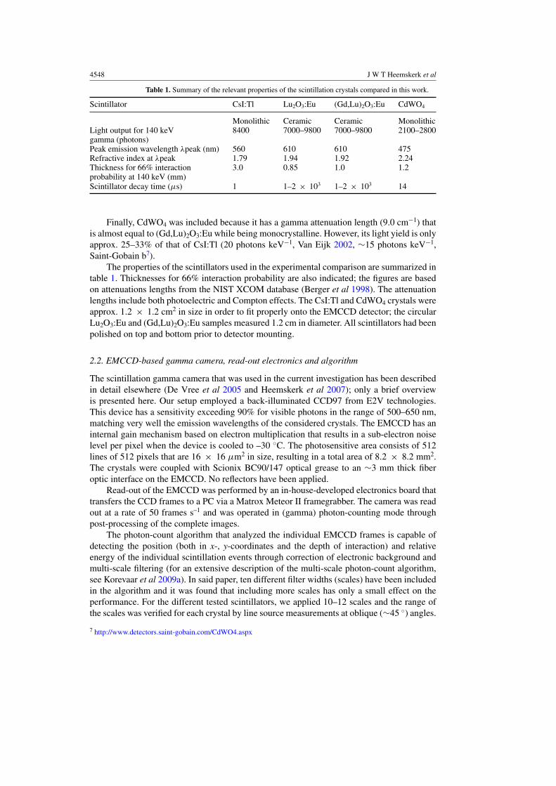
4548 J W T Heemskerk et al
Table 1. Summary of the relevant properties of the scintillation crystals compared in this work.
Scintillator CsI:Tl Lu2O3:Eu (Gd,Lu)2O3:Eu CdWO4
Monolithic Ceramic Ceramic MonolithicLight output for 140 keV 8400 7000–9800 7000–9800 2100–2800gamma (photons)Peak emission wavelength λpeak (nm) 560 610 610 475Refractive index at λpeak 1.79 1.94 1.92 2.24Thickness for 66% interaction 3.0 0.85 1.0 1.2probability at 140 keV (mm)Scintillator decay time (μs) 1 1–2 × 10³ 1–2 × 10³ 14
Finally, CdWO4 was included because it has a gamma attenuation length (9.0 cm−1) thatis almost equal to (Gd,Lu)2O3:Eu while being monocrystalline. However, its light yield is onlyapprox. 25–33% of that of CsI:Tl (20 photons keV−1, Van Eijk 2002, ∼15 photons keV−1,Saint-Gobain b7).
The properties of the scintillators used in the experimental comparison are summarized intable 1. Thicknesses for 66% interaction probability are also indicated; the figures are basedon attenuations lengths from the NIST XCOM database (Berger et al 1998). The attenuationlengths include both photoelectric and Compton effects. The CsI:Tl and CdWO4 crystals wereapprox. 1.2 × 1.2 cm2 in size in order to fit properly onto the EMCCD detector; the circularLu2O3:Eu and (Gd,Lu)2O3:Eu samples measured 1.2 cm in diameter. All scintillators had beenpolished on top and bottom prior to detector mounting.
2.2. EMCCD-based gamma camera, read-out electronics and algorithm
The scintillation gamma camera that was used in the current investigation has been describedin detail elsewhere (De Vree et al 2005 and Heemskerk et al 2007); only a brief overviewis presented here. Our setup employed a back-illuminated CCD97 from E2V technologies.This device has a sensitivity exceeding 90% for visible photons in the range of 500–650 nm,matching very well the emission wavelengths of the considered crystals. The EMCCD has aninternal gain mechanism based on electron multiplication that results in a sub-electron noiselevel per pixel when the device is cooled to –30 ◦C. The photosensitive area consists of 512lines of 512 pixels that are 16 × 16 μm2 in size, resulting in a total area of 8.2 × 8.2 mm2.The crystals were coupled with Scionix BC90/147 optical grease to an ∼3 mm thick fiberoptic interface on the EMCCD. No reflectors have been applied.
Read-out of the EMCCD was performed by an in-house-developed electronics board thattransfers the CCD frames to a PC via a Matrox Meteor II framegrabber. The camera was readout at a rate of 50 frames s–1 and was operated in (gamma) photon-counting mode throughpost-processing of the complete images.
The photon-count algorithm that analyzed the individual EMCCD frames is capable ofdetecting the position (both in x-, y-coordinates and the depth of interaction) and relativeenergy of the individual scintillation events through correction of electronic background andmulti-scale filtering (for an extensive description of the multi-scale photon-count algorithm,see Korevaar et al 2009a). In said paper, ten different filter widths (scales) have been includedin the algorithm and it was found that including more scales has only a small effect on theperformance. For the different tested scintillators, we applied 10–12 scales and the range ofthe scales was verified for each crystal by line source measurements at oblique (∼45 ◦) angles.
7 http://www.detectors.saint-gobain.com/CdWO4.aspx

Experimental comparison of high-density scintillators for EMCCD-based gamma ray imaging 4549
Figure 1. Experimental setup for the determination of the spatial and energy resolution. A 99mTcsource is placed in proximity to the gamma camera, which is irradiated through a 30 μm-wide slit.
This allowed us to estimate the filter width that corresponds to scintillation events in the topof the crystal; the maximum filter width for each crystal was set such that it exceeded themaximum light spread of the scintillation events for that crystal thickness, to ensure inclusionof all events.
2.3. Measurements
For the different scintillators we compared the energy and spatial resolution and the darkcount rate. To this purpose, each scintillator was tested in our gamma camera set-up, shown infigure 1. A 30 μm-wide slit was placed on top of the camera, which was irradiated with a 99mTcsource with an activity of around 40 MBq. Because of source strength regulations in our lab,the average detector count rate was around or below one event per frame. For the electronicbackground correction, a series of frames without radio-active source was registered; forthe Lu2O3:Eu and (Gd,Lu)2O3:Eu measurements, this necessitated removal of the scintillator,because of their intrinsic radio-activity.
Energy spectra were calculated as histograms of the energy values of the events detectedin an area covering half of the CCD surface with the slit centered in the image. This wasto prevent edge effects of the circular Lu2O3:Eu and (Gd,Lu)2O3:Eu samples in the cornersof the EMCCD to affect the comparison. The energy resolution is the ratio of the measuredfull-width-at-half-maximum (FWHM) of the photopeak and the photopeak position in theenergy spectrum. Scintillation events within energy windows ranging from −50% to +50%of the energy peak value were included in the calculation of the spatial resolution and the darkcount measurements.
The spatial resolution of the camera was determined as the measured full-width-at-half-maximum (FWHM) of the profile of the image of the slit, corrected for the width of theslit itself (De Vree et al 2005). To show the effects of electronic noise and inherent (176Lu)scintillator activity, we have measured the number of background events (per frame within theappropriate energy window) for each scintillator when the camera was not irradiated (i.e. darkcounts).

4550 J W T Heemskerk et al
Figure 2. Energy spectra for different scintillation crystals, each with 66% interaction probabilityfor 140 keV gammas.
Figure 3. Profiles of the line source for different scintillation crystals, each with 66% interactionprobability for 140 keV gammas.
Table 2. Results of the energy resolution, spatial resolution and dark count measurements.
Crystal CsI:Tl Lu2O3:Eu (Gd,Lu)2O3:Eu CdWO4
Energy resolution (%) 53 84 75 80Spatial resolution(FWHM) (μm) 147 65 75 87(FWTM) (μm) 465 190 238 285Dark counts (frame−1) 1.4 × 10⁻³ 8.8 × 10⁻² 4.8 × 10⁻² 2.0 × 10⁻³
3. Results
Figures 2 and 3 show the energy spectra and line profiles obtained with the differentscintillators. The values for energy and spatial resolution derived from these figures, as well asthe dark counts, are listed in table 2. In figure 4, we plot spatial (x-axis) versus energy resolution

Experimental comparison of high-density scintillators for EMCCD-based gamma ray imaging 4551
Figure 4. Comparison of the performance of the different scintillation crystals, by displayingmeasured spatial versus energy resolution.
(y-axis), indicating the relation between these quantities for the different scintillators. In sucha plot, the scintillator with the best overall performance will be found in the lower-left corner.
As can be inferred from table 2 and figure 4, there is no single scintillator in this studythat has the best performance in terms of spatial and energy resolution. The energy resolution(figure 2) obtained with CsI:Tl (53%) is significantly better than that obtained with the CdWO4,(Gd,Lu)2O3:Eu and Lu2O3:Eu scintillators (80%, 75% and 84%, respectively). However, theLu2O3:Eu ceramic achieves the best spatial resolution, (closely) followed by the (Gd,Lu)2O3:Euand CdWO4 scintillators (FWHM: 65, 75 and 87 μm, respectively, FWTM: 190, 238 and285 μm respectively). The spatial resolution of the CsI:Tl crystal (147 μm FWHM, 465 μmFWTM) is considerably worse, by more than a factor of 2. One can see that the full-width-at-tenth-maximum (FWTM) values of the line source profiles scale roughly with the FWHMvalues.
From table 2, one notes that the numbers of dark counts for Lu2O3:Eu and (Gd,Lu)2O3:Euare significantly higher than for CdWO4 and CsI:Tl.
4. Discussion
The use of a denser scintillating material considerably improves the spatial resolution of ourEMCCD-based gamma detector. This is because denser materials allow the use of thinnerscintillators for a certain desired interaction probability which reduces, on average, the lightspread on the EMCCD. There is a clear correlation between the thickness of the scintillationcrystal and the measured spatial resolution, with CsI:Tl having the worst (147 μm FWHM,465 μm FWTM) and Lu2O3:Eu having the best (65 μm FWHM, 190 μm FWTM) performancein this respect.
The (Gd,Lu)2O3:Eu and Lu2O3:Eu scintillators have a dark count rate which is significantlyhigher than that of CsI:Tl and CdWO4, by up to a factor of 60. We believe that this is causedby an intrinsic background level due to the natural radioactivity of 176Lu present in thesescintillators (about 50 Bq for our samples). The 176Lu decay generates scintillation events witha continuous energy spectrum up to ∼600 keV, overlapping with the spectrum of the 140 keV99mTc (true) events. The background of (Gd,Lu)2O3:Eu is approx. 40% below that of Lu2O3:Eu,

4552 J W T Heemskerk et al
corresponding to the different quantities of Lu in these scintillators. These numbers show thatfor expected event rates in preclinical multi-pinhole SPECT (a few events per frame), darkcounts (less than 0.1 per frame) constitute a small percentage of the total number of detectedevents.
The energy resolution is worse for the three newly tested scintillators compared to CsI:Tl.One would expect the thin scintillators Lu2O3:Eu and (Gd,Lu)2O3:Eu, with an expectedlight yield similar to that of CsI:Tl8, to perform better than CdWO4, but the results indicatedifferently. Measurements with a second (Gd,Lu)2O3:Eu scintillator have confirmed that thedegraded energy resolution was not due to high optical scatter or a badly mounted sampleas there was only a minor improvement of energy resolution from the reported 75% to 70%,which is just outside the statistical error of ± 3%. The effect of the intrinsic 176Lu activity onthe energy resolution is also expected to be small. There is a wide but low background in theenergy spectrum when the crystal is unirradiated.
There are several possible explanations for the reduced energy resolution. First, it ispossible that the CCD detects less photons than expected, as the exact light yields of thescintillator samples in our gamma camera setup are unknown. Furthermore, the relatively highoptical density (refractive index) of Lu2O3:Eu and (Gd,Lu)2O3:Eu results in a smaller criticalangle of reflection, reducing the relative amount of scintillation photons reaching the CCDcompared to a setup with CsI:Tl.
Another reason for the reduced energy resolution could be that, contrary to our initialassumption, the integration time of the CCD is not sufficient to collect all of the scintillationphotons for Lu2O3:Eu and (Gd,Lu)2O3:Eu in a single frame, especially if the scintillationoccurs just before the end of the integration period (20 ms). We have estimated this effect bysimulating scintillation events detected by the EMCCD with a 50% energy resolution and 1or 2 ms decay time, arriving randomly in the integration time window (frame) and integratingthese light signals till the frame is read out. We found that this partial integration doesindeed degrade the energy resolution; about 10–20% (relatively) of the energy resolution ofLu2O3:Eu and (Gd,Lu)2O3:Eu is due to this effect. Experimentally, we have noted (in particularfor Lu2O3:Eu) that some events seem to be spread out over two CCD frames, supporting thishypothesis. A correction for this partial integration effect may improve the energy resolutionof Lu2O3:Eu and (Gd,Lu)2O3:Eu to surpass the one of CdWO4. We do not expect afterglow toplay a role here because (i) no afterglow was observed by us in decay time measurements and(ii) the count rates are rather low, limiting afterglow buildup, if afterglow would neverthelessbe present.
The tested CdWO4 scintillator is a monocrystalline material that does not suffer fromoptical scatter, and has less than half the thickness of the CsI:Tl scintillator. Therefore,compared to CsI:Tl, the light spots on the detector are relatively small and well defined.However, its energy resolution is worse than that of CsI:Tl; we believe that this is mainlycaused by the fact that its light yield is expected to be only ∼25–30% of that of CsI:Tl.Moreover, it has a higher optical refractive index than CsI:Tl, which further reduces theamount of scintillation photons to be detected by the EMCCD. Note that energy resolution isless important for small animal imaging than for clinical applications, as scatter rejection isoften not required due to the lower amount of scatter in animals compared to humans.
Given our results, we believe that for improving both the energy and spatial resolution ofour gamma camera, one should find a scintillator which has at least an attenuation coefficientand light yield as high as Lu2O3:Eu has, but without natural radioactive isotopes. Moreover, to
8 We would like to point out that the light yield of CsI:Tl is expected to degrade by ± 10% at a temperature of −30 ◦C;the light yields of Lu2O3:Eu and (Gd,Lu)2O3:Eu are not known exactly, but like CdWO4 they are not expected to havea strong temperature dependence.

Experimental comparison of high-density scintillators for EMCCD-based gamma ray imaging 4553
obtain an energy resolution similar to CsI:Tl or better, its light yield should be considered inconjunction with the refractive index. For the presently used scintillation detection algorithm,the energy resolution will be better when the scintillator has a decay time smaller than theCCD integration period (<1 ms). The development of a novel detection algorithm may relaxthe latter requirement.
5. Conclusion
Dense, though ‘slow’, scintillators CdWO4, (Gd,Lu)2O3:Eu and Lu2O3:Eu considerablyimprove the spatial resolution of our photon-counting gamma camera compared to CsI:Tl,however at the cost of degraded energy resolution. This can be attributed to the reducedlight yield (in the case of CdWO4), the refractive index of the scintillator (for CdWO4,(Gd,Lu)2O3:Eu and Lu2O3:Eu) or the slow decay time (of (Gd,Lu)2O3:Eu and Lu2O3:Eu). Weconclude that the investigation of dense scintillators that were previously not considered ingamma detectors because of their slow decay times, opens new possibilities for improving thespatial resolution of CCD-based scintillation cameras.
Acknowledgments
The authors would like to thank Paul Schotanus of Scionix Netherlands for valuable discussionsand providing us with the CsI:Tl and CdWO4 scintillators. This work was sponsored inpart by the (Netherlands) Ministry of Economic Affairs, Agriculture and Innovation IOPphotonics grant IPD067766. The lutetia-based materials development was supported by theUS Department of Energy (Enhanced Surveillance Subprogram), and conducted by LawrenceLivermore National Laboratory under Contract DE-AC52-07NA27344.
References
Barber H B 1999 Applications of semiconductor detectors to nuclear medicine Nucl. Instrum Methods A 436 102–10Beekman F J 2007 Radiation detection apparatus Patent WO 2007/011214 A1Beekman F J and Vastenhouw B 2004 Design and simulation of a high-resolution stationary SPECT system for small
animals Phys. Med. Biol. 49 4579–92Berger M J, Hubbell J H, Seltzer S M, Chang J, Coursey J S, Sukumar J, Zucker D S and Olsen K 1998 NIST XCOM:
Photon Cross Sections Database, http://www.nist.gov/pml/data/xcom/index.cfmCaballo J, Baily A, Kitchen I, Prydderch M, Clark A, Turchetta R and Wells K 2007 Digital autoradiography using
room temperature CCD and CMOS imaging technology Phys. Med. Biol. 52 4993–5011Cherepy N J et al 2011 Performance of europium-doped strontium iodide, transparent ceramics and bismuth-loaded
polymer scintillators Proc. SPIE 8142 81420WDe Vree G A, Westra A H, Moody I, Van der Have F, Ligtvoet C M and Beekman F J 2005 Photon-counting gamma
camera based on an electron-multiplying CCD IEEE Trans. Nucl. Sci. 52 Part 1 580–88Goorden M C, Rentmeester M C M and Beekman F J 2009 Theoretical analysis of full-ring multi-pinhole brain
SPECT Phys. Med. Biol. 54 6593–610Gruner S M, Tate M W and Eikenberry E F 2002 Charge-coupled device area x-ray detectors Rev. Sci.
Instrum. 73 2815–42He Z, Li W, Knoll G F, Wehe D K, Berry J and Stahle C M 1999 3-D position sensitive CdZnTe gamma-ray
spectrometers Nucl. Instrum. Methods A 422 173–78Heemskerk J W T, Korevaar M A N, Kreuger R, Ligtvoet C M, Schotanus P and Beekman F J 2009 A micro-machined
retro-reflector for improving light yield in ultra-high-resolution gamma cameras Phys. Med. Biol. 54 3003–14Heemskerk J W T, Westra A H, Linotte P M, Ligtvoet C M, Zbijewski W and Beekman F J 2007 Front-illuminated
versus back-illuminated photon-counting CCD-based gamma camera: important consequences for spatialresolution and energy resolution Phys. Med. Biol. 52 N149–62
Karellas A, Liu H, Reinhardt C, Harris L J and Brill A B 1993 Imaging of radionuclide emissions with a low-noisecharge-coupled device IEEE Trans. Nucl. Sci. 40 979–82

4554 J W T Heemskerk et al
Korevaar M A N, Goorden M C, Heemskerk J W T and Beekman F J 2011 Maximum-likelihood scintillation detectionfor EM-CCD based gamma cameras Phys. Med. Biol. 56 4785–801
Korevaar M A N, Heemskerk J W T, Goorden M C and Beekman F J 2009a Multi-scale algorithm for improvedscintillation detection in a CCD based gamma cameras Phys. Med. Biol. 54 831–42
Korevaar M A N, Heemskerk J W T and Beekman F J 2009b A pinhole gamma camera with optical depth-of-interaction elimination Phys. Med. Biol. 54 N267–72
MacDonald J H, Wells K, Reader A J and Ott R J 1997 A CCD-based tissue imaging system Nucl. Instrum. MethodsA A392 220–6
Matteson J L et al 1997 CdZnTe arrays for astrophysics applications Proc. SPIE 3115 160–75Meng L J 2006 An intensified EMCCD camera for low energy gamma ray imaging applications IEEE Trans.
Nucl. Sci. 53 2376–84Meng L J, Clinthorne N H, Skinner S, Hay R V and Gross M 2006 Design and feasibility study of a single photon
emission microscope system for small animal I-125 imaging IEEE Trans. Nucl. Sci. 53 1168–78Miller B W, Bradford B H, Barrett H H, Shestakova I, Singh B and Nagarkar V V 2006 Single-photon spatial and
energy resolution enhancement of a columnar CsI(Tl)/EMCCD gamma-camera using maximum-likelihoodestimation Proc. SPIE 6142 61421T
Nagarkar V V, Shestakova I, Gaysinskiy V, Tipnis S V, Singh B, Barber W, Hasegawa B and Entine G 2006 ACCD-based detector for SPECT IEEE Trans. Nucl. Sci. 53 Part 1 54–58
Peterson T E and Furenlid L R 2011 SPECT detectors: the Anger camera and beyond Phys. Med. Biol. 56 R145–82Phillips W C, Stanton M, Stewart A, Qian H, Ingersoll C and Sweet R M 2000 Multiple CCD detector for
macromolecular x-ray crystallography J. Appl. Cryst. 33 243–51Ponchut C, Zontone F and Graafsma H 2005 Experimental comparison of pixel detector arrays and CCD-based
systems for x-ray area detection on synchrotron beamlines IEEE Trans. Nucl. Sci. 52 1760–5Rentmeester M C M, Van der Have F and Beekman F J 2007 Optimizing multi-pinhole SPECT geometries using an
analytical model Phys. Med. Biol. 52 2567–81Rogulski M M, Barber H B, Barrett H H, Shoemaker R L and Woolfenden J M 1993 Ultra-high resolution brain
SPECT imaging—simulation results IEEE Trans. Nucl. Sci. 40 Part 1 1123–9Seeley Z M, Kuntz J D, Cherepy N J and Payne S A 2011 Transparent Lu2O3:Eu ceramics by sinter and HIP
optimization Opt. Mater. 33 1721–6Shi Y, Shen Q W and Shi J L 2009 Processing and scintillation properties of Eu3+ doped Lu2O3 transparent ceramics
Opt. Mater. 31 729–33Suzuki M, Yamamoto M, Kumasaka T, Sato K, Toyokawa H, Aries I F, Jerram P A, Gullick D and Ueki T 1999 A
multiple-CCD x-ray detector and its basic characterization J. Synchrotron Radiat. 6 6–18Teo B K, Shestakova I, Sun M, Barber W C, Hasegawa B H and Nagarkar V V 2006 Evaluation of a EMCCD detector
for emission-transmission computed tomography IEEE Trans. Nucl. Med. 53 Part 1 2495–9Van Eijk C W E 2002 Inorganic scintillators in medical imaging Phys. Med. Biol. 47 R85–106Vavrik D, Jakubek J, Visschers J, Pospisil S, Ponchut C and Zemankova J 2002 First tests of a medipix-1 pixel detector
for x-ray dynamic defectoscopy Nucl. Instrum. Methods A 487 216–23