Expansion and Characterization of Neonatal Cardiac Pericytes Provides a Novel Cellular Option for...
23
Expansion and Characterization of Neonatal Cardiac Pericytes Provides a Novel Cellular Option for Tissue Engineering in Congenital Heart Disease Elisa Avolio, PhD;* Iker Rodriguez-Arabaolaza, BSc;* Helen L. Spencer, PhD;* Federica Riu, PhD; Giuseppe Mangialardi, MD, PhD; Sadie C. Slater, PhD; Jonathan Rowlinson, MSc; Valeria V. Alvino, MSc; Oluwasomidotun O. Idowu, BSc; Stephanie Soyombo, BSc; Atsuhiko Oikawa, PhD; Megan M. Swim, MSc; Cherrie H. T. Kong, PhD; Hongwei Cheng, PhD; Huidong Jia, MD, PhD; Mohamed T. Ghorbel, PhD; Jules C. Hancox, BSc, PhD, FSB; Clive H. Orchard, DSc, PhD, FSB; Gianni Angelini, MD; Costanza Emanueli, PhD; Massimo Caputo, MD, FRCS; Paolo Madeddu, MD, FAHA Background-—Living grafts produced by combining autologous heart-resident stem/progenitor cells and tissue engineering could provide a new therapeutic option for definitive correction of congenital heart disease. The aim of the study was to investigate the antigenic profile, expansion/differentiation capacity, paracrine activity, and pro-angiogenic potential of cardiac pericytes and to assess their engrafting capacity in clinically certified prosthetic grafts. Methods and Results- —CD34 pos cells, negative for the endothelial markers CD31 and CD146, were identified by immunohis- tochemistry in cardiac leftovers from infants and children undergoing palliative repair of congenital cardiac defects. Following isolation by immunomagnetic bead-sorting and culture on plastic in EGM-2 medium supplemented with growth factors and serum, CD34 pos /CD31 neg cells gave rise to a clonogenic, highly proliferative (>20 million at P5), spindle-shape cell population. The following populations were shown to expresses pericyte/mesenchymal and stemness markers. After exposure to differentiation media, the expanded cardiac pericytes acquired markers of vascular smooth muscle cells, but failed to differentiate into endothelial cells or cardiomyocytes. However, in Matrigel, cardiac pericytes form networks and enhance the network capacity of endothelial cells. Moreover, they produce collagen-1 and release chemo-attractants that stimulate the migration of c-Kit pos cardiac stem cells. Cardiac pericytes were then seeded onto clinically approved xenograft scaffolds and cultured in a bioreactor. After 3 weeks, fluorescent microscopy showed that cardiac pericytes had penetrated into and colonized the graft. Conclusions-—These findings open new avenues for cellular functionalization of prosthetic grafts to be applied in reconstructive surgery of congenital heart disease. ( J Am Heart Assoc. 2015;4:e002043 doi: 10.1161/JAHA.115.002043) Key Words: cells • congenital heart defects • grafting • myocardium • pediatrics C ongenital heart disease (CHD) is the most common type of birth defect, with a reported prevalence ranging from 6 to 13 per 1000 live births. 1,2 In the United Kingdom alone, there are 4600 babies born with CHD each year. Despite a constant decline in mortality rate, CHD still represents the primary cause of death among infants in industrialized countries. The definitive therapeutic option for these patients is corrective surgery. 3 However, prostheses currently used to reconstruct complex cardiac defects, including synthetic Gore-tex material, xenografts and homografts, are unable to match the growth of an infant’s heart and they become dysfunctional over time. 4 Consequently, CHD patients usually undergo repeated risky, distressing, and costly operations for replacement of failed grafts. The cost of reoperation on a From the Divisions of Experimental Cardiovascular Medicine (E.A., I.R.A, H.L.S., F.R., G.M., S.C.S., J.R., V.V.A., O.O.I., S.S., A.O., P.M.), Cardiac Surgery (G.A), and Congenital Heart Surgery (M.M.S., H.J., M.T.G., M.C.) and School of Physiology and Pharmacology (C.H.T.K., H.C., J.C.H., C.H.O.) and Vascular Pathology and Regeneration (C.E.), Bristol Heart Institute, University of Bristol, United Kingdom; Imperial College of London, London, United Kingdom (G.A., C.E.). Accompanying Figures S1 through S4 are available at http://jaha.ahajournals.org/content/4/6/e002043/suppl/DC1 *Dr Avolio, Mr Rodriguez-Arabaolaza, and Dr Spencer contributed equally to the work. Correspondence to: Paolo Madeddu, MD, Experimental Cardiovascular Medicine Division, Bristol Heart Institute, University of Bristol, Bristol Royal Infirmary–Level 7, Upper Maudlin St, BS2 8HW Bristol, United Kingdom. E-mail: [email protected] and Massimo Caputo, MD, Division of Congenital Heart Surgery, Bristol Heart Institute, University of Bristol, Bristol Royal Infirmary–Level 7, Upper Maudlin St, BS2 8HW Bristol, United Kingdom. E-mail: [email protected] Received March 31, 2015; accepted May 22, 2015. ª 2015 The Authors. Published on behalf of the American Heart Association, Inc., by Wiley Blackwell. This is an open access article under the terms of the Creative Commons Attribution-NonCommercial License, which permits use, distribution and reproduction in any medium, provided the original work is properly cited and is not used for commercial purposes. DOI: 10.1161/JAHA.115.002043 Journal of the American Heart Association 1 ORIGINAL RESEARCH by guest on June 21, 2015 http://jaha.ahajournals.org/ Downloaded from by guest on June 21, 2015 http://jaha.ahajournals.org/ Downloaded from by guest on June 21, 2015 http://jaha.ahajournals.org/ Downloaded from by guest on June 21, 2015 http://jaha.ahajournals.org/ Downloaded from by guest on June 21, 2015 http://jaha.ahajournals.org/ Downloaded from by guest on June 21, 2015 http://jaha.ahajournals.org/ Downloaded from
-
Upload
independent -
Category
Documents
-
view
1 -
download
0
Transcript of Expansion and Characterization of Neonatal Cardiac Pericytes Provides a Novel Cellular Option for...

Expansion and Characterization of Neonatal Cardiac PericytesProvides a Novel Cellular Option for Tissue Engineering in CongenitalHeart DiseaseElisa Avolio, PhD;* Iker Rodriguez-Arabaolaza, BSc;* Helen L. Spencer, PhD;* Federica Riu, PhD; Giuseppe Mangialardi, MD, PhD;Sadie C. Slater, PhD; Jonathan Rowlinson, MSc; Valeria V. Alvino, MSc; Oluwasomidotun O. Idowu, BSc; Stephanie Soyombo, BSc;Atsuhiko Oikawa, PhD; Megan M. Swim, MSc; Cherrie H. T. Kong, PhD; Hongwei Cheng, PhD; Huidong Jia, MD, PhD;Mohamed T. Ghorbel, PhD; Jules C. Hancox, BSc, PhD, FSB; Clive H. Orchard, DSc, PhD, FSB; Gianni Angelini, MD; Costanza Emanueli, PhD;Massimo Caputo, MD, FRCS; Paolo Madeddu, MD, FAHA
Background-—Living grafts produced by combining autologous heart-resident stem/progenitor cells and tissue engineering couldprovide a new therapeutic option for definitive correction of congenital heart disease. The aim of the study was to investigate theantigenic profile, expansion/differentiation capacity, paracrine activity, and pro-angiogenic potential of cardiac pericytes and toassess their engrafting capacity in clinically certified prosthetic grafts.
Methods and Results-—CD34pos cells, negative for the endothelial markers CD31 and CD146, were identified by immunohis-tochemistry in cardiac leftovers from infants and children undergoing palliative repair of congenital cardiac defects. Followingisolation by immunomagnetic bead-sorting and culture on plastic in EGM-2 medium supplemented with growth factors and serum,CD34pos/CD31neg cells gave rise to a clonogenic, highly proliferative (>20 million at P5), spindle-shape cell population. Thefollowing populations were shown to expresses pericyte/mesenchymal and stemness markers. After exposure to differentiationmedia, the expanded cardiac pericytes acquired markers of vascular smooth muscle cells, but failed to differentiate into endothelialcells or cardiomyocytes. However, in Matrigel, cardiac pericytes form networks and enhance the network capacity of endothelialcells. Moreover, they produce collagen-1 and release chemo-attractants that stimulate the migration of c-Kitpos cardiac stem cells.Cardiac pericytes were then seeded onto clinically approved xenograft scaffolds and cultured in a bioreactor. After 3 weeks,fluorescent microscopy showed that cardiac pericytes had penetrated into and colonized the graft.
Conclusions-—These findings open new avenues for cellular functionalization of prosthetic grafts to be applied in reconstructivesurgery of congenital heart disease. ( J Am Heart Assoc. 2015;4:e002043 doi: 10.1161/JAHA.115.002043)
Key Words: cells • congenital heart defects • grafting • myocardium • pediatrics
C ongenital heart disease (CHD) is the most common typeof birth defect, with a reported prevalence ranging from
6 to 13 per 1000 live births.1,2 In the United Kingdom alone,there are �4600 babies born with CHD each year. Despite aconstant decline in mortality rate, CHD still represents theprimary cause of death among infants in industrializedcountries. The definitive therapeutic option for these patients
is corrective surgery.3 However, prostheses currently used toreconstruct complex cardiac defects, including syntheticGore-tex material, xenografts and homografts, are unable tomatch the growth of an infant’s heart and they becomedysfunctional over time.4 Consequently, CHD patients usuallyundergo repeated risky, distressing, and costly operations forreplacement of failed grafts. The cost of reoperation on a
From the Divisions of Experimental Cardiovascular Medicine (E.A., I.R.A, H.L.S., F.R., G.M., S.C.S., J.R., V.V.A., O.O.I., S.S., A.O., P.M.), Cardiac Surgery (G.A), andCongenital Heart Surgery (M.M.S., H.J., M.T.G., M.C.) and School of Physiology and Pharmacology (C.H.T.K., H.C., J.C.H., C.H.O.) and Vascular Pathology andRegeneration (C.E.), Bristol Heart Institute, University of Bristol, United Kingdom; Imperial College of London, London, United Kingdom (G.A., C.E.).
Accompanying Figures S1 through S4 are available at http://jaha.ahajournals.org/content/4/6/e002043/suppl/DC1
*Dr Avolio, Mr Rodriguez-Arabaolaza, and Dr Spencer contributed equally to the work.Correspondence to: Paolo Madeddu, MD, Experimental Cardiovascular Medicine Division, Bristol Heart Institute, University of Bristol, Bristol Royal Infirmary–Level7, Upper Maudlin St, BS2 8HW Bristol, United Kingdom. E-mail: [email protected] and Massimo Caputo, MD, Division of Congenital Heart Surgery, Bristol HeartInstitute, University of Bristol, Bristol Royal Infirmary–Level 7, Upper Maudlin St, BS2 8HW Bristol, United Kingdom. E-mail: [email protected] March 31, 2015; accepted May 22, 2015.
ª 2015 The Authors. Published on behalf of the American Heart Association, Inc., by Wiley Blackwell. This is an open access article under the terms of the CreativeCommons Attribution-NonCommercial License, which permits use, distribution and reproduction in any medium, provided the original work is properly cited and isnot used for commercial purposes.
DOI: 10.1161/JAHA.115.002043 Journal of the American Heart Association 1
ORIGINAL RESEARCH
by guest on June 21, 2015http://jaha.ahajournals.org/Downloaded from by guest on June 21, 2015http://jaha.ahajournals.org/Downloaded from by guest on June 21, 2015http://jaha.ahajournals.org/Downloaded from by guest on June 21, 2015http://jaha.ahajournals.org/Downloaded from by guest on June 21, 2015http://jaha.ahajournals.org/Downloaded from by guest on June 21, 2015http://jaha.ahajournals.org/Downloaded from

healthcare budget is substantial. In the United States, thedirect healthcare expenditure for every 100 cases undergoingcardiac reoperation is over $2.4 million.
Cell therapy and tissue engineering hold promises for therepair of injuries to, or congenital defects of complex organsincluding the heart.5,6 The underlying concept is that incor-poration of cells shall confer prosthetic grafts with thecharacteristics of a living tissue that grows and remodels in aphysiologic manner in parallel with cardiac and whole-bodygrowth.7,8 Initial preclinical and clinical studies focused on theimplantation of valve leaflets seeded with vein-derivedautologous endothelial cells (ECs),9 or peripheral blood–derived endothelial progenitor cells.10,11 However, for com-plex cardiac defects, such as tetralogy of Fallot, which requireboth valve replacement and ventricular reconstruction, aspectrum of cells able to reconstitute all the components ofthe heart should be considered. Multipotent stem cells(SCs) offer the all-in-one solution, whereas lineage-committedprogenitor cells represent a safer option.
Pericytes are attracting attention for use in regenerativemedicine and tissue engineering approaches.12 In the heart,pericytes act as an interface between the coronary vascula-ture and cardiomyocyte compartment, playing an importantrole in maintenance of cardiac homeostasis and repair.13
Whether pericytes represent a homogeneous populationremains a matter of debate. A recent study reported thatimmunosorted CD146pos/CD34neg perivascular cells fromfetal and adult myocardium share certain phenotypic anddevelopmental similarities with skeletal muscle pericytes, yetexhibit different antigenic, myogenic, and angiogenic proper-ties, thus suggesting the heterogeneity of pericytes derivedfrom diverse anatomical locations.14 Additionally, a differentpericyte subtype expressing the transmembrane glycopro-teins CD34 and CD105 together with stemness markers, butnegative for CD146 as well as vasculogenic (a-smooth-muscleactin), hematopoietic (CD45), and endothelial (CD31) mark-ers, has been described by us and others in the adventitia oflarge vessels.15–18 Our group was the first to identify that theadventitia of human fetal aorta contains CD34pos progenitorcells capable of inducing vasculogenesis, angiogenesis, andmyogenesis15 and starting from this discovery has thenmoved to appreciate the presence of CD34pos pericyteprogenitors in adult saphenous vein leftover material fromcoronary artery bypass graft surgery.16 Importantly, we haveshown that, in models of peripheral and myocardial ischemia,transplantation of adventitial pericytes stimulates tissuerepair both directly, acting as vascular progenitor cells, andindirectly, promoting the formation/maturation of new ves-sels, attracting pro-angiogenic monocytes and resident car-diac SCs (CSCs) and inhibiting cardiac fibrosis.15,16,19
Comparing fetal and adult pericytes, we have also appre-ciated the higher plasticity of those derived from the youngest
donors.15,16 This prompted us to hypothesize that leftovermaterial from pediatric cardiac surgery might represent a richsource of cells to be applied in regenerative medicineapproaches. Since this has not been studied so far, we haveembarked on this program of research. Hence, the presentstudy aimed to verify whether cells similar to adventitialpericytes exist in human hearts. Using a standard operatingprocedure previously set up to obtain pericytes from surplusof human veins, we have been able to isolate and expandmillions of pericytes from small cardiac leftovers obtainedduring CHD surgery. We found that these cardiac pericytes(CPs) are able to differentiate into vascular smooth musclecells (VSMCs), attract ECs and CSCs, produce extracellularmatrix proteins, and colonize a decellularized xenograftroutinely used in cardiac surgery. Pericytes expanded fromsmall leftovers of palliative surgery may represent anew therapeutic cell product for tissue engineering–basedcorrection of CHD.
Methods
EthicsClinical characteristics of patients recruited to the study areshown in Table. Studies complied with the principles stated inthe Declaration of Helsinki. The protocol for collection ofcardiac leftovers frompatients undergoing corrective surgery ofCHD was approved by the North Somerset and South BristolResearch Ethics Committee (REC reference 11/SW/0122). Theprotocol for collection of saphenous veins samples wasapproved by Bath Research Ethics Committee (REC reference06/Q2001/197). Adult patients and pediatric patients’ custo-dians gave written informed consent for inclusion in the study.
Table. Clinical Characteristics of Patients With SuccessfulExpansion of Cardiac Pericytes
CellLine Age Source Pathology
1 4 months Atrium Ventricular septal defect
2 6 months Atrium Ventricular septal defect
3 1 month Atrium Total anomalous pulmonary venousconnection
4 2 months Atrium Hypoplastic left ventricle
5 6 months Atrium Tetralogy of Fallot
6 4 years Atrium Tetralogy of Fallot
7 6 months Ventricle Ventricular septal defect
8 6 months Ventricle Ventricular septal defect
9 6 months Ventricle Tetralogy of Fallot
10 17 months Ventricle Tetralogy of Fallot
DOI: 10.1161/JAHA.115.002043 Journal of the American Heart Association 2
Cardiac Pericytes and Congenital Heart Disease Avolio et alORIG
INALRESEARCH
by guest on June 21, 2015http://jaha.ahajournals.org/Downloaded from
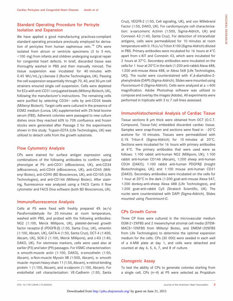
Standard Operating Procedure for PericyteIsolation and ExpansionWe have applied a good manufacturing practices-compliantstandard operating procedure previously employed for deriva-tion of pericytes from human saphenous vein.16 CPs wereisolated from atrium or ventricle specimens (3 to 5 mm,<100 mg) from infants and children undergoing surgical repairfor congenital heart defects. In brief, discarded tissue wasthoroughly washed in PBS and then manually minced. Thetissue suspension was incubated for 40 minutes with0.45 WU/mL/g Liberase 2 (Roche Technologies, UK). Passingthe cell suspension sequentially through 70, 40, and 30-lm cellstrainers ensured single cell suspension. Cells were depletedfor ECswith anti-CD31 conjugated beads (Miltenyi Biotech, UK),following the manufacturer’s instructions. The remaining cellswere purified by selecting CD34+ cells by anti-CD34 beads(Miltenyi Biotech). Target cells were cultured in the presence ofEGM2 medium (Lonza, UK) supplemented with 2% fetal bovineserum (FBS). Adherent colonies were passaged to new culturedishes once they reached 60% to 70% confluence and frozenstocks were generated after Passage 2 for the experimentsshown in this study. Trypsin-EDTA (Life Technologies, UK) wasutilized to detach cells from the growth substrate.
Flow Cytometry AnalysisCPs were stained for surface antigen expression usingcombinations of the following antibodies to confirm typicalphenotype at P5: anti-CD31 (eBioscience, UK), anti-CD34(eBioscience), anti-CD44 (eBioscience, UK), anti-CD45 (Milt-enyi Biotec), anti-CD90 (BD Biosciences, UK), anti-CD105 (LifeTechnologies), and anti-CD146 (Miltenyi Biotec). After stain-ing, fluorescence was analyzed using a FACS Canto II flowcytometer and FACS Diva software (both BD Biosciences, UK).
Immunofluorescence AnalysisCells at P5 were fixed with freshly prepared 4% (w/v)Paraformaldehyde for 20 minutes at room temperature,washed with PBS, and probed with the following antibodies:NG2 (1:100, Merck Millipore, UK), platelet-derived growthfactor receptor-b (PDGFR-b) (1:50, Santa Cruz, UK), vimentin(1:100, Abcam, UK), GATA-4 (1:50, Santa Cruz), OCT-4 (1:400,Abcam, UK), SOX-2 (1:100, Merck Millipore), and c-Kit (1:40,DAKO, UK). For stemness markers, cells were used also atearlier (P3) and later (P9) passages. For VSMC characterization:a–smooth-muscle actin (1:100, DAKO), a-smoothelin (1:50,Abcam), a-Non-muscle Myosin IIB (1:500, Abcam), a- smoothmuscle–myosin heavy chain 11 (1:50, Abcam),a-retinol-bindingprotein 1 (1:100, Abcam), and a-calponin (1:100, Abcam). Forendothelial cell characterization: VE-Cadherin (1:50, Santa
Cruz), VEGFR-2 (1:50, Cell signaling, UK), and von WillebrandFactor (1:50, DAKO, UK). For cardiomyocyte cell characteriza-tion: a-sarcomeric Actinin (1:500, Sigma-Aldrich, UK) andConnexin 43 (1:40, Santa Cruz). For detection of intracellularantigens, cells were permeabilized for 10 minutes at roomtemperaturewith 0.1% (v/v) Triton X100 (Sigma-Aldrich) dilutedin PBS. Primary antibodies were incubated for 16 hours at 4°Capart from c-KIT and Connexin 43, which were incubated for2 hours at 37°C. Secondary antibodies were incubated on thecells for 1 hour at 20°C in the dark (1:200 anti-rabbit Alexa 488,1:200 anti-mouse Alexa 488, or Alexa 568 [Life Technologies,UK]). The nuclei were counterstained with 40,6-diamidino-2-phenylindole (DAPI) (Sigma-Aldrich). Slidesweremounted usingFluoromount-G (Sigma-Aldrich). Cells were analyzed at a 9400magnification. Adobe Photoshop software was utilized tocompose and overlay the images (Adobe). All experiments wereperformed in triplicate with 3 to 7 cell lines assessed.
Immunohistochemical Analysis of Cardiac TissueTissue sections 8 lm thick were obtained from OCT (O.C.T.Compound, Tissue-Tek) embedded discarded cardiac tissue.Samples were snap-frozen and sections were fixed in �20°Cacetone for 10 minutes. Tissues were permeabilized with0.1% Triton-X (Sigma-Aldrich) for 10 minutes at 20°C.Sections were incubated for 16 hours with primary antibodiesat 4°C. The primary antibodies that were used were asfollows: 1:100 rabbit anti-human NG2 (Millipore, UK); 1:100rabbit anti-human CD146 (Abcam), 1:200 sheep anti-humanCD34 (DAKO); 1:100 rabbit anti-human PDGFRb (InsightBiotechnologies, UK); and 1:100 mouse anti-human CD31(DAKO). Secondary antibodies were incubated on the cells for1 hour at 20°C in the dark (1:200 goat-anti-mouse Alexa 547,1:200 donkey-anti-sheep Alexa 488 (Life Technologies), and1:200 goat-anti-rabbit Cy5 (Stratech Scientific, UK). Thenuclei were counterstained with DAPI (Sigma-Aldrich). Slidesmounted using Fluoromount-G.
CPs Growth CurveThree CP lines were cultured in the microvascular mediumEGM-2+2%FBS and 2 mesenchymal stromal cell media (STEM-MACS+10%FBS from Miltenyi Biotec, and DMEM+20%FBSfrom Life Technologies) to determine the optimal expansionmedium for the cells. CPs (30 000) were seeded in each wellof a 6-MW plate at day 1, and cells were detached andcounted at day 4, 5, 6, 7, and 8 of culture.
Clonogenic AssayTo test the ability of CPs to generate colonies starting froma single cell, CPs (n=4) at P5 were selected as Propidium
DOI: 10.1161/JAHA.115.002043 Journal of the American Heart Association 3
Cardiac Pericytes and Congenital Heart Disease Avolio et alORIG
INALRESEARCH
by guest on June 21, 2015http://jaha.ahajournals.org/Downloaded from
Iodide negative (live cells) and sorted into single cells usinga BD Bioscience Influx sorter (BD Biosciences, UK). Sortedcells were placed into each well of a 96-well culture plate(Greiner Bio-one, UK). The sorted cells were cultured up to4 weeks in EGM-2 and the number of colonies generatedwas counted.
Multilineage Differentiation Toward the 3Cardiovascular LineagesThe ability of CPs to differentiate into cardiomyocytes, ECs orVSMCs were tested following exposure to inductive media aspreviously described.16,20,21 Cells were seeded at 5000 cells/cm2 and allowed to become confluent. The culture mediaexchange every 3 days, for 14 to 21 days. The followingdifferentiation methods and media were used: (1) Endothelialcell differentiation: ECs were generated using either apreviously described method—which provides the culture ofcells in medium enriched with 10 ng/mL human VEGF(vascular endothelial growth factor, PeproTech EC Ltd, UK)—or CFU-Hill Liquid Medium Kit (StemCell Technologies, UK).(2) Cardiomyocyte cell differentiation: 2 different previouslypublished methods were employed. The first is based on theculture of cells with a medium containing ascorbic acid(Sigma-Aldrich), 10 ng/mL human basic fibroblast growthfactor, 10 ng/mL human VEGF, and 10 ng/mL human insulin-like growth factor 1 (all from PeproTech EC Ltd). The secondmethod described in Smits et al, consists of an initial 3 day-long incubation of the cells with 5 lmol/L 5-aza-20-deoxy-cytidine (Sigma-Aldrich) followed by the culture of cells with amedium containing ascorbic acid and 1 ng/mL humantransforming growth factor b-1 (PeproTech EC Ltd). (3)Vascular smooth muscle cell differentiation: differentiationmedium added with 20 ng/mL of human PDGF-BB (Pepro-Tech). All the experiments were performed in triplicate with 2to 4 cell lines assessed.
Analysis of CPs Cell Secretome by ELISAThe quantities of the following secreted proteins weredetermined in CPs cultured when cells were exposed tonormoxic conditions for 48 hours in serum-free medium:human VEGF, basic fibroblast growth factor, hepatocytegrowth factor, angiopoietin-1, angiopoietin-2, and stromalcells–derived factor 1, transforming growth factor-b1, andprocollagen type I (all from R&D Systems, UK). ELISAswere performed following manufacturer’s instructions. Cyto-kine concentrations were expressed normalizing the datafor the number of the cells at the end of the collectiontime, and for the time of incubation. Four different CPslines were assessed and experiments were performed intriplicate.
In Vitro Matrigel AssayNetwork formation was performed as described previously.16
Briefly, CPs were labeled with VyBrant diI (Life Technologies)and seeded with human umbilical vein endothelial cells(HUVECs, Lonza, UK, 1:4 ratio of CPs to HUVECs, respec-tively) in a 96-well plate at 7000 cells/well on top of 70 lLthick-coated Matrigel (BD Biosciences) in EGM2 with FBS for6 hours. HUVECs and CPs were also seeded alone as acontrol. To assess the effect of CP-secretome, CP-condi-tioned media diluted 1:2 with fresh EGM-2 and FBS wereincubated with 7000 HUVECs/well on top of 70 lL thick-coated Matrigel for 6 hours. HUVECs with un-conditionedEGM-2 were also seeded alone as a control. FBS concen-tration was the same in all the experimental conditions. CPs-alone network formations were also assessed at different cellnumbers (2000, 5000, 7000). Images were taken underbright-field at 59 and the length of the networks wasmeasured. All experiments were performed in triplicate with3 to 4 cell lines assessed.
Labeling of Cells With DiI TrackerFor selected in vitro experiments, CPs were stained with thelong-term cell trackers VyBrant diI (Life Technologies). DiI wasdiluted 1:1000 in PBS and incubated with confluent cells(adherent to the culture plate) for 5 minutes at 37°C and thenon ice for an additional 15 minutes, in the dark. Cells werethen washed with PBS, left to recover for 24 hours, and usedfor experiments.
Transwell Migration AssayTo test the capacity of CPs secretome to induce themigration of human CSC and HUVECs, 60 000 CSC orHUVECs were seeded in 24-well plates on a 6.5-mmTranswell� with 5.0-lm pore polycarbonate membraneinsert (Corning, UK), in serum-free EGM-2 medium (Lonza).In the bottom of the wells, 0.5 mL of CPs serum-freeconditioned medium was added. Serum-free EGM-2 mediumwas used as negative control. Serum-free EGM-2 supple-mented with human VEGFa (100 ng/mL; PeproTech EC Ltd)was used as a positive control for HUVECs. Cells wereincubated for 16 hours at 37°C. At the end of this period,the membrane inserts were washed with PBS, fixed with ice-cold methanol for 15 minutes at room temperature, stainedwith DAPI, and mounted on a slide. Membranes wereanalyzed with an epifluorescence microscope at 9200magnification; 10 fields were randomly acquired and cellscounted. Migrated cells were expressed as a percentage.Experiments were performed in duplicate. Four different CPslines were assessed.
DOI: 10.1161/JAHA.115.002043 Journal of the American Heart Association 4
Cardiac Pericytes and Congenital Heart Disease Avolio et alORIG
INALRESEARCH
by guest on June 21, 2015http://jaha.ahajournals.org/Downloaded from

Measurement of Membrane Current andIntracellular Ca2+
To measure membrane current, CPs were placed in anexperimental chamber mounted on the stage of an invertedmicroscope, and superfused with solution containing (inmmol/L): 140 NaCl, 4 KCl, 2 CaCl2, 1 MgCl2, 10 glucose, 5HEPES (pH 7.4 with NaOH) at 35 to 37°C. Membrane currentswere monitored with the whole-cell patch-clamp technique,using an AxoPatch-1D amplifier (Axon Instruments, USA). Thepipette solution contained (in mmol/L): 110 KCl, 10 NaCl, 10HEPES, 0.4 MgCl2, 5 glucose, 5 K2ATP, 0.5 GTP–Tris, with orwithout 5 1,2-bis(o-aminophenoxy)ethane-N,N,N0,N0-tetra-acetic acid (pH 7.1 with KOH). Protocols were generatedand data recorded using pClamp 10 software (Axon Instru-ments) via an A/D converter (Digidata 1440A, Axon Instru-ments/Molecular Devices, USA). Whole-cell membranecurrents were recorded in voltage-clamp mode using eithera ramp protocol, in which membrane potential was rampedfrom �120 mV to +80 mV over 400 ms from a holdingpotential of either �40 mV or �80 mV, or a step protocol, inwhich membrane potential was stepped between �120 mVand +80 mV in increments of 10 mV for 500 ms, from aholding potential of either �40 mV or �80 mV. Ramps andsteps were applied at a frequency of 0.2 Hz. Data werefiltered at 1 kHz and digitized at 10 kHz.
To measure intracellular Ca2+, CPs were incubated with5 lmol/L Fluo-4/AM (Life Technologies) for 25 minutes atroom temperature, before being rinsed in control solution. Thecells were then placed in an experimental chamber mountedon the stage of either a Fluoview 1200 (Olympus, Germany) orPascal (Zeiss, Germany) inverted confocal microscope, andsuperfused with control solution at room temperature. Fluo-4fluorescence was stimulated at 488 nm. Fluorescence wasnormalized to F/F0 as described previously, following correc-tion for photobleaching.22 Cells were stimulated eitherelectrically, via parallel Pt wires connected to an SD8 GrassStimulator, or using 10 lmol/L BT3-IP3/AM (a membrane-permeable form of IP3) or 10 mmol/L caffeine added to thesuperfusate.
Assays on Gene Expression
RNA isolation and quantitative real time (RT)polymerase chain reaction (PCR)
Extracted total RNA was reverse-transcribed into single-stranded cDNA using a High Capacity RNA-to-cDNA Kit (LifeTechnologies) or using specific primers provided with theTaqman miRNA assay and microRNA Reverse Transcription Kit(Life Technologies). The reverse transcription–PCR was per-formed using first-strand cDNA with TaqMan Fast UniversalPCR Master Mix (Life Technologies). Quantitative PCR was
performed on a LightCycler480 Real-Time PCR system (RocheTechnologies, UK). Quantitative PCR parameters for cyclingwere as follows: 50°C incubation for 2 minutes, 95°C for10 minutes, 40 cycles of PCR at 95°C for 15 seconds, and60°C for 1 minute. The following Taqman assays were used:UBC (Hs00824723_m1) as a housekeeper, for smooth muscledifferentiation characterization: calponin 1 (Hs00154543_m1);smooth muscle–myosin heavy chain (Hs00224610_m1); ret-inol-binding protein 1 (Hs01011512_g1); smooth muscle actin(Hs00426835_g1), smoothelin (Hs00199489_m1); for endo-thelial differentiation characterization: KDR (Hs00911700_m1); CD31 (Hs00169777_m1); von WillebrandFactor (Hs01109446_m1); for cardiomyocytes differentiationcharacterization: Brachyury (Hs00610080_m1); Connexin 43(Hs00748445_s1); NKX2.5 (Hs00231763_m1); MYH7(Hs01116032_m1); ISLET1 (Hs00158126_m1); CACNAC1(Hs00167681_m1), Tbx5 (Hs03675785_s1). All reactionswere performed in a 10-lL reaction volume in triplicate. ThemRNA expression level was determined using the 2�DCt
method. Each reaction was performed in triplicate.
Static Culture of Cardiac Pericytes on theCorMatrixPieces of CorMatrix� ECM� (CorMatrix Cardiovascular,Sunnyvale, CA) — a decellularized xenograft materialclinically approved for use in cardiac surgery — of �1.5-cm diameter were cut and placed in wells of a 24-well plate.To fix the bioscaffold pieces to the bottom of the wells,CellCrown inserts were used (Sigma-Aldrich). Prior to theseeding, the bioscaffolds were incubated for 2 days withEGM-2 media. CPs (50 000) at P5 were seeded into eachCorMatrix-containing well and maintained for 7 days on thebioscaffolds. The bioscaffolds were fixed in 4% Paraformal-dehyde for 20 hours at 4°C and imbedded in paraffin orOCT-frozen. Eight-micrometer sections were examined forthe presence of CPs using anti-vimentin, NG2, and PDGFR-bantibodies (see above), which were incubated for 20 hoursat 4°C after permeabilization and blocking. Secondaryantibody was incubated on the sections for 1 hour at roomtemperature. The nuclei were counterstained with DAPI(Sigma-Aldrich). The slides were mounted using Fluoro-mount-G� mounting media (Sigma-Aldrich) and immunoflu-orescence pictures were taken after 24 hours both at 920and 940.
Dynamic Culture of CPs on CorMatrixPericytes were seeded onto the CorMatrix scaffold (CorMatrixCardiovascular) at 0.5 million cells/cm2 and cultured for1 week under static conditions followed by 2 weeks underdynamic conditions. For the dynamic cell culture, the pericyte-
DOI: 10.1161/JAHA.115.002043 Journal of the American Heart Association 5
Cardiac Pericytes and Congenital Heart Disease Avolio et alORIG
INALRESEARCH
by guest on June 21, 2015http://jaha.ahajournals.org/Downloaded from

seeded CorMatrix was grown in an InBreath Bioreactor(Harvard Apparatus, Holliston, MA). The device ensuresmaintenance of sterility and stabilization, automation, andscale-up/-out of the cellularization process through hydrody-namic stimuli and control of nutrients and oxygen transport tothe cells. The conduit was stitched to the rotating arm of theBioreactor and stitched back onto itself so as to fashion atube shape through the center of which runs the rotating arm.The Bioreactor was filled with EGM-2 medium and placed intothe incubator at 37°C with the medium being changed twice aweek. At the end of the total 3 weeks of culture, the viabilityof the seeded cells on the scaffolds was detected using theBiotium fluorescent viability/cytotoxicity Assay kit. Fluores-cence imaging was carried out on the whole thickness ofgraft. Tissue-engineered scaffolds were then analyzed byhistological staining of the nuclei and of the extracellularmatrix components elastin and collagen, detected, respec-tively, by hematoxylin and eosin (H&E) and Elastic Van Giesonstaining.
Statistical AnalysisGraphPad Prism was used to perform the statistical analysis.Due to the limited number of samples (n=3 to 4, too small), anormality test could not be performed. For this reason, 2-group analysis was performed by nonparametric Mann–Whitney test. Values were expressed as means�standarderror of the mean (SEM). Probability values (P) <0.05 wereconsidered significant.
Results
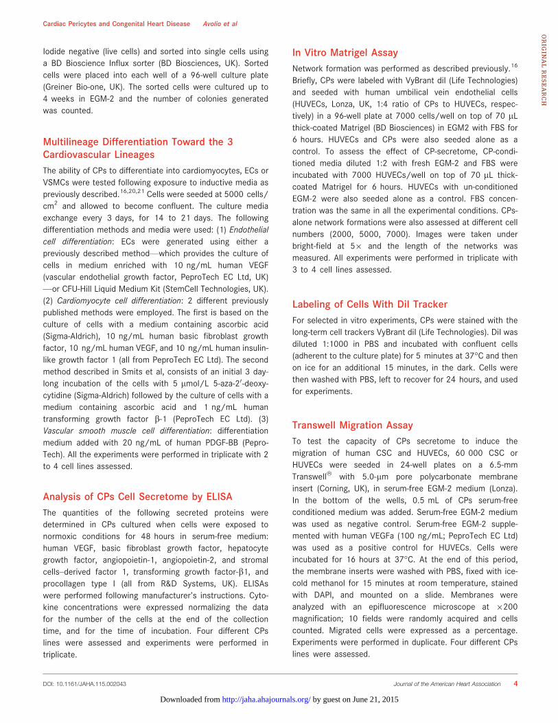
Immunohistochemical Localization of CPsMultiple staining immunohistochemistry and confocal micros-copy were used for cell localization in cardiac tissue(Figure 1). CD31pos/CD34pos ECs were typically recognizedaround the lumen of capillaries and arterioles. Moreover, weidentified CD31neg/CD34pos/NG2pos cells around capillaryECs (Figure 1A) and especially within the layer external toNG2pos VSMCs in arterioles (Figure 1B). Additionally,CD31neg/CD34pos CPs do not express CD146 (Figure 1C).
Isolation/Expansion and Characterization of CPsThe good manufacturing practices-compliant standard operat-ing procedure, previously employed for derivation of saphenousvein pericytes,16 proved to be highly efficient when applied tocardiac leftovers, with an expansion success (�20 million cellsat P5, in 4 to 6 weeks) of 10 samples out of 13 examined. This isa remarkable result, considering the weight of source cardiactissue in comparison with saphenous veins (Figure S1). CPs can
be successfully expanded in EGM-2 supplemented with 2% FBS(both from Lonza), a microvascular media that we already usefor optimal expansion of saphenous vein pericytes. The attemptto culture CPs in 2 mesenchymal stromal cell media (STEM-MACS+10%FBS fromMiltenyi Biotec, and DMEM+20%FBS fromLife Technologies) clearly showed that EGM-2 is the optimalmedium for expansion of CPs (Figure S2).
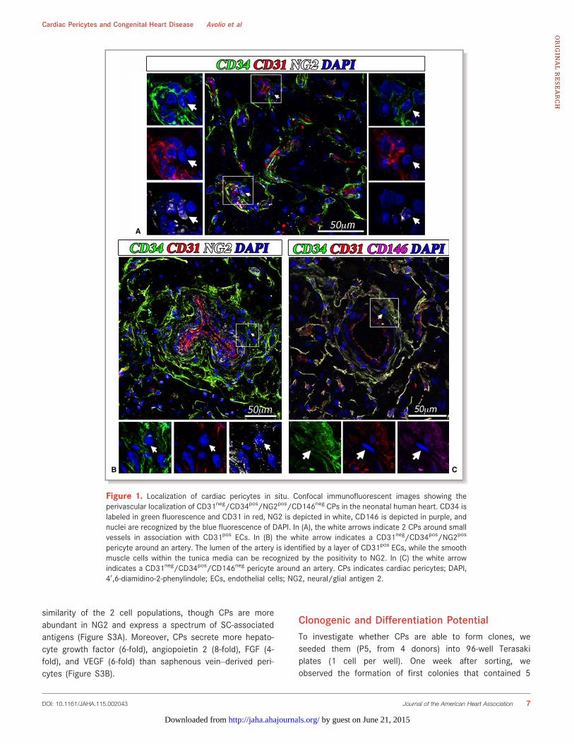
With use of contrast phase microscopy, expanded cellsshow typical spindle-shape morphology (Figure 2A). Cells atP5 (n=8 donors) were assayed for their antigen expressionby flow cytometry and immunocytochemistry, which showedsuperimposable characteristics of atrium- and ventricle-derived cells. Flow-cytometry analysis of expanded cellsindicates high abundance of CD44 and CD105 and variableexpression of CD90 (Figure 2B). In contrast, there was ascarcity/absence of endothelial-CD31 and hematopoietic-CD45 markers. Another unique characteristic is the lack ofCD146, which distinguishes the isolated population fromCD146pos pericytes described previously in postmortemsamples from fetal and adult hearts.14 Moreover, as shownby us with saphenous vein–derived pericytes, expansion inculture was associated with an expected downregulation ofCD34. Immunocytochemical analyses showed the consis-tent expression of vimentin (100%), NG2 (95%), andPDGFRb (58%). Additionally, expanded cells were positivefor the cardiac transcription factor GATA-4 (47%) and forthe stemness markers OCT-4 (64%), SOX-2 (54%), andNANOG (34%) (Figure 3A). Around 30% of the totalpopulation showed coexpression of OCT4/NANOG andSOX-2/NANOG (Figure 3B). The expression of stemnessmarkers slightly decreased along with cell expansion, frompassage 3 to passage 5 of culture (OCT-4 58%, SOX-2 50%,NANOG 28%), but the difference was not significant (Mann–Whitney test, OCT-4 P=0.72, SOX-2 P=0.23, NANOGP=0.63) (Figure 3C). As expected, a further decreasingtrend was observed up to passage 9 (OCT-4 39% andNANOG 14%, Mann–Whitney test P3 versus P9: OCT-4P=0.4, NANOG P=0.1). Finally, CPs did not express themast/stem cell growth factor receptor (SCFR/c-Kit), indi-cating their distinction from classical cardiac stem cellscurrently used in clinical trials23,24.
We next assessed the ability of CPs (n=4 donors) tosecrete growth factors, cytokines, and extracellular matrixprotein (Figure 4). We found that hepatocyte growth factorwas the most abundant secreted factor, followed byangiopoietin 2, VEGF-A, angiopoietin 1, stromal cell derivedfactor-1a, and fibroblast growth factor-B. Furthermore, wefound that CPs release procollagen type 1, a majorconstituent of cardiac extracellular matrix, in conditionedmedia (0.8�0.1 ng/mL, n=4 lines). Instead, CPs do notrelease transforming growth factor-b1 in the culture media.Comparison with saphenous vein–derived pericytes indicates
DOI: 10.1161/JAHA.115.002043 Journal of the American Heart Association 6
Cardiac Pericytes and Congenital Heart Disease Avolio et alORIG
INALRESEARCH
by guest on June 21, 2015http://jaha.ahajournals.org/Downloaded from
similarity of the 2 cell populations, though CPs are moreabundant in NG2 and express a spectrum of SC-associatedantigens (Figure S3A). Moreover, CPs secrete more hepato-cyte growth factor (6-fold), angiopoietin 2 (8-fold), FGF (4-fold), and VEGF (6-fold) than saphenous vein–derived peri-cytes (Figure S3B).
Clonogenic and Differentiation PotentialTo investigate whether CPs are able to form clones, weseeded them (P5, from 4 donors) into 96-well Terasakiplates (1 cell per well). One week after sorting, weobserved the formation of first colonies that contained 5
A
B C
Figure 1. Localization of cardiac pericytes in situ. Confocal immunofluorescent images showing theperivascular localization of CD31neg/CD34pos/NG2pos/CD146neg CPs in the neonatal human heart. CD34 islabeled in green fluorescence and CD31 in red, NG2 is depicted in white, CD146 is depicted in purple, andnuclei are recognized by the blue fluorescence of DAPI. In (A), the white arrows indicate 2 CPs around smallvessels in association with CD31pos ECs. In (B) the white arrow indicates a CD31neg/CD34pos/NG2pos
pericyte around an artery. The lumen of the artery is identified by a layer of CD31pos ECs, while the smoothmuscle cells within the tunica media can be recognized by the positivity to NG2. In (C) the white arrowindicates a CD31neg/CD34pos/CD146neg pericyte around an artery. CPs indicates cardiac pericytes; DAPI,40,6-diamidino-2-phenylindole; ECs, endothelial cells; NG2, neural/glial antigen 2.
DOI: 10.1161/JAHA.115.002043 Journal of the American Heart Association 7
Cardiac Pericytes and Congenital Heart Disease Avolio et alORIG
INALRESEARCH
by guest on June 21, 2015http://jaha.ahajournals.org/Downloaded from

to 20 cells. At 4 weeks, we found that CPs from atrium orventricle give rise to similar numbers of clones (10% and11% of seeded cells, respectively), a result that surpassesthe reported clonogenic activity of saphenous vein–derivedpericytes (7%).16
We next exposed CPs (n=4 donors) to differentiationstimuli for derivation of vascular and cardiac lineages. Asshown in Figure 5A, CPs do not express typical VSMCantigens. Following exposure to a differentiation mediumenriched with human platelet-derived growth factor BB(PDGFBB),25 they initially acquired the expression of nonmus-cle myosin B, which is typical of synthetic VSMCs, and thenadditionally expressed retinol-binding protein 1, a-smooth-muscle actin, and smooth muscle–calponin, suggesting theirmaturation into a contractile phenotype. The analysis of geneexpression by quantitative real time-PCR (Figure 5B) con-firmed a marked induction of a-smooth-muscle actin (average18-fold increase after 14 days) and SM-calponin (157-fold),while typical antigens of mature contractile VSMCs were lessupregulated (smooth muscle–myosin heavy chain, average5.5-fold increase after 14 days, and smoothelin, 4.2-fold
increase). In addition, retinol-binding protein 1 mRNA levelspeaked at 7 days (3.1-fold) and returned to basal level at14 days.
Culture of CPs in CFU-Hill Medium or a medium enrichedwith human VEGF-A failed to induce the expression of matureendothelial proteins as detected by immunocytochemistry(KDR/VEGFR2, von Willebrand Factor, VE-Cadherin) (data notshown); quantitative PCR analysis confirmed only a slightincrease of von Willebrand Factor (2.5-fold), while CD31 andKDR were not increased or even downregulated (5-folddecrease), respectively, after the differentiation protocolcompared to baseline CPs (Figure 5B).
To induce the differentiation into cardiomyocytes, weadopted 2 protocols that differ from each other in terms ofgrowth factor composition and presence or absence of theepigenetic modifier 5-Azacytidine.20,21 We observed that CPsacquire the expression of a-Sarc-actinin and connexin-43, butdo not show the localized pattern typically seen in maturecardiomyocytes (Figure 5C). Furthermore, quantitative realtime-PCR analyses (Figure 5B) confirmed the induction ofconnexin-43 (average 9-fold increase after 21 days), T-box
A B
Figure 2. In vitro characterization of cardiac pericytes (CPs). (A) Representative phase-contrast opticalimage of CPs in culture (in the upper image magnification is 9200). (B) Representative flow cytometryhistograms of cultured CPs at P5. Isotype control IgG staining profiles are shown by the red border linehistograms, while specific antibody staining profiles are shown by full green histograms. Data are expressedas means�SEM (n=8 CPs).
DOI: 10.1161/JAHA.115.002043 Journal of the American Heart Association 8
Cardiac Pericytes and Congenital Heart Disease Avolio et alORIG
INALRESEARCH
by guest on June 21, 2015http://jaha.ahajournals.org/Downloaded from
A
B
C
Figure 3. Immunofluorescence characterization of CPs. (A) Epifluorescence microscopy analysis of CPs at P5 for theexpression of the pericyte markers vimentin, NG2, and PDGFR-b, the cardiac transcriptional factor GATA binding protein 4(GATA-4) and the stemnessmarkers octamer-binding transcription factor 4 (OCT-4), (sex determining region Y)-box 2 (SOX-2), and homeoboxNANOG (NANOG). Nuclei are shownby the blue fluorescence of DAPI. Scale bar: 50 lm.Bar graphs showthe percentage of cells positive for eachmarker. Data are expressed asmeans�SEM (n=4 CPs. Only for stemnessmarkers,n=7). (B) Analysis of coexpression of stemness markers in n=3 lines of CPs, at passage 3 of culture. White arrows in theimmunofluorescencemicroscopy pictures identify CPs coexpressing the indicated stemness markers; scale bar: 50 lm. Inthe bar graph, cells coexpressing the stemness markers are indicated as the percent of the entire population of cells. (C)Graph showing the expression of stemness markers in CPs at passages 3 and 5 of culture. Values are plotted asmeans�SEM, n=3. Values were not significantly different between P3 and P5 (Mann–Whitney test, OCT-4 P=0.72, SOX-2P=0.23, NANOGP=0.63). CPs indicates cardiac pericytes;DAPI, 40,6-diamidino-2-phenylindole; NG2, neural/glial antigen2;PDGFR-b, platelet-derived growth factor receptor-b.
DOI: 10.1161/JAHA.115.002043 Journal of the American Heart Association 9
Cardiac Pericytes and Congenital Heart Disease Avolio et alORIG
INALRESEARCH
by guest on June 21, 2015http://jaha.ahajournals.org/Downloaded from

transcription factor (Tbx5, a marker of cardiac development,1.5-fold), and calcium channel, voltage-dependent, L type,a1C subunit (CNAC1C, average 18-fold). However, themesoderm marker brachyury, early cardiac markers Islet-1,and NK2 homeobox 5 (NKX2.5) as well as myosin heavy chain7, cardiac muscle, b (MYH7) could not be detected. Failure ofCPs to acquire typical cardiac transcription factors mayindicate the incapacity to transverse the whole differentiationprocess.
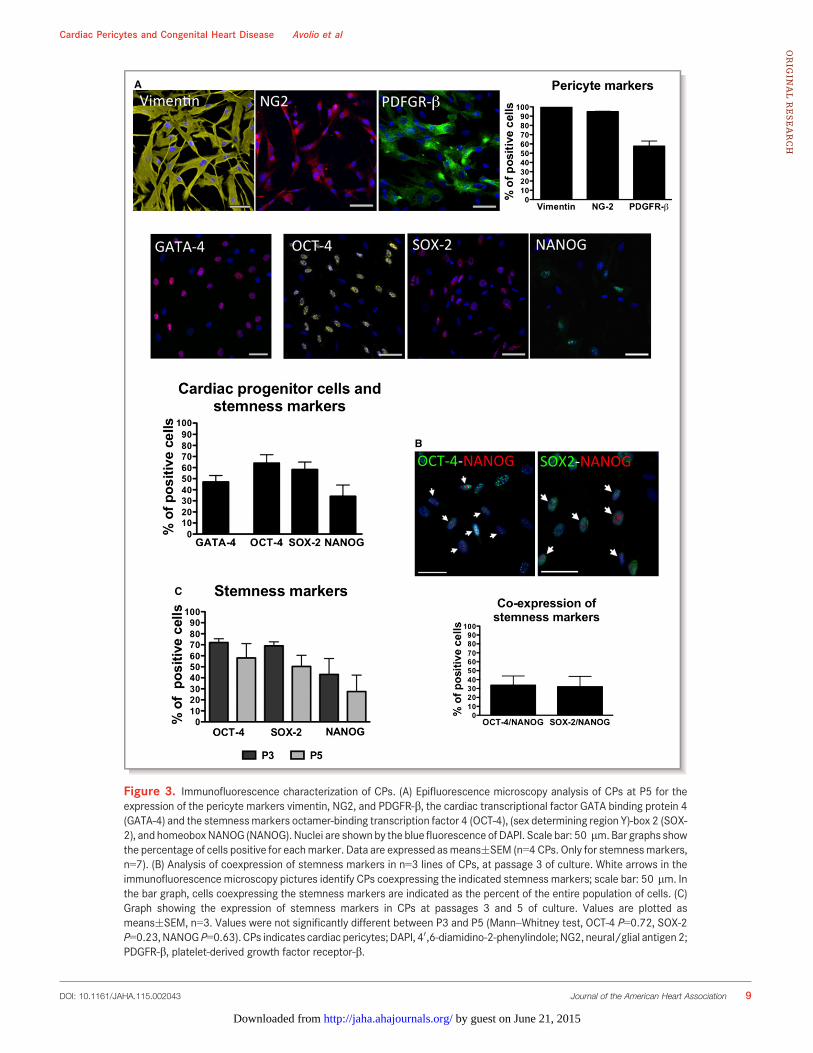
To further verify the cardiomyogenic potential of CPs, wenext assessed their electrical properties before and afterinduction of differentiation. Figure 6 shows examples ofmembrane current (Figure 6A) and intracellular Ca2+ (Fig-ure 6B) measured from representative undifferentiated (left)and differentiated (right) CPs. In undifferentiated cells, depo-larization of the cell membrane resulted in an outwardlyrectifying membrane current, but did not activate voltage-dependent Na+ or Ca2+ currents (n=3); these currents were alsoabsent during step depolarizations (n=2). Membrane currentwas similar when holding potential was �80 mV or �40 mV(which would inactivate Na+ current), and in the absence andpresence of the intracellular Ca2+ chelator 1,2-bis(o-aminophe-noxy)ethane-N,N,N0,N0-tetraacetic acid. Na+ and Ca2+ currentsnormally occur in electrically excitable cells, so these datasuggest that the CPs are not electrically excitable and do notexhibit currents that are regulated by intracellular Ca2+. Theydo, however, appear to have mechanisms for ion flux across the
cell membrane. Consistent with these data, electrical stimula-tion did not cause a rise of intracellular Ca2+ (n=9; not shown),although the cells possess releasable stores of intracellularCa2+, indicated by a transient increase of Ca2+ followed by asustained rise during application of BT3-IP3/AM (n=5 out of 5),as shown in the left panels of Figure 6B. The right panels showthat cellular electrophysiology appeared unchanged followingcardiomyocyte differentiation. However, there appeared to beno increase in intracellular Ca2+ in response to BT3-IP3/AM(n=6 out of 6 cells). Similarly caffeine, which causes Ca2+
release from intracellular stores via ryanodine receptors, had noeffect on intracellular Ca2+ (n=5 out of 5 cells). As a positivecontrol, 110 mmol/L CaCl2 was added to the perfusate, whichresulted in an increase in intracellular Ca2+ in 3 out of 4 cells(insert in lower right panel of Figure 6B).
Interaction of CPs With ECs and CSCsRecruitment of cardiac cells into the graft is important for itsfunctionalization. CPs may help this process by releasingchemoattractant factors. In line with this possibility, resultsillustrated in Figure 7A and 7B indicate that the conditionedmedium of CPs exerts strong attractive actions on c-kitpos
CSCs and ECs in an in vitro migration assay (8-fold increase inCSC and 15-fold increase in ECs migration as compared tounconditioned media; both n=4 donors, Mann–Whitney test,P<0.03). In addition, CPs are able to form networks inMatrigel in a dose-related fashion (Figure S4) and to enhancethe network forming activity of ECs depending on the directcontact between the 2 cell types (2-fold; n=3, Mann–Whitney,P<0.05) (Figure 7C and 7D). These data confirm our previousfindings of cooperative interactions between adventitialpericytes and ECs instrumental to promotion of vasculargrowth and stabilization.16 Instead, the proangiogenic effectwas not observed when ECs were incubated with CP-conditioned media (Figure 7E). However, cautiousness isnecessary when excluding a paracrine action of CPs in theobserved phenomena, as direct contacts are often necessaryto deliver chemical signals between cells. Furthermore, wehave recently shown that cells in coculture influence theparacrine activity of each other.26
Successful Seeding in Clinical-Grade GraftsFinally, we seeded CPs (509103 or 509104 cells/cm2) onCorMatrix, a clinically approved xenograft material, andcultured them for 1 week under static conditions. In addi-tional experiments, at the end of the 1-week static culture, thepericyte-engineered graft was transferred to a double-cham-ber rotating bioreactor, for an additional 2 weeks. Successfulcellularization of the graft texture was demonstrated withhistological stainings for cell nuclei and cytoplasm (hematox-
Figure 4. CPs secretome. Histograms show the amount ofsecreted factors in CP-conditioned media, normalized for thevolume of the collected supernatant, cell number, and time ofincubation. Data are expressed as means�SEM (n=4 CPs). ANG1indicates angiopoietin-1; ANG2, angiopoietin-2; CPs, cardiacpericytes; FGF, fibroblast growth factor; HGF, hepatocyte growthfactor; SDF-1, stromal cells–derived factor 1; VEGF, vascularendothelial growth factor.
DOI: 10.1161/JAHA.115.002043 Journal of the American Heart Association 10
Cardiac Pericytes and Congenital Heart Disease Avolio et alORIG
INALRESEARCH
by guest on June 21, 2015http://jaha.ahajournals.org/Downloaded from

A
B C
Figure 5. Differentiation of CPs toward the cardiovascular lineage. (A) At immunofluorescenceanalysis, undifferentiated CPs are negative for characteristic VSMC markers. After culture for14 days with an inductive medium, cells express markers typical of the synthetic and early contractilephenotype (n=4 CPs). The VSMC markers are identified by green fluorescence; nuclei are shown bythe blue fluorescence of DAPI. T0 indicates the time at which the differentiation has been started, T7and T14 indicate the differentiation time (in days) from T0. Scale bar: 50 lm. (B) The table shows theexpression of endothelial, vascular smooth muscle, and cardiomyocyte genes in CPs cultured for upto 21 days with inductive media. Numbers indicate the average fold change in gene expressioncompared to the basal undifferentiated CPs. Data are represented as means�SEM. (C) Immuno-fluorescence analysis of cardiomyocyte markers showing expression of a-Sarc Actinin (in redfluorescence) and Connexin-43 (in green). In blue, DAPI. For differentiation protocols, see Methods.n=2 CPs for endothelial and smooth muscle, n=3 for cardiac differentiation. CACNA1C indicatesCalcium channel, voltage-dependent, L type, alpha 1C subunit; CPs, cardiac pericytes; DAPI, 40,6-diamidino-2-phenylindole; KDR/VEGFR2, vascular endothelial growth factor receptor 2; Myh7, myosinheavy chain 7, cardiac muscle, b; ND, not determined; NKX2.5, NK2 homeobox 5; RBP1, retinol bindingprotein 1; SM-MHC, smoothmuscle–myosin heavy chain; VSMC, vascular smoothmuscle cell;a-SMA,a-smooth muscle actin; Tbx5, T-box transcription factor; vWF, von Willebrand Factor.
DOI: 10.1161/JAHA.115.002043 Journal of the American Heart Association 11
Cardiac Pericytes and Congenital Heart Disease Avolio et alORIG
INALRESEARCH
by guest on June 21, 2015http://jaha.ahajournals.org/Downloaded from
A
B
Figure 6. Electrophysiology study in cardiac pericytes (CPs) before and after differentiation toward the cardiomyocyte lineage. (A)Membrane current recorded from representative undifferentiated (left) and differentiated (right) CPs, during a depolarizing voltage rampfrom �120 to +80 mV over 400 ms (shown below). (B) A time-series of confocal images of intracellular Ca2+, monitored using the Ca2+-sensitive fluorescent indicator Fluo-4, from representative undifferentiated (left) and differentiated (right) CPs during application of amembrane-permeable form of IP3 to release Ca from intracellular stores. The rise in intracellular Ca2+ is denoted by the color shift fromlight blue toward red (shown in scale bar). The vertical scale bars indicate 20 lm. Normalized Fluo-4 fluorescence is plotted against timein the bottom panels, where the presence of BT3-IP3/AM is indicated by the gray bar; the time-point for each image is indicated by a filledcircle. The inset in the right panel shows the increase of intracellular Ca2+ following addition of 110 mmol/L CaCl2 to the extracellularsolution.
DOI: 10.1161/JAHA.115.002043 Journal of the American Heart Association 12
Cardiac Pericytes and Congenital Heart Disease Avolio et alORIG
INALRESEARCH
by guest on June 21, 2015http://jaha.ahajournals.org/Downloaded from

A B
C
E
D
Figure 7. CPs recruit ECs and CSCs and support network formation by HUVECs. A and B, Inmultitranswell migration assay CP-CM showed the ability to enhance migration of CSCs (A) and HUVECs(B) (VEGF 100 ng/mL was used as positive control). Data are represented as means�SEM. *P<0.03 vsEGM-2. C and D, Representative phase-contrast and fluorescent images of HUVECs, CPs, andHUVECs+CPs in 4:1 ratio forming tubular networks when cultured for 6 hours on Matrigel substrate. In(C) CPs were labeled with the long-term cell tracker DiI (in red fluorescence) in order to investigate theirability to cooperate with HUVECs during the formation of tubular structures. Images have been acquiredusing a 9200 magnification. Histograms summarize quantitative data of the tube length per field. Dataare represented as means�SEM. n=3 per each group. *P<0.04 vs HUVECs, §P<0.05 vs CPs. E,Representative phase-contrast images of HUVECs incubated with CP-CM or EGM-2 for 6 hours onMatrigel substrate. Magnification 950. Histograms summarize quantitative data of the tube length perfield. Data are represented as means�SEM. n=4 per each group. CP-CM indicates cardiac pericytesconditioned medium; CPs, cardiac pericytes; CSCs, cardiac stem cells; ECs, endothelial cells; EGM-2,endothelial growth medium 2; HUVECs, human umbilical vascular endothelial cells; VEGF, vascularendothelial growth factor.
DOI: 10.1161/JAHA.115.002043 Journal of the American Heart Association 13
Cardiac Pericytes and Congenital Heart Disease Avolio et alORIG
INALRESEARCH
by guest on June 21, 2015http://jaha.ahajournals.org/Downloaded from

ylin and eosin) and for the extracellular matrix componentselastin and collagen (Elastic Van Gieson staining) (Figure 8); inaddition, maintenance of the original phenotype was docu-mented by fluorescence staining with vimentin, NG2, andPDGFRb antibodies (Figure 9). Cell viability within the engi-neered scaffold was confirmed with live-cells imaging usingthe Biotium viability/cytotoxicity Assay kit (Figure 10).
DiscussionClinical cell therapy for heart disease has been aimed atpromoting the healing of ischemic myocardium in adultpatients. New evidence suggests that SC therapy couldpotentially offer a new treatment standard for patients withCHD.27 Importantly, the neonatal heart offers a wider andmore na€ıve spectrum of cellular options than the adult heart.
The field is still in its beginning, with initial clinical studiesshowing evidence of feasibility and safety.28
Here, we show new data on progenitor cells that could beconsidered for tissue engineering in patients with CHD.Using a standard operating procedure previously set up toobtain pericytes from human saphenous veins, we have nowisolated, expanded, and characterized a novel population ofCPs from both atria and ventricle of infants and children withCHD.
The idea of “recycling” and expanding regenerative cellsfrom tiny surplus material of palliative surgery suggests asustainable therapeutic undertaking. The reported expansionsuccess (�20 million cells at P5, in 4 to 6 weeks in 77% ofsamples examined) largely surpasses the outcome previouslyobtained with saphenous vein leftovers.16 This may be due toeither a greater abundance or higher proliferation rate offounder cells from the neonatal heart as compared withcounterpart cells from the adult vasculature. The higherexpression of SC markers by, and greater clonogenic activityof, cardiac versus vascular pericytes suggests that the formerare more immature elements capable of high expansioncapacity. Furthermore, CPs have the ability to differentiateinto VSMCs under in vitro inductive conditions, whereas thisproperty was lacking in adult vascular pericytes.16 However,analysis of specification into other cardiovascular lineagesindicates a relative restriction of CP differentiation capacity. Infact, they do not express typical EC markers before and afterstimulation by endothelial inducers. Additionally, the incom-plete manifestation of cardiac transcription factors/proteincombined with lack of developing electrophysiological prop-erties of mature cardiomyocytes indicates the incapacity ofCPs to transverse the whole cardiomyogenic process. Themeasurement of membrane current and intracellular Ca2+
shows the feasibility of making such measurements fromneonatal CPs. These experiments also show that expansionresults in cells that exhibit transmembrane ion flux, whichdoes not change linearly with voltage, and thus appears to bepresumably controlled by ion channels in the cell membrane.The voltage dependence of membrane current indicates anonspecific cation conductance, as described previously inother pericyte populations.29 Similarly, the observation, inagreement with previous work,29 that IP3 can cause anincrease of intracellular Ca2+ indicates that pericytes haveintracellular Ca2+ handling mechanisms, including the abilityto sequester Ca2+ into an intracellular organelle, from which itcan be released, presumably via IP3 receptors. However, thelack of Ca2+ and Na+ currents, and the inability of electricalstimulation to cause an increase of intracellular Ca2+, showthat these cells differ from electrically excitable cells, such ascardiomyocytes. The differentiation protocol used in thepresent study appeared to have no effect on membranecurrents, although the ability of IP3 to release intracellular
Figure 8. Analysis of CPs-engineered scaffold. Scaffolds wereanalyzed by histological staining of the nuclei and of theextracellular matrix components elastin and collagen, detected,respectively, by H&E and EVG. Magnification: 9200. CPsindicates cardiac pericytes; EVG, Elastic Van Gieson staining;H&E, hematoxylin and eosin.
DOI: 10.1161/JAHA.115.002043 Journal of the American Heart Association 14
Cardiac Pericytes and Congenital Heart Disease Avolio et alORIG
INALRESEARCH
by guest on June 21, 2015http://jaha.ahajournals.org/Downloaded from

Ca2+ stores appeared to be lost, and the lack of response tocaffeine suggests absence of Ca2+ release from intracellularstores via ryanodine receptors (the normal mechanism forCa2+ release in cardiac myocytes, in response to transmem-brane Ca2+ influx).
Our results are apparently at variance with the findings ofChen et al,14 who showed that a small fraction of CD146pos
pericytes manifest a cardiomyogenic potential following apulse 5-azacytidine treatment and coculture in vitro withneonatal cardiomyocytes. We used 2 protocols for inductionof cardiomyocyte differentiation, including 1 that employs5-azacytidine as an epigenetic modifier favoring differentia-tion. The use of cardiomyocytes in coculture might havehelped the cardiomyogenic induction of CD146pos pericytes;however, as stated by Chen et al, the possibility of fusion ofpericytes with feeder cells cannot be excluded. Moreover,given that Chen and colleagues worked on cardiac biopsiesthat were obtained postmortem from subjects that had diedfrom noncardiac causes, we cannot exclude the possibility
that differences in cell behavior could be associated with theheart conditions and/or with the standard operating proce-dure. While we processed our samples immediately afterharvesting, it is conceivable the necrosis in samples mighthave been present for a longer time before reaching the tissueculture laboratory and the undefined causes of death mighthave affected the epigenetic and molecular signature inChen’s samples.
Although unable to differentiate in ECs and cardiomyo-cytes, CPs possess other reconstitutive properties thatmake them relevant for tissue repair and engineering. Weshow that CPs exert chemoattractive activity toward ECsand form tubular networks in Matrigel alone and in acooperative fashion with ECs. They also produce largeamounts of growth factors and cytokines implicated inangiogenesis. Additionally, CP conditioned media remarkablystimulate the recruitment of human CSCs. CPs also releaseprocollagen type 1, a major constituent of cardiac extracel-lular matrix.
A C
B
Figure 9. CPs engraft in the CorMatrix. CPs (P5) were seeded onto CorMatrix� ECM�. After culturing the bioscaffolds with CPs in EGM2 for1 week in static conditions and 2 weeks in dynamic conditions (within a bioreactor), they were included in molds with O.C.T. Compound, Tissue-Tek (OCT) as depicted in the schematic figure (A). After 24 hours in the freezer, longitudinal sections were cut from the OCT blocks containingthe bioscaffolds. Sections from the middle of these samples were stained with pericyte markers anti-vimentin, anti-NG2, and anti-PDGFR-bantibodies to check for the presence of CPs inside the bioscaffolds both in static (B) and dynamic conditions (C). In (C), the figures in the rightpanel are shown with higher magnification for better observation of CPs engrafted within the graft. Nuclei were counterstained with DAPI. Scalebar: 100 lm. CPs indicates cardiac pericytes; DAPI, 40,6-diamidino-2-phenylindole; ECM, extracellular matrix; EGM2, endothelial growth medium2; NG2, neural/glial antigen 2; PDGFR-b, platelet-derived growth factor receptor-b.
DOI: 10.1161/JAHA.115.002043 Journal of the American Heart Association 15
Cardiac Pericytes and Congenital Heart Disease Avolio et alORIG
INALRESEARCH
by guest on June 21, 2015http://jaha.ahajournals.org/Downloaded from

Finally, in view of creating a pericyte-engineered graft forcorrection of congenital heart defects, we seeded CPs in axenograft scaffold under static and dynamic conditions.Engineering of prosthetic grafts with autologous SCs hasthe 2-fold aim of reducing the thrombogenic risk through re-endothelialization of the graft’s luminal surface and conferringthe properties of a living tissue that grows and remodels in aphysiologic manner in parallel with cardiac and whole bodygrowth. Results show that CPs are able to penetrate deeplywithin the graft texture after 3 weeks of culture in abioreactor system, and that cells within the graft are viable.Moreover, cells maintain the original antigenic phenotype.
With respect to other competing/complementary solutionsfor cellularization of cardiac grafts, CPs appear to be a morerefined and safer cell therapy product. For instance, CPs canbe isolated/expanded from the tiny discarded tissue(<100 mg) obtained at palliative CHD surgery, whereas thescarcity of other SCs in the heart precludes this possibility.Moreover, pericytes do not induce calcification whenimplanted in the heart of mice, as observed with mesenchy-mal SCs.19 CPs may be preferred to pericytes from other
organs, because the former do not require an electiveharvesting procedure and are assumed to engraft moreefficiently in their native tissue. A recent clinical trial showedthe feasibility, safety, and therapeutic efficacy of intracoro-nary administration of autologous cardiosphere-derived cellsin children with hypoplastic left heart syndrome.28 Compar-ison of CPs and cardiosphere-derived cells indicates somesimilarities, as both cell populations express CD105, CD90,vimentin, and GATA4 and are negative for CD31 and CD45.However, different from cardiosphere-derived cells, CPs donot express Tbx5, NKX2.5, and FLK1. Furthermore, cardio-sphere-derived cells classically express c-Kit,24 which isinstead absent in CPs. Therefore, the 2 cell types mayrepresent distinct and potentially complementary therapeuticelements within the pool of cardiac SCs.
In conclusion, we provide a method to isolate and expanda unique population of pericytes from surgery remnants ofcongenitally defective hearts. This is of high clinicalrelevance because the starting material is usually availableduring palliative surgery without additional risk or burden tothe patient. The method allows easy preparation and
Figure 10. Live-cells imaging analysis of cardiac pericytes viability within the CorMatrix scaffold culturedin the bioreactor. The viability of the cells seeded onto the scaffold was detected using the Biotiumviability/cytotoxicity Assay kit. In representative fluorescence images, live cells are indicated by thecytoplasmic green fluorescence of Calcein AM, while very rare dead cells are detectable by the nuclear redfluorescence of EthD-III. Magnification: 9100.
DOI: 10.1161/JAHA.115.002043 Journal of the American Heart Association 16
Cardiac Pericytes and Congenital Heart Disease Avolio et alORIG
INALRESEARCH
by guest on June 21, 2015http://jaha.ahajournals.org/Downloaded from

expansion of the cells to provide elements that accomplishimportant requirements for cell therapy and tissue engineer-ing, being expandable in vitro and able to produce essentialextracellular protein and angiogenic factors, stimulate angio-genesis, attract CSCs, and engraft into biodegradableprosthetic material with high efficiency. Pericyte-engineeredgrafts could be prepared using a good manufacturingpractices-compliant protocol at the time of the first operationin patients born with tetralogy of Fallot or the more severevariant of pulmonary atresia (usually a palliative proceduresuch as a shunt) using a small amount of tissue collectedfrom the right atrium. The tissue-engineered grafts will thenbe ready for implantation within the 3- to 4-month gap thatnormally separates the palliative operation at birth with thefinal complete surgical correction. Although a long multistepprocedure is required to test the good manufacturingpractices quality of a cellular product in order to reachclinical applicability, including preclinical in vivo studies inanimal models of CHD, with this first study we aimed atdemonstrating the feasibility of isolating and expandingseveral million of CPs from tiny myocardial samples of theyoungest patients, and their properties that may elect CPs asgood candidates for use in reconstructive surgery. Currently,the scarce availability of commercial animal models of complexCHD limits the possibility of testing the in vivo regenerativeability of CPs, but our surgical team is working in this direction.Indeed, our next step will be represented by feasibility studieswith CPs in an animal model of CHD. This approach may opennew avenues for correction of complex congenital cardiacdefects with immense medical and social benefits.
AcknowledgmentsWe are thankful to the Wolfson Bioimaging Facility of the Universityof Bristol for the use of the confocal microscopes.
Sources of FundingThis work was supported by (1) manufacture scaleup ofhuman pericyte progenitor cells for regenerative medicine,MRC Translational Stem Cell Research Grant; (2) BristolBiomedical Research Unit in Cardiovascular Disease (lead forRegenerative Medicine workpackage), National InstituteHealth Research Biomedical Research Unit (NIHR BRU); (3)preclinical trial with human pericyte progenitors in a largeanimal model of myocardial infarction, BHF special projectgrant; (4) BHF Centre of Regenerative Medicine; and (5) theSir Jules Thorn award 2014.
DisclosuresNone.
References1. Khoshnood B, Lelong N, Houyel L, Thieulin AC, Jouannic JM, Magnier S,
Delezoide AL, Magny JF, Rambaud C, Bonnet D, Goffinet F; Group ES.Prevalence, timing of diagnosis and mortality of newborns with congenitalheart defects: a population-based study. Heart. 2012;98:1667–1673.
2. Tennant PW, Pearce MS, Bythell M, Rankin J. 20-year survival of children bornwith congenital anomalies: a population-based study. Lancet. 2010;375:649–656.
3. McElhinney DB. Recent progress in the understanding and management ofpostoperative right ventricular outflow tract dysfunction in patients withcongenital heart disease. Circulation. 2012;125:e595–e599.
4. Tweddell JS, Pelech AN, Frommelt PC, Mussatto KA, Wyman JD, Fedderly RT,Berger S, Frommelt MA, Lewis DA, Friedberg DZ, Thomas JP Jr, Sachdeva R,Litwin SB. Factors affecting longevity of homograft valves used in rightventricular outflow tract reconstruction for congenital heart disease. Circula-tion. 2000;102:III130–III135.
5. Bernstein HS, Srivastava D. Stem cell therapy for cardiac disease. Pediatr Res.2012;71:491–499.
6. Pincott ES, Burch M. Potential for stem cell use in congenital heart disease.Future Cardiol. 2012;8:161–169.
7. Scholl FG, Boucek MM, Chan KC, Valdes-Cruz L, Perryman R. Preliminaryexperience with cardiac reconstruction using decellularized porcine extracel-lular matrix scaffold: human applications in congenital heart disease. World JPediatr Congenit Heart Surg. 2010;1:132–136.
8. Bertipaglia B, Ortolani F, Petrelli L, Gerosa G, Spina M, Pauletto P, Casarotto D,Marchini M, Sartore S; Vitalitate Exornatum Succedaneum Aorticum LaboreIngenioso Obtenibitur P. Cell characterization of porcine aortic valve anddecellularized leaflets repopulated with aortic valve interstitial cells: theVESALIO Project (Vitalitate Exornatum Succedaneum Aorticum Labore Ingeni-oso Obtenibitur). Ann Thorac Surg. 2003;75:1274–1282.
9. Hibino N, McGillicuddy E, Matsumura G, Ichihara Y, Naito Y, Breuer C, ShinokaT. Late-term results of tissue-engineered vascular grafts in humans. J ThoracCardiovasc Surg. 2010;139:431–436, 436.e1–2.
10. Cebotari S, Tudorache I, Schilling T, Haverich A. Heart valve and myocardialtissue engineering. Herz. 2010;35:334–341.
11. Dohmen PM, Lembcke A, Holinski S, Pruss A, Konertz W. Ten years of clinicalresults with a tissue-engineered pulmonary valve. Ann Thorac Surg.2011;92:1308–1314.
12. Vono R, Spinetti G, Gubernator M, Madeddu P. What’s new in regenerativemedicine: split up of the mesenchymal stem cell family promises new hope forcardiovascular repair. J Cardiovasc Transl Res. 2012;5:689–699.
13. Nees S, Weiss DR, Juchem G. Focus on cardiac pericytes. Pflugers Arch.2013;465:779–787.
14. Chen WC, Baily JE, Corselli M, Diaz M, Sun B, Xiang G, Gray GA, Huard J, PeaultB. Human myocardial pericytes: multipotent mesodermal precursors exhibitingcardiac specificity. Stem Cells. 2015;33:557–573.
15. Invernici G, Emanueli C, Madeddu P, Cristini S, Gadau S, Benetti A, Ciusani E,Stassi G, Siragusa M, Nicosia R, Peschle C, Fascio U, Colombo A, Rizzuti T,Parati E, Alessandri G. Human fetal aorta contains vascular progenitor cellscapable of inducing vasculogenesis, angiogenesis, and myogenesis in vitro andin a murine model of peripheral ischemia. Am J Pathol. 2007;170:1879–1892.
16. Campagnolo P, Cesselli D, Al Haj Zen A, Beltrami AP, Krankel N, Katare R,Angelini G, Emanueli C, Madeddu P. Human adult vena saphena containsperivascular progenitor cells endowed with clonogenic and proangiogenicpotential. Circulation. 2010;121:1735–1745.
17. Klein D, Weisshardt P, Kleff V, Jastrow H, Jakob HG, Ergun S. Vascular wall-resident CD44+ multipotent stem cells give rise to pericytes and smoothmuscle cells and contribute to new vessel maturation. PLoS One. 2011;6:e20540.
18. Crisan M, Yap S, Casteilla L, Chen CW, Corselli M, Park TS, Andriolo G, Sun B,Zheng B, Zhang L, Norotte C, Teng PN, Traas J, Schugar R, Deasy BM, BadylakS, Buhring HJ, Giacobino JP, Lazzari L, Huard J, Peault B. A perivascular originfor mesenchymal stem cells in multiple human organs. Cell Stem Cell.2008;3:301–313.
19. Katare R, Riu F, Mitchell K, Gubernator M, Campagnolo P, Cui Y, Fortunato O,Avolio E, Cesselli D, Beltrami AP, Angelini G, Emanueli C, Madeddu P.Transplantation of human pericyte progenitor cells improves the repair ofinfarcted heart through activation of an angiogenic program involving micro-RNA-132. Circ Res. 2011;109:894–906.
20. Beltrami AP, Cesselli D, Bergamin N, Marcon P, Rigo S, Puppato E, D’Aurizio F,Verardo R, Piazza S, Pignatelli A, Poz A, Baccarani U, Damiani D, Fanin R,Mariuzzi L, Finato N, Masolini P, Burelli S, Belluzzi O, Schneider C, Beltrami CA.Multipotent cells can be generated in vitro from several adult human organs(heart, liver, and bone marrow). Blood. 2007;110:3438–3446.
DOI: 10.1161/JAHA.115.002043 Journal of the American Heart Association 17
Cardiac Pericytes and Congenital Heart Disease Avolio et alORIG
INALRESEARCH
by guest on June 21, 2015http://jaha.ahajournals.org/Downloaded from

21. Smits AM, van Vliet P, Metz CH, Korfage T, Sluijter JP, Doevendans PA,Goumans MJ. Human cardiomyocyte progenitor cells differentiate intofunctional mature cardiomyocytes: an in vitro model for studying humancardiac physiology and pathophysiology. Nat Protoc. 2009;4:232–243.
22. Cheng H, Lederer WJ, Cannell MB. Calcium sparks: elementary eventsunderlying excitation-contraction coupling in heart muscle. Science.1993;262:740–744.
23. Chugh AR, Beache GM, Loughran JH, Mewton N, Elmore JB, Kajstura J,Pappas P, Tatooles A, Stoddard MF, Lima JA, Slaughter MS, Anversa P, BolliR. Administration of cardiac stem cells in patients with ischemiccardiomyopathy: the SCIPIO trial: surgical aspects and interim analysis ofmyocardial function and viability by magnetic resonance. Circulation.2012;126:S54–S64.
24. Malliaras K, Makkar RR, Smith RR, Cheng K, Wu E, Bonow RO, Marban L,Mendizabal A, Cingolani E, Johnston PV, Gerstenblith G, Schuleri KH, Lardo AC,Marban E. Intracoronary cardiosphere-derived cells after myocardial infarction:evidence of therapeutic regeneration in the final 1-year results of theCADUCEUS trial (CArdiosphere-Derived aUtologous stem CElls to reverseventricUlar dySfunction). J Am Coll Cardiol. 2014;63:110–122.
25. Wanjare M, Kuo F, Gerecht S. Derivation and maturation of synthetic andcontractile vascular smooth muscle cells from human pluripotent stem cells.Cardiovasc Res. 2013;97:321–330.
26. Avolio E, Meloni M, Spencer HL, Riu F, Katare R, Mangialardi G, Oikawa A,Rodriguez-Arabaolaza I, Dang Z, Mitchell K, Reni C, Alvino VV, Rowlinson J, LiviU, Cesselli D, Angelini G, Emanueli C, Beltrami AP, Madeddu P. Combinedintramyocardial delivery of human pericytes and cardiac stem cells additivelyimproves the healing of mouse infarcted hearts through stimulation of vascularand muscular repair. Circ Res. 2015;116:e81–e94.
27. Wehman B, Kaushal S. The emergence of stem cell therapy for patients withcongenital heart disease. Circ Res. 2015;116:566–569.
28. Ishigami S, Ohtsuki S, Tarui S, Ousaka D, Eitoku T, Kondo M, Okuyama M,Kobayashi J, Baba K, Arai S, Kawabata T, Yoshizumi K, Tateishi A, Kuroko Y,Iwasaki T, Sato S, Kasahara S, Sano S, Oh H. Intracoronary autologous cardiacprogenitor cell transfer in patients with hypoplastic left heart syndrome: theTICAP prospective phase 1 controlled trial. Circ Res. 2015;116:653–664.
29. Kawamura H, Kobayashi M, Li Q, Yamanishi S, Katsumura K, Minami M, WuDM, Puro DG. Effects of angiotensin II on the pericyte-containing microvas-culature of the rat retina. J Physiol. 2004;561:671–683.
DOI: 10.1161/JAHA.115.002043 Journal of the American Heart Association 18
Cardiac Pericytes and Congenital Heart Disease Avolio et alORIG
INALRESEARCH
by guest on June 21, 2015http://jaha.ahajournals.org/Downloaded from
Supplementary Material Cardiac Pericytes paper
1
SUPPLEMENTARY MATERIAL
ONLINE FIGURE I
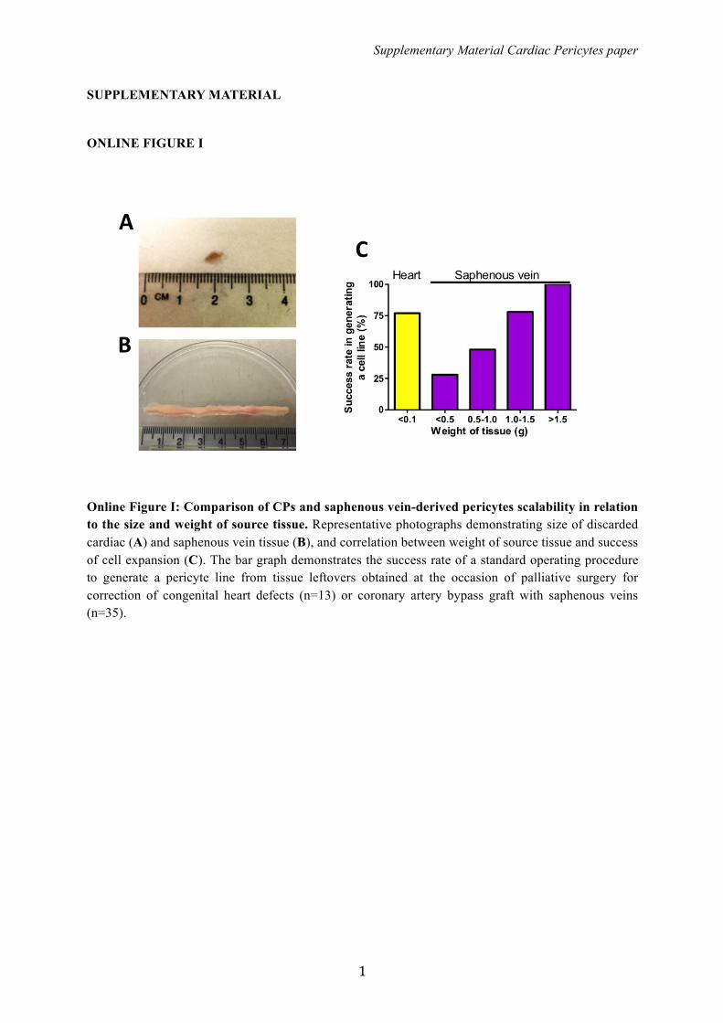
Online Figure I: Comparison of CPs and saphenous vein-derived pericytes scalability in relation to the size and weight of source tissue. Representative photographs demonstrating size of discarded cardiac (A) and saphenous vein tissue (B), and correlation between weight of source tissue and success of cell expansion (C). The bar graph demonstrates the success rate of a standard operating procedure to generate a pericyte line from tissue leftovers obtained at the occasion of palliative surgery for correction of congenital heart defects (n=13) or coronary artery bypass graft with saphenous veins (n=35).

Supplementary Material Cardiac Pericytes paper
2
ONLINE FIGURE II
Online Figure II: Comparison of growth rate of CPs in 3 different culture media. (A) Images of CPs growing in EGM-2 + 2%FBS (Lonza), STEM-MACS + 10%FBS (Miltenyi Biotec) and DMEM + 20%FBS (Life Technologies). Magnification 25X. (B) Graph showing the growth rate of 3 lines of CPs cultured with the 3 media. 30,000 cells were seeded in each well of a 6MW-plate at day 1, and cells were detached and counted at day 4, 5, 6, 7 and 8 of culture. Values are plotted as MEAN+SEM.
Supplementary Material Cardiac Pericytes paper
3
ONLINE FIGURE III
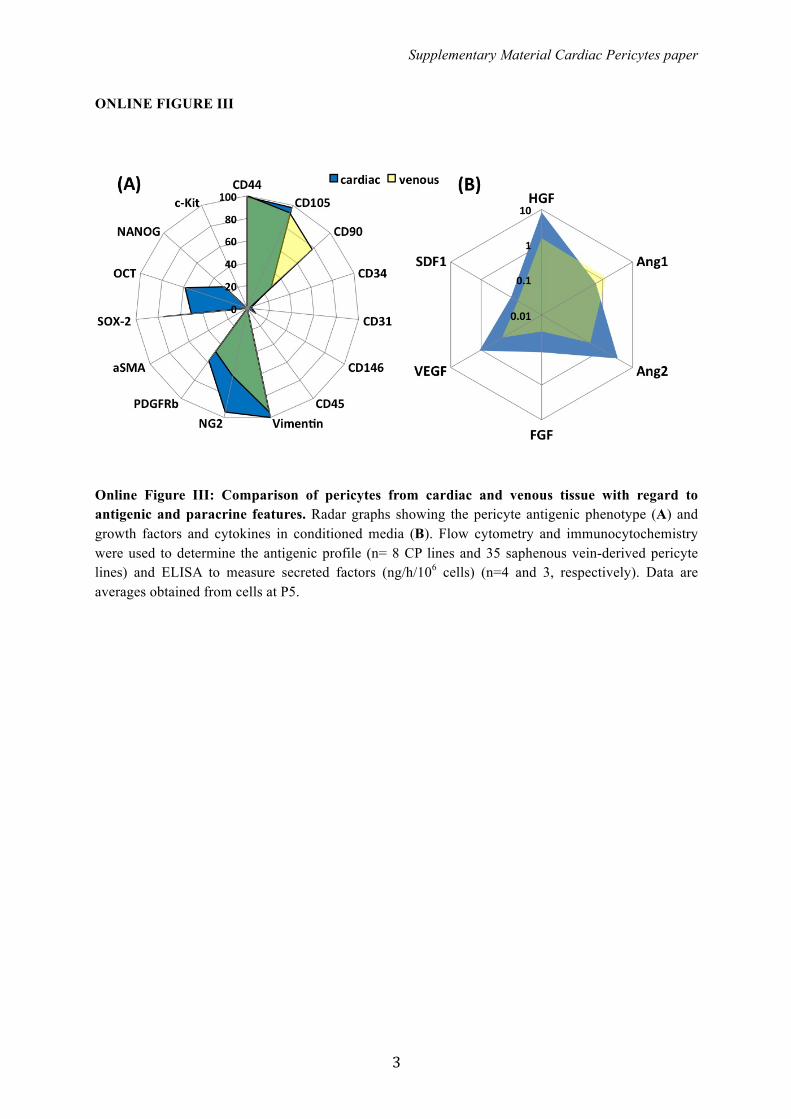
Online Figure III: Comparison of pericytes from cardiac and venous tissue with regard to antigenic and paracrine features. Radar graphs showing the pericyte antigenic phenotype (A) and growth factors and cytokines in conditioned media (B). Flow cytometry and immunocytochemistry were used to determine the antigenic profile (n= 8 CP lines and 35 saphenous vein-derived pericyte lines) and ELISA to measure secreted factors (ng/h/106 cells) (n=4 and 3, respectively). Data are averages obtained from cells at P5.
Supplementary Material Cardiac Pericytes paper
4
ONLINE FIGURE IV
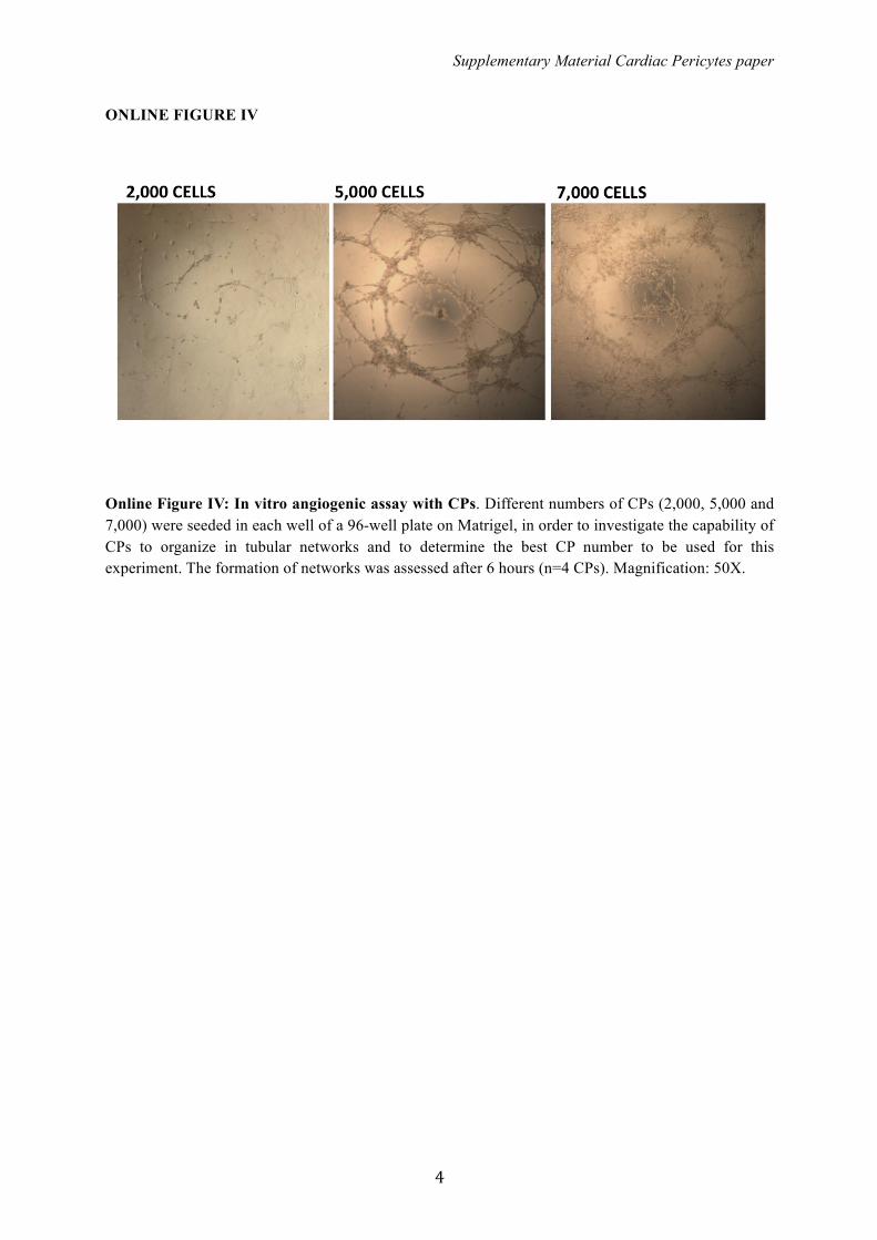
Online Figure IV: In vitro angiogenic assay with CPs. Different numbers of CPs (2,000, 5,000 and 7,000) were seeded in each well of a 96-well plate on Matrigel, in order to investigate the capability of CPs to organize in tubular networks and to determine the best CP number to be used for this experiment. The formation of networks was assessed after 6 hours (n=4 CPs). Magnification: 50X.

Massimo Caputo and Paolo MadedduMohamed T. Ghorbel, Jules C. Hancox, Clive H. Orchard, Gianni Angelini, Costanza Emanueli,
Soyombo, Atsuhiko Oikawa, Megan M. Swim, Cherrie H. T. Kong, Hongwei Cheng, Huidong Jia,Sadie C. Slater, Jonathan Rowlinson, Valeria V. Alvino, Oluwasomidotun O. Idowu, Stephanie
Elisa Avolio, Iker Rodriguez-Arabaolaza, Helen L. Spencer, Federica Riu, Giuseppe Mangialardi,Option for Tissue Engineering in Congenital Heart Disease
Expansion and Characterization of Neonatal Cardiac Pericytes Provides a Novel Cellular
Online ISSN: 2047-9980 Dallas, TX 75231
is published by the American Heart Association, 7272 Greenville Avenue,Journal of the American Heart AssociationThe doi: 10.1161/JAHA.115.002043
2015;4:e002043; originally published June 16, 2015;J Am Heart Assoc.
http://jaha.ahajournals.org/content/4/6/e002043World Wide Web at:
The online version of this article, along with updated information and services, is located on the
http://jaha.ahajournals.org/content/suppl/2015/06/16/JAHA.115.002043.DC1.htmlData Supplement (unedited) at:
for more information. http://jaha.ahajournals.orgAccess publication. Visit the Journal at
is an online only OpenJournal of the American Heart AssociationSubscriptions, Permissions, and Reprints: The
by guest on June 21, 2015http://jaha.ahajournals.org/Downloaded from