examining different levels of prevention of birth defects and ...
576
EXAMINING DIFFERENT LEVELS OF PREVENTION OF BIRTH DEFECTS AND FETAL ALCOHOL SPECTRUM DISORDER BY Y. INGRID GOH, HBSc. A thesis submitted in conformity with the requirements for the degree of Doctor of Philosophy Graduate Department of Pharmaceutical Sciences, University of Toronto © Copyright 2009 by Y. Ingrid Goh. All Rights Reserved. Unauthorized presentation, reproduction or publication is prohibited.
-
Upload
khangminh22 -
Category
Documents
-
view
1 -
download
0
Transcript of examining different levels of prevention of birth defects and ...

EXAMINING DIFFERENT LEVELS OF PREVENTION OF BIRTH DEFECTS AND FETAL ALCOHOL SPECTRUM
DISORDER
BY
Y. INGRID GOH, HBSc.
A thesis submitted in conformity with the requirements
for the degree of Doctor of Philosophy
Graduate Department of Pharmaceutical Sciences,
University of Toronto
© Copyright 2009 by Y. Ingrid Goh. All Rights Reserved. Unauthorized presentation, reproduction or publication is prohibited.

EXAMINING DIFFERENT LEVELS OF PREVENTION OF BIRTH DEFECTS
AND FETAL ALCOHOL SPECTRUM DISORDER
Y. Ingrid Goh
Doctor of Philosophy
Department of Pharmaceutical Sciences, University of Toronto, 2009.
ABSTRACT
While all women hope to deliver a healthy baby, approximately 3-5%
babies are affected by birth defects. Birth defects can occur naturally or be
induced by teratogens. Alcohol is a known teratogen that causes fetal alcohol
spectrum disorder (FASD), the most commonly known cause of
neurobehavioural and neurodevelopmental deficits. Individuals affected with
FASD are likely to be involved with or require additional assistance from
healthcare, education, social services, and justice sectors. Due to this immense
burden, effective prevention of FASD can have a major public impact.
Prevention of FASD can occur at different levels: primary prevention (preventing
alcohol-induced birth defects from occurring in the first place); secondary
prevention (preventing alcohol-induced birth defects from developing or
progressing); tertiary prevention (improving the outcome of individuals affected
with FASD); and quaternary prevention (preventing another child from being
affected with FASD).
ii

The objective of this thesis was to explore a multilevel birth defect and
FASD prevention strategy. Primary prevention by was investigated by maternal
multivitamin supplementation to optimize fetal growing conditions, as alcoholics
are commonly deficient in nutrients. A meta-analysis of maternal multivitamin
supplementation demonstrated a decreased risk for certain congenital anomalies
and pediatric cancers.
Secondary prevention was investigated by a randomized double-blinded
placebo-controlled evaluating the ability of high doses of antioxidants (vitamin C
and vitamin E) to mitigate the effects of prenatal alcohol exposure. The study
was ceased due to safety concerns regarding high doses of vitamin C and
vitamin E in preeclamptic studies.
Tertiary prevention was investigated by anonymous meconium screening
of babies of Grey-Bruce, Ontario residents delivering at or transferred to St.
Joseph’s Health Care in London, Ontario. A 30% prevalence of fatty acid ethyl
esters (FAEE) positive meconium was observed at this high-risk unit.
Meconium screening is also a means of quaternary prevention since
positive screens also identify mothers who were unable to stop consuming
alcohol after 13 weeks of pregnancy, and therefore are at risk of delivering
another child who is prenatally exposed to alcohol. The identification and
engagement of these mothers into treatment programs constitutes primary
prevention of FASD in subsequent pregnancies.
Abstract
iii

ACKNOWLEDGEMENTS
I would like to thank the following individuals for their integral support and
contributions during the course of my training and the completion of my Doctoral
thesis:
The following research would not have been possible without my
collaborators and collaborating institutions. Thank you to APOTEX, Grey-Bruce
Health Unit, Pharmavite, and St. Joseph’s Health Care. A big thank you to the
nurses at St. Joseph’s Health Care, Dr. Henry Roukema, and research
coordinator, Lisa Lum.
The research contained within the thesis was supported by the CIHR FAS-
NET grant, Public Health Agency of Canada, University of Toronto Fellowship,
and Ben Cohen Bursary Fund.
To the members of the Division of Clinical Pharmacology and Toxicology,
Motherisk Team and Laboratory at the Hospital for Sick Children: thank you for
your time, patience, knowledge, instruction and support during these years.
To the staff of our department: Dr. Shinya Ito and Dr. Irena Nulman for
constant guidance through many challenging clinical cases.
To: Adrienne Einarson and Myla Moretti for their support.
To the administrative staff: Hila Halshtok, Leah Moscato, Niki Balamatsis,
and Sarah Bondy. The department would not be able to run without you—you
are never thanked enough.
iv

Thank you to the fellows who I have worked with through my years of
study, particularly Dr. Alon Shrim, Dr. Alejandro Nava-Ocampo, Dr. Facundo
Garcia-Bournissen, Dr. Saskia DeWilt, Dr. Toshihiro Tanaka, Dr. Yaron
Finkelstein, and especially the late Dr. Marina Avner.
To the Motherisk Laboratory, especially Dr. Tatyana Karaskov: for your
invaluable and unlimited knowledge.
To the Motherisk Counsellors I have had the pleasure working with: Dr.
Aida Ebrara, Dr. Angela Chua, Anna Sivojelesova, Caroline Maltepe, Dr.
Enkelejda Bollano, Eunji Kim, Dr. Josephine Djulus, Kathy Mastali, Malaika
Babb, Maud Rouleau, Dr. Michael Tan, Pina Bozzo, and Yvette Navioz.
To my fellow graduate students: Amelia Yip, Amy Lee, Brian Chan,
Brittany Sauvé, Cameron Gilbert, Carolyn Tam, Christelle Gedeon, Daphne
Chan, Daniella Caprara, Eric Ahn, Erika Pollex, Jean-Jacques Dugoua, Kate
McKenna, Maria Nesterenko, Neda Ebrahimi, Patrina Gunness, Patricia Nguyen,
Rebecca Hancock, Sabina Vohra, Sandy Grupp, Sarit Shor, Simerpaul Gill,
Sondra Vandervart, Violette Gijsen, and Vivian Kulaga. A special thanks to
fellow graduate students Janine Hutson, Joey Gareri, Lisa O’Brien, Moumita
Sarkar, and Sanjog Kalra for your support, advice, and companionship.
I would like to acknowledge my supervisor and committee members for
guiding me through this journey. To my committee members: Dr. Bhushan
Kapur, Dr. Peter Selby, Dr. Thomas R. Einarson, and Dr. Wendy J. Ungar—
thank you for your invaluable scientific guidance. Your accomplishments and
expertise has always challenged me to be a better scientist.
Acknowledgements
v

No words can express my deepest gratitude to my supervisor, Dr. Gideon
Koren. Thank you for taking a chance with me and providing me with clinical and
research opportunities that I would have otherwise not have experienced. Thank
you for being a fabulous supervisor and extraordinary mentor. Your passion and
devotion to research field is unparalleled. Your scientific and artistic talent
inspires me daily. Thank you for your unending confidence and encouragement.
Through it all my parents and family have always stood by me through my
education. Thank you for your unending encouragement and understanding.
Your support has meant everything.
Acknowledgements
vi

TABLE OF CONTENTS
ABSTRACT...................................................................................................................... II
ACKNOWLEDGEMENTS ............................................................................................ IV
LIST OF ABBREVIATIONS ...................................................................................XVIII
LIST OF TABLES ....................................................................................................... XXI
LIST OF APPENDICES .......................................................................................... XXIV
CHAPTER 1 ...................................................................................................................... 1
1. GENERAL INTRODUCTION ........................................................................................................... 2
1.1 BIRTH DEFECTS ..................................................................................................................... 2
1.2 TERATOGENICITY OF ETHANOL ....................................................................................... 3
1.3 PREVENTION IN PREGNANCY ............................................................................................ 4
1.4 LEVELS OF PREVENTION OF BIRTH DEFECTS IN PREGNANCY .............................. 5
1.4.1 PRIMARY.............................................................................................................................. 5
1.4.2 SECONDARY....................................................................................................................... 5
1.4.3 TERTIARY ............................................................................................................................ 6
1.4.4 QUATERNARY .................................................................................................................... 6
1.5 NEED FOR MULTILEVEL PREVENTION IN PREGNANCY ............................................. 7
1.6 LEVELS OF PREVENTION FOR FETAL ALCOHOL SPECTRUM DISORDER ............ 7
1.6.1 PRIMARY.............................................................................................................................. 7
1.6.2 SECONDARY....................................................................................................................... 8
1.6.3 TERTIARY ............................................................................................................................ 8
1.6.4 QUATERNARY .................................................................................................................... 8
vii

1.7 NEED FOR MULTILEVEL PREVENTION FOR FETAL ALCOHOL SPECTRUM
DISORDER................................................................................................................................ 9
CHAPTER 2 .................................................................................................................... 10
2 OVERVIEW OF THESIS RESEARCH: RATIONALE, HYPOTHETICAL FRAMEWORK,
AND OBJECTIVES ......................................................................................................................... 11
2.1 RATIONALE............................................................................................................................ 11
2.2 HYPOTHESIS ......................................................................................................................... 11
2.3 OBJECTIVES OF RESEARCH ............................................................................................ 11
CHAPTER 3 .................................................................................................................... 14
3. REVIEW OF THE LITERATURE................................................................................................... 15
3.1 PREVENTION ......................................................................................................................... 15
3.1.1 CLASSIFICATION OF PREVENTION ........................................................................... 15
3.1.1.1 UNIVERSAL PREVENTION.................................................................................................. 15
3.1.1.2 SELECTIVE PREVENTION................................................................................................... 17
3.1.1.3 INDICATED PREVENTION ................................................................................................... 18
3.1.2 LEVELS OF PREVENTION ............................................................................................. 19
3.1.2.1 PRIMARY ................................................................................................................................. 19
3.1.2.2 SECONDARY .......................................................................................................................... 20
3.1.2.3 TERTIARY ............................................................................................................................... 20
3.1.2.4 QUATERNARY ....................................................................................................................... 21
3.2 SCREENING, DIAGNOSIS, AND TREATMENT/INTERVENTION ................................ 21
3.2.1 SCREENING ...................................................................................................................... 21
3.2.2 DIAGNOSIS........................................................................................................................ 24
3.2.3 TREATMENT/INTERVENTION AND PROGNOSIS .................................................... 24
Table of Contents
viii

3.3 SCREENING IN PREGNANCY ............................................................................................ 24
3.4 NUTRITION DURING PREGNANCY .................................................................................. 25
3.4.1 RECOMMENDED DAILY INTAKE OF NUTRIENTS DURING PREGNANCY........ 25
3.4.2 NUTRITION AND ALCOHOL .......................................................................................... 29
3.5 MULTIVITAMINS.................................................................................................................... 30
3.5.1 MULTIVITAMIN USE IN PREGNANCY......................................................................... 30
3.6 FOLIC ACID AND NEURAL TUBE DEFECTS .................................................................. 33
3.7 ALCOHOL, PREGNANCY, AND FETAL ALCOHOL SPECTRUM DISORDER .......... 35
3.7.1 ALCOHOL USE AMONGST PREGNANT AND CHILDBEARING-AGED WOMEN..
.............................................................................................................................................. 35
3.7.2 DRINKING DEFINITIONS ................................................................................................ 37
3.7.3 ALCOHOL ABUSE VS. ALCOHOL DEPENDENCE .................................................. 38
3.7.4 FETAL EXPOSURE TO ETHANOL ............................................................................... 39
3.7.5 TERATOGENIC EFFECTS OF ETHANOL ................................................................... 39
3.7.5.1 CLINICAL MANIFESTATIONS............................................................................................. 39
3.7.5.2 PREVALENCE/INCIDENCE OF FETAL ALCOHOL SPECTRUM DISORDER ........... 40
3.7.5.3 SCREENING FOR FETAL ALCOHOL SPECTRUM DISORDER................................... 40
3.7.5.3.1 SCREENING MOTHERS ANTENATALLY/POSTNATALLY ..................................... 41
3.7.5.3.2 BIOMARKERS ................................................................................................................... 42
3.7.5.3.2.1 MECONIUM ................................................................................................................ 42
3.7.5.3.2.2 HAIR ............................................................................................................................ 43
3.7.5.3.2.3 CORD BLOOD ........................................................................................................... 44
3.8 FATTY ACID ETHYL ESTERS............................................................................................. 44
3.8.1 SYNTHESIS OF FATTY ACID ETHYL ESTERS ......................................................... 45
3.8.2 DEGRADATION OF FATTY ACID ETHYL ESTERS .................................................. 48
3.8.3 FATTY ACID ETHYL ESTERS AS BIOMARKERS OF DAMAGE ........................... 49
3.8.4 MECONIUM AS A DEPOT FOR FATTY ACID ETHYL ESTERS ............................. 51
3.8.5 VALIDATION OF MECONIUM AS A BIOMARKER FOR PRENATAL ALCOHOL
EXPOSURE ........................................................................................................................ 54
Table of Contents
ix

3.8.6 FATTY ACID ETHYL ESTERS AND PREDICTION FOR FETAL ALCOHOL
SPECTRUM DISORDER.................................................................................................. 57
CHAPTER 4 .................................................................................................................... 59
4 PRELUDE ......................................................................................................................................... 60
CHAPTER 5 .................................................................................................................... 61
5. PRENATAL MULTIVITAMIN SUPPLEMENTATION AND RATES OF CONGENITAL
ANOMALIES: A META-ANALYSIS ............................................................................................. 62
5.1 ABSTRACT ............................................................................................................................. 63
5.2 INTRODUCTION .................................................................................................................... 65
5.3 METHODS ............................................................................................................................... 66
5.4 RESULTS ................................................................................................................................ 67
5.5 DISCUSSION .......................................................................................................................... 84
5.6 CONCLUSIONS ..................................................................................................................... 85
5.7 STATEMENT OF SIGNIFICANCE AND IMPACT ............................................................. 86
CHAPTER 6 .................................................................................................................... 87
6. PRENATAL MULTIVITAMIN SUPPLEMENTATION AND RATES OF PEDIATRIC
CANCERS: A META-ANALYSIS.................................................................................................. 88
6.1 ABSTRACT ............................................................................................................................. 89
6.2 INTRODUCTION .................................................................................................................... 90
6.3 METHODS ............................................................................................................................... 91
6.4 RESULTS ................................................................................................................................ 93
6.5 DISCUSSION .......................................................................................................................... 99
Table of Contents
x

6.6 STATEMENT OF SIGNIFICANCE AND IMPACT ........................................................... 103
CHAPTER 7 .................................................................................................................. 104
7. THE USE OF ANTIOXIDANTS IN THE TREATMENT OF FASD ......................................... 105
7.1 THE ANTIOXIDANT EFFECT: CAN WE MITIGATE FETAL ALCOHOL SPECTRUM
DISORDER WITH ANTIOXIDANTS?................................................................................ 105
7.2 MEGA-DOSE VITAMIN C AND E IN PREVENTING FASD: THE DECISION TO
TERMINATE THE STUDY PREMATURELY ................................................................... 112
7.2.1 ABSTRACT ...................................................................................................................... 113
7.2.2 ARTICLE........................................................................................................................... 113
7.3 STATEMENT OF SIGNIFICANCE AND IMPACT ........................................................... 116
CHAPTER 8 .................................................................................................................. 118
8. DEVELOPMENT OF CANADIAN SCREENING TOOLS FOR FETAL ALCOHOL
SPECTRUM DISORDER .............................................................................................................. 119
8.1 STRUCTURED ABSTRACT............................................................................................... 120
8.2. PROJECT OVERVIEW........................................................................................................ 121
8.2.1 PROJECT RATIONALE ................................................................................................. 121
8.2.2 METHOD........................................................................................................................... 122
8.3 ESSENTIAL COMPONENTS OF SCREENING .............................................................. 122
8.4 SCREENING CRITERIA IN THE CONTEXT OF FASD .................................................. 124
8.5 COMPARISONS WITH OTHER UNIVERSAL SCREENING PROGRAMS FOR
CHILDREN ............................................................................................................................ 125
8.6 IMPACT OF SCREENING................................................................................................... 126
8.7 POPULATION VARIABILITY ............................................................................................. 127
8.8 POPULATION VARIABILITY AND KEY SCREENING DOMAINS .............................. 127
Table of Contents
xi

8.9 PHASE 1: SCIENTIFIC EVALUATION OF SCREENING METHODS ......................... 129
8.9.1 EVALUATION OF SCREENING TOOLS AND METHODS ..................................... 129
8.9.1.1 NEUROBEHAVIOURAL METHODS ................................................................................. 129
8.9.1.2 FACIAL DYSMORPHOLOGY............................................................................................. 132
8.9.1.3 MECONIUM TESTING FOR ETHANOL CONJUGATES ............................................... 134
8.9.1.4 GROWTH RETARDATION ................................................................................................. 135
8.9.1.5 YOUTH JUSTICE POPULATION....................................................................................... 136
8.9.1.6 CLINIC TOOLS ..................................................................................................................... 139
8.9.1.7 COMMUNITY TOOLS .......................................................................................................... 141
8.9.2 PROMISING APPROACHES ........................................................................................ 142
8.9.3 KEY CONSIDERATIONS............................................................................................... 143
8.10 PHASE 2: FEASIBILITY OF IMPLEMENTATION OF SCREENING METHODS ....... 144
8.10.1 METHODS ................................................................................................................... 144
8.10.2 SCREENING TOOLS ................................................................................................. 145
8.10.2.1 SCREENING FAEE IN MECONIUM .................................................................................. 145
8.10.2.1.1 EASE OF USE................................................................................................................ 146
8.10.2.1.2 ACCESSIBILITY ............................................................................................................ 146
8.10.2.1.3 COST ............................................................................................................................... 146
8.10.2.1.4 EXPERTISE .................................................................................................................... 147
8.10.2.1.5 CULTURAL APPROPRIATENESS ............................................................................ 147
8.10.2.1.6 FACTORS TO FACILITATE IMPLEMENTATION.................................................... 147
8.10.2.1.7 BARRIERS TO IMPLEMENTATION .......................................................................... 148
8.10.2.2 YOUTH JUSTICE SCREENING TOOLS .......................................................................... 148
8.10.2.2.1 EASE OF USE................................................................................................................ 148
8.10.2.2.2 ACCESSIBILITY ............................................................................................................ 149
8.10.2.2.3 COST ............................................................................................................................... 149
8.10.2.2.4 EXPERTISE .................................................................................................................... 149
8.10.2.2.5 CULTURAL APPROPRIATENESS ............................................................................ 149
8.10.2.2.6 FACTORS TO FACILITATE IMPLEMENTATION.................................................... 150
8.10.2.2.7 BARRIERS TO IMPLEMENTATION .......................................................................... 150
Table of Contents
xii

8.10.2.3 MODIFIED CHILD BEHAVIOR CHECKLIST ................................................................... 150
8.10.2.3.1 EASE OF USE................................................................................................................ 151
8.10.2.3.2 ACCESSIBILITY ............................................................................................................ 151
8.10.2.3.3 COST ............................................................................................................................... 151
8.10.2.3.4 EXPERTISE .................................................................................................................... 151
8.10.2.3.5 CULTURAL APPROPRIATENESS ............................................................................ 152
8.10.2.3.6 FACTORS TO FACILITATE IMPLEMENTATION.................................................... 152
8.10.2.3.7 BARRIERS TO IMPLEMENTATION .......................................................................... 152
8.10.2.4 FACIAL DYSMORPHOLOGY............................................................................................. 152
8.10.2.4.1 EASE OF USE................................................................................................................ 153
8.10.2.4.2 ACCESSIBILITY ............................................................................................................ 153
8.10.2.4.3 COST ............................................................................................................................... 153
8.10.2.4.4 EXPERTISE .................................................................................................................... 153
8.10.2.4.5 CULTURAL APPROPRIATENESS ............................................................................ 154
8.10.2.4.6 FACTORS TO FACILITATE IMPLEMENTATION.................................................... 154
8.10.2.4.7 BARRIERS TO IMPLEMENTATION .......................................................................... 154
8.10.2.5 MATERNAL HISTORY OF SUBSTANCE ABUSE ......................................................... 154
8.10.2.5.1 EASE OF USE................................................................................................................ 155
8.10.2.5.2 ACCESSIBILITY ............................................................................................................ 155
8.10.2.5.3 COST ............................................................................................................................... 156
8.10.2.5.4 EXPERTISE .................................................................................................................... 156
8.10.2.5.5 CULTURAL APPROPRIATENESS ............................................................................ 156
8.10.2.5.6 FACTORS TO FACILITATE IMPLEMENTATION.................................................... 157
8.10.2.5.7 BARRIERS TO IMPLEMENTATION .......................................................................... 157
8.10.2.6 DIAGNOSTIC CLINIC – INTAKE PROCEDURE............................................................. 157
8.10.2.6.1 EASE OF USE................................................................................................................ 158
8.10.2.6.2 ACCESSIBILITY ............................................................................................................ 158
8.10.2.6.3 COSTS............................................................................................................................. 158
8.10.2.6.4 EXPERTISE .................................................................................................................... 159
8.10.2.6.5 CULTURAL APPROPRIATENESS ............................................................................ 159
8.10.2.6.6 FACTORS TO FACILITATE IMPLEMENTATION.................................................... 159
Table of Contents
xiii

8.10.2.6.7 BARRIERS TO IMPLEMENTATION .......................................................................... 159
8.10.2.7 MEDICINE WHEEL TOOLS................................................................................................ 160
8.10.2.7.1 EASE OF USE................................................................................................................ 160
8.10.2.7.2 ACCESSIBILITY ............................................................................................................ 160
8.10.2.7.3 COST ............................................................................................................................... 161
8.10.2.7.4 EXPERTISE .................................................................................................................... 161
8.10.2.7.5 CULTURAL APPROPRIATENESS ............................................................................ 162
8.10.2.7.6 FACTORS TO FACILITATE IMPLEMENTATION.................................................... 162
8.10.2.7.7 BARRIERS TO IMPLEMENTATION .......................................................................... 162
8.10.3 OPPORTUNITIES ....................................................................................................... 163
8.10.3.1 INFRASTRUCTURE IMPROVEMENTS............................................................................ 163
8.10.3.2 WORKING WITHIN LIMITS AND CONSTRAINTS ......................................................... 164
8.10.3.3 RAISING AWARENESS AND KNOWLEDGE ................................................................. 164
8.10.3.4 CAPACITY ............................................................................................................................. 165
8.10.3.5 TARGETING .......................................................................................................................... 166
8.10.4 GAPS ............................................................................................................................ 166
8.10.4.1 READINESS TO ACCEPT FASD SCREENING .............................................................. 166
8.10.4.2 FUNDING AND DIAGNOSTIC CAPACITY ...................................................................... 166
8.10.4.2.1 STRATEGIES FOR OBTAINING FUNDING SUPPORT ......................................... 167
8.10.4.3 AWARENESS AND IMPLEMENTATION STRATEGIES..................................................... 167
8.10.5 RECOMMENDATIONS.............................................................................................. 169
8.10.6 FUTURE DIRECTIONS — IMPLEMENTATION.................................................... 169
8.11 STATEMENT OF SIGNIFICANCE AND IMPACT ........................................................... 170
CHAPTER 9 .................................................................................................................. 171
9. RATES OF FETAL ALCOHOL EXPOSURE AMONG NEWBORNS IN A HIGH-RISK
OBSTETRIC UNIT ......................................................................................................................... 172
9.1 ABSTRACT ........................................................................................................................... 173
9.2 INTRODUCTION .................................................................................................................. 174
Table of Contents
xiv

9.3 METHODS ............................................................................................................................. 175
9.3.1 SAMPLE COLLECTION................................................................................................. 175
9.3.2 SPECIMEN HANDLING ................................................................................................. 176
9.3.3 SAMPLE PREPARATION ............................................................................................. 177
9.3.4 INSTRUMENTATION...................................................................................................... 177
9.4 RESULTS .............................................................................................................................. 178
9.5 DISCUSSION ........................................................................................................................ 182
9.6 STATEMENT OF SIGNIFICANCE AND IMPACT ........................................................... 186
CHAPTER 10................................................................................................................ 187
10. OVERALL DISCUSSION AND CONCLUSION ................................................................... 188
10.1 SUMMARY AND DISCUSSION OF RESEARCH FINDINGS ........................................ 188
10.1.1 PRIMARY LEVEL OF PREVENTION OF BIRTH DEFECTS AND FETAL
ALCOHOL SPECTRUM DISORDER BY MEANS OF MULTIVITAMINS
SUPPLEMENTATION................................................................................................ 188
10.1.1.1 NUTRIENTS AND FETAL ALCOHOL SPECTRUM DISORDER ................................. 189
10.1.2 SECONDARY LEVEL OF PREVENTION OF FETAL ALCOHOL SPECTRUM
DISORDER BY MEANS OF EARLY PREVENTION WITH ANTIOXIDANTS .. 191
10.1.3 TERTIARY PREVENTION OF FETAL ALCOHOL SPECTRUM DISORDER BY
MEANS OF MECONIUM SCREENING IN HIGH-RISK POPULATIONS.......... 194
10.1.4 QUATERNARY PREVENTION OF FETAL ALCOHOL SPECTRUM DISORDER
BY MEANS OF MECONIUM SCREENING ............................................................ 195
10.2 CONSIDERATIONS IN MECONIUM SCREENING......................................................... 197
10.2.1 ETHICAL CONSIDERATION IN MECONIUM SCREENING............................... 197
10.2.2 ECONOMIC IMPACT OF MECONIUM SCREENING........................................... 200
10.2.3 IMPLEMENTATION OF MECONIUM AS A SCREENING TOOL ...................... 201
Table of Contents
xv

10.3 IMPLEMENTATION OF A MULTILEVEL PREVENTION PROGRAM FOR FETAL
ALCOHOL SPECTRUM DISORDER ................................................................................ 203
10.4 LIMITATIONS ....................................................................................................................... 204
10.4.1 INTERINDIVIDUAL VARIATIONS ........................................................................... 204
10.4.2 GENETIC VARIATIONS ............................................................................................ 205
10.4.3 ENVIRONMENTAL VARIATIONS ........................................................................... 205
10.5 BENEFITS AND LIMITATIONS OF MULTILEVEL PREVENTION OF FETAL
ALCOHOL SPECTRUM DISORDER ................................................................................ 206
10.5.1 UNIVERSAL ................................................................................................................ 206
10.5.2 SELECTIVE ................................................................................................................. 207
10.5.3 INDICATED.................................................................................................................. 207
10.5.4 PRIMARY PREVENTION .......................................................................................... 208
10.5.5 SECONDARY PREVENTION ................................................................................... 208
10.5.6 TERTIARY PREVENTION ........................................................................................ 209
10.5.7 QUATERNARY PREVENTION ................................................................................ 209
10.5.8 MULTILEVEL PREVENTION VERSUS SINGLE LEVEL OF PREVENTION... 210
10.6 SUSTAINABILITY OF PREVENTION............................................................................... 210
10.7 OVERALL SIGNIFICANCE AND CLINICAL IMPLICATIONS ...................................... 211
10.7.1 OVERALL SIGNIFICANCE....................................................................................... 211
10.7.2 CLINICAL IMPLICATIONS ....................................................................................... 212
10.7.3 AFFIRMATION AND ENDORSEMENT OF MULTIVITAMIN
SUPPLEMENTATION PRIOR TO AND DURING PREGNANCY....................... 214
10.7.4 AFFIRMATION OF SCREENING MECONIUM FOR FATTY ACID ETHYL
ESTERS ....................................................................................................................... 215
10.7.5 AFFIRMATION AND ENDORSEMENT OF MULTILEVEL PREVENTION FOR
FETAL ALCOHOL SPECTRUM DISORDER ........................................................ 216
10.8 FUTURE DIRECTIONS ....................................................................................................... 216
10.9 AREAS FOR FURTHER DEVELOPMENT....................................................................... 217
Table of Contents
xvi

10.10 CONCLUSION ..................................................................................................................219
REFERENCES ............................................................................................................. 221
APPENDICES............................................................................................................... 263
Table of Contents
xvii

LIST OF ABBREVIATIONS
[E12] ethyl laurate
[E14] ethyl myristate
[E16] ethyl palmitate
[E18] ethyl stearate
[E18:1] ethyl oleate
[E18:2] ethyl linoleate
ADHD attention-deficit hyperactivity disorder
AEAT acyl-coA o-acyl transferase
AI adequate intake
ALL acute lymphoblastic leukemia
ALT alanine aminotransferase
AML acute myeloid leukemia
ARND alcohol related neurobehavioural deficits
AST aspartate aminotransferase
AUDIT alcohol use disorders identification test
BRFSS Behavioural Risk Factor Surveillance System
BSC Brief Screen Checklist
CADEC Clinic for Alcohol & Drug Exposed Children
CAGE cut-down, annoyed, guilty, eye-opener
CAPHC Canadian Association of Paediatric Health Centres
CBCL Child Behavior Checklist
xviii

CDBC Complex Developmental Behavioural Conditions
CDT carbohydrate deficient transferrin
CHD congenital heart defect
CNS central nervous system
CI confidence interval
DRI dietary reference intake
DSM-IV Diagnostic and Statistical Manual of Mental Disorder
EAR estimated average requirement
FAE fetal alcohol effect
FAEE fatty acid ethyl esters
FAS fetal alcohol syndrome
FASD fetal alcohol spectrum disorder
GC-FID gas chromatography flame ionization detection
GC-MS gas chromatography mass spectrometry
GGT gamma glutamyl transpeptidase
HIV human immunodeficiency virus
IQ intelligence quotient
HS-SPME headspace solid phase microextraction
LARGE Labrador Alcohol Research Group
MAST Michigan alcohol screening test
MTHFR methylenetetrahydrofolate reductase
NPV negative predictive value
NTD neural tube defect
List of Abbreviations
xix

OR odds ratio
pFAS partial fetal alcohol syndrome
PAE prenatal alcohol exposure
PHAC Public Health Agency of Canada
PIC Personality Inventory for Children
PNET primitive neuroectodermal tumours
PPV positive predictive value
PRAMS Pregnancy Risk Assessment Monitoring System
RBP retinol binding protein
RDA recommended dietary allowance
RNI recommended nutrient intake
RR risk ratio
SAMHSA Substance Abuse and Mental Health Services Administration
T-ACE tolerance, annoyed, cut-down, eye-opener
TLFB timelime follow-back
TWEAK tolerance, worry, eye-opener, amnesia, cut-down
UL tolerable upper intake level
US United States
List of Abbreviations
xx

LIST OF TABLES
TABLE 1. DIETARY REFERENCE INTAKES (DRIS): RECOMMENDED INTAKE OF VITAMINS AND MINERALS FOR PREGNANT INDIVIDUALS ....................................................27
TABLE 2. CHARACTERISTICS OF ARTICLES INCLUDED IN META-ANALYSIS.....................95 TABLE 3. MATERNAL AND NEONATAL CHARACTERISTICS OF THE GREY-BRUCE
PERINATAL INTENSIVE CARE GROUP ...............................................................181
xxi

LIST OF FIGURES
FIGURE 1. MATERNAL MULTIVITAMIN CONSUMPTION BEFORE AND DURING THE FIRST TRIMESTER OF PREGNANCY AND RISK OF NTD IN THEIR CHILDREN (CASE-CONTROL STUDIES) .......................................................................................................69
FIGURE 2. MATERNAL MULTIVITAMIN SUPPLEMENTATION BEFORE AND DURING THE FIRST
TRIMESTER OF PREGNANCY AND RISK OF NTD IN THEIR CHILDREN (COHORT STUDIES AND RANDOMIZED CONTROLLED TRIALS)..............................................70
FIGURE 3. MATERNAL MULTIVITAMIN CONSUMPTION BEFORE AND DURING THE FIRST
TRIMESTER OF PREGNANCY AND RISK OF CLEFT PALATE IN THEIR CHILDREN (CASE-CONTROL STUDIES).........................................................................................71
FIGURE 4. MATERNAL MULTIVITAMIN SUPPLEMENTATION BEFORE AND DURING THE FIRST
TRIMESTER OF PREGNANCY AND RISK OF CLEFT PALATE IN THEIR CHILDREN (COHORT STUDIES AND RANDOMIZED CONTROLLED TRIALS)................................72
FIGURE 5. MATERNAL MULTIVITAMIN SUPPLEMENTATION BEFORE AND DURING THE FIRST
TRIMESTER OF PREGNANCY AND RISK OF CLEFT LIP WITH/OUT PALATE IN THEIR CHILDREN (CASE-CONTROL STUDIES) ...............................................................73
FIGURE 6. MATERNAL MULTIVITAMIN CONSUMPTION BEFORE AND DURING THE FIRST
TRIMESTER OF PREGNANCY AND RISK OF CLEFT LIP WITH/OUT PALATE IN THEIR CHILDREN (COHORT STUDIES AND RANDOMIZED CONTROLLED TRIALS) ................74
FIGURE 7. MATERNAL MULTIVITAMIN CONSUMPTION BEFORE AND DURING THE FIRST
TRIMESTER OF PREGNANCY AND RISK OF URINARY TRACT ANOMALIES IN THEIR CHILDREN (CASE-CONTROL STUDIES) ...............................................................75
FIGURE 8. MATERNAL MULTIVITAMIN CONSUMPTION BEFORE AND DURING THE FIRST
TRIMESTER OF PREGNANCY AND RISK OR URINARY TRACT ANOMALIES IN THEIR CHILDREN (COHORT STUDIES AND RANDOMIZED CONTROLLED TRIALS) ................76
FIGURE 9. MATERNAL MULTIVITAMIN CONSUMPTION BEFORE AND DURING THE FIRST
TRIMESTER OF PREGNANCY AND RISK OF CARDIOVASCULAR DEFECTS IN THEIR CHILDREN (CASE-CONTROL STUDIES) ...............................................................77
FIGURE 10. MATERNAL MULTIVITAMIN CONSUMPTION BEFORE AND DURING THE FIRST
TRIMESTER OF PREGNANCY AND RISK OF CARDIOVASCULAR DEFECTS IN THEIR CHILDREN (COHORT STUDIES AND RANDOMIZED CONTROLLED TRIALS) ................78
FIGURE 11. MATERNAL MULTIVITAMIN SUPPLEMENTATION BEFORE AND DURING THE
FIRST TRIMESTER OF PREGNANCY AND RISK OF LIMB DEFECT IN THEIR CHILDREN (CASE-CONTROL STUDIES) ..............................................................................79
xxii

FIGURE 12. MATERNAL MULTIVITAMIN CONSUMPTION BEFORE AND DURING THE FIRST TRIMESTER OF PREGNANCY AND RISK OF LIMB DEFECTS IN THEIR CHILDREN (COHORT STUDIES AND RANDOMIZED CONTROLLED TRIALS)................................80
FIGURE 13. MATERNAL MULTIVITAMIN CONSUMPTION BEFORE AND DURING THE FIRST
TRIMESTER OF PREGNANCY AND RISK OF CONGENITAL HYDROCEPHALUS IN THEIR CHILDREN (CASE-CONTROL STUDIES) ...............................................................81
FIGURE 14. MATERNAL MULTIVITAMIN CONSUMPTION BEFORE AND DURING THE FIRST
TRIMESTER OF PREGNANCY AND THE RISK OF CONGENITAL HYDROCEPHALUS IN THEIR CHILDREN (COHORT STUDIES AND RANDOMIZED CONTROL TRIALS).............82
FIGURE 15. MATERNAL MULTIVITAMIN CONSUMPTION DURING THE FIRST TRIMESTER OF
PREGNANCY AND RISK OF NTD IN THEIR CHILDREN (CASE-CONTROL STUDIES).....83 FIGURE 16. MATERNAL MULTIVITAMIN CONSUMPTION AND RISK FOR ACUTE
LYMPHOBLASTIC LEUKEMIA (ALL) IN THEIR CHILDREN ........................................96 FIGURE 17. MATERNAL MULTIVITAMIN CONSUMPTION AND RISK FOR NEUROBLASTOMA IN
THEIR CHILDREN.............................................................................................97 FIGURE 18. MATERNAL MULTIVITAMIN CONSUMPTION AND RISK FOR PEDIATRIC BRAIN
TUMOURS IN THEIR CHILDREN ..........................................................................98 FIGURE 19. MECONIUM COLLECTION AND SCREENING ACCOUNTABILITY ...................180
List of Figures
xxiii

LIST OF APPENDICES
APPENDIX A. LIST OF PEER-REVIEWED PUBLICATIONS………………………………….264 APPENDIX B. LIST OF PUBLISHED ABSTRACTS…………………………………………….266 APPENDIX C. LIST OF BOOK CHAPTERS……………………………………………………..268 APPENDIX D. ETHICS APPROVAL DOCUMENTATION.……………………………………..269 APPENDIX E. ADDITIONAL LITERATURE REVIEW…………………………………………..273 APPENDIX F. PEER-REVIEWED PUBLICATIONS……………………………………………..461
xxiv

Chapter 1
General Introduction
1

1. GENERAL INTRODUCTION
The following chapter will categorize birth defects. It will provide examples
of teratogens, specifically focusing on alcohol. In addition, the chapter will
highlight modes of prevention and how these modes of prevention can be applied
to preventing fetal alcohol spectrum disorder.
1.1 BIRTH DEFECTS
It is estimated that 3-5% of babies are born with birth defects 1. Out of
these, 1-3% birth defects will be identified at birth 2. Birth defects are the leading
cause of death in babies less than one year of age 3.
Teratology is the study of abnormal fetal development. The word
teratology arises from the Greek root teras which means monster. Teratogenic
effects can be categorized into different groups. They can result in death
(spontaneous abortion, stillbirth), physical malformation, reproductive effects,
carcinogenesis, developmental effects, and behavioural effects. The following
are examples of teratogens (agents causing birth defects) demonstrating each of
the above listed teratogenic effects. Misoprostol is a non-steroidal anti-
inflammatory medication that is used for the treatment of arthritis. However this
drug can induce miscarriages of the fetus as it induces contractions of the uterus
and causes softening of the cervix 4. Thalidomide, a drug for the treatment of
leprosy, can cause phocomelia, a birth defect of the long limbs 5.
Diethylstilbestrol is an estrogen that was previously used in women who were
Chapter 1 2

prone to have repeated miscarriages. This drug, however, was found to be
associated with reproductive teratology and carcinogenesis 6,7. There was an
increased risk of infertility, vaginal or cervical cancer, and reproductive tract
structural defects in females with in utero exposure 6,7. An increased risk for
hypospadias and feminization was observed in males with in utero exposure 6,7.
Alcohol is associated with both developmental and behavioural teratogenesis
such as deficits in fine and gross motor skills and hyperactivity 8.
1.2 TERATOGENICITY OF ETHANOL
Maternal ethanol consumption during pregnancy can result in fetal alcohol
spectrum disorder (FASD). FASD, as it is currently known, is a broad term
encompassing irreversible damage that can result from alcohol consumption
during pregnancy 9,10. It is further deconstructed into diagnoses including fetal
alcohol syndrome (FAS), partial fetal alcohol syndrome (pFAS), fetal alcohol
effects (FAE), and alcohol related neurobehavioural deficits (ARND). Children
affected with FASD may exhibit pathognomonic physical changes to the face and
neurobehavioural impairments which may result in secondary disabilities 11,12. As
FASD has a known cause and is a major public health problem, steps should be
taken towards preventing this teratogenic consequence.
Chapter 1 3

1.3 PREVENTION IN PREGNANCY
Prevention in pregnancy focuses on activities that reduce the risk of a
baby being born with a birth defect, thereby decreasing the morbidity and
mortality. Prevention in pregnancy can occur through universal prevention
programs which are targeted to the general population. For instance, both men
and women can be provided information on how to reduce the risks of birth
defects. An example is education regarding the teratogenic effects of drugs such
as isotretinoin 13. Another example of universal prevention includes offering all
women tests to screen for birth defects, for example, ultrasound screening for
physical birth defects. Universal prevention could also take form as a campaign.
For example, advertising campaigns in both print and media have been
conducted warning women of the harmful effects of alcohol consumption during
pregnancy 14,15.
Prevention can also occur in a selected pregnant population who share
the same risk factors of having a baby with birth defects, and for whom the
incidence is higher than the average population. By identifying select
populations, interventions can be instituted to counter the risk. For example,
mothers who take folate antagonists should be informed by their healthcare
provider to take additional folic acid in order to reduce the risk of neural tube
defects 16.
Prevention programs in pregnancy can also target mothers who have a
child with (a) birth defects(s). In this indicated prevention method, pregnant
women must be screened to identify that they have a child with a birth defect.
Chapter 1 4

Ultrasound screening may potentially identify birth defects in utero. This may
prompt earlier intervention either in utero or immediately after the delivery of the
child 17.
1.4 LEVELS OF PREVENTION OF BIRTH DEFECTS IN PREGNANCY
1.4.1 PRIMARY
Primary prevention in pregnancy aims to prevent or reduce the risk of birth
defects before they occur. This may be accomplished by preventing the
pregnancy from occurring e.g. contraception; health promotion e.g. education
women regarding the teratogenic effects of certain exposures; or improving
nutritional status to reduce metabolic deficiencies which may be associated with
teratogenic effects.
1.4.2 SECONDARY
Secondary prevention in pregnancy is early intervention. Secondary
prevention can enable medical or surgical treatment of the birth defect prior to
birth of the child. This can be achieved by correcting metabolic deficiencies e.g.
gestational diabetes. In addition, in utero surgical intervention may be conducted
to minimize adverse consequences of certain neural tube defects and urinary
tract defects 18,19,20. Perhaps the most controversial mode of secondary
Chapter 1 5

prevention is the therapeutic abortion of the fetus after it is identified as having a
birth defect.
1.4.3 TERTIARY
Tertiary prevention in pregnancy targets the individuals who have the birth
defects with the goal of improving their outcome by treating their condition or
preventing its progression. This can be achieved by screening individuals who
may be affected by genetic disorders (e.g. phenylketonuria) and instituting
necessary preventive measures. Individuals who have physically visible birth
defects (e.g. oral cleft) may receive surgical interventions to correct or decrease
the severity of the defect.
1.4.4 QUATERNARY
Quaternary prevention in pregnancy prevents another pregnancy from
being affected by the same birth defect (essentially primary prevention for the
next baby born to the same mother). If a mother is identified as having a risk
factor for delivering a child with a birth defect (e.g. alcoholism), actions could be
taken to treat her condition, thereby reducing the chances of her delivering a
child affected with the same birth defect.
Chapter 1 6

1.5 NEED FOR MULTILEVEL PREVENTION IN PREGNANCY
From the above it is observed that there are many factors that can
influence the outcome of the fetus. Healthcare during the preconceptional period
and during pregnancy is therefore important in decreasing the risk of birth
defects. The health behaviour of mothers may also be influenced by knowledge,
attitude and behaviours of the individuals. Due to the complexities of factors
affecting the outcome of pregnancies, a multilevel approach is needed to prevent
birth defects in pregnancy.
1.6 LEVELS OF PREVENTION FOR FETAL ALCOHOL SPECTRUM
DISORDER
1.6.1 PRIMARY
Primary prevention of FASD is to provide the fetus the optimal
environment for development, free from teratogenic levels of alcohol exposure.
This may be accomplished by preventing the pregnancy from occurring, e.g.
contraception; health promotion, e.g. educating women regarding teratogenic
effects of alcohol so that they will choose not to drink while planning or during
pregnancy; or improving nutritional status to reduce metabolic deficiencies due to
alcohol consumption, which may increase the risk of teratogenic effects.
Chapter 1 7

1.6.2 SECONDARY
Secondary prevention of FASD is early intervention during pregnancy.
Ideally, an intervention would be administered to the growing fetus to reverse the
damage. However, to date, there are no known in utero interventions that can
reverse or decrease the adverse effects of prenatal alcohol exposure. The other
controversial alternative is the therapeutic abortion of the alcohol-exposed fetus
which will ensure that a child will not be born with FASD.
1.6.3 TERTIARY
Tertiary prevention of FASD targets the individual who has FASD to
improve their quality of life. This can be achieved by screening and diagnosis of
individuals. As a result they can be engaged in supportive intervention programs
which will reduce their risk of developing secondary disabilities.
1.6.4 QUATERNARY
Quaternary prevention of FASD prevents another child from being born
with FASD. This level of prevention targets the mother. By educating the mother
about the consequences of alcohol consumption during pregnancy and treating
her alcoholism, there will be a reduced risk of her delivering another child
affected with FASD.
Chapter 1 8

1.7 NEED FOR MULTILEVEL PREVENTION FOR FETAL ALCOHOL
SPECTRUM DISORDER
Although FASD can be prevented by avoiding alcohol consumption during
pregnancy, its prevention is not simple as it is complicated by maternal addiction.
It is estimated that approximately 50% of individuals return to their addictive
habits within several months of detoxification 21. Therefore, in conjunction with
the multiple factors affecting pregnancy, factors that influence maternal alcohol
consumption need to be considered. Due to the combined complexities, a
multilevel approach is needed to prevent FASD.
Chapter 1 9

Chapter 2
Overview of Thesis Research: Rationale, Hypothetical
Framework, and Objectives
10

2 OVERVIEW OF THESIS RESEARCH: RATIONALE, HYPOTHETICAL
FRAMEWORK, AND OBJECTIVES
2.1 RATIONALE
FASD is the most commonly known cause of mental disabilities and is a
major public health problem which affects many service sectors 22. Since the
etiology of FASD is known, this birth defect is potentially 100% preventable 23.
However, the prevention of FASD is not simple because it is complicated by
maternal addiction. Due to the multiple factors contributing to the increased risk
of FASD, the need for a multilevel prevention approach is required.
2.2 HYPOTHESIS
The hypothesis underlying this thesis is that prevention of birth defects
and FASD can occur at primary, secondary, tertiary, and quaternary levels.
2.3 OBJECTIVES OF RESEARCH
The objective of this thesis was to investigate the effectiveness of a
multilevel approach to the prevention of birth defects and FASD. At the primary
level of prevention, alcohol exposure to the gamete is prevented, which optimizes
maternal health status. The secondary level of prevention is to intervene during
Chapter 2 11

pregnancy to prevent the progression of alcohol-related damage. The tertiary
level of prevention is intervention for individuals who are prenatally-exposed to
alcohol. The quaternary level of prevention is to work with mothers to prevent
their next child from being exposed to alcohol in their next pregnancy.
There are many approaches that can be investigated within each level or
prevention. This thesis focuses on two strategies that involve all four levels of
prevention: the use of multivitamins during pregnancy and the screening of
meconium. In the primary level of prevention, one strategy to decrease the risk
of FASD prior to its occurrence is to optimize the health of the mother. Alcohol
contains many calories but lacks nutrients; therefore, chronic drinkers may be
deficient in vitamins and minerals. Since folate deficiency is commonly observed
in chronic alcohol-consuming individuals, it has been proposed that high levels of
formic acid may be the cause of FASD 24. Supplementation of prenatal
multivitamins prior to pregnancy could improve maternal nutrient status, thereby
decreasing the risk of birth defects. In the secondary level of prevention, the use
of high doses of vitamin C and vitamin E was investigated to evaluate the ability
of antioxidants to mitigate the adverse effects or prenatal alcohol exposure. In
the tertiary level of prevention, the use of screening tools to identify FASD
affected children was investigated. Screening for fatty acid ethyl esters (FAEE)
in meconium was used to determine the prevalence of alcohol-exposed
pregnancies being delivered at a high-risk birthing unit. The use of meconium
screening was also used as a mode of quaternary level prevention since a
positive screen identifies mothers who were unable to stop consuming alcohol
Chapter 2 12

after 13 weeks of pregnancy. This mode of screening can potentially identify
high-risk pregnant women and therefore provide an opportunity to engage them
into programs that will prevent her subsequent pregnancy from being exposed to
alcohol, thereby preventing FASD in her next child.
Chapter 2 13

Chapter 3
Review of the Literature
14

3. REVIEW OF THE LITERATURE
3.1 PREVENTION
3.1.1 CLASSIFICATION OF PREVENTION
Prevention is a positive activity conducted within a population that will
reduce the risk, burden, morbidity, or mortality of a disease or problem. Last
defines prevention as “actions aimed at eradicating, eliminating, or minimizing
the impact of disease and disability, or if none to these is feasible, retarding the
progress of disease and disability” 25.
3.1.1.1 UNIVERSAL PREVENTION
Universal prevention programs are targeted to the general population in
an attempt to prevent or delay a disease or problem. Gordon initially defined
universal prevention as a measure that would result in an outcome that is
“desirable for everyone in the eligible population where the benefits outweigh the
costs for everyone” 26. Mrazek et al. defined universal prevention as an
intervention that is “targeted to the general public or whole population group that
has not been identified on the basis of individual risk” where the “intervention is
desirable for everyone in the eligible population” 27.
In universal prevention, all individuals in the populations are given
information and skills that are necessary to prevent the disease or problem. As
Chapter 3 15

such, universal prevention is delivered to large groups without screening for any
persons who are at elevated risk of developing the disease or problem.
Measures in this category can be administered to both the general public and
members in specific groups. Usually universal preventive measures can be
implemented without professional advice or assistance. Overall, universal
prevention programs may vary in size, design, and structure.
A benefit of universal prevention is that it targets members of the general
population. It is expected that the entire population shares the risk, and therefore
would benefit from this method of prevention 28. Universal prevention targets all
individuals in the general population rather than select individuals. Since there is
a larger audience, universal prevention has a greater potential for increasing
awareness. Universal prevention is less intrusive because it does not attempt to
resolve the problem, but try to prevent or delay the problem from occurring. In
addition individuals are not targeted because of their risk factors. Although the
total cost to run a universal prevention program may be greater, the cost per
person is usually lower than other screening strategies.
Disadvantages to universal prevention include that, in reality, the risk may
vary greatly amongst individuals in the population. If designed incorrectly, there
may also be language and cultural barriers. The effects of universal prevention
are hard to measure as it is difficult to determine the recipients of the programs.
In addition, long-term effects of universal prevention are not immediately
apparent. Furthermore, it is difficult to identify if the absence of disease is due to
Chapter 3 16

the prevention program itself or other environmental influences. An example of a
universal prevention program is infant vaccination programs.
3.1.1.2 SELECTIVE PREVENTION
Selective prevention programs are preventive strategies that target
specific populations because they share the same risk factors of developing the
problems or disease which is higher than average. By identifying the select
population, interventions can be instituted that are designed to counter the risk
factor. Gordon and Mrazek et al. define selected prevention as a desirable
method for individuals who are a “member of a subgroup whose risk of being ill is
above average” 26,27. The subgroups may be distinguished by age, gender,
occupation, family history, or other evident characteristics, but individuals within
the subgroups upon personal examination are perfectly well” 27.
The advantage of selective prevention is that it targets an entire subgroup
regardless of their risk. The cost is also justified because the increased risk of
illness balances the benefits against risk. An individual’s personal risk is not
specifically assessed or identified because it is based on an at-risk subgroup
membership. The disadvantage of selective prevention is that it may result in
labeling or stigma. This process is also more intensive as it requires mores
resources to identify the subgroup. An example of selective prevention is
substance abuse prevention programs in populations with high drug-use and low-
socioeconomic status.
Chapter 3 17

3.1.1.3 INDICATED PREVENTION
Indicated prevention involves a population with symptoms. The population
must be screened to identify these individuals with symptoms who are not yet
diagnosed with a problem or disease. Gordon defines indicated prevention as
prevention that is desirable for individuals who are found to have “a risk factor or
condition that identifies them as being high risk for the future development of a
disease” 26. Mrazek et al. defines indicated prevention as prevention that target
“high-risk individuals who are identified as having minimal but detectable signs or
symptoms” but “who do not meet diagnostic criterion at the current time” 27. The
objective of indicated prevention is not just the reduction of the problem or
disease, but also the reduction in the length and severity of the symptoms and
the delaying of the onset of the problem or disease.
The advantages of indicated prevention is that it can identify individuals
who have minimal but detectable/early signs, symptoms, or risk factors of the
problem or disease but who do not meet diagnostic criteria at that time. This
targeted approach may be less expensive to implement as a whole; however, it
has a higher cost per person and requires more intensive commitment of
resources to the individual. Another disadvantage is that persons may be
labeled in this process. An example of indicated prevention is treatment
programs and aftercare for individuals who are illicit drugs users who are not yet
addicted.
Chapter 3 18

3.1.2 LEVELS OF PREVENTION
Prevention can also be stratified into different levels. Introduced in 1957,
the levels of prevention were initially divided into three levels 29. However,
Jamoulle proposed a quaternary level of prevention in 1986 30.
Last defines primordial prevention as “actions and measures that inhibit
the emergence and establishment of environmental, economic, social, and
behavioural conditions, cultural patterns of livings, etc., known to increase the
risk of disease” (e.g. improving housing availability, reducing child poverty) 25.
3.1.2.1 PRIMARY
Primary prevention aims to prevent individuals from the
development/occurrence of a problem/disease. In doing so, it decreases the
incidence of the problem/disease 27. Incidence is defined as the number of newly
diagnosed cases per population in a certain time period. It does not include
people who already have disease. Incidence is helpful to determine if a disease
is linked to new conditions/risk factors.
Primary prevention is often accomplished by health promotion and specific
protection. It can be accompanied by personal or communal efforts. Last states
that “protection of health by the disease in the susceptible population” can be
achieved by “enhancing nutritional status, immunizing against communicable
disease, and eliminating environmental risks” 25. An example of primary
prevention is educational and health promotion campaigns.
Chapter 3 19

3.1.2.2 SECONDARY
Secondary prevention is early intervention in diagnosed cases or in the
presence of risk factors. By early disease detection or screening there is an
increased opportunity to cure, prevent spread, progression and emergence of
symptoms, adverse consequence or complications via treatment, thereby
controlling disease and minimizing disability. This is also known as health
maintenance 29. Secondary prevention can decrease the consequence of
disease, thereby decreasing its prevalence in the population if it is cured.
Prevalence refers to the number of established cases of the problem/disease in a
population at any given time 27. It includes both new and existing cases. An
example of secondary prevention is the treatment of hypertension so that an
individual will not have a stroke or myocardial infarction.
3.1.2.3 TERTIARY
Tertiary prevention targets the person who already has symptoms of the
disease. It directs efforts to recovery or rehabilitation of the disease/condition
after it has developed. It looks to specifically decrease chronic effects or
disability associated with a problem or disease 27,29. The objective is to “treat the
patient so the disease does not worsen or reoccur, to prevent damage and pain
from the disease, to prevent the disease-related complications, to improve care
to individuals with the disease, and to rehabilitate people with the disease to a
healthy state” 25. An example of tertiary prevention is the use of anticoagulants
by individuals who have had a heart attack or stoke.
Chapter 3 20

3.1.2.4 QUATERNARY
The objective of quaternary prevention is to prevent other persons in the
community from getting a disease/problem that has been established in others in
the population 30. Quaternary prevention is usually primary prevention for
another individual. Specifically, it is the “action taken to identify patients at risk of
overmedicalization, to protect patients from new medical invasions, and to
suggest interventions which are ethically acceptable” 31. It is relevant in
particular to chronic disease and conditions 30. An example of quaternary
prevention is travel restriction from an area with an outbreak to other uninfected
areas.
3.2 SCREENING, DIAGNOSIS, AND TREATMENT/INTERVENTION
3.2.1 SCREENING
Screening is a strategy that can be used to detect a disease or problem in
individuals within a population. Screening tests are administered to individuals
who are usually asymptomatic. The objective of screening is early identification,
leading to earlier interventions and management to reduce the morbidity and
mortality associated with the disease or problem. The UK National Screening
Committee defines screening as: “A public health service in which members of a
defined population, who do not necessarily perceive they are at risk of, or are
Chapter 3 21

already affected by a disease or its complications, are asked a question or
offered a test, to identify those individuals who are more likely to be helped than
harmed by further tests or treatment to reduce the risk of a disease or its
complication” 32.
According to the World Health Organization, to successfully implement a
screening program the following conditions should be met:
i. A suitable test should exist;
ii. The disease or condition that is being screened for should be important
medically, socially, or economically;
iii. The natural history of the disease should be understood and the
population at risk should be identifiable;
iv. The test should be acceptable to the population;
v. The condition should be recognizable at an early stage;
vi. There must be an accepted and effective treatment for the condition;
vii. There should be facilities for assessment, diagnosis and rehabilitation;
viii. Interventions should be acceptable to the population
ix. The cost of screening should not be disproportionate to the cost of
caring for the affected individuals
x. Screening programs should be a continuing process 33.
Although screening has the potential to save or improve the quality of life
through early diagnosis of serious conditions, it is not a fool-proof process.
Screening can reduce the risk of developing a condition or its complications, but
it cannot offer a guarantee of protection. In any screening program, there is an
Chapter 3 22

irreducible minimum of false positive results (wrongly reported as having the
condition) and false negative results (wrongly reported as not having the
condition).
A good screening test is an inexpensive, quickly administered tool. A
good screening test demonstrates both high sensitivity and high specificity.
Sensitivity is the ability to correctly identify persons with the condition in the
population who are positive. Specificity is the ability to correctly identify persons
without the condition in the population who are truly negative. The higher the
sensitivity and specificity reported, the greater the accuracy of the test. However,
as this is not always achievable, one may opt for a test with high sensitivity and
low specificity to ensure they are identifying all individuals who could potentially
be positive. Successful screening will identify more than the possibly affected
persons who have the condition. The positive predictive value (PPV) is the
probability of the condition among individuals with a positive test. The negative
predictive value (NPV) is the probability of no condition among those with a
negative test. A reference standard, i.e. an alternative method, to determine the
condition independent of the screening test is required. In most cases,
individuals who screen positive are referred for further assessment and diagnosis
for confirmation of the condition. A screening test has to be accepted by the
population in which it is being used, otherwise it is unlikely to be implemented. In
addition, there has to be evidence that there is infrastructure for appropriate
treatment/intervention in the individuals to improve their outcome.
Chapter 3 23

3.2.2 DIAGNOSIS
Diagnosis is the confirmation of a disease. Diagnosis should not be
confused with screening. Individuals diagnosed with an ailment are actually
affected, whereas screening identifies individuals who may be affected by the
ailment.
3.2.3 TREATMENT/INTERVENTION AND PROGNOSIS
Treatment/interventions generally target individuals who have high
symptom levels or diagnosable disorders at the current time 34. The objectives of
interventions are to decrease the symptoms and/or severity of the disease.
Prognosis is the prediction of how a disease will progress and the individual’s
chance of recovery.
3.3 SCREENING IN PREGNANCY
Half of all pregnancies are unplanned and many adults are not
immediately aware of health and lifestyle factors that may influence the outcome
of pregnancy. It is therefore important to screen during pregnancy for known
reproductive risks 35. A report on pre-conceptional health identifies eight areas of
screening: reproductive awareness, environmental toxins and teratogens,
nutrition and folic acid, genetics, substance use, medical conditions and
medications, infectious diseases and vaccination, and psychosocial concerns 36.
Other methodologies of screening currently employed during pregnancy include
Chapter 3 24

ultrasound and glucose tolerance tests. In addition, screens are employed after
the delivery of the baby including thyroid testing and genetic testing for rare
conditions.
3.4 NUTRITION DURING PREGNANCY
3.4.1 RECOMMENDED DAILY INTAKE OF NUTRIENTS DURING
PREGNANCY
Increased nutrients are necessary during pregnancy to address the
physiological changes in pregnancy. Inadequate nutrition may result in negative
consequences for both the mother and fetus, including iron deficient anemia 37,
pre-eclampsia 38, poor fetal growth 39, and low birth weight 40.
Recommended Nutrient Intake (RNI) has recently been replaced by
Dietary Reference Intake (DRI) as guidelines for nutrient intake. DRIs are
scientifically-based quantitative estimates of nutrient reference values for healthy
populations 41. DRIs are used to assess and plan nutrient intakes of individuals
and populations. DRIs encompass Estimated Average Requirement (EAR),
Recommended Dietary Allowance (RDA), Adequate Intake (AI), and Tolerable
Upper Intake Level (UL). The EAR is the median usual intake value that is
estimated to meet the requirement of half the healthy individuals in a life-stage
and gender group. The EAR is based on criterion of adequacy from published
literature. The RDA is the average daily dietary intake level that is sufficient to
Chapter 3 25

meet the nutrient requirement of nearly all (97-98%) healthy individuals in a
particular life-stage and gender group. The RDA is calculated based on the
EAR. The AI is the recommended average approximations or estimates of
nutrient intake by a group (or groups) of apparently healthy people who are
assumed to be maintaining an adequate nutritional state. The AI is expected to
meet or exceed the needs of most individuals in a specific life-stage and gender
group. The UL is the highest level of continuing daily nutrient intake that is likely
to pose no risk of adverse health effects in almost all individuals in the life-stage
group for which it has been designed.
The DRI for pregnant women is the average daily dietary intake that is
sufficient to meet the nutrient requirement of nearly all healthy pregnant women
(97-98%) 42. The DRI of nutrients in pregnancy is listed in Table 1.
Chapter 3 26

Tabl
e 1.
Die
tary
Ref
eren
ce In
take
s (D
RIs
): R
ecom
men
ded
Inta
ke o
f Vita
min
s an
d M
iner
als
for P
regn
ant I
ndiv
idua
ls
Vita
min
A
(µg/
day)
Vita
min
C
(mg/
day)
Vita
min
D
(µg/
day)
Vita
min
E
(mg/
day)
Vita
min
K
(µg/
day)
Thia
min
e
(mg/
day)
Rib
ofla
vin
(mg/
day)
Nia
cin
(mg/
day)
Vita
min
B6
(mg/
day)
Fola
te
(µg/
day)
Vita
min
B12
(µg/
day)
Pan
toth
enic
Aci
d
(mg/
day)
Bio
tin
(µg/
day)
Cho
line
(mg/
day)
Non
-
preg
nant
9-13
yea
rs
600
45
5 11
60
0.
9 0.
9 12
1
300
1.8
4 20
37
5
14-1
8 ye
ars
700
65
5 15
75
1
1 14
1.
2 40
0 2.
4 5
25
400
19-3
0 ye
ars
700
75
5 15
90
1.
1 1.
1 14
1.
3 40
0 2.
4 5
30
425
31-5
0 ye
ars
700
75
10
15
90
1.1
1.1
14
1.3
400
2.4
5 30
42
5
Pre
gnan
cy
14-1
8 ye
ars
750
80
5 15
75
1.
4 1.
4 18
1.
9 60
0 2.
6 6
30
450
19-3
0 ye
ars
770
85
5 15
90
1.
4 1.
4 18
1.
9 60
0 2.
6 6
30
450
31-5
0 ye
ars
770
85
5 15
90
1.
4 1.
4 18
1.
9 60
0 2.
6 6
30
450
Lact
atio
n
14-1
8 ye
ars
1200
11
5 5
19
75
1.4
1.6
17
2 50
0 2.
8 7
35
550
19-3
0 ye
ars
1300
12
0 5
19
90
1.4
1.6
17
2 50
0 2.
8 7
35
550
31-5
0 ye
ars
1300
12
0 5
19
90
1.4
1.6
17
2 50
0 2.
8 7
35
550
Chapter 3 27

Cal
cium
(mg/
day)
Chr
omiu
m
(µg/
day)
Cop
per
(µg/
day)
Iodi
ne
(µg/
day)
Iron
(mg/
day)
Mag
nesi
um
(mg/
day)
Man
gane
se
(mg/
day)
Mol
ybde
num
(µg/
day)
Sel
eniu
m
(µg/
day)
Zinc
(mg/
day)
Non
-
preg
nant
9-13
yea
rs
1300
21
70
0 12
0 8
240
1.6
34
40
8
14-1
8 ye
ars
1300
24
89
0 15
0 15
36
0 1.
6 43
55
9
19-3
0 ye
ars
1000
25
90
0 15
0 18
31
0 1.
8 45
55
8
31-5
0 ye
ars
1000
25
90
0 15
0 18
32
0 1.
8 45
55
8
Pre
gnan
cy
14-1
8 ye
ars
1300
29
10
00
220
27
400
2 50
60
12
19-3
0 ye
ars
1000
30
10
00
220
27
350
2 50
60
11
31-5
0 ye
ars
1000
30
10
00
220
27
360
2 50
60
11
Lact
atio
n
14-1
8 ye
ars
1300
44
13
00
290
10
360
2.6
50
70
13
19-3
0 ye
ars
1000
45
13
00
290
9 31
0 2.
6 50
70
12
31-5
0 ye
ars
1000
45
13
00
290
9 32
0 2.
6 50
70
12
Ada
pted
from
Nat
iona
l Aca
dem
y of
Sci
ence
s
Chapter 3 28

3.4.2 NUTRITION AND ALCOHOL
Nutritional deficiencies of vitamins and minerals have been reported in
alcoholics 43. These deficiencies can arise due to one or a combination of factors
including decreased dietary intake due to the high caloric content of alcohol,
epigastric pain resulting from gastritis, decreased absorption, abnormal
metabolism, decreased storage and increased loss of nutrients, and limited
finances 43. In fact chronic severe drinking has been associated with malnutrition
due to malabsorption of vitamins and fats 44,45.
Alcohol has 7.1kcal/g energy value 46. One study observed that women
drinking one or more drinks per day weighed on average 2.3kg less than non-
drinkers 47. A study of individuals consuming more than 30% of their total
calories as alcohol had decreases in proteins, fat, vitamin A, vitamin C, thiamin,
calcium, and iron 48,49. This is exacerbated by the impaired absorption of
vitamins from the small intestines 50.
Decreased storage space of vitamins within the body may occur due to
the deposition of fibrous tissue and fats, as well as cellular degeneration, causing
the increased release of vitamins from the liver. In addition, increased vitamins
may be required to repair liver injury.
Chronic alcoholic patients are deficient in various vitamins including folate,
vitamin B6, thiamine, and vitamin A 51. Folic acid, thiamine, riboflavin, nicotinic
acid, and pyridoxine are the most commonly deficient vitamins observed in
alcoholics 52. Ethanol interferes with the metabolism of folic acid, pyridoxine, and
thiamine 53.
Chapter 3 29

Chronic ethanol ingestion has been associated with low hepatic retinoid
levels in alcoholics 54, low vitamin C 55, vitamin D 56, vitamin K, folic acid 57,58,59,
vitamin B12 60,61, riboflavin 62, thiamine 63,64, vitamin E 65, magnesium 66, zinc 67,
and magnesium 68.
3.5 MULTIVITAMINS
Most multivitamin/mineral supplements contain at least 10 vitamins or
minerals with a wide range of doses. There is no regulatory or generally
accepted definition of a multivitamin-mineral supplement product. Many
multipurpose multivitamin-mineral supplements contain several fat-soluble
vitamins, seven or more water-soluble vitamins, and severe or more minerals 69.
Thus for research purposes, the lack of uniformity limits the generalizability of the
results of studies investigating multivitamin preparations.
3.5.1 MULTIVITAMIN USE IN PREGNANCY
Multivitamin supplements are the most widely used dietary supplements
amongst American adults. The 1999-2000 National Health and Nutrition
Examination Survey reported multivitamin use in 35% of adults 70. Park et al.
examined multivitamin usage in African American, Native American, Japanese
American, Latino, and Caucasian population where 50% of participants used
multivitamins. No substantial significant difference amongst ethnicities were
observed 71.
Chapter 3 30

Czeizel et al. reported that multivitamin supplementation was associated
with an increased chance of multiple pregnancies 72. Geltman et al. reported that
daily supplementation with multivitamins and iron was associated with a
decreased risk for anemia in high-risk infants 73. Nilsen et al. reported that
women supplementing with folic acid and/or multivitamins before or any time
during the pregnancy had a 26% reduction in placental abruption (odds ratio
[OR]=0.74, 95% confidence interval [CI] 0.65-0.84) and a 40% reduction of
preterm labour (OR=0.60, 95% CI 0.49-0.73) 74. A meta-analysis by Haider et al.
reported that multivitamin supplementation during pregnancy was associated
with reduced risk for low birth weight (risk ratio [RR]=0.83, 95% CI 0.76-0.91),
small for gestational age (RR=0.92, 95% CI 0.86-0.99), and anemia (RR=0.61,
95% CI 0.52-0.71) 75. No statistically significant difference was observed for
preterm births (RR=0.92, 95% CI 0.82-1.04) and perinatal mortality (RR=1.05,
95% CI 0.90-1.23) 75. Bodnar et al. reported that in a study of 1,835 women less
than 16 weeks pregnant, regular users of prenatal multivitamins were associated
with a 45% decreased risk for pre-eclampsia compared to non-users (OR=0.55,
95% CI 0.32-0.95) 76. Rumbold et al. conducted a meta-analysis to examine the
effects of multivitamin supplementation during pregnancy. Supplementation
resulted in an increased chance of multiple pregnancy (OR=1.36, 95% CI 1.00-
1.85) 77. No statistically significant difference was observed for fetal loss
(RR=1.09, 95% CI 0.95-1.25), early or late miscarriage (OR=1.09, 95% CI 0.94-
1.26), stillbirth (OR=1.03, 95% CI 0.51-2.09), and congenital malformations
(OR=1.69, 95% CI 0.81-3.53) 77.
Chapter 3 31

A meta-analysis by Lumley et al. investigating the supplementation of
folate with or without multivitamins reported a decreased risk of neural tube
defect (OR=0.28, 95% CI 0.13-0.58) 78. No significant difference was observed
for miscarriage (OR=1.12, 95% CI 0.98-1.29), ectopic pregnancy (OR=1.09, 95%
CI 0.47-2.55), stillbirth (OR=0.78, 95% CI 0.34-1.78), multiple gestation
(OR=1.40, 95% CI 0.93-2.11), limb reduction (OR=0.29, 95% CI 0.04-8.34),
conotruncal heart defect (OR=0.74, 95% CI 0.16-3.32), oral facial clefts
(OR=0.76, 95% CI 0.24-2.37), and other birth defects (OR=0.76, 95% CI 0.38-
1.51) 78. A study by Yuksiv et al. reported a higher risk for congenital anomalies
in women who supplemented with multivitamins 79. However that study was
limited by the fact that the majority of women were of a low socioeconomic
background 79. In addition there was no consistent pattern of birth defects and
the data in this study were sparse 79. Czeizel et al. reported that
supplementation decreased the risk of malformations 80. Chen et al. reported
that in a randomized controlled trial of women receiving periconceptional
multivitamin supplementation versus no intervention, 0.35/1,000 pregnancies
were affected with a neural tube defect in the supplemented group whereas
1.80/1,000 pregnancies were affected with neural tube defect in the
unsupplemented group 81. Czeizel et al. reported that mothers supplementing
with multivitamins in the second and third trimester of pregnancy did not
demonstrate a difference in the occurrence of multiple congenital anomalies
between the supplementing group and non-supplementing groups 82.
Chapter 3 32

The effects of multivitamin supplementing in pregnancy have been
investigated 83,84,85,86,87. Fawzi et al. reported that women with HIV receiving
multivitamin supplementation had a reduced risk of anemia in pregnancy and
improved hemoglobin concentrations and children born to mothers had a
reduced risk for anemia 88. Fawzi et al. also reported that multivitamin
supplementation in women with HIV decreased the incidence of low birth weight
(RR=0.82, 95% CI 0.70-0.95). It also reduced the risk for small gestational age
(RR=0.77, 95% CI 0.80-0.97) 89. Merchant et al. reported that multivitamin
supplementation in women with HIV resulted in better mental and psychological
development 90. Makola et al. reported that multinutrient-fortified beverages was
associated with a reduced risk of anemia in pregnancy and improved hemoglobin
concentrations 91.
3.6 FOLIC ACID AND NEURAL TUBE DEFECTS
Folic acid is essential for growth and differentiation, repair, and host
defense 92 and hence it is essential for fetal development. During embryogenesis
and fetal growth, nucleic acid, and protein synthesis are reliant on the supply of
folate and therefore the requirement for maternal folate increases during this
period of cell formation 93. A deficient folate supply or problem in its metabolism
may result in impaired cell formation and tissue growth 94. As such, nucleic acids
will not be synthesized and cells will be unable to manufacture enough DNA for
mitosis therefore resulting in abnormal cell division 93. Proteins, lipids and myelin
Chapter 3 33

will also not be methylated due to the inhibition of the methylation cycle. Since
cells are rapidly dividing during the fetal period, they are most susceptible to
irregularities in DNA production. Folate deficiency or impairments in genetic
folate metabolism are proposed mechanisms that cause congenital birth defects
93. It is estimated that congenital birth defects affect 5% of individuals; however,
only 2-3% are recognized at birth 95,96. It has been proposed that folic acid
supplementation may correct folate levels in deficient mothers or compensate for
innate folate metabolism abnormalities to decrease the risk of congenital birth
defects 97.
Neural tube defects (NTDs) comprise of malformations of the cranium,
spine, and nervous system including anencephaly, spina bifida, encephalocele,
and meningocele. NTDs are a major cause of mortality in newborns secondary
to congenital heart defects 98. NTDs are estimated to affect 0.5 – 8/1,000 live
births 98. The prevalence of NTDs varies by geographic region and ethnicity;
however, it has been estimated that NTDs affect 300,000 infants worldwide 99.
Folic acid supplementation during pregnancy has long been associated with
decreasing the risk of NTDs. Studies associating folate supplementation in
decreasing NTDs date back to the 1960s 100. The exact mechanism by which
folate supplementation prevents NTDs and its recurrence remains unknown 98.
Chapter 3 34

3.7 ALCOHOL, PREGNANCY, AND FETAL ALCOHOL SPECTRUM
DISORDER
3.7.1 ALCOHOL USE AMONGST PREGNANT AND CHILDBEARING-AGED
WOMEN
In 2003, Statistics Canada reported that over 51% of females over 12
years of age reported that they consumed alcohol at least once a month and
22% reported that they were occasional drinkers 101. Approximately 60% of
women reported drinking occasionally 102. In 2002, the Behavioral Risk Factor
Surveillance System (BRFSS), a random-digit-dialed survey, surveyed women
18-44 years of age regarding their use of alcohol during pregnancy. Participants
were asked about their alcohol consumption 30 days prior to the interview.
Approximately 10% of pregnant women reported alcohol consumption
during pregnancy 103. Approximately 2% of pregnant women engaged in binge
drinking (>5 drinks per occasion) or frequent alcohol use (>7 drinks per week) 103.
Women reported they did not use birth control because they wanted a pregnancy
(52.4%), did not care whether pregnancy occurred (19.1%), did not think they
would become pregnant (14.3%), did not want to use birth control (5.7%), feared
the side effects of birth control (4.2%), thought they were too old to become
pregnant (1.8%), could not pay for birth control (1.3%), or had a lapse in use of
birth control method (1.2%) 103. The prevalence of any use of alcohol in women
of childbearing age was 52.6% 103. The prevalence of alcohol use in women who
Chapter 3 35

might become pregnant was 54.9% 103. Pregnant women who had higher rates
of alcohol consumption tended to be single and older in age 104.
The Youth Risk Behavior Surveillance System is an anonymous voluntary
survey conducted in grade 9-12 students in public and private schools 105.
Approximately 35% reported they were sexually active and 16% reported that
either they or their partner used birth control pills to prevent pregnancy 105. The
reported prevalence of birth control use prior to last sexual intercourse was
24.0% in Caucasian, 21.1% in African American, and 9.1% in Hispanic female
students 105. Approximately 22.5% of the sexually active students reported
drinking alcohol or using drugs prior to intercourse 105. Approximately 17.7% of
the female sexually active students reported drinking alcohol or using drugs prior
to intercourse 105. The reported prevalence of alcohol or drug use prior to sexual
intercourse was 19.8% in Caucasian, 12.9% in African American, and 16.5% in
Hispanic students 105.
Pregnant women 18-44 years were interviewed during 1995-1999 about
their alcohol consumption in the preceding month 106. The prevalence of drinking
(minimum one drink) during pregnancy deceased from 16.3% in 1995 to 11.4% in
1997 but rose to 12.8% in 1999 106. The rates of binge drinking (>5 drinks on one
occasion) were decreased from 2.9% in 1995 to 1.8% in 1997 but rose to 2.7% in
1999 106. The rates of frequent drinking (>7 drinks per week or >5 drinks on one
occasion) decreased from 3.5% in 1995 to 2.1% in 1997 but rose to 3.3% in 1999
106. Characteristics of women who reported these drinking habits during
pregnancy included >30 years of age, employed, and unmarried 106.
Chapter 3 36

The Pregnancy Risk Assessment Monitoring System ( PRAMS), is a
standardized data collecting questionnaire issued to women delivering a live
infant in participating states in the United States 107. The questionnaire is first
mailed to women and if there is no response they are contacted by telephone 107.
The 2001 PRAMS report observed that the prevalence of unintended pregnancy
was 33.7-52% and was more prevalent in women under 20 years of age 103. The
prevalence of alcohol use in the last three months of pregnancy ranged from 3.4-
9.9% 103. The highest prevalence of alcohol use in the last three months of
pregnancy was reported by older, non-Hispanic women with more than a high
school education and a high income 103. Pregnant women who drank during any
trimester of pregnancy were more likely to be not Caucasian, unmarried,
younger, and work full-time outside of home 108. The majority of women stopped
drinking or drastically reduced drinking once they discovered they were pregnant
108,109. However 20% continued to drink during pregnancy 102.
The time to recognition of pregnancy between drinkers and non-drinkers
did not differ significantly. The average number of days from the last menstrual
period to pregnancy recognition was 31 days (7-227 days). The median number
of days was 32.0 in non-drinkers, 31.0 in occasional drinkers, 31.0 in regular
drinkers, and 30.0 in heavy drinkers 110.
3.7.2 DRINKING DEFINITIONS
A standard drink is 12 ounces (340mL) of beer, 8 ounces (227mL) of malt
liquor, 5 ounces (142mL) of wine, and 1.5 ounces (43mL) of 80 proof distilled
Chapter 3 37

spirits (all contain the same amount of alcohol) 111,112. Abstainers are defined as
individuals who do not drink alcohol. Moderate alcohol consumption is no more
than one drink per day in women and two drinks per day in men 113. Heavy
drinking is defined as an average of more than one drink per day in women and
an average of more than two drinks per day in men 112. Binge drinking is defined
as a pattern of alcohol consumption that results in a blood alcohol concentration
that is equal to or above 0.08% which corresponds to four or more drinks in a
single occasion for women and five or more drinks in a single occasion for men
within two hours 114.
3.7.3 ALCOHOL ABUSE VS. ALCOHOL DEPENDENCE
The Diagnostic and Statistical Manual of Mental Disorder (DSM-IV)
defines alcohol abuse as when an individual has one of the following: continued
use despite social or interpersonal problems, hazardous use, legal problems, or
neglect of role obligations due to ongoing alcohol consumption 115. The DSM-IV
defines alcohol dependence as when an individual has three or more of the
following: tolerance; withdrawal; unsuccessful attempts or persistent desire to
stop use; use in longer duration or in larger amounts than intended; activities
given up in favour of use; time spent in obtaining, using, or recovering from
substance effects; and continued use despite physical or physiological problems
due to ongoing alcohol consumption 115.
Chapter 3 38

3.7.4 FETAL EXPOSURE TO ETHANOL
As ethanol is a small molecule, it is readily able to cross the placenta and
accumulates in the fetus at levels proportionate to maternal blood alcohol levels
at approximately one hour 116. Levels of ethanol in the amniotic fluid have been
reported to reach 50% of the maternal blood levels 116. A study following the
elimination of ethanol from human amniotic fluid observed a mean rate of
0.08±0.03 mg/mL/h 117. Thus at 3.5 hours after maternal ingestion, although
there was no detectable ethanol in maternal blood, there was still detectable
levels in amniotic fluid 117.
3.7.5 TERATOGENIC EFFECTS OF ETHANOL
Since the brain begins to form from the third week of pregnancy and
continues to form throughout the pregnancy, it is the organ most susceptible to
ethanol damage. This is evidenced by the manifestations observed in FASD.
FASD is noted to affect behaviour and other neurological functions.
3.7.5.1 CLINICAL MANIFESTATIONS
Maternal alcohol consumption during pregnancy adversely affects the
fetus and can result in FASD. FASD ranges in presentation from FAS to other
alcohol-related disabilities including ARND and FAE. FASD encapsulates
individuals affected with behavioural changes with and/or without the presence of
physical changes 9,118. FAS is characterized by facial dysmorphia including a
smooth philtrum, thin upper lip, and small eyes 9,11. ARND includes deficits in
Chapter 3 39

basic cognitive functioning, social, emotional, and behavioural difficulties 119.
Those affected by FASD may have difficulties in planning, organization, and
attention; failure to learn from consequences; memory deficit; speech/language
visuospatial functioning; spatial memory; and deficits in verbal learning
120,121,122,123,124,125.
3.7.5.2 PREVALENCE/INCIDENCE OF FETAL ALCOHOL SPECTRUM
DISORDER
Prenatal alcohol exposure is one of the leading causes of mental
retardation in the western world 22. The prevalence of FASD in the United States
has been reported as 0.5-9.1/1,000 live births 23,126. The prevalence of FASD in
South Africa has been reported as 39.2-46.4/1,000 births 127. To date, there is
no available estimate of the prevalence of FASD in the Canadian population 9.
Due to the high estimated prevalence rates observed in other countries, it is
important to ascertain the prevalence of FASD in Canada.
3.7.5.3 SCREENING FOR FETAL ALCOHOL SPECTRUM DISORDER
There is currently no standardized screening tool for FASD. This is most
likely because there is no single biological marker that will quickly identify the
problem unlike genetic conditions which have a single specific biomarker.
Moreover, the cognitive and behavioural effects associated with FASD are not
specific to prenatal alcohol exposure alone. Since FASD affects persons
accessing multiple societal systems, it is important that professionals in frontline
Chapter 3 40

settings including healthcare workers (including family physicians, nurses,
pediatricians, psychiatrists), allied healthcare workers (physiotherapists,
occupational therapist, speech pathologists), teachers, administrators, school
psychologists, special education consultants, and youth justice personnel,
including judges, probation officers, and corrections officers, actively screen for
these individuals.
3.7.5.3.1 SCREENING MOTHERS ANTENATALLY/POSTNATALLY
Asking mothers or expectant mothers about their alcohol consumption
during pregnancy is one method of screening for prenatal alcohol exposure.
Ideally, if the mother answers truthfully, this screening method would be both
sensitive and specific. However, this is not always the case. Mothers self-
reporting may provide an incorrect assessment of alcohol use due to reasons
including recall bias, fear of losing their child, embarrassment, and fear of stigma
128,129. Another method of screening is to identify mothers who have a known
history of substance abuse.
Screening for FASD can be initiated as during pregnancy. Maternal self-
report of alcohol consumption is the main source of information that healthcare
providers rely on when assessing drinking patterns 130,131,132. Limitations to
maternal self-report include difficulty recalling the quantity and frequency of
alcohol intake, denial, embarrassment, fear of stigma, punishment, incarceration,
or involuntary commitment associated with alcohol consumption during
pregnancy. To overcome these obstacles, healthcare providers may consider
Chapter 3 41

the use of screening questionnaires including: AUDIT (Alcohol Use Disorders
Identification Test), CAGE (Cut-down, Annoyed, Guilty, Eye-opener), TWEAK
(Tolerance, Worry, Eye-opener, Amnesia, Cut-down), T-ACE (Tolerance,
Annoyed, Cut-down, Eye-opener), MAST (Michigan Alcohol Screening Test), and
TLFB (Timeline Follow-back) 133,134,135. The T-ACE has a reported 76%
sensitivity and 89% specificity, while the TWEAK has a reported 70% sensitivity
and 79% specificity 136. Although these screening instruments identify heavy
drinking patterns, they do not measure prenatal alcohol exposure 136. Since
screening of biological mothers is not always accurate or an available option,
other tools for screening for FASD must be explored.
3.7.5.3.2 BIOMARKERS
3.7.5.3.2.1 MECONIUM
Meconium is the first fecal matter passed in the newborn. Fatty acid ethyl
esters (FAEE) are non-oxidative metabolites of ethanol that are formed in the
body by esterification of ethanol with free fatty acids. FAEE do not pass the
placenta 137. As such, the presence of FAEE in the meconium represents the
true estimate of fetal ethanol exposure 138. Meconium begins forming at
approximately 12 weeks of pregnancy. Meconium can be collected up to
approximately 72 hours after birth. Commonly quantified FAEE include linoleic,
palmitic, oleic, steric, and palmitoleic ethyl esters 139. Several FASD screening
studies have been undertaken by measuring FAEE in meconium. A study
Chapter 3 42

screening meconium (n=422) from November-December 1999 in a Hawaiian
regional birthing center observed a prevalence of 16.7% positive FAEE 139. An
anonymous population study collected meconium from all birthing centres in
Grey-Bruce, Ontario and reported a 2.5% FAEE positive meconium 140. A study
of meconium (n=900) collected in Montevideo, Uruguay observed a 44% positive
FAEE rate 141.
The advantages of meconium screening are that it is an easy, non-
invasive method of screening of otherwise discarded material. FAEE is thermo-
and photosensitive, therefore it should be stored in opaque containers in the
freezer and shipped on dry ice 142. The disadvantage of meconium screening is
that there is a limited window to collect meconium. In addition, since meconium
begins forming at approximately 12 weeks of pregnancy, it is only able to detect
prenatal ethanol exposure in the second and third of pregnancy. Moreover, there
has not yet been a correlation of how much FAEE correspond to the
development of FASD.
3.7.5.3.2.2 HAIR
FAEE can enter the hair by capillary blood supply, sebaceous glands, and
sweat glands. Neonatal hair begins forming at approximately 20 weeks of
pregnancy and can be collected up to three months after birth, after which the
neonatal hair typically sheds 143. Hydrophobic molecules can accumulate in the
hair shaft. Studies in neonates have demonstrated that FAEE have been
quantified in the hair of babies exposed to excessive quantities of alcohol 144,145.
Chapter 3 43

The advantages to this method of screening include the fact that hair is easy to
collect, that it is a non-invasive procedure and that there is a longer window for
collection. The disadvantages to this method of screening include that it is only
able to document exposure from the 20th week of pregnancy. Some cultures also
have sensitivities regarding cutting infant hair. This test, however, is in its
developmental stages and its clinical sensitivity and specificity have not yet been
determined. Further studies need to be undertaken before this tool can be used
as a screening tool for FASD.
3.7.5.3.2.3 CORD BLOOD
In an attempt to identify a potential screening tool for FASD, Gallot et al.
measured aspartate aminotransferase (AST), alanine aminotransferase (ALT),
gamma-glutamyl transpeptidase (GGT), and carbohydrate deficient transferrin
(CDT) in fetal cord blood of exposed neonates immediately after birth over a 1-
year period 146. Of 870 samples, only two cases of FASD were identified and
there were no significant correlations between maternal and cord blood
biomarkers 146. Thus, using these parameters is not an effective means of
screening for prenatal alcohol exposure.
3.8 FATTY ACID ETHYL ESTERS
Fatty acid ethyl esters (FAEE) are neutral hydrophobic non-oxidative
products of ethanol metabolism that are formed by the esterification products of
Chapter 3 44

ethanol and fatty acids or fatty acyl-CoA 147,148,149,150. FAEE were first identified
in total body lipid extracts of rats that received intravenous ethanol 151.
FAEE have been detected in high concentrations in organs damaged by
ethanol abuse and may be associated ethanol-induced damage 147,148,152,153,154.
FAEE have been proposed as markers for acute and chronic ethanol
consumption 155.
3.8.1 SYNTHESIS OF FATTY ACID ETHYL ESTERS
The formation of FAEE is catalyzed by two enzymes: microsomal acyl-
coA:ethanol O-acyltransferase (AEAT) and FAEE synthase 156,157,158,159. The
involvement of coenzyme A in the synthesis of FAEE was discovered by Grigor
et al. who observed this in rat microsomes 158. FAEE synthesis has also been
reported to be catalyzed by cholesterol esterase, carboxylesterase, and
lipoproteinlipase 156,160,161,162. FAEE synthase is found predominantly in the
pancreas and liver; however, these enzymes have also been identified in rat liver
and pancreas 163,164, mice brain 165, and rabbit heart 157, and rat adipose tissue
160. Four FAEE synthase enzymes have been identified in the human heart
166,167,168,169. FAEE synthase has three isoforms 170. FAEE are synthesized at
high rates in the heart and other organs lacking oxidative ethanol metabolism 171.
Fatty acids are used as fuel and components of biological membranes 172.
Fatty acids are supplied by dietary sources or synthesized by cytosolic enzymes
in the liver 172. Non-esterified fatty acids are not present in living cells in high
concentrations but are rapidly esterified for storage as triglycerides or
Chapter 3 45

phospholipids 172. Not all cellular fatty acids are equally available for FAEE
synthesis 173. Newly incorporated fatty acids are preferably esterified in the
nuclear membrane 173. Fatty acids are most likely esterified in the endoplasmic
recticulum where the majority of eicosanoids are synthesized 173. It is likely that
an esterified fatty acid source provides the fatty acids for FAEE synthesis 173.
Grigor and Bell showed that microsomal preparations from rat liver were able to
synthesize FAEE from fatty acyl-CoA and ethanol 158; however, unconverted free
fatty acids could not be used for FAEE synthesis 158.
Non-esterified fatty acids in the cell cytosol of the myocyte react with
ethanol in the presence of AEAT or FAEE synthase to form FAEE which bind and
accumulate within the mitochondrial inner membrane 147,148,174,175. Bora et al.
demonstrated that binding of ethyl oleate to mitochondria increases linearly with
time 174.
FAEE have been also reported to accumulate in the tissues of chronic
alcoholics 176. FAEE are delivered to tissues by low density lipoproteins (LDLs)
through the blood 154. In response to the ingestion of ethanol, FAEE have been
reported to be synthesized in the myocardium, brain, pancreas, and blood (white
blood cells, especially lymphocyte-monocyte fraction), and red blood cells
147,167,166,169,177,178,179,180,181,182. FAEE have been reported to be detected in adult
hair 183.
Significant synthesis of FAEE by human mononuclear cells has been
reported after exposure to ethanol 184. FAEE were reported to be transported by
lipoprotein particles and albumin 185. The majority of FAEE found in blood is
Chapter 3 46

located in the plasma component. Approximately 7% is found in the red blood
cell 175. FAEE uptake by human mononuclear cells has been reported as fast as
0.08 minutes and FAEE production increases from 10-20% over 120 minutes 184.
FAEE have also been reported to be synthesized in the placenta, cord blood,
hair, and meconium 138,139,145,183,186,187,188,189,190.
FAEE synthesis is rapid after ethanol exposure. Endogenous FAEE
synthesis greatly exceeds uptake from exogenous sources 184. Kabakibi et al.
observed that in an in vitro model using HepG2 cells, FAEE synthesis was
observed after 5 minutes of incubation with ethanol and reached plateau at two
hours of incubation 185. They observed that FAEE were synthesized by
microsomal FAEE synthase and cytosolic FAEE synthase 185. Microsomal FAEE
synthase preferentially uses fatty acyl-CoA as a substrate whereas cytosolic
FAEE synthase accepts both unesterified fatty acid and fatty acyl-CoA as
substrates with a slight preference for fatty acyl-CoA 185.
To examine the additive effect of fatty acid supplementation and ethanol
consumption on the synthesis of FAEE, 100mM oleic acid was incubated in the
presence of 25, 50, or 100mM ethanol 184. The combination of 100mM ethanol
and 100mM oleic acid increased the formation of FAEE compared to 100mM
ethanol alone 184. Dan et al. also demonstrated that the addition of 100mM of
fatty acid resulted in the increase of corresponding FAEE species production in
HepG2 cells 191.
Chapter 3 47

3.8.2 DEGRADATION OF FATTY ACID ETHYL ESTERS
Most FAEE in the stomach remain intact 192. FAEE are rapidly hydrolyzed
in the gastrointestinal tract in the duodenum 193. FAEE hydrolysis degrades
intracellular FAEE over several hours 184.
Initial studies in blood suggested that FAEE have a reported biological
half-life of 20-24 hours 148. However FAEE have been reported in blood for a
minimum of 24 hours after ethanol consumption and readily concentrate in
adipose tissue 179. Serum FAEE levels have been observed to correlate with
ethanol levels and persist for a minimum of 24 hours following ethanol ingestion
179.
In humans, the FAEE elimination half-life was reported as 173 minutes
whereas the terminal half-life was 693 minutes with no observable correlation to
peak blood ethanol levels 179. Saghir et al. reported that FAEE has a short half-
life of 58 seconds in serum 192. Borucki et al. demonstrated that in healthy
humans FAEE remained elevated in serum for over 44 hours post-ethanol
consumption 182. FAEE did not immediately decrease to zero 182. There was a
decrease to a level that was above the level of quantitation (0.024-0.087mol/L)
that plateaued for approximately 15.3 hours 182. This indicated that there is a
baseline range of FAEE that are produced by natural human metabolism. They
reported a sensitivity of 35% and a specificity of 0 182. They also hypothesized
that high effluxes are due to delayed elimination (reflux of FAEE out of storage
compartments) 182. Borucki reported that increased FAEE levels could be
observed up to 100 hours after ethanol consumption 194. A steep decline at
Chapter 3 48

317nmol/h was observed in the first 17 hours 182. This was followed by a slow
decline at 3nmol/h 182. Borucki et al. reported in a study of patients undergoing
withdrawal that serum FAEE concentrations were not associated with prior
concentrations 182. Alhomsi et al. reported in an experiment examining FAEE
degradation and the transfer of fatty acids derived from different lipid fractions
that approximately 45% of fatty acids were intact after 2 hours of exposure to
FAEE, and less than 10% of FAEE were esterified in human mononuclear cells
184.
Compared to oxidative metabolites of ethanol, FAEE have a prolonged
half-life in tissue. A reported half-life of 16 hours has been observed in rabbit
adipose tissue and a reported half-life of one week has been reported in mouse
placenta 186,195. Bearer et al. reported that pregnant rabbits treated on day seven
with ethanol had placental accumulation of ethyl stearate whereas ethyl oleate
was detected in the maternal heart and liver 186. The animals were sacrificed on
day 14 and significant levels of FAEE were present in placental tissue,
suggesting that the presence of FAEE synthase in the placenta 186. The placenta
half-life was greater than 50-80 hours 186. In comparison, in rabbit adipose tissue
a half-life of 16 hours was observed 195. Doyle reported that the half-life of FAEE
in human adipose tissue is 6-24 hours 178,196.
3.8.3 FATTY ACID ETHYL ESTERS AS BIOMARKERS OF DAMAGE
In contrast to oxidative metabolites of ethanol, non-oxidative metabolites
have been demonstrated to be toxic in experimental systems. FAEE have been
Chapter 3 49

implicated in mediating ethanol-induced organ damage 147. The first suggestion
of FAEE-associated damage was in an autopsy study 181. A study by Laposata
et al. reported high concentrations of FAEE and FAEE synthase (enzyme
responsible for synthesizing FAEE) in tissues obtained in postmortem autopsy of
individuals with chronic ethanol abuse and who were intoxicated at death 181.
Alhomsi et al. reported observing cellular toxicity resulting in apoptosis
and necrosis an in vitro study evaluating FAEE in human mononuclear cells 197.
Aleryani et al. demonstrated that patients with chronic liver or pancreatic disease
release FAEE synthase into their blood which causes non-oxidative ethanol
metabolism in the plasma 198. FAEE have been associated with increasing the
fragility of isolated pancreatic lyposomes 199. Infusion of FAEE within lipoproteins
in vivo showed that they were associated with cellular damage to pancreatic
acinar cells 200. FAEE have been associated with the disruption of oxidative
phosphorylation in the mitochondria 176,174. Gubitosi et al. demonstrated that
FAEE accelerate the kinetics of voltage-induced activation of the human brain
delayed rectifier potassium channel Kv1.1 201.
Free fatty acids have been associated with inhibition/decrease of growth,
decreased DNA and protein synthesis, decreased cell replication/proliferation in
HepG2 cells, increased rat pancreatic lysosomal fragility in vitro, structural
changes and instability of human erythrocytes, activation of the human neuronal
potassium ion channel, pancreatitis-like toxicity in a rat model implicated in
alcoholic pancreatitis, and decreased protein synthesis in rat pancreatic acinar
cells 154,181,199,202,201,203.
Chapter 3 50

Some FAEEs react with carboxylesterase enzyme and are hydrolyzed by
the mitochondria to fatty acids 147,148,174. FAEE have been reported to cause the
uncoupling of oxidative phosphorylation in vitro in isolated mitochondria 176. The
uncoupling of oxidative phosphorylation results in damage to the ATP synthesis
activity of the mitochondria which can lead to cellular dysfunction and cell death
176,204,205. This is a proposed mechanism of end-organ damage observed in adult
alcoholics 176,174. In vitro studies in human HepG2 cells observed a decrease in
protein synthesis and cell formation in response to FAEE 154.
FAEE can hydrolyze into free fatty acids and ethanol when incubated with
mitochondria or LDL particles 192,148. FAEE and fatty acids can cause calcium
dependent necrosis 206. FAEE have been associated with impaired replication
and protein synthesis in cultured HepG2 cells 154. FAEE have toxic effects on a
number of cell types and in different settings 175,207.
3.8.4 MECONIUM AS A DEPOT FOR FATTY ACID ETHYL ESTERS
Meconium is expelled in the first few bowel movements of a newborn. It is
commonly dark green or black in colour and is odorless. This matrix is
composed of water, desquamated epithelial cells from the gastrointestinal tract
and skin, lanugo (fine neonatal hair), fatty material from the vernix aseosa, bile
acids and salts, cholesterol and sterol precursors, blood group substances,
enzymes, mucopolysaccharides, sugars, lipids, proteins, trace metals, various
pancreatic and intestinal secretions, and residues of swallowed amniotic fluid
208,209,210,211,212,213.
Chapter 3 51

Meconium formation begins around the 12th-14th week of gestation where
fetal swallowing begins 211,212. This is verified by the ability to quantify illicit drugs
in the meconium of fetuses of early gestational age 214. It is believed that by fetal
swallowing of amniotic fluid and expelling of urine into the amniotic fluid that fetal
exposures can be concentrated into the meconium 210,211. Since ethanol is a
small water-soluble molecule, it can easily cross the placenta by passive
diffusion. As such, placental blood flow is a limiting factor for transport of ethanol
to the fetus 215.
Meconium has been proposed as an alternative method to traditional
(blood or urine) methods of testing for ethanol exposure. Meconium is a
cumulative matrix where xenobiotics and endogenous substrates can accumulate
during the second and third trimester of pregnancy. Meconium is an optimal
matrix because it is considered metabolically inactive 216. Analysis of meconium
was proposed by Callahan et al. 217,218. Traditional tests to identify fetal ethanol
exposure include neonatal and maternal urine analysis. However these test
results only reflect recent exposure to ethanol prior to delivery. Meconium
collection is an easy, non-invasive process that is more easily collected than
urine 217,219. Urine collection may contaminate the meconium sample; however it
results in a higher sensitivity of detection 220.
Limitations to meconium collection include the narrow window for sample
collection. One study reported that meconium is evacuated up to 125 hours after
birth; however, optimal collection occurs within the first 72 hours after birth, as a
Chapter 3 52

transition period of meconium and feces follows 208,220. This may be different for
low birth weight infants or infants who have meconium stained delivery 221.
Different technologies are employed by different laboratories in the
analysis of meconium (e.g. solid-phase extraction versus silica gel
chromatography and gas chromatography-flame ionization detector [GC-FID]
versus gas chromatography-mass spectrometry [GC-MS]) and may contribute to
the differences reported in specificity and sensitivity. Quantitative determination
of FAEE by headspace solid phase microextraction (HS-SPME) and GC-MS
using deuterated internal standards have been previously demonstrated to be
very selective and sensitive in detecting FAEE in hair 183,187.
Since FAEE have a longer half-life compared to ethanol and its oxidative
metabolites, it has been proposed to use FAEE as a biomarker for prenatal
alcohol exposure. Increased levels of FAEE have been quantified in neonates
with in utero exposure to ethanol 145,189,222. Bearer et al. reported that ethyl
linoleate is detectable in meconium and is a useful marker in identifying
newborns exposed to high alcohol concentrations during pregnancy 223. Bearer
et al. also reported that ethyl laurate (12:OEE), ethyl myristate (14:OEE), ethyl
palmitate (16:OEE), ethyl stearate (18:OEE), ethyl oleate (18:1EE), ethyl
linoleate (18:2EE), ethyl linolenate (18: 3EE), and ethyl arachidonate (20:4EE)
were detected in meconium samples 222. The predominant forms of FAEE were
ethyl linoleate>ethyl oleate>ethyl linolenate 222. Since the placenta contains
enzymes that are able to degrade FAEE, the FAEE generated by the fetus are
most representative of fetal exposure 137.
Chapter 3 53

The half-life of FAEE in meconium is currently unknown. It is believed that
the half-life of FAEE in meconium is extended in duration. Since meconium is
metabolically inert (nothing in the normal biological function reacts with it), it is
believed that drug metabolites are stable within this matrix. Other recreational
drugs used in the second and third trimesters of pregnancy can be quantified in
meconium 214. Previous studies in mice placenta (a metabolically active tissue)
demonstrated that the half-life of FAEE was one week 186. An unpublished study
by Bearer et al. reported that meconium stored under nitrogen at 37°C had no
degradation of ethyl palmitate, ethyl oleate, ethyl linoleate, and ethyl
arachidonate 186.
3.8.5 VALIDATION OF MECONIUM AS A BIOMARKER FOR
PRENATAL ALCOHOL EXPOSURE
The validity of FAEE in meconium has been tested in a guinea pig model
and the total FAEE concentration in guinea pigs chronically dosed with ethanol
was 80-fold and 28-fold higher than a comparative sucrose and water control
group, respectively 224.
Bearer et al. reported the sensitivity and specificity of meconium testing in
women who most likely abstained from alcohol use and in women who admitted
drinking during the third trimester of pregnancy 225. The specificity of the test was
51% in women who abstained in the last month of pregnancy 225. The specificity
of the test was 68% in women who reported drinking in the third trimester of
pregnancy 225. The specificity of the test was 66% in women who reported
Chapter 3 54

drinking in the second trimester of pregnancy 225. The sensitivity of the test was
68% in women who drank in the second and third trimester of pregnancy 225. In
comparing the sensitivity and specificity of meconium tests to screening
questionnaires, meconium screening had a 74% sensitivity and a 43% specificity
whereas the questionnaires had a 79% sensitivity and a 83% specificity 134. In
another study, Bearer et al. observed a sensitivity of 84.2% and a specificity of
83.3% with ethyl oleate in meconium of babies born to South African mothers
who drank an average of three or more drinks per day 226.
In attempts to quantify baseline levels of FAEE detected in meconium, a
study collected samples of meconium from babies born to non-drinking mothers
from Toronto and Jerusalem 227. This study compared meconium from babies
born to mothers from Toronto and Jerusalem who reported social drinking (one
drink on an isolated occasion to one drink per month throughout pregnancy) and
heavy drinkers (two drinks/day to binge drinking [more than five drinks on
occasion]). Six FAEE (ethyl laurate [E12], myristate [E14], palmitate [E16],
stearate [E18], oleate [E18:1] and linoleate [E18:2]) were extracted by GC-FID
227. In 197 babies born to non-drinking women and in two alcohol-consuming
(binge or heavy drinking) populations, a positive cut off of a cumulative sum of
≥2nmol/g FAEE resulted in 100% sensitivity and 98% specificity for alcohol-
exposure 227. Longer esters were often quantified in concentrations >2.0nmol/g
whereas shorter esters were quantified in concentrations <2.0nmol/g 227.
Another study reported a four-fold higher level of FAEE was detected in
Chapter 3 55

meconium of infants born to alcoholic mothers compared to those who were not
exposed to ethanol 145.
Bearer et al. compared the meconium of babies born to abstainers to
those born to non-abstainers from Cleveland, Ohio and Amman, Jordan and
statistically significant differences for ethyl linoleate and ethyl arachidonate were
observed between newborns of abstainers and the non-abstainers 223. In women
who drank three drinks per week in the third trimester of pregnancy, the
sensitivity and specificity of ethyl linoleate was 75% and 58% 223.
The differences in the sensitivity and specificity findings reported from
different authors may be due to the methodology used to conduct the analysis
(e.g. solid-phase extraction versus silica gel chromatography and GC-FID versus
GC-MS-MS) may contribute to the differences observed, HS-SPME GC-MS
183,187. Studies using a flame ionization detector may be limited as this method is
not specific in identifying some FAEE (e.g. ethyl linoleate) and thus results
should be verified using a detection system with greater specificity (e.g. mass
spectrometer).
FAEE levels in meconium have correlated well with maternal self-report of
ethanol consumption 145,222,226. Bearer et al. that ethyl oleate correlated well with
maternal self-report from a Cape Town, South Africa population with a 84.2%
sensitivity and 83.3% specificity 226. Conversely, Derauf et al. were able to
quantify FAEE in meconium of mothers who denied alcohol consumption 228.
This may be attributed to underreporting from the mother. Interestingly, Derauf
et al. also were unable to quantify FAEE in mothers who admitted drinking during
Chapter 3 56

the third trimester where two women consumed one drink daily, one woman had
two drinks per week, and one woman had unspecified frequency 228.
Another limitation is that meconium analysis is based on wet weight of
meconium. Meconium is approximately 70%±10% water content; however, the
water content of the meconium varies between patients and meconium also
varies in the amount of time it remains on the diaper prior to its collection 229. As
such a comparison of the amount of FAEE between samples based on wet
weight is difficult to achieve.
Although a cumulative FAEE (ethyl palmitate, ethyl palmitoleate, ethyl
stearate, ethyl oleate, ethyl linolate, ethyl linolenate, and ethyl arachidonate)
positive cutoff of 2nmol/g has been validated in a large population of non-drinking
women and women with prenatal alcohol exposure, a threshold has not been
established for dose-response characteristics of the cumulative meconium FAEE
test (the number of drinks required to cause a positive screen) 229.
3.8.6 FATTY ACID ETHYL ESTERS AND PREDICTION FOR FETAL
ALCOHOL SPECTRUM DISORDER
Assessing the validity of FAEE as a biomarker for in utero ethanol
ingestion is difficult because there is no gold standard to compare the maternal
retrospective self-report to, which can be unreliable. When there is no gold
standard for validity assessment, sensitivity and specificity calculations may not
be possible.
Chapter 3 57

The amount of drug seen by the fetus is undoubtedly a reflection of
maternal consumption, metabolism, placental transfer, fetal metabolism, and
elimination 230. Limitations of FAEE formation could include different diets,
genetic background, and environmental exposure.
Factors resulting in a false positive include FAEE production as a result of
metabolism of food (e.g. vanilla extract), and physiologic conditions. A small
quantity of ethanol is synthesized in the body. As ethanol is present in a small
quantity in medication and food additives, and a by-product of physiologic
metabolism in the human gut, small quantities of alcohol may be detected in the
meconium. Since ethanol is endogenously produced by the gut flora this can
result in a baseline level of FAEE to be formed in the meconium 231. One study
reported that significant alcohol exposure is reflective of a cumulative FAEE level
greater than 10,000ng/g 139. Given these circumstances there has not been any
correlation between the amount of FAEE present in the meconium and the ability
to predict individuals affected with FASD.
Chapter 3 58

Chapter 4
Prelude
59

4 PRELUDE
The following chapters contain studies demonstrating examples for each
level of prevention. The primary level of prevention is demonstrated by maternal
prenatal multivitamin supplementation and its association with decreasing the
risk of congenital anomalies and certain pediatric cancers. The secondary level
of prevention is demonstrated by the means of pharmacologic intervention during
pregnancy to decrease the risk of FASD. The tertiary level of prevention is
demonstrated by early screening of infants for FASD. The quaternary level of
prevention is demonstrated by identifying at mothers who are who have a high-
risk of delivering a future child affected with the same birth defect. Mothers can,
thereby, be provided with the necessary interventions to decrease the risk of
delivering another child affected with the same birth defect.
Chapter 4 60

Chapter 5
Prenatal Multivitamin Supplementation and Rates of
Congenital Anomalies: A Meta-analysis
61

5. PRENATAL MULTIVITAMIN SUPPLEMENTATION AND RATES OF
CONGENITAL ANOMALIES: A META-ANALYSIS
Goh YI, Bollano E, Einarson TR, Koren G.
Prenatal multivitamin supplementation and rates of congenital anomalies: a
meta-analysis.
J Obstet Gynaecol Can. 2006 Aug;28(8):680-9.
Authors contribution:
Protocol development: YIG, TRE, GK
Literature search and abstraction: YIG, EB, TRE
Analysis: YIG, TRE
Manuscript preparation: YIG, EB, TRE, GK
Chapter 5 62

5.1 ABSTRACT
Background: The use of folic acid-fortified multivitamin supplements has long
been associated with decreasing the risk of neural tube defects. Several studies
have also proposed the effectiveness of these supplements in preventing other
birth defects; however, such effects have never been systematically examined.
Objective: We conducted a systematic review and meta-analysis to evaluate the
protective effect of folic acid-fortified multivitamin supplements on other
congenital anomalies.
Methods: We searched Medline, PubMed, EMBASE, Toxline, Healthstar, and
Cochrane databases for studies describing the outcome of pregnancies in
women using multivitamin supplements that were published in all languages from
January 1966 to July 2005. The references from all collected articles were
reviewed for additional articles. Two independent reviewers who were blinded to
the source and identity of the articles extracted data based on predetermined
inclusion and exclusion criteria. Using a random effects model, rates of
congenital anomalies in babies born to women who were taking multivitamin
supplements were compared with rates in the offspring of controls who were not.
Results: From the initial search, 92 studies were identified; 41 of these met the
inclusion criteria. Use of multivitamin supplements provided consistent protection
against neural tube defects (random effects odds ratio [OR]=0.67 95%
confidence intervals [95% CI] 0.58–0.77 in case control studies; OR=0.52 95% CI
0.39–0.69 in cohort and randomized controlled studies), cardiovascular defects
(OR=0.78 95% CI 0.67–0.92 in case control studies; OR=0.61 95% CI 0.40–0.92
Chapter 5 63

in cohort and randomized controlled studies), and limb defects (OR=0.57 95% CI
0.38–0.85 in case control studies; OR=0.25 95% CI 0.05–1.15 in cohort and
randomized controlled studies). For cleft palate, case control studies showed
OR=0.76 95% CI 0.62–0.93, and cohort and randomized controlled studies
showed OR=0.42 95% CI 0.06–2.84; for oral cleft with or without cleft palate,
case control studies showed OR=0.63 95% CI 0.54–0.73, and cohort and
randomized controlled studies showed OR=0.58 95% CI 0.28–1.19; for urinary
tract anomalies, case control studies showed OR=0.48 95% CI 0.30–0.76, and
cohort and randomized controlled studies showed OR=0.68 95% CI 0.35–1.31;
and for congenital hydrocephalus case control studies showed OR=0.37 95% CI
0.24–0.56, and cohort and randomized controlled studies showed OR=1.54 95%
CI 0.53–4.50. No effects were shown in preventing Down syndrome, pyloric
stenosis, undescended testis, or hypospadias.
Conclusion: Maternal consumption of folic acid-containing prenatal multivitamins
is associated with decreased risk for several congenital anomalies, not only
neural tube defects. These data have major public health implications, because
until now fortification of only folic acid has been encouraged. This approach
should be reconsidered.
Chapter 5 64

5.2 INTRODUCTION
One in 33 children born in Canada and the United States has a birth
defect 232,233. In 2005, it was estimated that about 150 000 babies are born in
North America each year with a birth defect 234. The burden of illness and the
economic cost of birth defects are extremely high 235,236. More than a decade
ago, the preventative role of maternal folate supplementation on the occurrence
and the recurrence of neural tube defects was documented in several studies
237,238,239. Subsequently, preconceptional fortification with folic acid has been
shown to reduce the rates of neural tube defects in North America 240.
During the last decade, several studies have suggested that folic acid-
fortified multivitamins may also prevent other congenital anomalies 241. Botto et
al. suggested, on the basis of several studies, that there was a decreased risk for
orofacial clefts, limb deficiencies, and cardiovascular abnormalities in babies
whose mothers received multivitamin supplementation 242. To date, however, no
systematic review has been conducted to examine existing evidence for the
potential of folic acid-containing multivitamins to decrease the risk of congenital
anomalies other than neural tube defects. The objective of the present study
was to conduct a meta-analysis of studies comparing rates of congenital
malformation among women taking vitamin supplements with the rates in
controls.
Chapter 5 65

5.3 METHODS
We conducted a search of existing studies that focused on pre- and
periconceptional maternal ingestion of multivitamins and the rates of
malformation in the offspring. The outcome of interest was congenital
malformations. All original research articles using randomized controlled trial,
case control, or cohort studies were included. All selected articles contained
reports of maternal intake of multivitamins during pregnancy, a control group, and
raw data describing rates of healthy and malformed children. Articles that did not
report usage of multivitamins during pregnancy, articles that focused on specific
vitamins, articles describing mothers exposed to other known teratogens, review
articles, letters to the editor, and data reports from abstracts or meetings were
excluded. Articles were searched using the terms “multivitamin,”
“pregnancy,” and “malformation” in Medline (January 1966–July 2005), PubMed
(1950–July 2005), EMBASE (January 1980–July 2005), Toxline (January 1960–
July 2005), Healthstar (January 1966–July 2005), and Cochrane database in all
languages. The references from all collected articles were reviewed to locate
other original studies. Two reviewers blinded to authors’ names, institution, and
journal title assessed all of the articles collected using the selection criteria
described above. Data were extracted from these articles to collection forms in
2×2 tables. In cases of discrepancy between the reviewers that were not
resolved by discussion, the article in question was reviewed by a third blinded
reviewer. The odds ratios and 95% confidence intervals were calculated for each
Chapter 5 66

study using Review Manager 4.2.7 (2004, The Cochrane Collaboration).
Homogeneity among effects was tested by calculating chi-square.
5.4 RESULTS
Ninety-two articles were compiled from initial searches of the databases
and reference review. Forty-one studies were eligible for the meta-analysis
based on the inclusion and exclusion criteria
80,243,244,245,246,242,247,248,249,250,251,252,253,254,255,256,257,258,243,259,260,261,262,263,264,265,266,267,
268,269,270,271,272,273,238,274,275,276,277,278,279,280. There were 27 case control studies,
four randomized control trials, and 10 cohort studies.
Fifty-one articles were excluded because they did not report malformation
rates, focused specifically on folic acid, did not contain a control group, were
review articles, or contained data that were identical to previous studies by the
same authors. The use of multivitamin supplementation by the mothers from
before the time of conception was associated with a consistent protective effect
against neural tube defects (OR=0.67, 95% CI 0.58–0.77 in case control studies;
OR=0.52, 95% CI 0.39–0.69 in cohort and randomized controlled studies),
cardiovascular defects (OR=0.78, 95% CI 0.67–0.92 in case control studies;
OR=0.61, 95% CI 0.40–0.92 in cohort and randomized controlled studies), and
limb defects (OR=0.57, 95% CI 0.38–0.85 in case control studies; OR=0.25, 95%
CI 0.05–1.15 in cohort and randomized controlled studies).
Chapter 5 67

Multivitamin supplementation beginning before pregnancy demonstrated a
protective effect in case control studies against cleft palate (OR=0.76, 95% CI
0.62–0.93), oral cleft with or without cleft palate (OR=0.63, 95% CI 0.54–0.73 in
case control studies), urinary tract anomalies (OR=0.48, 95% CI 0.30–0.76), and
congenital hydrocephalus (OR=0.37, 95% CI 0.24–0.56), whereas no
significance was observed in cohort and randomized controlled studies against
cleft palate (OR=0.42, 95% CI 0.06–2.84), oral cleft with or without cleft palate
(OR=0.58, 95% CI 0.28–1.19), urinary tract anomalies (OR=0.68, 95% CI 0.35–
1.31), and congenital hydrocephalus (OR=1.54, 95% CI 0.53–4.50) (Figures 1–
14). In addition, women who began supplementation in the first trimester after
learning of the pregnancy showed a protective effect for neural tube defects
(OR=0.80, 95% CI 0.72–0.89) (Figure 15). There was no heterogeneity among
the studies.
In contrast, multivitamin supplementation was not associated with a
protective effect for Down syndrome (OR=0.56, 95% CI 0.26–1.19 in cohort and
randomized controlled studies), congenital pyloric stenosis (OR=1.10, 95% CI
0.79–1.53 in case control studies; OR=0.20, 95% CI 0.02–1.68 in cohort and
randomized controlled studies), undescended testis (OR=0.81, 95% CI 0.40–1.64
in cohort studies), and hypospadias (OR=0.44, 95% CI 0.13–1.43 in cohort and
randomized controlled studies).
Chapter 5 68

Figu
re 1
. Mat
erna
l mul
tivita
min
con
sum
ptio
n be
fore
and
dur
ing
the
first
trim
este
r of p
regn
ancy
an
d ris
k of
NTD
in th
eir c
hild
ren
(cas
e-co
ntro
l stu
dies
)
Chapter 5 69

Figu
re 2
. Mat
erna
l mul
tivita
min
sup
plem
enta
tion
befo
re a
nd d
urin
g th
e fir
st tr
imes
ter o
f pr
egna
ncy
and
risk
of N
TD in
thei
r chi
ldre
n (c
ohor
t stu
dies
and
rand
omiz
ed c
ontr
olle
d tr
ials
)
Chapter 5 70

Figu
re 3
. Mat
erna
l mul
tivita
min
con
sum
ptio
n be
fore
and
dur
ing
the
first
trim
este
r of p
regn
ancy
an
d ris
k of
cle
ft pa
late
in th
eir c
hild
ren
(cas
e-co
ntro
l stu
dies
)
Chapter 5 71

Figu
re 4
. Mat
erna
l mul
tivita
min
sup
plem
enta
tion
befo
re a
nd d
urin
g th
e fir
st tr
imes
ter o
f pr
egna
ncy
and
risk
of c
left
pala
te in
thei
r chi
ldre
n (c
ohor
t stu
dies
and
rand
omiz
ed c
ontr
olle
d tr
ials
)
Chapter 5 72

Figu
re 5
. Mat
erna
l mul
tivita
min
sup
plem
enta
tion
befo
re a
nd d
urin
g th
e fir
st tr
imes
ter o
f pr
egna
ncy
and
risk
of c
left
lip w
ith/o
ut p
alat
e in
thei
r chi
ldre
n (c
ase-
cont
rol s
tudi
es)
Chapter 5 73

Figu
re 6
. Mat
erna
l mul
tivita
min
con
sum
ptio
n be
fore
and
dur
ing
the
first
trim
este
r of p
regn
ancy
an
d ris
k of
cle
ft lip
with
/out
pal
ate
in th
eir c
hild
ren
(coh
ort s
tudi
es a
nd ra
ndom
ized
con
trol
led
tria
ls)
Chapter 5 74

Figu
re 7
. Mat
erna
l mul
tivita
min
con
sum
ptio
n be
fore
and
dur
ing
the
first
trim
este
r of p
regn
ancy
an
d ris
k of
urin
ary
trac
t ano
mal
ies
in th
eir c
hild
ren
(cas
e-co
ntro
l stu
dies
)
Chapter 5 75

Figu
re 8
. Mat
erna
l mul
tivita
min
con
sum
ptio
n be
fore
and
dur
ing
the
first
trim
este
r of p
regn
ancy
an
d ris
k or
urin
ary
trac
t ano
mal
ies
in th
eir c
hild
ren
(coh
ort s
tudi
es a
nd ra
ndom
ized
con
trol
led
tria
ls)
Chapter 5 76

Figu
re 9
. Mat
erna
l mul
tivita
min
con
sum
ptio
n be
fore
and
dur
ing
the
first
trim
este
r of p
regn
ancy
an
d ris
k of
car
diov
ascu
lar d
efec
ts in
thei
r chi
ldre
n (c
ase-
cont
rol s
tudi
es)
Chapter 5 77

Figu
re 1
0. M
ater
nal m
ultiv
itam
in c
onsu
mpt
ion
befo
re a
nd d
urin
g th
e fir
st tr
imes
ter o
f pre
gnan
cy
and
risk
of c
ardi
ovas
cula
r def
ects
in th
eir c
hild
ren
(coh
ort s
tudi
es a
nd ra
ndom
ized
con
trol
led
tria
ls)
Chapter 5 78

Figu
re 1
1. M
ater
nal m
ultiv
itam
in s
uppl
emen
tatio
n be
fore
and
dur
ing
the
first
trim
este
r of
preg
nanc
y an
d ris
k of
lim
b de
fect
in th
eir c
hild
ren
(cas
e-co
ntro
l stu
dies
)
Chapter 5 79

Figu
re 1
2. M
ater
nal m
ultiv
itam
in c
onsu
mpt
ion
befo
re a
nd d
urin
g th
e fir
st tr
imes
ter o
f pre
gnan
cy
and
risk
of li
mb
defe
cts
in th
eir c
hild
ren
(coh
ort s
tudi
es a
nd ra
ndom
ized
con
trol
led
tria
ls)
Chapter 5 80

Figu
re 1
3. M
ater
nal m
ultiv
itam
in c
onsu
mpt
ion
befo
re a
nd d
urin
g th
e fir
st tr
imes
ter o
f pre
gnan
cy
and
risk
of c
onge
nita
l hyd
roce
phal
us in
thei
r chi
ldre
n (c
ase-
cont
rol s
tudi
es)
Chapter 5 81

Figu
re 1
4. M
ater
nal m
ultiv
itam
in c
onsu
mpt
ion
befo
re a
nd d
urin
g th
e fir
st tr
imes
ter o
f pre
gnan
cy
and
the
risk
of c
onge
nita
l hyd
roce
phal
us in
thei
r chi
ldre
n (c
ohor
t stu
dies
and
rand
omiz
ed c
ontr
ol
tria
ls)
Chapter 5 82

Figu
re 1
5. M
ater
nal m
ultiv
itam
in c
onsu
mpt
ion
durin
g th
e fir
st tr
imes
ter o
f pre
gnan
cy a
nd ri
sk o
f NTD
in th
eir
child
ren
(cas
e-co
ntro
l stu
dies
)
Chapter 5 83

5.5 DISCUSSION
The present meta-analysis confirms initial impressions that the use of
multivitamins fortified with folic acid by women before conception and continuing
through the first trimester is associated with a decrease in several serious major
malformations. To our knowledge, this is the first systematic review and meta-
analysis to examine and document these protective effects.
The majority of the studies included in the meta-analysis were case
control studies, although there were also several randomized controlled trials and
cohort studies. Not surprisingly, case control studies are more sensitive in
showing significant effects for preventing specific malformations than cohort or
randomized studies. The observation that all the studies for the majority of
endpoints were statistically homogenous lends credibility to the documented
effects.
There was heterogeneity in the case control studies and cohort studies
examining neural tube defects (8 and 11 studies, respectively). Exclusion of one
small case control study 247 and one small cohort study 273, each of which
showed no protective effect, renders the results homogeneous without changing
the overall effect size. Moreover, we could not detect a publication bias by
employing the funnel plot. Our study, however, is limited by the fact that
multivitamin supplements in differing studies may have varied in their
composition. Presently, there are widely publicized recommendations by various
authorities for women to supplement with folate at daily doses of at least 0.4mg
(4 mg for women at higher risk) to reduce the risk of delivering a child with neural
Chapter 5 84

tube defects. In many centres women are advised to begin taking prenatal
vitamin supplements when they decide to attempt to conceive, merely to allow
them sufficient folate supplementation. It is currently impossible to discern
whether folic acid or other vitamins are critical in the prevention of other birth
defects.
Only a fraction of women currently take prenatal vitamin supplements at
the time of conception, partly because one half of all pregnancies are unplanned.
Serious consideration should be given to fortification of flour or other food staples
with other vitamins in addition to folate. With increased surveillance of changes
in malformation rates as a result of folate fortification, and subsequently larger
cohorts, it will be possible to determine whether folate fortification itself is capable
of protecting against birth defects other than neural tube defects.
5.6 CONCLUSIONS
The results of the present meta-analysis support the use of prenatal
multivitamin preparations containing folic acid to reduce the incidence of several
congenital anomalies, including neural tube defects, cardiovascular anomalies,
oral cleft, urinary tract anomalies, congenital hydrocephalus, and limb defects.
Randomized trials will be necessary to prove which specific vitamin(s) render
protective effects.
Chapter 5 85

5.7 STATEMENT OF SIGNIFICANCE AND IMPACT
To our knowledge this is the first meta-analysis to investigate the effects of
multivitamins supplementation in pregnancy and the occurrence of birth defects
other than neural tube defects. Since women who are chronic alcohol
consumers are at increased risk for being nutrient deficient, there is a potential
increased risk for the child to develop other birth defects in addition to FASD.
Thus the following results reaffirm the importance of multivitamin
supplementation during pregnancy, especially in high-risk populations.
Chapter 5 86

Chapter 6
Prenatal Multivitamin Supplementation and Rates of
Pediatric Cancers: A Meta-analysis
87

6. PRENATAL MULTIVITAMIN SUPPLEMENTATION AND RATES OF
PEDIATRIC CANCERS: A META-ANALYSIS
Goh YI, Bollano E, Einarson TR, Koren G.
Prenatal multivitamin supplementation and rates of pediatric cancers: a meta-
analysis.
Clin Pharmacol Ther. 2007 May;81(5):685-91.
Authors contribution:
Protocol development: YIG, TRE, GK
Literature search and abstraction: YIG, EB, TRE
Analysis: YIG, TRE
Manuscript preparation: YIG, EB, TRE, GK
Chapter 6 88

6.1 ABSTRACT
Prenatal supplementation of folic acid has been shown to decrease the
risk of several congenital malformations. Several studies have recently
suggested a potential protective effect of folic acid on certain pediatric cancers.
The protective role of prenatal multivitamins has not been elucidated. We
conducted a systematic review and meta-analysis to assess the potential
protective effect of prenatal multivitamins on several pediatric cancers. Medline,
PubMed, EMBASE, Toxline, Healthstar, and Cochrane databases were searched
for studies published in all languages from 1960 to July 2005 on multivitamin
supplementation and pediatric cancers. References from all articles collected
were reviewed for additional articles. Two blinded independent reviewers
assessed the articles for inclusion and exclusion. Rates of cancers in women
supplemented with multivitamins were compared with unsupplemented women
using a random effects model.
Sixty-one articles were identified in the initial search, of which, seven
articles met the inclusion criteria. There was an apparent protective effect for
leukemia (odds ratio [OR]=0.61, 95% confidence interval [CI] 0.50–0.74),
pediatric brain tumors (OR=0.73, 95% CI 0.60–0.88) and neuroblastoma
(OR=0.53, 95% CI 0.42–0.68). In conclusion, maternal ingestion of prenatal
multivitamins is associated with a decreased risk for pediatric brain tumors,
neuroblastoma, and leukemia. Presently, it is not known which constituent(s)
among the multivitamins confer this protective effect.
Chapter 6 89

6.2 INTRODUCTION
It is estimated that 9,510 children in the United States under the age of 15
were diagnosed with cancer in 2005 281. The most prevalent forms of childhood
cancer are leukemia, malignant brain and spinal cord tumors, and neuroblastoma
282,283. Leukemia is estimated to account for 25–35% of pediatric cancers 284.
The two major morphological types of blood-borne cancers are acute lymphocytic
leukemia (ALL) and acute myeloid leukemia (AML). The American Cancer
Society estimates that 2,670 and 1,196 children were diagnosed with ALL and
AML in 2005, respectively 283. Malignant brain and spinal cord tumors occur in
2,200 (17%) of pediatric cancers 283. These cancers include astrocytoma,
primitive neuroectodermal tumors, and medulloblastoma 282. Neuroblastoma is
diagnosed in approximately 650 American children annually 283. An estimated
463 children die from ALL in the United States each year 283 and only about half
of children with brain tumors will survive more than five years 283.
A large number of investigations into the epidemiology of these pediatric
cancers have been undertaken in an attempt to identify risk factors and
protective agents. Investigators have looked for relationships between genes
and environmental exposures such as radiation 285,286, N-nitroso compounds 287,
pesticides 288, tobacco 289,290, electromagnetic frequencies 291, infectious agents
285,292,293, parental occupation 294, drugs 295,296, alcohol 297, infant feeding 298,
multivitamins 299,300, and cancer.
It is now generally accepted that women of childbearing potential should
supplement with folic acid before pregnancy and in early pregnancy to decrease
Chapter 6 90

the proven risk of neural tube defects. This suggested relationship was proven
by the British and Hungarian randomized studies of supplementation with folic
acid before pregnancy and in early pregnant women 252,264. Folic acid fortification
of flour has subsequently resulted in decreasing rates of neural tube defects
301,302. In addition, it has most recently been suggested that folic-acid containing
multivitamins may also be beneficial in preventing congenital anomalies other
than neural tube defects 242,303. Botto et al. 242 noted that there was an apparent
decreased risk for orofacial clefts, limb deficiencies, and cardiovascular
abnormalities with multivitamin supplementation. In addition, Bailey et al. 303
reported a decrease of cardiovascular abnormalities and orofacial clefts. A
recent study conducted by our group followed the prevalence of neuroblastoma
rates after folic acid fortification of flour in Ontario, Canada, showing an apparent
protective effect 302. To date, several studies have investigated the potential
effect of multivitamin use before and in early pregnancy on rates of common
pediatric tumors. The objective of this study was to conduct a systematic review
and meta-analysis of prenatal multivitamin use before and in early pregnancy
and the risk of pediatric cancers.
6.3 METHODS
A search of the existing literature regarding pre- and periconceptional
ingestion of multivitamins and the rates of cancer in offspring was undertaken.
The outcome of interest was pediatric cancer. All original research articles using
Chapter 6 91

case–control or cohort study design were included. Included articles must have
contained a control group of healthy children with accounts of maternal intake of
multivitamins during pregnancy. In addition, all included articles must have
contained raw data of number of cases and controls using multivitamins. We
excluded articles that did not involve women taking multivitamins during
pregnancy or focused on specific vitamins, mothers exposed to other known
teratogens, review articles, or data reported in abstracts or meetings.
Articles were searched using the terms multivitamin, pregnancy, cancer, and
neoplasms in Medline (1966–July 2005), PubMed (–July 2005), EMBASE (1980–
July 2005), Toxline (1960–July 2005), Healthstar (–July 2005), and the Cochrane
database in all languages. References from all collected articles were reviewed
for additional original studies of interest.
All of the articles were reviewed using the above selection criteria by two
reviewers who were blinded to the study outcome, names, and institutions of
authors. Data from the articles were extracted by the two reviewers onto
collection forms. In cases of discrepancies, discussions were undertaken and if
unresolved, the article was reviewed by a third blinded reviewer who served as a
tiebreaker. All data were entered into 2×2 tables. OR and 95% CI were
calculated for each case–control study using Review Manager 4.2.7 (2004, The
Cochrane Collaboration). Homogeneity among effects was tested by calculating
χ2. A funnel plot was used to assess publication bias, following which the Begg–
Mazumdar test was executed to calculate Kendall’s t; a test that evaluates the
agreement between the effect and variances.
Chapter 6 92

6.4 RESULTS
Sixty-one articles were compiled from initial searches and reference
review. Using our exclusion criterion, seven articles were eligible for inclusion:
two articles addressing brain tumors, two addressing neuroblastoma, and two
articles addressing leukemia, and one article addressing both brain tumors and
leukemia 287,296,304,305,306,307,308,309,310,311 (Table 2). All studies were of case–
control design. Thirty-eight articles were excluded because they did not contain
information on maternal multivitamin use during pregnancy
299,312,313,314,315,316,317,318,319,320,321,322,323,324,325,326,327,328,329,330,314,331,332,333,334,335,336,33
7,338,339,340,341,341,342,343,344,345,346. Three studies were excluded because they dealt
with vitamin ingestion by children 294,337,347. Two studies were excluded because
they combined multivitamin and iron supplementation into one category 348,349.
One article reported on folic acid fortification in food 302. Two articles were
excluded because they focused only on folate 348,349. Three studies were
rejected because they did not provide complete raw data to allow analysis
310,311,350. Two papers were rejected because they were duplicates of articles
published in different journals: 351,352 one contained data that were previously
published 287, and one was rejected because it was a news report 353.
Use of prenatal multivitamins by the pregnant mothers was associated
with a protective effect for childhood leukemia (OR=0.64, 95% CI 0.53–0.78). As
pediatric leukemia has different origins, ALL and AML were analyzed separately.
The ingestion of prenatal multivitamins was associated with a protective effect for
Chapter 6 93

ALL (OR=0.61, 95% CI 0.50–0.74) (Figure 16). There was no significant
heterogeneity among the ALL studies (X2=1.27, I2=0%). There was only one
study that reported information regarding AML therefore it could not be meta-
analyzed; nevertheless it suggested a protective effect as well. The use of
multivitamins by pregnant mothers was associated with a protective effect for
several solid tumors. Supplementation with prenatal vitamins was associated
with a decreased risk for neuroblastoma (OR=0.53, 95% CI 0.42–0.68) (Figure
17). Prenatal supplementation was also associated with decreased risk for
pediatric brain tumors (OR=0.73, 95% CI 0.60–0.88) (Figure 18). A funnel plot
did not show significant publication bias.
Chapter 6 94

Tabl
e 2.
Cha
ract
eris
tics
of a
rtic
les
incl
uded
in m
eta-
anal
ysis
Aut
hor
Year
Pu
blis
hed
Cou
ntry
Date
s of
stu
dy
Age
of
child
ren
(yea
rs)
Lang
uage
Mat
chin
g C
ontr
ols
Prim
ary
Out
com
eM
atch
ing
vari
able
s
Sara
sua
et a
l.19
94Un
ited
Stat
es (D
enve
r)Ja
nuar
y 1,
197
6-De
cem
ber 3
1, 1
983
0-14
Engl
ish
rand
om d
igit
dial
ing
brai
n tu
mou
rs, A
LL
+/-3
yea
rs o
f age
; te
leph
one
exch
ange
ar
ea
Buni
n et
al.
1994
Unite
d St
ates
, Can
ada
1986
-198
9<6
not
spec
ified
rand
om d
igit
dial
ing*
astro
cytic
glio
ma
mat
chin
g ar
ea c
ode
and
next
5 d
igits
of
phon
e nu
mbe
r; da
te
of b
irth
+/-1
yea
r and
ra
ce; b
lack
and
non
-bl
ack
Mic
hael
ek e
t al.
1996
Unite
d St
ates
(New
Yor
k)Ja
nuar
y 1,
197
6-De
cem
ber 1
, 198
7<1
5En
glis
hbi
rth c
ertif
icat
e re
gist
ryne
urob
last
oma
birth
yea
r; ra
ce
Pres
ton-
Mar
tin e
t al.
1996
Unite
d St
ates
(Wes
t Coa
st)
1984
-199
1<2
0En
glis
h,
Span
ish
rand
om d
igit
dial
ing
brai
n tu
mou
rsag
e; g
ende
r; ar
ea
code
Pres
ton-
Mar
tin e
t al.
1998
Unite
d St
ates
, Fra
nce,
Ital
y,
Isra
el, C
anad
a, A
ustra
lia19
76-1
994
varie
d by
lo
catio
nno
t sp
ecifi
edva
ried
by lo
catio
nbr
ain
tum
ours
varie
d by
loca
tion
Ols
han
et a
l.20
02Un
ited
Stat
es, C
anad
aM
ay 1
, 199
2-A
pril
30, 1
994
<19
Engl
ish,
Sp
anis
hra
ndom
dig
it di
alin
gne
urob
last
oma
date
of b
irth+
/-6
mon
ths
for c
ases
<3
year
s O
R da
te o
f bi
rth +
/-1 y
ear f
or
case
s >3
yea
rs
Wen
et a
l.20
02Un
ited
Stat
es, C
anad
a,
Aus
tralia
Janu
ay 1
, 198
9-Ju
ne 1
5, 1
993
<15
Engl
ish
rand
om d
igit
dial
ing*
ALL
+/-2
yea
rs o
f age
; ra
ce; t
elep
hone
are
a co
de a
nd e
xcha
nge
Ross
et a
l.20
05Un
ited
Stat
esJa
nuar
y 19
97-
Oct
ober
200
2<2
0En
glis
hph
ysic
ian
patie
nt ro
ster
ALL
, AM
L
*=ra
ndom
dig
it di
alin
g cr
iterio
n re
laxe
d in
ord
er to
find
a m
atch
Chapter 6 95

Figu
re 1
6. M
ater
nal m
ultiv
itam
in c
onsu
mpt
ion
and
risk
for a
cute
lym
phob
last
ic le
ukem
ia (A
LL) i
n th
eir c
hild
ren
Chapter 6 96

Figu
re 1
7. M
ater
nal m
ultiv
itam
in c
onsu
mpt
ion
and
risk
for n
euro
blas
tom
a in
thei
r chi
ldre
n
Chapter 6 97

Figu
re 1
8. M
ater
nal m
ultiv
itam
in c
onsu
mpt
ion
and
risk
for p
edia
tric
bra
in tu
mou
rs in
thei
r chi
ldre
n
Chapter 6 98

6.5 DISCUSSION
There is a large body of evidence supporting the protective effect of folic acid in
decreasing the effect of neural tube defects 252,264. In addition, folic-acid
containing multivitamins have also been associated with prevention of other
congenital anomalies other than neural tube defects 303. The data from the
present meta-analysis suggest that prenatal supplementation of multivitamins
containing folic acid is associated with an overall 18% protective decreased risk
for pediatric brain tumors, 47% for neuroblastoma, and 36% protective effect for
leukemia. To our knowledge, this is the first systematic review and meta-
analysis examining such protective effect. Based on these data, one can
estimate that maternal multivitamin supplementation may prevent 900 cases of
pediatric leukemia and 300–400 cases of pediatric brain tumors annually in the
United States.
The most apparent limitation of all studies considered in this meta-analysis
is their retrospective design and the potential for recall or reporting bias. The
bias may stem from parents of the case group wishing to attribute their child’s
cancer to a cause. On the other hand, recall bias may alter the exposure rate
they report. Hence, reporting bias may result in over-reporting or lack of
multivitamin use. In addition, some studies specifically asked about multivitamin
use, whereas other studies posed an open-ended question on whether women
took any medications.
Second, the composition of the multivitamins probably varied, as did the
timing and duration of exposure. This may be a limitation because different
Chapter 6 99

components within the multivitamin may be responsible for these protective
effects. However, as different brands of multivitamins contain different amounts
of vitamins and minerals, it is difficult to ascertain which component is
responsible for the protective effect. In addition, women who began
supplementing before pregnancy and continue throughout pregnancy may have
a different risk of delivering a child with pediatric cancer compared to women who
began supplementing after discovering they were pregnant. Considering that,
different vitamins and minerals are important in the production and replication of
DNA and cells, mothers who began supplementation prior to pregnancy may
theoretically have a lower risk of delivering a child with pediatric cancer.
Third, the selection of control groups varied between studies. The
majority of the investigators utilized a random-digit telephone-dialing method;
however, their inclusion criterion varied in certain circumstances
287,296,297,305,306,308. In cases where matches could not be found to meet the
original criteria, investigators loosened different criteria in order to find a match
306. In addition, as random dialing was used, some households with multiple
phone lines had a greater chance of selection. In terms of identifying controls,
one study matched by birth certificate registry 307, whereas another matched by
physician’s patient roster 309. Most studies matched based on ±1 years of age;
however, there was a study that matched on ±2 years of age 296 or ±3 years of
age 305. In addition, a fair number of studies did not match for ethnicity 305 or
matched on a black/non-black basis 306. Moreover, social class differences were
not considered 305. Mothers of lower social class may not have had sufficient
Chapter 6 100

resources to afford a well-balanced diet. Maternal medical history and
medication intake were also not reported in all articles. Confounding effects such
as absorption problems and drug interactions involving multivitamins were
therefore not addressed.
In addition to the articles included in this meta-analysis, one rejected
article, which did not present raw data, also reported an apparent protective
effect. A study conducted by Bunin et al. 310 of the Children’s Cancer Group from
1986 to 1989 found that the use of multivitamins during the first 6 weeks of
pregnancy decreased the risk of primitive neuroectodermal brain tumors
(OR=0.56, P=0.02). In addition, a later case–control study, which also did not
report raw data, used data from the Children’s Cancer Study Group to compare
children who were diagnosed with retinoblastoma between 1982 and 1985, and
found a decreased occurrence of retinoblastoma with prenatal multivitamin
supplementation in both sporadic and heritable tumors (OR=0.4, P=0.03,
OR=0.2, P=0.02, respectively) 311. Although our conclusions suggest that folic-
acid containing multivitamins are associated with a decreased risk for certain
pediatric cancers, the available data do not allow determination of which of the
constituent(s) may cause these protective effects. The papers included in the
study were based on maternal reports of multivitamin use. As such, the
components and the quantity of each vitamin contained within the multivitamins
were not available in some of the papers included.
It may be possible that the observed effect may be related to folic acid. It
has been hypothesized that the potential association of folate deficiencies and
Chapter 6 101

pediatric cancer is due to partially altered DNA methylation and impaired DNA
synthesis and repair 354. This transformation may be the source for the primitive
neuroectodermal tumors 354. In addition, polymorphism of the MTHFR gene may
also be an important etiologic factor 355,356,357. Polymorphism of C677T and
A1298C may reduce the risk of ALL 358. In contrast, the same authors
hypothesized that folate may also enhance the development and progression of
already existing, undiagnosed premalignant or malignant lesions 359,360.
Conversely, other researchers reported that folate supplementation may prevent
breast cancer 361,362,363,364,365, colorectal adenoma, and carcinoma in adults 359,
and folate receptor overexpression has been noted in ependymomas 366.
Several other vitamins have also been investigated regarding their ability
to prevent cancer. The antioxidant mechanism of vitamins C and E has been
investigated in the reduction of nitrosation process in cured meats and the
formation of carcinogens 287.
To date, there have been no experimental data establishing a direct
relationship between multivitamins and the pathogenesis of pediatric brain
tumors. Because folic acid is the standard prophylactic therapy to reduce the risk
of neural tube defects in pregnancy, there is no way to ethically conduct a
randomized-control trial to separate these effects. The only possible method to
elucidate whether the observed protective effect is due to folic acid itself or other
vitamins would be to conduct a head-to-head comparison of folic acid versus folic
acid containing multivitamins. This, however, is not feasible given the large
sample size that would need to be followed for a long period of time. Presently,
Chapter 6 102

many women actively planning pregnancy commence prenatal multivitamins
before conception, and hence it is not likely that such comparison is presently
feasible.
In conclusion, prenatal multivitamins containing folic acid appear to be
associated with a significant protective effect on three common pediatric cancers.
Given that women who are considering pregnancy are generally advised to
supplement with folic acid, the results from this study suggest that
supplementation with a folic acid-containing multivitamin may be a preferred
method.
6.6 STATEMENT OF SIGNIFICANCE AND IMPACT
To our knowledge this is the first meta-analysis to investigate the effects of
multivitamins supplementation in pregnancy and the occurrence of pediatric
cancers. Since women who are chronic alcohol consumers are at increased risk
for being nutrient deficient, there is a potential increased risk for the child to
develop certain pediatric cancers. Thus the following results suggest an
additional incentive for multivitamin supplementation during pregnancy.
Chapter 6 103

Chapter 7
The Use of Antioxidants in the Treatment of Fetal Alcohol
Spectrum Disorder
104

7. THE USE OF ANTIOXIDANTS IN THE TREATMENT OF FASD
7.1 THE ANTIOXIDANT EFFECT: CAN WE MITIGATE FETAL ALCOHOL
SPECTRUM DISORDER WITH ANTIOXIDANTS?
Goh YI, Rovet J, Ungar WJ, Koren G.
The Antioxidant Effect: Can We Mitigate Fetal Alcohol Spectrum Disorder With
Antioxidants?
JFAS Int 2005;3:e10 - April 2005
Authors contribution:
Protocol development: YIG, JR, WJU, GK
Manuscript preparation: YIG, JR, WJU, GK
Chapter 7 105

It is estimated that 46% of women consume alcohol 367. Given that
approximately half of pregnancies are unplanned there is a potential of 23% of
babies being unknowingly exposed to alcohol. It is known that alcohol
consumption during pregnancy can potentially result in a child having fetal
alcohol spectrum disorder. In the full presentation, fetal alcohol syndrome may
be the clinical result. FAS alone, affects 1-4 of 1,000 live births 126,368. Children
with FASD are less likely to be diagnosed than children with FAS because they
rely on confirmation of the mother drinking in pregnancy and these children do
not exhibit the pathognomonic facial changes.
The majority of studies on FASD have focused on the mechanism of
damage. One such mechanism is oxidative stress which is a result in the
production of reactive oxidative species that are generated by the metabolism of
ethanol.
It is known that the primary prevention of FASD is avoiding alcohol
consumption during pregnancy. This is usually hard for women who have
unplanned pregnancy, and a still more challenging goal for women who are
addicted to alcohol. To date there is no known treatment for women who have
consumed alcohol during pregnancy to increase the chances for having a healthy
child.
In vitro studies have shown that antioxidant treatment can attenuate
ethanol-damaged neural cells 369. In pregnant diabetic women, antioxidants were
shown to be beneficial by preventing pre-eclampsia 370. The above evidence has
led us to design a trial to evaluate the effectiveness of antioxidants in mitigating
Chapter 7 106

fetal damage from alcohol exposure in pregnancy. We present this protocol with
the hope it will facilitate similar research in other countries. As importantly, other
centers may consider joining us in this protocol.
The primary objective of the study is to evaluate whether antioxidants in
combination with prenatal multivitamin supplementation will impact the outcome
of alcohol exposed pregnancies. The secondary objective is to evaluate the
cost-effectiveness of implementing this treatment. It is hypothesized that
together these treatments will be beneficial to improving the fetus’ health and will
result in savings to the health care system and society.
The study is a randomized, three-arm, double-blinded, placebo-controlled
trial. One hundred and eighty nine women will be asked to participate in this trial
where they will be randomized into one of three possible groups. The first group
will receive study medication and prenatal multivitamins. The second group will
receive placebo and prenatal multivitamins. The third group will not receive any
medications but will be advised to obtain prenatal multivitamins containing at
least 0.4mg of folic acid as recommended by Health Canada and the FDA. All
groups will receive information and counseling from research staff and if need be,
referred to specialists or other social services. All women participating will be
advised to discontinue drinking alcohol.
Participants included in the trial must be 0-24 weeks pregnant with a
TWEAK score of 3 or greater, a history of binge drinking (5 or more drinks)
during pregnancy. Participants will be excluded if they have any co-morbid
condition(s) that prevents them from providing meaningful consent. Participants
Chapter 7 107

are currently being enrolled into the study through the Motherisk Alcohol and
Substance Use Helpline 1-877-327-4636. However, recruitment will be extended
to hospitals, treatment programs for addicted women, hostels, shelters, food
banks, and community centers.
Pregnant women will be prescreened and informed about the study. If
they elect to participate an appointment will be set up either at The Hospital for
Sick Children or in their homes. At the first visit, subjects will be asked to provide
written informed consent to participate in the study. The medical and obstetrical
history as well as social circumstances will be documented. In addition,
participants will complete a series of questionnaires and be seen by a study
physician for a physical assessment. Blood and urine will be collected to assess
for any underlying maternal medical conditions and measure baseline antioxidant
levels.
The participant’s family physician will be notified of her participation in the
study and will be contacted to verify medical history and to access results of tests
pertaining to the pregnancy. In addition, results from the blood and urine tests
will be sent to participant’s doctors. Participants who do not have a family
physician will be referred to a family physician by the study staff.
All participants will be- asked to take a daily multivitamin and participants
in the treatment arms will be asked to take one of each tablet daily. All subjects
will receive harm reduction counseling throughout the study. Diaries will be
provided to participants to monitor compliance, adverse events, nutrient intake
and any problems that may be experienced through the study. The frequency of
Chapter 7 108

contact is required to keep the trial participants engaged. Participants
randomized into the drug or placebo arm will be contacted at least two times a
week. Participants randomized into the counseling arm will be contacted once
every two weeks. During this contact, participants will be asked about their
general health, pregnancy complications, adverse effects, drinking and drug use
pattern and compliance to study.
Due to the vulnerability and high-risk nature of our patient population,
measures have been built into the study to encourage retention and compliance
with the study and its interventions. These may include the use of outreach
workers, food vouchers, transportation assistance, home help and other
strategies.
Women will return to the study clinic every two months to have a medical
examination, return diaries and remaining study drug, complete questionnaires,
give blood and urine samples, monitor compliance and medical status. Subjects
will be issued new diaries and study drug at this time. This pattern will continue
until the birth of the child.
Study staff will visit the mother within 24 hours of giving birth. At this time,
each mother will be interviewed about the outcome of their pregnancy and any
birth complications. The baby will be assessed by study physicians for any
dysmorphologies and APGAR scores will be recorded. Meconium and hair will
be collected from the baby and analyzed for fatty acid ethyl esters as a biomarker
for prenatal ethanol exposure 145,189,371.
Chapter 7 109

The child will be assessed at 1, 3, 6, and 14 months of life. Each visit will
consist of a physical examination, a full-scale psychometric test, a health
resource use, economic, resource utilization and time loss questionnaire.
Participants who complete the study will have the option of having their
child followed yearly at the Motherisk clinic. This is important since the full
adverse effects of in utero alcohol exposure may take years to unmask.
Following children as they develop will enable close monitoring of learning,
intelligence, behavioural change, and physical functions. Healthcare resource
utilization will be measured as well as use of community and educational
services. From this information, modeling of the impact of long-term cost and
consequences will be performed.
To the best of our knowledge, this is the first study to examine the effects
of drug treatment to attenuate the onset of FAS and ARND. If successful, it will
have tremendous implications for the reduction of the most prevalent and
preventable type of mental deficiency. Potential confounders in the study
include, age, maternal illness and co-morbidities, other maternal exposures in
pregnancy, nutrition, socio-economic status, and education. The randomized
design aims to balance these potential confounders among the groups. In
addition, although this is a double-blinded, placebo-controlled randomized trial,
the third arm receiving only counseling will not be blinded. As such, there may
be bias in some outcome measurements. The two treatment arms, however, will
be blinded to the participants and to the investigators conducting the
Chapter 7 110

assessments. The results will therefore accurately reflect the potential of
antioxidants to attenuate alcohol-related problems in fetuses exposed in utero.
Chapter 7 111

7.2 MEGA-DOSE VITAMIN C AND E IN PREVENTING FASD: THE
DECISION TO TERMINATE THE STUDY PREMATURELY
Goh YI, Ungar WJ, Rovet J, Koren G.
Mega-dose Vitamin C and E in Preventing FASD: The Decision to Terminate the
Study Prematurely
J FAS Int 2007;5:e3 - April 3, 2007
Authors contribution:
Protocol development: YIG, JR, WJU, GK
Manuscript preparation: YIG, JR, WJU, GK
Chapter 7 112

7.2.1 ABSTRACT
Since 2004 we have been conducting a randomized control double-blind
trial on the favourable effect of mega-vitamin C and E in pregnant women with
heavy alcohol exposure. The study was terminated in the summer of 2006 due
to new evidence showing intrauterine growth restriction caused by such
treatment in women with pre-eclampsia.
7.2.2 ARTICLE
FASD affects 1 of 100 children 126. As FASD results from alcohol
exposure during pregnancy mothers are advised to discontinue drinking when
planning pregnancy. However, a large number of pregnancies are unplanned or
unrecognized until fairly late in gestation. In the 1988 National Maternal and
Infant Health Survey on drinking by pregnant women Floyd et al. reported that
45% women surveyed reported consuming alcohol during the three months
before finding out they were pregnant 372. As such, providing a means of
ameliorating the damage inflicted by maternal use of alcohol is of important
interest.
One of the mechanisms believed to cause FASD is oxidative damage
generated by oxidative stress which occurs during alcohol exposure. Previous
experimental studies have demonstrated that ethanol damage to neural cells can
be attenuated by treatment with antioxidants 369,373. In addition, previous studies
have also documented the potential benefits of mega-dose antioxidant vitamin
supplementation with vitamin C and E in diabetic and pre-eclamptic women
Chapter 7 113

374,370. As a result of these previous studies, we designed the EViCE
(Effectiveness of Vitamin C and E in alcohol exposed pregnancies) study to
examine the effectiveness of mega-dosing of Vitamin C and E in mitigating the
effects of ethanol in alcohol exposed women. This was a randomized control
study initiated in the fall of 2004. In the study, women with alcohol-exposed
pregnancies were randomized into three groups: vitamin, placebo, and
counseling only. Women assigned to the vitamin group received a 1000mg
vitamin C, 400IU vitamin E, and a prenatal multivitamin containing folic acid.
Women assigned to the placebo group received two placebos and a prenatal
multivitamin containing folic acid. Women assigned to the counseling group did
not receive any vitamin therapy but were advised to take prenatal multivitamins
containing folic acid. The rationale for including a counseling group was to
examine the effect of usual care.
As part of the ethicality of any human study, it is critical to follow up the
world experience to see whether new knowledge may affect the conduct of the
trial. The turning point of the EViCE trial arose with the publication of pre-
eclampsia trials using vitamin C and E 375,376. The trial of vitamin C and E by
Rumbold et al. suggested that supplementation with these vitamins did not
reduce the risk of pre-eclampsia 375. More important, however, was the
randomized control trial published by Poston et al. where women randomized to
receive antioxidants delivered more low birth weight babies compared to the
placebo arm 376. The study by Poston et al. investigated the effects of 1000mg
vitamin C and 400IU RRR-vitamin E. Of the 2,395 patients analyzed, despite
Chapter 7 114

similar incidences of pre-eclampsia (RR=0.97, 95% CI 0.80-1.17), babies born to
women who took antioxidants had lower birth weight (RR=1.15, 96% CI 1.02-
1.30) 376. This difference could not be accounted for by gestational age. These
findings have resulted in major changes in the research involving pregnant
women receiving mega-doses for vitamin C and E and led us to assess the
viability of our trial. In August 2006, a safety committee meeting was convened
to review the justification of continuing the trial. The committee was comprised of
the study investigator as an observer, neurologist, toxicologist, and two
obstetricians, one of whom coordinated a vitamin C and E study for pre-
eclampsia. Data regarding birth outcomes of the EViCE study were presented to
the safety committee. There were no cases of low birth weight babies observed
in our trial. The committee reviewed the decision on a Toronto-based vitamin C
and vitamin E study in pre-eclampsia which was also terminated. The committee
concluded that recruitment into the trial should be discontinued and that the
ongoing participants be followed to the designated study end points.
A previous review of vitamin C supplementation during pregnancy did not
reveal any adverse fetal effects 377. Similarly a review of vitamin E
supplementation did not report any adverse effects 378,379. However, a recent
prospective observational study conducted by our group in pregnant women
supplementing with mega-doses of vitamin E, detected an apparent decrease in
mean birth weight that could not be explained by other variables including
maternal age, gestational age, and smoking 380. The safety committee
concluded that the principle of equipoise was violated with the results of a new
Chapter 7 115

randomized study showing vitamin C and vitamin E to be associated with a
clinically significant risk for intrauterine growth retardation in pre-eclampsia.
We subsequently encountered a previous trial in pregnancy that was
suspended prior to its completion—a placebo-controlled trial of women receiving
nicotine patches 381. One mother reported excess symptoms associated with
withdrawal and excess fetal movements when she used her study medication. It
was revealed that she had been randomly assigned to placebo, hence exposing
the fetus to the risk of nicotine withdrawal.
Our study is being renewed with a different design omitting the placebo
arm and adding a dose-escalating strategy. Together these trials demonstrate
the importance of continuous monitoring of studies for unseen adverse effects,
especially in a population as vulnerable as pregnant alcoholic women.
7.3 STATEMENT OF SIGNIFICANCE AND IMPACT
To our knowledge this is the first attempt to investigate the use of high
doses of vitamin C and vitamin E in alcohol exposed pregnancies in the attempt
to mitigate alcohol-related damage. During the course of this study, over 2,000
women were screened for the study, of which eight women were randomized and
completed the study. All eight babies delivered to the eight women were not low
birth weight (<2500 grams). Due to the premature discontinuation of the study
we are unable to evaluate the overall effects due to small sample size. Due to
Chapter 7 116

the possible association of high doses of vitamin C and vitamin E and low birth
weight, other alternatives of antioxidant therapy should be considered.
Chapter 7 117

Chapter 8
Development of Canadian Screening Tools for Fetal
Alcohol Spectrum Disorder
118

8. DEVELOPMENT OF CANADIAN SCREENING TOOLS FOR FETAL
ALCOHOL SPECTRUM DISORDER
Goh YI, Chudley AE, Clarren SK, Koren G, Orrbine E, Rosales T, Rosenbaum C;
Taskforce for Development of FASD Screening Tools.
Development of Canadian screening tools for fetal alcohol spectrum disorder.
Can J Clin Pharmacol. 2008 Summer;15(2):e344-66.
Authors contribution:
Protocol development: YIG, AEC, SKC, GK, EO, TR, CR
Literature search, clinic contact, evaluative processes: YIG, AEC, SKC, GK, EO,
TR, CR
Manuscript preparation: YIG, AEC, SKC, GK, EO, TR, CR
Chapter 8 119

8.1 STRUCTURED ABSTRACT
FASD is the most common cause of neurobehavioural handicap in North
America. Screening for FASD may facilitate diagnosis and hence management
of these children. We present a variety of screening tools for the identification of
children at risk for FASD.
We critically reviewed and evaluated published and practiced methods for
their potential of screening suspected cases, their epidemiological characteristics
(sensitivity, specificity, positive and negative predictive values) [Phase I], as well
as their feasibility [Phase II].
The following five tools were selected for the FASD screening toolkit:
screening fatty acid ethyl esters in neonatal meconium, the modified Child
Behavior Checklist, Medicine Wheel tool, Asante Centre Probation Officer Tool,
and maternal history of drinking and drug use.
The toolkit for FASD screening aims at screening different populations,
from the newborns to youth and at-risk mothers. It is anticipated that the toolkit
will facilitate diagnosis of FASD.
Chapter 8 120

8.2. PROJECT OVERVIEW
8.2.1 PROJECT RATIONALE
On March 1, 2005, Fetal Alcohol Spectrum Disorder: Canadian guidelines
for diagnosis were published in the Canadian Medical Association Journal 9. The
development of the guidelines was facilitated by the Public Health Agency of
Canada and Health Canada. The current capacity of diagnostic clinics, however,
is low compared to the number of patients referred for diagnosis 9. In addition,
the validity and reliability of FASD screening tools have not been verified. As a
result, healthcare and frontline professionals are inconsistent in the criteria in
which they use to screen and refer children for assessment and diagnosis of
FASD.
The Canadian Association of Paediatric Health Centres (CAPHC), funded
by the Public Health Agency of Canada (PHAC), facilitated a national initiative
entitled: “Developing a National Screening Tool Kit for Those Identified and
Potentially Affected by FASD”. This project brought together many FASD
experts and organizations to critically evaluate and recommend FASD screening
tools for a national screening toolkit. The objectives of this initiative were three-
fold: a) to survey and critically evaluate FASD screening tools and methods in the
published literature and used by clinics in Canada for referral to or acceptance
into diagnostic clinics; b) to evaluate epidemiological characteristics (sensitivity,
specificity, and predictive values) of these tools; and c) to develop practical
guidelines (toolkit), based on the identified and evaluated tools.
Chapter 8 121

8.2.2 METHOD
Professionals from diverse backgrounds, including FASD experts were
asked to critically review the literature related to the screening and identification
of FASD and to present their own research and findings. Nine panels were
created and focused on the following areas: impact of screening, population
variability, growth retardation, facial dysmorphology, neurobehavioural
characteristics (two panels), biomarkers in meconium, clinic tools and youth
justice population (A full listing of articles reviewed and workshop presentations
can be found on the CAPHC website at
www.caphc.org/documents_programs/fasd/).
8.3 ESSENTIAL COMPONENTS OF SCREENING
The UK National Screening Committee definition of screening was
adopted for the purposes of this task. It defines screening as: “A public health
service in which members of a defined population, who do not necessarily
perceive they are at risk of, or are already affected by a disease or its
complications, are asked a question or offered a test, to identify those individuals
who are more likely to be helped than harmed by further tests or treatment to
reduce the risk of a disease or its complications 32.”
Although screening has the potential to save or improve the quality of life
through early diagnosis of serious conditions, it is not a fool-proof process.
Chapter 8 122

Screening can reduce the risk of developing a condition or its complications, but
it cannot offer a guarantee of protection because there is always an irreducible
minimum of false positive cases (wrongly reported as having the condition) and
false negative cases (wrongly reported as not having the condition).
An effective screening tool is cost-effective and quickly administered.
Successful screening will identify a greater number of individuals than the true
number who are affected by the condition. In most cases, these individuals are
referred for further assessment and diagnosis for confirmation of the condition.
A good screening test should demonstrate both high sensitivity and high
specificity. Sensitivity is the ability to correctly identify persons with the condition
in the population who screen positive. Specificity is the ability to correctly identify
persons without the condition in the population who screen negative. The higher
the sensitivity and specificity reported, the greater the accuracy of the test. The
positive predictive value (PPV) is the probability of the condition among
individuals with a positive test. The negative predictive value is the probability of
no condition among those with a negative test. A reference standard (an
alternative method to determine the condition independent of the screening test)
is required. At present, such a standard is not available for FASD other than a
full diagnostic work-up.
Chapter 8 123

8.4 SCREENING CRITERIA IN THE CONTEXT OF FASD
According to the World Health Organization, to successfully implement a
screening program the following conditions should be met 33:
i. A suitable test should exist;
ii. The disease or condition that is being screened for should be important
medically, socially, or economically;
iii. The natural history of the disease should be understood and the
population at risk should be identifiable;
iv. The test should be acceptable to the population;
v. The condition should be recognizable at an early stage;
vi. There must be an accepted and effective treatment for the condition;
vii. There should be facilities for assessment, diagnosis and rehabilitation;
viii. Interventions should be acceptable to the population;
ix. The cost of screening should not be disproportionate to the cost of caring
for the affected individuals; and
x. Screening programs should be a continuing process.
The rationale for FASD screening meets most, but not all of the above screening
criteria.
Universal screening for FASD using these criteria can result in beneficial
outcomes. FASD results from maternal prenatal alcohol exposure. The
prevalence of FASD is estimated at 9.1 per 1000 live births, with an estimated
Chapter 8 124

lifetime cost for one individual exceeding $1 million 382,383,384,385. The screening
options for FASD are non-invasive and can result in earlier interventions such as
early diagnosis, special education, increased resources and environmental
modifications that have been shown to reduce the effects of developmental
disabilities in children with FASD. As a result there would be a reduction of
secondary disabilities, leading to societal savings that can offset screening costs.
A drawback to universal FASD screening is that there is currently no
widely validated, generally accepted screening test. In addition the acceptability
of various test methods has not been fully explored. Although the tests are non-
invasive, there may be ethical and stigma issues for children and their family as
well as time and cost issues for providers. In some areas of the country, FASD is
not universally recognized or is believed to occur only in aboriginal populations
only. Although facilities for diagnosis and assessment exist in Canada, they are
overwhelmed; current average wait times in diagnostic clinics are six months to
two years.
8.5 COMPARISONS WITH OTHER UNIVERSAL SCREENING PROGRAMS
FOR CHILDREN
Despite affecting an estimated 1/100 or over 330,000 Canadians, there is
currently no accepted standardized screening test for FASD in Canada
126,382,384,385. In contrast, rarer conditions such as phenylketouria and congenital
hypothyroidism are universally screened for at birth 386. These conditions,
Chapter 8 125

however, have specific and effective biomedical treatments. HIV, cord blood
testing, and testing for other rare genetic disorders have also become routine in
maternal and perinatal care 387,388.
Not only can a positively screened child be closely monitored for
developmental disabilities and receive earlier interventions that can decrease or
mitigate against FASD related secondary disabilities, but the mother can also
receive necessary interventions to reduce or prevent ethanol consumption.
Animal studies have demonstrated that ethanol exposed pups receiving earlier
intervention had better outcomes because of brain plasticity 389.
A diagnosis of FAS or ARND would enable increased access to services
and supports and more appropriate sentencing or conditions of probation after
sentencing of affected youth in the youth justice system. Detecting a FASD
affected child also identifies the addicted mother and possibly other children who
may be at risk. Helping mothers could potentially result in reduced alcohol
exposure in subsequent pregnancies 390.
8.6 IMPACT OF SCREENING
While FASD screening may facilitate early diagnosis and intervention, the
potential negative impact of screening on children’s and families’ lives must be
carefully considered. Families and communities may suffer from stigmatization
and screening may cause additional burden to already stressed families. The
overall system capacity to consistently provide interventions, supports and
Chapter 8 126

resources throughout the life stages for these children must be assessed and will
require political will and commitment for the long term.
Screening on a national level should be evaluated by careful cost-benefit
analysis, and compared with the benefits of screening for identified high risk
groups. The ability to reach the highest risk populations and the likelihood of
compliance with treatment must also be addressed.
8.7 POPULATION VARIABILITY
The epidemiology of FASD is quite variable. Overall, the prevalence of full
-blown FAS is estimated to be somewhere between 1-3/1000 live births
382,384,385,385. Rates can vary ethnically, culturally, and regionally dependent.
Published Canadian prevalence studies tend to focus on aboriginal populations,
with prevalence rates varying from 1.5 to 9.1 per 1,000 for FASD 382,384,385,385.
Approximately 12.5% of women of childbearing age are at-risk drinkers
(>7drinks/week – 4 or more drinks per occasion), suggesting the importance of
prevention efforts 391.
8.8 POPULATION VARIABILITY AND KEY SCREENING DOMAINS
Variability amongst the population may result in varied FASD effect and
may limit the ability to screen. Research has shown that ethnic group/genetic
factors, cultural/environment factors and age-related factors varied to such a
Chapter 8 127

significant degree that population-specific norms need to be developed 392.
Alcohol damage is affected by genetic factors, maternal drinking history and
pattern of drinking, mother’s nutrition and weight, and other risk factors, e.g.
smoking and drug use.
The most important risk factor is high blood alcohol concentration, and
associated variables such as: timing of exposure during fetal development, the
pattern of consumption, i.e. binge drinking and the frequency of use 103,393. The
Canadian Diagnostic Guidelines emphasize the importance of confirmed alcohol
exposure, rather than hearsay, lifestyle, other drug use or a history of alcohol
exposure used solely to indicate maternal alcohol consumption for a specific
pregnancy. Psychometric screening norms: functional norms on standardized
assessments vary across cultures. In addition behavioural expression of
disability can be affected by environment. A child’s genetic make-up may vary
from standardized screening norms, e.g. birth weight and growth; head
circumference; and facial features, e.g. palpebral fissure measurements. In
addition, facial features modify with age; some key psychometric assessments
are difficult before age five or six; and risks factors may vary depending on child’s
stage of development, e.g. behaviours such as lying, cheating, and stealing.
Chapter 8 128

8.9 PHASE 1: SCIENTIFIC EVALUATION OF SCREENING METHODS
8.9.1 EVALUATION OF SCREENING TOOLS AND METHODS
Panelists presented and critically reviewed research related to tools and
methods for FASD screening. Information was provided from critical review of
the literature as well as unpublished research findings and practical application of
clinic tools and methods in Canada. Benefits and limitations of tools and
methods were discussed in detail and are summarized in this section.
8.9.1.1 NEUROBEHAVIOURAL METHODS
The neurobehavioural profile for FASD is complex. Neurobehavioural
deficits/problems must be closely examined to distinguish between those caused
by FASD brain damage and those attributable to other causes or conditions. The
literature was critically reviewed to determine which specific neurobehavioural
deficit(s) constitute effective screening methods.
In practice, screening for FASD occurs frequently in children exhibiting
problem behaviour. It is typically initiated by non-clinicians e.g. teachers, foster
parents, and youth court workers. The checklists presently used are not
scientifically validated and clinic intake procedures may screen for a variety of
neurobehavioural deficits.
Confirmed alcohol exposure is required for referral for FASD assessment.
Clinic data has shown that First Nations children are more likely to be screened
Chapter 8 129

for FASD while non-aboriginal children are more likely to be considered ADHD.
A concise, validated neurobehavioural checklist would be a valuable tool.
At present there is no single, consistent neurobehavioural profile of FASD
in children. The literature search identified a number of cognitive, academic and
behavioural factors that are associated with FASD. Broad-based indicators for
screening from multiple sources are required, for example:
• alcohol exposure, without which a diagnosis cannot be made
• attention deficit disorder
• academic school performance problems
• behavioural school performance problems
• screening of specific high risk groups which may have built-in markers (e.g.
youth justice, Neonatal Abstinence Syndrome in infants)
In an attempt to develop a screening tool, Streissguth et al. proposed the
Fetal Alcohol Behaviour Scale (FABS) 394. This tool was unable to discriminate
between FASD and other clinical groups. The Personality Inventory for Children
(PIC) has also been considered as screening tools, but it can only be
administered by psychologists 395. The Child Behavior Checklist (CBCL), a well-
established tool for evaluating children’s behavioural problems is administered by
psychologists. Research from the Hospital for Sick Children in Toronto has
demonstrated the utility of items from the CBCL as a possible screening tool for
FASD behavioural phenotype which can be administered by non-clinicians 396.
Children with FASD were found to exhibit seven specific behavioural
Chapter 8 130

characteristics that were highly sensitive and specific for distinguishing them from
children with attention-deficit hyperactivity disorder (ADHD):
• acts too young for his/her age
• can’t concentrate/poor attention
• can’t sit still/restless/hyperactive
• disobedient at home
• no guilt after misbehaving
• impulsive/acts without thinking
• lying or cheating
This information was used to create a screening tool for referral for FASD
diagnosis. The modified CBCL test was further validated for children (6-16 years
of age) with or without hyperactivity and poor attention.
A systems approach to FASD screening has been proposed. This is
based on the premise that FASD is not a behavioural disorder, but a neurological
deficit (brain damage) resulting from prenatal alcohol exposure. The damage
can manifest differently depending on age and environmental factors. Screening
for FASD is strongly influenced by social system and the professionals working
within these systems. A systems approach includes a staged screening process
that examines problems in multiple profile domains that interfere with
development, investigate developmental history-risk factors, e.g. prenatal alcohol
and drug history, screening in communities with high prevalence, and
collaboration of professionals from various systems.
Chapter 8 131

The benefits of screening using the modified CBCL include its quick and
straightforward administration; it can be administered by trained non-clinicians or
a parent/caregiver who knows the child; it can be administered to all children; it
uses scientifically objective measures; standardized tools exist for assessing
cognitive and academic functioning; this tool may be able to differentiate between
non-FASD ADHD children and FASD affected children.
The limitations of screening using the modified CBCL include that
although the findings have been replicated in another cohort, the research has
not been replicated in a large population. It is currently being repeated in a larger
sample, being evaluated in persons with opposition defiant disorder and conduct
disorder, and potential confounders such as age, gender, socioeconomic status,
home situation and IQ effects are being examined. There may be rater bias by
the users. Users of the tool must have background in normal child development
to assess age appropriate behaviour. There are many overlaps with other
neurobehavioural deficits e.g. ADHD. The behaviours being screened can also
arise due to prenatal/genetic factors or environmental factors or experience.
Although a statistical significant difference was observed, this does not mean that
there are clinically significant differences. Finally, there is a circularity of
diagnosis—an individual has FASD and therefore has neurobehavioural deficits.
8.9.1.2 FACIAL DYSMORPHOLOGY
Children affected with FAS are characterized by three dysmorphic facial
features: a poorly formed philtrum, thin vermillion border of the upper lip and a
Chapter 8 132

short palpebral fissure length. The majority of children affected with FASD do not
exhibit these facial characteristics. Assessment of facial dysmorphology, using a
tool for diagnostic purposes, was considered for its applicability as a screening
method. Measurements could be obtained manually by using a ruler.
Alternatively digital photography coupled with measurement software could be
used to obtain measurements. A study in Seattle screened 2,000 children in
foster care, demonstrating a prevalence of 1/100 397.
The benefits of facial screening are it is a safe, non-invasive methodology
that is relatively low cost. Screening for facial dysmorphology has been
demonstrated to have very high sensitivity, specificity, and positive predictive
value 398. Digital cameras and software allow non-clinicians to interpret results
with high interobserver reliability. Screening using this tool will decrease duties
of diagnostic clinics.
The limitations of screening for facial dysmorphology include that a vast
majority of children with FASD do not present with facial dysmorphology. In
addition, the face changes with age. Facial features can be affected by genetics
and ethnicity and there are no specific ethnic norms available. Thus it is hard to
screen in ethnically diverse and mixed populations. Accurate measurements are
dependent on well-taken photograph. There is a need to distinguish between
“statistically significant” and “clinically relevant”.
Chapter 8 133

8.9.1.3 MECONIUM TESTING FOR ETHANOL CONJUGATES
Meconium begins forming at approximately 12th–14th week of pregnancy.
Prenatal exposures to chemicals can be quantified in meconium. As the fetus
swallows amniotic fluid, prenatal exposure to chemicals can be quantified in
meconium. Meconium measurement of fatty acid esters (FAEEs) (fatty acids
synthesized with ethanol) is a unique biological marker for fetal exposure to
ethanol.
Studies of fatty acid ethyl esters (FAEE) in meconium have been
conducted in Canada, the United States, Europe and South Africa 142,222,226,227.
FAEE are unique biological markers of fetal exposures to excessive maternal
drinking. Meconium levels of FAEEs above 2nmol/g identify heavy fetal alcohol
exposure from light exposure at very high specificity and sensitivity 226,227,230.
The benefit of screening meconium is that it is an objective, non-invasive,
sensitive and specific method that collects a natural waste product 229,399.
Meconium screening identifies both mother and child. Positive FAEE results
have been associated with lower Apgar scores, low birth weight and lower
executive functioning 141,400. Animal studies have also demonstrated a
relationship between FAEE levels and growth retardation as well as brain weight
224. Screening meconium can demonstrate prenatal exposure when maternal
self-report is not reliable. Meconium screening has been demonstrated to be
cost-effective 401.
A limitation of meconium screening is that it must be collected in the first
72 hours after delivery. Also, exposure during the first trimester of pregnancy is
Chapter 8 134

not captured in this screen. There are ethical concerns regarding informed
consent of the mothers and reporting positive screens to child protective
services.
8.9.1.4 GROWTH RETARDATION
Intrauterine growth can be influenced by a number of factors including
genetics, ethnicity, and diabetes. Growth retardation is considered as part of the
diagnosis process because alcohol is associated with impaired fetal growth.
Growth retardation as a screening mechanism may have merit in combination
with other biomarkers such as meconium screening.
The limitations of growth restrictions included that growth standards differ
for various populations. Only a small percentage of infants who are small for
gestational age are associated with prenatal alcohol exposure of more than two
drinks per day. The consequences of small for gestation age are significant e.g.
high fetal and higher infant mortality, short-term metabolic problems, and deficits
in growth and neuro-cognitive delays. However the sensitivity is 10-30%,
therefore a majority of cases are missed.
It was generally agreed that growth retardation on its own is not useful as
a screening method, but may have merit in combination with other perinatal
screens, such as meconium screening.
Chapter 8 135

8.9.1.5 YOUTH JUSTICE POPULATION
The youth justice population poses unique challenges for screening of
FASD. There is evidence in the literature that individuals affected with FASD
represent a disproportionately large number of youth and adults in the criminal
justice system 402. In youth corrections, behavioural characteristics may drive
interventions. Failure to feel remorse or understand consequences of actions
has been described as neurobehavioural characteristics of some of those
affected by FASD. This presents a challenge to the youth justice system to find
appropriate deterrents/incentives for this population.
The FASD Youth Justice Project Manitoba screens youth 12-18 years of
age with no prior FASD diagnosis and confirmed prenatal alcohol exposure who
are undergoing pre-sentencing in Winnipeg. Items included on the screening
questionnaire include:
• repeated failure to comply
• lacking empathy
• poor school experiences
• difficulties within institutions: compliance, peers, academics
• unable to connect actions with consequences
• not affected by past punishment
• followers, rather than leaders, in crime
• crimes involving risky behaviour for little gain
Screening was effective in this selective population and resulted in 50/178
individuals receiving diagnostic assessment which resulted in 30 being
Chapter 8 136

diagnosed with FASD, 29 with ARND. The screener had a 60% positive
predictive value (personal communication, Manitoba FASD Youth Justice
Program).
In the Youth Justice system in Saskatchewan, judges are instructed to
screen based on the criteria used in the FASD Youth Justice Project in Manitoba.
If the judge observes these characteristics, the youth court worker collects
alcohol history through interview with the mother or a reliable source.
The FASD Screening Tool Project in Saskatchewan has reviewed a
number of screening tools and conducted a research study to validate a
screening tool for use with offenders. In a collaborative research approach,
agreement was reached on 28 risk factor items. The screening tool had a high
inter-rater reliability 0.82, with a high validity 76% (personal communication,
FASD Screening Tool Project).
A study conducted at Stony Mountain Institution near Winnipeg, Manitoba
screened all offenders undergoing preliminary assessment. A Brief Screen
Checklist (BSC) that included behavioural and historical indicators and maternal
alcohol consumption was used to identify individuals for further assessment.
Information was collected from the offender, parole officers and collateral
sources. The study concluded that the incidence of FASD was ten times greater
in the study sample compared to the North American population. The BSC items
were highly correlated with a diagnosis of FASD. In addition, there was a high
rate of neuropsychological impairments found in the study sample (personal
communication, Stony Mountain Institution).
Chapter 8 137

The Asante Centre for Fetal Alcohol Syndrome Probation Officer
Screening & Referral Form is completed for all youth adjudicated on probation
orders who reside in the Vancouver Coastal and Fraser Regions who are
suspected of having fetal alcohol spectrum disorder. The tool is a pre-coded
questionnaire which collects information on social and neuro-developmental
history of the youth as well as the probation officer’s knowledge of the youth and
FASD. The survey tool was designed to screen youth for referral based on
environmental factors and personal (neurobehavioural) factors. Referrals were
made based on the combination of either one social/environmental factor plus
two personal factors or no environmental/social factors but at least three
personal factors. 26.5% of youth met the FASD criteria for further assessment
(personal communication, Asante Centre for Fetal Alcohol Syndrome).
Subsequent to the Workshop, the Steering Committee members reviewed
the Saskatchewan Fetal Alcohol Spectrum Disorders Functional Screening Tool.
This comprehensive functional screening tool, which is still in development, was
not considered appropriate for inclusion as a recommended tool at this time.
The benefits for screening within the youth justice system is that by investing in
the youth justice system by screening for FASD individuals it will enable the
correct services of the needs and services for individuals to reduce the risk of re-
offending, thereby resulting in long-term cost- and time-savings. A further benefit
is that the tool can also be administered by frontline workers.
The limitations of screening within the youth justice system include that
there is no validated screening tool for young offenders. It may also be difficult to
Chapter 8 138

collect information regarding maternal alcohol exposure. Frontline workers may
be resistant to use screening techniques and there are issues regarding
obtaining consent and maintaining the confidentiality of the information. On the
other hand, the family and/or youth may not agree to the assessment.
8.9.1.6 CLINIC TOOLS
Screening tool currently used in diagnostic clinics across Canada show
promise for broader application for different populations. However, all the tools
require further validation.
The Complex Developmental Behavioural Conditions (CDBC) Referral
Form is used by the CDBC Network in British Columbia which provides screening
and referral for provincial and regional developmental paediatric services. The
Program diagnostic assessment services are intended for children and youth
who have significant difficulties in multiple areas of function including those with
known or suspected history of exposure to substances with neurodevelopmental
effects. The referral form has been developed with guidelines that reflect the
diagnostic assessment process, i.e. development and learning, mental
health/behaviour, adaptive and social skills and biomarkers. Within the CDBC
Network, referrals are taken from paediatricians or child psychiatrists with
exceptions in remote areas where a family physician or nurse practitioner can
make a referral. This tool has the potential to be developed and used as a
screening tool by a wider range of providers, e.g. teachers, day care workers.
Chapter 8 139

The Clinic for Alcohol & Drug Exposed Children (CADEC) in Manitoba
does not use a screening checklist per se but provides the following criteria for
referral:
• Consent
• Age 9 months to 12 years
• Confirmation of prenatal alcohol exposure
• Readiness for the assessment — child and parents’ stability
• Behavioural and developmental concerns
• Realistic expectations of assessment
In addition, background information on the child’s birth history, medical
history, family history, school records, psychological assessment, and social
history is obtained. The diagnostic rate using these intake criteria was
approximately 50%. The specificity of this tool was 24.5%. The sensitivity was
100% but there are an unknown number of false negatives. These criteria have
not been validated (personal communication, CADEC).
The Labrador Alcohol Research Group (LARGE) is a primary health care
approach in Labrador to address FASD in the population (personal
communication, LARGE). Referral information includes family/household
information, family history, foster home involvement, neurobehavioural indicators,
school support, public health reports and medical information.
The benefits for these clinic screening tools are they screen for all
complex developmental behavioural conditions. They also reflect the diagnostic
criteria/domain used in the assessment process.
Chapter 8 140

These tools are limited because they must be completed by a specialist
physician. These tools also require further validation.
8.9.1.7 COMMUNITY TOOLS
The Medicine Wheel Tools were developed for the Elsipogtog Mi’gmag
First Nations community in New Brunswick (personal communication Elsipogtog
Mi’gmag First Nations). The tools employ the Medicine Wheel framework which
draws from traditional medicine in combination with scientific measures and
indicators. A set of tools has been designed for a staged approach to screening
and assessment in the school environment. The first stage of screening involves
the Medicine Wheel Student Index which is administered by the child’s teacher.
It explores mental, emotional, physical and social indicators along with spheres
of learning and special services received. The tool takes approximately 15
minutes to administer. Significant problems in cognitive sub-domains or
problems in one cognitive area coupled with problems in one or more of the
conduct, social sub-domains or physical domain suggest the need for the second
stage of screening involving the Medicine Wheel Developmental History. This is
a semi-structured parent interview—administered by a professional (other than
teacher) in collaboration with the parent. Children who have screened positive
proceed to diagnosis and assessment. A study screening 237 results in 29
referrals to diagnostic clinics where 67% were diagnosed with FASD.
The benefit of this screening tool includes that it is quick to administer. It
can be administered by properly trained teachers and relies on their judgment.
Chapter 8 141

The tools incorporate a First Nation’s worldview and framework providing cultural
context and relevance. Parents are engaged in the second step of the process
as such there is a feeling of contribution.
Limitations include that the tool has not been validated and assessed in
other populations.
8.9.2 PROMISING APPROACHES
It was recognized that there is not one screening tool or method that
would be suitable for all ages, cultures and environments. Several tools show
promise for universal and/or targeted screening and may be included in the
toolkit. Universal screening methods include meconium screening, and the
modified CBCL. The targeted screening methods include the Medicine Wheel
screening tools, screening for facial dysmorphology, and all presented youth
justice screening tools.
While challenged by attempts to identify effective screening methods, the
Steering Committee strongly felt that one should not disregard that maternal
history of drug or alcohol abuse has been shown to correlate strongly with
problem drinking in the index pregnancy. Hence it is important to consider
children of these mothers as being an at-risk group that needs careful follow-up.
These children can be considered for diagnostic assessment if concerns
regarding their appearance, growth, behaviour or development become evident.
Chapter 8 142

8.9.3 KEY CONSIDERATIONS
National screening initiatives must be considered within the context of
providers’ and health and educational/social services’ capacity to diagnosis, treat
and support families, children and youth with FASD throughout life stages.
Screening tools should be assessed for cultural appropriateness, age/stage of
development, and environment or genetic factors which may influence their
outcomes. Effective screening by non-clinicians will require training, as well as
their support and commitment. The screening tools will need to be given
consideration in terms of cost-effectiveness, public acceptance, and potential
stigmatization. The screening tools will need further validation. In addition to
screening, primary prevention plans should be developed.
Screening for FASD may be a challenging task as most screening
methods employ clinical and laboratory markers which are not part of the sought
condition itself, but rather strongly correlate with it. In the context of FASD, most
proposed screening methods constitute one or more signs of the syndrome itself.
Second, many of the proposed FASD screening methods have not been
validated for their epidemiological properties of sensitivity, specificity and
predictive values. Third, due to the limited diagnostic capacity there is a fear that
screening may result in a “positive screen” becoming a false “de facto diagnosis”.
There was a consensus in the Steering Committee that “screening” is not
“diagnosis” and should never be used as such. The Steering Committee felt
strongly that wide screening will empower a change in climate toward more
support for diagnosis and management of children and adults affected by FASD,
Chapter 8 143

as governments and other decision makers will realize the scope of this epidemic
in their jurisdictions. Fourth, given the paucity of validated screening methods, it
is apparent that different approaches and methods are required for screening
depending upon the life stage of the child — from infant to young adult.
8.10 PHASE 2: FEASIBILITY OF IMPLEMENTATION OF SCREENING
METHODS
A workshop with frontline providers of various disciplines and sectors (e.g.
health, education, social services, and youth justice) was hosted to assess the
feasibility of implementing select screening tools across the country.
In addition, a half-day session was held with First Nations and Inuit
organizations to assess the Medicine Wheel tool and identify issues affecting the
implementation of screening methods in their respective communities.
8.10.1 METHODS
Workshop participants were pre-assigned to small discussion groups to
review selected screening tools. Participants were assigned to groups based on
their professional background and likelihood of using the tool in their work. Each
group evaluated one of the six screening tools: FAEE screening in meconium,
Youth Justice screening tools, modified CBCL, facial dysmorphology, maternal
history of substance abuse, and the Clinic for Alcohol & Drug Exposed Children
intake process.
Chapter 8 144

Each group was led by a Steering Committee member. Participants first
received a review of the screening tool, following which, they were asked to
comment on the practical application of the screening tool and rate the screening
tool on a scale of 1-5. Tools were assessed on ease of use (1=very difficult,
5=very easy); accessibility (1=inaccessible, 5=very accessible); cost (1=very
expensive; 5=inexpensive); expertise (1=high level of expertise, 5=minimal
expertise); cultural appropriateness (1=very inappropriate, 5=very appropriate).
They were also asked to comment on factors to facilitate implementation and
barriers to implementation.
Subsequent to review of the screening tools, participants discussed gaps
and opportunities for screening and made recommendations on how to build
capacity.
8.10.2 SCREENING TOOLS
8.10.2.1 SCREENING FAEE IN MECONIUM
Screening for biomarkers in meconium is an objective non-invasive
method of universal screening. This screening method identifies both the mother
and child; therefore, systems should be in place for the management of both
persons prior to the implementation of screening. This method could provide
prevalence data. Obtaining consent for the collection must be carefully
considered amongst different languages and cultures.
Chapter 8 145

8.10.2.1.1 EASE OF USE
Participants were given a hands-on demonstration of meconium collection.
Meconium collection was given an average rating of very easy (average=5).
Collection was deemed much easier than collecting cord blood, asking screening
questionnaires or venipuncture. Due to light and temperature sensitivities, there
needs to be protective collection techniques. There may also be an issue for
specimens coming from remote communities requiring multiple transitions or are
delayed during transport. The analysis can be conducted in a laboratory with a
gas chromatograph.
8.10.2.1.2 ACCESSIBILITY
Meconium collection was deemed slight inaccessible (average=2).
Currently, only one lab in Canada processes meconium samples. Early hospital
discharge should not limit the accessibility of the test because infants are not
sent home before they pass their first stool. Issues were also identified regarding
follow-up, turn-around time to receive results, and sensitivity of the providers
review screening results with mothers. When conducted anonymously, there is
no opportunity to follow up with the mothers and the data can only be used to
assess the prevalence.
.
8.10.2.1.3 COST
Testing meconium was considered affordable (average=4). Costs related
to shipping and training staff were not included in this consideration.
Chapter 8 146

8.10.2.1.4 EXPERTISE
Meconium testing received an average rating for expertise required to
perform the test (average=3). Persons obtaining the sample would have to
receive training regarding obtaining consent for the screening test. Lab
technicians would also require training on conducting the analysis. Persons
disclosing screening results would have to be able to communicate the difference
between screening and diagnosis. They would also need training on substance
abuse issues.
8.10.2.1.5 CULTURAL APPROPRIATENESS
Meconium testing was deemed very culturally appropriate (average=5). It
is an objective test and with universal implementation there is little risk of
stigmatization of cultural/ethnic groups or communities. Although the test, itself,
is straight forward, language and cultural issues should be considered when
describing the screening test and communicating the test results.
8.10.2.1.6 FACTORS TO FACILITATE IMPLEMENTATION
Identifying a larger amount of prenatal alcohol exposure than self-reported
may increase awareness of this issue. This screen identifies two patients (i.e.
the mother and the child) who can potentially be helped.
Chapter 8 147

8.10.2.1.7 BARRIERS TO IMPLEMENTATION
Barriers to implementation of meconium screening as a universal screener
include that it requires the support from provinces and territories. In addition,
healthcare providers and organizations are already overworked and
understaffed, thus this may increase their workload. There needs to be a method
of support for both the child and mother who are identified as at risk.
The test is limited to detecting alcohol exposure after the first trimester;
therefore there may be a false sense of security when the meconium screens
negative. In addition there is a question as to who ‘owns’ the results. This may
be more relevant in custody cases.
Setting up a national laboratory requires further consideration. There
needs to be additional support from the public, healthcare professionals,
hospitals, and government for wide-scale implementation.
8.10.2.2 YOUTH JUSTICE SCREENING TOOLS
The youth justice screening tools focused on the Asante Centre for Fetal
Alcohol Syndrome Probation Officer Screening & Referral Form. This is a quick
and easily administered tool in trained workers. The validity of the tool needs to
be established prior to wider implementation.
8.10.2.2.1 EASE OF USE
The form was deemed very easy to use (average=5). The tool can be
administered within two to three minutes by trained frontline personnel.
Chapter 8 148

8.10.2.2.2 ACCESSIBILITY
The form was deemed accessible (average=4). To date, the tool has only
been used for pre-sentence referrals. The language of the form needs to be
simplified. Confirmation of prenatal alcohol exposure may be difficult to obtain in
this age group.
8.10.2.2.3 COST
Screening using this form is slightly inexpensive (average=4). Although
there is little cost associated with the form, there is a cost for training providers
regarding the administration of the form. Testing of the form in other
organizations will be able to confirm its validity.
8.10.2.2.4 EXPERTISE
The form was considered user-friendly and required minimal expertise
(average=4).
8.10.2.2.5 CULTURAL APPROPRIATENESS
The form was considered appropriate across many cultures (average=4).
Currently, the form is only available in English. It will require translation into
other languages; however, it may be difficult to translate certain concepts to other
languages.
Chapter 8 149

8.10.2.2.6 FACTORS TO FACILITATE IMPLEMENTATION
The Substance Abuse and Mental Health Services Administration
(SAMHSA) FASD Center of Excellence (US) has developed and validated a tool
and produced training manual for this population which may be able to assist in
the validation of this tool. The tool may be improved when combined with the
Saskatchewan Youth Justice Screening Project and FASD Functional Screening
Tool. This tool has contributed to the results of the recent National Round Table
on Youth Justice.
8.10.2.2.7 BARRIERS TO IMPLEMENTATION
Staff may be reluctant to ask about maternal alcohol consumption; and
validation of mother’s alcohol consumption may be difficult. The tool is currently
undergoing validation test. Securing on-going funding to provide training and
support, particularly in isolated communities, is a chronic problem. The tool may
need to be adapted for different languages and cultures. Providers would need
to be trained on the administration of this tool.
8.10.2.3 MODIFIED CHILD BEHAVIOR CHECKLIST
The tool is not inclusive of other brain domains such as executive
functioning, memory, abstract thinking. The objectivity of the assessor may be
influenced by differences in cultures, values and settings.
Chapter 8 150

8.10.2.3.1 EASE OF USE
The modified CBCL is a short and easy to use questionnaire (average=4).
An interviewer’s manual would be beneficial for administrators to clarify the
language. Some of the questions are open to interpretation and may elicit
different responses from the respondent.
8.10.2.3.2 ACCESSIBILITY
The modified CBCL was deemed accessible (average=4). It requires a
consent process when the information is being provided by someone other than
the legal guardian. Educators are most likely to be the first people to be a source
of information. Administering the tool in person would overcome literacy issues
or problems with interpretation of questions. The tool is straightforward to
translate.
8.10.2.3.3 COST
The modified CBCL was deemed inexpensive to administer (average=5).
There are low material costs and it takes a short time to complete.
8.10.2.3.4 EXPERTISE
Moderate level of expertise would be required to administer the modified
CBCL (average=3). The tool could be administered by trained frontline providers
Chapter 8 151

8.10.2.3.5 CULTURAL APPROPRIATENESS
The modified CBCL was deemed culturally appropriate (average=3).
Different/modified tools may be required for various age groups and populations
as there is an acceptance of certain behaviours varies by culture and
environment.
8.10.2.3.6 FACTORS TO FACILITATE IMPLEMENTATION
The modified CBCL integrates well with the diagnostic process. It can be
improved with a procedural manual to clarify the role of the referrer and the role
of the screener.
8.10.2.3.7 BARRIERS TO IMPLEMENTATION
It is not clear how the modified CBCL will account for social and
environmental influences on behaviour. The tool needs to undergo validation
testing in different cultural groups and ages/stages of development.
8.10.2.4 FACIAL DYSMORPHOLOGY
Screening for facial dysmorphology is only suitable for target populations.
It only screens for individuals with FAS, which is a small percentage of the FASD
population. Measurements can be challenging to obtain. Dysmorphology is not
feasible to use as a screening tool and would be more appropriately used in the
diagnostic process.
Chapter 8 152

8.10.2.4.1 EASE OF USE
The facial screening tool was deemed neither difficult nor easy to use.
Most professionals considered learning to take accurate photographs relatively
easy. Manual facial measurement, on the other hand, was seen as considerably
more difficult. Obtaining measurements require good hand and eye coordination.
8.10.2.4.2 ACCESSIBILITY
Facial measurement is objective; however it is limited by its lack of ethnic
norms. This is an accessible screen (average=3) and digital photographs can be
transmitted over distances. Interpretation of the results requires training.
8.10.2.4.3 COST
Opinions varied on the cost associated with facial screening. On average
it was deemed slightly inexpensive (average=4). The measurement software and
ruler were inexpensive; however the equipment and time commitment was
considered expensive.
8.10.2.4.4 EXPERTISE
Facial measurements would require a fair amount of expertise to
administer (average=2). Considerable training and hand and eye coordination
would be required to accurately measure, interpret, and deliver results
Chapter 8 153

8.10.2.4.5 CULTURAL APPROPRIATENESS
Facial screening was considered culturally inappropriate (average=2).
The measures are based on Caucasian norms and it is difficult to assess the
effects of genetic influences and cultural differences which may affect
interpretation of results.
8.10.2.4.6 FACTORS TO FACILITATE IMPLEMENTATION
Facial screening could be useful in a targeted population. If the child has
these dimorphic features, maternal alcohol use does not have to be confirmed.
8.10.2.4.7 BARRIERS TO IMPLEMENTATION
Facial screening does not identify the majority of persons affected with
FASD. Establishing facial norms for all cultures and persons of mixed heritage
would be difficult (require considerable resources). Facial features also fade with
age. Judgment on facial features could be used to label individuals. It is difficult
to accurately measure facial features. This tool does not screen for the majority
of individuals affected by FASD. This tool cannot be used on its own and is
typically used during the diagnostic process. As such the tool is not considered
to be appropriate as a general population screening tool.
8.10.2.5 MATERNAL HISTORY OF SUBSTANCE ABUSE
Screening for maternal history of substance (alcohol and drug) abuse
assumes that if the mother has a substance abuse problem, she is likely to have
Chapter 8 154

abused alcohol. If she has an alcohol abuse problem then it is likely there was
prenatal alcohol exposure. Substance abuse is defined as misuse or abuse. A
positive screen can result in children receiving necessary supports but may result
in stigmatization and isolation. Families have to be prepared to receive a positive
result.
8.10.2.5.1 EASE OF USE
Screening mothers was deemed neither difficult nor easy (average=3). It
would depend on who provided the information (e.g. friend, family member,
neighbour) as different people have varying judgment of what constitutes alcohol
and drug abuse. Service providers may be uncomfortable screening women.
8.10.2.5.2 ACCESSIBILITY
Screening for maternal history of alcohol use was deemed neither
accessible nor inaccessible (average=3). Although it would be easier to obtain
information from smaller community, there may an increased risk of
stigmatization. However fear of repercussions for the mother and child may
influence the response. Confirmation from multiple sources can assist in the
validation of information. The legalities of the collection of the information must
be considered.
Chapter 8 155

8.10.2.5.3 COST
Screening for maternal alcohol consumption is a slightly inexpensive
method (average=4) where the question can be added to existing questionnaires.
Professionals need to be trained on how to ask the question without alienating
the mother. There are costs associated with the treatment services provided to
the mother.
8.10.2.5.4 EXPERTISE
Maternal screening requires expertise to obtain reliable information
(average=2). This may include training on substance abuse and training on how
to approach the subject. With appropriate training any frontline worker or
compassionate community member could be able to screen mothers.
8.10.2.5.5 CULTURAL APPROPRIATENESS
Screening mothers was deemed culturally appropriate (average=3).
Community readiness to address prenatal alcohol exposure is essential. Some
physicians still promote alcohol consumption during pregnancy. The question
can be adapted into many languages and cultures. However care must be taken
to avoid stereotyping within cultures.
Chapter 8 156

8.10.2.5.6 FACTORS TO FACILITATE IMPLEMENTATION
A reporting body needs to track all screened individuals who are waiting
for diagnosis. In addition, screening provides an opportunity to assist the mother
in a supportive environment.
8.10.2.5.7 BARRIERS TO IMPLEMENTATION
Information received may be biased. Maintaining confidentiality in smaller
communities may be a challenge. In cases where there is a question of custody,
disclosure may be required. There must be capacity to support identified
mothers and children.
8.10.2.6 DIAGNOSTIC CLINIC – INTAKE PROCEDURE
The Clinic for Alcohol & Drug Exposed Children (CADEC) Intake
Procedure was selected to illustrate the strengths and challenges which are
encountered by both referring providers and diagnostic clinics in identifying,
referring and assessing children at risk for FASD. CADEC is a FASD diagnostic
clinic in Manitoba that receives referrals from a range of sources. The persons
making the referrals for CADEC are experts who are sensitive to family
conditions. There currently exists extensive wait lists for diagnosis. The
identification of children necessitates immediate case management and family
support. Since information is collected from multiple sources, ownership of
information is challenging.
Chapter 8 157

8.10.2.6.1 EASE OF USE
The CADEC intake procedure was deemed straightforward and neither
difficult nor easy to use (average=3). The overall process of collecting
information from multiple sources was considered time consuming and requires
an experienced and knowledgeable individual. Supports need to be available to
children waiting assessment.
8.10.2.6.2 ACCESSIBILITY
The CADEC intake procedure was deemed slightly inaccessible as it
requires culturally competent individuals and interpreters (average=2).
Healthcare providers need access to both electronic and paper documents. In
addition, it may be difficult to acquire information on prenatal alcohol exposure.
This information could impact the reunification of a child with their family.
Moreover, long wait times could have negative effects on the child and their
family.
8.10.2.6.3 COSTS
The CADEC intake procedure was deemed slightly inexpensive
(average=2). However with increased screening follows an increased need for
diagnostic capabilities and resources for follow up. Society must recognize that
the cost of early identification and treatment will result in long-term cost-savings.
Chapter 8 158

8.10.2.6.4 EXPERTISE
The CADEC intake procedure requires a fairly high level of expertise in
child growth and development, women’s health, and family counseling
(average=2). The family must be prepared to accept the potential consequences
of a positive screen.
8.10.2.6.5 CULTURAL APPROPRIATENESS
The CADEC screening form was deemed neither culturally appropriate not
inappropriate (average=3). This process requires screeners who are culturally
competent and interpreters who can assist the family through the process.
8.10.2.6.6 FACTORS TO FACILITATE IMPLEMENTATION
There should be clarity around who ‘owns’ the screening and diagnostic
results. In addition, support and follow-up are best offered by a case
management approach where all services are coordinated with the family by a
designated provider.
8.10.2.6.7 BARRIERS TO IMPLEMENTATION
The interface between community referral agents and diagnostic clinics is
crucial as part of a comprehensive and seamless approach for children and
families with FASD. Before implementing screening tools, capacity of diagnostic
clinics, as well as ‘fit’ and compatibility of screening tools with clinic intake
procedures should be assessed. The screening process adds to an already
Chapter 8 159

overworked system. In addition diagnosis will result in the need for increased
resources of supports and services. Despite the benefits of early screening and
diagnosis, family readiness must also be considered.
8.10.2.7 MEDICINE WHEEL TOOLS
The Medicine Wheel tools present a holistic approach to screening
involving a community approach. A video demonstrating the use of the Medicine
Wheel tools in a Mi’gmag community school was shown. A number of
interventions were provided to increase school performance and address
behaviour issues. As part of this process, a comprehensive screening program
was initiated in the school — all children were screened using the Medicine
Wheel tools. The school did not wait for a diagnosis to act. Supports were
provided to the parents to help them realize their personal goals.
8.10.2.7.1 EASE OF USE
The Medicine Wheel screening tools appear easy to use and are relevant
to First Nations and Inuit cultures (average=4). The tools are adaptable therefore
it is possible to use components that are suitable for individuals or communities.
8.10.2.7.2 ACCESSIBILITY
The Medicine Wheel screening too was deemed accessible but would
require translation (average=4). The tool is completed by teachers who knows
the child well. Unfortunately the tool is not applicable to high school students
Chapter 8 160

(young adults). The tool could be tailored to individual communities but this will
require additional work.
8.10.2.7.3 COST
The cost of the Medicine Wheel screening tool was deemed slightly
inexpensive (average=4). The cost of ignoring the child who may have FASD is
greater than the cost delivering services. The materials are inexpensive,
however the training required to correctly administer the tool and the high teacher
turnover may result in high costs. Thus measures should be taken to retain
teaching staff and also train community members. The implementation of this
tool on a national level would be complicated and potentially expensive.
8.10.2.7.4 EXPERTISE
The Medicine Wheel screening tool was deemed to require a fair amount
of expertise (average=2). There is limited professional expertise available to
First Nation and Inuit communities thus it is important to develop experts within
the community. Experts or leaders would be required to coordinate or sustain
the program. The tool provides an opportunity for the parents to contribute to the
screening but requires a psychologist/social worker. The implementation of the
tool is dependent on the philosophy of individuals. For examples some
professionals may not embrace the holistic approach.
Chapter 8 161

8.10.2.7.5 CULTURAL APPROPRIATENESS
The Medicine Wheel screening tools was deemed culturally appropriate
for First Nations and Inuit communities (average=4). The tools would require
translation, which may be difficult. Since these tools are pre-developed, some
communities may feel they have a lack of input. Some communities may resist
this screening approach because of the bias/prejudice that may be associated.
8.10.2.7.6 FACTORS TO FACILITATE IMPLEMENTATION
The Medicine Wheel tools are holistic and based on the framework of
aboriginal teaching. The tool can be administered by teachers and is a systems
approach to screening that includes the family. It also evaluates various
determinants of health. The tool is able to evaluate the child’s progress while
emphasizing the child’s positive attributes. There are opportunities to adapt the
tool to different First Nations and Inuit communities. This should be done with
the input of community workers and parents. The Medicine Wheel is able to be
applied in any school setting therefore it can be generalized to the Canadian
population.
8.10.2.7.7 BARRIERS TO IMPLEMENTATION
Barriers to implementation of this screening include that First Nations
communities have limited access to assessment and diagnostic services. In fact
diagnostic capacity is not existent in some Inuit communities.
Chapter 8 162

The tools may have to be adapted. The academic curriculum varies
across the country. In addition, social and cultural norms also vary across
communities therefore receiving a formal education at school may not be as
important as learning traditional skills.
It may be difficult to achieve support from different levels of governments
as some governments may not wish to address FASD and related issues. There
also must be a method of establishing collaborations between health and
education sectors.
Within First Nations communities there is a legacy of psychological
inferiority. Some highly skilled personnel from First Nations communities feel a
lack of respect from mainstream counterparts and are reluctant to engage with
mainstream organizations and providers. This can be a barrier to information
sharing, referrals and program development and participation.
The tool needs to be adapted/translated and validated before its use.
8.10.3 OPPORTUNITIES
8.10.3.1 INFRASTRUCTURE IMPROVEMENTS
The establishment of a secretariat at an international level would
centralize efforts. Standardized national screening would enable the collection of
data and statistics. The data would demonstrate that there is a greater need for
diagnostic centres. Personnel in the field are too overworked to evaluate their
Chapter 8 163

efforts and publish their data. There needs to be an ICD-10 code for FASD
and/or alcohol-exposure.
8.10.3.2 WORKING WITHIN LIMITS AND CONSTRAINTS
In northern First nations and Inuit communities there is lack of and time-
limited funding available for FASD awareness and programming. There is a
negative impact on communities when programs and initiatives are suspended
due to the lack of funding. There are a lack of FASD coordinators and an
absence of screening and diagnostic services within the region and limited
support services.
There is a lack of consistent messaging from the medical establishments
in Eastern Canada regarding FASD awareness and prevention. Conversely in
Western Canada there are more education and training opportunities for
professionals and FASD education is included in the school curriculum.
FASD awareness must be addressed within the context of overall quality
of life improvements and efforts to address the root causes of substance abuse.
Raising awareness requires different strategies and approaches depending on
who is being targeted.
8.10.3.3 RAISING AWARENESS AND KNOWLEDGE
A community strategy for prevention should be developed and there
should be improved primary prevention strategies. Community leaders and
champions in each sector should be identified. There should be political support
Chapter 8 164

for research within different sectors. Local media outlets should be used to
engage public dialogue around FASD. Persons affected by FASD should also
communicate with the public.
Education of professionals within different sectors would result in better
support for affected individuals. Delivering presentations at conferences will
increase awareness of professionals e.g. doctors, lawyers, and justice workers.
Promoting the importance of screening across the community is also important.
Educating and providing professionals with screening checklists will also
heighten their awareness. Training frontline providers on how to approach
women who may be drinking during pregnancy and harm reduction would
decrease the “shame and blame” mentality.
Education of children and youth would also improve awareness.
Awareness should be increased amongst vendors and servers of alcoholic
beverages. Perhaps placing warning labels on beverages would be effective.
Hosting a workshop or focus group twice a year for persons involved in FASD
prevention and intervention will provide opportunities to brainstorm community
awareness strategies.
8.10.3.4 CAPACITY
Screening will result in an estimate of the prevalence FASD, which would
advocate for the establishment of more diagnostic clinics and associated
services. In addition, there needs to be an evaluation of what is needed to
Chapter 8 165

support families as well as criteria for a waiting list for screening and assessment
should be developed.
8.10.3.5 TARGETING
As FASD is a society-wide issue, education should target both women and
men from all cultures and economic backgrounds. Conducting outreach to
sexually active youths is also important.
8.10.4 GAPS
8.10.4.1 READINESS TO ACCEPT FASD SCREENING
There currently exists a regional difference in the level of readiness to
accept FASD screening.
.
8.10.4.2 FUNDING AND DIAGNOSTIC CAPACITY
FASD diagnosis is a multifaceted process that requires the expertise of a
multidisciplinary team 9. Current funding for FASD diagnostic efforts is limited.
Intermittent, time-limited funding allows initiatives to begin while sustainability
remains an ongoing challenge. Due to the limited diagnostic capacity and long
waiting lists, screening may run the risk of being a diagnostic tool. Thus frontline
workers must be educated on how to interpret the screening information. Interim
supports for individuals between screening and diagnosis must be established.
Chapter 8 166

8.10.4.2.1 STRATEGIES FOR OBTAINING FUNDING SUPPORT
Engage community leaders and a national champion to raise awareness
and advocate for resources and sustained funding. Raise awareness within
governments by providing evidence with regard to lifelong costs for those
affected with FASD. Develop partnerships with medical organizations such as
hospitals and universities to elicit support. Promote opportunities to continue
professional education. Analyze existing data from clinics to provide further
evidence.
8.10.4.3 AWARENESS AND IMPLEMENTATION STRATEGIES
The participants of the workshop proposed many methods of increasing
FASD awareness and the implementation of the screening toolkit across the
country. It was proposed that a secretariat be developed to coordinate FASD-
related activities at the national/provincial/territorial level. A national champion
should be identified to advocate and raise awareness of FASD. FASD screening
should be advocated within the context of other child health initiatives for
sustainable long-term funding.
By using standardized screening tools across the country, screening
initiatives could be coordinated on a national level and included in PHAC’s
strategic plan. Screening and diagnostic capacity and supports could be fostered
with a continuum of care framework. The use of a standardized screening
method would also result in the collection of vital statistics, providing prevalence
data and estimates of life time costs.
Chapter 8 167

As a first step, it would be necessary to assess the readiness of the
community to address FASD. Increased awareness of FASD amongst the
community and its leaders could be used to elicit support. Awareness could
focus on prevention and education regarding the effects of alcohol consumption
during pregnancy. Public dialogue could be initiated through local media. Efforts
should be directed to the entire community to avoid singling out or stigmatizing
specific groups within the community. Community forums could be assembled to
review and adapt the screening tools and methods.
It was recognized that a cross-sectoral approach must be employed to
engage professionals in health, social services, education, and recreation.
Frontline providers would need training on approaching women who may be
drinking in order to implement harm reduction strategies. Physicians would need
to improve their understanding and ability to understand FASD. Medical training
and awareness could be improved by partnerships with medical organizations,
hospitals and medical schools. Medical researchers should be informed of the
links between alcohol and brain damage and initiatives should be undertaken to
engage graduate students in FASD data aggregation and analysis. Awareness
should be raised amongst lawyers and other youth justice professionals.
Perhaps this could be modeled after other existing projects e.g. British Columbia
Probation Officer snapshot survey and professional development support.
Professionals should convene in forums to share knowledge and lessons
learned. In addition the gaps between mainstream and First Nations and Inuit
professionals need to be addressed.
Chapter 8 168

8.10.5 RECOMMENDATIONS
After reviewing and evaluating information collected from the literature
review, diagnostic clinic surveys, and the workshops that were held with the
scientific experts and frontline provider, the Steering Committee recommended
that screening tools be included in the toolkit based on criterion including
sensitivity, specificity, positive and negative predictive values, and practical
applicability (ease of use, accessibility, cost, expertise required, and cultural
appropriateness). Since it was recognized that there is no “one size fits all”
screening tool, multiple tools were recommended to screen for FASD amongst
children and youth of different ages, stages, and within diverse settings.
Universal screening of all mothers for prenatal alcohol exposure was also
recommended. Universal screening of FAEE in meconium could be conducted in
newborns. Screening using the modified CBCL could be conducted in children 6-
18 years of age. Screening using the Medicine Wheel tool could be conducted in
children 4-14 years of age. Screening using the Asante Centre Probation Officer
Tool could be conducted in the youth population. Facial dysmorphology was
eliminated by consensus as a screening tool.
8.10.6 FUTURE DIRECTIONS — IMPLEMENTATION
The workshop identified challenges and opportunities for implementation
of FASD screening. In order to effectively implement screening, community,
provincial, and professional participation and cooperation will be required. A
Chapter 8 169

staged process of screening is appropriate and most effective across various
settings, sectors, providers, and communities.
A toolkit will now be assembled containing manuals and tools required for
screening and the interpretation of results. Focus groups and workshops will be
conducted with policy makers and stakeholders to garner support, assess
readiness and identify methods of improving knowledge transfer and the
adoption of the toolkit into practice. In addition, assessment of resource
requirements will occur prior to the implementation of this screening program.
8.11 STATEMENT OF SIGNIFICANCE AND IMPACT
To our knowledge this is the first attempts to systematically evaluate
FASD screening tools for children. The following results will be used in the
development of a national FASD screening toolkit, thereby standardizing FASD
screening across Canada.
Chapter 8 170

Chapter 9
Rates of Fetal Alcohol Exposure Among Newborns in a High-
Risk Obstetric Unit
171

9. RATES OF FETAL ALCOHOL EXPOSURE AMONG NEWBORNS IN A
HIGH-RISK OBSTETRIC UNIT
Goh YI, Hutson JR, Lum L, Roukema H, Gareri J, Lynn H, Koren G
Rates of fetal alcohol exposure among newborns in a high-risk obstetric unit.
(in review)
Authors contribution:
Protocol development: YIG, JRH, LL, HR, JG, HL, GK
Data collection: YIG, LL, HR
Analysis: YIG, JRH
Manuscript preparation: YIG, JRH, LL, HR, JG, HL, GK
Chapter 9 172

9.1 ABSTRACT
Meconium FAEE are sensitive and specific biomarkers for prenatal alcohol
exposure (PAE) in pregnancy. We recently reported a 2.5% rate of FAEE
positive meconium a general population sample of infants born in the region of
Grey-Bruce, Ontario. Women in this region with high-risk pregnancies are
transferred to a tertiary-care facility in London, Ontario. The objective of this
study was to determine, in a population-based sample, whether high-risk
pregnancies are associated with an increased risk of in utero alcohol exposure.
Grey-Bruce residents transferred to the high-risk obstetric unit of St. Joseph's
Health Care in London, Ontario were identified and consented to this anonymous
prevalence study. Meconium was collected and analyzed for FAEE using GC-
MS. The prevalence of FAEE positive meconium was compared to the
population-based prevalence in the Grey-Bruce. Fifty meconium specimens
were collected from eighty-eight infants born from August 1, 2006-July 1, 2007.
Fifteen (30%) tested positive for FAEE conferring a 12-fold increased risk as
compared to the general incidence in Grey-Bruce (RR=12.04, 95% CI=6.40-
22.65, p<0.0001). Referral to a high-risk tertiary unit conferred a 12-fold
increased risk for in utero alcohol exposure. These results suggest that high-risk
pregnancies should be screened for PAE and followed-up for potential diagnosis
of fetal alcohol spectrum disorder.
Chapter 9 173

9.2 INTRODUCTION
Fetal alcohol spectrum disorder (FASD) is estimated to affect up to 1/100
North American births 126. This disorder is characterized by physical
characteristics and/or a pervasive pattern of neurobehavioural deficits. The
diagnosis of FASD, which in the majority of cases requires evidence of maternal
alcohol consumption, has proven to be difficult as maternal reporting may be
hampered by fear, embarrassment and guilt.
The use of biological markers has emerged as a viable method for the
identification of in utero alcohol exposure 223,403. The use of an objective
laboratory screening method can potentially eliminate the bias associated with
maternal self-reporting. Meconium begins forming at approximately 12 weeks of
gestation and serves as a reservoir of fetal chemical exposures during the
second and third trimesters of pregnancy. Ethanol undergoes non-oxidative
metabolism and conjugates to fatty acids to form fatty acid ethyl esters (FAEE).
FAEE in meconium have been demonstrated as biomarkers of in utero prenatal
alcohol exposure 226,230. Once formed, FAEE do not cross the human placenta,
therefore FAEE detected in meconium represent fetal exposure to ethanol 137.
In an attempt to assess the feasibility of meconium as a universal neonatal
screening tool, our group recently set out to anonymously screen all babies born
in the Grey-Bruce region of Ontario 404. While this study determined the rate of
fetal alcohol exposure in the general population, in Grey Bruce, all cases of high-
risk maternal (e.g. pre-eclampsia) and/or fetal (e.g. intrauterine growth
retardation) conditions are transferred out of the region to St. Joseph’s Health
Chapter 9 174

Care, a tertiary healthcare centre in London, Ontario. The National Institute of
Child Health and Human Development defines high-risk pregnancy as a
pregnancy that is affected by one or more of the following factors: young or old
maternal age, overweight or underweight, having problems in previous
pregnancies, or preexisting health conditions which can result in complications
including pre-eclampsia and preterm labour 405.
The objective of our study was to compare the prevalence of FAEE
positive meconium in the general Grey-Bruce population to the high-risk Grey-
Bruce population referred to a tertiary healthcare setting in London, Ontario. We
hypothesized that a higher rate of FAEE positive meconium would be observed
in tertiary versus primary healthcare settings as alcohol consumption during
pregnancy may result in, or be associated with high-risk pregnancies.
9.3 METHODS
9.3.1 SAMPLE COLLECTION
The study was approved by the Research Ethics Boards of the Hospital
for Sick Children, Toronto, Ontario; Grey-Bruce Health Services Ethics
Committee; the University of Western Ontario, and St. Joseph’s Health Care,
London, Ontario. In all of these institutions the study was exempt from written
informed consent due to the study’s anonymous nature. The meconium was
collected anonymously and therefore investigators were unable to trace the
Chapter 9 175

identity of the patient. Individuals agreed to participate in the study by oral
consent which could be withdrawn at any time. The procedures followed were in
accordance with the ethical standards of the responsible committee on human
experimentation (institutional or regional) and with the Helsinki Declaration of
1975, as revised in 1983.
Babies born to Grey-Bruce residents delivering or transferred to St.
Joseph’s Health Care over a one year period were screened by nurses or the
onsite research coordinator. Mothers who were residents of Grey-Bruce were
informed of the anonymous meconium collection and were offered the option of
participating in the study. They received specimen bags and instructions for
meconium collection. Mothers declining to participate were instructed to mark a
designated box on the specimen bag instead of depositing a specimen. Mothers
agreeing to participate in the study were instructed to deposit their baby’s
meconium-containing diaper into the specimen bag and inform the nurse that the
specimen was collected. At the end of the one-year period, charts of all pregnant
women from the region of Grey-Bruce admitted to St. Joseph’s Health Care were
reviewed to collect information on maternal characteristics.
9.3.2 SPECIMEN HANDLING
All specimen bags were stored at -20°C freezer until transported on dry
ice to The Hospital for Sick Children in Toronto, Ontario. Upon arrival, each
meconium specimen was transferred into a 50mL labeled Sarstedt® screw cap
Chapter 9 176

conical polypropylene tube and stored in an opaque container in a -20°C freezer
until analysis.
9.3.3 SAMPLE PREPARATION
The FAEE were extracted using a modified method including head space
solid microextraction (MS-SPME) followed by gas chromatography with mass
spectrometry (GC-MS) as proposed by Pragst 187. Meconium samples were
thawed and approximately 50 mg meconium was aliquoted for analysis.
Deuterated standards of ethyl palmitate, ethyl oleate, ethyl linoleate, and ethyl
stearate were added to serve as internal standards. Samples were vortexed in
phosphate buffer (0.1M, pH 7.6). The mixture was transferred to glass SPME
vial (10 ml, 22.5x46mm) (Supelco, Bellefonte, PA) and capped with a 18mm, 35
Shore A Screw Cap with PTFE/Silicone Septa (Supelco, Bellefonte, PA). A
standard curve was prepared using blank meconium and the four FAEE at
concentrations of 2, 5, 10, 20, 50, and 100ng.
9.3.4 INSTRUMENTATION
HS-SPME was carried out using an auto-injector (AOC-5000 Shimadzu,
Kyoto, Japan) with a polydimethylsiloxane/divinylbenzene fiber (PDMS/DVB)
from Supelco (Bellefonte, PA). The fibre was injected into a gas chromatograph
(GC-2010 Shimadzu, Kyoto, Japan) [FactorFour Capillary Column (30mx0.25µm,
Varian, Lake Forest, CA)] with a mass spectrometer (GCMS-QP2010; Shimadzu,
Kyoto, Japan) operated in electron ionization mode and scanned from m/z 80-
Chapter 9 177

350. The detection limits of the FAEE were 15ng/g for ethyl palmitate, ethyl
stearate, and ethyl linolate and 50ng/g for ethyl oleate. The assay precision was
between 3.64-13.15% depending on the FAEE and the concentration, which is
consistent with other SPME methods 187. Samples were quantified by comparing
the ratio of FAEE to the deuterated internal standards. The resulting
chromatograms were analyzed using Varian Star Chromatography workstation
software v.4.5 (Varian Associates Inc. ©1989-1996). Meconium with cumulative
FAEE concentrations greater than 2nmol/g were considered positive for
significant prenatal alcohol exposure in accordance with published guidelines 227.
The rate of positive FAEE meconium results in the high-risk Grey-Bruce
population delivering at a tertiary care center was compared to the previously
established rate of the general low-risk Grey-Bruce population 404.
9.4 RESULTS
Meconium collection occurred over a one-year period. The study
collection commenced August 1, 2006 and concluded July 31, 2007. During this
period a total of 3,129 infants were born at St. Joseph’s Health Care. Eighty-
three mothers from the Grey-Bruce area delivered at St. Joseph’s Health Care.
Only 64 mothers from Grey-Bruce were identified by the screening procedure. A
total of fifty meconium specimens were collected (Figure 19). Fifteen specimens
(30%) tested positive (>2nmol/g) for FAEE. The characteristics of maternal
clinical conditions and fetal outcomes in this population are listed in Table 3.
Chapter 9 178

In the Grey Bruce general population, a total of 682 meconium samples
were collected and analyzed where there were no documented cases of refusal
of participation in meconium testing 404. Of these 682 samples, 17 specimens
(2.5%) yielded a positive FAEE result 404. Comparison of the prevalence in the
general population of Grey-Bruce (2.5%) to that of infants born in the high-risk
unit (30%) yielded a 12-fold higher risk (RR=12.04, 95% CI=6.40-22.65,
p<0.0001).
Chapter 9 179
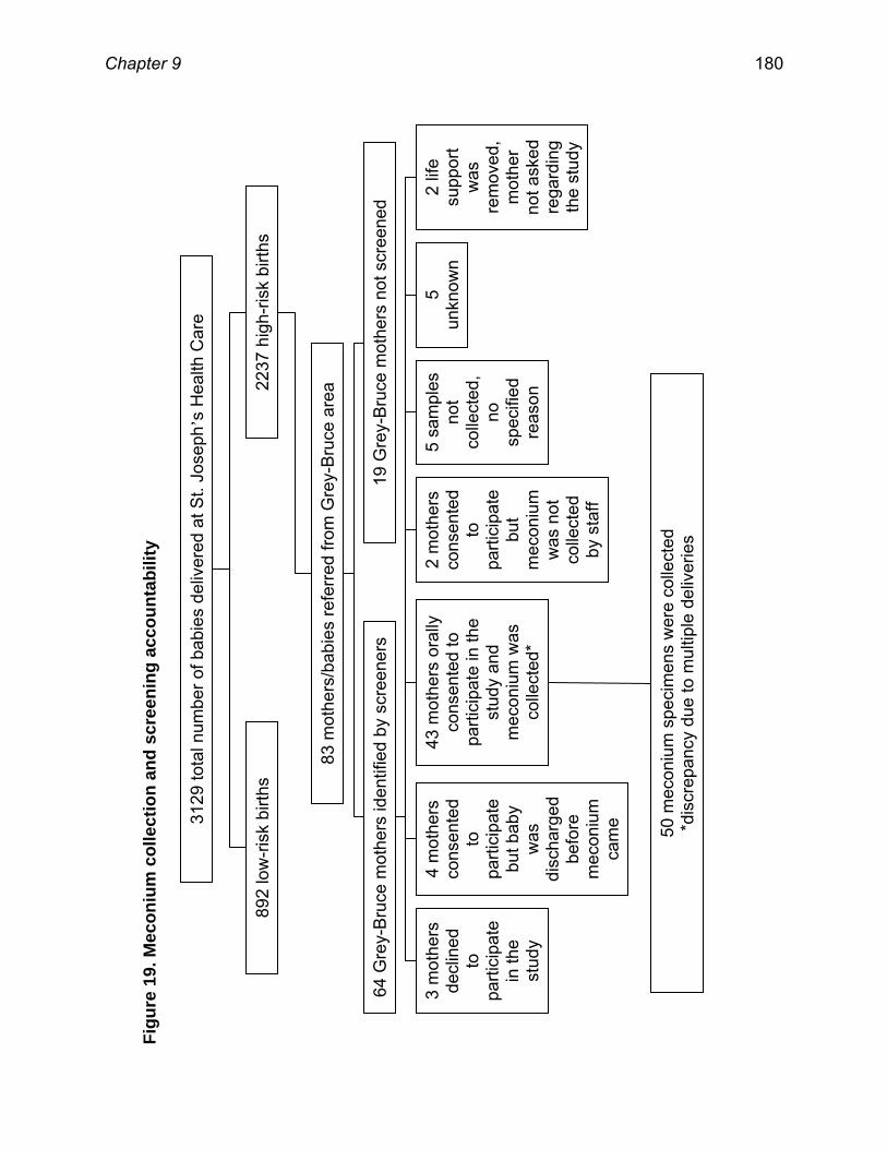
Figu
re 1
9. M
econ
ium
col
lect
ion
and
scre
enin
g ac
coun
tabi
lity
3129
tota
l num
ber o
f bab
ies
deliv
ered
at S
t. Jo
seph
’s H
ealth
Car
e
892
low
-risk
birt
hs
2237
hig
h-ris
k bi
rths
83 m
othe
rs/b
abie
s re
ferr
ed fr
om G
rey-
Bru
ce a
rea
64 G
rey-
Bru
ce m
othe
rs id
entif
ied
by s
cree
ners
19 G
rey-
Bru
ce m
othe
rs n
ot s
cree
ned
3 m
othe
rs
decl
ined
to
pa
rtici
pate
in
the
stud
y
4 m
othe
rs
cons
ente
d to
pa
rtici
pate
bu
t bab
y w
as
disc
harg
ed
befo
re
mec
oniu
m
cam
e
43 m
othe
rs o
rally
co
nsen
ted
to
parti
cipa
te in
the
stud
y an
d m
econ
ium
was
co
llect
ed*
2 m
othe
rs
cons
ente
d to
pa
rtici
pate
bu
t m
econ
ium
w
as n
ot
colle
cted
by
sta
ff
5 sa
mpl
es
not
colle
cted
, no
sp
ecifi
ed
reas
on
5 un
know
n 2
life
supp
ort
was
re
mov
ed,
mot
her
not a
sked
re
gard
ing
the
stud
y
50 m
econ
ium
spe
cim
ens
wer
e co
llect
ed
*dis
crep
ancy
due
to m
ultip
le d
eliv
erie
s
Chapter 9 180

Table 3. Maternal and neonatal characteristics of the Grey-Bruce perinatal intensive care group
Maternal Characteristics Number of Cases Percent of Cases Anatomical uterine anomalies 7 15.22% Gestational hypertension 7 15.22% Previous caesarian section 7 15.22% Advanced maternal age 5 10.87% Postdated labour 5 10.87% Previous fetal demise 5 10.87% Previous chromosomal and other congenital anomalies
5 10.87%
Intrauterine growth retardation 4 8.70% Placenta previa 3 6.52% Breech present 3 6.52% Previous postpartum hemorrhage 3 6.52% Gestational diabetes 3 6.52% Preeclampsia 3 6.52% Oligohydramnios 2 4.35% Previous neonatal death 2 4.35% Chronic Maternal Conditions Number of Cases Percent of Cases Factor IV Leiden 2 4.35% Epilepsy 1 2.17% S. Lupus Erythmatosus 1 2.17% Type II diabetes 1 2.17% Depression 1 2.17% Rh sensitization 1 2.17% Fetal/Neonatal Characteristics Number of Cases Percent of Cases Prematurity 16 34.78% Meconium aspiration 5 10.87% Congenital malformation 4 8.70% Dilated ventricles 2 4.35% Pneumothorax 2 4.35% Fetal arrhythmia 2 4.35% Intracranial hemorrhage 1 2.17% Hypoxic ischemia 1 2.17%
Chapter 9 181

9.5 DISCUSSION
In the present study, higher rates of FAEE positive meconium were
documented in a high-risk tertiary healthcare setting compared to primary
healthcare settings serving the same regional population. Identical research
design was applied to both studies. The general population captured babies
born within the region, whereas the high risk subset aimed at recruiting cases
leaving the region to give birth in a tertiary-care unit. Of importance, there were
no refusals to this anonymous study among the general population in Grey
Bruce, while there were three refusals among the high risk cohort, suggesting
that the true relative risk may be even higher. Although some mothers were
missed during the screening procedure, we do not believe that this created a
recruitment bias, because the reasons for omission in the general population
were technical (e.g. insufficient meconium). A chart review of all of the women
transferred the high-risk unit demonstrated that history of substance abuse was
not identified as the primary reason for the referral in any case. Since the
meconium collection was anonymous, there is no way to identify which positive
FAEE results were associated with particular maternal or fetal outcomes.
Previous studies have shown that mild, sporadic drinking beyond the first
trimester of pregnancy does not translate to positive FAEE test (i.e. levels above
2nmol/g) 230. Our study detected a 12-fold higher rate of FAEE positive
meconium (30% vs. 2.5%) among babies born in a high-risk obstetric unit as
compared to the general population of Grey-Bruce 404. The true rate of fetal
alcohol exposure in our study could have been higher, as we did encounter three
Chapter 9 182

refusals among the high-risk group, whereas no refusals occurred in the primary
care groups.
The sensitivity and specificity of FAEE detection in meconium have been
reported by several groups 223,226,230,406. Although Bearer and Chan report a high
sensitivity and specificity of FAEE (Bearer reported a sensitivity of 88% and
specificity of 64%) 226,230, Ostrea reported a sensitivity of 26.9% and specificity of
96.8% 226,406. Although Ostrea et al. report ethyl linoleate having a low
specificity, it had an extremely high positive predictive value, indicating that if
ethyl linoleate was detected in the meconium it is highly likely due to fetal alcohol
exposure 406. The variability of reported values of specificity and sensitivity are
likely due to differences in the analytical methods used to establish them as well
as differences in the sensitivity of maternal reporting. Unlike the previous studies
that used solid phase extraction followed by GC-FID or silica gel
chromatography, the present study used GC-HS-SPME. Analysis of FAEE using
GC-HS-SPME has been demonstrated to be highly sensitive and specific in hair
187. In addition, because FAEE do not cross the placenta once they are formed,
it is reflective of fetal alcohol exposure 137. Importantly, to date there have not
been any documented cases of false positives with meconium testing. In fact,
elevated levels of FAEE in meconium have been associated with FASD deficits
including growth restriction, decreased executive function, and poorer
neurodevelopmental outcome 400,407. A limitation is that FAEE in meconium only
detects second and third trimester exposure, hence the test is unable to detect
prenatal alcohol exposure in the first trimester. Maternal nutrition may
Chapter 9 183

theoretically be a confounding factor as poor nutrition may result in less fatty acid
content, which may result in less FAEE production in nutritionally-deficient
individuals 408,409. The positive cut-off rate utilized by our laboratory of >2nmol/g
FAEE was determined in a previous study quantifying the amount of FAEE in
meconium of babies born to women who did not consume any ethanol
(abstainers), consumed very little quantities (social drinkers), and women who
self-reported heavy prenatal alcohol consumption 227. The cut-off limit had a
sensitivity of 100% and a specificity of 98.4%.
Studies evaluating the stability of FAEE in meconium demonstrated that
FAEE are sensitive to temperature and light 139. Approximately 86% of the FAEE
in meconium was degraded within 24 hours when specimens were stored at
room temperature in light whereas 60% of the FAEE in the meconium was
degraded within 24 hours when specimens were stored in the dark 139. In our
study diapers were immediately placed in the -20°C freezer once collected.
Moreover the opaque diaper reduced the likelihood of degradation due to light. It
is currently unknown whether conditions other than alcohol use could result in a
positive meconium result in women of high-risk status.
Affecting 1/100 individuals, FASD is the leading preventable cause of
developmental disability in North America 103. In comparison to other screened
neonatal disease, such as phenylketouria and congenital hypothyroidism, the
prevalence of FASD is much higher (1% vs. 0.008%, 0.016-0.022%, respectively)
404,410,411. Moreover, economic studies have estimated that the lifetime cost for
FASD exceeds $1 million CAD 383. Another economic study estimated $4.0
Chapter 9 184

billion was spent in the United States 412. Due to the population health and
economic impact, screening neonates who may possibly be affected with FASD
is of great public health importance. Persons diagnosed with FASD at a later
stage in life are at a higher risk of having secondary disabilities 413. Screening for
in utero alcohol exposure at birth can prompt referral for diagnosis at an early
point in time, thereby allowing early intervention for both mother and child.
Implementation of a universal screening program must be complemented with a
structured follow-up program for both mothers and infants, meeting their
psychological and physiological needs, as well as providing the necessary social
supports.
The high prevalence of positive meconium FAEE among newborns in a
high-risk obstetric unit may stem from two overlapping mechanisms:
1) Ethanol-induced perinatal risks, such as prematurity and
intrauterine growth retardation 414,
2) Chronic alcohol using women tend to exhibit higher rates of
perinatal comorbidities due to neglect, suboptimal healthcare, higher rates
of gestational diabetes, hypertension and psychiatric conditions 415.
In conclusion, the present findings suggest that screening neonates in
high-risk perinatal units for prenatal alcohol exposure may have merit as a
routine practice to identify and follow-up neonates at risk for FASD, as well as
identifying and mobilizing resources to alcohol-dependent mothers.
Chapter 9 185

9.6 STATEMENT OF SIGNIFICANCE AND IMPACT
To our knowledge this is the first attempt to screen the meconium of a
selected high-risk population. The following results suggest that selective
screening in high-risk population may be an effective method of identifying
babies with prenatal alcohol exposure.
Chapter 9 186

Chapter 10
Overall Discussion and Conclusion
187

10. OVERALL DISCUSSION AND CONCLUSION
10.1 SUMMARY AND DISCUSSION OF RESEARCH FINDINGS
In this thesis a multilevel approach to birth defect and FASD prevention
has been examined using specific examples in each level of prevention. The
primary level of prevention is demonstrated by the means of maternal
multivitamin supplementation. The secondary level of prevention is
demonstrated by the means of pharmacologic intervention during pregnancy.
The tertiary level of prevention is demonstrated by early screening of the infant.
Finally, the quaternary level of prevention is demonstrated by identifying at
mothers who are who have a high-risk of delivering a future child affected with
the same birth defect.
10.1.1 PRIMARY LEVEL OF PREVENTION OF BIRTH DEFECTS AND FETAL
ALCOHOL SPECTRUM DISORDER BY MEANS OF MULTIVITAMINS
SUPPLEMENTATION
Folic acid supplementation has long been associated with reducing the
risk of NTD. The results from the meta-analyses suggests that prenatal
multivitamin supplementation is associated with a decreased risk for certain
congenital birth defects and pediatric cancers 416,417.
Maternal health optimization by multivitamin supplementation is one
example of primary level prevention of birth defects. Alcohol dependent
individuals have been shown to be deficient in nutrients due to the empty calories
Chapter 10 188

consumed with ethanol 44. As such, mothers are likely to be deficient in nutrients.
With the deficiency of nutrients, mothers are likely to have an increased risk of
delivering a child with birth defects. Therefore with maternal ethanol
consumption, not only is a child at risk of developing FASD, their risk for having
other birth defects is also increased. Prenatal multivitamin supplementation has
been associated with a decreased risk for oral clefts 416,418,419. FAS is
characterized by facial dysmorphology 8. As such supplementation with
multivitamins may potentially decrease the risk of adverse facial dysmorphology
related to FAS.
This example of primary level prevention of FASD through multivitamin
supplementation is a model that should be implemented universally. Currently all
Canadian pregnant women are recommended to supplement with 0.4mg folic
acid to decrease the risk of neural tube defects 420. Given the results from the
meta-analyses suggesting that prenatal multivitamin supplementation is
associated with the decrease of other birth defects and certain pediatric cancers
it appears beneficial for all women to supplement with multivitamins.
.
10.1.1.1 NUTRIENTS AND FETAL ALCOHOL SPECTRUM DISORDER
Due to the differing composition of the multivitamins contained in studies,
it is difficult to determine which component(s) is/are responsible for these
protective effects. In addition, it is difficult to ascertain whether these effects are
due to a single component within the multivitamin or if it results from a synergistic
effect. Furthermore, the mechanisms underlying the associated protective effect
Chapter 10 189

have yet to be elucidated. Below are examples of selected components within
multivitamins and how may protect against FASD.
Since ethanol metabolism results in the generation of oxidative stress the
use of antioxidants may be beneficial in decreasing the generated reactive
oxidative species. In pregnancy, one highly studied antioxidant is folic acid
(vitamin B19). Folic acid requirements increase during pregnancy due to the
increased synthesis of DNA and one-carbon transfer reactions such as
nucleotide synthesis and cell division 421,422. As such, pregnant women are at a
higher risk of developing folate deficiency than non-pregnant women 423.
Moreover, chronic alcohol consumers have been demonstrated to have folate
deficiency 44. As such, women who are chronic consumers of alcohol are usually
folate deficient prior to pregnancy and thus have a higher risk of delivering a child
with a birth defect 303. The combination of folate deficiency and the increased
need for folate during pregnancy may synergistically result in negative outcomes
for the fetus.
Methanol is a chemical that is endogenously produced in the body but
also present in most alcoholic beverages 424,425,426. Since ethanol is preferentially
metabolized by alcohol dehydrogenase compared to methanol, methanol and its
metabolite, formic acid, may be detected in higher levels in blood of individuals
who consume alcohol 24. Formic acid is a toxic substance that is known to cross
the placenta and cause cellular damage 427,428. Since folate is a cofactor for the
elimination of formic acid, folate deficiency may result in the accumulation of
formic acid in the mother. Formic acid generates oxygen radicals which are
Chapter 10 190

responsible for cellular damage 429. This has previously been demonstrated in
studies where animals with high formic acid levels and decreased formic acid
elimination rates were observed to be folate deficient 430,431,432. Moreover, in vitro
studies have observed that pretreatment of brain cultures with folic acid
decreased formic acid-induced neuronal death 24. The reduction of antioxidant
capacity is observed in addition to the decreased presence of folate 433,434.
Together the above factors may explain the mechanism of damage resulting in
FASD.
Another proposed mechanism of how ethanol causes FASD involves
retinol. Retinol is bound to retinol binding protein (RBP) and oxidized from retinal
to retinoic acid in a similar oxidation pathway as ethanol 435. This conversion is
catalyzed by alcohol dehydrogenase, aldehyde dehydrogenase, and cytochrome
P450 435. The involvement of these enzymes suggests that fetal alcohol
syndrome may arise due to inhibition of aldehyde-catalyzed retinoic acid
synthesis 436.
10.1.2 SECONDARY LEVEL OF PREVENTION OF FETAL ALCOHOL
SPECTRUM DISORDER BY MEANS OF EARLY PREVENTION WITH
ANTIOXIDANTS
In attempts to investigate the secondary level of FASD prevention, a
double-blinded randomized controlled trial was conducted to examine the
potential protective abilities of antioxidants to mitigate ethanol related damage
437. The rationale for the use of antioxidants is based on the proposed
Chapter 10 191

mechanism that FASD results from oxidative stress generated by the metabolism
of ethanol 438,439,440. Since oxidative stress has been implicated in the formation
of FASD in animal studies, trials of antioxidants have been undertaken 369.
Vitamin C is important in the antioxidant defense as it acts as a free radical
scavenger that protects tissue from free radical damage (reactive oxygen
molecules), thereby preventing oxidative stress 441. Vitamin C also acts
synergistically with vitamin E, another antioxidant which protects phospholipid
fatty acids (components of the cell membrane) from free radical oxidation, as it
converts the oxidized form of vitamin E to the active form of vitamin E 441.
A systematic review of the literature investigated the role of antioxidants in
the prevention of experimental FASD, demonstrating that antioxidants can
attenuate damage induced by ethanol 369. Informed by this systematic review we
conducted a study to examine whether the combination of vitamin C (1mg),
vitamin E (400IU), and multivitamin supplementation would be able to mitigate
the adverse effects of fetal alcohol exposure at a secondary prevention level 437.
As previously stated, during the course of this trial, several studies evaluating the
use of antioxidants in pre-eclampsia observed that the combination of vitamin C
and vitamin E had no protective effect for pre-eclampsia and, in fact, was
associated with an increased risk of low birth weight which could not be
explained by any other cause 376. Many trials involving the supplementation of
vitamin C and vitamin E in pregnant women were suspended due to these
findings.
Chapter 10 192

Since low birth weight is a common adverse effects observed in alcohol-
exposed pregnancies, this study was cancelled during its active recruitment
phase to ensure that the study was not putting the fetuses or their mothers at
increased health risk. This is especially important in high risk, alcohol dependent
and vulnerable women. In this case of this study, at its outset, both animal
research and clinical trials regarding pre-eclampsia suggested that high dose
vitamin C and vitamin E were safe 369,370. However, new information became
available during a trial resulting in its discontinuation. This highlights the need to
continually monitor safety during clinical trials. At the Hospital for Sick Children,
similar to all institutions following the Tricouncil Code of Ethics, continuous
evaluation of emerging data is demanded and has to be reported to the
Research Ethics Board. Should new information suggesting increased risks
arise, studies should be reevaluated by regulatory bodies to decide whether or
not to proceed.
Had this example of secondary level prevention demonstrated
effectiveness, it would have ideally been targeted to a selected population of
women who had alcohol-exposure during pregnancy. Unfortunately, there is
currently no known in utero intervention to prevent FASD. There are several
animal studies suggesting the possibility of other nutrients to consider for in utero
intervention 24,442,443. Given the findings with folic acid as previously discussed,
perhaps high doses of folic acid may be beneficial in mitigating alcohol-induced
damage associated with FASD 24. Another area being investigated is the use of
choline 442,443. Studies by Thomas et al. have demonstrated that the
Chapter 10 193

administration of choline in to animals reduced the behavioural detriment and
improved the body weight of alcohol-exposed 442,443.
10.1.3 TERTIARY PREVENTION OF FETAL ALCOHOL SPECTRUM
DISORDER BY MEANS OF MECONIUM SCREENING IN HIGH-RISK
POPULATIONS
The anonymous screening of meconium of Grey-Bruce babies delivered
or transferred to St. Joseph’s Hospital in London, Ontario revealed a 30%
positive rate for prenatal alcohol exposure as determined by the presence of
FAEE. The results of this study suggest that infants of high-risk pregnancies of
the residents from Grey-Bruce delivering at St. Joseph’s Health Care in London,
Ontario are at higher risk of testing positive for FAEE. Certainly this is a highly
selected population; however, the high proportion of FAEE positives suggests
that there is a significant increased risk of ethanol exposure in the second and
third trimester of pregnancy in this group. High amounts of ethanol exposure and
high FAEE levels have been associated with a higher risk of developing FASD
444.
FASD has been associated with high costs both to healthcare and social
system 445. Streissguth et al. demonstrated that early identification and
appropriate intervention decreased the risk of developing secondary co-
morbidities associated with FASD 413. Since earlier screening and diagnosis
results in an improved prognosis, there is likely to be reduced costs expended on
services that would otherwise be used by these individuals. In fact, a study by
Chapter 10 194

Hopkins et al. evaluating the costs associated with universal and targeted
meconium screening calculated a cost-savings associated with both universal
and targeted screening methods 401.
The advantage of screening FAEE in meconium is that it is a non-invasive
test, as such, samples are easily collected. In addition, it is an unbiased test that
is able to demonstrate prenatal exposure when maternal self-report is unreliable.
The limitations of meconium screening include that meconium commences
forming at approximately the thirteenth week of pregnancy. As such, any
exposures prior to this period will not be represented in the meconium. The
period of meconium collection is also time-sensitive, with a small window of
approximately 72 hours to obtain the sample. In addition, premature babies may
have a small sample of meconium which is delayed in excretion time.
It is unknown whether there are elements that would result in false positive
tests of meconium. For example, increased sugar levels may result in the
increased generation of endogenous alcohol, which in turn may result in a false
positive. If increased sugar levels do result in a false positive FAEE screening
may be less specific for identifying prenatal ethanol exposure.
10.1.4 QUATERNARY PREVENTION OF FETAL ALCOHOL SPECTRUM
DISORDER BY MEANS OF MECONIUM SCREENING
Meconium was demonstrated by our study as an effective means of
screening for mothers who are unable to discontinue drinking in the second and
third trimester of pregnancy. Maternal self-reporting of alcohol consumption was
Chapter 10 195

significantly lower than the prevalence of prenatal alcohol exposure determined
by meconium screening. Maternal underreporting may be due to maternal guilt,
fear, or shame. However this may also be confounded by the method in which
the question was administered to the mother. The framing of the question plays
an important role in determining how mothers will report. For instance, if mothers
were asked in a manner that construes alcohol consumption in a negative light, it
is likely that they will respond that they had no exposure. On the other hand, if
mothers were asked in a non-accusatory manner, she would be more likely to
answer more truthfully.
Meconium screening provides an unbiased method of screening for
maternal alcohol exposure during the latter portion of pregnancy. If mothers are
screened positive by their baby’s meconium, it suggests that there may have
been significant alcohol consumption since the positive screen cut-off level was
established in women who drank two or more drinks per day or were binge
drinkers 227. By identifying mothers, they can be engaged to participate in
programs to change their alcohol consumption habits, thereby decreasing their
chances of delivering another child who may be affected by FASD. As such,
meconium screening constitutes both a means of tertiary prevention by enabling
early intervention in exposed babies, as well as quaternary prevention by
identifying mother who are at risk for having future alcohol exposed pregnancies.
The results for the study suggests that meconium should be considered in high-
risk environments and perhaps screening babies in neonatal intensive care units
is a god area to begin screening. Overall, screening meconium to identify babies
Chapter 10 196

and mothers at risk should be considered to be implemented on a universal level
in order to increase its acceptance and avoid marginalization of certain
populations. However, with the universal implementation of this screening
process, there are many ethical issues to consider.
10.2 CONSIDERATIONS IN MECONIUM SCREENING
10.2.1 ETHICAL CONSIDERATION IN MECONIUM SCREENING
There are important ethical considerations associated with meconium
screening. Generally speaking, the objective of screening is to identify a
population which is at a higher risk of developing a disease/problem. This
involves screening individuals who do not exhibit symptoms and identify them as
being at risk. Ethical screening needs to maintain and preserve individual’s
autonomy, beneficence, non-maleficence, and justice 446,447.
Screening should involve informed consent. Informed consent is the right
of the individual to receive information in order to evaluate treatment options with
risk and benefits prior to making a decision. There is a fundamental right to
refuse to participate in screening without fear of any consequence of refusal. In
Canada there is no specific law specifying that written informed consent is
required prior to screening; however, medical practitioners, under ethical
guidelines, usually obtain verbal consent prior to screening 448. The American
Academy of Pediatrics Newborn Screening Task Force recommends parents to
Chapter 10 197

be informed prior to screening of their infant 449. In the United States, the United
States Health Insurance Portability and Accountability Act states that individuals
have the right to control their personal information.
One of the dilemmas that arise with neonatal meconium screening is the
issue of autonomy. Autonomy is the ability of competent persons to make their
own decisions. While the baby is not capable to make decisions, the decision
rests on its caregiver. Ideally, the caregiver would act in the best interest of the
child; however, this may be conflicted if the caregiver is at risk of being identified
as being the contributor to the child’s problem (i.e. the mother drank during
pregnancy). The autonomy of this process is therefore complex. This dilemma
is commonly overcome by the authorities (e.g. children’s protective services)
taking over the role of guardian.
A second dilemma arising from neonatal meconium screening is the issue
of beneficence and non-maleficence. Beneficence is the ability to demonstrate
the benefits over the risks of the screening program and non-maleficence is the
ability to avoid causation of harm. The benefit of neonatal meconium screening
is that it identifies babies who may be at greater risk for developing FASD and
their alcohol dependent mothers. As such, the child could benefit by early
diagnosis which could lead to early intervention and better outcome, preventing
the onset of secondary disabilities. Conversely, screening may lead to
stigmatization and labeling of the child and at times, removal of child from the
custody of the mother. Importantly, in areas where there is limited diagnostic
capacity, screening may wrongly become a de facto diagnosis. Thus prior to the
Chapter 10 198

implementation of a screening program there must be a means of diagnosis and
treatment.
A third dilemma is justice. Justice is the fairness of risk and benefits.
Justice may depend on numerous variables such as whether the child will have
access to diagnosis, access to service, and whether parents can afford the
service. Furthermore since limited resources exist to implement interventions,
there is a dilemma of whether it is right to screen when limited diagnosis and
treatment options are available.
The process of screening may result in negative outcomes for the family.
First there is a potential for misuse and abuse of information collected through
the screening process. The misuse of screening information may result in the
denial of healthcare coverage for the family. Second, there may be social
stigmatization of the family within the community. This effect may be more
profoundly observed if the community is small. There is great tangible risk for the
parent of the babies, especially the mother, as the screening may bring to light
alcohol consumption during pregnancy, implying the inability to stop drinking 446.
This may result in the involvement of child welfare. Third, the screening process
may impinge on reproductive autonomy (a woman’s ability to control reproductive
choices). Mothers may feel pressured to terminate future pregnancies. In
addition, parents may be ostracized by professionals who are not respectful of
their situation. As such parents may be weary of healthcare professionals and
may refuse support services. Fourth, the child may be removed from the birth
family and placed in foster care. Together these factors including fear, guilt and
Chapter 10 199

stigmatization may cause women to decline participating in the screening
process. Women who are already weary of the healthcare system may go
underground to avoid the hostile environments.
There is always the question of who owns the test results. Meconium is
an excrement of the child, who is themselves, is an individual; however, the
results are also reflective of maternal exposure. Similar issues were raised with
neonatal screening for HIV, as the identification of antibodies in the infant
translates to the identification of an infected mother. However, over the years
HIV testing in pregnancy has gained acceptance.
In Indiana a prescreening process occurs prior to meconium screening.
The prescreen identifies risk factors such as maternal confirmation of substance
use during present or past pregnancies, lack of prenatal care, low birth weight,
unexplained abruption placenta, and the presence of withdrawal symptoms in the
newborn is conducted prior to screening meconium 450.
10.2.2 ECONOMIC IMPACT OF MECONIUM SCREENING
In 2005, the Ontario Government established a state-of-the-art screening
program at The Children’s Hospital of Eastern Ontario. This $18 million newborn
screening program screens for 27 rare genetic diseases including 20 inherited
metabolic disorders, 4 endocrine disorders and 3 blood disorders 451. All of the
disorders tested within this program are rarer than the prevalence of FASD. If
the screening program for FASD is implemented and is successful, it will result in
the decrease of alcohol consumption in future pregnancies which would
Chapter 10 200

eventually lead to decrease in FASD. A decrease in FASD will result in a
decrease of associated costs.
Lupton et al. reported that in 1998 the economic burden associated with
caring for individuals affected with FAS was estimated to exceed $4 billion US
with an average lifetime cost of $2 million US for individuals 412. Stade et al.
reported that in 2003, individuals up to 65 years of age with FASD would have a
total average lifetime costs of $1 million CAN 452. FASD affects not only the
health costs but also education, community services and the corrections sector
as well.
A cost-utility analysis was conducted by Hopkins et al. in 2004 comparing
universal and targeted meconium screening in newborns to no screening 401. An
incremental cost of $65,875 CAN per QALY (quality-adjusted life year) was
gained from the societal perspective 401. Screening a targeted group of infants
whose older sibling diagnosed with FASD was less costly than universal
screening and more effective than no screening 401. Targeted screening in high-
risk populations may be an attractive option in terms of economic benefit and
establishing policy objectives and then expanding screening to a larger level.
10.2.3 IMPLEMENTATION OF MECONIUM AS A SCREENING TOOL
An important question with regards to the implementation of meconium
screening is on which level it should be implemented: universal versus selected
and mandatory versus voluntary. Universal mandatory screening violates the
principles of autonomy and justice 447. No choice is offered in this process and
Chapter 10 201

there is no consideration of the risks and benefits in the program. Universal
voluntary screening will satisfy the criterion of ethical screening; however, may
result in decreased compliance and may construe unwillingness to participate in
the screening process as guilt. Selected mandatory screening may result in
greater identification, but it not only does it violate the principles of autonomy and
justice; it also ostracizes the targeted population. Targeted voluntary screening
may result in the least participation because the population is being ostracized,
therefore given a choice individuals may decline to participate in this form of
screening. The drawback of screening in a targeted approach is that individuals
who are not in the population will be missed in this screening process. Many
researchers recommend to not screen universally because of the potentially high
costs involved in the process. However, a recent report evaluating costs
associated with universal or targeted meconium screening suggested that there
would be cost-savings associated with both types of levels of screening
compared to no screening at all 401.
Prior to its implementation as a screening tool, a follow-up procedure must
be established. When an infant screens positive, a referral path for diagnosis
and treatment should be available in the community. Mothers need to be
informed that their child may have the potential to be affected. Informing the
mother of the positive screen is a delicate procedure that will require the
experience of a skilled individual. Mothers need to be informed that a positive
screen does not necessarily imply that their infant is affected with FASD;
however it highlights the need to follow the infant closely as they age to
Chapter 10 202

immediately source assistance if their child is not achieving their age-expected
goals. In addition, mothers who are screened positive by meconium must
receive counseling, evaluation and intervention of their own illness. Identifying
and treating women who have alcohol dependence would enhance their quality
of life, improve their ability to parent their children and decrease the risk of having
another child that is affected with FASD. Without such pre-requisites, screening
per se will become futile, if not counter productive.
10.3 IMPLEMENTATION OF A MULTILEVEL PREVENTION PROGRAM FOR
FETAL ALCOHOL SPECTRUM DISORDER
Since a multilevel prevention program is a big undertaking, care should be
used in the designing and implementation of such a complex program. Ideally
prevention programs should be developed to best suit the prevention of FASD as
well as the needs of the target population. As such a rigourous planning process
is required to identify interventions that would be acceptable and effective in its
target population. A conceptual framework that may be helpful in the design and
implementation of a multilevel FASD prevention program is the PRECEDE-
PROCEED model. The PRECEDE-PROCEDE model is based on the premise
that lasting behaviour change will occur when individuals are provided with
education, motivation, and tools that are necessary to make a voluntary change
in their behaviour 453. It is a nine phase model that identifies predisposing
factors, reinforcing factors, enabling causes in educational diagnosis, evaluation
Chapter 10 203

and policy, and regulatory and organizational constructs in educational and
environmental development 453.
A limitation of using this model is that it is an expensive, complex process
that will require long planning and expansive personnel to execute. In the case
of a multilevel prevention strategy for FASD, programs will have to be designed
so they integrate together to prevent redundancy and therefore additional costs.
10.4 LIMITATIONS
Despite multiple efforts to reduce teratogenic risks of alcohol during
pregnancy there are factors which affect the subgroup risks within the population.
10.4.1 INTERINDIVIDUAL VARIATIONS
Food and energy intakes vary amongst individuals due to different body
sizes and lifestyles. As such, well- nourished women may have different risks
compared to malnourished women. Socioeconomic and cultural factors can also
impact nutrient levels. Mothers who are unable to afford food have a higher risk
of being nutrient deficient. In addition, different cultures may have different
dietary practices which may alter risk factors. Furthermore, access to necessities
such as food and clean water varies amongst countries which may also alter the
risks to the fetus.
Maternal health status and age may be a contributing factor to the
development of birth defects. The standard of healthcare varies worldwide. As
Chapter 10 204

such there may be differing status of health as well as management of medical
conditions from country to country. Medications or procedures used in the
management of medical conditions may alter the risk for birth defects. Increase
maternal age may increase the risk of genetic related birth defects.
10.4.2 GENETIC VARIATIONS
Genetic variations in the mother may alter her susceptibility to deliver a
child affected with birth defects. For instance, genetic differences between the
absorption and processing of vitamins and minerals may impact the formation of
the fetus. For example women with the MTHFR gene may be at increased risk of
delivering a child with a neural tube defect.
10.4.3 ENVIRONMENTAL VARIATIONS
Environmental variation may contribute to the outcome of the pregnancy.
Maternal illness during pregnancy may affect the development of the fetus.
Exposures to medications prior and during pregnancy may impact the formation
of birth defects. Exposures to recreational drugs during pregnancy may also
impact the formation of birth defects. Occupational exposures or other
environmental exposures may also be a confounder.
Chapter 10 205

10.5 BENEFITS AND LIMITATIONS OF MULTILEVEL PREVENTION OF
FETAL ALCOHOL SPECTRUM DISORDER
10.5.1 UNIVERSAL
The benefit of a universal FASD prevention strategy is that it targets all
members of the general population and therefore has desirable outcomes for the
entire population. This prevention strategy assumes that all women of
reproductive age can become pregnant and are at risk for alcohol exposure and
will therefore benefit from the prevention effort. Since universal campaigns are
aimed large audiences, it has a greater potential for success. Universal
prevention is also less intrusive because it does not target individual risk factors
or label individuals as alcohol consumers or at risk for FASD. An example of
universal FASD prevention includes educational campaigns. Although this type
of prevention attempts to educate the population in general, it requires a lot of
effort to ensure that all individuals in the population are educated. As such it may
not be possible to reach all of the individuals in the population.
The disadvantages of universal FASD prevention include the fact that, in
reality, the risk may vary greatly amongst individuals in population. If prevention
programs are designed incorrectly there may be language and cultural barriers.
Since the effects of universal prevention are hard to measure as it is difficult to
determine if the program itself is responsible for decreasing the incidence of
FASD or if it is a combination of factors such as other environmental influences.
Chapter 10 206

10.5.2 SELECTIVE
The advantage of selective prevention is that it targets an entire subgroup
regardless of their risk for FASD. The cost is justified because the increased risk
of FASD balances the benefits against risk. In selective prevention, an
individual’s risk of FASD is not specifically assessed or identified as it is based
on an at-risk subgroup membership. Selective prevention has a higher in
sensitivity; as such there will be cost-savings associates with this model. The
disadvantage of selective prevention of FASD is that it may result in labeling or
stigma. In addition, this process is more intensive as it requires more man power
to identify the subgroup. Although identification of individuals may be quicker in
selected populations, it will produce a false sense of security.
10.5.3 INDICATED
The advantage of indicated prevention is that it identifies individuals who
have symptoms of FASD who have not yet been diagnosed. The benefit of
indicated prevention is that there would be highest in sensitivity of all three. This
approach may be less expensive to implement as a whole; however, it has a high
cost per person and requires a greater commitment of time and resources to the
individual. The disadvantage is that persons may be labeled in this process and
it also may result in a false sense of security in the population for which the
prevention is not indicated.
Chapter 10 207

10.5.4 PRIMARY PREVENTION
The benefit of primary prevention is that FASD is being prevented prior to
its occurrence. As such, this method would be the ideal method of prevention.
Women of reproductive age would avoid exposing their fetus to alcohol and
therefore avoid any worry associated with prenatal alcohol exposure. The
disadvantage of primary prevention of FASD is that although educating
individuals may increase awareness, they may not necessarily use this
information. For example, women who drink may not necessarily use
contraception and may become pregnant. In addition women who are dependent
on alcohol may not easily be able to voluntarily discontinue drinking.
10.5.5 SECONDARY PREVENTION
The benefit of preventing FASD at the secondary level is that the majority
of women discover that they are pregnant after consuming alcohol. As such they
would benefit if there was something that they could do to decrease the risk of
FASD after recognizing that they are pregnant. The disadvantage is that
currently the only fail-proof method of preventing FASD is medical abortion. Until
a means of mitigating in utero alcohol damage is discovered, immediate
cessation of alcohol consumption will reduce the risk of the development of
FASD in the infant but will not address any damage that has already occurred.
Chapter 10 208

10.5.6 TERTIARY PREVENTION
The benefit of preventing FASD at the tertiary level is that individuals can
be engaged and may receive necessary interventions to decrease the
occurrence of secondary disabilities that are observed such as poor achievement
in school, dependent living, socioeconomic problems, and legal problems.
Tertiary prevention may be an important option for adoptive parents. Adoptive
parents are eager to provide a nurturing environment to their adoptive children
and as such are keen to know about their child’s health status. Tertiary
prevention by means of screening after birth will enable adoptive parents to
prepare to potential issues which may later arise thereby prompting them to seek
immediate action should they notice any developmental delays or abnormal
behavioural.
10.5.7 QUATERNARY PREVENTION
The benefit of preventing FASD at the quaternary level is that by treating
the mother for her alcoholism, quaternary prevention is essentially primary
prevention for her next child. The disadvantage of quaternary FASD prevention
is that the mother must be willing to engage in treatment programs in order to
decrease her alcohol exposure. In addition if treatment of the mother is
unsuccessful then the risk of her second child developing FASD remains the
same.
In cases where the child is removed from the custody of the mother the
mother may or may not be motivated to be engaged into treatment. Some
Chapter 10 209

mothers may be motivated to receive treatment in order to obtain custody of their
child. On the other hand, some mothers may self-medicate with alcohol to deal
with her grief of the child being removed. As such they may be unlikely
candidates to be engaged into treatment programs and therefore run the risk of
delivering another child who is affected by FASD.
10.5.8 MULTILEVEL PREVENTION VERSUS SINGLE LEVEL OF
PREVENTION
The above examples illustrate multiple levels of FASD prevention. Each
level of prevention has its advantages and disadvantages. In addition, each level
of prevention targets a different audience. If prevention is to occur only on a
single level, it is evident that there will be individuals who will be overlooked. In
such cases, the prevention may not reach its intended audience. By using a
multilevel paradigm, there are more opportunities to prevent FASD i.e. if an
individual is not engaged at one level; there is an opportunity to engage the
individual when they are at the next level. Because the multilevel prevention
model is cyclical, there will always be an opportunity to engage individuals.
10.6 SUSTAINABILITY OF PREVENTION
In the development with prevention methods, the sustainability of these
preventive efforts should be considered. Sustainability should be considered at
the onset of program development. In addition to sustained funding, there also
Chapter 10 210

needs to be sustained involvement from the entire community including
community agencies, schools, and institutions. The emergence of new
professions may arise with the creation of these multilevel prevention programs.
In addition there may be opportunities for professional development.
10.7 OVERALL SIGNIFICANCE AND CLINICAL IMPLICATIONS
10.7.1 OVERALL SIGNIFICANCE
Public health promotion in pregnancy is important for maintaining both the
mother’s health status and providing optimal growing conditions for the fetus. By
providing an ideal environment, the risks of birth defects can be decreased. The
benefit of using a multilevel strategy is that it has the potential to reach a vast
audience. In doing so it does not single out one population. The limitation to a
multilevel strategy is that it is an expensive endeavour. Although a multilevel
strategy is most likely to reach the majority of the population, it may not reach all
individuals. In addition to become generally accepted, a prevention strategy
needs to garner the acceptance of the general population.
Supplementation will decrease the risk for congenital anomalies and
pediatric cancers. With the event that supplementation can reduce these
problems; supplementation with multivitamins is a cost-effective method of
preventing congenital anomalies and pediatric cancers. It is likely that there
would be cost savings associated with this process. Moreover, not all persons in
Chapter 10 211

the world have accessibility to nutrition. Thus supplementation will enable them
to reach protective levels.
Currently many individuals affected with FASD are not identified.
Meconium screening is a method to screen for mother and children who are at
risk. This is the first study to record the prevalence of FAEE meconium in a high-
risk population.
10.7.2 CLINICAL IMPLICATIONS
With the implementation of multilevel approach it is likely that it will result
in a decrease in FASD. Early interventions have been demonstrated to be
associated with better prognosis in individuals affected with FASD 413. Health
Canada recommends that pregnant women supplement with 0.4mg of folic acid
420. The Institute of Medicine recommends that pregnant women supplement
with 0.6mg of folic acid 42. The results from these multivitamin studies have
changed the recommendations from the Society of Obstetricians and
Gynecologists of Canada 454. They now currently recommend using
supplementing with a multivitamin containing 1mg of folic acid 454. Recent
studies from Canada and the United States suggest that the majority of women
of reproductive age do not have protective amount of folate (900nmol) stores as
previously calculated by Daly et al. 455.
As such it was suggested that mothers who do not supplement with folic
acid prior to pregnancy and may have high risk of folic acid depletion due to the
increase folic acid demands during pregnancy. This may be confounded with
Chapter 10 212

other conditions which may cause folate deficiency. These women should
consider supplementing with a multivitamin containing 5mg of folic acid in order
to quickly increase their folate status to the protective level 454.
The clinical utility of meconium screening has been demonstrated to
identify individuals at risk of being diagnosed with FASD. As such screening for
this prevalent problem should be considered. Perhaps meconium screening
should commence in high-risk units of tertiary healthcare centers and then later
expand to the entire population to ensure greater acceptance since the test will
not target a specific population. If meconium screening does occur, a greater
capacity of FASD diagnosis needs to be developed. Currently wait lists span
from 6 months to 2 years 456. In addition to the need increased diagnostic
capacity, training of healthcare professionals needs to also occur. Healthcare
professionals need to be educated regarding the collection and shipment of the
meconium. Moreover, they have to be capable of obtaining informed consent
and interpreting the results of the screen to the mother. Despite high initial costs
associated with the implementation of a meconium screening program, it will
result in long-term cost savings. In addition by identifying a child affected with
FASD we also identify a mother who may not be aware of the impact of prenatal
alcohol exposure or a mother who is not able to discontinue drinking during
pregnancy. As such the mother can receive the necessary education or
intervention to decrease the risk of her delivering another child affected by FASD.
Chapter 10 213

10.7.3 AFFIRMATION AND ENDORSEMENT OF MULTIVITAMIN
SUPPLEMENTATION PRIOR TO AND DURING PREGNANCY
These finding from the study reaffirms the need for supplementation prior
and during pregnancy. The current recommendations are for pregnant women to
supplement with folic acid; however these results suggest not only
supplementing with folic acid alone, but supplementing with multivitamins
containing higher levels of folic acid. Unfortunately, multivitamin supplementation
does not occur universally around the world as it is not routine practice of the
country that or persons within the country cannot afford to supplement with
multivitamins. First world countries such as Canada and the United States
recommend supplementing with folic acid and have incorporated folic acid into
food by enriching flour. On the other hand, some first world countries such as
the United Kingdom and France do not fortify their flour with folic acid. In
contrast, third world countries that may not have access to multivitamins may
benefit the most by providing multivitamins to mothers who may not receive
adequate nutrient intake. Maternal supplementation with prenatal multivitamins
can decrease the risk for congenital anomalies and pediatric cancer. A decrease
in these birth defects would eventually result in cost-savings to the healthcare
system and society. Studies have already demonstrated that folic acid
fortification of food has resulted in cost-benefit in Canada, United States, South
Africa, Netherlands, and Chile 457,458,459,460,461. Waitzman et al. estimated that the
lifetime medical costs for spina bifida was over $635,000 462. Wald et al.
Chapter 10 214

estimated that with an investment of 1 cent per person per year to fortify flour,
$1,000 in savings in neural tube defects are realized 70.
Education of the benefits of multivitamin supplementation should
commence as early as in school when students are taught about human
reproduction. Physicians should consider discussing the importance of
multivitamin supplementation during pregnancy with their patients as part of their
yearly physical so that patients are reminded of the benefits. The benefits of
multivitamin supplementation can also be promoted across the general
population.
10.7.4 AFFIRMATION OF SCREENING MECONIUM FOR FATTY ACID ETHYL
ESTERS
The results from the meconium screening study endorse the use of
screening of meconium for fatty acid ethyl esters. Screening identifies children
who are at risk of being diagnosed with FASD. With the prevalence of FASD
being estimated at 1/100, and the costs associated estimated at $4.0 billion there
is a great potential for cost-savings 412. The initial launch of a meconium
screening program may commence in a selected or indicated population;
however, the overall goal is to implement universal screening on a national level
similar to other newborn screening programs.
Chapter 10 215

10.7.5 AFFIRMATION AND ENDORSEMENT OF MULTILEVEL PREVENTION
FOR FETAL ALCOHOL SPECTRUM DISORDER
A multilevel and multi-targeted approach is needed to decrease the risk of
developing FASD. Since the population varies widely in risk factors, uptake
capacity, comprehension, cultures, and socioeconomics this multilevel approach
may be the best method of reaching all individuals compared to a single level of
approach. Through the use of a multilevel approach a wider audience can be
reached therefore there is a greater chance of preventing FASD before it occurs
and there will be lesser chance of missing individuals who are affected with
FASD.
10.8 FUTURE DIRECTIONS
The most important step to the implementation of a multilevel strategy
during for FASD is the need to engagement professionals from many disciplines.
By engaging individuals across multiple fields, identical messages can be
reinforced by multiple individuals on the target population. In addition,
multidisciplinary teams can be formed which can collaborate in creating
prevention strategies as well as diagnosis and treatment. Moreover prevention
strategies should be implemented across sectors such as healthcare, education,
and social services.
Chapter 10 216

An infrastructure must be built to support the prevention strategy.
Whether it is a centre where individuals can receive answers to their questions or
a clinic that can provide diagnosis or a program to assist in the quality of life of
individuals affected with FASD. The infrastructure needs to be built with support
from stable funding sources. Many initiatives are ineffective because of the
limited funding they receive. For instance funding may be limited to a 5-year
term where the first few years may be focused on the development of the
initiative so that when a model is implemented for use in the community, it only
lasts for a short duration after which, communities are left helpless.
There is a need to increase awareness of FASD. Without increasing
awareness, there will be no drive to screen for this prevalent problem.
10.9 AREAS FOR FURTHER DEVELOPMENT
With regards to multivitamin supplementation to optimize maternal health,
further investigation should be conducted determine what are the optimal levels
of each vitamin and mineral within each prenatal multivitamin. There are
currently no regulations with regards to the proportion of components within
multivitamins.
In addition given the fact that it is recognized that certain vitamins are
depleted with alcohol consumption, fortification of alcoholic beverages with these
vitamins may improve nutrient status. However, fortifying alcoholic beverages
Chapter 10 217

may endorse alcohol consumption because individuals may use faulty reasoning
that they contain nutrients to justify their drinking.
With regards to meconium screening, the next step following the results of
the anonymous screening study is to conduct a study where specimens will be
screened with the identification of mother and child. Mothers will have to provide
written informed consent to participate in the study. By participating in this study,
children will be followed as they age and have access to necessary interventions
they require. This study will enable investigators to determine the participation
rate in this type of screening program. Results from this study can also assist in
the creation of policy and recommend what type of infrastructure is necessary
with this type of screening process. The following step would be the widespread
implementation of meconium as a screening tool which is included with the other
types of genetic screens that are routinely conducted in newborns.
Further studies that need to be conducted with regard to meconium
screening include ascertaining whether dietary consumption or lack of
consumption of fatty acid have an impact on the overall rate and quantity of
formation of FAEE. It is unknown whether FAEE degrades in utero and how
much of an effect this degradation would contribute to the overall quantification in
meconium. Factors resulting in false positive screens have yet to be identified.
In addition the exact dose-response correlation between the number of drinks
and the quantity of FAEE produced has yet to be determined. Furthermore the
exact dose-response correlation between the quantity of FAEE and the prognosis
of being diagnosed with FASD is unknown.
Chapter 10 218

The development and validation of FASD screening tools needs to
continue. Identification of other screening tools should be undertaken. A study
to determine the prevalence of FASD of Canada needs to be undertaken.
With the implementation of these preventive measures, a re-evaluation of
costs should be conducted to recognize how much cost-savings have been
achieved with multilevel intervention.
10.10 CONCLUSION
Pregnancy is a sensitive time for fetal development as such it is important
to reduce any avoidable risk to the fetus. Since ethanol is the known cause of
FASD steps should be taken to prevent the occurrence of this avertable birth
defect. Due to the complexity of alcoholism, instituting prevention in a single
level may only address a small proportion of the population. This thesis
demonstrates that a multilevel prevention strategy should be implemented to
prevent birth defects and FASD. It identifies two main examples which cover all
four levels of prevention: the use of multivitamins and neonatal meconium
screening. The use of multivitamins can be beneficial in the primary and
secondary level of preventing FASD. In the primary level all women should
receive prenatal multivitamin supplementation prior to pregnancy to increase her
nutrient status prior to pregnancy. This is especially important in nutrient
deficient populations such as chronic alcoholics. By optimizing maternal health
status, there will be a decreased risk of delivering a child affected with congenital
Chapter 10 219

anomalies and pediatric cancers. In the secondary level women can take steps
towards improving the outcome of her baby once she recognizes that she is
pregnant. For instance commencing multivitamin supplementation once
discovering pregnancy will attempt to increase maternal nutrient levels to
protective levels. In addition in an attempt to evaluate a pharmacologic
intervention for alcohol exposed pregnancies a randomized controlled trial was
undertaken to establish the effectiveness of high-dose vitamins during
pregnancy. In the tertiary level once a baby that has prenatal exposure to
alcohol is born steps should be taken to reduce secondary effects and improve
the overall quality of life. Screening babies’ meconium for FAEE will identify
babies who are prenatally exposed to alcohol. This will enable the referral to a
follow-up program that will be able to institute the necessary interventions if
needed. Moreover, by screening and identifying babies we are also identifying
the mother. In the quaternary level of prevention, identifying the mother allows
for the implementation of interventions which will decrease the likelihood of her
delivering another child affect with FASD. Due to the multiple factors which can
contribute to FASD, the use of a multilevel prevention strategy would be able to
reach a wider audience and therefore be more effective in overcoming this
devastating problem.
Chapter 10 220

REFERENCES
1. Robinson, A, Linden, M, Clinical Genetics Handbook. Boston:Blackwell
Scientific Publications, 1993.
2. Hoyert, DL, Mathews, TJ, Menacker, F, Strobino, DM, Guyer, B, Annual Summary of Vital Statistics: 2004. Pediatrics 117(1):168-183, 2006.
3. Detrait, ER, George, TM, Etchevers, HC, Gilbert, JR, Vekemans, M, Speer, MC, Human Neural Tube Defects: Developmental Biology, Epidemiology, and Genetics. Neurotoxicol Teratol 27(3):515-524, 2005.
4. Gomez Ponce de Leon, R, Wing, D, Fiala, C, Misoprostol for Intrauterine Fetal Death. Int J Gynaecol Obstet 99 Suppl 2:S190-S193, 2007.
5. Grainger, R, Morris, A, Ward, P, Phocomelic Deformity and Maternal Thalidomide Administration. A Report of Four Cases. Br J Radiol 35:687-691, 1962.
6. Mittendorf, R, Teratogen Update: Carcinogenesis and Teratogenesis Associated With Exposure to Diethylstilbestrol (DES) in Utero. Teratology 51(6):435-445, 1995.
7. Herbst, AL, Diethylstilbestrol and Adenocarcinoma of the Vagina. Am J Obstet Gynecol 181(6):1576-1578, 1999.
8. Jones, KL, Smith, DW, Recognition of the Fetal Alcohol Syndrome in Early Infancy. Lancet 2(7836):999-1001, 1973.
9. Chudley, AE, Conry, J, Cook, JL, Loock, C, Rosales, T, LeBlanc, N, Fetal Alcohol Spectrum Disorder: Canadian Guidelines for Diagnosis. CMAJ 172(5 Suppl):S1-S21, 2005.
10. National Center on Birth Defects and Develpmental Disabilities, Fetal Alcohol Syndrome: Guidelines for Referral and Diagnosis. Last updated: 2004. http://www.cdc.gov/ncbddd/fas/documents/FAS_guidelines_accessible.pdf. Date accessed: Jan 30, 2006.
11. Clarren, SK, Recognition of Fetal Alcohol Syndrome. JAMA: The Journal of the American Medical Association 245(23):2436-2439, 1981.
References 221

12. Kopera-Frye, K, Dehaene, S, Streissguth, AP, Impairments of Number Processing Induced by Prenatal Alcohol Exposure. Neuropsychologia 34(12):1187-1196, 1996.
13. Weinberg, JM, IPLEDGE Allegiance. Cutis 76(6):356, 2005.
14. Awopetu, O, Brimacombe, M, Cohen, D, Fetal Alcohol Syndrome Disorder Pilot Media Intervention in New Jersey. Can J Clin Pharmacol 15(1):e124-e131, 2008.
15. Casiro, OG, Stanwick, RS, Pelech, A, Taylor, V, Public Awareness of the Risks of Drinking Alcohol During Pregnancy: the Effects of a Television Campaign. Child Health Committee, Manitoba Medical Association. Can J Public Health 85(1):23-27, 1994.
16. Hernandez-Diaz, S, Werler, MM, Walker, AM, Mitchell, AA, Folic Acid Antagonists During Pregnancy and the Risk of Birth Defects. N Engl J Med 343(22):1608-1614, 2000.
17. Haberman, S, Burgess, T, Klass, L, Cohn, BD, Minkoff, HL, Acute Bowel Perforation in a Fetus With Gastroschisis. Ultrasound Obstet Gynecol 15(6):542-544, 2000.
18. Bruner, JP, Tulipan, N, Reed, G, Davis, GH, Bennett, K, Luker, KS, Dabrowiak, ME, Intrauterine Repair of Spina Bifida: Preoperative Predictors of Shunt-Dependent Hydrocephalus. Am J Obstet Gynecol 190(5):1305-1312, 2004.
19. Walsh, DS, Adzick, NS, Foetal Surgery for Spina Bifida. Semin Neonatol 8(3):197-205, 2003.
20. McLean, RH, Gearhart, JP, Jeffs, R, Neonatal Obstructive Uropathy. Pediatr Nephrol 2(1):48-55, 1988.
21. Janowsky, DS, Fawcett, J, Meszaros, K, Verheul, R, Core Heritable Personality Characteristics and Relapse in Alcoholics. Alcohol Clin Exp Res 25(5 Suppl ISBRA):94S-98S, 2001.
22. Abel, EL, Sokol, RJ, Fetal Alcohol Syndrome Is Now Leading Cause of Mental Retardation. Lancet 2(8517):1222, 1986.
23. May, PA, Gossage, JP, Estimating the Prevalence of Fetal Alcohol Syndrome. A Summary. Alcohol Res Health 25(3):159-167, 2001.
24. Kapur, BM, Vandenbroucke, AC, Adamchik, Y, Lehotay, DC, Carlen, PL, Formic Acid, a Novel Metabolite of Chronic Ethanol Abuse, Causes Neurotoxicity, Which Is Prevented by Folic Acid. Alcohol Clin Exp Res 31(12):2114-2120, 2007.
References 222

25. Last, J, A Dictionary of Epidemiology. New York:Oxford University Press, 2001.
26. Gordon, R, An Operational Classification of Disease Prevention in Steinberg, J, Silverman, M (eds). Rockville:US Dept of Health and Human Services, 1987.
27. Mrazek, P, Haggerty, R, Committee of Prevention of Mental Disorders, Institute of Medicine, Reducing Risk for Mental Disorders: Frontiers for Preventive Intervention Research. 1994.
28. Sorensen, S, Drug Abuse Prevention for the General Population. Rockville, MD:National Institute on Drug Abuse, 1999.
29. Commission on Chronic Illness, Commission on Chronic Illness Volume 1. Cambridge:Harvard University Press, 1957.
30. Jamoulle, M, Information Et Informatisation En Médecine Générale in Les Intofrmer-G-Iciens. Presses Universitaires de Namur, 1986.
31. Jamoulle, M, Quaternary Prevention: Prevention As You Never Heard Before. Last updated: Jul 20, 2000. http://www.ulb.ac.be/esp/mfsp/quat-en.html. Date accessed: Mar 1, 2008.
32. UK National Screening Committee, What Is Screening? Last updated: Mar 3, 2008. http://www.nsc.nhs.uk/whatscreening/whatscreen_ind.htm. Date accessed Apr 23, 2008.
33. Wilson, J, Jungner, G, Principles and Practice of Screening for Disease. Geneva: WHO . Last updated: 1968. http://www.who.int/bulletin/volumes/86/4/07-050112BP.pdf. Date accessed: Jan 1, 2008.
34. Weisz, J, Sandler, I, Durlak, J, Anton, B, Promoting and Protecting Youth Mental Health Through Evidence Based Prevention and Treatment. American Psychologist 60(6):628-648, 2005.
35. Roth, LK, Taylor, HS, Risks of Smoking to Reproductive Health: Assessment of Women's Knowledge. Am J Obstet Gynecol 184(5):934-939, 2001.
References 223

36. Johnson, K, Posner, SF, Biermann, J, Cordero, JF, Atrash, HK, Parker, CS, Boulet, S, Curtis, MG, Recommendations to Improve Preconception Health and Health Care--United States. A Report of the CDC/ATSDR Preconception Care Work Group and the Select Panel on Preconception Care. MMWR Recomm Rep 55(RR-6):1-23, 2006.
37. Christian, P, Micronutrients and Reproductive Health Issues: an International Perspective. J Nutr 133(6):1969S-1973S, 2003.
38. Roberts, JM, Balk, JL, Bodnar, LM, Belizan, JM, Bergel, E, Martinez, A, Nutrient Involvement in Preeclampsia. J Nutr 133(5 Suppl 2):1684S-1692S, 2003.
39. Specker, B, Nutrition Influences Bone Development From Infancy Through Toddler Years. J Nutr 134(3):691S-695S, 2004.
40. Ramakrishnan, U, Nutrition and Low Birth Weight: From Research to Practice. Am J Clin Nutr 79(1):17-21, 2004.
41. Health Canada, Dietary Reference Intakes. Last updated: Jan 23, 2006. http://www.hc-sc.gc.ca/fn-an/nutrition/reference/dri_ques-ques_anref-eng.php#1a. Date accessed Mar 1, 2006.
42. Food and Nutrition Board, IoM, Dietary Reference Intakes for Thiamin, Riboflavin, Niacin, Vitamin B6, Folate, Vitamin B12, Pantothenic Acid, Biotin, and Choline. A Report of the Standing Committee on the Scientific Evaluation of Dietary Reference Intakes and Its Panel on Folate, Other B Vitamins, and Choline. Washington:National Academy Press, 1998.
43. Mezey, E, Alcoholic Liver Disease: Roles of Alcohol and Malnutrition. Am J Clin Nutr 33(12):2709-2718, 1980.
44. Lieber, CS, Relationships Between Nutrition, Alcohol Use, and Liver Disease. Alcohol Res Health 27(3):220-231, 2003.
45. Mezey, E, Intestinal Function in Chronic Alcoholism. Ann N Y Acad Sci 252:215-227, 1975.
46. Lieber, CS, Alcohol: Its Metabolism and Interaction With Nutrients. Annu Rev Nutr 20:395-430, 2000.
47. Liu, S, Serdula, MK, Williamson, DF, Mokdad, AH, Byers, T, A Prospective Study of Alcohol Intake and Change in Body Weight Among US Adults. Am J Epidemiol 140(10):912-920, 1994.
References 224

48. Gruchow, HW, Sobocinski, KA, Barboriak, JJ, Alcohol, Nutrient Intake, and Hypertension in US Adults. JAMA: The Journal of the American Medical Association 253(11):1567-1570, 1985.
49. Hillers, VN, Massey, LK, Interrelationships of Moderate and High Alcohol Consumption With Diet and Health Status. Am J Clin Nutr 41(2):356-362, 1985.
50. Krasner, N, Cochran, KM, Russell, RI, Carmichael, HA, Thompson, GG, Alcohol and Absorption From the Small Intestine. 1. Impairment of Absorption From the Small Intestine in Alcoholics. Gut 17(4):245-248, 1976.
51. Hoyumpa, AM, Mechanisms of Vitamin Deficiencies in Alcoholism. Alcohol Clin Exp Res 10(6):573-581, 1986.
52. Leevy, CM, Baker, H, Tenhove, W, Frank, O, Cherrick, GR, B-Complex Vitamins in Liver Disease of the Alcoholic. Am J Clin Nutr 16:339-346, 1965.
53. Mitchell, D, Wagner, C, Stone, WJ, Wilkinson, GR, Schenker, S, Abnormal Regulation of Plasma Pyridoxal 5'-Phosphate in Patients With Liver Disease. Gastroenterology 71(6):1043-1049, 1976.
54. Leo, MA, Lieber, CS, Hepatic Vitamin A Depletion in Alcoholic Liver Injury. N Engl J Med 307(10):597-601, 1982.
55. Bonjour, JP, Vitamins and Alcoholism. I. Ascorbic Acid. Int J Vitam Nutr Res 49(4):434-441, 1979.
56. Gascon-Barre, M, Influence of Chronic Ethanol Consumption on the Metabolism and Action of Vitamin D. J Am Coll Nutr 4(5):565-574, 1985.
57. World, MJ, Ryle, PR, Jones, D, Shaw, GK, Thomson, AD, Differential Effect of Chronic Alcohol Intake and Poor Nutrition on Body Weight and Fat Stores. Alcohol Alcohol 19(4):281-290, 1984.
58. Russell, RM, Rosenberg, IH, Wilson, PD, Iber, FL, Oaks, EB, Giovetti, AC, Otradovec, CL, Karwoski, PA, Press, AW, Increased Urinary Excretion and Prolonged Turnover Time of Folic Acid During Ethanol Ingestion. Am J Clin Nutr 38(1):64-70, 1983.
59. Halsted, CH, Robles, EA, Mezey, E, Decreased Jejunal Uptake of Labeled Folic Acid ( 3 H-PGA) in Alcoholic Patients: Roles of Alcohol and Nutrition. N Engl J Med 285(13):701-706, 1971.
References 225

60. Lindenbaum, J, Lieber, CS, Effects of Chronic Ethanol Administration on Intestinal Absorption in Man in the Absence of Nutritional Deficiency. Ann N Y Acad Sci 252:228-234, 1975.
61. Lindenbaum, J, Lieber, CS, Alcohol-Induced Malabsorption of Vitamin B12 in Man. Nature 224(5221):806-, 1969.
62. Kim, CI, Roe, DA, Development of Riboflavin Deficiency in Alcohol-Fed Hamsters. Drug Nutr Interact 3(2):99-107, 1985.
63. Tomasulo, PA, Kater, RM, Iber, FL, Impairment of Thiamine Absorption in Alcoholism. Am J Clin Nutr 21(11):1341-1344, 1968.
64. Thomson, AD, Baker, H, Leevy, CM, Patterns of 35S-Thiamine Hydrochloride Absorption in the Malnourished Alcoholic Patient. J Lab Clin Med 76(1):34-45, 1970.
65. Marotta, F, Labadarios, D, Frazer, L, Girdwood, A, Marks, IN, Fat-Soluble Vitamin Concentration in Chronic Alcohol-Induced Pancreatitis. Relationship With Steatorrhea. Dig Dis Sci 39(5):993-998, 1994.
66. Flink, EB, Magnesium Deficiency in Alcoholism. Alcohol Clin Exp Res 10(6):590-594, 1986.
67. Bartholomay, A, Robin, E, Vallee, R, Wacker, W, Zinc Metabolism in Hepatic Dysfunction. I. Serum Zinc Concentrations in Laennec's Cirrhosis and Their Validation by Sequential Analysis. N Engl J Med 255(9):403-408, 1956.
68. Rivlin, RS, Magnesium Deficiency and Alcohol Intake: Mechanisms, Clinical Significance and Possible Relation to Cancer Development (a Review). J Am Coll Nutr 13(5):416-423, 1994.
69. Dwyer, JT, Holden, J, Andrews, K, Roseland, J, Zhao, C, Schweitzer, A, Perry, CR, Harnly, J, Wolf, WR, Picciano, MF, Fisher, KD, Saldanha, LG, Yetley, EA, Betz, JM, Coates, PM, Milner, JA, Whitted, J, Burt, V, Radimer, K, Wilger, J, Sharpless, KE, Hardy, CJ, Measuring Vitamins and Minerals in Dietary Supplements for Nutrition Studies in the USA. Anal Bioanal Chem 389(1):37-46, 2007.
70. Radimer, K, Bindewald, B, Hughes, J, Ervin, B, Swanson, C, Picciano, MF, Dietary Supplement Use by US Adults: Data From the National Health and Nutrition Examination Survey, 1999-2000. Am J Epidemiol 160(4):339-349, 2004.
References 226

71. Chung-Park, MS, Evaluation of a Pregnancy Prevention Programme Using the Contraceptive Behavior Change Model. J Adv Nurs 61(1):81-91, 2008.
72. Czeizel, AE, Vargha, P, Periconceptional Folic Acid/Multivitamin Supplementation and Twin Pregnancy. Am J Obstet Gynecol 191(3):790-794, 2004.
73. Geltman, PL, Meyers, AF, Mehta, SD, Brugnara, C, Villon, I, Wu, YA, Bauchner, H, Daily Multivitamins With Iron to Prevent Anemia in High-Risk Infants: a Randomized Clinical Trial. Pediatrics 114(1):86-93, 2004.
74. Nilsen, RM, Vollset, SE, Rasmussen, SA, Ueland, PM, Daltveit, AK, Folic Acid and Multivitamin Supplement Use and Risk of Placental Abruption: a Population-Based Registry Study. Am J Epidemiol 167(7):867-874, 2008.
75. Haider, BA, Bhutta, ZA, Multiple-Micronutrient Supplementation for Women During Pregnancy. Cochrane Database Syst Rev (4):CD004905, 2006.
76. Bodnar, LM, Tang, G, Ness, RB, Harger, G, Roberts, JM, Periconceptional Multivitamin Use Reduces the Risk of Preeclampsia. Am J Epidemiol 164(5):470-477, 2006.
77. Rumbold, A, Middleton, P, Crowther, CA, Vitamin Supplementation for Preventing Miscarriage. Cochrane Database Syst Rev (2):CD004073, 2005.
78. Desandes, E, Clavel, J, Berger, C, Bernard, JL, Blouin, P, De, LL, Demeocq, F, Freycon, F, Gembara, P, Goubin, A, Le, GE, Pillon, P, Sommelet, D, Tron, I, Lacour, B, Cancer Incidence Among Children in France, 1990-1999. Pediatr Blood Cancer 43(7):749-757, 2004.
79. Yuskiv, N, Honein, MA, Moore, CA, Reported Multivitamin Consumption and the Occurrence of Multiple Congenital Anomalies. Am J Med Genet A 136(1):1-7, 2005.
80. Czeizel, AE, Dobó, M, Vargha, P, Hungarian Cohort-Controlled Trial of Periconceptional Multivitamin Supplementation Shows a Reduction in Certain Congenital Abnormalities. Birth Defects Res A Clin Mol Teratol 70(11):853-861, 2004.
81. Chen, G, Song, X, Ji, Y, Zhang, L, Pei, L, Chen, J, Liu, J, Li, C, Zheng, X, Prevention of NTDs With Periconceptional Multivitamin Supplementation Containing Folic Acid in China. Birth Defects Res A Clin Mol Teratol , 2008.
References 227

82. Czeizel, AE, Puho, EH, Banhidy, F, No Association Between Periconceptional Multivitamin Supplementation and Risk of Multiple Congenital Abnormalities: a Population-Based Case-Control Study. Am J Med Genet A 140(22):2469-2477, 2006.
83. Fawzi, WW, Msamanga, GI, Spiegelman, D, Urassa, EJ, McGrath, N, Mwakagile, D, Antelman, G, Mbise, R, Herrera, G, Kapiga, S, Willett, W, Hunter, DJ, Randomised Trial of Effects of Vitamin Supplements on Pregnancy Outcomes and T Cell Counts in HIV-1-Infected Women in Tanzania. Lancet 351(9114):1477-1482, 1998.
84. Fawzi, WW, Msamanga, G, Hunter, D, Urassa, E, Renjifo, B, Mwakagile, D, Hertzmark, E, Coley, J, Garland, M, Kapiga, S, Antelman, G, Essex, M, Spiegelman, D, Randomized Trial of Vitamin Supplements in Relation to Vertical Transmission of HIV-1 in Tanzania. J Acquir Immune Defic Syndr 23(3):246-254, 2000.
85. Fawzi, WW, Msamanga, GI, Spiegelman, D, Wei, R, Kapiga, S, Villamor, E, Mwakagile, D, Mugusi, F, Hertzmark, E, Essex, M, Hunter, DJ, A Randomized Trial of Multivitamin Supplements and HIV Disease Progression and Mortality. N Engl J Med 351(1):23-32, 2004.
86. Villamor, E, Msamanga, G, Spiegelman, D, Antelman, G, Peterson, KE, Hunter, DJ, Fawzi, WW, Effect of Multivitamin and Vitamin A Supplements on Weight Gain During Pregnancy Among HIV-1-Infected Women. Am J Clin Nutr 76(5):1082-1090, 2002.
87. Villamor, E, Saathoff, E, Bosch, RJ, Hertzmark, E, Baylin, A, Manji, K, Msamanga, G, Hunter, DJ, Fawzi, WW, Vitamin Supplementation of HIV-Infected Women Improves Postnatal Child Growth. Am J Clin Nutr 81(4):880-888, 2005.
88. Fawzi, WW, Msamanga, GI, Kupka, R, Spiegelman, D, Villamor, E, Mugusi, F, Wei, R, Hunter, D, Multivitamin Supplementation Improves Hematologic Status in HIV-Infected Women and Their Children in Tanzania. Am J Clin Nutr 85(5):1335-1343, 2007.
89. Fawzi, WW, Msamanga, GI, Urassa, W, Hertzmark, E, Petraro, P, Willett, WC, Spiegelman, D, Vitamins and Perinatal Outcomes Among HIV-Negative Women in Tanzania. N Engl J Med 356(14):1423-1431, 2007.
90. McGrath, N, Bellinger, D, Robins, J, Msamanga, GI, Tronick, E, Fawzi, WW, Effect of Maternal Multivitamin Supplementation on the Mental and Psychomotor Development of Children Who Are Born to HIV-1-Infected Mothers in Tanzania. Pediatrics 117(2):e216-e225, 2006.
References 228

91. Makola, D, Ash, DM, Tatala, SR, Latham, MC, Ndossi, G, Mehansho, H, A Micronutrient-Fortified Beverage Prevents Iron Deficiency, Reduces Anemia and Improves the Hemoglobin Concentration of Pregnant Tanzanian Women. J Nutr 133(5):1339-1346, 2003.
92. Hall, J, Solehdin, F, Folic Acid for the Prevention of Congenital Anomalies. Eur J Pediatr 157(6):445-450, 1998.
93. Locksmith, GJ, Duff, P, Preventing Neural Tube Defects: the Importance of Periconceptional Folic Acid Supplements. Obstet Gynecol 91(6):1027-1034, 1998.
94. Hibbard, BM, Folates and the Fetus. S Afr Med J 49(30):1223-1226, 1975.
95. Christianson, RE, van den Berg, BJ, Milkovich, L, Oechsli, FW, Incidence of Congenital Anomalies Among White and Black Live Births With Long-Term Follow-Up. Am J Public Health 71(12):1333-1341, 1981.
96. Marden, P, Smith, D, McDonald, M, Congenital Anomalies in the Newborn Infant, Including Minor Variations. A Study of 4,412 Babies by Surface Examination for Anomlies and Buccal Smear for Sex Chromatin. J Pediatr 64:357-371, 1964.
97. Scott, J, Kirke, P, Molloy, A, Daly, L, Weir, D, The Role of Folate in the Prevention of Neural-Tube Defects. Proc Nutr Soc 53(3):631-636, 1994.
98. Fleming, A, The Role of Folate in the Prevention of Neural Tube Defects: Human and Animal Studies. Nutr Rev 59(8 Pt 2):S13-S20, 2001.
99. Shibuya K, Murray CJL, Congenital Anomalies in Murray CJL, LA (eds). Boston:Harvard University Press, 1998.
100. Smithells, RW, Incidence of Congenital Abnormalities in Liverpool, 1960-64. Br J Prev Soc Med 22(1):36-37, 1968.
101. Statistics Canada, Canadian Community Health Survey. Ottawa:Statistics Canada, 2003.
102. Stratton, K, Howe, C, Battaglia, F, Fetal Alcohol Syndrome: Diagnosis, Epidemiology, Prevention, and Treatment. Washington:National Academy Press, 1996.
References 229

103. Centers for Disease Control and Prevention, Alcohol Consumption Among Women Who Are Pregnant or Who Might Become Pregnant--United States, 2002. MMWR Morb Mortal Wkly Rep 53(50):1178-1181, 2004.
104. US Department of Health and Human Services, Summary of Findings From the 1999 National Household Survey on Drug Abuse. Washington:US Department of Health and Human Services, 2000.
105. Eaton, DK, Kann, L, Kinchen, S, Shanklin, S, Ross, J, Hawkins, J, Harris, WA, Lowry, R, McManus, T, Chyen, D, Lim, C, Brener, ND, Wechsler, H, Youth Risk Behavior Surveillance--United States, 2007. MMWR Surveill Summ 57(4):1-131, 2008.
106. Centers for Disease Control and Prevention, Alcohol Use Among Women of Childbearing Age-United States, 1991-1999. MMWR Morb Mortal Wkly Rep 51(13):273-276, 2002.
107. Centers for Disease Control and Prevention, Prevalence of Selected Maternal Behaviors and Experiences, Pregnancy Risk Assessment Monitoring System (PRAMs), 1999. MMWR CDC Surveill Summ 51(SS-2):1-28, 2002.
108. Day, NL, Cottreau, CM, Richardson, GA, The Epidemiology of Alcohol, Marijuana, and Cocaine Use Among Women of Childbearing Age and Pregnant Women. Clin Obstet Gynecol 36(2):232-245, 1993.
109. Serdula, M, Williamson, DF, Kendrick, JS, Anda, RF, Byers, T, Trends in Alcohol Consumption by Pregnant Women. 1985 Through 1988. JAMA: The Journal of the American Medical Association 265(7):876-879, 1991.
110. Edwards, EM, Werler, MM, Alcohol Consumption and Time to Recognition of Pregnancy. Matern Child Health J 10(6):467-472, 2006.
111. National Institute on Alcohol Abuse on Alcoholism, What Is a Standard Drink? Last updated: 2005. http://pubs.niaaa.nih.gov/publications/Practitioner/pocketguide.pocket_guide2.htm. Date accessed: Aug 1, 2008.
112. Brimacombe, M, Nayeem, A, Adubato, S, Dejoseph, M, Zimmerman-Bier, B, Fetal Alcohol Syndrome Related Knowledge Assessment and Comparison in New Jersey Health Professional Groups. Can J Clin Pharmacol 15(1):e57-e65, 2008.
References 230

113. United States Department of Agriculture and United States Department of Health and Human Services, Dietary Guidelines for Americans. Chapter 9: Alcoholic Beverages. Last updated: Jul 9, 2008. http://www.health.gov/DIETARYGUIDELINES/dga2005/document/html/chapter9.htm Date accessed: Sep 1, 2008.
114. National Institute of Alcohol Abuse and Alcoholism, NIAAA Newsletter: NIAAA Council Approves Definition of Binge Drinking. Last updated: Dec, 2004. http://pubs.niaaa.nih.gov/publications/Newsletter/winter2004/Newsletter_Number3.pdf. Date accessed: Mar 23, 2006.
115. American Psychiatric Association, Diagnostic and Statistical Manual of Mental Disorders. Washington:American Psychiatric Association, 1994.
116. Idanpaan-Heikkila, J, Jouppila, P, Akerblom, HK, Isoaho, R, Kauppila, E, Koivisto, M, Elimination and Metabolic Effects of Ethanol in Mother, Fetus, and Newborn Infant. Am J Obstet Gynecol 112(3):387-393, 1972.
117. Brien, JF, Loomis, CW, Tranmer, J, McGrath, M, Disposition of Ethanol in Human Maternal Venous Blood and Amniotic Fluid. Am J Obstet Gynecol 146(2):181-186, 1983.
118. Streissguth, AP, Sampson, PD, Barr, HM, Neurobehavioral Dose-Response Effects of Prenatal Alcohol Exposure in Humans From Infancy to Adulthood. Ann N Y Acad Sci 562:145-158, 1989.
119. Green, JH, Fetal Alcohol Spectrum Disorders: Understanding the Effects of Prenatal Alcohol Exposure and Supporting Students. J Sch Health 77(3):103-108, 2007.
120. Mattson, SN, Goodman, AM, Caine, C, Delis, DC, Riley, EP, Executive Functioning in Children With Heavy Prenatal Alcohol Exposure. Alcohol Clin Exp Res 23(11):1808-1815, 1999.
121. Mattson, SN, Schoenfeld, AM, Riley, EP, Teratogenic Effects of Alcohol on Brain and Behavior. Alcohol Res Health 25(3):185-191, 2001.
122. Olson, HC, Feldman, JJ, Streissguth, AP, Sampson, PD, Bookstein, FL, Neuropsychological Deficits in Adolescents With Fetal Alcohol Syndrome: Clinical Findings. Alcohol Clin Exp Res 22(9):1998-2012, 1998.
References 231

123. Kodituwakku, PW, Handmaker, NS, Cutler, SK, Weathersby, EK, Handmaker, SD, Specific Impairments in Self-Regulation in Children Exposed to Alcohol Prenatally. Alcohol Clin Exp Res 19(6):1558-1564, 1995.
124. Uecker, A, Nadel, L, Spatial but Not Object Memory Impairments in Children With Fetal Alcohol Syndrome. Am J Ment Retard 103(1):12-18, 1998.
125. Mattson, SN, Riley, EP, Delis, DC, Stern, C, Jones, KL, Verbal Learning and Memory in Children With Fetal Alcohol Syndrome. Alcohol Clin Exp Res 20(5):810-816, 1996.
126. Sampson, PD, Streissguth, AP, Bookstein, FL, Little, RE, Clarren, SK, Dehaene, P, Hanson, JW, Graham, JM, Jr., Incidence of Fetal Alcohol Syndrome and Prevalence of Alcohol-Related Neurodevelopmental Disorder. Teratology 56(5):317-326, 1997.
127. May, PA, Brooke, L, Gossage, JP, Croxford, J, Adnams, C, Jones, KL, Robinson, L, Viljoen, D, Epidemiology of Fetal Alcohol Syndrome in a South African Community in the Western Cape Province. Am J Public Health 90(12):1905-1912, 2000.
128. Barr, HM, Streissguth, AP, Identifying Maternal Self-Reported Alcohol Use Associated With Fetal Alcohol Spectrum Disorders. Alcohol Clin Exp Res 25(2):283-287, 2001.
129. Morrow-Tlucak, M, Ernhart, CB, Sokol, RJ, Martier, S, Ager, J, Underreporting of Alcohol Use in Pregnancy: Relationship to Alcohol Problem History. Alcohol Clin Exp Res 13(3):399-401, 1989.
130. Chang, G, Alcohol-Screening Instruments for Pregnant Women. Alcohol Res Health 25(3):204-209, 2001.
131. Russell, M, Martier, SS, Sokol, RJ, Mudar, P, Jacobson, S, Jacobson, J, Detecting Risk Drinking During Pregnancy: a Comparison of Four Screening Questionnaires. Am J Public Health 86(10):1435-1439, 1996.
132. Savage, CL, Wray, JN, Ritchey, PN, Fulmer, M, Measurement of Maternal Alcohol Consumption in a Pregnant Population. Subst Abus 23(4):211-214, 2002.
133. Bradley, KA, Badrinath, S, Bush, K, Boyd-Wickizer, J, Anawalt, B, Medical Risks for Women Who Drink Alcohol. J Gen Intern Med 13(9):627-639, 1998.
References 232

134. Russell, M, Martier, SS, Sokol, RJ, Mudar, P, Bottoms, S, Jacobson, S, Jacobson, J, Screening for Pregnancy Risk-Drinking. Alcohol Clin Exp Res 18(5):1156-1161, 1994.
135. Sokol, RJ, Delaney-Black, V, Nordstrom, B, Fetal Alcohol Spectrum Disorder. JAMA: The Journal of the American Medical Association 290(22):2996-2999, 2003.
136. Savage, C, Wray, J, Ritchey, PN, Sommers, M, Dyehouse, J, Fulmer, M, Current Screening Instruments Related to Alcohol Consumption in Pregnancy and a Proposed Alternative Method. J Obstet Gynecol Neonatal Nurs 32(4):437-446, 2003.
137. Chan, D, Knie, B, Boskovic, R, Koren, G, Placental Handling of Fatty Acid Ethyl Esters: Perfusion and Subcellular Studies. J Pharmacol Exp Ther 310(1):75-82, 2004.
138. Koren, G, Chan, D, Klein, J, Karaskov, T, Estimation of Fetal Exposure to Drugs of Abuse, Environmental Tobacco Smoke, and Ethanol. Ther Drug Monit 24(1):23-25, 2002.
139. Moore, C, Jones, J, Lewis, D, Buchi, K, Prevalence of Fatty Acid Ethyl Esters in Meconium Specimens. Clin Chem 49(1):133-136, 2003.
140. Gareri J, Rao C, Koren G, Lynn H, Handley M, Prevalence of Fetal Ethanol Exposure in a Regional Population by Meconium Analysis of Fatty Acid Ethyl Esters. Proceedings of the International Association of Therapeutic Drug Monitoring and Clinical Toxicology Meeting , 2007.
141. Hutson, JR, Magri, R, Suarez, H, Miguez, H, Koren G, Fatty Acid Ethyl Esters and Cotinine in Meconium Are Predictors of Birth Weight in a Uruguay Cohort. Can J Clin Pharm 14(2):e169, 2007.
142. Moore, CM, Lewis, D, Fatty Acid Ethyl Esters in Meconium: Biomarkers for the Detection of Alcohol Exposure in Neonates. Clin Chim Acta 312(1-2):235-237, 2001.
143. Furdon, SA, Clark, DA, Scalp Hair Characteristics in the Newborn Infant. Adv Neonatal Care 3(6):286-296, 2003.
144. Caprara, DL, Klein, J, Koren, G, Baseline Measures of Fatty Acid Ethyl Esters in Hair of Neonates Born to Abstaining or Mild Social Drinking Mothers. Ther Drug Monit 27(6):811-815, 2005.
145. Klein, J, Karaskov, T, Korent, G, Fatty Acid Ethyl Esters: a Novel Biologic Marker for Heavy in Utero Ethanol Exposure: a Case Report. Ther Drug Monit 21(6):644-646, 1999.
References 233

146. Gallot, D, de, C, I, Boussiron, D, Ughetto, S, Vendittelli, F, Legros, FJ, Roszyk, L, Llorca, PM, Lemery, D, Sapin, V, Limits of Usual Biochemical Alcohol Markers in Cord Blood at Term: a Fetal/Maternal Population-Based Study. Clin Chem Lab Med 45(4):546-548, 2007.
147. Lange, LG, Bergmann, SR, Sobel, BE, Identification of Fatty Acid Ethyl Esters As Products of Rabbit Myocardial Ethanol Metabolism. J Biol Chem 256(24):12968-12973, 1981.
148. Lange, LG, Sobel, BE, Myocardial Metabolites of Ethanol. Circ Res 52(4):479-482, 1983.
149. Kinnunen, PM, Lange, LG, Identification and Quantitation of Fatty Acid Ethyl Esters in Biological Specimens. Anal Biochem 140(2):567-576, 1984.
150. Laposata, M, Fatty Acid Ethyl Esters: Short-Term and Long-Term Serum Markers of Ethanol Intake. Clin Chem 43(8 Pt 2):1527-1534, 1997.
151. Goodman, D, Deykin, D, Fatty Acid Ethyl Ester Formation During Ethanol Metabolism in Vivo. Proc Soc Exp Biol Med 113:65-67, 1963.
152. Bird, DA, Szczepiorkowski, ZM, Trace, VC, Laposata, M, Low-Density Lipoprotein Reconstituted With Fatty Acid Ethyl Esters As a Physiological Vehicle for Ethyl Ester Delivery to Intact Cells. Alcohol Clin Exp Res 19(5):1265-1270, 1995.
153. Laposata, M, Szczepiorkowski, ZM, Brown, JE, Fatty Acid Ethyl Esters: Non-Oxidative Metabolites of Ethanol. Prostaglandins Leukot Essent Fatty Acids 52(2-3):87-91, 1995.
154. Szczepiorkowski, ZM, Dickersin, GR, Laposata, M, Fatty Acid Ethyl Esters Decrease Human Hepatoblastoma Cell Proliferation and Protein Synthesis. Gastroenterology 108(2):515-522, 1995.
155. Laposata, M, Fatty Acid Ethyl Esters: Nonoxidative Ethanol Metabolites With Emerging Biological and Clinical Significance. Lipids 34 Suppl:S281-S285, 1999.
156. Lange, LG, Nonoxidative Ethanol Metabolism: Formation of Fatty Acid Ethyl Esters by Cholesterol Esterase. Proc Natl Acad Sci U S A 79(13):3954-3957, 1982.
157. Mogelson, S, Lange, LG, Nonoxidative Ethanol Metabolism in Rabbit Myocardium: Purification to Homogeneity of Fatty Acyl Ethyl Ester Synthase. Biochemistry 23(18):4075-4081, 1984.
References 234

158. Grigor, MR, Bell, IC, Jr., Synthesis of Fatty Acid Esters of Short-Chain Alcohols by an Acyltransferase in Rat Liver Microsomes. Biochim Biophys Acta 306(1):26-30, 1973.
159. Polokoff, MA, Bell, RM, Limited Palmitoyl-CoA Penetration into Microsomal Vesicles As Evidenced by a Highly Latent Ethanol Acyltransferase Activity. J Biol Chem 253(20):7173-7178, 1978.
160. Tsujita, T, Okuda, H, Fatty Acid Ethyl Ester Synthase in Rat Adipose Tissue and Its Relationship to Carboxylesterase. J Biol Chem 267(33):23489-23494, 1992.
161. Tsujita, T, Okuda, H, Fatty Acid Ethyl Ester-Synthesizing Activity of Lipoprotein Lipase From Rat Postheparin Plasma. J Biol Chem 269(8):5884-5889, 1994.
162. Chang, W, Waltenbaugh, C, Borensztajn, J, Fatty Acid Ethyl Ester Synthesis by the Isolated Perfused Rat Heart. Metabolism 46(8):926-929, 1997.
163. Kaphalia, BS, Green, SM, Ansari, GA, Fatty Acid Ethyl and Methyl Ester Synthases, and Fatty Acid Anilide Synthase in HepG2 and AR42J Cells: Interrelationships and Inhibition by Tri-o-Tolyl Phosphate. Toxicol Appl Pharmacol 159(2):134-141, 1999.
164. Hamamoto, T, Yamada, S, Murawaki, Y, Kawasaki, H, Effect of Ethanol Feeding on Fatty Acid Ethyl Ester Synthase Activity in the Liver and Pancreas of Rats Fed a Nutritionally Adequate Diet or a Low Protein Diet. Biochem Pharmacol 42(5):1148-1150, 1991.
165. Hungund, BL, Goldstein, DB, Villegas, F, Cooper, TB, Formation of Fatty Acid Ethyl Esters During Chronic Ethanol Treatment in Mice. Biochem Pharmacol 37(15):3001-3004, 1988.
166. Bora, PS, Spilburg, CA, Lange, LG, Purification to Homogeneity and Characterization of Major Fatty Acid Ethyl Ester Synthase From Human Myocardium. FEBS Lett 258(2):236-239, 1989.
167. Bora, PS, Lange, LG, Homogeneous Synthase I From Human Myocardium Is a Glutathione S-Transferase. Ann N Y Acad Sci 625:827-829, 1991.
168. Bora, PS, Spilburg, CA, Lange, LG, Identification of a Satellite Fatty Acid Ethyl Ester Synthase From Human Myocardium As a Glutathione S-Transferase. J Clin Invest 84(6):1942-1946, 1989.
References 235

169. Bora, PS, Wu, X, Spilburg, CA, Lange, LG, Purification and Characterization of Fatty Acid Ethyl Ester Synthase-II From Human Myocardium. J Biol Chem 267(19):13217-13221, 1992.
170. Heith, AM, Morse, CR, Tsujita, T, Volpacelli, SA, Flood, JG, Laposata, M, Fatty Acid Ethyl Ester Synthase Catalyzes the Esterification of Ethanol to Cocaine. Biochem Biophys Res Commun 208(2):549-554, 1995.
171. Beckemeier, ME, Bora, PS, Fatty Acid Ethyl Esters: Potentially Toxic Products of Myocardial Ethanol Metabolism. J Mol Cell Cardiol 30(11):2487-2494, 1998.
172. Diczfalusy, MA, Bjorkhem, I, Einarsson, C, Alexson, SE, Formation of Fatty Acid Ethyl Esters in Rat Liver Microsomes. Evidence for a Key Role for Acyl-CoA: Ethanol O-Acyltransferase. Eur J Biochem 259(1-2):404-411, 1999.
173. Li, Y, Watkins, BA, Conjugated Linoleic Acids Alter Bone Fatty Acid Composition and Reduce Ex Vivo Prostaglandin E2 Biosynthesis in Rats Fed N-6 or N-3 Fatty Acids. Lipids 33(4):417-425, 1998.
174. Bora, PS, Farrar, MA, Miller, DD, Chaitman, BR, Guruge, BL, Myocardial Cell Damage by Fatty Acid Ethyl Esters. J Cardiovasc Pharmacol 27(1):1-6, 1996.
175. Best, CA, Laposata, M, Fatty Acid Ethyl Esters: Toxic Non-Oxidative Metabolites of Ethanol and Markers of Ethanol Intake. Front Biosci 8:e202-e217, 2003.
176. Lange, LG, Sobel, BE, Mitochondrial Dysfunction Induced by Fatty Acid Ethyl Esters, Myocardial Metabolites of Ethanol. J Clin Invest 72(2):724-731, 1983.
177. Wright, M, Bieser, KJ, Kinnunen, PM, Lange, LG, Nonoxidative Ethanol Metabolism in Human Leukocytes: Detection of Fatty Acid Ethyl Ester Synthase Activity. Biochem Biophys Res Commun 142(3):979-985, 1987.
178. Doyle, KM, Bird, DA, al-Salihi, S, Hallaq, Y, Cluette-Brown, JE, Goss, KA, Laposata, M, Fatty Acid Ethyl Esters Are Present in Human Serum After Ethanol Ingestion. J Lipid Res 35(3):428-437, 1994.
179. Doyle, KM, Cluette-Brown, JE, Dube, DM, Bernhardt, TG, Morse, CR, Laposata, M, Fatty Acid Ethyl Esters in the Blood As Markers for Ethanol Intake. JAMA: The Journal of the American Medical Association 276(14):1152-1156, 1996.
References 236

180. Gorski, NP, Nouraldin, H, Dube, DM, Preffer, FI, Dombkowski, DM, Villa, EM, Lewandrowski, KB, Weiss, RD, Hufford, C, Laposata, M, Reduced Fatty Acid Ethyl Ester Synthase Activity in the White Blood Cells of Alcoholics. Alcohol Clin Exp Res 20(2):268-274, 1996.
181. Laposata, EA, Lange, LG, Presence of Nonoxidative Ethanol Metabolism in Human Organs Commonly Damaged by Ethanol Abuse. Science 231(4737):497-499, 1986.
182. Borucki, K, Kunstmann, S, Dierkes, J, Westphal, S, Diekmann, S, Bogerts, B, Luley, C, In Heavy Drinkers Fatty Acid Ethyl Esters in the Serum Are Increased for 44 Hr After Ethanol Consumption. Alcohol Clin Exp Res 28(7):1102-1106, 2004.
183. Auwarter, V, Sporkert, F, Hartwig, S, Pragst, F, Vater, H, Diefenbacher, A, Fatty Acid Ethyl Esters in Hair As Markers of Alcohol Consumption. Segmental Hair Analysis of Alcoholics, Social Drinkers, and Teetotalers. Clin Chem 47(12):2114-2123, 2001.
184. Alhomsi, K, Cluette-Brown, JE, Laposata, M, Fatty Acid Ethyl Esters in Human Mononuclear Cells: Production by Endogenous Synthesis Greatly Exceeds the Uptake of Preformed Ethyl Esters. Alcohol Clin Exp Res 30(3):560-566, 2006.
185. Kabakibi, A, Morse, CR, Laposata, M, Fatty Acid Ethyl Esters and HepG2 Cells: Intracellular Synthesis and Release From the Cells. J Lipid Res 39(8):1568-1582, 1998.
186. Bearer, CF, Gould, S, Emerson, R, Kinnunen, P, Cook, CS, Fetal Alcohol Syndrome and Fatty Acid Ethyl Esters. Pediatr Res 31(5):492-495, 1992.
187. Pragst, F, Auwaerter, V, Sporkert, F, Spiegel, K, Analysis of Fatty Acid Ethyl Esters in Hair As Possible Markers of Chronically Elevated Alcohol Consumption by Headspace Solid-Phase Microextraction (HS-SPME) and Gas Chromatography-Mass Spectrometry (GC-MS). Forensic Sci Int 121(1-2):76-88, 2001.
188. Wurst, FM, Alexson, S, Wolfersdorf, M, Bechtel, G, Forster, S, Alling, C, Aradottir, S, Jachau, K, Huber, P, Allen, JP, Auwarter, V, Pragst, F, Concentration of Fatty Acid Ethyl Esters in Hair of Alcoholics: Comparison to Other Biological State Markers and Self Reported-Ethanol Intake. Alcohol Alcohol 39(1):33-38, 2004.
189. Mac, E, Pacis, M, Garcia, G, Ostrea, EM, Jr., A Marker of Fetal Exposure to Alcohol by Meconium Analysis. Pediatr Res. 35:238A, 1994.
References 237

190. Klein, J, Chan, D, Koren, G, Neonatal Hair Analysis As a Biomarker for in Utero Alcohol Exposure. N Engl J Med 347(25):2086, 2002.
191. Dan, L, Cluette-Brown, JE, Kabakibi, A, Laposata, M, Quantitation of the Mass of Fatty Acid Ethyl Esters Synthesized by Hep G2 Cells Incubated With Ethanol. Alcohol Clin Exp Res 22(5):1125-1131, 1998.
192. Saghir, M, Werner, J, Laposata, M, Rapid in Vivo Hydrolysis of Fatty Acid Ethyl Esters, Toxic Nonoxidative Ethanol Metabolites. Am J Physiol 273(1 Pt 1):G184-G190, 1997.
193. Gross, RC, Eigenbrodt, EH, Farquhar, JW, Endogenous Triglyceride Turnover in Liver and Plasma of the Dog. J Lipid Res 8(2):114-125, 1967.
194. Borucki, K, Dierkes, J, Wartberg, J, Westphal, S, Genz, A, Luley, C, In Heavy Drinkers, Fatty Acid Ethyl Esters Remain Elevated for Up to 99 Hours. Alcohol Clin Exp Res 31(3):423-427, 2007.
195. Laposata, EA, Scherrer, DE, Lange, LG, Fatty Acid Ethyl Esters in Adipose Tissue. A Laboratory Marker for Alcohol-Related Death. Arch Pathol Lab Med 113(7):762-766, 1989.
196. DePergola, G, Kjellstrom, C, Holm, C, Conradi, N, Pettersson, P, Bjorntorp, P, The Metabolism of Ethyl Esters of Fatty Acids in Adipose Tissue of Rats Chronically Exposed to Ethanol. Alcohol Clin Exp Res 15(2):184-189, 1991.
197. Alhomsi, K, Selig, M, Sustic, T, Katrangi, E, Weissig, V, Laposata, M, Induction of Apoptosis and Necrosis in Human Peripheral Blood Mononuclear Cells by Fatty Acid Ethyl Esters. Alcohol Clin Exp Res 32(3):534-543, 2008.
198. Aleryani, S, Kabakibi, A, Cluette-Brown, J, Laposata, M, Fatty Acid Ethyl Ester Synthase, an Enzyme for Nonoxidative Ethanol Metabolism, Is Present in Serum After Liver and Pancreatic Injury. Clin Chem 42(1):24-27, 1996.
199. Haber, PS, Wilson, JS, Apte, MV, Pirola, RC, Fatty Acid Ethyl Esters Increase Rat Pancreatic Lysosomal Fragility. J Lab Clin Med 121(6):759-764, 1993.
200. Werner, J, Saghir, M, Fernandez-del, CC, Warshaw, AL, Laposata, M, Linkage of Oxidative and Nonoxidative Ethanol Metabolism in the Pancreas and Toxicity of Nonoxidative Ethanol Metabolites for Pancreatic Acinar Cells. Surgery 129(6):736-744, 2001.
References 238

201. Gubitosi-Klug, RA, Gross, RW, Fatty Acid Ethyl Esters, Nonoxidative Metabolites of Ethanol, Accelerate the Kinetics of Activation of the Human Brain Delayed Rectifier K+ Channel, Kv1.1. J Biol Chem 271(51):32519-32522, 1996.
202. Tyulina, OV, Prokopieva, VD, Dodd, RD, Hawkins, JR, Clay, SW, Wilson, DO, Boldyrev, AA, Johnson, P, In Vitro Effects of Ethanol, Acetaldehyde and Fatty Acid Ethyl Esters on Human Erythrocytes. Alcohol Alcohol 37(2):179-186, 2002.
203. Ponnappa, BC, Rubin, E, Modeling Alcohol's Effects on Organs in Animal Models. Alcohol Res Health 24(2):93-104, 2000.
204. Borst, P, Loos, J, Christ, E, Slater, E, Uncoupling Activity of Long-Chain Fatty Acids. Biochim Biophys Acta 62:509-518, 1962.
205. Jezek, P, Freisleben, HJ, Fatty Acid Binding Site of the Mitochondrial Uncoupling Protein. Demonstration of Its Existence by EPR Spectroscopy of 5-DOXYL-Stearic Acid. FEBS Lett 343(1):22-26, 1994.
206. Criddle, DN, Sutton, R, Petersen, OH, Role of Ca2+ in Pancreatic Cell Death Induced by Alcohol Metabolites. J Gastroenterol Hepatol 21 Suppl 3:S14-S17, 2006.
207. Laposata, M, Fatty Acid Ethyl Esters: Ethanol Metabolites Which Mediate Ethanol-Induced Organ Damage and Serve As Markers of Ethanol Intake. Prog Lipid Res 37(5):307-316, 1998.
208. Gourley, GR, Kreamer, B, Arend, R, Excremental Studies in Human Neonates. Identification of Zinc Coproporphyrin As a Marker for Meconium. Gastroenterology 99(6):1705-1709, 1990.
209. Moore, C, Negrusz, A, Lewis, D, Determination of Drugs of Abuse in Meconium. J Chromatogr B Biomed Sci Appl 713(1):137-146, 1998.
210. Ostrea, EM, Jr., Knapp, DK, Romero, A, Montes, M, Ostrea, AR, Meconium Analysis to Assess Fetal Exposure to Nicotine by Active and Passive Maternal Smoking. J Pediatr 124(3):471-476, 1994.
211. Kwong, TC, Ryan, RM, Detection of Intrauterine Illicit Drug Exposure by Newborn Drug Testing.National Academy of Clinical Biochemistry. Clin Chem 43(1):235-242, 1997.
212. Davis, J, Dobbing, J, Scientific Foundations in Paediatrics. Philadelphia:WB Saunders, 1974.
References 239

213. Chan, D, Klein, J, Koren, G, New Methods for Neonatal Drug Screening. NeoReviews 4:e1-e8, 2004.
214. Ostrea, EM, Jr., Romero, A, Knapp, DK, Ostrea, AR, Lucena, JE, Utarnachitt, RB, Postmortem Drug Analysis of Meconium in Early-Gestation Human Fetuses Exposed to Cocaine: Clinical Implications. J Pediatr 124(3):477-479, 1994.
215. Szeto, HH, Kinetics of Drug Transfer to the Fetus. Clin Obstet Gynecol 36(2):246-254, 1993.
216. Abusada, GM, Abukhalaf, IK, Alford, DD, Vinzon-Bautista, I, Pramanik, AK, Ansari, NA, Manno, JE, Manno, BR, Solid-Phase Extraction and GC/MS Quantitation of Cocaine, Ecgonine Methyl Ester, Benzoylecgonine, and Cocaethylene From Meconium, Whole Blood, and Plasma. J Anal Toxicol 17(6):353-358, 1993.
217. Ryan, RM, Wagner, CL, Schultz, JM, Varley, J, DiPreta, J, Sherer, DM, Phelps, DL, Kwong, T, Meconium Analysis for Improved Identification of Infants Exposed to Cocaine in Utero. J Pediatr 125(3):435-440, 1994.
218. Callahan, CM, Grant, TM, Phipps, P, Clark, G, Novack, AH, Streissguth, AP, Raisys, VA, Measurement of Gestational Cocaine Exposure: Sensitivity of Infants' Hair, Meconium, and Urine. J Pediatr 120(5):763-768, 1992.
219. Maynard, EC, Amoruso, LP, Oh, W, Meconium for Drug Testing. Am J Dis Child 145(6):650-652, 1991.
220. Gareri, J, Klein, J, Koren, G, Drugs of Abuse Testing in Meconium. Clin Chim Acta 366(1-2):101-111, 2006.
221. Verma, A, Dhanireddy, R, Time of First Stool in Extremely Low Birth Weight (< or = 1000 Grams) Infants. J Pediatr 122(4):626-629, 1993.
222. Bearer, CF, Lee, S, Salvator, AE, Minnes, S, Swick, A, Yamashita, T, Singer, LT, Ethyl Linoleate in Meconium: a Biomarker for Prenatal Ethanol Exposure. Alcohol Clin Exp Res 23(3):487-493, 1999.
223. Bearer, CF, Santiago, LM, O'Riordan, MA, Buck, K, Lee, SC, Singer, LT, Fatty Acid Ethyl Esters: Quantitative Biomarkers for Maternal Alcohol Consumption. J Pediatr 146(6):824-830, 2005.
References 240

224. Brien, JF, Chan, D, Green, CR, Iqbal, U, Gareri, J, Kobus, SM, McLaughlin, BE, Klein, J, Rao, C, Reynolds, JN, Bocking, AD, Koren, G, Chronic Prenatal Ethanol Exposure and Increased Concentration of Fatty Acid Ethyl Esters in Meconium of Term Fetal Guinea Pig. Ther Drug Monit 28(3):345-350, 2006.
225. Bearer, CF, Biomarkers in Pediatric Environmental Health: a Cross-Cutting Issue. Environ Health Perspect 106 Suppl 3:813-816, 1998.
226. Bearer, CF, Jacobson, JL, Jacobson, SW, Barr, D, Croxford, J, Molteno, CD, Viljoen, DL, Marais, AS, Chiodo, LM, Cwik, AS, Validation of a New Biomarker of Fetal Exposure to Alcohol. J Pediatr 143(4):463-469, 2003.
227. Chan, D, Bar-Oz, B, Pellerin, B, Paciorek, C, Klein, J, Kapur, B, Farine, D, Koren, G, Population Baseline of Meconium Fatty Acid Ethyl Esters Among Infants of Nondrinking Women in Jerusalem and Toronto. Ther Drug Monit 25(3):271-278, 2003.
228. Derauf, C, Katz, AR, Easa, D, Agreement Between Maternal Self-Reported Ethanol Intake and Tobacco Use During Pregnancy and Meconium Assays for Fatty Acid Ethyl Esters and Cotinine. Am J Epidemiol 158(7):705-709, 2003.
229. Bearer, CF, Meconium As a Biological Marker of Prenatal Exposure. Ambul Pediatr 3(1):40-43, 2003.
230. Chan, D, Klein, J, Koren, G, Validation of Meconium Fatty Acid Ethyl Esters As Biomarkers for Prenatal Alcohol Exposure. J Pediatr 144(5):692, 2004.
231. Ostrovsky, YM, Pronko, PS, Shishkin, SN, Kolesnikov, VB, Volynets, SI, An Attempt to Evaluate Diagnostic and Prognostic Significance of Blood Endogenous Ethanol in Alcoholics and Their Relatives. Alcohol 6(2):97-102, 1989.
232. Canadian Perinatal Surveillance System, Infant Mortality 2002. Last updated: 2005. http://www.phac-aspc.gc.ca/rhs-ssg/factshts/mort_e.html. Date accessed: May 1, 2005.
233. Department of Health and Human Services, CfDCaP, Birth Defects. Last updated: 2005. http://www.cdc.gov/node.do/id/0900f3ec8000dffe. Date accessed: Jan 9, 2005.
234. March of Dimes, Birth Defects. Last updated: 2005. http://www.marchofdimes.com/pnhec/4439_1206.asp. Date accessed.
References 241

235. Kinsman, SL, Doehring, MC, The Cost of Preventable Conditions in Adults With Spina Bifida. Eur J Pediatr Surg 6 Suppl 1:17-20, 1996.
236. Hunt, O, Burden, D, Hepper, P, Johnston, C, The Psychosocial Effects of Cleft Lip and Palate: a Systematic Review. Eur J Orthod 27(3):274-285, 2005.
237. Laurence, KM, Carter, CO, David, PA, Major Central Nervous System Malformations in South Wales. II. Pregnancy Factors, Seasonal Variation, and Social Class Effects. Br J Prev Soc Med 22(4):212-222, 1968.
238. Smithells, RW, Sheppard, S, Schorah, CJ, Seller, MJ, Nevin, NC, Harris, R, Read, AP, Fielding, DW, Possible Prevention of Neural-Tube Defects by Periconceptional Vitamin Supplementation. Lancet 1(8164):339-340, 1980.
239. Laurence, KM, James, N, Miller, MH, Tennant, GB, Campbell, H, Double-Blind Randomised Controlled Trial of Folate Treatment Before Conception to Prevent Recurrence of Neural-Tube Defects. Br Med J (Clin Res Ed) 282(6275):1509-1511, 1981.
240. Mills, JL, Signore, CC, Folic Acid and the Prevention of Neural-Tube Defects. N Engl J Med 350(21):2209-2211, 2004.
241. Czeizel, AE, Dobo, M, Dudas, I, Gasztonyi, Z, Lantos, I, The Hungarian Periconceptional Service As a Model for Community Genetics. Community Genet 1(4):252-259, 1998.
242. Botto, LD, Olney, RS, Erickson, JD, Vitamin Supplements and the Risk for Congenital Anomalies Other Than Neural Tube Defects. Am J Med Genet C Semin Med Genet 125C(1):12-21, 2004.
243. Botto, LD, Khoury, MJ, Mulinare, J, Erickson, JD, Periconceptional Multivitamin Use and the Occurrence of Conotruncal Heart Defects: Results From a Population-Based, Case-Control Study. Pediatrics 98(5):911-917, 1996.
244. Rosano, A, Botto, LD, Olney, RS, Khoury, MJ, Ritvanen, A, Goujard, J, Stoll, C, Cocchi, G, Merlob, P, Mutchinick, O, Cornel, MC, Castilla, EE, Martinez-Frias, ML, Zampino, G, Erickson, JD, Mastroiacovo, P, Limb Defects Associated With Major Congenital Anomalies: Clinical and Epidemiological Study From the International Clearinghouse for Birth Defects Monitoring Systems. Am J Med Genet 93(2):110-116, 2000.
References 242

245. Botto, LD, Correa, A, Erickson, JD, Racial and Temporal Variations in the Prevalence of Heart Defects. Pediatrics 107(3):E32, 2001.
246. Botto, LD, Mulinare, J, Erickson, JD, Occurrence of Omphalocele in Relation to Maternal Multivitamin Use: a Population-Based Study. Pediatrics 109(5):904-908, 2002.
247. Bower, C, Stanley, FJ, Periconceptional Vitamin Supplementation and Neural Tube Defects; Evidence From a Case-Control Study in Western Australia and a Review of Recent Publications. J Epidemiol Community Health 46(2):157-161, 1992.
248. Briggs, RM, Vitamin Supplementation As a Possible Factor in the Incidence of Cleft Lip/Palate Deformities in Humans. Clin Plast Surg 3(4):647-652, 1976.
249. Correa-Villaseñor, A, Cragan, J, Kucik, J, O'Leary, L, Siffel, C, Williams, L, The Metropolitan Atlanta Congenital Defects Program: 35 Years of Birth Defects Surveillance at the Centers for Disease Control and Prevention. Birth Defects Res A Clin Mol Teratol 67(9):617-624, 2003.
250. Czeizel, AE, Dudas, I, Prevention of the First Occurrence of Neural-Tube Defects by Periconceptional Vitamin Supplementation. N Engl J Med 327(26):1832-1835, 1992.
251. Czeizel, AE, Prevention of Congenital Abnormalities by Periconceptional Multivitamin Supplementation. BMJ 306(6893):1645-1648, 1993.
252. Czeizel, AE, Dudas, I, Metneki, J, Pregnancy Outcomes in a Randomised Controlled Trial of Periconceptional Multivitamin Supplementation. Final Report. Arch Gynecol Obstet 255(3):131-139, 1994.
253. Czeizel, AE, Reduction of Urinary Tract and Cardiovascular Defects by Periconceptional Multivitamin Supplementation. Am J Med Genet 62(2):179-183, 1996.
254. Czeizel, AE, Timar, L, Sarkozi, A, Dose-Dependent Effect of Folic Acid on the Prevention of Orofacial Clefts. Pediatrics 104(6):e66, 1999.
255. Czeizel, AE, Folic Acid and Human Malformations: Misunderstandings. Reprod Toxicol 15(4):441-444, 2001.
256. Hayes, C, Werler, MM, Willett, WC, Mitchell, AA, Case-Control Study of Periconceptional Folic Acid Supplementation and Oral Clefts. Am J Epidemiol 143(12):1229-1234, 1996.
References 243

257. Itikala, PR, Watkins, ML, Mulinare, J, Moore, CA, Liu, Y, Maternal Multivitamin Use and Orofacial Clefts in Offspring. Teratology 63(2):79-86, 2001.
258. Central Technical Co-ordinating Unit, ICTCUI, Multicentric Study of Efficacy of Periconceptional Folic Acid Containing Vitamin Supplementation in Prevention of Open Neural Tube Defects From India. Indian J Med Res 112:206-211, 2000.
259. Kirke, PN, Daly, LE, Elwood, JH, A Randomised Trial of Low Dose Folic Acid to Prevent Neural Tube Defects. The Irish Vitamin Study Group. Arch Dis Child 67(12):1442-1446, 1992.
260. Li, DK, Daling, JR, Mueller, BA, Hickok, DE, Fantel, AG, Weiss, NS, Periconceptional Multivitamin Use in Relation to the Risk of Congenital Urinary Tract Anomalies. Epidemiology 6(3):212-218, 1995.
261. Loffredo, CA, Epidemiology of Cardiovascular Malformations: Prevalence and Risk Factors. Am J Med Genet 97(4):319-325, 2000.
262. Mills, JL, Rhoads, GG, Simpson, JL, Cunningham, GC, Conley, MR, Lassman, MR, Walden, ME, Depp, OR, Hoffman, HJ, The Absence of a Relation Between the Periconceptional Use of Vitamins and Neural-Tube Defects. National Institute of Child Health and Human Development Neural Tube Defects Study Group. N Engl J Med 321(7):430-435, 1989.
263. Milunsky, A, Jick, H, Jick, SS, Bruell, CL, MacLaughlin, DS, Rothman, KJ, Willett, W, Multivitamin/Folic Acid Supplementation in Early Pregnancy Reduces the Prevalence of Neural Tube Defects. JAMA: The Journal of the American Medical Association 262(20):2847-2852, 1989.
264. MRC Vitamin Study Research Group, Prevention of Neural Tube Defects: Results of the Medical Research Council Vitamin Study. MRC Vitamin Study Research Group. Lancet 338(8760):131-137, 1991.
265. Mulinare, J, Cordero, JF, Erickson, JD, Berry, RJ, Periconceptional Use of Multivitamins and the Occurrence of Neural Tube Defects. JAMA: The Journal of the American Medical Association 260(21):3141-3145, 1988.
266. Scanlon, KS, Ferencz, C, Loffredo, CA, Wilson, PD, Correa-Villasenor, A, Khoury, MJ, Willett, WC, Preconceptional Folate Intake and Malformations of the Cardiac Outflow Tract. Baltimore-Washington Infant Study Group. Epidemiology 9(1):95-98, 1998.
References 244

267. Seller, MJ, Nevin, NC, Periconceptional Vitamin Supplementation and the Prevention of Neural Tube Defects in South-East England and Northern Ireland. J Med Genet 21(5):325-330, 1984.
268. Shaw, GM, Lammer, EJ, Wasserman, CR, O'Malley, CD, Tolarova, MM, Risks of Orofacial Clefts in Children Born to Women Using Multivitamins Containing Folic Acid Periconceptionally. Lancet 346(8972):393-396, 1995.
269. Shaw, GM, Schaffer, D, Velie, EM, Morland, K, Harris, JA, Periconceptional Vitamin Use, Dietary Folate, and the Occurrence of Neural Tube Defects. Epidemiology 6(3):219-226, 1995.
270. Shaw, GM, O'Malley, CD, Wasserman, CR, Tolarova, MM, Lammer, EJ, Maternal Periconceptional Use of Multivitamins and Reduced Risk for Conotruncal Heart Defects and Limb Deficiencies Among Offspring. Am J Med Genet 59(4):536-545, 1995.
271. Shaw, GM, Velie, EM, Wasserman, CR, Risk for Neural Tube Defect-Affected Pregnancies Among Women of Mexican Descent and White Women in California. Am J Public Health 87(9):1467-1471, 1997.
272. Shaw, GM, Zhu, H, Lammer, EJ, Yang, W, Finnell, RH, Genetic Variation of Infant Reduced Folate Carrier (A80G) and Risk of Orofacial and Conotruncal Heart Defects. Am J Epidemiol 158(8):747-752, 2003.
273. Sheppard, S, Nevin, NC, Seller, MJ, Wild, J, Smithells, RW, Read, AP, Harris, R, Fielding, DW, Schorah, CJ, Neural Tube Defect Recurrence After 'Partial' Vitamin Supplementation. J Med Genet 26(5):326-329, 1989.
274. Smithells, RW, Sheppard, S, Schorah, CJ, Seller, MJ, Nevin, NC, Harris, R, Read, AP, Fielding, DW, Walker, S, Vitamin Supplementation and Neural Tube Defects. Lancet 2(8260-61):1425-, 1981.
275. Thompson, SJ, Torres, ME, Stevenson, RE, Dean, JH, Best, RG, Periconceptional Multivitamin Folic Acid Use, Dietary Folate, Total Folate and Risk of Neural Tube Defects in South Carolina. Ann Epidemiol 13(6):412-418, 2003.
276. Tolarova, M, Harris, J, Reduced Recurrence of Orofacial Clefts After Periconceptional Supplementation With High-Dose Folic Acid and Multivitamins. Teratology 51(2):71-78, 1995.
References 245

277. van Rooij, I, Swinkels, DW, Blom, HJ, Merkus, HM, Steegers-Theunissen, RP, Vitamin and Homocysteine Status of Mothers and Infants and the Risk of Nonsyndromic Orofacial Clefts. Am J Obstet Gynecol 189(4):1155-1160, 2003.
278. Werler, MM, Shapiro, S, Mitchell, AA, Periconceptional Folic Acid Exposure and Risk of Occurrent Neural Tube Defects. JAMA: The Journal of the American Medical Association 269(10):1257-1261, 1993.
279. Werler, MM, Hayes, C, Louik, C, Shapiro, S, Mitchell, AA, Multivitamin Supplementation and Risk of Birth Defects. Am J Epidemiol 150(7):675-682, 1999.
280. Wild, J, Read, AP, Sheppard, S, Seller, MJ, Smithells, RW, Nevin, NC, Schorah, CJ, Fielding, DW, Walker, S, Harris, R, Recurrent Neural Tube Defects, Risk Factors and Vitamins. Arch Dis Child 61(5):440-444, 1986.
281. National Cancer Institute, General Information About Childhood Medulloblastoma. Last updated: 2005. http://www.cancer.gov/cancerinfo/pdq/treatment/childmedulloblastoma/Keypoint1. Date accessed: May 1, 2005.
282. Gurney, JG, Smith, M, Ross, JA, Cancer Among Infants in Ries, L, Smith, M, Gurney, J (eds). Bethesda:National Cancer Institute, 1999.
283. American Cancer Society, What Are Key Statistics About Neuroblastoma? Last updated: Oct 28, 2003. http://www.cancer.org/docroot/cri/content/cri_2_4_1x_what_are_the_key_statistics_for_neuroblastoma_31.asp. Date accessed: May 1, 2005.
284. Parkin, DM, Stiller, CA, Draper, GJ, Bieber, CA, The International Incidence of Childhood Cancer. Int J Cancer 42(4):511-520, 1988.
285. Anderson, LM, Diwan, BA, Fear, NT, Roman, E, Critical Windows of Exposure for Children's Health: Cancer in Human Epidemiological Studies and Neoplasms in Experimental Animal Models. Environ Health Perspect 108 Suppl 3:573-594, 2000.
286. Neglia, JP, Meadows, AT, Robison, LL, Kim, TH, Newton, WA, Ruymann, FB, Sather, HN, Hammond, GD, Second Neoplasms After Acute Lymphoblastic Leukemia in Childhood. N Engl J Med 325(19):1330-1336, 1991.
References 246

287. Preston-Martin, S, Yu, MC, Benton, B, Henderson, BE, N-Nitroso Compounds and Childhood Brain Tumors: a Case-Control Study. Cancer Res 42(12):5240-5245, 1982.
288. Zahm, SH, Ward, MH, Pesticides and Childhood Cancer. Environ Health Perspect 106 Suppl 3:893-908, 1998.
289. Sorahan, T, Prior, P, Lancashire, RJ, Faux, SP, Hulten, MA, Peck, IM, Stewart, AM, Childhood Cancer and Parental Use of Tobacco: Deaths From 1971 to 1976. Br J Cancer 76(11):1525-1531, 1997.
290. Pang, D, McNally, R, Birch, JM, Parental Smoking and Childhood Cancer: Results From the United Kingdom Childhood Cancer Study. Br J Cancer 88(3):373-381, 2003.
291. Exposure to Power-Frequency Magnetic Fields and the Risk of Childhood Cancer. UK Childhood Cancer Study Investigators. Lancet 354(9194):1925-1931, 1999.
292. Ross, JA, Davies, SM, Potter, JD, Robison, LL, Epidemiology of Childhood Leukemia, With a Focus on Infants. Epidemiol Rev 16(2):243-272, 1994.
293. Lehtinen, M, Koskela, P, Ogmundsdottir, HM, Bloigu, A, Dillner, J, Gudnadottir, M, Hakulinen, T, Kjartansdottir, A, Kvarnung, M, Pukkala, E, Tulinius, H, Lehtinen, T, Maternal Herpesvirus Infections and Risk of Acute Lymphoblastic Leukemia in the Offspring. Am J Epidemiol 158(3):207-213, 2003.
294. Cordier, S, Iglesias, MJ, Le, GC, Guyot, MM, Mandereau, L, Hemon, D, Incidence and Risk Factors for Childhood Brain Tumors in the Ile De France. Int J Cancer 59(6):776-782, 1994.
295. Robison, LL, Buckley, JD, Daigle, AE, Wells, R, Benjamin, D, Arthur, DC, Hammond, GD, Maternal Drug Use and Risk of Childhood Nonlymphoblastic Leukemia Among Offspring. An Epidemiologic Investigation Implicating Marijuana (a Report From the Childrens Cancer Study Group). Cancer 63(10):1904-1911, 1989.
296. Wen, W, Shu, XO, Potter, JD, Severson, RK, Buckley, JD, Reaman, GH, Robison, LL, Parental Medication Use and Risk of Childhood Acute Lymphoblastic Leukemia. Cancer 95(8):1786-1794, 2002.
297. Shu, XO, Ross, JA, Pendergrass, TW, Reaman, GH, Lampkin, B, Robison, LL, Parental Alcohol Consumption, Cigarette Smoking, and Risk of Infant Leukemia: a Childrens Cancer Group Study. J Natl Cancer Inst 88(1):24-31, 1996.
References 247

298. Phillips, DH, Martin, FL, Williams, JA, Wheat, LM, Nolan, L, Cole, KJ, Grover, PL, Mutagens in Human Breast Lipid and Milk: the Search for Environmental Agents That Initiate Breast Cancer. Environ Mol Mutagen 39(2-3):143-149, 2002.
299. Thorne, RN, Pearson, AD, Nicoll, JA, Coakham, HB, Oakhill, A, Mott, MG, Foreman, NK, Decline in Incidence of Medulloblastoma in Children. Cancer 74(12):3240-3244, 1994.
300. Preston-Martin, S, Pogoda, JM, Mueller, BA, Lubin, F, Modan, B, Holly, EA, Filippini, G, Cordier, S, Peris-Bonet, R, Choi, W, Little, J, Arslan, A, Results From an International Case-Control Study of Childhood Brain Tumors: the Role of Prenatal Vitamin Supplementation. Environ Health Perspect 106 Suppl 3:887-892, 1998.
301. Ray, JG, Folic Acid Food Fortification in Canada. Nutr Rev 62(6 Pt 2):S35-S39, 2004.
302. French, AE, Grant, R, Weitzman, S, Ray, JG, Vermeulen, MJ, Sung, L, Greenberg, M, Koren, G, Folic Acid Food Fortification Is Associated With a Decline in Neuroblastoma. Clin Pharmacol Ther 74(3):288-294, 2003.
303. Bailey, LB, Berry, RJ, Folic Acid Supplementation and the Occurrence of Congenital Heart Defects, Orofacial Clefts, Multiple Births, and Miscarriage. Am J Clin Nutr 81(5):1213S-1217S, 2005.
304. Sarasua, S, Savitz, DA, Cured and Broiled Meat Consumption in Relation to Childhood Cancer: Denver, Colorado (United States). Cancer Causes Control 5(2):141-148, 1994.
305. Bunin, GR, Kuijten, RR, Boesel, CP, Buckley, JD, Meadows, AT, Maternal Diet and Risk of Astrocytic Glioma in Children: a Report From the Childrens Cancer Group (United States and Canada). Cancer Causes Control 5(2):177-187, 1994.
306. Michalek, AM, Buck, GM, Nasca, PC, Freedman, AN, Baptiste, MS, Mahoney, MC, Gravid Health Status, Medication Use, and Risk of Neuroblastoma. Am J Epidemiol 143(10):996-1001, 1996.
307. Preston-Martin, S, Pogoda, JM, Mueller, BA, Holly, EA, Lijinsky, W, Davis, RL, Maternal Consumption of Cured Meats and Vitamins in Relation to Pediatric Brain Tumors. Cancer Epidemiol Biomarkers Prev 5(8):599-605, 1996.
References 248

308. Olshan, AF, Smith, JC, Bondy, ML, Neglia, JP, Pollock, BH, Maternal Vitamin Use and Reduced Risk of Neuroblastoma. Epidemiology 13(5):575-580, 2002.
309. Ross, JA, Blair, CK, Olshan, AF, Robison, LL, Smith, FO, Heerema, NA, Roesler, M, Periconceptional Vitamin Use and Leukemia Risk in Children With Down Syndrome: a Children's Oncology Group Study. Cancer 104(2):405-410, 2005.
310. Bunin, GR, Kuijten, RR, Buckley, JD, Rorke, LB, Meadows, AT, Relation Between Maternal Diet and Subsequent Primitive Neuroectodermal Brain Tumors in Young Children. N Engl J Med 329(8):536-541, 1993.
311. Bunin, GR, Meadows, AT, Emanuel, BS, Buckley, JD, Woods, WG, Hammond, GD, Pre- and Postconception Factors Associated With Sporadic Heritable and Nonheritable Retinoblastoma. Cancer Res 49(20):5730-5735, 1989.
312. Peters, JM, Preston-Martin, S, London, SJ, Bowman, JD, Buckley, JD, Thomas, DC, Processed Meats and Risk of Childhood Leukemia (California, USA). Cancer Causes Control 5(2):195-202, 1994.
313. Breslow, NE, Beckwith, JB, Epidemiological Features of Wilms' Tumor: Results of the National Wilms' Tumor Study. J Natl Cancer Inst 68(3):429-436, 1982.
314. Bunin, GR, Kramer, S, Marrero, O, Meadows, AT, Gestational Risk Factors for Wilms' Tumor: Results of a Case-Control Study. Cancer Res 47(11):2972-2977, 1987.
315. Bunin, GR, Buckley, JD, Boesel, CP, Rorke, LB, Meadows, AT, Risk Factors for Astrocytic Glioma and Primitive Neuroectodermal Tumor of the Brain in Young Children: a Report From the Children's Cancer Group. Cancer Epidemiol Biomarkers Prev 3(3):197-204, 1994.
316. Bunin, GR, Maternal Diet During Pregnancy and Risk of Brain Tumors in Children. Int J Cancer Suppl 11:23-25, 1998.
317. Cnattingius, S, Zack, MM, Ekbom, A, Gunnarskog, J, Kreuger, A, Linet, M, Adami, HO, Prenatal and Neonatal Risk Factors for Childhood Lymphatic Leukemia. J Natl Cancer Inst 87(12):908-914, 1995.
318. Cook, MN, Olshan, AF, Guess, HA, Savitz, DA, Poole, C, Blatt, J, Bondy, ML, Pollock, BH, Maternal Medication Use and Neuroblastoma in Offspring. Am J Epidemiol 159(8):721-731, 2004.
References 249

319. Farwell, JR, Dohrmann, GJ, Flannery, JT, Medulloblastoma in Childhood: an Epidemiological Study. J Neurosurg 61(4):657-664, 1984.
320. Foreman, NK, Pearson, AD, Maternal Diet and Primitive Neuroectodermal Brain Tumors in Children. N Engl J Med 329(26):1963, 1993.
321. Gilman, EA, Wilson, LM, Kneale, GW, Waterhouse, JA, Childhood Cancers and Their Association With Pregnancy Drugs and Illnesses. Paediatr Perinat Epidemiol 3(1):66-94, 1989.
322. Gold, EB, Leviton, A, Lopez, R, Austin, DF, Gilles, FH, Hedley-Whyte, ET, Kolonel, LN, Lyon, JL, Swanson, GM, Weiss, NS, The Role of Family History in Risk of Childhood Brain Tumors. Cancer 73(4):1302-1311, 1994.
323. Golding, J, Paterson, M, Kinlen, LJ, Factors Associated With Childhood Cancer in a National Cohort Study. Br J Cancer 62(2):304-308, 1990.
324. Hamrick, SE, Olshan, AF, Neglia, JP, Pollock, BH, Association of Pregnancy History and Birth Characteristics With Neuroblastoma: a Report From the Children's Cancer Group and the Pediatric Oncology Group. Paediatr Perinat Epidemiol 15(4):328-337, 2001.
325. Howe, GR, Burch, JD, Chiarelli, AM, Risch, HA, Choi, BC, An Exploratory Case-Control Study of Brain Tumors in Children. Cancer Res 49(15):4349-4352, 1989.
326. Kaatsch, P, Kaletsch, U, Krummenauer, F, Meinert, R, Miesner, A, Haaf, G, Michaelis, J, Case Control Study on Childhood Leukemia in Lower Saxony, Germany. Basic Considerations, Methodology, and Summary of Results. Klin Padiatr 208(4):179-185, 1996.
327. Kaplan, S, Novikov, I, Modan, B, Nutritional Factors in the Etiology of Brain Tumors: Potential Role of Nitrosamines, Fat, and Cholesterol. Am J Epidemiol 146(10):832-841, 1997.
328. Kaye, SA, Robison, LL, Smithson, WA, Gunderson, P, King, FL, Neglia, JP, Maternal Reproductive History and Birth Characteristics in Childhood Acute Lymphoblastic Leukemia. Cancer 68(6):1351-1355, 1991.
329. Kinnier-Wilson, LM, Kneale, GW, Stewart, AM, Childhood Cancer and Pregnancy Drugs. Lancet 2(8241):314-315, 1981.
330. Kramer, S, Meadows, AT, Jarrett, P, Evans, AE, Incidence of Childhood Cancer: Experience of a Decade in a Population-Based Registry. J Natl Cancer Inst 70(1):49-55, 1983.
References 250

331. Kuijten, RR, Bunin, GR, Nass, CC, Meadows, AT, Gestational and Familial Risk Factors for Childhood Astrocytoma: Results of a Case-Control Study. Cancer Res 50(9):2608-2612, 1990.
332. Kuijten, RR, Bunin, GR, Risk Factors for Childhood Brain Tumors. Cancer Epidemiol Biomarkers Prev 2(3):277-288, 1993.
333. Lannering, B, Marky, I, Nordborg, C, Brain Tumors in Childhood and Adolescence in West Sweden 1970-1984. Epidemiology and Survival. Cancer 66(3):604-609, 1990.
334. Li, FP, Fraumeni, JF, Jr., Rhabdomyosarcoma in Children: Epidemiologic Study and Identification of a Familial Cancer Syndrome. J Natl Cancer Inst 43(6):1365-1373, 1969.
335. Lindblad, P, Zack, M, Adami, HO, Ericson, A, Maternal and Perinatal Risk Factors for Wilms' Tumor: a Nationwide Nested Case-Control Study in Sweden. Int J Cancer 51(1):38-41, 1992.
336. Linet, MS, Gridley, G, Cnattingius, S, Nicholson, HS, Martinsson, U, Glimelius, B, Adami, HO, Zack, M, Maternal and Perinatal Risk Factors for Childhood Brain Tumors (Sweden). Cancer Causes Control 7(4):437-448, 1996.
337. McCredie, M, Maisonneuve, P, Boyle, P, Antenatal Risk Factors for Malignant Brain Tumours in New South Wales Children. Int J Cancer 56(1):6-10, 1994.
338. McKinney, PA, Cartwright, RA, Saiu, JM, Mann, JR, Stiller, CA, Draper, GJ, Hartley, AL, Hopton, PA, Birch, JM, Waterhouse, JA, The Inter-Regional Epidemiological Study of Childhood Cancer (IRESCC): a Case Control Study of Aetiological Factors in Leukaemia and Lymphoma. Arch Dis Child 62(3):279-287, 1987.
339. Neglia, JP, Smithson, WA, Gunderson, P, King, FL, Singher, LJ, Robison, LL, Prenatal and Perinatal Risk Factors for Neuroblastoma. A Case-Control Study. Cancer 61(11):2202-2206, 1988.
340. Petridou, E, Trichopoulos, D, Kalapothaki, V, Pourtsidis, A, Kogevinas, M, Kalmanti, M, Koliouskas, D, Kosmidis, H, Panagiotou, JP, Piperopoulou, F, Tzortzatou, F, The Risk Profile of Childhood Leukaemia in Greece: a Nationwide Case-Control Study. Br J Cancer 76(9):1241-1247, 1997.
341. Roman, E, Ansell, P, Bull, D, Leukaemia and Non-Hodgkin's Lymphoma in Children and Young Adults: Are Prenatal and Neonatal Factors Important Determinants of Disease? Br J Cancer 76(3):406-415, 1997.
References 251

342. Schoenberg, BS, Schoenberg, DG, Christine, BW, Gomez, MR, The Epidemiology of Primary Intracranial Neoplasms of Childhood. A Population Study. Mayo Clin Proc 51(1):51-56, 1976.
343. Schuz, J, Kaatsch, P, Kaletsch, U, Meinert, R, Michaelis, J, Association of Childhood Cancer With Factors Related to Pregnancy and Birth. Int J Epidemiol 28(4):631-639, 1999.
344. Schuz, J, Kaletsch, U, Meinert, R, Kaatsch, P, Spix, C, Michaelis, J, Risk Factors for Neuroblastoma at Different Stages of Disease. Results From a Population-Based Case-Control Study in Germany. J Clin Epidemiol 54(7):702-709, 2001.
345. Stewart, A, Webb, J, Hewitt, D, A Survey of Childhood Malignancies. Br Med J 1(5086):1495-1508, 1958.
346. Zack, M, Adami, HO, Ericson, A, Maternal and Perinatal Risk Factors for Childhood Leukemia. Cancer Res 51(14):3696-3701, 1991.
347. Shu, XO, Gao, YT, Brinton, LA, Linet, MS, Tu, JT, Zheng, W, Fraumeni, JF, Jr., A Population-Based Case-Control Study of Childhood Leukemia in Shanghai. Cancer 62(3):635-644, 1988.
348. Lubin, F, Farbstein, H, Chetrit, A, Farbstein, M, Freedman, L, Alfandary, E, Modan, B, The Role of Nutritional Habits During Gestation and Child Life in Pediatric Brain Tumor Etiology. Int J Cancer 86(1):139-143, 2000.
349. Thompson, JR, Gerald, PF, Willoughby, ML, Armstrong, BK, Maternal Folate Supplementation in Pregnancy and Protection Against Acute Lymphoblastic Leukaemia in Childhood: a Case-Control Study. Lancet 358(9297):1935-1940, 2001.
350. Schwartzbaum, JA, Influence of the Mother's Prenatal Drug Consumption on Risk of Neuroblastoma in the Child. Am J Epidemiol 135(12):1358-1367, 1992.
351. Preston-Martin, S, Pogoda, JM, Mueller, BA, Lubin, F, Holly, EA, Filippini, G, Cordier, S, Peris-Bonet, R, Choi, W, Little, J, Arslan, A, Prenatal Vitamin Supplementation and Risk of Childhood Brain Tumors. Int J Cancer Suppl 11:17-22, 1998.
352. Preston-Martin, S, Pogoda, JM, Mueller, BA, Lubin, F, Modan, B, Holly, EA, Filippini, G, Cordier, S, Peris-Bonet, R, Choi, W, Little, J, Arslan, A, Prenatal Vitamin Supplementation and Pediatric Brain Tumors: Huge International Variation in Use and Possible Reduction in Risk. Childs Nerv Syst 14(10):551-557, 1998.
References 252

353. McNeil, C, Vitamins During Pregnancy Linked to Lower Risk of Childhood Brain Tumors. J Natl Cancer Inst 89(20):1481-1482, 1997.
354. Ries, L, Smith, M, Gurney, J, Linet, M, Tamra, T, Young, J, Bunin, G, Cancer Incidence and Survival Among Children and Adolescents: United States SEER Program 1975-1995. Bethesda:National Cancer Institute, SEER Program, 1999.
355. Frosst, P, Blom, HJ, Milos, R, Goyette, P, Sheppard, CA, Matthews, RG, Boers, GJ, den, HM, Kluijtmans, LA, van den Heuvel, LP, A Candidate Genetic Risk Factor for Vascular Disease: a Common Mutation in Methylenetetrahydrofolate Reductase. Nat Genet 10(1):111-113, 1995.
356. Weisberg, I, Tran, P, Christensen, B, Sibani, S, Rozen, R, A Second Genetic Polymorphism in Methylenetetrahydrofolate Reductase (MTHFR) Associated With Decreased Enzyme Activity. Mol Genet Metab 64(3):169-172, 1998.
357. van der Put, NM, Gabreels, F, Stevens, EM, Smeitink, JA, Trijbels, FJ, Eskes, TK, van den Heuvel, LP, Blom, HJ, A Second Common Mutation in the Methylenetetrahydrofolate Reductase Gene: an Additional Risk Factor for Neural-Tube Defects? Am J Hum Genet 62(5):1044-1051, 1998.
358. Skibola, CF, Smith, MT, Kane, E, Roman, E, Rollinson, S, Cartwright, RA, Morgan, G, Polymorphisms in the Methylenetetrahydrofolate Reductase Gene Are Associated With Susceptibility to Acute Leukemia in Adults. Proc Natl Acad Sci U S A 96(22):12810-12815, 1999.
359. Kim, YI, Folate and Carcinogenesis: Evidence, Mechanisms, and Implications. J Nutr Biochem 10(2):66-88, 1999.
360. Charles, D, Ness, AR, Campbell, D, Davey, SG, Hall, MH, Taking Folate in Pregnancy and Risk of Maternal Breast Cancer. BMJ 329(7479):1375-1376, 2004.
361. Zhang, S, Hunter, DJ, Hankinson, SE, Giovannucci, EL, Rosner, BA, Colditz, GA, Speizer, FE, Willett, WC, A Prospective Study of Folate Intake and the Risk of Breast Cancer. JAMA: The Journal of the American Medical Association 281(17):1632-1637, 1999.
362. Rohan, TE, Jain, MG, Howe, GR, Miller, AB, Dietary Folate Consumption and Breast Cancer Risk. J Natl Cancer Inst 92(3):266-269, 2000.
363. Negri, E, La, VC, Franceschi, S, Re: Dietary Folate Consumption and Breast Cancer Risk. J Natl Cancer Inst 92(15):1270-1271, 2000.
References 253

364. Levi, F, Pasche, C, Lucchini, F, La, VC, Dietary Intake of Selected Micronutrients and Breast-Cancer Risk. Int J Cancer 91(2):260-263, 2001.
365. Shrubsole, MJ, Jin, F, Dai, Q, Shu, XO, Potter, JD, Hebert, JR, Gao, YT, Zheng, W, Dietary Folate Intake and Breast Cancer Risk: Results From the Shanghai Breast Cancer Study. Cancer Res 61(19):7136-7141, 2001.
366. Weitman, SD, Frazier, KM, Kamen, BA, The Folate Receptor in Central Nervous System Malignancies of Childhood. J Neurooncol 21(2):107-112, 1994.
367. US Department of Health and Human Services, 2003 National Survey on Drug Use & Health: Results. Last updated: 2004. http://www.oas.samhsa.gov/NHSDA/2k3NSDUH/2k3results.htm#ch3. Date accessed: Feb 8, 2005.
368. Abel, EL, An Update on Incidence of FAS: FAS Is Not an Equal Opportunity Birth Defect. Neurotoxicol Teratol 17(4):437-443, 1995.
369. Cohen-Kerem, R, Koren, G, Antioxidants and Fetal Protection Against Ethanol Teratogenicity. I. Review of the Experimental Data and Implications to Humans. Neurotoxicol Teratol 25(1):1-9, 2003.
370. Chappell, LC, Seed, PT, Briley, AL, Kelly, FJ, Lee, R, Hunt, BJ, Parmar, K, Bewley, SJ, Shennan, AH, Steer, PJ, Poston, L, Effect of Antioxidants on the Occurrence of Pre-Eclampsia in Women at Increased Risk: a Randomised Trial. Lancet 354(9181):810-816, 1999.
371. Bearer, C, Swick, A, Singer, L, FAEE: Biomarker for Prenatal Alcohol Use. Alcohol Clin Exp.Res 20A:139A, 1996.
372. Floyd, RL, Decoufle, P, Hungerford, DW, Alcohol Use Prior to Pregnancy Recognition. Am J Prev Med 17(2):101-107, 1999.
373. Mitchell, JJ, Paiva, M, Heaton, MB, Vitamin E and Beta-Carotene Protect Against Ethanol Combined With Ischemia in an Embryonic Rat Hippocampal Culture Model of Fetal Alcohol Syndrome. Neurosci Lett 263(2-3):189-192, 1999.
374. Cederberg, J, Siman, CM, Eriksson, UJ, Combined Treatment With Vitamin E and Vitamin C Decreases Oxidative Stress and Improves Fetal Outcome in Experimental Diabetic Pregnancy. Pediatr Res 49(6):755-762, 2001.
References 254

375. Rumbold, AR, Crowther, CA, Haslam, RR, Dekker, GA, Robinson, JS, Vitamins C and E and the Risks of Preeclampsia and Perinatal Complications. N Engl J Med 354(17):1796-1806, 2006.
376. Poston, L, Briley, AL, Seed, PT, Kelly, FJ, Shennan, AH, Vitamin C and Vitamin E in Pregnant Women at Risk for Pre-Eclampsia (VIP Trial): Randomised Placebo-Controlled Trial. Lancet 367(9517):1145-1154, 2006.
377. Rumbold, A, Crowther, CA, Vitamin C Supplementation in Pregnancy. Cochrane Database Syst Rev (2):CD004072, 2005.
378. Rumbold, A, Crowther, CA, Vitamin E Supplementation in Pregnancy. Cochrane Database Syst Rev (2):CD004069, 2005.
379. Poston, L, Raijmakers, M, Kelly, F, Vitamin E in Preeclampsia. Ann N Y Acad Sci 1031:242-248, 2004.
380. Boskovic, R, Gargaun, L, Oren, D, Djulus, J, Koren, G, Pregnancy Outcome Following High Doses of Vitamin E Supplementation. Reprod Toxicol 20(1):85-88, 2005.
381. Selby, P, Kapur, B, Hackman, R, Koren, G, No One Asked the Baby -- an Ethical Issue in Placebo-Controlled Trials in Pregnant Smokers. Can J Clin Pharmacol 12(2):e180-e181, 2005.
382. Chavez, GF, Cordero, JF, Becerra, JE, Leading Major Congenital Malformations Among Minority Groups in the United States, 1981-1986. MMWR CDC Surveill Summ 37(3):17-24, 1988.
383. Stade, B, Ungar, WJ, Stevens, B, Beyen, J, Koren, G, Cost of Fetal Alcohol Spectrum Disorder in Canada. Can Fam Physician 53(8):1303-1304, 2007.
384. Sokol, RJ, Clarren, SK, Guidelines for Use of Terminology Describing the Impact of Prenatal Alcohol on the Offspring. Alcohol Clin Exp Res 13(4):597-598, 1989.
385. Sampson, PD, Bookstein, FL, Barr, HM, Streissguth, AP, Prenatal Alcohol Exposure, Birthweight, and Measures of Child Size From Birth to Age 14 Years. Am J Public Health 84(9):1421-1428, 1994.
386. Hanley, WB, Demshar, H, Preston, MA, Borczyk, A, Schoonheyt, WE, Clarke, JT, Feigenbaum, A, Newborn Phenylketonuria (PKU) Guthrie (BIA) Screening and Early Hospital Discharge. Early Hum Dev 47(1):87-96, 1997.
References 255

387. Walfish, PG, Ginsberg, J, Rosenberg, RA, Howard, NJ, Results of a Regional Cord Blood Screening Programme for Detecting Neonatal Hypothyroidism. Arch Dis Child 54(3):171-177, 1979.
388. Dorval, V, Ritchie, K, Gruslin, A, Screening HIV in Pregnancy: a Survey of Prenatal Care Patients. Can J Public Health 98(5):379-382, 2007.
389. Sussman, R, Koren G, Attenuating the Effect of Prenatal Alcohol Exposure With Postnatal Interventions: Critical Review of Animal Studies and Applications to Clinical Research. J FAS Int 4(e13), 2006.
390. Kvigne, VL, Leonardson, GR, Borzelleca, J, Brock, E, Neff-Smith, M, Welty, TK, Alcohol Use, Injuries, and Prenatal Visits During Three Successive Pregnancies Among American Indian Women on the Northern Plains Who Have Children With Fetal Alcohol Syndrome or Incomplete Fetal Alcohol Syndrome. Matern Child Health J , 2008.
391. Ingersoll, K, Floyd, L, Sobell, M, Velasquez, MM, Reducing the Risk of Alcohol-Exposed Pregnancies: a Study of a Motivational Intervention in Community Settings. Pediatrics 111(5 Part 2):1131-1135, 2003.
392. Moore, ES, Ward, RE, Wetherill, LF, Rogers, JL, utti-Ramo, I, Fagerlund, A, Jacobson, SW, Robinson, LK, Hoyme, HE, Mattson, SN, Foroud, T, Unique Facial Features Distinguish Fetal Alcohol Syndrome Patients and Controls in Diverse Ethnic Populations. Alcohol Clin Exp Res 31(10):1707-1713, 2007.
393. May, PA, Gossage, JP, Marais, AS, Hendricks, LS, Snell, CL, Tabachnick, BG, Stellavato, C, Buckley, DG, Brooke, LE, Viljoen, DL, Maternal Risk Factors for Fetal Alcohol Syndrome and Partial Fetal Alcohol Syndrome in South Africa: a Third Study. Alcohol Clin Exp Res 32(5):738-753, 2008.
394. Streissguth, AP, Bookstein, FL, Barr, HM, Press, S, Sampson, PD, A Fetal Alcohol Behavior Scale. Alcohol Clin Exp Res 22(2):325-333, 1998.
395. Roebuck, TM, Mattson, SN, Riley, EP, Behavioral and Psychosocial Profiles of Alcohol-Exposed Children. Alcohol Clin Exp Res 23(6):1070-1076, 1999.
396. Nash, K, Rovet, J, Greenbaum, R, Fantus, E, Nulman, I, Koren, G, Identifying the Behavioural Phenotype in Fetal Alcohol Spectrum Disorder: Sensitivity, Specificity and Screening Potential. Arch Womens Ment Health 9(4):181-186, 2006.
References 256

397. Astley, SJ, Stachowiak, J, Clarren, SK, Clausen, C, Application of the Fetal Alcohol Syndrome Facial Photographic Screening Tool in a Foster Care Population. J Pediatr 141(5):712-717, 2002.
398. Astley, SJ, Clarren, SK, Diagnosing the Full Spectrum of Fetal Alcohol-Exposed Individuals: Introducing the 4-Digit Diagnostic Code. Alcohol Alcohol 35(4):400-410, 2000.
399. Chan, D, Caprara, D, Blanchette, P, Klein, J, Koren, G, Recent Developments in Meconium and Hair Testing Methods for the Confirmation of Gestational Exposures to Alcohol and Tobacco Smoke. Clin Biochem 37(6):429-438, 2004.
400. Peterson, J, Kirchner, HL, Xue, W, Minnes, S, Singer, LT, Bearer, CF, Fatty Acid Ethyl Esters in Meconium Are Associated With Poorer Neurodevelopmental Outcomes to Two Years of Age. J Pediatr 152(6):788-792, 2008.
401. Hopkins, RB, Paradis, J, Roshankar, T, Bowen, J, Tarride, JE, Blackhouse, G, Lim, M, O'reilly, D, Goeree, R, Longo, CJ, Universal or Targeted Screening for Fetal Alcohol Exposure: a Cost-Effectiveness Analysis. J Stud Alcohol Drugs 69(4):510-519, 2008.
402. Conry, J, Fast, DK, Fetal Alcohol Syndrome and the Criminal Justice System. Vancouver:Fetal Alcohol Syndrome Resource Society, 2000.
403. Stoler, JM, Huntington, KS, Peterson, CM, Peterson, KP, Daniel, P, Aboagye, KK, Lieberman, E, Ryan, L, Holmes, LB, The Prenatal Detection of Significant Alcohol Exposure With Maternal Blood Markers. J Pediatr 133(3):346-352, 1998.
404. Gareri, J, Lynn, H, Handley, M, Rao, C, Koren, G, Prevalence of Fetal Ethanol Exposure in a Regional Population-Based Sample by Meconium Analysis of Fatty Acid Ethyl Esters. Ther Drug Monit 30(2):239-245, 2008.
405. National Institute of Child Health and Human Development, High-Risk Pregnancy. Last updated: Dec 4, 2006. http://www.nichd.nih.gov/health/topics/high_risk_pregnancy.cfm. Date accessed: Oct 3, 2007.
406. Ostrea, EM, Jr., Hernandez, JD, Bielawski, DM, Kan, JM, Leonardo, GM, Abela, MB, Church, MW, Hannigan, JH, Janisse, JJ, Ager, JW, Sokol, RJ, Fatty Acid Ethyl Esters in Meconium: Are They Biomarkers of Fetal Alcohol Exposure and Effect? Alcohol Clin Exp Res 30(7):1152-1159, 2006.
References 257

407. Noland, JS, Singer, LT, Arendt, RE, Minnes, S, Short, EJ, Bearer, CF, Executive Functioning in Preschool-Age Children Prenatally Exposed to Alcohol, Cocaine, and Marijuana. Alcohol Clin Exp Res 27(4):647-656, 2003.
408. Stark, KD, Beblo, S, Murthy, M, Buda-Abela, M, Janisse, J, Rockett, H, Whitty, JE, Martier, SS, Sokol, RJ, Hannigan, JH, Salem, N, Jr., Comparison of Bloodstream Fatty Acid Composition From African-American Women at Gestation, Delivery, and Postpartum. J Lipid Res 46(3):516-525, 2005.
409. Stark, KD, Beblo, S, Murthy, M, Whitty, JE, Buda-Abela, M, Janisse, J, Rockett, H, Martier, SS, Sokol, RJ, Hannigan, JH, Salem, N, Jr., Alcohol Consumption in Pregnant, Black Women Is Associated With Decreased Plasma and Erythrocyte Docosahexaenoic Acid. Alcohol Clin Exp Res 29(1):130-140, 2005.
410. Zaffanello, M, Maffeis, C, Zamboni, G, Multiple Positive Results During a Neonatal Screening Program: a Retrospective Analysis of Incidence, Clinical Implications and Outcomes. J Perinat Med 33(3):246-251, 2005.
411. Waisbren, SE, Doherty, LB, Bailey, IV, Rohr, FJ, Levy, HL, The New England Maternal PKU Project: Identification of at-Risk Women. Am J Public Health 78(7):789-792, 1988.
412. Lupton, C, Burd, L, Harwood, R, Cost of Fetal Alcohol Spectrum Disorders. Am J Med Genet C Semin Med Genet 127(1):42-50, 2004.
413. Streissguth, AP, Bookstein, FL, Barr, HM, Sampson, PD, O'Malley, K, Young, JK, Risk Factors for Adverse Life Outcomes in Fetal Alcohol Syndrome and Fetal Alcohol Effects. J Dev Behav Pediatr 25(4):228-238, 2004.
414. Koren, G, Nulman, I, Chudley, AE, Loocke, C, Fetal Alcohol Spectrum Disorder. CMAJ 169(11):1181-1185, 2003.
415. Mojtabai, R, Singh, P, Implications of Co-Occurring Alcohol Abuse for Role Impairment, Health Problems, Treatment Seeking, and Early Course of Alcohol Dependence. Am J Addict 16(4):300-309, 2007.
416. Goh, YI, Bollano, E, Einarson, TR, Koren, G, Prenatal Multivitamin Supplementation and Rates of Congenital Anomalies: a Meta-Analysis. J Obstet Gynaecol Can 28(8):680-689, 2006.
References 258

417. Goh, YI, Bollano, E, Einarson, TR, Koren, G, Prenatal Multivitamin Supplementation and Rates of Pediatric Cancers: a Meta-Analysis. Clin Pharmacol Ther 81(5):685-691, 2007.
418. Badovinac, RL, Werler, MM, Williams, PL, Kelsey, KT, Hayes, C, Folic Acid-Containing Supplement Consumption During Pregnancy and Risk for Oral Clefts: a Meta-Analysis. Birth Defects Res A Clin Mol Teratol 79(1):8-15, 2007.
419. Johnson, CY, Little, J, Folate Intake, Markers of Folate Status and Oral Clefts: Is the Evidence Converging? Int J Epidemiol 37(5):1041-1058, 2008.
420. Public Health Agency of Canada, Folic Acid: Why All Women Who Could Become Pregnant Should Be Taking Folic Acid. Last updated: Feb 27, 2008. http://www.phac-aspc.gc.ca/fa-af/index-eng.php. Date accessed: Aug 8, 2008.
421. Cunningham, F, MacDonald, P, Gant, N, Leveno, K, Gilstrap, L, Williams Obstetrics. Norwalk:Appleton & Lange, 1993.
422. Scriver, C, Beaudet, A, Sly, W, Valle, D, The Metabolic and Molecular Bases of Inherited Disease. New York:McGraw-Hill, 1995.
423. Bailey, L, Folate in Health and Disease. New York:Marcel Dekker, 1995.
424. Gonzalez, LM, Gonzalez-Lara, R, A Selective Separation of Alcoholic Beverages and Distilled Wines by Gas Chromatography. J Chromatogr Sci 32(5):195-198, 1994.
425. Sprung, R, Bonte, W, Lesch, OM, [Methanol--an Up-to-Now Neglected Constituent of All Alcoholic Beverages. A New Biochemical Approach to the Problem of Chronic Alcoholism]. Wien Klin Wochenschr 100(9):282-288, 1988.
426. Majchrowicz, E, Metabolic Correlates of Ethanol, Acetaldehyde, Acetate and Methanol in Humans and Animals. Adv Exp Med Biol 56:111-156, 1975.
427. Liesivuori, J, Kosma, VM, Naukkarinen, A, Savolainen, H, Kinetics and Toxic Effects of Repeated Intravenous Dosage of Formic Acid in Rabbits. Br J Exp Pathol 68(6):853-861, 1987.
428. Dorman, DC, Bolon, B, Struve, MF, LaPerle, KM, Wong, BA, Elswick, B, Welsch, F, Role of Formate in Methanol-Induced Exencephaly in CD-1 Mice. Teratology 52(1):30-40, 1995.
References 259

429. Lanigan, S, Final Report on the Safety Assessment of Methyl Alcohol. Int J Toxicol 20 Suppl 1:57-85, 2001.
430. Noker, PE, Eells, JT, Tephly, TR, Methanol Toxicity: Treatment With Folic Acid and 5-Formyl Tetrahydrofolic Acid. Alcohol Clin Exp Res 4(4):378-383, 1980.
431. Tephly, TR, The Toxicity of Methanol. Life Sci 48(11):1031-1041, 1991.
432. Noker, PE, Tephly, TR, The Role of Folates in Methanol Toxicity. Adv Exp Med Biol 132:305-315, 1980.
433. Bagnyukova, TV, Powell, CL, Pavliv, O, Tryndyak, VP, Pogribny, IP, Induction of Oxidative Stress and DNA Damage in Rat Brain by a Folate/Methyl-Deficient Diet. Brain Res 1237:44-51, 2008.
434. Chou, YF, Yu, CC, Huang, RF, Changes in Mitochondrial DNA Deletion, Content, and Biogenesis in Folate-Deficient Tissues of Young Rats Depend on Mitochondrial Folate and Oxidative DNA Injuries. J Nutr 137(9):2036-2042, 2007.
435. Duester, G, Involvement of Alcohol Dehydrogenase, Short-Chain Dehydrogenase/Reductase, Aldehyde Dehydrogenase, and Cytochrome P450 in the Control of Retinoid Signaling by Activation of Retinoic Acid Synthesis. Biochemistry 35(38):12221-12227, 1996.
436. Deltour, L, Ang, HL, Duester, G, Ethanol Inhibition of Retinoic Acid Synthesis As a Potential Mechanism for Fetal Alcohol Syndrome. FASEB J 10(9):1050-1057, 1996.
437. Goh, YI, Ungar, WJ, Rovet, J, Koren G, Mega-Dose Vitamin C and E in Preventing FASD: The Decision to Terminate the Study Prematurely. J FAS Int 5(e3), 2007.
438. Haorah, J, Ramirez, SH, Floreani, N, Gorantla, S, Morsey, B, Persidsky, Y, Mechanism of Alcohol-Induced Oxidative Stress and Neuronal Injury. Free Radic Biol Med , 2008.
439. Zima, T, Fialova, L, Mestek, O, Janebova, M, Crkovska, J, Malbohan, I, Stipek, S, Mikulikova, L, Popov, P, Oxidative Stress, Metabolism of Ethanol and Alcohol-Related Diseases. J Biomed Sci 8(1):59-70, 2001.
440. Lieber, CS, Role of Oxidative Stress and Antioxidant Therapy in Alcoholic and Nonalcoholic Liver Diseases. Adv Pharmacol 38:601-628, 1997.
References 260

441. Sies, H, Stahl, W, Vitamins E and C, Beta-Carotene, and Other Carotenoids As Antioxidants. Am J Clin Nutr 62(6 Suppl):1315S-1321S, 1995.
442. Thomas, JD, La Fiette, MH, Quinn, VR, Riley, EP, Neonatal Choline Supplementation Ameliorates the Effects of Prenatal Alcohol Exposure on a Discrimination Learning Task in Rats. Neurotoxicol Teratol 22(5):703-711, 2000.
443. Thomas, JD, Biane, JS, O'Bryan, KA, O'Neill, TM, Dominguez, HD, Choline Supplementation Following Third-Trimester-Equivalent Alcohol Exposure Attenuates Behavioral Alterations in Rats. Behav Neurosci 121(1):120-130, 2007.
444. Burd, L, Hofer, R, Biomarkers for Detection of Prenatal Alcohol Exposure: A Critical Review of Fatty Acid Ethyl Esters in Meconium. Birth Defects Res A Clin Mol Teratol 82(7):487-493, 2008.
445. Stade, BC, Stevens, B, Ungar, WJ, Beyene, J, Koren, G, Health-Related Quality of Life of Canadian Children and Youth Prenatally Exposed to Alcohol. Health Qual Life Outcomes 4:81, 2006.
446. Marcellus, L, Is Meconium Screening Appropriate for Universal Use? Science and Ethics Say No. Adv Neonatal Care 7(4):207-214, 2007.
447. Beauchamp, T, Childress, J, Principles of Biomedical Ethics. Oxford:Oxford University Press, 2001.
448. Hanley, W, Newborn Screening in Canada--Are We Out of Step? Paediatrics & Child Health 10(4):203-207, 2005.
449. Health Resources and Services Administration, American Academy of Pediatrics Newborn Screening Task Force, Serving the Family From Birth to the Medical Home. Newborn Screening: a Blueprint for the Future - a Call for a National Agenda on State Newborn Screening Programs. Pediatrics 106(2 Pt 2):389-422, 2000.
450. Fetus and Newborn Committee, Joint Statement: Prevention of Fetal Alcohol Syndrome (FAS) and Fetal Alcohol Effects (FAE) in Canada. Paediatric & Child Health 2(2):143-145, 1997.
451. Ministry of Health and Long-Term Care, Ontario Becomes National Leader in Newborn Screening. Last updated: Nov 2, 2005. http://www.health.gov.on.ca/english/media/news_releases/archives/nr_05/nr_110205.pdf. Date accessed: Oct 27, 2007.
References 261

452. Stade, BC, The Burden of Prenatal Exposure to Alcohol: Measurement of Quality of Life and Costs. 2003.
453. Green, L, Kreuter, M, Health Promotion Planning: An Educcational and Ecological Approach. Mountain View:Mayfield Publishing, 1999.
454. Wilson, RD, Johnson, JA, Wyatt, P, Allen, V, Gagnon, A, Langlois, S, Blight, C, Audibert, F, Desilets, V, Brock, JA, Koren, G, Goh, YI, Nguyen, P, Kapur, B, Pre-Conceptional Vitamin/Folic Acid Supplementation 2007: the Use of Folic Acid in Combination With a Multivitamin Supplement for the Prevention of Neural Tube Defects and Other Congenital Anomalies. J Obstet Gynaecol Can 29(12):1003-1026, 2007.
455. Daly, S, Mills, JL, Molloy, AM, Conley, M, Lee, YJ, Kirke, PN, Weir, DG, Scott, JM, Minimum Effective Dose of Folic Acid for Food Fortification to Prevent Neural-Tube Defects. Lancet 350(9092):1666-1669, 1997.
456. Cooke, J, FAS Diagnosis: Health Canada's Activities. Last updated: 2002. http://www.knowledgenetwork.ca/fasI/webcast/session4/ppt/pp_cooke.ppt. Date accessed: Oct 23, 2007.
457. Ray, JG, Efficacy of Canadian Folic Acid Food Fortification. Food Nutr Bull 29(2 Suppl):S225-S230, 2008.
458. Bentley, TG, Weinstein, MC, Willett, WC, Kuntz, KM, A Cost-Effectiveness Analysis of Folic Acid Fortification Policy in the United States. Public Health Nutr :1-13, 2008.
459. Sayed, AR, Bourne, D, Pattinson, R, Nixon, J, Henderson, B, Decline in the Prevalence of Neural Tube Defects Following Folic Acid Fortification and Its Cost-Benefit in South Africa. Birth Defects Res A Clin Mol Teratol 82(4):211-216, 2008.
460. Jentink, J, van de Vrie-Hoekstra NW, de Jong-van den Berg LT, Postma, MJ, Economic Evaluation of Folic Acid Food Fortification in The Netherlands. Eur J Public Health 18(3):270-274, 2008.
461. Hertrampf, E, Cortes, F, National Food-Fortification Program With Folic Acid in Chile. Food Nutr Bull 29(2 Suppl):S231-S237, 2008.
462. Waitzman, NJ, Romano, PS, Scheffler, RM, Estimates of the Economic Costs of Birth Defects. Inquiry 31(2):188-205, 1994.
References 262

Appendices
263

APPENDIX A. LIST OF PEER-REVIEWED PUBLICATIONS
Koren G, Goh YI, Klieger C. Folic acid: the right dose. Can Fam Physician. 2008 Nov;54(11):1545-7. Goh YI, Chudley AE, Clarren SK, Koren G, Orrbine E, Rosales T, Rosenbaum C; Taskforce for Development of FASD Screening Tools. Development of Canadian screening tools for fetal alcohol spectrum disorder. Can J Clin Pharmacol. 2008 Summer;15(2):e344-66. Goh YI, Koren G. Prenatal supplementation with multivitamins and the incidence of pediatric cancers: clinical and methodological considerations. Pediatr Blood Cancer. 2008 Feb;50(2 Suppl):487-9; discussion 498. Goh YI, Koren G. Folic acid in pregnancy and fetal outcomes. J Obstet Gynaecol. 2008 Jan;28(1):3-13. Wilson RD, Johnson JA, Wyatt P, Allen V, Gagnon A, Langlois S, Blight C, Audibert F, Désilets V, Brock JA, Koren G, Goh YI, Nguyen P, Kapur B; Genetics Committee of the Society of Obstetricians and Gynaecologists of Canada and The Motherisk Program. Pre-conceptional vitamin/folic acid supplementation 2007: the use of folic acid in combination with a multivitamin supplement for the prevention of neural tube defects and other congenital anomalies. J Obstet Gynaecol Can. 2007 Dec;29(12):1003-26. Koren G, Goh I. Increasing folate supplementation for selected groups of Canadian women. J Obstet Gynaecol Can. 2007 Dec;29(12):992-6. Goh YI, Koren G, Kay J. The inaugural meeting of Ontario FASD clinics - March 15, 2007, Toronto. J FAS Int 2007;5:e6 - July 23, 2007. Goh YI, Bollano E, Einarson TR, Koren G. Prenatal multivitamin supplementation and rates of pediatric cancers: a meta-analysis. Clin Pharmacol Ther. 2007 May;81(5):685-91. Goh YI, Ungar WJ, Rovet J, Koren G. Mega-dose vitamin C and E in preventing FASD: the decision to terminate the study prematurely. J FAS Int 2007;5:e3 - April 3, 2007.
264

Goh YI, Bollano E, Einarson TR, Koren G. Prenatal multivitamin supplementation and rates of congenital anomalies: a meta-analysis. J Obstet Gynaecol Can. 2006 Aug;28(8):680-9. Goh YI. FASTRAC: the right track but the wrong engine: a critical review of the fetal alcohol teaching and research awareness campaign. JFAS Int 2006;4:e14 - August 28, 2006. Goh YI. Does sporadic alcohol consumption affect the fetus? JFAS Int 2005;3:e20 - Nov. 2005. Goh YI, Rovet J, Ungar WJ, Koren G. The antioxidant effect: can we mitigate fetal alcohol spectrum disorder with antioxidants? JFAS Int 2005;3:e10 - April 2005. Goh YI. Knowledge is the key to prevention. J FAS Int 2003;1:e21 – 2003.
Appendix A 265

APPENDIX B. LIST OF PUBLISHED ABSTRACTS
Goh YI, Hutson JR, Lum L, Roukema H, Koren G. Quantifying the rates of prenatal alcohol exposure amongst newborns in a high-risk obstetric unit. Alcohol Clin Exp Res 32(S1):201A, 2008. Goh YI, Hutson JR, Roukema H, Koren G. Risk of fetal alcohol exposure among newborns in a high risk unit. Clin Pharm Ther 83(S1):S75, 2008. Goh YI, Hutson JR, Roukema H, Koren G. Interim analysis of the prevalence of fatty acid ethyl esters in meconium of Grey-Bruce babies born in a tertiary healthcare centre. J FAS Int, 5,e7, 2007. Goh YI, Velazquez-Armeta EY, Nava-Ocampo AA, Koren G. Is there a difference? A meta-analysis of oral ethanol kinetics in men and women. Alcohol Clin Exp Res 31(S2), 2007. Goh, YI, Ungar, WJ, Koren, G. Literature review of economic evaluation of multivitamin supplementation during pregnancy and congenital malformations. Can J Clin Pharm 14(2):e132, 2007. Goh YI, Velazquez-Armeta EY, Nava-Ocampo AA, Koren G. The Difference Between Men and Women: A Systematic Review of Oral Ethanol Kinetics Between Men and Women. Clin Pharm Ther 81(S1):PII-95 pS80, 2007. Goh YI, Bollano E, Einarson TR, Koren G. A Meta-analysis of Prenatal Multivitamin Supplementation and the Rates of Congenital Anomalies. Can J Clin Pharm 13(2), 2006. Goh, Y.I., Bollano, E., Einarson, T.R., Koren, G. Prenatal Multivitamin Supplementation and the Rates of Pediatric Cancers: A Meta-analysis. Teratology Society, June 24-27, 2006. Goh YI, Bollano E, Einarson TR, Koren G. Prenatal Multivitamin Supplementation And The Rates Of Leukemia And Two Common Pediatric Tumours: A Meta-Analysis. Clin Pharm Ther 79(2):P6 2006.
266

Goh YI, Avner M, Rovet J, Ungar WJ, Koren G. The Challenges in Recruiting Pregnant Women with Alcohol Exposure to Participate in a Randomized-Control Trial. Alcohol Clin Exp Res 29(S1), 2005.
Appendix B 267

APPENDIX C. LIST OF BOOK CHAPTERS Goh YI, Koren G, Ungar WJ. Economic Evaluations in Child Protection, in Economic Evaluation in Child Health Ungar ed., London, Oxford University Press, 2008. (in press) Goh, YI, Chapter 23: Mineral and vitamin supplementation before, during and after conception, in Maternal-Fetal Nutrition during Pregnancy and Lactation Koren G ed., 2008. (in press) Goh, YI, Chapter 20: Folate Acid and Congenital Birth Defects: A Review, Maternal Fetal Toxicology, in Medication Safety in Pregnancy and Breastfeeding Koren G ed., 4th Edition, Toronto: McGraw Hill. 2007.
268

APPENDIX D. ETHICS APPROVALS DOCUMENTATION
269

Appendix D 270

Appendix D 271

APPENDIX E. ADDITIONAL LITERATURE REVIEW
1. FETAL DEVELOPMENT
Following the fertilization of an egg and its implantation into the uterine
wall, the embryo undergoes gastrulation. The epiblast cells form the primitive
streak and mesoderm, which later develop into the notochord and somites. The
somites differentiate into myotomes (precursors of skeletal muscle), sclerotomes
(precursors of vertebrae), and dermatomes (precursors of dermal components).
During the fourth week of pregnancy, the notochord forms the neural plate and
undergoes neurulation to become the neural tube. Neural crests cells from the
neural tube form the neural folds resulting in the formation of the head and neck.
The neuroepithelial layer of the neural canal form glioblasts, which differentiate
into astrocytes, oligodendrocytes and ependymal cells, and produce cerebral
spinal fluid. The neural crest cells also migrate to form the peripheral nervous
system. Neural crest cells differentiate into three main sections: foregut, midgut,
and hindgut, from which the organs develop. The following table lists the period
of formation of selected organs within in the fetus.
Organ Begins Formation Ends Formation Heart 19 days 9 weeks
Digestive tract 26 days 12 weeks Lungs 26 days birth
Urogenital system 19 days 38 weeks Limb buds 4 weeks 12 weeks
Head and neck 4 weeks 20 weeks Eyes 3 weeks 20 weeks Ears 3 weeks 24 weeks
Brain and cranial nerves 3 weeks birth
272

The period where fetal organs are most susceptible to teratogens is during
their time of development. Since different organs develop at different stages of
pregnancy, there is a window during which teratogens may exert their effect
(Figure A1).
Appendix E 273

Figu
re A
1. D
evel
opm
ent o
f the
hum
an fe
tus.
http
://w
ww
.mot
heris
k.or
g
Appendix E 274

2. CONGENITAL ANOMALIES
2.1 NEURAL TUBE DEFECT
The formation of the neural tube during the third and fourth week of
pregnancy, neurulation, is an important process fetal development. It results in
the formation of the brain and spinal cord, the basis of the nervous system.
Neural tube defects are birth defects of the brain and spinal cord which result
from the failure of the neural tube to close. This can be due to the abnormal
induction of sclerotomes and the neural tube. The most common types of neural
tube defects are anencephaly and myelomeningocele 1. These are both open
neural tube defects where the spinal cord is exposed to the external environment
as opposed to be protected by bones, muscle, and skin.
Anencephaly originates from the failure of the rostral end of the neural
tube to close, resulting in the incomplete formation or absence of the cranial vault
and cerebral hemisphere, which is lethal combination 2. Anencephaly comprises
50-65% of all neural tube defects 3.
Myelomeningocele is the most common form of neural tube defect which
includes spina bifida cystica and open spina bifida. It results from the improper
closure of the neural tube in the vertebral column. This open lesion in the caudal
spine contains dysplastic spinal cord which may lack in neural function below the
level of the defect. Myelomeningocele can vary in size and location, and can
result in a range of consequences—from no physical handicap to lifelong
disabilities 3. Affected patients usually have reduced ability to walk, or require
Appendix E 275

the use of a wheelchair, have little or no bowel and/or bladder control, and
require frequent surgical interventions to minimize the effects of hydrocephalus
4,5.
2.1.1 PREVALENCE
The prevalence of neural tube defects varies geographically 1,6,7,8. Neural
tube defects have been estimated to affect 0.6-1/1,000 (3,000-4,000)
pregnancies in the United States 1,3,7,8. In Canada the prevalence of neural tube
defects is estimated to affect 0.86-1.58/1,000 births 9. An incidence of
1.23/1,000 was reported in United Arab Emirates 10. A prevalence of
10.73/10,000 was reported in France 11. In the United Kingdom, a study reported
a 2.8/1,000 prevalence 12. A Northern Ireland study reported a prevalence of
7.1/1,000 13. In China a prevalence of neural tube defects was reported at
138.7/10,000 births 14,15. The prevalence was 17.03/10,000 in Chile 16.
There also appears to be cultural differences amongst the occurrence of
neural tube defects. The prevalence of neural tube defects amongst Hispanic
women has been reported to be three time higher than the Caucasian population
17,18,19,20,21. A 1990-1994 study from California observed that the rate of neural
tube defects was highest in the Hispanic population (odds ratio [OR]=1.12, 95%
confidence interval [CI] 1.04–1.21), followed by Caucasians (OR=0.96, 95% CI
0.89–1.04), African Americans (OR=0.75, 95% CI 0.59–0.91), and Asians
(OR=0.75, 95% CI 0.60–0.90) 22.
Appendix E 276

2.1.2 EPIDEMIOLOGY
Neural tube defects are associated with genetic syndromes including
Meckel-Gruber syndrome (an autosomal recessive lethal malformation involving
the defects of the kidney, liver and central nervous system), trisomies 13 and 18
and other chromosomal rearrangements 1. Chromosome abnormalities are
observed in 5-17% of neural tube defects 23,24,25. Genes in the region of 13q33-
34 associated with 13q deletion syndrome has been associated with neural tube
defects 26. Spina bifida and anencephaly can occur within the same family thus
there may be a common genetic linkage 27,28,29. The recurrence risk for siblings
is approximately 2-5% 30,31.
Alterations in genes involved in folic acid metabolism including genes
encoding folate receptors, 5,10-methylenetetrahydrofolate reductase (MTHFR),
and cystathionine β-synthase, have been associated with an increased risk for
neural tube defects 32,33,34. Homozygosity for the C677T thermolabile variant of
the MTHFR gene is also a risk for neural tube defects 32,35. Maternal
periconceptional folic acid supplementation has been reported to reduce the risk
for neural tube defects (50-70%) 36. However the mechanism of how folic acid
reduces the risk of neural tube defects is unclear and may be altered by these
genetic effects.
Folate acts as a cofactor for an enzyme involved in DNA and RNA
biosynthesis, and is also a supplier of methyl groups to the methylation cycles 37.
Folate deficiency results in the up-regulation of folate receptors 38. Some
mothers who deliver children with neural tube defects have autoantibodies that
Appendix E 277

bind to folate receptors on the placental membrane, thus blocking the binding of
folic acid 39. Since folate has a higher affinity for the folate receptor it may be
able to displace these autoantibodies if administered in high doses 39.
Maternal medical conditions (diabetes and obesity) and medication use
(anti-convulsant medications and folate antagonists) are associated with an
increased risk for neural tube defects 40,41,42,43. Environmental exposure to
chemicals (e.g. chlorine, cadmium) have been associated with an increased risk
of neural tube defects 44.
2.1.3 TREATMENT AND PROGNOSIS
A higher mortality rate for children with spina bifida in the first year of life
has been observed compared to the general population 1. Severe cases of
neural tube defects are incompatible with life. Surgical correction may be
undertaken to correct neural tube defects. Conversely, minor cases of neural
tube defects do not require any intervention.
Neural tube defects are one of the most costly birth defects that requires
multiple surgeries and result in a lifetime of disability 45. There are many
healthcare costs associated with severe neural tube defects. Costs associated
with medical care for myelomeningocele have been estimated at over $70,000
(adjusted to 2001 dollars) annually for the first 20 years of life, which include the
costs associated with an average of five surgeries per year in the first five years
of life (20 year lifetime cost is $1.4 million/case) 1,46. The highest cost per case
was observed in individuals who also had conditions that limited their activity
Appendix E 278

(e.g., cerebral palsy [$503,000], Down syndrome [$451,000], and spina bifida
[$294,000]) 47. In addition, these conditions had among the highest total lifetime
costs ($2.4 billion, $1.8 billion, and $489 million, respectively), reflecting their
relatively high incidences 47. Neural tube defects are estimated to have a total
cost of $489 million and the cost per new case is estimated at $294,000 47.
2.2 CARDIOVASCULAR DEFECTS
Congenital cardiac defects are the most common type of congenital
anomalies and account for 20% of all congenital defects. Congenital cardiac
defects are the leading cause of death in infants under one year of age 48.
2.2.1 PREVALENCE
The estimated prevalence of moderate and severe forms of congenital
heart defects is 5-8/1,000 live births 49,50. If functionally unimportant
cardiovascular abnormalities and small muscular ventricular septal defects are
included, the prevalence can be as high as 75/1,000 live births 49. The
prevalence of severe forms of heart defects is 2.5/1,000 live births 49.
Approximately 74% of all congenital heart defects are recognized in the first year
of life, 18% between 1-4 years of age, and 8% is between 5-13 years of age 51.
The most common cardiovascular birth defects are ventricular septal
defect, patent ductus arteriosus, atrial septal defect, pulmonary stenosis, atrial
stenosis, coarctation of aorta 49. Less common cardiovascular births defects
Appendix E 279

include pulmonary atresia, hypoplastic left heart syndrome, total anomalous
pulmonary venous connection, and transposition of great arteries 49.
Ventricular septal defects occur in approximately 2-5% live births and
occur as an isolated defect in 12/10,000 live births 48,49. They are the most
common form of congenital cardiac defect. Ventricular septal defect is
characterized by a hole in the interventricular septum resulting from the deficient
development of the proximal truncoconal swellings causing the failure of the
muscular and membranous ventricular septa to fuse. This may be accompanied
by an endocardial cushion defect and excessive perforation of the muscular
ventricular septum. Overall, this results in massive left to right shunting of the
blood and pulmonary hypertension.
Dextrocardia is where the primitive heart tube loops to the right instead of
left, resulting in situs inversus (reversal of organs).
Atrial septal defects occur in approximately 29-56/100,000 live births 49,52.
It is characterized by a hole in the atrial septum resulting from the septum
secundum being too short to cover the ostium secundum. This results in the left
to right flow of blood causing the enlargement of right ventricle and pulmonary
trunk and heart failure.
Atrioventricular septal defects occurs in approximately 27-34/100,000 live
births 49,52. It is a malformation of the inlet of the heart resulting from the failure
of the superior and inferior cushions to fuse, causing the incomplete closure of
the septum premium and the ventricular septum. This affects the atrial septum,
Appendix E 280

ventricular septum and atrioventricular valve. Pulmonary hypertension can result
from left to right shunting of blood and can lead to congestive heart failure.
Persistent truncus arteriosus occurs in approximately 1/10,000 live births
49. It results from the failure of the truncoconal septa and ventricular
membranous septum to form. Thus blood from both sides of the heart to mix
causing the body and lungs to receive partially oxygenated blood.
Patent ductus arteriosus occurs in 50-57/100,000 live births 49,50. It results
from the failure of the ductus arteriosus to close between the proximal
descending aorta to the pulmonary artery after birth. It comprises 5-10% of
congenital cardiac defects 49,50. Patent ductus arteriosus is three times more
common in girls than boys 53.
Transposition of the great arteries occurs in approximately 30/100,000
infants 49,50. It results from the failure of the truncoconal septal to spiral within the
outflow tracts, resulting in the pulmonary artery arising left ventricle which
empties into the pulmonary circulation and the aorta arising from the right
ventricle which empties into the systemic circulation. It is the most common type
of malformation that causes cyanosis in the newborn 49,50.
Tetralogy of Fallot occurs in approximately 31-35/100,000 live births 49,50.
It results from the misalignment of the muscular outlet septum resulting in
pulmonary stenosis. In addition, there is a ventricular septal defect and right
displacement of aorta resulting in right ventricular hypertrophy due to outflow
obstruction. This defect results in cyanosis of the newborn.
Appendix E 281

Coarctation of aorta occurs in approximately 35-36/100,000 live births
49,50. It results from abnormal proliferation of ectopic ductal tissue within the
aortic arch causing a narrowing of the distal aortic arch and hypoplasia of the
proximal aorta or aortic arch. Approximately 40% of cases present with other
cardiac malformations such as such as ventral septal defect, bicuspid aortic
valve, atrial septal defect, and mitral valve problems 48. This defect is occurs
twice as often in males than females 54.
Interruption of the aortic arch occurs in approximately 6.6-8/100,000 live
births 50,55. They are a result of the failure of a portion of the aorta to develop.
The interruption may be distal to the left subclavian artery (type A) or between
the left carotid and the left subclavian arteries (type B). Interruption of the aortic
arch associated with a major cardiac abnormality including ventricular septal
defect, aortopulmonary window, truncus arteriosus (truncus), or other complex
malformations. Type B interruption is associated with 22q11 deletion (Di George
syndrome) 48.
Aortic valve stenosis occurs in approximately 20-26/100,000 live births
49,50. It results from the restriction of blood flow through the aortic valve.
Hypoplastic left heart syndrome occurs in approximately 23-34/100,000
live births 49,50. It is characterized by a small and imperforate aortic valve,
underdeveloped left ventricle and hypoplastic or atretic mitral valve, which results
in no forward flow of blood through the left heart. The pulmonary venous return
enters the right atrium from the left atrium and the pulmonary artery is the only
Appendix E 282

outlet from the heart. The hypoplastic aortic arch and very small ascending aorta
act as a conduit for flow into the coronary arteries.
Pulmonary atresia occurs in 13-21/100,000 live births 49,50. This
malformation it is characterized by the absence of direct connection between the
heart and the lungs. The two major forms are pulmonary atresia with intact
ventricular septum and pulmonary atresia with ventricular septal defect (tetralogy
of Fallot with pulmonary atresia). Pulmonary atresia with intact ventricular
septum is characterized by a severely hypoplastic right ventricle and tricuspid
valve with well-developed pulmonary arteries supplied by a patent ductus.
Pulmonary atresia with ventricular septal defect is characterized by two ventricles
with a single subaortic ventricle septal defect and variable pulmonary artery
blood supply.
Pulmonary stenosis occurs in 53-65/100,000 live births 49,50. It is an
isolated abnormality that results from the incomplete opening of the pulmonary
valve due to fusion of the valve cusps.
Total anomalous pulmonary venous connection occurs in 9/100,000 live
births 49,50. It is an isolated malformation resulting from the pulmonary veins to
make a direct connection with the left atrium causing pulmonary venous blood to
enter the systemic circulation.
Ebstein's anomaly occurs in 1/200,000 live births 56. It is characterized by
cardiac enlargement, advanced echocardiographic severity score, cyanosis, and
severe regurgitation of the tricuspid valve 57,58.
Appendix E 283

Malformation of the tricuspid valve and right ventricle which can cause
adherence of the septal posterior leaflets to the myocardium, displacement of the
functional annulus, dilation of the right ventricle, structural changes to the anterior
leaflet e.g. redundancy or fenestrations, and dilation of the right atrioventricular
junction 59,60.
Conotruncal anomalies and atrioventricular canal defects are the most
prevalent lesions among those with severe lesions, whereas atrial septal defect,
ventricle septal defect, and patent ductus arteriosus were the most common
lesions among those with other congenital heart defects in children 61.
Approximately 52% of children with congenital heart defects are female 61. Shunt
lesions (atrial septal defect, patent ductus arteriosus, atrioventricular canal
defects) are more common in females than males (9.95/1,000 vs. 7.92/1,000)
whereas transposition complexes and coarctations are more common in males
(0.31/1,000 vs. 0.22/1,000 and 0.30/1,000 vs. 0.19/1,000, respectively) 61.
The rates of congenital heart defect may vary amongst cultures. One
article reported that atrial septal defect and coarctation of aorta occurs more
often in the Caucasian population than in African American or Hispanic
populations 49. Another study reported an excess of complex congenital heart
defect in Asian infants whereas there was an excess of coarctation of aorta was
observed amongst non-Asian infants 62.
Appendix E 284

2.2.2 EPIDEMIOLOGY
Congenital cardiac defects may due to multifactorial circumstances
including disturbance to normal developmental mechanisms, single-gene
mutations, chromosomal aberrations, and teratogens. Congenital heart defects
can be due to genetic factors 63. The most often chromosomal effect associated
with congenital heart disease is trisomy 21 and deletion in chromosome band
22q11 64.
Congenital cardiac defects have been associated with maternal diabetes
65,66. One study reported a five-fold increased risk of congenital heart disease in
infants born to mothers with pre-existing diabetes 66. Another study reported that
paternal occupations of jewellery making, welding, lead soldering, ionizing
radiation and paint stripping were associated with a higher risk of congenital
heart defects in the infant 67.
2.2.3 TREATMENT AND PROGNOSIS
Congenital heart disease is the most common form of birth defect with a
high risk of mortality 68. The most prevalent life-threatening defects include
coarctation of the aorta, aortic stenosis, and ventricular septal defect 68.
Congenital heart defects account for 3% of deaths in infancy 68. A 1985-1994
study in the Northern region of the United Kingdom reported that the 1-year
mortality for infants born with congenital heart defects was 18% 51. The 1-year
mortality of infants born in Scotland from 1988-1994 was 25% 69. The 5-year
Appendix E 285

mortality of infants with congenital heart defects born in Glasgow from 1980-1997
was also 25% 70.
Approximately three-quarters of infants who have congenital heart defects
will be diagnosed by one year of age 48. Approximately 4% will die by 16 years of
age 48. Approximately 50% of infants with a congenital heart defect will require
an invasive diagnostic procedure and 40% will need cardiac surgery 71
Severe aortic valve stenosis presents with heart failure, cardiovascular
collapse or early death 72. It can be surgically corrected or a balloon catheter
may be inserted. The majority of infants with aortic valve stenosis are
discharged without being diagnosed 52. There is a 21% mortality in infants who
are not identified in the first year of life 52.
Atrial septal defects are usually asymptomatic at birth and 59% are
diagnosed after one year of age 50. They may be heard as a murmur and are
identified by echocardiography 50. Atrial septal defects can be surgically
corrected with a good prognosis and quality of life. Left untreated, symptoms
usually do not manifest until adulthood where chronic volume in the right side of
the heart may cause pulmonary hypertension, atrial fibrillation, or congestive
heart failure 73.
Coarctation of the aorta presents with heart failure, cardiovascular
collapse or death 74. Infants are managed with prostaglandin infusion prior to
surgical intervention. Surgical intervention has been associated with a
decreased mortality 74.
Appendix E 286

Atrioventricular septal defect presents with heart failure and premature
death will occur if left untreated. Atrioventricular septal defects can be surgically
corrected. Infants with high pulmonary resistance will not be identified until
surgical correction is not possible 48.
Hypoplastic left heart syndrome can present with heart failure and are
usually diagnosed two days postnatally. However approximately 40% of cases
are unrecognized at six weeks of age 52. Left untreated, infants will die of heart
failure or irreversible pulmonary vascular disease. Infants are managed with
prostaglandin infusion prior to surgical intervention. Surgical repair involves the
closure of the ventricular septal defect, division of the single atrioventricular valve
into left and right valves, and closure of atrial septal defect 75. Survival rates
have been reported from 25%-50% with surgery 76,77.
Interruption of the aortic arch can present with rapid deterioration to heart
failure or death upon closure of the duct 52. Infants are managed with
prostaglandin infusion prior to surgical repair of the aortic arch. Surgical mortality
rates have been reported from 35-57% 52,78.
Pulmonary atresia has a wide presentation. Infants with pulmonary
atresia with intact ventricular septum will die without treatment; however, infants
with pulmonary atresia with ventricular sepal defect may survive into adulthood
without intervention. A 56% mortality rate has been reported in untreated infants
79. Infants with pulmonary atresia with intact ventricular septum may receive a
shunt to replace the ductus, following which a cavopulmonary connection (right
heart bypass) is created. Infants with pulmonary atresia with ventricular septal
Appendix E 287

defect may receive surgery to close the ventricular septal defect and a conduit
may be inserted to connect the right ventricle to the pulmonary arteries.
Pulmonary stenosis has a wide presentation. Mild cases will be
asymptomatic, whereas severe cases present with congestive heart failure. Most
children are not diagnosed prior to their discharge from the hospital 50. Mild
stenosis is usually left untreated, whereas moderate to severe stenosis require a
valvotomy which is usually accomplished with a transvenous balloon dilation and
is associated with a good prognosis 48.
Tetralogy of Fallot presents with cyanosis 52. The median age of death in
an untreated patient is two to seven years 49. Treatment usually involves an
aortopulmonary shunt to increase pulmonary artery flow. Surgical correction of
the ventricular septal defect and correction of the pulmonary outflow obstruction
are associated with a good prognosis and a 1% mortality rate 48.
Total anomalous pulmonary venous connection usually results in death
prior to one year of age if left untreated 78. Surgical correction to reconnect the
pulmonary vein to the left atrium decreases the mortality rate to 8-35% 52.
Transposition of the great arteries results in death without treatment 78.
Infants are usually managed with prostaglandin infusion to maintain ductal
patency. A balloon atrial septostomy may be conducted prior to surgical arterial
switch operation. The mortality associated with surgical arterial switch is
approximately 6% 80.
Appendix E 288

Ventricular septal defects occur in 2-5% of infants but usually close
spontaneously. Larger ventricular septal defects which do not naturally close
may require surgical correction 49.
Patent ductus arteriosus usually close spontaneously. However some
may have to be surgically closed by transcatheter closure. Closure of the ductus
is associated with good prognosis 48.
Ebstein’s anomaly has a poor prognosis without surgical intervention 81.
Surgical repair with pacing by a tricuspid valve prosthesis insertion has resulted
in 85% of children reaching adulthood 82,83,84,85.
Congenital cardiovascular defects account for 3% hospital costs in
children 86. A 1992 study reported a total cost of $360 million US spent annually
on congenital cardiovascular defects with a cost of $262,000 per new case 47. A
1997 study estimated that $3.4 billion US was spent on pediatric cardiovascular
care in California 86.
2.3 ORAL CLEFT AND CLEFT PALATE
Complete or partial failure of the fusion of facial swellings can result in
facial cleft. The most common types are cleft lip and cleft palate 87. These two
conditions can occur together. Cleft lip is due to the underdevelopment of the
mesenchyme of the maxillary swellings that result in inadequate contact of the
maxillary swelling with the medial nasal process and intermaxillary process. This
may occur due to inadequate migration of the neural crest or excessive cell death
during modeling of the nasal placode and maxillary swelling 87. Cleft palate can
Appendix E 289

also result from the failure from the palatine shelves to fuse during the seventh to
tenth week of gestation. It may also be caused by inadequate growth of the
palatine shelves, failure of the shelves to elevate at the proper time, an
excessively wide head, failure of the shelves to fuse, and secondary rupture after
fusion. An oral cleft can be a minor notch in the vermilion border to completely
separate lips from the lateral philtrum.
2.3.1 INCIDENCE
The incidence of oral cleft and cleft palate vary from race, gender and
geographical location. The incidence was reported as 0.21-0.41/1,000 live births
in African-Americans, 1.14-2.13/1,000 live births in Orientals, and 0.77-
1.40/1,000 live births in Caucasians 88.
Oral clefts are reported to occur more often in boys than girls 89. In
addition, boys are reported to have more severe clefts than girls 90. Girls are
reported to have more isolated cleft palates than boys 90,91. The incidence
across different countries has been reported as 1.0/1,000 South America,
1.12/1,000 United States, 1.33/1,000 Italy, 1.47/1,000 Belgium and Netherlands,
1.69/1,000 Denmark, 1.74/1,000 Finland, 1.75/1,000 France, 1.81/1,000
Czechoslovakia and 13.4-30.7/10,000 China 90,92,93,94,95,96,97,98,99.
2.3.2 EPIDEMIOLOGY
Oral cleft or cleft palate may be of genetic origin or associated with
teratogens. Genes that have been associated with oral cleft or cleft palate
Appendix E 290

include SKI/MTHFR, TGFβ2, TGFα, MSX1, PVRL1, TGFβ3, GABRβ3, RARα,
and BCL3 100. Maternal cigarette smoking has been associated with increasing
the risk or oral clefts which may possibly be related to the increase serum carbon
monoxide levels which effect cytochrome oxidase 101,102,103,104. Folate deficiency
has also been associated with an increased risk for oral clefts 105,106. Maternal
corticosteroid and anticonvulsant use has also been associated with an
increased risk of oral clefts 107,108. It is postulated that the mechanism may be
related to the inhibition of enzymes in the nicotinamide adenine dinucleotide
(NADH) dehydrogenase electron transport chain 109.
2.3.3 TREATMENT AND PROGNOSIS
Oral cleft and cleft palate can be surgically corrected. The majority of
children who have the correction prior to 18 months will have normal speech 110.
Speech therapy can also potentially improve speech quality. In addition, dental
and orthodontic treatment may be required to maintain good dentition.
2.4 LIMB DEFECTS
Limb malformations are divided into three major groups: a/hypoplasia,
supernumerary structures, and fusion/separation defects. These include amelia
(complete absence of a limb/failure of formation), rudimentary limb (severe
hypoplasia of a limb), terminal transverse (absence or hypoplasia of distal
segments of axial sides of the limb), longitudinal preaxial (absence or hypoplasia
of preaxial structures), longitudinal postaxial (absence or hypoplasia of postaxial
Appendix E 291

structures), intercalary (absence or hypoplasia of the proximal part of the limb),
split hand/foot (absence or hypoplasia of central axis involving digits), and digital
deficiencies (asymmetric deficiencies of the digits, transverse arrest, longitudinal
arrest, failure of differentiation, duplication, overgrowth, undergrowth, congenital
constriction band syndrome, or generalized skeletal abnormalities) 111.
2.4.1 PREVALENCE
Limb deficiency has been reported to occur with other major congenital
anomalies 12-33% of the time 111,112,113. The most common defect is polydactyly
(presence of supernumerary digits) which occurs in 5-19/10,000 live births
114,115,116.
In Canada the prevalence of limb defects from 1966-1984 was 6.0/10,000
births 117. Evans et al. reported from 1975-1984 the overall prevalence of limb
defects was 0.55/1,000 births or 1/1,816 in Hungary 111. A Swedish registry of
malformations reported 81% infants affected with upper limb defects and 42%
infants affected with lower limb defects 118. From 1964-1965 the Swedish
national registry reported the prevalence of single limb malformation as
4.5/10,000 births and multiple malformations as 8.3/10,000 births 118. A United
States study report the prevalence of limb deficiency as 3.8-5.3/10,000 births
119,120. A registry from 1983-1993 of 11 nations reported that the prevalence of
limb deficiency associated with major congenital anomalies was 1.3/10,000
births, 0.7/10,000 live births, and 0.6/10,000 stillbirths 121.
Appendix E 292

The absence of limbs or severe hypoplasia of limb skeletal structures has
been reported in 3-8/10,000 live births 122. Upper limbs are two times likely to be
affected than lower limbs 122. Unilateral defects are four times likely to occur than
bilateral defects 122. Limb defects are more likely to occur on the right side 122.
The incidence of upper limb malformations has been estimated as 22.91/10,000
births; polydactyly as 9.5/10,000 births; transverse failure of formation at
hand/finger level as 5.8/10,000 births; syndactyly as 1.5/10,000 births;
hypoplasia as 1.3/10,000 births; radial club hand as 0.8/10,000 births; ring
constriction as 0.6/10,000 births; and amelia as 0.2/10,000 births 123,124.
2.4.2 EPIDEMIOLOGY
The cause of most limb defects are unknown 122. Congenital limb
deficiency may be due to genetic variation, environmental teratogen, or gene-
environment interaction. Polydactyly and ectrodactyly tend to run in families with
autosomal dominant, autosomal recessive or x-linked pattern of inheritance 122.
Drugs known to cause limb defects include aspirin, dimethadione, retinoic acid,
thalidomide 125,126.
2.4.3 TREATMENT AND PROGNOSIS
Surgery is performed to achieve maximum limb function and prosthesis
may be used to improve cosmetic appearance. Physiotherapy and occupational
therapy can be undertaken to improve limb function. Limb defects have been
Appendix E 293

estimated to result in a total cost of $167-$170 million 47. The cost per new case
is estimated as $199 and $99,000, for lower-limb and upper-limb respectively 47.
2.5 URINARY TRACT DEFECTS
Urinary tract defects include abnormalities of the kidney and urinary tract.
Abnormalities of the kidney include unilateral or bilateral renal agenesis (absence
of kidney), horseshoe kidney (fusion of the poles of 2 kidneys), duplex kidneys
(kidneys with 2 pelvic structures and 2 ureters), multiple ureters, renal hypoplasia
(reduced kidney size), and dysplasia (kidney with abnormal structures).
Abnormalities of the tract include pyelectasis (dilation of the renal pelvis to
10mm), hydronephrosis (dilation of the renal pelvis more than 10mm), posterior
urethral values, megaureter, vesicoureteral reflux (retrograde flow of urine into
the upper tracts), pelviureteric junction stenosis (obstruction between the renal
pelvis and ureter), and congenital urethral obstruction 127.
Congenital urethral obstruction of the posterior urethral valves can result
in a variety of problems that vary in severity depending on the timing and degree
of obstruction as well as the anatomy of the upper tracts 127. Urethral
obstructions include bladder agenesis (absence of bladder), bladder extrophy
(incomplete closure of interior abdominal wall), bladder obstruction, and urachal
sinus or vesicourachal diverticulum (urachus remains open and communicates
with the bladder). When fetal renal tissue persists amongst matured renal tissue
it is called a nephrogenic rest. Nephrogenic rests are observed in 1% of infant
Appendix E 294

kidneys at autopsy and less than 1% of these nephrogenic rests will develop into
Wilms’ tumor 128.
2.5.1 INCIDENCE
Approximately 10% of newborns have developmental abnormalities of the
urinary tract; however, the majority do not result in clinical problems 127.
Congenital abnormalities of the kidney and urinary tract occur in 1/500 live births
129. Unilateral renal agenesis occurs in 1/1,000 live births, whereas bilateral
agenesis occurs in 1-3/10,000 live births 130. Renal agenesis has been reported
to occur more often in males and is also more often observed on the left side 130.
Renal dysplasia occurs in 2-4/1,000 births 130. Kidney abnormalities of shape
and position are the most common 131. Horseshoe kidney is the most common
renal fusion abnormality that occurs in 1/500 live births 130. It is observed more
often in boys than girls 131. Fetal pelvic dilation is observed in approximately 1-
5% of pregnancies 132. Posterior urethral values occurs in 1/5,000-8000 births
and is often observed in boys 130. Bladder extrophy occurs in 1/10,000-40,000
births 130. Bladder agenesis is rare and incompatible with life 130.
2.5.2 EPIDEMIOLOGY
Approximately half of all cases of childhood renal failure result from
anomalous development of ureteric bud or metanephros 130. An important
process of kidney formation is the signaling process that results in the outgrowth
of the ureter from the mesonephric duct 129. Defects during this induction period
Appendix E 295

can result in the loss or gain of function, resulting in renal agenesis or the
formation of multiple ureters 129. Agenesis occurs before the fifth week of
gestation and can be due to either genetic or environmental effects 130. Renal
agenesis has been reported to be associated with failure of GDNF-RET signaling
133. There is a 5% recurrence in affected families 130. Malformations of the ureter
are the most common abnormalities in kidney formation.
Nephron precursor cells transition from mesenchyme to epithelial cells,
forming the renal vesicle. Defects in nephron induction of the mesonephric duct
or metanephric mesenchyme can result in nephrogenic rests which are believed
to be precursors of Wilms’ tumours 129. Wilms’ tumor affects 8/1,000,000
individuals 129.
2.5.3 TREATMENT AND PROGNOSIS
Congenital abnormalities of the kidney and urinary tract can result in renal
failure or result in increased risk for hypertension or cardiovascular disease later
in life 129. Defects in nephron formation can increase the risk for Wilms’ tumour
129. Urethral obstruction is usually treated with ablation in the first day of life
because it may result in death in infancy 134. Duplex kidneys usually have no
renal impairment; however some may result in the presence of vesicoureteral
reflux or obstruction 132. Vesicoureteral reflux is associated with urinary tract
infection and renal scarring 127. It usually disappears with growth; however there
is an increased risk of urinary infection, hypertension, functional deterioration of
the kidney, pyelonephritis, and renal scarring 127,135. Bladder agenesis is rare
Appendix E 296

and incompatible with life. Bladder extrophy is surgically correctable; however
patients with extrophy have a 700 times higher risk of bladder carcinoma 130.
3 PEDIATRIC CANCERS
Cancer is the second leading cause of death in children 1-14 years old in
the United States (U.S.) 136. In 2008, it is estimated that 10,730 U.S. children 0-
14 years will develop cancer 137. Approximately 10% of all cancers diagnosed in
children less than 15 years of age are during infancy 138. Infantile cancers are
more often diagnosed during infancy than in adulthood 138. Common pediatric
cancers include embryonal tumors e.g. neuroblastoma; brain tumors e.g.
medulloblastoma; hepatoblastoma; retinoblastoma; and Wilms’ tumor 138. The
Canadian Cancer Society reports that the overall incidence of childhood cancers
have remained relatively stable from 1985-2008 and the mortality rates have
decreased 139.
Causes that have been implicated in neonatal and infantile cancers
include genetic, intrauterine, environmental, and transplacental sources 140. The
Surveillance Epidemiology and End Results (SEER) program reported that the
incidence of pediatric cancers was 183.4/1,000,000 in 1970 to 189/1,000,000 in
1980 and 220/1,000,000 in 1990 141. Of the 220 cases diagnosed in 1990,
approximately 17% were neonatal tumors 141. Congenital cancers have an
estimated prevalence of 1/12,500 to 13,700 total births 142. It is estimated that
Appendix E 297

7,000 children are diagnosed in the United States annually with 10% diagnosed
in the first year of life, 2% during the first month, and 1% on the first day of life
142.
3.1 LEUKEMIA
Childhood leukemia is a cancer of the hematopoietic system. The majority
involve malignant transformation of lymphoid progenitor cells 143. The major
morphological subtypes of leukemia include acute lymphoblastic leukemia (ALL)
and acute myeloblastic leukemia (AML) 144. Acute leukemia is the most common
childhood cancer 144. It accounts for approximately 35% of all childhood cancers
under 15 years of age and 12% of all childhood cancers in children 15-19 years
of age 144,145. Of the estimated 2,500 annual cases, 80% are acute lymphoblastic
leukemia, 15% are acute myelogenous leukemia, and 5% are chronic leukemia
146. Leukemia occurs twice as frequently as solid tumors in children 1-14 years
of age, whereas in infants solid tumors occur five times as frequently 147.
The word leukemia is derived from the Greek word meaning “white blood”.
These cancers are of the blood-forming or hematopoietic tissues. In the
hematopoietic system, undifferentiated pluripotent stem cells in the bone marrow
proliferate, differentiate, and mature in the myeloid and lymphoid cell lines.
Myeloid cell mature to form red blood cells, monocytes, granulocytes and
platelets, while lymphoid cells mature to form B cells and T cells. Altered
hematopoiesis occurs in leukemia resulting in arrested cell development.
Appendix E 298

Leukemia can occur at any stage of the hematopoietic system. It can
spread from the bone marrow into blood, lymph nodes, spleen, liver, central
nervous system, and other organs. Leukemia arises from a neoplastic
transformation of a single cell which is expanded by cell division. The most likely
cell to undergo transformation is a pluripotent stem cell or a progenitor cell 148,148.
Congenital and neonatal leukemia (in infants under one month of age)
occurs in 4.7/1,000,000 live births per year, representing less than 1% of all
childhood leukemia 149. Leukemia is the second most common congenital
malignancy after neuroblastoma 149,150,151. Congenital leukemia has a higher
mortality rate than any other congenital cancer 149,150,151,152,152. Congenital
leukemia can be distinguished from leukemia that occurs in older children as it
presents with profound leukocytosis, central nervous system involvement,
unfavorable morphology, leukemia cutis, and abnormal karyotypes, all of which
as associated with poor prognosis 147.
3.1.1 ACUTE LYMPHOBLASTIC LEUKEMIA
Acute lymphoblastic leukemia (ALL) is a rapidly proliferative cancer that is
characterized by a large number of immature lymphocytes 153. The rapid
multiplication of lymphoblasts results in the replacement of normal cells. In
addition it can take over normal parts of the bone marrow, resulting in bone
marrow failure 153.
Appendix E 299

3.1.1.1 INCIDENCE
ALL is the most common childhood malignancy 154. It comprises of one-
fourth of all childhood cancers and three-fourths of all newly diagnosed leukemia
155. Approximately 60% of cases are diagnosed in the first five years of life 156.
Approximately 2,500-4,000 cases of ALL are diagnosed annually in the United
States, of which two-thirds are diagnosed in children and adolescents 157,158,159.
In the United States an incidence of 3-4/100,000 children under 15 years
of age have been reported 154,160. ALL is the most frequent form of cancer in
Canadian children under 20 years of age 161. Approximately 250 cases are
diagnosed per year 161. This accounts for 20% of all childhood cancers
diagnosed in Canada 161. It is estimated that 400-450 new cases of ALL are
diagnosed in the United Kingdom each year 162.
ALL has a distinctive age distribution 163. An increased incidence is
observed after birth which peaks between two and five years of age followed by a
decline to a steady rate and then an increase incidence later in life is observed
163,164. This is mainly due to pre B-ALL cases (referred to as "common ALL") in
this age range 164. Infant ALL accounts for 2.5-5% of all childhood leukemia
165,166,167,168,169.
Leukemia is more prevalent in boys compared to girls 145. This is
especially marked in adolescent boys with T-cell ALL 170. ALL is more common
in the Caucasian population compared to African Americans 171. Frequencies
are reported ranging from 13%-18% in Asian populations 172,173.
Appendix E 300

3.1.1.2 ETIOLOGY AND PATHOLOGY
The etiology of ALL is unknown for the majority of case 174,175.
Relationships have been drawn to ionizing radiation and some rare genetic
abnormalities 176,177,178. Genetic syndromes including Down syndrome (trisomy
21), Bloom’s syndrome, Fanconi’s anemia, Neurofibromatosis, Klinefelter’s
syndrome, Immuno-deficiency and Ataxia telangiectasia, trisomy G, and
Schwachmann’s syndrome, have been associated with increased risk for ALL
164,179,180. Down syndrome, alone, is associated with a 10-20 fold increased risk
of both ALL and acute myeloid leukemia (AML) 179. Studies investigating in utero
and postnatal environmental factors have suggested that leukemia is related to
ionizing radiation, some antineoplastic agents, and certain toxic chemicals 157.
The only confirmed cause associated with ALL is ionizing radiation 181.
Epidemiologic studies have failed to identify associations with other
environmental factors including electromagnetic fields, radon exposure,
pesticides, and maternal smoking 182,183.
Leukemia results from genetic mutation and transformation of a single
hematopoietic progenitor cell during maturation. Approximately 85% ALL are of
B-cell lineage, of which 2-3% are mature B-cell ALL expressing surface
immunoglobulin and CD20+. Approximately 20-30% have pre-B-cell phenotype
with presence of cytoplasmic but no surface immunoglobulin—an intermediate
stage of B-cell differentiation. The remaining cases are early pre B-cell type (B-
precursor ALL) which lack immunoglobulin expression and are identified by
Appendix E 301

common ALL antigen (CD10) and terminal deoxynucleotidyl transferase (CD19).
Approximately 15% of ALL are of T-cell lineage (the stage of thymic
differentiation). The majority of the cases have an intermediate or mature
phenotype. T-cell ALL is often diagnosed in teenage males with leukocytosis,
meningeal involvement, and mediastinal lymphadenopathy and has similar
prognosis to B-cell ALL 184.
Leukemic cells often express markers of their hematopoietic lineage.
Approximately 7-25% express myeloid markers 185. Non-random genetic
abnormalities have been identified in childhood ALL 186.
Cytogenic chromosomal abnormalities have been observed in ALL
patients. Due to the chromosomal abnormalities exhibited in congenital and
neonatal ALL it is postulated that leukemia may arise due to chromosomal
fragility. Leukemia are characterized by chromosomal translocations that involve
over 200 genes 147. Approximately 80% of all infant acute leukemia is observed
with chromosomal translocations of the MLL gene at 11q23 147. Abnormalities
involving chromosome band 11q23, close to topoisomerase II binding sites
suggest that in utero exposure to topoisomerase II inhibitors may be associated
with fetal chromosome damage, thus increasing the risk of leukemia 187. Infant
ALL is characterized by a high frequency of 11q23 rearrangements, in particular
that of t(4;11)(q21;23) 165,168,188,189,190,191. The t(4;11) is the best characterized
translocation among those leukemia involving the MLL gene. Infants with the
t(4;11) have a significantly worse prognosis than older children with the same
translocation 192,193. Translocation t(12:22), the most common cytogenetic
Appendix E 302

abnormality in childhood ALL but is not detectable by routine karyotyping
analysis 194. In contrast to pediatric leukemia, few cases of infant ALL have a
hyperdiploid karyotype, the t(1;19)(q23;p13) or the Philadelphia chromosome. In
addition, blast cells of infants are less likely to express the CD10 and CD 34
antigens 165,168,188,195,196,197,189,190.
The leukemic process may originate in utero 147. Evidence of leukemic
cells at birth in autopsies of stillborns with leukemia and diagnosis of leukemia in
monozygotic twins with karyotypic abnormalities support in utero leukemogenesis
198. There is a high concordance rate of leukemia in identical twins—if one
develops leukemia, there is a 25% risk that the twin will develop leukemia as well
199. The concordance in genotype of in monozygotic twins diagnosed with
leukemia has been as great as 100% in children under one year of age 199.
Characteristics of the leukemic blasts of each pair of twins have been found to be
identical in twin pairs who have presented with leukemia at time periods ranging
from birth to 15 months of age. One plausible explanation for this phenomenon
is that an in utero leukemogenic event occurred in one twin and shared placental
circulation allowed the transfer of the leukemic clone and its establishment in the
second twin 200,201,202.
There is evidence suggesting that childhood ALL can be initiated
prenatally, demonstrated by characteristic chromosomal translocations that are
present at birth 203,204,205,206. Evidence of the prenatal origin of ALL was
demonstrated by Gale et al. in a retrospective study where they backtracked
MLL/AF4 fusion gene, a leukemo-specific clonal marker, to Guthrie cards (dried
Appendix E 303

blood spots on filter-paper obtained by heel prick) of three children who
developed ALL at the ages of five months to two years 204. Guthrie cards positive
for the TEL/AML1 (ETV6/RUNX1) were observed in more than ten ALL patients
with this fusion gene 205,207,208,209. The TEL-AML fusion occurs in approximately
25% of common ALL in children diagnosed between two and ten years of age 210.
Congenital leukemia has also been associated with chromosomal abnormalities
including Down syndrome, trisomy 9 and trisomy 13 147,211,212,213.
An alternative explanation for the origin of congenital leukemia involves
the "two-hit" hypothesis 214,215. The two-hit hypothesis suggests that a germline
or somatic mutation in an allele of a gene occurring at an early stage of
embryonic development would result in increased susceptibility such that a
subsequent mutation or exposure to another agent would result in the loss of
heterozygosity, resulting in the development of leukemia.
Genes involved in leukemia include mixed lineage leukemia (MLL) (gene
on chromosome 11), translocation-ETS-leukemia (TEL)(gene on chromosome
12) and AML1 147. There is strong evidence supporting that the rearrangements
of MLL gene or TEL-AML1 gene fusion occurs in utero 147. TEL-AML1 is a fusion
protein that inhibits normal AML1 transcriptional activity resulting in the alteration
of self-renewal capacity and differentiation capacity of hematopoietic stem cells
216. Infants with ALL often have acquired ALL1/MLL/HRX gene fusions as the
major consistent genetic abnormality 213,179,217. The frequency of the
ALL1/MLL/HRX gene fusion in infants can be as high as 75%.
Appendix E 304

ALL in infancy and in older children appear to be clinically distinct 218. ALL
of infancy is associated with a high leukocyte count at presentation,
hepatosplenomegaly and central nervous system involvement 166,188,179. Taken
together, all these aspects taken together suggest that the classic form of infant
ALL originates in a stem cell that has not fully committed to lymphoid
differentiation 213.
Little is known about the etiology of childhood leukemia 219. The risk of
childhood ALL is higher among white males, children aged two to five years, and
in children with certain congenital disorders. Only exposure to in utero ionizing
radiation and some genetic syndromes, such as Down, are considered causal
and, together, are estimated to account for only 5–6% of childhood ALL cases
220,221.
It is hypothesized that the link between folate deficiency and cancer risk is
due to the impairment of DNA synthesis and repair, and alteration of DNA
methylation. Usually, folate, in the form of 5,10-methylenetetrahydrofolate, acts
as a methyl donor for the conversion of deoxyuridylate to thymidylate. However,
in conditions where folate levels are diminished then this process is impaired and
may result in the misincorporation of uridine into DNA, leading to chromosomal
aberrations and malignant transformations 144. In addition to folate levels,
polymorphisms in folate dependant enzymes have been linked to various types
of cancer including ALL in children 222,223,224,225.
Appendix E 305

3.1.1.3 TREATMENT AND PROGNOSIS
There has been a dramatic increase in the survival rates of children
affected with ALL in the last 40 years 157,169,226,227. Previously associated with a
poor prognosis (15%), childhood T-cell ALL or mature B-cell ALL with the
improvement of treatment, has a cure rate of approximately 80% in diagnosed
children 169,221,228,226,229,230,231,232,233,169,234,235,236. On the other hand children living
in disadvantaged countries without adequate treatment have a cure rate of less
than 35% 237,238,239,240. African-American children have the same cure rates as
Caucasian children when given equal access to effective treatment 241. Male
children have also been shown to better prognosis when receiving treatment
242,243.
Age and leukocyte count at diagnosis are strong indicators of ALL
prognosis 216. Prognosis decreases with increased age and increased leukocyte
count. Congenital ALL has a 20-30% survival rate whereas older children have a
80% survival rate 165,166,188,179,244,245,246. Event-free survival rates in adolescents
15-20 years of age have improved 247,248.
Poor prognosis has been attributed to unfavorable features at presentation
(high white count and extramedullary involvement, including frequent CNS
disease), but there may be other characteristics which may affect prognosis,
such as mechanisms of drug resistance 147. Children between 1-9 years of age
with a leukocyte count of <50x109/L at standard risk 157.
Appendix E 306

Genetic abnormalities of leukemia have an effect on the prognosis
230,249,250. Different biologic subgroups have different event-free survival rates
from 20% in cases with the BCR-ABL1 or MLL-AF42 oncogene fusions to 90% in
those with the TEL-AML1 fusion 251. Cytogenetic subtypes of ALL are often
observed in children 2-9 years of age and are associated with low leukocyte
count. These are associated with a good prognosis and high cure rate.
Hypodiploidy with less than 45 chromosomes and 24-28 chromosomes (near-
haploidy) are associated with increased risk factors 252. Hypodiploidy (<2%
pediatric cases) (<45 chromosomes per leukemia cell) confers a poor outcome.
Prognosis is worse for low hypodiploidy (33-39 chromosomes) or near-haploidy
(23-29 chromosomes) 216,216. B-cell precursor ALL with hyperdiploidy (>50
chromosomes per leukemia cell) and t(12:21) with the TEL-AML1 (a fusion gene
that includes the 5 portion of TEL (translocation-ETS-leukemia) gene on
chromosome 12, which encodes a nuclear phosphoprotein that is a member of
the ETS family of transcription factors, the majority of the coding region of AML1,
and another transcription-factor gene that encodes the alpha subunit of core-
binding factor which regulates the formation of definitive hematopoietic stem
cells) fusion gene comprises of 50% of childhood cases and has a favourable
prognosis 253. Translocation t(12:22), the most common cytogenetic abnormality
in childhood ALL is not detectable by routine karyotyping analysis 194. However
the translocation t(12:22) is associated with a good prognosis.
Hyperdiploidy with 51-65 chromosomes is also associated with a good
prognosis. The increase in chromosomes are often associated with
Appendix E 307

chromosomes 4, 10, 17, and 21 254. Intracellular accumulation of methotrexate
polyglutamates is increased in these leukemic cells which results in prolonged
retention of antifolate activity of methotrexate 255.
Approximately <20% children with favourable genetic features (>50
chromosomes hyperdiploidy and TEL-AML1 fusion) and relapse will occur in 1/3
of individuals with high-risk abnormalities (Philadelphia chromosome with BCR-
ABL fusion t(4:11) with MLLAF4 fusion) which may be cured with chemotherapy
157.
Various genetic subgroups can determine prognosis of leukemia. Age
influences the prognosis of genetic lesions. Patients with t(4:11) with favourable
age or good initial treatment may have a favourable outcome than others
157,192,256,257,258. The fusion of t(4:11) with the MLL-AF4 [a chimeric protein
consisting of the N-terminal portion of MLL (mixed-lineage leukemia) encoded by
the gene on chromosome 11 and the C-terminal portion of AF4 (ALL1 fused gene
from chromosome 4)] disrupts the normal expression pattern of homeobox
genes, causing a change in the self-renewal and growth of hematopoietic stem
cells and committed progenitor cells. Poor prognosis is associated with t(4:11)
with the MLL-AF4 fusion gene with poor prognosis in infants compared to older
children 249,259. T-cell ALL with t(11:19) with MLL-ENL fusion and overexpression
of HOX11 is associated with a good prognosis 250,259,260,261. Structural
abnormalities of chromosome band 11q23 is associated with a poorer prognosis
262.
Appendix E 308

Translocations involving protooncogenes on chromosome 8, 9, and 22 are
also associated with a poor prognosis. Mature B-cell ALL is associated with the
translocation of MYC protooncogene from chromosome 8 to an immunoglobin
gene either the heavy chain on chromosome 14 or light chains on chromosome 2
and 22, respectively. The Philadelphia chromosome t(9:22)(q34:q11)
translocation is in 3-5% of childhood ALL. The Philadelphia chromosome is
associated with <20% disease-free survival rate with intensive conventional
chemotherapy. Allogeneic hematopoietic stem cell transplantation is currently
the only therapy that has altered the long-term outcome of this form of ALL.
Prognosis is better in children 1-9 years of age compared to adolescents and
adults 249,263,264. Subsets of patients with Philadelphia chromosome (presenting
with low leukocyte count), 1-9 years of age, with good initial response to therapy
are able to be cured with intensive chemotherapy without allogeneic stem cell
transplantation 263,265.
Translocation t(1:19)(q23;p13) is the most common translocation in
childhood ALL occurring in 25% of children with pre-B-cell ALL 252. Despite being
the most common cytogenetic abnormality in this subtype, it is associated with
high incidence of treatment failure. Intensive chemotherapy has improved the
outcome of a subset of these patients to rates of those without cytogenetic
abnormalities. Patients with B-cell ALL have poor prognosis with conventional
ALL therapy but have an 80% survival rate when treated 3-6 months of rapid-
sequence, intensive chemotherapy 266.
Treatment of ALL is directed according to phenotype, genotype, and risk.
Appendix E 309

ALL is highly responsive to chemotherapy. ALL is the only leukemic subtype that
is treated with short-term intensive chemotherapy 267,268. Total therapy process is
used to treat ALL. Total therapy for childhood leukemia was first described by
George et al. 269. Although treatment duration may differ, it consistently includes
four stages: remission-induction therapy, followed by intensification/consolidation
therapy, central nervous system (CNS) prophylaxis or preventative therapy, and
maintenance or continuation treatment. The average duration of treatment of
ALL is 2-2 ½ years 270,271.
The objective of remission-induction therapy is to eradicate more than
99% leukemic cells and restore normal hematopoiesis and performance status.
Approximately 97-99% of children achieve morphologic marrow remission after
four weeks of induction. Complete remission occurs in 98% of children. When
normal hematopoiesis is restored intensification/consolidation therapy follows.
Intensification/consolidation therapy is administered after remission induction to
eradicate residual blast cells and to try to overcome drug resistance. Evidence
has suggested that this treatment procedure has improved long-term survival in
high-risk patients 167,169,179,236,272. Giving one or more courses of rotational,
multiagent chemotherapy during the first 6-8 months after diagnosis has been
demonstrated to reduce the risk of relapse 272,273,274. Reinduction treatment is a
repetition of induction therapy initially received and is administered during the
first few months of remission 226,243,275.
Maintenance/continuance therapy is done when remission is achieved.
Prolonged maintenance therapy is an integral part of ALL protocols. This
Appendix E 310

process is 2-2.5 years in duration. This period may be longer in girls than boys
263,272. Without maintenance/continuance therapy there is a greater change for
relapse within 2-4 months 276,277.
CNS preventative therapy is considered as leukemia may have subclinical
CNS involvement at the time of diagnosis. The CNS can act as a sanctuary site
of leukemic cells because the blood brain barrier can protect them from systemic
chemotherapy. Therefore it is important to institute CNS prophylaxis at an early
stage to eradicate leukemic cells which may have passed the blood brain barrier.
However a problem with CNS preventative therapy is the possibility of long term
neurotoxicity and development of brain tumors 187,278.
Irradiation is associated with the development of secondary neoplasms
and increased risk of mortality 279. It is usually recommended for patients at high
risk of relapse [T-cell ALL with leukocyte counts of 100 × 109 per liter /
>100,000/mm3 (extreme hyperleukocytosis), Philadelphia chromosome positive
ALL and presence of CNS leukemia at diagnosis] 258,280,281,282. One study
reported that children who did not receive radiation therapy and had 10 or more
years of event-free survival 279. Intrathecal therapy has been proposed as an
alternative method of treatment in order to avoid cranial irradiation 281,282,283.
Stem cell transplantation is for high-risk conditions and for relapsed cases.
Stem cell transplantation has been effective for preventing further leukemia
recurrence 284,285. Transplantation is beneficial for individuals with BCR-ABL+
ALL or in persons who had a poor initial response to treatment 286,287.
Appendix E 311

Adverse events resulting from treatment usually occur within first decade
after diagnosis 288,289. Factors increasing the risk for relapse in the central
nervous system include high-risk genetic features, T-cell immunophenotype, a
large leukemia-cell burden, and the presence of leukemia cells in the
cerebrospinal fluid (even from iatrogenic introduction through a traumatic lumbar
puncture) 290,291. A proportion of patients will die of leukemic relapse, a second
cancer, or treatment-related complications during the five years 288,289.
3.2 EMBRYONAL CANCERS
3.2.1 CENTRAL NERVOUS SYSTEM MALIGNANCIES
Tumors of the central nervous system are the second most common
cancer in childhood 292,293. Brain tumors are the second most common cancer in
children after leukemia 154,294,295. Central nervous system malignancies account
for 20% of childhood cancer in developed countries 145. Brain tumors are fifth
among cancers of the neonate 296.
Intradural intramedullary tumors are derived from neuroepithelial tissues
and divided into neuronal, glial, and primitive neuroepithelial subgroups.
Neuronal tumors include gangliocytomas. Glial tumors include astrocytomas,
ependymoma, and oligodendrogliomas. Primitive neuroectoderm tumours
(PNETs) include medulloblastoma 297.
Astrocytomas and PNETs are the most common gliomas in children 298.
PNETs are embryonal tumors composed of undifferentiated or minimally
Appendix E 312

differentiated neuroepithelial cells which have the capacity to differentiate to
astrocytes, ependymal cells, melanocytes or muscle cells 299,300,301. PNETs are
rare, small, malignant, blue round-celled neoplasms of the central and peripheral
nervous system which are neoplastic transformations of primitive neuroepithelial
cells 299,302,303,304,305. Hart et al. first used PNETs to describe undifferentiated
cerebral tumors 306. Rorke and Becker et al. later defined PNETs as a central
nervous system tumors predominantly composed of primitive neuroepithelial cells
that are classified by their cellular differentiation 307,308.
Medulloblastoma is also known as PNET of the posterior
fossa/cerebellum. Medulloblastoma and PNETs are histologically similar but
they occur in different areas of the central nervous system 297.
Supratentorial primitive neuroectodermal tumors (SPNET) are rare
embryonal tumors of the central nervous system that account for 2.5% of
childhood brain tumors 309. They are similar to medulloblastoma; however, they
have poorer response to conventional therapy 309,310,311.
They are identified based on their localization into Ewing sarcomas,
lymphomas, rhabdomyosarcomas, medulloblastomas, undifferentiated small-
celled carcinomas and small-celled tumors of thoracic-pulmonary origin or Askin
tumors 305.
3.2.1.1 INCIDENCE
Brain and other central nervous system tumors are the most common
solid tumors in children and the second most common type of pediatric cancer
Appendix E 313

312. According to a worldwide survey, the ten most common types of brain
tumors in infancy are astrocytoma, medulloblastoma, ependymoma, choroid
plexus papilloma, PNET, teratoma, sarcoma, meningioma, ganglioglioma, and
neuroblastoma 313. Astrocytoma is the most common type of central nervous
system tumor (40%), followed by medulloblastoma/PNET (22%), other gliomas
(12%) and ependymomas (10%) 221,314.
Brain tumors account for approximately 7.2% of all neonatal cancer
152,296,315,316,317. Neonatal intracranial tumors has been reported to range from
0.5-11% of childhood brain tumours 316,318,319,320,321,322,323,324. CNS tumors are
highest in children under five years of age 325,326. In the first year of life they
account for up to 10%, and in the first two years for up to 18%, of the total
incidence of pediatric brain tumors 313,327. Central nervous system tumours occur
less often in the fetus and neonate than in older children 328. Approximately 18%
of brain tumours present during the first year of life are diagnosed in the perinatal
period 320. PNETs most commonly occur in children than adults 329. The majority
of childhood CNS tumors are diagnosed before five years of age, although a
second peak in incidence at 13 years of age is seen for astrocytoma 330.
The incidence of astrocytic glioma and medulloblastoma/PNET differ in
age and gender, suggesting different etiologies 331. Astrocytomas are more
benign in young children 328. Approximately half of all medulloblastoma/PNET in
children is diagnosed before six years of age. Medulloblastoma are more
aggressive in infancy than older children 328. Medulloblastoma are common brain
Appendix E 314

tumors in children but rare in adults 314. The incidence of brain tumours is higher
in male children 221,312.
Annual incidence of brain tumors vary from decades, institution, and
countries 149,323,332,333. The annual incidence is approximately 34-36 per million
children under 15 years of age 332,333,334. Approximately 40,000 children
worldwide develop CNS tumors 335. In Canada, brain tumours account for 22%
of new cases of cancers in children less than 15 years of age, translating to
approximately 230 children per year 336. In the United States, brain tumours
account for approximately 20% of all childhood cancers 221. The average annual
incidence rate in the United States is approximately 30/1,000,000 children under
19 years of age 221. In the United Kingdom the incidence of primary malignant
brain tumors is 2.02/100,000 337. A German cancer registry reported an
incidence of 3.6/100,000 births 338. In Japan the incidence is 4.1/100,000 births
322. Overall the incidence of central nervous system tumors ranges between 24-
27/1,000,000 children each year 220. Recent reports have observed an increased
incidence of childhood brain tumours in developed countries 294,339,340,341,342.
3.2.1.2 ETIOLOGY AND PATHOLOGY
Categories of pediatric tumors include embryonal tumors, glioneuronal
tumors, and others (including germ cell tumors, primitive neuroectodermal
tumors, and craniopharyngiomas) 343. Congenital or perinatal brain tumors differ
from tumors presenting in later childhood and adulthood in location, clinical
presentation, histological distribution of tumor types, tumor characteristics, and
Appendix E 315

prognosis 344. Primary intracranial tumors in older children are often located
posterior fossa (brainstem and cerebellum) whereas fetal and neonatal tumors
are mostly located above the tentorium 313,319,322,323,345,346. Fetal and neonatal
tumors are associated with macrocephaly, dystocia, stillbirth, and intracranial
teratomas. Intracranial teratomas are found in at least one-third of perinatal
tumor cases 347,348,349,350,351. Approximately 55% brain tumors arise in the
posterior fossa, or are infratentorial, and 45% are supratentorial 352.
Glial neoplasms (gliomas) are the most common brain tumors of children
353. Since these tumors display histological characteristics of their mature central
nervous system glia they are named similarly: astrocytoma (astrocytes),
oligodendroglioma (oligodendrocytes), and ependymoma (ependymal cells) 353.
Astrocytomas, the most commonly observed neuroglial tumors in infancy
and childhood, are composed of astrocytes with varying differentiations 298,328.
Pilocytic astrocytoma is a benign form of glioma that occurs commonly in
childhood that is characterized by tightly packed piloid (hairlike) tumor cell
processes. They often are located in the cerebellum 353. The majority of pilocytic
astrocytomas have normal karyotypes; however, chromosomal alterations have
been reported including deletions on the long arm of chromosome 17p and a
silent p53 mutation (exon 9, codon 324) 354.
Diffuse (fibrillary) astrocytomas are often located in white matter and often
extensively invade adjacent gray matter structures 353,355. Consequently they are
they are difficult to eradicate by surgery alone because of poor demarcation
between tumor and surrounding normal tissues 353. Chromosome abnormalities
Appendix E 316

in 9, 13, 17 are observed in malignant childhood astrocytomas (anaplastic
astrocytoma and glioblastoma) 353. Glioblastomas have two pathways of
development, approximately 95% are primary, whereas the remaining 5% are
secondary, developing in patients with a diagnosis of astrocytoma 356,357.
Brainstem glioma comprise 8-10% of all pediatric central nervous system
tumors 358. Approximately 80% of brainstem gliomas are infiltrating and 50% are
malignant 298. Chromosome abnormalities reported in brainstem gliomas include
loss of portions of chromosome 17p, mutations of p53 and allelic losses of
chromosome 10q 359.
Oligodendrogliomas are infiltrating glial neoplasms primarily composed of
oligodendrocytes 360. They are often located in the cerebral hemispheres but can
also be found anywhere in the neuraxis which may be associated with increased
intracranial pressure and the occurrence of seizures in these patients
359,361,362,363,364. Chromosomal abnormalities reported in oligodendrogliomas
include allelic deletions of chromosomes 1p and 19q 365,366.
Ependymomas comprise more than 50% of tumors found in children less
than five years of age 353,367. They are often formed from remnants of the
ependymal lining in the ventricular system of the posterior fossa, and often arise
in the fourth ventricle but can also arise in the lateral ventricles 353,368. They can
cause hydrocephalus, increased intracranial pressure, and areas of hemorrhage
368. Chromosomal abnormalities reported with ependymomas include monosomy
and deletions of chromosome 22, trisomy of chromosome 7, loss of sex
Appendix E 317

chromosomes, and structural rearrangements of chromosome 2 359. All
ependymomas carry the risk of metastasis and recurrence 358.
Gangliogliomas are made of two components: a well-differentiated
neoplastic astroglial element and mature neurons including large vesicular nuclei;
prominent nucleoli; cytoplasmic Nissl substance; immunoreactivity for neuronal
antigens, such as neurofilament protein and synaptophysin; and have little
proliferative activity 369. Approximately 75% gangliogliomas are located in the
temporal lobe but they can also be found in the brainstem, spinal cord, optic
nerve and chiasm, pineal gland, and cerebellum 353. Calcification may occur in
varying degrees and gangliogliomas tend to occur in clusters 370. No genetic
abnormalities have been reported with gangliogliomas 353. Desmoplastic infantile
gangliogliomas account for 0.1% of primary intracranial tumors 371. They are
huge, well circumscribed, supratentorial masses that are superficially located and
often involve the leptomeninges 353. They often occur in patients less than two
years of age 353.
Embryonal tumors include the medulloblastoma and related primitive
neuroectodermal tumors, medulloepithelioma, and CNS atypical
teratoid/rhabdoid tumor 353.
Medulloblastomas arise from the vermis of the cerebellum and grow into
the fourth ventricle and adjacent cerebellar hemispheres 368. As such these
tumors may effect the posterior fossa 358. Chromosomal abnormalities reported
with medulloblastomas include loss of portions of chromosome 17p due to
deletions, breakage, unbalanced translocations, or homologous recombination
Appendix E 318

372. Cmyc protein expression has also been identified in medulloblastomas and
PNET tumor specimens and cell lines 373.
PNETs are among the most common malignant solid tumor of childhood
374. They are malignant tumors that have glial and neuronal features 353. This
poorly differentiated tumor has hyperchromatic nuclei and poorly defined cell
borders 353. PNETs are usually located in the cerebellum but can also arise in
other areas of the cerebrum and peripheral nerves 299,302,304,375. In rare
instances, PNETs have been reported in spinal and intramedullary localizations
299,304,376,377,378. PNETs often result in ventricular obstruction and increase
intracranial pressure 353. Chromosomal abnormalities commonly reported with
PNET include the loss of heterozygosity of chromosome 17p (30-50%) which is
associated with a poor prognosis 379,380.
The most common intracranial germ cell tumor is the germinoma 353.
Germinomas are biphasic tumors containing a neoplastic germ cell component
and a non-neoplastic small mature lymphocyte component 353. Approximately
5%-50% of germinomas contain syncytiotrophoblastic tumor giant cells 353. Other
germ cell tumors include embryonal carcinoma, endodermal sinus tumor (yolk
sac tumor), choriocarcinoma, and teratoma (mature and immature) 353.
Little is known about the etiology of childhood brain tumors 314.
Inconclusive results have suggested associations with exposures to dietary N-
nitroso compounds, pesticides, farm animals, paternal smoking, and parental
occupation 221,381,382.
Appendix E 319

There is limited evidence suggesting that a family history of cancer is
more common among families of childhood brain tumor patients 383. A study in
twins suggested that there is not a strong genetic component for childhood CNS
tumors 384. A Swedish population-based cohort study observed an increase risk
of astrocytoma development in first-degree relatives with astrocytoma 385. This
may be due to genetic mutations such as germline mutations of the p53 gene 386.
A study of gliomas observed a higher rate of chromosome instability (the mean
number of spontaneous breaks per 1,000 peripheral blood lymphocytes) in
patients compared to controls 387.
Brain tumors have been associated with other conditions such as Li-
Fraumeni syndrome 388, Gardner syndrome 389,390, neurofibromatosis (von
Recklinghausen disease) 221,391,392, tuberous sclerosis 393, Turcot syndrome 394,
and nevoid basal cell carcinoma syndrome (Gorlin syndrome) 347.
Investigation into maternal consumption of cured meats and brain tumours
were investigated because cured meats are a dietary source of nitrates. N-
nitrosamides are formed from N-nitroso compounds. N-nitrosamides are
alkylating compounds that can result in the formation of DNA adducts 395.
Several epidemiological studies have associated maternal cured meat intake
during pregnancy to be associated with elevated risk of childhood brain tumors
382,396,397,398,399,382,400. Conversely one study reported that maternal consumption
of cured meats was not associated with medulloblastoma/PNET 398. Another
study reported that maternal consumption of cured meats were not associated
with medulloblastoma/PNET, in contrast to other childhood brain tumors 314. A
Appendix E 320

decreased risk for medulloblastoma/PNET was associated with maternal
consumption of fruit (OR=0.5, 95% CI 0.3-0.8) 314. Vitamin C and E are
nitrosation inhibitors that reduce the conversion of nitrite to nitric oxide thus
blocking the formation of N-nitroso compounds 401,402,403,404.
Maternal consumption of fruits and vegetables has been associated with a
decreased risk of brain tumours. One study reported that frequent maternal
consumption of fruits and vegetables and high intake of vitamin C and folate
during pregnancy was associated with a decrease risk for
medulloblastoma/PNET 398. Another study observed that consumption of fruits
and juices may decrease the risk (OR=0.06 95% CI 0.3-1.1) and vitamin C
(OR=0.6 95%CI 0.4-1); however, folate and vegetables showed no association
314.
Some epidemiological studies suggested that prenatal multivitamin use is
associated with reduced risk for brain tumor 399,400,405. Decreased risks were
observed for PNETs 398, astrocytoma 382, and studies of all types of pediatric
brain tumors combined 400. The largest study containing data from North
America and Europe observed decreased risk for brain tumor (OR=0.5, 95% Cl
0.3-0.8) with supplementation in all three trimesters of pregnancy 405.
Environmental factors have been analyzed in epidemiological studies to
discern their association with brain tumors 406. The studies of parental
occupation-related chemical exposures have yielded negative, inconclusive and
contradictory results, for mothers exposed during pregnancy 407. A study of
Appendix E 321

mothers who resided on a farm during pregnancy reported a higher risk for PNET
(OR=3.7 95% Cl, 0.8-23.9; P=0.06) 382.
Exposure to high doses of ionizing radiation 399,408 or radiotherapy for
leukemia have been associated with brain tumors 334,409,410. Studies of prenatal
radiation exposure have been conflicting, and those showing elevated relative
risks usually lack sufficient sample size to establish significance. A study of
babies born to Japanese atomic bomb survivors exposed in utero observed no
increased incidence of brain tumors; however, there were risks for other cancers
411. There has not been evidence suggesting an association with low-level
electromagnetic field exposures 323,324. The role of radiofrequency exposures is
currently under investigation 412.
Up to 15% of human cancers can be attributed to infections: bacterial,
viral, or parasitic including Epstein–Barr virus (associated with Burkitt’s
lymphoma, Hodgkins’s disease, and nasopharyngeal carcinoma); Hepatitis C
(with liver carcinoma); human papilloma virus (HPV) (with cervical cancer); and
human immunodeficiency virus (HIV) (with Kaposi’s sarcoma) 406,413,414.
Inconclusive results have been reported with infection during pregnancy, infancy,
or childhood from both self report 382,415,416 and medical record data 417,418,419,420.
The studies are also limited by the small number of cases 421,422. The effect of
simian virus (SV40) also has conflicting results. Vaccines contaminated with
SV40 were reported to be associated with increasing the risk for ependymoma
and choroids plexus papilloma 423. Conversely one study reported no increased
risk for brain tumors 424. Molecular studies have also had conflicting results. A
Appendix E 322

study investigating frequencies of SV40 T antigens observed higher frequencies
of brain tumor compared to normal samples 425. Large T antigens were observed
in 100% of ependymomas and choroid plexus tumors; however none were
measured in oligodendroglioma or pineocytoma tumors 426.
Maternal smoking has been proposed as a possible cause of childhood
brain cancer as the carcinogous components of cigarette smoke can cross the
animal placenta 427,428,429. Epidemiological studies investigating the effects of
cigarettes and the relation to brain cancer in children have found incongruent
results 418,430,431. A recent meta-analysis of studies reported a summary relative
risk estimate of 1.05 432.
Other studies suggests an association with beer (OR=4.0; 95% CI 1.1-
22.1, P=0.04) and kerosene for astrocytoma (OR=8.9; 95% Cl 1.1-71.1; P=0.04)
382. Since the etiology of brain tumors probably involves the interactions of genes
and environmental exposures a large sample size would be needed to discern
the etiology of this multifactorial disease 433.
3.2.1.3 TREATMENT AND PROGNOSIS
Surgical removal of the tumour is usually the first step in treatment. The
goal of resection is to relieve the intracranial pressure and remove as much of
the tumour as possible. The extent of surgical resection of the tumour correlates
with prognosis in many cases 352. Gross total resection is usually associated with
improved overall survival 368,434. Radical resections are usually the primary
method of treatment in children younger than two years of age in order to defer
Appendix E 323

radiation exposure for as long as possible 352. Adverse effects which may arise
from surgical intervention include cerebellar mutism (posterior fossa syndrome)
358. This is characterized by emotional lability, mutism and high-pitched crying
358. Other symptoms may include brainstem dysfunction with dysphagia and
cranial nerve palsies, hypotonia, and ataxia 358. Some tumors, due to their
location and involvement with surrounding matter, are not resectable 358.
Unfortunately some patients will experience tumor recurrence despite near total
or gross total resection 435,436.
The majority of brain tumors respond to radiation therapy. Radiation
protocols are usually one month in duration and delivered five days a week 352.
The goal of radiation is to reduce or temporarily stabilize the tumor size, resulting
in a better quality of life 358. Side effects of cranial radiation are variable and may
occur during therapy or after radiation has been completed 352. Radiation is
associated with neurocognitive effects and endocrinopathies 358. Children who
receive radiotherapy have demonstrated cognitive decline 437,437,438. Advances in
radiation oncology such as stereotactic radiosurgery, brachytherapy, and S-
dimensional conformal radiotherapy, proton beam therapy, or conformal and
intensity modulated radiation techniques have resulted in decreased adverse
sequelae to surrounding normal tissue 358,406.
Chemotherapy in combination with radiation therapy has demonstrated a
better prognosis that radiation alone 439. Some tumors such as ependymoma are
not responsive to chemotherapy 358. The risk and benefits of dosage should be
considered with chemotherapy—the dose must be high enough to cross the
Appendix E 324

blood-brain barrier to reach the target tumor; however, low enough to avoid
neurotoxicity including seizures, changes in cerebral white matter, and alteration
in mental status 440.
Combination therapy (surgery, radiation, and/or chemotherapy) is
associated with the best chance of long-term survival 352. However, treatment is
not as successful as other childhood cancers 335.
The prognosis for childhood CNS tumors are poor compared to other
common childhood malignancies including, leukemia, lymphomas, and
neuroblastoma 154,299,302,303,304,335,441,442,443,444,445,446.
Malignant brain tumors are the common cause of cancer mortality in
children 312,335,351,406. Generally, the prognosis of congenital brain tumors is
based on tumor type, size and location of the tumor, histology, stage/metastasis,
degree of surgical resection, and condition of the neonate at the time of
diagnosis 352,406.
Prognosis of medulloblastoma has improved over the last 80 years with
the advances of treatment 447. Medulloblastoma has transformed from being
uniformly fatal to having a relatively good prognosis 447. Characteristics of
children with better prognosis in recurrent medulloblastoma include patients with
tumors that are relatively naïve to adjuvant therapy, patients whose tumors can
be completely resected at second surgery, patients who have experienced a
single site recurrence, and patients with tumors that have demonstrated
sensitivity to chemotherapy. 448,449,359,450,451,452,453. The treatment of infants with
Appendix E 325

medulloblastoma is problematic as irradiation is associated with a high risk of
intellectual, skeletal, and endocrine sequelae 152,454,455,456,457.
A study reporting the five-year relative survival rate for children between
1975-1984 and 1985-1994 respectively were 52% and 55% for PNETs, 47% and
57% for other gliomas, and 39% and 56% for ependymomas 335. Brainstem
glioma has a poor prognosis with the median survival ranging from 9-11 months.
Only 20% survive past one year 358. The annual mortality rate for CNS tumors in
United States children <15 years of age decreased from 1.0/100,000 deaths in
1973 to 0.78/100,000 deaths in 1994 221.
Brain tumor survival rates are inversely proportional to age. Prognosis for
neonates with brain tumors is poor compared to older infants and children with
brain tumors. Survival rates for PNET are 20% for infants diagnosed before one
year of age compared to 75% for young teenagers with astrocytoma 221. From
1986-1994 in the United States, the reported 5-year survival rate was 45% for
<1-year-olds, 59% for 1-4-year-olds, 64% for 5-9-year-olds, 70% for 10-14-year-
olds, and 77% for 15-19-year-olds 141.
Prognosis for the fetus and neonate with PNETs, in general, remains
dismal 328. The prognosis worsens with increasing tumor size and decreasing
gestational age at diagnosis 333,458,459,460. Most infants with antenatally diagnosed
intracranial teratomas die before or shortly after birth 328. Infants have a worse
prognosis than older children, although the outcome appears to be improved by
chemotherapy 457. A study of 250 fetal and neonatal brain tumors reported an
overall survival rate was 28% 328.
Appendix E 326

3.2.2 NEUROBLASTOMA
Neuroblastoma is the leading malignant embryonal tumor of the perinatal
period 149,461,160,462. It is the third most common pediatric cancer after acute
leukemia and tumors of the central nervous system 463. It is the second most
common solid tumor in childhood following brain tumors 149,152,160,462.
Neuroblastoma is the most common extracranial solid tumor in children 464,465,466.
Neuroblastoma is a malignant neoplasm of the autonomic nervous system
that is derived from neural crest cells 461,467,468. These neural crest cells
differentiate into sympathetic neurons 469. As such, the primary site of these
primitive type tumors is along the migration pathway and can often involve the
adrenal glands and sympathetic chain 470. It is estimated that the primary sites of
neuroblastoma are 74.5% adrenal, 11.8% retroperitoneum, 5.5% posterior
mediastinum, 4.4% neck, and 1% occurring in multiple sites 462. Since these
tumors manifest commonly extradurally, it may result in neurologic damage due
to compression of the spinal cord 469.
Most primary tumours (65%) occur within the abdomen, with at least half
of these arising in the adrenal medulla. Other common sites of disease include
the neck, chest, and pelvis 471.
Neuroblastomas account for 30% to 50% of all tumors occurring in the
newborn period 151,316,472. Approximately 30% of tumors are recognized on the
first day of life and an additional 20% within the first week 473.
Appendix E 327

3.2.2.1 PREVALENCE AND INCIDENCE
Neuroblastoma is estimated to affect 20.2 in 1,000,000 children under five
years of age 474. Approximately 30% of cases occur in the first year of life
461,464,471,475. Approximately half of the newly diagnosed patients are between 1-4
years of age 464,471. The median age of diagnosis has been reported at 22
months 476. It accounts for more than 7% of malignancies in patients under 15
years of age 463. Neuroblastoma accounts for approximately 15% of all pediatric
oncology deaths 463.
In Canada neuroblastoma accounts for 7% of all childhood cancers and
11% of pediatric oncologic mortalities 477. In the United States neuroblastoma
accounts for 6-10% of all childhood cancers and 15% of pediatric oncologic
mortalities 154. In the United States the estimated incidence of neuroblastoma is
1/10,000 live births, translating to approximately 500 new cases annually 154.
Neuroblastoma rarely occur in older children and adults 473.
Neuroblastoma in fetuses and neonates are different from neuroblastoma in
older in children in both clinical and biologic features 467,478. The incidence is
highly age-dependent, peaking during infancy and rapidly declining with older
age. Children less than five years of age account for nearly 90% of all
neuroblastoma diagnoses in a given year 479.
3.2.2.2 ETIOLOGY AND PATHOLOGY
Little is known about the etiology of neuroblastoma. Its lineage stems
from primordial neural crest cells. Neuroblasts, immature nerve cells of the fetus,
Appendix E 328

mature into nerve cells, fibers, and cells of the adrenal gland 480. Therefore
tumors can develop anywhere in the sympathetic nervous system and the
adrenal medulla. At birth neuroblasts mature or disappear; however, some
studies have observed small quantities of neuroblasts in newborns which later
mature or disappear 481.
It is unclear what genetic mutation results in the conversion of neuroblast
to neuroblastoma. However since neuroblastoma affects young children it
suggests that prenatal and perinatal factors may have an important role it its
pathogenesis 461.
Cystic neuroblastoma is a pathologic variant of neuroblastoma in situ that
occurs mostly in the perinatal period and is associated highest survival rate
152,482,483. It is characterized with small, focal nests of neuroblasts situated within
an intact cyst wall (rather than diffuse infiltrates of tumor cells) and have
aneuploid DNA content, lack of chromosome 1p deletion, and absence of MYCN
amplification 484,485.
The literature on prenatal and perinatal factors associated with
neuroblastoma is relatively limited and the results of these various studies are
inconsistent 461. Conflicting studies with regard to neuroblastoma and the
association with preterm birth (<37 weeks), post-term (>41 weeks) birth, low birth
weight (<2,500 g) and high birth weight (>4,000 g) have been published
486,487,488,489,490,491. In addition there has been conflicting studies with regard to
maternal reproductive history such as fetal losses or use of sex hormones
486,489,490,492,493,494,495.
Appendix E 329

Birth defects constitute a factor that has been most consistently
positively associated with neuroblastoma 495,496,497,498,499,500,501,502,503. A case-
control study investigating the role of birth-related characteristics, birth defects
and maternal reproductive history in the etiology of neuroblastoma compared 191
neuroblastoma cases and 1,681 controls 461. A positive association between
congenital malformations and neuroblastoma cases was observed (OR=2.2
95%CI 1.1–4.5) 461. This effect was more pronounced in children less than one
year of age (OR=16.8, 95%CI 3.1–90), while no association was observed in
children greater than one year of age (OR=1.0, 95% CI: 0.3–2.9) 461.
Since neuroblastoma arises from neural crest cells and is diagnosed at a
very early age, it is suspected that it may have a prenatal origin 504. Therefore
the need to evaluate prenatal/perinatal interventions is warranted.
3.2.2.3 TREATMENT AND PROGNOSIS
Like all cancers, the prognosis of neuroblastoma is dependent on the
stage of diagnosis. The prognosis for neuroblastoma, however, is also
dependent on the age of diagnosis 485,505. Fetuses and infants diagnosed before
one year of age have a favourable prognosis 468,506,507. Older children or adults
diagnosed with neuroblastoma have a high mortality 507. The five-year survival
from diagnosis has been reported as 83% for infants, 55% for children 1-5 years
of age, and 40% for children greater than five years of age 508.
Genetic variants also predict the prognosis 509. Fetal and neonatal
neuroblastomas often present with normal MYCN copy number and hyperdipoid
Appendix E 330

DNA index which are associated with a favourable prognosis 510,511.
Neuroblastomas identified in older individuals often present with amplification of
the MYCN oncogene (an oncogenic transcription factor which functions in the
regulation of proliferation, differentiation, transformation and apoptosis and
diploid tumors) 512,513. These two factors are associated with more advanced
stages of the disease and a poor prognosis 510,511. Individuals with this high-risk
clinical phenotype have a long-term survival of than 40% 514,515. Improvements in
diagnostic capabilities, disease management and supportive care have
contributed to improved survival rates in individuals with neuroblastoma 508.
More than 85% of neuroblastomas secrete catecholamine metabolites,
homovanillic acid or vanillylmandelic acid, which are quantifiable in urine 516.
These markers have been used in many screening programs to detect
neuroblastoma 516,517,518,519,520,521,522. Currently all infants are screened in Japan
whereas selective screening occurs in other countries in the presence of
symptoms 464. A study evaluating the cost-effectiveness of screening observed
that United States and Canada could have avoided $574.1 million in healthcare
costs by not screening for neuroblastoma between 1989 and 2002 466.
Surgical interventions for neuroblastoma include resection of tumor,
staging by examination and biopsy of advanced-stage disease 464. Resection of
the tumor occurs for early stage tumors 464. In some variants surgical resection
may not be required as the tumors may differentiate and spontaneously regress
despite no specific treatment 464. Combination chemotherapy is successful in
patients with advanced primary, refractory or metastatic neuroblastomas 464. The
Appendix E 331

most commonly used agents are cyclophosphamide, iphosphamide, vincristine,
doxorubicin (adriamycin), cisplatin, carboplatin, etoposide (VP-16) and melphalan
464. Advanced-stage tumors are often treated with combination chemotherapy to
reduce the size prior to resection 464. Bone marrow-ablative therapy with total
body irradiation or melphalan with subsequent bone marrow transplant may be
useful in patients with high-risk neuroblastomas 515. Radiation therapy has been
successful in decreasing the local relapse rate for high-risk neuroblastoma 464.
Radiation is often used in conjunction with chemotherapy to improve resectability
of advance-stage tumors 464. Radiation is generally not used in early-stage
tumors and is contraindicated for intraspinal tumors 464.
Immunotherapy with retinoids have demonstrated to sensitize
neuroblastomas to cytotoxic lymphocytes 523. Chemotherapy in combination with
anti-GD2 immunotherapy has been effective after failed surgery 524.
Immunotherapy targeting dendritic cells in in vivo models have demonstrated
tumor reduction 525.
4 MULTIVITAMINS AND DISEASE PREVENTION
Many individuals use multivitamin/mineral supplements for prophylactic or
disease-mitigating purposes. Multivitamin-multimineral supplements are used by
many individuals in hopes of reducing the risk of cancer, cardiovascular disease
or other chronic disease. Inadequate intakes of multivitamins have been
Appendix E 332

associated with an increased risk of chronic diseases including coronary heart
disease, cancer and osteoporosis 526. A randomized control study of persons
receiving supplementation observed no difference for death from stroke
(OR=0.85 95% CI 0.61-1.18) 527.
Multivitamin supplementation did not change alter the incidence for
coronary heart disease in two studies (OR=0.87 95% CI 0.70-1.07 and OR=0.76
95% CI 0.65-0.90) 528,529. Multivitamin supplementation also did not alter the risk
for cardiovascular disease (OR=1.11 0.91-1.36) 530.
Huang et al. reviewed randomized controlled trials of multivitamin and
mineral supplementation in preventing cancer, cardiovascular disease, cataracts,
and age-related macular degeneration 531. No significant differences were
observed for cardiovascular disease and cataracts 531. A decreased mortality
rate for stroke was observed 531. A slowing of progression of macular
degeneration was observed; however, these trials did not have regular
multivitamin supplementation 531. They only used a combination of a few
vitamins or minerals. The Clinical Trial of Nutritional Supplements 2008 reported
in a randomized trial of 55-75 year olds receiving multivitamins, a decrease in
total lens events was observed in participants assigned to the
multivitamin/mineral formulation compared with those assigned to the placebo
(hazard ratio [HR], 0.82; 95% CI, 0.68-0.98; P = 0.03) 532. A randomized placebo
controlled trial of antioxidant vitamin and mineral supplementation observed no
difference in the risk for cancer (OR=0.90 95% CI 0.76-1.06) 533. Ishitani et al.
reported that in a study of women over 45 years of age, supplementation of
Appendix E 333

multivitamins was not significantly associated with overall risk of breast cancer
and may, in fact, decrease the risk of breast cancer in women consuming alcohol
or women with estrogen receptor negative–progesterone receptor negative
breast cancer 534.
Aasheim et al. reported that in a study of morbidly obese adults
significantly lower concentrations of vitamin B6, vitamin C, 25-hydroxyvitamin D,
and lipid-standardized vitamin E levels were observed when compared to
controls (P<0.01). In addition, the status of vitamins A, vitamin B1, vitamin B2,
vitamin B12 and folic acid were adequate in most of the patients (95-100%) 535.
In the Linxian General Population, multivitamin supplementation was not
associated with an increased risk of cancer incidence (RR=0.84 95% CI 0.71-
1.00), cancer mortality (RR=0.87 95% CI 0.75-1.00), stomach cancer mortality
(RR=0.79 95% CI 0.64-0.99) and stroke mortality (RR=0.91 95%CI 0.84-0.99)
536. The SU.VI.MAX study reported no benefit with antioxidant supplementation
(RR=1.04 95%CI 0.60-1.29) 537.
Ribeiro et al. reported that multivitamin supplementation in the elderly was
able to reduce H2O2-induced DNA breakage 538. The elderly may be more
susceptible as there may be an age-dependent increase of DNA damage 539.
Appendix E 334

5 AWARENESS OF THE EFFECTS OF ALCOHOL-USE DURING
PREGNANCY
Most women recognize that the fetus is most at risk of being harmed in the
first three months of pregnancy 540. Little et al. reported that in a telephone
survey of the general population 541. Respondents were asks what beverages
would have an undesirable effect, if consumed during pregnancy 541. If they
responded with alcohol then they were asked which type more harmful and
asked how much a woman could safely drink during pregnancy 541.
Approximately 90% of the respondents reported that at least one alcoholic
beverage was possible harmful to the unborn child 541. One-fourth said that
women should abstain from pregnancy, while 16% said that abstinence should
be maintained on special occasions 541. 73% of women who felt that alcohol was
harmful were able to name at least one effect of alcohol on the fetus 541.
Fox et al. administered a questionnaire asking the general population
regarding if they thought that drinking during pregnancy affected the baby and if
they were aware of FAS 542. Approximately 88% of the female respondents
reported that alcohol consumption increased the risk for birth defects and low
birth weight 542. Approximately 87% of the female respondents reported that
alcohol consumption increased the risk for miscarriage and mental retardation
542. Approximately 61% of female respondents reported that they had heard of
FAS; however only 25% were able to choose a correct description of FAS 542.
MacKinnon et al. conducted a survey of high school student to assess their
Appendix E 335

knowledge of FAS 543. Approximately 73.2% of the students reported hearing of
FAS where 46.9% identified birth defects associated with FAS 543. 94.8%
reported that FAS could be prevented, 50.3% of students believed that FAS
could be cured and 48.5% believed that FAS was inherited 543. Approximately
96.3% reported that FAS was caused by too much drinking during pregnancy 543.
Approximately 86.0% reported that physical and mental retardation could occur
with prenatal alcohol exposure 543. 83.8% believed it would be beneficial for
women to avoid alcohol and 87.3% believed that occasional alcohol use during
pregnancy would increase the risk of the baby of having birth defects 543. On the
other hand, 25.2% responded that it was okay for a woman to have 4-5 drinks in
one occasion during pregnancy 543. Approximately 78.8% reported abstinence
was the best during pregnancy whereas the remaining portion reported a higher
quantity 543.
A study by Butters et al. administered questionnaires to women awaiting
delivery. 55% of the respondents thought that alcohol should be avoided during
pregnancy while 28% considered it safe to consume one drink a week 540. 51%
of the participants thought that alcohol consumption in pregnancy would result in
growth retardation and 66% thought that it would result in fetal abnormalities 540.
No relationship between age and attitude to drinking alcohol in pregnancy was
observed 540. Interestingly women from higher social class said that it would be
safe to consume more than one alcohol drink per day in pregnancy compared to
those in lower class 540.
Appendix E 336

Chambers et al. conducted a survey regarding alcohol use in pregnancy in
Latina women 544. Approximately 43% women reported alcohol consumption
during pregnancy and 20% reported at least one binge episode during pregnancy
544. Factors associated with periconceptional drinking included young age, high
education, high education, English language, higher generation since
immigration, higher acculturation level, lower parity, young age at first drink and
smoking history. Factors associated with binge drinking during the
periconceptional period included higher education, lack of religion, English
language, lower parity, smoking history and ability to drink more than four drinks
per occasion. Interestingly, 55% of women in the study had heard of FAS and
38% of the women could describe symptoms associated with FAS 544.
Blume et al. asked Hispanic women about their beliefs of alcohol
consumption during pregnancy 545. Respondents said they believed that drinking
could help their pregnancy. This was correlated with a greater number of
drinking events in pregnancy 545.
Habbick et al. reported that despite provincial and national education
campaigns there has been no change in the pattern of alcohol consumption
during pregnancy in Saskatchewan 546. Awareness has reportedly increased
over time 547,548. In 1992, the awareness rate of African American women was
80% 549. In 1990 after the introduction of the warning labels, 39% women age
18-29 years who were heavy drinkers were aware of the warning labels whereas
only 12% women who were abstainers were aware 550. Kaskuta et al. reported
that in a national telephone survey of the general population, 42% women 18-29
Appendix E 337

said the saw the warning label and 33% remembered the contents of the label.
Approximately 33% women 30-39 said they saw the warning label and 28%
remembered the contents of the label 550. In 1991 44% of the less devout
Mormons said they had seen the label, whereas 15% of the more devout said
they had seen the label 551,552.
Hankin et al. reported that in a survey of African American women, the
introduction of warning labels increased awareness from 29% to 78% 549. Hankin
et al. observed that warning labels had the most effect on “light” drinkers who
had little risk 553,553. Similar findings were noted in other studies 548,552,548,554.
LaChausse et al. reported that in a study randomizing women to receive
education or no education, knowledge of FAS increased from the pretest in the
group receiving intervention 555. The study, however did not affect the attitudes
of drinking during pregnancy 555.
6 METABOLISM OF ETHANOL
After ethanol consumption, 20% is absorbed through the stomach and
80% is absorbed through the small intestine into the blood 556. Ethanol is
distributed throughout the body. Since the body cannot store ethanol it must be
eliminated to prevent its accumulation in and destruction of cells and organs. A
small fraction of ethanol is metabolized prior to reaching the peripheral circulation
557,558.
Appendix E 338

Oxidation in the liver serves as the major pathway of ethanol elimination.
The major pathway of oxidation is the conversion of ethanol to acetic acid 559.
Alcohol is first metabolized by alcohol dehydrogenase and coenzyme NAD+ to
acetaldehyde 559. This process generates NADH 559. The NADH generated
through this process is thought to cause several effects 559. NADH contributes to
the conversion of pyruvic acid to lactic acid which can result in lactic acid build-up
and hypoglycemia 559. NADH may be used to synthesize glycerol and fatty acids.
In addition, NADH may be used in the electron transport chain to synthesize ATP
559.
Acetaldehyde is an unstable compound and forms free radical structures
which can result in damage to embryonic neural crest cells 560,561. Acetaldehyde
is then converted by aldehyde dehydrogenase to acetic acid 559. Acetic acid is
converted by ACSS2 to acetyl-CoA 559. Acetyl-CoA is used in lipid synthesis and
energy generation. Acetyl-CoA enters the citric acid cycle to produce water and
carbon dioxide 559.
Alcohol is also metabolized in the liver by cytochrome P450IIE1
(CYP2E1). The metabolism of ethanol by CYP2E1 generates oxidative stress
and reactive oxygen species 562. In the oxidative pathway, alcohol
dehydrogenase is thought to comprise of 60% of hepatic elimination whereas
CYP2E1 is thought to comprise the other 40% 563,556. Catalase also oxidizes
ethanol in the presence of H2O2, however this contributes to a clinically
insignificant amount of ethanol metabolism 564. Other mechanisms of ethanol
Appendix E 339

metabolism include non-oxidative metabolism (see Chapter on fatty acid ethyl
esters).
Overall, the elimination of ethanol ranges from 10 mg/dL/hr to 25 mg/dL/hr
(2.2 to 5.4 mmol/L/hr) 565. It is believed that 90-95% of ethanol is metabolized by
liver ADH 565, <5% metabolized my microsomal P450 enzymes CYP2E1
(CYP2A1 and CYP3A4 are involved), and 2-5% are excreted unchanged
throught breath, urine and sweat 566,565.
The rate of alcohol metabolism in the liver varies amongst individuals.
Alteration in metabolism may be genetically related. Enzymatic polymorphisms
in alcohol dehydrogenase can alter elimination as well as CYP2E1 567. It can
also be altered by the presence of inducers or inhibitors of enzymes. Aspirin,
cigarette smoking and sugar have been reported to slow gastric emptying
568,569,570. Conversely gastric bypass surgery, erythromycin, and ranitidine
increase gastric emptying 571,572,573.
Gastric emptying plays a major role in the absorption of alcohol in the
intestine. Food high in fat content slows the emptying rate, thus prolonging the
alcohol absorption process 569,574. Alcohol absorption is slower after having a
meal compared to on an empty stomach thus individuals have decreased peak
serum concentrations and oral bioavailability 569,570.
6.1 FACTORS AFFECTING ETHANOL METABOLISM IN WOMEN
Ethanol metabolism can also be influenced by gender. Many studies have
suggested a difference of ethanol elimination between men and women
Appendix E 340

575,576,576,577,578,579,580,581,581,582,583,584,585,586,587,587,588,588, whereas other studies have
observed no difference 584,589,589,590.
Some studies report higher elimination rates in women
575,576,577,579,580,581,582,583,585,586,587,588,591,592,593. One study observed differences in
blood alcohol concentration 594. Conversely, some studies did not observe any
differences in elimination kinetics between genders 589,594,595,584,596,590. Women
have a greater ethanol bioavailability because they have less gastric alcohol
dehydrogenase activity 558. In addition, since women have a smaller amount of
total body water than men, they have a higher blood alcohol content (serum
concentration) 573,597.
Sex hormones can influence the metabolism of ethanol. Estrogen is
reported to influence the activity of alcohol dehydrogenase in the liver, whereas
testosterone is said to inhibit the activity 585,586,587,589,591,593,595,598,599 600. A study
by Dettling observed that women with high progesterone levels had an
elimination rate of 0.2044±0.0414 g/kg/h whereas women with low progesterone
had an elimination rate of 0.1850±0.0276 g/kg/h 601. There are conflicting studies
as to whether menstrual cycles alter pharmacokinetics 558,602,603,604.
7 MECHANISMS OF ETHANOL-INDUCED FETAL INJURY
The mechanism of how ethanol induces its teratogenic effect remains
unknown. Hypotheses of possible mechanisms include altered placental nutrient
Appendix E 341

transport, umbilical artery constriction due to fetal hypoxia, and increase oxygen
consumption secondary to ethanol, abnormal muscle organogenesis,
prostaglandin and fetal cAMP effects, and altered hormone metabolism 605. One
hypothesis of ethanol induced damage is the oxidative stress generated by
ethanol metabolism 606.
Non-oxidative metabolism of ethanol results in the generation of FAEE.
FAEE causes the release of intracellular calcium from the endoplasmic reticulum.
Elevated calcium levels in the cytosol can result in the conversion of FAEE into
FAEE in the mitochondria. The presence of fatty acids inhibits the synthesis of
mitochondrial adenosine triphosphate (ATP) which affects calcium-activated
ATPases. In addition elevated calcium concentration results in mitochondrial
depolarization.
Prenatal ethanol consumption is responsible for FASD. However it is
currently unknown how much ethanol exposure is responsible for causing FASD.
This may be due to a combination of factors including timing of exposure,
duration of exposure, quantity of exposure, maternal history, concomitant
medications, genetics, etc. It is unknown if more damage is acquired by multiple
exposures versus longer exposure. It has also been postulated that multiple
exposures to high quantities may result in more damage since there would be
increased fluxes of oxidative stress 605. Since the organogenesis begin in the
first trimester, it is commonly believed by the general public that exposures to
external chemicals are dangerous during this period. However since the brain is
Appendix E 342

the most targeted organ by ethanol this is not the case. In fact damage can
occur the whole duration of the pregnancy.
8 SCREENING FOR FETAL ALCOHOL SPECTRUM DISORDER
8.1 SCREENING MOTHERS ANTENATALLY/POSTNATALLY
Asking mothers about their alcohol consumption during pregnancy is one
method of screening for prenatal alcohol exposure. Ideally, if the mother
answers truthfully, this screening method would be both sensitive and specific.
However, this is not always the case. Mothers may provide an incorrect
assessment of alcohol use due to reasons including recall bias, fear of losing
their child, embarrassment, and fear of stigma 607,608. Another method of
screening is to identify mothers who have a known history of substance abuse.
Screening for FASD can be initiated as early as during pregnancy.
Maternal self-report of alcohol consumption is the main source of information that
healthcare providers rely on when assessing for drinking patterns 609,610,611.
Limitations to maternal self-report include difficulty recalling the quantity and
frequency of alcohol intake, denial, embarrassment, fear of stigma, punishment,
incarceration or involuntary commitment associated with alcohol consumption
during pregnancy. To overcome these obstacles healthcare providers may
consider the use of screening questionnaires including: AUDIT (Alcohol Use
Disorders Identification Test), CAGE (Cut-down, Annoyed, Guilty, Eye-opener),
Appendix E 343

TWEAK (Tolerance, Worry, Eye-opener, Amnesia, Cut-down), T-ACE
(Tolerance, Annoyed, Cut-down, Eye-opener), MAST (Michigan Alcoholism
Screening Test), and TLFB) (Timeline Follow-back) 612,613,614. The T-ACE has a
reported 76% sensitivity and 89% specificity while the TWEAK has a reported
70% specificity and 79% specificity 615. Although these screening instruments
identifies heavy drinking patterns they do not measure prenatal alcohol exposure
615. Since screening of biological mothers is not always accurate or an available
option, other tools for screening must also be explored.
8.2 PHYSICAL SCREENING TOOLS
8.2.1 FACIAL PHENOTYPE
FAS is characterized by facial dysmorphia: small palpebral fissures,
smooth philtrum and thin upper lip 616. Astley et al. derived a quantitative case
definition of the FAS facial phenotype by evaluating children 0-10 years of age
between January 1993-January 1995 617. They demonstrated that hypoplastic
midface, smooth philtrum and thin upper lip are best differentiated in children with
and without FAS 617. D-scores were 100% sensitive, 87.2% specific (3-point
Likert scale) 617. From their facial studies, Astley et al. developed a photographic
screening tool for the FAS facial phenotype 618. This tool employs the 4-digit
diagnostic code (growth deficiency, FAS face phenotype, central nervous system
(CNS) damage/dysfunction, and gestational alcohol exposure and D-score to
measure the magnitude of expression of FAS facial phenotype 619,618 and D-
Appendix E 344

score to measure the magnitude of expression of FAS facial phenotype 618. To
evaluate this screening tool, they recruited 42 subjects with FAS and compared
them to 84 controls without FAS 618. Photographs were obtained aligned to the
frontal plane and phenotypic expressions were recorded on a 5-point Likert scale
618. Facial measurements had 99% sensitivity, 95% specificity, and 98%
accuracy 618. Sensitivity and specificity were not affected by race, gender, and
age 618.
Using the facial screening tool in the foster care population of the Region
4 Foster Care Passport Program, they screened children age 0-12 years
between March 1999-September 2001 620. Facial features were ranked using the
Lip-Philtrum Guide in their Fetal Alcohol Syndrome Facial Photographic Analysis
Software (Version 1.0.0.) 618,621. The prevalence of FAS in this population was
10/1,000 620. The screening tool performed with 100% sensitivity, 99.8%
specificity, 85.7% predictive value positive, and 100% predictive value negative
620. Astley et al. proposed that the face not only could be a screening tool but a
method of diagnosing FAS since craniofacial anthropometry coupled with
multivariate analysis can identify individuals with FAS 619,622,623.
Avner et al. validated the facial photographic method proposed by Astley
et al. using their software 624. A study of 40 children resulted in four false positive
cases and no false negative cases—100% sensitivity, 64% specificity 624. They
observed that the computer-assisted measurement tended to underestimate the
true length of the palpebral fissure, especially in children under four years old 624.
As such, this method may serve as a useful FAS screening tool since it will
Appendix E 345

identify more individuals rather than miss children potentially affected with FAS
624.
To examine ethnic differences, a study of South African students was
conducted to assess the suitability of the reference values for facial phenotype
625. Facial measurements: palpebral fissure length (PFL), interpupillary distance
(IPD), inner canthal distance (ICD) and outer canthal distance (OCD) were
obtained in a group of black South African boys (n=17) and girls (n=17) of 7
years of age 625. Eye distance measurements in the study did not reflect
published measurements 625. Another study examining unique facial features
amongst ethnic populations analyzed facial differences between Cape Coloured,
Finnish Caucasian, African American, and North American Caucasian 626.
Reduced size of the eye orbit was a consistent feature discriminating FAS 626.
However, each population had unique, overlapping variables—demonstrating
that ethnic differences in the presentation of FAS do exist 626. Another facial
landmark study observed that adding mid-face hypoplasia with palpebral fissure
length, upper lip thickness, and philtrum smoothness was able to reveal
differences between FAS and normal subjects of subjects of different ethnic
variations 627
The advantages of using facial phenotype screening are that it is
standardized, objective, reproducible, and can diagnosis with the presence of
CNS dysfunction/facial anomaly. It is also an inexpensive screen that does not
require professional expertise during photograph collection. Photographs can be
cropped if needed and transferred to a centralized facility for interpretation.
Appendix E 346

A disadvantage is that the photographs need to be taken by a trained
person to obtain the correct image: the camera must be aligned horizontally, and
facial expression must have eyes opened and lips gently closed. Another
disadvantage is that it will only screen for FAS, therefore persons affected with
FASD (not exhibiting the facial features) will screen negative 628. It is also
important to screen at an early age as facial features associated with prenatal
alcohol exposure change and become less distinctive as individuals age 629.
Ethnic difference may affect the expression of facial phenotype 625. Normative
ranges for all ethnicities have not yet been determined. Moreover, it may be
difficult to derive normal values for interracial children.
8.2.2 INTRAUTERINE GROWTH RESTRICTION
Burd et al. published a checklist they use to screen for FAS 630. Poitra et
al. published their experience using this 32-item FAS screening test in
kindergarten students 631. Burd et al. reported in the normal sample this
screening tool had a 100% sensitivity, 94% sensitivity, with a positive predictive
value of 92% and accuracy of 94% 630. Staff received four hours of training on
how to use the screening tool and a 10-minute screening was conducted by the
school after obtaining consent 631. 1,384 students were screened over a 9-year
period during which time 69 (5%) screened positive 631. After referral to
diagnostic centres for diagnosis, 7 (10%) were found to have FAS or partial FAS
631. The screening tool was 100% sensitive, 95.43% specific, and 95% accurate
631. As such it is efficient for community-based screenings. The advantage of
Appendix E 347

this screening method is that it is a low cost screening program that schools
should be able to complete without additional financial, logistical, or technical
support that screens for FASD. The disadvantage is that no neurobehavioural
considerations are considered in this screening method. Although studies have
demonstrated that ultrasound screening for small for gestation age have
demonstrated 80-90% sensitivity intrauterine growth restriction has a low
specificity as there are many conditions which may result in intrauterine growth
restriction 632,633.
8.3 NEUROIMAGING
8.3.1 ULTRASOUND
A study by Bookstein et al. assessed 18 children 5-16 weeks after birth
using ultrasound imaging of 50 freeze-frame midsaggital sections 634. They
observed that the midline corpus callosum of infants exposed prenatally to
alcohol exhibited abnormality of the splenium 634. Despite the use of
ultrasonography, a non-invasive and relatively inexpensive test, this study was
limited by its small sample size and has not been reproduced to demonstrate its
validity and reproducibility. Ultrasound also requires a skilled technologist and
radiologist to interpret the results.
Appendix E 348

8.3.2 ELECTROENCEPHALOGRAPHY (EEG)
A literature search of studies using electroencephalography (EEG)
techniques identified 17 papers in infants exposed to alcohol 635. Disturbances in
sleep cycles and arousals were apparent depending on trimester of exposure 635.
Also observed were sensory impairment suggestive of atypical brain maturation
and impairments in attention and cognitive functions 635. The advantage of EEG
testing is that it is noninvasive, generally available and inexpensive, and does not
require active response. The disadvantages are that there is a lack of control
factors in studies, there is questionable reliability, and there is difficult comparing
studies comparing different measures.
8.3.3 MAGNETIC RESONANCE IMAGING (MRI)
Magnetic resonance has been used to detect chemical elements e.g.
markers of neuronal integrity and cell death. Functional MRI (fMRI) has also
been utilized to identify differences. fMRI may help characterize the neural
underpinnings of abnormalities. Magnetic resonance spectroscopy (MRS) is
used to quantify metabolites in body tissues. MRI studies have demonstrated
that persons with FASD have reduction in size of the cranial vault 636,637,638,639,
reduced brain size 636,637,638,639,640,641, alteration in size and shape of corpus
callosum 642,643,644,645,646, displacement of corpus callosum 647, reduction of basal
ganglia size 636,638, reduction of cerebellum size 647, reduction in hippocampus 640,
reduction in white matter in cerebrum 639, altered corpus callosum
648,649,642,650,651,652,653,654, frontal and parietal lobe anomalies 641,647, reduced
Appendix E 349

surface area and volume of cerebellum 655, altered frontal-striatal response 656,
abnormal cortical thickness 657, and reduced volume of basal ganglia 647. Greater
inferior-middle frontal lobe activity was observed in FASD in children and adults
658. MRS studies demonstrated altered N-acetylaspartate/choline metabolite
ratios in persons affected with FASD compared to controls 659,660.
The advantage to screening using MRI is that it is a non-invasive test that
has no age restriction. The disadvantage, however, include the cost. It is an
expensive piece of medical equipment that is not readily available in all areas.
This immobile machine also requires a skilled operator and radiologist for
interpretation. Therefore persons in remote communities are unable to obtain
easy access. Furthermore MRI has neither been validated as a screening tool
nor has its specificity and sensitivity been determined. Perhaps MRI may be
more effective in the diagnostic process to confirm neurological irregularities and
the development of interventions.
8.3.4 DIFFUSION TENSOR IMAGING (DTI)
Diffusion tensor imaging (DTI) uses the diffusion of water molecules to
measure the tissue microstructural integrity that is useful in characterizing white
matter 661. Diffusion tensor imaging may be useful in studying
neurodevelopmental disorders, including FASD, because it is sensitive to
abnormalities against the ‘‘background’’ changes of normal development 662.
Microstructural abnormalities have been observed in patients with FAS 661. One
study used DTI to examine the corpus callosum in adults with FASD and
Appendix E 350

observed lower fractional anisotrophy and higher mean diffusivity were observed
in splenium and genu of the corpus callosum in the alcohol-exposed group
versus controls 663. No associations were found between the DTI measures and
dysmorphia score, IQ, or processing speed 663. Another study of 14 children with
FASD aged 10-13 years and matched controls exhibited a trend toward smaller
total cerebral volume in the FASD group (p=0.057) 664. There was also a greater
mean diffusivity observed in the isthmus of the corpus callosum of FASD
subjects (p=0.013), suggesting micro-structural abnormalities in this region 664.
The advantages and disadvantages of DTI are identical to those of MRI.
8.4 PSYCHOLOGICAL/NEUROBEHAVIOURAL
Children with prenatal alcohol exposure may present with a complex
diagnostic picture and a wide variety of psychological symptoms. Persons
affected with FASD have deficits in cognitive and academic functioning,
psychological disorders behavioural problems, and difficulties with independent
living 665. Neuropsychological sequelae including executive functioning
difficulties have been observed 666,667,668. Social skills deficits including poor
social judgment, failure to learn from experience, difficulty understanding
consequences of actions, aggression, inappropriate sexual behaviour,
delinquency, lack of understanding of social cues, and communicating in social
contexts have also been observed 629,669,670,671,672. Hyperactivity and attention
problems are some of the most frequently reported symptoms associated with
prenatal alcohol exposure and reported in the research literature 666,673,674.
Appendix E 351

Individuals with FASD also often demonstrate impulsivity, poor judgment, and
great difficulty learning from consequences 670,675,676.
Exposure to alcohol in the first and second trimesters has been associated
with lower overall academic achievement. Lower reading scores, spatial and
verbal memory and learning were associated with second trimester binge
drinking as were problems in processing and arithmetic 675,677,678. Mattson et al.
reviewed IQ in many studies with children diagnosed with FAS and found a mean
of 65.73 (20-120) 677. The mean IQ for FASD was 72.26 (47.4-98.2) 677. They
concluded that high levels of prenatal alcohol exposure are related to increased
deficits in intellectual functioning 677.
There is limited literature regarding using psychological evaluations to
screen for FASD. The majority of literature reports on psychological testing as a
process used in of FASD diagnoses. These testing methods include the use of
Bayley Scales, Wechsler Intelligence Scale for Children (WISC-III), Griffiths
Mental Developmental Scales, Wechsler Preschool and Primary Scale of
Intelligence (WPPSI-R), Fagan Test of Infant Intelligence, Children’s Memory
Scale (CMS), Behavioral Rating Inventory of Executive Function (BRIEF), Parent
and Teachers Conners’ Ratings Scales-Revised (CRS-R), and Child Behavioral
Checklist (CBCL) 679,680,681,682,680,683,684.
Streissguth et al. set out to develop a scale that would describe the
behaviours of FASD 685. They assembled a list of 68 short descriptors “Personal
Behaviors Checklist” that were answered by someone familiar with the child’s
behavior and condensed the checklist into 36-item scale called the Fetal Alcohol
Appendix E 352

Behavior Scale (FABS) 685. A screening study was conducted to evaluate its
ability to detect persons affected with FASD and a normative study to evaluate
the sensitivity of the test in children 685. 186 caregivers completed the five minute
questionnaire for their children 685. The Cronbach’s coefficient was 0.89,
indicating high reliability 685. FABS scores appear to be correlated with maternal
alcohol problems and reflect the behavioral phenotype of fetal alcohol fairly
specifically rather than being raised in an alcoholic environment 685. Further
studies are needed to clarify its utility in a diagnosis or screening context.
Instruments like the FABS should not be used clinically for diagnosis without
additional evidence of prenatal alcohol exposure.
Nash et al. evaluated the CBCL with a sample of children diagnosed with
FASD and ADHD 686. Greenbaum had previously shown that the CBCL was able
to successfully distinguish between FASD and normal children 687. Parents of 54
children (11 FAS, 43 ARND) completed the CBCL. The CBCL has open-ended
questions and a rating scale of 113 behavioural descriptors. Greenbaum had
previously demonstrated in a sample of 35 children affected with ARND that
there were significant differences in 62 items when compared to a control group
of 35 matched for age, gender, and socioeconomic status 687. Twelve items were
significantly different p<0.001 687. These were ‘acts too young for age’, ‘argues’,
‘can’t concentrate=poor attention’, ‘can’t sit still=restless=hyperactive’, ‘cruelty,
bullying or meanness to others’, ‘disobedient at home’, ‘no guilt after
misbehaving’, ‘impulsive=acts without thinking’, ‘lying or cheating’, ‘showing
off=clowning’, ‘steals from home’, and ‘steals outside’. In this study the 12 items
Appendix E 353

were scored. Seven of the 12 items strongly differentiated FASD children from
ADHD and normal controls (p<0.001) 686. They were ‘‘no guilt’’, ‘‘lying or
cheating’’, ‘‘can’t concentrate’’, ‘‘restless’’, ‘‘impulsive’’, ‘‘disobedient’’, and ‘‘acts
young’’686. Six items differentiated the FAS/ARND from ADHD group (P<0.001)
686. They were ‘‘no guilt’’, ‘‘cruelty’’, ‘‘acts young’’, ‘‘steals from home’’, ‘‘steals
outside’’, and ‘‘lying or cheating’’ 686. A 86% sensitivity and 82% specificity were
observed with 6 of the 7 items when comparing FASD, ADHD, and controls 686.
A 81% sensitivity and 72% specificity were observed with three of six items when
comparing FASD vs. ADHD group 686.
From these observations Nash et al. proposed that a FASD screening tool
should be considered involving a two-step approach: first identify behaviours
suggesting FASD and then discriminate FASD from ADHD. The advantage of
using this screening tool is that it specifically differentiates between FASD and
ADHD. The limitation is that these are primary results which have not been
replicated in a large sample size. In addition it has not been validated in different
ethnicities or languages.
8.5 JUSTICE SYSTEM
The neurological impairments observed in persons affected by FASD
including learning disabilities, poor judgment, impulsivity, increase the
susceptibility to criminal behaviour and victimization 688. These individuals
socialize with maladaptive or socially deviant individuals who are more accepting
of their behaviours. As such there is a higher chance of persons affected by
Appendix E 354

FASD to get into trouble with the law and become involved with the justice
system 689. Identifying individuals in the corrections system affected with FASD
is very important. In fact they often are involved with nonviolent crimes with
repeated offences or failed compliance to parole conditions 690. Inherent
disabilities may pose as a barrier to programs, skills learned, and/or treatment
received while incarcerated 689. These include substance abuse treatment,
anger management, and vocational training. By identifying and understanding
the neurobehavioural problems, programs can be developed that would result in
altered sentencing or probation plans for offenders. Programs may be custom
designed to the individuals’ ability thereby improving the overall impact of the
intervention. Identification is important in youth juvenile systems because early
identification can prevent secondary disabilities 691. This could result in future
cost-savings to the corrections system and a decrease in future criminal activity
692
Systematic screening followed by diagnosis would assist in identifying
affected individuals, thus enabling the tailoring of interventions during their
incarceration and follow-up. A sensitive and specific screening procedure would
only need to screen individuals once. Information required for screening would
include access to medical records, childhood pictures, and past and current
psycho-educational testing. Corrections systems staff would need to be trained
to recognize characteristics of individuals affected with FASD and trained on
management strategies. Screening strategies include collecting height, weight,
head circumference, analysis of facial photographs, exposure data, cognitive
Appendix E 355

testing, educational assessments, behavioral data, sensory impairments, IQ or
achievement testing.
Fast et al. reported their experiences with the ALARM in the criminal
justice system. ALARM is a mnemonic that stands for adaptive behaviour,
language, reasoning and memory. Impairment in these characteristics may be
observed in a person with FASD. In a sample of 253 adolescents and adults
affected with FASD, 60% had some contact with the law and 35% were
incarcerated 693. Juvenile offenders alone comprised of 23% of the incarcerated
individuals 693. The mean age of trouble with the law was reported at 12.8 years
693.
Conry et al. published the first prevalence estimate in the correction
population in British Columbia. Over a 1-year period, youth undergoing forensic
psychiatric inpatient assessment were assessed for FASD 694. 23.3% were
diagnosed with FAE and 1% had a diagnosis of FAS 694.
Burd et al. reported that of 43 states or major city correctional facilities in
the United States, only Minnesota reported a FASD screening program 695. Only
three states’ correction facilities and one city’s correction facility have access to
diagnostic services for offenders who have FASD 695. To overcome this disparity
they proposed four potential screening strategies for FASD in the correction
system 696. They used the FAS Indicator tool as well as data from their records
(IQ, data, height, weight, face) to screen offenders for FASD 630. The FAS
Indicator tool is currently not validated and as such more research has to be
undertaken. Option one is to screen five inmates who are most likely to have
Appendix E 356

FAS 696. Option two is to screen 20-30 inmate records per day 696. Option three
is to have a prospective screening program by a designated date where all
person entering the system will be screened 696. Option four is to screen all
offenders in the facility using the tool 696. The advantages of conducting these
investigations are the ability to determine the prevalence of FASD within
correctional facilities, identify costs associated with the screening process,
provide those who screen positive access to diagnosis, assess the sensitivity,
specificity, and accuracy of the screening tool as well as the positive and
negative predictive values. The major barrier to this form of screening is that
there the lack of infrastructure and resources currently available to conduct the
screen, diagnose and treat the individuals who are identified by this screening
method. Implementation of this screening tool requires cooperation of the staff
from receiving training to an increase in staff to receive training and an increase
in staff workload. Incomplete records will also pose as a barrier to the screening
program. Once there are positive screens there are limited diagnostic facilities
available. The other major barrier of screening is difficulty in obtaining the
maternal history of alcohol exposure. As such, persons with FAS are more likely
to be identified compared to persons with ARND.
Appendix E 357

9 DIAGNOSIS OF FETAL ALCOHOL SPECTRUM DISORDER
Initially identified by Lemoine et al. in the late 1960s 697. FAS was not
internationally recognized until Jones et al. reported this anomaly and coined the
term fetal alcohol syndrome 648. Diagnostic guidelines for FASD have been
published by the United States and Canada 698,699. These guidelines both
address the physical and neurological changes that are observed in persons
affected with FASD. Physical changes include the characteristic pattern of facial
abnormalities short palpebral fissures, smooth philtrum, thin vermilion border of
upper lip; evidence of growth retardation; and evidence of central nervous
system abnormalities 700. Both guidelines emphasize the need for a
multidisciplinary team for the diagnosis of FASD. This team should be comprised
of a physician experienced in evaluating a broad range of developmental
disabilities, psychologist and/or neuropsychologist, educational specialist, skilled
maternal interviewer, physical occupational and speech and language therapist
698,699. In addition, consideration of other developmental disabilities should be
ruled out prior to the designation of FASD.
9.1 DIAGNOSTIC CAPACITY
There is currently limited capacity to diagnose FASD affected individuals
701. Diagnosis is also hampered by the fact that there are a lack of available
clinics 701. A minimum of 3-4 month waiting list for clinics currently exist at some
FASD diagnostic clinics 702. Given that clinics are not readily accessible in rural
Appendix E 358

regions, residents also have to travel to access clinics 701. The advances in
technology have enabled the commencement of tele-diagnosis; however, there
are limited professionals available to perform this service. Difficulty obtaining
stable funding has been a dilemma for many diagnostic clinics and additional
funding would be required from the education ministry to fund adapted school
programs 702. Given the current limited resources, we need to utilize resources
available in community and enhance support to individuals and families living in
community 701,702.
Before the broad implementation of a screening tool, it is essential that the
diagnostic capacity for FASD is available. Currently there is at least a 3-4 month
wait time at some FASD diagnostic clinics 702. Goulden reported that by adopting
the Canadian diagnostic guidelines for FASD, there would be an estimated 50%
increase in the clinic workload 702. To our knowledge there has not been any
investigation of this hypothesis. However, if a nationwide screening tool is
implemented it is possible for diagnostic wait times to increase if the number of
clinics do not increase since screening currently occurs in a small proportion of
the population. Accessing diagnostic services could be difficult due to the lack of
availability of clinics 701. In addition, rural residents need to travel to access
clinics 701. The advances in technology have enabled the commencement of
tele-diagnosis; however, there are limited professionals available. Difficulty
obtaining stable funding has been a dilemma for many diagnostic clinics and
additional funding would be required from the education ministry to fund school
programs 702. Given the current limited resources, we need to utilize resources
Appendix E 359

available in community and enhance support to individuals and families living in
community 701.
10 INTERVENTIONS FOR FETAL ALCOHOL SPECTRUM DISORDER
Streissguth et al. demonstrated that with early intervention of children
affected with FASD have better outcomes. In doing so, their risk for developing
secondary disabilities is reduced. Interventions that have been demonstrated to
be successful in the treatment of FASD affected individuals include individualized
planning, structured teaching, and cognitive-behavioural methods.
10.1 EDUCATIONAL INTERVENTIONS
Individuals affected with FASD have individual learning profiles. Schools
need to tailor academic and functional support for each child to develop their
skills to function as an adult. Individuals with FASD have varied IQ and deficits
676. In fact some individuals are ineligible to receive services because they have
average intellectual abilities. Due to difficulties in executive function, individuals
have differences in learning sequences, retaining information in memory for later,
following directions, organizing information, inhibiting emotional responses and
may be overly active 667.
Assessment of necessary supports and modifications should be done to
create individualized goals and objectives. Developing structured routines
Appendix E 360

results in predictability and understanding. Adapting the environment decreases
distractions and helps with organization and structure.
10.2 JUSTICE INTERVENTIONS
Due to the neurological impairments associated with FASD affected
individuals have a greater chance of getting into trouble with the law and become
involved with the justice system 689. Identifying individuals in the corrections
system affected with FASD is important because they are often repeat offenders
and also fail to comply with parole conditions 690. These inherent disabilities can
also pose as a barrier to programs, skills learned, and/or treatment received
while incarcerated 689. By identifying and understanding the neurobehavioural
problems, programs can be individually tailored that would result in effective
sentencing or probation plans for offenders. Programs may be custom designed
to the individuals’ ability thereby improving the overall impact of the intervention.
Moreover educating persons involved in the corrections system including judges,
lawyers, justice workers about the complexity of FASD will also assist with the
management strategies.
Appendix E 361

11 IMPACT OF FETAL ALCOHOL SPECTRUM DISORDER
11.1 ECONOMIC
There is a high cost associated with FASD 703,704,705. Abel et al.
estimated that the annual cost of FASD to the healthcare system was $74.6
million US annually in 1991 703. Harwood et al. estimated societal costs of FASD
to be as high as $9.69 billion US 704. A cross-sectional survey by Stade et al.
reported that the total adjusted annual expenditure per FASD person aged 1-21
years was $14,342 (95%CI $12,986-$15,698) 705. The average cost of FASD
annually was $344,208,000 (95%CI $311,664,000-$376,752,000) in 2007 705.
Cumulative costs of healthcare expenditures related to FAS have been estimated
between $75 million and $9.7 billion annually 706.
11.2 SOCIETAL
Previous studies have suggested that earlier intervention in FASD affected
individuals will decrease the risk of developing secondary disabilities 707. As
such the potential economic costs may decrease with earlier identification of
these individuals. Thus it is important to develop a screening program that will
enable individuals to be referred to diagnostic centres to receive full evaluation.
In doing so, those who are diagnosed with a FASD condition can receive the
necessary support or interventions required at an earlier point in life to reduce
secondary disabilities and the use of resources associated with these secondary
disabilities.
Appendix E 362

12 PRECEDE-PROCEED MODEL
The PRECEDE-PROCEED model was designed by Green and Kreuter in
1970’s and provides a theoretical framework for developing a health promotion
program 708. The PRECEDE-PROCEED framework is a model that provides
structure so that the most appropriate intervention strategies can be identified
708,709. The model is based on the premise that lasting behaviour change will
occur when individuals are provided with education, motivation, and tools that are
necessary to make a voluntary change in their behaviour 708. The model assists
in the development of health education in different situations 708. Identifying
these factors make it possible to adapt the intervention to different types of
situations.
The PRECEDE-PROCEED model is used in public health as its
framework considers multiple factors that "shape health status and help the
planner arrive at a highly focused subset of those factors as targets for
intervention" 708. The PRECEDE-PROCEED model has been used in a variety of
processes including asthma, work-related injury, and public health promotion
708,710. The PRECEDE-PROCEED model allows for planning for a variety of
situations.
The PRECEDE-PROCEED model is a nine phase model. The PRECEDE
phase consists of social assessment to identify and evaluate the social problems
Appendix E 363

which impact the quality of life of a target population; epidemiological
assessment defined as program objectives which define the target population,
the desired outcome, and how much benefit the target population should benefit,
and by when that benefit should occur; behavioral/environmental assessment
focusing on systematic identification of health practices and other factors which
seem to be linked to health problems; educational/ecological assessment to
determine which factors should be modified to result in behavior change; and
administrative/policy assessment to identify areas that could hinder or facilitate
development of the program including policy and resources. The compatibility of
the program with the goals and objectives should be evaluated. Overall the first
five stages is to identify the health problems and their determinants 711.
The PROCEED phase consists of implementation of the program; process
evaluation to evaluate how the program is being implemented; impact evaluation
to measure the program effectives in terms of intermediate objectives and
changes in predisposing, enabling, and reinforcing factors; and outcome
evaluation which measure the program’s ability to meet the overall objectives and
changes in health and social benefits 708.
The acronym PRECEDE-PROCEED stands for predisposing, reinforcing,
and enabling causes in educational diagnosis and evaluation and policy,
regulatory and organizational constructs in educational and environmental
development. The predisposing stage identifies epidemiological factors of the
problem as well as characteristics of individuals who are considered at risk of
developing the problem. Predisposing factors also include individuals’
Appendix E 364

knowledge, attitudes, beliefs, and perceived abilities that influence their health
behaviours 712. Predisposing factors must be changed before enabling factors
and reinforcing factors can be addressed.
Reinforcing factors are incentives to continue behaviours. This can
include support received from professionals, peers, or social groups. In addition
there may be physical or social benefits. Reinforcement can be either positive or
negative depending on the attitudes and behaviours of the individuals. For
instance knowledge received by individuals can positive influence motivation in
individuals seeking help and increase the vigilance of persons involved (family or
friends). On the other hand social stigma can be a negative reinforcing factor.
Stigmatization may contribute to delays in treatment. In addition there may be
confusion, anger, isolation and shame 713.
Enabling factors are determinants/conditions that facilitate change. These
can include availability, accessibility and affordability or resources, environmental
conditions, supportive policies, and necessary skills 712.
It is important to identify which factors present as barriers to the
introduction of change. Both behaviour and environmental factors influence the
above three (predisposing factors, enabling factors, and reinforcing) factors.
Understanding the beliefs and values of individuals in the communities may
assist in the introduction of change into the community.
The evaluation process of this model allows for evaluates safety and
efficacy of treatment; establishment of causation; large scale surveillance; health
assessments; and determines if prevention and intervention is effective.
Appendix E 365

The advantages of this model are that it assists in the characterization of
the resources, identifies educational and behavioural barriers, and organizational
factors in the community. As such, interventions and learning opportunities are
tailored to the needs and cultural values of the community 714. Because of the
rigourous planning involved with this model it is likely that the interventions will be
appropriate for the population. The orderly and sequential nature of this model
can be useful in organizing research and serve as a framework for planning and
implementation of intervention strategies 711. Clear measurable goals enable
better evaluation of the program. The use of the PRECEDE-PROCEED model
forces the planner to justify why the problem is selected for intervention using
qualitative and quantitative data to identify priorities. The model also considers
that problems are influenced by multiple, interrelated factors.
Limitations to the model include that the planning process is complex due
to the interaction of a multitude of variables. In addition, the recipients of the
intervention have different risk factors and thus there may be many subgroups
that need to be targeted within the population.
13 META-ANALYTICAL METHOD
13.1 SYSTEMATIC REVIEWS
A systematic review is a quantitative or qualitative comprehensive search
for studies on a specific topic that are appraised and synthesized according
Appendix E 366

clearly defined predetermined method and criteria 715,716. Systematic reviews
have been reported to be the highest level of research design evaluating
effectiveness of interventions 717. When conducted properly, this method is able
to systematically critically appraise and synthesize studies and reports while
avoiding bias 717,718. Systematic review can include appraisal of single trials,
appraisals of sources of evidence and meta-analysis 719.
13.2 META-ANALYSIS
Meta-analysis is a systematic method that uses quantitative statistical
procedures to combine, synthesize, and integrate information across several
independent studies 720,721. This systematic process enables the analysis of data
from separate studies (randomized controlled trial, observational or cross design)
in a rigorous structured manner 721,715. Meta-analysis can be conducted using
the results of two or more primary studies evaluating healthcare interventions 716.
Data are systematically are pooled from individual studies, weighed, and
reanalyzed using statistical methods to produce generalizable conclusions of
treatment effect with high statistical power 721,722. Meta-analysis is useful in
overcoming divergent results and can assist in overcoming the discordant
interpretations and inferences made by some reviews 716,721.
13.2.1 HISTORY
The world meta-analysis stems from the Greek word “meta” for “after” and
“analysis” for “break up”. The process of meta-analysis dates back to the 17th
Appendix E 367

century 723. In 1904, Karl Pearson was the first medical researcher to use this
technique to combine data from different studies to examine the preventive effect
of serum inoculations against enteric fever 715,724. The first meta-analysis was
published in 1955 on the effects of therapeutic intervention versus placebo 725.
The term meta-analysis was coined by psychologist, Gene Glass, in 1976
721,715,726. The process of meta-analysis became popular in the 1980s. The
Cochrane Collaboration was formed in the 1990s to prepare, maintain and
disseminate comprehensive and systemic reviews of the effects of healthcare 727.
13.2.2 META-ANALYTIC OBJECTIVES
The objective of conducting a meta-analysis is to provide a stable and
precise estimate of a treatment effect and explain heterogeneity between results
of studies 715. Due to the limited amount of strong evidence available in reviews
as well as small populations in studies, it is difficult to form definitive conclusions
about interventions/treatment 715,728,729. Meta-analysis is able to overcome this
by combining trials and is evaluating the same intervention in a number of
smaller but comparable trials 718,721,730. Meta-analysis will also increase the
power of the outcomes enabling subgroup analysis 720,721,731,732. Combining the
individual studies will increase the sample size, thereby augmenting the
statistical power of the analysis and precision of the treatment effect or
epidemiologic association 720. In addition studies with results lacking in statistical
significance or demonstrating contradictory results are hard to decipher
720,721,715,732,733. Pooling independent studies in a well-conducted meta-analysis,
Appendix E 368

allows for an objective appraisal of the evidence would resolve uncertainties or
disagreements 720,715. Meta-analysis may provide a solution when results
disagree in magnitude or direction of effect, a large single trial is too costly and
time-consuming to perform 721.
Meta-analysis improves the overall reliability and accuracy of a conclusion
718. It improves the overall reliability and accuracy of the conclusions as well as
the precision of the estimated measure of treatment effect or epidemiologic
association by narrowing the effect size 720,721,715.
Using a large number of individual studies or patient data enables for
testing of a priori hypotheses regarding treatment effects in subgroups of patients
e.g. male vs. female, old vs. younger, disease states 718,734. The combination of
all relevant findings results in a consistent and generalizable finding across
populations 721. It assists in the generalizability of the results 729. It may also
highlight areas where there is a lack of adequate evidence 715. It can also assist
in the calculation of sample sizes 715. Therefore it can potentially prevent delays
in introduction of effective treatments into clinical practice 721,721. It is also able to
answer new research questions 721. It may also counterbalance excess
enthusiasm accompanying the release of a new drug 721. Qualitative information
can be evaluated by weighing various studies according to methodological
quality 721. Quantitative information can be evaluated by pooling results of
different studies to get high statistical power 720. Meta-analysis allows
quantification of epidemiologic association between a risk factor treatment effect
and an outcome 720.
Appendix E 369

13.2.3 META-ANALYTIC METHODOLOGY
Meta-analysis is a systematic process. In brief, a clear objective should
be stated with a protocol prior to the comprehensive search of primary studies
including objectives, rules of inclusion/exclusion, plan to evaluate quality of study,
summary descriptive measures, description of methods used to extract, detailed
statistical analysis describing the procedures to analyze the data 721,735,736.
Studies are selected for inclusion using clearly defined and reproducible eligibility
criteria, appraised for their quality, the data pooled, the results synthesized
statistically, and sensitivity analysis conducted 715,737. The objectives and all the
components of meta-analysis should be stated before obtaining data to guard
against fishing expedition (data dredging) 715. It is important to have a
transparent review process described in sufficient detail where readers are able
to replicate the quantitative component of the argument 737,738. The meta-
analysis should be designed, conducted and reported by researchers who do not
have conflicts of interest 721,738.
13.2.4 LITERATURE SEARCH STRATEGY
The quality and completeness of a meta-analysis depends on the
comprehensive extent of the literature search and trials included 715. The meta-
analysis literature search should be conducted using a clearly delineated, and
exhaustive search strategy 737. A meta-analysis should report the search
strategies used to identify studies, the sources (and their description) used in the
Appendix E 370

search, the name and number of sources, the date on which they were searched,
and the period covered by the search 737. The breath and depth of the search
can be examined by observing the strategy used to search each source 738. The
methods should list the terms used to search bibliographic databases 738. Terms
including terms related to the condition, intervention and method of trials may be
used to conduct the search 738.
In manual searches, reports should include the number of searchers and
their background. In addition, a description of the procedure of the hand
searches should be explained 715,738. Together this will enable readers to judge
the comprehensiveness of the literature search 718. Articles should be searched
in a variety of languages to avoid language bias 739,740.
It is challenging to include all of the relevant trials available in a meta-
analysis 718. A search of medical literature may result in many articles and these
article may vary in quality and conclusions 721.
A literature search should employ multiple methods including manual or
computer search 721. Furthermore, consulting reference sections of papers,
books, manuals, workshop proceedings may further identify primary studies to
include to the meta-analysis 741. Additional articles may be obtained by scanning
all records available in a bibliographic database, hand searching journals, theses,
textbooks, and looking for unpublished information 742. Searching electronic
bibliographic databases is the most cost-effective source of identifying articles
followed by hand searching and direct contact with investigators and sponsoring
Appendix E 371

organizations 718,721,743. Employing only one search method decreases sensitivity
as relevant articles may be missed 716.
Bibliographic databases vary in scope, currency, accessibility and cost 744.
The most popular electronic bibliographic database is MEDLINE. MEDLINE is
the most widely used database containing over 400 indexed journals from Jan 1,
1966-present 745. EMBASE is a European database which was constructed in
1980 745. A search should occur across multiple databases as unindexed studies
may introduce bias 744.
The advantage of using an electronic database for searches is that it can
quickly search for numerous concepts in many references simultaneously 719,727.
On the other hand, it is limited by inadequate indexing as the coding system may
not include all possible coding terms. Authors may not list relevant search terms
and due to poor quality of original trials they may be missed in a search. In
addition there may be inconsistencies between indexers which may decrease the
identification of articles 746. A study by Dickersin et al. demonstrated that only
30-80% of known randomized controlled trials are identifiable using the MedLine
search engine 747. Moreover, articles published before the creation of the
database may not be indexed. In addition there may be a delay of article
indexing after its publication 745.
A search of indexed material that are not captured in database searches
should be identified manually 745. Manual searches by hand are advantageous
as it is more accurate in identifying articles. In doing so, it increases the yield of
search retrievals of trials which may have been missed in database searches.
Appendix E 372

The disadvantage of manually searching by hand is that it is time-consuming 745.
Another method of manual searching is to examine the reference list of all
eligible trials identified by other methods 748. Scanning reference lists of
published reports may result in reference bias as authors are more likely to cite
trials with positive outcomes for the intervention 718. Retrieving articles may also
be a problem if one does not have access to the journals in which they were
published 721,749,750,751.
Searching for only published articles will result in publication bias, as
journals are more likely to publish studies with positive results than negative
results 752. There is a decreased propensity of investigators to write and submit
or reviewers and editors to accept manuscripts of trials with negative outcomes
745. This may result in the failure to identify and include unpublished trials,
thereby overestimating the effect of the interventions 745. Identifying unpublished
trials are more challenging than identifying published trials there was previously
no compulsory registration of trials therefore it is difficult to estimate the total
number of trials that existed 753. If unpublished studies are identified journals
may be reluctant to publish reviews with unpublished data 721,754. The
introduction of clinical trial registries has been helpful in decreasing publication
bias 745.
Contacting experts in the field or corresponding authors to identify
additional published or other unpublished sources of research is also a time-
consuming effort 745. Often there is a varied response rate and multiple
reminders are necessary 755. Contacting pharmaceutical companies regarding
Appendix E 373

sponsored trials may result in identifying trials that favour using their product
743,756.
Electronic databases enable fair ease of searching for published data,
however, unpublished studies are difficult to obtain. To avoid publication bias, a
search should be conducted for both published and unpublished data searches
757. This may be due to publication bias as negative studies may be harder to
publish and therefore not available in the literature 758. To overcome this
dilemma, a database of unpublished studies may be created 745,758.
In light of the recent creation of registries of clinical trials this may be a
source of information for future meta-analysis and help overcome publication
bias 759. This process is, however, limited by the fact that there are multiple
registries. If there was an international registry it would overcome this hurdle.
Most meta-analysis reports do not include a detailed description of the
searching process 745. However lack of or poor description does not necessarily
infer a poor search as authors may have been unaware of the importance of
reporting search methods or they could have been constrained by journal space
757.
13.2.5 INCLUSION/EXCLUSION
Standardized criteria for inclusion/exclusion of studies in meta-analysis do
not exist 718,715,737. A priori definition of eligibility criteria for studies included and
excluded with straightforward selection criteria should be defined 737,747.
Appendix E 374

Inclusion criterion bias occurs when the investigator creates a set of selection
criteria based on primary review of literature 737.
Reviewers need to define and justify selection criteria. Clear/concise
selection criteria should be defined to minimize bias, and errors when reviewers
are selecting studies to be included in systemic review 718,737. Selection criteria
should describe which studies are eligible for inclusion on basis of description of
population, intervention, outcome measures, study design, languages,
published/unpublished, and quality of study 715,760,720. Care needs to be
exercised to prevent the exclusion of relevant studies or inclusion of inadequate
studies 721,761.
In the meta-analysis evaluating drug treatments, some believe that only
randomized studies should be included because of the potential bias in non-
randomized studies 721. The determination of whether studies are similar is
subjective 721,759,762. To reduce the subjectivity, protocols have been developed
for the selection of trials 762. For example Gelber and Goldhirsch proposes
identifying articles by a very specific and objective process 721,759. On the other
hand, Sacks proposes reading the methodology to avoid bias arising from the
study results 718.
Regardless, the types of studies that have been included in the meta-
analysis must be explicitly stated so that readers can decide whether the trials
were appropriately incorporated into the meta-analysis and contain adequate
power to answer the clinical question 721. By including all studies in the meta-
Appendix E 375

analysis, it would increase the representativeness of the conclusions; however, it
would decrease the statistical validity because it is less rigourous 763.
13.2.6 QUALITY ASSESSMENT
Quality assessment of studies for inclusion and exclusion criterion is
important in assessing the quality of the of the studies 764,765. Quality has
different meanings—from methodological aspect from design to reporting, and
generalizability of the results or a combination 764,765. Quality evaluation could
consider literary style of the report, clinical relevance of research question,
likelihood of producing biased results, prevision and extent to which it is possible
to generalize result, appropriateness of statistical analysis, presentation of data,
and ethical implications of intervention evaluation 738,764. Internal validity is the
confidence that the trial design, conduct, analysis and presentation have
minimized or avoided biased comparisons of the interventions under evaluation
738. A description of the methods/tool used to do the quality assessment should
be included 745. Authors should describe the circumstances in which
assessments were generated 738. If the reviewers did not assess the quality of
the trials included they should explain why 721.
Prior to quality assessment of the article, the articles must be cleaned for
identification 764,765,766,767. The assessment should be conducted without
knowledge of authors, institution, sponsorship, publication year, journal, and
study results. Published empirical studies addressing these issues showed that
the assessment under these conditions yielded lower and more consistent scores
Appendix E 376

compared to assessments under open conditions 767. This implies that bias
could be introduced by open assessments; however, there are also data showing
that abstraction in masked conditions does not affect the overall results of
systematic reviews 745. Overall, masking the conditions reduces the likelihood of
bias but increases the resources required to conduct the reviews 745.
Quality assessment of a trial could provide information to the reviewers
during meta-analysis but could also lead to bias or inaccurate conclusions 745.
As such, meta-analysis should describe the tools and methods to conduct quality
assessment and provide the reader enough information to allow for the
recalculation of effect estimates 721,768. Tools used to conduct quality
assessment include the Jadad scale and Downs and Black scores 769,770.
At least two reviewers should independently assess the quality of the
included studies in order to minimize the risk of selection bias 768. The use of two
independent reviewers have been able to reduce the possibility that relevant
reports will be discarded 763. Reviewers are asked to review the methods and
results section of studies and rate the quality of the study from 0.0-1.0 with the
author, institution, information withheld 763. Quality scores may be used to
exclude studies 763. Consensus will be required to determine the quality score of
the article 764. The scores are based on the presence or absence of information
reported by the study. Individual components are quick and easy to score but
provide minimal information about the overall quality of trials 771. A checklist or
scale of several components can overcome this problem. Checklist differ from
scales because checklist component are evaluated separately and don’t have
Appendix E 377

numeral scores whereas in a scale each item is scored numerically and an
overall quality score is generated 769. Jadad et al. report that there are at least
nine checklist and 25 scales available in the literature for evaluating quality of
literature 765. However only 1 scale has been developed following established
methodological procedures 716. Other instruments that have been extensively
validated and produce reliable scores even in individuals have not been trained
to use it 737,772. Only one validated assessment tool for measuring quality 737.
The more valid the systemic review, the more confidence readers have in its
results for use in clinical practice 745.
The ideal reports contain information on the number and background of
individuals who generated the assessments, extent of agreement of
assessments, methods used to reconcile discrepant assessments, whether
assessment were generated under masked conditions 773,774.
Quality assessment will inform reader about the credibility of the evidence
in the literature 773,774. Some considerations based on quality may include or
exclude trials, conduct sensitivity analyses allowing comparison between the
results of trials with different quality, display graphically the results of each of the
trial according to their quality, perform cumulative meta-analyses using quality
assessments as the input sequence, weight trials according to their quality 745.
Ideally reviewers would deal with perfect trials, thereby avoiding worry of the
quality of the included trials 745.
Trials conducted in real-life are imperfect. Therefore the reviewer is reliant
on the reported information to evaluate the quality of the trial 764. This may result
Appendix E 378

in a well-reported but poorly designed trial judged to be of high quality, while a
well-designed trial poorly reported would be judged as bad quality 745. This may
prompt some meta-analytic reviewers to not assess the trial quality 745. The
assessment of trial quality, may limit the inclusion of trials into the study 718,721
13.2.7 DATA ABSTRACTION
Data from published articles should be abstracted by at least two
independent reviewers and the results compared 718. Data abstraction forms
should be generated 715. Abstractors should be blinded to names of authors,
institutions, journals, source of funding, and acknowledgements 775,776. This can
be accomplished by photocopying papers, removing title page, concealing
journal identifications, blacking out with marker, scanning text into computers and
preparing standardized formats 718.
Data extracted from the original study should include all summary
statistics, estimates of treatment effects or epidemiologic association (differences
between groups, odds ratios, relative risks) are computed separately for each
study 718. Extraction of raw data is best 718. Investigators must be certain that
subjects are not counted more than once when there are multiple publications
reporting results of a single study 718.
13.2.8 POOLING RESULTS AND ASSESSING HETEROGENEITY
It is important to determine whether it is rational to pool (combine) results
of trials if the population, intervention, comparison group or outcomes measures
Appendix E 379

differed 718. The homogeneity of the outcomes of the trials can be used to
assess the statistical validity 718. The forest plot can be used to assess the
heterogeneity or homogeneity 718. Results in similar directions suggest
homogeneity whereas results in different directions suggests significant
heterogeneity is present 718. If significant heterogeneity is present, authors
should identify potential sources contributing to the heterogeneity 777,778,779,780.
Pooling techniques can assess the treatment effect of homogenous studies. If
the treatment effect is not homogenous (same/similar results are obtained from
the studies) then a decision has to be made whether to analyze subsets that do
display homogeneity or to analyze all studies together and try to account for the
lack of homogeneity using random effects 721. Increased power due to pooling
should be carefully interpreted 721. Subgroups analysis can result in problems
including multiple comparison, wrong interpretation of differences, interactions
715.
The meta-analysis of randomized controlled trials and the meta-analysis of
epidemiological studies should be made distinct. Randomized controlled trials
provide an unbiased estimate of the underlying treatment effect as such
variability could be attributed to random variation. In epidemiological studies,
e.g. case-control, cross-sectional or cohort studies, variability may be due to
effects such as confounders and bias. As such estimates of association may
deviate from the true causal effects that occur due to chance alone. Combining
epidemiological studies may result in falsely precise and biased estimates of
association 777,778.
Appendix E 380

To determine if the treatment effects are similar, a test of homogeneity is
conducted to appraise the consistency across studies 777,779,780. If the treatment
effects are not homogenous, a decision must be made whether to analyze
subsets that does display homogeneity or to analyze all the studies jointly and
attempt to take into account the lack of homogeneity 721,781. Using the random
effects method, the treatment effect of homogenous studies can be derived using
pooling techniques and calculating significance or 95% CIs.
The qualitative approach involves weighing various studies according to
methodological quality, evaluating scientific validity, pooling results of different
studies to create a large study with and large statistical power 721. However
heterogeneity may occur because baseline characteristics are different 721.
Graphical analysis can be used to detect homogeneity including Mantel-
Haenszel Chi-square test for homogeneity and regression techniques 782,783,784.
Heterogeneity may occur due to inappropriate use of metric; small study
numbers, different baseline risks within the patient population; correlated data
from studies; predictable outcome and stratified randomization; manufactured
data; and duplicate publications 782,785,786,787. Large between-study heterogeneity
due to genuine diversity or bias is more common in epidemiological studies
compared to randomized trials 716.
13.2.9 DISCORDANCE
Discordance among meta-analysis can occur when there are differing
population of patients, intervention, outcome measure, selection criteria,
Appendix E 381

application of selection criteria, strategies to search literature, method to
measure outcomes, end points, human error (random/systematic), methods to
assess quality, interpretations of quality assessments, methods to incorporate
quality assessment in review and statistical methods 716. Discordance may be
confusing to clinicians and patients 716. Differences may lead to different
healthcare decisions with implications for patient outcomes, costs of treatment, or
both especially if one review suggests clinically important benefits while the other
review suggests lack of benefit/harm 715.
13.2.10 SENSITIVITY ANALYSIS
Sensitivity analysis is used to assess the robustness of the findings 715.
After the overall effect is calculated by fixed and random effects model, a
separate meta-analysis is performed on a subset of the studies that have
common features. The sensitivity analysis will yield similar results and justify the
conclusion of the original analysis. If there is a difference then the cause needs
to be identified.
13.2.11 ANALYSIS
Analysis can be conducted using the fixed effects model which considers
variability that is due to random variation, or the random effects model which
assumes that each study has its own source of variation which results in wider
confidence intervals than the fixed effects model 715. Effects are randomly
distributed and the central point of distribution is the focus of the combined effect
Appendix E 382

estimate 780. Significant differences between the combined calculated effect of
the fixed and random effects models be observed if the studies are markedly
heterogeneous 718,715.
13.2.12 META-ANALYTICAL RESULTS
13.2.13 ODDS RATIO
The odds is the number of the subjects in a group who achieve the
endpoint divided by the number of patients who do not 718. The odds ratio (OR)
is the ratio of two odds: the ratio of the odds of the treatment group to the odds of
the control group 718,715. The risk is the number of subjects in a group who
achieve the endpoint divided by the total number of patients in the group 718. The
risk ratio/relative risk (RR) is represented by the risk of ratio of the risk of the
treatment group to the risk of the control group 718. If the odds ratios or relative
risk is greater than 1 then there is an increased risk for the outcome being
present in the intervention/treatment group 718. If the odds ratio or relative risk is
less than 1 then there is a decreased risk for the outcome being present in the
intervention/treatment group 718. If the odds ratio or relative risk is equal to 1
then there is no difference observed between the intervention/treatment group—
the outcome is just as likely to be present in both groups 718.
The 95% confidence interval (95% CI) is the range where 95% of the true
population treatment will lie 718. The range of the confidence interval indicates
the precision of the estimate—the wider the range, the less the precision 718. If
Appendix E 383

the interval is very long, there is less certainty of the accuracy of the study
predicting the true size of the effect 718. If the confidence interval includes 1, it is
unable to demonstrate a statistically significant difference between the two
groups being compared 718. However if the two comparison groups does not
include 1, there is a statistically significant difference 718.
The p-value represents the probability that the observed differences
between the treatment group and control may have occurred by chance 718. If
the p<0.05 it means implies that the observed difference may have occurred by
chance 5% of the time.
13.2.14 FOREST PLOTS
Following abstraction of data, the calculation of the treatment effect with
confidence intervals should be undertaken 718. Odds ratios or relative risk ratios
and risk difference are generated 718. The overall treatment effect is calculated
using weighted averages of individual studies as this will more likely reflect the
“true effect” of the intervention 718. The weight of an individual study is calculated
by using the inverse of the variance (the square of standard error) of the
treatment 718. The results from the meta-analysis are displayed as a forest plot
718. The authors’ name of the primary studies appears on the left side of the
forest plot 718. Each study is represented by a square on a horizontal line. The
area of the black box represents the weighting of the study 718. The horizontal
lines extending from the black box represent the 95% confidence interval (CI) 718.
The squares indicate the point estimate and the 95% CI of the odds ratio of the
Appendix E 384

individual studies 718. The 95% CI represents where the true effect in 95% of the
occasions would occur if the study was conducted repeatedly. The confidence
interval provides an estimate of the reliability of the results thus, representing the
true effects of the study if it were to be conducted repeatedly 718. The vertical line
in the middle of the graph where the OR=1.0 is the line of no effect of treatment
(P>0.05). This indicates where the treatment/intervention has no effect 715. If the
confidence interval includes where the OR=1.0 then the effect of the
treatment/intervention and the controls are not significant at conventional levels
where p=0.05 718. The calculated overall treatment effect/odds ratio and 95%
confidence interval is represented by the diamond at the bottom of the forest plot
718. This is calculated by using weighted averages of individual odds ratios 718.
The centre of the diamond represents the combined treatment effect 718. The
horizontal tips of the diamond represent the combined 95% confidence interval
718. If the diamond is to the left of the vertical line (OR=1.0, line of no effect) then
there are fewer occurrences of the outcomes of interest compared to the
intervention/treatment group 718. If the diamond is to the right of the vertical line
(OR=1.0, line of no effect) then there are more occurrences of the outcomes of
interest compared to the intervention/treatment group 718. When the diamond
does not touch the vertical line (OR=1.0, line of no effect) there is a statistically
significant difference between the intervention/treatment and control groups 715.
Funnel plots are used to assess if bias is present in the studies selected
for meta-analysis 788. It assumes that the precision of the treatment effect with
the increase of sample size. Thus small studies will scatter at one end of the
Appendix E 385

graph whereas large studies will converge at the other end 788. In the absence of
bias, the overall result is a graph resembling a symmetrical inverted funnel 721. It
the graph is asymmetrical or skewed it indicates that there is bias present 788.
The advantages of conducting meta-analysis is that it estimates the size of
the treatment effect 721. Due to its rigourous process, the method of meta-
analysis lessens its subjectivity 721. The combination of like studies increases the
statistical power of the results, thus improving the generalizability of the results
721,737. Thus meta-analysis can resolve controversies between conflicting studies
and assist policy makers in making decisions 737. In addition, it can identify areas
of insufficient research and generate new hypotheses 737.
The limitation of conducting meta-analysis is that the meta-analysis is only
as good as the quality of the included studies. First, it is difficult to identify all
studies for inclusion 737,746. Electronic searches only identify 50% of all relevant
articles and searching by hand is a labour intensive process 721. It is also difficult
to identify unpublished articles which results in publication bias 737.
A meta-analysis is also limited by how well studies are conducted and
reported 721. The data must be complete, and the information precise 721. The
conclusions of the studies are limited by the differing characteristics of its
participants, different measurements obtained, different objectives, and different
duration in which the data is captured. Together these factors affect the
generalizability of the results 788.
Appendix E 386

13.2.15 BIAS
Examination for bias is essential during the meta-analytic process 789.
Sources that bias can be attributed to include publication bias, location bias,
language bias, citation bias, multiplication bias, bias in provision of data and
quality of the study. Publication bias may occur when conducting meta-analysis.
This may be due to unpublished trials (~10%), trials published in languages other
than English (~10%), quality of trials, and trials that are not indexed in MEDLINE
(~5%) results 788. Published studies that are not indexed in major databases are
usually missed when conducting search 788. Studies with significant results are
more likely to get published than those without 788,790. In addition, authors are
also less likely to submit negative findings for publication because they anticipate
rejection 746,775,788,791. Investigators working in non-English countries and less
developed counties are less likely to be published and may only publish in their
local journals 788. These local journals are also less likely to be indexed in
databases 788. Articles that are published repeatedly result in citation bias and
multiple publication bias. The inadvertent duplication of data within a meta-
analysis may result in the overestimation of treatment effect 788.
Inclusion bias may result in the selection of certain studies to include the
meta-analysis while other studies may be excluded 788. To overcome this
dilemma, all studies that meet the basic criteria should be included and then
sensitivity analysis should be performed afterward to determine if they truly have
an effect on the overall result and are a contributor to bias 788.
Appendix E 387

REFERENCES
1. Detrait, ER, George, TM, Etchevers, HC, Gilbert, JR, Vekemans, M, Speer, MC, Human Neural Tube Defects: Developmental Biology, Epidemiology, and Genetics. Neurotoxicol Teratol 27(3):515-524, 2005.
2. Thomas, JA, Markovac, J, Ganong, WF, Anencephaly and Other Neural Tube Defects. Front Neuroendocrinol 15(2):197-201, 1994.
3. Tolmie, J, Neural Tube Defects and Other Congenital Malformations of the Central Nervous System in Emery, A, Rimoins, D (eds). New York:Churchill Livingston Inc, 1996.
4. Dolk, H, De Wals, P, Gillerot, Y, Lechat, MF, Ayme, S, Cornel, M, Cuschieri, A, Garne, E, Goujard, J, Laurence, KM, Heterogeneity of Neural Tube Defects in Europe: the Significance of Site of Defect and Presence of Other Major Anomalies in Relation to Geographic Differences in Prevalence. Teratology 44(5):547-559, 1991.
5. EUROCAT Working Group, Prevalence of Neural Tube Defects in 20 Regions of Europe and the Impact of Prenatal Diagnosis, 1980-1986. EUROCAT Working Group. J Epidemiol Community Health 45(1):52-58, 1991.
6. Centers for Disease Control and Prevention, Spina Bifida and Anencephaly Before and After Folic Acid Mandate--United States, 1995-1996 and 1999-2000. MMWR Morb Mortal Wkly Rep 53(17):362-365, 2004.
7. Finnell, RH, Gould, A, Spiegelstein, O, Pathobiology and Genetics of Neural Tube Defects. Epilepsia 44 Suppl 3:14-23, 2003.
8. Nakano, KK, Anencephaly: a Review. Dev Med Child Neurol 15(3):383-400, 1973.
9. De Wals, P, Tairou, F, Van Allen, MI, Uh, SH, Lowry, RB, Sibbald, B, Evans, JA, Van den Hof, MC, Zimmer, P, Crowley, M, Fernandez, B, Lee, NS, Niyonsenga, T, Reduction in Neural-Tube Defects After Folic Acid Fortification in Canada. N Engl J Med 357(2):135-142, 2007.
10. Samson, GR, The Incidence and Demography of Neural Tube Defects in Abu Dhabi, United Arab Emirates (1992-1999). J Trop Pediatr 49(4):256-257, 2003.
Appendix E 388

11. Alembik, Y, Dott, B, Roth, MP, Stoll, C, Prevalence of Neural Tube Defects in Northeastern France, 1979-1994. Impact of Prenatal Diagnosis. Ann Genet 40(2):69-71, 1997.
12. Creasy, MR, Alberman, ED, Congenital Malformations of the Central Nervous System in Spontaneous Abortions. J Med Genet 13(1):9-16, 1976.
13. MacHenry, JC, Nevin, NC, Merrett, JD, Comparison of Central Nervous System Malformations in Spontaneous Absortions in Northern Ireland and South-East England. Br Med J 1(6175):1395-1397, 1979.
14. Li, Z, Ren, A, Zhang, L, Ye, R, Li, S, Zheng, J, Hong, S, Wang, T, Li, Z, Extremely High Prevalence of Neural Tube Defects in a 4-County Area in Shanxi Province, China. Birth Defects Res A Clin Mol Teratol 76(4):237-240, 2006.
15. Moore, CA, Li, S, Li, Z, Hong, SX, Gu, HQ, Berry, RJ, Mulinare, J, Erickson, JD, Elevated Rates of Severe Neural Tube Defects in a High-Prevalence Area in Northern China. Am J Med Genet 73(2):113-118, 1997.
16. Nazer, J, Lopez-Camelo, J, Castilla, EE, [ECLAMC: 30-Year Study of Epidemiological Surveillance of Neural Tube Defects in Chile and Latin America]. Rev Med Chil 129(5):531-539, 2001.
17. Harris, JA, Shaw, GM, Neural Tube Defects--Why Are Rates High Among Populations of Mexican Descent? Environ Health Perspect 103 Suppl 6:163-164, 1995.
18. Velie, EM, Shaw, GM, Impact of Prenatal Diagnosis and Elective Termination on Prevalence and Risk Estimates of Neural Tube Defects in California, 1989-1991. Am J Epidemiol 144(5):473-479, 1996.
19. Shaw, GM, Velie, EM, Wasserman, CR, Risk for Neural Tube Defect-Affected Pregnancies Among Women of Mexican Descent and White Women in California. Am J Public Health 87(9):1467-1471, 1997.
20. Canfield, MA, Annegers, JF, Brender, JD, Cooper, SP, Greenberg, F, Hispanic Origin and Neural Tube Defects in Houston/Harris County, Texas. II. Risk Factors. Am J Epidemiol 143(1):12-24, 1996.
Appendix E 389

21. Canfield, MA, Annegers, JF, Brender, JD, Cooper, SP, Greenberg, F, Hispanic Origin and Neural Tube Defects in Houston/Harris County, Texas. I. Descriptive Epidemiology. Am J Epidemiol 143(1):1-11, 1996.
22. Feuchtbaum, LB, Currier, RJ, Riggle, S, Roberson, M, Lorey, FW, Cunningham, GC, Neural Tube Defect Prevalence in California (1990-1994): Eliciting Patterns by Type of Defect and Maternal Race/Ethnicity. Genet Test 3(3):265-272, 1999.
23. Hume, RF, Jr., Drugan, A, Reichler, A, Lampinen, J, Martin, LS, Johnson, MP, Evans, MI, Aneuploidy Among Prenatally Detected Neural Tube Defects. Am J Med Genet 61(2):171-173, 1996.
24. Kennedy, D, Chitayat, D, Winsor, EJ, Silver, M, Toi, A, Prenatally Diagnosed Neural Tube Defects: Ultrasound, Chromosome, and Autopsy or Postnatal Findings in 212 Cases. Am J Med Genet 77(4):317-321, 1998.
25. Khoury, MJ, Beaty, TH, Liang, KY, Can Familial Aggregation of Disease Be Explained by Familial Aggregation of Environmental Risk Factors? Am J Epidemiol 127(3):674-683, 1988.
26. Luo, J, Balkin, N, Stewart, JF, Sarwark, JF, Charrow, J, Nye, JS, Neural Tube Defects and the 13q Deletion Syndrome: Evidence for a Critical Region in 13q33-34. Am J Med Genet 91(3):227-230, 2000.
27. Hall, JG, Neural Tube Defects, Sex Ratios, and X Inactivation. Lancet 2(8519):1334-1335, 1986.
28. Park, CH, Stewart, W, Khoury, MJ, Mulinare, J, Is There Etiologic Heterogeneity Between Upper and Lower Neural Tube Defects? Am J Epidemiol 136(12):1493-1501, 1992.
29. Seller, MJ, Neural Tube Defects: Are Neurulation and Canalization Forms Causally Distinct? Am J Med Genet 35(3):394-396, 1990.
30. Risch, N, Linkage Strategies for Genetically Complex Traits. II. The Power of Affected Relative Pairs. Am J Hum Genet 46(2):229-241, 1990.
31. Risch, N, Linkage Strategies for Genetically Complex Traits. III. The Effect of Marker Polymorphism on Analysis of Affected Relative Pairs. Am J Hum Genet 46(2):242-253, 1990.
32. Botto, LD, Yang, Q, 5,10-Methylenetetrahydrofolate Reductase Gene Variants and Congenital Anomalies: a HuGE Review. Am J Epidemiol 151(9):862-877, 2000.
Appendix E 390

33. Speer, MC, Nye, J, McLone, D, Worley, G, Melvin, EC, Viles, KD, Franklin, A, Drake, C, Mackey, J, George, TM, Possible Interaction of Genotypes at Cystathionine Beta-Synthase and Methylenetetrahydrofolate Reductase (MTHFR) in Neural Tube Defects. NTD Collaborative Group. Clin Genet 56(2):142-144, 1999.
34. Afman, LA, Lievers, KJ, Kluijtmans, LA, Trijbels, FJ, Blom, HJ, Gene-Gene Interaction Between the Cystathionine Beta-Synthase 31 Base Pair Variable Number of Tandem Repeats and the Methylenetetrahydrofolate Reductase 677C > T Polymorphism on Homocysteine Levels and Risk for Neural Tube Defects. Mol Genet Metab 78(3):211-215, 2003.
35. Whitehead, AS, Gallagher, P, Mills, JL, Kirke, PN, Burke, H, Molloy, AM, Weir, DG, Shields, DC, Scott, JM, A Genetic Defect in 5,10 Methylenetetrahydrofolate Reductase in Neural Tube Defects. QJM 88(11):763-766, 1995.
36. Milunsky, A, Jick, H, Jick, SS, Bruell, CL, MacLaughlin, DS, Rothman, KJ, Willett, W, Multivitamin/Folic Acid Supplementation in Early Pregnancy Reduces the Prevalence of Neural Tube Defects. JAMA: The Journal of the American Medical Association 262(20):2847-2852, 1989.
37. Gregory JF 3rd, The Bioavailability of Folate in Bailey LB (eds). New York:Marcel Dekker Inc, 1995.
38. Antony, AC, The Biological Chemistry of Folate Receptors. Blood 79(11):2807-2820, 1992.
39. Rothenberg, SP, da Costa, MP, Sequeira, JM, Cracco, J, Roberts, JL, Weedon, J, Quadros, EV, Autoantibodies Against Folate Receptors in Women With a Pregnancy Complicated by a Neural-Tube Defect. N Engl J Med 350(2):134-142, 2004.
40. Kaneko, S, Battino, D, Andermann, E, Wada, K, Kan, R, Takeda, A, Nakane, Y, Ogawa, Y, Avanzini, G, Fumarola, C, Granata, T, Molteni, F, Pardi, G, Minotti, L, Canger, R, Dansky, L, Oguni, M, Lopes-Cendas, I, Sherwin, A, Andermann, F, Seni, MH, Okada, M, Teranishi, T, Congenital Malformations Due to Antiepileptic Drugs. Epilepsy Res 33(2-3):145-158, 1999.
41. Cabrera, RM, Hill, DS, Etheredge, AJ, Finnell, RH, Investigations into the Etiology of Neural Tube Defects. Birth Defects Res C Embryo Today 72(4):330-344, 2004.
Appendix E 391

42. Holmes, LB, Harvey, EA, Coull, BA, Huntington, KB, Khoshbin, S, Hayes, AM, Ryan, LM, The Teratogenicity of Anticonvulsant Drugs. N Engl J Med 344(15):1132-1138, 2001.
43. Loeken, MR, Advances in Understanding the Molecular Causes of Diabetes-Induced Birth Defects. J Soc Gynecol Investig 13(1):2-10, 2006.
44. De, SK, Dey, SK, Andrews, GK, Cadmium Teratogenicity and Its Relationship With Metallothionein Gene Expression in Midgestation Mouse Embryos. Toxicology 64(1):89-104, 1990.
45. Waitzman, NJ, Romano, PS, Scheffler, RM, Estimates of the Economic Costs of Birth Defects. Inquiry 31(2):188-205, 1994.
46. Worley, G, Rosenfeld, LR, Lipscomb, J, Financial Counseling for Families of Children With Chronic Disabilities. Dev Med Child Neurol 33(8):679-689, 1991.
47. Centers for Disease Control and Prevention, Economic Costs of Birth Defects and Cerebral Palsy--United States, 1992. MMWR Morb Mortal Wkly Rep 44(37):694-699, 1995.
48. Knowles, R, Griebsch, I, Dezateux, C, Brown, J, Bull, C, Wren, C, Newborn Screening for Congenital Heart Defects: a Systematic Review and Cost-Effectiveness Analysis. Health Technol Assess 9(44):1-iv, 2005.
49. Hoffman, JI, Kaplan, S, The Incidence of Congenital Heart Disease. J Am Coll Cardiol 39(12):1890-1900, 2002.
50. Wren, C, Richmond, S, Donaldson, L, Temporal Variability in Birth Prevalence of Cardiovascular Malformations. Heart 83(4):414-419, 2000.
51. Wren, C, O'Sullivan, JJ, Survival With Congenital Heart Disease and Need for Follow Up in Adult Life. Heart 85(4):438-443, 2001.
52. Wren, C, Richmond, S, Donaldson, L, Presentation of Congenital Heart Disease in Infancy: Implications for Routine Examination. Arch Dis Child Fetal Neonatal Ed 80(1):F49-F53, 1999.
53. Cascos, AS, Sagredo, JM, Genetics of Patent Ductus Arteriosus. Basic Res Cardiol 70(4):456-466, 1975.
54. Koller, M, Rothlin, M, Senning, A, Coarctation of the Aorta: Review of 362 Operated Patients. Long-Term Follow-Up and Assessment of Prognostic Variables. Eur Heart J 8(7):670-679, 1987.
Appendix E 392

55. Powell, CB, Stone, FM, Atkins, DL, Watson, DG, Moller, JH, Operative Mortality and Frequency of Coexistent Anomalies in Interruption of the Aortic Arch. Am J Cardiol 79(8):1147-1148, 1997.
56. Correa-Villasenor, A, Ferencz, C, Neill, CA, Wilson, PD, Boughman, JA, Ebstein's Malformation of the Tricuspid Valve: Genetic and Environmental Factors. The Baltimore-Washington Infant Study Group. Teratology 50(2):137-147, 1994.
57. Celermajer, DS, Bull, C, Till, JA, Cullen, S, Vassillikos, VP, Sullivan, ID, Allan, L, Nihoyannopoulos, P, Somerville, J, Deanfield, JE, Ebstein's Anomaly: Presentation and Outcome From Fetus to Adult. J Am Coll Cardiol 23(1):170-176, 1994.
58. Knott-Craig, CJ, Overholt, ED, Ward, KE, Ringewald, JM, Baker, SS, Razook, JD, Repair of Ebstein's Anomaly in the Symptomatic Neonate: an Evolution of Technique With 7-Year Follow-Up. Ann Thorac Surg 73(6):1786-1792, 2002.
59. Anderson, KR, Lie, JT, Pathologic Anatomy of Ebstein's Anomaly of the Heart Revisited. Am J Cardiol 41(4):739-745, 1978.
60. Anderson, KR, Danielson, GK, McGoon, DC, Lie, JT, Ebstein's Anomaly of the Left-Sided Tricuspid Valve: Pathological Anatomy of the Valvular Malformation. Circulation 58(3 Pt 2):I87-I91, 1978.
61. Marelli, AJ, Mackie, AS, Ionescu-Ittu, R, Rahme, E, Pilote, L, Congenital Heart Disease in the General Population: Changing Prevalence and Age Distribution. Circulation 115(2):163-172, 2007.
62. Sadiq, M, Stumper, O, Wright, JG, De Giovanni, JV, Billingham, C, Silove, ED, Influence of Ethnic Origin on the Pattern of Congenital Heart Defects in the First Year of Life. Br Heart J 73(2):173-176, 1995.
63. Burn, J, Brennan, P, Little, J, Holloway, S, Coffey, R, Somerville, J, Dennis, NR, Allan, L, Arnold, R, Deanfield, JE, Godman, M, Houston, A, Keeton, B, Oakley, C, Scott, O, Silove, E, Wilkinson, J, Pembrey, M, Hunter, AS, Recurrence Risks in Offspring of Adults With Major Heart Defects: Results From First Cohort of British Collaborative Study. Lancet 351(9099):311-316, 1998.
64. Goodship, J, Cross, I, LiLing, J, Wren, C, A Population Study of Chromosome 22q11 Deletions in Infancy. Arch Dis Child 79(4):348-351, 1998.
Appendix E 393

65. Ferencz, C, Rubin, JD, McCarter, RJ, Clark, EB, Maternal Diabetes and Cardiovascular Malformations: Predominance of Double Outlet Right Ventricle and Truncus Arteriosus. Teratology 41(3):319-326, 1990.
66. Wren, C, Birrell, G, Hawthorne, G, Cardiovascular Malformations in Infants of Diabetic Mothers. Heart 89(10):1217-1220, 2003.
67. Correa-Villasenor, A, Ferencz, C, Loffredo, C, Magee, C, Paternal Exposures and Cardiovascular Malformations. The Baltimore-Washington Infant Study Group. J Expo Anal Environ Epidemiol 3 Suppl 1:173-185, 1993.
68. Moller, JH, Allen, HD, Clark, EB, Dajani, AS, Golden, A, Hayman, LL, Lauer, RM, Marmer, EL, McAnulty, JH, Oparil, S, Report of the Task Force on Children and Youth. American Heart Association. Circulation 88(5 Pt 1):2479-2486, 1993.
69. Scottish Programme for Clinical Effectiveness in Reproductive Health, Babies With Anomalies: a Scottish Overview. Edinburgh:Edinburgh Information and Statistics Division NHS Scotland, 2001.
70. Dastgiri, S, Gilmour, WH, Stone, DH, Survival of Children Born With Congenital Anomalies. Arch Dis Child 88(5):391-394, 2003.
71. Ferencz, C, Loffredo, C, Rubin, J, Magee, C, Epidemiology of Congenital Heart Disease: the Baltimore-Washington Infant Study, 1981-1989 in Anderson, R (eds). Mount Kisco:Futura Publishing Company Inc., 1993.
72. Abu-Harb, M, Hey, E, Wren, C, Death in Infancy From Unrecognised Congenital Heart Disease. Arch Dis Child 71(1):3-7, 1994.
73. Hoffman, JI, Incidence, Mortality, and Natural History in Anderson, R, Macartney, F, Shinebourne, E, Tynan, M (eds). London:Churchill Livingstone, 1987.
74. Franklin, O, Burch, M, Manning, N, Sleeman, K, Gould, S, Archer, N, Prenatal Diagnosis of Coarctation of the Aorta Improves Survival and Reduces Morbidity. Heart 87(1):67-69, 2002.
75. Ishino, K, Stumper, O, De Giovanni, JJ, Silove, ED, Wright, JG, Sethia, B, Brawn, WJ, de Leval, M, The Modified Norwood Procedure for Hypoplastic Left Heart Syndrome: Early to Intermediate Results of 120 Patients With Particular Reference to Aortic Arch Repair. J Thorac Cardiovasc Surg 117(5):920-930, 1999.
Appendix E 394

76. Andrews, R, Tulloh, R, Sharland, G, Simpson, J, Rollings, S, Baker, E, Qureshi, S, Rosenthal, E, Austin, C, Anderson, D, Outcome of Staged Reconstructive Surgery for Hypoplastic Left Heart Syndrome Following Antenatal Diagnosis. Arch Dis Child 85(6):474-477, 2001.
77. Brackley, KJ, Kilby, MD, Wright, JG, Brawn, WJ, Sethia, B, Stumper, O, Holder, R, Wyldes, MP, Whittle, MJ, Outcome After Prenatal Diagnosis of Hypoplastic Left-Heart Syndrome: a Case Series. Lancet 356(9236):1143-1147, 2000.
78. Samanek, M, Children With Congenital Heart Disease: Probability of Natural Survival. Pediatr Cardiol 13(3):152-158, 1992.
79. Leonard, H, Derrick, G, O'Sullivan, J, Wren, C, Natural and Unnatural History of Pulmonary Atresia. Heart 84(5):499-503, 2000.
80. Pretre, R, Tamisier, D, Bonhoeffer, P, Mauriat, P, Pouard, P, Sidi, D, Vouhe, P, Results of the Arterial Switch Operation in Neonates With Transposed Great Arteries. Lancet 357(9271):1826-1830, 2001.
81. MacMahon, B, McKeown, T, Record, R, The Incidence and Life Expectation of Children With Congenital Heart Disease. Br Heart J 15(2):121-129, 1953.
82. Allen, MR, Hayes, DL, Warnes, CA, Danielson, GK, Permanent Pacing in Ebstein's Anomaly. Pacing Clin Electrophysiol 20(5 Pt 1):1243-1246, 1997.
83. Khositseth, A, Danielson, GK, Dearani, JA, Munger, TM, Porter, CJ, Supraventricular Tachyarrhythmias in Ebstein Anomaly: Management and Outcome. J Thorac Cardiovasc Surg 128(6):826-833, 2004.
84. Greason, KL, Dearani, JA, Theodoro, DA, Porter, CB, Warnes, CA, Danielson, GK, Surgical Management of Atrial Tachyarrhythmias Associated With Congenital Cardiac Anomalies: Mayo Clinic Experience. Semin Thorac Cardiovasc Surg Pediatr Card Surg Annu 6:59-71, 2003.
85. Somerville, J, Management of Adults With Congenital Heart Disease: an Increasing Problem. Annu Rev Med 48:283-293, 1997.
86. Chang, RK, Klitzner, TS, Resources, Use, and Regionalization of Pediatric Cardiac Services. Curr Opin Cardiol 18(2):98-101, 2003.
Appendix E 395

87. Eppley, BL, van Aalst, JA, Robey, A, Havlik, RJ, Sadove, AM, The Spectrum of Orofacial Clefting. Plast Reconstr Surg 115(7):101e-114e, 2005.
88. Ross, R, Johnston, M, Cleft Lip and Palate. Baltimore, MD:Williams and Wilkins, 1972.
89. Felix-Schollaert, B, Prahl-Andersen, B, Puyenbroek, J, Boomsma, D, De Incidentie Van Cheilognathopalatoschisis in Nederland. Tijdschr Kindergen 54:90-95, 1986.
90. Derijcke, A, Eerens, A, Carels, C, The Incidence of Oral Clefts: a Review. Br J Oral Maxillofac Surg 34(6):488-494, 1996.
91. Meskin, LH, Pruzansky, S, Gullen, WH, An Epidemiologic Investigation of Factors Related to the Extent of Facial Clefts. I. Sex of Patient. Cleft Palate J 5:23-29, 1968.
92. Xiao, KZ, [Epidemiology of Cleft Lip and Cleft Palate in China]. Zhonghua Yi Xue Za Zhi 69(4):192-4, 14, 1989.
93. Shaw, GM, Croen, LA, Curry, CJ, Isolated Oral Cleft Malformations: Associations With Maternal and Infant Characteristics in a California Population. Teratology 43(3):225-228, 1991.
94. Menegotto, BG, Salzano, FM, Epidemiology of Oral Clefts in a Large South American Sample. Cleft Palate Craniofac J 28(4):373-376, 1991.
95. Tolarova, M, Orofacial Clefts in Czechoslovakia. Incidence, Genetics and Prevention of Cleft Lip and Palate Over a 19-Year Period. Scand J Plast Reconstr Surg Hand Surg 21(1):19-25, 1987.
96. Calzolari, E, Milan, M, Cavazzuti, GB, Cocchi, G, Gandini, E, Magnani, C, Moretti, M, Garani, GP, Salvioli, GP, Volpato, S, Epidemiological and Genetic Study of 200 Cases of Oral Cleft in the Emilia Romagna Region of Northern Italy. Teratology 38(6):559-564, 1988.
97. Stoll, C, Alembik, Y, Dott, B, Roth, MP, Epidemiological and Genetic Study in 207 Cases of Oral Clefts in Alsace, North-Eastern France. J Med Genet 28(5):325-329, 1991.
98. Coupland, MA, Coupland, AI, Seasonality, Incidence, and Sex Distribution of Cleft Lip and Palate Births in Trent Region, 1973-1982. Cleft Palate J 25(1):33-37, 1988.
Appendix E 396

99. Jensen, BL, Kreiborg, S, Dahl, E, Fogh-Andersen, P, Cleft Lip and Palate in Denmark, 1976-1981: Epidemiology, Variability, and Early Somatic Development. Cleft Palate J 25(3):258-269, 1988.
100. Murray, JC, Gene/Environment Causes of Cleft Lip and/or Palate. Clin Genet 61(4):248-256, 2002.
101. Shaw, GM, Lammer, EJ, Maternal Periconceptional Alcohol Consumption and Risk for Orofacial Clefts. J Pediatr 134(3):298-303, 1999.
102. Carinci, F, Pezzetti, F, Scapoli, L, Martinelli, M, Avantaggiato, A, Carinci, P, Padula, E, Baciliero, U, Gombos, F, Laino, G, Rullo, R, Cenzi, R, Carls, F, Tognon, M, Recent Developments in Orofacial Cleft Genetics. J Craniofac Surg 14(2):130-143, 2003.
103. Chung, KC, Kowalski, CP, Kim, HM, Buchman, SR, Maternal Cigarette Smoking During Pregnancy and the Risk of Having a Child With Cleft Lip/Palate. Plast Reconstr Surg 105(2):485-491, 2000.
104. Lieff, S, Olshan, AF, Werler, M, Strauss, RP, Smith, J, Mitchell, A, Maternal Cigarette Smoking During Pregnancy and Risk of Oral Clefts in Newborns. Am J Epidemiol 150(7):683-694, 1999.
105. Prescott, NJ, Malcolm, S, Folate and the Face: Evaluating the Evidence for the Influence of Folate Genes on Craniofacial Development. Cleft Palate Craniofac J 39(3):327-331, 2002.
106. Schubert, J, Schmidt, R, Syska, E, B Group Vitamins and Cleft Lip and Cleft Palate. Int J Oral Maxillofac Surg 31(4):410-413, 2002.
107. Park-Wyllie, L, Mazzotta, P, Pastuszak, A, Moretti, ME, Beique, L, Hunnisett, L, Friesen, MH, Jacobson, S, Kasapinovic, S, Chang, D, av-Citrin, O, Chitayat, D, Nulman, I, Einarson, TR, Koren, G, Birth Defects After Maternal Exposure to Corticosteroids: Prospective Cohort Study and Meta-Analysis of Epidemiological Studies. Teratology 62(6):385-392, 2000.
108. Carmichael, SL, Shaw, GM, Maternal Corticosteroid Use and Risk of Selected Congenital Anomalies. Am J Med Genet 86(3):242-244, 1999.
109. Johnston, M, Bronsky, P, Millcobsky, G, Embryogenesis of Cleft Lip and Palate in McCarthy, J (eds). Philadelphia:Saunders, 1990.
110. Shah, C, Wong, D, Management of Children With Cleft Lip and Palate. Can Med Assoc J 122(1):19-24, 1980.
Appendix E 397

111. Evans, JA, Vitez, M, Czeizel, A, Congenital Abnormalities Associated With Limb Deficiency Defects: a Population Study Based on Cases From the Hungarian Congenital Malformation Registry (1975-1984). Am J Med Genet 49(1):52-66, 1994.
112. Calzolari, E, Manservigi, D, Garani, GP, Cocchi, G, Magnani, C, Milan, M, Limb Reduction Defects in Emilia Romagna, Italy: Epidemiological and Genetic Study in 173,109 Consecutive Births. J Med Genet 27(6):353-357, 1990.
113. Kallen, B, Rahmani, TM, Winberg, J, Infants With Congenital Limb Reduction Registered in the Swedish Register of Congenital Malformations. Teratology 29(1):73-85, 1984.
114. Ivy, R, Congenital Anomalies. Plast Reconstr Surg 20:400-411, 1957.
115. Sesgin, M, Stark, R, The Incidence of Congenital Defects. Plast Reconstr Surg Transplant Bull 27:261-267, 1961.
116. Castilla, EE, Lugarinho da, FR, da Graca, DM, Bermejo, E, Cuevas, L, Martinez-Frias, ML, Epidemiological Analysis of Rare Polydactylies. Am J Med Genet 65(4):295-303, 1996.
117. Froster-Iskenius, UG, Baird, PA, Limb Reduction Defects in Over One Million Consecutive Livebirths. Teratology 39(2):127-135, 1989.
118. Kallen, B, Winberg, J, A Swedish Register of Congenital Malformations. Experience With Continuous Registration During 2 Years With Special Reference to Multiple Malformations. Pediatrics 41(4):765-776, 1968.
119. Lin, S, Marshall, EG, Davidson, GK, Roth, GB, Druschel, CM, Evaluation of Congenital Limb Reduction Defects in Upstate New York. Teratology 47(2):127-135, 1993.
120. Yang, Q, Khoury, MJ, James, LM, Olney, RS, Paulozzi, LJ, Erickson, JD, The Return of Thalidomide: Are Birth Defects Surveillance Systems Ready? Am J Med Genet 73(3):251-258, 1997.
121. Rosano, A, Botto, LD, Olney, RS, Khoury, MJ, Ritvanen, A, Goujard, J, Stoll, C, Cocchi, G, Merlob, P, Mutchinick, O, Cornel, MC, Castilla, EE, Martinez-Frias, ML, Zampino, G, Erickson, JD, Mastroiacovo, P, Limb Defects Associated With Major Congenital Anomalies: Clinical and Epidemiological Study From the International Clearinghouse for Birth Defects Monitoring Systems. Am J Med Genet 93(2):110-116, 2000.
Appendix E 398

122. Gramellini, D, Fieni, S, Vadora, E, Prenatal Diagnosis of Isolated Limb Defects: an Updated Review. Fetal Diagn Ther 20(2):96-101, 2005.
123. Lamb, DW, Wynne-Davies, R, Soto, L, An Estimate of the Population Frequency of Congenital Malformations of the Upper Limb. J Hand Surg [Am] 7(6):557-562, 1982.
124. Watson, S, The Principles of Management of Congenital Anomalies of the Upper Limb. Arch Dis Child 83(1):10-17, 2000.
125. Sanders, DD, Stephens, TD, Review of Drug-Induced Limb Defects in Mammals. Teratology 44(3):335-354, 1991.
126. Grainger, R, Morris, A, Ward, P, Phocomelic Deformity and Maternal Thalidomide Administration. A Report of Four Cases. Br J Radiol 35:687-691, 1962.
127. Piepsz, A, Antenatally Detected Hydronephrosis. Semin Nucl Med 37(4):249-260, 2007.
128. Lonergan, GJ, Martinez-Leon, MI, Agrons, GA, Montemarano, H, Suarez, ES, Nephrogenic Rests, Nephroblastomatosis, and Associated Lesions of the Kidney. Radiographics 18(4):947-968, 1998.
129. Schedl, A, Renal Abnormalities and Their Developmental Origin. Nat Rev Genet 8(10):791-802, 2007.
130. Cohen, HL, Kravets, F, Zucconi, W, Ratani, R, Shah, S, Dougherty, D, Congenital Abnormalities of the Genitourinary System. Semin Roentgenol 39(2):282-303, 2004.
131. Jequier, S, Paltiel, H, Lafortune, M, Ureterovesical Jets in Infants and Children: Duplex and Color Doppler US Studies. Radiology 175(2):349-353, 1990.
132. Ismaili, K, Hall, M, Donner, C, Thomas, D, Vermeylen, D, Avni, FE, Results of Systematic Screening for Minor Degrees of Fetal Renal Pelvis Dilatation in an Unselected Population. Am J Obstet Gynecol 188(1):242-246, 2003.
133. Costantini, F, Shakya, R, GDNF/Ret Signaling and the Development of the Kidney. Bioessays 28(2):117-127, 2006.
134. Dinneen, MD, Duffy, PG, Posterior Urethral Valves. Br J Urol 78(2):275-281, 1996.
Appendix E 399

135. Jodal, U, The Natural History of Bacteriuria in Childhood. Infect Dis Clin North Am 1(4):713-729, 1987.
136. Pizzo, P, Poplack, D, Principles and Practice of Pediatric Oncology. Philadelphia:Lippincott-Raven Publishers, 2002.
137. American Cancer Society, Cancer Facts & Figures 2008. Atlanta:American Cancer Society, 2008.
138. Gurney, JG, Smith, M, Ross, JA, Cancer Among Infants in Ries, L, Smith, M, Gurney, J (eds). Bethesda:National Cancer Institute, 1999.
139. Canadian Cancer Society, Cancer in Children and Youth Ages 0-19 - 2008. Last updated: Apr 12, 2008. http://www.cancer.ca/ccs/internet/standard/0,3182,3543_14279_398316293_langId-en,00.html. Date accessed: Aug 1, 2008.
140. Kaye, SA, Robison, LL, Smithson, WA, Gunderson, P, King, FL, Neglia, JP, Maternal Reproductive History and Birth Characteristics in Childhood Acute Lymphoblastic Leukemia. Cancer 68(6):1351-1355, 1991.
141. Gurney, JG, Ross, JA, Wall, DA, Bleyer, WA, Severson, RK, Robison, LL, Infant Cancer in the U.S.: Histology-Specific Incidence and Trends, 1973 to 1992. J Pediatr Hematol Oncol 19(5):428-432, 1997.
142. Reaman, G, Special Considerations for the Infant With Cancer in Pizzo, P, Poplack, DG (eds). Philadelphia:Lippincott-Raven Publishers, 1989.
143. Margolin, J, Poplack, D, Acute Lymphoblastic Leukemia in Pizzo, P, Poplack, D (eds). Philadelphia:Lippincott-Raven, 1997.
144. Lightfoot, T, Aetiology of Childhood Leukemia. Bioelectromagnetics Suppl 7:S5-S11, 2005.
145. Parkin, DM, Stiller, CA, Draper, GJ, Bieber, CA, The International Incidence of Childhood Cancer. Int J Cancer 42(4):511-520, 1988.
146. Smith, M, Gurney, J, Ries, L, Gurney, J, Leukemia in Ries, L, Smith, M, Gurney, J, Linet, M, Tamra, T, Young, JL, Bunin, G (eds). Bethesda:National Cancer Institute, SEER Program, 1999.
147. Sande, JE, Arceci, RJ, Lampkin, BC, Congenital and Neonatal Leukemia. Semin Perinatol 23(4):274-285, 1999.
Appendix E 400

148. Bonnet, D, Dick, JE, Human Acute Myeloid Leukemia Is Organized As a Hierarchy That Originates From a Primitive Hematopoietic Cell. Nat Med 3(7):730-737, 1997.
149. Bader, JL, Miller, RW, US Cancer Incidence and Mortality in the First Year of Life. Am J Dis Child 133(2):157-159, 1979.
150. Birch, JM, Blair, V, The Epidemiology of Infant Cancers. Br J Cancer Suppl 18:S2-S4, 1992.
151. Gale, GB, D'Angio, GJ, Uri, A, Chatten, J, Koop, CE, Cancer in Neonates: the Experience at the Children's Hospital of Philadelphia. Pediatrics 70(3):409-413, 1982.
152. Isaacs, H, Jr., Congenital and Neonatal Malignant Tumors. A 28-Year Experience at Children's Hospital of Los Angeles. Am J Pediatr Hematol Oncol 9(2):121-129, 1987.
153. US National Library of Medicine and the National Institutes of Health, Medline Plus Encyclopedia on ALL. Last updated: 2007. http://www.nlm.nih.gov/medlineplus/ency/article/000541.htm. Date accessed: Mar 23, 2008.
154. Gurney, JG, Severson, RK, Davis, S, Robison, LL, Incidence of Cancer in Children in the United States. Sex-, Race-, and 1-Year Age-Specific Rates by Histologic Type. Cancer 75(8):2186-2195, 1995.
155. Pui, CH, Acute Lymphoblastic Leukemia in Children. Curr Opin Oncol 12(1):3-12, 2000.
156. Parkin, DM, International Incidence of Childhood Cancer, Volume II. Lyon:IARC Scientific Publications, 1998.
157. Pui, CH, Evans, WE, Acute Lymphoblastic Leukemia. N Engl J Med 339(9):605-615, 1998.
158. Parker, SL, Tong, T, Bolden, S, Wingo, PA, Cancer Statistics, 1997. CA: A Cancer Journal for Clinicians 47(1):5-27, 1997.
159. Colby-Graham, MF, Chordas, C, The Childhood Leukemias. J Pediatr Nurs 18(2):87-95, 2003.
160. Young, JL, Jr., Miller, RW, Incidence of Malignant Tumors in U. S. Children. J Pediatr 86(2):254-258, 1975.
Appendix E 401

161. Canadian Cancer Society, Special Topic: Childhood Cancer (Ages 0 to 14). Last updated: 2008. http://www.cancer.ca/vgn/images/portal/cit_776/10/11/20526213Special_Topic_2008.pdf. Date accessed: Aug 1, 2008.
162. Greaves, MF, Wiemels, J, Origins of Chromosome Translocations in Childhood Leukaemia. Nat Rev Cancer 3(9):639-649, 2003.
163. Linet, MS, Devesa, SS, Descriptive Epidemiology of Childhood Leukaemia. Br J Cancer 63(3):424-429, 1991.
164. Margolin, J, Steuber, C, Poplack, DG, Acute Lymphoblastic Leukemia in Pizzo, P, Poplack, DG (eds). Philadelphia:Lippincott Williams & Wilkins, 2002.
165. Ferster, A, Bertrand, Y, Benoit, Y, Boilletot, A, Behar, C, Margueritte, G, Thyss, A, Robert, A, Mazingue, F, Souillet, G, Philippe, N, Sollou, G, Suciu, S, Otten, J, Improved Survival for Acute Lymphoblastic Leukaemia in Infancy: the Experience of EORTC-Childhood Leukaemia Cooperative Group. Br J Haematol 86(2):284-290, 1994.
166. Reaman, G, Zeltzer, P, Bleyer, WA, Amendola, B, Level, C, Sather, H, Hammond, D, Acute Lymphoblastic Leukemia in Infants Less Than One Year of Age: a Cumulative Experience of the Children's Cancer Study Group. J Clin Oncol 3(11):1513-1521, 1985.
167. Schorin, MA, Blattner, S, Gelber, RD, Tarbell, NJ, Donnelly, M, Dalton, V, Cohen, HJ, Sallan, SE, Treatment of Childhood Acute Lymphoblastic Leukemia: Results of Dana-Farber Cancer Institute/Children's Hospital Acute Lymphoblastic Leukemia Consortium Protocol 85-01. J Clin Oncol 12(4):740-747, 1994.
168. Pui, CH, Behm, FG, Downing, JR, Hancock, ML, Shurtleff, SA, Ribeiro, RC, Head, DR, Mahmoud, HH, Sandlund, JT, Furman, WL, 11q23/MLL Rearrangement Confers a Poor Prognosis in Infants With Acute Lymphoblastic Leukemia. J Clin Oncol 12(5):909-915, 1994.
169. Reiter, A, Schrappe, M, Ludwig, WD, Hiddemann, W, Sauter, S, Henze, G, Zimmermann, M, Lampert, F, Havers, W, Niethammer, D, Chemotherapy in 998 Unselected Childhood Acute Lymphoblastic Leukemia Patients. Results and Conclusions of the Multicenter Trial ALL-BFM 86. Blood 84(9):3122-3133, 1994.
Appendix E 402

170. Pui, CH, Boyett, JM, Relling, MV, Harrison, PL, Rivera, GK, Behm, FG, Sandlund, JT, Ribeiro, RC, Rubnitz, JE, Gajjar, A, Evans, WE, Sex Differences in Prognosis for Children With Acute Lymphoblastic Leukemia. J Clin Oncol 17(3):818-824, 1999.
171. McNeil, DE, Cote, TR, Clegg, L, Mauer, A, SEER Update of Incidence and Trends in Pediatric Malignancies: Acute Lymphoblastic Leukemia. Med Pediatr Oncol 39(6):554-557, 2002.
172. Liang, DC, Shih, LY, Yang, CP, Hung, IJ, Chen, SH, Jaing, TH, Liu, HC, Chang, WH, Multiplex RT-PCR Assay for the Detection of Major Fusion Transcripts in Taiwanese Children With B-Lineage Acute Lymphoblastic Leukemia. Med Pediatr Oncol 39(1):12-17, 2002.
173. Gill, HK, Keoh, TS, Dhaliwal, JS, Moore, S, Kim, TS, Hassan, R, Karim, FA, Zakaria, Z, Murad, S, Mohamed, M, Li Ho, CM, Ibrahim, H, Rahman, EJ, TEL-AML1 Frequency in Multi-Ethnic Malaysian Pediatric Acute Lymphoblastic Leukemia. Cancer Genet Cytogenet 156(2):129-133, 2005.
174. Lightfoot, TJ, Roman, E, Causes of Childhood Leukaemia and Lymphoma. Toxicol Appl Pharmacol 199(2):104-117, 2004.
175. Ross, JA, Davies, SM, Potter, JD, Robison, LL, Epidemiology of Childhood Leukemia, With a Focus on Infants. Epidemiol Rev 16(2):243-272, 1994.
176. US National Academy of Sciences, CoBEoIR, BFIR-V Rep. Washington:National Academy of Sciences, 1990.
177. Cleary, ML, A Promiscuous Oncogene in Acute Leukemia. N Engl J Med 329(13):958-959, 1993.
178. Ross, JA, Spector, LG, Robison, LL, Olshan, AF, Epidemiology of Leukemia in Children With Down Syndrome. Pediatr Blood Cancer 44(1):8-12, 2005.
179. Pui, CH, Childhood Leukemias. N Engl J Med 332(24):1618-1630, 1995.
180. Rubnitz, JE, Crist, WM, Molecular Genetics of Childhood Cancer: Implications for Pathogenesis, Diagnosis, and Treatment. Pediatrics 100(1):101-108, 1997.
181. Smith, MA, Chen, T, Simon, R, Age-Specific Incidence of Acute Lymphoblastic Leukemia in U.S. Children: in Utero Initiation Model. J Natl Cancer Inst 89(20):1542-1544, 1997.
Appendix E 403

182. Lubin, JH, Linet, MS, Boice, JD, Jr., Buckley, J, Conrath, SM, Hatch, EE, Kleinerman, RA, Tarone, RE, Wacholder, S, Robison, LL, Case-Control Study of Childhood Acute Lymphoblastic Leukemia and Residential Radon Exposure. J Natl Cancer Inst 90(4):294-300, 1998.
183. Linet, MS, Hatch, EE, Kleinerman, RA, Robison, LL, Kaune, WT, Friedman, DR, Severson, RK, Haines, CM, Hartsock, CT, Niwa, S, Wacholder, S, Tarone, RE, Residential Exposure to Magnetic Fields and Acute Lymphoblastic Leukemia in Children. N Engl J Med 337(1):1-7, 1997.
184. Uckun, FM, Reaman, G, Steinherez, PG, Arthur, DC, Sather, H, Trigg, M, Tubergen, D, Gaynon, P, Improved Clinical Outcome for Children With T-Lineage Acute Lymphoblastic Leukemia After Contemporary Chemotherapy: a Children's Cancer Group Study. Leuk Lymphoma 24(1-2):57-70, 1996.
185. Uckun, FM, Sather, HN, Gaynon, PS, Arthur, DC, Trigg, ME, Tubergen, DG, Nachman, J, Steinherz, PG, Sensel, MG, Reaman, GH, Clinical Features and Treatment Outcome of Children With Myeloid Antigen Positive Acute Lymphoblastic Leukemia: a Report From the Children's Cancer Group. Blood 90(1):28-35, 1997.
186. Ferrando, AA, Look, AT, Clinical Implications of Recurring Chromosomal and Associated Molecular Abnormalities in Acute Lymphoblastic Leukemia. Semin Hematol 37(4):381-395, 2000.
187. Mulhern, RK, Fairclough, D, Ochs, J, A Prospective Comparison of Neuropsychologic Performance of Children Surviving Leukemia Who Received 18-Gy, 24-Gy, or No Cranial Irradiation. J Clin Oncol 9(8):1348-1356, 1991.
188. Chessells, JM, Eden, OB, Bailey, CC, Lilleyman, JS, Richards, SM, Acute Lymphoblastic Leukaemia in Infancy: Experience in MRC UKALL Trials. Report From the Medical Research Council Working Party on Childhood Leukaemia. Leukemia 8(8):1275-1279, 1994.
189. Ludwig, WD, Bartram, CR, Harbott, J, Koller, U, Haas, OA, Hansen-Hagge, T, Heil, G, Seibt-Jung, H, Teichmann, JV, Ritter, J, Knapp, W, Gadner, H, Thiel, E, Riehm, H, Phenotypic and Genotypic Heterogeneity in Infant Acute Leukemia. I. Acute Lymphoblastic Leukemia. Leukemia 3(6):431-439, 1989.
190. Kaneko, Y, Shikano, T, Maseki, N, Sakurai, M, Sakurai, M, Takeda, T, Hiyoshi, Y, Mimaya, J, Fujimoto, T, Clinical Characteristics of Infant Acute Leukemia With or Without 11q23 Translocations. Leukemia 2(10):672-676, 1988.
Appendix E 404

191. Lampert, F, Harbott, J, Ritterbach, J, Cytogenetic Findings in Acute Leukaemias of Infants. Br J Cancer Suppl 18:S20-S22, 1992.
192. Pui, CH, Frankel, LS, Carroll, AJ, Raimondi, SC, Shuster, JJ, Head, DR, Crist, WM, Land, VJ, Pullen, DJ, Steuber, CP, Clinical Characteristics and Treatment Outcome of Childhood Acute Lymphoblastic Leukemia With the T(4;11)(Q21;Q23): a Collaborative Study of 40 Cases. Blood 77(3):440-447, 1991.
193. Pui, CH, Carroll, LA, Raimondi, SC, Shuster, JJ, Crist, WM, Pullen, DJ, Childhood Acute Lymphoblastic Leukemia With the T(4;11)(Q21;Q23): an Update. Blood 83(8):2384-2385, 1994.
194. Shurtleff, SA, Buijs, A, Behm, FG, Rubnitz, JE, Raimondi, SC, Hancock, ML, Chan, GC, Pui, CH, Grosveld, G, Downing, JR, TEL/AML1 Fusion Resulting From a Cryptic T(12;21) Is the Most Common Genetic Lesion in Pediatric ALL and Defines a Subgroup of Patients With an Excellent Prognosis. Leukemia 9(12):1985-1989, 1995.
195. Chessells, JM, Leukaemia in the Young Child. Br J Cancer Suppl 18:S54-S57, 1992.
196. Ishii, E, Okamura, J, Tsuchida, M, Kobayashi, M, Akiyama, Y, Nakahata, T, Kojima, S, Hanada, R, Horibe, K, Sato, T, Infant Leukemia in Japan: Clinical and Biological Analysis of 48 Cases. Med Pediatr Oncol 19(1):28-32, 1991.
197. Eden, OB, Shaw, MP, Lilleyman, JS, Richards, S, Children Under Two Years Treated According to the Medical Research Council UKALL VIII Study and Trial 1980-1984 (on Behalf of the Medical Research Council Working Party on Leukaemia in Childhood). Br J Cancer Suppl 18:S58-S62, 1992.
198. MacMahon, B, Levy, M, Prenatal Origin of Childhood Leukemia. Evidence From Twins. N Engl J Med 270:1082-1085, 1964.
199. Greaves, MF, Maia, AT, Wiemels, JL, Ford, AM, Leukemia in Twins: Lessons in Natural History. Blood 102(7):2321-2333, 2003.
200. Clarkson, BD, Boyse, EA, Possible Explanation of the High Concoddance for Acute Leukaemia in Monozygotic Twins. Lancet 1(7701):699-701, 1971.
Appendix E 405

201. Gill Super, HJ, Rothberg, PG, Kobayashi, H, Freeman, AI, Diaz, MO, Rowley, JD, Clonal, Nonconstitutional Rearrangements of the MLL Gene in Infant Twins With Acute Lymphoblastic Leukemia: in Utero Chromosome Rearrangement of 11q23. Blood 83(3):641-644, 1994.
202. Mahmoud, HH, Ridge, SA, Behm, FG, Pui, CH, Ford, AM, Raimondi, SC, Greaves, MF, Intrauterine Monoclonal Origin of Neonatal Concordant Acute Lymphoblastic Leukemia in Monozygotic Twins. Med Pediatr Oncol 24(2):77-81, 1995.
203. Greaves, M, Childhood Leukaemia. BMJ 324(7332):283-287, 2002.
204. Gale, KB, Ford, AM, Repp, R, Borkhardt, A, Keller, C, Eden, OB, Greaves, MF, Backtracking Leukemia to Birth: Identification of Clonotypic Gene Fusion Sequences in Neonatal Blood Spots. Proc Natl Acad Sci U S A 94(25):13950-13954, 1997.
205. Wiemels, JL, Cazzaniga, G, Daniotti, M, Eden, OB, Addison, GM, Masera, G, Saha, V, Biondi, A, Greaves, MF, Prenatal Origin of Acute Lymphoblastic Leukaemia in Children. Lancet 354(9189):1499-1503, 1999.
206. McHale, CM, Smith, MT, Prenatal Origin of Chromosomal Translocations in Acute Childhood Leukemia: Implications and Future Directions. Am J Hematol 75(4):254-257, 2004.
207. Maia, AT, Koechling, J, Corbett, R, Metzler, M, Wiemels, JL, Greaves, M, Protracted Postnatal Natural Histories in Childhood Leukemia. Genes Chromosomes Cancer 39(4):335-340, 2004.
208. McHale, CM, Wiemels, JL, Zhang, L, Ma, X, Buffler, PA, Guo, W, Loh, ML, Smith, MT, Prenatal Origin of TEL-AML1-Positive Acute Lymphoblastic Leukemia in Children Born in California. Genes Chromosomes Cancer 37(1):36-43, 2003.
209. Hjalgrim, LL, Madsen, HO, Melbye, M, Jorgensen, P, Christiansen, M, Andersen, MT, Pallisgaard, N, Hokland, P, Clausen, N, Ryder, LP, Schmiegelow, K, Hjalgrim, H, Presence of Clone-Specific Markers at Birth in Children With Acute Lymphoblastic Leukaemia. Br J Cancer 87(9):994-999, 2002.
210. Ford, AM, Bennett, CA, Price, CM, Bruin, MC, Van Wering, ER, Greaves, M, Fetal Origins of the TEL-AML1 Fusion Gene in Identical Twins With Leukemia. Proc Natl Acad Sci U S A 95(8):4584-4588, 1998.
Appendix E 406

211. Rosner, F, Lee, SL, Down's Syndrome and Acute Leukemia: Myeloblastic or Lymphoblastic? Report of Forty-Three Cases and Review of the Literature. Am J Med 53(2):203-218, 1972.
212. Van den Berghe, H, Fryns, JP, Verresen, H, Congenital Leukaemia With 46,XX,t(Bq+,Cq-) Cells. J Med Genet 9(4):468-470, 1972.
213. Biondi, A, Cimino, G, Pieters, R, Pui, CH, Biological and Therapeutic Aspects of Infant Leukemia. Blood 96(1):24-33, 2000.
214. Buckley, JD, The Aetiology of Cancer in the Very Young. Br J Cancer Suppl 18:S8-12, 1992.
215. Wheldon, TE, Mairs, RJ, Barrett, A, Wheldon, EG, Gibson, BE, Alternative Models for Early Onset of Childhood Leukaemia. Br J Cancer Suppl 18:S13-S19, 1992.
216. Pui, CH, Evans, WE, Treatment of Acute Lymphoblastic Leukemia. N Engl J Med 354(2):166-178, 2006.
217. Greaves, MF, Infant Leukaemia Biology, Aetiology and Treatment. Leukemia 10(2):372-377, 1996.
218. Felix, CA, Reaman, GH, Korsmeyer, SJ, Hollis, GF, Dinndorf, PA, Wright, JJ, Kirsch, IR, Immunoglobulin and T Cell Receptor Gene Configuration in Acute Lymphoblastic Leukemia of Infancy. Blood 70(2):536-541, 1987.
219. Darby, SC, Roman, E, Links in Childhood Leukaemia. Nature 382(6589):303-304, 1996.
220. Little, J, Epidemiology of Childhood Cancer. Lyon:International Agency for Research on Cancer, IARC Scientific Publications, 1999.
221. Ries, L, Smith, M, Gurney, J, Linet, M, Tamra, T, Young, J, Bunin, G, Cancer Incidence and Survival Among Children and Adolescents: United States SEER Program 1975-1995. Bethesda:National Cancer Institute, SEER Program, 1999.
222. Franco, RF, Simoes, BP, Tone, LG, Gabellini, SM, Zago, MA, Falcao, RP, The Methylenetetrahydrofolate Reductase C677T Gene Polymorphism Decreases the Risk of Childhood Acute Lymphocytic Leukaemia. Br J Haematol 115(3):616-618, 2001.
223. Thompson, JR, Gerald, PF, Willoughby, ML, Armstrong, BK, Maternal Folate Supplementation in Pregnancy and Protection Against Acute Lymphoblastic Leukaemia in Childhood: a Case-Control Study. Lancet 358(9297):1935-1940, 2001.
Appendix E 407

224. Krajinovic, M, Lamothe, S, Labuda, D, Lemieux-Blanchard, E, Theoret, Y, Moghrabi, A, Sinnett, D, Role of MTHFR Genetic Polymorphisms in the Susceptibility to Childhood Acute Lymphoblastic Leukemia. Blood 103(1):252-257, 2004.
225. Wiemels, JL, Smith, RN, Taylor, GM, Eden, OB, Alexander, FE, Greaves, MF, Methylenetetrahydrofolate Reductase (MTHFR) Polymorphisms and Risk of Molecularly Defined Subtypes of Childhood Acute Leukemia. Proc Natl Acad Sci U S A 98(7):4004-4009, 2001.
226. Gaynon, PS, Prognostic Factors in Acute Lymphoblastic Leukemia. J Pediatr Hematol Oncol 22(5):403-404, 2000.
227. Pui, CH, Boyett, JM, Rivera, GK, Hancock, ML, Sandlund, JT, Ribeiro, RC, Rubnitz, JE, Behm, FG, Raimondi, SC, Gajjar, A, Razzouk, B, Campana, D, Kun, LE, Relling, MV, Evans, WE, Long-Term Results of Total Therapy Studies 11, 12 and 13A for Childhood Acute Lymphoblastic Leukemia at St Jude Children's Research Hospital. Leukemia 14(12):2286-2294, 2000.
228. Schrappe, M, Camitta, B, Pui, CH, Eden, T, Gaynon, P, Gustafsson, G, Janka-Schaub, GE, Kamps, W, Masera, G, Sallan, S, Tsuchida, M, Vilmer, E, Long-Term Results of Large Prospective Trials in Childhood Acute Lymphoblastic Leukemia. Leukemia 14(12):2193-2194, 2000.
229. Pui, CH, Campana, D, Evans, WE, Childhood Acute Lymphoblastic Leukaemia--Current Status and Future Perspectives. Lancet Oncol 2(10):597-607, 2001.
230. Pui, CH, Relling, MV, Downing, JR, Acute Lymphoblastic Leukemia. N Engl J Med 350(15):1535-1548, 2004.
231. Goldberg, JM, Silverman, LB, Levy, DE, Dalton, VK, Gelber, RD, Lehmann, L, Cohen, HJ, Sallan, SE, Asselin, BL, Childhood T-Cell Acute Lymphoblastic Leukemia: the Dana-Farber Cancer Institute Acute Lymphoblastic Leukemia Consortium Experience. J Clin Oncol 21(19):3616-3622, 2003.
232. Schrappe, M, Reiter, A, Zimmermann, M, Harbott, J, Ludwig, WD, Henze, G, Gadner, H, Odenwald, E, Riehm, H, Long-Term Results of Four Consecutive Trials in Childhood ALL Performed by the ALL-BFM Study Group From 1981 to 1995. Berlin-Frankfurt-Munster. Leukemia 14(12):2205-2222, 2000.
Appendix E 408

233. Brenner, H, Kaatsch, P, Burkhardt-Hammer, T, Harms, DO, Schrappe, M, Michaelis, J, Long-Term Survival of Children With Leukemia Achieved by the End of the Second Millennium. Cancer 92(7):1977-1983, 2001.
234. Al-Nasser, A, El-Solh, H, De Vol, E, El-Hassan, I, Alzahrani, A, Al-Sudairy, R, Al-Mahr, M, Al-Musa, A, Al-Jefri, A, Saleh, M, Rifai, S, Belgaumi, A, Osman, L, Ashraf, K, Salim, M, Silo, A, Roberts, G, Improved Outcome for Children With Acute Lymphoblastic Leukemia After Risk-Adjusted Intensive Therapy: a Single-Institution Experience. Ann Saudi Med 28(4):251-259, 2008.
235. Veerman, AJ, Hahlen, K, Kamps, WA, Van Leeuwen, EF, de Vaan, GA, Solbu, G, Suciu, S, Van Wering, ER, Van der Does-Van der Berg, High Cure Rate With a Moderately Intensive Treatment Regimen in Non-High-Risk Childhood Acute Lymphoblastic Leukemia. Results of Protocol ALL VI From the Dutch Childhood Leukemia Study Group. J Clin Oncol 14(3):911-918, 1996.
236. Rivera, GK, Raimondi, SC, Hancock, ML, Behm, FG, Pui, CH, Abromowitch, M, Mirro, J, Jr., Ochs, JS, Look, AT, Williams, DL, Murphy, S, Dahl, G, Kalwinsky, D, Evans, W, Kun, L, Simone, J, Crist, W, Improved Outcome in Childhood Acute Lymphoblastic Leukaemia With Reinforced Early Treatment and Rotational Combination Chemotherapy. Lancet 337(8733):61-66, 1991.
237. Wagner, HP, Antic, V, The Problem of Pediatric Malignancies in the Developing World. Ann N Y Acad Sci 824:193-204, 1997.
238. Metzger, ML, Howard, SC, Fu, LC, Pena, A, Stefan, R, Hancock, ML, Zhang, Z, Pui, CH, Wilimas, J, Ribeiro, RC, Outcome of Childhood Acute Lymphoblastic Leukaemia in Resource-Poor Countries. Lancet 362(9385):706-708, 2003.
239. Bonilla, M, Moreno, N, Marina, N, deReyes, G, Shurtleff, SA, Downing, JR, Behm, FG, Harrison, PL, Ribeiro, RC, Pena, O, Crist, WM, Antillon, FG, Acute Lymphoblastic Leukemia in a Developing Country: Preliminary Results of a Nonrandomized Clinical Trial in El Salvador. J Pediatr Hematol Oncol 22(6):495-501, 2000.
240. Pedrosa, F, Bonilla, M, Liu, A, Smith, K, Davis, D, Ribeiro, RC, Wilimas, JA, Effect of Malnutrition at the Time of Diagnosis on the Survival of Children Treated for Cancer in El Salvador and Northern Brazil. J Pediatr Hematol Oncol 22(6):502-505, 2000.
Appendix E 409

241. Pui, CH, Sandlund, JT, Pei, D, Rivera, GK, Howard, SC, Ribeiro, RC, Rubnitz, JE, Razzouk, BI, Hudson, MM, Cheng, C, Raimondi, SC, Behm, FG, Downing, JR, Relling, MV, Evans, WE, Results of Therapy for Acute Lymphoblastic Leukemia in Black and White Children. JAMA: The Journal of the American Medical Association 290(15):2001-2007, 2003.
242. Silverman, LB, Gelber, RD, Dalton, VK, Asselin, BL, Barr, RD, Clavell, LA, Hurwitz, CA, Moghrabi, A, Samson, Y, Schorin, MA, Arkin, S, Declerck, L, Cohen, HJ, Sallan, SE, Improved Outcome for Children With Acute Lymphoblastic Leukemia: Results of Dana-Farber Consortium Protocol 91-01. Blood 97(5):1211-1218, 2001.
243. Pui, CH, Sandlund, JT, Pei, D, Campana, D, Rivera, GK, Ribeiro, RC, Rubnitz, JE, Razzouk, BI, Howard, SC, Hudson, MM, Cheng, C, Kun, LE, Raimondi, SC, Behm, FG, Downing, JR, Relling, MV, Evans, WE, Improved Outcome for Children With Acute Lymphoblastic Leukemia: Results of Total Therapy Study XIIIB at St Jude Children's Research Hospital. Blood 104(9):2690-2696, 2004.
244. Barredo, JC, Yusuf, U, Abboud, M, Laver, J, Successful Treatment of Relapsed Infant Acute Lymphoblastic Leukemia With Intensive Antimetabolite-Based Chemotherapy. Med Pediatr Oncol 29(4):256-259, 1997.
245. Crist, W, Pullen, J, Boyett, J, Falletta, J, van Eys, J, Borowitz, M, Jackson, J, Dowell, B, Frankel, L, Quddus, F, Clinical and Biologic Features Predict a Poor Prognosis in Acute Lymphoid Leukemias in Infants: a Pediatric Oncology Group Study. Blood 67(1):135-140, 1986.
246. Reaman, GH, Steinherz, PG, Gaynon, PS, Bleyer, WA, Finklestein, JZ, Evans, R, Miller, DR, Sather, HN, Hammond, GD, Improved Survival of Infants Less Than 1 Year of Age With Acute Lymphoblastic Leukemia Treated With Intensive Multiagent Chemotherapy. Cancer Treat Rep 71(11):1033-1038, 1987.
247. Boissel, N, Auclerc, MF, Lheritier, V, Perel, Y, Thomas, X, Leblanc, T, Rousselot, P, Cayuela, JM, Gabert, J, Fegueux, N, Piguet, C, Huguet-Rigal, F, Berthou, C, Boiron, JM, Pautas, C, Michel, G, Fiere, D, Leverger, G, Dombret, H, Baruchel, A, Should Adolescents With Acute Lymphoblastic Leukemia Be Treated As Old Children or Young Adults? Comparison of the French FRALLE-93 and LALA-94 Trials. J Clin Oncol 21(5):774-780, 2003.
Appendix E 410

248. de Bont, JM, Holt, B, Dekker, AW, van der Does-van den Berg, Sonneveld, P, Pieters, R, Significant Difference in Outcome for Adolescents With Acute Lymphoblastic Leukemia Treated on Pediatric Vs Adult Protocols in the Netherlands. Leukemia 18(12):2032-2035, 2004.
249. Mancini, M, Scappaticci, D, Cimino, G, Nanni, M, Derme, V, Elia, L, Tafuri, A, Vignetti, M, Vitale, A, Cuneo, A, Castoldi, G, Saglio, G, Pane, F, Mecucci, C, Camera, A, Specchia, G, Tedeschi, A, Di, RF, Fioritoni, G, Fabbiano, F, Marmont, F, Ferrara, F, Cascavilla, N, Todeschini, G, Nobile, F, Kropp, MG, Leoni, P, Tabilio, A, Luppi, M, Annino, L, Mandelli, F, Foa, R, A Comprehensive Genetic Classification of Adult Acute Lymphoblastic Leukemia (ALL): Analysis of the GIMEMA 0496 Protocol. Blood 105(9):3434-3441, 2005.
250. Armstrong, SA, Look, AT, Molecular Genetics of Acute Lymphoblastic Leukemia. J Clin Oncol 23(26):6306-6315, 2005.
251. Uckun, FM, Pallisgaard, N, Hokland, P, Navara, C, Narla, R, Gaynon, PS, Sather, H, Heerema, N, Expression of TEL-AML1 Fusion Transcripts and Response to Induction Therapy in Standard Risk Acute Lymphoblastic Leukemia. Leuk Lymphoma 42(1-2):41-56, 2001.
252. Pui, CH, Carroll, AJ, Raimondi, SC, Land, VJ, Crist, WM, Shuster, JJ, Williams, DL, Pullen, DJ, Borowitz, MJ, Behm, FG, Look, T, Clinical Presentation, Karyotypic Characterization, and Treatment Outcome of Childhood Acute Lymphoblastic Leukemia With a Near-Haploid or Hypodiploid Less Than 45 Line. Blood 75(5):1170-1177, 1990.
253. Sutcliffe, MJ, Shuster, JJ, Sather, HN, Camitta, BM, Pullen, J, Schultz, KR, Borowitz, MJ, Gaynon, PS, Carroll, AJ, Heerema, NA, High Concordance From Independent Studies by the Children's Cancer Group (CCG) and Pediatric Oncology Group (POG) Associating Favorable Prognosis With Combined Trisomies 4, 10, and 17 in Children With NCI Standard-Risk B-Precursor Acute Lymphoblastic Leukemia: a Children's Oncology Group (COG) Initiative. Leukemia 19(5):734-740, 2005.
254. Paulsson, K, Panagopoulos, I, Knuutila, S, Jee, KJ, Garwicz, S, Fioretos, T, Mitelman, F, Johansson, B, Formation of Trisomies and Their Parental Origin in Hyperdiploid Childhood Acute Lymphoblastic Leukemia. Blood 102(8):3010-3015, 2003.
Appendix E 411

255. Whitehead, VM, Vuchich, MJ, Lauer, SJ, Mahoney, D, Carroll, AJ, Shuster, JJ, Esseltine, DW, Payment, C, Look, AT, Akabutu, J, Accumulation of High Levels of Methotrexate Polyglutamates in Lymphoblasts From Children With Hyperdiploid (Greater Than 50 Chromosomes) B-Lineage Acute Lymphoblastic Leukemia: a Pediatric Oncology Group Study. Blood 80(5):1316-1323, 1992.
256. Johansson, B, Moorman, AV, Haas, OA, Watmore, AE, Cheung, KL, Swanton, S, Secker-Walker, LM, Hematologic Malignancies With T(4;11)(Q21;Q23)--a Cytogenetic, Morphologic, Immunophenotypic and Clinical Study of 183 Cases. European 11q23 Workshop Participants. Leukemia 12(5):779-787, 1998.
257. Dordelmann, M, Reiter, A, Borkhardt, A, Ludwig, WD, Gotz, N, Viehmann, S, Gadner, H, Riehm, H, Schrappe, M, Prednisone Response Is the Strongest Predictor of Treatment Outcome in Infant Acute Lymphoblastic Leukemia. Blood 94(4):1209-1217, 1999.
258. Pui, CH, Acute Lymphoblastic Leukemia. Pediatr Clin North Am 44(4):831-846, 1997.
259. Pui, CH, Gaynon, PS, Boyett, JM, Chessells, JM, Baruchel, A, Kamps, W, Silverman, LB, Biondi, A, Harms, DO, Vilmer, E, Schrappe, M, Camitta, B, Outcome of Treatment in Childhood Acute Lymphoblastic Leukaemia With Rearrangements of the 11q23 Chromosomal Region. Lancet 359(9321):1909-1915, 2002.
260. Ferrando, AA, Neuberg, DS, Staunton, J, Loh, ML, Huard, C, Raimondi, SC, Behm, FG, Pui, CH, Downing, JR, Gilliland, DG, Lander, ES, Golub, TR, Look, AT, Gene Expression Signatures Define Novel Oncogenic Pathways in T Cell Acute Lymphoblastic Leukemia. Cancer Cell 1(1):75-87, 2002.
261. Ferrando, AA, Neuberg, DS, Dodge, RK, Paietta, E, Larson, RA, Wiernik, PH, Rowe, JM, Caligiuri, MA, Bloomfield, CD, Look, AT, Prognostic Importance of TLX1 (HOX11) Oncogene Expression in Adults With T-Cell Acute Lymphoblastic Leukaemia. Lancet 363(9408):535-536, 2004.
262. Uckun, FM, Herman-Hatten, K, Crotty, ML, Sensel, MG, Sather, HN, Tuel-Ahlgren, L, Sarquis, MB, Bostrom, B, Nachman, JB, Steinherz, PG, Gaynon, PS, Heerema, N, Clinical Significance of MLL-AF4 Fusion Transcript Expression in the Absence of a Cytogenetically Detectable T(4;11)(Q21;Q23) Chromosomal Translocation. Blood 92(3):810-821, 1998.
Appendix E 412

263. Arico, M, Valsecchi, MG, Camitta, B, Schrappe, M, Chessells, J, Baruchel, A, Gaynon, P, Silverman, L, Janka-Schaub, G, Kamps, W, Pui, CH, Masera, G, Outcome of Treatment in Children With Philadelphia Chromosome-Positive Acute Lymphoblastic Leukemia. N Engl J Med 342(14):998-1006, 2000.
264. Gleissner, B, Gokbuget, N, Bartram, CR, Janssen, B, Rieder, H, Janssen, JW, Fonatsch, C, Heyll, A, Voliotis, D, Beck, J, Lipp, T, Munzert, G, Maurer, J, Hoelzer, D, Thiel, E, Leading Prognostic Relevance of the BCR-ABL Translocation in Adult Acute B-Lineage Lymphoblastic Leukemia: a Prospective Study of the German Multicenter Trial Group and Confirmed Polymerase Chain Reaction Analysis. Blood 99(5):1536-1543, 2002.
265. Schrappe, M, Arico, M, Harbott, J, Biondi, A, Zimmermann, M, Conter, V, Reiter, A, Valsecchi, MG, Gadner, H, Basso, G, Bartram, CR, Lampert, F, Riehm, H, Masera, G, Philadelphia Chromosome-Positive (Ph+) Childhood Acute Lymphoblastic Leukemia: Good Initial Steroid Response Allows Early Prediction of a Favorable Treatment Outcome. Blood 92(8):2730-2741, 1998.
266. Atra, A, Gerrard, M, Hobson, R, Imeson, JD, Ashley, S, Pinkerton, CR, Improved Cure Rate in Children With B-Cell Acute Lymphoblastic Leukaemia (B-ALL) and Stage IV B-Cell Non-Hodgkin's Lymphoma (B-NHL)--Results of the UKCCSG 9003 Protocol. Br J Cancer 77(12):2281-2285, 1998.
267. Patte, C, Auperin, A, Michon, J, Behrendt, H, Leverger, G, Frappaz, D, Lutz, P, Coze, C, Perel, Y, Raphael, M, Terrier-Lacombe, MJ, The Societe Francaise D'Oncologie Pediatrique LMB89 Protocol: Highly Effective Multiagent Chemotherapy Tailored to the Tumor Burden and Initial Response in 561 Unselected Children With B-Cell Lymphomas and L3 Leukemia. Blood 97(11):3370-3379, 2001.
268. Lee, EJ, Petroni, GR, Schiffer, CA, Freter, CE, Johnson, JL, Barcos, M, Frizzera, G, Bloomfield, CD, Peterson, BA, Brief-Duration High-Intensity Chemotherapy for Patients With Small Noncleaved-Cell Lymphoma or FAB L3 Acute Lymphocytic Leukemia: Results of Cancer and Leukemia Group B Study 9251. J Clin Oncol 19(20):4014-4022, 2001.
269. George, P, Hernandez, K, Hustu, O, Borella, L, Holton, C, Pinkel, D, A Study of "Total Therapy" of Acute Lymphocytic Leukemia in Children. J Pediatr 72(3):399-408, 1968.
Appendix E 413

270. Eden, OB, Lilleyman, JS, Richards, S, Shaw, MP, Peto, J, Results of Medical Research Council Childhood Leukaemia Trial UKALL VIII (Report to the Medical Research Council on Behalf of the Working Party on Leukaemia in Childhood). Br J Haematol 78(2):187-196, 1991.
271. Childhood ALL Collaborative Group, Duration and Intensity of Maintenance Chemotherapy in Acute Lymphoblastic Leukaemia: Overview of 42 Trials Involving 12 000 Randomised Children. Childhood ALL Collaborative Group. Lancet 347(9018):1783-1788, 1996.
272. Chessells, JM, Bailey, C, Richards, SM, Intensification of Treatment and Survival in All Children With Lymphoblastic Leukaemia: Results of UK Medical Research Council Trial UKALL X. Medical Research Council Working Party on Childhood Leukaemia. Lancet 345(8943):143-148, 1995.
273. Nachman, JB, Sather, HN, Sensel, MG, Trigg, ME, Cherlow, JM, Lukens, JN, Wolff, L, Uckun, FM, Gaynon, PS, Augmented Post-Induction Therapy for Children With High-Risk Acute Lymphoblastic Leukemia and a Slow Response to Initial Therapy. N Engl J Med 338(23):1663-1671, 1998.
274. Chessells, JM, The Management of High-Risk Lymphoblastic Leukaemia in Children. Br J Haematol 108(2):204-216, 2000.
275. Schrappe, M, Reiter, A, Ludwig, WD, Harbott, J, Zimmermann, M, Hiddemann, W, Niemeyer, C, Henze, G, Feldges, A, Zintl, F, Kornhuber, B, Ritter, J, Welte, K, Gadner, H, Riehm, H, Improved Outcome in Childhood Acute Lymphoblastic Leukemia Despite Reduced Use of Anthracyclines and Cranial Radiotherapy: Results of Trial ALL-BFM 90. German-Austrian-Swiss ALL-BFM Study Group. Blood 95(11):3310-3322, 2000.
276. Bowman, WP, Shuster, JJ, Cook, B, Griffin, T, Behm, F, Pullen, J, Link, M, Head, D, Carroll, A, Berard, C, Murphy, S, Improved Survival for Children With B-Cell Acute Lymphoblastic Leukemia and Stage IV Small Noncleaved-Cell Lymphoma: a Pediatric Oncology Group Study. J Clin Oncol 14(4):1252-1261, 1996.
277. Patte, C, Michon, J, Frappaz, D, Leverger, G, Rubie, H, Soussain, C, Pico, JL, Therapy of Burkitt and Other B-Cell Acute Lymphoblastic Leukaemia and Lymphoma: Experience With the LMB Protocols of the SFOP (French Paediatric Oncology Society) in Children and Adults. Baillieres Clin Haematol 7(2):339-348, 1994.
Appendix E 414

278. Bleyer, WA, Poplack, DG, Prophylaxis and Treatment of Leukemia in the Central Nervous System and Other Sanctuaries. Semin Oncol 12(2):131-148, 1985.
279. Pui, CH, Cheng, C, Leung, W, Rai, SN, Rivera, GK, Sandlund, JT, Ribeiro, RC, Relling, MV, Kun, LE, Evans, WE, Hudson, MM, Extended Follow-Up of Long-Term Survivors of Childhood Acute Lymphoblastic Leukemia. N Engl J Med 349(7):640-649, 2003.
280. Conter, V, Schrappe, M, Arico, M, Reiter, A, Rizzari, C, Dordelmann, M, Valsecchi, MG, Zimmermann, M, Ludwig, WD, Basso, G, Masera, G, Riehm, H, Role of Cranial Radiotherapy for Childhood T-Cell Acute Lymphoblastic Leukemia With High WBC Count and Good Response to Prednisone. Associazione Italiana Ematologia Oncologia Pediatrica and the Berlin-Frankfurt-Munster Groups. J Clin Oncol 15(8):2786-2791, 1997.
281. Pullen, J, Boyett, J, Shuster, J, Crist, W, Land, V, Frankel, L, Iyer, R, Backstrom, L, van Eys, J, Harris, M, Extended Triple Intrathecal Chemotherapy Trial for Prevention of CNS Relapse in Good-Risk and Poor-Risk Patients With B-Progenitor Acute Lymphoblastic Leukemia: a Pediatric Oncology Group Study. J Clin Oncol 11(5):839-849, 1993.
282. Tubergen, DG, Gilchrist, GS, O'Brien, RT, Coccia, PF, Sather, HN, Waskerwitz, MJ, Hammond, GD, Prevention of CNS Disease in Intermediate-Risk Acute Lymphoblastic Leukemia: Comparison of Cranial Radiation and Intrathecal Methotrexate and the Importance of Systemic Therapy: a Childrens Cancer Group Report. J Clin Oncol 11(3):520-526, 1993.
283. Komp, DM, Fernandez, CH, Falletta, JM, Ragab, AH, Humphrey, GB, Pullen, J, Moon, T, Shuster, J, CNS Prophylaxis in Acute Lymphoblastic Leukemia: Comparison of Two Methods a Southwest Oncology Group Study. Cancer 50(6):1031-1036, 1982.
284. Barrett, AJ, Horowitz, MM, Pollock, BH, Zhang, MJ, Bortin, MM, Buchanan, GR, Camitta, BM, Ochs, J, Graham-Pole, J, Rowlings, PA, Bone Marrow Transplants From HLA-Identical Siblings As Compared With Chemotherapy for Children With Acute Lymphoblastic Leukemia in a Second Remission. N Engl J Med 331(19):1253-1258, 1994.
Appendix E 415

285. Buhrer, C, Hartmann, R, Fengler, R, Dopfer, R, Gadner, H, Gerein, V, Gobel, U, Reiter, A, Ritter, J, Henze, G, Superior Prognosis in Combined Compared to Isolated Bone Marrow Relapses in Salvage Therapy of Childhood Acute Lymphoblastic Leukemia. Med Pediatr Oncol 21(7):470-476, 1993.
286. Hunault, M, Harousseau, JL, Delain, M, Truchan-Graczyk, M, Cahn, JY, Witz, F, Lamy, T, Pignon, B, Jouet, JP, Garidi, R, Caillot, D, Berthou, C, Guyotat, D, Sadoun, A, Sotto, JJ, Lioure, B, Casassus, P, Solal-Celigny, P, Stalnikiewicz, L, Audhuy, B, Blanchet, O, Baranger, L, Bene, MC, Ifrah, N, Better Outcome of Adult Acute Lymphoblastic Leukemia After Early Genoidentical Allogeneic Bone Marrow Transplantation (BMT) Than After Late High-Dose Therapy and Autologous BMT: a GOELAMS Trial. Blood 104(10):3028-3037, 2004.
287. Thomas, X, Boiron, JM, Huguet, F, Dombret, H, Bradstock, K, Vey, N, Kovacsovics, T, Delannoy, A, Fegueux, N, Fenaux, P, Stamatoullas, A, Vernant, JP, Tournilhac, O, Buzyn, A, Reman, O, Charrin, C, Boucheix, C, Gabert, J, Lheritier, V, Fiere, D, Outcome of Treatment in Adults With Acute Lymphoblastic Leukemia: Analysis of the LALA-94 Trial. J Clin Oncol 22(20):4075-4086, 2004.
288. Hudson, MM, Jones, D, Boyett, J, Sharp, GB, Pui, CH, Late Mortality of Long-Term Survivors of Childhood Cancer. J Clin Oncol 15(6):2205-2213, 1997.
289. Mertens, AC, Yasui, Y, Neglia, JP, Potter, JD, Nesbit, ME, Jr., Ruccione, K, Smithson, WA, Robison, LL, Late Mortality Experience in Five-Year Survivors of Childhood and Adolescent Cancer: the Childhood Cancer Survivor Study. J Clin Oncol 19(13):3163-3172, 2001.
290. Pui, CH, Toward Optimal Central Nervous System-Directed Treatment in Childhood Acute Lymphoblastic Leukemia. J Clin Oncol 21(2):179-181, 2003.
291. Burger, B, Zimmermann, M, Mann, G, Kuhl, J, Loning, L, Riehm, H, Reiter, A, Schrappe, M, Diagnostic Cerebrospinal Fluid Examination in Children With Acute Lymphoblastic Leukemia: Significance of Low Leukocyte Counts With Blasts or Traumatic Lumbar Puncture. J Clin Oncol 21(2):184-188, 2003.
292. Brooks, DR, Mucci, LA, Hatch, EE, Cnattingius, S, Maternal Smoking During Pregnancy and Risk of Brain Tumors in the Offspring. A Prospective Study of 1.4 Million Swedish Births. Cancer Causes Control 15(10):997-1005, 2004.
Appendix E 416

293. Apte, M, McCarroll, J, Pirola, R, Wilson, J, Pancreatic MAP Kinase Pathways and Acetaldehyde. Novartis Found Symp 285:200-211, 2007.
294. Miller, RW, Young, JL, Jr., Novakovic, B, Childhood Cancer. Cancer 75(1 Suppl):395-405, 1995.
295. National Cancer Institute, Cancer Statistics 1973-1988. Bethesda:National Cancer Institute, 1991.
296. Xue, H, Horwitz, JR, Smith, MB, Lally, KP, Black, CT, Cangir, A, Takahashi, H, Andrassy, RJ, Malignant Solid Tumors in Neonates: a 40-Year Review. J Pediatr Surg 30(4):543-545, 1995.
297. Rorke, LB, Gilles, FH, Davis, RL, Becker, LE, Revision of the World Health Organization Classification of Brain Tumors for Childhood Brain Tumors. Cancer 56(7 Suppl):1869-1886, 1985.
298. Kleihues, P, Cavenee, W, Pathology and Genetics of Tumors of the Nervous System. Lyon:International Agency for Research on Cancer, 2000.
299. Deme, S, Ang, LC, Skaf, G, Rowed, DW, Primary Intramedullary Primitive Neuroectodermal Tumor of the Spinal Cord: Case Report and Review of the Literature. Neurosurgery 41(6):1417-1420, 1997.
300. Sevick, RJ, Johns, RD, Curry, BJ, Primary Spinal Primitive Neuroectodermal Tumor With Extraneural Metastases. AJNR Am J Neuroradiol 8(6):1151-1152, 1987.
301. Kleihues, P, Burger, PC, Scheithauer, BW, The New WHO Classification of Brain Tumours. Brain Pathol 3(3):255-268, 1993.
302. Kosnik, EJ, Boesel, CP, Bay, J, Sayers, MP, Primitive Neuroectodermal Tumors of the Central Nervous System in Children. J Neurosurg 48(5):741-746, 1978.
303. Kepes, JJ, Belton, K, Roessmann, U, Ketcherside, WJ, Primitive Neuroectodermal Tumors of the Cauda Equina in Adults With No Detectable Primary Intracranial Neoplasm--Three Case Studies. Clin Neuropathol 4(1):1-11, 1985.
304. Albrecht, CF, Weiss, E, Schulz-Schaeffer, WJ, Albrecht, T, Fauser, S, Wickboldt, J, Hess, CF, Primary Intraspinal Primitive Neuroectodermal Tumor: Report of Two Cases and Review of the Literature. J Neurooncol 61(2):113-120, 2003.
Appendix E 417

305. Isotalo, PA, Agbi, C, Davidson, B, Girard, A, Verma, S, Robertson, SJ, Primary Primitive Neuroectodermal Tumor of the Cauda Equina. Hum Pathol 31(8):999-1001, 2000.
306. Hart, MN, Earle, KM, Primitive Neuroectodermal Tumors of the Brain in Children. Cancer 32(4):890-897, 1973.
307. Becker, LE, Hinton, D, Primitive Neuroectodermal Tumors of the Central Nervous System. Hum Pathol 14(6):538-550, 1983.
308. Rorke, LB, The Cerebellar Medulloblastoma and Its Relationship to Primitive Neuroectodermal Tumors. J Neuropathol Exp Neurol 42(1):1-15, 1983.
309. Gaffney, CC, Sloane, JP, Bradley, NJ, Bloom, HJ, Primitive Neuroectodermal Tumours of the Cerebrum. Pathology and Treatment. J Neurooncol 3(1):23-33, 1985.
310. Jakacki, RI, Pineal and Nonpineal Supratentorial Primitive Neuroectodermal Tumors. Childs Nerv Syst 15(10):586-591, 1999.
311. Rorke, LB, Trojanowski, JQ, Lee, VM, Zimmerman, RA, Sutton, LN, Biegel, JA, Goldwein, JW, Packer, RJ, Primitive Neuroectodermal Tumors of the Central Nervous System. Brain Pathol 7(2):765-784, 1997.
312. Baldwin, RT, Preston-Martin, S, Epidemiology of Brain Tumors in Childhood--a Review. Toxicol Appl Pharmacol 199(2):118-131, 2004.
313. Di Rocco, C, Iannelli, A, Ceddia, A, Intracranial Tumors of the First Year of Life. A Cooperative Survey of the 1986-1987 Education Committee of the ISPN. Childs Nerv Syst 7(3):150-153, 1991.
314. Bunin, GR, Kushi, LH, Gallagher, PR, Rorke-Adams, LB, McBride, ML, Cnaan, A, Maternal Diet During Pregnancy and Its Association With Medulloblastoma in Children: a Children's Oncology Group Study (United States). Cancer Causes Control 16(7):877-891, 2005.
315. Borch, K, Jacobsen, T, Olsen, JH, Hirsch, F, Hertz, H, Neonatal Cancer in Denmark 1943-1985. Pediatr Hematol Oncol 9(3):209-216, 1992.
316. Campbell, AN, Chan, HS, O'Brien, A, Smith, CR, Becker, LE, Malignant Tumours in the Neonate. Arch Dis Child 62(1):19-23, 1987.
317. Parkes, SE, Muir, KR, Southern, L, Cameron, AH, Darbyshire, PJ, Stevens, MC, Neonatal Tumours: a Thirty-Year Population-Based Study. Med Pediatr Oncol 22(5):309-317, 1994.
Appendix E 418

318. Wakai, S, Arai, T, Nagai, M, Congenital Brain Tumors. Surg Neurol 21(6):597-609, 1984.
319. Ortega-Aznar, A, Romero-Vidal, FJ, de la Torre, J, Castellvi, J, Nogues, P, Neonatal Tumors of the CNS: a Report of 9 Cases and a Review. Clin Neuropathol 20(5):181-189, 2001.
320. Haddad, SF, Menezes, AH, Bell, WE, Godersky, JC, Afifi, AK, Bale, JF, Brain Tumors Occurring Before 1 Year of Age: a Retrospective Reviews of 22 Cases in an 11-Year Period (1977-1987). Neurosurgery 29(1):8-13, 1991.
321. Isaacs, H, Jr., Perinatal (Congenital and Neonatal) Neoplasms: a Report of 110 Cases. Pediatr Pathol 3(2-4):165-216, 1985.
322. Oi, S, Kokunai, T, Matsumoto, S, Congenital Brain Tumors in Japan (ISPN Cooperative Study): Specific Clinical Features in Neonates. Childs Nerv Syst 6(2):86-91, 1990.
323. Radkowski, MA, Naidich, TP, Tomita, T, Byrd, SE, McLone, DG, Neonatal Brain Tumors: CT and MR Findings. J Comput Assist Tomogr 12(1):10-20, 1988.
324. Sakamoto, K, Kobayashi, N, Ohtsubo, H, Tanaka, Y, Intracranial Tumors in the First Year of Life. Childs Nerv Syst 2(3):126-129, 1986.
325. Gurney, JG, Davis, S, Severson, RK, Fang, JY, Ross, JA, Robison, LL, Trends in Cancer Incidence Among Children in the U.S. Cancer 78(3):532-541, 1996.
326. Gurney, JG, Topical Topics: Brain Cancer Incidence in Children: Time to Look Beyond the Trends. Med Pediatr Oncol 33(2):110-112, 1999.
327. Balestrini, MR, Micheli, R, Giordano, L, Lasio, G, Giombini, S, Brain Tumors With Symptomatic Onset in the First Two Years of Life. Childs Nerv Syst 10(2):104-110, 1994.
328. Isaacs, H, Jr., I. Perinatal Brain Tumors: a Review of 250 Cases. Pediatr Neurol 27(4):249-261, 2002.
329. De Tommasi, A, De Tommasi, C, Occhiogrosso, G, Cimmino, A, Parisi, M, Sanguedolce, F, Ciappetta, P, Primary Intramedullary Primitive Neuroectodermal Tumor (PNET)--Case Report and Review of the Literature. Eur J Neurol 13(3):240-243, 2006.
330. Shaw, AK, Li, P, Infante-Rivard, C, Early Infection and Risk of Childhood Brain Tumors (Canada). Cancer Causes Control 17(10):1267-1274, 2006.
Appendix E 419

331. Schoenberg, BS, Christine, BW, Whisnant, JP, The Resolution of Discrepancies in the Reported Incidence of Primary Brain Tumors. Neurology 28(8):817-823, 1978.
332. Janisch, W, Schreiber, D, Martin, H, Gerlach, H, [Primary Intracranial Tumors As Cause of Death in the Fetus and Infant]. Zentralbl Allg Pathol 129(2):75-89, 1984.
333. Werb, P, Scurry, J, Ostor, A, Fortune, D, Attwood, H, Survey of Congenital Tumors in Perinatal Necropsies. Pathology 24(4):247-253, 1992.
334. Kuijten, RR, Bunin, GR, Risk Factors for Childhood Brain Tumors. Cancer Epidemiol Biomarkers Prev 2(3):277-288, 1993.
335. Bleyer, WA, Epidemiologic Impact of Children With Brain Tumors. Childs Nerv Syst 15(11-12):758-763, 1999.
336. Health Canada, Diagnosis and Initial Treatment of Cancer in Canadian Children 0 to 14 Years, 1995-2000. Ottawa:Health Canada, 2003.
337. Bloom, HJ, Glees, J, Bell, J, Ashley, SE, Gorman, C, The Treatment and Long-Term Prognosis of Children With Intracranial Tumors: a Study of 610 Cases, 1950-1981. Int J Radiat Oncol Biol Phys 18(4):723-745, 1990.
338. Janisch, W, [Epidemiology of Primary Tumors of the Central Nervous System in the 1st Year of Life]. Arch Geschwulstforsch 55(6):489-494, 1985.
339. Dalmasso, P, Pastore, G, Zuccolo, L, Maule, MM, Pearce, N, Merletti, F, Magnani, C, Temporal Trends in the Incidence of Childhood Leukemia, Lymphomas and Solid Tumors in North-West Italy, 1967-2001. A Report of the Childhood Cancer Registry of Piedmont. Haematologica 90(9):1197-1204, 2005.
340. McNally, RJ, Kelsey, AM, Cairns, DP, Taylor, GM, Eden, OB, Birch, JM, Temporal Increases in the Incidence of Childhood Solid Tumors Seen in Northwest England (1954-1998) Are Likely to Be Real. Cancer 92(7):1967-1976, 2001.
341. Dreifaldt, AC, Carlberg, M, Hardell, L, Increasing Incidence Rates of Childhood Malignant Diseases in Sweden During the Period 1960-1998. Eur J Cancer 40(9):1351-1360, 2004.
342. Kenney, LB, Miller, BA, Ries, LA, Nicholson, HS, Byrne, J, Reaman, GH, Increased Incidence of Cancer in Infants in the U.S.: 1980-1990. Cancer 82(7):1396-1400, 1998.
Appendix E 420

343. Davis, FG, Malmer, BS, Aldape, K, Barnholtz-Sloan, JS, Bondy, ML, Brannstrom, T, Bruner, JM, Burger, PC, Collins, VP, Inskip, PD, Kruchko, C, McCarthy, BJ, McLendon, RE, Sadetzki, S, Tihan, T, Wrensch, MR, Buffler, PA, Issues of Diagnostic Review in Brain Tumor Studies: From the Brain Tumor Epidemiology Consortium. Cancer Epidemiol Biomarkers Prev 17(3):484-489, 2008.
344. Birch, JM, Marsden, HB, A Classification Scheme for Childhood Cancer. Int J Cancer 40(5):620-624, 1987.
345. Jellinger, K, Seitelberger, F, [Neuropathology of Brain Tumors in Childhood]. Wien Med Wochenschr 120(47):855-861, 1970.
346. Buetow, PC, Smirniotopoulos, JG, Done, S, Congenital Brain Tumors: a Review of 45 Cases. AJR Am J Roentgenol 155(3):587-593, 1990.
347. Lacombe, D, Chateil, JF, Fontan, D, Battin, J, Medulloblastoma in the Nevoid Basal-Cell Carcinoma Syndrome: Case Reports and Review of the Literature. Genet Couns 1(3-4):273-277, 1990.
348. Sabet, LM, Congenital Glioblastoma Multiforme Associated With Congestive Heart Failure. Arch Pathol Lab Med 106(1):31-34, 1982.
349. Fort, DW, Rushing, EJ, Congenital Central Nervous System Tumors. J Child Neurol 12(3):157-164, 1997.
350. Wienk, MA, Van Geijn, HP, Copray, FJ, Brons, JT, Prenatal Diagnosis of Fetal Tumors by Ultrasonography. Obstet Gynecol Surv 45(10):639-653, 1990.
351. Geraghty, AV, Knott, PD, Hanna, HM, Prenatal Diagnosis of Fetal Glioblastoma Multiforme. Prenat Diagn 9(9):613-616, 1989.
352. Kline, NE, Sevier, N, Solid Tumors in Children. J Pediatr Nurs 18(2):96-102, 2003.
353. Yachnis, AT, Neuropathology of Pediatric Brain Tumors. Semin Pediatr Neurol 4(4):282-291, 1997.
354. von Deimling, A, Louis, DN, Menon, AG, von, AK, Petersen, I, Ellison, D, Wiestler, OD, Seizinger, BR, Deletions on the Long Arm of Chromosome 17 in Pilocytic Astrocytoma. Acta Neuropathol 86(1):81-85, 1993.
Appendix E 421

355. Patt, S, Gries, H, Giraldo, M, Cervos-Navarro, J, Martin, H, Janisch, W, Brockmoller, J, P53 Gene Mutations in Human Astrocytic Brain Tumors Including Pilocytic Astrocytomas. Hum Pathol 27(6):586-589, 1996.
356. Ohgaki, H, Kleihues, P, Genetic Pathways to Primary and Secondary Glioblastoma. Am J Pathol 170(5):1445-1453, 2007.
357. Ohgaki, H, Kleihues, P, Population-Based Studies on Incidence, Survival Rates, and Genetic Alterations in Astrocytic and Oligodendroglial Gliomas. J Neuropathol Exp Neurol 64(6):479-489, 2005.
358. Partap, S, Fisher, PG, Update on New Treatments and Developments in Childhood Brain Tumors. Curr Opin Pediatr 19(6):670-674, 2007.
359. Graham, D, Lantos, P, Greenfield's Neuropathology. London:Arnold, 1997.
360. Russell, D, Rubenstein, L, Pathology of Tumors of the Nervous System. Baltimore:Williams and Wilkins, 1989.
361. Jannoun, L, Bloom, HJ, Long-Term Psychological Effects in Children Treated for Intracranial Tumors. Int J Radiat Oncol Biol Phys 18(4):747-753, 1990.
362. Yule, SM, Hide, TA, Cranney, M, Simpson, E, Barrett, A, Low Grade Astrocytomas in the West of Scotland 1987-96: Treatment, Outcome, and Cognitive Functioning. Arch Dis Child 84(1):61-64, 2001.
363. Mulhern, RK, Palmer, SL, Reddick, WE, Glass, JO, Kun, LE, Taylor, J, Langston, J, Gajjar, A, Risks of Young Age for Selected Neurocognitive Deficits in Medulloblastoma Are Associated With White Matter Loss. J Clin Oncol 19(2):472-479, 2001.
364. Mulhern, RK, Palmer, SL, Neurocognitive Late Effects in Pediatric Cancer. Curr Probl Cancer 27(4):177-197, 2003.
365. Reifenberger, J, Reifenberger, G, Liu, L, James, CD, Wechsler, W, Collins, VP, Molecular Genetic Analysis of Oligodendroglial Tumors Shows Preferential Allelic Deletions on 19q and 1p. Am J Pathol 145(5):1175-1190, 1994.
366. Smith, JS, Perry, A, Borell, TJ, Lee, HK, O'Fallon, J, Hosek, SM, Kimmel, D, Yates, A, Burger, PC, Scheithauer, BW, Jenkins, RB, Alterations of Chromosome Arms 1p and 19q As Predictors of Survival in Oligodendrogliomas, Astrocytomas, and Mixed Oligoastrocytomas. J Clin Oncol 18(3):636-645, 2000.
Appendix E 422

367. Rickert, CH, Paulus, W, Epidemiology of Central Nervous System Tumors in Childhood and Adolescence Based on the New WHO Classification. Childs Nerv Syst 17(9):503-511, 2001.
368. Korshunov, A, Neben, K, Wrobel, G, Tews, B, Benner, A, Hahn, M, Golanov, A, Lichter, P, Gene Expression Patterns in Ependymomas Correlate With Tumor Location, Grade, and Patient Age. Am J Pathol 163(5):1721-1727, 2003.
369. Wolf, HK, Muller, MB, Spanle, M, Zentner, J, Schramm, J, Wiestler, OD, Ganglioglioma: a Detailed Histopathological and Immunohistochemical Analysis of 61 Cases. Acta Neuropathol 88(2):166-173, 1994.
370. Miller, DC, Lang, FF, Epstein, FJ, Central Nervous System Gangliogliomas. Part 1: Pathology. J Neurosurg 79(6):859-866, 1993.
371. VandenBerg, SR, Desmoplastic Infantile Ganglioglioma and Desmoplastic Cerebral Astrocytoma of Infancy. Brain Pathol 3(3):275-281, 1993.
372. Aldosari, N, Rasheed, BK, McLendon, RE, Friedman, HS, Bigner, DD, Bigner, SH, Characterization of Chromosome 17 Abnormalities in Medulloblastomas. Acta Neuropathol 99(4):345-351, 2000.
373. Bruggers, CS, Tai, KF, Murdock, T, Sivak, L, Le, K, Perkins, SL, Coffin, CM, Carroll, WL, Expression of the C-Myc Protein in Childhood Medulloblastoma. J Pediatr Hematol Oncol 20(1):18-25, 1998.
374. Crist, WM, Kun, LE, Common Solid Tumors of Childhood. N Engl J Med 324(7):461-471, 1991.
375. Liu, HM, Yang, WC, Garcia, RL, Noh, JM, Malhotra, V, Leeds, NE, Intraspinal Primitive Neuroectodermal Tumor Arising From the Sacral Spinal Nerve Root. J Comput Tomogr 11(4):350-354, 1987.
376. Jaksche, H, Wockel, W, Wernert, N, Primary Spinal Medulloblastomas? Neurosurg Rev 11(3-4):259-265, 1988.
377. Mawrin, C, Synowitz, HJ, Kirches, E, Kutz, E, Dietzmann, K, Weis, S, Primary Primitive Neuroectodermal Tumor of the Spinal Cord: Case Report and Review of the Literature. Clin Neurol Neurosurg 104(1):36-40, 2002.
378. Freyer, DR, Hutchinson, RJ, McKeever, PE, Primary Primitive Neuroectodermal Tumor of the Spinal Cord Associated With Neural Tube Defect. Pediatr Neurosci 15(4):181-187, 1989.
Appendix E 423

379. Cogen, PH, Daneshvar, L, Metzger, AK, Duyk, G, Edwards, MS, Sheffield, VC, Involvement of Multiple Chromosome 17p Loci in Medulloblastoma Tumorigenesis. Am J Hum Genet 50(3):584-589, 1992.
380. Batra, SK, McLendon, RE, Koo, JS, Castelino-Prabhu, S, Fuchs, HE, Krischer, JP, Friedman, HS, Bigner, DD, Bigner, SH, Prognostic Implications of Chromosome 17p Deletions in Human Medulloblastomas. J Neurooncol 24(1):39-45, 1995.
381. Bunin, GR, Nongenetic Causes of Childhood Cancers: Evidence From International Variation, Time Trends, and Risk Factor Studies. Toxicol Appl Pharmacol 199(2):91-103, 2004.
382. Bunin, GR, Kuijten, RR, Boesel, CP, Buckley, JD, Meadows, AT, Maternal Diet and Risk of Astrocytic Glioma in Children: a Report From the Childrens Cancer Group (United States and Canada). Cancer Causes Control 5(2):177-187, 1994.
383. Dearlove, JV, Fisher, PG, Buffler, PA, Family History of Cancer Among Children With Brain Tumors: a Critical Review. J Pediatr Hematol Oncol 30(1):8-14, 2008.
384. Buckley, JD, Buckley, CM, Breslow, NE, Draper, GJ, Roberson, PK, Mack, TM, Concordance for Childhood Cancer in Twins. Med Pediatr Oncol 26(4):223-229, 1996.
385. Sanai, N, varez-Buylla, A, Berger, MS, Neural Stem Cells and the Origin of Gliomas. N Engl J Med 353(8):811-822, 2005.
386. Malmer, B, Adatto, P, Armstrong, G, Barnholtz-Sloan, J, Bernstein, JL, Claus, E, Davis, F, Houlston, R, Il'yasova, D, Jenkins, R, Johansen, C, Lai, R, Lau, C, McCarthy, B, Nielsen, H, Olson, SH, Sadetzki, S, Shete, S, Wiklund, F, Wrensch, M, Yang, P, Bondy, M, GLIOGENE an International Consortium to Understand Familial Glioma. Cancer Epidemiol Biomarkers Prev 16(9):1730-1734, 2007.
387. Kleihues, P, Louis, DN, Scheithauer, BW, Rorke, LB, Reifenberger, G, Burger, PC, Cavenee, WK, The WHO Classification of Tumors of the Nervous System. J Neuropathol Exp Neurol 61(3):215-225, 2002.
388. Li, FP, Fraumeni, JF, Jr., Soft-Tissue Sarcomas, Breast Cancer, and Other Neoplasms. A Familial Syndrome? Ann Intern Med 71(4):747-752, 1969.
389. Cohen, SB, Familial Polyposis Coli and Its Extracolonic Manifestations. J Med Genet 19(3):193-203, 1982.
Appendix E 424

390. Leblanc, R, Familial Adenomatous Polyposis and Benign Intracranial Tumors: a New Variant of Gardner's Syndrome. Can J Neurol Sci 27(4):341-346, 2000.
391. Baptiste, M, Nasca, P, Metzger, B, Field, N, MacCubbin, P, Greenwald, P, Armbrustmacher, V, Waldman, J, Carlton, K, Neurofibromatosis and Other Disorders Among Children With CNS Tumors and Their Families. Neurology 39(4):487-492, 1989.
392. Rodriguez, HA, Berthrong, M, Multiple Primary Intracranial Tumors in Von Recklinghausen's Neurofibromatosis. Arch Neurol 14(5):467-475, 1966.
393. Kapp, JP, Paulson, GW, Odom, GL, Brain Tumors With Tuberous Sclerosis. J Neurosurg 26(2):191-202, 1967.
394. Turcot, J, Despres, J, St Pierre, F, Malignant Tumors of the Central Nervous System Associated With Familial Polyposis of the Colon: Report of Two Cases. Dis Colon Rectum 2:465-468, 1959.
395. Dietrich, M, Block, G, Pogoda, JM, Buffler, P, Hecht, S, Preston-Martin, S, A Review: Dietary and Endogenously Formed N-Nitroso Compounds and Risk of Childhood Brain Tumors. Cancer Causes Control 16(6):619-635, 2005.
396. Preston-Martin, S, Pogoda, JM, Mueller, BA, Holly, EA, Lijinsky, W, Davis, RL, Maternal Consumption of Cured Meats and Vitamins in Relation to Pediatric Brain Tumors. Cancer Epidemiol Biomarkers Prev 5(8):599-605, 1996.
397. Kuijten, RR, Bunin, GR, Nass, CC, Meadows, AT, Gestational and Familial Risk Factors for Childhood Astrocytoma: Results of a Case-Control Study. Cancer Res 50(9):2608-2612, 1990.
398. Bunin, GR, Kuijten, RR, Buckley, JD, Rorke, LB, Meadows, AT, Relation Between Maternal Diet and Subsequent Primitive Neuroectodermal Brain Tumors in Young Children. N Engl J Med 329(8):536-541, 1993.
399. Preston-Martin, S, Yu, MC, Benton, B, Henderson, BE, N-Nitroso Compounds and Childhood Brain Tumors: a Case-Control Study. Cancer Res 42(12):5240-5245, 1982.
400. Sarasua, S, Savitz, DA, Cured and Broiled Meat Consumption in Relation to Childhood Cancer: Denver, Colorado (United States). Cancer Causes Control 5(2):141-148, 1994.
Appendix E 425

401. Mirvish, SS, Formation of N-Nitroso Compounds: Chemistry, Kinetics, and in Vivo Occurrence. Toxicol Appl Pharmacol 31(3):325-351, 1975.
402. Mirvish, SS, Experimental Evidence for Inhibition of N-Nitroso Compound Formation As a Factor in the Negative Correlation Between Vitamin C Consumption and the Incidence of Certain Cancers. Cancer Res 54(7 Suppl):1948s-1951s, 1994.
403. Tannenbaum, SR, Wishnok, JS, Leaf, CD, Inhibition of Nitrosamine Formation by Ascorbic Acid. Am J Clin Nutr 53(1 Suppl):247S-250S, 1991.
404. Ivankovic, S, Zeller, WJ, Schmahl, D, Preussmann, R, [Ascorbic Acid Hinderance of the Prenatal Carcinogenic Effect of Ethylurea and Nitrite]. Naturwissenschaften 60(11):525, 1973.
405. Preston-Martin, S, Pogoda, JM, Mueller, BA, Lubin, F, Modan, B, Holly, EA, Filippini, G, Cordier, S, Peris-Bonet, R, Choi, W, Little, J, Arslan, A, Results From an International Case-Control Study of Childhood Brain Tumors: the Role of Prenatal Vitamin Supplementation. Environ Health Perspect 106 Suppl 3:887-892, 1998.
406. Mazewski, CM, Hudgins, RJ, Reisner, A, Geyer, JR, Neonatal Brain Tumors: a Review. Semin Perinatol 23(4):286-298, 1999.
407. Bondy, ML, Wrensch, MR, Epidemiology of Primary Malignant Brain Tumours. Baillieres Clin Neurol 5(2):251-270, 1996.
408. Rodvall, Y, Pershagen, G, Hrubec, Z, Ahlbom, A, Pedersen, NL, Boice, JD, Prenatal X-Ray Exposure and Childhood Cancer in Swedish Twins. Int J Cancer 46(3):362-365, 1990.
409. Rimm, IJ, Li, FC, Tarbell, NJ, Winston, KR, Sallan, SE, Brain Tumors After Cranial Irradiation for Childhood Acute Lymphoblastic Leukemia. A 13-Year Experience From the Dana-Farber Cancer Institute and the Children's Hospital. Cancer 59(8):1506-1508, 1987.
410. Farwell, JR, Dohrmann, GJ, Flannery, JT, Medulloblastoma in Childhood: an Epidemiological Study. J Neurosurg 61(4):657-664, 1984.
411. Yoshimoto, Y, Neel, JV, Schull, WJ, Kato, H, Soda, M, Eto, R, Mabuchi, K, Malignant Tumors During the First 2 Decades of Life in the Offspring of Atomic Bomb Survivors. Am J Hum Genet 46(6):1041-1052, 1990.
412. McGregor, A, WHO Launches Mobile-Phone Hazards Study. Lancet 351(9098):276, 1998.
Appendix E 426

413. Kinlen, L, Infections and Immune Factors in Cancer: the Role of Epidemiology. Oncogene 23(38):6341-6348, 2004.
414. Anderson, LM, Diwan, BA, Fear, NT, Roman, E, Critical Windows of Exposure for Children's Health: Cancer in Human Epidemiological Studies and Neoplasms in Experimental Animal Models. Environ Health Perspect 108 Suppl 3:573-594, 2000.
415. Linos, A, Kardara, M, Kosmidis, H, Katriou, D, Hatzis, C, Kontzoglou, M, Koumandakis, E, Tzartzatou-Stathopoulou, F, Reported Influenza in Pregnancy and Childhood Tumour. Eur J Epidemiol 14(5):471-475, 1998.
416. Pavlovic, MV, Jarebinski, MS, Pekmezovic, TD, Janicijevic, MA, Risk Factors From Brain Tumors in Children and Adolescents: a Case-Control Study in Belgrade, Serbia. Eur J Neurol 12(7):508-513, 2005.
417. Fear, NT, Roman, E, Ansell, P, Bull, D, Malignant Neoplasms of the Brain During Childhood: the Role of Prenatal and Neonatal Factors (United Kingdom). Cancer Causes Control 12(5):443-449, 2001.
418. Linet, MS, Gridley, G, Cnattingius, S, Nicholson, HS, Martinsson, U, Glimelius, B, Adami, HO, Zack, M, Maternal and Perinatal Risk Factors for Childhood Brain Tumors (Sweden). Cancer Causes Control 7(4):437-448, 1996.
419. Birch, JM, Hartley, AL, Teare, MD, Blair, V, McKinney, PA, Mann, JR, Stiller, CA, Draper, GJ, Johnston, HE, Cartwright, RA, The Inter-Regional Epidemiological Study of Childhood Cancer (IRESCC): Case-Control Study of Children With Central Nervous System Tumours. Br J Neurosurg 4(1):17-25, 1990.
420. McKinney, PA, Juszczak, E, Findlay, E, Smith, K, Thomson, CS, Pre- and Perinatal Risk Factors for Childhood Leukaemia and Other Malignancies: a Scottish Case Control Study. Br J Cancer 80(11):1844-1851, 1999.
421. Dickinson, HO, Nyari, TA, Parker, L, Childhood Solid Tumours in Relation to Infections in the Community in Cumbria During Pregnancy and Around the Time of Birth. Br J Cancer 87(7):746-750, 2002.
422. Nyari, TA, Dickinson, HO, Parker, L, Childhood Cancer in Relation to Infections in the Community During Pregnancy and Around the Time of Birth. Int J Cancer 104(6):772-777, 2003.
Appendix E 427

423. Fisher, SG, Weber, L, Carbone, M, Cancer Risk Associated With Simian Virus 40 Contaminated Polio Vaccine. Anticancer Res 19(3B):2173-2180, 1999.
424. Strickler, HD, Rosenberg, PS, Devesa, SS, Hertel, J, Fraumeni, JF, Jr., Goedert, JJ, Contamination of Poliovirus Vaccines With Simian Virus 40 (1955-1963) and Subsequent Cancer Rates. JAMA: The Journal of the American Medical Association 279(4):292-295, 1998.
425. Huang, H, Reis, R, Yonekawa, Y, Lopes, JM, Kleihues, P, Ohgaki, H, Identification in Human Brain Tumors of DNA Sequences Specific for SV40 Large T Antigen. Brain Pathol 9(1):33-42, 1999.
426. Zhen, HN, Zhang, X, Bu, XY, Zhang, ZW, Huang, WJ, Zhang, P, Liang, JW, Wang, XL, Expression of the Simian Virus 40 Large Tumor Antigen (Tag) and Formation of Tag-P53 and Tag-PRb Complexes in Human Brain Tumors. Cancer 86(10):2124-2132, 1999.
427. Correa, E, Joshi, PA, Castonguay, A, Schuller, HM, The Tobacco-Specific Nitrosamine 4-(Methylnitrosamino)-1-(3-Pyridyl)-1-Butanone Is an Active Transplacental Carcinogen in Syrian Golden Hamsters. Cancer Res 50(11):3435-3438, 1990.
428. Schuller, HM, Jorquera, R, Reichert, A, Castonguay, A, Transplacental Induction of Pancreas Tumors in Hamsters by Ethanol and the Tobacco-Specific Nitrosamine 4-(Methylnitrosamino)-1-(3-Pyridyl)-1-Butanone. Cancer Res 53(11):2498-2501, 1993.
429. Anderson, LM, Hecht, SS, Dixon, DE, Dove, LF, Kovatch, RM, Amin, S, Hoffmann, D, Rice, JM, Evaluation of the Transplacental Tumorigenicity of the Tobacco-Specific Carcinogen 4-(Methylnitrosamino)-1-(3-Pyridyl)-1-Butanone in Mice. Cancer Res 49(14):3770-3775, 1989.
430. Norman, MA, Holly, EA, Ahn, DK, Preston-Martin, S, Mueller, BA, Bracci, PM, Prenatal Exposure to Tobacco Smoke and Childhood Brain Tumors: Results From the United States West Coast Childhood Brain Tumor Study. Cancer Epidemiol Biomarkers Prev 5(2):127-133, 1996.
431. Pershagen, G, Ericson, A, Otterblad-Olausson, P, Maternal Smoking in Pregnancy: Does It Increase the Risk of Childhood Cancer? Int J Epidemiol 21(1):1-5, 1992.
432. Huncharek, M, Kupelnick, B, Klassen, H, Maternal Smoking During Pregnancy and the Risk of Childhood Brain Tumors: a Meta-Analysis of 6566 Subjects From Twelve Epidemiological Studies. J Neurooncol 57(1):51-57, 2002.
Appendix E 428

433. Davis, FG, McCarthy, BJ, Epidemiology of Brain Tumors. Curr Opin Neurol 13(6):635-640, 2000.
434. Albright, AL, Wisoff, JH, Zeltzer, PM, Boyett, JM, Rorke, LB, Stanley, P, Effects of Medulloblastoma Resections on Outcome in Children: a Report From the Children's Cancer Group. Neurosurgery 38(2):265-271, 1996.
435. Merchant, TE, Mulhern, RK, Krasin, MJ, Kun, LE, Williams, T, Li, C, Xiong, X, Khan, RB, Lustig, RH, Boop, FA, Sanford, RA, Preliminary Results From a Phase II Trial of Conformal Radiation Therapy and Evaluation of Radiation-Related CNS Effects for Pediatric Patients With Localized Ependymoma. J Clin Oncol 22(15):3156-3162, 2004.
436. Goldwein, JW, Glauser, TA, Packer, RJ, Finlay, JL, Sutton, LN, Curran, WJ, Laehy, JM, Rorke, LB, Schut, L, D'Angio, GJ, Recurrent Intracranial Ependymomas in Children. Survival, Patterns of Failure, and Prognostic Factors. Cancer 66(3):557-563, 1990.
437. Packer, RJ, Meadows, AT, Rorke, LB, Goldwein, JL, D'Angio, G, Long-Term Sequelae of Cancer Treatment on the Central Nervous System in Childhood. Med Pediatr Oncol 15(5):241-253, 1987.
438. Ris, MD, Packer, R, Goldwein, J, Jones-Wallace, D, Boyett, JM, Intellectual Outcome After Reduced-Dose Radiation Therapy Plus Adjuvant Chemotherapy for Medulloblastoma: a Children's Cancer Group Study. J Clin Oncol 19(15):3470-3476, 2001.
439. Packer, RJ, Sutton, LN, Elterman, R, Lange, B, Goldwein, J, Nicholson, HS, Mulne, L, Boyett, J, D'Angio, G, Wechsler-Jentzsch, K, Reaman, G, Cohen, B, Bruce, D, Rorke, L, Molloy, P, Ryan, J, Lafond, D, Evans, A, Schut, L, Outcome for Children With Medulloblastoma Treated With Radiation and Cisplatin, CCNU, and Vincristine Chemotherapy. J Neurosurg 81(5):690-698, 1994.
440. Greenberg, ML, Fisher, PG, Freeman, C, Korones, DN, Bernstein, M, Friedman, H, Blaney, S, Hershon, L, Zhou, T, Chen, Z, Kretschmar, C, Etoposide, Vincristine, and Cyclosporin A With Standard-Dose Radiation Therapy in Newly Diagnosed Diffuse Intrinsic Brainstem Gliomas: a Pediatric Oncology Group Phase I Study. Pediatr Blood Cancer 45(5):644-648, 2005.
441. Landis, SH, Murray, T, Bolden, S, Wingo, PA, Cancer Statistics, 1998. CA: A Cancer Journal for Clinicians 48(1):6-29, 1998.
442. Von Behren, J, Reynolds, P, Birth Characteristics and Brain Cancers in Young Children. Int J Epidemiol 32(2):248-256, 2003.
Appendix E 429

443. Farwell, J, Flannery, JT, Cancer in Relatives of Children With Central-Nervous-System Neoplasms. N Engl J Med 311(12):749-753, 1984.
444. Jooma, R, Hayward, RD, Grant, DN, Intracranial Neoplasms During the First Year of Life: Analysis of One Hundred Consecutive Cases. Neurosurgery 14(1):31-41, 1984.
445. Kumar, R, Tekkok, IH, Jones, RA, Intracranial Tumours in the First 18 Months of Life. Childs Nerv Syst 6(7):371-374, 1990.
446. Asai, A, Hoffman, HJ, Hendrick, EB, Humphreys, RP, Becker, LE, Primary Intracranial Neoplasms in the First Year of Life. Childs Nerv Syst 5(4):230-233, 1989.
447. Packer, RJ, Goldwein, J, Nicholson, HS, Vezina, LG, Allen, JC, Ris, MD, Muraszko, K, Rorke, LB, Wara, WM, Cohen, BH, Boyett, JM, Treatment of Children With Medulloblastomas With Reduced-Dose Craniospinal Radiation Therapy and Adjuvant Chemotherapy: A Children's Cancer Group Study. J Clin Oncol 17(7):2127-2136, 1999.
448. Bouffet, E, Doz, F, Demaille, MC, Tron, P, Roche, H, Plantaz, D, Thyss, A, Stephan, JL, Lejars, O, Sariban, E, Buclon, M, Zucker, JM, Brunat-Mentigny, M, Bernard, JL, Gentet, JC, Improving Survival in Recurrent Medulloblastoma: Earlier Detection, Better Treatment or Still an Impasse? Br J Cancer 77(8):1321-1326, 1998.
449. Dupuis-Girod, S, Hartmann, O, Benhamou, E, Doz, F, Mechinaud, F, Bouffet, E, Coze, C, Kalifa, C, Will High Dose Chemotherapy Followed by Autologous Bone Marrow Transplantation Supplant Cranio-Spinal Irradiation in Young Children Treated for Medulloblastoma? J Neurooncol 27(1):87-98, 1996.
450. Mahoney, DH, Jr., Strother, D, Camitta, B, Bowen, T, Ghim, T, Pick, T, Wall, D, Yu, L, Shuster, JJ, Friedman, H, High-Dose Melphalan and Cyclophosphamide With Autologous Bone Marrow Rescue for Recurrent/Progressive Malignant Brain Tumors in Children: a Pilot Pediatric Oncology Group Study. J Clin Oncol 14(2):382-388, 1996.
451. Thomas, PR, Duffner, PK, Cohen, ME, Sinks, LF, Tebbi, C, Freeman, AI, Multimodality Therapy for Medulloblastoma. Cancer 45(4):666-669, 1980.
452. van Eys, J, Baram, TZ, Cangir, A, Bruner, JM, Martinez-Prieto, J, Salvage Chemotherapy for Recurrent Primary Brain Tumors in Children. J Pediatr 113(3):601-606, 1988.
Appendix E 430

453. Bowers, DC, Gargan, L, Weprin, BE, Mulne, AF, Elterman, RD, Munoz, L, Giller, CA, Winick, NJ, Impact of Site of Tumor Recurrence Upon Survival for Children With Recurrent or Progressive Medulloblastoma. J Neurosurg 107(1 Suppl):5-10, 2007.
454. Miller, PD, Albright, AL, Posterior Dural Arteriovenous Malformation and Medulloblastoma in an Infant: Case Report. Neurosurgery 32(1):126-130, 1993.
455. Krischer, JP, Ragab, AH, Kun, L, Kim, TH, Laurent, JP, Boyett, JM, Cornell, CJ, Link, M, Luthy, AR, Camitta, B, Nitrogen Mustard, Vincristine, Procarbazine, and Prednisone As Adjuvant Chemotherapy in the Treatment of Medulloblastoma. A Pediatric Oncology Group Study. J Neurosurg 74(6):905-909, 1991.
456. Ashwal, S, Hinshaw, DB, Jr., Bedros, A, CNS Primitive Neuroectodermal Tumors of Childhood. Med Pediatr Oncol 12(3):180-188, 1984.
457. Whelan, HT, Krouwer, HG, Schmidt, MH, Reichert, KW, Kovnar, EH, Current Therapy and New Perspectives in the Treatment of Medulloblastoma. Pediatr Neurol 18(2):103-115, 1998.
458. Washburne, JF, Magann, EF, Chauhan, SP, Fratkin, JD, Morrison, JC, Massive Congenital Intracranial Teratoma With Skull Rupture at Delivery. Am J Obstet Gynecol 173(1):226-228, 1995.
459. Horton, D, Pilling, DW, Early Antenatal Ultrasound Diagnosis of Fetal Intracranial Teratoma. Br J Radiol 70(840):1299-1301, 1997.
460. Alagappan, A, Shattuck, KE, Rowe, T, Hawkins, H, Massive Intracranial Immature Teratoma With Extracranial Extension into Oral Cavity, Nose, and Neck. Fetal Diagn Ther 13(5):321-324, 1998.
461. Munzer, C, Menegaux, F, Lacour, B, Valteau-Couanet, D, Michon, J, Coze, C, Bergeron, C, Auvrignon, A, Bernard, F, Thomas, C, Vannier, JP, Kanold, J, Rubie, H, Hemon, D, Clavel, J, Birth-Related Characteristics, Congenital Malformation, Maternal Reproductive History and Neuroblastoma: the ESCALE Study (SFCE). Int J Cancer 122(10):2315-2321, 2008.
462. Isaacs, H, Jr., Fetal and Neonatal Neuroblastoma: Retrospective Review of 271 Cases. Fetal Pediatr Pathol 26(4):177-184, 2007.
463. National Cancer Institute, Surveillance, Epidemiology and End Results Database. Last updated: Apr 15, 2008. http://seer.cancer.gov/. Date accessed Jun 19, 2008.
Appendix E 431

464. Ishola, TA, Chung, DH, Neuroblastoma. Surg Oncol 16(3):149-156, 2007.
465. Barrette, S, Bernstein, ML, Robison, LL, Samson, Y, Brossard, J, Weitzman, S, Woods, WG, Incidence of Neuroblastoma After a Screening Program. J Clin Oncol 25(31):4929-4932, 2007.
466. Soderstrom, L, Woods, WG, Bernstein, M, Robison, LL, Tuchman, M, Lemieux, B, Health and Economic Benefits of Well-Designed Evaluations: Some Lessons From Evaluating Neuroblastoma Screening. J Natl Cancer Inst 97(15):1118-1124, 2005.
467. Grosfeld, JL, Rescorla, FJ, West, KW, Goldman, J, Neuroblastoma in the First Year of Life: Clinical and Biologic Factors Influencing Outcome. Semin Pediatr Surg 2(1):37-46, 1993.
468. Acharya, S, Jayabose, S, Kogan, SJ, Tugal, O, Beneck, D, Leslie, D, Slim, M, Prenatally Diagnosed Neuroblastoma. Cancer 80(2):304-310, 1997.
469. Wilson, PE, Oleszek, JL, Clayton, GH, Pediatric Spinal Cord Tumors and Masses. J Spinal Cord Med 30 Suppl 1:S15-S20, 2007.
470. Marina, N, Biology and Therapy of Pediatric Malignant Solid Tumors. Cancer Chemother Biol Response Modif 18:550-589, 1999.
471. Brodeur GM, Maris JM, Neuroblastoma in Pizzo PA, Poplack DG (eds). Philadelphia:J B Lippincott Company, 2006.
472. Sotelo-Avila, C, Gonzalez-Crussi, F, Congenital Tumors in Wigglesworth, J, Singer, D (eds). Boston:Blackwell, 1991.
473. Lukens, JN, Neuroblastoma in the Neonate. Semin Perinatol 23(4):263-273, 1999.
474. Ries LAG, Melbert D, Krapcho M, Stinchcomb DG, Howlader N, Horner MJ, Mariotto A, Miller BA, Feuer EJ, Altekruse SF, Lewis DR, Clegg L, Eisner MP, Reichman M, Edwards BK, SEER Cancer Statistics Review, 1975-2005. Last updated: Nov, 2007. http://seer.cancer.gov/csr/1975_2005/. Date accessed Jun 19, 2008.
475. Desandes, E, Clavel, J, Berger, C, Bernard, JL, Blouin, P, De, LL, Demeocq, F, Freycon, F, Gembara, P, Goubin, A, Le, GE, Pillon, P, Sommelet, D, Tron, I, Lacour, B, Cancer Incidence Among Children in France, 1990-1999. Pediatr Blood Cancer 43(7):749-757, 2004.
Appendix E 432

476. Perel, Y, Valteau-Couanet, D, Michon, J, Lavrand, F, Coze, C, Bergeron, C, Notz, A, Plantaz, D, Chastagner, P, Bernard, F, Thomas, C, Rubie, H, [Prognosis of Neuroblastoma in Childhood. Methods of Assessment and Clinical Use]. Arch Pediatr 11(7):834-842, 2004.
477. Gao, RN, Levy, IG, Woods, WG, Coombs, BA, Gaudette, LA, Hill, GB, Incidence and Mortality of Neuroblastoma in Canada Compared With Other Childhood Cancers. Cancer Causes Control 8(5):745-754, 1997.
478. Saylors, RL, III, Cohn, SL, Morgan, ER, Brodeur, GM, Prenatal Detection of Neuroblastoma by Fetal Ultrasonography. Am J Pediatr Hematol Oncol 16(4):356-360, 1994.
479. Maris, JM, Hogarty, MD, Bagatell, R, Cohn, SL, Neuroblastoma. Lancet 369(9579):2106-2120, 2007.
480. Mayo Clinic, Neuroblastoma. Last updated: Aug 14, 2008. http://www.mayoclinic.com/print/neuroblastoma/DS00780/DSECTION=all&METHOD=print. Date accessed: Aug 18, 2008.
481. American Cancer Society, What Are Key Statistics About Neuroblastoma? Last updated: Aug 4, 2006. http://www.cancer.org/docroot/CRI/content/CRI_2_4_1X_What_are_the_key_statistics_about_neuroblastoma_31.asp?rnav=cri. Date accessed: Mar 20, 2008.
482. Ho, PT, Estroff, JA, Kozakewich, H, Shamberger, RC, Lillehei, CW, Grier, HE, Diller, L, Prenatal Detection of Neuroblastoma: a Ten-Year Experience From the Dana-Farber Cancer Institute and Children's Hospital. Pediatrics 92(3):358-364, 1993.
483. Kozakewich, HP, Perez-Atayde, AR, Donovan, MJ, Fletcher, JA, Estroff, JA, Shamberger, RC, Diller, L, Cystic Neuroblastoma: Emphasis on Gene Expression, Morphology, and Pathogenesis. Pediatr Dev Pathol 1(1):17-28, 1998.
484. Shimada, H, Chatten, J, Newton, WA, Jr., Sachs, N, Hamoudi, AB, Chiba, T, Marsden, HB, Misugi, K, Histopathologic Prognostic Factors in Neuroblastic Tumors: Definition of Subtypes of Ganglioneuroblastoma and an Age-Linked Classification of Neuroblastomas. J Natl Cancer Inst 73(2):405-416, 1984.
485. Shimada, H, Umehara, S, Monobe, Y, Hachitanda, Y, Nakagawa, A, Goto, S, Gerbing, RB, Stram, DO, Lukens, JN, Matthay, KK, International Neuroblastoma Pathology Classification for Prognostic Evaluation of Patients With Peripheral Neuroblastic Tumors: a Report From the Children's Cancer Group. Cancer 92(9):2451-2461, 2001.
Appendix E 433

486. Buck, GM, Michalek, AM, Chen, CJ, Nasca, PC, Baptiste, MS, Perinatal Factors and Risk of Neuroblastoma. Paediatr Perinat Epidemiol 15(1):47-53, 2001.
487. Daling, JR, Starzyk, P, Olshan, AF, Weiss, NS, Birth Weight and the Incidence of Childhood Cancer. J Natl Cancer Inst 72(5):1039-1041, 1984.
488. Johnson, CC, Spitz, MR, Neuroblastoma: Case-Control Analysis of Birth Characteristics. J Natl Cancer Inst 74(4):789-792, 1985.
489. Schuz, J, Kaletsch, U, Meinert, R, Kaatsch, P, Spix, C, Michaelis, J, Risk Factors for Neuroblastoma at Different Stages of Disease. Results From a Population-Based Case-Control Study in Germany. J Clin Epidemiol 54(7):702-709, 2001.
490. Schuz, J, Kaatsch, P, Kaletsch, U, Meinert, R, Michaelis, J, Association of Childhood Cancer With Factors Related to Pregnancy and Birth. Int J Epidemiol 28(4):631-639, 1999.
491. Yeazel, MW, Ross, JA, Buckley, JD, Woods, WG, Ruccione, K, Robison, LL, High Birth Weight and Risk of Specific Childhood Cancers: a Report From the Children's Cancer Group. J Pediatr 131(5):671-677, 1997.
492. Hamrick, SE, Olshan, AF, Neglia, JP, Pollock, BH, Association of Pregnancy History and Birth Characteristics With Neuroblastoma: a Report From the Children's Cancer Group and the Pediatric Oncology Group. Paediatr Perinat Epidemiol 15(4):328-337, 2001.
493. Neglia, JP, Smithson, WA, Gunderson, P, King, FL, Singher, LJ, Robison, LL, Prenatal and Perinatal Risk Factors for Neuroblastoma. A Case-Control Study. Cancer 61(11):2202-2206, 1988.
494. Schwartzbaum, JA, Influence of the Mother's Prenatal Drug Consumption on Risk of Neuroblastoma in the Child. Am J Epidemiol 135(12):1358-1367, 1992.
495. Urayama, KY, Von, BJ, Reynolds, P, Birth Characteristics and Risk of Neuroblastoma in Young Children. Am J Epidemiol 165(5):486-495, 2007.
496. Agha, MM, Williams, JI, Marrett, L, To, T, Zipursky, A, Dodds, L, Congenital Abnormalities and Childhood Cancer. Cancer 103(9):1939-1948, 2005.
Appendix E 434

497. Menegaux, F, Olshan, AF, Reitnauer, PJ, Blatt, J, Cohn, SL, Positive Association Between Congenital Anomalies and Risk of Neuroblastoma. Pediatr Blood Cancer 45(5):649-655, 2005.
498. Merks, JH, Caron, HN, Hennekam, RC, High Incidence of Malformation Syndromes in a Series of 1,073 Children With Cancer. Am J Med Genet A 134(2):132-143, 2005.
499. George, RE, Lipshultz, SE, Lipsitz, SR, Colan, SD, Diller, L, Association Between Congenital Cardiovascular Malformations and Neuroblastoma. J Pediatr 144(4):444-448, 2004.
500. Altmann, AE, Halliday, JL, Giles, GG, Associations Between Congenital Malformations and Childhood Cancer. A Register-Based Case-Control Study. Br J Cancer 78(9):1244-1249, 1998.
501. Foulkes, WD, Buu, PN, Filiatrault, D, Leclerc, JM, Narod, SA, Excess of Congenital Abnormalities in French-Canadian Children With Neuroblastoma: a Case Series Study From Montreal. Med Pediatr Oncol 29(4):272-279, 1997.
502. Narod, SA, Hawkins, MM, Robertson, CM, Stiller, CA, Congenital Anomalies and Childhood Cancer in Great Britain. Am J Hum Genet 60(3):474-485, 1997.
503. Chow, EJ, Friedman, DL, Mueller, BA, Maternal and Perinatal Characteristics in Relation to Neuroblastoma. Cancer 109(5):983-992, 2007.
504. Olshan, AF, Smith, JC, Bondy, ML, Neglia, JP, Pollock, BH, Maternal Vitamin Use and Reduced Risk of Neuroblastoma. Epidemiology 13(5):575-580, 2002.
505. Ikeda, H, Iehara, T, Tsuchida, Y, Kaneko, M, Hata, J, Naito, H, Iwafuchi, M, Ohnuma, N, Mugishima, H, Toyoda, Y, Hamazaki, M, Mimaya, J, Kondo, S, Kawa, K, Okada, A, Hiyama, E, Suita, S, Takamatsu, H, Experience With International Neuroblastoma Staging System and Pathology Classification. Br J Cancer 86(7):1110-1116, 2002.
506. Granata, C, Fagnani, AM, Gambini, C, Boglino, C, Bagnulo, S, Cecchetto, G, Federici, S, Inserra, A, Michelazzi, A, Riccipetitoni, G, Rizzo, A, Tamaro, P, Jasonni, V, De, BB, Features and Outcome of Neuroblastoma Detected Before Birth. J Pediatr Surg 35(1):88-91, 2000.
Appendix E 435

507. Bernstein, ML, Leclerc, JM, Bunin, G, Brisson, L, Robison, L, Shuster, J, Byrne, T, Gregory, D, Hill, G, Dougherty, G, A Population-Based Study of Neuroblastoma Incidence, Survival, and Mortality in North America. J Clin Oncol 10(2):323-329, 1992.
508. Lacayo, N, Marina, N, Neuroblastoma. Last updated: Nov 14, 2007. http://www.emedicine.com/ped/topic1570.htm. Date accessed: Apr 1, 2008.
509. Brodeur, GM, Neuroblastoma: Biological Insights into a Clinical Enigma. Nat Rev Cancer 3(3):203-216, 2003.
510. Look, AT, Hayes, FA, Shuster, JJ, Douglass, EC, Castleberry, RP, Bowman, LC, Smith, EI, Brodeur, GM, Clinical Relevance of Tumor Cell Ploidy and N-Myc Gene Amplification in Childhood Neuroblastoma: a Pediatric Oncology Group Study. J Clin Oncol 9(4):581-591, 1991.
511. Brodeur, GM, Azar, C, Brother, M, Hiemstra, J, Kaufman, B, Marshall, H, Moley, J, Nakagawara, A, Saylors, R, Scavarda, N, Neuroblastoma. Effect of Genetic Factors on Prognosis and Treatment. Cancer 70(6 Suppl):1685-1694, 1992.
512. van Noesel, MM, Versteeg, R, Pediatric Neuroblastomas: Genetic and Epigenetic 'Danse Macabre'. Gene 325:1-15, 2004.
513. Luscher, B, Larsson, LG, The Basic Region/Helix-Loop-Helix/Leucine Zipper Domain of Myc Proto-Oncoproteins: Function and Regulation. Oncogene 18(19):2955-2966, 1999.
514. De Bernardi, B, Nicolas, B, Boni, L, Indolfi, P, Carli, M, Cordero Di Montezemolo, L, Donfrancesco, A, Pession, A, Provenzi, M, di Cataldo, A, Rizzo, A, Tonini, GP, Dallorso, S, Conte, M, Gambini, C, Garaventa, A, Bonetti, F, Zanazzo, A, D'Angelo, P, Bruzzi, P, Disseminated Neuroblastoma in Children Older Than One Year at Diagnosis: Comparable Results With Three Consecutive High-Dose Protocols Adopted by the Italian Co-Operative Group for Neuroblastoma. J Clin Oncol 21(8):1592-1601, 2003.
515. Matthay, KK, Villablanca, JG, Seeger, RC, Stram, DO, Harris, RE, Ramsay, NK, Swift, P, Shimada, H, Black, CT, Brodeur, GM, Gerbing, RB, Reynolds, CP, Treatment of High-Risk Neuroblastoma With Intensive Chemotherapy, Radiotherapy, Autologous Bone Marrow Transplantation, and 13-Cis-Retinoic Acid. Children's Cancer Group. N Engl J Med 341(16):1165-1173, 1999.
Appendix E 436

516. Tuchman, M, Lemieux, B, Auray-Blais, C, Robison, LL, Giguere, R, McCann, MT, Woods, WG, Screening for Neuroblastoma at 3 Weeks of Age: Methods and Preliminary Results From the Quebec Neuroblastoma Screening Project. Pediatrics 86(5):765-773, 1990.
517. Sawada, T, Hirayama, M, Nakata, T, Takeda, T, Takasugi, N, Mori, T, Maeda, K, Koide, R, Hanawa, Y, Tsunoda, A, Nagahara, N, Yamamoto, K, Mass Screening for Neuroblastoma in Infants in Japan. Interim Report of a Mass Screening Study Group. Lancet 2(8397):271-273, 1984.
518. Nishi, M, Miyake, H, Takeda, T, Shimada, M, Takasugi, N, Sato, Y, Hanai, J, Effects of the Mass Screening of Neuroblastoma in Sapporo City. Cancer 60(3):433-436, 1987.
519. Woods, WG, Tuchman, M, Robison, LL, Bernstein, M, Leclerc, JM, Brisson, LC, Brossard, J, Hill, G, Shuster, J, Luepker, R, Byrne, T, Weitzman, S, Bunin, G, Lemieux, B, A Population-Based Study of the Usefulness of Screening for Neuroblastoma. Lancet 348(9043):1682-1687, 1996.
520. Carlsen, NL, Neuroblastoma: Epidemiology and Pattern of Regression. Problems in Interpreting Results of Mass Screening. Am J Pediatr Hematol Oncol 14(2):103-110, 1992.
521. Honjo, S, Doran, HE, Stiller, CA, Ajiki, W, Tsukuma, H, Oshima, A, Coleman, MP, Neuroblastoma Trends in Osaka, Japan, and Great Britain 1970-1994, in Relation to Screening. Int J Cancer 103(4):538-543, 2003.
522. Berthold, F, Baillot, A, Hero, B, Schurr, P, Nerenz, A, Hunneman, DH, Sander, J, Which Cases Are Found and Missed by Neuroblastoma Screening at 1 Year? Results From the 1992 to 1995 Study in Three Federal States of Germany. J Clin Oncol 17(4):1200, 1999.
523. Vertuani, S, De, GA, Levitsky, V, Kogner, P, Kiessling, R, Levitskaya, J, Retinoids Act As Multistep Modulators of the Major Histocompatibility Class I Presentation Pathway and Sensitize Neuroblastomas to Cytotoxic Lymphocytes. Cancer Res 63(22):8006-8013, 2003.
524. Kushner, BH, Kramer, K, LaQuaglia, MP, Cheung, NK, Curability of Recurrent Disseminated Disease After Surgery Alone for Local-Regional Neuroblastoma Using Intensive Chemotherapy and Anti-G(D2) Immunotherapy. J Pediatr Hematol Oncol 25(7):515-519, 2003.
Appendix E 437

525. Shimizu, T, Berhanu, A, Redlinger, RE, Jr., Watkins, S, Lotze, MT, Barksdale, EM, Jr., Interleukin-12 Transduced Dendritic Cells Induce Regression of Established Murine Neuroblastoma. J Pediatr Surg 36(8):1285-1292, 2001.
526. Fletcher, RH, Fairfield, KM, Vitamins for Chronic Disease Prevention in Adults: Clinical Applications. JAMA: The Journal of the American Medical Association 287(23):3127-3129, 2002.
527. Blot, WJ, Li, JY, Taylor, PR, Guo, W, Dawsey, SM, Li, B, The Linxian Trials: Mortality Rates by Vitamin-Mineral Intervention Group. Am J Clin Nutr 62(6 Suppl):1424S-1426S, 1995.
528. Schiff, E, Friedman, SA, Stampfer, M, Kao, L, Barrett, PH, Sibai, BM, Dietary Consumption and Plasma Concentrations of Vitamin E in Pregnancies Complicated by Preeclampsia. Am J Obstet Gynecol 175(4 Pt 1):1024-1028, 1996.
529. Rimm, EB, Stampfer, MJ, The Role of Antioxidants in Preventive Cardiology. Curr Opin Cardiol 12(2):188-194, 1997.
530. Losonczy, KG, Harris, TB, Havlik, RJ, Vitamin E and Vitamin C Supplement Use and Risk of All-Cause and Coronary Heart Disease Mortality in Older Persons: the Established Populations for Epidemiologic Studies of the Elderly. Am J Clin Nutr 64(2):190-196, 1996.
531. Huang, HY, Caballero, B, Chang, S, Alberg, AJ, Semba, RD, Schneyer, CR, Wilson, RF, Cheng, TY, Vassy, J, Prokopowicz, G, Barnes, GJ, Bass, EB, The Efficacy and Safety of Multivitamin and Mineral Supplement Use to Prevent Cancer and Chronic Disease in Adults: a Systematic Review for a National Institutes of Health State-of-the-Science Conference. Ann Intern Med 145(5):372-385, 2006.
532. Maraini, G, Sperduto, RD, Ferris, F, Clemons, TE, Rosmini, F, Ferrigno, L, A Randomized, Double-Masked, Placebo-Controlled Clinical Trial of Multivitamin Supplementation for Age-Related Lens Opacities. Clinical Trial of Nutritional Supplements and Age-Related Cataract Report No. 3. Ophthalmology 115(4):599-607, 2008.
533. Hercberg, S, Galan, P, Preziosi, P, Bertrais, S, Mennen, L, Malvy, D, Roussel, AM, Favier, A, Briancon, S, The SU.VI.MAX Study: a Randomized, Placebo-Controlled Trial of the Health Effects of Antioxidant Vitamins and Minerals. Arch Intern Med 164(21):2335-2342, 2004.
Appendix E 438

534. Ishitani, K, Lin, J, Manson, JE, Buring, JE, Zhang, SM, A Prospective Study of Multivitamin Supplement Use and Risk of Breast Cancer. Am J Epidemiol 167(10):1197-1206, 2008.
535. Aasheim, ET, Hofso, D, Hjelmesaeth, J, Birkeland, KI, Bohmer, T, Vitamin Status in Morbidly Obese Patients: a Cross-Sectional Study. Am J Clin Nutr 87(2):362-369, 2008.
536. Yang, CS, Sun, YH, Yang, QP, Miller, KW, Li, GY, Zheng, SF, Ershow, AG, Li, JY, Blot, WJ, Nutritional Status of the High Esophageal Cancer Risk Population in Linxian, People's Republic of China: Effects of Vitamin Supplementation. Natl Cancer Inst Monogr 69:23-27, 1985.
537. Hercberg, S, [The SU.VI.MAX Study, a Randomized, Placebo-Controlled Trial on the Effects of Antioxidant Vitamins and Minerals on Health]. Ann Pharm Fr 64(6):397-401, 2006.
538. Ribeiro, ML, Arcari, DP, Squassoni, AC, Pedrazzoli, J, Jr., Effects of Multivitamin Supplementation on DNA Damage in Lymphocytes From Elderly Volunteers. Mech Ageing Dev 128(10):577-580, 2007.
539. Sohal, RS, Mockett, RJ, Orr, WC, Mechanisms of Aging: an Appraisal of the Oxidative Stress Hypothesis. Free Radic Biol Med 33(5):575-586, 2002.
540. Butters, L, Howie, CA, Awareness Among Pregnant Women of the Effect on the Fetus of Commonly Used Drugs. Midwifery 6(3):146-154, 1990.
541. Little, RE, Grathwohl, HL, Streissguth, AP, McIntyre, C, Public Awareness and Knowledge About the Risks of Drinking During Pregnancy in Multnomah County, Oregon. Am J Public Health 71(3):312-314, 1981.
542. Fox, SH, Brown, C, Koontz, AM, Kessel, SS, Perceptions of Risks of Smoking and Heavy Drinking During Pregnancy: 1985 NHIS Findings. Public Health Rep 102(1):73-79, 1987.
543. MacKinnon, DP, Williams-Avery, RM, Pentz, MA, Youth Beliefs and Knowledge About the Risks of Drinking While Pregnant. Public Health Rep 110(6):754-763, 1995.
544. Chambers, CD, Hughes, S, Meltzer, SB, Wahlgren, D, Kassem, N, Larson, S, Riley, EP, Hovell, MF, Alcohol Consumption Among Low-Income Pregnant Latinas. Alcohol Clin Exp Res 29(11):2022-2028, 2005.
Appendix E 439

545. Blume, AW, Resor, MR, Knowledge About Health Risks and Drinking Behavior Among Hispanic Women Who Are or Have Been of Childbearing Age. Addict Behav 32(10):2335-2339, 2007.
546. Habbick, BF, Nanson, JL, Snyder, RE, Casey, RE, Schulman, AL, Foetal Alcohol Syndrome in Saskatchewan: Unchanged Incidence in a 20-Year Period. Can J Public Health 87(3):204-207, 1996.
547. Greenfield, TK, Warning Labels: Evidence on Harm Reduction From Long-Term American Surveys in Plant, M, Single, E, Stockwell, T (eds). New York:Free Association Books, 1997.
548. Dufour, M, Williams, G, Campbell, K, Aitken, S, Knowledge of FAS and the Risks of Heavy Drinking During Pregnancy, 1985 and 1990. Alcohol Health Res World 18:86-92, 1994.
549. Hankin, JR, Sloan, JJ, Firestone, IJ, Ager, JW, Sokol, RJ, Martier, SS, Has Awareness of the Alcohol Warning Label Reached Its Upper Limit? Alcohol Clin Exp Res 20(3):440-444, 1996.
550. Kaskutas, L, Greenfield, TK, First Effects of Warning Labels on Alcoholic Beverage Containers. Drug Alcohol Depend 31(1):1-14, 1992.
551. Mayer, R, Smith, K, Scammon, D, Evaluating the Impact of Alcohol Warning Labels. Advances in Consumer Research 18:706-714, 1991.
552. Scammon, D, Mayer, R, Smith, K, Alcohol Warnings: How Do You Know When You Have Had One Too Many? Journal of Public Policy and Marketing 10:214-218, 1991.
553. Hankin, JR, Sloan, JJ, Firestone, IJ, Ager, JW, Sokol, RJ, Martier, SS, Townsend, J, The Alcohol Beverage Warning Label: When Did Knowledge Increase? Alcohol Clin Exp Res 17(2):428-430, 1993.
554. Graves, K, An Evaluation of the Alcohol Warning Label: a Comparison of the United States and Ontario, Canada in 1990 and 1991. Journal of Public Policy and Marketing 12:19-29, 1993.
555. Lachausse, RG, The Effectiveness of a Multimedia Program to Prevent Fetal Alcohol Syndrome. Health Promot Pract , 2006.
556. Pizon, AF, Becker, CE, Bikin, D, The Clinical Significance of Variations in Ethanol Toxicokinetics. J Med Toxicol 3(2):63-72, 2007.
557. Julkunen, RJ, Di, PC, Lieber, CS, First Pass Metabolism of Ethanol--a Gastrointestinal Barrier Against the Systemic Toxicity of Ethanol. Life Sci 37(6):567-573, 1985.
Appendix E 440

558. Frezza, M, Di, PC, Pozzato, G, Terpin, M, Baraona, E, Lieber, CS, High Blood Alcohol Levels in Women. The Role of Decreased Gastric Alcohol Dehydrogenase Activity and First-Pass Metabolism. N Engl J Med 322(2):95-99, 1990.
559. Lieber, CS, The Metabolism of Alcohol. Sci Am 234(3):25-33, 1976.
560. Webster, WS, Walsh, DA, McEwen, SE, Lipson, AH, Some Teratogenic Properties of Ethanol and Acetaldehyde in C57BL/6J Mice: Implications for the Study of the Fetal Alcohol Syndrome. Teratology 27(2):231-243, 1983.
561. Holownia, A, Ledig, M, Mapoles, J, Menez, JF, Acetaldehyde-Induced Growth Inhibition in Cultured Rat Astroglial Cells. Alcohol 13(1):93-97, 1996.
562. Lieber, CS, Cytochrome P-4502E1: Its Physiological and Pathological Role. Physiol Rev 77(2):517-544, 1997.
563. Matsumoto, H, Matsubayashi, K, Fukui, Y, Evidence That Cytochrome P-4502E1 Contributes to Ethanol Elimination at Low Doses: Effects of Diallyl Sulfide and 4-Methyl Pyrazole on Ethanol Elimination in the Perfused Rat Liver. Alcohol Clin Exp Res 20(1 Suppl):12A-16A, 1996.
564. Keilin, D, Hartree, EF, Properties of Catalase. Catalysis of Coupled Oxidation of Alcohols. Biochem J 39(4):293-301, 1945.
565. Norberg, A, Jones, AW, Hahn, RG, Gabrielsson, JL, Role of Variability in Explaining Ethanol Pharmacokinetics: Research and Forensic Applications. Clin Pharmacokinet 42(1):1-31, 2003.
566. Lieber, CS, The Discovery of the Microsomal Ethanol Oxidizing System and Its Physiologic and Pathologic Role. Drug Metab Rev 36(3-4):511-529, 2004.
567. Derr, RF, Simulation Studies on Ethanol Metabolism in Different Human Populations With a Physiological Pharmacokinetic Model. J Pharm Sci 82(7):677-682, 1993.
568. Kechagias, S, Jonsson, KA, Norlander, B, Carlsson, B, Jones, AW, Low-Dose Aspirin Decreases Blood Alcohol Concentrations by Delaying Gastric Emptying. Eur J Clin Pharmacol 53(3-4):241-246, 1997.
569. Johnson, RD, Horowitz, M, Maddox, AF, Wishart, JM, Shearman, DJ, Cigarette Smoking and Rate of Gastric Emptying: Effect on Alcohol Absorption. BMJ 302(6767):20-23, 1991.
Appendix E 441

570. Schwartz, JG, Salman, UA, McMahan, CA, Phillips, WT, Gastric Emptying of Beer in Mexican-Americans Compared With Non-Hispanic Whites. Metabolism 45(9):1174-1178, 1996.
571. Klockhoff, H, Naslund, I, Jones, AW, Faster Absorption of Ethanol and Higher Peak Concentration in Women After Gastric Bypass Surgery. Br J Clin Pharmacol 54(6):587-591, 2002.
572. Edelbroek, MA, Horowitz, M, Wishart, JM, Akkermans, LM, Effects of Erythromycin on Gastric Emptying, Alcohol Absorption and Small Intestinal Transit in Normal Subjects. J Nucl Med 34(4):582-588, 1993.
573. Amir, I, Anwar, N, Baraona, E, Lieber, CS, Ranitidine Increases the Bioavailability of Imbibed Alcohol by Accelerating Gastric Emptying. Life Sci 58(6):511-518, 1996.
574. Waller, JA, Management Issues for Trauma Patients With Alcohol. J Trauma 30(12):1548-1553, 1990.
575. Ammon, E, Schafer, C, Hofmann, U, Klotz, U, Disposition and First-Pass Metabolism of Ethanol in Humans: Is It Gastric or Hepatic and Does It Depend on Gender? Clin Pharmacol Ther 59(5):503-513, 1996.
576. Baraona, E, Abittan, CS, Dohmen, K, Moretti, M, Pozzato, G, Chayes, ZW, Schaefer, C, Lieber, CS, Gender Differences in Pharmacokinetics of Alcohol. Alcohol Clin Exp Res 25(4):502-507, 2001.
577. Cole-Harding, S, Wilson, JR, Ethanol Metabolism in Men and Women. J Stud Alcohol 48(4):380-387, 1987.
578. Crippens, D, White, ML, George, MA, Jaworski, JN, Brunner, LJ, Lancaster, FE, Gonzales, RA, Gender Differences in Blood Levels, but Not Brain Levels, of Ethanol in Rats. Alcohol Clin Exp Res 23(3):414-420, 1999.
579. Graham, K, Wilsnack, R, Dawson, D, Vogeltanz, N, Should Alcohol Consumption Measures Be Adjusted for Gender Differences? Addiction 93(8):1137-1147, 1998.
580. Gubala, W, Zuba, D, Gender Differences in the Pharmacokinetics of Ethanol in Saliva and Blood After Oral Ingestion. Pol J Pharmacol 55(4):639-644, 2003.
581. Jones, AW, Andersson, L, Influence of Age, Gender, and Blood-Alcohol Concentration on the Disappearance Rate of Alcohol From Blood in Drinking Drivers. J Forensic Sci 41(6):922-926, 1996.
Appendix E 442

582. Kwo, PY, Ramchandani, VA, O'Connor, S, Amann, D, Carr, LG, Sandrasegaran, K, Kopecky, KK, Li, TK, Gender Differences in Alcohol Metabolism: Relationship to Liver Volume and Effect of Adjusting for Body Mass. Gastroenterology 115(6):1552-1557, 1998.
583. Li, TK, Beard, JD, Orr, WE, Kwo, PY, Ramchandani, VA, Thomasson, HR, Variation in Ethanol Pharmacokinetics and Perceived Gender and Ethnic Differences in Alcohol Elimination. Alcohol Clin Exp Res 24(4):415-416, 2000.
584. Lucey, MR, Hill, EM, Young, JP, Demo-Dananberg, L, Beresford, TP, The Influences of Age and Gender on Blood Ethanol Concentrations in Healthy Humans. J Stud Alcohol 60(1):103-110, 1999.
585. Mezey, E, Influence of Sex Hormones on Alcohol Metabolism. Alcohol Clin Exp Res 24(4):421, 2000.
586. Sutker, PB, Goist, KC, Jr., Allain, AN, Bugg, F, Acute Alcohol Intoxication: Sex Comparisons on Pharmacokinetic and Mood Measures. Alcohol Clin Exp Res 11(6):507-512, 1987.
587. Van Thiel, DH, Gavaler, JS, Ethanol Metabolism and Hepatotoxicity. Does Sex Make a Difference? Recent Dev Alcohol 6:291-304, 1988.
588. Zeiner, AR, Kegg, PS, Blackburn, M, Stratton, R, Gender Differences in Peak Acetaldehyde Concentration After an Acute Dose of Ethanol. Neurobehav Toxicol Teratol 5(2):201-204, 1983.
589. Haber, PS, Metabolism of Alcohol by the Human Stomach. Alcohol Clin Exp Res 24(4):407-408, 2000.
590. Seidl, S, Jensen, U, Alt, A, The Calculation of Blood Ethanol Concentrations in Males and Females. Int J Legal Med 114(1-2):71-77, 2000.
591. Mishra, L, Sharma, S, Potter, JJ, Mezey, E, More Rapid Elimination of Alcohol in Women As Compared to Their Male Siblings. Alcohol Clin Exp Res 13(6):752-754, 1989.
592. Ely, M, Hardy, R, Longford, NT, Wadsworth, ME, Gender Differences in the Relationship Between Alcohol Consumption and Drink Problems Are Largely Accounted for by Body Water. Alcohol Alcohol 34(6):894-902, 1999.
593. Rabinovitz, M, Van Thiel, DH, Dindzans, V, Gavaler, JS, Endoscopic Findings in Alcoholic Liver Disease: Does Gender Make a Difference? Alcohol 6(6):465-468, 1989.
Appendix E 443

594. Marshall, AW, Kingstone, D, Boss, M, Morgan, MY, Ethanol Elimination in Males and Females: Relationship to Menstrual Cycle and Body Composition. Hepatology 3(5):701-706, 1983.
595. Arthur, MJ, Lee, A, Wright, R, Sex Differences in the Metabolism of Ethanol and Acetaldehyde in Normal Subjects. Clin Sci (Lond) 67(4):397-401, 1984.
596. Nyberg, S, Wahlstrom, G, Backstrom, T, Poromaa, IS, No Difference in Responsiveness to a Low Dose of Alcohol Between Healthy Women and Men. Pharmacol Biochem Behav 78(3):603-610, 2004.
597. Cowan, JM, Jr., Weathermon, A, McCutcheon, JR, Oliver, RD, Determination of Volume of Distribution for Ethanol in Male and Female Subjects. J Anal Toxicol 20(5):287-290, 1996.
598. Mezey, E, Sharma, S, Rennie, L, Potter, JJ, Sex Differences in Gastric Alcohol Dehydrogenase Activity in Sprague-Dawley Rats. Gastroenterology 103(6):1804-1810, 1992.
599. Teschke, R, Wannagat, FJ, Lowendorf, F, Strohmeyer, G, Hepatic Alcohol Metabolizing Enzymes After Prolonged Administration of Sex Hormones and Alcohol in Female Rats. Biochem Pharmacol 35(3):521-527, 1986.
600. Teschke, R, Wiese, B, Sex-Dependency of Hepatic Alcohol Metabolizing Enzymes. J Endocrinol Invest 5(4):243-250, 1982.
601. Dettling, A, Skopp, G, Graw, M, Haffner, HT, The Influence of Sex Hormones on the Elimination Kinetics of Ethanol. Forensic Sci Int 177(2-3):85-89, 2008.
602. Jones, BM, Jones, MK, Alcohol Effects in Women During the Menstrual Cycle. Ann N Y Acad Sci 273:576-587, 1976.
603. Mumenthaler, MS, Taylor, JL, Yesavage, JA, Ethanol Pharmacokinetics in White Women: Nonlinear Model Fitting Versus Zero-Order Elimination Analyses. Alcohol Clin Exp Res 24(9):1353-1362, 2000.
604. Haddad, L, Milke, P, Zapata, L, de, lF, Jr., Vargas-Vorackova, F, Lorenzana-Jimenez, M, Corte, G, Tamayo, J, Kaplan, M, Marquez, M, Kershenobich, D, Effect of the Menstrual Cycle in Ethanol Pharmacokinetics. J Appl Toxicol 18(1):15-18, 1998.
Appendix E 444

605. Goodlett, CR, Horn, KH, Zhou, FC, Alcohol Teratogenesis: Mechanisms of Damage and Strategies for Intervention. Exp Biol Med (Maywood ) 230(6):394-406, 2005.
606. Henderson, GI, Chen, JJ, Schenker, S, Ethanol, Oxidative Stress, Reactive Aldehydes, and the Fetus. Front Biosci 4:D541-D550, 1999.
607. Barr, HM, Streissguth, AP, Identifying Maternal Self-Reported Alcohol Use Associated With Fetal Alcohol Spectrum Disorders. Alcohol Clin Exp Res 25(2):283-287, 2001.
608. Morrow-Tlucak, M, Ernhart, CB, Sokol, RJ, Martier, S, Ager, J, Underreporting of Alcohol Use in Pregnancy: Relationship to Alcohol Problem History. Alcohol Clin Exp Res 13(3):399-401, 1989.
609. Chang, G, Alcohol-Screening Instruments for Pregnant Women. Alcohol Res Health 25(3):204-209, 2001.
610. Russell, M, Martier, SS, Sokol, RJ, Mudar, P, Jacobson, S, Jacobson, J, Detecting Risk Drinking During Pregnancy: a Comparison of Four Screening Questionnaires. Am J Public Health 86(10):1435-1439, 1996.
611. Savage, CL, Wray, JN, Ritchey, PN, Fulmer, M, Measurement of Maternal Alcohol Consumption in a Pregnant Population. Subst Abus 23(4):211-214, 2002.
612. Bradley, KA, Badrinath, S, Bush, K, Boyd-Wickizer, J, Anawalt, B, Medical Risks for Women Who Drink Alcohol. J Gen Intern Med 13(9):627-639, 1998.
613. Russell, M, Martier, SS, Sokol, RJ, Mudar, P, Bottoms, S, Jacobson, S, Jacobson, J, Screening for Pregnancy Risk-Drinking. Alcohol Clin Exp Res 18(5):1156-1161, 1994.
614. Sokol, RJ, Delaney-Black, V, Nordstrom, B, Fetal Alcohol Spectrum Disorder. JAMA: The Journal of the American Medical Association 290(22):2996-2999, 2003.
615. Savage, C, Wray, J, Ritchey, PN, Sommers, M, Dyehouse, J, Fulmer, M, Current Screening Instruments Related to Alcohol Consumption in Pregnancy and a Proposed Alternative Method. J Obstet Gynecol Neonatal Nurs 32(4):437-446, 2003.
616. Clarren, SK, Recognition of Fetal Alcohol Syndrome. JAMA: The Journal of the American Medical Association 245(23):2436-2439, 1981.
Appendix E 445

617. Astley, SJ, Clarren, SK, A Fetal Alcohol Syndrome Screening Tool. Alcohol Clin Exp Res 19(6):1565-1571, 1995.
618. Astley, SJ, Clarren, SK, A Case Definition and Photographic Screening Tool for the Facial Phenotype of Fetal Alcohol Syndrome. J Pediatr 129(1):33-41, 1996.
619. Astley, SJ, Clarren, SK, Diagnosing the Full Spectrum of Fetal Alcohol-Exposed Individuals: Introducing the 4-Digit Diagnostic Code. Alcohol Alcohol 35(4):400-410, 2000.
620. Astley, SJ, Stachowiak, J, Clarren, SK, Clausen, C, Application of the Fetal Alcohol Syndrome Facial Photographic Screening Tool in a Foster Care Population. J Pediatr 141(5):712-717, 2002.
621. Astley, SJ, Clarren, SK, Measuring the Facial Phenotype of Individuals With Prenatal Alcohol Exposure: Correlations With Brain Dysfunction. Alcohol Alcohol 36(2):147-159, 2001.
622. Moore, ES, Ward, RE, Jamison, PL, Morris, CA, Bader, PI, Hall, BD, The Subtle Facial Signs of Prenatal Exposure to Alcohol: an Anthropometric Approach. J Pediatr 139(2):215-219, 2001.
623. Moore, ES, Ward, RE, Jamison, PL, Morris, CA, Bader, PI, Hall, BD, New Perspectives on the Face in Fetal Alcohol Syndrome: What Anthropometry Tells Us. Am J Med Genet 109(4):249-260, 2002.
624. Avner, M, Henning, P, Koren, G, Nulman, I, Validation of the Facial Photographic Method in Fetal Alcohol Spectrum Disorder Screening and Diagnosis. J FAS Int 4(e20):1-7, 2006.
625. Douglas, TS, Viljoen, DL, Eye Measurements in 7-Year-Old Black South African Children. Ann Hum Biol 33(2):241-254, 2006.
626. Moore, ES, Ward, RE, Wetherill, LF, Rogers, JL, utti-Ramo, I, Fagerlund, A, Jacobson, SW, Robinson, LK, Hoyme, HE, Mattson, SN, Foroud, T, Unique Facial Features Distinguish Fetal Alcohol Syndrome Patients and Controls in Diverse Ethnic Populations. Alcohol Clin Exp Res 31(10):1707-1713, 2007.
627. Mutsvangwa, T, Douglas, TS, Morphometric Analysis of Facial Landmark Data to Characterize the Facial Phenotype Associated With Fetal Alcohol Syndrome. J Anat 210(2):209-220, 2007.
628. Little, BB, Snell, LM, Rosenfeld, CR, Gilstrap, LC, III, Gant, NF, Failure to Recognize Fetal Alcohol Syndrome in Newborn Infants. Am J Dis Child 144(10):1142-1146, 1990.
Appendix E 446

629. Streissguth, AP, Aase, JM, Clarren, SK, Randels, SP, LaDue, RA, Smith, DF, Fetal Alcohol Syndrome in Adolescents and Adults. JAMA: The Journal of the American Medical Association 265(15):1961-1967, 1991.
630. Burd, L, Cox, C, Poitra, BA, Wentz, M, Ebertowski, M, Martsolf, J, Kerbeshian, MG, Klug, MG, The FAS Screen: a Rapid Screening Tool for Fetal Alcohol Syndrome. Addict Biol 4:329-336, 1999.
631. Poitra, BA, Marion, S, Dionne, M, Wilkie, E, Dauphinais, P, Wilkie-Pepion, M, Martsolf, JT, Klug, MG, Burd, L, A School-Based Screening Program for Fetal Alcohol Syndrome. Neurotoxicol Teratol 25(6):725-729, 2003.
632. Rosendahl, H, Kivinen, S, Detection of Small for Gestational Age Fetuses by the Combination of Clinical Risk Factors and Ultrasonography. Eur J Obstet Gynecol Reprod Biol 39(1):7-11, 1991.
633. Neilson, JP, Munjanja, SP, Whitfield, CR, Screening for Small for Dates Fetuses: a Controlled Trial. Br Med J (Clin Res Ed) 289(6453):1179-1182, 1984.
634. Bookstein, FL, Connor, PD, Covell, KD, Barr, HM, Gleason, CA, Sze, RW, McBroom, JA, Streissguth, AP, Preliminary Evidence That Prenatal Alcohol Damage May Be Visible in Averaged Ultrasound Images of the Neonatal Human Corpus Callosum. Alcohol 36(3):151-160, 2005.
635. D'Angiulli, A, Grunau, P, Maggi, S, Herdman, A, Electroencephalographic Correlates of Prenatal Exposure to Alcohol in Infants and Children: a Review of Findings and Implications for Neurocognitive Development. Alcohol 40(2):127-133, 2006.
636. Mattson, SN, Riley, EP, Jernigan, TL, Ehlers, CL, Delis, DC, Jones, KL, Stern, C, Johnson, KA, Hesselink, JR, Bellugi, U, Fetal Alcohol Syndrome: a Case Report of Neuropsychological, MRI and EEG Assessment of Two Children. Alcohol Clin Exp Res 16(5):1001-1003, 1992.
637. Mattson, SN, Riley, EP, Jernigan, TL, Garcia, A, Kaneko, WM, Ehlers, CL, Jones, KL, A Decrease in the Size of the Basal Ganglia Following Prenatal Alcohol Exposure: a Preliminary Report. Neurotoxicol Teratol 16(3):283-289, 1994.
638. Mattson, SN, Riley, EP, Sowell, ER, Jernigan, TL, Sobel, DF, Jones, KL, A Decrease in the Size of the Basal Ganglia in Children With Fetal Alcohol Syndrome. Alcohol Clin Exp Res 20(6):1088-1093, 1996.
Appendix E 447

639. Archibald, SL, Fennema-Notestine, C, Gamst, A, Riley, EP, Mattson, SN, Jernigan, TL, Brain Dysmorphology in Individuals With Severe Prenatal Alcohol Exposure. Dev Med Child Neurol 43(3):148-154, 2001.
640. Riikonen, RS, Nokelainen, P, Valkonen, K, Kolehmainen, AI, Kumpulainen, KI, Kononen, M, Vanninen, RL, Kuikka, JT, Deep Serotonergic and Dopaminergic Structures in Fetal Alcoholic Syndrome: a Study With nor-Beta-CIT-Single-Photon Emission Computed Tomography and Magnetic Resonance Imaging Volumetry. Biol Psychiatry 57(12):1565-1572, 2005.
641. Sowell, ER, Thompson, PM, Mattson, SN, Tessner, KD, Jernigan, TL, Riley, EP, Toga, AW, Regional Brain Shape Abnormalities Persist into Adolescence After Heavy Prenatal Alcohol Exposure. Cereb Cortex 12(8):856-865, 2002.
642. Riley, EP, Mattson, SN, Sowell, ER, Jernigan, TL, Sobel, DF, Jones, KL, Abnormalities of the Corpus Callosum in Children Prenatally Exposed to Alcohol. Alcohol Clin Exp Res 19(5):1198-1202, 1995.
643. Hynd, GW, Semrud-Clikeman, M, Lorys, AR, Novey, ES, Eliopulos, D, Lyytinen, H, Corpus Callosum Morphology in Attention Deficit-Hyperactivity Disorder: Morphometric Analysis of MRI. J Learn Disabil 24(3):141-146, 1991.
644. Bhatara, VS, Lovrein, F, Kirkeby, J, Swayze, V, Unruh, E, Johnson, V, Brain Function in Fetal Alcohol Syndrome Assessed by Single Photon Emission Computed Tomography. S D J Med 55(2):59-62, 2002.
645. Bookstein, FL, Streissguth, AP, Sampson, PD, Connor, PD, Barr, HM, Corpus Callosum Shape and Neuropsychological Deficits in Adult Males With Heavy Fetal Alcohol Exposure. Neuroimage 15(1):233-251, 2002.
646. Bookstein, FL, Sampson, PD, Streissguth, AP, Connor, PD, Geometric Morphometrics of Corpus Callosum and Subcortical Structures in the Fetal-Alcohol-Affected Brain. Teratology 64(1):4-32, 2001.
647. Riley, EP, McGee, CL, Sowell, ER, Teratogenic Effects of Alcohol: a Decade of Brain Imaging. Am J Med Genet C Semin Med Genet 127(1):35-41, 2004.
648. Jones, KL, Smith, DW, Recognition of the Fetal Alcohol Syndrome in Early Infancy. Lancet 2(7836):999-1001, 1973.
Appendix E 448

649. Clarren, SK, Alvord, EC, Jr., Sumi, SM, Streissguth, AP, Smith, DW, Brain Malformations Related to Prenatal Exposure to Ethanol. J Pediatr 92(1):64-67, 1978.
650. Swayze, VW, Johnson, VP, Hanson, JW, Piven, J, Sato, Y, Giedd, JN, Mosnik, D, Andreasen, NC, Magnetic Resonance Imaging of Brain Anomalies in Fetal Alcohol Syndrome. Pediatrics 99(2):232-240, 1997.
651. Riikonen, R, Salonen, I, Partanen, K, Verho, S, Brain Perfusion SPECT and MRI in Foetal Alcohol Syndrome. Dev Med Child Neurol 41(10):652-659, 1999.
652. Clark, CM, Li, D, Conry, J, Conry, R, Loock, C, Structural and Functional Brain Integrity of Fetal Alcohol Syndrome in Nonretarded Cases. Pediatrics 105(5):1096-1099, 2000.
653. Bookstein, FL, Sampson, PD, Connor, PD, Streissguth, AP, Midline Corpus Callosum Is a Neuroanatomical Focus of Fetal Alcohol Damage. Anat Rec 269(3):162-174, 2002.
654. Johnson, VP, Swayze, VW, II, Sato, Y, Andreasen, NC, Fetal Alcohol Syndrome: Craniofacial and Central Nervous System Manifestations. Am J Med Genet 61(4):329-339, 1996.
655. Sowell, ER, Thompson, PM, Mattson, SN, Tessner, KD, Jernigan, TL, Riley, EP, Toga, AW, Voxel-Based Morphometric Analyses of the Brain in Children and Adolescents Prenatally Exposed to Alcohol. Neuroreport 12(3):515-523, 2001.
656. Fryer, SL, Tapert, SF, Mattson, SN, Paulus, MP, Spadoni, AD, Riley, EP, Prenatal Alcohol Exposure Affects Frontal-Striatal BOLD Response During Inhibitory Control. Alcohol Clin Exp Res 31(8):1415-1424, 2007.
657. Sowell, ER, Mattson, SN, Kan, E, Thompson, PM, Riley, EP, Toga, AW, Abnormal Cortical Thickness and Brain-Behavior Correlation Patterns in Individuals With Heavy Prenatal Alcohol Exposure. Cereb Cortex 18(1):136-144, 2008.
658. Malisza, KL, Allman, AA, Shiloff, D, Jakobson, L, Longstaffe, S, Chudley, AE, Evaluation of Spatial Working Memory Function in Children and Adults With Fetal Alcohol Spectrum Disorders: a Functional Magnetic Resonance Imaging Study. Pediatr Res 58(6):1150-1157, 2005.
Appendix E 449

659. Fagerlund, A, Heikkinen, S, utti-Ramo, I, Korkman, M, Timonen, M, Kuusi, T, Riley, EP, Lundbom, N, Brain Metabolic Alterations in Adolescents and Young Adults With Fetal Alcohol Spectrum Disorders. Alcohol Clin Exp Res 30(12):2097-2104, 2006.
660. Cortese, BM, Moore, GJ, Bailey, BA, Jacobson, SW, aney-Black, V, Hannigan, JH, Magnetic Resonance and Spectroscopic Imaging in Prenatal Alcohol-Exposed Children: Preliminary Findings in the Caudate Nucleus. Neurotoxicol Teratol 28(5):597-606, 2006.
661. Le Bihan, D, Molecular Diffusion, Tissue Microdynamics and Microstructure. NMR Biomed 8(7-8):375-386, 1995.
662. Wozniak, JR, Lim, KO, Advances in White Matter Imaging: a Review of in Vivo Magnetic Resonance Methodologies and Their Applicability to the Study of Development and Aging. Neurosci Biobehav Rev 30(6):762-774, 2006.
663. Ma, X, Coles, CD, Lynch, ME, Laconte, SM, Zurkiya, O, Wang, D, Hu, X, Evaluation of Corpus Callosum Anisotropy in Young Adults With Fetal Alcohol Syndrome According to Diffusion Tensor Imaging. Alcohol Clin Exp Res 29(7):1214-1222, 2005.
664. Wozniak, JR, Mueller, BA, Chang, PN, Muetzel, RL, Caros, L, Lim, KO, Diffusion Tensor Imaging in Children With Fetal Alcohol Spectrum Disorders. Alcohol Clin Exp Res 30(10):1799-1806, 2006.
665. Green, JH, Fetal Alcohol Spectrum Disorders: Understanding the Effects of Prenatal Alcohol Exposure and Supporting Students. J Sch Health 77(3):103-108, 2007.
666. Kodituwakku, PW, Handmaker, NS, Cutler, SK, Weathersby, EK, Handmaker, SD, Specific Impairments in Self-Regulation in Children Exposed to Alcohol Prenatally. Alcohol Clin Exp Res 19(6):1558-1564, 1995.
667. Mattson, SN, Goodman, AM, Caine, C, Delis, DC, Riley, EP, Executive Functioning in Children With Heavy Prenatal Alcohol Exposure. Alcohol Clin Exp Res 23(11):1808-1815, 1999.
668. Schonfeld, AM, Mattson, SN, Lang, AR, Delis, DC, Riley, EP, Verbal and Nonverbal Fluency in Children With Heavy Prenatal Alcohol Exposure. J Stud Alcohol 62(2):239-246, 2001.
669. Olson, HC, Feldman, JJ, Streissguth, AP, Sampson, PD, Bookstein, FL, Neuropsychological Deficits in Adolescents With Fetal Alcohol Syndrome: Clinical Findings. Alcohol Clin Exp Res 22(9):1998-2012, 1998.
Appendix E 450

670. Olson, HC, Morse, BA, Huffine, C, Development and Psychopathology: Fetal Alcohol Syndrome and Related Conditions. Semin Clin Neuropsychiatry 3(4):262-284, 1998.
671. Burd, L, Klug, MG, Martsolf, JT, Kerbeshian, J, Fetal Alcohol Syndrome: Neuropsychiatric Phenomics. Neurotoxicol Teratol 25(6):697-705, 2003.
672. Steinhausen, HC, Spohr, HL, Long-Term Outcome of Children With Fetal Alcohol Syndrome: Psychopathology, Behavior, and Intelligence. Alcohol Clin Exp Res 22(2):334-338, 1998.
673. Mattson, SN, Riley, EP, Parent Ratings of Behavior in Children With Heavy Prenatal Alcohol Exposure and IQ-Matched Controls. Alcohol Clin Exp Res 24(2):226-231, 2000.
674. Coles, CD, Platzman, KA, Raskind-Hood, CL, Brown, RT, Falek, A, Smith, IE, A Comparison of Children Affected by Prenatal Alcohol Exposure and Attention Deficit, Hyperactivity Disorder. Alcohol Clin Exp Res 21(1):150-161, 1997.
675. Streissguth, AP, Barr, HM, Bookstein, FL, Sampson, PD, Olson, HC, The Long-Term Neurocognitive Consequences of Prenatal Alcohol Exposure: a 14-Year Study. Psychol Sci 10(3):186-190, 1999.
676. Kodituwakku, PW, Kalberg, W, May, PA, The Effects of Prenatal Alcohol Exposure on Executive Functioning. Alcohol Res Health 25(3):192-198, 2001.
677. Mattson, SN, Riley, EP, A Review of the Neurobehavioral Deficits in Children With Fetal Alcohol Syndrome or Prenatal Exposure to Alcohol. Alcohol Clin Exp Res 22(2):279-294, 1998.
678. Olson, HC, Streissguth, AP, Sampson, PD, Barr, HM, Bookstein, FL, Thiede, K, Association of Prenatal Alcohol Exposure With Behavioral and Learning Problems in Early Adolescence. J Am Acad Child Adolesc Psychiatry 36(9):1187-1194, 1997.
679. Mattson, SN, Riley, EP, Delis, DC, Stern, C, Jones, KL, Verbal Learning and Memory in Children With Fetal Alcohol Syndrome. Alcohol Clin Exp Res 20(5):810-816, 1996.
680. Baron, IS, Test Review: Behavior Rating Inventory of Executive Function. Child Neuropsychol 6:235-238, 2000.
Appendix E 451

681. Rasmussen, C, McAuley, R, Andrews, G, Parental Ratings of Children With Fetal Alcohol Spectrum Disorder on the Behavior Rating Inventory of Executive Function (BRIEF). J FAS Int 5(e2):1-8, 2007.
682. Conry, J, Neuropsychological Deficits in Fetal Alcohol Syndrome and Fetal Alcohol Effects. Alcohol Clin Exp Res 14(5):650-655, 1990.
683. Willford, JA, Richardson, GA, Leech, SL, Day, NL, Verbal and Visuospatial Learning and Memory Function in Children With Moderate Prenatal Alcohol Exposure. Alcohol Clin Exp Res 28(3):497-507, 2004.
684. Adnams, CM, Kodituwakku, PW, Hay, A, Molteno, CD, Viljoen, D, May, PA, Patterns of Cognitive-Motor Development in Children With Fetal Alcohol Syndrome From a Community in South Africa. Alcohol Clin Exp Res 25(4):557-562, 2001.
685. Streissguth, AP, Bookstein, FL, Barr, HM, Press, S, Sampson, PD, A Fetal Alcohol Behavior Scale. Alcohol Clin Exp Res 22(2):325-333, 1998.
686. Nash, K, Rovet, J, Greenbaum, R, Fantus, E, Nulman, I, Koren, G, Identifying the Behavioural Phenotype in Fetal Alcohol Spectrum Disorder: Sensitivity, Specificity and Screening Potential. Arch Womens Ment Health 9(4):181-186, 2006.
687. Greenbaum, R, Nulman, I, Rovet, J, Koren, G, The Toronto Experience in Diagnosing Alcohol-Related Neurodevelopmental Disorder: a Unique Profile of Deficits and Assets. Can J Clin Pharmacol 9(4):215-225, 2002.
688. Fast, DK, Conry, J, The Challenge of Fetal Alcohol Syndrome in the Criminal Legal System. Addict Biol 9(2):161-166, 2004.
689. Boland FJ, Chudley AE, Grant BA, The Challenge of Fetal Alcohol Syndrome in Adult Offender Populations. Forum Corrections Res 14:61-64, 2003.
690. Chudley, AE, Kilgour, AR, Cranston, M, Edwards, M, Challenges of Diagnosis in Fetal Alcohol Syndrome and Fetal Alcohol Spectrum Disorder in the Adult. Am J Med Genet C Semin Med Genet 145(3):261-272, 2007.
691. Streissguth, AP, O'Malley, K, Neuropsychiatric Implications and Long-Term Consequences of Fetal Alcohol Spectrum Disorders. Semin Clin Neuropsychiatry 5(3):177-190, 2000.
Appendix E 452

692. Klug, MG, Burd, L, Fetal Alcohol Syndrome Prevention: Annual and Cumulative Cost Savings. Neurotoxicol Teratol 25(6):763-765, 2003.
693. Fast, DK, Conry, J, Loock, CA, Identifying Fetal Alcohol Syndrome Among Youth in the Criminal Justice System. J Dev Behav Pediatr 20(5):370-372, 1999.
694. Conry, J, Fast, DK, Fetal Alcohol Syndrome and the Criminal Justice System. Vancouver:Fetal Alcohol Syndrome Resource Society, 2000.
695. Burd, L, Selfridge, R, Klug, M, Bakko, S, Fetal Alcohol Syndrome in the United States Corrections System. Addict Biol 9(2):169-176, 2004.
696. Burd, L, Martsolf, JT, Juelson, T, Fetal Alcohol Spectrum Disorder in the Corrections System: Potential Screening Strategies. J FAS Int 2(e1), 2004.
697. Lemoine, P, Harousseau, H, Borteyru, JP, Menuet, JC, Children of Alcoholic Parents--Observed Anomalies: Discussion of 127 Cases. Ther Drug Monit 25(2):132-136, 2003.
698. Chudley, AE, Conry, J, Cook, JL, Loock, C, Rosales, T, LeBlanc, N, Fetal Alcohol Spectrum Disorder: Canadian Guidelines for Diagnosis. CMAJ 172(5 Suppl):S1-S21, 2005.
699. Manning, MA, Eugene, HH, Fetal Alcohol Spectrum Disorders: a Practical Clinical Approach to Diagnosis. Neurosci Biobehav Rev 31(2):230-238, 2007.
700. May, PA, Gossage, JP, White-Country, M, Goodhart, K, Decoteau, S, Trujillo, PM, Kalberg, WO, Viljoen, DL, Hoyme, HE, Alcohol Consumption and Other Maternal Risk Factors for Fetal Alcohol Syndrome Among Three Distinct Samples of Women Before, During, and After Pregnancy: the Risk Is Relative. Am J Med Genet C Semin Med Genet 127(1):10-20, 2004.
701. McFarlane, A, Rajani, H, Rural FASD Diagnostic Services Model: Lakeland Centre for Fetal Alcohol Spectrum Disorder. Can J Clin Pharmacol 14(3):e301-e306, 2007.
702. Goulden, KJ, Are FASD Guidelines Practical and Sustainable? CMAJ 173(9):1070-1071, 2005.
703. Abel, EL, Sokol, RJ, A Revised Estimate of the Economic Impact of Fetal Alcohol Syndrome. Recent Dev Alcohol 9:117-125, 1991.
Appendix E 453

704. Harwood, H, Napolitano, D, Economic Implications of the Fetal Alcohol Syndrome. Alcohol Health Res World 10(1):68-43, 1985.
705. Stade, B, Ungar, WJ, Stevens, B, Beyen, J, Koren, G, Cost of Fetal Alcohol Spectrum Disorder in Canada. Can Fam Physician 53(8):1303-1304, 2007.
706. Stratton, K, Howe, C, Battaglia, F, Fetal Alcohol Syndrome: Diagnosis, Epidemiology, Prevention, and Treatment. Washington:National Academy Press, 1996.
707. Streissguth, AP, Fetal Alcohol Syndrome: Early and Long-Term Consequences. NIDA Res Monogr 119:126-130, 1992.
708. Green, L, Kreuter, M, Health Promotion Planning: An Educcational and Ecological Approach. Mountain View:Mayfield Publishing, 1999.
709. Gielen, A, McDonald, E, Using the PRECEDE-PROCEED Planning Model to Apply Health Behaviour Theories in Glanz, K, Rimer, B, Lewis, F (eds). San Francisco:Jossey-Bass Publishers, 2002.
710. Antoniadis, A, Lubker, BB, Epidemiology As an Essential Tool for Establishing Prevention Programs and Evaluating Their Impact and Outcome. J Commun Disord 30(4):269-283, 1997.
711. Hecker, E, Feria de Salud, Implementation and Evaluation of a Community-Wide Health Fair. Pub Health Nurs 17:247-256, 2000.
712. Howat, P, Jones, S, Hall, M, Cross, D, Stevenson, M, The PRECEDE-PROCEED Model: Application to Planning a Child Pedestrian Injury Prevention Program. Inj Prev 3(4):282-287, 1997.
713. Ariffin, H, Chen, SP, Kwok, CS, Quah, TC, Lin, HP, Yeoh, AE, Ethnic Differences in the Frequency of Subtypes of Childhood Acute Lymphoblastic Leukemia: Results of the Malaysia-Singapore Leukemia Study Group. J Pediatr Hematol Oncol 29(1):27-31, 2007.
714. Watson, MR, Horowitz, AM, Garcia, I, Canto, MT, A Community Participatory Oral Health Promotion Program in an Inner-City Latino Community. J Public Health Dent 61(1):34-41, 2001.
715. Egger, M, Smith, GD, Phillips, AN, Meta-Analysis: Principles and Procedures. BMJ 315(7121):1533-1537, 1997.
716. Jadad, AR, Cook, DJ, Browman, GP, A Guide to Interpreting Discordant Systematic Reviews. CMAJ 156(10):1411-1416, 1997.
Appendix E 454

717. McGovern, D, Valori, R, Summerskill, W, Key Topics of Evidence Based Medicine. Oxford:BIOS Scientific Publishers, 2001.
718. Akobeng, AK, Understanding Systematic Reviews and Meta-Analysis. Arch Dis Child 90(8):845-848, 2005.
719. Chalmers, I, Altman, D, Systematic Review. London:BMJ Publishing Group, 1995.
720. D'Agostino, RB, Weintraub, M, Meta-Analysis: a Method for Synthesizing Research. Clin Pharmacol Ther 58(6):605-616, 1995.
721. Delahaye, F, Landrivon, G, Ecochard, R, Colin, C, Meta-Analysis. Health Policy 19(2-3):185-196, 1991.
722. Muir Gray, J, Evidence Based Healthcare. How to Make Health Policy and Management Decisions. London:Churchill Livingstone, 2001.
723. Plackett, R, Studies in the History of Probability and Statistics: VII. The Principple of the Artihmetic Mean. Biometrika 45:130-135, 1958.
724. Pearson, K, Report on Certain Enteric Fever Inoculation Statistics. BMJ 3:1243-1246, 1904.
725. Beecher, H, The Powerful Placebo. J Am Med Assoc 159(17):1602-1606, 1955.
726. Glass, G, Primary, Secondary, and Tertiary Meta-Analysis of Research. Educ Res 5:3-8, 1976.
727. Bero, L, Rennie, D, The Cochrane Collaboration. Preparing, Maintaining, and Disseminating Systematic Reviews of the Effects of Health Care. JAMA: The Journal of the American Medical Association 274(24):1935-1938, 1995.
728. Cochrane, A, 1931-1971: a Critical Review, With Particular Reference to the Medical Profession in Medicines for the Year 2000. London:Office of Health Economics, 1979.
729. Egger, M, Ebrahim, S, Smith, GD, Where Now for Meta-Analysis? Int J Epidemiol 31(1):1-5, 2002.
730. Egger, M, Davey Smith, G, Altman, D, Systematic Reviews in Health Care: Meta-Analysis in Context. London:BMJ Books 2001, 2001.
731. Lang, T, Secic, M, How to Report Statstics in Medicine. Philadelphia:American College of Physicans, 1997.
Appendix E 455

732. Beckman, DL, Cummings, JJ, Katwa, LC, Whitehurst, ME, Can Maternal Vitamin e Supplementation Prevent Lung Hypoplasia in the Nitrofen-Induced Rat Model of Congenital Diaphragmatic Hernia? Pediatr Res 57(3):392-395, 2005.
733. Neggers, YH, Goldenberg, RL, Tamura, T, Cliver, SP, Hoffman, HJ, The Relationship Between Maternal Dietary Intake and Infant Birthweight. Acta Obstet Gynecol Scand Suppl 165:71-75, 1997.
734. Mulrow, CD, Rationale for Systematic Reviews. BMJ 309(6954):597-599, 1994.
735. Pai, M, McCulloch, M, Gorman, JD, Pai, N, Enanoria, W, Kennedy, G, Tharyan, P, Colford, JM, Jr., Systematic Reviews and Meta-Analyses: an Illustrated, Step-by-Step Guide. Natl Med J India 17(2):86-95, 2004.
736. Greenhalgh, T, Papers That Summarise Other Papers (Systematic Reviews and Meta-Analyses). BMJ 315(7109):672-675, 1997.
737. Klassen, TP, Jadad, AR, Moher, D, Guides for Reading and Interpreting Systematic Reviews: I. Getting Started. Arch Pediatr Adolesc Med 152(7):700-704, 1998.
738. Jadad, AR, Moher, D, Klassen, TP, Guides for Reading and Interpreting Systematic Reviews: II. How Did the Authors Find the Studies and Assess Their Quality? Arch Pediatr Adolesc Med 152(8):812-817, 1998.
739. Chalmers, I, Dickersin, K, Chalmers, TC, Getting to Grips With Archie Cochrane's Agenda. BMJ 305(6857):786-788, 1992.
740. Peto, R, Why Do We Need Systematic Overviews of Randomized Trials? Stat Med 6(3):233-244, 1987.
741. Jadad, AR, McQuay, HJ, Searching the Literature. Be Systematic in Your Searching. BMJ 307(6895):66, 1993.
742. Jadad, AR, McQuay, HJ, Meta-Analyses to Evaluate Analgesic Interventions: a Systematic Qualitative Review of Their Methodology. J Clin Epidemiol 49(2):235-243, 1996.
743. Hewitt, P, Chalmers, TC, Using MEDLINE to Peruse the Literature. Control Clin Trials 6(1):75-83, 1985.
744. McKibbon, KA, Walker-Dilks, CJ, Beyond ACP Journal Club: How to Harness MEDLINE to Solve Clinical Problems. ACP J Club 120 Suppl 2:A10-A12, 1994.
Appendix E 456

745. Jadad, AR, Cook, DJ, Jones, A, Klassen, TP, Tugwell, P, Moher, M, Moher, D, Methodology and Reports of Systematic Reviews and Meta-Analyses: a Comparison of Cochrane Reviews With Articles Published in Paper-Based Journals. JAMA: The Journal of the American Medical Association 280(3):278-280, 1998.
746. Dickersin, K, Scherer, R, Lefebvre, C, Identifying Relevant Studies for Systematic Reviews. BMJ 309(6964):1286-1291, 1994.
747. Felson, DT, Bias in Meta-Analytic Research. J Clin Epidemiol 45(8):885-892, 1992.
748. Gotzsche, PC, Reference Bias in Reports of Drug Trials. Br Med J (Clin Res Ed) 295(6599):654-656, 1987.
749. Dickersin, K, Chan, S, Chalmers, TC, Sacks, HS, Smith, H, Jr., Publication Bias and Clinical Trials. Control Clin Trials 8(4):343-353, 1987.
750. Chao, S, Sack, H, Chalmers, T, The Epidemiology of Unpublished Randomized Control Trials. Clinical Research 30:234A, 1982.
751. Greenwald AG, Consequences of Prejudice Against the Null Hypothesis. Psychological Bulktio 82:1-20, 1975.
752. Dickersin, K, The Existence of Publication Bias and Risk Factors for Its Occurrence. JAMA: The Journal of the American Medical Association 263(10):1385-1389, 1990.
753. Cook, DJ, Guyatt, GH, Ryan, G, Clifton, J, Buckingham, L, Willan, A, McIlroy, W, Oxman, AD, Should Unpublished Data Be Included in Meta-Analyses? Current Convictions and Controversies. JAMA: The Journal of the American Medical Association 269(21):2749-2753, 1993.
754. Chalmers, I, Hetherington, J, Newdick, M, Mutch, L, Grant, A, Enkin, M, Enkin, E, Dickersin, K, The Oxford Database of Perinatal Trials: Developing a Register of Published Reports of Controlled Trials. Control Clin Trials 7(4):306-324, 1986.
755. Cho, MK, Bero, LA, The Quality of Drug Studies Published in Symposium Proceedings. Ann Intern Med 124(5):485-489, 1996.
756. Hewitt, P, Chalmers, TC, Perusing the Literature. Methods of Accessing MEDLINE and Related Databases. Control Clin Trials 6(2):168-177, 1985.
757. Oakes, M, The Logic and Role of Meta-Analysis in Clinical Research. Stat Methods Med Res 2(2):147-160, 1993.
Appendix E 457

758. Simes, RJ, Confronting Publication Bias: a Cohort Design for Meta-Analysis. Stat Med 6(1):11-29, 1987.
759. Sacks, HS, Berrier, J, Reitman, D, ncona-Berk, VA, Chalmers, TC, Meta-Analyses of Randomized Controlled Trials. N Engl J Med 316(8):450-455, 1987.
760. Meta-Analysis Under Scrutiny. Lancet 350(9079):675, 1997.
761. Peto, R, Collins, R, Gray, R, Large-Scale Randomized Evidence: Large, Simple Trials and Overviews of Trials. J Clin Epidemiol 48(1):23-40, 1995.
762. Gelber, RD, Goldhirsch, A, The Concept of an Overview of Cancer Clinical Trials With Special Emphasis on Early Breast Cancer. J Clin Oncol 4(11):1696-1703, 1986.
763. Chalmers, TC, Smith, H, Jr., Blackburn, B, Silverman, B, Schroeder, B, Reitman, D, Ambroz, A, A Method for Assessing the Quality of a Randomized Control Trial. Control Clin Trials 2(1):31-49, 1981.
764. Moher, D, Jadad, AR, Tugwell, P, Assessing the Quality of Randomized Controlled Trials. Current Issues and Future Directions. Int J Technol Assess Health Care 12(2):195-208, 1996.
765. Jadad, AR, Moore, RA, Carroll, D, Jenkinson, C, Reynolds, DJ, Gavaghan, DJ, McQuay, HJ, Assessing the Quality of Reports of Randomized Clinical Trials: Is Blinding Necessary? Control Clin Trials 17(1):1-12, 1996.
766. Chalmers, TC, Celano, P, Sacks, HS, Smith, H, Jr., Bias in Treatment Assignment in Controlled Clinical Trials. N Engl J Med 309(22):1358-1361, 1983.
767. Berlin, JA, Does Blinding of Readers Affect the Results of Meta-Analyses? University of Pennsylvania Meta-Analysis Blinding Study Group. Lancet 350(9072):185-186, 1997.
768. Clarke, M, Oxman, A, Selecting Studies. Cochrane Reviewers' Handbook 4.2.0. Oxford:Update Software, 2003.
769. Moher, D, Jadad, AR, Nichol, G, Penman, M, Tugwell, P, Walsh, S, Assessing the Quality of Randomized Controlled Trials: an Annotated Bibliography of Scales and Checklists. Control Clin Trials 16(1):62-73, 1995.
Appendix E 458

770. Downs, SH, Black, N, The Feasibility of Creating a Checklist for the Assessment of the Methodological Quality Both of Randomised and Non-Randomised Studies of Health Care Interventions. J Epidemiol Community Health 52(6):377-384, 1998.
771. Greenland, S, Invited Commentary: a Critical Look at Some Popular Meta-Analytic Methods. Am J Epidemiol 140(3):290-296, 1994.
772. Oxman, AD, Guyatt, GH, Validation of an Index of the Quality of Review Articles. J Clin Epidemiol 44(11):1271-1278, 1991.
773. Detsky, AS, Naylor, CD, O'Rourke, K, McGeer, AJ, L'Abbe, KA, Incorporating Variations in the Quality of Individual Randomized Trials into Meta-Analysis. J Clin Epidemiol 45(3):255-265, 1992.
774. Khan, KS, Daya, S, Jadad, A, The Importance of Quality of Primary Studies in Producing Unbiased Systematic Reviews. Arch Intern Med 156(6):661-666, 1996.
775. Moher, D, Fortin, P, Jadad, AR, Juni, P, Klassen, T, Le, LJ, Liberati, A, Linde, K, Penna, A, Completeness of Reporting of Trials Published in Languages Other Than English: Implications for Conduct and Reporting of Systematic Reviews. Lancet 347(8998):363-366, 1996.
776. Chalmers, TC, Problems Induced by Meta-Analyses. Stat Med 10(6):971-979, 1991.
777. Fleiss, JL, The Statistical Basis of Meta-Analysis. Stat Methods Med Res 2(2):121-145, 1993.
778. Davis, W, Breslow, N, Day, N, Statistical Methods in Cancer Research: Vol I: the Analysis of Case-Controls Tudies. Lyon:IARC Scientific Publishers, 1980.
779. DerSimonian, R, Laird, N, Meta-Analysis in Clinical Trials. Control Clin Trials 7(3):177-188, 1986.
780. Berlin, JA, Laird, NM, Sacks, HS, Chalmers, TC, A Comparison of Statistical Methods for Combining Event Rates From Clinical Trials. Stat Med 8(2):141-151, 1989.
781. Gerbarg, ZB, Horwitz, RI, Resolving Conflicting Clinical Trials: Guidelines for Meta-Analysis. J Clin Epidemiol 41(5):503-509, 1988.
782. Engels, EA, Schmid, CH, Terrin, N, Olkin, I, Lau, J, Heterogeneity and Statistical Significance in Meta-Analysis: an Empirical Study of 125 Meta-Analyses. Stat Med 19(13):1707-1728, 2000.
Appendix E 459

783. Whitely, WP, Rennie, D, Hafner, AW, The Scientific Community's Response to Evidence of Fraudulent Publication. The Robert Slutsky Case. JAMA: The Journal of the American Medical Association 272(2):170-173, 1994.
784. von Elm, E, Poglia, G, Walder, B, Tramer, MR, Different Patterns of Duplicate Publication: an Analysis of Articles Used in Systematic Reviews. JAMA: The Journal of the American Medical Association 291(8):974-980, 2004.
785. Higgins, JP, Thompson, SG, Deeks, JJ, Altman, DG, Measuring Inconsistency in Meta-Analyses. BMJ 327(7414):557-560, 2003.
786. Ioannidis, JP, Trikalinos, TA, Ntzani, EE, Contopoulos-Ioannidis, DG, Genetic Associations in Large Versus Small Studies: an Empirical Assessment. Lancet 361(9357):567-571, 2003.
787. Ioannidis, JP, Ntzani, EE, Trikalinos, TA, Contopoulos-Ioannidis, DG, Replication Validity of Genetic Association Studies. Nat Genet 29(3):306-309, 2001.
788. Egger, M, Schneider, M, Smith, GD, Meta-Analysis Spurious Precision? Meta-Analysis of Observational Studies. BMJ 316(7125):140-144, 1998.
789. Naylor, CD, Meta-Analysis and the Meta-Epidemiology of Clinical Research. BMJ 315(7109):617-619, 1997.
790. Sterling, T, Publication Decisions and Their Possible Effects on Inferences Drawn From Tests of Significance—or Vice Versa. J Am Stat Assoc 54:30-34, 1959.
791. Egger, M, Smith, GD, Schneider, M, Minder, C, Bias in Meta-Analysis Detected by a Simple, Graphical Test. BMJ 315(7109):629-634, 1997.
Appendix E 460

APPENDIX F. PEER-REVIEWED PUBLICATIONS
461

Appendix F 462

Appendix F 463

Appendix F 464

Appendix F 465

Appendix F 466

Appendix F 467

Appendix F 468

Appendix F 469

Appendix F 470

Appendix F 471

Appendix F 472

Appendix F 473

Appendix F 474

Appendix F 475

Appendix F 476

Appendix F 477

Appendix F 478
Appendix F 479

Appendix F 480

Appendix F 481

Appendix F 482

Appendix F 483

Appendix F 484

Appendix F 485

Appendix F 486

Appendix F 487

Appendix F 488

Appendix F 489

Appendix F 490

Appendix F 491

Appendix F 492

Appendix F 493

Appendix F 494

Appendix F 495

Appendix F 496

Appendix F 497

Appendix F 498

Appendix F 499

Appendix F 500

Appendix F 501

Appendix F 502

Appendix F 503

Appendix F 504

Appendix F 505

Appendix F 506

Appendix F 507

Appendix F 508

Appendix F 509

Appendix F 510

Appendix F 511

Appendix F 512

Appendix F 513

Appendix F 514

Appendix F 515

Appendix F 516

Appendix F 517

Appendix F 518

Appendix F 519

Appendix F 520

Appendix F 521

Appendix F 522

Appendix F 523

Appendix F 524

Appendix F 525

Appendix F 526

Appendix F 527

Appendix F 528

Appendix F 529

Appendix F 530

Appendix F 531

Appendix F 532

Appendix F 533

Appendix F 534

Appendix F 535

Appendix F 536

Appendix F 537

Appendix F 538

Appendix F 539

Appendix F 540

Appendix F 541

Appendix F 542

Appendix F 543

Appendix F 544

Appendix F 545

Appendix F 546

Appendix F 547

Appendix F 548

Appendix F 549

Appendix F 550

Appendix F 551

Appendix F 552