
Ex vivo cancer chemoprevention research possibilities
11
Environmental Toxicology and Pharmacology 21 (2006) 204–214 Ex vivo cancer chemoprevention research possibilities E. Ann Hudson a , Louise H. Fox a , Jeni C.A. Luckett b , Margaret M. Manson a,∗ a Cancer Biomarkers and Prevention Group, Departments of Biochemistry and Cancer Studies and Molecular Medicine, Biocentre, University of Leicester, Leicester LE1 7RH, UK b Department of Infection, Immunity and Inflammation, University of Leicester, LE1 9HN, UK Available online 30 August 2005 Abstract The concept of cancer prevention with naturally occurring or synthetic compounds is rapidly gaining momentum as a key field in cancer research. The availability of good models for the determination of the molecular mechanisms of these agents, which frequently have multiple sites of action within a cell, is key to the progression of the field. In this review, we concentrate on the emergence of several in vitro techniques that have significant advantages over more traditional monolayer cell culture, and/or in vivo models. In particular, we focus on the potential of 3D multicellular spheroid models as versatile intermediates between monolayer culture and tumours in situ. In these models, cell–cell interactions and cell–extracellular matrix interactions can closely mimic the environment to which tumour cells would be exposed in vivo, while maintaining the advantages of ease of manipulation of an in vitro system. The in vitro tube formation assay for the study of angiogenesis, the availability of human tissues for research, and the sophisticated technology surrounding DNA microarray and proteomics are also briefly discussed. © 2005 Elsevier B.V. All rights reserved. Keywords: Cancer chemoprevention; Spheroid; Angiogenesis; Cell signalling 1. Introduction Chemoprevention can be defined as the use of naturally occur- ring or synthetic agents to prevent, reverse or delay the onset or progression of a cancer. Many naturally occurring potentially useful agents have been identified through epidemiological stud- ies, in which associations have been found between high or low intake of particular dietary components, and incidence of tumours of various sites (WCRF and AICR, 1997). This has led to the isolation and identification of individual chemicals with proposed chemopreventive activity, such as indole-3-carbinol and sulforaphane from cruciferous vegetables, epigallocatechin gallate (EGCG) from green tea, resveratrol from red grape skins and curcumin from the spice turmeric, to name but a few. Other chemicals under consideration as chemopreventive agents were originally developed as anti-inflammatory drugs, chemother- Abbreviations: CAMs, cell adhesion molecules; ECM, extracellular matrix; EGCG, epigallocatechin gallate; EGFR, epidermal growth factor receptor; ERK, extracellular regulated kinase; MAPK, mitogen activated protein kinase; NF-B, nuclear factor kappa B; P-gp, P-glycoprotein; PI3K, phosphatidyl inositol-3- kinase; PKB, protein kinase B (Akt); TDLU, (breast) terminal duct lobular unit ∗ Corresponding author. Tel.: +44 116 223 1822; fax: +44 116 223 1840. E-mail address: [email protected] (M.M. Manson). apeutic agents or endocrine agents, but showed evidence of preventive activity in clinical trials, for example, aspirin and sulindac in colon (Chan et al., 2004; Giardiello et al., 2002; Ruegg et al., 2003), and tamoxifen and raloxifene in breast can- cers (Serrano et al., 2004). Many of the newer generation of target-specific anti-cancer agents, such as the epidermal growth factor receptor (EGFR) inhibitors, exhibit mechanistic proper- ties shared by the naturally occurring chemopreventive agents and in time may themselves prove to be of use in prevention. While many chemopreventive agents have been shown to have anti-tumour activity per se, many of their mechanisms of action are directed towards preventing the development or pro- gression of tumours at a comparatively early stage, in contrast to a chemotherapeutic or anti-cancer agent that is designed to show activity against an established tumour in vivo (Fig. 1). The treatment strategy for a chemopreventive agent is there- fore completely different to that of an anti-cancer agent, both in terms of possible length of time of exposure to the drug and the acceptable toxicities of the treatments. Among the key target groups for the use of chemopreventive agents are people at high risk of developing cancer at a particular site, for example, women from families with a high rate of breast cancer in first-degree relatives. Such patients may be considered for chemopreventive treatment before showing any clinical signs of disease and must 1382-6689/$ – see front matter © 2005 Elsevier B.V. All rights reserved. doi:10.1016/j.etap.2005.07.011
-
Upload
nottingham -
Category
Documents
-
view
0 -
download
0
Transcript of Ex vivo cancer chemoprevention research possibilities

Environmental Toxicology and Pharmacology 21 (2006) 204–214
Ex vivo cancer chemoprevention research possibilities
E. Ann Hudsona, Louise H. Foxa, Jeni C.A. Luckettb, Margaret M. Mansona,∗a Cancer Biomarkers and Prevention Group, Departments of Biochemistry and Cancer Studies and Molecular Medicine, Biocentre,
University of Leicester, Leicester LE1 7RH, UKb Department of Infection, Immunity and Inflammation, University of Leicester, LE1 9HN, UK
Available online 30 August 2005
Abstract
The concept of cancer prevention with naturally occurring or synthetic compounds is rapidly gaining momentum as a key field in cancer research.The availability of good models for the determination of the molecular mechanisms of these agents, which frequently have multiple sites of actionwithin a cell, is key to the progression of the field. In this review, we concentrate on the emergence of several in vitro techniques that have significantadvantages over more traditional monolayer cell culture, and/or in vivo models. In particular, we focus on the potential of 3D multicellular spheroidmodels as versatile intermediates between monolayer culture and tumours in situ. In these models, cell–cell interactions and cell–extracellularmatrix interactions can closely mimic the environment to which tumour cells would be exposed in vivo, while maintaining the advantages of easeof manipulation of an in vitro system. The in vitro tube formation assay for the study of angiogenesis, the availability of human tissues for research,a©
K
1
rouilttpagaco
Eenk
ce ofnd2;
an-ofrowthper-gentsn.n tos of
r pro-trasted to
here-bothand
argett highmen
1d
nd the sophisticated technology surrounding DNA microarray and proteomics are also briefly discussed.2005 Elsevier B.V. All rights reserved.
eywords: Cancer chemoprevention; Spheroid; Angiogenesis; Cell signalling
. Introduction
Chemoprevention can be defined as the use of naturally occur-ing or synthetic agents to prevent, reverse or delay the onsetr progression of a cancer. Many naturally occurring potentiallyseful agents have been identified through epidemiological stud-
es, in which associations have been found between high orow intake of particular dietary components, and incidence ofumours of various sites (WCRF and AICR, 1997). This has ledo the isolation and identification of individual chemicals withroposed chemopreventive activity, such as indole-3-carbinolnd sulforaphane from cruciferous vegetables, epigallocatechinallate (EGCG) from green tea, resveratrol from red grape skinsnd curcumin from the spice turmeric, to name but a few. Otherhemicals under consideration as chemopreventive agents wereriginally developed as anti-inflammatory drugs, chemother-
Abbreviations: CAMs, cell adhesion molecules; ECM, extracellular matrix;GCG, epigallocatechin gallate; EGFR, epidermal growth factor receptor; ERK,xtracellular regulated kinase; MAPK, mitogen activated protein kinase; NF-�B,
apeutic agents or endocrine agents, but showed evidenpreventive activity in clinical trials, for example, aspirin asulindac in colon (Chan et al., 2004; Giardiello et al., 200Ruegg et al., 2003), and tamoxifen and raloxifene in breast ccers (Serrano et al., 2004). Many of the newer generationtarget-specific anti-cancer agents, such as the epidermal gfactor receptor (EGFR) inhibitors, exhibit mechanistic proties shared by the naturally occurring chemopreventive aand in time may themselves prove to be of use in preventio
While many chemopreventive agents have been showhave anti-tumour activity per se, many of their mechanismaction are directed towards preventing the development ogression of tumours at a comparatively early stage, in conto a chemotherapeutic or anti-cancer agent that is designshow activity against an established tumour in vivo (Fig. 1).
The treatment strategy for a chemopreventive agent is tfore completely different to that of an anti-cancer agent,in terms of possible length of time of exposure to the drugthe acceptable toxicities of the treatments. Among the key tgroups for the use of chemopreventive agents are people arisk of developing cancer at a particular site, for example, wo
uclear factor kappa B; P-gp, P-glycoprotein; PI3K, phosphatidyl inositol-3-inase; PKB, protein kinase B (Akt); TDLU, (breast) terminal duct lobular unit∗ Corresponding author. Tel.: +44 116 223 1822; fax: +44 116 223 1840.
E-mail address: [email protected] (M.M. Manson).
from families with a high rate of breast cancer in first-degreerelatives. Such patients may be considered for chemopreventivetreatment before showing any clinical signs of disease and must
382-6689/$ – see front matter © 2005 Elsevier B.V. All rights reserved.
oi:10.1016/j.etap.2005.07.011
E.A. Hudson et al. / Environmental Toxicology and Pharmacology 21 (2006) 204–214 205
Fig. 1. Comparison of action of chemopreventive and chemotherapeutic agents. Schematic diagram of the tumourigenic process from transformation throughpromotion and progression to tumour development. The numerous possible stages during the tumourigenic process that can be targeted chemopreventive agents(solid arrows) are compared with the more traditional end-point target of chemotherapeutic agents (dashed arrows).
therefore be considered to be healthy subjects. Of utmost impor-tance therefore, is a thorough understanding of the mechanismsof action prior to any chemopreventive agent being progressed tothe clinic. While it is often not possible to take a new chemopre-ventive agent directly from epidemiological and in vitro studiesstraight into the clinic without some form of animal testing, thereare now in vitro models and sophisticated techniques that willsignificantly further the quality and amount of research that isfeasible before moving in vivo.
1.1. Defining mechanisms of action of chemopreventiveagents
Chemopreventive agents have been shown to have a widerange of potential mechanisms, which are often found to betissue- or even cell line-specific. Commonly, agents are foundto target more that one signalling pathway within a particu-lar cell type, leading to multiple downstream effects on sig-nalling intermediates and effector proteins, ultimately resultingin decreased tumour cell proliferation, increased apoptosis orcell death, or inhibition of tumour-related processes such asangiogenesis or invasion and metastasis (Fig. 2). The primarytargets for the majority of naturally occurring chemopreventiveagents remain unknown, although there are several reports ofspecific interactions. A receptor for EGCG has recently beenidentified (Tachibana et al., 2004), as has its ability to inter-a ta asea er( ea ntiva 3;M 02B t toc otein
kinase (MAPK) pathway, the phosphatidyl inositol-3-kinase(PI3K)/protein kinase B (PKB/Akt) pathway and signalling tothe transcription factor nuclear factor kappa B (NF-�B). Eachof these contribute to cell survival, and are frequently foundto be disregulated with overexpression of various componentsof the pathways occurring in tumours, such as breast cancers(Datta et al., 1999; Nakatani et al., 1999; Nicholson et al., 2001;Sheng et al., 2001; Tsutsui et al., 2002). There is a significantliterature showing modulation of these survival pathways inresponse to chemopreventive agents, in a range of cell typescultured in monolayer, for example, curcumin and indole-3-carbinol (Chaudhury and Hruska, 2003; Chinni and Sarkar,
F ram ofr influ-e s andm ted byt
ct with the catalytic site of DNA methyltransferase (Fang el., 2003). Curcumin has been shown to inhibit lipoxygenctivity by binding directly to it in a non-competitive mannSkrzsypczak-Jankun et al., 2000, 2003). Several reviews havddressed potential mechanisms of action of chemoprevegents in detail (Aziz et al., 2003; Lamprecht and Lipkin, 200anson et al., 2000a,b, 2003; Surh, 2003; Thornalley, 20).riefly, the most commonly studied pathways with respechemoprevention include the EGFR/mitogen activated pr
eig. 2. Mechanistic targets of chemopreventive agents. Schematic diag
eceptor tyrosine kinase (RTK)-mediated signalling pathways that maynce and regulate proliferation, apoptosis or angiogenesis. Known siteechanisms of action for suspected chemopreventive agents are indica
he arrows.
206 E.A. Hudson et al. / Environmental Toxicology and Pharmacology 21 (2006) 204–214
2002; Howells et al., 2002; Plummer et al., 1999; Squires etal., 2003; Woo et al., 2003). Chemopreventive agents have alsobeen shown to modulate cell adhesion molecules (CAMs), thusaffecting cell–cell and cell–matrix interaction and therefore theability of the tumour cells to invade surrounding tissues (Diazet al., 2003; Gupta et al., 2000; Jaiswal et al., 2002; Meng et al.,2000; Weyant et al., 2000). Changes in expression of CAMshave been implicated in the tumourigenic process, reviewedby Behrens (1999), Berx and Van Roy (2001)andJones andWalker (1997). For example, E-cadherin has been proposed asa potent invasion/tumour suppressor of breast cancer, with lossof E-cadherin expression being associated with poor progno-sis. Indole-3-carbinol has been shown to modulate E-cadherinexpression in breast cells (Meng et al., 2000).
In this review, we focus on methods that are useful forthe study of mechanisms of action of chemopreventive agents.In particular, we will focus on the use of 3D multicellu-lar cultures as an intermediate between conventional in vitromonolayer culture and in vivo models or clinical trials. Othermodels, such as the tube formation assay for angiogenesis,high-throughput techniques of proteomics and genomics and theavailability and usefulness of human tissues, are briefly touchedupon.
2. In vitro models
2
atingt entst yerc nor-m them in-i osis,c ver,m ivo,i trixi e thv andw oveR eref annob vivN bsev n oa ,1
y coc sts,o epar g thee ed toi arek cellp
2.2. Three-dimensional multicellular culture
A major advance over these monolayer cultures is the useof three-dimensional cell culture. In vivo epithelial cells growas a sheet, but during tumourigenesis they develop into a 3Dmulticellular mass. When tumour cells are grown using tradi-tional culture techniques, many revert to a sheet-like monolayerarrangement. However, under particular growth conditions, nor-mally adherent cells lines can be induced to form balls of cellsor spheroids. For a brief history of the use of spheroids, dat-ing back to the 1940s, see the review bySantini and Rainaldi(1999). The use of tumour cell lines in 3D culture (multicellulartumour spheroids, MTS) was first introduced in the early 1970sby Sutherland and co-workers (Inch et al., 1971; Sutherland etal., 1971).
Several methods exist for the formation of spheroids(reviewed inKunz-Schughart et al., 1998; Santini and Rainaldi,1999). The two most commonly used methods are: (i) the liquidoverlay technique, whereby cells are seeded onto a non-adherentsurface, such as agarose and (ii) spinner cultures, in which cellsare prevented from adhering to surfaces by constant agitation.Both methods result in the formation of suspension cultures ofloose cell aggregates that then form spheroid structures overtime. In order to form spheroids the cells become more depen-dent on cell–cell interactions and independent of cell–matrixinteractions, this transition being a key factor in tumour growthi
entc ener-a thes cul-t typeso inv d iso imen-t gy oft ll,2 ase-m agenb sed inv xtra-c s, at thinl diumo ll,2 entE maln aciniw olarc ape(
roidr phys-i ayerc silym ge ofc f size
.1. Monolayer cell culture
There exists in the literature a huge amount of data relo potential mechanisms of action of chemopreventive aghe majority of which is derived from traditional monolaell culture using human tumour-derived and immortalisedal tissue-derived cell lines. This still probably remainsost cost-effective and practical method for rapidly exam
ng the effects of agents on proliferation, induction of apoptell cycle progression and cell signalling pathways. Howeonolayer culture is far removed from the situation in v
n particular with respect to cell–cell interactions, cell–manteractions and gross morphology. These factors influencery pathways that are crucial for tumour developmenthich may respond to chemopreventive agents (see abesults obtained purely with monolayer cultures should th
ore be interpreted in the light of these considerations and ce assumed to represent the effect an agent may have inevertheless, there are many examples where in vitro oations have been validated in vivo, such as the inductiopoptosis in colon cells in response to curcumin (Chen et al.999; Samaha et al., 1997).
Some drawbacks of monoculture may be addressed bulture of tumour cells with stromal cells such as fibroblar myoepithelial cells, either in the same chamber or in sate chambers with shared medium, thus partially mimickinxtracellular environment that tumour cells may be expos
n vivo (Jones et al., 2003). However, unless the populationsept separate, co-culture of cells makes it difficult to studyrocesses within a particular cell line.
,
e
).-t
o.r-f
-
-
n vivo.Multicellular spheroids can be grown with many differ
ell types, although by no means all cell lines, and are glly easy to manipulate in culture. The cells contained inpheroids continue to proliferate and can be maintained inure medium for several weeks, or embedded in variousf extracellular matrix, thus mimicking different conditionsivo. The environment in which the spheroids are culturef considerable importance, and depending on the exper
al design should, as far as possible, reflect the physiolohe tissue or origin (for a review, seeSchmeichel and Bisse003). For example, epithelial cells need a laminin-rich bent membrane, while for invasive carcinoma cells, a collased substrate, the environment to which they are expoivo, might be more relevant. Even the thickness of the eellular matrix (ECM) can affect behaviour of the spheroidhick layer being more favourable to differentiation than aayer. A further consideration is whether to use defined mer medium supplemented with serum (Schmeichel and Bisse003). Normal and tumour cells behave differently in differCM. For example, after 10 days of culture in Matrigel, noron-malignant cells tend to form growth-arrested polarisedith a central lumen, whereas malignant cells may form apolonies of continually growing cells that vary in size and shSchmeichel and Bissell, 2003).
The beauty of multicellular 3D culture is that the spheepresents an intermediate in terms of cell morphology andology, as well as the general complexity between monolulture and the situation in vivo, but in a versatile and eaanipulated form. Spheroids are amenable to a wide ran
ell biology and biochemical techniques. Measurements o

E.A. Hudson et al. / Environmental Toxicology and Pharmacology 21 (2006) 204–214 207
and growth of spheroids can be made using standard microscopytechniques with a graticule, or with an image analysis package orparticle sizer. Dissociation of the spheroids allows for analysisof many parameters including cell cycle progression and expres-sion of surface proteins such as cell adhesion molecules, by flowcytometry. Many chemopreventive agents have been shown tocause cell cycle arrest in monolayer culture (Agarwal et al., 2003;Ahn et al., 2003; Atherfold and Manson, 2002; Chen et al., 1999;Cover et al., 1999, 1998; Kim et al., 2003; Nemoto et al., 2001;Squires et al., 2003), and to modulate important CAMs includ-ing E-cadherin and integrins (see above). Sections of embeddedspheroids can be used for a whole range of histological andautoradiographical techniques, while free floating spheroids caneasily be harvested for protein expression analysis by Westernblotting, enabling the expression and phosphorylation status ofkey signalling pathways to be determined, e.g. MAPK and PI3Kpathways (see above). Spheroids can also be used for invasionand metastasis studies, for example, using a Boyden chamber;inhibition of invasion has been shown in response to a range ofchemopreventive agents in monolayer culture, for example (Liand Sarkar, 2002; Liao et al., 2003; Squires et al., 2003; Takadaet al., 2002).
Several studies have compared monolayer and spheroid cul-tures and found the latter to much more closely resemble thetissue or tumour of origin. Spheroids exhibit similar growthdynamics to tumours, starting off with an exponential phase,f r-a ,1 andt rtziat oths tured
ulars grafio ul-t t not erabd thet res-s latedw rin-n ivedf erin-p ratet reor-g dingE
s ot owes orec cellj sesi ase( des,r an
immediate serum-independent activation of ERKs and PKB/Aktwas observed, with levels of phospho-ERK1/2 remaining con-stitutively high after 48 h. The cell cycle regulator cyclin D1decreased as soon as cells were put into suspension but increasedagain as cell aggregates formed. Spheroid cultures of Ewingtumour cells were then compared with seven biopsy samples ofEwing tumours. High levels of phospho-ERK were found in alltumours, and high phospho-Akt in six of seven. Cyclin D1 levelswere reported to correlate with phospho-Akt in tumour sam-ples, with the expression patterns being more similar to thosein spheroids than monolayer cultures. Spheroids also showeda small, round cell morphology, an increase in well-developedcell–cell junctions and a proliferative index of approximately10%, similar to the 7–14% observed in primary tumours, andboth rates being significantly lower than in monolayer cultures(Lawlor et al., 2002).
Bissell and her group have published extensively on the cul-ture of breast epithelial cells in 3D and she is well known forher work on the tumour microenvironment. In a recent review,she discusses how the structure of the breast acinus grown ina laminin-rich basement membrane matrix closely resemblesthe in vivo mammary acinus, and also shows functional polar-ity (Bissell et al., 2003). Breast acini in 3D culture will alsoproduce milk protein�-casein if the ECM gel is made frombasement membrane, but not collagen.
As noted above, normal and tumour-derived cell lines oftenf malc rm-i usedt ts, inw var-i nor-m edh +ve,v 8Ta dm reateds -c ders eastt withE ioni d byc em-b oursi m-pa ath-w ese,w n the3 theo
eastt re-p eE2 he
ollowed by slower growth in which the proportion of prolifeting cells decreases while quiescent cells increase (Sutherland988). Analysis of growth of monolayer cultures, spheroid
umours using mathematical models such as the Gompeumour growth model, have generally shown that while bpheroid cultures and tumours fit the model, monolayer culo not.
Kerbel and co-workers compared a panel of multicellpheroids of gastric cell lines with the corresponding xenon vivo in immunocompromised mice (Mayer et al., 2001). Sixf 11 cell lines derived from solid tumours formed compact m
icellular spheroids. The authors found that spheroids, buhe monolayer or suspension cultures, reflected to a considegree the morphological and functional differentiation of
umour tissue of origin, and also showed similarity in the expion of CAMs. E-cadherin expression in spheroids correith differentiation phenotype of parental tumours; E-cadheegative, fully compact spheroids formed from cell lines der
rom poorly differentiated tumours, whereas all the E-cadhositive, partly compact spheroids originated from mode
o well-differentiated tumours. The authors also reportedanisation of several other cell adhesion molecules, inclupCAM, integrin�2/�1 and CD44s (Mayer et al., 2001).In another comparative study, the growth characteristic
he Ewing tumour model as a monolayer or as spheroids shignificant differences, again with the spheroid culture mlosely relating to primary tumours in cell morphology, cell–unctions, proliferative index and in activity of some kinancluding protein kinase B and extracellular regulated kinERK1/2), members of the PI3K and MAPK signalling cascaespectively (Lawlor et al., 2002). When placed in suspension,
n
s
t
tle
fd
orm different structures when cultured in 3D, with the norells forming tight, polarised spheroids and tumour cells fo
ng looser less organised aggregates. Bissell’s group havehis observation to great effect in their reversion experimenhich they showed that treatment of tumour spheroids with
ous inhibitors could revert the morphology to that of moreal cells (Wang et al., 2002). In this study, the authors culturuman breast tumour-derived cell lines, MCF7 (E-cadherinimentin −ve, low invasive and rapid growing), and Hs57nd MDA MB231 (E-cadherin−ve, vimentin +ve, invasive anetastatic) in 3D reconstituted basement membrane and t
pheroids with inhibitors of�1 integrin, MAPK or PI3K at conentrations causing approximately 50% growth inhibition untandard culture methods with no cell death. MDA MB231 brumour cells, which lack E-cadherin, were also transfected-cadherin cDNA. All three cell lines showed partial revers
n response to inhibition of these pathways, as determinehanges in morphology and polarity, growth in basement mrane or soft agar, and also by reduced ability to form tum
n nude mice. Combination of inhibitors led to almost colete reversion or cell death (Wang et al., 2002). As mentionedbove, chemopreventive agents typically target multiple pays within a tumour cell. Thus, experiments such as thith assessment of changes in growth and morphology iD matrix may prove to be extremely useful for assessingverall effect of an agent on the malignancy of a cell line.
In a further study, this group showed reversion of the brumour cell line T4-2 to a non-malignant phenotype witholarisation of cells and down-regulation of�1 integrin and thGFR in response to the PI3K inhibitor LY294002 (Liu et al.,004). Normalisation was specific to cells in 3D culture. T

208 E.A. Hudson et al. / Environmental Toxicology and Pharmacology 21 (2006) 204–214
authors also noted that the spatial distribution of PI3K was dif-ferent in the polarised spheroid cells, indicating a possible linkwith loss of polarity in tumourigenesis (Liu et al., 2004).
Colon adenocarcinoma cells, HT29, when grown as multi-cellular spheroids, have been shown to produce a filamentousnetwork on their surface, which is also seen in vivo for colontumours, but not in monolayer cultures (Santini and Rainaldi,1999).
Expression of integrins in spheroid cultures reflects thatfound in vivo in HRT-18 and CX-2 colorectal tumour cell lines(Hauptmann et al., 1995) and in A431 tumour cells (Waleh etal., 1994). Modulation of other CAMs including CD44, ICAM-1and LFA-3 has also been reported (Rainaldi et al., 1999).
One characteristic of many tumour cell-derived spheroids isthe development of a necrotic core, which again mimics the situ-ation in vivo, in which tumours develop central areas of necrosisas they outgrow their blood supply (Fig. 3). As spheroids grow,lack of growth factor, nutrient and oxygen permeation to thecentre, together with a build up of toxic metabolites, resultsin the formation of a hypoxic and necrotic core. This usuallydevelops in spheroids once they have reached a diameter of over500�m (Kunz-Schughart et al., 1998), although it may happenin smaller spheroids depending on the cell type and microenvi-ronment. Steep gradients of oxygen and growth-related factorscan develop over a distance of about 10–20 cell diameters,which approximates to the radial distance from a blood ves-s
This leaves a viable rim of cells around the spheroid (approxi-mately 100–300�m), consisting of proliferating and quiescentcells (Kunz-Schughart et al., 1998), with the majority of prolifer-ating cells occurring in the outer three to five layers (Sutherland,1988).
Necrosis has often been attributed solely to the developmentof a hypoxic core, but the consensus of opinion now pointsto the cause being multifactorial (Kunz-Schughart et al., 1998;Mueller-Klieser, 2000). Mueller-Klieser (2000)describes threetypes of spheroid with respect to the balance between hypoxiaand necrosis in which hypoxia either precedes, occurs simul-taneously with, or follows the onset of necrosis, with WiDrhuman colon carcinoma, MR1 oncogene-transformed rat embry-onic fibroblast and EMT6 mammalian sarcoma spheroids rep-resenting each subset, respectively. The role of apoptosis in thedevelopment of this necrotic core is less well documented, buthas been shown to occur by a pathway involving CD95 in col-orectal cell lines HT29, HRT-18 and CX-2 (Hauptmann et al.,1998).
Hypoxia is a critical event in the stimulation of angiogenesisin tumours in vivo (see later section), and is a potential tar-get for chemoprevention as mentioned above. The developmentof hypoxia within the central regions of the spheroid thereforemake it a useful model for the study of signalling events affect-ing angiogenesis in vitro, without having to resort to artificialchemical stimulus of the cells, e.g. cobalt chloride, and withoutt
FBEE
el where areas of necrosis develop in vivo (Sutherland, 1988).
ig. 3. Spheroids of breast tumour cells. Cells were grown in matrigel for 5 drdU was added to the cultures shown in (B) and (E) for 3 h before harvesting t-cadherin, (B) BrdU and (C) H and E. Spheroids produced by this cell line-cadherin, (E) BrdU and (F) H and E.
he need for a hypoxia chamber.
ays, before fixing in methacarn and embedding in wax for sectioning and staining.o label cells undergoing DNA synthesis. Panels A–C: MDA468 cells stained for—(A)are not uniform in shape. Panels D–F: ZR75 breast cancer cells stained for—(D)

E.A. Hudson et al. / Environmental Toxicology and Pharmacology 21 (2006) 204–214 209
A further modification of the 3D spheroid model can be madeby co-culture of cells from different origins, forming struc-tures within the spheroid that are even more representative oftissue in vivo. Several studies have taken this approach, com-bining epithelial tumour cells with endothelial cells, fibroblastsor immunocompetent cells (reviewed byKunz-Schughart et al.,1998).
Co-culture of breast tumour cell lines BT474, T47D andMCF7 or the prostate tumour cell line LNCaP with fibroblasts,was found to have significant effects on cell signalling and sur-vival of the tumour lines when grown as spheroids (Chung et al.,1997; Seidl et al., 2002). Based on these and other observations,Chung et al. (1997)proposed that prostate cancer progressesfrom androgen dependence to independence through interactionof cells with organ-specific stroma. These experiments highlightonce again that the microenvironment of a tumour cell can havea major impact on its behaviour and signalling pathways, andtherefore its response to other external stimuli including admin-istered agents such as potential chemopreventives.
Differences have been observed between the effects of drugson cells in monolayer and in vivo, which may in part be dueto spatial organisation and cell–cell interactions (Santini andRainaldi, 1999). The apparent increased resistance of cellsgrown in 3D has been linked to the multidrug resistance geneproduct P-glycoprotein (P-gp). As prostate tumour cell DU145spheroids increased in size, together with an increase in levelsoa onset -r mocp orer cellg antic
logr sh ibodi ulep s, ar dc pre-v jecU actso reasi lt-i 50%( ma intere th ot tions antio omac tioni
tativo ome
shortcomings of the model that should not be overlooked. Muchof the in vivo complexity is lost, such as a lack of extracellu-lar matrix produced by stromal cells, which plays an importantpart in the differentiation and tissue-specific gene expressionof tumours. In addition the effect of drugs on the surroundingstromal cells may affect the response of the tumour.
Not all cell lines will form spheroids. While WiDr humancolon carcinoma cells do so readily under a variety of cultureconditions, the breast tumour MCF7 cell line form spheroidsonly in optimised medium. Other cell lines simply form aggre-gates, without acquiring spherical geometry (Mueller-Klieser,2000).
With regard to hypoxia-stimulated angiogenesis, spheroidsdo not become vascularised in vitro, even in combination withendothelial cells.
However, the power and usefulness of this model is appar-ent in the context of a quotation from Mina Bissell’s laboratory,“optimally model design should recapitulate both the 3D organ-isation and the differentiated function of any given organ butat the same time allow experimental intervention” (Schmeicheland Bissell, 2003).
3. Angiogenesis
The process of angiogenesis is a promising target for chemo-preventive agents, the term “angioprevention” having beenc f ag evel-o et ofn pply,t mm ofs hen-s ti-a ntivea d-i ugss noids,a ARl anti-a aysa gen-e 2;Ta 02;T ,a
x ovoc ves-s d inr l skinfl to ad n oft draw-b nicv atedv
f hypoxia-inducible factor-1,Wartenberg et al. (2003)notedn up-regulation of P-gp. This protein was induced in resp
o hypoxia in vitro (Comerford et al., 2002). Increased drugesistance has also been reported for the MCF7 breast tuell line when grown as spheroids (Faute et al., 2002). Thishenomenon points to the multicellular spheroid being a mealistic model than monolayer culture for determination ofrowth effects of potential chemopreventive agents andancer drugs.
Spheroids have long been used as tools in radiation bioesearch, reviewed inSantini et al. (1999), and other studieave reported the effect of macromolecules such as ant
es, immunotoxins, cytokines, extracellular matrix molecroteins and gene-targeting vectors on tumour spheroideviewed inFracasso and Colombatti (2000). However, spheroiulture models have not yet been fully exploited in chemoention research, with few publications addressing the subsing spheroids of human colon carcinoma WiDr cells, extrf green tea were shown to delay growth by causing an inc
n the proportion of cells in G2/M phase of the cell cycle, resung in a decrease in spheroid volume of approximatelyMueller-Klieser et al., 2002). A decrease in cell–substratund cell–cell adhesion was also observed in this study, butstingly, the extract also resulted in an increase in the dep
he viable rim of the spheroids and glutathione concentrauggesting possible enhancement of cell viability due to thexidant effect of green tea. An earlier study using glioblastells in semisolid agar showed inhibition of spheroid forman response to green tea catechins (Sachinidis et al., 2000).
Although spheroids can be regarded as more represenf the in vivo situation than monolayer culture, there are s
ur
-
y
-
s
t.
e
-f,-
e
oined byTosetti et al. (2002). As discussed above, lack oood blood supply to areas of a tumour results in the dpment of hypoxia and nutrient starvation, and the onsecrosis. It is generally accepted that without a blood su
umours would not exceed a maximum size of about 1 m3,aking angioprevention a promising strategy for inhibition
mall primary tumours and metastases. In their compreive review,Tosetti et al. (2002)discuss the evidence for anngiogenic effects of a wide range of potential chemoprevegents, includingN-acetyl-l-cysteine; polyphenolics, inclu
ng the flavonoid EGCG; non-steroidal anti-inflammatory druch as celecoxib; steroid receptor ligands such as the retind also the peroxisome proliferator-activated receptor (PP�)
igands; protease inhibitors. Other studies have also shownngiogenic activity, together with effects on signalling pathwnd growth factors associated with up-regulation of angiosis, with curcumin (Arbiser et al., 1998; Shao et al., 200haloor et al., 1998), green tea catechins and EGCG (Kondo etl., 2002; Lamy et al., 2002; Lin et al., 1999; Singh et al., 20ang et al., 2003), resveratrol (Igura et al., 2001) and Honokioln extract ofMagnolia grandiflora (Bai et al., 2003).
Commonly used assays of angiogenesis include the ehick chorioallantoic membrane assay, in which bloodel proliferation in 3–4-day-old chick embryos is measureesponse to an agent added in vitro; and the in vivo dorsaap window chamber, in which a glass window is attachedorsal skin flap in mouse or rat, allowing direct visualisatio
he healing process in response to various agents. A majorack of the chorioallantoic method is that control of embryoascularisation may be quite different to that of tumour-relessel formation.
210 E.A. Hudson et al. / Environmental Toxicology and Pharmacology 21 (2006) 204–214
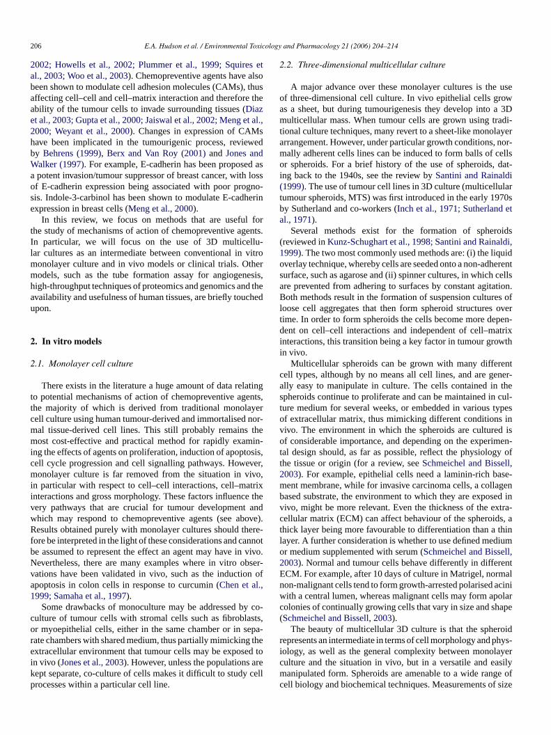
Fig. 4. The tube formation assay for angiogenesis. Brightfield image of immortomouse brain endothelial (IBE) cells (magnification,×10). Cells were seeded onto acollagen matrix and cultured overnight in the absence (control; left panel) or presence of basic fibroblast growth factor (bFGF; middle panel) or bFGFplus curcumin(40�M; right panel). Curcumin appeared to completely inhibit tube formation in response to stimulation with bFGF.
In vitro, endothelial cells can be used to measure the anti-angiogenic potential of agents in the tube formation assay. Theycan be induced to form tubules that resemble capillaries in vitroby plating cells onto a substrate such as Matrigel, with or with-out stimulation with angiogenic growth factors such as basicfibroblast growth factor or VEGF (Fig. 4). Suitable cells includeHUVECs derived from human umbilical vein, bovine aorticendothelial (BAE) cells, some endothelial cell lines, includingthe mouse immorto-brain endothelial cell line (IBEC) and thefirst human microvascular endothelial cell line (HMEC-1) thatretains the morphologic, phenotypic and functional characteris-tics of normal human microvascular endothelial cells (Ades etal., 1992). Use of the in vitro tube formation assays allows thestudy not only of physical capillary formation, and the signallingpathways responsible, but also the efficacy of chemopreventiveagents on these parameters.
4. Human tissues
The availability of fresh and frozen human tissue toresearchers in Europe is becoming more widespread, thanksto the establishment of groups such as the International Insti-tute for the Advancement of Medicine (IIAM) in 1996, fol-lowed soon after by the UK Human Tissue Bank (UKHTB;http://www.ukhtb.org) in 1998, and the European Network ofR eta andl beer lter-n
SA,i seRT nont whicw n bes rvedt prepr ssuei
The possibility of using human hepatocytes for drugmetabolism studies clearly has advantages over other in vitromodels, and is of particular interest for chemoprevention, asmodulation of drug metabolising enzymes including P450s, andphase II enzymes are long established mechanisms of actionof chemopreventive agents, which may affect the metabolism ofother chemicals including carcinogens, and indeed of the chemo-preventive agent itself (Ireson et al., 2001).
A human tissue-derived preclinical model of the breast hasbeen reviewed byKatdare et al. (2003); the breast terminal ductlobular unit (TDLU) is the smallest functional unit of breasttissue, and represents a major target site for carcinogenic trans-formation within the breast. Explants of TDLU can be culturedin vitro from breast reductions (non-cancerous) and cancerousbreasts. These cultures have been shown to be susceptible tochemical carcinogenesis and to stimulation with 17�-estradiol(Katdare et al., 2003).
The 184-B5 series of cell lines is derived from TDLU,including an immortalised, non-tumourigenic line and the 184-B5/HER line, in which the oncogene HER-2/neu has been over-expressed. Together with other markers such as lack of estrogenreceptor, and overexpression of p53 and the EGFR, this cell linerepresents a preclinical in vitro model for clinical comedo high-grade ductal carcinoma in situ (DCIS) (Katdare et al., 2003; Zhaiet al., 1993). The 184-B5/HER-cell line has already been used inchemoprevention studies of a range of phytochemicals includ-i wella iahe
5
o thed anal-y areaso
oft anti-t em.T tween
esearch Tissue Banks (ENRTB) (Anderson et al., 2001; Orrl., 2002). The safety and logistics, together with ethical
egal considerations of the use of human tissues, haveeviewed by the European Centre for the Validation of Aative Methods (ECVAM) (Anderson et al., 1998).
Tissue banks have existed for some time in the Uncluding IIAM (http://www.iiam.org) and the National Diseaesearch Interchange (NDRI;http://www.ndriresource.org).hese organisations make available to researchers
ransplantable donor tissues or surplus surgical tissues,ould otherwise go to waste. A wide range of tissues caourced, in a variety of formats including fresh or cryopreseissues, isolated hepatocytes, and microsome and cytosolations. In addition to healthy tissue, liver and other tumour tis also available from the UKHTB.
n
-h
a-
ng resveratrol, indole-3-carbinol, EGCG and genistein, ass retinoids and terpenoids (Katdare et al., 2003; Subbaramat al., 1998).
. DNA microarray, proteomics and chemigenomics
In recent years, technological breakthroughs have led tevelopment of a number of sophisticated gene and proteinsis techniques, which are proving to be invaluable to manyf research, including cancer.
DNA microarray allows the simultaneous determinationhe level of a large number of RNA species, thus giving quative information about the transcriptional profile of a systhe technology enables a direct comparison to be made be
E.A. Hudson et al. / Environmental Toxicology and Pharmacology 21 (2006) 204–214 211
pairs of samples, such as normal or diseased tissues or celllines, treated and non-treated tissues or cell lines. It is possi-ble therefore to identify previously unknown gene targets, theexpression of which is altered in response to a particular con-dition. Not only is the technology useful for identification ofgene changes in response to treatments with drugs, but also indetermining gene function, chromosomal loss or gain, tumourclassification, mutation detection, investigating mechanisms oftumour development and in drug discovery and development. Anoverview of microarray technology, and its relevance to breastcancer in particular can be found inCooper (2001)andYoung(2000).
A more recent development is that of the field of proteomics.This can be defined as a study of the function of all expressedproteins, and can be regarded as everything “post-genomic”(Tyers and Mann, 2003). In cancer research, proteomics hasdeveloped in particular out of a need for the identification ofreliable biomarkers of disease progression and response to ther-apy, and also from the observation that changes in the proteomicprofile of a system are not always predictable from genomicanalysis (Hanash, 2003). A joint venture between the NationalCancer Institute (NCI) and US Food and Drug Administra-tion (FDA) has resulted in the development of a diagnostictest for ovarian cancer, which relies on analysis of blood sam-ples to identify a “cancer profile” of peptides (Petricoin et al.,2002).
wsc am-p ple,a al inv t ofi , bup wedi
istrya cove tudyo bio-l an-i y ofi nds”( thed emi eenl sureb ghpa proc rsatii sings fromc lineso m-p thep rgea ca-t y(
6. Conclusions
Spheroid cultures should be regarded as a useful intermediatebetween monolayer culture and in vivo models, rather than as adirect replacement for either. However, for studies of molecu-lar mechanisms of action of chemopreventive agents, there areclear advantages to the use of the multicellular spheroid model.The benefits over monolayer culture are simply that spheroidsmimic much more closely the morphology, cell–cell interac-tions and general cell signalling pathways of tumours in vivo.The advantage of using an in vitro culture over in vivo tech-niques is related firstly to the ease with which spheroids canbe grown, handled and manipulated, and also to the speed withwhich large amounts of material can be generated. Cells can be ofhuman rather than rodent origin and they are amenable to a widerange of biochemical techniques. Using spheroids for mechanis-tic studies should enable the identification of more reliable andrelevant biomarkers, which can then be confirmed in vivo. Thein vitro tube formation assay is also a useful model in which tostudy inhibition of angiogenesis in response to chemopreven-tive agents. The model will be especially useful if combinedwith tumour spheroids, as the effects of chemopreventive agentson pro-angiogenic signalling and production of pro-angiogenicfactors by tumour cells on the development of tubules can thenbe observed directly.
A
nt-f ncerR ts.
R
A , S.,rtal-
l. 99,
A ature
A , M.,n ofapop-282.
A S.E.,CG,ells,DNA
A , devid,
n dert ofopean
A A.,G.,
e, J.,of
ations
As for DNA microarray profiling, proteomic analysis alloomparison of protein expression profiles from different sles, giving a broader picture of the effects of, for examchemopreventive agent, than is obtained from tradition
itro methods that rely on previous identification of a targenterest. Proteomics utilises a wide range of analysis toolsrimarily 2D electrophoresis and mass spectrometry, revie
n (Aebersold and Mann, 2003; Lee, 2001).Chemigenomics combines a genomic and a chem
pproach, and applies this to drug discovery. This termrs a very broad discipline, but can be defined as the “sf the genomic and/or proteomic response of an intact
ogical system – whether it be single cells or whole orgsms – to chemical compounds, or the study of the abilitsolated molecular targets to interact with such compouBredel and Jacoby, 2004). It can be used to acceleraterug discovery process, by studying the effect of large ch
cal libraries on whole biological systems, or to rapidly scribraries against selected targets, the outcome being meay assessment of phenotypic changes and by high-throussays. This technology is therefore becoming a favouredess with pharmaceutical companies. The enormous vety of chemigenomics means that it can be performed uingle cell organisms including bacteria and yeast, cellsomplex systems such as cultured human cancer cellr even whole organisms from worms to mice. Hit coounds can be identified on the basis of their effect onhenotype of the whole system, rather than just one tand then followed up with more detailed target identifi
ion and mechanistic studies, reviewed inBredel and Jacob2004).
t
-
-
dut-l-
,
t,
cknowledgements
We would like to thank Dr. Samantha Orr, UKHTB, De Moort University, and Professor Rosemary Walker, Breast Caesearch Unit, University of Leicester, for helpful commen
eferences
des, E.W., Candal, F.J., Swerlick, R.A., George, V.G., SummersBosse, D.C., Lawley, T.J., 1992. HMEC-1: establishment of an immoized human microvascular endothelial cell line. J. Invest. Dermato683–690.
ebersold, R., Mann, M., 2003. Mass spectrometry-based proteomics. N422, 198–207.
garwal, C., Singh, R.P., Dhanalakshmi, S., Tyagi, A.K., TecklenburgSclafani, R.A., Agarwal, R., 2003. Silibinin upregulates the expressiocyclin-dependent kinase inhibitors and causes cell cycle arrest andtosis in human colon carcinoma HT-29 cells. Oncogene 22, 8271–8
hn, W.S., Huh, S.W., Bae, S.M., Lee, I.P., Lee, J.M., Namkoong,Kim, C.K., Sin, J.I., 2003. A major constituent of green tea, EGinhibits the growth of a human cervical cancer cell line, CaSki cthrough apoptosis, G(1) arrest, and regulation of gene expression.Cell Biol. 22, 217–224.
nderson, R., Balls, M., Burke, M.D., Cummins, M., Fehily, D., Gray, N.Groot, M.G., Helin, H., Hunt, C., Jones, D., Price, D., Richert, L., RaR., Shute, D., Sladowski, D., Stone, H., Thasler, W., Trafford, J., vaValk, J., Weiss, T., Womack, C., Ylikomi, T., 2001. The establishmenhuman research tissue banking in the UK and several western Eurcountries. ATLA 29, 125–134.
nderson, R., O’Hare, M., Balls, M., Brady, M., Brahams, D., Burt,Chesne, C., Combes, R., Dennison, A., Garthoff, B., Hawksworth,Kalter, E., Lechat, A., Mayer, D., Rogiers, V., Sladowski, D., SoutheTrafford, J., van der Valk, J., van Zeller, A.-M., 1998. The availabilityhuman tissue for biomedical research. The report and recommendof ECVAM workshop 32. ATLA 26, 763–777.

212 E.A. Hudson et al. / Environmental Toxicology and Pharmacology 21 (2006) 204–214
Arbiser, J.L., Klauber, N., Rohan, R., van Leeuwen, R., Huang, M.T., Fisher,C., Flynn, E., Byers, H.R., 1998. Curcumin is an in vivo inhibitor ofangiogenesis. Mol. Med. 4, 378–383.
Atherfold, P.A., Manson, M.M., 2002. The chemopreventive agent EGCGinduces cell cycle arrest and apoptosis in HUVEC. Free Radic. Biol.Med. 33, 83.
Aziz, M.H., Kumar, R., Ahmad, N., 2003. Cancer chemoprevention by resver-atrol: in vitro and in vivo studies and the underlying mechanisms. Int. J.Oncol. 23, 17–28.
Bai, X., Cerimele, F., Ushio-Fukai, M., Waqas, M., Campbell, P.M., Govin-darajan, B., Der, C.J., Battle, T., Frank, D.A., Ye, K., Murad, E., Dubiel,W., Soff, G., Arbiser, J.L., 2003. Honokiol, a small molecular weightnatural product, inhibits angiogenesis in vitro and tumor growth in vivo.J. Biol. Chem. 278, 35501–35507.
Behrens, J., 1999. Cadherins and catenins: role in signal transduction andtumor progression. Cancer Metastasis Rev. 18, 15–30.
Berx, G., Van Roy, F., 2001. The E-cadherin/catenin complex: an impor-tant gatekeeper in breast cancer tumorigenesis and malignant progression.Breast Cancer Res. 3, 289–293.
Bissell, M.J., Rizki, A., Mian, I.S., 2003. Tissue architecture: the ultimateregulator of breast epithelial function. Curr. Opin. Cell Biol. 15, 753–762.
Bredel, M., Jacoby, E., 2004. Chemigenomics: an emerging strategy for rapidtarget and drug discovery. Nat. Rev. Genet. 5, 262–275.
Chan, A.T., Giovannucci, E.L., Schernhammer, E.S., Colditz, G.A., Hunter,D.J., Willett, W.C., Fuchs, C.S., 2004. A prospective study of aspirin useand the risk for colorectal adenoma. Ann. Intern. Med. 140, 157–166.
Chaudhury, L.R., Hruska, K.A., 2003. Inhibition of cell survival signal pro-tein kinase B/Akt by curcumin in human prostate cancer cells. J. Cell.Biochem. 89, 1–5.
Chen, H., Zhang, Z.S., Zhang, Y.L., Zhou, D.Y., 1999. Curcumin inhibits cellproliferation by interfering with the cell cycle and inducing apoptosis in
C le-3-1236
C statetudie
C .C.,n of
4.C ance
C L.F.arres. 59
C L.F.,clin-breahem
D y in
D tosissisloro-
F ng,Acanc
F obi-tancecan-
s 19,
F les in
Giardiello, F.M., Yang, V.W., Hylind, L.M., Krush, A.J., Petersen, G.M.,Trimbath, J.D., Piantadosi, S., Garrett, E., Geiman, D.E., Hubbard, W.,Offerhaus, G.J.A., Hamilton, S.R., 2002. Primary chemoprevention offamilial adenomatous polyposis with sulindac. N. Engl. J. Med. 346,1054–1059.
Gupta, S., Ahmad, N., Marengo, S.R., MacLennan, G.T., Greenberg, N.M.,Mukhtar, H., 2000. Chemoprevention of prostate carcinogenesis by alpha-difluoromethylornithine in TRAMP mice. Cancer Res. 60, 5125–5133.
Hanash, S., 2003. Disease proteomics. Nature 422, 226–232.Hauptmann, S., Denkert, C., Lohrke, H., Tietze, L., Ott, S., Klosterhalfen,
B., Mittermayer, C., 1995. Integrin expression on colorectal tumor-cellsgrowing as monolayers, as multicellular tumor spheroids, or in nude-mice.Int. J. Cancer 61, 819–825.
Hauptmann, S., Gebauer-Hartung, P., Leclere, A., Denkert, C., Pest,S., Klosterhalfen, B., Dietel, M., 1998. Induction of apopto-sis in the centre of multicellular tumour spheroids of colorectaladenocarcinomas—involvement of CD95 pathway and differentiation.Apoptosis 3, 267–279.
Howells, L.M., Gallacher-Horley, B., Houghton, C.E., Manson, M.M., Hud-son, E.A., 2002. Indole-3-carbinol inhibits protein kinase B/Akt andinduces apoptosis in the human breast tumor cell line MDA MB468 butnot in the nontumorigenic HBL100 line. Mol. Cancer Ther. 1, 1161–1172.
Igura, K., Ohta, T., Kuroda, Y., Kuji, K., 2001. Resveratrol and quercetininhibit angiogenesis in vitro. Cancer Lett. 171, 11–16.
Inch, W.R., McCredie, J.A., Sutherland, R.M., 1971. Growth of nodular car-cinomas in rodents compared with multi-cell spheroids in tissue culture.Growth 34, 271–282.
Ireson, C., Orr, S., Jones, D.J.L., Verschoyle, R., Lim, C.-K., Luo, J.-L., How-ells, L., Plummer, S., Jukes, R., Williams, M., Steward, W.P., Gescher,A., 2001. Characterisation of metabolites of the chemopreventive agentcurcumin in human and rat hepatocytes and in the rat in vivo, and eval-
E2
J tenin-ortantis in
J reastancerand
J ns in
K od-509–
K 03.tionnoma
K ibitigra-
ing.
K larmour
L r byncer
L t vas-s. 62,
L rage-wth
L ited
colon carcinoma cells. Anticancer Res. 19, 3675–3680.hinni, S.R., Sarkar, F.H., 2002. Akt inactivation is a key event in indo
carbinol-induced apoptosis in PC-3 cells. Clin. Cancer Res. 8, 1228–hung, L.W.K., Zhau, H.E., Wu, T.T., 1997. Development of human pro
cancer models for chemoprevention and experimental therapeutic sJ. Cell. Biochem. S28–S29, 174–181.
omerford, K.M., Wallace, T.J., Karhausen, J., Louis, N.A., Montalto, MColgan, S.P., 2002. Hypoxia-inducible factor-1-dependent regulatiothe multidrug resistance (MDR1) gene. Cancer Res. 62, 3387–339
ooper, C.S., 2001. Applications of microarray technology in breast cresearch. Breast Cancer Res. 3, 158–175.
over, C.M., Hsieh, S.J., Cram, E.J., Hong, C., Riby, J.E., Bjeldanes,Firestone, G.L., 1999. Indole-3-carbinol and tamoxifen cooperate tothe cell cycle of MCF-7 human breast cancer cells. Cancer Res1244–1251.
over, C.M., Hsieh, S.J., Tran, S.H., Hallden, G., Kim, G.S., Bjeldanes,Firestone, G.L., 1998. Indole-3-carbinol inhibits the expression of cydependent kinase-6 and induces a G1 cell cycle arrest of humancancer cells independent of estrogen receptor signalling. J. Biol. C273, 3838–3847.
atta, S.R., Brunet, A., Greenberg, M.E., 1999. Cellular survival: a plathree Akts. Genes Dev. 13, 2905–2927.
iaz, G.D., Li, Q.J., Dashwood, R.H., 2003. Caspase-8 and apopinducing factor mediate a cytochromec-independent pathway of apoptoin human colon cancer cells induced by the dietary phytochemical chphyllin. Cancer Res. 63, 1254–1261.
ang, M.Z., Wang, Y., Ai, N., Hou, Z., Sun, Y., Lu, H., Welsh, W., YaC.S., 2003. Tea polyphenol (−)-epigallocatechin-3-gallate inhibits DNmethyltransferase and reactivates methylation-silenced genes incell lines. Cancer Res. 63, 7563–7570.
aute, M.A.D., Laurent, L., Ploton, D., Poupon, M.F., Jardillier, J.C., Bchon, H., 2002. Distinctive alterations of invasiveness, drug resisand cell–cell organization in 3D-cultures of MCF7, a human breastcer cell line, and its multidrug resistant variant. Clin. Exp. Metastasi161–168.
racasso, G., Colombatti, M., 2000. Effect of therapeutic macromolecuspheroids. Crit. Rev. Oncol. Hematol. 36, 159–178.
.
s.
r
,t,
st.
-
er
uation of their ability to inhibit phorbol ester-induced prostaglandinproduction. Cancer Res. 61, 1058–1064.
aiswal, A.S., Marlow, B.P., Gupta, N., Narayan, S., 2002. Beta-camediated transactivation and cell–cell adhesion pathways are impin curcumin (diferuylmethane)-induced growth arrest and apoptoscolon cancer cells. Oncogene 21, 8414–8427.
ones, J.L., Shaw, J.A., Pringle, J.H., Walker, R.A., 2003. Primary bmyoepithelial cells exert an invasion-suppressor effect on breast ccells via paracrine down-regulation of MMP expression in fibroblaststumour cells. J. Pathol. 201, 562–572.
ones, J.L., Walker, R.A., 1997. Cell–cell and cell–stromal interactiobreast cancer invasion and metastasis. Int. J. Oncol. 11, 609–616.
atdare, M., Osborne, M.P., Telang, N.T., 2003. Novel cell culture mels for prevention of human breast cancer. Int. J. Oncol. 22,515.
im, Y.A., Lee, W.H., Choi, T.H., Rhee, S.H., Park, K.Y., Choi, Y.H., 20Involvement of p21WAF1/CIP1, pRB, Bax and NF-kappa B in inducof growth arrest and apoptosis by resveratrol in human lung carciA549 cells. Int. J. Oncol. 23, 1143–1149.
ondo, T., Ohta, T., Igura, K., Hara, Y., Kiji, K., 2002. Tea catechins inhangiogenesis in vitro, measured by human endothelial cell growth, mtion and tube formation, through inhibition of VEGF receptor bindCancer Lett. 180, 139–144.
unz-Schughart, L.A., Kreutz, M., Knuechel, R., 1998. Multicelluspheroids: a three dimensional in vitro culture system to study tubiology. Int. J. Exp. Pathol. 79, 1365–2613.
amprecht, S.A., Lipkin, M., 2003. Chemoprevention of colon cancecalcium, Vitamin D and folate: molecular mechanisms. Nat. Rev. Ca3, 601–614.
amy, S., Gingras, D., Beliveau, R., 2002. Green tea catechins inhibicular endothelial growth factor receptor phosphorylation. Cancer Re381–385.
awlor, E.R., Scheel, C., Irving, J., Sorensen, P.H.B., 2002. Anchoindependent multi-cellular spheroids as an in vitro model of grosignaling in Ewing tumors. Oncogene 21, 307–318.
ee, K.H., 2001. Proteomics: a technology-driven and technology-limdiscovery science. Trends Biotechnol. 19, 217–221.

E.A. Hudson et al. / Environmental Toxicology and Pharmacology 21 (2006) 204–214 213
Li, Y.W., Sarkar, F.H., 2002. Down-regulation of invasion and angiogenesis-related genes identified by cDNA microarray analysis of PC3 prostatecancer cells treated with genistein. Cancer Lett. 186, 157–164.
Liao, H.F., Chen, Y.Y., Liu, J.J., Hsu, M.L., Shieh, H.J., Liao, H.J., Shieh,C.J., Shiao, M.S., Chen, Y.J., 2003. Inhibitory effect of caffeic acidphenethyl ester on angiogenesis, tumor invasion, and metastasis. J. Agric.Food Chem. 51, 7907–7912.
Lin, J.-K., Liang, Y.-C., Lin-Shiau, S., 1999. Cancer chemoprevention bytea polyphenols through mitotic signal transduction cascade. Biochem.Pharmacol. 58, 911–915.
Liu, H., Radisky, D., Wang, F., Bissell, M.J., 2004. Polarity and proliferationare controlled by distinct signaling pathways downstream of PI3-kinasein breast epithelial tumor cells. J. Cell Biol. 164, 603–612.
Manson, M.M., Gescher, A., Hudson, E.A., Plummer, S.M., Squires,M.S., Prigent, S.A., 2000a. Blocking and suppressing mechanisms ofchemoprevention by dietary constituents. Toxicol. Lett. 112–113, 499–505.
Manson, M.M., Holloway, K.A., Howells, L.M., Hudson, E.A., Plum-mer, S.M., Squires, M.S., Prigent, S.A., 2000b. Modulation of signal-transduction pathways by chemopreventive agents. Biochem. Soc. Trans.28, 7–12.
Manson, M.M., Howells, L.M., Hudson, E.A., 2003. Signalling pathwaysas targets in cancer prevention. In: Vainio, H.U., Hietanen, E. (Eds.),Handbook of Experimental Pharmacology: Mechanisms in Carcinogenesisand Cancer Prevention. Springer-Verlag, Berlin.
Mayer, B., Klement, G., Kaneko, M., Man, S., Jothy, S., Rak, J., Kerbel, R.S.,2001. Multicellular gastric cancer spheroids recapitulate growth patternand differentiation phenotype of human gastric carcinomas. Gastroenterol-ogy 121, 839–852.
Meng, Q.H., Goldberg, I.D., Rosen, E.M., Fan, S.J., 2000. Inhibitory effectsof indole-3-carbinol on invasion and migration in human breast cancer
M Crit.
M 002.or
N R.J.,ient
J. Bi
N t G(1ys.
N prog-
O -vid,k, J.,lish-Tissu
P A.,.C.,rian
P A.,sionition
18,
R tialncerRes.
R drugsreality
Sachinidis, A., Seul, C., Seewald, S., Ahn, H.Y., Ko, Y., Vetter, H., 2000.Green tea compounds inhibit tyrosine phosphorylation of PDGF beta-receptor and transformation of A172 human glioblastoma. FEBS Lett.471, 51–55.
Samaha, H.S., Kelloff, G.J., Steele, V., Rao, C.V., Reddy, B.S., 1997. Modu-lation of apoptosis by sulindac, curcumin, phenylethyl-3-methylcaffeated,and 6-phenylhexyl isothiocyanate: apoptotic index as a biomarker in coloncancer chemoprevention and promotion. Cancer Res. 57, 1301–1305.
Santini, M.T., Rainaldi, G., 1999. Three-dimensional spheroid model in tumorbiology. Pathobiology 67, 148–157.
Santini, M.T., Rainaldi, G., Indovina, P.L., 1999. Multicellular tumourspheroids in radiation biology. Int. J. Radiat. Biol. 75, 787–799.
Schmeichel, K.L., Bissell, M.J., 2003. Modeling tissue-specific signaling andorgan function in three dimensions. J. Cell Sci. 116, 2377–2388.
Seidl, P., Huettinger, R., Knuechel, R., Kunz-Schughart, L.A., 2002. Three-dimensional fibroblast-tumor cell interaction causes downregulation ofRACK1 mRNA expression in breast cancer cells in vitro. Int. J. Cancer102, 129–136.
Serrano, D., Perego, E., Costa, A., Decensi, A., 2004. Progress in chemopre-vention of breast cancer. Crit. Rev. Oncol. Hematol. 49, 109–117.
Shao, Z.-M., Shen, Z.-Z., Liu, C.-H., Sartippour, M.R., Go, V.L., Heber,D., Nguyen, M., 2002. Curcumin exerts multiple suppressive effects onhuman breast carcinoma cells. Int. J. Cancer 98, 234–240.
Sheng, H.M., Shao, J.Y., DuBois, R.N., 2001. Akt/PKB activity is requiredfor Ha-Ras-mediated transformation of intestinal epithelial cells. J. Biol.Chem. 276, 14498–14504.
Singh, A.-K., Seth, P., Anthony, P., Husain, M.M., Madhavan, S., Mukhtar,H., Maheshwari, R.K., 2002. Green tea constituent epigallocatechin-3-gallate inhibits angiogenic differentiation of human endothelial cells.Arch. Biochem. Biophys. 401, 29–37.
Skrzsypczak-Jankun, E., McCabe, N.P., Selman, S.H., Jankun, J., 2000. Cur-tical
S nkun,d its
S .E.,.M.,ands toacol.
S Inoue,hibitsuman
S . Nat.
S rore-
S lularNatl.
T r for
T uki,c car-creas
T hibitherin106,
T esh-bil-12.
T reven-
cells. Breast Cancer Res. Treat. 63, 147–152.ueller-Klieser, W., 2000. Tumor biology and experimental therapeutics.
Rev. Oncol. Hematol. 36, 123–139.ueller-Klieser, W., Schreiber-Klais, S., Walenta, S., Kreuter, M.H., 2
Bioactivity of well-defined green tea extracts in multicellular tumspheroids. Int. J. Oncol. 21, 1307–1315.
akatani, K., Thompson, D.A., Barthel, A., Sakaue, H., Liu, W., Weigel,Roth, R.A., 1999. Up-regulation of Akt3 in estrogen receptor-deficbreast cancers and androgen-independent prostate cancer lines.Chem. 274, 21528–21532.
emoto, T., Kamei, S., Seyama, Y., Kubota, S., 2001. p53 independenarrest induced bydl-alpha-difluoromethylornithine. Biochem. BiophRes. Commun. 280, 848–854.
icholson, R.I., Gee, J.M.W., Harper, M.E., 2001. EGFR and cancernosis. Eur. J. Cancer 37, S9–S15.
rr, S., Alexandre, E., Clark, B., Combes, R., Fels, L.M., Gray, N., JonssonRylander, A.-C., Helin, H., Koistinen, J., Oinonen, T., Richert, L., RaR., Salonen, J., Teesalu, T., Thasler, W., Trafford, J., van der Valvon Versen, R., Weiss, T., Womack, C., Ylikomi, T., 2002. The estabment of a network of European human research tissue banks. CellBanking 3, 133–137.
etricoin III, E.F., Ardekani, A.M., Hitt, B.A., Levine, P.J., Fusaro, V.Steinberg, S.M., Mills, G.B., Simone, C., Fishman, D.A., Kohn, ELiotta, L.A., 2002. Use of proteomic patterns in serum to identify ovacancer. Lancet 359, 572–577.
lummer, S.M., Holloway, K.A., Manson, M.M., Munks, R.J.L., Kaptein,Farrow, S., Howells, L., 1999. Inhibition of cyclo-oxygenase 2 expresin colon cells by the chemopreventive agent curcumin involves inhibof NF-�B activation via the NIK/IKK signalling complex. Oncogene6013–6020.
ainaldi, G., Calcabrini, A., Arancia, G., Santini, M.T., 1999. Differenexpression of adhesion molecules (CD44, ICAM-1 and LFA-3) in cacells grown in monolayer or as multicellular spheroids. Anticancer19, 1769–1778.
uegg, C., Zaric, J., Stupp, R., 2003. Non steroidal anti-inflammatoryand COX-2 inhibitors as anti-cancer therapeutics: hypes, hopes andAnn. Med. 35, 476–487.
ol.
)
e
.
cumin inhibits lipoxygenase by binding to its central cavity: theoreand X-ray evidence. Int. J. Mol. Med. 6, 521–526.
krzsypczak-Jankun, E., Zhou, K.J., McCabe, N.P., Selman, S.H., JaJ., 2003. Structure of curcumin in complex with lipoxygenase ansignificance in cancer. Int. J. Mol. Med. 12, 17–24.
quires, M.S., Hudson, E.A., Howells, L., Sale, S., Houghton, CJones, J.L., Fox, L.H., Dickens, M., Prigent, S.A., Manson, M2003. Relevance of mitogen activated protein kinase (MAPK)phosphotidylinositol-3-kinase/protein kinase B (PI3K/PKB) pathwayinduction of apoptosis by curcumin in breast cells. Biochem. Pharm65, 361–376.
ubbaramaiah, K., Chung, W.J., Michaluart, P., Telang, N., Tanabe, T.,H., Jang, M., Pezzuto, J.M., Dannenberg, A.J., 1998. Resveratrol incyclooxygenase-2 transcription and activity in phorbol ester-treated hmammary epithelial cells. J. Biol. Chem. 273, 21875–21882.
urh, Y.-J., 2003. Cancer chemoprevention with dietary phytochemicalsRev. Cancer 3, 768–780.
utherland, R.M., 1988. Cell and environment interactions in tumor micgions: the multicell spheroid model. Science 240, 177–185.
utherland, R.M., McCredie, J.A., Inch, W.R., 1971. Growth of multicelspheroids in tissue culture as a model of nodular carcinomas. J.Cancer Inst. 46, 113–120.
achibana, H., Koga, K., Fujimura, Y., Yamada, K., 2004. A receptogreen tea polyphenol EGCG. Nat. Struct. Mol. Biol. 11, 380–381.
akada, M., Nakamura, Y., Koizumi, T., Toyama, H., Kamigaki, T., SuzY., Takeyama, Y., Kuroda, Y., 2002. Suppression of human pancreaticinoma cell growth and invasion by epigallocatechin-3-gallate. Pan25, 45–48.
ang, F.-Y., Nguyen, N., Meydani, M., 2003. Green tea catechins inVEGF-induced angiogenesis in vitro through suppression of VE-cadphosphorylation and inactivation of Akt molecule. Int. J. Cancer871–878.
haloor, D., Singh, A.K., Sidhu, G.S., Prasad, P.V., Kleinman, H.K., Mahwari, R.K., 1998. Inhibition of angiogenic differentiation of human umical vein endothelial cells by curcumin. Cell Growth Differ. 9, 302–3
hornalley, P.J., 2002. Isothiocyanates: mechanisms of cancer chemoptive action. Anti-Cancer Drugs 13, 331–338.

214 E.A. Hudson et al. / Environmental Toxicology and Pharmacology 21 (2006) 204–214
Tosetti, F., Ferrari, N., De Flora, S., Albini, A., 2002. Angioprevention: angio-genesis is a common and key target for cancer chemopreventive agents.FASEB J. 16, 2–14.
Tsutsui, S., Kataoka, A., Ohno, S., Murakami, S., Kinoshita, J., Hachitanda,Y., 2002. Prognostic and predictive value of epidermal growth factorreceptor in recurrent breast cancer. Clin. Cancer Res. 8, 3454–3460.
Tyers, M., Mann, M., 2003. From genomics to proteomics. Nature 422,193–197.
Waleh, N.S., Gallo, J., Grant, T.D., Murphy, B.J., Kramer, R.H., Sutherland,R.M., 1994. Selective down-regulation of integrin receptors in spheroidsof squamous cell carcinoma. Cancer Res. 54, 838–843.
Wang, F., Hansen, R.K., Radisky, D., Yoneda, T., Barcellos-Hoff, M.H.,Petersen, O.W., Turley, E.A., Bissell, M.J., 2002. Phenotypic reversion ordeath of cancer cells by altering signaling pathways in three-dimensionalcontexts. J. Natl. Cancer Inst. 94, 1494–1503.
Wartenberg, M., Ling, F.C., Muschen, M., Klein, F., Acker, H., Gassmann,M., Petrat, K., Putz, V., Hescheler, J., Sauer, H., 2003. Regulation of themultidrug resistance transporter P-glycoprotein in multicellular tumour
spheroids by hypoxia inducible factor-1 and reactive oxygen species.Faseb J. 17, U546–U567.
WCRF and AICR, 1997. Food, Nutrition and the Prevention of Cancer: AGlobal Perspective. American Institute for Cancer Research, Washington.
Weyant, M.J., Carothers, A.M., Bertagnolli, M.E., Bertagnolli, M.M., 2000.Colon cancer chemopreventive drugs modulate integrin-mediated signal-ing pathways. Clin. Cancer Res. 6, 949–956.
Woo, J.H., Kim, Y.H., Choi, Y.J., Kim, D.G., Lee, K.S., Bae, J.H., Min,D.S., Chang, J.S., Jeong, Y.J., Lee, Y.H., Park, J.W., Kwon, T.K., 2003.Molecular mechanisms of curcumin-induced cytotoxicity: induction ofapoptosis through generation of reactive oxygen species, down-regulationof Bcl-X-L and IAP, the release of cytochromec and inhibition of Akt.Carcinogenesis 24, 1199–1208.
Young, R.A., 2000. Biomedical discovery with DNA arrays. Cell 102, 9–15.Zhai, Y.F., Beittenmiller, H., Wang, B., Gould, M.N., Oakley, C., Essel-
man, W.J., Welsch, C.W., 1993. Increased expression of specific proteintyrosine phosphatases in human breast epithelial cells neoplastically trans-formed by the neu oncogene. Cancer Res. 53, 2272–2278.


![21242593[VARIMOT EX.].pdf](https://static.fdokumen.com/doc/165x107/6338b562e57b005bf2016bb4/21242593varimot-expdf.jpg)

