Evolutionary interactions between diabetes and development
15
Review Article Evolutionary interactions between diabetes and development R.G. Ahmed * Department of Zoology, Faculty of Science, Beni-Suef University, Beni-Suef, Egypt Contents 1. Introduction .................................................................................. 154 2. Diabetogenic agents............................................................................ 155 2.1. Streptozotocin (STZ) ...................................................................... 155 2.2. Alloxan (AL) ............................................................................. 155 3. Diabetes and spermatogenesis interactions ......................................................... 156 4. Diabetes and oogenesis interactions ............................................................... 156 5. Gestational diabetes (GDM) ...................................................................... 157 6. Perinatal diabetes ............................................................................. 158 7. Maternal teratology, embryopathy and diabetes interactions ........................................... 158 8. General summary about the maternal metabolic aberrations due to DM during pregnancy and a serious impact from fetus to young adult ................................................................. 160 9. General summary about the effect of DM on the different developmental stages ........................... 160 10. Recommendation.............................................................................. 161 References ................................................................................... 161 diabetes research and clinical practice 92 (2011) 153–167 article info Article history: Received 1 July 2010 Received in revised form 12 October 2010 Accepted 19 October 2010 Published on line 15 December 2010 Keywords: Diabetes Development Pregnancy abstract Because of the complications of diabetes affecting the mothers and their fetus/newborns are less known, this review examined the epidemiologic and mechanistic issues involved in the developmental programming of diabetic mothers. This overview showed that sperm, egg, zygote or blastocyst derived from diabetic parents may develop into offspring with high risk of any type of diabetes, even if placed in a normal uterus, producing developmental delay, embryopathy, geno- and cyto-toxicity, teratogenic changes, free radicals and apoptosis. These early insults may then lead to an increased rate of miscarriage and congenital anomalies depending on free radicals signaling and cell-death pathways involved by the diabetogenic agents. Furthermore, sperm, egg, zygote or blastocyst from normal parents will have an increased risk of diabetes if placed in a diabetic uterus. Interestingly, diabetes has deleterious effect on male/female reproductive functions and on the development of the blastocysts/embryos. Indeed, this review hypothesized that the long-term effects of diabe- tes during the pregnancy (gestational diabetes) may influence, generally, on the health of the embryos, newborns (perinatal life) and adulthood. However, there are obvious species differences between pregnant women and animal models. Thus, maintaining normogly- caemia during pregnancy may play an important role in a healthy life for the newborns. # 2010 Elsevier Ireland Ltd. All rights reserved. * Tel.: +2 082 2310 187. E-mail addresses: [email protected], [email protected]. Contents lists available at ScienceDirect Diabetes Research and Clinical Practice journal homepage: www.elsevier.com/locate/diabres 0168-8227/$ – see front matter # 2010 Elsevier Ireland Ltd. All rights reserved. doi:10.1016/j.diabres.2010.10.014
Transcript of Evolutionary interactions between diabetes and development
Review Article
Evolutionary interactions between diabetes and development
R.G. Ahmed *
Department of Zoology, Faculty of Science, Beni-Suef University, Beni-Suef, Egypt
Contents
1. Introduction. . . . . . . . . . . . . . . . . . . . . . . . . . . . . . . . . . . . . . . . . . . . . . . . . . . . . . . . . . . . . . . . . . . . . . . . . . . . . . . . . . 154
2. Diabetogenic agents. . . . . . . . . . . . . . . . . . . . . . . . . . . . . . . . . . . . . . . . . . . . . . . . . . . . . . . . . . . . . . . . . . . . . . . . . . . . 155
2.1. Streptozotocin (STZ) . . . . . . . . . . . . . . . . . . . . . . . . . . . . . . . . . . . . . . . . . . . . . . . . . . . . . . . . . . . . . . . . . . . . . . 155
2.2. Alloxan (AL) . . . . . . . . . . . . . . . . . . . . . . . . . . . . . . . . . . . . . . . . . . . . . . . . . . . . . . . . . . . . . . . . . . . . . . . . . . . . . 155
3. Diabetes and spermatogenesis interactions . . . . . . . . . . . . . . . . . . . . . . . . . . . . . . . . . . . . . . . . . . . . . . . . . . . . . . . . . 156
4. Diabetes and oogenesis interactions . . . . . . . . . . . . . . . . . . . . . . . . . . . . . . . . . . . . . . . . . . . . . . . . . . . . . . . . . . . . . . . 156
5. Gestational diabetes (GDM) . . . . . . . . . . . . . . . . . . . . . . . . . . . . . . . . . . . . . . . . . . . . . . . . . . . . . . . . . . . . . . . . . . . . . . 157
6. Perinatal diabetes . . . . . . . . . . . . . . . . . . . . . . . . . . . . . . . . . . . . . . . . . . . . . . . . . . . . . . . . . . . . . . . . . . . . . . . . . . . . . 158
7. Maternal teratology, embryopathy and diabetes interactions . . . . . . . . . . . . . . . . . . . . . . . . . . . . . . . . . . . . . . . . . . . 158
8. General summary about the maternal metabolic aberrations due to DM during pregnancy and a serious
impact from fetus to young adult . . . . . . . . . . . . . . . . . . . . . . . . . . . . . . . . . . . . . . . . . . . . . . . . . . . . . . . . . . . . . . . . . 160
9. General summary about the effect of DM on the different developmental stages . . . . . . . . . . . . . . . . . . . . . . . . . . . 160
10. Recommendation. . . . . . . . . . . . . . . . . . . . . . . . . . . . . . . . . . . . . . . . . . . . . . . . . . . . . . . . . . . . . . . . . . . . . . . . . . . . . . 161
References . . . . . . . . . . . . . . . . . . . . . . . . . . . . . . . . . . . . . . . . . . . . . . . . . . . . . . . . . . . . . . . . . . . . . . . . . . . . . . . . . . . 161
d i a b e t e s r e s e a r c h a n d c l i n i c a l p r a c t i c e 9 2 ( 2 0 1 1 ) 1 5 3 – 1 6 7
a r t i c l e i n f o
Article history:
Received 1 July 2010
Received in revised form
12 October 2010
Accepted 19 October 2010
Published on line 15 December 2010
Keywords:
Diabetes
Development
Pregnancy
a b s t r a c t
Because of the complications of diabetes affecting the mothers and their fetus/newborns are
less known, this review examined the epidemiologic and mechanistic issues involved in the
developmental programming of diabetic mothers. This overview showed that sperm, egg,
zygote or blastocyst derived from diabetic parents may develop into offspring with high risk
of any type of diabetes, even if placed in a normal uterus, producing developmental delay,
embryopathy, geno- and cyto-toxicity, teratogenic changes, free radicals and apoptosis.
These early insults may then lead to an increased rate of miscarriage and congenital
anomalies depending on free radicals signaling and cell-death pathways involved by the
diabetogenic agents. Furthermore, sperm, egg, zygote or blastocyst from normal parents will
have an increased risk of diabetes if placed in a diabetic uterus. Interestingly, diabetes has
deleterious effect on male/female reproductive functions and on the development of the
blastocysts/embryos. Indeed, this review hypothesized that the long-term effects of diabe-
tes during the pregnancy (gestational diabetes) may influence, generally, on the health of
the embryos, newborns (perinatal life) and adulthood. However, there are obvious species
differences between pregnant women and animal models. Thus, maintaining normogly-
caemia during pregnancy may play an important role in a healthy life for the newborns.
# 2010 Elsevier Ireland Ltd. All rights reserved.
* Tel.: +2 082 2310 187.
C o n t e n t s l i s t s a va i l a b l e a t S c i e n c e D i r e c t
Diabetes Researchand Clinical Practice
journal homepage: www.elsevier.com/locate/diabres
E-mail addresses: [email protected], [email protected].
0168-8227/$ – see front matter # 2010 Elsevier Ireland Ltd. All rights reserved.doi:10.1016/j.diabres.2010.10.014
d i a b e t e s r e s e a r c h a n d c l i n i c a l p r a c t i c e 9 2 ( 2 0 1 1 ) 1 5 3 – 1 6 7154
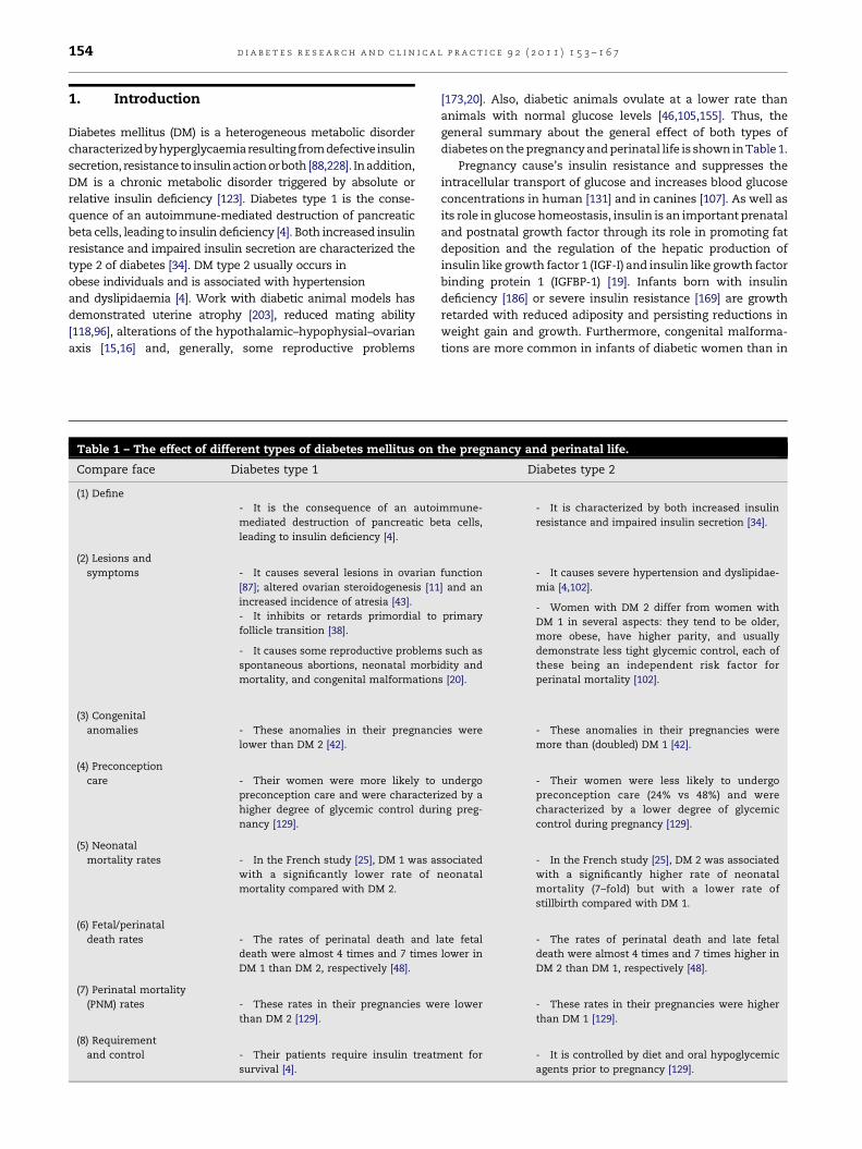
1. Introduction
Diabetes mellitus (DM) is a heterogeneous metabolic disorder
characterizedbyhyperglycaemiaresultingfromdefectiveinsulin
secretion, resistanceto insulinactionorboth [88,228]. Inaddition,
DM is a chronic metabolic disorder triggered by absolute or
relative insulin deficiency [123]. Diabetes type 1 is the conse-
quence of an autoimmune-mediated destruction of pancreatic
beta cells, leading to insulin deficiency [4]. Both increased insulin
resistance and impaired insulin secretion are characterized the
type 2 of diabetes [34]. DM type 2 usually occurs in
obese individuals and is associated with hypertension
and dyslipidaemia [4]. Work with diabetic animal models has
demonstrated uterine atrophy [203], reduced mating ability
[118,96], alterations of the hypothalamic–hypophysial–ovarian
axis [15,16] and, generally, some reproductive problems
Table 1 – The effect of different types of diabetes mellitus on
Compare face Diabetes type 1
(1) Define
- It is the consequence of an autoi
mediated destruction of pancreatic be
leading to insulin deficiency [4].
(2) Lesions and
symptoms - It causes several lesions in ovarian
[87]; altered ovarian steroidogenesis [11
increased incidence of atresia [43].
- It inhibits or retards primordial to
follicle transition [38].
- It causes some reproductive problem
spontaneous abortions, neonatal morb
mortality, and congenital malformation
(3) Congenital
anomalies - These anomalies in their pregnanc
lower than DM 2 [42].
(4) Preconception
care - Their women were more likely to
preconception care and were character
higher degree of glycemic control duri
nancy [129].
(5) Neonatal
mortality rates - In the French study [25], DM 1 was a
with a significantly lower rate of
mortality compared with DM 2.
(6) Fetal/perinatal
death rates - The rates of perinatal death and l
death were almost 4 times and 7 times
DM 1 than DM 2, respectively [48].
(7) Perinatal mortality
(PNM) rates - These rates in their pregnancies we
than DM 2 [129].
(8) Requirement
and control - Their patients require insulin treat
survival [4].
[173,20]. Also, diabetic animals ovulate at a lower rate than
animals with normal glucose levels [46,105,155]. Thus, the
general summary about the general effect of both types of
diabetes on the pregnancy and perinatal life is shown in Table 1.
Pregnancy cause’s insulin resistance and suppresses the
intracellular transport of glucose and increases blood glucose
concentrations in human [131] and in canines [107]. As well as
its role in glucose homeostasis, insulin is an important prenatal
and postnatal growth factor through its role in promoting fat
deposition and the regulation of the hepatic production of
insulin like growth factor 1 (IGF-I) and insulin like growth factor
binding protein 1 (IGFBP-1) [19]. Infants born with insulin
deficiency [186] or severe insulin resistance [169] are growth
retarded with reduced adiposity and persisting reductions in
weight gain and growth. Furthermore, congenital malforma-
tions are more common in infants of diabetic women than in
the pregnancy and perinatal life.
Diabetes type 2
mmune-
ta cells,
- It is characterized by both increased insulin
resistance and impaired insulin secretion [34].
function
] and an
- It causes severe hypertension and dyslipidae-
mia [4,102].
primary- Women with DM 2 differ from women with
DM 1 in several aspects: they tend to be older,
more obese, have higher parity, and usually
demonstrate less tight glycemic control, each of
these being an independent risk factor for
perinatal mortality [102].
s such as
idity and
s [20].
ies were - These anomalies in their pregnancies were
more than (doubled) DM 1 [42].
undergo
ized by a
ng preg-
- Their women were less likely to undergo
preconception care (24% vs 48%) and were
characterized by a lower degree of glycemic
control during pregnancy [129].
ssociated
neonatal
- In the French study [25], DM 2 was associated
with a significantly higher rate of neonatal
mortality (7–fold) but with a lower rate of
stillbirth compared with DM 1.
ate fetal
lower in
- The rates of perinatal death and late fetal
death were almost 4 times and 7 times higher in
DM 2 than DM 1, respectively [48].
re lower - These rates in their pregnancies were higher
than DM 1 [129].
ment for - It is controlled by diet and oral hypoglycemic
agents prior to pregnancy [129].
d i a b e t e s r e s e a r c h a n d c l i n i c a l p r a c t i c e 9 2 ( 2 0 1 1 ) 1 5 3 – 1 6 7 155
children of nondiabetic women [71]. On the other hand, several
experimental studies have suggested that the teratological
impact of a diabetic environment partly depends on thereactive
oxygen species (ROS) in the embryo or the capacity of ROS-
scavenging enzymes, or both [72,73,194,227,229,214,71]. As well,
oxidant stress associated with insulin resistance and non-
insulin-dependent diabetes mellitus [90,138] contributes to
poor insulin action [145,79,171].
Animal experiments have contributed much to our under-
standing of mechanisms of disease, but their value in predicting
the effectiveness of treatment strategies in clinical trials has
remained controversial [128,167,93,92,146]. In fact, clinical trials
are essential because animal studies do not predict with
sufficient certainty what will happen in humans [209]. Herein,
evolutionary interactions between diabetes and development
reviews the epidemiologic and mechanistic issues that exist
between a diabetic environment and the malalterations that
can occur at various developmental stages: from spermatogen-
esis and oogenesis to perinatal to metabolic programming in
fetal/child that lead to metabolic syndrome in adulthood. The
goal of this paper is to establish from clinical studies and
experimental models a review in support of a working
hypothesis that glycemic control during pre-gestational and
pregnancy plays a key role in the developmental processes and
may work against the evolution of diabetes.
2. Diabetogenic agents
Streptozotocin (STZ) and alloxan (AL) are pancreatic b-cell
selective toxins that have been extensively used to probe the
mechanisms underlying oxygen-mediated damage to rodent
b-cells. Both of these diabetogens reduce the level of
nicotinamide adenine dinucleotide in pancreatic islets and
inhibit proinsulin synthesis [225,176,126]. The ROS, produced
by AL and STZ, mediate b-cell necrosis and a permanent
insulin-dependent DM syndrome [126].
2.1. Streptozotocin (STZ)
DM by STZ is a degenerative disease that has deleterious
effects on male reproductive function [162,7], on oocyte
development [38] and on the development of the embryo,
particularly the metabolic and signaling systems
[17,73,217,86]. The mechanisms by which STZ induces diabe-
tes are not clearly understood [4]. Oxygen free radicals,
including hydroxyl radicals, have been suggested to be
involved in the toxic action of STZ [47,202,140,4]. STZ is a
chemically unstable molecule that accumulates in pancreatic
beta cells [182] and produces toxic radicals during its decay [4].
Highly reactive carbonium radicals originating from the decay
of STZ molecules might increase the production of oxygen free
radicals [47]. These highly reactive radicals exert direct or
indirect toxic effects on islet endothelium [70] and mediate
fragmentation of nuclear DNA in beta cells [225,202]. It is also
found that STZ, at low dose, damages pancreatic beta cells by
eliciting non-specific islet inflammation with infiltration by
mononuclear cells [121]. Nitric oxide generated by STZ has
been proposed to be involved in the damage of pancreatic beta
cells [109,6,4].
On the other hand, STZ is a potent alkylating agent known to
directly methylate DNA and is highly genotoxic, producing DNA
strand breaks [149,181,115,137], alkali-labile sites and DNA
adducts [26], unscheduled DNA synthesis [184,82], all types of
chromosomal aberrations [85,221,197,108,28,27], and cell death
[135,175,94]. Particularly, the mechanism of STZ-induced
hyperglycemia is considered as follows; STZ causes DNA strand
breaks in pancreatic islets and stimulates nuclear poly(ADP-
ribose) synthetase, and thus depletes the intracellular NAD and
NADP levels (3–6) [140]. The genotoxic effects of STZ indicates
that this compound induces DNA damage by alkylation of
specific sites on DNA bases and that free radicals generated
during STZ metabolization seems to play a significant role in the
mechanism of DNA damage and cytotoxicity by STZ [26]. Also,
The mutagenic activity of STZ in mammalian systems was
demonstrated in rats (1 or 10 mg/kg bw), in mice (400 mg per
mouse) [83] and in V79 cells of Chinese hamster [24]. Moreover,
STZ was found to be carcinogenic in rats, mice and hamster
[23,213,158,104,185,166].
To our knowledge, data on the carcinogenic effects of STZ
in humans are still unavailable. None of the existing reports
published regarding the clinical use of STZ alone or in
combination with other antineoplastic agents indicates
secondary drug-induced tumorigenesis [205,215,41,165]. As
the International Agency for Research on Cancer [103]
emphasizes, STZ should be regarded for practical purposes
as it were carcinogenic to humans. Accordingly, STZ is
classified by the IARC within Group 2B (The agent (mixture)
is possibly carcinogenic to humans. At present, the clinical use
of STZ is very limited due to the development of resistance of
human tumor cells and the severe toxicity and myelosuppres-
sion induced by the antibiotic [26,27]. A more intensive work
needs to be done regarding the mechanisms that confer
resistance to STZ and the factors that can reduce the toxic
effects of STZ on human subjects for this drug to become an
effective antineoplastic agent.
2.2. Alloxan (AL)
AL is a pyrimidine derivative compound (2,4,5,6-tetraoxypyr-
imidine 5,6-dioxyuracil) and a good inducer of diabetes in
experimental models [63,14,117,136]. A different dose of AL
changes the hypothalamic–hypophysial–ovarian axis in pre-
pubertal (35-d-old) rats [101] and causes mating failure, litters
with dead neonates, cannibalism [193,144], fetal resorptions
[65] and maternal death or pregnancy loss [193,10,144]. Also,
AL decreases the testis weight and testosterone level, and
inhibits the spermatogenesis process [183]. Thus, this review
suggests that AL-induced diabetes may cause a delay of
development or embryotoxicity depending on the dose,
duration and rout of AL-injection and developmental period.
On the other hand, ROS generation mediates AL cytotoxici-
ty [91,69] when it comes in contact with suitable thiols,
typically the tripeptide glutathione (GSH) [9,222]. AL-induced
cell damage has been attributed to the production of toxic
superoxide anion, hydroxyl radicals and hydrogen peroxide
[66]. Also, the selective cytotoxicity of AL to the pancreatic beta
cell is attributable to the conjunction of two features: a rapid
cellular uptake of the drug and an exquisite sensitivity of the
beta cell to peroxide [125]. Furthermore, AL caused DNA strand
d i a b e t e s r e s e a r c h a n d c l i n i c a l p r a c t i c e 9 2 ( 2 0 1 1 ) 1 5 3 – 1 6 7156
breaks to stimulate nuclear poly(ADP-ribose) synthetase,
thereby depleting intracellular NAD level and inhibiting
proinsulin synthesis [225,207,136]. Actually, islet DNA strand
breaks were observed in vivo by administration of AL to rats
[226,136]. The exact mechanism of DNA damage induced by AL
remains to be clarified, although various possible mechanisms
have been proposed.
Despite the above, an adequate experimental model is
unavailable for the study of the in vivo diabetes–development
(gestation) interaction, since there are controversies on both
the time of induction and the amount and route of the
diabetogenic substance (STZ or AL) injected, as well as its
effects on embryo and fetal development.
3. Diabetes and spermatogenesis interactions
DM has deleterious effects on male reproductive function,
possibly through an increase in oxidative stress [106,162,7].
About 90% of diabetic patients have disturbances in sexual
function, including a decrease in libido, impotence and
infertility, in the latter case due to testicular dysfunction
associated with sustained hyperglycaemia [32,106]. Also, in
patients with a severe type of DM, higher percentage of
immobile and pathological types of spermatozoa was
recorded [114]. However, the mechanisms of altered sper-
matogenesis in diabetic men are poorly understood. Also, the
experimental induction of DM in animal models using
chemical diabetogens is demonstrated to impair testicular
function progressively leading to decreased fertility [188]. The
reduction in sperm concentration and motility and im-
pairment in mating behaviour and sperm ejaculation were
found in Goto-Kakizaki (GK) rats treated with STZ [96,172].
Furthermore, Soudamani et al. also found that STZ induced
diabetes has detrimental effects on the maintenance and
establishment of fully differentiated epididymal epithelium
during sexual maturation [196]. Since Ref. [7] found no
differences in testicular cell concentration between different
groups, the decrease in sperm concentration is likely due to
the influence of severe hyperglycaemia in late stages of
spermatogenesis, possibly through an increase in ROS. The
consequences of such oxidative damage could include loss of
motility due lipid peroxidation [190,5] and induction of DNA
damage in the sperm nucleus [5]. From the above findings, it
can infer that DM may cause errors in spermiogenesis
affecting fertilizing potential.
While the role of oxidative stress in the development of
various diabetic complications is well known [18], the
involvement of oxidative stress mechanisms and their
contribution in the development of testicular dysfunctions
under diabetes is poorly understood. Cai et al. [31] and
Unlucerci et al. [208] recorded the occurrence of oxidative
impairments in testis and genotoxic effects in male germ cells
of diabetic rats administered STZ. However, studies on the
progression of oxidative damage, its impact on sperm
morphology/development in STZ-diabetic mice is totally
lacking. Accordingly, STZ induces oxidative damage in testicu-
lar and epididymal milieu in mice during the early diabetic
phase [188]. This assumes relevance since elevated oxidative
stress in the testicular milieu is demonstrated to have profound
implications on testicular physiology and sperm function
[3,54,2,1,159]. Hence, even slight alterations in ROS levels and
their detoxification can substantially affect the spermatoge-
netic process since germ cells are more susceptible to
peroxidative damage [98]. Based on the occurrence of oxidative
impairments in STZ-treated mice during both early and
progressive phase, it is hypothesized that oxidative stress
mechanisms may be wholly or in part contribute towards the
development of testicular dysfunction and degeneration under
situations of experimentally induced diabetes in animal models
[188]. DM and insulin resistance affect semen parameters and
impair distinct phases of spermatogenesis in male rats [12].
Animals with diabetes that did not receive insulin exhibited
extensive spermatogenic disruption [89].
Furthermore, results of Ref. [12] suggested the following
mechanisms in the impairment of spermatogenesis by DM:
(1) Disturbance in the functions of sertoli cells with or
without a disturbance or disruption in the physiology and/or
morphology of the blood–tesis barrier [170,139]; (2) Altera-
tions in the microenvironment provided by the sertoli cells,
either directly or resulting from changes in paracrine signals
from the seminiferous tubules [143]; and (3) Disruption of
ionic channels [164]. Taken together, these results allow me
to conclude that DM has adverse effects in energy levels,
sperm concentration and sperm motility in men or animal
model. In general, the reproductive dysfunctions both
structurally and functionally may occur during the
conditions of DM. Additional studies are warranted in
large scale to confirm the beneficial effect of insulin on
fecundity.
4. Diabetes and oogenesis interactions
Ovarian follicular development and growth are controlled by
pituitary gonadotrophins, luteinizing hormone (LH) and
follicle-stimulating hormone (FSH), and by local factors, such
as steroid hormones and growth factors [187]. Among
endocrine factors, insulin and growth hormone (GH), metab-
olism-related factors, are also crucial factors for follicular
development in the mammalian ovary [187]. Circulating
insulin concentrations exhibit diurnal variation, but also
change during the estrous cycle, with significantly increased
concentrations during the preovulatory period [127,13]. Such
insulin action is mediated through the insulin receptor,
which first appears in granulosa cells of preantral follicles
[177,220]. Also, Ref. [200] revealed exogenous insulin admin-
istration did not affect the follicle population but has shown
favourable effect on large follicle diameter. Thus, the
beneficial effect of insulin on ovulation rate might be due
to either increase in follicle recruitment or rescuing follicles
from atresia [200]. Insulin receptors (IRs) are widely distrib-
uted throughout all ovarian compartments, including gran-
ulosa, thecal and stromal tissue [152,151,153,67,68]. Previous
studies from Ref. [111] have shown that insulin acts as a
paracrine factor to facilitate transition from primordial to
primary follicle at the level of the oocyte. In other studies, Ref.
[50] has shown that IGF-I has a stimulatory effect on follicular
steroidogenesis and improved the quality of the oocyte and
embryo development. Other studies have suggested that IGF-I
d i a b e t e s r e s e a r c h a n d c l i n i c a l p r a c t i c e 9 2 ( 2 0 1 1 ) 1 5 3 – 1 6 7 157
and insulin enhance granulosa cell proliferation and increase
follicle diameter [230].
A combination between acute and long-term diabetes by
using STZ and genetic Akita mice caused significant delays in
germinal vesicle breakdown (GVB) and resumption of meiosis I
[38]. Indeed, Ref. [51] has reported that meiotic resumption is
attenuated in superovulated diabetic mice. Varying the
relative amounts of glucose and pyruvate can either induce
or impede GVB in cumulus cell-enclosed oocytes in the
presence of meiotic inhibitors such as hypoxanthine or
dibutyryl cAMP (dbcAMP); glucose provides an inhibitory
influence when high levels of pyruvate are present yet is
required for hormone-induced maturation [55,56]. Evidence
suggests that the glycolytic pathway mediates the inhibitory
action of glucose through the generation of adenosine
triphosphate (ATP) [55,57], and the positive action of glucose
requires the participation of the pentose phosphate pathway
[58–60]. Both the meiosis-inducing and -suppressing effects of
glucose on oocyte maturation appear to be mediated by the
gap junctional communication pathway that metabolically
couples the oocyte with the somatic compartment of the
follicle [77,57,61]. Moreover, the delay in FSH-stimulated
oocyte maturation experienced by the STZ-induced diabetic
mice could be only partially recreated in vitro with high-
glucose conditions, suggesting other factors may be involved
[43]. Whether the diabetic state influenced granulosa cell
intercellular communication? It is possible that the matura-
tional delay suspected in oocytes from diabetic mice will be a
result of poor paracrine communication between these two
compartments because of poor intercellular talk among
granulosa cells [38]. Colton et al. demonstrated a loss of
metabolic communication between granulosa cells and
oocytes in cumulus-enclosed oocytes (CEOs) from diabetic
mice [44]. Colton et al. [43] hypothesized that maternal
diabetes would have a detrimental effect on oocyte matura-
tion and that the follicular environment of the enclosed
cumulus complex and oocyte immediately after human
chorionic gonadotropin (hCG) administration would be ad-
versely affected. It is hypothesized that an insult or a
preprogramming event may occur at the oocyte stage
secondary to maternal hyperglycaemia that permanently
alters the course of normal development, and this manifests
first as a maturational delay. Therefore, there is evidence that
oocyte-directed insults may be the result of poorly controlled
diabetes during folliculogenesis in animal models as well as
humans and that these insults may result in later reproductive
failures, such as a higher incidence of malformation and
miscarriages in this population of patients.
In addition, Chang et al. [38] speculated that the follicles
and oocytes at all stages of development in Akita mice are
smaller compared with the follicles from the control animals
and these may reflect the abnormal cell growth and survival,
which would correlate with the increased apoptosis seen in
the CEOs from the diabetic mice. Similarly, several studies in
human oocytes have correlated small oocyte size with poorer
developmental potential and pregnancy rates [22,206]. It is
observed from the above mentioned results that diabetic
animals or women who experience uncontrolled or poorly
controlled diabetes during ovulation may suffer detrimental
effects on follicles or oocytes and then on fertilization and
development. Collectively, the reduction in ovulation rate may
be due to several causes as a following: (1) A defect in the blood–
follicle barrier in diabetic mice as indicated by a delay in the
hCG-stimulated influx of the serum glycoprotein, inter-a-
inhibitor, that is associated with a deficit in superoxide
dismutase activity [155]; (2) Failure to protect nitric oxide,
which is an important regulator of this process [189,156]; (3) A
defect in the hypothalamic–ovarian axis, a suppressed LH
surge, altered ovarian steroidogenesis, or a decrease in
hormone-binding responsiveness [46,11,105,155]; (4) A signifi-
cant reduction in metabolic coupling was demonstrated in
complexes from diabetic mice [204]; and (5) AMP-activated
protein kinase (AMPK) activity is decreased in the oocyte
complex in response to the diabetic condition, as has been
described in other tissues exposed toa diabetic milieu [15,16,81].
Furthermore, Colton et al. [44] shown that the type 1
diabetic condition significantly alters meiotic regulation in
mouse oocytes. Since glucose can play different roles in the
meiotic maturation of isolated oocytes, it was important to
assess how a diabetic environment would influence oocyte
maturation. It is apparent from the pre-said studies that
meiotic resumption is adversely affected in oocytes from
diabetic animals, which may impact negatively on subsequent
developmental capacity. My rationale is that, if the corrections
of these abnormalities in ova will achieve before the division
(in preantral follicles), the defect in meiotic maturation may
reverse and perhaps improve pregnancy outcomes in these
conditions. Concurrently, these data are therefore consistent
with the idea that diabetic conditions during oogenesis and
oocyte maturation have a detrimental impact on later
development and that continual exposure to elevated glucose
following fertilization further compounds this problem.
5. Gestational diabetes (GDM)
Whereas gestational DM reflects decreased ability to utilize
glucose, pregnancy also decreases the ability to produce
glucose via gluconeogenesis, glycogenolysis and lipolysis,
because the normal glucagon, norepinephrine, and cortisol
responses to hypoglycemia are blunted late in pregnancy
[45,33]. This decreased ability to produce glucose, combined
with the increased utilization of glucose by the placenta and
fetus, create a risk of developing hypoglycemia [107]. The latter
author said that despite similar findings of low plasma insulin
concentrations and suppressed glucose production in preg-
nant women, sheep and dogs, there are marked species
differences in the conditions called ‘‘pregnancy toxemia’’. In
sheep, glucose homeostasis is less well controlled during late
pregnancy than during early lactation, despite the significant-
ly higher glucose turnover during lactation than during late
gestation [179,180]. Also, in pregnant sheep, ketone body
utilization is impaired during late pregnancy, which aggra-
vates the hyperketonemia [95]. Hyperketonemia itself inhibits
hepatic glucose production, which aggravates the hypoglycemia
and hypocalcemia causes a decrease in plasma glucose
concentrations in pregnant, as well as in lactating and non-
pregnant (non-lactating sheep) [107]. In ovine, the primary cause
of pregnancy toxemia is a reduction inglucose production, rather
than the increased utilization by the fetus and placenta [180]. In
d i a b e t e s r e s e a r c h a n d c l i n i c a l p r a c t i c e 9 2 ( 2 0 1 1 ) 1 5 3 – 1 6 7158
dairy cattle, hypoglycemia and ketosis develop during the
immediate postpartum period and at peak lactation [99].
In women, the term ‘‘pregnancy toxemia’’ refers to
preeclampsia. It occurs in 5–7% of human pregnancies
[211,97]. Preeclampsia is a multisystemic disorder character-
ized by hypertension and proteinuria after 20 weeks of
gestation [195,119,122,211]. Renal failure, stroke, seizures
(eclampsia), HELLP syndrome (hemolysis (H), elevated liver
tests (EL) and low platelet count (LP)) and death are risks to the
mother [107]. The cause(s) of preeclampsia remains unknown,
but oxidative stress, inflammation, insulin resistance, obesity,
calcium imbalance, genetic factors, and antiangiogenic factors
causing endothelial dysfunction may all be involved [107].
6. Perinatal diabetes
Antenatally, insulin is secreted in response to the continuous
regulated supply of glucose across the placenta and its
primary role is one of promoting growth and fat deposition
[19]. Insulin secretion increases during the last trimester of
pregnancy and cord blood insulin levels correlate with size at
birth [64]. Postnatally, insulin’s role in glucose homeostasis
becomes paramount, secretion comes under the influence of
neural, neuroendocrine and enteroendocrine mechanisms
[19], and the insulin secretion is coupled to the enteric supply
of milk and release of incretins. Furthermore, abnormal
placentation, fetal growth retardation, death and premature
delivery are risks to the fetus [107]. However, animal studies
have demonstrated a relative lag in insulin secretion in the
newborn in response to dextrose infusion with a delayed peak
[150], and in humans there has been shown to be dispropor-
tionately elevated proinsulin levels [133]. In the perinatal
period, there is a transient wave of beta cell apoptosis and beta
cell neogenesis associated with hypoinsulinism and this is
influenced by the perinatal environment [163]. This apoptosis
may occur early postnatally or at weaning [178], and in rats it
occurs at the same time as a significant fall in the levels of
insulin growth factor 2 (IGF-II) expressions [147], and can be
prevented by over expression of IGF-II [148]. Moreover, Ref.
[148] appeared that an overexpression of IGF-II in fetal life has
a profound effect on islet morphology and causes islet
hyperplasia while reducing the attrition of islet cells by
apoptosis. Human post-mortem studies show similar evi-
dence of beta cell apoptosis with a new population of beta cells
compensating for the perinatal beta cell loss [110]. Also, in
humans, this transient insulin deficiency may be reflected in
the catabolism and early weight loss observed in all infants
[19]. Moreover, diabetic fathers (particularly multiple sub-
diabetogenic doses (MD) of streptozocin) mated with normal
females produced offspring with significantly higher juvenile
body weights than the controls (increase of approximately
0.5 g) [199]. These findings strongly suggest that any interfer-
ence with this process of remodelling may have a critical
impact on the ability of the pancreas to meet requirements for
insulin secretion in later life. Glucose crosses the placenta and
maternal hyperglycaemia during pregnancy results in in-
creased glucose concentrations in the fetus [192]. The latter
authors undertook that during pregnancy, in particular under
the influence of inappropriately high glucose concentrations,
altered brain cells, and later pancreatic beta cells, adipose and
muscle cells, and nephron development may occur, leading to
long-term consequences throughout life. It can infer that
insulin, perhaps of maternal origin, may play important roles
in perinatal development. In general, there is a balance
between cell replication, neogenesis and apoptosis during
fetal life. However, the ontogeny of insulin secretion during
fetal and early postnatal life is poorly defined and insulin
treatment in the management of preterm infants with
hyperglycaemia remains controversial.
7. Maternal teratology, embryopathy anddiabetes interactions
Both environmental factors and genetic predisposition seem
to be of importance in diabetic embryopathy [71]. Moreover,
several teratological pathways have been suggested, often
from clinical experience, and subsequently characterized in
various experimental systems. In general, the number of
different teratogenic agents identified would indicate that
diabetic embryopathy is of complex etiology [112,174,30,71].
The maternal teratogenic factors most often indicated are
hyperglycaemia [134] and ketonemia [30,142]. Major terato-
genic processes in embryonic tissues so far identified include
alterations of metabolic and signaling systems such as
metabolism of inositol [17], sorbitol [74], arachidonic acid/
prostaglandins (PG) [17], folic acid [217], and ROS [73], as well as
alterations in the activation of protein kinase C (PKC) isoforms
[86]. The embryonic formation of glycated proteins [76], and
the maternal and fetal genotypes [37] are also suggested to
influence the teratological events in diabetic pregnancy. This
part will deal with the teratological impact of diabetes
depends on excess of ROS.
Previous experimental studies have suggested that the
teratological impact of a diabetic environment partly depends
on excess of ROS in the embryo [72] as a consequence of either
increased free oxygen radical formation [73,227] or decreased
capacity of ROS-scavenging enzymes [194,229,214], or both
[71]. If the diabetic state is associated with a generalized
increase in oxidative stress, it might well be reflected in the
alterations in embryonic and fetal development during
pregnancy or causing embryonic death (abortion/miscarriage)
[49]. Previous work has also demonstrated that supplementa-
tion of antioxidative agents such as copper–zinc superoxide
dismutase (SOD) [72,73], N-acetylcysteine (NAC) [218], vita-
mins E and C [229] and folic acid [217] in vitro, as well as
butylated hydroxytoluene [75], vitamin E [36,161], vitamin C
[36], NAC [168] and folic acid [217] in vivo attenuate
malformation rate and diminish markers of oxidative stress,
e.g. by normalizing tissue levels of thiobarbituric acid-reactive
substance (TBARS) [191], isoprostane 8-iso-prostagladin (PG)
F2a [219,216] and carbonylated proteins [35]. Moreover, Ref. [72]
found that adding scavenging enzymes, e.g. SOD, catalase
(CAT) or glutathione peroxidase (GSH-Px), to the culture
medium protects rat embryos from dysmorphogenesis in-
duced by high glucose concentration in vitro. Teratogenic
concentrations of b-hydroxybutyrate or the branched chain
amino acid analogue a-ketoisocaproic acid can be blocked by
addition of SOD to the culture medium [73], and addition of
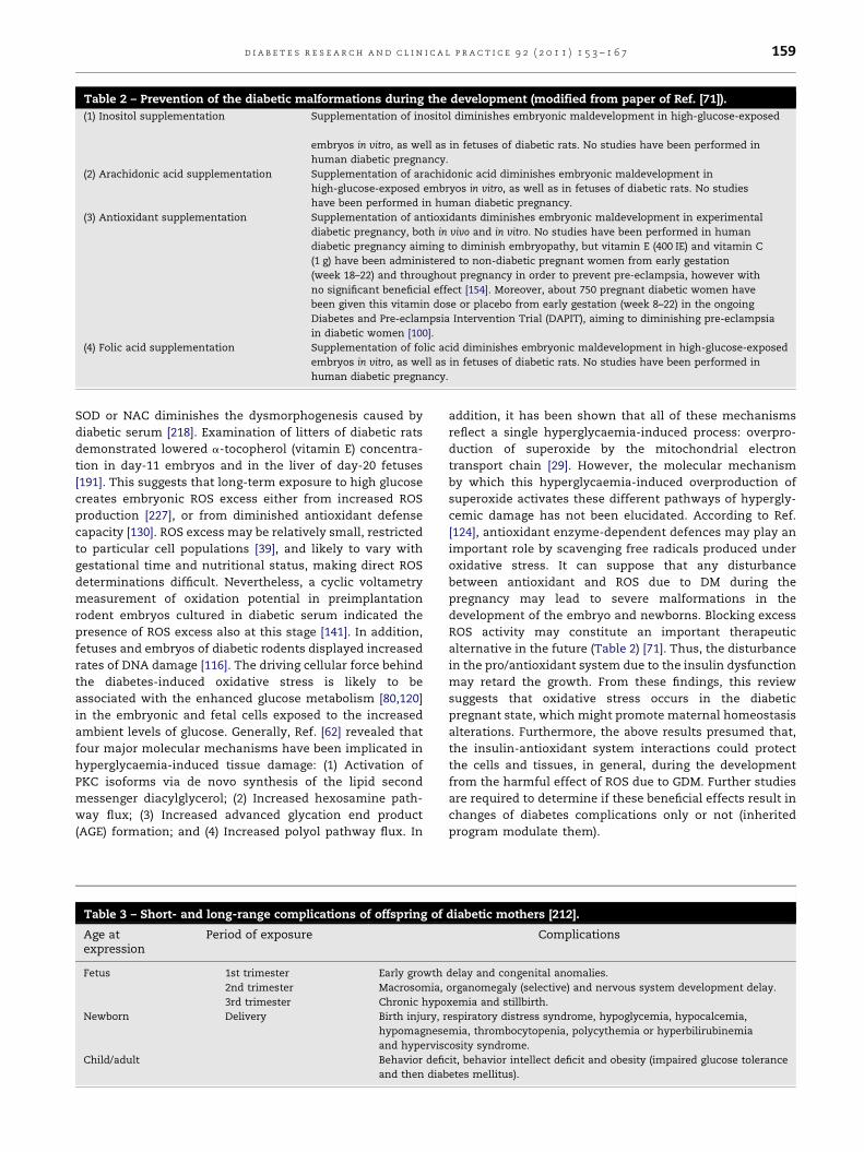
Table 2 – Prevention of the diabetic malformations during the development (modified from paper of Ref. [71]).
(1) Inositol supplementation Supplementation of inositol diminishes embryonic maldevelopment in high-glucose-exposed
embryos in vitro, as well as in fetuses of diabetic rats. No studies have been performed in
human diabetic pregnancy.
(2) Arachidonic acid supplementation Supplementation of arachidonic acid diminishes embryonic maldevelopment in
high-glucose-exposed embryos in vitro, as well as in fetuses of diabetic rats. No studies
have been performed in human diabetic pregnancy.
(3) Antioxidant supplementation Supplementation of antioxidants diminishes embryonic maldevelopment in experimental
diabetic pregnancy, both in vivo and in vitro. No studies have been performed in human
diabetic pregnancy aiming to diminish embryopathy, but vitamin E (400 IE) and vitamin C
(1 g) have been administered to non-diabetic pregnant women from early gestation
(week 18–22) and throughout pregnancy in order to prevent pre-eclampsia, however with
no significant beneficial effect [154]. Moreover, about 750 pregnant diabetic women have
been given this vitamin dose or placebo from early gestation (week 8–22) in the ongoing
Diabetes and Pre-eclampsia Intervention Trial (DAPIT), aiming to diminishing pre-eclampsia
in diabetic women [100].
(4) Folic acid supplementation Supplementation of folic acid diminishes embryonic maldevelopment in high-glucose-exposed
embryos in vitro, as well as in fetuses of diabetic rats. No studies have been performed in
human diabetic pregnancy.
d i a b e t e s r e s e a r c h a n d c l i n i c a l p r a c t i c e 9 2 ( 2 0 1 1 ) 1 5 3 – 1 6 7 159
SOD or NAC diminishes the dysmorphogenesis caused by
diabetic serum [218]. Examination of litters of diabetic rats
demonstrated lowered a-tocopherol (vitamin E) concentra-
tion in day-11 embryos and in the liver of day-20 fetuses
[191]. This suggests that long-term exposure to high glucose
creates embryonic ROS excess either from increased ROS
production [227], or from diminished antioxidant defense
capacity [130]. ROS excess may be relatively small, restricted
to particular cell populations [39], and likely to vary with
gestational time and nutritional status, making direct ROS
determinations difficult. Nevertheless, a cyclic voltametry
measurement of oxidation potential in preimplantation
rodent embryos cultured in diabetic serum indicated the
presence of ROS excess also at this stage [141]. In addition,
fetuses and embryos of diabetic rodents displayed increased
rates of DNA damage [116]. The driving cellular force behind
the diabetes-induced oxidative stress is likely to be
associated with the enhanced glucose metabolism [80,120]
in the embryonic and fetal cells exposed to the increased
ambient levels of glucose. Generally, Ref. [62] revealed that
four major molecular mechanisms have been implicated in
hyperglycaemia-induced tissue damage: (1) Activation of
PKC isoforms via de novo synthesis of the lipid second
messenger diacylglycerol; (2) Increased hexosamine path-
way flux; (3) Increased advanced glycation end product
(AGE) formation; and (4) Increased polyol pathway flux. In
Table 3 – Short- and long-range complications of offspring of
Age atexpression
Period of exposure
Fetus 1st trimester Early growth
2nd trimester Macrosomia,
3rd trimester Chronic hypo
Newborn Delivery Birth injury, r
hypomagnese
and hypervisc
Child/adult Behavior defic
and then diab
addition, it has been shown that all of these mechanisms
reflect a single hyperglycaemia-induced process: overpro-
duction of superoxide by the mitochondrial electron
transport chain [29]. However, the molecular mechanism
by which this hyperglycaemia-induced overproduction of
superoxide activates these different pathways of hypergly-
cemic damage has not been elucidated. According to Ref.
[124], antioxidant enzyme-dependent defences may play an
important role by scavenging free radicals produced under
oxidative stress. It can suppose that any disturbance
between antioxidant and ROS due to DM during the
pregnancy may lead to severe malformations in the
development of the embryo and newborns. Blocking excess
ROS activity may constitute an important therapeutic
alternative in the future (Table 2) [71]. Thus, the disturbance
in the pro/antioxidant system due to the insulin dysfunction
may retard the growth. From these findings, this review
suggests that oxidative stress occurs in the diabetic
pregnant state, which might promote maternal homeostasis
alterations. Furthermore, the above results presumed that,
the insulin-antioxidant system interactions could protect
the cells and tissues, in general, during the development
from the harmful effect of ROS due to GDM. Further studies
are required to determine if these beneficial effects result in
changes of diabetes complications only or not (inherited
program modulate them).
diabetic mothers [212].
Complications
delay and congenital anomalies.
organomegaly (selective) and nervous system development delay.
xemia and stillbirth.
espiratory distress syndrome, hypoglycemia, hypocalcemia,
mia, thrombocytopenia, polycythemia or hyperbilirubinemia
osity syndrome.
it, behavior intellect deficit and obesity (impaired glucose tolerance
etes mellitus).
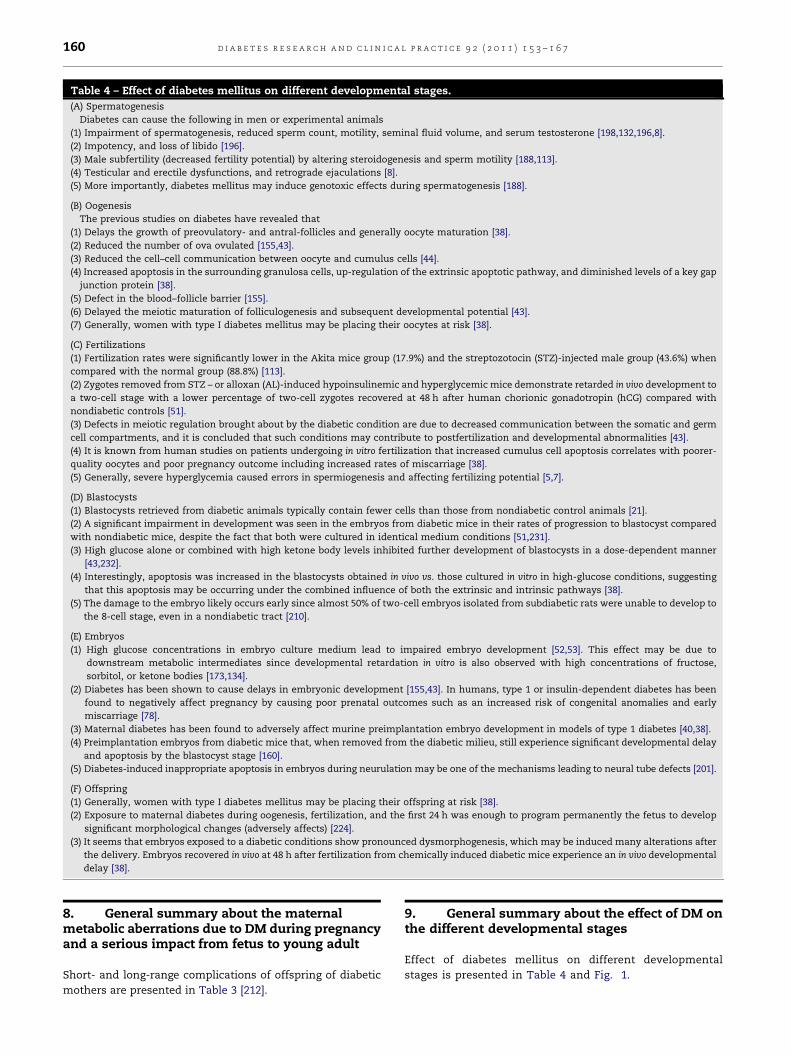
Table 4 – Effect of diabetes mellitus on different developmental stages.
(A) Spermatogenesis
Diabetes can cause the following in men or experimental animals
(1) Impairment of spermatogenesis, reduced sperm count, motility, seminal fluid volume, and serum testosterone [198,132,196,8].
(2) Impotency, and loss of libido [196].
(3) Male subfertility (decreased fertility potential) by altering steroidogenesis and sperm motility [188,113].
(4) Testicular and erectile dysfunctions, and retrograde ejaculations [8].
(5) More importantly, diabetes mellitus may induce genotoxic effects during spermatogenesis [188].
(B) Oogenesis
The previous studies on diabetes have revealed that
(1) Delays the growth of preovulatory- and antral-follicles and generally oocyte maturation [38].
(2) Reduced the number of ova ovulated [155,43].
(3) Reduced the cell–cell communication between oocyte and cumulus cells [44].
(4) Increased apoptosis in the surrounding granulosa cells, up-regulation of the extrinsic apoptotic pathway, and diminished levels of a key gap
junction protein [38].
(5) Defect in the blood–follicle barrier [155].
(6) Delayed the meiotic maturation of folliculogenesis and subsequent developmental potential [43].
(7) Generally, women with type I diabetes mellitus may be placing their oocytes at risk [38].
(C) Fertilizations
(1) Fertilization rates were significantly lower in the Akita mice group (17.9%) and the streptozotocin (STZ)-injected male group (43.6%) when
compared with the normal group (88.8%) [113].
(2) Zygotes removed from STZ – or alloxan (AL)-induced hypoinsulinemic and hyperglycemic mice demonstrate retarded in vivo development to
a two-cell stage with a lower percentage of two-cell zygotes recovered at 48 h after human chorionic gonadotropin (hCG) compared with
nondiabetic controls [51].
(3) Defects in meiotic regulation brought about by the diabetic condition are due to decreased communication between the somatic and germ
cell compartments, and it is concluded that such conditions may contribute to postfertilization and developmental abnormalities [43].
(4) It is known from human studies on patients undergoing in vitro fertilization that increased cumulus cell apoptosis correlates with poorer-
quality oocytes and poor pregnancy outcome including increased rates of miscarriage [38].
(5) Generally, severe hyperglycemia caused errors in spermiogenesis and affecting fertilizing potential [5,7].
(D) Blastocysts
(1) Blastocysts retrieved from diabetic animals typically contain fewer cells than those from nondiabetic control animals [21].
(2) A significant impairment in development was seen in the embryos from diabetic mice in their rates of progression to blastocyst compared
with nondiabetic mice, despite the fact that both were cultured in identical medium conditions [51,231].
(3) High glucose alone or combined with high ketone body levels inhibited further development of blastocysts in a dose-dependent manner
[43,232].
(4) Interestingly, apoptosis was increased in the blastocysts obtained in vivo vs. those cultured in vitro in high-glucose conditions, suggesting
that this apoptosis may be occurring under the combined influence of both the extrinsic and intrinsic pathways [38].
(5) The damage to the embryo likely occurs early since almost 50% of two-cell embryos isolated from subdiabetic rats were unable to develop to
the 8-cell stage, even in a nondiabetic tract [210].
(E) Embryos
(1) High glucose concentrations in embryo culture medium lead to impaired embryo development [52,53]. This effect may be due to
downstream metabolic intermediates since developmental retardation in vitro is also observed with high concentrations of fructose,
sorbitol, or ketone bodies [173,134].
(2) Diabetes has been shown to cause delays in embryonic development [155,43]. In humans, type 1 or insulin-dependent diabetes has been
found to negatively affect pregnancy by causing poor prenatal outcomes such as an increased risk of congenital anomalies and early
miscarriage [78].
(3) Maternal diabetes has been found to adversely affect murine preimplantation embryo development in models of type 1 diabetes [40,38].
(4) Preimplantation embryos from diabetic mice that, when removed from the diabetic milieu, still experience significant developmental delay
and apoptosis by the blastocyst stage [160].
(5) Diabetes-induced inappropriate apoptosis in embryos during neurulation may be one of the mechanisms leading to neural tube defects [201].
(F) Offspring
(1) Generally, women with type I diabetes mellitus may be placing their offspring at risk [38].
(2) Exposure to maternal diabetes during oogenesis, fertilization, and the first 24 h was enough to program permanently the fetus to develop
significant morphological changes (adversely affects) [224].
(3) It seems that embryos exposed to a diabetic conditions show pronounced dysmorphogenesis, which may be induced many alterations after
the delivery. Embryos recovered in vivo at 48 h after fertilization from chemically induced diabetic mice experience an in vivo developmental
delay [38].
d i a b e t e s r e s e a r c h a n d c l i n i c a l p r a c t i c e 9 2 ( 2 0 1 1 ) 1 5 3 – 1 6 7160
8. General summary about the maternalmetabolic aberrations due to DM during pregnancyand a serious impact from fetus to young adult
Short- and long-range complications of offspring of diabetic
mothers are presented in Table 3 [212].
9. General summary about the effect of DM onthe different developmental stages
Effect of diabetes mellitus on different developmental
stages is presented in Table 4 and Fig. 1.
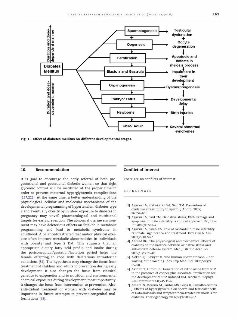
[()TD$FIG]
Fig. 1 – Effect of diabetes mellitus on different developmental stages.
d i a b e t e s r e s e a r c h a n d c l i n i c a l p r a c t i c e 9 2 ( 2 0 1 1 ) 1 5 3 – 1 6 7 161
10. Recommendation
It is goal to encourage the early referral of both pre-
gestational and gestational diabetic women so that tight
glycemic control will be instituted at the proper time in
order to prevent maternal hyperglycaemia complications
[157,223]. At the same time, a better understanding of the
physiological, cellular and molecular mechanisms of the
developmental programming of hypertension, diabetes type
2 and eventually obesity by in utero exposure to diabetes in
pregnancy may unveil pharmacological and nutritional
targets for early prevention. The abnormal uterine environ-
ment may have deleterious effects on fetal/child metabolic
programming and lead to metabolic syndrome in
adulthood. A balanced/restricted diet and/or physical exer-
cise often improve metabolic abnormalities in individuals
with obesity and type 2 DM. This suggests that an
appropriate dietary fatty acid profile and intake during
the periconceptual/gestation/lactation period helps the
female offspring to cope with deleterious intrauterine
conditions [84]. The hypothesis may change the focus from
treatment of children and adults to prevention during early
development. It also changes the focus from classical
genetics to epigenetics and to nutrition and environmental
chemical exposures during development; most importantly,
it changes the focus from intervention to prevention. Also,
antioxidant treatment of women with diabetes may be
important in future attempts to prevent congenital mal-
formations [49].
Conflict of interest
There are no conflicts of interest.
r e f e r e n c e s
[1] Agarwal A, Prabakaran SA, Said TM. Prevention ofoxidative stress injury to sperm. J Androl 2005;26:654–60.
[2] Agarwal A, Said TM. Oxidative stress, DNA damage andapoptosis in male infertility: a clinical approach. Br J UrolInt 2005;95:503–7.
[3] Agarwal A, Saleh RA. Role of oxidants in male infertility:rationale, significance and treatment. Urol Clin N Am2002;29:817–27.
[4] Ahmed RG. The physiological and biochemical effects ofdiabetes on the balance between oxidative stress andantioxidant defense system. Med J Islamic Acad Sci2005;15(1):31–42.
[5] Aitken RJ, Sawyer D. The human spermatozoon – notwaving but drowning. Adv Exp Med Biol 2003;518(2):85–98.
[6] Akihiro T, Hiromu S. Generation of nitric oxide from STZin the presence of copper plus ascorbate: implication forthe development of STZ induced DM. Biochem BiophysRes Commun 1998;245:11–6.
[7] Amaral S, Moreno AJ, Santos MS, Seica R, Ramalho-SantosJ. Effects of hyperglycaemia on sperm and testicular cellsof Goto-Kakizaki and streptozotocin-treated rat models fordiabetes. Theriogenology 2006;66(9):2056–67.
d i a b e t e s r e s e a r c h a n d c l i n i c a l p r a c t i c e 9 2 ( 2 0 1 1 ) 1 5 3 – 1 6 7162
[8] Amaral S, Oliveira PJ, Ramalho-Santos J. Diabetes and theimpairment of reproductive function: possible role ofmitochondria and reactive oxygen species. Curr DiabetesRev 2008;4(1):46–54.
[9] Ammon HP, Mark M. Thiols and pancreatic beta-cellfunction: a review. Cell Biochem Funct 1985;3:157–71.
[10] Angerwall L. Alloxan diabetes and pregnancy in the rat.Acta Endocrinol 1959;39(Suppl. 44):1.
[11] Angell CA, Tubbs RC, Moore AB, Barb CR, Cox NM.Depressed luteinizing hormone response to estradiolin vivo and gonadotropin-releasing hormone in vitro inexperimentally diabetic swine. Domest Anim Endocrinol1996;13:453–63.
[12] Arikawe AP, Daramola AO, Odofin AO, Obika LFO. Alloxan-induced and insulin-resistant diabetes mellitus affectsemen parameters and impair spermatogenesis in malerats. Reprod Health 2006;10(3):106–13.
[13] Armstrong DG, McEvoy TG, Baxter G, Robinson JJ, Hogg CO,Woad KJ, et al. Effect of dietary energy and protein onbovine follicular dynamics and embryo production in vitro:associations with the ovarian insulin-like growth factorsystem. Biol Reprod 2001;64:1624–32.
[14] Asayama K, Nyfeler F, English D, Pilkis SJ, Burr IM.Alloxan-induced free radical production in isolated cells.Diabetes 1984;33:1008–11.
[15] Babichev VN, Adamskaia EI, Pershkova TA. Basal andlulibren-stimulated gonadotropin secretion inovariectomized female rats with streptozotocin-induceddiabetes. Probl Endokrinol (Mosk) 1994;40:43–6.
[16] Babichev VN, Adamskaia EI, Pershkova TA. Analysis ofhypothalamo–hypophyseal–gonadal interrelationships infemale rats in experimentally induced diabetes. ProblEndokrinol (Mosk) 1994;40:46–50.
[17] Baker L, Piddington R, Goldman A, Egler J, Moehring J.Myo-inositol and prostaglandins reverse the glucoseinhibition of neural tube fusion in cultured mouseembryos. Diabetology 1990;33:593–6.
[18] Baynes JW, Thorpe SR. Role of oxidative stress in diabeticcomplications: a new perspective on an old paradigm.Diabetes 1999;48:1–9.
[19] Beardsall K, Dunger D. Insulin therapy in pretermnewborns. Early Hum Dev 2008;84:839–42.
[20] Becerra JE, Khoury MJ, Cordero JF, Eriksson JD. Diabetesmellitus during pregnancy and the risks due specific birthdefects: a population based case–control study. J Pediatr1990;85:1–9.
[21] Beebe LFS, Kaye PL. Maternal diabetes and retardedimplantation development of mice. Diabetes 1991;40:457–61.
[22] Bergh C, Broden H, Lundin K, Hamberger L. Comparison offertilization, cleavage and pregnancy rates of oocytes fromlarge and small follicles. Hum Reprod 1998;13:1912–5.
[23] Berman LD, Hayes JA, Sibay TM. Effect of streptozotocin inthe Chinese hamster (Cricetulus griseus). J Natl Cancer Inst1973;51:1287–94.
[24] Bhuyan BK, Peterson AR, Heidelberger C. Cytotoxicity,mutations and DNA damage produced in Chinese hamstercells treated with streptozotocin, its analogs, and N-methyl-N-nitro-N-nitrosoguanidine. Chem Biol Interact1976;13:173–9.
[25] Boulot P, Chabbert-Buffet N, d’Ercole C, Floriot M, FontaineP, Fournier A. French multicentric survey of outcome ofpregnancy in women with pregestational diabetes.Diabetes Care 2003;26(11):2990–3.
[26] Bolzan AD, Bianchi MS. Genotoxicity of streptozotocin.Mutat Res Rev Mutat Res 2002;512(2–3):121–34 (review).
[27] Bolzan AD, Bianchi MS. Chromosomal response of humanlymphocytes to streptozotocin. Mutat Res 2002;503:63–8.
[28] Bolzan AD, Gonzalez MC, Bianchi MS. The effect of 1,10-phenanthroline on the chromosome damage and sister-chromatid exchanges induced by streptozotocin inmammalian and insect cells. Mutat Res 2000;447:221–6.
[29] Brownlee M. Biochemistry and molecular cell biology ofdiabetic complications. Nature 2001;414:813–20.
[30] Buchanan TA, Denno KM, Sipos GF, Sadler TW. Diabeticteratogenesis. In vitro evidence for a multifactorialetiology with little contribution from glucose per se.Diabetes 1994;43:656–60.
[31] Cai L, Chen S, Evans T, Deng DX. Apoptotic germ-celldeath and testicular damage in experimental diabetes:prevention by endothelin antagonism. Urol Res2000;28:342–7.
[32] Cameron DF, Rountree J, Schultz RE, Repetta D, Murray FT.Sustained hyperglycemia results in testicular dysfunctionand reduced fertility potential in BBWOR diabetic rats. AmJ Physiol 1990;259:881–9.
[33] Canniff KM, Smith MS, Lacy DB, Williams PE, Moore MC.Glucagon secretion and autonomic signaling duringhypoglycemia in late pregnancy. Am J Physiol Regul IntegrComp Physiol 2006;291:R788–95.
[34] Catalano PM, Tyzbir ED, Wolfe RR, Calles J, Roman NM,Amini SB. Carbohydrate metabolism during pregnancy incontrol subjects and women with gestational diabetes. AmJ Physiol 1993;264:E60–67.
[35] Cederberg J, Basu S, Eriksson UJ. Increased rate of lipidperoxidation and protein carbonylation in experimentaldiabetic pregnancy. Diabetology 2001;44:766–74.
[36] Cederberg J, Eriksson UJ. Antioxidative treatment ofpregnant diabetic rats diminishes embryonicdysmorphogenesis. Birth Defects Res A Clin Mol Teratol2005;73:498–505.
[37] Cederberg J, Galli J, Luthman H, Eriksson UJ. IncreasedmRNA levels of Mn-SOD and catalase in embryos ofdiabetic rats from a malformation-resistant strain.Diabetes 2000;49:101–7.
[38] Chang AS, Dale AN, Moley KH. Maternal diabetesadversely affects preovulatory oocyte maturation,development, and granulosa cell apoptosis. Endocrinology2005;146(5):2445–53.
[39] Chen SY, Sulik KK. Free radicals and ethanol-inducedcytotoxicity in neural crest cells. Alcohol Clin Exp Res1996;20:1071–6.
[40] Chi MM, Pingsterhaus J, Carayannopoulos M, Moley KH.Decreased glucose transporter expression triggers BAX-dependent apoptosis in the murine blastocyst. J Biol Chem2000;275:40252–7.
[41] Clamon G, Riggs C, Stegink L, Traves M. Phase 2 trial ofstreptozotocin by continuous infusion for metastaticcolorectal carcinoma. Cancer Drug Deliv 1987;4:43–6.
[42] Clausen TD, Mathiesen E, Ekrom P, Hellmuth E, Mandrup-Poulsen T, Damm P. Poor pregnancy outcome in womenwith type 2 diabetes. Diabetes Care 2005;28:323–8.
[43] Colton SA, Pieper GM, Downs SM. Altered meioticregulation in oocytes from diabetic mice. Biol Reprod2002;67:220–31.
[44] Colton SA, Humpherson PG, Leese HJ, Downs SM.Physiological changes in oocyte-cumulus cell complexesfrom diabetic mice that potentially influence meioticregulation. Biol Reprod 2003;69:761–70.
[45] Connolly CC, Aglione LN, Smith MS, Lacy DB, Moore MC.Pregnancy impairs the counter regulatory response toinsulin-induced hypoglycemia in the dog. Am J PhysiolEndocrinol Metab 2004;287:E480–8.
d i a b e t e s r e s e a r c h a n d c l i n i c a l p r a c t i c e 9 2 ( 2 0 1 1 ) 1 5 3 – 1 6 7 163
[46] Cox NM, Muerer KA, Carlton CA, Tubbs RC, Mannis DP.Effects of diabetes mellitus during the luteal phase of theoestrus cycle on preovulatory follicular function,ovulation and gonadotrophins in gilts. J Reprod Fertil1994;101:77–86.
[47] Crouch RK, Gandy SE, Kimsey G, Galbraith RA, GalbraithGM, Buse MG. The inhibition of islet superoxide dismutaseby diabetogenic drugs. Diabetes 1981;30:235–41.
[48] Cundy T, Gamble G, Townend K. Perinatal mortality intype 2 diabetes. Diabetes Med 2000;17:33–9.
[49] Damasceno DC, Volpato GT, de Mattos ParanhosCalderon I, Cunha Rudge MV. Oxidative stress anddiabetes in pregnant rats. Anim Reprod Sci 2002;15, 72(3–4):235–44.
[50] Demeestere I, Gervy C, Centner J, Devreker F, Englert Y,Delbaere A. Effect of insulin-like growth factor-1 duringpreantral follicular culture on steroidogenesis, in vitrooocyte maturation, and embryo development in mice. BiolReprod 2004;70:1664–9.
[51] Diamond MP, Moley KH, Pellicer A, Vaughn WK,DeCherney AH. Effects of streptozotocin and alloxan-induced diabetes mellitus on mouse follicular and earlyembryo development. J Reprod Fertil 1989;86:1–10.
[52] Diamond MP, Harbert-Moley K, Logan J, Pellicer A, Lavy G,Vaughn WK, et al. Manifestation of diabetes mellitus onmouse follicular and pre-embryo developmenteffect of hyperglycaemia per se. Metab Clin Exp1990;39:220–4.
[53] Diamond MP, Pettway ZY, Logan J, Moley KH, Vaughn WK,DeCherney AH. Dose response effects of glucose, insulinand glucagon on mouse pre-embryo development.Metabolism 1991;40:466–70.
[54] Doreswamy K, Shrilatha B, Rajeshkumar T, Muralidhara.Nickel induced oxidative stress in testis of mice: evidencesof DNA damage and genotoxic effects. J Androl2004;25:996–1003.
[55] Downs SM, Mastropolo AM. The participation of energysubstrates in the control of meiotic maturation in murineoocytes. Dev Biol 1994;162:154–68.
[56] Downs SM, Houghton FD, Humpherson PG, Leese HJ.Substrate utilization and maturation of cumulus cell-enclosed mouse oocytes: evidence that pyruvate oxidationdoes not mediate meiotic induction. J Reprod Fertil1997;110:1–10.
[57] Downs SM. The influence of glucose, cumulus cells, andmetabolic coupling on ATP levels and meiotic control inthe isolated mouse oocyte. Dev Biol 1995;167:502–12.
[58] Downs SM, Humpherson PG, Martin KL, Leese HJ. Glucoseutilization during gonadotropin-induced meioticmaturation in cumulus cell-enclosed mouse oocytes. MolReprod Dev 1996;44:121–31.
[59] Downs SM, Humpherson PG, Leese HJ. Meiotic inductionin cumulus cell-enclosed mouse oocytes: involvement ofthe pentose phosphate pathway. Biol Reprod1998;58:1084–94.
[60] Downs SM, Utecht AM. Metabolism of radiolabeledglucose by mouse oocytes and oocyte-cumulus cellcomplexes. Biol Reprod 1999;60:1446–52.
[61] Downs SM. Adenosine blocks hormone-induced meioticmaturation by suppressing purine de novo synthesis. MolReprod Dev 2000;56:172–9.
[62] Du X, Matsumura T, Edelstein D, Rossetti L, Zsengeller Z,Szabo C, et al. Inhibition of GAPDH activity by poly(ADP-ribose) polymerase activates three major pathways ofhyperglycemic damage in endothelial cells. J Clin Invest2003;112:1049–57.
[63] Dunn JS, Sheehan HL, McLetchie NGB. Necrosis of isletsof Langerhans produced experimentally. Lancet 1943;1:484–7.
[64] Economides DL, Proudler A, Nicolaides KH. Plasma insulinin appropriate and small for gestational age fetuses. Am JObstet Gynecol 1989;160:1091–4.
[65] Ellison AC, Maren TH. The effects of metabolic alterationson teratogenesis. Johns Hopkins Med J 1972;130:87–94.
[66] El-Missiry MA. Enhanced testicular antioxidant system byascorbic acid in alloxan diabetic rats. Comp BiochemPhysiol Part C 1999;124:233–7.
[67] El-Roeiy A, Chen X, Roberts VJ, LeRoith D, Roberts Jr CT,Yen SS. Expression of insulin-like growth factor (IGF-1 andIGF-II) and the IGF-I. IGF-II, and insulin receptor genes andlocalization of the gene products in the human ovary. JClin Endocrinol Metab 1993;77:1411–8.
[68] El-Roeiy A, Chen X, Roberts VJ, Shimasaki S, Ling N, LeRoith D, et al. Expression of the genes encoding theinsulin-like growth factors (IGF-I and II), the IGF andinsulin receptors, and IGF binding proteins 1-6 and thelocalization of their gene products in normal andpolycystic ovary syndrome ovaries. J Clin EndocrinolMetab 1994;78:1488–96.
[69] Elsner M, Gurgul-Convey E, Lenzen S. Relative importanceof cellular uptake and reactive oxygen species for thetoxicity of alloxan and dialuric acid to insulin-producingcells. Free Radic Biol Med 2006;41:825–34.
[70] Enghofer M, Usadel KH, Beck O, Kusterer K. Superoxidedismutase reduces islet microvascular injury induced bystreptozotocin in the rat. Am J Physiol 1997;273:E376–382.
[71] Eriksson UJ. Congenital anomalies in diabetic pregnancy.Semin Fetal Neonatal Med 2009;14:85–93.
[72] Eriksson UJ, Borg LAH. Protection by free oxygen radicalscavenging enzymes against glucose-induced embryonicmalformations in vitro. Diabetology 1991;34:325–31.
[73] Eriksson UJ, Borg LAH. Diabetes and embryonicmalformations. Role of substrate-induced free-oxygenradical production for dysmorphogenesis in cultured ratembryos. Diabetes 1993;42:411–9.
[74] Eriksson UJ, Brolin SE, Naeser P. Influence of sorbitolaccumulation on growth and development of embryoscultured in elevated levels of glucose and fructose.Diabetes Res 1989;11:27–32.
[75] Eriksson UJ, Siman CM. Pregnant diabetic rats fed theantioxidant butylated hydroxytoluene show decreasedoccurrence of malformations in the offspring. Diabetes1996;45:1497–502.
[76] Eriksson UJ, Wentzel P, Minhas HS, Thornalley PJ.Teratogenicity of 3-deoxyglucosone and diabeticembryopathy. Diabetes 1998;47:1960–6.
[77] Fagbohun CF, Downs SM. Metabolic coupling andligand-stimulated meiotic maturation in the mouseoocyte–cumulus cell complex. Biol Reprod 1991;45:851–9.
[78] Farrell T, Neale L, Cundy T. Congenital anomalies in theoffspring of women with type 1, type 2 and gestationaldiabetes. Diabetes Med 2002;19:322–6.
[79] Faure P, Rossini E, Lafond JL, Richard MJ, Favier A, HalimiS. Vitamin E improves the free radical defence systempotential and insulin sensitivity of rats fed high fructosediet. J Nutr 1997;127:103–7.
[80] Fine EL, Horal M, Chang TI, Fortin G, Loeken MR. Evidencethat elevated glucose causes altered gene expression,apoptosis, and neural tube defects in a mouse model ofdiabetic pregnancy. Diabetes 1999;48:2454–62.
[81] Foreman D, Kolettios E, Garris DR. Diabetes prevents thenormal responses of the ovary to FSH. Endocr Res1993;19:187–205.
[82] Eizirik DL, Bjorklund A, Cagliero E. Genotoxic agentsincrease expression of growth arrest and DNA damage-inducible genes gadd 153 and gadd 45 in rat pancreaticislets. Diabetes 1993;42:738–45.
d i a b e t e s r e s e a r c h a n d c l i n i c a l p r a c t i c e 9 2 ( 2 0 1 1 ) 1 5 3 – 1 6 7164
[83] Gabridge MG, Denunzio A, Legator MS. Microbialmutagenicity of streptozotocin in animal-mediatedassays. Nature 1969;221:68–70.
[84] Gallou-Kabani C, Vige A, Gross MS, Boileau C, Rabes JP,Fruchart-Najib J, et al. Resistance to high-fat diet in thefemale progeny of obese mice fed a control diet during thepericonceptual, gestation, and lactation periods. Am JPhysiol Endocrinol Metab 2007;292(4):E1095–10100.
[85] Galloway SM, Greenwood SK, Hill RB, Bradt CI, Bean CL. Arole for mismatch repair in production of chromosomeaberrations by methylating agents in human cells. MutatRes 1995;346:231–45.
[86] Gareskog M, Wentzel P. N-acetylcysteine and a-cyano-4-hydroxycinnamic acid alter protein kinase C (PKC)-d andPKC-z and diminish dysmorphogenesis in rat embryoscultured with high glucose in vitro. Endocrinology2007;192:207–14.
[87] Garris DR. Effect of diabetes on uterine constriction,decidualization, vascularization and corpus luteumfunction in the pseudopregnant rat. Horm Metab Res1988;20:463–75.
[88] Gavin III JR, Alberti KGMM, Davidson MB, DeFronzo RA,Drash A, Gabbe SG, et al. Report of the expert committeeon the diagnosis and classification of diabetes mellitus.Diabetes Care 1997;20:1183–97.
[89] Gondos B, Bevier W. Effect of insulin on testicularalterations in the nonobese diabetic mouse. Ann Clin LabSci 1995;25(3):272–7.
[90] Gopaul NK, Anggard EE, Mallet AI, Betteridge DJ, Wolff SP,Nourooz-Zadeh J. Plasma 8-epi-prostaglandin F2a levelsare elevated in individuals with non-insulin dependentdiabetes mellitus. FEBS Lett 1995;368:225–9.
[91] Grankvist K, Marklund SL, Sehlin J, Taljedal IB. Superoxidedismutase, catalase and scavengers of hydroxyl radicalprotect against the toxic action of alloxan on pancreaticislets in vitro. Biochem J 1979;182:17–25.
[92] Hackam DG. Translating animal research into clinicalbenefit. BMJ 2007;334:163–4.
[93] Hackam DG, Redelmeier DA. Translation of researchevidence from animals to humans. JAMA 2006;296:1731–2.
[94] Harel A, Bloch O, Vardi P, Bloch K. Sensitivity of HaCatkeratinocytes to diabetogenic toxins. Biochem Pharmacol2002;63:171–8.
[95] Harmeyer J, Schlumbohm C. Pregnancy impairs ketonebody disposal in late gestating ewes: implications foronset of pregnancy toxaemia. Res Vet Sci 2006;81:254–64.
[96] Hassan AA, Hassouna MM, Taketo T, Gagnon C, ElhilaliMM. The effect of diabetes on sexual behavior andreproductive tract function in male rats. J Urol1993;149:148–54.
[97] Hay JE. Liver disease in pregnancy. Hepatology2008;47:1067–76.
[98] Hemachand T, Shaha C. Functional role of sperm surfaceglutathione-S-transferases and extra cellular glutathionein the haploid spermatozoa under oxidative stress. FEBSLett 2003;538:14–8.
[99] Herdt TH, Gerloff BJ. Ketosis. In: Howard JL, Smith RA,editors. Current veterinary therapy 4 food animal practice.WB Saunders; 1999. p. 226–8.
[100] Holmes VA, Young IS, Maresh MJ, Pearson DW, Walker JD,McCance DR. The diabetes and pre-eclampsiaintervention trial. Int J Gynaecol Obstet 2004;87:66–71.
[101] Howland BE, Zebrowski EJ. Gonadotropin levels in seraand pituitary glands of female rats treated with alloxan.Life Sci 1974;14:289–96.
[102] Huang DY, Usher RH, Kramer MS, Yang H, Morin L, FrettsRC. Determinants of unexplained antepartum fetaldeaths. Obstet Gynecol 2000;95(2):215–21.
[103] International Agency for Research on Cancer (IARC).Monographs (Suppl. 7); 1987. p. 72.
[104] Iwase M, Nunoi K, Sadoshima S, Kikuchi M, Fujishima M.Liver, kidney and is let cell tumors in spontaneouslyhypertensive and normotensive rats treated withStreptozotocin. Tohoku J Exp Med 1989;159:83–90.
[105] Jawerbaum A, Gonzalez ET, Faletti A, Novaro V, Vitullo A,Gimeno MA. Altered prostanoid production by cumulus–oocyte complexes in a rat model of non-insulin-dependent diabetes mellitus. Prostaglandins 1996;52:209–19.
[106] Jiang GY. Practical diabetes. Beijing: People’s HealthPublishing House; 1996. p. 295.
[107] Johnson CA. Glucose homeostasis during caninepregnancy: insulin resistance, ketosis, and hypoglycemia.Theriogenology 2008;70(9):1418–23.
[108] Kaina B, Ziouta A, Ochs K, Coquerelle T. Chromosomalinstability, cell death and apoptosis induced by O6-methylguanine in Mex�, Mex+ and methylation-tolerantmismatch repair comprornised cells: facts and models.Mutat Res 1997;381:227–41.
[109] Kaneto H, Fujii J, Seo HG, Suzuki K, Matsuoka T, NakamuraM, et al. Apoptotic cell death triggered by nitric oxide inpancreatic betacells. Diabetes 1995;44:733–8.
[110] Kassem SA, Ariel I, Thornton PS, Scheimberg I, Glaser B.Beta-cell proliferation and apoptosis in the developingnormal human pancreas and in hyperinsulinism ofinfancy. Diabetes 2000;49:1325–33.
[111] Kezele PR, Nilsson EE, Skinner MK. Insulin but not insulin-like growth factor-1 promotes the primordial to primaryfollicle transition. Mol Cell Endocrinol 2002;192:37–43.
[112] Khoury MJ, Becerra JE, Cordero JF, Erickson JD. Clinical–epidemiologic assessment of pattern of birth defectsassociated with human teratogens: application to diabeticembryopathy. J Pediatr 1989;84:658–65.
[113] Kim ST, Moley KH. Paternal effect on embryo quality indiabetic mice is related to poor sperm quality andassociated with decreased glucose transporter expression.Reproduction 2008;136(3):313–22.
[114] Kozlov GI, Kamalov KG. Sperm characterization inpatients with diabetes mellitus suffering from sexualdisorders]. Probl Endokrinol (Mosk) 1989;35(2):6–9.
[115] Kraynak AR, Storer RD, Jensen RD, Kloss MW, Soper KA,Clair JH, et al. Extent and persistence of streptozotocin-induced DNA damage and cell proliferation in rat kidneyas determined by in vivo alkaline elution and BrdUrdlabeling assays. Toxicol Appl Pharmacol 1995;135:279–86.
[116] Lee AT, Reis D, Eriksson UJ. Hyperglycaemia inducedembryonic dysmorphogenesis correlates with genomicDNA mutation frequency in vitro and in vivo. Diabetes1999;48:371–6.
[117] Lenzen S, Panten U. Alloxan: history and mechanism ofaction. Diabetologia 1988;31:337–42.
[118] Levi JE, Weinberg T. Pregnancy in alloxan diabetic rats.Proc Soc Exp Biol Med 1949;72:658–62.
[119] Levine RJ, Lam C, Qian C, Yu KF, Maynard SE, Sachs BP.Soluble endoglin and other circulating antiangiogenicfactors in preeclampsia. N Engl J Med 2006;355:992–1005.
[120] Li R, Thorens B, Loeken MR. Expression of the geneencoding the high-Km glucose transporter 2 by the earlypostimplantation mouse embryo is essential for neuraltube defects associated with diabetic embryopathy.Diabetology 2007;50:682–9.
[121] Like AA, Rosani A. Streptozotocin-induced pancreaticinsulitis: new model of diabetes mellitus. Science1976;193:415–7.
[122] Lindheimer MD, Umans JG. Explaining and predictingpreeclampsia. N Engl J Med 2006;355:1056–8.
d i a b e t e s r e s e a r c h a n d c l i n i c a l p r a c t i c e 9 2 ( 2 0 1 1 ) 1 5 3 – 1 6 7 165
[123] Luz J, Zemdegs JCS, Amaral LSG. Chronic lipoic acidtreatment worsens energy imbalances in streptozotocin-induced diabetic rats. Diabetes Metab 2009;35(2):137–42.
[124] Mahboob M, Rahman MF, Grover P. Serum lipidperoxidation and antioxidant enzyme levels in male andfemale diabetic patients. Singapore Med J 2005;46:322–4.
[125] Malaisse WJ, Malaisse-Lagae F, Sener A, Pipeleers DG.Determinants of the selective toxicity of alloxan to thepancreatic B cell. Proc Natl Acad Sci USA 1982;79(3):927–30.
[126] Mathews CE, Leiter EH. Constitutive differences inantioxidant defense status distinguish alloxan-resistantand alloxan-susceptible mice. Free Radic Biol Med1999;27(3/4):449–55.
[127] McCann JP, Hansel W. Relationship between insulin andglucose metabolism and pituitary–ovarian function infasted heifer. Biol Reprod 1986;34:630–41.
[128] McIntosh CHS, Pederson RA. Noninsulin-dependentanimal models of diabetes mellitus. In: McNeill JH, editor.Experimental models of diabetes. Boca Raton, FL: CRCPress; 1999. p. 337–98.
[129] Melamed N, Hod M. Women, diabetes, and pregnancy:perinatal mortality in pregestational diabetes. Int JGynecol Obstetr 2009;104(1):S20–4.
[130] Menegola E, Broccia ML, Prati M, Ricolfi R, Giavini E.Glutathione status in diabetes-induced embryopathies.Biol Neonate 1996;69:293–7.
[131] Metzger BE, Ravnikar V, Vileisis RA, Freinkel N.‘‘Accelerated starvation’’ and the skipped breakfast in latenormal pregnancy. Lancet 1982;13(1(8272)):588–92.
[132] Meyer K, Deutscher J, Anil M, Berthold A, Bartsch G, KiessW. Serum androgen levels in adolescents with type Idiabetes: relationship to pubertal stage and metaboliccontrol. J Endocrinol Invest 2000;23:362–8.
[133] Mitanchez-Mokhtari D, Lahlou N, Kieffer F, Magny JF,Roger M, Voyer M. Both relative insulin resistance anddefective islet beta-cell processing of proinsulin areresponsible for transient hyperglycaemia in extremelypreterm infants. J Pediatr 2004;113:537–41.
[134] Moley K, Chi M, Manchester J, McDougal D, Lowry O.Alterations of intraembryonic metabolites inpreimplantation mouse embryos exposed to elevatedconcentrations of glucose: a metabolic explanation for thedevelopmental retardation seen in preimplantationembryos from diabetic animals. Biol Reprod 1996;54:1209–16.
[135] Morgan NG, Cable HC, Newcombe NR, Williams GT.Treatment of cultured pancreatic b cells withstreptozotocin induces cell death by apoptosis. Biosci Rep1994;14:243–50.
[136] Murata M, Imada M, Inoue S, Kawanishi S. Metal-mediated DNA damage induced by diabetogenic alloxan inthe presence of NADH. Free Radic Biol Med 1998;25(4/5):586–95.
[137] Murata M, Takahashi A, Saito I, Kawanishi S. Site-specificDNA methylation and apoptosis: induction bydiabetogenic streptozotocin. Biochem Pharmacol1999;57:881–7.
[138] Nourooz-Zadeh J, Tajaddini-Sarmadi J, McCarthy S,Betteridge DJ, Wolff SP. Elevated levels of authentic plasmahydroperoxides in NIDDM. Diabetes 1995;44:1054–8.
[139] Ohl J, Lefebvre-Maunoury C, Wittemer C, Nisand G,Laurent MC, Hoffmann P. Nitric oxide donors for patientsundergoing IVF. A prospective, double-blind, randomized,placebo-controlled trial. Hum Reprod 2002;17:2615–20.
[140] OhkuwaI T, Sato Y, Naoil M. Hydroxyl radical formation indiabetic rats induced by streptozotocin. Life Sci1995;56(21):1790–8.
[141] Ornoy A, Kimyagarov D, Yaffe P, Abir R, Raz I, Kohen R.Role of reactive oxygen species in diabetes-induced
embryotoxicity: studies on pre-implantation mouseembryos culture in serum from diabetic pregnant women.Isr J Med Sci 1996;32:1066–73.
[142] Ornoy A, Zaken V, Kohen R. Role of reactive oxygenspecies (ROS) in the diabetes-induced anomalies in ratembryos in vitro: reduction in antioxidant enzymes andlow-molecular-weight antioxidants (LMWA) may be thecausative factor for increased anomalies. Teratology1999;60:376–86.
[143] Osborn BH, Haney AF, Misukonis MA, Weinberg JB.Inducible nitric oxide synthase expression by peritonealmacrophages in endometriosis-associated infertility.Fertil Steril 2002;77:46–51.
[144] Palomar-Morales M, Baiza LA, Verdın-Teran L, Roman-Ramos R, Altamirano-Lozano M, Mendez JD. Fetaldevelopment in alloxan-treated rats. Reprod Toxicol1998;12(6):659–65.
[145] Paolisso G, D’Amore A, Volpe C, Balbi V, Saccomanno F,Galzerano D, et al. Evidence for a relationship betweenoxidative stress and insulin action in non-insulin-dependent (type II) diabetic patients. Metabolism1994;43:1426–9.
[146] Perel P, Roberts I, Sena E, Wheble P, Briscoe C. Comparisonof treatment effects between animal experimentsand clinical trials: systematic review. BMJ 2007;334:197.
[147] Petrik J, Arany E, McDonald TJ, Hill DJ. Apoptosis in thepancreatic islet cells of the neonatal rat is associatedwith a reduced expression of insulin-like growth factor IIthat may act as a survival factor. Endocrinology1998;139:2994–3004.
[148] Petrik J, Pell JM, Arany E, McDonald TJ, Dean WL, Reik W,et al. Overexpression of insulin-like growth factor-II intransgenic mice is associated with pancreatic islet cellhyperplasia. Endocrinology 1999;140:2353–63.
[149] Pettepher CC, LeDoux SP, Bohr VA, Wilson GL. Repair ofalkali-labile sites within the mitochondrial DNA of RINr 38cells after exposure to the nitrosourea streptozotocin. JBiol Chem 1991;266:3113–7.
[150] Philipps AF, Dubin JW, Raye JR. Maturation of early-phaseinsulin release in the neonatal lamb. Biol Neonate1981;39:225–31.
[151] Poretsky L, Smith D, Seibel M, Pazianos A, Flier JS.Distribution and characterization of the insulin and IGF-Ireceptors in normal human ovary. J Clin Endocrinol Metab1985;61:728–34.
[152] Poretsky L, Smith D, Seibel M, Pazianos A, Moses AC, FlierJS. Specific insulin binding sites in the human ovary. J ClinEndocrinol Metab 1984;59:809–11.
[153] Portesky L, Kalin MF. The gonadotropic function ofinsulin. Endocrin Rev 1987;8:132–41.
[154] Poston L, Briley AL, Seed PT, Kelly FJ, Shennan AH.Vitamin C and vitamin E in pregnant women at risk forpre-eclampsia (VIP trial): randomised placebo controlledtrial. Lancet 2006;367:1145–54.
[155] Powers RW, Chamber C, Larsen WJ. Diabetes-mediateddecreases in ovarian superoxide dismutase activity arerelated to blood-follicle barrier and ovulation defects.Endocrinology 1996;137:301–10.
[156] Powers RW, Chen L, Russel PT, Larsen WJ. Gonadotropin-stimulated regulation of blood-follicle barrier is mediatedby nitric oxide. Am J Physiol 1995;268:E290–8.
[157] Pregnancy and neonatal care subgroup to the St VincentJoint Task Force for Diabetes. Report. 10 Queen AnneStreet, London UK W1M OBD: British Diabetic Association;1994.
[158] Rakieten N, Gordon BS, Beaty A, Cooney DA, Schein PS.Modification of renal tumorigenic effect of streptozotocinby nicotinarnide: spontaneous reversibility of
d i a b e t e s r e s e a r c h a n d c l i n i c a l p r a c t i c e 9 2 ( 2 0 1 1 ) 1 5 3 – 1 6 7166
streptozotocin diabetes. Proc Soc Exp Biol Med1976;151:356–61.
[159] Rajeshkumar T, Muralidhara. Induction of oxidative stressby organic hydroperoxides in testis and epididymal spermof rats. J Androl 2007;28:77–85.
[160] Ratchford AM, Chang AS, Chi MM-Y, Rachael S, Kelle HM.Maternal diabetes adversely affects AMP-activated proteinkinase activity and cellular metabolism in murine oocytes.Am J Physiol Endocrinol Metab 2007;293:E1198–206.
[161] Reece EA, Wu YK, Zhao Z, Dhanasekaran D. Dietaryvitamin and lipid therapy rescues aberrant signaling andapoptosis and prevents hyperglycaemia-induced diabeticembryopathy in rats. Am J Obstet Gynecol 2006;194:580–5.
[162] Rehman K, Beshay E, Carrier S. Diabetes and male sexualfunction. J Sex Reprod Med 2001;1:29–33.
[163] Reusens B, Remacle C. Programming of the endocrinepancreas by the early nutritional environment. Int JBiochem Cell Biol 2006;38:913–22.
[164] Reynaert NL, Ckless K, Wouters EF, van der Vliet A,Janssen-Heininger YM. Nitric oxide and redox signaling inallergic airway inflammation. Antioxid Redox Signal2005;7:129–43.
[165] Ridolfi R, Amaducci L, Derni S, Fabbri L, Innocenti MP,Vignutelli P. Chemotherapy with 5-fluorouracil andstreptozotocin in carcinoid tumors of gastrointestinalorigin: experiences with 13 patients. J Chemother1991;3:328–31.
[166] Robbiano L, Mereto E, Corbu C, Brambilla G. DNA damageinduced by seven N-nitroso compounds in primarycultures of human and rat kidney cells. Mutat Res1996;368:41–7.
[167] Rodrigues B, Poucheret P, Battell ML, McNeill JH.Streptozotocin-induced diabetes: induction,mechanisms(s), and dose dependency. In: McNeill JH,editor. Experimental models of diabetes. Boca Raton, FL:CRC Press; 1999. p. 3–17.
[168] Roest PA, van Iperen L, Vis S, Wisse LJ, Poelmann RE,Steegers-Theunissen RP. Exposure of neural crestcells to elevated glucose leads to congenital heartdefects, an effect that can be prevented by N-acetylcysteine. Birth Defects Res A Clin Mol Teratol2007;79:231–5.
[169] Rosenberg AM, Haworth JC, Degroot GW, Trevenen CL,Rechler MM. A case of leprechaunism with severehyperinsulinemia. Am J Dis Child 1980;134:170–5.
[170] Rosselli M, Keller PJ, Dubey RK. Role of nitric oxide in thebiology, physiology and pathophysiology of reproduction.Hum Reprod Update 1998;194:3–24.
[171] Rudich M, Kozlovsky N, Potashnik R, Bashan N. Oxidantstress reduces insulin responsiveness in 3T3-L1adipocytes. Am J Physiol 1997;272:E935–40.
[172] Sacarano WR, Messias AG, Oliva SU, Klinefelter GR,Kempinas WG. Sexual behaviour, sperm quantity andquality after short term streptozotocin-inducedhyperglycaemia in rats. Int J Androl 2006. doi: 10.1111/j.1365-2605.2006.00682.
[173] Sadler TW, Hunter ES, Balkan W, Horton WE. Effects ofmaternal diabetes on embryogenesis. Am J Perinatol1988;5:319–26.
[174] Sadler TW, Hunter ES, Wynn RE, Phillips LS. Evidence formultifactorial origin of diabetes-induced embryopathies.Diabetes 1989;38:70–704.
[175] Saini KS, Thompson C, Winterford CM, Walker NI,Cameron DP. Streptozotocin at low doses inducesapoptosis and at high doses causes necrosis in a murinepancreatic b-cell line, INS-1. Biochem Mol Biol Int1996;39:1229–36.
[176] Sakurai K, Ogiso T. Generation of alloxan radical in ratislet cells: participation of NADPH: cytochrome P-450reductase. Biol Pharm Bull 1994;17:1451–5.
[177] Samoto T, Maruo T, Ladines-Llave CA, Matsuo H, DeguchiJ, Barnea ER, et al. Insulin receptor expression in follicularand stromal compartments of the human ovary over thecourse of follicular growth, regression and atresia.Endocrine 1993;40:715–26.
[178] Scaglia L, Cahill CJ, Finegood DT, Bonner-Weir S.Apoptosis participates in the remodeling of the endocrinepancreas in the neonatal rat. Endocrinology1997;138:1736–41.
[179] Schlumbohm C, Harmeyer J. Hypocalcemia reducesendrogenous glucose production in hyperketonemicsheep. J Dairy Sci 2003;86:1953–62.
[180] Schlumbohm C, Harmeyer J. Twin-pregnancy increasessusceptibility of ewes to hypoglycaemic stress andpregnancy toxemia. Res Vet Sci 2008;84:286–99.
[181] Schmezer P, Eckert C, Liegibel UM. Tissue-specificinduction of mutations by streptozotocin in vivo. MutatRes 1994;307:495–9.
[182] Schnedl WJ, Ferber S, Johnson JH, Newgard CB. STZtransport and cytotoxicity. Specific enhancement inGLUT2-expressing cells. Diabetes 1994;43:1326–33.
[183] Sharaf AA, Kheir El Din A, Hamdy MA, Hafeiz AA. Effect ofascorbic acid on oxygen consumption, glycolysis and lipidmetabolism of diabetic rat testis. J Clin Chem ClinBiochem 1978;16:651–5.
[184] Shepherd J, Tsao MS, Duguid WP. Genotoxicity ofpancreatic chemical carcinogens to propagable culturednormal pancreatic epithelial cells. Exp Mol Pathol1990;53:203–10.
[185] Shepherd JG, Chen JR, Tsao MS, Duguid WP. Neoplastictransformation of propagable cultured rat pancreatic ductepithelial cells by azaserine and streptozotocin.Carcinogenesis 1993;14:1027–33.
[186] Shield JP. Neonatal diabetes: how research unravelling thegenetic puzzle has both widened our understanding ofpancreatic development whilst improving children’squality of life. Horm Res 2007;67:77–83.
[187] Shimizu T, Murayama C, Sudo N, Kawashima C, TetsukaM, Miyamoto A. Involvement of insulin and growthhormone (GH) during follicular development in the bovineovary. Anim Reprod Sci 2008;106:143–52.
[188] Shrilatha B, Muralidhara. Early oxidative stress in testisand epididymal sperm in streptozotocin-induced diabeticmice: its progression and genotoxic consequences. ReprodToxicol 2007;23(4):578–87.
[189] Shukovski L, Tsafriri A. The involvement of nitric oxide inthe ovulatory process in the rat. Endocrinology1994;135:2287–90.
[190] Sikka SC. Relative impact of oxidative stress on malereproductive function. Curr Med Chem 2001;8:851–62.
[191] Siman CM, Eriksson UJ. Vitamin E decreases theoccurrence of malformations in the offspring of diabeticrats. Diabetes 1997;46:1054–61.
[192] Simeoni U, Barker DJ. Offspring of diabetic pregnancy:long-term outcomes. Semin Fetal Neonatal Med2009;14:119–24.
[193] Sinden JA, Longwell BB. Effect of alloxan diabetes onfertility and gestation in the rat. Proc Soc Exp Biol Med1949;70:607–10.
[194] Sivan E, Lee Y, Wu Y, Reece E. Free radical scavengingenzymes in fetal dysmorphogenesis among offspring ofdiabetic rats. Teratology 1997;56:343–9.
[195] Solomon CG, Seely EW. Preeclampsia—searching for thecause. N Engl J Med 2004;350:641–2.
[196] Soudamani S, Yuvaraj S, Malini T, Balasubramanian K.Experimental diabetes has adverse effects on the
d i a b e t e s r e s e a r c h a n d c l i n i c a l p r a c t i c e 9 2 ( 2 0 1 1 ) 1 5 3 – 1 6 7 167
differentiation of ventral prostate during sexualmaturation of rats. Anat Rec Part A 2005;287A:1281–9.
[197] Srnith DC, Gerson SL, Liu L, Donelly S, Day R, Trump DL,et al. Carmustine and streptozocin in refractorymelanoma: an attempt at modulation of O6-alkylguaninealkyltransferase. Clin Cancer Res 1996;2:1129–34.
[198] Steger RW, Rabe MB. The effect of diabetes mellitus onendocrine and reproductive function. Proc Soc Exp BiolMed 1997;214:1–11.
[199] Steele EJ. Observations on offspring of mice made diabeticwith streptozocin. Diabetes 1988;37(8):1035–43.
[200] Suguna K, Mehrotra S, Agarwal SK, Hoque M, Shanker U,Singh SK, et al. Effect of exogenous insulinadministration on ovarian function, embryo/fetaldevelopment during pregnancy in goats. Anim Reprod Sci2009;111:202–13.
[201] Sun F, Kawasaki E, Akazawa S, Hishikawa Y, Sugahara K,Kamihira S, et al. Apoptosis and its pathway in earlypostimplantation embryos of diabetic rats. Diabetes ResClin Pract 2005;67:110–8.
[202] Takasu N, Komiya I, Asawa T, Nagasawa Y, Yamada T.Streptozotocin and alloxan induced H2O2 generation andDNA fragmentation in pancreatic islets. Diabetes1991;40:1141–5.
[203] Tatewaki R, Otani H, Tanka O, Kitada J. A morphologicalstudy on the reproductive organs as a possible cause ofdevelopmental abnormalities in diabetic NOD mice. HistolHistopathol 1989;4:343–58.
[204] Tesone M, Landenheim RG, Cheb-Terran R, Chiauzzi V,Solano A, Podesta E, et al. Comparisons between bioactiveand immunoactive luteinizing hormone (LH) inovariectomized streptozotocin-induced diabetic rats:response to LH-releasing hormone. Endocrinology1986;119:2412–6.
[205] Togni P, Sessa C, Varini M, Cavalli F. The combination ofmethyl-CCNU, vincristine, 5-fluorouracil andstreptozotocin in the treatment of advanced colo-rectaladenocarcinoma. Schweiz Med Wochenschr 1982;112:930–3.
[206] Trounson AO, Gosden RG. Biology and pathology of theoocyte. Cambridge, UK: Cambridge University Press; 2003.p. 307–309.
[207] Uchigata Y, Yamamoto H, Kawamura A, Okamoto H.Protection by superoxide dismutase, catalase, and poly(ADP-ribose) synthetase inhibitors against alloxan-streptozotocin-induced islet DNA strand breaks andagainst the inhibition of proinsulin synthesis. J Biol Chem1982;257:6084–8.
[208] Unlucerci T, Bekpinar S, Kocak H. Testis glutathioneperoxidase and phospholipid hydroperoxide glutathioneperoxidase activities in aminoguanidine-treated diabeticrats. Arch Biochem Biophys 2000;319:217–20.
[209] van der Worp HB, Howells DW, Sena ES, Porritt MJ, RewellS. Can animal models of disease reliably inform humanstudies? PLoS Med 2010;7(3). doi: 10.1371/journal.pmed.1000245. e1000245.
[210] Vesela J, Rehak P, Bara V, Koppel J. Effects of healthypseudopregnany milieu on development of 2-cellsubdiabetic mouse embryos. J Reprod Fertil 1994;100:561–5.
[211] Walsh SW. Obesity: a risk factor for preeclampsia. TrendsEndocrinol Metab 2007;18:365–70.
[212] Weintrob N, Kavp M, Hod M. Short- and long-rangecomplications in offspring of diabetic mothers. J DiabetComplications 1996;l0:294–301.
[213] Weisburger JH, Griswold DP, Prejean JD, Casey AE, WoodHB, Weisburger EK. The carcinogenic properties of someof the principal drugs used in clinical cancerchemotherapy. Recent Results Cancer Res 1975;52:1–17.
[214] Weksler-Zangen S, Yaffe P, Ornoy A. Reduced SOD activityand increased neural tube defects in embryos of thesensitive but not of the resistant Cohen diabetic ratscultured under diabetic conditions. Birth Defects Res AClin Mol Teratol 2003;67:429–37.
[215] Weltz MD, Perry DJ, Blom J, Butler WM. Methyl-CCNU, 5-fluoouracil, vincristine, and Streptozotocin in metastaticcolo-rectal carcinoma. J Clin Oncol 1983;1:135–7.
[216] Wentzel P, Eriksson UJ. 8-iso-PGF(2alpha) administrationgenerates dysmorphogenesis and increased lipidperoxidation in rat embryos in vitro. Teratology2002;66:164–8.
[217] Wentzel P, Gareskog M, Eriksson UJ. Folic acidsupplementation diminishes diabetes- and glucose-induced dysmorphogenesis in rat embryos in vivo andin vitro. Diabetes 2005;54:546–53.
[218] Wentzel P, Thunberg L, Eriksson UJ. Teratogenic effect ofdiabetic serum is prevented by supplementation ofsuperoxide dismutase and N-acetylcysteine in rat embryoculture. Diabetology 1997;40:7–14.
[219] Wentzel P, Welsh N, Eriksson UJ. Developmental damage,increased lipid peroxidation, diminished cyclooxygenase-2 gene expression, and lowered PGE2 levels in rat embryosexposed to a diabetic environment. Diabetes 1999;48:813–20.
[220] Willis D, Franks S. Insulin action in human granulosa cellsfrom normal and polycystic ovaries is mediated by theinsulin receptor and not the type-I insulin-like growthfactor receptor. J Clin Endocrinol Metab 1995;80:3788–90.
[221] Willson JK, Haaga JR, Trey KE, Stellato TA, Gordon NH,Gerson SL. Modulation of O6-alkylguaninealkyltransferase-directed DNA repair in metastatic coloncancers. J Clin Oncol 1995;13:2301–8.
[222] Winterbourn CC, Munday R. Glutathione-mediated redoxcycling of alloxan. Mechanism of SOD inhibition andmetal-catalyzed OH� formation. Biochem Pharmacol1989;38:271–7.
[223] World Health Organization (Europe) and InternationalDiabetes Federation (Europe). Diabetes care and researchin Europe: the St Vincent Declaration. Diabetes Med1990;7:360.
[224] Wyman A, Pinto AB, Sheridan R, Moley KH. One-cellzygote transfer from diabetic to nondiabetic mouse resultsin congenital malformations and growth retardation inoffspring. Endocrinology 2008;149(2):466–9.
[225] Yamamoto H, Uchigata Y, Okamoto H. Streptozotocin andalloxan induce DNA strand breaks and poly (ADP-ribose)synthetase in pancreatic islets. Nature 1981;294:284–6.
[226] Yamamoto H, Uchigata Y, Okamoto H. DNA strand breaksin pancreatic islets by in vivo administration of alloxan orstreptozotocin. Biochem Biophys Res Commun1981;103:1014–20.
[227] Yang X, Borg LAH, Eriksson UJ. Altered metabolism andsuperoxide generation in neural tissue of rat embryosexposed to high glucose. Am J Phys 1997;272:E173–180.
[228] Hamada Y, Fujii H, Fukagawa F. Role of oxidative stress indiabetic bone disorder. Bone 2009;45(1):S35–8.
[229] Zaken V, Kohen R, Ornoy A. Vitamins C and E improve ratembryonic antioxidant defense mechanism in diabeticculture medium. Teratology 2001;64:33–44.
[230] Zhao J, Taverne MA, Van Der Weijden GC, Bevers MM, VanDen Hurk R. Insulin-like growth factor-1 (IGF-I) stimulatesthe development of cultured rat pre-antral follicles. MolReprod Dev 2001;58:287–96.
[231] Moley KH. Diabetes and preimplantation events ofembryogenesis. Semin Reprod Endocrinol 1999;17:137–51.
[232] Zusman I, Yaffe P, Raz I, Bar-On H, Ornoy A. Effects ofhuman diabetic serum on the in vitro development ofearly somite rat embryos. Teratology 1989;39:85–92.