Evolution structurale et fonctionnelle de la composante ARN ...
189
Université de Montréal Evolution structurale et fonctionnelle de la composante ARN de la RNase P mitochondriale. par Elias Seif Département de biochimie faculté de médecine Thèse présentée à la Faculté des études supérieures en vue de l’obtention du grade de doctorat en Biochimie Avril, 2005 ©Elias Seif 2005 e
-
Upload
khangminh22 -
Category
Documents
-
view
0 -
download
0
Transcript of Evolution structurale et fonctionnelle de la composante ARN ...
Université de Montréal
Evolution structurale et fonctionnelle de la
composante ARN de la RNase P mitochondriale.
par
Elias Seif
Département de biochimie
faculté de médecine
Thèse présentée à la Faculté des études supérieures
en vue de l’obtention du grade de doctorat
en Biochimie
Avril, 2005
©Elias Seif 2005
e

Ui
oL 1t
o

Universitéde Montréal
Direction des bibliothèques
AVIS
L’auteur a autorisé l’Université de Montréal à reproduire et diffuser, en totalitéou en partie, par quelque moyen que ce soit et sur quelque support que cesoit, et exclusivement à des fins non lucratives d’enseignement et derecherche, des copies de ce mémoire ou de cette thèse.
L’auteur et les coauteurs le cas échéant conservent la propriété du droitd’auteur et des droits moraux qui protègent ce document. Ni la thèse ou lemémoire, ni des extraits substantiels de ce document, ne doivent êtreimprimés ou autrement reproduits sans l’autorisation de l’auteur.
Afin de se conformer à la Loi canadienne sur la protection desrenseignements personnels, quelques formulaires secondaires, coordonnéesou signatures intégrées au texte ont pu être enlevés de ce document. Rienque cela ait pu affecter la pagination, il n’y a aucun contenu manquant.
NOTICE
The author of this thesis or dissertation has granted a nonexclusive licenseallowing Université de Montréal to reproduce and publish the document, inpart or in whole, and in any format, solely for noncommercial educational andresearch purposes.
The author and co-authors if applicable retain copyright ownership and moralrights in this document. Neither the whole thesis or dissertation, norsubstantial extracts from it, may be printed or otherwise reproduced withoutthe author’s permission.
In comptiance with the Canadian Privacy Act some supporting forms, contactinformation or signatures may have been removed from the document. Whilethis may affect the document page count, it does flot represent any loss ofcontent from the document

QUniversité de Montréal
Faculté des études supérieures
Cette thèse intitulée:
Evolution structurale et fonctionnelle de la
composante ARN de la RNase P mitocliondriale.
présentée par:
Elias Seif
a été évaluée par un jury composé des personnes suivantes:
Dr. Serguei Chteinberg, président-rapporteur
Dr. B. Franz Lang, directeur de recherche
Dr. Pascal Chartrand, membre du jury
Dr. John Burke, examinateur externe
Dr. Pierre Beffiumeur, représentant du doyen de la FES
(

11
Résumé
La ribonucléase P (RNase P) est une endonucléase responsable de la
maturation en 5’ des ARNt chez les eubactéries, les archéobactéries, les eucaryotes, et
les organelles. La RNase P est une ribonucléoprotéine dans les trois domaines de la
vie, exception faite du chloroplaste de l’épinard et de la mitochondrie humaine où on
suspecte l’absence de la sous-unité ARN (ARN-P). Selon les études comparatives, les
ARN-P des trois règnes partagent un ancêtre commun. Les ARN-P eubactériens
constituent le site catalytique de la RNase P et sont capables de catalyser la
maturation des ARNt in vitro, en absence de la composante protéique (protéine-P).
Chez les eucaryotes, la RNase P mitochondriale (RNase Pmt) et nucléaire sont
distinctes, il a été proposé que l’ARN-P mitochondriale (ARN-Pmt) découle d’un
ancêtre commun proche des Œ-protéobactéries. La RNase Pmt la mieux étudiée est
celle de S. cerevisiae, sa sous-unité ARN est codée par le gène rnpB dans l’ADN
mitochondrial, alors que la composante protéique est codée par un gène nucléaire. La
présence de rnpB est irrégulière dans les génomes mitochondriaux. Il n’a été identifié
que chez le jakobide R. americana, le prasinophyte Nephroselmis olivacea, et certains
ascomycètes.
Mon projet a été établi dans le but d’éclaircir l’évolution de la sous-unité
ARN-Pmt avec les objectifs suivants: (j) identifier le plus grand nombre possible de
nouveaux gènes rnpB, (ii) établir des modèles de structure secondaire pour les ARN
Pmt identifiés, (iii) retracer l’évolution structurale cet ARN, et (iv) déterminer
l’impact de cette évolution sur l’activité catalytique. Pour se faire, nous avons effectué
des recherches in silico pour le gène rnpB dans tous les génomes mitochondriaux
séquencés. Notre recherche n’était fructueuse que chez les ascomycètes, les
zygomycètes et les jakobides. Chez les zygomycètes, notre collection de gènes rnpB a
été complétée en identifiant quatre nouveaux candidats par PCR. Par la suite, nous

C iii
avons établi des modèles de structures secondaires pour tous les nouveaux ARN
Pmt par comparaison phylogénétique, en se basant sur la structure de RecÏinomonas
americana, qui contient tous les éléments structuraux présents dans le modèle
consensus minimal eubactérien. Nous avons confirmé l’expression et la maturation de
six ARN-Pmt in vivo comme prévu par nos modèles, en cartographiant
expérimentalement leurs extrémités. En comparant les modèles de structure
secondaire, nous avons noté une perte graduelle des hélices ancestrales, un
phénomène qui s’accélère chez les champignons. Nous avons testé l’activité
catalytique des six ARN-Pmt les mieux conservés (jakobides, prasinophyte, et
Rhizopus), qui se sont révélés inactifs in vitro. Cependant, nous avons produit des
ARN-P chimériques catalytiques en fusionnant les séquences de l’ARN-P d’E. cou
avec celles des ARN-Pmt des jakobides et de N oÏivacea. Les études de mutagenèse
sur ces chimères ont permis d’identifier des éléments structuraux potentiellement
responsable de l’inactivité catalytique des ARN-Pmt. Nous suggérons que la
réduction structurale des ARN-Pmt est compensée in vivo par la composante
protéique. Nous discutons aussi des scénarios possibles pour expliquer l’absence du
gène rnpB des génomes mitochondriaux de la majorité des organismes étudiés.
Mots-clés: ribozyme, ribonucléoprotéine, jakobides, ascomycètes, zygomycètes,
ARNt, structure secondaire, comparaison phylogénétique, motif GNRA,
endonucléase.
o
C iv
Abstract
Ribonuclease P (RNase P) is an endonuclease that cleaves 5’ leader sequences
from tRNA precursors in Eubacteria, Archaea, Eukarya and organelles. In most
studied cases, RNase P is a ribonucleoprotein containing one RNA component (P
RNA), and one or several protein subunits (P-protein), with possible exceptions in
spinach cffloroplasts and human mitochondria, where protein-only ribonucleases are
suspected. Comparative studies suggest that P-RNAs from ah domains of life share a
common ancestor. Eubacterial P-RNAs have been shown to carry the catalytic centre
ofRNase P, and are active in the absence ofthe P-protein.
Mitochondrial RNase P (mtRNase P) is distinct from its nuclear counterpart,
and the mitochondrial RNA subunit (mtP-RNA) is thought to derive from the Œ
proteobacterial P-RNA. MtRNase P of S. cerevisiae has been intensely investigated.
Its RNA subunit is encoded by rnpB in the mitochondrial DNA, whereas the P-protein
is encoded by a nuclear gene. The presence of rnpB in mitochondria of other
eukaryotes is patchy; it has been identified in the jakobid Reclinomonas arnericana,
the prasinophyte Nephroselmis olivacea and few ascomycetes.
My project aims to elucidate the structural and functional evolution of mtP
RNA. The objectives are (i) to identify rnpB in mitochondrial genomes, (ii) to
establish secondary structure models for mtP-RNAs from several phylogenetic
groups, (iii) to trace back the structural evolution of the RNA subunit, and (iv) to
determine the impact of structural changes on the catalytic activity. Using in siÏico
procedures, we searched ahi pubhished mitochondrial genomes for rnpB and identified
new genes in ascomycetes, zygomycetes, and jakobids. In zygomycetes we identified
four additional rnp3 genes by PCR amplification. The extremities of six mtP-RNAs
were determined experimentaiiy; they ahi match the ends as predicted in secondary

Cstructure modeis. Secondary structure models were established for ail newly
identified mtP-RNAs based on comparisons with the R. americana RNA which shares
ail structurai elements with the minimum bacterial consensus. Structural comparisons
reveal the loss ofvarious elements in mtP-RNAs offungi.
Catalytic activity assays on the six best conserved (three jakobids, a
prasinophyte and two zygomycetes) mtP-RNAs reveal that they are inactive in vitro.
However, fusions of E. cou and mitochondriai P-RNA sequences from jakobids and
N olivacea produce active chimericai P-RNAs. Based on the analysis of mutations
introduced into chimerical P-RNAs, we are able to pinpoint reasons for mtP-RNAs
faiiure to cleave tRNA precursors in vitro. We suggest that the mitochondrial P
protein compensates for the reduction of secondary structure of mtP-RNAs. We wiil
also discuss possible scenarios for the absence of rnpB from most mitochondriai
genomes.
Keywords: ribozyme, ribonucieoprotein, jakobids, ascomycetes, zygomycetes,
tRNA, Secondary structure, phyiogenetic comparison, GNRA motif endonuclease.

C vi
Table des matières
Résumé ii
Abstract iv
Table des matières vi
Liste des tableaux xi
Liste des codes des acides aminés xvi
Remerciements xviii
Chapitre I Introduction
1.1. Homologie de la RNase P dans les trois domaines de la vie 3
1.2. fonctions cellulaires de la RNase P 7
1.2.1 Fonctions cellulaires de la RNase P chez les eubactéries 7
1.2.1.1 Régulation du cycle lytique du bactériophage P4 7
1.2.1.2 Maturation de l’ARN 4,5$ $
1.2.1.3 Régulation post-traductionnelle par la RNase P 9
1.2.1.3.1 Stabilisationde t’ARNmde Popéronhis chezS. typhimurium 9

C vii
1.2.1.3.2 La RNase P précipite la dégradation des ARNm chez E. cou 10
1.2.1.3.3 La RNase P est impliquée dans le transport membranaire 11
1.2.1.4 Maturation du ARNtm (ARN lOSa) 14
1.2.1.5 Maturation des ARNr chez les eubactéries 17
1.2.2 fonctions cellulaire de la RNase P chez les eucaryotes 17
1.2.2.1 Rôle de la RNase P eucaryote dans la maturation des ARNr 1$
1.2.2.2 Rôle de la RNase P eucaryote chez les virus 22
1.2.2.2.1 Rôle de la RNase P dans la réplication chez les rétrovirus 22
1.2.2.2.2 La RNase P clive les virus des plantes en 3’ 25
1.3. Reconnaissance du substrat 27
1.3.1 Les interactions entre le pré-ARNt et les composantes de la RNase P 27
1.3.2 Contribution modulaire à la reconnaissance du pré-ARNt 30
1.3.3 Reconnaissance des substrats autres que les pré-ARNt 30
1.3.4 Reconnaissance du substrat chez les eucaryotes et les archéobactéries 31
1.4. Un pseudo-nœud universel chez la RNase P 33
1.4.1 Site d’attache des ions Mg2 33

(1.4.2 La réaction de catalyse .36
1.5. La RNase P mitochondriale 39
1.5.1 Origine de la mitochondrie 39
1.5.2 La RNase P mitochondriale chez les champignons 43
1.5.3 L’impact de la génomique 46
1.6. Défmition du projet 49
Chapitre II Articles 50
Article 1 51
Article 2 63
Article 3 75
Chapitre III t Discussion 119
D. 1 Une nouvelle approche pour identifier les gènes rnpB 120
D.2 Evolution de la structure secondaire des ARN-Pmt 121
D.2.1 Modélisation par comparaison phylogénétique 121
D.2.2 Les structures secondaires des ARN-Pmt des isolats des jakobides sont les
mieux conservées 121
e
C ix
D.2.3 Evolution convergente vers une structure minimale chez tes champignons ... 125
D.2.4 Les ARN-Pmt cibles d’invasion des éléments à double hélices «DHE» 126
D.2.5 La perte des éléments structuraux ne suit pas un ordre défmi 129
D.2.6 Des motifs GNRA et YNMG dais les tétra-boucles du P3, P9. P12 et P18 ... 134
D.3 Effets de la réduction structurale sur l’activité catalytique des ARN-Pmt 137
D.3.1 Les ARN-Pmt ne sont pas catalytiques in vitro 137
D.3.2 Le domaine P6, 16-17 ne suffit pas pour rendre l’ARN-Pmt de R. americana
NZ actif 140
D.3.3 La fusion de l’ARN-P d’E. cou avec les ARN-Pmt produit des ARN
catalytiques 140
D.3.4 Raison multifactorielle pour l’absence d’activité catalytique chez les ARN-Pmt
143
D.3.5 Relation entre la conservation de la structure secondaire et l’activité catalytique
des chimères 144
D.4 La composante protéique compense-t-elle la réduction structurale7 146
D.5 Scénarios possibles pour expliquer la distribution discontinue du gène rnpB ... 14$
D.5.1 Les gènes rnpB sont dérivés et non-reconnaissables avec notre approche 14$
Ç D.5.2 Le gène rnpB codé par le génome nucléaire 149

X
D.5.3 Perte de la composanteARN.149
D.5.3.1 La composante ARN est absente chez la RNase P du chloroplaste des
épinards 149
D.5.3.2 La RNase P mitochondriale chez les humains : protéique ou nucléaire9 150
D.5.3.2.1 RNase P mitochondriale humaine protéique 150
D.5.3.2.2 L’ARN-Pmt humaine est identique à l’ARN-P de la RNase P nucléaire.. 151
Conclusions 153
Bibliographie clv
o xi
Liste des tableaux
Table D. 1: Comparaison entre les tailles des ARN-Pmt et la réduction de leur
structure secondaire 12$
Table D.2 t fréquences de présence des hélices d’origine eubactérieime chez les
ARN-Pmt 133
C xii
Liste des figures
Figure 1.12 Modèles de structure secondaire representant la structure de l’ARN-P chez
les trois domaines de vie 5
Figure 1.2: Schéma résumant les fonctions cellulaires de la RNase P chez E. cou et du
lien de ces fonctions au transport membranaire 12
Figure 1.3: Modèle d’action de l’ARNtm 15
Figure 1.4: Comparaison des pré-ARNr d’E. cou et de Saccharomyces cerevisiae 20
Figure 1.5: Modèle du clivage de l’ARNtMet initiateur et de la formation de l’amorce
pour la réplication des particules ressemblant aux rétrovirus chez la drosophile
(Retrovirus-like particles, RVLP) 23
figure 1.6 : Structure globale du complexe RNaseP/pré-ARNt d’E. cou 28
figure 1.7: Région d’interaction des ions métalliques avec la région du P1-P4 34
figure 1.8: Mécanisme de la catalyse par la RNase P 37
figure 1.9: Phylogénie des mitochondries et des Œ-protéobactéries 41
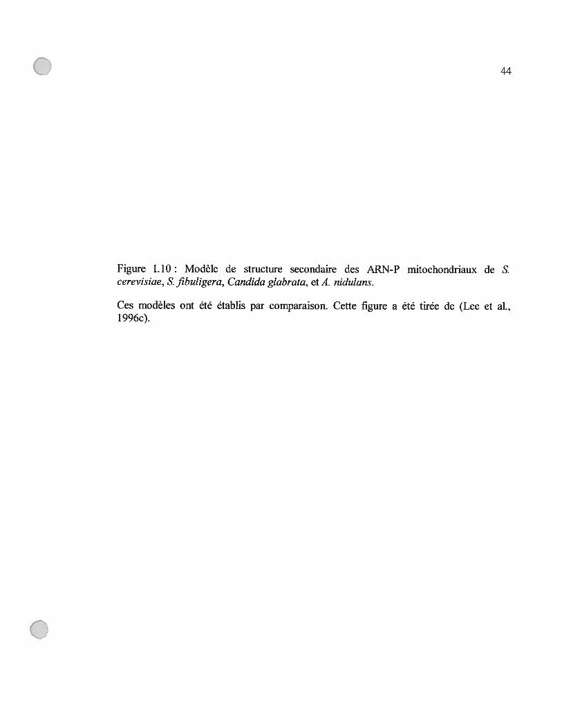
Figure 1.10: Modèle de structure secondaire des ARN-P mitochondriaux de 8.
cerevisiae, £ fibuligera, Candida glabrata, et A. nidulans 44
Figure 1.11: Distribution du gène rnpB chez les eucaryotes 47
figure D. 1: Modèle de structure secondaire de l’ARN-P de Rickettsia prowazekii et
de l’ARN-Pmt de R. americana NZ 123
C

o
xlii
figure D.2: Scénario hypothétique décrivant la perte des éléments structuraux chez
les ARN-Pmt 131
Figure D.3: Modèle du consensus minimal eubactérien et du « Micro ARN-P » 13$
figure D.4: Chimère d’ARN-P de R. americana NZ après l’ajout des P5,6,15,16,17.
141
C xiv
Liste des abréviations
A Adénine
ADN Acide déoxyribonucléique
ADNc ADN complémentaire
ADNmt ADN mitochondrial
ARN Acide ribonucléique
ARN Ml Sous-unité d’ARN de la ribonucléase P d’E. cou
ARN-Pmt Sous-unité d’ARN de la ribonucléase P mitochondriale
ARNr ARN ribosomique
ARNt ARN de transfert
C Cytosine
C5 Sous-unité protéique de la ribonucléase P d’E. cou
E. cou Escherichia cou
G Guanine
kb Kilo paires de bases (ADN) ou kilo bases (ARN)
kDa Kilodaltons
ARN-P Sous-unité d’ARN de la ribonucléase P
C’ Protéine-P Sous-unité protéique de la ribonucléase P
Qpré-ARNt Précurseur de l’ARN de transfert
ARN Ml Sous-unité d’ARN de la ribonucléase P d’E. cou
ARN-Pmt Sous-unité d’ARN de la ribonucléase P mitochondriale
RNase III Ribonucléase III
RNase E Ribonucléase E
RNase MRP Ribonucléase “Mitochondrial RNA Processing”
RNase P Ribonucléase P
rnpB Gène codant la sous-unité ARN de la RNase P
S Unité de constante de sédimentation, Svedberg
trii Gène codant un ARNt
T Thymine
U Uracyle
Liste des codes des acides aminés
A Ala Alanine
B Asx Asparagine ou acide aspartique
C Cys Cystéine
D Asp Acide aspartique
E Glu Acide glutamique
f Phe Phénylalanine
G Gly Glycine
H His Histidine
I 11e Isoleucine
K Lys Lysine
L Leu Leucine
M Met Méthionine
N Asp Asparagine
P Pro Proline
Q Gin Glutarnine
R Arg Arginine
(J $ Ser Sérine

T Thr Thréonine
V Val Valine
W Trp Tryptophane
Y Tyr Tyrosine
Z Gix Glutamine ou acide glutamique
xvii

C:Remerciements
Je tiens à remercier avec beaucoup de gratitude mon directeur de recherche, le
Dr. Franz Lang, pour sa disponibilité, sa patience, sa compréhension, et pour m’avoir
guidé tout au long de ce projet par ses nombreuses suggestions et commentaires.
Je veux remercier très sincèrement Lise forget pour les améliorations
suggérées pour cette thèse, ainsi que pour l’aide et les conseils techniques qu’elle m’a
offerts depuis mon arrivée dans le laboratoire. Egalement, je tiens à la remercier pour
son amitié et les discussions animées que nous avons partagées.
Ma reconnaissance va également à Jean-Francois Bouffard, Charles
Bullerwell, Alexandre Cadieux, Eric Delage, Yannick Jacob, Liisa Ko ski, Marie
Josée Laforest, Julien Lagarde, Jessica Leigh, Naiara Rodriguez-Ezpeleta, et Zhang
Wang pour leurs conseils techniques, les protocoles, les suggestions fructueuses qu’ils
m’ont fournis, et leur compagnie qui m’est très chère.
Je veux remercier aussi les membres du groupe de l’OGMP, dirigé par Dr
Gertraud Burger, pour leur soutien technique, pour l’accès à l’équipement de
séquençage et aux ordinateurs qui furent essentiels pour les travaux de ce projet de
recherche.
Finalement, je remercie le Conseil de Recherches Médicales du Canada pour
le soutien fmancier qui a permis de réaliser ce projet.
o
o
Je dédis ce mémoire à mes parents, mon frère et ma soeur, qui m ‘ont
encouragé constamment avec patience, et amour pour passer à travers
1 ‘épreuve du doctorat.
o
o xx
«L ‘évolution procède comme un bricoleur qui pendant des millions et des millions
d’années, remanierait lentement son oeuvre, la retouchant sans cesse, coupant ici,
allongeant là, saisissant toutes les occasions d’ajuster, de transformer, de créer. »
François JACOB
o
C
Chapitre I :Introduction
Q
CLa découverte de la structure de l’ADN, et du rôle de ce dernier dans la
conservation et la transmission de l’information génétique, ainsi que la découverte du
rôle des ARNm, et du système de traduction ont permis à Francis Crick en 195$ de
formuler ce qu’il appela le « dogme central de la biologie moléculaire »: l’information
génétique est transmise des acides nucléiques aux protéines et l’ADN n’est que le
support moléculaire d’une information qui s’exprime à travers les protéines,
notamment enzymatiques. Ce dogme limitait le rôle de l’ARN à celui de messager,
d’ARNt, ou d’ARNr. Toutefois, les résultats obtenus par H. Fraenket-Conrat et G. S.
Schramm sur te virus de la mosaïque du tabac prouvèrent que l’ARN peut lui aussi
servir de dépositaire de l’information génétique. Ce dogme ftt encore transgressé, et
le rôle de l’ARN revue suite à la découverte de deux ribozymes, l’intron auto
épissable de l’ARN ribosomique de Tetrahymena thermophila par le groupe de
Thomas R. Cech (Kruger et al., 1982) et l’activité catalytique de l’ARN (ARN Ml)
de la ribonucléase P (RNase P) d’Escherichia cou par le groupe de Sidney Altman
(Guerrier-Takada et al., 1983). Dorénavant, l’ARN sera reconnu comme ayant des
propriétés catalytiques. En 1989, l’importance de ces découvertes a été soulignée par
l’attribution du prix Nobel en chimie aux deux chercheurs, conjointement. Depuis, la
famille des ribozymes naturels a grandi par l’ajout de nouveaux introns du groupe I et
II (Peebles et al., 1986), et des ribozymes «en tête de marteau» (Hammerhead;
(forster et Symons, 1987), «en épingle à cheveux» (Hairpin; (Feldstein et al., 1989;
Hampe! et Tritz, 1989; Haseloif et Gerlach, 1989), du virus de l’hépatite delta (Wu et
al., 1989), «Varkud satellite» de Neurospora (Saville et al., 1990), et des
«riboswitch» qui clivent les ARNm (Barrick et al., 2004; Mandal et al., 2003; Nahvi
et al., 2004); (Winkler et al., 2004).
L’hypothèse originale qui écartait le rôle catalytique de l’ARN était fondée sur
l’observation que ce dernier est peu diversifié en terme de groupements fonctionnels
par rapport aux protéines. A pH neutre, il ne possède aucun groupe fonctionnel
donneur ou accepteur de protons, et aucun groupement chargé positivement, alors que
les protéines contiennent les lysines et arginines qui sont chargées positivement à
C 2
pH7. Chez les ARN catalytiques, cette limite est détournée par l’utilisation des
cations qui peuvent agir comme des acides selon la définition de Lewis, des
accepteurs d’électrons, d’où la dépendance de l’activité catalytique des ribozymes aux
ions métalliques. Le cation divalent le plus fréquemment utilisé est le magnésium, qui
peut être remplacé par le manganèse très souvent. D’autres cations sont aussi utilisés
comme le calcium, le plomb, ou le cobalt. En plus de leur rôle dans le catalyse, les
cations divalents (Mg++, Mn++ ...), et les cations monovalents (Na+, Li+ ...)neutralisent les charges négatives très fortes présentes sur les groupes phosphates des
ARN (Saenger, 1984), et par conséquent peuvent stabiliser le repliement des
conformations actives des ribozymes.
Aussitôt découverts, les ribozymes ont constitué un argument pour avancer la
théorie du «monde ARN», selon laquelle la vie sur terre a débuté avec des molécules
simples probablement des ARN qui, à la fois, encodent l’information génétique, et
catalysent les réactions chimiques nécessaire pour le maintien d’une forme primitive
de vie. Dans ce contexte, l’ARN de la RNase P, l’ARN-P, fut considéré comme une
relique de ce monde. Sa présence dans les trois domaines de la vie, les eubactéries, les
archéobactéries, et les eucaryotes, suggère que l’apparition de cet ARN est très
ancienne, et qu’elle précède possiblement la divergence des trois groupes.
Dans l’introduction de cette thèse, je discuterai des fondements d’un
consensus parmi les scientifiques qui propose une origine commune pour la
composante ARN de la RNase P dans les trois domaines de la vie, pour ensuite
décrire brièvement les connaissances actuelles sur le rôle cellulaire de la RNase P, son
mode de reconnaissance du substrat et son mécanisme de catalyse. Par la suite, je
discuterai l’origine de la RNase P mitochondriale, et de l’état des connaissances sur ce
sujet avant le début du mon projet de doctorat.
C

C 3
1.1. Homologie de la RNase P dans les trois domaines de la vie
Il existe un consensus parmi les scientifiques selon lequel les ARN-P présents
dans les trois domaines de la vie partagent le même ancêtre. Ce consensus est
supporté par la fonction commune que ces ARNs accomplissent, et par des études
comparatives au niveau de la séquence primaire et de la structure secondaire.
Jusquà ce jour, la RNase P est la seule ribonucléase connue qui coupe
spécifiquement la région guide en 5’ des précurseurs des ARNt (S’leader sequence),
et ceci dans les trois domaines de la vie ainsi que dans les organelles des eucaryotes.
Il s’agit d’une fonction essentielle, puisque, chez Schizosaccharomyces pombe,
$accharomyces cerevisiae, et E. cou, 1’ inactivation de la RNase P nucléaire par
l’élimination du gène codant la composante ARN condamne la cellule à la mort
(Cherayil et al., 1987; Lee et al., 1991; Waugh et Pace, 1990). A l’exception possible
dans quelques organelles (voir article 1), il a été montré que l’activité catalytique de la
RNase P dépend de la sous-unité ARN chez les eucaryotes et les procaryotes, et ceci
en abolissant l’activité catalytique de la RNase P par un traitement à la ribonucléase
de Micrococcus (franklin et al., 1995; Stark et al., 197$), en éliminant le gène codant
l’ARN-P (voir plus haut), ou en montrant que les ARN-P eubactériens et quelques
ARN-P archéobactériens sont actifs in vitro (Gardiner et al., 1985; Guerrier-Takada et
al., 19835; Pannucci et al., 1999; Wagner et al., 2001).
L’origine commune des ARN-P provenant des différents règnes de vie est
supportée par les études comparatives. Suite à l’alignement de dix séquences d’ARN
P, provenant des eubactéries, archéobactéries, eucaryotes et une séquence
mitochondriale, cinq motifs universellement conservés ont été identifiés au niveau de
la séquence primaire (CRI-V)(Chen et Pace, 1997). Ces régions ont été identifiées
aussi chez les ARN-P des plastides (de la Cruz et Vioque, 2003), organelles
responsables de la photosynthèse. Cette homologie est encore plus marquée au niveau
C 4
de la structure secondaire, puisque comme chez la plupart des ARN, la conservation
au niveau de la structure secondaire des ARN-P est plus forte que celle observée au
niveau de la séquence primaire. Les études de modélisation des ARN-P par
comparaison phylogénétique ont mené à des structures secondaires consensus chez les
eubactéries (Siegel et aI., 1996) et les archéobactéries (Haas et al., 1996), similaires
entre elles et atix modèles proposés pour les ARN-P des eucaryotes £ pombe, Homo
sapiens et Dino rerio (Frank et al., 2000; Tranguch et Engelke, 1993). Tous ces
modèles partagent un noyau conservé qui inclut une structure en forme de cage
maintenue par un pseudo-nœud (Chen et Pace, 1997; Forster et Altman, 1990b), une
boucle interne entre les hélices Pli-14, et quatre hélices en forme de croix (Figure
1.1) (Chen et Pace, 1997).
En se basant sur les données décrites ci-dessus, nous pouvons conclure qu’une
ARN-P était présente chez l’ancêtre commun des trois domaines de la vie, il y a 3 à 4
milliards d’années (Feng et al., 1997), une estimation qui restent très controversée. Il
est fort probable que l’apparition de cet ARN date de la même époque que
l’apparition des ARNt et des ribosomes.
C

0 5
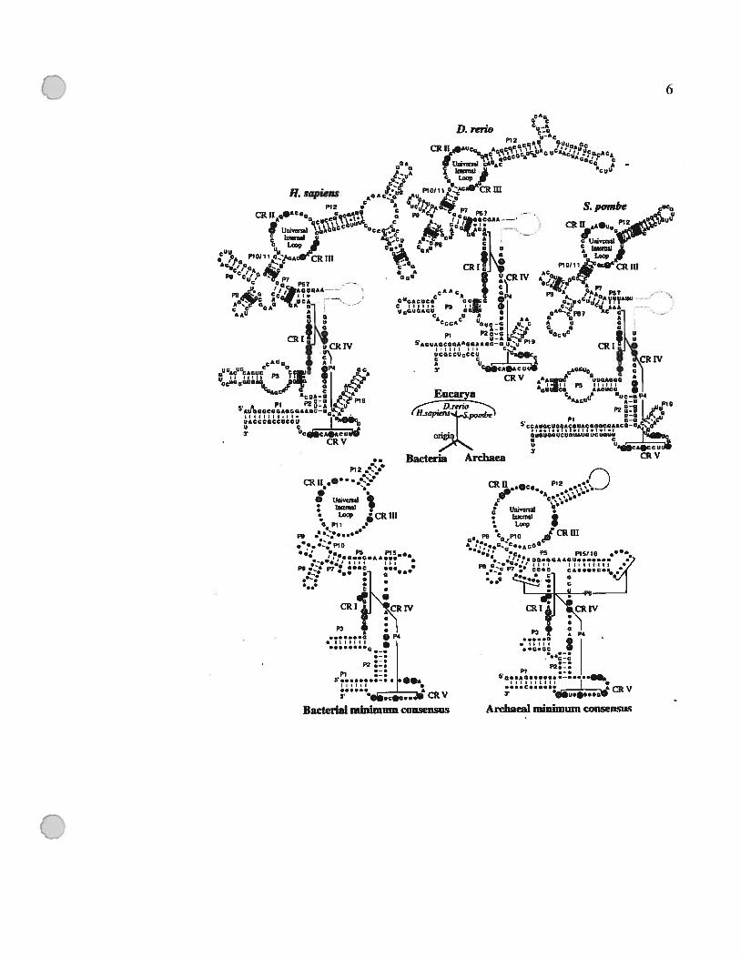
Figure 1.1: Modèles de structure secondaire representant la structure de l’ARN-P chez
les trois domaines de vie.
Les modèles du consensus minimal des eubactéries (Siegel et al., 1996) et des
archéobactéries (Haas et al., 1996), dans lesquels les nucléotides invariables sont
indiqués par des lettres. Les nucléotides qui sont universellement conservés sont
indiqués par des cercles pleins. Pour les structures des ARN-P nucléaires de H
sapiens, Danio rerlo et S. pombe: le nom d’hélice contenant un point d’interrogation,
marque une incertitude de l’annotation de cette structure à cause de la variation de
séquence primaire; les lignes grises indiquent la position du P15 par rapport aux
modèles bactériens; dans les boîtes, on indique les covariations qui appuient la
présence de ces structures (Frank et al., 2000; Tranguch et Engelke, 1993). Cette
figure a été tirée de Chen et Pace, (1997).
o
C
..
P12
CR tt.0 •
.O urnvenal:
Loop CRIIIG pli •
I.
.•e. .2: P10‘. PH
• ‘,,G..G.A*...II,,
- III•
PH •:. p • ...c ..... H
:: I :CRI1j ‘ÇRIV
PH + :1G • P4
t::::
•
r• 0.CG... CRV
Bacterial minimum consensus
I.
••3
G-C
PI50.SAG 1A•...c...... 4CRV
Archaeal minimum consensus
D. reno
6
P12
IL sapiens
V
CRI w
7
1.2. Fonctions cellulaires de la RNase P
Comme mentionné plus haut, la RNase P dans les trois règnes de vie est
responsable de la maturation des précurseurs des ARNt (pré-ARNt). Cependant, la
RNase P peut couper plusieurs autres substrats qui ressemblent structurellement à des
ARNt.
1.2.1 Fonctions cellulaires de la RNase P chez les eubactéries
Les rôles accomplis par la RNase P chez les eubactéries sont les mieux connus.
Elle est responsable de la maturation de l’ARN CI, l’ARN 4,5 S, certains ARNm,
l’ARNtm, et les ARNr.
1.2.1.1 Régulation du cycle lytique du bactériophage P4
L’état de lysogénie du bactériophage P4 est sous le contrôle d’un petit ARN,
appelé ARN CI, qui cause la terminaison précoce de la transcription de l’opéron
responsable de la lyse cellulaire (Briani et al., 2001). Cet ARN confère une immunité
pour la cellule hôte contre le bactériophage P4 en s’attachant à des séquences
complémentaires à sa propre séquence, située en amont du premier gène de l’opéron
ciblé; par la suite, la transcription sera terminée par un mécanisme dépendant du facteur
Rho (Briani et al., 1996). Il a été montré in vivo, que le rôle de la RNase P dans ce
processus est au niveau de la maturation en 5’ de l’ARN CI (Forti et al., 1995). Selon le
modèle prédit de la structure secondaire, les extrémités de cet ARN peuvent se replier
pour former une hélice, un CCA protubérant en 3’, et une région simple brin en 5’ (forti
et al., 2002), ce qui constitue un substrat idéal pour la RNase P (Hansen et al., 2001).
o
o $
1.2.1.2 Maturation de l’ARN 4,5$
L’ARN 4,5S a été baptisé ainsi en rapport avec sa mobilité électrophorétique sur
les gels de polyacrylamide. Il est très stable; in vivo, il est présent à une concentration
comparable à celle d’un ARNt. L’ARN 4,5$ se retrouve chez plusieurs procaryotes et le
gène qui le code, le ft, est essentiel pour la survie d’E. cou. Cet ARN est l’homologue
structural de l’ARN 7$ chez les archéobactéries et les eucaryotes. Ce dernier fait partie
de la particule $RP (signal Recognition Particle), une particule formée de six protéines et
d’une molécule d’ARN, qui est essentielle pour la translocation des protéines à travers la
membrane du réticulum endoplasmique (Poritz et al., 198$). Les études de
complémentation ont montré que le gène codant l’ARN 7$ peut remplacer le ffs chez E.
cou (Brown, 1991). In vivo, l’ARN 4,5 S lie la protéine fffi qui a une taille de 4$kD. La
fffi est l’homologue de la protéine eucaryote SRP54, et fait partie d’une particule SRP
eubactérienne impliquée dans le trafic des protéines de sécrétion chez les eubactéries
(Nagai et al., 2003; Ribes et al., 1990). Plusieurs études ont montré que l’ARN 4,5S est
aussi impliqué dans la traduction, où il facilite l’étape de translocation durant la synthèse
des polypeptides sur le ribosome en interagissant avec le facteur d’élongation Ef-G
(Nakamura et al., 2001; Sagar et al., 2004).
La RNase P est responsable de la maturation de l’ARN 4,5S, comme suggéré par
l’accumulation du précurseur de l’ARN 4,5S in vivo dans les mutants de rnpB
thermosensibles (Bothwell et al., 1976). Ce rôle a été confirmé par des études in vitro,
qui ont montré que la RNase P coupe d’une façon endonucléolytique l’extrémité 5’ du
précurseur pour générer deux fragments, l’ARN 4,5S mature et la région en 5’ de cet
ARN (Bothwell et al., 1976; Guerrier-Takada et Altman, 1984).
o
9
1.2.1.3 Régulation post-traductionnelle par la RNase P
Des études effectuées chez $aimonelïa lyphimurium et E. cou ont montré que la
RNase P est impliquée dans la maturation des ARNm de plusieurs opérons (voir plus
bas). Cette maturation affecte la stabilité de ces ARNm ce qui constitue une régulation
post-traductionnelle de l’expression de ces opérons.
1.2.1.3.1 Stabilisation de I’ARNm de J’opéron his chez S. typhimurium
Les gènes responsables de la biosynthèse de l’acide aminé histidine sont codés par
l’opéron his. Chez S. typhirnurium, les études de protection contre la digestion par la
nticléase $1, des mesures de la stabilité de l’ARN, des expériences de retardement sur gel
et des tests de transcription in vitro ont montré que les 5 cistrons les plus distaux (en 3’)
de 1’ARNm de l’opéron his sont coupés du reste du messager, et forment un ARN de
3900 nucléotides. Ce nouvel ARN est plus stable que son précurseur (Alifano et al.,
1992). Les résultats d’une étude utilisant des mutants thermosensibles pour la RNase P et
la RNase E ont montré que les deux ribonucléases ont un rôle dans la maturation de
l’ARN de 3900 nucléotides (Alifano et al., 1994). Les investigations plus poussées ont
montré que la RNase E coupe le transcrit primaire de l’opéron his à une distance de 620
nucléotides en amont de l’extrémité 5’ de l’ARN 3900 nucléotides mature, formant ainsi
un précurseur de cet ARN. Par la suite, ce précurseur sera coupé par la RNase P à un site
situé à 76 nucléotides en amont du codon d’initiation de la traduction du cistron hisB. La
coupure par la RNase P est situé en 5’ d’une région qui peut être repliée en une structure
tige boucle suivie de la séquence NCCA (Alifano et al., 1994). Il a été suggéré que la
stabilité de l’ARN de 3900 nucléotides est conférée par cette structure qui le protège
contre les nucléases (Alifano et al., 1996; Bouvet et Belasco, 1992). L’efficacité de la
coupure du précurseur par la RNase P est modulée par la traduction du messager; cette
coupure requiert que le ribosome soit attaché au cistron hisB (Alifano et al., 1992;
C Alifano et al., 1994). Il y a deux hypothèses pour expliquer comment les ribosomes
C 10
attachés à l’ARN du hisB augmentent l’efficacité de la coupure par la RNase P, soit en
favorisant la formation de la structure tige-boucle qui peut être reconnue par la RNase P,
soit en stabilisant temporairement le précurseur en le protégeant contre la dégradation 5’
3’, ce qui augmente le nombre des précurseurs intacts disponibles (Alifano et al.,
1996). Ce système de stabilisation du messager permet de coupler la stabilité du
messager à la disponibilité des ribosomes.
1.2.1.3.2 La RNase P précipite la dégradation des ARNm chez E. cou
En utilisant des mutants d’E. cou thermosensibles pour la RNase P (cellules
A49)(Baer et al., 1989), il a été montré que cette dernière est impliquée dans la
maturation des ARNm des opérons tna (dégradation du tryptophane), his (biosynthèse de
l’histidine), rbs (transport du D-ribose), secG (excrétion de protéines), lac (lactose), ainsi
que des OREs b0669 et b0671 (Li et Altman, 2003; Li et Altman, 2004). Les auteurs ont
cloné et transcrit in vitro certaines régions intergéniques de ces messagers, et ils ont
montré que la RNase P coupe à l’intérieur de ces régions, et entre le début du transcrit et
le codon d’initiation de la traduction de l’opéron secG. Chez les mutants thermosensibles
pour la RNase P, les auteurs ont observé une augmentation de la quantité d’ARNm du
tna, et une disparition des intermédiaires de ce messager qui sont les produits de la
maturation par la RNase P. Dans ces mutants, l’activité catalytique du tnaA est plus
élevée que dans les cellules normales, ce qui est compatible avec l’augmentation de la
quantité d’ARNm observée. En ce basant sur ces données, et sur le fait que les produits
de la RNase P sont des bons substrats pour la RNase E (Coburn et Mackie, 1999), les
auteurs ont testé la sensibilité des messagers de lacYA avant et après la coupure par la
RNase P à la digestion par la RNase E. Ils ont remarqué que la RNase E dégrade cet
ARNm d’une façon très efficace après la coupure par la RNase P contrairement à ce
qu’on observe pour le transcrit original. Suite à ces observations, les auteurs ont proposé
un modèle selon lequel la RNase P coupe les transcrits de certains opérons, ce qui les
C rend disponible à la dégradation par la RNase E. Ce modèle a été confirmé in vivo en

C Il
clonant la région intergénique de l’opéron lac en amont d’un gène rapporteur, et en
transformant des cellules sauvages et mutants thermosensibles avec cette construction (Li
et Altrnan, 2004).
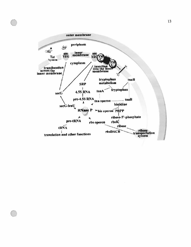
1.2.1.3.3 La RNase P est impliquée dans le transport membranaire
En examinant les fonctions déjà décrites plus haut, on remarque que la RNase P
est un élément clé dans le transport transmembranaire chez E. coli. Elle est impliquée
dans quatre fonctions en rapport avec ce transport; (i) la maturation de l’ARN de la SRP
(Signal Recognition Particle) eubactérienne (ARN 4,5S) (ii) et de la région 5’ non
traduite du secG, ainsi que (iii) du contrôle de l’expression de la perméase du
tryptophane et (iv) des gènes impliqués dans le transport du ribose (voir section
T.2.l.3.2)(Figure I.2)(Li et Altman, 2003). Ce nouveau rôle n’avait pas été suspecté
précédemment, et dans l’avenir, il serait important d’étudier la régulation de l’expression
de la RNase P afm de mieux comprendre comment cette dernière fait le lien entre la
traduction, en étant impliquée dans la maturation des ARNt et des ARNr (voir la section
1.2.1.4), et le transport membranaire au niveau de la cellule.
C

12
Figure 1.2 Schéma résumant les fonctions cellulaires de la RNase P chez E. cou et du
lien de ces fonctions au transport membranaire.
Cette figure a été tirée de (Li et Altman, 2003).
J.y
VNç-nd
uy--
-.
I
/fi
4HS//1
,//annqniaw/,//.)ueJqmawiauui
fJOlI!JIfluflIr7/l)‘isuin
q’i/IUU3EJÔhIS)U1r
/uniqduvb/J—lLb4S
-!1itUQhItUI!)SA‘À
mçrjdu3d
JuEJqlrnwijino
unijrpoku.ij---.-noqu
aqdsoqd-ç-asoqu“4
jjjnnndn;zqJ
q
unndoEUJ
uusdosqi
SUOIPUILJ..LaqwUoiirpur.a1
van
Vf!H44Id
o
Q ET
C 14
1.2.1.4 Maturation du ARNtm (ARN lOSa)
Initialement, 1’ARNtm était connu comme l’ARN lOSa. Les études de
modélisation comparative de ce gène chez Mycobacterium tuberculosis, E. cou et
Bacillus subtilis ont révélé une similarité entre cet ARN et la moitié de la structure de
lARNtM qui correspond à la tige acceptrice et l’hélice T (Komine et al., 1994; Tyagi et
Kinger, 1992; Ushida et al., 1994). La tige acceptrice de l’ARN lOSa accepte l’acide
aminé alanine in vivo et in vitro (Komine et al., 1994). L’autre partie de cet ARN encode
une protéine “tag”, et peut donc servir d’ARNm d’où son nouveau nom ARNtm
(Gueneau de Novoa et Williams, 2004). Le rôle de l’ARNtm est de s’attacher aux
ribosomes bloqués par des ARNm partiellement dégradés; une fois l’ARNtm attaché, sa
partie codante sera traduite, et la protéine encodée s’ajoute à la protéine incomplète
produite par l’ARNm brisé. Par la suite, la partie “tag” sera reconnue par une protéase
associée à la membrane, la protéase tsp qui dégradera la protéine “tag” ainsi que la
protéine partiellement traduite (Figure I.3)(Gueneau de Novoa et Williams, 2004). Par ce
mécanisme, l’ARNtm débloque les ribosomes et permet de dégrader les protéines
incomplètes.
Il a été montré que la RNase P coupe l’extrémité 5’ du précurseur de l’ARNtm en
cartographiant l’extrémité 5’ d’un ARNtm provenant d’un mutant thermosensible pour la
RNase P, et ceci par extension d’amorce (Komine et al., 1994). Dans le cadre de mes
travaux de doctorat, j’ai participé à des expériences et des analyses dans lesquelles nous
avons identifié pour la première fois des ARNtm mitochondriaux potentiels chez les
jakobides, et nous avons montré que l’ARN-P d’E. cou (ARN Ml) peut couper in vitro
les précurseurs de ces ARNtm mitochondriaux putatifs (Jacob et al., 2004).
o

0 15
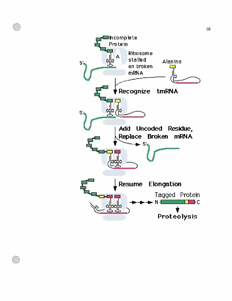
figure 1.3: Modèle d’action de 1’ARNtm.
La première partie de la figure montre un ribosome bloqué par un ARNm incomplet ou
partiellement dégradé. Ce ribosome reconnaît l’ARNtm, qui rentre dans le site A du
ribosome, remplace l’ARN messager et débloque le ribosome. Les résidus encodés par
l’ARNtm seront ajoutés à la protéine partiellement synthétisée à partir de l’ARNm
incomplet. Ces résidus ajoutés vont servir de signal pour la tsp qui va dégrader la
protéine plus tard. La figure a été tirée du site de l’ARNtm
(http://www.indiana.eduktmRNA /)(Gueneau de Novoa et Williams, 2004).
C
o
5,
5’
Tagged__Protein—.‘--+- -.- N - C
tProteolys is
o
16
lin c o m p le t eProtein
APibosomestalle clon brokenmRNA
Alanine
tmRNÀ
J
Bmken mRNA
Elonqation

17
1.2.1.5 Maturation des ARNr chez les eubactéries
Chez les eubactéries, la RNase P est impliquée dans la maturation des ARNr au
niveau du transcrit primaire. Elle est responsable de la maturation en 5’ des pré-ARNt
présents dans la région intergénique situées entre l’ARN 30S et 1 6S et qui peut inclure
un ou plusieurs ARNt (figure 1.4). Dans des cellules mutées pour la RNase III, la RNase
P peut compenser cette défectuosité en libérant les précurseurs des ARNs 16$ et 23$;
ces molécules sont alors utilisables pour former des ribosomes fonctionnels (King et al.,
1986; Morrissey et Tollervey, 1995). Par conséquent, il a été suggéré que la RNase P est
une alternative à la RNase III dans la maturation des ARNr. Il est commun de trouver de
la redondance dans les mécanismes qui produisent des molécules essentielles pour la
survie de la cellule (Morrissey et Tollervey, 1995).
1.2.2 Fonctions cellulaire de la RNase P chez les eucaryotes
La RNase P eucaryote est plus complexe que la RNase P eubactérienne. Chez les
humains, elle contient aux moins dix sous-unités protéiques en plus de la sous-unité
ARN (Guerrier-Takada et al., 2002), comparé à une seule protéine chez les eubactéries.
Cependant, à part la maturation des ARNt, les rôles documentés de la RNase P eucaryote
se limitent à seulement deux fonctions possibles et mal connues, la maturation des ARNr
et la réplication des virus. Le plus grand nombre de protéines-P reflète la plus grande
complexité des cellules eucaryotes par rapport aux eubactéries; alors que le petit nombre
de fonctions accomplis découle de deux raisons possibles. Premièrement, il est fort
probable que plusieurs fonctions restent inconnues, et deuxièmement, la complexité des
cellules eucaryotes amène une spécialisation dans les fonctions; par exemple, la RNase P
semble avoir perdu son rôle dans la maturation des ARNr au profit d’une nouvelle
ribonucléase, la RNase MRP.
1$
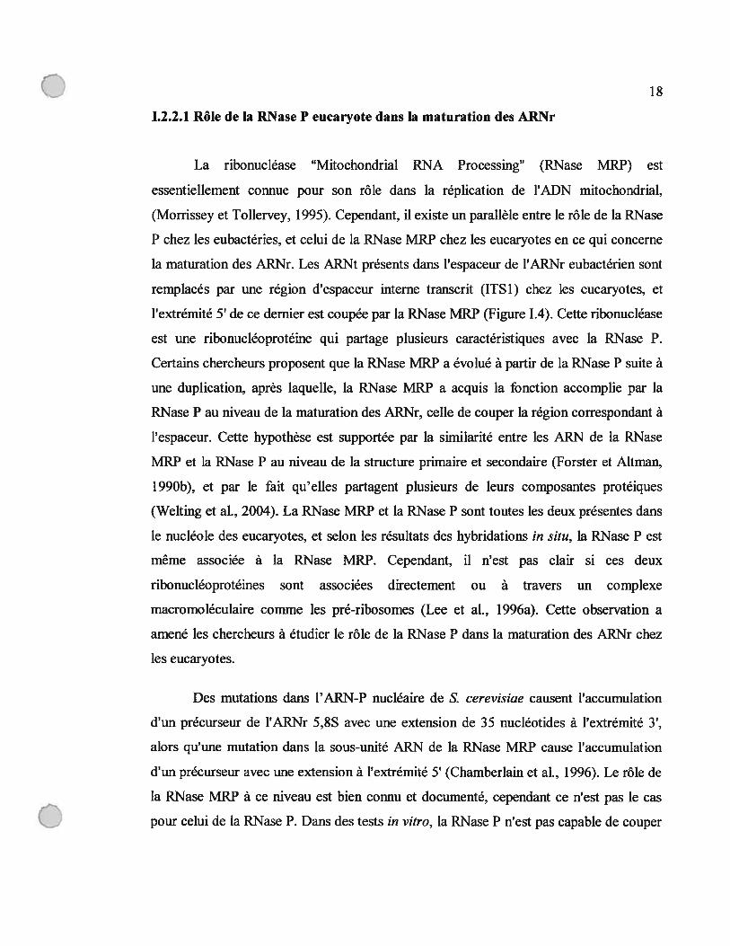
1.2.2.1 Rôle de la RNaseP eucaryote dans la maturation des ARNr
La ribonucléase “Mitochondrial RNA Processing” (RNase MRP) est
essentiellement connue pour son rôle dans la réplication de l’ADN mitochondrial,
(Morrissey et Tollervey, 1995). Cependant, il existe un parallèle entre le rôle de la RNase
P chez les eubactéries, et celui de la RNase MRP chez les eucaryotes en ce qui concerne
la maturation des ARNr. Les ARNt présents dans l’espaceur de l’ARNr eubactérien sont
remplacés par une région d’espaceur interne transcrit (ITS1) chez les eucaryotes, et
l’extrémité 5’ de ce dernier est coupée par la RNase MRP (figure 1.4). Cette ribonucléase
est une ribonucléoprotéine qui partage plusieurs caractéristiques avec la RNase P.
Certains chercheurs proposent que la RNase MRP a évolué à partir de la RNase P suite à
une duplication, après laquelle, la RNase MRP a acquis la fonction accomplie par la
RNase P au niveau de la maturation des ARNr, celle de couper la région correspondant à
l’espaceur. Cette hypothèse est supportée par la similarité entre les ARN de la RNase
MRP et la RNase P au niveau de la structure primaire et secondaire (forster et Altman,
1990b), et par le fait qu’elles partagent plusieurs de leurs composantes protéiques
(Wehing et al., 2004). La RNase MRP et la RNase P sont toutes les deux présentes dans
le nucléole des eucaryotes, et selon les résultats des hybridations in situ, la RNase P est
même associée à la RNase MRP. Cependant, il n’est pas clair si ces deux
ribonucléoprotéines sont associées directement ou à travers un complexe
macromoléculaire comme les pré-ribosomes (Lee et al., 1996a). Cette observation a
amené les chercheurs à étudier le rôle de la RNase P dans la maturation des ARNr chez
les eucaryotes.
Des mutations dans l’ARN-P nucléaire de S. cerevisiae causent l’accumulation
d’un précurseur de l’ARNr 5,$S avec une extension de 35 nucléotides à l’extrémité 3’,
alors qu’une mutation dans la sous-unité ARN de la RNase MRP cause l’accumulation
d’un précurseur avec une extension à l’extrémité 5’ (Chamberlain et al., 1996). Le rôle de
la RNase MRP à ce niveau est bien connu et documenté, cependant ce n’est pas le cas
C pour celui de la RNase P. Dans des tests in vitro, la RNase P n’est pas capable de couper

19
l’extrémité 3’ du précurseur de 1’ARNr 5,8S. Cette observation indique que le
précurseur non mature isolé dans l’étude utilisant une ARN-P mutée n’est pas le produit
direct de la mutation du gène rnpB. Dans ce cas, de quoi découle cette anomalie dans la
maturation de l’ARN 5,$S ? Plusieurs hypothèses ont été émises. Il a été proposé que la
RNase P est impliquée dans des voies de maturation de l’ARNr inconnues jusqu’à
maintenant, ou que la mutation dans la RNase P affecte la structure des particules
ribonucléoprotéiques dont elle fait partie, et par suite elle cause une
o

U 20
Figure 1.4: Comparaison des pré-ARNr d’E. cou et de $accharomyces cerevisiae.
L’arrangement de base est conservé, mais on note trois différences majeures.
Premièrement, l’équivalent de la grande sous-unité ribosomique bactérieime (ARNr 23S)
est divisée en deux par l’insertion de la région ITS2 (intergenic transcribed spacer) chez
la majorité des eucaryotes. Deuxièmement, PARNr 5S est synthétisé d’une façon
indépendante chez les eucaryotes. Troisièmement, les eubactéries possèdent des ARNt
dans l’espaceur entre les ARNr 16S et 23S qui sont différents selon la bactérie.
Les sites de coupure par les RNase P et MRP sont indiqués par des flèches. Cette figure a
été tirée de (Lee et al., 1996a).
o
o 21
RNase P
Prokaryotes 1 6S tRNA 23S 5S
I
-u5S
o
I I t)’I I t)’
I I U’I j fl\
I
Eukai
U
5.8SMRP
28S

22
anomalie dans les voies de maturation des ARNr dans lesquelles ces particules sont
impliquées; cependant ces hypothèses restent à démontrer (Chamberlain et al., 199$;
Chamberlain et al., 1996).
1.2.2.2 Rôle de la RNase P eucaryote chez les virus
Chez les virus, la RNase P peut cliver des éléments ressemblant à des ARNt dans
les génomes ARN du virus de l’hépatite C et de plusieurs virus chez les plantes (voir plus
bas). Cependant, le rôle le mieux connu de la RNase P eucaryote découle du fait qu’elle
est responsable de la maturation des ARNt, et que ces derniers sont utilisés comme
amorces pour l’initiation de la reverse transcription des génomes ARN des rétrovirus et
des rétrotransposons. Chez le rétrotransposon de la famille copia, on note même que la
RNase P est capable de couper pas seulement au 5’ de l’ARNt, mais à l’intérieur de
l’ARNt afm de produire l’amorce requis (voir plus bas). Cette réaction est nommée
«hyperprocessing ».
1.2.2.2.1 Rôle de la RNase P dans la réplication chez les rétrovirus
Chez les drosophiles, on retrouve des particules qui ressemblent à des rétrovirus
(RVLP) et qui contiennent une molécule d’ARN qui a une taille de 5 kb et certains petits
ARN incluant des ARNt. Une activité de transcription inverse a été détectée dans ces
particules qu’on pense être des intermédiaires de la transposition d’un élément
appartenant à la famille de réstrotransposon copia (Kikuchi et al., 1986). En général, la
réplication des génomes ARN des rétrovirus débute par le site d’attache de l’amorce où
les derniers 1$ nucléotides de l’extrémité 3’ d’une molécule d’ARNt s’apparient et
servent d’amorce pour la reverse transcriptase (Mak et Kleiman, 1997). Chez les
particules RVLP des drosophiles (décrites plus haut), une séquence central, située autour
de la boucle de l’anticodon de lARNtMet d’initiation est complémentaire au site d’attache
u
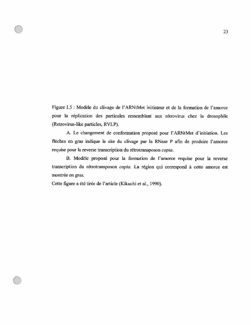
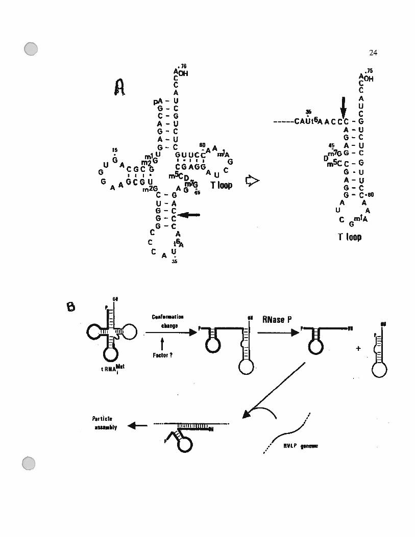
Figure 1.5 : Modèle du clivage de l’ARNtMet initiateur et de la formation de l’amorce
pour la réplication des particules ressemblant aux rétrovirus chez la drosophile
(Retrovirus-like particles, RVLP).
A. Le changement de conformation proposé pour l’ARNtMet d’initiation. Les
flèches en gras indique le site du clivage par la RNase P afm de produire l’amorce
requise pour la reverse transcription du rétrotransposon copia.
B. Modèle proposé pour la formation de l’amorce requise pour la reverse
transcription du rétrotransposon copia. La région qui correspond à cette amorce est
montrée en gras.
Celle figure a été tirée de l’article (Kikuchi et al., 1990).
• 76
g gA
pA- UG-CC-GA-UG-CA
‘5 G-lu
U A mr CGCG‘J II,.
G GCGUA” m2G
C-U-A
6-CC A
CtAU
35
.75AçCC
I Au
5 C
CAUI6AACCC-G
A-UG-C
Q A-Um7GG - C
Dmscc - GG-uA-UG-CG - C80
A Au A
C mIA
T Ioop
24
Ur BD
-AGUUCC nA1*1 Ii ØCGAGG
mC0 Au
AQ TloopG
o
C
13tnf.rmatio.
change
*f
Fader?
t
RNase P
—+SI
Particlentemhly 4-
3%) 7 RVLP gencet
C 25
de l’amorce de la reverse transcription et non pas l’extrémité 3’ de l’ARNt mature. Il a
été montré que dans ce cas, l’ARNt en question peut assumer une conformation
alternative et peut être coupé par la RNase P pour générer l’amorce requise pour la
réplication (Kilcuchi, 1995; Kilcuchi et al., 1990) (Figure 1.5).
Le mécanisme que l’IRES utilise pour initier la synthèse protéique reste inconnu,
mais certains éléments structuraux sont bien conservés dans ce site, entre autre la
similarité structurale aux ARNt. Cette ressemblance pourrait être l’élément clé pour la
reconnaissance des IRES par les ribosomes et le déclenchement de la synthèse protéique
(Lyons et Robertson, 2003). En effectuant des tests in vitro, dans le but de trouver des
cibles potentielles pour la thérapie génique utilisant la RNase P humaine, des chercheurs
ont montré que cette dernière peut reconnaître et cliver le génome du virus de l’hépatite
C à deux endroits (Nadal et al., 2002). Le premier est au niveau de l’IRES, et le
deuxième est situé à l’intérieur du génome, entre la région qui code les gènes structuraux
et les gènes non structuraux. Cependant, il n’existe aucun argument qui supporte un rôle
pour ces clivages dans la physiologie des virus, puisque pour l’instant, il n’a pas été
montré que ces clivage ont lieu in vivo, ni que le virus de l’hépatite C accède au noyau
(Bartenscifiager et Lohmann, 2000), là où la majorité de la RNase P nucléaire réside,
donc il se peut que ce clivage sois le résultat du simple fait que la région ressemble
structurellement à un ARNt, une propriété qui serait utilisée pour accéder aux ribosomes.
1.2.2.2.2 La RNase P clive les virus des plantes en 3’
Chez les plantes, la plupart des génomes ARN simple brin des virus contiennent
des structures en forme d’hélices et de pseudo-noeuds proches de leur extrémité 3’ (Pleij
et al., 1987), qui forment une structure en forme de «L» similaire à celle des ARNt. Ces
Q 26
éléments structuraux peuvent être aminoacylés par l’ajout de la valine pour les
tymovfrus et les furovirus, la tyrosine pour les bromovirus, cucumovirus et hordeivfrus,
et par l’histidine pour les tobamovirus (voir références dans (Wientges et al., 2000)). En
plus, ces structures peuvent être clivés par la RNase P in vitro (Guerrier-Takada et al.,
1988), servir de substrat pour l’ajout du CCA par la nucleotidyl-transferase, et lier les
facteurs d’élongation de la traduction Ef-Tu et Ef-la (Wientges et al., 2000).
Initialement, il a été proposé que ces structures sont des reliques du monde ARN, où ils
servaient dans la réplication des virus ARN (Weiner et Maizels, 1987). Récemment, il a
été montré que ces éléments agissent comme des «enhancer» dans la traduction
(Matsuda et Dreher, 2004).
o
Q 27
1.3. Reconnaissance du substrat
Comme décrit précédemment, plusieurs substrats sont reconnus par la RNase P.
Ceci nous mène à se poser la question comment la RNase P reconnaît-elle ces substrats?
Les études expérimentales, essentiellement menées chez les eubactéries, ont montré que
les deux composantes, protéique et ARN, de la ribonucléoprotéine sont impliquées dans
la reconnaissance du substrat. Dans ce qui suit, je résumerai ce qui est connu quant aux
interactions entre un pré-ARNt et chacune de ces composantes, pour ensuite décrire un
mécanisme de reconnaissance qui englobe le rôle de chacune de celles-ci.
1.3.1 Les interactions entre le pré-ARNt et les composantes de la RNase P
Chez E. cou, les études de pontage, mutagenèse, et interférence chimique,
effectuées sur l’ARN-P ont montré que le domaine « S », appelé domaine de spécificité,
et qui est constitué de la région s’étendant le P7 au P12 (en bleu dans la figure 1.6)
(Harris et Christian, 2003), rentre en contact avec la tige et la boucle T du pré-ARNt
(Knap et al., 1990; LaGrandeur et al., 1994; Nolan et al., 1993) (Loria et Pan, 1997; Pan
et al., 1995).
Le domaine « C », appelé domaine catalytique, qui est formé des hélices P1-P5, et
P15 (en vert dans la figure 1.6), interagit avec le motif NCCA en 3’ des pré-ARNt et
avec le nucléotide en position -1 (en amont) du site de coupure (Kirsebom et $vard,
1994; LaGrandeur et al., 1994; Oh et al., 199$).
Quant à la protéine-P, la comparaison des constantes cinétiques et d’affmité au
substrat de l’holoenzyme et de l’ARN-P seul, indique qu’elle interagit avec les pré
ARNt, au niveau de la région guide en 5’ des ARNt (5’ leader sequence) (Crary et al.,
1998; Kurz et al., 199$). Les études de pontage ont montré que cette dernière s’attache à
une fente sur la surface de la composante protéique (Niranjanakumari et al., 199$).
o

0 28
figure 1.6 Structure globale du complexe RNaseP/pré-ARNt d’E. cou.
(a) Consensus de la structure secondaire des ARN-P eubactériens (Hall et Brown, 2001).
La lettre P est utilisée pour designer une hélice, J pour la région en simple brin qui joint
deux hélices. Le domaine « S » de spécificité est en bleu et le domaine «C> de catalyse
en vert. La tige acceptrice ainsi que la boucle et l’hélice T sont en rouge. La région guide
en 5’ des ARNt (5’ leader sequence) est en noir, et l’interaction de cette région avec la
protéine-P est montrée en forme d’ovale gris. La flèche indique le site de coupure par la
RNase P. Les lignes indiquent une interaction entre les sites aux extrémités de ces lignes.
Les lettres en majuscule sur le modèle consensus représentent les nucléotides conservés
universellement.
(b) Modèle de la structure tertiaire du complexe RNaseP/pré-ARNt (Chen et al., 199$;
Massire et al., 199$). Le code de couleur est le même que dans (a). Les positions
probables de la protéine-P et de la région guide en 5’ de l’ARNt sont marquées
respectivement par des lignes coupées noire et rouge.
Cette figure a été tirée de (Harris et Christ jan, 2003).
o

oo
-D
ÎIz
i\
n o p

C 30
1.3.2 Contribution modulaire à la reconnaissance du pré-ARNt
Les expériences de mutagenèse dirigée ont permis d’élucider la contribution de
chaque module à la reconnaissance du substrat. Elles ont dévoilé qu’en absence du
domaine «S », le domaine « C» de l’ARN-P et la composante protéique peuvent
interagir, lier le substrat et accomplir la réaction catalytique, aussi efficacement pour un
pré-ARNt que pour un substrat qui consiste en une région simple brin suivie d’une hélice
courte (Loria et Pan, 2001). Ces expériences, ainsi que les études enzymologiques et
structurales supportent le modèle mécanistique de reconnaissance du substrat selon
lequel la protéine-P interagit avec la région guide en 5’ de l’ARNt (Christian et al.,
2002b), et qu’elle augmente le taux de catalyse en favorisant l’attache du pré-ARNt (le
substrat) à la ribonucléoprotéine et la relâche de l’ARNt mature (le produit) (Harris et
Christian, 2003). Quant au domaine «C », il contient le site de catalyse et rentre en
contact avec le pré-ARNt au niveau du site de coupure sans montrer aucune spécificité;
cette dernière sera octroyée par le domaine de spécificité «S », qui lie la boucle et
l’hélice T, et qui est responsable de la différenciation entre un ARNt et les autres
substrats.
1.3.3 Reconnaissance des substrats autres que les pré-ARNt
Le modèle précédent explique comment un pré-ARNt aussi bien qu’un pré
ARNtm (précurseur de l’ARNtm) est reconnu par la RNase P, puisque la structure des
ARNtm a conservé la tige acceptrice, le motif RCCA, ainsi que la boucle et l’hélice T
(Williams et Bartel, 1996). Mais comment les autres substrats sont-ils reconnus?
Présentement, l’état des connaissances ne permet pas de répondre à cette question d’une
façon détaillée, mais deux éléments clés sont à signaler. Premièrement, la RNase P n’a
pas besoin des boucle et tige T pour reconnaître un substrat. Les expériences de
mutagenèse ont montré que le substrat minimal pour la RNase P d’E. cou peut se limiter
à un ARN qui contient une hélice de 4 à 16 pb qui mime la tige acceptrice, et d’une
région simple brin en 5’ formée d’au moins un nucléotide (Forster et Altman, 1990a;
C Kirsebom et Vioque, 1995; Liu et Altman, 1996); pour une coupure efficace par l’ARN-
C 31
P seul in vitro, le motif CCA est nécessaire. Deuxièmement, la reconnaissance des
substrats se fait au niveau de la structure tertiaire, et à ce niveau, il a été montré que la
protéine-P permet à l’ARN d’accommoder de nouveaux substrats plus efficacement,
probablement en adaptant la structure générale du ribozyme (Kirsebom et Vioque, 1995;
Liu et Altman, 1994; Vioque et al., 198$).
1.3.4 Reconnaissance du substrat chez les eucaryotes et les archéobactéries
L’information présentée dans la section précédente provient des eubactéries, et
essentiellement d’E. cou et B. subtilis; et elle ne décrit pas nécessairement le mode de
reconnaissance du substrat dans les autres règnes. Chez les eucaryotes, nous observons
certaines différences à ce niveau. Premièrement, le CCA en 3’ terminal des ARNt n’est
pas encodé dans le génome des eucaryotes comme chez E. cou, mais plutôt rajouté après
la maturation des pré-ARNt. Deuxièmement, chez les humains, le substrat minimal de la
RNase P doit obligatoirement contenir la tige et la boucle T (Yuan et Altman, 1995); en
plus de la tige acceptrice et de la région simple brin en 5’. La nécessité de la présence de
l’hélice T pour le clivage par la RNase P eucaryote la rend moins versatile que la RNase
P eubactérienne; biologiquement, ceci se traduit par l’observation que son seul substrat
connu est le pré-ARNt. Très peu d’information existe sur la reconnaissance du substrat
par la RNase P eucaryote à part ce qui a été présenté plus haut. Ceci est la conséquence
du fait que les ARN-P eucaryotes seuls ne sont pas actifs, et que l’association des
composantes protéiques et ARN in vitro n’a pas réussi à reproduire une activité
catalytique.
Les archéobactéries ont une composante ARN qui ressemble à celle des
eubactéries. mais une composante protéique qui ressemble à celle des eucaryotes (Hall et
Brown, 2002). Pour l’instant aucune étude ne s’est attardée à la reconnaissance des
substrats chez les archéobactéries. en partie, parce que les premiers ARN-P
archéobactériens testés n’étaient pas actifs in vitro. Mais récemment, les ARN-P des
méthanobactéries, thermococcis, et halobactériades se sont révélés actifs in vitro en
absence de la composante protéique (Pannucci et al., 1999). De plus, un groupe de
0 32
chercheurs a reconstitué une activité catalytique, en associant quatre composantes de la
sous-unité protéique et l’ARN-P de l’archéobactérie hyper thermophile Pyrococcus
horikoshii (Kouzuma et al.. 2003), ce qui ouvre la porte à de nouvelles études dans
l’avenir sur la reconnaissance du substrat par celle RNase P.
C 33
1.4. Un pseudo-nœud universel chez la RNase P
Comme décrit dans la section 1 de cette introduction, ce qui rassemble les
RNase P de tous les règnes c’est l’ARN-P et plus précisément, le site catalytique formé
par l’hélice P4 qui se replie en un pseudo-nœud (Chen et Pace, 1997; forster et Altman,
1990b). Cette structure a été identifiée chez tous les ARN-P connus sans exception, et
son importance dans la catalyse a été documentée. Dans ce qui suit, je résumerai ce qui
est connu sur l’implication de cette région dans la réaction de catalyse.
1.4.1 Site d’attache des ions Mg2
La conservation universelle du pseudo-nœud formé du P4 a attiré l’attention des
chercheurs, puisqu’elle laissait suspecter un rôle biologique central. Cette observation a
été renforcée par le fait que les mutants qui manquent la région Pi -P4 sont inactifs, alors
que la délétion des autres hélices est moins critique ($chlegl et al., 1994).
Les études de pontage ont montré que les nucléotides du P4 et de la région simple
brin avoisinante sont très proche (<3 A°) du site de coupure du substrat (Christian et
Harris, 1999; Christian et al., 199$; Kufel et Kirsebom, 1996). La majorité des groupes
fonctionnels importants pour la réaction de catalyse s’y trouvent (Harris et Pace, 1995b;
Kaye et al., 2002a; Kazantsev et Pace, 1998), notamment les atomes d’oxygène des
groupes phosphates des nucléotides A67, G6$, et A352 chez l’ARN-P d’E. cou. Le
remplacement de ces derniers par des atomes de souffle baisse le taux de catalyse de
1000 à 10 000 fois (Christian et al., 2000; Harris et Pace, 1995b)(figure 1.7). L’effet de
ces changements peut être renversé en utilisant des ions métalliques thiophiliques (Mn2
ou Cd2)(Christian et al., 2002a; Hardt et al., 1995; Harris et Pace, 1995b), ce qui montre
que l’interaction des ions Mg2 avec le P4 est cruciale pour la catalyse. La nature des
nucléotides dans le P4 influence la spécificité des ions impliqués dans la catalyse; par
exemple, il a été montré qu’échanger le C70 par un U cause une augmentation de la
capacité du ribozyme à utiliser le Ca2 (Frank et Pace, 1997). Dans le P4, on retrouve
aussi un bulge universellement conservé (U69) (figure 1.7), les études de mutagenèse ont
— montré que ce bulge est important pour le maintien d’une géométrie favorable à l’attache
des ions métalliques (Kaye et al., 2002b; Schmitz et Tinoco, 2000).

Q 34
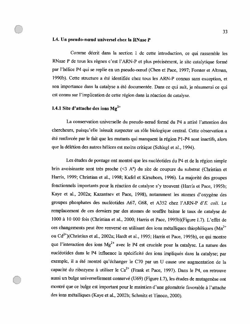
figure 1.7 : Région d’interaction des ions métalliques avec la région du P1-P4
(A) Modèle de structure tertiaire de la région P1-P4 (Chen et al., 199$;
Massire et al., 199$). Les nucléotides U69, A67 et A66 sont en bleu
foncé et rouge respectivement. Les groupes fonctionnels sensibles au
changement des atomes d’oxygène en souffre sont en forme de
sphères jaunes.
(B) Structure secondaire de la jonction des hélices P1-P4. Ce modèle
montre l’empilement (stacking) des quatre hélices. Les nucléotides
conservés universellement sont en lettres majuscules. Les sites
sensibles aux changements en phosphothiorate sont marqués par des
flèches jaunes.
Cette figure a été tirée de (Kaye et al., 2002b).
o
o
‘n
Gø
o
•mr
II
LI
s..,,’,
û
i
11
11
11
1•••‘,Ø
c’,’,••
N.
II
Ir
—.
‘,•
•.,.
—>
II
I:1
II
II
•<
rL
)Z
Lê
s
D’
o
Q-

C 36
1.4.2 La réaction de catalyse
L’attache des ions métalliques au P4 et à la région avoisinante est la clé de la
réaction de catalyse, car ces ions vont catalyser la réaction de clivage pour produire un
ARNt mature avec un groupe phosphate en 5’, et un groupe hydroxyle en 3’ de la région
guide en 5’ des ARNt (Guerrier-Takada et al., 1986). Le cation divalent le plus efficace
pour accomplir cette réaction est le magnésium suivi du manganèse, et du calcium (Smith
et al., 1992). Ces cations ionisent une molécule d’eau pour produire un ion hydroxyle qui
s’attaque au lien phosphodiester du substrat par un mécanisme de substitution
nucléophile concertée (SN2) (Cassano et al., 2004; Smith et Pace, 1993) (Figure 1.8). Les
autres rôles de ces cations sont d’induire la catalyse en interagissant simultanément avec
le phosphomonoester et le nucléophile durant l’état de transition, en stabilisant la charge
négative sur le groupe partant (Cassano et al., 2004), et en orientant le groupe hydroxyle
d’une manière à faciliter la réaction chimique (Chen et al., 1997) (Figure 1.8).
La conservation universelle du P4, l’observation que l’ARN-P soit nécessaire
pour l’activité de la RNase P chez les eucaryotes et les archéobactéries (Cherayil et al.,
1987; Franklin et al., 1995; Lee et al., 1991; Waugh et Pace, 1990), et le fait que le
magnésium est requis pour l’activité catalytique de la RNase P dans tous les règnes
suggèrent que cette région assure le même rôle chez tous les ARN-P, et que fort
probablement le mécanisme de catalyse par ces derniers ressemble à ce qui a été présenté
plus haut (Figure 1.8).
o

0 37
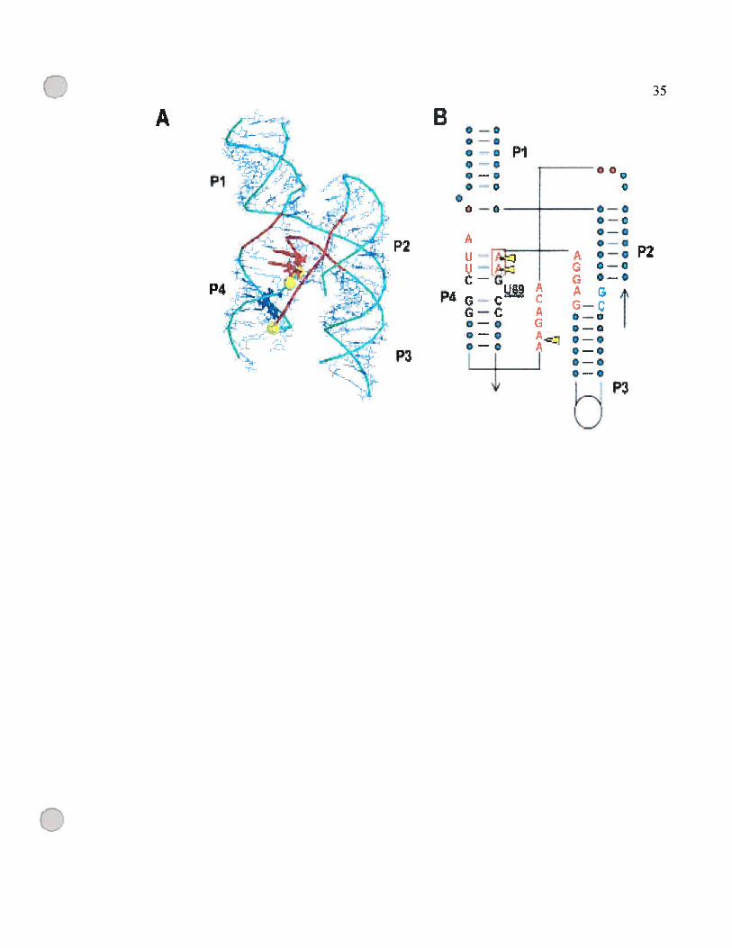
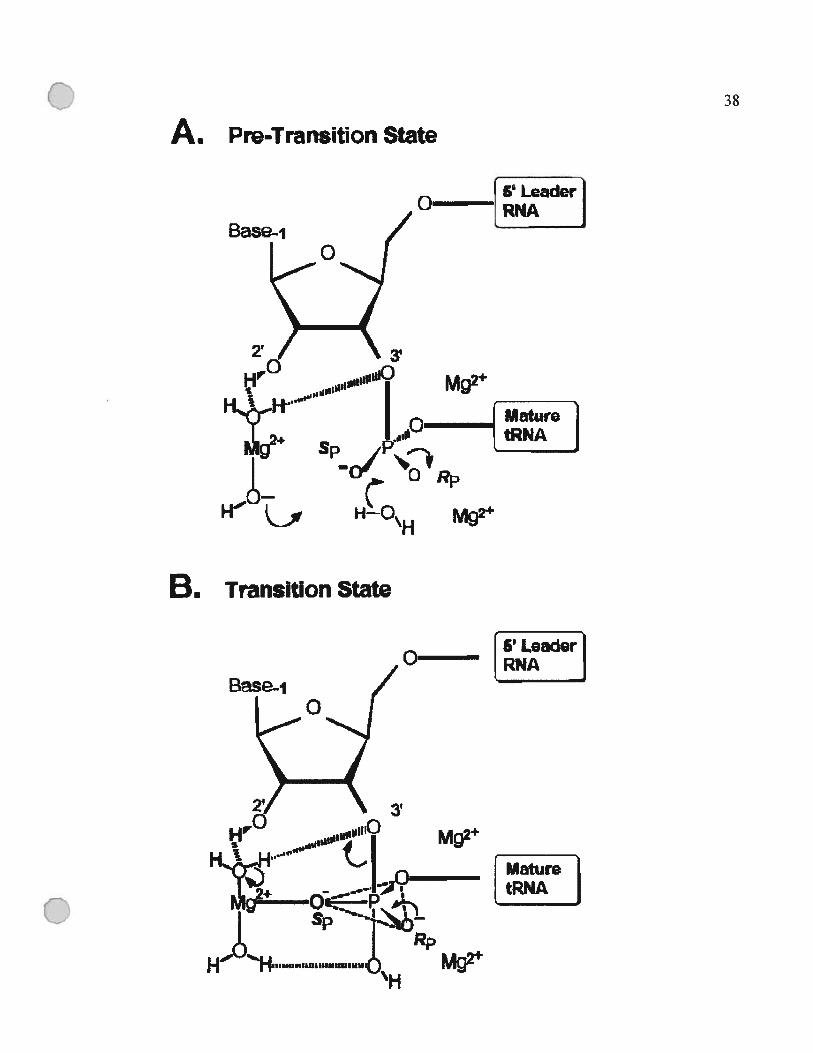
figure 1.8 : Mécanisme de la catalyse par la RNase P basé sur les modèles proposés dans
(Guerrier-Takada et al., 1986) et (Smith et Pace, 1993).
(A) État de pré-transition. Le groupe [Mg(OH)J, fait une coordination indirecte avec
le 2’ 0H en amont du site de coupure, et ionise une molécule d’eau.
(B) État de transition. L’oxygène pro-$p est en coordination avec l’hydrate de
magnésium. Deux ions Mg2 stabilisent l’oxygène pro-Rp et 03’ pendant le
clivage du lien 03’-P.
Cette figure a été tirée de (Chen et al., 1997).
O
38
A. Pre’Transition State
B.
5 Leadero LR
Q
MaturetRNA
oBase-i
2 3
Mg2
Rp
Mg2
Transition State
Base-j
______
z’ Leaderjo A]
3!
Mg
Mg
C 39
1.5. La RNase P mitochondriale
Le pseudo-nœud formé du P4 est reconnu comme une structure de base de
l’ARN-P, et dans certains ARN-P mitochondriaux (ARN-Pmt) c’est le seul motif
conservé (voir plus bas). Les ARN-Pmt sont parmi les ARN-P les plus divergents. Ils
sont très hétérogènes en taille et en structure secondaire, ce qui rend l’étude de leur
évolution d’un intérêt particulier. Dans la section qui suit, je résumerai ce qui est connu
concernant l’origine de la RNase P mitochondriale, sa composition, la distribution du
gène qui encode la sous-unité ARN, et l’impact de la génomique sur ce domaine.
1.5.1 Origine de la mitochondrie
Au début des années 70, Margulis a fait renaître une hypothèse qui existait depuis
plus d’un siècle (voir les références dans (Lang et al., 1999)), et qui propose que la
mitochondrie soit le résultat d’une endosymbiose entre une cellule eucaryote, et une
eubactérie (Margulis, 1970). Les facteurs qui ont amorcé cette endosymbiose (échange
de métabolites,. . .)(Martin et Mifiler, 199$), la nature de la cellule hôte (eucaryote
primiti± ou fusion d’une eubactérie avec une archéobactérie)(Moreira et Lopez-Garcia,
199$), et les conditions dans lesquelles cet événement a eu lieu sont encore débattus
(Gray et al., 1999), mais le support pour l’origine u-protéobactérienne de la
mitochondrie ne fait que grandir au fur et à mesure que les séquences des génomes
mitochondriaux et eubactériens deviennent disponibles (Gray et Spencer, 1996; Gray,
1989). Cette conclusion est basée sur l’analyse des gènes codés dans les génomes
mitochondriaux (Gray et al., 1999). Il y a même eu des tentatives pour chercher le sous-
groupe à l’intérieur des a-protéobactéries auquel s’apparente l’ancêtre de la
mitochondrie. Ces tentatives montrent que ce dernier peut être aussi proche du groupe
des Rickettsia que celui des Rhizobium ou des RhodosporiÏlum (figure 1.9), cependant le
nombre de génomes Œ-protéobactériens et mitochondriaux complètement séquencés
présentement ne permet pas de clôre cette question (Lang et al., 1999).
C

C’ 40
Dans ce contexte, l’origine de la RNase P mitochondriale devrait être la même
que celle de la mitochondrie, donc eubactériemie; à moins que les composantes de
l’activité de la RNase P mitochondriale proviennent du noyau de la cellule hôte.
C
0 41
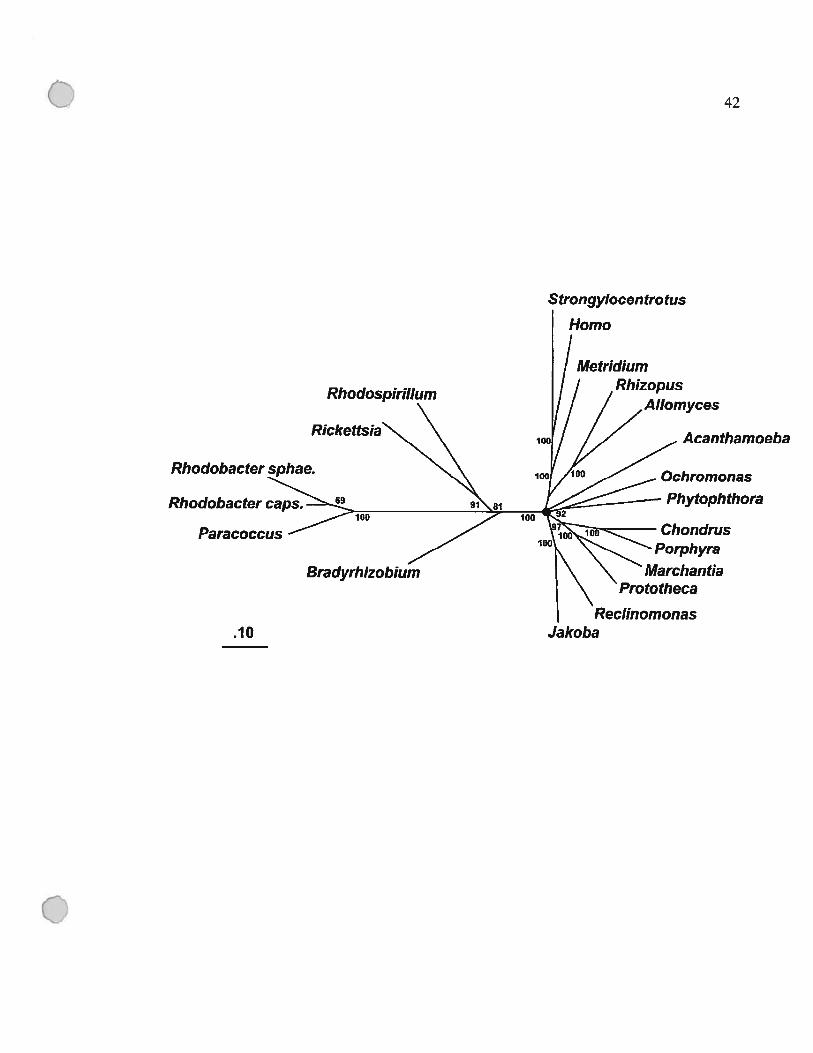
Figure 1.9 : Phylogénie des mitochondries et des Œ-protéobactéries
L’analyse a été fait avec les séquences protéiques concaténées des gènes suivants:
apocytochrome b (Cob), et cytochrome oxidase, sous-unités 1 à 3 (Coxl-3). Les taxons
inclus représentent les groupes phylogénétiques majeurs des eucaryotes et toutes les
séquences disponibles des a-protéobactéries. Les analyses phylogénétiques ont été faites
en utilisant PROTDIST/fITCH, qui permet une correction JinfNei pour les sites avec des
taux de changements variables. Le coefficient de variation utilisé est 0.5. Le cercle noir
indique des branches courtes fusionnées qui ne montrent pas de résolution (support par
boot strap 60%). L’échelle montre la moyenne de nombre de substitutions par site. La
topologie présentée est supportée par une analyse de maximum de vraisemblance. Les
organismes et séquences (numéro d’accession dans GenBank entre parenthèses) sont
$trongyÏocenfrotus purpuratus (oursin de mer; X12631), Paracoccus denitrficans
(X05$29, M17522, X05934, X05$28), R. prowazekii (AJ235270 to AJ235273),
Rhodobacter sphaeroides [X56157, X62645, M576$0, C45 164 (proteine)J,
RhodospirilÏum rubrum (X55 387), Rhodobacter capsulatus (X05630), Bradyrhizobium
japonicum (J03 176, X6$547); le reste des séquences sont disponibles dans (Lang et al.,
1999). -
Cette figure a été tirée de (Lang et al., 1999).
o
o
o
42
Strongylocentrotus
Homo
Metridium
Rhodospïrillum
Rickettsïa
Paracoccus
Acanthamoeba
Bradyrhizobïum
Phytophthora
ChondrusPorphyra
Marchantia
.10 JakobaReclinomanas
C1.5.2 La RNase P mitochondriale chez les champignons
La première RNase P mitochondriale qui a été identifiée est celle de 8. cerevisiae
(Martin et al., 1983; Underbrink-Lyon et al., 1983). Dans ce cas, l’ARN-Pmt est codée
dans le génome mitochondrial. Sa séquence primaire et sa structure secondaire ne
ressemblent pas à celles de l’ARN-P nucléaire, et l’activité mitochondriale est
indépendante de celle du noyau, ce qui laisse croire que l’origine de l’ARN-Pmt chez
cette levure est fort probablement eubactérienne et non pas nucléaire. La composante
protéique est codée par le génome nucléaire et n’est similaire à aucune protéine-P
eubactérienne ou nucléaire connue; elle est dix fois plus grande que la protéine-P d’E.coli
(Dang et Martin, 1993; Morales et al., 1992). Les études comparatives de séquences ont
permis d’identifier plusieurs nouveaux gènes rnpB codés dans les mitochondries de
levures phylogénétiquement voisines de S. cerevisiae (voir article 1). L’activité de la
RNase P mitochondriale a été étudiée aussi chez l’ascomycète filamenteux AspergiÏlus
nidulans, où une ARN-Pmt a été identifiée et purifiée (Lee et al., 1996b). Quant à la
composante protéique, elle reste toujours inconnue mais sept protéines se retrouvent
purifiées avec l’activité catalytique. Alors une question se pose, est-ce qu’il s’agit de sept
sous-unités protéiques ? Est-ce que certaines de ces protéines sont simplement des
impuretés ? Pour l’instant aucune réponse n’a été donnée.
Contrairement à ce qu’on observe chez les eubactéries, les ARN-Pmt de S.
cerevisiae et d’Aspergillus nidulans ne sont pas catalytiques in vitro, mais ils sont tous
les deux essentiels pour l’activité catalytique de la RNase P mitochondriale (Lee et al.,
1996e; Underbrink-Lyon et al., 1983). Les ARN-Pmt chez les champignons se
caractérisent par des séquences riches en A et U, et une taille qui varie entre 140 nts chez
$accharornycopsis fibuligera et 423 nts chez S. cerevisiae (Stribinskis et al., 1996), ce
qui rend leur repliement en structure secondaire par comparaison phylogénétique une
tâche très ardue. Les modèles de structure secondaire qui existaient pour ces ARN avant
le début de mon projet se limitaient aux Pi et P4 (f igure 1.10).
C
0 44
figure 1.10: Modèle de structure secondaire des ARN-P mitochondriaux de S.cerevisiae, S. fibuligera, Candida glabrata, et A. nidulans.
Ces modèles ont été établis par comparaison. Cette figure a été tirée de (Lee et al.,1 996c).
Q

45
Aspergitius nidutans
(li)— A
5’ P’U AAAUAUAAAGAUAUAA c-
AUAUAUUUCUAUAUU G,GAU
I A A (157)A P4 AAUUUCGGCC
L— 111111
AGGAAAGCCGACACu
$accharomyces cerevisiae
(28)— ui
AUAUUAAUU UAA C111111 III II A
3,AC u AUUA 0A (347)A AA
I UUCGUAUI II I I I ILAGGAAAG CAUAAAU
u
Saccharomycopsis fibuligera
PiAUUAUUUAUUUGUU A
AAAI liA A ($1)
3’ UUA AAI UUCGUAUUL— I I I I I I I
AGGAAAG C A UAAAAu
Candida glabrata
5’ P1 AAUA A
A UAUAUAUUAUAU CA
U AUAUAUGAUAUA U3’G U DA A (16e
A ‘ AUUCGAAUUII II III
L AGAAAAGCAUAAAA
o
Lee,Y.C., Lee,B.J. et Kang,R.S. (1996) Eur. J. Biochem. Vol. 235:297-303.
C 46
1.5.3 L’impact de la génomique
Dans les dernières années, les techniques de séquençage et d’analyse de
séquences ont évolué d’une manière révolutionnaire et ont permis d’obtenir la séquence
complète d’un grand nombre de génomes mitochondriaux. Dans le domaine de la
génomique mitochondrial, l’impact de cette percée technologique s’est traduit par la
découverte d’un génome mitochondrial conservé chez le jakobide Reclinomonas
americana ressemblant à un génome eubactérien en miniature (Lang et al., 1997). Ce
génome contient le plus grand nombre de gènes codés par une mitochondrie, dont
plusieurs n’ont jamais été retrouvés chez une mitochondrie précédemment comme les
gènes encodant les sous-unités d’une ARN-polymérase eubactérienne (rpoA-D),
plusieurs gènes de protéines ribosomiques (rpl], 10, 18, 19, 27, 3], 32), de protéines
impliquées dans le transport membranaire (secY, yejW), facteur d’élongation (tufA) et de
composantes de la chaîne respiratoire (atp3, coxi]). En plus, il encode une ARN-Pmt qui
contient tous les éléments structuraux présents dans le modèle du consensus minimal de
la structure secondaire des ARN-P eubactériens (voir Figure 2A de l’article 1). Cette
observation indique que l’ARN-Pmt chez les jakobides a été hérité de l’endosymbiote
eubactérien. Cette trouvaille a été suivie par l’identification d’un ARN-Pmt codé par le
génome mitochondrial de l’algue verte NephroseÏmis olivacea, qui lui aussi ressemble
aux ARN-P eubactériens (Turmel et al., 1999).
Cette percée technologique a aussi dévoilé que la plupart des génomes
mitochondriaux séquencés n’encodent pas de gène rnpB (Figure 1.11). En fait, seuls les
jakobides et l’algue verte N olivacea encodent un, à l’extérieur du règne des
champignons. Et même à l’intérieur de ce règne, la distribution de ce gène est très
sporadique, aucun basidiomycète ou chytridiomycète ne l’encode. Dans les génomes
mitochondriaux complètement sequencés des ascomycètes, seul A. nidulans et quelques
levures encodent un gène rnpB alors qu’il est absent chez la majorité.
C
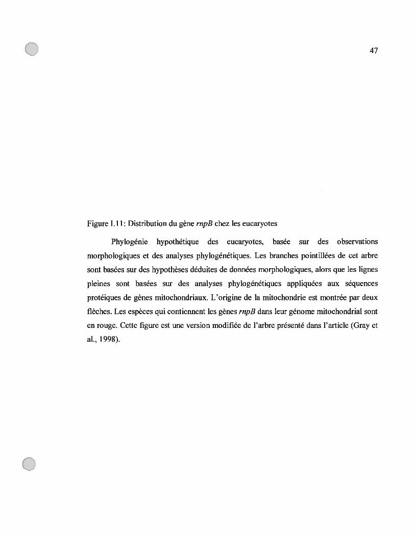
0 47
Figure 1.11: Distribution du gène rnpB chez les eucaryotes
Phylogénie hypothétique des eucaryotes, basée sur des observations
morphologiques et des analyses phylogénétiques. Les branches pointillées de cet arbre
sont basées sur des hypothèses déduites de données morphologiques, alors que les lignes
pleines sont basées sur des analyses phylogénétiques appliquées aux séquences
protéiques de gènes mitochondriaux. L’origine de la mitochondrie est montrée par deux
flèches. Les espèces qui contiennent les gènes rnpB dans leur génome mitochondrial sont
en rouge. Cette figure est une version modifiée de l’arbre présenté dans l’article (Gray et
al., 199$).
o

48
9.
Iretortamona]“/77’/7Spizeilomyces
Physarum* RiuzopusDidymium * SchizophyllumCafetena ‘Scbizosaceharomyces\ .‘. SacciraromyCes
\ \““ Ochromonas
\ \ . Ch,ysodutymu$Pawiova \ \ j.., PelagomoflaSIsochrysis \ \ Developayella
\ PhytophihoraThraustochytriUm
Leishmania Trypanosoma/1
Bd Eugtena* ,c. reinhardtiiRickettsw-bke / ,/ 7 ../ C. eugametoSbacteria ‘ / z Cyanophora‘ ,‘
PedinomonasI Cruzella ./ .7’ RlwdomonaS / Chondrus /7’//7
Goniomonas / / // Porphyra
ZEZLridium
JabaRechnomoflas .. //Hartmanneilâ’///Acanthamoeba /Polysphondylium
DictyostdiUm
BkphaaÏParamecium
TetrabymenaGonyautax
Plasmodium
o
49
1.6. Définition du projet
Mon projet a débuté afm de mieux comprendre l’évolution de l’ARN-Pmt. Mes
buts étaient (j) de trouver de nouveaux gènes rnp3 dans les génomes mitochondriaux
déjà séquencés en se basant sur les régions consensus identifiées chez l’ARN-Pmt de R.
americana, (ii) établir des modèles de structure secondaire pour tous les ARN-Pmt par
comparaison phylogénétique d’une façon systématique afm de mieux comprendre
l’évolution de cette structure, (iii) tester l’activité catalytique in vitro de l’ARN-Pmt de
R. americana et (iv) de faire des liens entre la structure secondaire et l’activité
catalytique de ces ARN.
Dans le chapitre suivant, je présenterai mes résultats concernant la distribution du
gène encodant la sous-unité ARN de la RNase P mitochondriale (rnpB)(article I),
l’évolution de la structure secondaire de la composante ARN chez les champignons
(articles 1 et 2), et les jakobides (article 3), ainsi que l’effet de cette évolution sur
l’activité catalytique des ARN-P (article 3).
o

o
Chapitre II : Articles
C

o
51
Article 1
Mitochondrial RNase P RNAs in ascomycete fungi: lineage
specific variations in RNA secondary structure.
Elias R. Seif, Lise Forget, Nancy C. Martin’ and B. Franz Lang*
Program in Evolutionary Biology, Canadian Institute for Advanced Research,
Département de Biochimie, Université de Montréal, 2900 Boul. Edouard-Montpetit,
Montréal, Québec, F131 1J4, Canada.
University of Louisville, Department of Biochemistry and Molecular Biology, School
ofMedicine, University ofLouisville, Louisville, KY 40292-0001, USA.
RNA, 2003, vol.9, no. 9,pp. 1073-1083.
Contribution of each author:
Elias Seif: did most of the experimental work, resuits analysis, and preparation of the
manuscript and figures.
Lise Forget : helped in the experimental work.
Nancy C. Martin: participated in manuscript preparation, and supplied experimental
data.
B. franz Lang : participated in manuscript and figure preparation.
Mitochondrial RNase P RNAs in ascomycete fungi:IJneage-specific variations in RNA secondary structure
[LIAS R. SElF,1 LISE FORG[T,1 NANCY C. MARTIN,2 and B. FRANZ LANG1Program in Evolutionary Biology, Canadian Institute for Advanced Research, Département de Biochimie, Université de Montréal,
Montréal, Québec H3T 1J4, Canada2University of Louisville, Department of Biochemistry and Molecular Biology, School of Medicine, University of Louisville,Louisville, Kentucky 40292-0001, USA
ABSTRACT
The RNA subunit of mitochondriai RNase P (mtP-RNA) is encoded by a mitochondrial gene (rnp8) in several ascomycete fungiand in the protists Recinomonas americana and Nephroselmis olivacea. By searching for universally conserved structuralelements, we have identified previously unknown rnpB genes in the mitochondrial DNAs (mtDNAs) of two fission yeasts,Schizosaccharomyces pombe and Schizosaccharomyces odosporus; in the budding yeast Pichia canadensis; and in the archiascomycete Taphrina deformans. The expression of mtP-RNAs of the predicted sue was experimentaily confirmed in the twofission yeasts, and their precise 5’ and 3’ ends were determined by sequencing of cDNAs generated from circularized mtP-RNAs.Comparative RNA secondary structure modeling shows that in contrast to mtP-RNAs of the hvo protists R. americana and N.olivacea, those of ascomycete fungi ail have highiy reduced secondary structures. In certain budding yeasts, such as Saccharomycopsis fibuligera, we find only the two most conserved pairings, Pi and P4. A Pi8 pairing is conserved in Saccharomycescerevisiae and its close relatives, whereas nearly haif of the minimum baderial consensus structure is retained in the RNAs offission yeasts, Aspergiflus nidulans and Taphrina deformans. The evolutionary implications of the redudion of mtP-RNAstructures in ascomycetes will be discussed.
Keywords: [volution; phylogenetic modeling; RNA processing; organelle; rnp8; yeasts
C
INTRODUCTION
RNase P is a ribonucleoprotein (for exceptions, sec below)that is universally prcscnt in eubacteria, archaebacteria, andeukaryotes, as wefl as m mitochondria and cffloroplasts. Itparlicipates in the processing of tRNAs, 4.5 S RNAs, andother small RNAs, by endonucleolytic removal of 5’ leadersequences from RNA precursors (Peck-Miller and Mtman1991).
The RNA subunits ofeubacterial RNase P (P-RNA) havebeen intensely studied and are currently best understood.Escherichia cou P-RNA is essential for the enzymatic activityin vivo and is die only subunit necessary for activity in vitro(Stark et al. 1978; Kole and Mtman 1981; Guerrier-Takadaet al. 1983). Comparisons of ail known P-RNAs have reveaied significant primary and secondai-y structure similarilies, indicating that these molecules evolved from a com
mon ancestral RNA. The in vitro acfivity of P-RNAs in ifieabsence of a pro tein component bas been demonstrated innumerous instances (Guerricr-Takada et al. 1983; Gardineret al. 1985; Wagner et al. 2001). Nevertheless, die singleeubacterial protein component of RNase P (P-protein) isessentiat for the enzymatic activity in vivo. It has been sugges[ed [bat die protein partidpates in formation of an active-site architecture, hy interacting with the 5’ leader oftRNAs (Crary et al. 1998), and that it increases the catalyticactivity of RNase P by acting as an electrostatic shield between die negatively charged P-RNA and tRNAs (GuerrierTakada et al. 1983; Gardiner et al. 1985; Christian et al.2002). The P-protein a]so broadens the substrate specifidtyof RNase P, by enhancing its affimity for non-tRNA substrates. for example, 4.5 S RNA and C4 RNA precarsors areprocessed more eflïdently by a P-RNA/P-protein complexthan by P-RNA alone (Peck-Miiler and Mtman 1991; Hartmann et al. 1995).
The P-RNA secondary structure model of Archaebacteriais strikingly similar to that ofeubacteria, except for the lackof the P13, P14, and Pis helices (Brown and I-Iaas 1995;Brown 1999). Archaeal P-RNAs were long thought to be
Reprint requests to: B. Priez Lang, Département de Biochimie, Université de Montréa], 2900 Boul. Edouard-Montpetit, Montréal, Québec,H3T 1J4. Canada; fax: (514) 343-2210.
Article and publication are at http://www.majournal.orgfcgi/doi/10. t26lIrna.5880403.
RNA (2003), 9:1073—1083. Published by Cold Spnng Harbor Laboratory Press. Copyright © 2003 RNA Society. 7073
Seif et aL
inactive without their protein partner (sec Brown and Haas1995). However, ifie P-RNAs ofMethanobacteria, Thermococci, and Halobacteria do have catalytic activity under cxtreme ionic conditions (Pannuca et al. 1999). In fact, detetion mutants show that the structural elements missing inarchaeal P-RNAs are also dispensable for in vitro catalysisby the E. cou RNA molecule (Dan et al. 1992; Haas et al.1994). Alffiough archaebactenal P-RNA structures resemblethose ofeubacteria, the archaeal holoenzyme is mucli Iarger.It has at Ieast four protein stibunits that appear to be homologs ofthe eukaryotic P-proteins (Hall and Brown 2002).
Eukaryotic nuclear P-RNAs have been investigated in de-tait in ascomycete fungi aiid in animais. They have no catalytic activity in vitro and, as in archaebacteria, consist of anRNA subunit md several proteins, at least fine in yeast and10 in human (Gopalan et al. 2002; Guemer-Takada et al.2002). A secondary structure model of the eukaryotic PRNA conforms convincingly to the bactenal consensusstructure with only minor deviations (Chen and Pace 1997;Prank et aI. 2000). In plants, RNase P has been purified, anda P-RNA component lias been suggested based on the sensitivity of the carrot RNase P activity to micrococcal nuclease treatment (Franklin et al. 1995). Although the nuclearRNase P from wheat is resistant to nuclease treaflnent, itsdensity and its low isoelectric point also indicate the presence ofa P-RNA subunit (Arends and Schôn 1997). In fact,RNase P from rat liver (Jayantlii md Van Tuyle 1992) andthe archaebacteritim Siilfolobus solfata ricus (Darr et al.1990) resist micrococcal nuclease treatment but do containa P-RNA subunit (Aliman et al. 1993; Harris et al. 2001).This indicates that the RNA suhunit is protected from digestion by P-proteins.
Mitochondria and cMoroplasts contain distinct, organelle-specific RNase P activities. The analysis of organdllarP-RNAs lias been complicated by the patchy occurrence ofthe rnpB gene, both in chloroplast and mitochondrial DNAs(mtDNAs). The chloroplast-encoded P-RNA of Cyanophoraparadoxa folds into a Cyanobacteria-like secondary structure (in agreement with ifie cyanobacterial origin of chloroplasts) and is essenfial for RNase P activity (Baum et al.1996; Pascual md Vioque 1999). Chloroplast DNA (cpDNA)encoded rnpB genes have been found only in the green algaNepîtroselmis olivacea (Turmel et aI. 1999), ifie red algaePorpizyra purpurea (Reith md Munholland 1995), andCvanidiun-, culdarittin. We identffled the latter, previouslyunrecognized rnpB gene, at positions 99,959—100,305 ofGenBank record AP022186, (Glôckner et al. 2000). Ail otherknown cpDNAs do not seem to encode a P-RNA. A recentprediction of a maize chloroplast rnpB gene (Colins et al.2000) is based on rather weak secondary structure similaritics to known homologs, does flot consider otherwise highlyconserved pnmary sequence motifs, and lias flot been confirmed experimentally. tnferences that do flot take primarysequence conservation into account are compromised because A+T-rich ch]oroplast sequences cm fit almost any
given consensus structure and are, therefore, of little predictive value. Moreover, several biochemical studies indicatethat chloroplast RNase P (cpRNase P) may flot contain anorganelle coded P-RNA. for instance, spinach cpllNase P activity resists micrococcal nuclease treatment md bas physicatproperties consistent with the presence of a protein-only enzyrne. These characteristics indicate that spinach cpRNase P isindeed a protein-only enzyme (Thomas et al. 2000).
Mitochondrial RNase P activities have been studied invarious yeasts (sec below), the ascomycete fungus Aspergiubts nidulans (Lee et al. 1996a), human (Doersen et al. 1985;Rossmanith and Karwan 1998; Puranam and Attardi 2001;Rossmaniffi md Potuschak 2001), Trypanosorna brucei (Salavati et al. 2001), potato (Marchfelder and Brennicke1994), wheat (Hanic-Joyce and Gray 1990), and canot(Franklin et al. 1995). The most detailed information on thebiochemical and genetic properties ofmitochondrial RNaseP (mtP-RNA) is available for Saccharoinyces cerevisiae. ItsRNA subunit was first idenfified as a mitocliondrially encoded molecule hy anatyzing yeast mitochondrial mutantsdeficient in mitochondrial tRNA processing and proteinsynthesis (Millet and Martin 1983; Underbrink-Lyon et al.1983). The unusually large protein subunit bas bcen shownto lie nucleus-encoded (Morales et al. 1992; Dang and Martin 1993).
Addiflonal rupB genes have been identifled by sequencesimilarity in mtDNAs of numerous budding yeasts (T. guabrata, Clark-Wa&er et al. 1985; Saccharoinycopsis fibuuigera,Wise and Martin 1991a; Kinyverornyces lactis, Wilson et al.1989; Saccharornyces exiguus, Wise and Mai-tin 1991b; SaccÏzczrornyces dougtasii; Ragnini et al. 1991; Saccharornyceschevalieri, Sacctzarornyces ellipsoideozis, and Saccharornycesdiastatictis, Sbisa et al. 1996; and Saccizarornyces castellii,Petersen et al. 2002), inc protist Redinornonas arnericana(Lang et al. 1997), md the prasinophyte green alga N. ouivacea (Turmel et al. 1999), but not in inc mtDNAs of plants,animais, a great number ofprotists, or nonascomycete ftmgi(Lang et aI. 1999). From an evolutionary standpoint, it ispuzzling that the occurrence of mitochondrially encodedrnpB genes is so patchy. Have these genes been lost from themtDNAs of plants, animais, most fungi, md protists, or dowe fail to identify them because they are so extremely derived? In fact, higffly derived, extremely A+U-rich mtPRNAs are charactenstic in yeast. Consequently, predictionsofyeast mtP-RNA secondary structures are difficuh, despitethe availability of comparative data. In addition, inc drasticsize variations of these RNA molecules obscure inc identification of RNA secondary structure elements (Wise andMai-tin 1991a). For example, inc respective lengths of predicted mtP-RNAs are 423 nt for S. cerevisiae (Stribinskis etal. 1996), 227 nt for Tortdopsis guabrata (Shu et al. 1991),and as short as 140 nt for Sacclzarornycopsisfibztuigera (Wiseand Martin 1991a).
The mitochondnally encoded mtP-RNA of inc protistRecuinornonas arnericaiza was inc first identffied to contain
C
1074 RNA, Vol. 9, No. 9
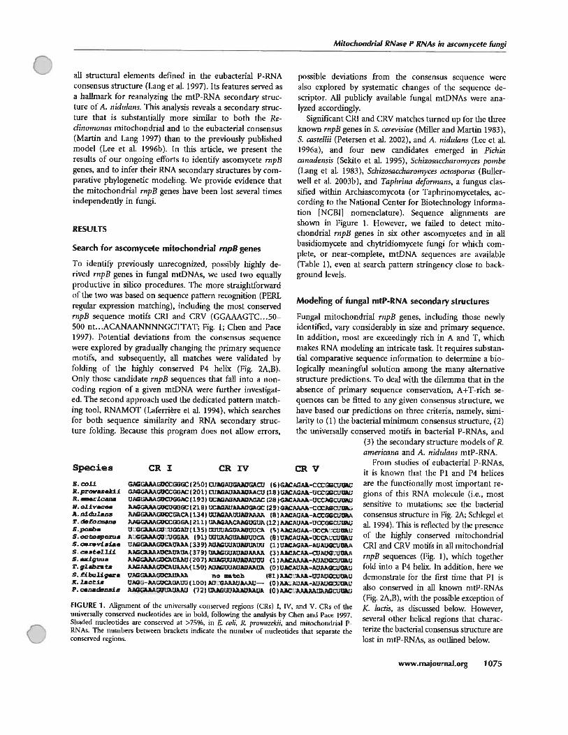
Mitochondriai RNase P RNAs in ascomycete fungi
ail structural elements defmed in die eubacterial P-RNAconsenstts structure (Lang et aI. 1997). Its features served asa hallmark for reanalyzing the mtP-RNA secondary struc[tire of A. nidulans. Ibis analysis reveals a secondary structure [bat is substantially more similar to both the Reclin o,nonas mitochondrial and to the eubacterial consensus(Martin and Lang 1997) [han to die previously publishedmode! (Lee et al. 1996b). In this article, we present dieresuits of our ongoing efforts to identil’ ascomycete rnpBgenes, and to infer their RNA secondary structures by comparative phylogenetic modeling. We provide evidence thatdie mitochondrial rnpB genes have been lost several timesindependently in fungi.
RESULTS
Search for ascomycete mitochondrial rnpfl genes
b identify previously tmrecognized, possïbly highly derived rnpB genes in fungal mtDNAs, we used two equallyproductive in silico procedures. The more straightforwardof die two was based on sequence pallem recognition (PERLregular expression matching), including die most conservedrnpB sequence motifs CRI and CRV (GGAAAGTC.. .50—500 nt. . .ACANAANNNNGCTTAT; Fig. 1; Chen and Pace1997). Potential deviations from the consensus sequencewere explored by graduaily changing die primary sequencemotifs, and subsequently, ail matches were validated byfolding of the highly conserved P4 helix (Fig. 2A,B).Only diose candidate rnpB sequences that fail into a noncoding region of a given mtDNA were further investigateU. The second approach used the dedicated pattern matching tool, RNAMOT (Laferrière et al. 1994), which searchesfor bodi sequence similarity aid RNA secondary structure folding. Because this program does not allow errors,
possible deviations from die consensus seqttence werealso explored by systematic changes of the sequence descriptor. Ail publicly available fungal mtDNAs were anaIyzed accordingly.
Significant CRI and CRV matches turned tip for die threcknown rnpB genes in S. cerevisiae (Miller and Martin 1983),S. castellii (Petersen et al. 2002), and A. nidulans (Lee et al.1996a), and four new candidates emerged in Pidilaainadensis (Sekito et al. 1995), Schizosaccharotnyces poînbe(Lang et aI. 1983), Schizosaccharornyces octosporus (Bullerwell et al. 2003b), and Taphrina deforinans, a fungus classified witbin Archiascomycota (or Taphrinomycetales, according to die National Center for Bioteclmology Information [NCBIJ nomenclature). Sequence alignments areshown in Figure 1. However, we failed to detect mitochondrial rnpB genes in six other ascomycetes and in ailbasidiomycete and chytridiomycete ftmgi for which complete, or near-complete, mtDNA sequences are available(Table 1), even at search pattem stringency close to hackground levels.
Modeling of fungal mtP-RNA secondary structures
Fungaf mitochondrial rnpB genes, including [hose newlyidentffled, vary considerably in size and primaiy sequence.In addition, most are exceedingly rich in A and T, whichmakes RNA modeling an intricate task. It requires substantial comparative sequence information to determine a biologicaily meaningful solution among the many alternativestructure predictions. To deal widi tise dilemma diat in theabsence of primai-y sequence conservation, A-i-T-rich sequences can be fitted to any given consensus structure, wehave based oui- predictions on [hi-ce criteria, namely, similarity to (1) die bacterial minimum consensus structure, (2)the universally conserved motifs in bactenal P-RNAs, and
(3) tIse secondary structure models of R.aineriainci and A. nidultnzs mtP-RNA.
From studies of eubacterial P-RNAs,it is known that die Pi and P4 heicesare the functionally most important regions of this RNA molecitle (i.e., rnostsensitive to mutations; sec die bacteriaiconsensus structure in Fig. 2A; Scinegel etal. 1994). fhis is reflected by the presenceof Ilie bigffly conserved mitochondrialCRI and CRV motifs in ail mitochondrialrnpB sequences (Fig. 1), winch togetherfold into a P4 helix. In addition, here wedemonstrate for tise first lime that Plisalso conserved in ail known mtP-RNAs(Fig. 2A,B), with die possible exception ofK. lactis, as discussed below. However,several other heical regions that characteHze tIse bacterial consensus structure arelost in mtP-RNAs, as outlined below.
Species CR I
B. couR. prowazakllR. am..rlcanaN allvc.aA. nldularmr. cieformaasSprnjb.S. octoaporu.S. c.xevisIaeS. 1iiiS. .xlguua‘. glibra taS. flbulig.ri. lactlgP. canacienaia
CRIV CRV
G&AUcCGGC t 2 50) Tî?At3AUGAU (6 tGA-Cœ:;t;GA.AAiCCGGAC (201) CtAGA9AàAACU (18 t A-ucCt
U.iAUCUGGC (193) (281 GAM.A-UCCA5GA.ksCUGGGC 1218) UCGAtAMGAGc (29)AACC0P.C t U 4 t UUAUUADAAAA (8)kUCCGG(211) AAACAUGUA (12 ) A1AUA-UGOtIsli (135) UciÂGRAUUCA (5)ÀAGt.A-tICC.:tE
AG3ikAçUUGGA (91) UUUA A.WUUCÀ (8)tÂUÀ-0CCRCUGi.AAUCAUkAA (339) AUUAt1?.Gi3auU (1) UAGAA-kUGUtP
AUCUAUA (379) U3.AGutjAUADAAA (3) WACUAUC?,À.kACÀUAAx3 (207) Ut1ÀUADAtaiU (1) AAAÂ-P1iAt3GC. W.AAG?V..GUCRU.AÀ( 15G) AUGUUAUAUAÀUA (0) UUA-AUAAGC
GCRt1.AA no matc) (81 )xCPAk-t3GCUR’U’-AALUCRUAUU( 100) U GAAA[ikAU— (Q )aA;3-AUA9G.T3A.AÂGAiJU1UA).1J (72) 0AGUUAÀAUAM)A t0)AA).AÀUAGcJt
FIGURE 1. Aligninent of the universally conserved regions (CRi) I, W, and V. CRs of theuniversally conserved nucteotides are in bold, fottowing die analysis hy Chen and Pace 1997.Shaded nucleotides are conserved aL >75%, in E. 0)11, R. prowazekii, and mitochondriat PRNAs. Ihe numbers between brackets indicate die number of nudeotides that separate theconserved regions.
www.rnajournal.org 1075
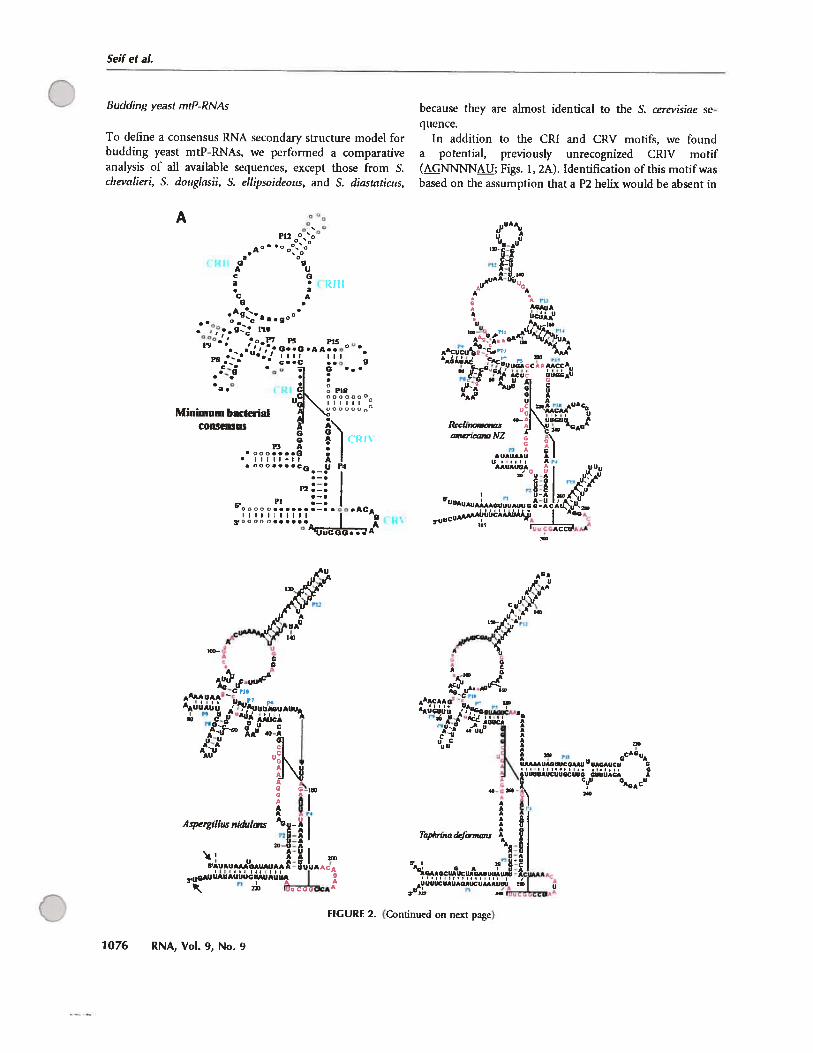
Seif et aL
Budding yeast mtP-RNAs
To define a consensus RNA secondary structure model forbudding yeast mtP-RNAs, we performed a comparativeanalysis of ail available sequences, except those from S.cbe’,’cilieri, S. dougtasii, S. ettipsoideous, and S. diastaticus,
Pl200’
)• r.‘°o’’ •,P7 p pi
• •‘‘7eG..o.AA..o° e•• liii IIIP8. • c..c •.
‘
:•a.°
• A• O00 •••C4 U 74
P2 e —.
I—.PI ._.
6’OOOO ACAIIIIIlIIII I o
y00000...... I A° ‘UuCGG...A
because they are almost identical to flic S. cerevisiae sequence.
In addition to the CRI and CRV motifs, we foimda potential, previously unrecognized CRIV motif(NNNN; figs. 1, 2A). Identification ofthis motif wasbased on the assumption that a P2 helix would be absent in
AUA%
UUAU
u-C-G
A-U U,*UAAUù
A A
AA
Minimum bacterialconsensus
1phrrna dejonnans
FIGURE 2. (Continued on next page)
7076 RNA, Vol. 9, No. 9
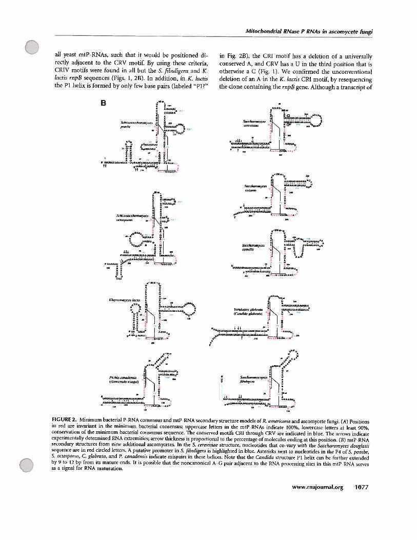
Mitochondrial RNase P RNAs in ascomycote fungi
ail yeast mtP-RNAs, such that it would he positioned directly adjacent to the CRV motif By usmg these criteria,CRW motifs were found in ail but the S. fibuligera and K.lactis rnpB sequences (Figs. 1, 23). In addition, m K. lactisthe Pi helix is formed by only few base pairs (laheled “Pi?”
R
.
prm.hc : uu&AAAu V
n.,
r_t?
&kOCkT,flrC,
y
Iom,.,
I’ï,l,i,, “
(sI,,,,,,,a.,j ,)
in fig. 2B), the CRI motif lias a deletion of a universallyconserved A, and CRV lias a U in the third position that isotherwise a C (fig. 1). We confirmed the unconventionaldeletion of an A in [he K. lactis CRI motif, by resequencingthe clone containing the rnpB gene. Although a transcript of
I
“‘irrE)
U
:
A *LCflfl*
c,odh, —
ghhnaCnhiS,g&SrnI,.)
UI
“1u
L?
U &xuluum.nn-up.i.sO flhuIrgum
UAUUULLfl
L,.
FIGURE 2. Minimum bactenal P-RNA consenstis and mtP-RNA sccondary Structure models of R. arnericana aaid ascomycete fungi. (A) Positionsin red are invariant in tise minimum bacteriai consensus uppercase letters in tise mtP-RNM indicate 100%, lowercase letters at least 90%,conservation of the minimum bacterial consensus sequence. Tise conserved motifs CRI through CRV are indicated in bloc. Tise arrows indicateexperimcntatly dctcrmincd RNA extremities; arrow thickness is proportional to the pcrcentagc of molccules ending at this position. (B) mtP-RNAsecondai-y structures from rime additional ascomycetes. In the S. ccres’isiae structure, nucleotides that co-vary with tise Sacchnrornyces douglnsiisequence are in red circled letters. A putative promoter in S. fibidigera is highuighted in blue. Asterisks next to nucleotides in tise P4 of S. pombe,S. ocrosporus, C. glabrata, and P. canadensis indicate mispairs in these helices. Note that the Candida structure Pi helix cars be ftirthcr extended
O by 9 to 12 bp from its mature ends. It is possible that tise noncanonical A—G pair adjacent to tise RNA processing sites in this mtP-RNA servesas a signal for RNA maturation.
www.rnajournal.org 7077

Seiftat
TABLE 1. Occurrence and features of rnpB n cornp!ete fungal mtDNA sequences
mtDNA- l-’5,7,8,Organismal group encoded rnpBa P2 P3 P15 P13 9,10,12 Accession #
Ascomycota (Euascomycola)Aspergillus nidulans U O E 1 b
Hypocrea jecorina E NC_003388Neurospora crassa EPodospora anserina E NC_O01 320Veriicillium lecanli E AF487277
Ascomycota (Hemiascomycota)Candida albicans E NC_002653Candidagiabrata U O O E U E AJ511533Pichia canadensis U O O E U E NC_001762Saccharomvces castellii U O O E U E AF437291Saccharomyces cerevisiae U O O E U E NC_001 224Yarrowia lipolytica E NC_002659
Ascomycota (others)Schizosaccharomyces japon icus E NC_004332Schizosaccharomvces octosporus U U U U U E A[275271Schizosaccharomyces pornbe U U U U O NC_001 326Taphrina deformans U U U E U U AY262107’1
Basidiomycota” E
Chytridiomycotae E
FiIIed and empty squares stand for presence or absence of the gene or helices, respectively.mtP-RNAs contain Pi and P4.
bThe A. nidulans sequence s amost complete: ODAS1, CAA33481, AAA99207, AAA31737, CAA25707, AAA31736, CAA23994, X15442,P15956, CAA23995, CAA33116, X00790, X15441, X06960,J01387, and X01507.“Completed at Whitehead Institute, currently available al http //pages.sI u.edu/taculty/kennellj/genbank.html.‘the GenBank record documents the rnpB sequence, the complete sequence s unpublished.e,itechondriaI topO genes have flot been identiiied in basidiomycete (canthamilus ciba rius, ctnpubl.; Cryptococcus neotormans, NCOO_4336;Microbotryum violaceum, unpubl.; Schizophyllum commune, NC_003049) or chytridiomycete fungi (Allomyces macrogynus, NC 001715;Harpochytrium94, AY1 82005; Harpochytrium 105, AY1 82006; Hyaloraphklium curvatum, NC_003048).
tise expected size is fbund in K. lactis mitochondria (Wilsonet al. 1989), the high incidence of noncanonical featuresraises the question whether or flot rupB codes for a functional mtP-RNA.
Further comparative analysis revealed anoffier conservedbase pairing, P18 (fig. 2A), in tise five phylogeneticaflyclosely related organisms S. cerevisiae, S. casteltii, T. glabmta,S. exigu us, and K. lactis (Fig. 2B). These helical regions startat tise same position as P18 of R. americaita and as P18 inthe bacterial minimum consensus mode!, 2 nt upstrearn ofthe CRIV motif (Fig. lB). Additional support for this Pislieux cornes from a compensatory exchange of a base pair(G C to A—U) bettveen tise S. cerevisicie and S. dougÏasiimtP-RNAs (see tise circled nucleotides of the S. cerevisiaestructure in Fig. 2B). In P. cmtadensis, a P18 pairing ispossible (Iabeled “Pis?” in Fig. 2B); however, it is flot bcated at tise same position relative to tise CRIV motif. Finally, we did not detect a CRIV motif or aPiS pairing in tiseS. fibuligero mtP-RNA.
Fission yeasts
Tise S. pombe rnpB gene tvas only identified recently, aithough the complete sequence ofmtDNA lias been available
for more than a decade. Dilferences in both tise CRI andCRV motifs, relative 10 tIse consensus (Fig. 1), made itdifficuit to recognize ibis gene. After identifying potentialCRI, CRIV, and CRV motifs in an intergenic region of tiseS. pombe mtDNA, we contirmed the presence of an rnpBhomoiog in S. octosporus at tise same location in themtDNA, flanked by tise same tRNA genes, truA and trnG.
The comparative structttral modeiing of the two Schizosacchoromyces sequences reveals flot oniy tise presence of Piand P4, as in budding yeasts, but also P2 and P3 pafringsthat distinguish them from most other fungal RNA structures (Fig. 2B). Moreover, we identilied potential P15 andPis pairings. However, only tise S. pombe Pis is located althe same distance from CRIV as in other species, and tisepotential S. octosporus P18 helix would be only 3 bp long(Fig. 2B). The central sequence of rnpB contains three motifs that are identical in tise fission yeasts (4, 6, and 9 nt long;fig. 2B), but witliout similarity to the bacterial consensus.
T. deformct;ts bas been grouped together with ScÏuzosaccharomyces species in phylogenetic analyses (terrned archiascomyetes, Nisisida and Sugiyama 1993; for an alternativeview, see Bullerwell et al. 2003b; Leigh et al. 2003), but tisemtP-RNA secondary structure of T. deformans exhibits
107$ RNA, Vol. 9, No. 9
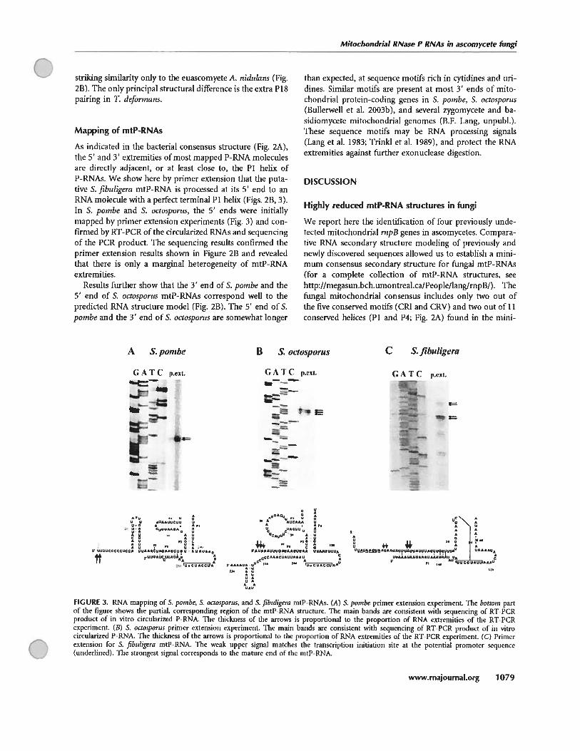
Mitochondrial RNase P RNAs in ascomycete fungi
striking similanty onlyto the euascomyete A. nidulans (Fig.2B). The only principal structural difference is the extra Pispairing in T. deformans.
Mapping of mtP-RNAs
As indicated in the bactenal consensus structure (Fig. 2A),the 5’ and 3’ extremities ofmost rnapped P-RNA moleculesare directly adjacent, or at least close to, the Pi helix of
P-RNAs. We show here by primer extension that the putative S. fibuligera mtP-RNA is processed at its 5’ end to anRNA moÏecule with a perfect terminal Pi helix (Figs. 2B, 3).In S. pwnbe and S. octosporus, the 5’ ends were initiallymapped by primer extension experiments (Fig. 3) and confirmed by RT-PCR of the circularized RNAs and sequendngof the PCR product. The sequencing resuits confirmed theprimer extension resuits shown in figure 2B and revealedthat there is only a marginal heterogeneity of mtP-RNAextremities.
Results further show that ifie 3’ end of S. poinbe and the5’ end of S. octosportis mtP-RNAs correspond well to thepredicted RNA structure model (Fig. 2B). The 5’ end of S.poinbe and the 3’ end of S. octosporus are somewhat longer
than expected, at sequence motifs rich in cytidines and undines. Similar motifs are present at most 3’ ends of mitochondrial protein-coding genes in S. pombe, S. octosporus(Bullerwell et al. 2003b), and several zygomycete and basidiomycete mitochondrial genomes (B.F. Lang, unpubl.).These sequence motifs may be RNA processing signais(Lsmg et al. 1983; TrinM et al. 1989), arid protect the RNAextremities against further exonuclease digestion.
DISCUSSION
Highly reduced mtP-RNA structures in fungi
We report here the identification of four previously undetected mitochondrial rnpB genes in ascomycetes. Comparative RNA secondary structure modeling of previously andnewly discovered sequences allowed us to establish a minimum consensus secondary structure for fungal mtP-RNAs(for a complete collection of mtP-RNA structures, sechttp://megasun.bch.umontreal.ca/People/lang/mpB/). Thefungal mitochondrial consensus includes only two out ofthe five conserved motifs (CRI and CRV) and two out of liconserved helices (Pi and P4; Fig. 2A) found in the mini-
A S. pombe B S. octosporus C £fibuligeru
G A T C p.esi. G A T C p.et.
: ‘‘E
G A ‘F ( p.est.
•uU,
•unnuucuur,
dU
uU 0u
U R Q U C C U UUc.UUUQ U U
r, ,, ‘+4* r, ‘t UUUUCCCCCUCCA UUA*ACUAOA&UCUOU
-•UUUUfl U’AUAAAUUUOAUAAUUUAA UUAAUUUUA
tt aUUU*ÙCCUUUQÂQU
A A
uC\U U
0 44’ U
UVAUA*ÇUAUUUUUAUUUAUUAUUUAUUUOUuUu.I UUflAAU
UUAAAU*UUUAAUA*UU&AUJSA________ ÊQ
PI ,,. tii,,Cai,auufl*A*U
FIGURE 3. RNA mapping of S. pombe, S. octosporus, and S. fibidigera mtP-RNAs. (A) S. poinbe primer extension experiment. The bottant partof the figure shows the partial, corresponding region of the mtP-RNA structure. The main hands are consistent with sequencing of RT-PCRproduct of in vitro circularized PRNA. The thickness of the anows is proportional to the proportion of RNA extremities of the RT-PCRexpenment. (B) S. octosporus primer extension expcrhnent. The main bands arc consistent with sequencing of RT-PCR product of in vitrocircularized P-RNA. The thickness of the arrows is proportional to the proportion of RNA extremities of the RT-PCR experiment. (C) Primerextension for S. fibuligera mtP-RNA. Tise weak upper signal matches tise transcription initiation site at the potential promoter sequence(underlined). The strongest signal corresponds to the mature end of the mtP-RNA.
C
www.rnajournaI.org 7079
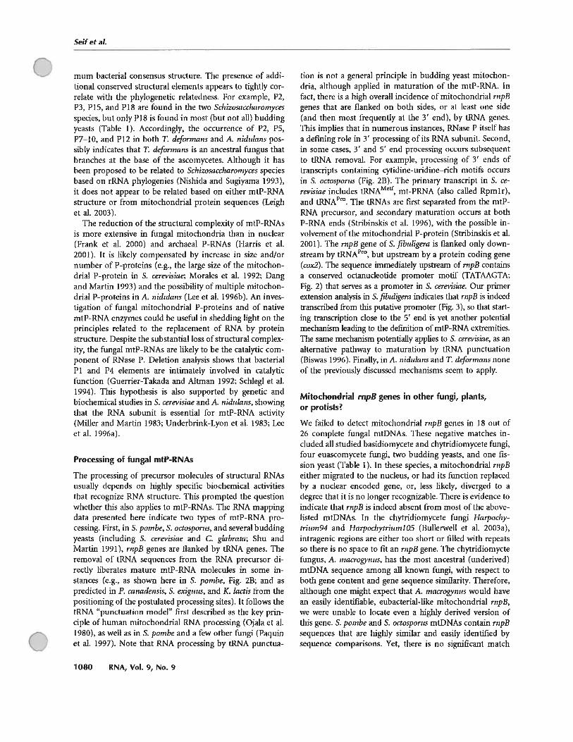
Soif t al.
mum hacterial consensus structure. The presence of additional conserved structural elements appears to fightly cor-
relate with the phylogenetic relatedness. For example, P2,P3, P15, and P18 are found in the two Schizo5accharornycesspecies, but oniy P18 is found in most (but flot ail) buddingyeasts (Table 1). Accordingly, the occurrence of P2, P5,P7—l0, and P12 in hoth T. deforinans and A. nididans possibly indicates that T. deforinans is an ancestral fungus thatbranches at the base of the ascomycetes. Although it hasheen proposed to be related to ScÏiizosaccharornyces speciesbased on rRNA phylogenies (Nishida and Sugiyama 1993),it does flot appear to be related based on either mtP-RNAstructure or from mitochondrial protein sequences (Leigliet al. 2003).
The reduction of the structural complexity of mtP-RNAsis more extensive in fungal mitochondria than in nuclear(Frank et al. 2000) and arcliaeal P-RNAs (Harris et al.2001). It is likely compensated by increase in size and/ornumber of P-proteins (e.g., the large size of the mitochondnal P-protein in S. cerevisiae; Morales et al. 1992; Dangand Martin 1993) arid the possihility of multiple mitocliondrial P-proteins in A. nididans (Lee et al. 1996b). An investigation of fungal mitochondnal P-proteins and of nativemtP-RNA enzymes could be useful in shedding light on theprindples related to the replacement of RNA by proteinstructure. Despite the substantial loss of structural complexity, the fungal mtP-RNAs are likely to be the catalytic component of RNase P. Deletïon analysis shows that bacterialPi and P4 elements are intimately involved in catalyticfunction (Guerrier-Takada and Mtman 1992; Scfflegl et al.1994). This hypothesis is also supported by genetic andbiochemical studies in S. cerevisine and A. nidulans, showingthat the RNA subunit is essential for mtP-RNA activity(Miller anci Martin 1983; Underhrink-Lyon et al. 1983; Leeet al. 1996a).
Processing of fungal mtP-RNAs
The processing of precursor molecules of structural RNAsusually depends on highly specilhc biochemical activitiesthat recognize RNA structure. This prompted the questionwliether this also applies to mtP-RNAs. The RNA mappingdata presented liere indicate two types of mtP-RNA processing. First, in S. pombe, S. octosporus, and several buddingyeasts (including S. cerevisiac and C. glabrata; Shu andMartin 1991), ntpB genes are flanked by tRNA genes. Theremoval of tRNA sequences from the RNA precursor directly liberates mature mtP-RNA molecules in some in-stances (e.g., as shown here in S. poinbe, Fig. 2B; and aspredicted in P. canadensis, S. exiguus, and K. tactis from thepositioning ofthe postulated processing sites). It follows thetRNA “punctuation model” first described as the key principle of liuman mitochondrial RNA pro cessing (Ojala et al.1980), as weil as in S. pombe and a few other fungi (Paquinet al. 1997). Note that RNA processing by tRNA ptlnctua
tion is not a general prindple in hudding yeast mitochondria, although apphed in maturation of the mtP-RNA. Infact, there is a high overail incidence ofmitochondrial ritpBgenes that are ftanked on hoth sides, or at least one sicle(and then most frequently at the 3’ end), hy IRNA genes.This implies that in numerous instances, RNase P itseli liasa defming role in 3’ processing ofits RNA suhunit. Second,in some cases, 3’ and 5’ end processing occurs subseqtientto tRNA removal. For example, processing of 3’ ends oftranscripts containing cytidine-uridine—rich motifs occursin S. octosporus (Fig. 2B).The primary transcript in S. cerevisiae includes tRNAMeIt, mt-PRNA (also cailed Rpmlr),and tRNAPT0. The tRNAs are first sepamted from the mtPRNA precursor, and secondary maturation occurs at bothP-RNA ends (Stribinskis et al. 1996), with the possible involvement ofthe mitochondnal P-protein (Stribinskis et al.2001). The nipB gene of S. fibuligera is flanked only downstream by tRNAr0, but ups[ream by a protein coding gene(cox2). The sequence immediately upstream of rnpB containsa conserved octanucleotide promoter motif (TATAAGTA;Fig. 2) that serves as a promoter in S. cerevisiae. Our primerextension analysis in S. fibuligera indicates that rnpB is indeedtranscribed from this putative promoter (Fig. 3), so that starting transcription close to the 5’ end is yet another potentialmechanism leading to the defmition of mtP-RNA extremities.The same mechanism potentiaily applies to S. cerevisiae, as analternative pathway to maturation by tRNA punctuation(Biswas 1996). Finaily, in A. itidulans and T. deforinctns noneof the previously discussed mechanisms seem to apply.
Mitochondrial rnpb genes in other fungi, pbnts,or protists?
We failed to detect mitochondrial ntpB genes in 18 out of26 complete fungal mtDNAs. These negative matches included all studied basidiomycete and chytridiomycete fungi,four euascomycete fungi, two budding yeasts, and one fission yeast (Table 1). In these species, a mitochondrial rnpBeither migrated to the nucleus, or had its function replacedby a nticlear encoded gene, or, less likely, diverged to adegree that it is no longer recognizable. There is evidence toindicate that rnpB is indeed absent from most of the abovelisted mtDNAs. In the chytridiomycete fungi HaipocÏiytriurn94 and HarpochytriurnlO5 (Bullerweil et al. 2003a),intragenic regions are either too short or fiiled with repeatsso there is no space to fit an rnpB gene. The chytridiomyctefungtis, A. Inacrogynus, has the most ancestral (underived)mtDNA sequence among ail known fungi, with respect tohoth gene content and gene sequence similarity. Therefore,although one might expect that A. Inacrogynus would havean easily identifiable, eubacterial-like mitochondrial rnpB,we were tinable in locate even a higffly derived version oftins gene. S. pombe and S. octosporus mtDNAs contain rnpBsequences that are higffly similar and easily identilïed bysequence comparisons. Yet, there is no significant matchC
1080 RNA, Vol. 9, No. 9
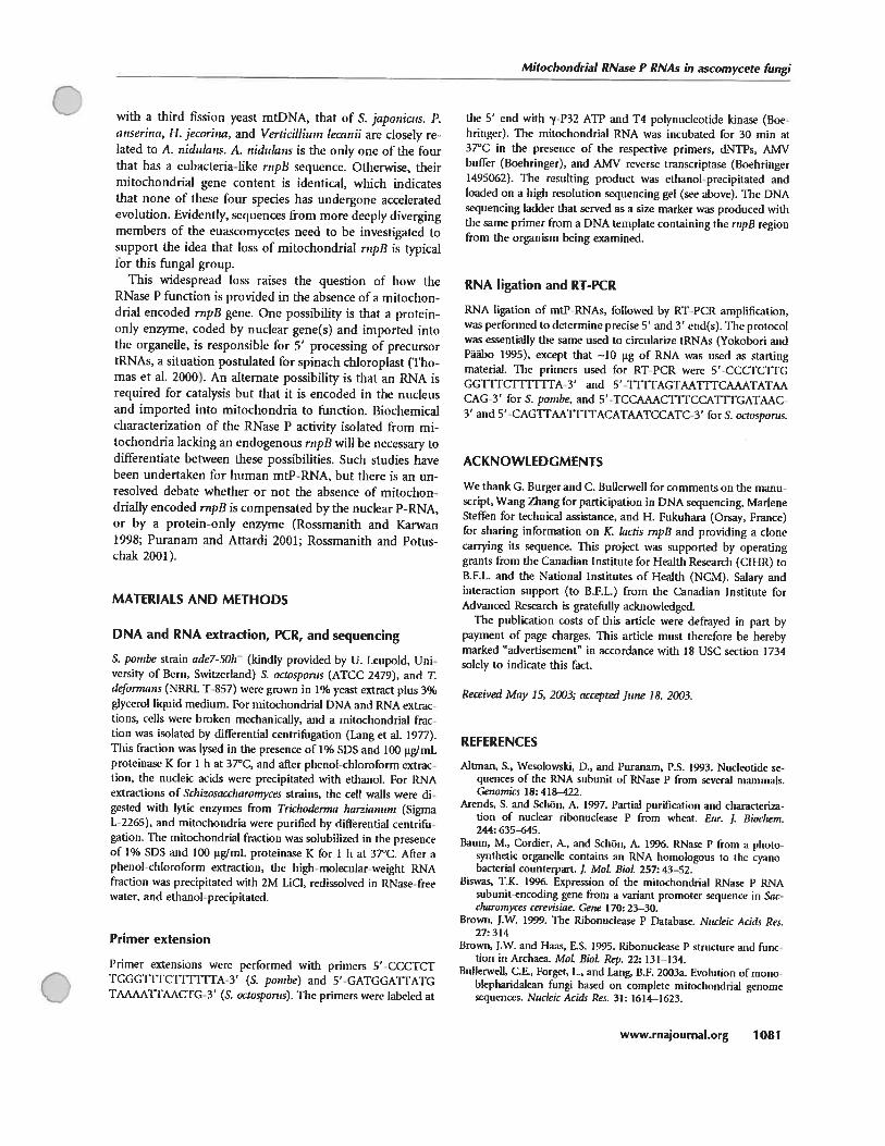
Mitochondrial RNase P RNAs in ascomycete fungi
with a duird fission yeast mtDNA, that of S. japonicus. P.anseriija, I-I. jecorina, and Verticilliu,n teamii are closely reÏated to A. 1,idulan5. A. nidula,zs is tise only one of tise fourthat bas a eubacteria-like rnpB sequence. Otherwise, thefrmitochondnal gene content is identical, which mdicatesthat none of these four species lias undergone acceleratedevolution. Evidently, sequcnces from more deeply divergingmembers of the euascomycctes need to be mvestigated tosupport the idea titaf loss of mitochondrial rnpB is typicalfor this fungal group.
This widespread loss raises the question of how theRNase P function is prowded in the absence of a mitochondrial encoded rnpB gene. One possibffily is that a proteinonly enzyme, coded by nuclear gene(s) and imported intothe organelle, is responsible for 5’ processing of precursortRNAs, a situation postulated for spmach chloroplast (Thomas et ai. 2000). An altemate possibility is that an RNA isreqttired for catalysis but that it is encoded in the nucleusand imported into mitochondria to function. Biochemicalcharacterization of the RNase P activity isolated from mitochondria lacking an endogenous rnpB will be necessary todifferentiate between these possibilities. Such studies havebeen undertaken for human mtP-RNA, but there is an unresolved debate wheffier or not the absence of mitochondnally encoded rnpB is compensated by tise nuclear P-RNA,or by a protein-only enzyme (Rossmanith and Karwan1998; Puranam and Attardi 2001; Rossmanith and Potuschak 2001).
MATERIAIS AND METHODS
DNA and RNA extraction, PCR, and sequencing
S. pombe strain adel-50h (kindly provided by U. Leupoid, Univcrsity of Bern, Switzerland) S. octosponu (ATCC 2479), and T.defonnans (NRRL T-$57) wcrc grown in 1% yeast extract plus 3%glycerol liquid medium. For mitochondrial DNA and RNA extractions, ceils were btoken mcchanically, and a mitochondriai fraction tvas isolated bv differential centrifugation (Lang et al. 1977).This fraction was lysed in the presence of 1% SDS and 100 pg/rnLproteinase K for I h at 37°C, and after phcnoi-chloroform extraction, the nucleic acids were precipitated with ctiuanol. for RNAextractions of Schizosaccharomyces strains. the ceil wails were digestcd with lytic enzymes from Trichoderma Ï,aianuni (SigmaL-2265), and mitochondria were purified by differential centrifugation. Thc mitochondriai fraction vas solubilized in the presenceof 1% SDS and 100 pg/mL proteinase K for 1 h al 37°C. After aphenol-chloroform extraction, the high-moiecutar-weight RNAfraction was precipitated with 2M LiCI, redissoived in RNase-freewater, and ethanol-precipitated.
Primer extension
Primer extensions were performed with primers 5’-CCCTCTTGGG1TfCTflTITA-3’ (S. pombe) sud 5’-GATGGATFATGTAAAATTAACTG-3’ (S. octosporus). Thc primers were labcled at
tise 5’ end with ‘y-P32 ATP sud 14 polynucleotide kinase (Boehnnger). TIse mitochondriai RNA was incubated for 30 min at37°C in the presence of the respective primers, dNTPs, AMVbuffer (Boebringer), and MvIV reverse transcriptase (Boehringer1495062). The resuiting product was ethanol-precipitated andioaded on a high resolution sequencing gel (sec above). The DNAsequencing Iadder that served as a size marker was produced withthe same primer froin a DNA template colitaining tise rnpB regionfrom tise organism being examined.
RNA ligation and RT-PCR
RNA ligation of mtP-RNAs, followed by RT-PCR amplification,svas performed to determine precise 5’ and 3’ end(s). Tise protocolvas essentially the same used to circularize tRNAs (Yokobori andPààbo 1995), except ttiat —10 pg of RNA tvas used as startingmaterial. Tise primers used for RT-PCR were 5’-CCCTCTFGGG1TUCTYFTTTA-3’ ami 5’ -THTAGTAAYfTCAAATATAACAG-3’ for S. ponibe, and 5’-TCCAAACTITCCATfTGATAAC-3’ and 5’-CAGTFAATTTFACATAATCCATC-3’ forS. ocwsporus.
ACKNOWLEDGMENTS
We thank G. Burger atid C. Bullenveil for commcnts on the manu-script, Wang Zhang for participation in DNA sequencing, MarleneSteffen for technical assistance, and H. Fukuhara (Orsay, France)for sharing information on K. kictis rupB sud providing a donccarrying lis sequence. This project vas supported by operatiriggrants from tise Canadian Institute for Heaith Research (CIHR) toB.f.L. and tise National Institutes of Health (NCM). Salary andinteraction support (to B.f.L.) from tise Canadian Institute forAdvanced Rcscarch is gratefluliy acknowtcdgcd.
The publication costs of this article were defrayed in part bypayment of page charges. This article nsust therefore be herebymarked “advertisement” in accordance with 1$ USC section 1734solel’ to indicate this fact.
Received May 15, 2003; accepted June 18, 2003.
REFERENCES
Altinan, S., Wcsoiowski, D., and Puranam, P.S. 1993. Nucieotidc sequences of tise RNA subunit of RNase P from several mammais.Genomics 18: 418—422.
Arends, S. and Schôn, A. 1997. Partial purification and characterizanon of nuclear nbonucleasc P from wheat. Eur. J. Biodiem.244: 635—645.
Baum, M., Cordier, A., and SchOn, A. 1996. RNase P from a photosynthctic organclle contains an RNA hoinologous to thc cyanobacterial counterpart. j Mot BioL 257: 43—52.
Biswas, T.K. 1996. E’cpression of tise mitochondrial RNase P RNAsubunit-encoding gene from a variant promoter sequence in Saccharonzvces cerevisiae. Gene 170: 23—30.
Brown, J.W. 1999. TIse Ribonuciease P Database. Nt,cleic Acids Res.27: 314
Brown, J.W. and Haas, ES. 1995. Ribonuclease P structure sud funclion in Archaea. MoL BioL Rep. 22: 13 1—134.
Builerwell, C.E, forget, L., and Lang, B.f. 2003a. Evolution otmonohiephaiidalcan fungi based on complete mitochondnal genomescquenccs. Ntickic Acids Res. 31: 1614—1623.
C
www.rnajournaI.org 1081

Seif et aL
BuIlerwell, C.E., Leigh, J., forget, L., and Lang, B.F 2003b. A comparison ofthree fission yeast mitochondrial genomes. Ntic!eicAc;cÏsRes. 31: 759—768.
Chen, J.L. and Pace, N.R. 1997. Identification of the universally consen’ed core of ribonuclease P RNA. RNA 3: 557—560.
Christian, E.L., Zahier, N.H., Kaye, N.M., and Harris, M.E. 2002.Analysis of substrate recognition by die ribonucleoprotein endonuclease RNase P. Methods 28: 307—322.
Clark Walker, G.D., McArthur, C.R., and Sriprakash, K.S., 1985. Location of transcriptional control signais and transtèr RNA sequences in Tonilopsis glabrara mitochondrial DNA. EMBO J.4: 465—473.
CoHins, U., Moulton, V., and Penny, D. 2000. Use ot RNA sccondarystructure for studying the evolution of RNase P and RNase MRP.
J. MoL EvoL 51: 194—204.Crary, S.M., Niranjanakumari, S., aiid Fierke, C.A. 1998. Thc protein
component of Bacillus subtilis ribonuclease P increases catalyticeffidency by enhancing interactions with the 5’ leader sequence ofpre-tRNAA5p. Biochemistry 37: 9409—9416.
Dang, Y.L. and Martin, N.C. 1993. Yeast mitochondrial RNase Psequence of the RPM2 gene and demonstration that its productis s protein subunit of the enzyme. J. Bio!. Che,n. 269: 1979119796.
Datr, S.C., Pace, B., and Pace, N.R 1990. Characterization of ribonuclease P from the archaebactenum Sulfotobus so!fataricus. J. Bio!.Chem. 265: 12927—12932.
Darr, S.C., Zito, K., Smidi, D., and Pace, N.R. 1992. Contributions ofphylogenetically variable structurai elements to die ftinction of dieribozyme ribonuclease P. Biochernistry 31: 328—333.
Doersen, C.J., Guerrier-’takada, C., Akman, S., and Attardi, G. 1985.Characterization of an RNase P activity from Hela ceil mitochondtia Comparison with the cytosol RNase P activity. J. Biot Citent260: 5942 5949.
Frank, D.N., Adamidi, C., Ehringer, M.A., Pitulle, C., and Pace, N.R.2000. Phylogenetic-comparative analysis of the eukaryal ribonuclease P RNA. RMA 6: 1895—1904.
franidin, S.E., Zwick, M.G., and Johnson, J.D. 1995. Characterizationsud partial purification of two pre-tRNA 5’-processing activitiesfrom Daucus carrota (carrot) suspension ceils. Plant J. 7: 553—563.
Gardiner, K.J., Marsh, 1.1., and Pace, N.R. 1985. Ion dcpendcncc ofdie Bacilus subtilis RNase P reaction. j Biot Client. 260: 5415—5419.
Glôckner, G., Rosenthal, A., and Valentin, K. 2000. The structure andgene repertoire of an ancient red algal plastid genome. J. Mol. Evo!.51: 382—390.
Gopalan, V., Vioque, A., and Mtman, S. 2002. RNase P: Variations anduses. J. Biot he,n. 277: 6759—6762.
Gucrrier-Takada, C., sud Altrnan, S. 1992. Reconstitution of enzymatie activity from fragnients of Ml RNA. Froc. at!. Acad. Sci.89: 1266—1270.
Guerrier-Takada, C., Gardiner, K., MarsÏi, T., Pace, N., and Altinan, S.1983. Tlic RNA moiety ofribonuclease P is the catalytic subunit ofthe enzyme. Ce!! 35: 819—857.
Guerrier-Takada, C., Eder, P5., Gopalan, V., and Mtman, s. 2002.Purification and characterization of Rpp25, an RNA-binding protein subunit of hmnan ribonuclease P. RNA 8: 290—295.
Haas, E.S,, Brown, J.W., Pitulle, C., and Pace, N.R 1994. furtherperspective on die catalytic core sud secondary structure of ribonuclease P RNA. Proc. NatL Acad. Sci. 91:2527—2531.
Hall, T.A. sud Brown, J.W. 2002. Archaeal RNase P has multipleprotein subunits homologous to eukaryotic nudear RNase P proteins. RNA 8:296—306.
Hanic-Joyce, P.J. and Gray, M.W. 1990. Processing of transfer RNAprecursors in a wheat mitochondrial cxtract. j Bio!. Chcm.265: 13782—13791.
Harris, J.K., Haas, ES., Williams, D., Frsuk, D.N., sud Brown, J.W.2001. New insight into RNase P RNA structure from comparativeanalysis of die archaeal RNA. RNA 7: 220—232.
flartmami, R.K., Heinrich, J., Schiegi, J., and Schuster, FI. 1995. Pre
cursor of 01 anfisense RNA of bacteriophages PI and P7 is asubstrate for RNase P of Escherichia cati. Froc. Nat!. Acad. Set.92: 5822—5826.
Jayanthi, G.?. and Van Tuyle, G.C. 1992. Characterization of ribonuclease P isolated from rat liver cytosol. Arciz. Biochent. Biophys.296: 264—270.
Kole, R. sud Mtman, 5. 1981. Properties of purified ribonticlease Pfrom Eschcrichia cati. Biochemistry 20: 1902—1906.
Laferriere, A., Gautheret, D., and Cedergren, R. 1994. An RNA patternmatching program widi enhanced performance and portability.C’ornpuL Appt Biosci. 10: 211—212.
I.ang, B.F., Burger, G., Doxiadis, I., Thomas, D.Y., Bandlow, W., sudKaudewitz, F. 1977. A simple method for the large-scale preparaflou of mitochondria from microorgsuisms. AnaL Biochent.77:110—121.
Lang, B.F., Ahnc, F., Distler, S., Trinkl, H., Kaudewitz, F., sud WoIf, K.1983. Sequence ot the mitochondrial DNA, arrangement of genessud processing of their transcripts in Scluzosaccharoinyces ponibe.In Motecular bio!ogy of rite fission yeast (eds. A. Nasim, et al.),pp. 3118-3 119, Academic Press, San Diego, CA.
Ling, B.P., Burger, G., O’Kelly, C.J., Cedergren, R., Golding, G.B.,Lemieux, C., Ssukoff, D., Turmel, M., sud Gray, M.W. 1997. Anancestral mitochondrial DNA resembling a eubactetial genome inminiature. Nature 387: 493—497.
Lang, B.F., Gray, M.W., sud Burger, G. 1999. Mitochondrial genomeevolution sud die origin of eukaryotes. Aniut. Rev. Genet. 33: 351—397.
Lee, Y.C., Lee, B.J., Hwang, D.S., sud Ksug, H.S. 1996a. Purificationsud characterizaflon of mitochondrial ribonuctease P from Aspergillus nidutans. fur. J. Biochem. 235: 289—296.
Lee, Y.C., Lee, B.J., and Kang, I-l.S. 1996b. The RNA component ofmitochondrial ribonuclease P from Aspergi!!us nidu!ans. fur. J.Biochem. 235: 297 303.
Leigh, J., Seif E, Rodriguez, N., Jacob, Y., and Lang, B.F. 2003. Fungalevolution meets fungal geriolnics. In Handbook of flsnga! biotechno!ogy, 2nd cd. (ed. Arora D.). Marcet Dekkcr Inc., New York (inpress).
Marchfelder, A. sud Brennicke, A. 1994. Characterization sud partialpurification of tRNA processing activities from potato mitochondna. Plant Physio!. 105: 1247—1254.
Martin, C.A. sud Lsug, B.F. 1997. Mitochondrial RNase P: The RNAfamily grows. Nucleic Acids Syrnp. Ser. 36: 42—44.
Miller, D.L. and Martin, N.C. 1983. Characterization of the yeastinitochondrial locus necessary for tRNA biosynthesis: DNA sequence analysis and identification of s new transcript. Ce!! 3: 911—917.
Morales, M.J., Dang, Y.L., Loti, Y.C., Stilo, P., sud Martin, N.C. 1992.A 105 k-Da protein is required for yeast mitochondrial RNase Pacfivity. Prise. Natt Acad. Sci. 89: 9875—9879.
Nishida, H. and Sugiyama, J. 1993. Phylogenetic retationships amongTaphrina, Saitoe!!a, and other higher fungi. Mot Bio!. Ei’oL10:431—436.
Ojala, D., Merkel, C., Gelfand, R., sud Attardi, G. 1980. The tRNAgenes punctuate the reading ut genetic information in human mitochondrial DNA. Ccli 22: 393—403.
Psunucci, J.A., Haas, ES., Hall, T.A., Harris, J.K., and Brown, J.W.1999. RNase P RNAs from some archaea are catalytically active.Froc. NatL AcacL Sci. 96: 7803—780$.
Paquin, B., Laforest, M.J., forget, L, Roewer, I., Wang, Z., Longcore,J., sud Lang, B.F. 1997. The fungal mitochondrial genome project:Evolution of fttngal mitochondrial genomes sud their gene expression. Cuir. Genet. 31: 380—395.
Pascual, A. sud Vioque, A. 1999. Functional reconstitution ot RNaseP activity from a plastid RNA subunit sud s cysuobacterial proteinsuhunit. fEBS Leu. 442: 7—10.
Peck-Miller, K.A. sud Mtman, S. 1991. Kiiietics of die processing ofthe precursor 10 4.5S RNA, a naturafly occurring suhstrate forRNase P trom Escherichia cati, j MoL BioL 221: 1—5.
Petersen, R.F., Langkjaer, RB., Flvidtfeldt, J., Gartner, J., Palmen, W.,
C
C1082 RNA, Vol. 9, No. 9
Mitochondrial RNas P RNAs in ascomycete fungi
Ussery, D.W., and Piskur, J. 2002. Inheritance and organisation ofOie initocliotidrial genome differ betweett two Satcharomycesycasts. J. Mol. Bio!. 31$: 627—636.
Puranasu, R.S. and Attardi, G. 2001. Tise RNase P assodated with1-IcLi ccli mitochondria contains an essential RNA componentidentical in sequellce 10 that oC Oie rniciear RNase P. MoL cet!. BioL21: 548—561.
Ra%nini, A., Grisanti, P., Rinaldi, T., frontal!, L., and Palleschi, C.1991. Mitochondriai genome ot Sacchara,nyces dottglasii: Genescoding kr coinponents of tue protein syrithetic apparatus. Cm-r.Geitet. 19: 169—174.
Reith, M. aiid Munholtand, J. 1995. Complete nucteotide sequence uttise Porph,sra purpurca chloropiast genome. Plant MoL Bio!. Rcptr.13: 333—335.
Rossmanith, W. and Karwan, R.M. 1998. Characterization of humanmitochondrial RNase P: Novel aspects in tRNA processnig. Biochcm. Biophys. Res. Commun. 247: 234—241.
Rossinaiuth, W. and Potushak, T. 2001. Difference betweeu mitochondrial RNase P and nuclear RNase P. MoL Ce!L BioL 21: 8236—8237.
Salavati, R., Panigrahi, A.K., aiid Stuart, K.D. 2001. Mitochondrialribonuctease P activity of Trspanosoma brttcei. MoL Biochcm. ParasituL 115: 109—117.
Sbisa, E., Pcsole, G., Tullo, A., and Saccone, C. 1996. The evolution oCthe RNasc P- and RNase !.flU’- associatcd RNAs: Phylogcneticanalysis and nucteoride substitution rate. J. Mot. EvoL 43: 46—57.
Schiegi, J., Hardt, XV.D., Erdisiarni, X’.A., and Hartmami, RK. 1994.Contribution oC structural elements to Thermos thermophilns ribonucicase P RNA flinction. EMBO j 13: 4863—4369.
Sekito, T., Okamoto, K., Kitano, FI., and Yoshida, K. 1995. Tise complete mitochondrial DNA sequence of liausenula ivingei revealsnew characteristic of yeast mitochondna Ct,rr. Getwt. 28:39—53.
Shu, HI-1. and Martin, N.C. 1991. RNasc P RNA in Candida ghibratamitochondria is transcribed with substratc tRNAs. Xnckic .4cidsRes. 19: 6221—6226.
Shu, H.H., Wisc, C.A., Clark-’sValker, G.D., antI Martin, N.C. 1991. Agene required for RNase P activity in Candida (Torulopsis) giabratamitochondria codes for a 227-nucicotide RNA with homoiogy tobac terial RNasc P RNA. MoL CeIL Biot. 11: 1662—1667.
Stark, DC., Kole, R., Bowman, E.J., and Mtman, S. 1978. RibonucleaseP: An enzyme with an essential RNA component. P,oc. NatL Acad.Sci. 75: 3717—372 1.
Stribinskis, V., Gao, G.J., Solo, P., Dang, Y.L., and Martin, N.C. 1°96.Yeast niitochondrial RNase P RNA syr thesis is attered iii an RNastP protein suhunit mutant insïghts into tise hiogenesis oC a mitochondrial IU’SA-processtng enzyme. 1loL Cci!. Bail. 16: 343136.
Stribinskis, V., Gao, G.J., Hlis, S.R., antI Martin, XC. 2001. Rpm2, theprotein subunit oC mitochondriat RNase P in Sacchammycescercvisiae, also has a rote in the translation oC mitochondriallycncoded subunits of cytochrome e oxidase. Genettes 15$: 573—585.
Thomas, OC., Li, X., and Gegenheimer, P. 2000. Chloroplast ribonuctease P does not utilïze tise ribozvme-type prc-IRNA cleavagemcchanism. RNA 6: 545—553.
Trinld, H., Lang, B.f., antI Wotf, K. 1989. Nuclcotide sequence ut tisegene encoding tise small ribosomal RNA in tise mitochondrial genome of thc fission yeast SchCostwcharomyces pornbe. Ntwleic AcidsRcs. 17: 6730.
Tunnel, M., Lemieux, C., Burger, G., Laiig, BI-., Otis, C., Plante, I.,antI Gray, M.W. 1999. The complete mitochondrïal DNA sequences of NephroseÏmis olivacea and Pedmoutonas mmor. Tworadically diftercnt cvotutionary patterns within green aigac. Plantccli 11: 1717—1730.
Underbrink-Lyon, K., Muter, D.L, Rois, N.A., Fukuhara, H., andMartin, XC. 1983. Characterization ota yeast mitochondriai locusncccssary for tRNA biosynthesis: Deletion mapping antI restrictionmappilig studies. Mol. Gen. Gcnct. 191: 512—518.
Wagner, M., fingerbut, C., Gross, H.J., and Schon, A. 2001. The firstphytoptasma RNase P RNA provides ncw insights into tise sequcnce requiremcnts ofthis ribozyme. Nuclcicflcids Res. 29: 2661—2665.
Wilson, C., Ragnini, A., antI fukuhara, H. 1989. Analysis of the cegions coding for transter RNAs in Ktuyieroinyces laais mitochondrial DNA. Ntwkic Acids Res. 17: 4485—4491.
X\’isc, C.A. antI Martisi, XC. 1991a. Drarnatic size variation ut yeastmitochondrial RNAs suggests that RNase P RNAs eau be quitesmall. J. BioL Chena 266: 19154—19157.
1991b. Sequence analysis of Saccharomyccs exigutts mitochondrial DNA rcveals an RNase P RNA flartked by two tRNA genes.Nuckic AnUs Res. 19: 4773.
Yokobori, S. antI Paabo, S. 1995. Transter RNA edifing in land snaitmitochondria. Froc. l’iut!. Actai. Sci. 92: 10432—10435.
o
Cwwwrnajournal.org 7083
63
Article 2
Comparative mitochondrial genomics in zygomycetes:
bacteria-like RNase P RNAs, mobile elements, and a close
source of the group I intron invasion in angiosperms.
Elias Seif’, Jessica Leigh2, Yu Liu’, Ingeborg Roewer3, Lise Forget’,
and B. Franz Lang’
NucÏeic Acids Research, 2005, vol.33, no. 2, pp. 734-744
‘Program in Evolutionary Biology, Canadian Institute for Advanced Research,
Département de Biochimie, Université de Montréal, 2900 Boul. Edouard-Montpetit,
Montréal, Québec, H3T 1J4, Canada.2 Department of Biochemistry and Molecular Biology, Dafflousie University, Halifax
(Nova Scotia), B3H 4H7, Canada.
3National Research Council of Canada, Plant Biotechnology Institute,
110 Gymnasium Place, Saskatoon, SK, S7N 0W9 Canada.
Contribution of each author:
Elias Seif: did most of the experimental work, participated in genome analysis, and
phylogenetic reconstruction, and prepared the manuscript and figures.
Jesscia Leigh: participated in phylogenetic reconstruction and preparing the
manuscript and figures.
Yu Liu: participated in phylogenetic reconstruction.
Lise Forget and Ingeborg Roewer : participated in the experimental work.
B. franz Lang: participated in genome anlaysis, phylogenetic reconstruction, and
preparing the manuscript and figures.
Published online February 2, 2005
734—744 Nucleic Acids Rcsearch, 2005, Vol. 33, No. 2
( doi:10i093/narlgkil99
Comparative mîtochondrial genomics inzygomycetes: bacteria-like RNase P RNAs,mobile elements and a close source of thegroup I întron invasion in angiospermsElïas Self1, Jessica Leïgh2, Yu Liu’, Ingeborg Roewer3, Lise Forget’ and B Franz LangI*
1Program in Evolutionary Biology, Canadian Institute for Advanced Research; Centre Robert Cedergren,Département de Biochimie, Université de Montréal, 2900 Boulevard Edouard-Montpetit, Montréal,Québec, Canada H3T 1J4, 2Department of Biochemistry and Molecular Biology, Dalhousie University,Halifax (Nova Scotia), Canada B3H 4H7 and 3National Research Council of Canada, PlantBiotechnology Institute, 110 Gymnasium Place, Saskatoon, SK, Canada S7N 0W9
DDBJIEMBUGenBank accession nos
ABSTRACT
To generate data for comparative analyses ofzygomycete mitochondrial gene expression, wesequenced mtDNAs of three distantly related zygomycetes, Rhizopus oryzae Moflierella verticillataand Smittium culisetae. They ail contain the standardfungal mitochondrial gene set, plus rnpB, the geneencoding the RNA subunit of the mitochondrialRNase P fmtP-RNA) and rps3, encoding ribosomalprotein S3 (the latter lacking in R.oiyzae). The mtPRNAs 0f R.oryzae and 0f additional zygomycete relatives have the most eubacteria-like RNA structuresamong fungi. Precise mapping of the 5’ and 3’ terminiof the R.oryzae and M. verticillata mtP-RNAs confirmstheir expression and processing at the exact sites predicted by secondary structure modeling. The 3’ RNAprocessing of zygomycete mitochondrial mRNAs,SSU-rRNA and mtP-RNA occurs at the C-richsequence motifs similar to those identified in fissionyeast and basidiomycete mtDNAs. The C-rich motifsare included in the mature transcripts, and are likelygenerated by exonucleolytic trimming of RNA 3’termini. Zygomycete mtDNAs feature a variety ofinsertion elements: fi) mtDNAs of R.oryzae andM.veflicillata were subject to invasions by doublehairpin elements; (ii) genes of ail three species containnumerous mobile group I introns, including one that sclosestto an intron that invaded angiosperm mtDNAs;and (iii) at least one additional case of a mobile
element, characterized by a homing endonucleaseinsertion between partially duplicated genes[Paquin,B., Laforest,M.]., Forget,L, Roewer,L,Wang,Z., Longcore,J. and Lang,B.F. (1997) Curr.Genet., 31, 380—395]. The combined mtDNA-encodedproteins contain insufficient phylogenetic signal todemonstrate monophyly of zygomycetes.
INTRODUCTION
fungi constimte a huge group of highly diverse organisms,including some of the most-sflidied and best-understoodeukarvotic model systems: ‘bakers east (Saccharornucescereviçiae), fission yeast (Sc1ii:osaccIîarorn’cer pombe),and the filamentou s euascomycetes Netirospora crassaand Aspergilltts nidulans. These species ail belong to theAscomycota. Substantially fewer scientific shidies havebeen performed in members of the sister phylum Basidiomycota and very littie is known in the remaining twophyla, Zygornycota and Chytridiornycota, often classifiedas ‘lower fungi’. This expression is a taxonomically vagueconcept borrowed from Aristoties philosophy, visioningdfrected evolution from the simple (primitive, 10w) tothe highly complex. The misnorner is most evidcntin the higher’ ascomycetes and hasidiomycetes, whichevolve toward microscopic. unicellular and geneticallysimpbfied yeast-like organisms in soma lineages, and towardmorphologically complex, gene-rich and biochemicallyversatile multicellular organisms in others.
A]though the availability ofcornplctc nuclearand mitochondrial sequences of more than a dozen ascomycetes provides a
To whorn correspondence should be addressed. Tel: +1 514 343 5842; Fax: ±1 514 343 2210;
AY863211, AY863212. AY8632133. AYu161439—AY861412
(F Thc Author 2005. Published by Oxford University Press. Ml rights rcserved.
/ The online version of this article bas bçen published under an open access model. Users are entitled to use. reproduce. disseminate. or displav Éhe open accessÇ version of Ibis article for non-commercial purposes provided that: the original authorship is properly and fully attributcd: the Journal and Oxford University Press
are attributcd as the original place of publication with the correct citation details given: ifan article is subscquentlyreproduced ordisseminatednol in its entïrety butonly in pari or as a derivative svork Ibis must be clcarly indicaied. For commercial re-use, please contact journals.permissinns(Foupjoumals.org

Nucteic Acids Research, 2005, Vol. 33, No. 2 735
strong basis for biochemical investigations, only a fewcomplete mitochondrial scquences are known from chytrids,and none froin zygomycctes, a situation that bas motivatedthe work presented here. In fact, the number of zygomycetenuclear gene sequences (mostly rRNA sequences) is so limitedthat it is impossible to determine with confidence whether orflot Zygomycota is a monophyletic taxon (1—3). The lackof resolution in these analyses is consistent with estimatesthat even the combined LSU- and SSU-rRNA would containfar too littic information to resolve many ftmgal phylogeneticrelationships with confidence (4). This dataset is at mostsufficient for resolving ftngal inferences below the phylumlevel (5,6).
Sequences of complete mtDNAs from several zygomycetesmight be a first step in remediating this situation. Mitochondrial phylogenies can be based on up to 13 proteinsequences, and have been shown to resolve deep divergencesin the ftngal and animal lineages (7—9). For instance,Alexopolous et al. (10) indicate that ‘additional study isneeded to determine whether the class (Trichomycetes) is amonophyletic group belonging to Zygomycota, or merely acollection of orders grouped together on the basis of aunique shared habitat. Molecular phylogenies based onrRNA sequences were successful in moving the orderAmoebidiales away from zygomycetes, although they werethen placed with mesomycetozoan rotists, whose phylogenetic affiliation vas unresolved and controversial (li).Only subsequent analysis with multiple mitochondrial proteinsplaccd this group at the base of the animal divergence, withhigh confidence (9). This example clearly illustrates therequirement of rnu]ti-gene datasets with at least severalthousand amino acid positions. for resolution of trecs at thekingdom level.
Furthermore, zygomycete mtDNAs are of considerableinterest for comparative gene expression silidies: our preliminary data indicated the presence of a mitochondrial rnpBgene, which encodes the RNA subunit of RNase P. theenzymaticaily active pan of an endonuclease (ribonuclcoprotein) responsible for tRNA maturation. In mitochondria,the size and sequence of the RNA subunit varies substantially,which bas considerably complicated its identification. Thegene is apparentiy absent from ail compietely sequencedbasidiomycetc and chytridiomycete mtDNAs, and bas apatchy distribution in ascomycetes (12); to date, there areno published data on zygomycete mitochondriai RNasc P(mtP-RNA).
To help remediate the lack of data for phylogenetïcinferences, and to facilitate biochemicai investigations andcomparative mitochondrial genome analyses, we havesequenced mtDNAs from three distantiy related zygomycetes,the Mucorales Rhi:opuv orv:ae, the Mortierellales Mortiere liai.’erticitiata and the Harpellales Srnittiurn culisetac. Itithis article, we compare their mitochondrial genomes(gene content, gene organization, genetic code and widclyconserved 3’ RNA processing sites). We will then presentsecondary structure models and expression data for sevennewly identified zygomycete mtP-RNAs. Finally, wewill test whether or flot zygomycetes are monophyletic, andprovide evidence that the group I introns invasion of eux]gene in angiosperms originated in a zygomycete close toRhizopus.
MATERIALS ANO METHODS
Strains and culture conditions
The various zygomycete strains were obtained from KeriyO’Donneh (National Center for Agricultural UtilizationRcsearch, Peoria, IL; NRRL), R.W. Lichtwardt (Departmentof Botany, University of Kansas, Laurence, KS; RWL) andCaroiyn Babcock (Canadian Collection of Fungal Cultures,Ottawa; DAOM). All strains, R.orvae (DAOM 14842$,previousiy designated as Rhi:opus .vtoionzfei-), R.stoionifet(DAOM 194667), Rhi:opus oligasporus (NRRL 2710),M.verticillata (NRRL 6337), Radiomvces spectabilis(NRRL 2753), Mucor mucedo (NRRL 3635) and S.ctdise,’ae(strain 18-3; R.W. Lichtwardt), were grown in YG mediumconsisting of 0.5% yeast extract and 3% glycerol. Liquidcultures of 500 ml in 2 L Erlenmeyer flasks were grown atroom temperanire under gentie shairing (100 r.p.m.).
DNA and RNA extractions
For rntDNA and RNA extractions, the ceils were broken mechanically, and a mitochondrial fraction was isolated by differentiai centrifugation (8). This fraction was lysed in thepresence of 1% SDS and 100 jig/mi proteinase K at 50CCfor 1 h, and after phenol—chloroform extraction, the nucleicacids were precipitated with ethanol. For RNA purifications,the high molecular weight RNA fraction was precipitated with2 M LiC1. rcdissolved in RNase-free water and ethanolprecipitated. MtDNAs from ail zygornycete strains wercpurified from total cellular nucleic acids by Cesium chloride/bisbenzimide density gradient centrifugation.
Cloning and sequencing of complete mtDNAs
Library construction and DNA sequencing foHowed previouslypubiished protocols (8). Briefiy, mtDNAs were physicallysheared by nebuhzation (13), and a size fraction of 1300—4000 bp was recovered after agarose gel electropboresis.The DNA vas incubated with a mixture of T7 DNA polymerase and Escherichia cati DNA polymerase I (tbe Klenowfragment) to generate blunt ends, and then cloned into theEcoRV cloning site of the phagemid pBFL6 (B. F. Lang,unpubhshed data). Recombinant plasmids containing mtDNAinserts were identified by coiony hybridization using mtDNAas a probe. Clones contained in the random hbraries weresequenced to ‘-‘-8-fo1d coverage, and remaining gaps wereclosed by primer walldng or sequencing of PCR-amplifiedDNA fragments. The expccted quahty of the sequcnccdrntDNAs is <1 error in 10 000 bp.
lie mtDNA sequences of R.arvcae, M.vetticiiiata andS.cuhsetae have been deposited in Gen3ank (accession nosAY$63212. AY863211 and AY8632133, respectiveiy).
PCR amplification uf rnpB genes
Mitochonddai rnpB genes of R.stoionifer, M.mucedo,R.spectahiiis and R.oiigosporus were PCR-amplified from
100 ng of the respective mtDNAs in a 50 10 reaction mixture[200 [tM UNTP. 2.5 mM MgCb. 2 nM ofprimers, 5 [tI of IOxbuffer and 3 U of DNA polymerases mixture from the Expandhigli fidelity kit (Roche Catalog no. 1732650) and degenerateprimers]. The annealing temperature of the PCR amplificationwas 50CC. Sequences of the degenerate primers are
736 Nuclejc Acids Research, 2005, Vol. 33, No. 2
C—5’-GTAATGGCAGCATACTAGACTCAT-3’ and 5’-TTG-AACTCCCAAGTTTTATGTATG-3’. 11e amplificationproducts were cloned and sequenced. The rnpB sequencesof R.stolonifer. R.oligosporus, M.rnucedo and Rspectohilishave been deposited in GenBank (accession nos AY86 1439,AY86 1440, AY86 1441 and AY86 1442. respectively).
RNA circularization by ligation and RT—PCR
RNA ligation of mtP-RNAs, followcd by RT—PCR amplification, was performed to determine the precise 5’ and 3’ terminiof RNase P RNAs according to the prcviously publishedprotocols (12). The prirners are 5’-CTCYTATAGGATAATA-CAAAGITG-3’ and 5’-GGCCGAAGAATAAAGAGGGA-3’for M.venicittota, and 5’-ACCCTAATmCATFAGATATTT-3’ and 5’-AATCCTTAGTAAGGATAGCTT-3’ for R.orv:ae.
RT-PCR of mtP-RNA from R.oryzae
Mitochondrial RNA ofR.o,v;ae was treated with DNase I andextracted with phenollchloroform, until no genomic DNAcould be amplified by PCR using the mtP-RNA-specificpri mers 5’-TTCHAGAGHAAATAAGCC-3’ and 5’-TTGGAGGAAAGTCCGGG-3’. Following these treatments,we amplified the mtP-RNA by RT—PCR as described aboveusing a sample without reverse transcription as negative control. Following amplification and separation on a 0.8% agarosegel, the resulting DNA fragment was cloned and sequenced.
Si mapping
DNA oligonucleotides and a 10 bp DNA ladder (Invitrogen10821-015) were Iabeled at their 3’ termini with ddATP- 2P(Amersharn PB10235) and terminal deoxynucleotidyl transferase (MBI fermentas EPOI6I) according to the manufacturer’ s recommendafions.
A total of 100 000 c.p.m. of gel-purified labeledoligonucleotides were hybridized to ‘—.10 ig of totalRNA, as descdbed in the protocol by Hahn and Breeden(http :I/www .fhcrc.orgflabs/hahnlmethods/mol_bio_methls l_oligo_probe.html), and Si nuclease digestions werc carriedmit at 3TC for 30 mm, aftcr the addition of 20 U of Si nucleaseand the buffer provided by the manufacturer (MBI fermentasEN0321). hie product vas then ethanol-precipitated and dissolved in 4 pi of RNase-free water. An aliquot of 2 pi of theproduct was mixed with 2 pi of the dye solution provided withthe 10 bp ladder (Invftrogen 10821-015), denatured at 75Cand loaded on a 7% polyacrylamide denaturing sequencinggel. The following oligonucleotides were used:
SSU-rRl’TA, 5’-AAATAAAGGGTTTAATATATTGGGA-GGGACTTATTGTCCCCCCGGTAATAACCATTCAGCCACTCGTTCCCGAACGGCT-3’(OïI mRNA, 5’-CAGATTCTAAGGGGGTGTTATATTA-TTAATTTAATTAAGATTGAACTGGTAATGAATTTACAGTATGGA-3’.
Phylogenetic inference
Mitochondrial protein sequences from ail cornpietelysequenced zygomycetes, chytridiomvcetes and basidiomy
( cetes were included in phylogenetic inference (for speciesnames and GenBank accession nos, see legend of Figure 5).Protein sequences from apocytochrorne h (Cob), as well as
7 subunits of NADH dehydrogenase (Nad), 3 cytochrome coxidase (Cox) subunits and 2 ATP synthetase (Atp) subunitswere aiigned with Muscle (14), concatenated and trimmedwith Gblocks [using default parameters (15)] to removeambiguously aligned regions. 11e resulting alignment contained 2890 aligned positions. Maximum lilcelihood (ML)phylogenies wcre inferred from this alignment using bothPHYML (16) and IQPNNI (17); ML bootstrap support wasdetermined based on 100 replicates, using both programs. Ailphylogenies were inferred using the JTT amino acid substitution moUd and gamma distribution (P) correction for variation
of rates across sites.Plwlogenetic inference hased on concatenated datasets can
lead to tree reconstruction artefacts, such as long-branchattraction, resulting from differences in relative evo]utionaryrates between genes [sometimes referred to as the covariontike behavior of different genes (18)]. Idealiy, during the MLtree search procedure, nuisance parameters, such as branchlengths and the P distribution shape parameter (cc), couldbe optimized separately for each gene; however, this rnethodresults in the estimation of many more parameters, potentiallymore than can be statistically justified. For this reason, wepartitioned the dataset into four functional categories: Atp(253 positions), Cob (361 positions), Cox ($76 positions)and Nad (1400 positions). The additional parameters includedin this partitioned dataset were justified using the X test(P < 0.000 1). Using this partitioned dataset, a topologv wasinferred from this alignment using MrBayes, with hranchiengths and a parameter unlinked across partitions (19). Inaddition, an ML trec was determined from this partitioneddataset using an adaptation ofthe method of(18). AIl possibletree topologies were generated, with constraints of groups thatreceived at least 95% bootstrap support under usinghoth IQPNNI and PHYML (Supplemcntary Figure t). Logiikelihoods were calculated separately for each partition usingPHYÏvŒ, under each of the topologies, and the sum over ailpartitions vas calculated for each tree. 11e tree found tomaximize the sum log-likeiihood of flic dataset was takentu be the ML tree (this mcthod is referred to henceforth asseparate anaiysis). Bootstrap support was also calculated usingboth of these methods, based on 100 replicates. Lilcelihoodratio tests were performed using genewise optimized site likelihoods, given the 99 tree topologies described in Supplementary Figure 1, and using Tree-Puzzle to generate sitewiselikelihoods (20) along with CONSEL (21).
Additionally, phylogenetic analysis of c]osely related fungal, green algal and plant intronic open reading frames (ORFs)of the same car] intron (for species names and GenBankaccession nos, sec legend of figure 5) were carried out.Sequences were aligned using Muscle and triinmed withGblocks, and the resulting aiignrnent was manually refined.An ML phylogeny vas inferred using IQPNNI, and ML bootstrap support was determined based on 100 replicates.
RESULTS AND DISCUSSION
Genes in addition to the standard fungal set,rnpB and rps3
11e mitochondrial genomes of the zygomycetes R.orvae(previously listed incorrectly as R.stotonifer), M.vcrtieitlata
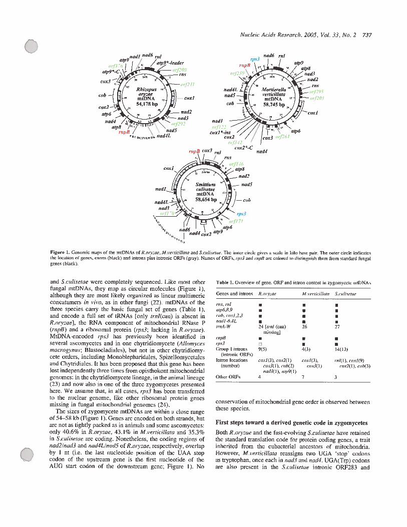
Nucleic’ Acid,ç Re,çearch, 2005, Vol. 33, No. 2 737
nadi nud6 mlutp9 // /usp9*kadcr
z ‘:
uad4/.<utp8 /‘ \ uad5
nad-IL
and S.cutisetae were completcly sequenced. Like most otherfiingal rntDNAs, they map as circular molecules (Figure 1),although they are most iilcely organized as lincar muitimericconcatamers in t’ivo, as in other fiingi (22). mtDNAs of thethree species carry the basic ifingai set of genes (Table 1),and encode a full set of tRNAs [only trnl(cau) is absent inR.orvzae], the RNA component of mitochondriai RNase P(rnpB) and a ribosomai protein (ips3; lacking in R.orv:ae).MtDNA-encoded rps3 bas previousiy been identified inseveral ascomycetes and in one chytridiomycete (Attornycesmacrogvnus; Blastocladiaies), but not in other chytridiomycete orders, including Monoblepharidales, Spizellornycetalesand Chytridiales. It bas becrs proposed that t.his gene has beenlost independently three times from opisthokont mitochondriaigenomes: in the chytridiomycete lineage, in the animal lineage(23) and now also in one of the three zygomycetes presentedhere. We assume that. in ail cases, ipx3 has been transferredto the nuciear genome, iike offier ribosomal protein genesmissing in fungai mitochonddal genomes (24).
The sizes of zygomycete mtDNAs are within a close rangeof 54—58 kb (Figure 1). Genes are encoded on hoth strands, butare not as tightly packed as in animais and some ascomycetes:only 40.6% in R.orv:ae, 43.1% in M.uerndlllata and 35.3%in S.ctitisetae are coding. Nonetheiess, the coding regions ofnad2/nad3 and nad4LlnadS of R.orvzae, respcctivciy, overlapby 1 nt (i.e. the iast nucleotide position of the UAA stopcodon of the upstream gene is the first nucleotide of theAUG start codon of the downstream gene; Figure 1). No
nad6 ru!rps3
rup8 \iFI .. / aip9
‘ud2
,,ud4L LgÏ ,l’foriierella \‘*—rus
i;ad5 s t’erfic’il!aia
cob ï
:‘
/ /cv,.3
cox2 aCnadd
Table 1. Ovcrview of gene. ORF and intron content in zygomvcete mtDNAs
Genes anti introns Ro,-v:ae Mie,rjcjllaru Sc,ilismtae
i-nt roiarp6,$,9coi,, coul,2,3 •cadi -6 ,4LtrnA-W 24 [trot (eau) 26 27
missingirnpBîps3Group I introns 9(5) 4(3) 14(13)
(intronic ORFs)Intron locations caxit3), cox2(1) coxl(3), ,ni(l), coxi(9)
(numhcr) corifl ), iob(2) cox3( I) cox2tl). t-oh(3)nad3( 1). aip9t I)
Other OREs 4 7 3
conservation of mitochondriai genc order is observed betweenthese species.
First steps toward a derived genetic code in zygomycetes
Both R.oiy:ae and the fa.st-evolving S.cttÏisetae have retainedthe standard translation code for protcin coding genes, a traitinherited from the cubacterial ancestors of mitochondria.However, li.verticittata reassigns two UGA stop’ codonsas tryptophan, once each in nad3 and nad4. UGA(Trp) codonsare also prcsent in the S.cutisetae intronic 0RF283 and
o
rupB coLr3 ,ui,.,\ / / rn.ç
‘Urr “r’6nadt cox2utp9
Figure 1. Gcnomic maps of the mtDNAs of R.rny:ae, M.re,iicillata and Sculiserac. The inner circle gives a scalc in kilo hase pair. 131e outcr circle indicatcsihe location cf penes. exons (black) and hurons plus inironic ORFs n-ay). Names of ORFs. ipt3 anti rnpb are colored tu distinguish thcm from standard fungalrunes (black).
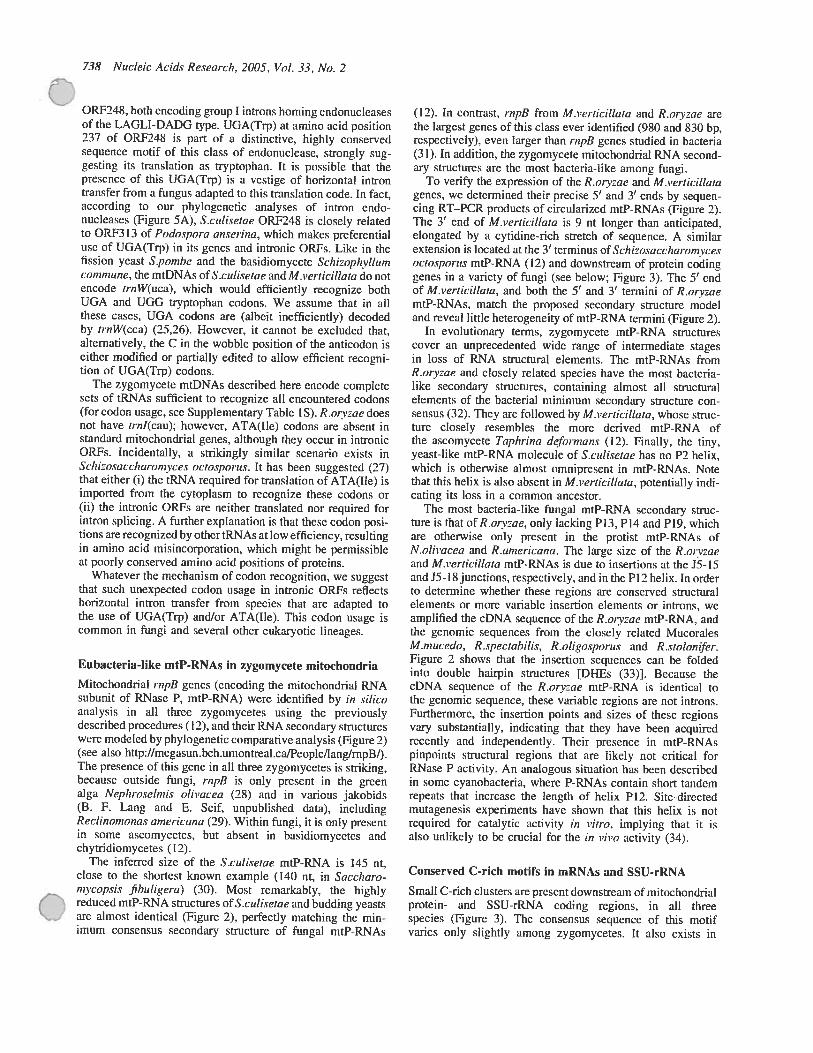
738 Nucteic AciU.ç Research. 2005, VoL 33, No. 2
0RF248, both encoding group I introns homing endonucleasesof the LAGU-DADG type. UGA(Trp) at amino acid position237 cf 0RF248 is part of a distinctive, highly conservedsequence motif of this class of endonuclease, strongly suggesting its translation as tryptophan. It is possible that thepresence cf this UGA(Trp) is a vestige of horizontal introntransfer from a fungus adapted to this translation code. In fact,according te our phylogenetic analyses cf intron endonuc]eases (figure 5A), S.culisetae 0RF24$ is closely relatedto ORF3 13 cf Podospora anserina, which makes preferentialuse of UGA(Trp) in its genes and intronic ORFs. Like in thefission yeast S.pornhe and the basidiomycete Schiwpln’tlumcommune. the mtDNAs of S.c’ulisetae andM.ierticillata do flotencode trnW(uca), which would efficiently recognize bothUGA and UGO tryptophan codons. We assume that in ailthese cases, UGA codons are (albeit inefficiently) decodedby rrnW(cca) (25,26). However, it cannot be excluded that,alternatively, the C in the wobble position of the anticodon iseither modified or partially edited to ailow efficient recognitien of UGA(Trp) codons.
The zygomycete mtDNAs described here encode completesets of tRNAs sufficient to recognize ail encountered codons(for codon usage, sec Supplementary Table 1 S). R.or:ae doesnot have trnl(cau): however, ATA(Ile) codons are absent instandard mitochondriai genes, although they occur in intronicORFs. Incidentally, u strikingiy similar scenario exists inSchïosaccharomvces octosporus. It lias heen suggested (27)that either (j) the tRNA required for translation of ATA(Ile) isimported from the cytoplasm to recognize these codons or(ii) the intronic ORFs are neither translated nef required forintron splicing. A fiirther explanation is that these codon positions are recognized by other tRNAs at low efficiency, resuitingin amine acid misincorporation, which might be permissibleat pooriy conserved amine acid positions cf proteins.
Whatever the mechanism cf codon recognitien, we suggestthat such unexpected codon usage in intronic ORFs reflectshorizontal intron transfer from species that are adapted tethe use of UGA(Tfp) andlor ATA(Ile). This codon usage iscommon in ftngi and several other eukaryotic lineages.
Eubacteria-like mtP-RNAs in zvgomycete mitochondria
Mitochondriai rnpB genes (encoding the mitochondriai RNAsubunit of RNase P, mtP-RNA) were identified by in siticoanalysis in ail three zygomycetes using the previouslydescribed procedures (12), and their RNA secondaiy structureswere modeied hy phylogenetic comparative analysis (Figure 2)(sec aise http://megasun.bch.uiuontrea1.caIPeople/langJmpBf.The presence of this gene in ail three zygonlycetes is striking,because outside fungi, rnpB is only present in the grcenaigu Nephrosetmis olivacea (28) and in varieus jakebids(3. F. Lang and E. Scif, unpublished data), includingRectinomonas arnericana (29). Within fiingi, it is only presentin some ascemycetes, but absent in basidiemycetes andchytridiomycetes (12).
The infeffed size of the S.culisetae mtP-RNA is 145 nt,close te the shortest known exampie (140 nt, in Saccharornycopsis fihuligera) (30). Most remarkabiy, thc highly
( reduced mtP-RNA structures of S.culisetae and budding yeastsare aimest identical (Figure 2), perfectly matching the minimum consensus secondary structure of fiingal mtP-RNAs
(12). In ccntrast, rnpB frem Mi’erticillata and R.orvae arethe iargest gdnes of this class ever identified (980 and $30 bp,respectively), even larger than rnpfl genes sflidied in bacteria(31). In addition, the zygemycete mitechondriai RNA secondary structures are the mest hacteria-like among fiingi.
To verify the expression cf the R.orv:ae and M.verticitlatagenes, we determined their precise 5’ and 3’ ends by sequencing RT—PCR products of circuiarized mtP-RNAs (Figure 2).The 3’ end cf M.verticillata is 9 nt longer than anticipated,elongated by a cytidine-rich stretch cf sequence. A similarextension is iecated at the 3’ terminus cf Schizosaccharomvcesoctosporus mtP-RNA (12) and downstream cf protein cedinggenes in a variety of fungi (sec beiew; Figure 3). The 5’ endcf M.verticittata, and hoth the 5’ and 3’ termini cf R.orvraemtP-RNAs, match the preposed secondary structure modeiand reveai Jittle heterogeneity of mtP-RNA termini (Figure 2).
In evoiutienary terms, zygemycete mtP-RNA structurescever an unprecedented wide range cf intermediate stagesin less of RNA structural eiements. The mtP-RNAs fromR.ory:ae and ciosely related species have the most bacterialike secondarv structures, containing almost a]l structuraielements cf the bacterial minimum secondary structure consensus (32). They are feflowed hy M.verticillata, whose structure closely resembles the more derived mtP-RNA cfthe asccmycete Taphrina deformans (12) Finally, the tiny,yeast-like mtP-RNA molecule cf S.cutisetae bas no P2 lieux,which is otherwisc aimest omnipresent in mtP-RNAs. Notethat this lieux is also absent in Mverticillata, petentiaily mdicating its less in a common ancestor.
The most bacteria-like fungal mtP-RNA secondary structure is that efR.oyzae, only lacldng P13, P14 and P19, whichare ctherwise cnly present in the protist mtP-RNAs ofN.otivacea and R.americana. The large size cf the R.orv:aeand Merticiltata mtP-RNAs is due to insertions ut the J5-15and J5- 18 junctions, respectively, andin the P12 helix. in ordertu determine whether these regions are conserved structuralelements or more variable insertion elements or intrens, weampiified the cDNA sequence of the R.orv:ae mtP-RNA, andthe genemic sequences from the ciosely related MuccraiesM.mucedo, R.spectahilis, R .otigosporus and R.stotonitr.Figure 2 shows that the insertion sequences can be foidedinto double hairpin structures [DFIEs (33)]. Because thecDNA sequence cf the R.orv:ae mtP-RNA is identical tothe genomic sequence, these variable regions are flot introns.furthermcre, the insertion points and sizes cf these regicnsvary substantially, indicating that they have been acquiredrecently and independently. Their presence in mtP-RNAspinpoints structural regions that are likely flot critical forRNase P activity. An anaiegeus situation lias been dcscribedin sortie cyanobacteria, where P-RNAs contain short tandemrepcats that increase thc iength cf heix P12. Site-dfrcctedmutagenesis experiments have shown that tins lieux is notrequired for catalytic activity in vitro, imptying that it isaise uniilceiy to be crucial for the in vivo activity (34).
Conserved C-rich motifs in mRNAs and SSU-rRNA
Small C-rich clusters are present dewnstream cf mitochendrialprotein- and SSU-rRNA coding regions, in ail threespecies (Figure 3). The consensus sequence cf this motifvaries only slightiy ameng zygomycetes. It aise exists in

• AUJ nia
AU
U UU A
Pli
AJrU’”_ u
A
u• “tUU A
A UU A
AAI U
Sniiutiurn U u
culrvetae _IAJ
U’ U UAAUCAU3UUAUUUAAUAUUU
UUAGUG AAUA AUJ A3’ pI LAJA UU
J,,, COOCAUIAA)U°
Nucleic Acidv Research, 2005, Vol. 33, No. 2 739
J3A
Anu4,
Rhizupuson’zae
lU AUUUAUUUGG
A 11111111 A
AAAUAAAC CUU
U—Al’A U—A
2IAA —cAu—Uni, PI G AU -
5, UU??UUfJGU?DU_AAUAAcLI
uAAiAUJ
lIA CGGCUU’A ri su
Figure 2. Secondary structure models for mtP-RNAs from R.on:ae. R.srolonifer 194667. R.oligo.spoins. Rspectahifls. M.mueedo. Scuilserne and M.rerricillrno.
O Positions in red are invariant in the minimum bactedal consensus (32); uppercase letters in thc mtP-RNAs indicate 100%, lowercase 90%, conservation ofthcminimumbacterial consensus sequence. Tbe arrosvspinpointexperimentally determined termini; arrow length tsproponional In thepercentage of molecules endingal a deflned position. Double hairpin clements are named in green. The kw nuclcotides colored blue in Ihe Rstolonjfer mtP-RNA mndel arc different in ils closerelative R.orvzoe.
JUA..
A
ïPA
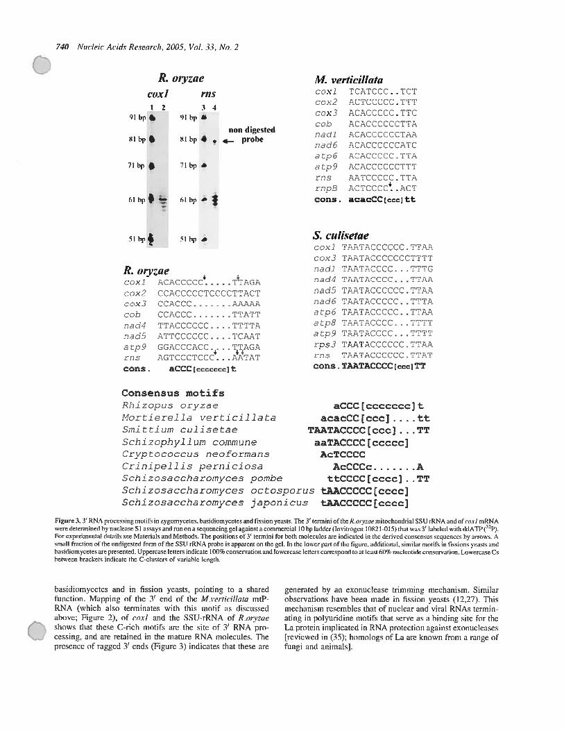
740 Nucleic Acids Research, 2005, Vol. 33, No. 2
R. o,yzaectxl rn.ç12 34
fil bp :, fil bp *
C ITATT
Consensus motifs
Rhizopus orvze
Mortiere1Ii verticiilata
Smitium cuiisetaeSchizophylium commune
Crvptococcus necfornnsCriniceilis nerniciosaSchi zosacchdromyces pombe
S. ct,tisetaeccx TAATACCCCCC. TCAAccx3 TAA’’CCCCCCC;CTTT
TAATACCCC. . .TTTGndd4 TAATACCCC. . . TTAA
nd5 TAATACCCCCC - TTAAndd6 IAATACCCCC.. CITAa6 TAATACCCCC. .TTAA
tp8 TAATACCCC...TC’i’I
‘AA’’ACCCC..’’’TTrcs3 ‘rAA’:’ACt:CCCc.’:’TAArn.s A’ACCCCCC.’”’A’’
cons . TAATACCCC [cccJTT
aCCC [ccccccc] tacacCC [ccc] . .. . et
TAATACCCC[cccj . TTaaTACCCC [ccccc]
AcTCCCC
AcCCCc ACCCCCC[cccc] . .
Schizosccharomyces octosporus tAACCCCC[cccc]Schizossccharomvces japonicus tAACCCCC[cccc]
Figure 3. 3’ RNA processing motifs in zygomycetes, basidiomyceles and fission yeasts. The 3’ termini of the R.ory:ae mitochondrial SSU rRNA ansi of coxi mRNAwere determined hy nuclease SI assays and mn on a sequencing gel against a commercial 10 hp ladder (Invitrogen 10821-015) that was 3’ lahelecl with UdATP (P).for experimental details see Matcrials and Methods. The positions ot 3’ termini for both molecules are indicated in the derivcd consensus sequences by arroses. Asmall fraction of the undigested form of the SSU rRNA probe is apparent on the gel. In the lower part ofthe figure, additional, similar motifs in fissions yeasts andbasidiomycetes are presented. Uppercase letters indicale 100% conservation and Iowercase letters correspond 10 al least 60% nucleotide conservation. Lowercase Cehetween brackets indicate the C-clusters of variable length.
basidiomycetes and in fission yeasts, pointing to a sharedfunction. Mapping of the 3’ end of the M.s’ertidlltata mtPRNA (which also terminates with this motif as discussedahove; Figure 2), of coi! and the SSIJ-rRNA of R.orvzae
O shows that these C-rich motifs are the site of 3’ RNA processing, and are retained in the mature RNA mo]ecules. Thepresence of ragged 3’ ends (Figure 3) indicates that these are ftngi and animais].
)l hp l hp
fl1fll digetcd81 hp SI hp 4 • .— probe
71 hp • 71 hp —
J1/f ,.‘ertkillcttcicxJ TCATCCC. . ICIcox2 ACTCCCCC ITT
ccx. ACACCCCC . TICc-oh ACACCCCCTTTA
naci ACACCCCCCTAA
cat ACACCCCCCATCatpE ACACCCCC.TTA
c3t ACACCCCCCTTT
ïcs AÏsTCCCÇC . TTArnpB ACTCCCC ACTcons. acacCC [ccc] tt
SI bp 4 il hp
R. tnyzaecexi ACAC.CCC TACA
cex2 OCACC000TCCCCTTACT—
fl d In 3 O
a L9r I S
cons.
T’I’AC CCCI AAT 1CC C C C CAAT
CGACCCACC.. . .TJAGAASTCCçTI:ZC. . . AATAT
aCCC tccccccc) t
generated by an exonuclease trimming mechanism. Similarobservations have heen made in fission yeasts (12,27). Thismechanism resembles that of nuclear and viral RNAs terminating in poiyuridine motifs that serve as a hinding site for theLa protein impiicated in RNA protection against exonucleases[reviewed in (35); homologs of La are known from a range of

Niwlew Aeids Research, 2005, Vnl .33, Wo. 2 741
194 nts 46n1s
utpfi url360 arpt-C(GIY-YIG)
212 nts 36nts
«(p9 ort376 atp9’-L(GIY-YIG)
140 uts 54 uts
cox2 orf342 cat2 *_C
Figure 4. Schematic view o[atp6 reions ofA.,ncic,ocv,,iis andA.arhu.sc,jI,,ç(36).atp9ofR.ory:ae andcox2ofM.retiri/1ata.Coding seuenccsarcenclosedin boxes and intergenic spacers are rcpresentcd by u tbick une. Black boxesindicate sequences present hcforc the invasion by the corresponding ORF. Grayboxes represent ORfs and newly acquireci sequcnces.
More instances of mobile endonuclease elements?
As reported earlicr, the chytridiomycete ftngus A.niacrogvnushas a novel mtDNA insertion element whose sequence isabsent in a close relative, Altomvces arbusculu,ç. Ibis clementconsists of a duplicate C-terminus of u foreign otp6 gcnc,plus an ORF encoding an endonuclease that is responsiblefor its mobility (36). The inserted atp6 portion ix fiiscd inphase with the resident gene (Figure 4), reconstituting anobviously ftnctional hybrid gene of standard length andamino acid conservation. In fact, it has been shown that homing endonucleases can be mobile even independent of intronsand genes (37), and that thcy are capable of carrving geneticmaterial from one site to another when they migrate.
lntriguingly. similar gene hyhrids are present in themtDNAs of R.orv:ae (atp9*C) and M.verticillota (co.r2*C),including 0RF376 (R.orvzae) and 0RF342 (M.i’erticillata)(Figure 3). 0RF342 bas no significant similarity to knownendonucleases, but 0RF376, like the mobile element endonuclease of A.rnacrogvnus (36), encodes a protein related tohoming endonucleases of the GIY-YIG type (38,39). 11e samestructural organization is seen in the atpb-ORf360-atp6-Cgene region in A.inacmgvnns, atp9ORF376atp9*C inR.orv:oe and co.v2-ORF312-cox2-C in M.verticillatci(Figure 3), indicating the presence of similar mobile elements.However, contrary to the A.rnacrogvnus case, we currentlydo not have biochemical evidence for the endonucleolyticactivity. Both the cox25-C and atp9*C fragments ofthe two zygomycetes encode C-terminal ends that are tOO%identical at the amino acid level, suggestin that the source ofpotentiai transfers are closely rclated zygomycete species.
Lateral transfer of a group I intron from zygomyceteto angiosperm mitochondria
A significant portion of the genomes described here is occupied by introns (R.oiyae 15.8%; M.verticillata 8.6%; and
Sjtil,çetoc’ 27.4%). With 14 bittons, the mtDNA ofS.cidisetaecontains the largest number, 9 of which are ]ocated in the cox]gene (Table 1). Hère, ail the identified zygomycete introns areof group I and 22 contain intronic OREs: sixteen of theLAGLI-DADO type and six of the GIY-YIG type. In R.oryrae,
identifled one intron [coxl-il(0RF305)], which is mostsimilar to introns inserted at the saine positions of angiosperm
genes (highest BLAST expect value of e’ with Philodendron oxvcardiurn, Lamiunt sp. and Malpighia glahia).Because this ix the only group I intron in vascu]ar plantmtDNAs, it bas most likely been acqufred by lateral transfer.The hypothesis of recent horizontal transfer of this intron froma fungal donor to flowering plants (40) is further hased on theincongruence between the intron and organismal phylogenies,aud its doser phylogenetic relationship to a fungal intron thanto those of Marchantia and Prototheca (40—43). However,becatise of the high mobility of this intron and the possibilityof multiple lateral transfers, the published phylogenetic inferences have to be interpreted with caution.
The presence of additionai, highh’ similar introns inthree fimgi, R.orv:ae, S.culisetac (this paper) andMonoblephareltal5 (8), ailows us to more rigorously testthe hypothesis of Vaughn et al. by phylogenetic analysis(f igurc 5A). Our atialysis reveals that ORFs from R.orv:ae,Monohlcphareltal5 and the three angiosperms group togetherwith high support (98%). 0RF305 from R.orv:ae ix the closestand tnost similar relative of the angiosperm ORFs, suggestingthat the ftngal donor of the group I intron and its resident ORFvas a zygomvcete. Ibis scenario is biologically meaningful,because symbiotic mycorrhizal zygomycetes live in closeassociation with most plants. However, note that ORf248from S.ctdisetae branches with 0RF3 13 from P.anserina.This observation, along with the prescnce of u UGA codonspecifying tryptophan in 0RF24$ (sec above), suggests a second case of luterai transfer. this time from u euascomyceteclose to P.anserina, into a zygomycete.
Phylogenetic analysis with standard mitochondrialproteins: are zygomycetes paraphyletic?
The availability of complete mtDNAs from three distant zygomycetes provides an opportuuity for testing the monophylyof Zygomycota. Tree topologies were biferred from a 2890position concatenated proteirt alignment using two standardML-bascd methods. MrBayes and separate ML analysis. 11eresults of these analyses are summarized in figure 5B, whichpresents the tree produced by IQPNNI, along with bootstrapvalues from ail methods. AIl mcthods produced similar rcsults,except that separate analysis recovered monophyly ofS.rutisetae and M.s’erticitlaia (81% hootstrap support). Thelatter topo]ogy vas aiso found in thc ‘credibie set’ in threeindcpendent runs of MrBayes (5.3, 44.7 amI 99.0% posteriorprobability, respcctively; MrBaves bootstrap support for thisgrouping was 45%). Although thèse data support both of thèsetopologies, k ix interesting that the monophyiy of S.ctdiseiaeand M.verticitlata ix bdtrer supported under more sophisticated(yct statistically valid) models.
Although this phylogeny is generally robust, bootstrapsupport values indicate two major areas of unccrtaintv: theposition ofA.marrogvnus and the relative branching positions
t,tp6
.4.urbuscu!us
4macroq’,r:,y
R.orvzae
W. eerticiltata
(
Cof the zygornycetes. lndeed, also the approximately unbiased
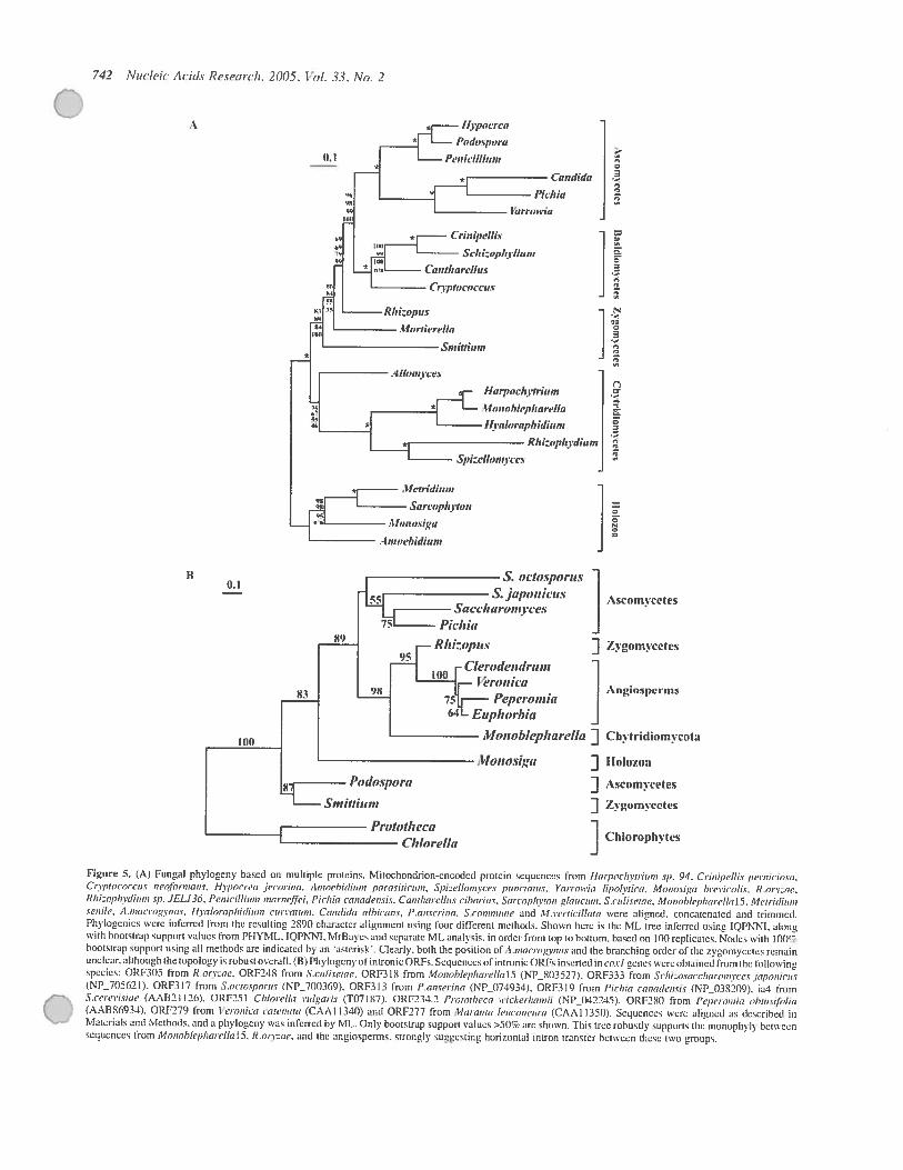
742 Vuc1’4t: Acicis Research. 2005, Vol. 33. No. 2
Figure 5. A Fungal phylogeny based on multiple proteins. Mitochondrion-encoded protein sequences from JJo,-poehvtt-iiini s,,. 94. C,-mnipellis peiniciosa.Crvprococcus neoti,r,nons. Hrjiûrrea /ecorina, ,4nioehidiun, parasitici,,,i.SpieIIo,nvcps pt,flrtaius. Ya,-,’owia tipolvtictt. Alonosiga I)teti(OIts. R.ore;ae.Ithi:ophvdiuni •gi. JELI3Ô. Penicilliun, inarneffel, Pichia canadensis. Cantharellus ciharins. Sarcophvron glatîcum, S.culisetae. Monublepharellul 5. Metridh,,n,çenllt’. A.niacrogvnus, Hvaltiraphicliu,n cuisatum. Caflf(jcIa albicani, P.a,,çt’rina. S.co,u,niu,e and M.,e,,icillata wcre aligned. concatenatcd and trimmcd.Phylogenies were infcrrcd front the resulting 2890 charecter alignoient usine four different methods. Shown here is the ML tree inferrcd using IQPNNI. alongwith hootstrap support values from PFIYML. TQPNNI. MrBaycs and scparatc sfl. analysis. in orfter from top in honom. hascd on 00 replicaies. Nodes with 1 00’hootstrap support using ail methods are indicated bv an asterisk’. Ciearly. both tise position of ..macrogvnus and tise branching order of tise zygomycetes remainunclear. altt,c,usïh tise topolouv is robust overali. R)Phyiogcny of intronic ORFs. Sequeitces otintronic ORFs inserted inco.rI genes were obtaincd frorndic followingspecios: 0RF305 from R.tnvaae. 0RF218 from S.culisetae. 0RF318 from Monoh[epha,c!lal5 (NP...803527). ORf333 from Schi:osacchainn,ycc.s japosiicustNP 705621). 0RF317 frorn ..octosporus (NP_700369i. 0RF313 front P.anserins INP_074034). ORFS 19 from Pichia canadensis tNP 038209). ia1 from5.ce,e,i.siae IAAB2I ] 26). 0RP251 ChIo,dlla vulgasis (T07187). 0Rf234.2 Prorotheca stickerhamll (NPJ)42245). ORF214f) from Pcpctonhin ohti,s,thui,
s iAABS69i4) 0RF279 fiom I uoiltca s tus nato (CAi I s40) md 0RF277 from lltr as itt 15 /1(005w u (CA sl ia0) Sequzncs wci alt.,u5d as dcsvrtbul inMaterials and Methods. and a phylogcny vas infcrrcd by tvff.. Only hootstrap support values >50% arc sbown. Titis trec rohusdy supports tise monophyly bciwcensequences from Monoblepharellal5. R.on:ae. and tise ang,osperms. strongI suggest,ng horizontal huron transfer hctween thcsc two groups.
0.1
ilj’poc;ca
Schfropbylliitii
et
],e
Hurpociirtriuiu
llo,,uhk’pl,urt’Ih,
0.1
I !I•()1lÎctz
1F]7 Zgomvcetes
]J’Ioiioblepliarelht 7 Chtridtornvcota
7 I1oloioi
7 Ascomcettts
7 Zygoinycetcs
]
Potttsporct
S,,,ittiu,n
itoiiosra
ProtothecaCIi(reIlct

G
o
iikelihood ratio test suggcsts these saine probicins. When sitevise likeiihoods are calculated separatelv for cadi functionalciass, tic confidence set contained a total of 17 trce topologiesthat failed to reject tic data OE < 0.05). Arnong these topologies, A.macrogvnus branches immediately above, immediatelv below or monophyletic with the chytridiomycetes, andR.f?rv:ae and M.uerI,(il/ata are found te he cither monophvletic or paraphyletic (with cither species hranching moredceplv tian rie other). Àlthough S.ciilisetae and M.verticillcitaare menophyletic in tic best trec under tus model of separateÏv op[imizcd functional classes, ail topologies in whici ailthree zygomycetes are monophyletic are rejected. It is worthnoting that several obvious wrong positions for S.uuli,vetacwere aise ohserved (e.g. as most ancestral among fungi. oramong the chytridiomyeetes). suggesting that the accelcratedevolutionary rate in t1is species causes a iong-hranch attraction artefact (44). Similar resuits are obtaincd when sitewisclïkelihoods were calculatcd from tic fully concatenated aligoment. Clearly. these data arc insufficient to rcsolve tic phylogcny cf the zygomvcetcs, most likely bceausc these tirecspecies diverge deeply witiin ftngi, and at relativelv shortdistance from caci otier. In suci a situation, two strategiescan be uscd te resolve tic ditemma, addition cf more zvgomycete and ncigiboriug fungal lineages or addition cf moresequenec per species. As our protein dataset is alreadv hasedon eompicre mtDNAs, tus latter strategv iinplics resortingte expresscd sequence tag andlor nuclear gcnorne sequences.a currentlv ongoing projcct.
SUPPLEMENTARY MATERIAL
Supplementar Material is available at NAR Online.
ACKNOWLEDGEMENTS
We thank Gertraud Burger (Université de Montréal) for ielpftleomments on tic manuscHpt, Michael W. Gray and MuiraySchnare for help in tic analysis cf tic genes for small and largerRNAs. Xerry ODonnelt (National Center for AgriculturalUtilization Researci, Peoria. IL). R. W. Lichtwardt(Department cf Botany. Univcrsity of Kansas, Laurence,KS) and Carolyn Babcock (Canadian Coliection of fungalCultures. Ottawa) for supply of fungal strains. We fiirtieracknowledge the excellent teehnical assistance cf Z. Wang.Salary and interaction support from tic Canadian Instimtescf Heaith Research (MOP 42475: B.F.L.), tic Canadianlnstimte for Advanced Rcsearch (CIAR: B.f.L.). tic NovaSeotia Healti Researci Foundation (il.). and tic Bourscs«Excellence biT CIHR: Y.L.) and supplv of laboratorvequipmcnt and inforniatics infrastructure hv GenomeQuebec/Canada is gratefullv acknowledged. Funding tu paytic Open Access publication charges for titis article wasprovided by tic CIHR.
REFERENCES
L Voigt.K.. Cigelnik.E. anti ODonnell.K. l]999)Phvlogcny anti PCRidentification ot clinicallv important Zygornvcctes bascd on nuclcarribosornal—DNA sequence data..!. Clin. MîcrobioL. 37. 3957—3964.
Nucleic Acids Research, 2005. Vol. 33, No. 2 733
2. Tanahe.Y.. O’Donnell.K,. Saika’x’a.M. anti Suivama.J. 2000)Molecular phvloacny of parasitic Zvgomvcota (Dimargantales.coopataoles) haseti on nuclear small suhunit rihosomal DNA sequences.Mol. Phrloieitei. Evol.. t6. 253—262.
3. Voigt.K. anti WostemeyerJ. rtOhX)) Reliahie amplification of actin gantsfacilitates tieep-levci phlogenv. Microhiol. Rex.. 155. 179—195,
4. flcrhec.M.L.. Carmcan.D,A. anti \Vinka.K. 1) Rihosomal DNA antiresolution of branchin ortier amona the ascomvcota: how manvijucienticles are enouh’? Mol. Philo ac’net. EroL2l)0017. 337—3.14. 17.
5. Kurtiman.C.P. 20)13) Phvloaanctie circumscnption of Sacchariirttsccs.Ktuyveromvces anti other iiicnihars of tho Saccharornvcctaceae. anti tueproposai of ihc new minera taichancca, Nakaseomyccs. Naumovia.Vanderwaltozvma anti Zygotomiaspora, 15415 Yeait Rus.. 4, 233—245.
6. James,T.Y.. Porter.D.. Lcander,C.A.. Vilgaivs.R. anti Longcorej.E.(2000) Molecular phulogenettes of ha Chytridiornycota supports theutilitv of ultrastructural data tn chvtrtti systematics. Con. .1. Bot..78. 226—350.
7. Leiuh.J.. Seif.E.. Rotiriguea.N.. Jaeob.Y. anti Lang,B.f. (2003) Futigalevolution meets fungal genomics. In Arora.D.K. (cd.). i-1anUhoo/ ofDingo! Biorechnologv. 2ncl edo. ivktreel Dekker Inc.. N’Y. 145—161.
8. Bullcrwcll.C.E.. forget.L. anci Langil.f. rt003) Evolution ofmonoblepharitiaican fungi hased on complete mitochondtial genomesequences. iViuleic ,tcicis Rush, 161.1—1623.
9. Lang.fl.F.. O’Kellv.C.. Nerad.T., Gray.M.W. anti flttrgcr.G. (2002)‘flic closcst unicellular relatives of animais. Ctuï. Biol.. 12,1773—1778.
10. Alexopolous.C.J.. Mims,C.W. anti Biacksvell,M. 1996) Introducto,’vil vcohigv. John Wiley & Sorts, N’Y.
11. Ustinovaj.,Krienitt,1,.antil’luss.V.A. 2000)Hraloi’ai’hidim’ici,is’tmtniis flot o oreen aiga. but u lower fungus: .\mochzdiuni parasitirum is
not a tungus. but a mambar of Élit DRIPs. Prottst. 151. 253—262.12. Seif.F.R.. Forgeai... Martin.N.C. anci Lang.13.F. (2003) Mitochondnai
RNase P RNAs in ascoraxcere fungi: lineage-specific variations in
RNA secondarv structura. RAÀ. 9. 1073—1083.13. Okpotiu.C.M.. Robertsott,D.. Boss,W.F.. Togasaki.R.K. and
Surzvcki.S.J. 19941 Rapid isolation of nuclci from canot suspensionculture celis usine a BioNchull7cr. flioTerhniqirut. 16. 151—159.
14. Ftigar.R.C. 12004) MIJSCLE: u multiple sequence aligttniettt niethociwith retiuceti time anti spacc complextty. BMC Bioinftirn:atic’. 5. 113.
15. Castrasana.J. 120(10) Sclection of cousarvcd hloclcs frons multiplealignmcnts tor their use in phslogenetic analvsis. Mol. Bio].Ei’ol.. 17. 540—552.
6. GuindonS, anti Gascuci.O 2003) A simple, fast, anti accurate algorithirt10 estimate large phylogenies hy maximum likelihooti. Susr. Bio!..52. 696—71)4.
17. Vinh ie.S. anti Von Haeseler.A. 12111)4) IQPNN1: moving fast through tracspacc anti stopping intime. Mol. Bio!. Erol.. 21. 1565—1571.
1$. flapteste,E., Biinkrnatiti,H.. Lee.J.A., MooreD.V., Sensen,C.W..Gorclon.P..Dumfle,L..Gaaslerlanti.T..Lopez.P..Muller.M. etaL 12002)The anulysisof 100 genes supports the grouping of three highly divergentarnoebae: Dk’ryosu’lium. Euttunoeha. anti cllastniarnoeba. Proc. Ncitl,lcrad, Se!. USA. 99, i414—14]9.
19. Ronquist.F. antI FluelsenlteckJ.P. (2003) Mi’fluycs 3: Bayesianphyiogenetic inference unticr mmcd models. flioinfhrmatics.19, 1572—1574.
20. Schmidt.H.A,. Strimmcr.K.. Vintrron,!et. anti von Hacsclcr.A. 2(102)TREE-PLZZLE: maximum likelihooti pli logenetic anal sis usinequartaIs anti pcrallel computirra. flioinfirmatirs, 18. 502—50-.
21. Shimotiaira.H. anti Hasegawa.M. 1200]) CONSEL: for assassine theconfidence of phylogenetic trac selection. fl!oiotisrnatics.17. 1206—1247.
22. Betitiich.À.J. (1996) Structural analvstsnfmitochondrial DNA moleculcsfrom fungi anti plants using moving pictures anti pulseti-flelci gelelectrophorcsis. J. Moi. Bis!., 255. 564—588.
23. Bullerwell.C.E.. Burger.G. anti l.ang.B.F. 2000, À novel motif foriclentifving ipx3 hocnologs in fungal rtiitochitndrial genornes.l14nds Biocheni .5e!.. 25. 363—365.
24. Lang.B.F.. Gray.M.W. anti fluiger.G. (1999) Mitoehontirial genomeevulutiiin anti thc iidgtn of euk.u’otcs. ‘l non. Rut’. Gener.. 33. 35—397.
25. flullcrwell.C.E.. Leighj.. Scif.F Longcorc.J.E. anti Lang.B.F. (2003)Evolutton of the fungi anti their mttochontlrtc.l genonles. In Àrora.D.K.anti Khachatourkcns.G.G. t edO. Applieci sWsc’oloc’v Ont! Biorechnologs.Elsevier Science. Amsterdam. Vol. 3. 133—159.
744 Nueleic Acids Research, 2005, ot. 33, No. 2
ç;
26. Paquin.B.. Lalorest.MJ.. Forget.L. Roersar.I.. Wang.Z.. Lonccore,J.anti Lanrefl f. 1997) ‘flic fttnal mirochondrial genome projeci:evoluliort cf (unirai nittochondrial renotues anti iheir urne expression.Cuir. Gant’r.. 31. 3X0—395.
27. Bullerv,eIl.C.E.. Letch.J., forgei.L. anti Lang.B.F. 20)13) A comparisoncf three fission yeasi nutochonciriai genomes. Nua!cic AriSa Ras..31. 759—761.
2X. Turmei.M.. Lemieux.C.. flurger.G.. Latrr.flF.. Otis.C.. Plantai. antiGra.M.W. (1999) lIta complate rnitochoncfrial DNA sequences cfAep?,,osel,,tis ,Aitarea ancl Pedi,rc,non,is nhinor. Two radically differentevoluttooar pattems isitlun orcen airae. l’ian, CeI1. 11. 1717—1730.
29. Lanrc.fl.f.. Burger.G.. O’Keli\ .C.J.. Cadergren.R.. Goldinc.G.B.,Lemteux.C.. Sankof .D.. Tunnel.M. anti Cra) MW. (1997, An ancestralrtutoalrondriai DNA resembling a euhacterial cenome in miniature.Aatinie. 387. 493—497.
30. Wise.C’.A. anti Martin.N.C. (1991) Dramalic size variation cf yeastuiitocltoirdrial RNAs suzrests litai RNase P RNAs eau ha quite small..1. flic!. Chenu,. 266. 19151—19157.
31. Browtr.J.W. (1999 The Rihorrucleae P Database. Audeic Acids Ras..27. 311.
32. Sie,ie1,R.V.. Danta,A.fl.. Haas.E.S.. Brown.J.W. anti Pacc.N.R. (1996)llrcp/ttsnnalnu’,,wns simplifies oui view of tOc catalylic core ofohonuclease P RNA. RA.4. 2.452—462.
33. Paquin.B.. Laforest.M.J. aol Lan.fl.F. (2(1(1(1 Doubie-hairpin elemcntsin (ire mitocironcinai DNA cf Allc,nvnes: evidence for mchilit. Mol.Bio!. Etc!. 17. 1760—1761.
34. Vioque.A. (1 997)The RNasc P RNA irctncvanohactarïa. short tandemiyrepeaieti repetttive (STRR) sequences are present vithin tire RNaseP RNA cane in hctcroc)st-icrnunc cvanchactcria. Nodale AcldsRas.. 25. 3471—3477.
35. Wc(jn.SI anti Cedervaii.T. 2002) TIre La ptcteilt. Atour. Rit.fiiochen, . 71. 375—103.
36. Paquitt.B.. Lalcrest.MJ. anti Lang.B.f. (1994) htierspecific trattsldr ofrnitocirondrhl caties in funci anti ereation cf a homologous hybriti gene.Pis),. tv,ntl AraS. Sci. USA. 91. ii $07—i ($10.
37. Edcls,S.R. anti Golet.L. (1992) Artificial mobile DNA clamantccnstructecl ibm the EcoRI entionuclease cane. Pica. lVatl AraS. Sci.USA. 89. 1544—1547.
31. Buraer.G. anti Wenrcr.S. (19X5) Ihe mutoclrontinal CRi] catie rnRelus)) forai rnzssa has an intron tirai contaios a novai t pe cf liRE.J. Mol, fltol.. 186. 23 1—212.
39. Michei.f. anti Cummincs,D.J. (1915) Analsis of ciass I intronsin a tttcchondnai plasmid associatcd wiih senescence ofPodospota cuoc’,i,ra tes cais extraordinarv resemhianeeo tire letraht mena rihosorital intron. Cura. Genet..
10. 69—79.4)). Vauchn.J.C.. Mason.M.T.. Sper-Wiritts.G.L.. Kuhlmttan.f’. anti
Paimer.J.D. 1995) fungal origin by horizoniai iransfer cf u pistilrjiitcchcndriai group 1 ilrtrcir in 10e chitiieric Cor! germe cf Pape, ourla.J. rho!, Rio!.. 41. 563—572.
41. Adaots.K.L., ClcntcnLs.MJ. anti VaughirJ.C. (1998) TOc l’eperomramitoclrontiriai atrI group I (triton: tiunna of irorizontai transferanti subsequent evoiutiont of the inircn 31o!. LiaS.. 4.6 19—696.
42. Clrc.Y.. Qiu.Y.L.. Kuhtirttatr.P. anti I’ahriter.J.D. (1991) Explosiveinvasion of plant mitocirontiria by u gtotip I intron. Proc. AttilAca,!. Sa!.USA. 95. 14244—14219.
43. Palr;rar.J.D..Atiams.K.L..Cho.Y..Parkinson.C.L..Qiu.Y.L.andsong.K.(211X0 Dynamic evolution cf plant nritcchotrdriai genomcs: mobilecaties anti introns anti irichly variable ututation ratas. P oc’. Ara,! AraS.Ici USA 97. 696(1—6966.
14. Fcisensteitr.J. (1971) Case in winch pansimcnv anti coinpatihiiitymetltods rvill be pcsiti ely nusicading. Svst. Zoo!.. 27.401—410.
C 75
Article 3
Fusions of mitochondrial with E. cou ribonuclease P RNAs are
catalytically active.
Elias Seif’, Alexandre Cadieux and B. franz Lang
Manuscript in preparation.
Canadian Institute for Advanced Research, Centre Robert Cedergren, Département de
Biochimie, Université de Montréal, 2900 Boulevard Edouard-Montpetit, Montréal,
Québec, H31 1J4, Canada.
Authors contribution:
Elias Seif: developed detailed experimental design and did most of the experiments,
data analysis and preparation ofmanuscript and figures.
Alexandre Cadieux: did part ofthe experimental work.
Franz Lang : helped design the experiments, and prepare the manuscript and figures.
o
76
Abstract
Mitochondrially encoded RNA subunits of mitochondrial RNase P (mtP-RNA) have
been identified in jakobid flagellates such as RecÏinomonas americana, the
prasinophyte NephroseÏrnis olivacea, and in numerous ascomycete and zygomycete
ftmgi. Whereas the secondary structures of ascomycete mtP-RNAs are highly reduced
compared to thefr bacterial counterparts, the jakobid, prasinophyte and zygomycete
mtP-RNAs retain most features of the minimum bacterial structure, and are
potentially active in vitro in the absence of a protein subunit. Here we describe the
precise mapping of R. americana and N olivacea mtP-RNA ends, and the
comparative structural analysis of six additional jakobid RNAs. We further
investigate ribozyme activities of mtP-RNAs and fmd that even the most bacteria-like
molecules ofjakobids are inactive in vitro. However, when certain domains ofjakobid
and N olivacea RNAs are exchanged with E. cou P-RNA sequences, they regain the
capacity to specifically cleave tRNA precursors. In vitro mutagenesis ofthese hybrid
mtP-RNAs provides evidence for the importance ofvarious structural elements for the
ribozyme activity, including a remnant P3 pairing in S. ecuadoriensis, and GNRA
tetra-loops in 114 of I libera and in L1$ of both Jakoba libera and R. arnericana
#50394. We will discuss why native mtP-RNAs fail to catalyze 5’ tRNA processing
in vitro, and discuss reasons for the evolutionary tendency to reduce if not eliminate
the mitochondrial RNA subunit ofRNase P.
C
C 77
Introduction
Ribonuclease P (RNase P) is an endonuclease that removes 5’ leader sequences from
tRNA precursors in eubacteria, archaebacteria, eukaryotes and organelles. In most
studied cases, RNase P is a ribozyme composed of an RNA component (P-RNA), and
one or several protein subunits (P-protein; (Altman et al., 1995; Frank & Pace, 199$;
Hartmann & Hartmann, 2003)). Possible exceptions are in spinach chloroplasts
(Thomas et al., 1995; Thomas et al., 2000b) and in human mitochondria (Rossmanith
& Karwan., 1998; Puranam & Attardi, 2001; Rossmanith & Potuschak, 2001) where
protein-only ribonucleases are suspected.
When present, the P-RNA subunit is indispensable for the endonucleolytic
activity of RNase P, as shown by RNase treatment in a broad variety of cases
including eubacteria, arcbaebacteria, eukaryotes, plastids and mitochondria (e.g.,
($tark et al., 197$; Gardiner & Pace, 1980; Kiine et al., 1981; Hollingsworth &
Martin, 1986; Jayanthi & Van Tuyle, 1992; Baum et al., 1996; Lee et al., 1996a; Han
et al., 199$; Vainauskas et al., 1998; Stathopoulos et al., 2001). Furthermore, ail
tested eubacterial and some archaebacterial (Methanobacteria, Thermococci, and
Halobacteria) P-RNAs, catalyse 5’ tRNA processing in vitro in the absence of the P
protein (Guerrier-Takada et al., 1983; Gardiner et al., 1985; Pannucci et al., 1999;
Wagner et al., 2001). Although eukaryotic P-RNAs lack such an in-vitro activity, the
mechanism of cleavage of the RNase P holoenzyme is RNA-based, similar to that of
eubacteria (Thomas et al., 2000a).
C
G 7$
P-RNA from E. cou (Ml RNA) was the first multiple-turnover ribozyme to
be identified (Guerrier-Takada et al., 1983), and is by far the best-studied P-RNA.
Secondary and tertiary structure models have been proposed for this RNA, based on
phylogenetic comparative analyses, enzymatic probing, and cross linking experiments
(Brown et al., 1996; Westhof et al.. 1996; Haas & Brown, 199$; Massfre et al., 199$).
Ail P-RNAs except those of some mitochondria (for details see below) contairi two
main folding domains (Loria & Pan, 1996), the catalytic domain (“C” domain,
including helices PI-6 and P15-I$), and the specificity domain (“S” domairi; Fig. I).
The “C” domain extends from the 5’ and 3’ ends ofthe RNA and includes Pi-6, 15-
18 helices, loops (L), and single stranded junctions (J; figure I B). Two main
functions were assigned to the C-domain: (i) binding of Mg2 ions that are essential
for catalysis (Christian et al., 2000; Kaye et al., 2002; Tanaka et al., 2004a), which is
mainly accomplished by P4 and the single stranded region in the junction between P3
and P4 (J3/4); and (ii) binding ofthe 3’ terminal RCCA oftRNA precursors by the
GGU motif that is present in the internai loop between P15 and P16 ofthe E. cou P
RNA (L15/i6; (Kirsebom & Svard, 1994; Kufel & Kirsebom, 199$; Brannvall et al.,
2004)). The S-domain, which includes P7-14 helices, loops and junctions, is mainly
responsible for interactions with the T stem-loop region of pre-tRNA substrates.
Recently, the structure of the S-domain of the Thermus thermophilus P-RNA (which
is most sirnilar to E. cou P-RNA, defmed as type A) was resolved at 2.9 À
(Krasilnikov et aI., 2004). Although the overali folding of the “S” domains of T.
thermophiÏus and BaciÏlus subtilis (type B) differ, they share an opening structure
C 79
formed by P9, P10 and Pli and the non-helical module JI 1/12-J12/1 1 (Krasilnikov
et al., 2003). It has been suggested that this common structure accommodates the pre
tRNA (specifically T-stem and T-loop) and orients it for interaction with the catalytic
domain (LaGrandeur et al., 1994; Odeil et al., 1998). It is known that tetraloops ofthe
GNRA type confer thermodynamic stability of short RNA double helices (Heus &
Pardi, 1991); further, that they stabilize tertiary structures of large RNAs by intra
molecular interactions (Jaeger et al., 1994; Murphy & Cech, 1994; Cate et aI., 1996).
Phylogenetic comparative studies and cross-Hnking studies in E. cou suggest that C
and S- domains interact through tertiary interactions of GNRA tetraloops, between
L1$/P$, and L9/P1 (Brown et al., 1996; Massire et al., 1997). Sirnilar interactions
occur within the S-domain, between L14/P8 and L13/P12. GNRA motifs in E. cou P
RNA contribute to the catalytic activity as shown by in-vitro mutagenesis. A
significant decrease of E. cou RNAse P activity was noted when certain GNRA
motifs were exchanged by UNCG tetraloop motifs (Pomeranz Krummel & Altman,
1999), which are otherwise common in RNA molecules (Proctor et al., 2002).
Genes encoding P-RNAs (rnpB) have been determined in hundreds of species,
and secondary structure models were proposed for P-RNAs from the three major
domains of life, including plastids (Brown, 1999) and mitochondria (Seif et al., 2003;
Seif et al., 2005). Comparative studies suggest that ail P-RNAs are homologs, sharing
up to five sequence motifs (CRI-V; (Chen & Pace, 1997)), a cage-shaped pseudo-knot
(P1-P4; (Forster & Altman, 1990)), a four-way junction (P7-i0), and a large internai
loop comprised between Pli, P12, P13 and P14 (J11/12, J12/13, J14/11) (Chen &
o $0
Pace, 1997). The most notable departures from this established consensus structure
exist in mitochondria ofbudding yeasts and the zygomycete Srnittium cuÏisetae, where
P-RNA secondary structures are limited to Pi and P4 (Lee et al., 1996b; Seif et al.,
2003; Seif et al., 2005; Talla et al., 2005). Mitochondrial P-RNAs (mtP-RNAs) from
other ffingal mitochondria are less derived, intermediate between those of yeast
mitochondria and Eubacteria. As mitochondria originated from an Œ-Proteobacterium,
the reduced forms of ffingal mtP-RNAs are best explained by loss of structural
elements from an Œ-Proteobacterium-like P-RNA (Seif et al., 2003). indeed,
mitochondria of the bacterivorous protist Reclinomonas americana (member of the
jakobid flageliates) have the most bacteria-lilce mitochondrial genome based on
various features (Lang et al., 1997; Jacob et al., 2004), including its mtP-RNA. It
contains ah structural elements present in the minimum eubacterial consensus (Lang
et al., 1997), p13, P14, plus a P19 helix otherwise present in the mtP-RNA of the
prasinophyte Nephrosetmis olivacea (Turmel et al., 1999; Seif et al., 2005), and in u
proteobacterial P-RNAs (fig. lA, B). Whereas mtP-RNAs have been well
characterized in various fungi (Shu & Martin, 1991; Wise & Martin, 1991; Lee et al.,
1996b; Seif et al., 2003; Seif et al., 2005), little is known about the more eubacteria
like mtP-RNAs from jakobids or from N olivacea. In particular, it is currently
unknown whether or flot these mtP-RNAs have a catalytic activity in vitro, in the
absence ofa protein subunit.
In this study, we show that the mtP-RNAs from R. americana and N olivacea
are transcribed and processed according to predictions based on RNA secondary

o
$1
structure models. b gain information on the evolution of the jakobid mtP-RNAs,
we identified six rnpB genes iii newly sequenced mitochondrial genomes. We ffirther
investigate the in vitro catalytic activity of several mtP-RNAs and show that the
native RNA molecules are inactive, but that the activity can be partially restored in
chimeras with E. cou P-RNA, and augmented through a variety of mutations. The
reasons for the lack ofactivity of native mtP-RNAs will be discussed.

C 82
MATERIAL AND METHODS
Ccli cultures and nucleic acid extractions
An axenic strain of N oÏivacea (NIES-484; a prasinophyte green alga distributed by
the National Institute for Envfronmental Forum, Japan) was kindly provided by Drs.
M. Turmel and C. Lemieux. Ceils from this species were grown as previously
described (Turmel et al., 1999). The jakobids Reclinomonas americana (New Zealand
strain, ATCC 50394), Reclinomonas americana (ATCC 50283), Reclinomonas
americana (ATCC 50284), Reclinornonas sp. (ATCC 50633) and Seculamonas
ecuadoriensis (ATCC 5068$) were grown in WCL medium, whereas the two jakobids
adapted to saline growth conditions, Jakoba libera (ATCC 50422) and Jakoba
bahamiensis (ATCC 50695), were cultivated in F12 medium. The recipes for these
growth media are found at
http://megasun.bch.umontreal.calPeople/lang/FMGP/methods.html. For large-scale
cultures, jakobids were incubated in 2.5-liter culture flasks under gentie shaking, at
24°C for several days. They were daily feU with live Enterobacter aerogenes bacteria
(ATCC 1304$). When reaching the late logarithmic growth phase (afier 5 days),
ceils were harvested by centrifugation and the nucleic acids were extracted as
described (Seif et al., 2003).
MtP-RNA 5’ and 3’ end mapping
About 10 jig of mitochondrial RNA from were used to determine the precise 5’ or 3’
ends of mtP-RNAs, by RNA ligation, RT-PCR amplification and cDNA sequencing,
Ç:$3
as previously described (Seif et al., 2003). Primer sequences for R. arnericana are 5’
TAAATGAGTCCAGACTTTCCT 3’ and 5’ GAGCAAAACCATTA
GGTTGCT 3’; and 5’ CTATTTCATACTAGCTCT 3’ and 5’ GTGCAAGAAT
TTAATTCGCT 3’ for N olivacea.
Construction of chimerical P-RNAs
The rnpB genes of E. cou, R. americana #50394, I libera, £ ecuadoriensis R.
oîyzae, and N olivacea were cloned in pfBS (a modified bluescript vector) and used
as templates for the following PCR amplifications. Primer sequences for construction
of chimerical P-RNAs and for other mutagenesis experiments are available as
supplementaiy information. The cloning vector (pFBS) is amplified together with one
of the two (mitochondrial or E. cou) domains, which permits direct cloning of
chimerical rnp3 genes, afier DNA ligation.
For construction of type I chimeras (see Fig. 2A for the precise extensions of
the mitochondrial and bacterial sequences), mitochondrial P-RNAs were amplified to
include Pi-4, P18, 19 plus J15/1$. The respective E. cou P-RNA (Ml RNA) domain
was amplified to include P5-P17. For type II chimera (see Figure 3A) the amplified
mitochondrial domain contains P5, 7-15; and the E. cou domain of Pi-4, J 15/18 and
P18. All amplification products were end-repaired (unmodified T7 DNA polymerase
from New England Biolabs, #M02565S, plus the Klenow fragment from Roche,
#100$404) at 14°C for 30 minutes. After denaturation ofthe polymerases at 65°C for
10 minutes, the DNA fragments were purified on low melting agarose gel (FMC
o $4
Bioproducts #50 102). The PCR products including the vector sequence was
dephosphoryÏated with aikaline phosphatase (Boebringer Mannheim #7 13023),
followed by extraction with phenoL’chloroform and ethanol precipitation, whereas the
corresponding fragment containing the other haif of the P-RNA sequence was
phosphorylated with 14 polynucleotide kinase (USB #27073602). The two fragments
were then ligated atl4°C ovemight, transformed, and cloned with the expected
sequence identified by DNA sequencing.
In vitro synthesis ofRNA and RNase P activity assays
The intemally radio-labelled precursor tRNA substrates, and the unlabelled P-RNAs
were synthesized in vitro and purified as described previously (Jacob et al., 2004).
DNA templates for in vitro synthesis ofRNA were prepared by PCR amplification of
respective P-RNA sequences, by inclusion of a T7 RNA polymerase promoter in one
of the two primers used for amplification. Purified RNAs were dissolved in lx PA
buffer (50 mM Tris, pH 7.5, 100 mlvi NH4CT, 10 mM MgC12), heated for 5 min at
65°C and then allowed to slowly cool to 37 oc.
The catalytic activity of P-RNAs was tested using 1,000 cpm of substrate
under two conditions: (i) as used for testing the activity of bacterial P-RNAs
(Guerrier-Takada et al., 1986), including lx PA buffer (50 mlvi Tris, pH 7.5. lOOmM
NH4C1, 10 mM MgC12), 100 mM MgC12 and 4% PEG (polyethylene glycol), or (ii)
as used for testing the archaebacterial activity (50 mM Tris pH 8, 300 mM MgC12, 2.5
M ammonium acetate, 0.1% $D$, and 0.05% Nonidet P-40; (Pannucci et al., 1999)).

85
For assays including the E. cou P-protein, 5nM ofthe protein was added to 0.5 nM
of P-RNA in binding buffer (2OmM Hepes-KOH, pH 8, 400mM NH4OAc, lOmM
Mg(OAc)2, 5% glycerol), the mixture was heated for 5 minutes at 37°C, afier which
1,000 cpm of tRNA precursor (substrate) was added and the mix incubated at 37°C
ovemight.
Reaction products were loaded on 7% denaturing polyacrylamide gels as
described (Jacob et al., 2004), gels were exposed for 2 hours on a Biorad molecular
imaging screen K (#1707841), and images were developed using the Biorad
molecular imager FX and analyzed with the “Quantity One” software.
Genetic complementation assay
BL21 ceils that carry a rnpA mutation in the E. cou chromosome (C5 R46H;
(Guerrier-Takada et al., 1995)) and that contain prophages encoding the T7 RNA
polymerase, were used for the genetic complementation assay (the permissive and
non-permissive temperatures for this E. cou strain are 30°C and 43°C, respectively).
The mutant ceils were transformed with plasmid constructions encoding respective
mtP-RNA constructions, and grown ovemight at 30°C on LB media with 5 jig/ml
tetracycline (rnpA chromosomal selection) and 100 tg/ml ampicillin (plasmid
selection). Several independent transformants were then grown on two different (pre
warmed) agar plates at either 43°C or 30°C. The genetic complementation ofthe
various P-RNAs was assessed by examining colony growth after overnight incubation
at the non-permissive temperature (Baer et al., 1989). Ceils transformed with vector

ç, 86
alone and a vector encoding Ml RNA served as the negative and positive controls,
respectively.
Q
C $7
RESULTS
R. americana and ]V olivacea mtP-RNAs of predicted size are expressed in vivo.
To verify that the mtP-RNAs from N olivacea and R. arnericana #50394 are
transcribed, and processed in vivo at the sites as predicted (Lang et al., 1999; Turmel
et al., 1999), we mapped thefr precise extremities by ligation ofthe 5’ and 3’ ends of
mitochondrial RNAs, RT-PCR, and sequencing of the amplification products. In both
cases, the 5’ and 3’ extremities matched the iiiferred secondary structure models, with
littie heterogeneity ofthe RNA extremities (Fig. lA, B).
Bacteria-hke mtP-RNAs in jakobids
In order to obtain data for the comparative analysis ofthe bacteria-lilce mtP-RNAs in
the jakobid R. arnericana #50394 (Lang et al., 1997), we sequenced mitochondrial
DNAs (mtDNAs) of various other jakobids inciuding R. americana #502$3, R.
americana #502$4, Reclinomonas sp. #50633, Seculamonas ecuadoriensis, Jakoba
libera, and Jakoba bahamiensis (Burger G and Lang BF; unpublished data). By
searching universally conserved motifs of P-RNAs, and the universally conserved
pseudo-knot (P4) (Forster & Altman, 1990; Chen & Pace, 1997; Seif et al., 2003), we
identified rnpB genes in ail jakobid mtDNAs. Their predicted mtP-RNA secondary
structures are similar to those of R. arnericana #50394 (Lang et al., 1997), containing
ail structural eiements of the minimum bacterial secondary structure (Brown et al.,
1994). The only exception is in S. ecuadoriensis, which has a higlily reduced P3 (Fig.
lB). following a comparative analysis of ail jakobid mtP-RNAs, we can update the

C 8$
previously pubiished secondary structure mode! for R. arnericana #50394 (Lang et
aÏ., 1997) by extending the P18 helix, and by assigning a GNRA tetra-loop to L1$ that
is conserved among jakobids except in S. ecuadoriensis (Fig. 1 B). Also note that the
P19 helix, which is a iandmark feature of few eubacteriai lineages including u
Proteobacteria, is also present in jakobid (except J bahamiensis), and in the
prasinophyte mtP-RNAs.
Bacteria-like mtP-RNAs are catatytically inactive in vitro
To determine if any of the bacteria-like mtP-RNAs are catalytic in vitro, we have
chosen seven candidates that contain ail or most of the structural e!ements as defmed
in the minimum bacterial P-RNA structure (Fig. lA), including the jakobids R.
americana #50394, S. ecuadoriensis, I libera, I bahamiensis, the prasinophyte N
otivacea, and the zygomycetes Rhizopus oryzae and Rhizopus oligosporus (Seif et al.,
2005). Respective mtP-RNAs were synthesized in vitro, and their catalytic activity
tested in vitro using the RNA precursors ofmitochondria! tRNA’° from R. americana
and tRNA from R. oiyzae as substrates. None of the mtP-RNAs exhibits a cata!ytic
activity, under any testing conditions (ribozyme concentration, low and high sait,
addition ofPEG, etc). The activity ofthe R. americana #50394 mtP-RNA was further
tested in vitro, in the presence of the E. cou P-protein (the protein subunit of E. cou
RNase P; resu!ts not shown), and in an in vivo complementation assay in the thermo
sensitive E. cou A49 mutant, which has a defective P-protein (Baer et ai., 1989). A!so
these additional assays tumed out negative (data not shown).
C $9
MtP-RNA domains can be complementary to E. cou P-RNA sequences
To understand why bacteria-like mtP-RNAs are flot active in vitro, i.e., in order to
locate specific structural elements that are lacking for activity, we constructed
chimerical P-RNAs by combining half-molecules of E. cou with mitochondrial P
RNAs.
Type I chimeras contain the most conserved structural elements of mtP-RNAs
including PI-4, 1$, and 19; the remaining parts ofthe molecule are from E. cou (fig.
2A). Type I chimeras have various degrees of in-vitro activities, highest in I libera
and followed in order by R. americana #50394, £ ecuadoriensis, and N olivacea
(f ig. 2B,C). The chimera formed with the R. oryzae mtP-RNA is inactive. In type II
chimeras, the mitochondrial ami E. cou portions ofthe P-RNA molecule are reversed
with respect to type I chimeras (f ig. 3A). The R. americana #50394 P-RNA has the
highest catalytic activity followed by I tibera and S. ecuadoriensis; N otivacea is
inactive (Fig 2B,C). Type II chimeras are inactive at lower sait concentrations (e.g.,
lOOmM NRCl, 110 mM MgCl2, 5OmM Tris, pH 7.5) and require 10 times higher
ribozyme concentrations to achïeve leveis of activity comparable to type I, at higlier
sait concentrations (fig. 2B,C).
When incubated in the presence of the E. cou P-protein (C5), the catalytic
activity ofthe type II chimera of R. americana #50394 is strongly increased, but only
slightly in I libera (figure 3C). Other type II chimeras are unaffected by the addition
ofthe protein subunit, as are ail type I chimeras (data flot shown).
C 90
Mutation of conserved GNRA tetra-Ioops in jakobid mtP-RNAs reduce catalytic
activity
Comparative sequence analysis suggests the conservation of GNRA tetra-loops in L 18
ofRectinomonas and Jakoba species, and in L14 ofJakoba species (fig. lB), which
is consistent with evidence from Œ-Proteobacteria (Brown, 1999) and E. cou
(Pomeranz Krummel & Altman, 1999). In order to test the impact ofthese motifs on
the catalytic activity of mtP-RNAs, these tetra-loops were mutated in the
corresponding chimerical P-RNAs. According to the resuits with E. cou P-RNA, this
should lead to the loss of the catalytic activity at low (< 20 mlvi) MgC12
concentrations (Pomeranz Krummel & Altman, 1999). Indeed, mutations ofthe tetra
loops in L18 from GUAA to AUAA, in type I chimeras ofI libera and R. americana
#50394, have no visible effect on catalytic activities at 100 mlvi MgC12 or higher (data
flot shown). However, both mutants fail to cut pre-tRNA’ at 3OmM MgC12 (f ig.
4A). Similarly, a mutation ofthe tetra-loop in L14 ofI libera from GAGA to GAGG
(applied to the type II chimera) drastically decreases the catalytic activity even at
300mM MgCI2 (fig. 4B).
Functionally important £3 remnant in mtP-RNA of S. ecuadoriensis
P3 is conserved in ail bacterial P-RNAs, varying in length between 11 and 65
nucleotides (Tanaka et al., 2004b), and is also present in jakobid mtP-RNAs except
for S. ecuadoriensis, which has a potential remnant of this helix with a single A-U

C 91
pair (Fig. lB). To verify its significance, the A-U pair was mutated to either A-A or
C-G (Fig. 5B). At 300 mlvi MgC12, no significant difference in catalytic activity is
observed. However, at 100 mlvi the catalytic activity decreased more than twofold for
the AxA mutant, but did flot affect the mutation in which A-U was replaced with a C-
G base pair (f ig. 5). These resuits support the contribution of P3, or at least a siniilar
structure with a single Watson-Crick base pair, to the catalytic activity of mtP-RNA
of S. ecuadoriensis.
Other structural e]ements with impact on the catalytic activity of mtP-RNAs
In E. cou, the presence of P6, a GGU motif in L 15/16 (positions 292-294) have been
shown to have an impact on the catalytic activity of the P-RNA, (Haas et al., 1991;
Darr et al., 1992; Kirsebom & Svard, 1994). To evaluate the importance of these
structural elements (lacking in ail mtP-RNAs), the domains P5, P6, P15, P16 from E.
cou were grafted into the R. americana #50394 wifd type mtP-RNA. The addition of
these elements was insufficient to produce a catalytically active RNA (data flot
shown).
In addition, we tested the impact of correcting a deviation in the consensus
sequence ofCRV of R. americana #50394 (mutation ofG292 to A), and a deletion of
P19, winch is neither present in the E. cou P-RNA, nor in the mtP-RNA of I
bahamiensis. The deletion of P19 from the respective R. americana #50394 chimera
P-RNA causes a minor (20%) decrease of catalytic activity (Table 1). In contrast,
introduction of a GNRA motif in L9 of the R. americana #50394 type II chimera
C 92
increases the activity (50%), as does the repiacement of A292 by a G in the
universaiiy conserved motif CRV in the type I chimera (370% at iow MgC12
concentrations; see Table 1).
finally, as mitochondrial genes are A+T rich, we verified if an increase of the
G+C content in helicai regions of mtP-RNAs might stabilize the RNA secondary
structure, thereby increasing the catalytic activity. In one experiment, two A-U pairs
in P4 ofa type I chimera were replaced with G-C pairs (fig. lB, Table 1), resulting in
a 320 % increase of the catalytic activity at a iower sait concentration (1 OOmIvI
MgCI2, 100 mM NH4C1), but no significant change under higher sait conditions
(300mM MgCi2, 2.5M ammonium acetate).
o
C
93
DISCUSSION
Mitochondrial P-protein is essential for mtRNase P activity
Six new jakobid mitochondrial rnpB genes have been identified that encode mtP
RNAs with bacteria-like secondary structures similar to that of R. arnericana #50394.
Ail tested jakobid, N olivacea and fungal mtP-RNAs are catalyticaily inactive in
vitro, a result that is flot surprising because eukaryotic, chloroplast and most
archaebacterial P-RNAs require a protein subunit for activity, and because mtP-RNAs
are more A+T rich than most bacterial P-RNAs and will less efficiently fold into a
catalyticaiiy active structure. Therefore, we assume that jakobid, like ail other mtP
RNAs (e.g., yeast or A. nidulans; (Lee et al., 1996a; Lee et al., 1996b; Groom et al.,
1998)), require a more substantial participation of one or more P-proteins to produce
an active RNase P.
Although the E. cou P-protein does flot restore the activity of the native R.
aniericana #50394 mtP-RNA, it significantly enhances the activity of a jakobid
mitochondriaL1E. cou P-RNA chimeras, suggesting littie specificity of the protein
subunit. This view is in lime with the moderate conservation of eubacterial P-proteins
(Gopalan et ai., 1997), and the lack of conservation between the known yeast
mitochondrial P-protein (Stribinskis et al., 2001) and its eubacterial counterparts. To
address these questions in more detail, it is paramount to identify mitochondrial P
protehis in species other than yeast, and that have mtP-RNAs such as various fungi
(Seif et al., 2003; Seif et al., 2005) or the jakobid flageilates.

C 94
Both mitochondrial and bacterial P-RNA sequences interact with the E. cou P
protein
R. arnericana #50394 and I libera type II P-RNA chimeras have low catalytic
activities that significantly increase afler the addition of the E. cou P-protein, whereas
P-RNA chimeras containiiig £ ecuadoriensis and N: olivacea sequences remain
inactive.
Investigations of bacterial P-RNAs indicate the existence of several P-Protein
— P-RNA interactions. In B. subtilis and E. cou, both C- and S-domains contribute to
protein binding according to RNase TI protection or Dimethyl Sulfate (DMS) and
Fe(II)-EDTA footprinting experiments (Vioque et al., 198$; Talbot & Altrnan, 1994;
Loria et al., 1992; Biswas et al., 2000). These resuits are supported by nucleotide
analogue interference mapping (NAIM) in 3. subtiÏis (Rox et al., 2002), which predict
the presence of three major domains of P-protein interaction, two of winch located in
the C domain. This miglit explain why in our experiments only type II chirneras are
activated by the E. cou P-protein.
Secondary structure conservation strongly affects the activity of chimerical P
RNAs
The bacteria-like mtP-RNAs of jakobids and N olivacea have retained structural
features that produce catalytically active E. cou - mtP-RNA chimeras of varying
degrees. The efficiency of complementation appears to correlate with the overali
degree of secondary structure conservation, in comparison to the E. cou structure, and
C 95
other structural features such as conserved tetra-loops. We suspect that the high
activity of the type I chimera ofJ libera is due to the relatively high G+C content of
helical regions of its mtP-RNA, a view that is confirmed by the increase of the
catalytic activity of the type I chimera of R. americana #50394 in which two A-U
pairs are exchanged by G-C pairs (Table I).
The catalytic activity of type I chimeras is an order of magnitude higher than
those of type II chimeras. Again, thîs can be explained by the different levels of
secondary structure conservation of the two mtP-RNA haif-molecules. In type I
chimeras, the mitochondrial domain is best conserved, both among mtP-RNAs, and in
comparison to eubacterial P-RNAs, whereas type II chimeras include the more
variable mitochondrial S-domain. This region of mtP-RNAs is also more easily lost
during the course ofmitochondrial evolution (Seif et al., 2003).
Possible tertiary interactions in mtP-RNAs
The presence of GNRA-type motifs in Li8 and L 14 of mtP-RNAs are possibly
involved in long range interactions with P$, lilce what has been proposed to exist in E.
cou (Brown et al., 1996). Similar GNRA interactions predicted for group I and II self
splicing introns (Michel & Westhof 1990; Costa et al., 1997), ribosomal RNAs
(Gutell, 1996) and bacterial RNase P RNAs (Brown et al., 1996) have allowed to
establish the rules that govem GNRA ioop interactions with RNA helices. The last
adenine ofa GNRA motif(A4) interacts invariably with a C-G base pair, and the base
pair following this C-G interacts with the nucleotide at the third position ofthe tetra
C 96
loop (R3), co-varying according to the nature of the purine. An A at the third
position (A3) is predicted to interact preferably with a C-G, with some tolerance for
other base pairs (Miche! & Westhof 1990), and a G at the third position (G3) is
predicted to interact primarily with a U-A, and with minimal tolerance for A-U, and
no tolerance for C-G or G-C. Following these rules, we expect Li$ to perfectly
interact with P8 in mtP-RNAs of R. americana #50394, Reclinomonas sp. #50633 and
R. americana #50284. In these cases, the Li 8/P$ interactions are shown by broken
mes in Fig. 1. In R. americana #50283 and I libera these tertiary interactions are
also possible, but not in I bahamiensis. To test these and other possible tertiary
interactions, in vitro assays of native jakobid mtP-RNAs have to be developed, which
will require the identification and purification of the respective mitochondrial P
proteins that are expected to be more efficient in reconstitutions of the ribozyme
activity in vitro than the E. cou P-protein.
Conclusions
Bacteria-lilce mtP-RNAs are flot catalytic in vitro, unlike their bacterial counterparts,
but the conservation of some structura! features that are required for activity has been
demonstrated in chimeras with E. cou P-RNA. Our investigations indicate that the
lack ofactivity of native jakobid mtP-RNAs is due to the cumulative effect of(i) their
relatively high A+U content, disfavouring thermodynamic stability compared to G+C
rich bacterial P-RNAs; (ii) the absence of P6 and of certain GNRA motifs affecting
their ability to form long-range interactions; (iii) the absence of the GGU motif in

97
Li 5/L 16, disfavouring substrate binding; and (iv) deviations from the eubacterial
consensus sequences that have a significant impact on the catalytic activity of mtP
RNAs, as demonstrated for CRV of R. arnericana. Obviously, any of these factors
alone does flot explain the lack of activity of mtP-RNAs, since relatively A+U rich
bacterial P-RNAs and a ‘micro P-RNA’ lacking some of the GNRÀ motifs and P6
(Siegel et al., 1996) are catalytically active, and because minor deviations from the
consensus sequences are tolerated in bacterial P-RNAs (Rasmussen & Nolan, 2002).
In addition, binding of the 3’ terminal RCCA of tRNA by L15 is flot essential for
catalytic activity as shown in E. cou (Guerrier-Takada et al., 1984), and in
Cyanobacteria (Pascual & Vioque, 1999).
It is obvious that imperfections of mtP-RNAs accumulate in the course of
evolution, which must be compensated by substantial changes to the protein moiety of
mitochondrial RNase P. The identification of mitochondrial P-proteins will be an
important next step in understanding the fimction and evolution of mitochondrial
RNase P.
C

9$
Acknowledgements:
We wish to thank Dr. Sidney Altman (Yale University) and Dr. Cediia Guerrier-Takada
(Yale University) for tecimical advice and for providing E. cou P-protein and the A49
mutant strain. We further acknowledge the excellent technical assistance of Jean
francois Bouffard and Lise forget. Salary and interaction support from the Canadian
Institutes of Health Research (MOP 42475; BFL), the Canadian Institute for Advanced
Research (CIAR; BFL), and supply of laboratory equipment and informatics
infrastructure by Genome Quebec/Canada is gratefully acknowledged.
o
C 99
References
Altman S, Kirsebom LA, Talbot S. 1995. Recent studies of RNase P. In: Sol! D,
RajBhandry U, eds. tRNA Structure, Biosynthesis, and functions. Washington
DC: American Society for Microbiology Press. pp 67-7$.
Baer MF, Wesolowski D, Altman S. 1989. Characterization in vitro of the defect in a
temperature-sensitive mutant of the protein subunit of RNase P from Escherichia
cou. JBacterioÏ 171:6862-6$66.
Baum M, Cordier A, Schon A. 1996. RNase P from a photosynthetic organelle contains
an RNA homologous to the cyanobacterial counterpart. JMoI Biol 25 7:43-52.
Biswas R, Ledman DW, fox RO, Altman S, Gopalan V. 2000. Mapping RNA-protein
interactions in ribonuclease P from Escherichia cou using disuffide-linked
EDTA-fe. J Mol Biol 296:19-31.
Brannvall M, Kikovska E, Kirsebom LA. 2004. Cross ta& between the +73/294
interaction and the cleavage site in RNase P RNA mediated cleavage. Nucleic
Acids Res 32:5418-5429.
Brown JW. 1999. The Ribonuclease P Database. Nucleïc Acids Res 27:3 14.
Brown JW, Hans ES, Gilbert DG, Pace NR. 1994. The Ribonuclease P database. Nucleic
Acids Res 22:3660-3662.
Brown JW, Nolan JM, flans ES, Rubio MA, Major f, Pace NR. 1996. Comparative
analysis of ribonuclease P RNA using gene sequences from natural microbial
populations reveals tertiary structural elements. Proc Natl Acad Sci U S A
93:3001-3006.
Cate JH, Gooding AR, Podeli E, Zhou K, Golden BL, Kundrot CE, Cech TR, Doudna
JA. 1996. Crystal structure of a group I ribozyme domain: principles of RNA
packing. Science 273:1678-1685.
Chen JL, Pace NR. 1997. Identification ofthe universally conserved core ofribonuclease
P RNA. RNA 3:557-560.

o
100
Christian EL, Kaye NM, Harris ME. 2000. Helix P4 is a divalent metal ion binding
site in the conserved core ofthe ribonuclease P ribozyme. RNA 6:511-519.
Costa M, Deme E, Jacquier A, Miche! F. 1997. Multiple tertiary interactions involving
domain II of group II se!f-sp!icing introns. JMoÏ Biol 267:520-536.
Darr SC, Zito K, $mith D, Pace NR. 1992. Contributions of phy!ogeneticaliy variable
structura! elements to the function of the ribozyme ribonuclease P. Biochemistry
31:328-333.
forster AC, A!tman S. 1990. Similar cage-shaped structures for the RNA components of
ail ribonuc!ease P and ribonuc!ease MRP enzymes. Cet! 62:407-409.
Frank DN, Pace NR. 199$. Ribonuc!ease P: unity and diversity in a tRNA processing
ribozyme. Annu Rev Biochem 67:153-180.
Gardiner K, Pace NR. 1980. RNase P of Baci!lus subtilis has a RNA component. J Biol
Chem 255:7507-7509.
Gardiner U, Marsh TL, Pace NR. 1985. Ion dependence ofthe Baci!!us subti!is RNase P
reaction. J Bio! Chem 260:5415-54 19.
Gopalan V, Baxevanis AD, Landsman D, Altman S. 1997. Ana!ysis ofthe functiona! role
of conserved residues in the protein subunit of ribonuclease P from Escherichia
cou. JMo! Biol 267:818-$29.
Groom KR, Heyman HC, Steffen MC, Hawkins L, Martin NC. 1998. Kluyveromyces
lactis SEF1 and its Saccharomyces cerevisiae homologue bypass the unknown
essentia! function, but flot the mitochondria! RNase P function, of the S.
cerevisiae RPM2 gene. Yeast 14:77-87.
Guerrier-Takada C, Gardiner K, Marsh T, Pace N, A!tman S. 1983. The RNA moiety of
ribonuclease P is the cata!ytic subunit ofthe enzyme. Celi 35:849-$57.
Guerrier-Takada C, Haydock K, Allen L, Altman S. 1986. Meta! ion requirements and
other aspects of the reaction cata!yzed by Ml RNA, the RNA subunit of
ribonuclease P from Escherichia cou. Biochemistry 25:1509-1515.
C 101
Guerrier-Takada C, Li Y, Altman S. 1995. Artificial regulation ofgene expression in
Escherichia cou by RNase P. Froc NattAcadSci USA 92:11115-11119.
Guerrier-Takada C, McClain WH, Altman S. 1984. Cleavage oftRNA precursors by the
RNA subunit of E. cou ribonuclease P (Ml RNA) is influenced by Y-proximal
CCA in the substrates. Ceil 38:219-224.
Guteil RR. 1996. Comparative sequence analysis and the structure of 16S and 23$
rRNA. In: Zimmermann MA, Dahlberg AE, eds. Ribosornat RNA: Structure,
EvoÏution, Processing, and function in Protein Biosynthesis. Boca Raton, FL:
CRC press.
Haas ES, Brown JW. 1998. Evolutionary variation in bacterial RNase P RNAs. NucÏeic
Acids Res 26:4093-4099.
Haas ES, Morse DP, Brown JW, Schmidt fJ, Pace NR. 1991. Long-range structure in
ribonuclease P RNA. Science 254:853-$56.
Han 5J, Lee BJ, Kang HS. 1998. Purification and characterization of the nuclear
ribonuclease P ofAspergillus nidulans. Eur JBïochem 251 :244-251.
Hartmann E, Hartmann RK. 2003. The enigma of ribonuclease P evolution. Trends
Genet 19:56 1-569.
Heus HA, Pardi A. 1991. Structural features that give rise to the unusual stability of
RNA hairpins containing GNRA ioops. Science 253:191-194.
Hollingsworth MJ, Martin NC. 1986. RNase P activity in the mitochondria of
Saccharomyces cerevisiae depends on both mitochondrion and nucleus-encoded
components. Mol Celi Biol 6:1058-1064.
Jacob Y, Seif E, Paquet P0, Lang BF. 2004. Loss of the mRNA-lilce region in
mitochondrial tmRNAs ofjakobids. RATA 10:605-614.
Jaeger L, Michel f, Westhof E. 1994. Involvement of a GNRA tetraloop in long-range
RNA tertiary interactions. JMoI Biol 236:1271-1276.
Jayanthi GP, Van Tuyle GC. 1992. Characterization of ribonuclease P isolated from rat
liver cytosol. Arch Biochem Biophys 296:264-270.
C
C 102
Kaye NM, Zahier NH, Christian EL, Harris ME. 2002. Conservation of helical
structure contributes to flinctional metal ion interactions in the catalytic domain
ofribonuclease P RNA. JMoÏ Biol 324:429-442.
Kirsebom LA, Svard 5G. 1994. Base pafring between Escherichia cou RNase P RNA
and its substrate. EMBOJ]3:4870-4876.
Kiine L, Nishikawa S, Sol! D. 1981. Partial purification of RNase P from
Schizosaceharomyces pombe. J Bio! Chem 256:5058-5063.
Krasilnikov AS, Xiao Y, Pan T, Mondragon A. 2004. Basis for structural diversity in
homologous RNAs. Science 306:104-107.
Krasilnikov AS, Yang X, Pan T, Mondragon A. 2003. Crystal structure ofthe specificity
domain ofribonuclease P. Nature 421:760-764.
Kufel J, Kirsebom LA. 199$. The P15-loop of Escherichia cou RNase P RNA is an
autonomous divalent metal ion binding domain. RNA 4:777-788.
LaGrandeur TE, Huftenhofer A, Nouer I-IF, Pace NR. 1994. Phylogenetic comparative
chemical footprint analysis of the interaction between ribonuclease P RNA and
tRNA. EMBO J 13:3945-3952.
Lang Bf, Burger G, O’Kelly CJ, Cedergren R, Golding GB, Lemieux C, Sankoff D,
Turmel M, Gray MW. 1997. An ancestral mitochondrial DNA resembling a
eubacterial genome in miniature. Nature 387:493-497.
Lang BF, Gray MW, Burger G. 1999. Mitochondrial genome evolution and the origin of
eukaryotes. Annu Rev Genet 33:35 1-397.
Lee YC, Lee BJ, Hwang DS, Kang HS. 1996a. Purification and characterization of
mitochondrial ribonuclease P from Aspergillus nidulans. Eur J Biochem 235:289-
296.
Lee YC, Lee BJ, Kang HS. 1996b. The RNA component ofmitochondrial ribonuclease P
from Aspergillus nidulans. Eur JBiochem 235:297-303.
o

C 103
Loria A, Niranjanakumari S, fjerke CA, Pan T. 199$. Recognition of a pre-tRNA
substrate by the Bacillus subtilis RNase P holoenzyme. Biochemistiy 37:15466-
15473.
Loria A, Pan T. 1996. Domain structure ofthe ribozyme from eubacterial ribonuclease P.
RNA 2:551-563.
Massfre C, Jaeger L, Westhof E. 1997. Phylogenetic evidence for a new tertiary
interaction in bacterial RNase P RNAs. RNA 3:553-5 56.
Massfre C, Jaeger L, Westhof E. 199$. Derivation of the three-dimensional architecture
ofbacterial ribonuclease P RNAs from comparative sequence analysis. JMo1 Biol
279:773-793.
Miche! F, Westhof E. 1990. Modelling of the three-dimensional architecture of group I
catalytic introns based on comparative sequence analysis. J Mol Biol 216:585-
610.
Murphy fL, Cech TR. 1994. GAAA tetra!oop and conserved bulge stabilize tertiary
structure ofa group I intron domain. JMoÏ BioÏ 236:49-63.
Odel! L, Huang V, Jakacka M, Pan T. 199$. Interaction of structural modules in substrate
binding by the ribozyme from Bacillus subtilis RNase P. Nucleic Acids Res
26:3717-3723.
Pannucci JA, Haas ES, Hall TA, Harris JK, Brown JW. 1999. RNase P RNAs from some
Archaea are catalytically active. Froc NatlAcadSci USA 96:7803-780$.
Pascual A, Vioque A. 1999. Substrate binding and catalysis by ribonuclease P from
cyanobacteria and Escherichia coli are affected differently by the 3’ terminal CCA
in tRNA precursors. Froc NatlAcad $ci USA 96:6672-6677.
Pomeranz Krummel DA, Altman S. 1999. Verification of phylogenetic predictions in
vivo and the importance of the tetraloop motif in a catalytic RNA. Froc Nati
AcadSci USA 96:11200-11205.

ç 104
Proctor DJ, Schaak JE, Bevi!acqua JM, faizone CJ, Bevilacqua PC. 2002. Isolation
and characterization of a family of stable RNA tetraloops with the motif YNMG
that participate in tertiary interactions. Biochemistry 41:12062-12075.
Puranam R$, Attardi G. 2001. The RNase P associated with HeLa ce!! mitochondria
contains an essentia! RNA component identical in sequence to that ofthe nuclear
RNase P. Mol Ccli BioÏ 21:548-561.
Rasmussen TA, Nolan JM. 2002. G350 of Escherichia cou RNase P RNA contributes to
Mg2+ binding near the active site ofthe enzyme. Gene 294:177-185.
Rossmanith W, Karwan RM. 199$. Characterization of human mitochondria! RNase P:
nove! aspects in tRNA processing. Biochem Biophys Res Commun 247:234-241.
Rossmanith W, Potuschak T. 2001. Difference between mitochondria! RNase P and
nuc!ear RNase P. Mol Ccli Biol 21:8236-$237.
Rox C, feltens R, Pfeiffer T, Hartmann RK. 2002. Potential contact sites between the
protein and RNA subunit in the Bacillus subtilis RNase P ho!oenzyme. JMo1 Bio!
315:551-560.
$eif E, Leigh J, Liu Y, Roewer I, forget L, Lang Bf. 2005. Comparative mitochondria!
genomics in zygomycetes: bacteria-!ike RNase P RNAs, mobi!e e!ements and a
c!ose source of the group I intron invasion in angiosperms. Nucleic Acids Res
33:734-744. Print 2005.
Seif ER, forget L, Martin NC, Lang Bf. 2003. Mitochondrial RNase P RNAs in
ascomycete fungi: lineage-specific variations in RNA secondary structure. RNA
9:1073-1083.
Shu HH, Martin NC. 1991. RNase P RNA in Candida g!abrata mitochondria is
transcribed with substrate tRNAs. NucÏeic Acids Res 19:6221-6226.
Siege! RW, Banta AB, Hans ES, Brown 1W, Pace NR. 1996. Mycoplasma fermentans
simp!ifies our view ofthe catalytic core ofribonuc!ease P RNA. RNA 2:452-462.
Stark BC, Ko!e R, Bowman EJ, Altman S. 1978. Ribonuc!ease P: an enzyme with an
essential RNA component. Froc NatlAçadSci USA 75:3717-3721.
o

C
105
Stathopoulos C, Tekos A, Zarkadis 1K, Drainas D. 2001. Extensive deproteinization
of Dictyostelium discoideum RNase P reveals a new catalytic activity. Eur J
Biochem 268:2134-2140.
Stribinskis V, Gao GJ, Sulo P, Ellis SR, Martin NC. 2001. Rpm2p: separate domains
promote tRNA and Rpmlr maturation in Saccharomyces cerevisiae mitochondria.
Nucleic Acids Res 29:3 63 1-363 7.
Talbot SJ, Altman S. 1994. Gel retardation analysis ofthe interaction between C5 protein
and Ml RNA in the formation of the ribonuclease P holoenzyme from
Escherichia coli. Biochemist,y 33:1399-1405.
Talla E, Anthouard V, Bouchier C, Frangeul L, Dujon B. 2005. The complete
mitochondrial genome of the yeast Kluyveromyces thermotolerans. FEBS Lett
579:30-40.
Tanaka T, Ando T, Haga S, K&uchi Y. 2004a. Examining the bases of the J3/4 domain
ofEscherichia cou ribonuclease P. Biosci Biotechnot Biochem 68:13$8-1 392.
Tanaka T, Kanda N, Kikuchi Y. 2004b. The P3 domain of E. coli ribonuclease P RNA
can be truncated and replaced. FEBSLett 577:101-104.
Thomas BC, Chamberlain J, Engeike DR, Gegenheimer P. 2000a. Evidence for an RNA
based catalytic mechanism in eukaryotic nuclear ribonuclease P. RNA 6:554-562.
Thomas BC, Gao L, Stomp D, Li X, Gegenheimer PA. 1995. Spinach chloroplast RNase
P: a putative protein enzyme. NucÏeic Acids $ymp Ser:95-9$.
Thomas BC, Li X, Gegenheimer P. 2000b. Chloroplast ribonuclease P does flot utilize
the ribozyme-type pre-tRNA cleavage mechanism. RATA 6:545-553.
Turmel M, Lemieux C, Burger G, Lang 3F, Otis C, Plante I, Gray MW. 1999. The
complete mitochondrial DNA sequences of Nephroselmis olivacea and
Pedinomonas minor. Two radically different evolutionary patterns within green
algae. Plant Celi 11:1717-1730.
Vainauskas S, $tribinskis V, Padegimas L, Juodka B. 1998. Partial characterization of
the ribonuclease P from Tetrahymena pyriformis. Biochimie 80:595-604.
e
106
Vioque A, Arnez J, Altman S. 198$. Protein-RNA interactions in the RNase P
holoenzyme from Escherichia cou. JM0Ï Bio! 202:835-$4$.
Wagner M, fingerhut C, Gross Hi, Schon A. 2001. The first phytoplasma RNase P RNA
provides new insights into the sequence requirements of this ribozyme. Nucteic
Acids Res 29:2661-2665.
Westhof E, Wesolowski D, Altman S. 1996. Mapping in three dimensions ofregions in a
catalytic RNA protected from aftack by an fe(I1)-EDTA reagent. J Mol Bio!
258:600-613.
Wise CA, Martin NC. 1991. Dramatic size variation of yeast mitochondrial RNAs
suggests that RNase P RNAs can be quite small. JBiol Chem 266:19154-191 57.
C 107
Legends to figures
Figure 1: $econdary structure models ofbacteria-Iike mtP-RNAs.
A. Minimum bacterial consensus (Brown et al., 1994) and mtP-RNA secondary structure
models for N otivacea (Turmel et al., 1999), and Rhïzopus oryzae (Self et al., 2005). The
S- and C- domains are indicated in the minimum bacterial consensus, by a broken red
une.
B. MtP-RNA secondary structure models for the jakobids Reclinomonas
americana #50294 (#50394), R. americana #502$3, R. americana #502$4,
ecuadoriensis, I Zibera, and I baharniensis. In the model of R. americana #50284,
nucleotides in green denote the few sequence differences to Reclinomonas sp. #50633.
Positions in red are invariant in the minimum bacterial consensus; uppercase letters
indicate 100%, lowercase >80% conservation of the minimum bacterial consensus
sequence. The arrows pinpoint experimentally determined RNA termini; the arrow length
is proportional to the percentage of molecules ending at a given position. Nucleotides
boxed in black correspond to the mutations that were introduced to increase the GC
content of P4 (see also Table 1). A(292) that has been mutated to correct the deviation
from CRV sequence (Table I), is circled in black.
Figure 2: Catalytic activity assays of type I chimeras.
A. Secondary structure model for type I chimeras. The E. cou domain is shown in red,
and the mitochondrial domain in black.
C

C
10$
B. Catalytic assays of type I chimeras with pre-tRNA° , under conditions optimal
for archaebacterial RNase P. 1,000 cpm of substrate was incubated at 37°C for 1$ hours
in the presence of 100 nM of ribozyme, in 300 mM MgC12, 2.5 M ammonium acetate,
50 mM Tris pH 8, 0.1% $DS, and 0.05% Nonidet P-40. Ail tested RNAs show activity
except the Rhizopus oiyzae chimera. 10 nM of E. cou P-RNA was used as the positive
controi, and substrate oniy as negative control.
C. Catalytic assays of type I chimeras with pre-tRNA°, at high sait conditions. Assays
were performed in 50 mM Tris, pH 7.5, lOOmM N1-L3C1, 110 mlvi MgCY2, with ribozyme
concentrations as in fig. 2B. Under these conditions, only the I libera and R. americana
#50394 chimeras are catalyticaiiy active.
Figure 3: Catalytic activity assays of type II chimeras.
A. Secondary structure model for type II chimeras. The E. cou domain is shown in red,
and the mitochondrial domain in black.
B. Catalytic assays of type II chimeras with pretRNAPt0 and pre-tRNA’, at sait
conditions optimal for archaebacterial RNase P. 1,000 cpm of substrate were incubated at
37°C for 1$ hours in the presence of 1 iiM of ribozyme, in 300 mM MgC12, 2.5 M
ammonium acetate, 50 mM Tris pH $, 0.1% SDS, and 0.05% Nonidet P-40. I Ïibera and
R. americana #50394 have the highest catalytic activity foilowed by I tibera and S.
ecuadoriensis, where as N. olivacea is inactive.
C. Catalytic assays of type II chimeras in the presence ofE. cou P-protein.
.
109
The assays were performed as described in material and methods except that the
concentrations ofP-RNAs and P-protein was five fold higher.
Figure 4: Catalytic assays of GNRA tetra-loop mutants in chimeras of I Zibera and R.
arnericana #50394.
A. Catalytic assays for mutants in L1$ of I libera and R. americana #50394 type I
chimeras. Experiments with the respective wild type (GUAA) and mutant sequences are
labelled in the figure. In the negative control, no ribozyme was included. The assays
were performed with lOOnM of the corresponding P-RNA, for 18 hours at 370 C, with
1,000 cpm ofpre-tRNA’ in a buffer containing 30 mM MgC12, 100 mM NH4CI, 50 mM
Tris pH 7.5.
B. Catalytic assays ofa mutant ofLl4 in the type II chimera 0fI libera.
Experiments with the wild type (GAGA) and mutant sequence (GAGG) are labelled in
the figure. Substrate, no ribozyme was included. The assays conditions were” 1,000 nM
of ribozyme for 18 hours at 370 C, with the conditions optimal for archaebacterial P
RNAs (see material and methods).
Figure 5: P3 mutagenesis in S. ecuadoriensis.
A. Catalytic assays for mutants at the base of P3.
Position 32 is marked by an asterisk (*) in both A and B. 100 nM of the corresponding
RNAs were incubated with 1,000 cpm ofpretRNAPt0 in a buffer containing 100 mI\4
MgC12, 2.5 M ammonium acetate, 50 mM Tris pH 8, 0.1% $DS, and 0.05% Nonidet P-
110
40. The *AxA mutant has a twofold decrease in catalytic activity compared to the
*Gc mutant and the *uA wild type sequence. “Sub.” denotes substrate only (negative
control).
B. Secondary structure model of P3 and surrounding nucleotides.
o

0Table I: Effect ofspecific mutations in R. americana type I and type II chimeras.
Mutations Relative catalytic activity
300 mM MgC12 100 mM MgC12 20 mM MgC12
2.5 M CH3COONH4 100 mM NR1C1 100 mM NH4C1
(50 mM Tris pH 8. 0.1% (50 mM Tris pH 7.5) (50 mlvi Tris pH 7.5)
SDS, and 0.05% NP-40)
AP6 30% - -
L15/16-CCA 50%- -
AP19 80%- -
P4 G+C rich 100% 320% -
A-G 292 110% 120% 370%
L9GAGA 150%- -
Key to mutant code:
AP6; mispairs in P6. L15/16-CCA: mutation of GGU motif to CCA. AP19: deletion of
P19. P4 G+C rich: exchange of A-U base pairs by G-C pairs in P4 (black boxed in Fig.
1). A-G 292: mutation of A292 to G in CRV (black encircled in fig. 1). L9 GAGA:
GAGA introduced into L9.
Catalytic efficiency was defmed as the percentage of substrate cleaved by the mutant,
compared to the original chimera. For ail tests, 100 nM of type I chimera mutant P-RNA
(or 1,000 nM of type II chimera mutant P-RNA) were incubated for 1$ hours at 370 C, in
presence of 1,000 cpm pretRNA0. The reaction buffers are described in the Table.
o
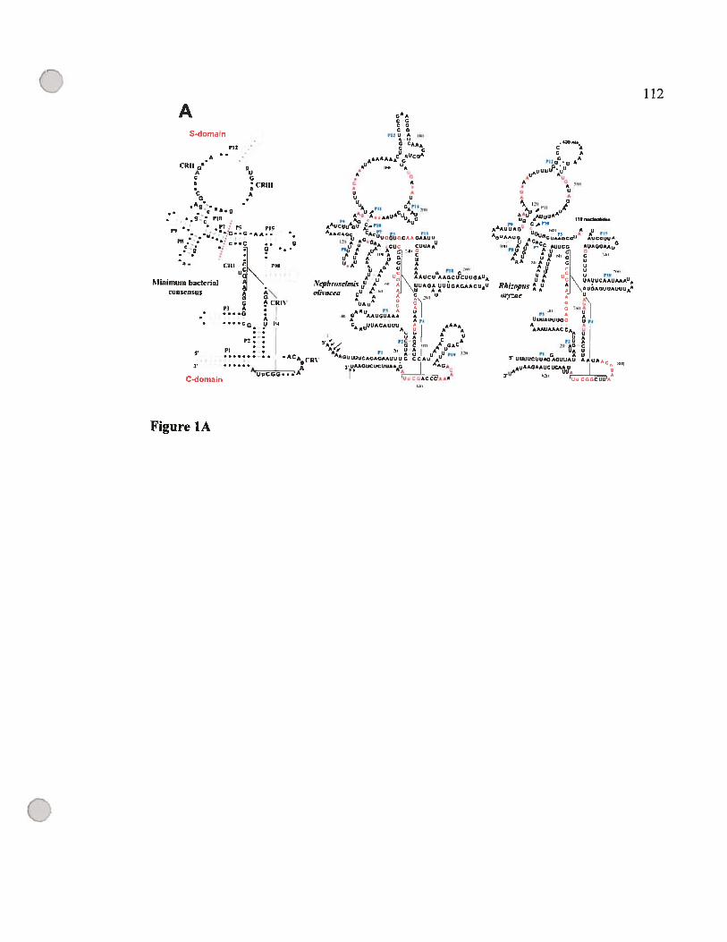
£12
A..CR11 a q
A UC G
CR111
AG
•
PIC
F rs PICL
¾G..G.AA..
:: .:C .. g
Y• CRI PIN
G CRRP3 A
G• A
G N
:..... : .•ACA9CRV
3.“iT1CGG. .
C-domain
oA
5-domain
CCGCGCG
AAACCC
, CA
112
CC A
P12Q0*Q AA
Minimum baclerialConsensus
sCCUCAUCCU,
hAACACACCA GCA
‘2 C A1’) A5 C
CA UG AU
5’ UCUUCCCACAGCCAU CACACC
AACQAAÔj2fl
1))
Figure lA
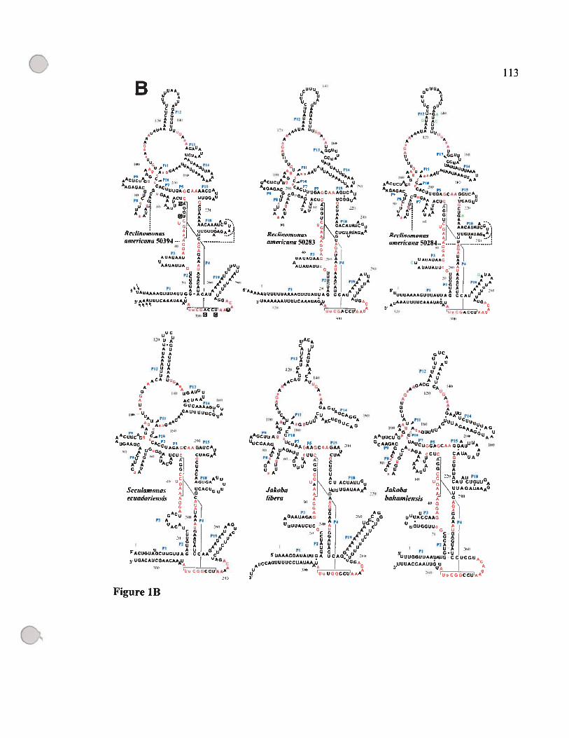
B uLU
UU AU
(2:’ 1313
AU
A AA
AGAUAA UCUAA0
OU_3H A0UAtuh4
AUf GAA 000UAAG A A
AACUCU%S, C_l’A) AAA
AUGAGAC GAUUG%CA13AACCA3C GA
UUGGAU
A,UG LU
u G! AAA (‘1313 A —.AACAAAUC u
- \ uuouuuc., \u A
americ-u,,a 50394 ---
Pi 13 &AUAUAAU n
U A..
AAUAUUA - UUjU
U A U A)(5 Ag AÂ
0A
UA AU513 AUI AuUA0UAAA0003JAUU G 0 A CAU uA..
-13::C1313ACCuA313(
U 0A
U Cu
UC GU
AUUAAUAU
A1lA U03,
‘1313,
uu.,, UAA
A U,lAU1*AUAAUIAu
,AC0310A613 C_rio
AAGAQAC CAC - :5 13113
C0IGUUUGACAAGGUCA0
pAA ACU UCAGUU
UU2: ACL”
AU UA” 013 gUI A
A lIS A.13N AACAUAUC00
Rec-Iinorno,iay I: \gUcUuAGAo.
ainericant, 502K3— Y
UAUAUAA_3U Ap1
2131313 AUAUAUU- 1:13,
‘“U A UAOU A A
U A 113A A131G C u
g U AA u 113313
000AAAA000UAUUAG CCAU
AAAUUAAUAAUG.
II13
o
C
(2133 UUGAUUA
AUriz U A
UAUA
ACA U.A,
ci ‘0auu
A ACUAF (13,3)
U GUCAAÀG0G
11131 U0 AAUA
3U’ ACA,,.,G’
AA13UUCAG_3 -
AUGAUGC CAC :5 Pi”
5’)UG,GUA13GAJCU
3 AU UCU CUAGAA
AUA UACG G
CU 03 AA C.1 AIlS A0U55.’ AGUCA
u
SecuIunwna.% J ‘\UCACU uU
c’cnadorkn.Js A C
AA jG CU “s, A
Pi A - AisUACAU 213313 AAGU
31G UU A A A
1(13 U A A uOC 0U
uA AAC5ACUGUAUGCUUGUU A C AA0 C03.00ACAUCGAACAAAU
c. CAACCUIAAA13
Figure lB
o 113
uACA
CAU
rIz guA
III:G C 4)3
AACAU AU
A ‘A
Jakobalibera
6UCu A
AU
PlI UAUAU
Au
Gu G uuu AA
u 1(4A Iii u0 Cu
111131AA H AG UA
- AG UAAUUCU G’
CAS ‘.5
0C0A AU A13313GGAUA AI)
CAUA
A G Uci Au -
c --1313A AU’U,. \ CAU A
Jakoha A \uU1AGAJAAA6
hai,an,w,z.is
UI” 13U0UACCAAA Au • ApI
UGUGGUU
C GUACG,
tUA UU A . III)
s: PI G.uuuuGGU0AAuAuG CCUCGUs,
uUUACCAAUuG U3:13
113)13L UGCCU11’
II)
G31i A
4GAAUAGA..
UUUUAUCUC
PI A
5uAAAGGAuAuU G A
iA
U0 GUAAA’13:130 3’

I
zI
êtêê4
‘V6
«I
e.6s.Q)E-cuQ)o>1
j.
p
IDDIEo.Ioo4-E
Q
4
C
o.0
D—
4
Q
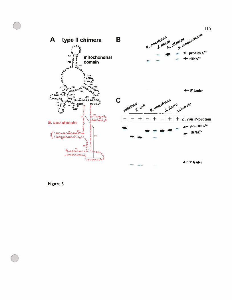
0 115
A type II chimera Eu
AAu
U A
4— pre-tRNAUU A
120 CG mitochondrial. 4— tRNA”UA
CG --
domainA U 40
AUA AA API3
GAGAUAA
A UCUAAUA
AA 160uU
AAGuAAGAAAuuÀ:uuAA 4— 5’ leaderP9 A CI’I AAAA’CUCU 0G C 200 rIoAAGAGAc CP7pç
CCGU6ACUC UUGGAU
“°c G 60 A U
U A A UUG A
AAAU
G GDcf9AGC —
— + — + — + + E.coliP-proteinA C2CGUIiAGE. cou domain A
re-tRNA’AGG A
tRNA0A uc0cGGGc?cGGA’GG G
Acc A
G UC-GVAGC
P2 A UC GA-U
5GAAGCUGACCA G-C ACGA
CUUUGACUGGcPA GUUCGGCCCIAA
4— 5’ leader
Figure 3
o

oQ
—
_ ç’.
.
9
..
74
, &
——
——
r—. 7 7 7
—
4r7
I—
r)
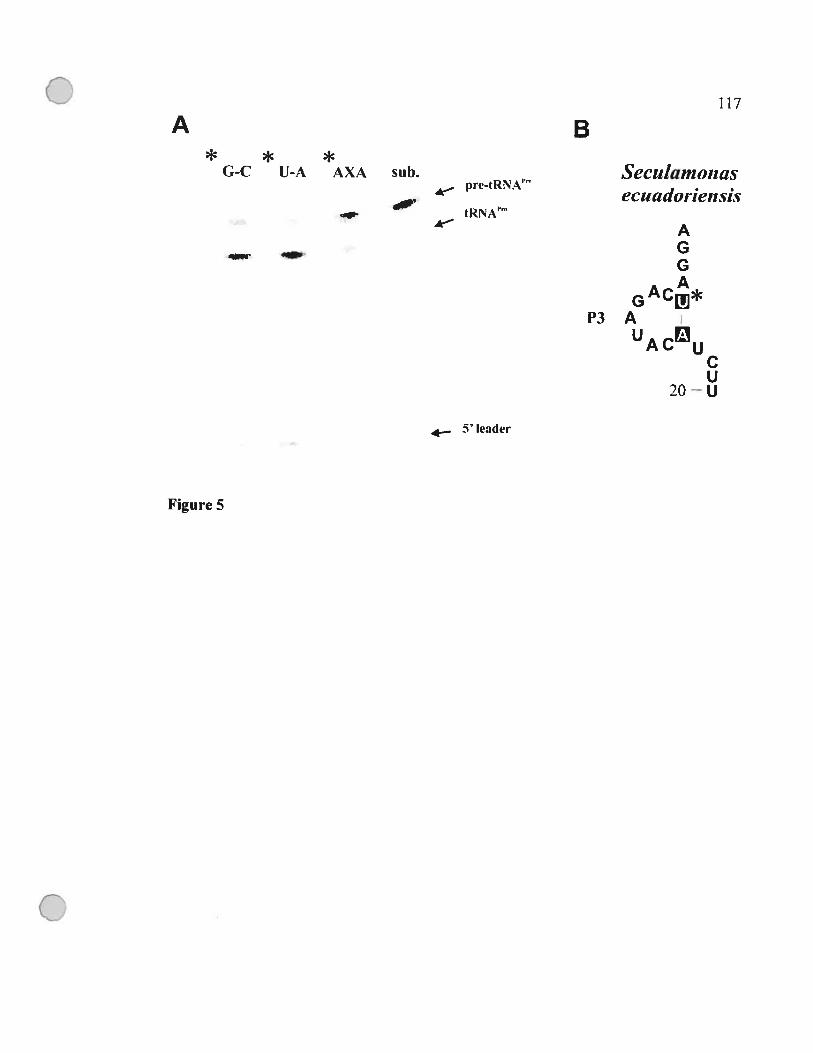
117
A B* * *
G-C U-A AXA sf13. Seciitamonasprc-tRNA’” .
ectictdoi-te,isist RNA”
AG
GpJ*P3 A
A uCU
20—U
5’ leader
Figure 5
o
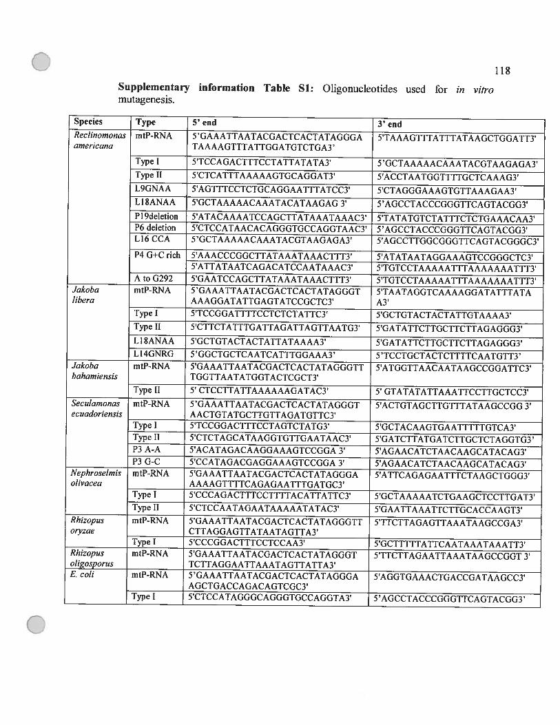
11$
Supplementary information Table Si: Oligonucleotides used for in vitromutagenesis.
Species Type 5’ end 3’ endRecljno,nonas mtP-RNA 5 ‘GAAATTAATACGACTCACTATAGGGA 5’TAAAGTTTATTTATAAGCTGGATT3’americana TAAAAGTTTATTGGATGTCTGA3’
Type J 5’TCCAGACTTTCCTATTATATAY 5 ‘GCTAAAAACAAATACGTAAGAGA3Type II 5’CTCATTTAAAAAGTGCAGGAT3’ 5’ACCTAATGGTTTTGCTCAAAG3’L9GNAA 5’AGTTTCCTCTGCAGGAATTTATCC3’ 5’CTAGGGAAAGTGTTAAAGAA3’Li $ANAA 5’GCTAAAAACAAATACATAAGAG 3’ 5’AGCCTACCCGGGTTCAGTACGG3’Pi 9deletion 5’ATACAAAATCCAGCTTATAAATAAAC3’ 5’TATATGTCTATTTCTCTGAAACAA3’P6 deletion 5’CTCCATAACACAGGGTGCCAGGTAAC3’ 5’AGCCTACCCGGGTTCAGTACGG3’L16 CCA 5’GCTAAAAACAAATACGTAAGAGA3’ 5’AGCCTTGGCGGGTTCAGTACGGGC3’
P4 G+C rich 5’AAACCCGGCTTATAAATAAAC1TT3’ 5’ATATAATAGGAAAGTCCGGGCTC3’5’ATTATAATCAGACATCCAATAAAC3’ 5’TGTCCTAAAAATTTAAAAAAATTT3’
A to G292 5’GAATCCAGCTTATAAATAAACTTT3’ 5’TGTCCTAAAAATTTAAAAAAATTT3’Jakoba mtP-RNA 5 ‘GAAATTAATACGACTCACTATAGGGT 5’TAATAGGTCAAAAGGATATTTATAlibera AAAGGATATTGAGTATCCGCTC3’ A3’
Type I 5’TCCGGATTTfCCTCTCTATTC3’ 5’GCTGTACTACTATTGTAAAA3’Type ii 5’CTTCTATTTGATTAGATTAGTTAATG3’ 5’GATATTCTTGCTTCTTAGAGGG3’LI SANAA 5’GCTGTACTACTATTATAAAA3’ 5’GATATTCTTGCTTCTTAGAGGG3’Li 4GNRG 5 ‘GGCTGCTCAATCATTTGGAAA3’ 5 ‘TCCTGCTACTCTTTTCAATGTT3’
Jakoba mtP-RNA 5’GAAATTAATACGACTCACTATAGGGTT 5’ATGGTTAACAATAAGCCGGATfC3’bahamiensis TGGTTAATATGGTACTCGCT3’
Type II 5’ CTCCTTATTAAAAAAGATAC3’ 5’ GTATATATTAAATTCCTTGCTCC3’Secidamonas mtP-RNA 5 ‘GAAATTAATACGACTCACTATAGGGT 5’ACTGTAGCTTGTTTATAAGCCGG 3’ecuadoriensis AACTGTATGCTTGTTAGATGTTC3’
Type I 5’TCCGGACTTTCCTAGTCTATG3’ 5’GCTACAAGTGAATTTTTGTCA3’Type Il 5’CTCTAGCATAAGGTGTTGAATAAC3’ 5’GATCTTATGATCTTGCTCTAGGTG3’P3 A-A 5’ACATAGACAAGGAAAGTCCGGA 3’ 5’AGAACATCTAACAAGCATACAG3’P3 G-C 5’CCATAGACGAGGAAAGTCCGGA 3’ 5’AGAACATCTAACAAGCATACAG3’
Nephroselmis mtP-RNA 5’GAAATTAATACGACTCACTATAGGGA 5’ATTCAGAGAATITCTAAGCTGGG3’olivacea AAAAGTTTTCAGAGAATTTGATGC3’
Type I 5’CCCAGACTTTCCTTTTACATTATTC3’ 5’GCTAAAAATCTGAAGCTCCTTGAT3’Type 11 5’CTCCAATAGAATAAAAATATAC3’ 5’GAATTAAATTCTTGCACCAAGT3’
Rhizopus mtP-RNA 5’GAAATTAATACGACTCACTATAGGGTT 5’TTCTTAGAGTTAAATAAGCCGA3’otyzae CTTAGGAGTTATAATAGTTA3’
Type I 5’CCCGGACTTTCCTCCAA3’ 5’GCTTTTTATTCAATAAATAAATT3’Rhizopus mtP-RNA 5’GAAATTAATACGACTCACTATAGGGT 5’TTCTTAGAATTAAATAAGCCGGT 3’oligosporus TCTTAGGAATTAAATAGTTATTA3’E. cou mtP-RNA 5’GAAATTAATACGACTCACTATAGGGA 5’AGGTGAAACTGACCGATAAGCC3’
AGCTGACCAGACAGTCGC3’Type I 5’CTCCATAGGGCAGGGTGCCAGGTA3’ 5’AGCCTACCCGGGTTCAGTACGG3’

C 119
Chapitre III : Discussion
G

C 120
D.1 Une nouvelle approche pour identifier les gènes rnpB
Les outils informatiques utilisés pour l’identification des gènes protéiques,
comme «Blast» et «fasta », se basent essentiellement sur la séquence primaire de ces
gènes comme critère de recherche, et ils ne sont pas efficaces pour l’identification des
gènes rnpB pour deux raisons. Premièrement, la conservation chez les gènes codant des
ARN structuraux est essentiellement au niveau de la structure secondaire.
Deuxièmement, les gènes codant la composante ARN de la RNase P mitochondriale sont
riches en A et T, particulièrement chez les ascomycètes, ce qui cause des artefacts
majeurs avec les approches informatiques traditionnelles.
Pour remédier à ces problèmes, la première étape du projet était d’établir une
nouvelle approche pour identifier les gènes rnp3 dans les génomes mitochondriaux
complètement séquencés d’une façon systématique, afm d’avoir le plus grand
échantillonnage taxonomique possible. Nous avons adopté une approche qui est basée
sur l’identification de motifs de séquences universellement conservés et sur
l’identification des hélices PI et P4 simultanément. Cette méthode a été décrite dans
l’article 1. Elle nous a permis d’identifier de nouveaux gènes rnpB chez les ascomycètes,
les zygomycètes, et les jakobides (voir les articles 1, 2, 3). Une trouvaille inattendue était
celle du gène rnpB dans le génome mitochondrial de S. pombe, dont la séquence était
disponible depuis le début des années 1980, mais personne n’avait réussi à identifier le
gène rnpB. Les candidats identifiés par cette méthode chez les champignons,
Schizosaccharomyces pombe, Schizosaccharomyces octosporus, Mortieretia verticillata,
et Rhizopus oryzae ont été cartographiés, et nous avons montré que ces candidats sont
transcrits et maturés in vivo comme prédit par les modèles de structures secondaires que
nous avons établis (voir articles 1 et 2). Notre approche a fait ses preuves une fois de
plus, lorsqu’elle a été utilisée par le groupe de chercheurs qui a séquencé et analysé le
génome mitochondrial de l’ascomycète Kluyveromyces thermototerans (Talla et al.,
2005).
Ç
C 121
D.2 Evolution de la structure secondaire des ARN-Pmt
D.2.1 Modélisation par comparaison phylogénétique
Suite à l’identification de nouveaux gènes rnpB, nous avons établi des modèles de
structures secondaires pour les ARN-Pmt correspondants, par la méthode de comparaison
phylogénétique. Cette approche a déjà fait ses preuves dans le passé (Pace et al., 1989),
et reste la méthode la plus efficace pour établir des modèles de base. Par la suite, ces
modèles peuvent être validés et raffmés par les méthodes en laboratoire, comme la
cristallisation suivie par une étude aux rayons X (Ferre-D’Arnare et al., 1998), la
résonance magnétique nucléaire (Shen et al., 1995), le pontage (crosslinking)(Chen et al.,
199$), la mutagenèse, ainsi que les méthodes de protection enzymatique et chimique
(Brenowitz et al., 2002; Brunel et Romby, 2000; Ehresmann et al., 19$7; Knapp, 1989).
Pour établir nos modèles, nous nous sommes basés sur les structures secondaires déjà
publiées chez les eubactéries et chez la mitochondrie de RecÏinomonas arnericana NZ
(Brown, 1999; Lang et al., 1997). Dans certains cas, nous avons utilisé le logiciel
«MfoÏd» pour générer les hélices possibles dans des petites régions qui varient entre 20
et 50 nucléotides (Zuker, 1989). L’utilité de ce logiciel était limitée car ce dernier ne peut
détecter les pseudo-nœuds, et donc ne pouvait recréer la région entourant le P4, la partie
la plus conservée chez les ARN-Pmt.
D.2.2 Les structures secondaires des ARN-Pmt des isolats des jakobides sont tes
mieux conservées
Grâce aux nouveaux modèles établis chez les jakobides et suite à l’identification de cinq
nouveaux gènes rnpB chez Seculamonas ecuadoriensis, Jakoba libera, Jakoba
bahamiensis, Rectinomonas americana 50283 et R. americana 50284, nous avons mis à

C 122
jour le modèle de la structure secondaire de l’ARN-Pmt de R. americana NZ en
allongeant le P18 (voir article 3). La structure secondaire des isolats de R. americana est
la plus similaire à celle des eubactéries parmi les ARN-Pmt connus. Si on compare cette
structure à celles des ARN-P des Œ—protéobactéries, et plus précisément à celle de
Rickettsia prowazekii, nous remarquons l’absence des hélices P6, P16, et P17 (figure
D.1). Parmi les jakobides, nous notons aussi l’absence du P3 chez L ecuadoriensis, du
P13 chez I Ïibera et I baharniensis, et du P19 chez I baharniensis seulement. Le P14
est absent chez le prasinophyte (algue verte) NephroseÏmis oÏivacea (Turmel et al., 1999)
(voir Figure 1 de l’article 3).
U 123
Figure D.1 Modèle de structure secondaire de l’ARN-P de Rickettsiaprowazekii
et de 1’ARN-Pmt de R. americana NZ.
Les nucléotides en rouge sont universellement conservés (Chen et Pace, 1997),
alors que les nucléotides en vert dans le modèle de R. prowazekil montrent les
éléments structuraux absents dans le modèle de l’ARN-Pmt de R. americana NZ.
o
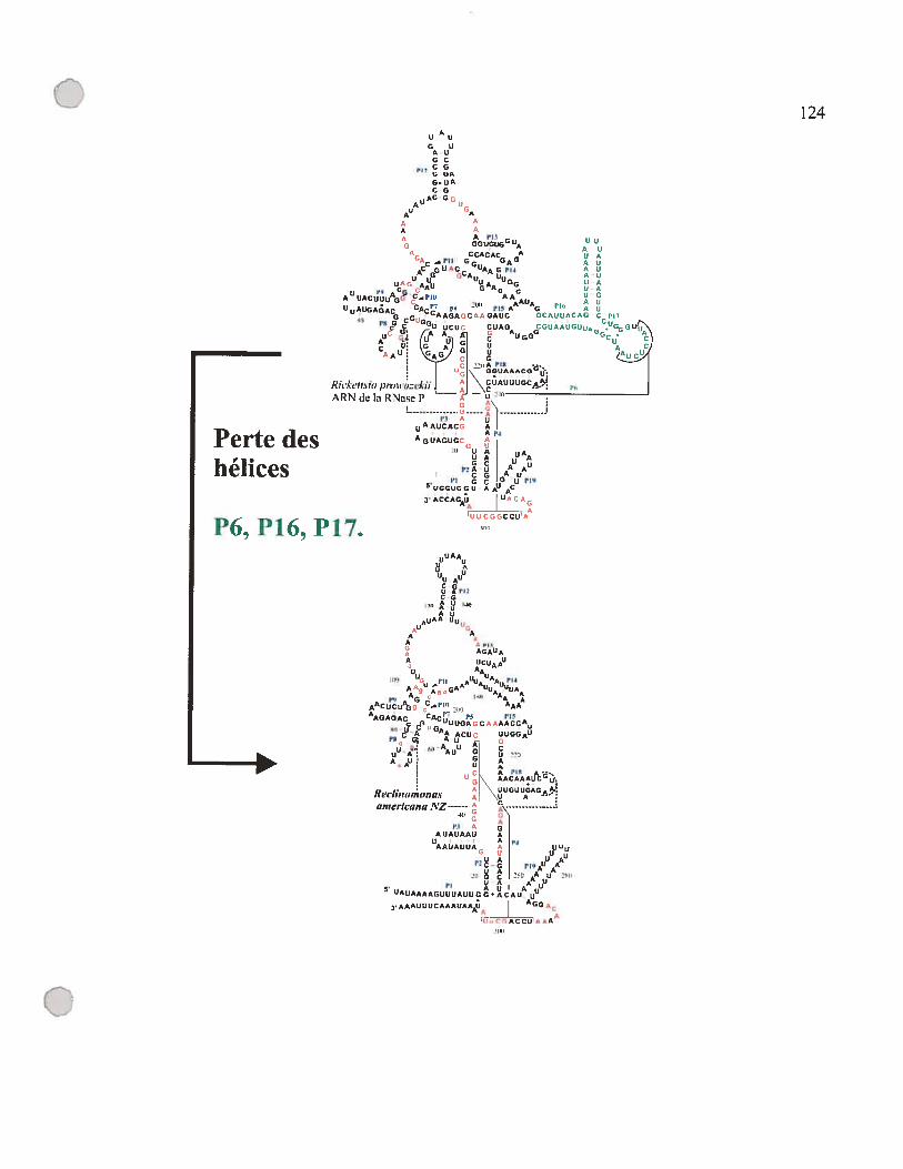
U Au
G UAUOC
,CG- U
G. UACG
AC G,AU U
UA A P13 uG GGUGUGGIJA
‘A CCACAC A U A
Ac_Phl GG GAG U UC 0UR’C UAAGPI4 A U
G UCA u A UGG
- AAAUU AUGA1AC CACP7 P5
- PIS AA A0 ° A UG G-. CAAGAGCAAGAUC GCAUUACAG CPI’
PU G GGu UCUU CUAG CGUAAUGU5
A1 % ( G
C Au06 Gc.
Rick’tisit, pnhhbtzekiiL \GM p
ARN de la RNaSU P A
U I3 A u
AAuCAC(, AAp3A
UA AUA UAOC U u
P2AU AACG AU
Ph G C G uPl95hGGUCCU AAU AC
3’ACCAGAU
LJ CCUA
124
Perte deshélices
P6,P16,P17.
o31N1
C 125
D.2.3 Evolution convergente vers une structure minimale chez les champignons
Chez les champignons, les ARN-Pmt du genre Rhizopus partagent le plus grand
nombre d’hélices avec le modèle de R. americana NZ (voir article 2). L’ARN-Pmt chez
Rhizopus otyzae n’a perdu que trois hélices, les P13, P14, et P19. Les données
présentement disponibles montrent que ces hélices sont absentes chez tous les
champignons et laissent croire qu’elles ont été perdues chez leur ancêtre commun. Nous
retrouvons une grande variation dans les modèles de structure secondaire de l’ARN-Pmt
chez les champignons, mais nous avons noté que deux hélices se trouvent chez tous les
ARN-Pmt, le Pi, le P4 et la région qui les entourent et qui inclut les régions conservées
CRI et CRV. Nous avons même retrouvé des ARN-Pmt qui se limitent à ces deux hélices
chez les levures, et chez le zygomycète $mittium culisetae. Est-ce un hasard que dans
deux lignées évolutives différentes, celles des zygomycètes et des ascomycètes, nous
retrouvons deux ARN-Pmt qui n’ont retenu que ces deux hélices? Cette observation
laisse croire que le Pi, le P4 et les régions en simple brin avoisinantes, qui forment le site
catalytique chez les ARN-P eubactériens, ont un rôle important et incontournable dans la
fonction des ARN-Pmt.
L’étude structurale et comparative que nous avons menée nous apprend que les
ARN-Pmt ont perdu plusieurs des éléments structuraux présents chez les ARN-P des a
protéobactéries (Figure D. 1), et que cette perte est plus accentuée chez les champignons
que chez les protistes (articles 1, 2 et 3). Est-ce que cette perte est nécessairement
accompagnée par une réduction de la taille de ces ARN? Et est-ce qu’elle suit un ordre
défmi?
o
126
D.2.4 Les ARN-Pmt cibles d’invasion des éléments à double hélices «filE»
Pour répondre à la première question, nous avons comparé la taille des ARN-Pmt,
ou du gène rnpB, d’onze espèces, et le degré de réduction structurale de ces ARN (Table
D. 1). Nous avons défmi la réduction structurale chez un ARN-Pmt, comme étant
l’absence d’hélices qui ont déjà existé dans un modèle ancestral. Pour des fms pratiques,
nous avons considéré l’ARN-Pmt de R. americana NZ comme étant le modèle ancestral,
puisque ce dernier contient le plus grand nombre d’hélices et donc ressemble le plus à
l’ancêtre commun des ARN-Pmt. Nous avons remarqué que les ARN-Pmt de MortierelÏa
verticillata, Rhizopus oiyzae, Taphrina deformons, S. cerevisiae, et N olivacea ont une
taille plus grande que celle de l’ARN-Pmt de R. americana NZ (voir Table Di et les
articles 2 et 3), alors que leur structure secondaire est moins conservée que chez ce
dernier. Néanmoins, les ARN-Pmt qui montrent les modèles structuraux les plus réduits,
ceux de S. fibuligera et S. culisetae, ont les plus petites tailles, 140 et 18$ nucléotides
respectivement. Ces résultats montrent que la taille d’un ARN-Pmt ne reflète pas
nécessairement le degré de conservation de sa structure secondaire, même si dans
certains cas nous trouvons une corrélation. En fait, la taille des ARN-Pmt ne diminue pas
nécessairement suite à la perte des éléments structuraux présents dans les formes plus
ancestrales, pour la simple raison que d’autres éléments structuraux apparaissent chez ces
ARN.
Certains éléments structuraux sont apparus suite à l’invasion du gène rnpB par
des éléments mobiles comme c’est le cas chez les ARN-Pmt de plusieurs zygomycètes
(article 2). Dans ce cas, nous avons observé l’apparition de grands inserts, dont les tailles
varient entre 57 et 430 nucléotides, dans le P12 et dans les jonctions entre le P5 et le Pi $
ou P15, selon l’espèce. La séquence de ces régions peut être repliée en éléments doubles
hélices (Double Helical Elements)(Paquin et al., 2000). Ces éléments sont des éléments
C

127
mobiles riches en GC, formés de deux hélices séparées souvent par un nucléotide. Ils
envahissent les régions intergéniques des génomes mitochondriaux ainsi que les ARN
ribosomiques. Notre étude a montré que ces éléments ne sont pas elimincés dans l’ARN
Pmt mature de R. oryzue, ce qui suggère qu’ils sont insérés dans des régions non-critique
pour la fonction des ARN-Pmt (article 2).

128
Nom de l’espèce Groupe phylogénétique hélice(s) absente(s) Taille de l’ARN-Pmt
M verticillata Zygomycète P2, 3, 13-15, 19 982
R. oiyzae Zygomycète P13, 14, 19 829
S. cerevisiae Ascomycète P2, 3, 5, 7-15, 19 423
N oÏivacea Prasinophyte P13 359
T deformans Ascomycète P3, 11, 13-15, 19 319
I libera Jakobide P13 307
S. ecuadoriensis Jakobide P3 306
S. pombe Ascomycète P5, 7-14, 19 273
A. niduÏans Ascomycète P3, 11, 13-15, 18, 19 232
L cuÏisetae Zygomycète P2, 3, 5, 7-15, 18, 19 188
S.fibuligera Ascomycète P2, 3, 5, 7-15, 18, 19 140
Table D.!: Comparaison entre les tailles des ARN-Pmt et la réduction de leur structure
secondaire.
L’échantillon des espèces choisies représente la diversité des ARN-Pmt, et contient les
espèces qui ont la plus grande variation par rapport à la taille de leur ARN-Pmt. Les
hélices énumérées sont celles présentes chez R. americana NZ, mais absentes chez les
espèces correspondantes. La taille de l’ARN-Pmt est celle déterminée expérimentalement
lorsque cette information est disponible. Les tailles indiquées en italique ont été calculées
à partir de la taille du gène rnp3.
129
Un autre élément apparu chez les ARN-Pmt des zygomycètes est l’hélice
située entre le P5 et le P7. Elle varie de taille entre 20 et 100 nucléotides. Une telle hélice
existe dans les modèles de structure secondaire des ARN-P eubactériens du type B
similaire à Bacillus subtitis (Haas et Brown, 1998), où elle est identifiée comme P5.1.
Comme pour les ARN-Pmt, les ARN-P eubactériens de type B sont caractérisés par leur
séquence riche en A et U. Est-ce que l’apparition de cette hélice à la même position que
chez les eubactéries implique qu’elle est importante pour la stabilisation de la structure
des ARN-Pmt riches en A et U? Où est-ce simplement une coïncidence? Vu que la
fonction exacte de cette hélice est inconnue pour l’instant, nous ne favorisons aucune
hypothèse.
D.2.5 La perte des éléments structuraux ne suit pas un ordre défini
Pour déterminer si la perte des éléments structuraux chez les ARN-Pmt suit un
ordre défmi, nous avons établi un scénario de la disparition de ces éléments, en se basant
sur les connaissances disponibles concernant la phylogénie des organismes où ces ARN
ont été identifié, et le modèle de structure secondaire de 1’ARN-Pmt chez R. americana
NZ (Figure D.2).
Chez les jakobides, les premières hélices perdues sont soit le P3 ou le P13. Chez
N olivacea, la seule hélice perdue est le P13. Quant aux champignons, nous remarquons
qu’il y a une multitude de pertes, qui ne semblent pas suivre un ordre défini. Par contre,
nous avons noté que les P13, 14 et 19 sont absents dans tous les modèles (figure D.2), et
que les hélices P5,7-10,12, qui forment en grande partie le domaine «S» chez les
eubactéries sont perdues en un seul bloc. Nous avons remarqué que la présence de
certaines hélices est plus fréquente que d’autres; alors nous avons calculé la fréquence de
présence de chacune des hélices. Pour le faire, nous avons choisi un échantillon de
modèle de structure secondaire d’ARN-Pmt d’une manière à représenter toutes les
C
Ç:130
variations observées, mais en éliminant les modèles provenant d’espèces proches,
afm de ne pas surreprésenter les groupes phylogénétiques où les séquences sont plus
disponibles, comme les levures (Table D.2). Mis à part le Pi et le P4 présents chez tous
les espèces, cette étude montre que le P18 est le plus fréquent à 75%, suivi du P2 et du
domaine P5, 7-10,12 à 67%. D’autre part, le P13 est le moins fréquent à 17% suivi par le
P14 et le P19 à 33%. Ces données suggèrent que chez les ARN-Pmt, le domaine «C»
(Pi, 4 100%; P18 75%; P2 67%; P15 50%; P3 42%; P19 33%) est plus conservé que le
domaine « S» (P7-i0 67%; P11 50%; 1433%; P13 17%;).
G
o
1.,Ii
figure D.2: Scénario hypothétique décrivant la perte des éléments structuraux
chez les ARN-Pmt.
L’emplacement des espèces sur cette figure est basé sur les phylogénies décrites
dans l’article 2 et (Gray et al., 199$). Les hélices perdues sont indiquées en
rouge.
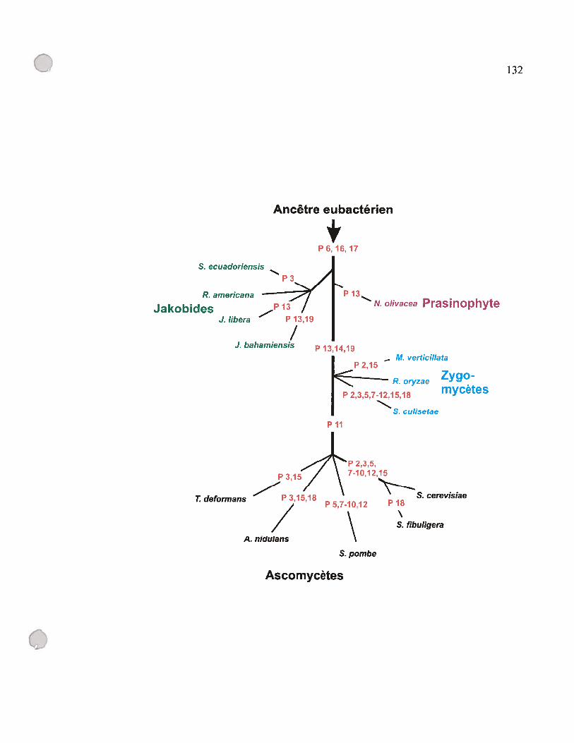
o
Ancêtre eubactérien
4P 6, 16, 17
132
S. ecuadoriensis
R. americana
Jakobides 13
J.Iibera — P 13,19
/J. bahamiensis
Ascomycètes
— M. verticillata
ZygoR. oryzae
P 2,3,5,7-12,15,18 mycetes
S. culisetae
S. cerevisiaeP 18
S. fibuligera
P 13
P 13,14,19
N. olivacea Prasinophyte
p ii
P 3,15
P 3.15,18T deformansP 5,7-1 0,12
/\A. nidulans
S. pombe

133
Identification de l’hélice Nombre de présence d’une fréquence en pourcentage
hélice / nombre d’espèces
P1,4 12/12 100%
P18 9/12 75%
P2 8/12 67%
P5,7,$,9,10,12 $/12 67%
P15 6/12 50%
P11 6/12 50%
P3 5/12 42%
P19 4/12 33%
P14 4/12 33%
P13 2/12 17%
Table D.2 : fréquences de présence des hélices d’origine eubactérienne chez les
ARN-Pmt.
L’échantillon des espèces qui ont été considérées pour cette étude inclut R.
americana NZ, J libera, S. ecuadoriensis, N oÏivacea, R. olyzae, M verticitÏata,
S. cuÏisetae, T deformans, A. nidulans, S. pombe, S. fibuligera, et S. cerevisiae.

-
134
D.2.6 Des motifs GNRA et YNMG dans les tétra-bouctes du P3, P9, P12 et Pi$
L’étude structurale que nous avons menée jusqu’à maintenant s’est limitée au
niveau de la présence ou l’absence des hélices héritées de l’ancêtre eubactérien, mais
d’autres informations peuvent en être retirées. Par exemple, nous avons remarqué la
présence des motifs GNRA (NG, A, T, C; RG, A) dans (i) le P9 de R. olyzae, Mucor
mucedo, Rhizopus stolonfer 194667, et Rhizopus oligosporus, (ii) le Pi $ de tous les
jakobides à part S. ecuadoriensis, et (iii) le P12 de J libera et J bahamiensis. Comme
mentionné dans l’article 3, les tétra-boucles de la famille GNRA peuvent stabiliser les
hélices dans lesquelles elles sont présentes, interagir avec des régions éloignées de
l’ARN ou avec une protéine (Belanger et al., 2004; Correli et al., 199$; Legault et al.,
199$). Chez E. cou, le motif GNRA du L1$ interagit avec le P8, celui du L9 avec le Pi,
et celui du L13 avec le P8 (Brown et al., 1996; Harris et Pace, 1995a; Massfre et al.,
199$). La mutation de ces motifs diminue l’activité catalytique de la RNase P (Pomeranz
Krummel et Altman, 1999). Nous avons remarqué aussi la présence du motif UUAG
dans la tétra-boucle du P3 chez I libera (article 3). Ce motif appartient à la famille des
tétra-boucle YNMG (Y=U, C; M= A, C)(Proctor et al., 2002). Cette famille de tétra
boucles est particulièrement stable, et peut servir de site d’attache pour les protéines, ou
interagir former des interactions tertiaires à l’intérieur de l’ARN (Ghisolfi-Nieto et al.,
1996; Proctor et al., 2002). Même s’il est présent chez l’ARN-P d’E.coli, ce motif n’a
jamais fait l’objet d’une étude par mutagenèse afin d’évaluer son rôle ou son importance
pour l’activité catalytique. Il est connu que le P3 contribue à l’attache de l’ARN-P
d’E. cou à la protéine-P (Biswas et al., 2000), ce qui soulève la possibilité que ce motif y
soit impliqué aussi.
Tous les motifs décrits plus haut sont présents chez l’ARN-P d’E. cou. Chez les
Œ-protéobactéries le motif GNRA est présent dans le L9 des ARN-P de Caulobacter
crescentus, Agrobacterium tumefaciens, Rhodobacter capsulatus, Rhodospirillum
rubrum et chez onze des quinze isolats de WoÏbachia (Andersson et al., 1998; Brown et
C

135
al., 1991; Brown et al., 1996; Harris et al., 2001) (http:,: www.mbio.ncsu.edu/
RNaseP/ home.html). Le motif GNRA est omniprésent dans le L14 et L1$, de toutes les
Œ-protéobactéries étudiées; alors que le motif YNMG du P3 y est absent. La présence des
motifs GNRA chez les ARN-Pmt des jakobides et chez les a-protéobactéries suggère que
ces motifs étaient présents chez l’ancêtre commun de ces deux groupes. En ce qui
concerne le motif de la famille YNMG du P3 chez I libera, nos données ne nous
permettent pas de supporter l’hypothèse que ce motif provient de l’ancêtre eubactérien,
même si cette possibilité demeure une hypothèse pertinente.
En résumé, notre étude structurale comparative nous mène à conclure que les
ARN-Pmt ont perdu plusieurs des éléments structuraux présents chez leur ancêtre
eubactérien, et que cette réduction structurale a créé des modèles de structures
secondaires très hétérogènes, spécialement chez les mitochondries des champignons.
Dans ce cas, nous observons une corrélation entre la perte accélérée d’éléments
structuraux et la longueur des branches des arbres phylogénétiques basées sur les
séquences mitochondriaux. En fait, les ARN-Pmt des levures et du zygomycète L
culisetae sont les plus réduits (articles I et 2); et en parallèle, leur phylogénie
mitochondriale montre de longues branches ce qui d’ailleurs crée des artefacts
«d’attraction des longues branches» qui rendent la reconstruction phylogénétique
difficile (article 2)(Bullerwell et al., 2003; Leigh et al., 2003).
Cette étude nous emmène à poser des questions quant à l’effet de cette réduction
structurale sur l’activité catalytique des ARN-Pmt. Est-ce qu’ils sont catalytiques comme
leur ancêtre eubactérien en absence de la composante protéique? Si oui, comment cette
réduction structurale affecte l’activité catalytique? Sinon, quels éléments sont cruciaux
pour maintenir une activité catalytique chez les ARN-P? Une autre observation intrigante
est la disparition des hélices P5, P7-10, et P12 en un seul bloc. Pourquoi est-ce que toutes
ces hélices disparaissent d’une façon simultanée? Et comment la RNase P compense-t
C’,
136
elle cette perte? Et fmalement, pourquoi le Pi et le P4 sont-ils omniprésents chez
toutes les espèces étudiées?
o

CV 137
D.3 Effets de la réduction structurale sur l’activité catalytique des ARN-Pmt
D.3.1 Les ARN-Pmt ne sont pas catalytiques in vitro
Les modèles de structure secondaire des ARN-Pmt des jakobides, de N olivacea, et du
genre Rhizopus chez les zygomycètes ont retenu les mêmes hélices qu’on retrouve dans
le modèle du consensus minimal de la structure de l’ARN-P eubactérien (Figure D.3A),
en plus du P12 et P18, ainsi que les P13, 14 et 19 dans certains cas (voir articles 2 et 3).
Un ARN-P avec une structure similaire à ce modèle théorique a été synthétisé in vitro à
partir de la séquence d’ARN-P de MycopÏasma fermentas (Micro ARN-P). Cette
molécule a montré de l’activité catalytique 600 fois plus faible que celle de l’ARN natif
(Siegel et al., 1996)(Figure D.3B). En se basant sur cette observation, nous avons produit
et testé l’activité catalytique des ARN-Pmt de R. americana NZ, I Ïibera, S.
ecuadoriensis, N olivacea, R. oryzae et R. otigosporus in vitro (voir article 3). Nos tests
ont été négatifs pour toutes ces molécules. Chez S. cerevisiae, un groupe de chercheurs a
déjà purifié la RNase P mitochondriale, et il a rapporté que l’activité catalytique dépend
des deux composantes, I’ARN-Pmt et la protéine-P (Hollingsworth et Martin, 1986).
Donc, nous estimons que ce soit le cas aussi pour les ARN-Pmt que nous avons étudiés.
Présentement, les composantes protéiques de la RNase P mitochondriale chez ces
espèces sont inconnues; alors nous avons complémenté 1’ARN-Pmt de R. americana NZ
avec la protéine-P d’E. cou, la protéine C5. Encore une fois, notre résultat était négatif
(article 3). Ce résultat n’est pas surprenant vu la distance phylogénétique et
évolutionnaire qui sépare E. cou et les mitochondries, et, sachant que la protéine-P d’E.
cou ne peut complémenter l’ARN-P du plastide du glaucocystophyte (algue)
Cyanophora paradoxa, alors que celle de la cyanobactérie Synechocystis sp. PCC 6803
en est capable (Pascual et Vioque, 1999). Dans l’avenir, il faudra essayer de
complémenter les ARN-Pmt avec une protéine-P provenant d’une a—protéobactérie, le
groupe eubactérien le plus proche phylogénétiquement des mitochondries (Gray et al.,
1999).
C
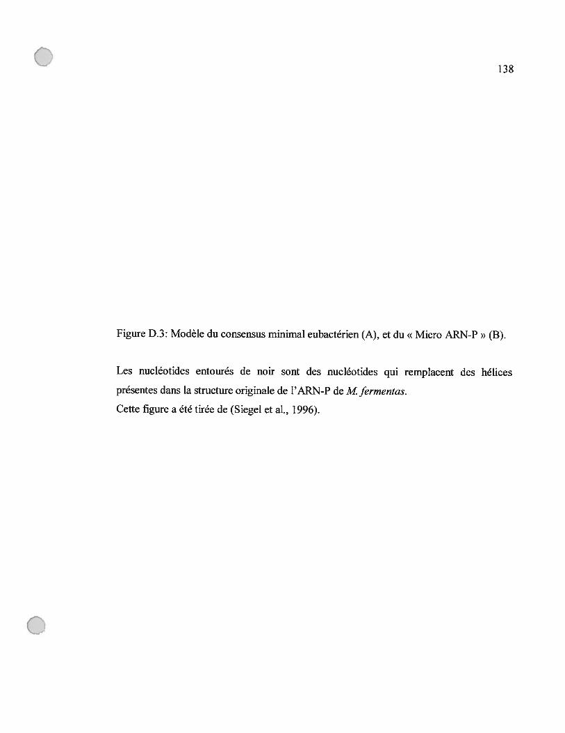
0138
figure D.3: Modèle du consensus minimal eubactérien (A), et du « Micro ARN-P» (B).
Les nucléotides entourés de noir sont des nucléotides qui remplacent des hélices
présentes dans la structure originale de l’ARN-P de M fermentas.
Cette figure a été tirée de ($iegel et al., 1996).
o

gG
G
AG ee_.rn ••A0 a
• 01N a .gC
• cc.g:_ P19•0,P7 P5 Pi!
•t
•
e e1 ‘ I: u iiin •‘•ce.c ••
g
:; c G
e ee
o.
1I•Oe.eeeeG A• iii•2CeteeIC6 J
p4P2 e— e
“ :::coooeeeeeete—eecsAC
Il. f- A
3’ OA,UJCGGeee!A
B G
CA G
U1C\C G
4P9 c SA A 122
AGA AU çGU AUGGCG°
C*P10GG. Ç»,G, I PIS
A,C GUUAGAAACCC AU il I;: U
8’j $(TYAU CGAUQ GGG U
AG G JP5G1 G AUGG
A C U C A 160GAA ACL 0
40 A
AJ GA AG uG
P3 A AA
• A4GUUCGUC u
I GL20 A—U
0GP2 A — U — 110
A—U
Ç nU-ACG
CncAGCAU UGG—C
liiiUCGUA AU
s A%U9GGUGCAA
201
139
A
A
o
Q
Xev t» ttcwueasw
GAUC- 100% conserved2ilC - 80% cor.senede- ni present in ail RNAoo- ni pmsen: in 80% ofRNÀs

140
0.3.2 Le domaine P6, 16-17 ne suffit pas pour rendre 1’ARN-Pmt de R.
americana NZ actif
A première vue, le domaine P6, P16-17 qui est présent chez R. prowazekii et E.
cou mais absent chez les ARN-Pmt, pourrait être la raison pour laquelle ces derniers sont
non-catalytiques in vitro (figure D.1). En conséquence, nous avons initié une étude de
mutagenèse en ajoutant ce domaine provenant de l’ARN-P d’E. cou chez l’ARN-Pmt de
R. americana NZ (Figure D.4). L’ARN-P chimérique que nous avons créé contient deux
éléments connus pour avoir un impact majeur sur l’activité catalytique de l’ARN-P (i)
l’hélice P6 (Darr et al., 1992; Haas et al., 1991), (ii) et le L15 incluant le motif GGU qui
s’attache au pré-ARNt en liant le RCCA en 3’ de ce dernier (voir la section 1.3.3).
Malgré ces ajouts, nous n’avons détecté aucune activité catalytique. pour cet ARN-P
chimérique (article 3). Ce résultat nous a emmené à conclure que la perte de ce domaine,
seule, ne justifie pas l’absence de l’activité catalytique chez les ARN-Pmt. Notre
conclusion est renforcée par deux autres observations. Premièrement, le domaine en
question existe chez l’ARN-P du plastide de C. paradoxa (Shevelev et al., 1995), et ce
dernier n’est pas actif in vitro (Baum et al., 1996). Deuxièmement, chez le micro ARN-P,
ces domaines sont manquants et ce dernier est actif (Siegel et al., 1996)(figure D.3B).
0.3.3 La fusion de l’ARN-P d’E. cou avec les ARN-Pmt produit des ARN
catalytiques
Afm de vérifier si certaines hélices ou régions en simple brin manquent des
éléments cruciaux pour l’activité catalytique, nous avons construit deux ARN
chimériques en fusionnant les séquences de l’ARN-P d’E. cou avec celles provenant de
l’ARN-Pmt de R. americana NZ (article 3). Dans la chimère de type I, la région Pi-4,
P18, P19, qui selon nos études comparatives est la mieux conservée à travers tous les
ARN-Pmt (voir D.2.5), a été fusionnée au domaine complémentaire d’E. cou (article 3).

141
Figure D.4: Chimère d’ARN-P de R. americana NZ après l’ajout des P5,6,15,16,17.
La séquence provenant d’ E. cou est en bleu, et le reste de la séquence provient de
1’ARN-Pmt de R. americana NZ. Les nucléotides en rouge sont les positions conservées
dans le consensus minimal eubactérien (voir la figure D.3).
o
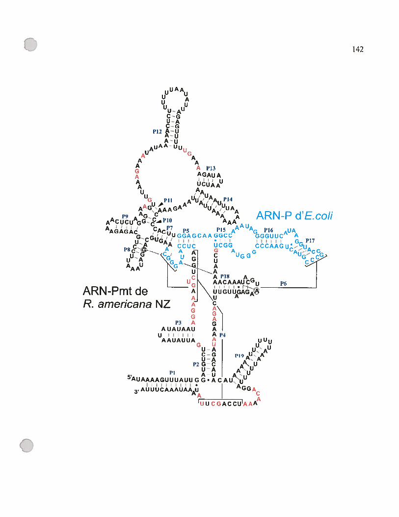
142
uA Au
U AL
U— AUU
C—GU—AC-G
P12 A—UA—eA-u
AU’ UuLG
AA
A Ap13GA AGAUAA UCUAAUA
AA
G 1’11 AuU%jAAuJl4AAGfAAGAA /,UA
A,9 AGC A ARN-Pd’Eco//AACUCUAGGPb0 AAA
AA UAAAGAGAC C”7 p P15 A G P16 AUA
G G AAGCC GGGUUC Au GA I I I I I •GGP17— A CCUC UCGG CCCAAGUc
U—e--. UG GuA G]
G A G AUG
AAAU N Ui--AI CI APIS AL9GN P6
ARN-Pmt de AI \ UUGUUGAGJ \u A
R. americana NZ A ÇGG
P3 A GIAUAUAAU A Iui Ap4
AAUAUUA I UUUU
U—AAUJAC—G I
U-A I AP2G C I
UAI A5AuAAAAGuuUAuuGG.AcAuUUAUi A
11111111. ‘
3AUUUCAAAUAAU GGAAA
UU CGACCU’AAÀ’
o

c) 143
La chimère de type II a été construite à l’inverse de la chimère de Type I; la région PI-4,
P18 provient d’E. cou alors que la partie complémentaire provient de R. americana NZ.
Nous avons trouvé que les deux chimères sont actives (article 3), ce qui suggèrent que
chez 1’ARN-Pmt de R. arnericana NZ, l’absence d’activité catalytique ne peut être
expliquée par le manque d’un seul élément critique.
D.3.4 Raison multifactorielle pour l’absence d’activité catalytique chez les ARN
Pmt
Les études de mutagenèse que nous avons effectuées sur les chimères de R.
americana NZ ont montré que l’augmentation du contenu en GC du P4, la correction de
l’identité du nucléotide A292 dans le motif universellement conservé « CRV », et l’ajout
de motifs GNRA dans la tétra-boucle L9 augmentent l’activité catalytique des chimères
(article 3). Ces résultats ainsi que ceux de la section précédente nous emmènent à
suggérer que l’absence de l’activité catalytique chez les ARN-Pmt est causée par les
facteurs suivants: (i) la séquence riche en A et U des ARN-Pmt, (ii) l’absence des motifs
GNRA et du P6, (iii) la déviation même mineur dans les séquences des motifs
universellement conservés, (iv) et fmalement l’absence du motif GGU dans le L15. Les
deux premiers éléments contribuent à la stabilisation de la conformation active de la
ribozyme en stabilisant les hélices, et les interactions tertiaires. La déviation des
séquences peut avoir un rôle dans la liaison des ions Mg2 dans le cas du CRV, et
l’absence du motif GGU dans le L15 défavorise la liaison au substrat (voir article 3).
o
Ç 144
Nous proposons que l’effet additif de tous ces éléments rend les ARN-Pmt non
catalytiques, puisque chacune des raisons énumérées, à elle seule, ne justifie pas qu’un
ARN-P avec une structure aussi conservée que celle de l’ARN-Pmt de R. americana NZ
soit inactif. En fait, ces éléments ont déjà été rapportés manquants dans des ARN-P
catalytiques, (j) les séquences des ARN-P eubactériens de type B sont riches en A et U,
(ii) 92 % des modèles eubactériens ont la déviation de G à A dans le CRV à la position
correspondante au G350 chez E. cou (Rasmussen et Nolan, 2002), (iii) le P6 et les tétra
boucles du motif GNRA qu’on retrouve chez E. cou sont absentes chez le micro ARN-P
(Siegel et al., 1996), (iv) et fmalement, la mutation du motif GGU du L15 n’est pas
suffisante pour démolir l’activité catalytique de notre chimère (article 3).
D.3.5 Relation entre la conservation de la structure secondaire et l’activité
catalytique des chimères
De la même façon que pour l’ARN-Pmt de R. americana NZ, nous avons
construit des chimères de type I et 11 pour les jakobides I Ïibera, S. ecuadoriensis, et le
prasinophyte N olivacea. Nous avons aussi construit une chimère de type I seulement
potir le zygomycète Rhizopus oryzae (article 3). Nous avons noté que l’activité
catalytique des chimères de type I est plus grande que celle des ARN chimériques de
type II. Il a fallu augmenter la concentration d’ARN des chimères de type II d’un facteur
dix, pour produire une activité catalytique équivalente à celle des chimères de type I.
Cette observation peut être expliquée par le fait que le domaine « C » des ARN-Pmt est
mieux conservé que le domaine « $ » (voir la section D.2.5), et ceci se traduit par la
capacité du domaine « » de mieux complémenter les séquences d’E. cou.
o

C 145
Nous avons remarqué aussi que l’activité catalytique des chimères varie selon le
degré de conservation des ARN-Pmt originaux (article 3). Chez les chimères de type I,
les ARN-Pmt des jakobides forment les chimères les plus actives catalytiquement. suivis
par N olivacea. Le zygomycète R. otyzae n’a pas montré d’activité (article 3). Pour les
chimères de type II, celles provenant des jakobides sont actives, alors que celle de N
oÏivacea ne l’est pas. A l’intérieur des jakobides, les différences d’activité catalytiques
sont plus petites, vue que la structure secondaire est très similaire. Les chimères
construites pour S. ecuadoriensis sont les moins actives. Pour le type I, J libera est plus
active que R. arnericana NZ et vice-versa pour le type II. L’observation que la chimère
de type I de J libera montre une plus grande activité catalytique que la chimère de R.
arnericana NZ, même si les deux ARN-Pmt originaux ont le même nombre d’hélices
dans ce domaine, pourrait être expliquée par deux hypothèses. Premièrement, la séquence
mitochondriale du domaine « C > de J libera est plus riche en GC que celle de R.
americana NZ, ce qui pourrait être un avantage si, comme nous avons proposé plus haut
(section D.4), le contenu en A et U des ARN-Pmt a un impact négatif sur leur activité
catalytique. Deuxièmement, la tétra-boucle du P3 chez J libera appartient à la famille
des motifs YNMG comme décrit plus haut. Pour vérifier l’importance de cette boucle,
nous avons détruit le motif UUCG en échangeant le G terminal par un A. Suite aux tests
catalytiques in vitro, nous n’avons noté aucun impact significatif sur l’activité
catalytique, et par conséquent nous avons écarté la deuxième hypothèse (résultats non
montrés).
En résumé, notre étude montre que les ARN-Pmt ont subi une réduction graduelle
de leur structure secondaire, et qu’ils ont perdu des éléments clés pour l’activité
catalytique au niveau de leur séquence primaire les motifs GNRA, la déviation des
motifs universellement conservés. Ces déviations de la forme ancestrale font que les
ARN-Pmt sont non catalytiques et que leur capacité de complémenter les séquences d’E.
cou est affectée par cette perte. Mais comment cette perte est-elle compensée in vivo?
L 146
D.4 La composante protéique compense-t-elle la réduction structurale?
Chez £ cerevisiae, l’ARN-Pmt dépend de la composante protéique pour son
activité catalytique (Hollingsworth et Martin, 1986). Nous proposons que ce soit aussi le
cas pour les ARN-Pmt étudiés, et que celle protéine compense pour la perte structurale
des ARN-Pmt. Pour l’instant, seule la séquence de la protéine-P mitochondriale
(protéine-Pmt) de L cerevisiae est connue (Morales et al., 1992). Cette protéine a une
taille de 119 kDa, 10 fois plus grande que la protéine-P eubactérienne qui a une taille de
14 kDa, et ne montre pas de similarité avec les séquences eubactériennes. Il a été montré
que la protéine-P de £ cerevisiae (RPM2) fait partie de la RNase P mitochondriale par
des expériences génétique et par immunoprécipitation (Morales et al., 1992). Les études
de délétion du gène rprn2 chez des levures haploïdes ont montré que ce gène est impliqué
dans une deuxième fonction essentielle pour la survie de la cellule, indépendante de la
RNase P (Kassenbrock et al., 1995). Il a été proposé que la partie C-terminale de la
protéine est responsable de la fonction reliée à la composante protéique de la RNase Pmt,
alors que la partie N-terminale est impliquée dans la fonction essentielle pour la survie de
la levure en présence de sources de carbone fermentable (glucose, éthanol), donc
indépendante de la fonction de la RNase P mitochondriale (Stribinskis et al., 200 la;
Stribinskis et al., 2001 b). La taille relativement grande de la protéine-Pmt peut être
expliquée par le fait qu’elle accomplit deux fonctions, et peut-être que celle
augmentation de taille compense pour la perte structurale chez la composante ARN.
Pour comprendre comment la protéine-P compense pour la réduction structurale
de l’ARN-Pmt, il faudra faire une étude comparative des séquences protéiques provenant
de plusieurs groupes phylogénétiques pour élucider l’évolution de celle composante,
comme nous avons fait pour l’ARN-Pmt. Ce genre d’étude pourra dévoiler si on retrouve
encore des composantes protéiques qui ressemblent à la protéine-P eubacterieime, et on
C
147
pourrait faire une correlation entre la disparition de certains domaines chez l’ARN
Pmt et l’apparition de domaines compensatoires chez la protéine-P. Pour l’instant tine
telle étude n’est pas possible à cause du manque des séquences, mais grâce aux projets de
séquençages génomiques et des banques d’ADNc en cours, une telle étude sera possible
dans un avenir rapproché.
C14$
D.5 Scénarios possibles pour expliquer la distribution discontinue du gène rnpB
Des gènes rnpB ont été identifiés chez tous les jakobides connus, et chez certains
zygomycètes et ascomycètes. Mais où se cache ce gène chez la grarnie majorité des
organismes chez lesquels tin tel gène n’a pas été trouvé? Est-ce qu’il existe ? Plusieurs
scénarios sont possibles pour expliquer cette observation, et dans ce qui suit nous allons
discuter ces scénarios, et évaluer la pertinence de chacun en se basant sur les données
disponibles.
D.5.1 Les gènes rnpB sont dérivés et non-reconnaissables avec notre approche
Chez S. pombe, le gène rnpB n’a été identifié qu’après presque deux décennies dela disponibilité de la séquence du génome mitochondrial, à cause de la réduction au
niveau de la séquence primaire de ce gène. Alors, il est possible que la séquence de
certains gènes soit dérivée, et qu’elle ne soit plus reconnaissable. Comme décrit dansl’article 1, cette hypothèse, bien que possible, n’est surtout pas applicable chez certains
chytridiomycètes, où la séquence des génomes mitochondriaux est complètementannotée, et où il ne reste plus assez de régions non codantes pour inclure un gène rnpB, à
moins que ce gène enchevauche un autre. Deuxièmement, malgré le fait que les gènes
rnpB chez les champignons subissent une réduction structurale accélérée, nous avons
remarqué que ce n’est pas le cas chez les protistes qui évoluent plus lentement. Alors
dans les meilleures des chances, ce scénario pourrait être applicable chez les organismesdont le génome mitochondrial évolue vite (en conséquence forment de longues branchesdans une phylogénie mitochondriale) mais n’explique pas la répartition globale du gènernpB.
o
C149
0.5.2 Le gène rnpB codé par le génome nucléaire
Si ce gène n’est pas codé par le génome mitochondrial, il est possible qu’il le soit
par le génome nucléaire, comme pour la composante protéique de la RNase P
mitochondriale de S. cerevisiae. En fait, les composantes protéiques et ARN de la RNase
MRP (Mitochondrial RNA Processiiig) sont codées par le noyau et importées par la
mitochondrie chez les animaux (Davis et al., 1995; Li et al., 1994). Alors est-ce que c’est
le cas pour la RNase P mitochondriale? Même si pour l’irstant aucun gène qui code un
ARN-Pmt n’a été retrouvé dans les génomes nucléaires complètement séquencés, cette
hypothèse est probable et pourrait être vérifiée dans un avenir rapproché vu la multitude
de projet de séquençage en cours.
0.5.3 Perte de la composante ARN
Si nous avons de la difficulté à identifier le gène rnpB, une raison possible
pourrait être l’absence de ce dernier. Car il est possible que la RNase P mitochondriale
soit formée d’une composante protéique seulement; ou encore que la RNase P nucléaire
soit importée dans la mitochondrie.
0.5.3.1 La composante ARN est absente chez la RNase P du chloroplaste des
épinards
Chez les épinards, l’activité catalytique de la RNase P dans le cifioroplaste
(RNase P-cp) a été purifiée, et il a été montré que cette activité est résistante aux
traitements aux nucléases (Thomas et aI., 1995). En plus, les caractéristiques physiques
de l’extrait suggèrent l’absence d’acides nucléiques. Finalement, les études sur le
mécanisme chimique de coupure des pré-ARNt montrent qu’il s’agit d’un mécanisme
C
C150
différent de celui adopté par la RNase P eubactérienne et eucaryote (Thomas et al.,
2000). Toutes ces observations laissent croire que la RNase P-cp, dans ce cas, ne contient
pas de composante ARN.
Ceci suggère qu’il est fort possible que la même situation se soit produite dans la
lignée mitochondriale, et que l’ARN-Pmt a été perdu au profit de la protéine-P. Cette
hypothèse a même été proposée pour la RNase P mitochondriale chez les humains.
D.5.3.2 La RNase P mïtochondriale chez les humains : protéique ou nucléaire?
Chez les humains, deux hypothèses ont été avancées pour décrire la RNase P
mitochondriale. La première, avancée en 199$ par le groupe des docteurs Rossmanith et
Karwan, stipule que celle activité est formée d’une protéine seulement (Rossmanith et
Karwan, 1998). En 2001, le groupe des docteurs Puranam et Attardi proposa que la
RNase P mitochondriale contient une sous-unité ARN identique à celle de la RNase P
nucléaire (Puranam et Attardi, 2001).
D.5.3.2.1 RNase P mitochondriale humaine protéique
L’équipe Rossmanith-Karwan a purifié l’activité catalytique de la RNase Pmt, en
utilisant la capacité de cette dernière de cliver le pré-ARNt mitochondrial, alors que la
RNase P nucléaire (RNase Pnu) ne reconnaît pas ce substrat (Rossmanith et al., 1995). Ils
ont rapporté que la RNase Pmt montre les propriétés physiques d’une enzyme protéique
en ce qui concerne la vitesse de sédimentation sur un gradient de glycérol, et la densité
sur un gradient de Cs2SO4 (Rossmanith et Karwan, 1998). Selon les auteurs, lecoefficient de sédimentation de la RNase Pmt est 9S alors que celui de la RNase Pnu est
16S. Ils ont noté que l’activité catalytique mitochondriale résiste aux traitements à la
nucléase de Micrococcus et à la ribonucléase A. finalement, ils ont essayé en vain de
C
C 151
détecter des ARN potentiels dans l’extrait par marquage radioactif des extrémités 5’
et 3’, une technique régulièrement utilisée pour ce genre d’expérience. Selon les auteurs,
seul un ARN d’une taille de 25 nucléotides ou moins aurait pu passer inaperçu durant
leurs analyses, mais vu que la taille du plus petit ARN-P connu est autour de 140
nucléotides (Wise et Martin, 1991), celui de la levure Saccharomycopsis fibutigera, ils
ont exclu la présence d’un ARN-P potentiel. Toutes ces observations les ont emmené à
proposer que la mtRNase P est une enzyme protéique; mais ils suggèrent d’interpréter
ces résultats avec prudence, car la résistance à la nucléase de micrococcus a déjà été
rapportées chez la RNase P de l’archéobactérie 8ufoÏobus acidocaldarius, et les
nuRNase P de Xenopus Ïaevis et de S. cerevisiae. Cependant, dans ces cas, la sous-unité
ARN a été identifiée par marquage radioactif’, ce qui n’est pas le cas pour la RNase Pmt
chez les humains (voir les références dans (Rossmanith et Karwan, 199$)).
D.5.3.2.2 L’ARN-Pmt humaine est identique à l’ARN-P de la RNase P nucléaire
L’équipe du docteur Allardi a identifié en 1985 une activité RNase Pmt humaine
en purifiant les mitochondries, et en les traitant avec de la nucléase de micrococcus pour
éliminer l’activité RNase Pnu contaminante. Cette étude a montré que l’activité
catalytique de la RNase Pmt est sensible à la pronase (protéinase) et la nucléase de
micrococcus (Doersen et al., 19$5). Cette étude a été répétée en 2001 en traitant la
fraction mitochondriale avec de la digitonine afm de solubiliser la membrane externe et
les contaminants nucléaires qu’elle pourrait emmener (Puranam et Attardi, 2001). Les
résultats de cette étude confirment ce qui a été rapporté en 1985, et les auteurs ajoutent
que la densité de la RNase Pmt est similaire à celle de la RNase Pnu, et que la
composante ARN des deux est identique. Dans la publication de leurs résultats, les
auteurs proposent que le groupe du docteur Karwan a effectué ses études avec des
cellules congelées ce qui cause une dégradation des ARN; et ils ajoutent qu’ils n’ont pas
C

C 152
trouvé l’activité catalytique rapportée dans l’étude de ce groupe qui montre un
coefficient de sédimentation différent de la RNase Pnu.
En résumé, il n’existe pas de consensus pour décrire la RNase Pmt chez les
humains, chacune des équipes a défendu ses résultats dans une lettre publiée en 2001
(Rossmanith et Potuschak, 2001). Les résultats discordants me semblent être dus au fait
que chacune des équipes a caractérisé une activité catalytique différente, si on se fie aux
coefficients de sédimentation et à la résistance aux nucléases. Est-ce qu’il existe deux
activités RNase P dans la mitochondrie des humains? Si c’est le cas ce serait une
première. Est-ce que la rigueur des expérimentations est la cause de ces observations?
Pour clarifier la situation, la détermination du gène codant la « RNase Pmt protéique»
identifiée par l’équipe du docteur Karwan serait cruciale, et elle incarnerait la première
séquence d’une RNase P protéique.
o
C153
Conclusions
En conclusion, le projet que nous avons amorcé a permis d’identifier quatre
nouveaux gènes rnp3 chez les ascomycètes, sept chez les zygomycètes, et cinq chez les
jakobides. Nous avons établis de nouveaux modèles de structure secondaire pour tous les
ARN-P connus par comparaison phylogénétique. La cartographie des extrémités 5’ et 3’de six ARN-Pmt a montré que ces ARN sont transcrits et clivés à des positions
concordantes avec nos modèles. L’étude structurale comparative nous emmène à
conclure que ces modèles sont réduits comparés aux modèles eubactériens, et que cette
réduction, perte d’hélices, s’accélère chez les champignons et spécialement chez les
ascomycètes et le zygomycète & cuÏisetae. Cette perte ne suit pas un ordre défmi et la
taille de ces ARN ne reflète pas nécessairement leur état de conservation. Les Pi et P4
sont universellement présents, et le domaine «C» (catalytique) est mieux conservé que
le domaine « S» (spécificité). Malgré la grande similarité entre les ARN-P eubactériens
et les ARN-Pmt, ces derniers ne sont pas catalytiques. Nous proposons que plusieurs
facteurs en sont responsables. La séquence riche en A et U, l’absence des motifs GNRAdans le L9 et L12, l’absence du P6, et du motif GGU dans le L15, et fmalement la
déviation des séquences consensus dans les régions universellement conservées.Cependant, certains domaines des ARN-Pmt des jakobides et de N oÏivacea deviennentcatalytiques, lorsque fusionnés à des domaines complémentaires provenant d’E. cou. Il
existe une corrélation entre le degré de conservation de la structure secondaire et
l’activité catalytique des chimères. Le domaine «C », chimère type I, complémente lOfois mieux les séquences d’E. cou que le reste de la molécule, chimère type II. Et leschimères produites avec les séquences d’ARN-Pmt originaux mieux conservésproduisent des chimères avec une plus grande activité catalytique.
Nous proposons qu’in vivo, la composante protéique compense pour la réductionstructurale chez les ARN-Pmt, et nous espérons qu’une étude comparative au niveau des
L

C 154
protéines P révélera la corrélation entre l’évolution des deux composantes. Pour
l’instant, les données disponibles ne permettent pas une telle étude, mais grâce aux
projets génomiques en cours, une telle étude sera possible dans un avenir rapproché.
Nous n’avons pas réussi à identifier un gène rnpB dans les génomes
mitochondriaux de la majorité des groupes taxonomiques. Nous proposons plusieurs
scénarios pour expliquer cette observation, (i) ces ARN sont trop réduits pour être
détectés, (ii) ils sont codés par le génome nucléaire, (iii) la composante ARN n’est plus
requise, ou (iv) la RNase P nucléaire effectue la maturation des ARNt mitochondriaux.
Q
clv
Bibliographie
Alifano, P., fani, R., Lio, P., Lazcano, A., Bazzicalupo, M., Carlomagno, M.S., Bruni,C.B., Rivellini, f., Piscitelli, C., Arraiano, C.M., Blasi, V. and Nappo, A.G.(1996) Hïstidirie biosynthetic pathway and genes: structure, regulation, andevolution. Microbiol Rev, 60, 44-69.
Alifano, P., Piscitelli, C., Blasi, V., Rivellini, F., Nappo, A.G., Bruni, C.B. andCarlomagno, M.S. (1992) Processing ofa polycistronic rnRNA requires a 5’ ciselement and active translation. Mol Microbiol, 6, 787-798.
Alifano, P., Rivellini, f., Piscitelli, C., Arraiano, C.M., Bruni, C.B. and Carlomagno,M.S. (1994) Ribonuclease E provides substrates for ribonuclease P-dependentprocessing of a polycistronic mRNA. Genes Dcv, 8, 3021-3031.
Andersson, S.G., Zomorodipour, A., Andersson, J.O., $icheritz-Ponten, T., Alsmark,U.C., Podowski, R.M., Naslund, A.K., Eriksson, A.S., Winkler, 11.11. andKurland, C.G. (1998) The genome sequence ofRickettsiaprowazekii and theorigin ofmitochondria. Nature, 396, 133-140.
Baer, M.F., Wesolowski, D. and Altman, S. (1989) Characterization in vitro ofthe defectin a temperature-sensitive mutant ofthe protein subunit ofRNase P fromEscherichia cou. JBacteriol, 171, 6862-6866.
Barrick, J.E., Corbino, K.A., Winkler, W.C., Nahvi, A., Mandal, M., Collins, J., Lee, M.,Roth, A., Sudarsan, N., Jona, I., Wickiser, J.K. and Breaker, R.R. (2004) NewRNA motifs suggest an expanded scope for riboswitches in bacterial geneticcontrol. Froc Nati Acad Sci USA, 101, 6421-6426.
Bartenschlager, R. and Lohmann, V. (2000) Replication of hepatitis C virus. J Gen Virol,81, 1631-164$.
Baum, M., Cordier, A. and Schon, A. (1996) RNase P from a photosynthetic organellecontains an RNA homologous to the cyanobacterial counterpart. JMoÏ Biol, 257,43-52.
Belanger, F., Gagnon, M.G., Steinberg, S.V., Cunningham, P.R. and Brakier-Gingras, L.(2004) Study ofthe ffinctional interaction ofthe 900 Tetraloop of 1 6S ribosomalRNA with helix 24 within the bacterial ribosome. JM0Ï Biol, 338, 683-693.
Biswas, R., Ledman, D.W., Fox, R.O., Altman, S. and Gopalan, V. (2000) MappingRNA-protein interactions in ribonuctease P from Escherichia cou using disulfidelinked EDTA-fe. J Mol Bio!, 296, 19-31.
Bothwell, Ai., Garber, R.L. and Altman, S. (1976) Nucleotide sequence and in vitroprocessing ofa precursor molecule to Escherichia cou 4.5 S RNA. JBiol Chem,251, 7709-7716.
Bouvet, P. and Belasco, J.G. (1992) Control ofRNase E-mediated RNA degradation by5’-terminal base pairing in E. cofi. Nature, 360, 48$-491.
Brenowitz, M., Chance, M.R., Dhavan, G., Takamoto, K., Brunel, C. and Romby, P.(2002) Probing the structural dynamics of nucleic acids by quantitative timeresolved and equilibrium hydroxyl radical “footprinting”. Curr Opin Struct Biol,12, 648-653.
Briani, f., Deho, G., forti, f. and Ghisotti, D. (2001) The plasmid status of satellite( bacteriophage P4. Flasmid, 45, 1-17.
clvi
Briani, F., Zangrossi, S., Ghisotti, D. and Deho, G. (1996) A Rho-dependenttranscription termination site regulated by bacteriophage P4 RNA immunityfactor. VfroÏogy, 223, 5 7-67.
Brown, J.W. (1999) The Ribonuclease P Database. NucÏeic Acids Res, 27, 314.Brown, J.W., Hans, ES., lames, B.D., Hunt, D.A., Liu, J.S. and Pace, N.R. (1991)
Phylogenetic analysis and evolution ofRNase P RNA in proteobacteria. JBacteriol, 173, 3855-3863.
Brown, J.W., Nolan, J.M., Haas, E.S., Rubio, M.A., Major, F. and Pace, N.R. (1996)Comparative analysis ofribonuclease P RNA using gene sequences from naturalmicrobial populations reveals tertiary structural elements. Proc Nati Acad Sci U $A, 93, 300 1-3006.
Brown, S. (1991) Genes for 7S RNAs can replace the gene for 4.5S RNA in growth ofEscherichia cou. JBacterioÏ, 173, 1835-1837.
Bmnel, C. and Romby, P. (2000) Probing RNA structure and RNA-ligand complexeswith chemical probes. Methods Enzymol, 318, 3-21.
Bullerwell, C.E., Leigh, J., forget, L. and Lang, B.F. (2003) A comparison ofthreefission yeast mitochondrial genomes. NucÏeic Acids Res, 31, 759-76$.
Cassano, A.G., Anderson, V.E. and Harris, M.E. (2004) Analysis ofsolvent nucleophileisotope effects: evidence for concerted mechanisms and nucleophilic activationby metal coordination in nonenzymatic and ribozyme-catalyzed phosphodiesterhydrolysis. Biochemistty, 43, 10547-10559.
Chamberlain, J.R., Lee, Y., Lane, W.S. and Enge&e, D.R. (1998) Purification andcharacterization ofthe nuclear RNase P holoenzyme complex reveals extensivesubunit overlap with RNase MRP. Genes Dey, 12, 1678-1690.
Chamberlain, J.R., Pagan, R., Kindelberger, D.W. and Engelke, D.R. (1996) An RIJase PRNA subunit mutation affects ribosomal RNA processing. Nucleic Acids Res, 24,3158-3166.
Chen, J.L., Nolan, J.M., Harris, M.E. and Pace, N.R. (199$) Comparative photocrosslinking analysis ofthe tertiary structures ofEscherichia cou and Bacillus subtilisRNase P RNAs. EMBOJ, 17, 1515-1525.
Chen, J.L. and Pace, N.R (1997) Identification ofthe universally conserved core ofribonuclease P RNA. RNA, 3, 557-560.
Chen, Y., Li, X. and Gegenheimer, P. (1997) Ribonuclease P catalysis requires Mg2+coordinated to the pro-RP oxygen ofthe scissile bond. Biochernistiy, 36, 2425-243$.
Cherayil, B., Krupp, G., Schuchert, P., Char, S. and Sou, D. (19$7) The RNAcomponents of Schizosaccharomyces pombe RNase P are essential for celiviability. Gene, 60, 157-161.
Christian, E.L. and Harris, M.E. (1999) The track ofthe pre-tRNA 5’ leader in theribonuclease P ribozyme-substrate complex. Biochemistiy, 3$, 12629-1263$.
Christian, E.L., Kaye, N.M. and Harris, M.E. (2000) Helix P4 is a divalent metal ionbinding site in the conserved core ofthe ribonuclease P ribozyme. RNA, 6, 511-519.
Christian, E.L., Kaye, N.M. and Harris, M.E. (2002a) Evidence for a polynuclear metalion binding site in the catalytic domain ofribonuclease P RNA. EMBO J, 21,
Q 2253-2262.
clvii
Christian, E.L., McPheeters, D.S. and Harris, M.E. (199$) Identification of individualnucleotides iii the bacterial ribonuclease P ribozyme adjacent to the pre-tRNAcleavage site by short-range photo-cross-linking. Biochemistiy, 37, 17618-17628.
Christian, E.L., Zahier. N.H., Kaye, N.M. and Harris, M.E. (2002b) Analysis ofsubstraterecognition by the ribonucleoprotein endonuclease RNase P. Methods, 28, 307-
Cobum, G.A. and Mackie, G.A. (1999) Degradation ofmRNA in Escherichia cou: an oldproblem with some new twists. Frog Nucleic Acid Res Mol Biol, 62, 55-102.
Correil, C.C., Munishkin, A., Chan, Y.L., Ren, Z., Wool, I.G., Steitz, T.A. (199$) Crystatstructure ofthe ribosomal RNA domain essential for binding elongation factors.Froc NatlAcad Sci USA, 95, 13436-13441.
Crary, S.M., Niranjanakumari, S. and fjerke, C.A. (199$) The protein component ofBacillus subtilis ribonuclease P increases catalytic efficiency by enhancinginteractions with the 5’ leader sequence ofpre-tRNAAsp. Biochemistry, Vol. 37,pp. 9409-9416.
Dang, Y.L. and Mai-tin, N.C. (1993) Yeast mitochondrial RNase P. Sequence oftheRPM2 gene and demonstration that its product is a protein subunit ofthe enzyme.JBiol Chem, 26$, 19791-19796.
Darr, S.C., Zito, K., Smith, D. and Pace, N.R. (1992) Contributions ofphylogeneticallyvariable structural elements to the fiinction ofthe ribozyme ribonuclease P.Biochemistiy, 31, 328-333.
Davis, A.F., Jeong-Yu, S. and Clayton, D.A. (1995) Distribution ofRNase MRP RNAduring Xenopus laevis oogenesis. Mol Reprod Dey, 42, 3 59-368.
de la Cruz, J. and Vioque, A. (2003) A structural and functional study ofplastid RNAshomologous to catalytic bacterial RNase P RNA. Gene, 321, 47-56.
Doersen, C.J., Guerrier-Takada, C., Altman, S. and Attardi, G. (1985) Characterization ofan RNase P activity from HeLa ce!! mitochondria. Comparison with the cytosolRNase P activity. JBiol Chem, 260, 5942-5949.
Ehresmann, C., Baudin, f., Mougel, M., Romby, P., Ebel, J.P. and Ehresmann, B. (1987)Probing the structure ofRNAs in solution. Nucleic Acids Res, 15, 9109-9128.
feldstein, P.A., Buzayan, J.M. and Bruening, G. (1989) Two sequences participating inthe autolytic processing of satellite tobacco ringspot virus complementary RNA.Gene, 82, 53-61.
feng, D.F., Cho, G. and Doolittle, R.f. (1997) Determining divergence times with aprotein dock: update and reevaluation. Froc Nati Acad Sci USA, 94, 1302$-13033.
Ferre-D’Amare, A.R., Zhou, K. and Doudna, J.A. (199$) Crystal structure ofa hepatitisdelta virus ribozyme. Nature, 395, 567-574.
forster, A.C. and Altman, S. (1990a) Extemal guide sequences for an RNA enzyme.Science, 249, 783-786.
Forster, A.C. and A!tman, S. (1990b) Similar cage-shaped structures for the RNAcomponents of ail ribonuclease P and ribonuclease MRP enzymes. CeIl, 62, 407-409.
Forster, A.C. and Symons, R.H. (1987) SeIf-cleavage ofvirusoid RNA is performed bythe proposed 55-nucleotide active site. Celi, 50, 9-16.
C
clviii
Forti, F., Dragoni, I., Briani, F., Deho, G. and Ghisofti, D. (2002) Characterization ofthesmall antisense CI RNA that regulates bacteriophage P4 immunity. J Mol Biol,315, 541-549.
Forti, F., Sabbattini, P., Sironi, G., Zangrossi, S., Deho, G. and Ghisotti, D. (1995)Immunity determinant ofphage-plasmid P4 is a short processed RNA. JMo1Biol, 249, $69-$7$.
Frank, D.N., Adamidi, C., Ehringer, M.A., Pitulle, C. and Pace, N.R. (2000)Phylogenetic-comparative analysis ofthe eukaryal ribonuclease P RNA. RNA, 6,1895-1904.
Frank, D.N. and Pace, N.R. (1997) In vitro selection for altered divalent metal specificityinthe RNase P RNA. Froc NatlAcad Sci USA, 94, 14355-14360.
Franklin, S.E., Zwick, M.G. and Johnson, J.D. (1995) Characterization and partialpurification oftwo pre-tRNA 5’-processing activities from Daucus carrota (carrot)suspension ceils. Plant J 7, 553-563.
Gardiner, K.J., Marsh, T.L. and Pace, N.R. (19$5) Ion dependence ofthe Bacillus subtilisRNase P reaction. JB1o1 Chem, 260, 5415-5419.
Ghisolfi-Nieto, L., Joseph, G., Puvion-Dutilleul, F., Amafric, F. and Bouvet, P. (1996)Nucleolin is a sequence-specific RNA-binding protein: characterization oftargetson pre-ribosomal RNA. JM01 Biol, 260, 34-53.
Gray, M. and Spencer, D. (1996) Organellar evolution. In Roberts, D., Sharp, P.,Alderson, G. and Colins, M. (eds.), Evolution ofliicrobial L/ê. CambridgeUniversity Press, pp. 109-126.
Gray, M.W. (1989) Origin and evolution ofmitochondrial DNA. Annu Rev Celi Biot, 5.25-50.
Gray, M.W., Burger, G. and Lang, B.F. (1999) Mitochondrial evolution. Science, 283,1476-1481.
Gray, M.W., Lang, B.F., Cedergren, R., Golding, G.B., Lemieux, C., Sankofl D.,Turmel, M., Brossard, N., Delage, E., Littlejohn, T.G., Plante, I., Rioux, P., Saint-Louis, D., Zhu, Y. and Burger, G. (199$) Genome structure and gene content inprotist mitochondrial DNAs. Nucleic Acids Res, 26, 865-$7$.
Gueneau de Novoa, P. and Williams, K.P. (2004) The tmRNA website: reductiveevolution oftmRNA in plastids and other endosymbionts. Nucleic Acids Res, 32,D104-108.
Guerrier-Takada, C. and Altman, S. (1984) Catalytic activity of an RNA moleculeprepared by transcription in vitro. Science, 223, 285-286.
Guerrier-Takada, C., Eder, P.S., Gopalan, V. and Altman, S. (2002) Purification andcharacterization ofRpp25, an RNA-binding protein subunit ofhumanribonuclease P. RNA, 8, 290-295.
Guerrier-Takada, C., Gardiner, K., Marsh, T., Pace, N. and Altman, S. (19$3) The RNAmoiety ofribonuclease P is the catalytic subunit ofthe enzyme. Celi, 35, $49-857.
Guerrier-Takada, C., Haydock, K., Allen, L. and Mtman, S. (1986) Metal ionrequirements and other aspects ofthe reaction catalyzed by Ml RNA, the RNAsubunit ofribonuclease P from Escherichia cou. Biochemistîy, 25, 1509-15 15.
Guerrier-Takada, C., van Beilcum, A., Pleij, C.W. and Altman, S. (198$) Novel reactionsofRNAase P with a tRNA-llke structure in tumip yellow mosaic virus RNA.Ccli, 53, 267-272.
clix
Haas, E.S., Armbruster, D.W., Vucson, B.M., Daniels, C.J. and Brown, J.W. (1996)Comparative analysis ofribonuclease P RNA structure in Archaea. Nucteic AcidsRes, 24, 1252-1259.
Haas, E.S. and Brown, J.W. (199$) Evolutionary variation in bacterial RNase P RNAs.Nucleic Acids Res, 26, 4093-4099.
Haas, E.S., Morse, D.P., Brown, J.W., $chmidt, F.J. and Pace, N.R. (1991) Long-rangestructure in ribonuclease P RNA. Science, 254, $53-$56.
Hall, T.A. and Brown, J.W. (2001) The ribonuclease P family. Methods Enzymot, 341,56-77.
Hall, T.A. and Brown, J.W. (2002) Archaeal RNase P has multiple protein subunitshomologous to eukaryotic nuclear RNase P proteins. RNA, 8, 296-306.
Hampel, A. and Tritz, R. (1989) RNA catalytic properties ofthe minimum (-)sTRSVsequence. Biochemistry, 2$, 4929-4933.
Hansen, A., Pfeiffer, T., Zuleeg, T., Limmer, S., Ciesio]ka, J., Feltens, R and Hartmann,R.K. (2001) Exploring the minimal substrate requfrements for trans-cleavage byRNase P holoenzymes from Escherichia cou and Bacillus subtilis. Mol Microbiol,41, 131-143.
Hardt, W.D., Warnecke, J.M., Erdmann, V.A. and Hartmann, R.K. (1995) Rpphosphorothioate modifications in RNase P RNA that interfere with tRNAbinding. EMBOJ 14, 2935-2944.
Harris, J.K., Haas, E.S., Williams, D., frank, D.N. and Brown, J.W. (2001) New insightinto RNase P RNA structure from comparative analysis ofthe archaeaÏ RNA.RNA, 7, 220-232.
Harris, M.E. and Christian, E.L. (2003) Recent insights into the structure and function oftheribonucleoprotein enzyme ribonuclease P. Curr Opin Struct Biol, 13, 325-
Harris, M.E. and Pace, N.R. (1995a) Analysis ofthe tertiary structure ofbacterial RNaseP RNA. Mol BiolRep, 22, 115-123.
Harris, M.E. and Pace, N.R. (1995b) Identification of phosphates involved in catalysis bythe ribozyme RNase P RNA. RNA, 1, 210-21$.
Haseloff, J. and Gerlacli, W.L. (1989) Sequences required for self-catalysed cleavage ofthe satellite RNA oftobacco ringspot virus. Gene, 82, 43-52.
Hollingsworth, M.J. and Martin, N.C. (1986) RNase P activity in the mitochondria ofSaccharomyces cerevisiae depends on both mitochondrion and nucleus-encodedcomponents. Mol Ceil Biol, 6, 105$-1064.
Jackson, R.J. and Wickens, M. (1997) Translational controls impinging on the 5’-untranslated region and initiation factor proteins. Curr Opin Genet Dey, 7, 233-241.
Jacob, Y., Seif E., Paquet, P.O. and Lang, B.F. (2004) Loss ofthe mRNA-like region inmitochondrial tmRNAs ofjakobids. RNA, 10, 605-6 14.
Kassenbrock, C.K., Gao, G.J., Groom, K.R., Sulo, P., Douglas, M.G. and Martin, N.C.(1995) RPM2, independently of its mitochondrial RNase P flmnction, suppressesan 1SP42 mutant defective in mitochondrial import and is essential for normalgrowth. Mol Ceti Biol, 15, 4763-4770.
Kaye, N.M., Christian, E.L. and Harris, M.E. (2002a) NMM and site-specific functional
O group modification analysis ofRNase P RNA: magnesium dependent structure
clx
within the conserved Pl-P4 multihelix junction contributes to catalysis.Biochemistiy, 41, 4533-4545.
Kaye, N.M., Zahier, N.H., Christian, E.L. and Karris, M.E. (2002b) Conservation ofhelical structure contributes to functional metal ion interactions in the catalyticdomain ofribonuclease P RNA. JM01 Biol, 324, 429-442.
Kazantsev, A.V. and Pace, N.R. (199$) Identification by modification-interference ofpurine N-7 and ribose 2’-OH groups critical for catalysis by bacterial ribonucleaseP. RNA, 4, 937-947.
Kikuchi, Y. (1995) RNase P as hyperprocessing enzyme: a mode! for formation ofabiologically functional tRNA fragment. Mol Biol Rep, 22, 171-175.
Kikuchi, Y., Ando, Y. and Shiba, T. (1986) Unusual priming mechanism ofRNAdirected DNA synthesis in copia retrovirus-like partie les of Drosophila. Nature,323, $24-$26.
Kikuchi, Y., Sasaki, N. and Ando-Yamagami, Y. (1990) Cleavage oftRNA withiii themature tRNA sequence by the catalytic RNA ofRNase P: implication for theformation ofthe primer tRNA fragment for reverse transcription in copiaretrovirus-lilce particles. Froc NatlAcad Sci USA, 87, $105-$109.
King, T.C., Sirdeskmukh, R. and Scifiessinger, D. (1986) Nucleolytic processing ofribonucleic acid transcripts in procaryotes. Microbiol Rev, 50, 42$-451.
Kirsebom, L.A. and Svard, S.G. (1994) Base pairing between Escherichia cou RNase PRNA and its substrate. EMBO J, 13, 4870-4876.
Kirsebom, L.A. and Vioque, A. (1995) RNase P from bacteria. Substrate recognition andfunction ofthe protein subunit. Mol Biol Rep, 22, 99-109.
Knap, A.K., Wesolowski, D. and A!tman, S. (1990) Protection from chemicalmodification ofnucleotides in complexes ofMl RNA, the catalytic subunit ofRNase P from E cou, and tRNA precursors. Biochimie, 72, 779-790.
Knapp, G. (1989) Enzymatic approaches to probing ofRNA secondary and tertiarystructure. Methods Enzymol, 180, 192-212.
Komine, Y., Kitabatake, M., Yokogawa, T., Nishikawa, K. and Inokuchi, H. (1994) AtRNA-like structure is present in lOSa RNA, a small stable RNA fromEscherichia cou. Froc NatlAcad$ci USA, 91, 9223-9227.
Kouzuma, Y., Mizoguchi, M., Takagi, H., fukuhara, H., Tsukamoto, M., Numata, T. andKimura, M. (2003) Reconstitution ofarchaeal ribonuclease P from RNA and fourprotein components. Biochem Biophys Res Commun, 306, 666-673.
Kruger, K., Grabowski, P.J., Zaug, A.J., Sands, J., Gottschllng, D.E. and Cech, T.R.(1982) Self-sp!icing RNA: autoexcision and autocyclization ofthe ribosomalRNA intervening sequence ofTetrahymena. CeÏl, 31, 147-157.
Kufel, J. and Kirsebom, L.A. (1996) Different cleavage sites are aligned differently in theactive site ofMl RNA, the catalytic subunit of Escherichia cou RNase P. FrocNatlAcadSci USA, 93, 6085-6090.
Kurz, J.C., Niranjanakumari, S. and fjerke, C.A. (199$) Protein component ofBacillussubtilis RNase P specifica!ly enhances the affmity for precursor-tRNAAsp.Biochemistiy, 37, 2393-2400.
LaGrandeur, T.E., Huflenhofer, A., Nouer, H.F. and Pace, N.R. (1994) Phylogeneticcomparative chemical footprint analysis ofthe interaction between ribonuclease P
Ç RNA and tRNA. EMBOJ, 13, 3945-3952.

C
clxi
Lang, B.F., Burger, G., O’Kelly, C.J., Cedergren, R., Golding, 0.3., Lemieux, C.,$ankoff D., Tunnel, M. and Gray, M.W. (1997) An ancestral mitochondrialDNA resembling a eubacterial genome in miniature. Nature, 387, 493-497.
Lang, B.F., Gray, M.W. and Burger, G. (1999) Mitochondrial genome evolution and theorigin ofeukaryotes. Annu Rev Genet, 33, 35 1-397.
Lee, B., Matera, A.G., Ward, D.C. and Crafi, J. (1996a) Association ofRNasemitochondrial RNA processing enzyme with ribonuclease P in higher orderedstructures in the nucleolus: a possible coordinate role in ribosome biogenesis.Froc NatlAcad $ci USA, 93, 11471-1 1476.
Lee, J.Y., Rohiman, C.E., Molony, LA. and Engelke, D.R. (1991) Characterization ofRPR1, an essential gene encoding the RNA component of Saccharomycescerevisiae nuctear RNase P. Mol Ce!! Bio!, 11, 72 1-730.
Lee, Y.C., Lee, B.J., Hwang, D.S. and Kang, H.S. (1996b) Purification andcharacterization of mitochondrial ribonuclease P from Aspergi!lus nidu!ans. Eur JBiochem, 235, 289-296.
Lee, Y.C., Lee, B.J. and Kang, H.S. (1996c) The RNA component ofmitochondrialribonuclease P from Aspergillus nidulans. Eur JBiochem, 235, 297-3 03.
Legault, P., Li, J., Mogridge, J., Kay, L.E. and Greenblatt, J. (1998) NMR structure ofthe bacteriophage lambda N peptide/boxB RNA complex: recognition ofaGNRA fold by an arginine-rich motif. Ce!!, 93, 289-299.
Leigh, J., Seif, E., Rodriguez, N., Jacob, Y. and Lang, B.f. (2003) Fungal Evolutionmeets fungal Genomics. In Arora, D. (ed.), Handbook ofFungaÏ Biotechnology.Marcel Dekker Inc., New York, pp. 145-161.
Li, K., Smagula, C.S., Parsons, W.J., Richardson, J.A., Gonzalez, M., Hagler, H.K. andWilïiams, R.S. (1994) Subcellular partitioning ofMRP RNA assessed byultrastructural and biochemical analysis. J CeÏ! Biol, 124, $71-882.
Li, Y. and Altman, 5. (2003) A specific endoribonuclease, RNase P, affects geneexpression ofpolycistronic operon mRNAs. Froc Nat!AcadSci USA, 100,13213-1321$. Epub 12003 Oct 13229.
Li, Y. and Altman, 5. (2004) Polarity effects in the lactose operon of Escherichia cou. JMo!Bio!, 339, 3 1-39.
Liu, f. and Altman, S. (1994) Differential evolution ofsubstrates for an RNA enzyme inthe presence and absence of its protein cofactor. Ce!!, 77, 1093-1100.
Liu, F. and Altman, S. (1996) Requirements for cleavage by a rnodified RNase P ofasmall model substrate. Nucleic Acids Res, 24, 2690-2696.
Loria, A. and Pan, T. (1997) Recognition ofthe T stem-loop ofa pre-tRNA substrate bythe ribozyme from Bacillus subtilis ribonuclease P. Biochemistty, 36, 6317-6325.
Loria, A. and Pan, T. (2001) Modular construction for function ofa ribonucleoproteinenzyme: the catalytic domain of Bacillus subtilis RNase P complexed with B.subti!is RNase P protein. Nuc!eic Acids Res, 29, 1892-1897.
Lyons, A.J. and Robertson, H.D. (2003) Detection oftRNA-like structure through RNaseP cleavage of viral internai ribosome entry site RNAs near the AUG start triplet. JBiol Chem, 278, 26844-26850.
Mak, J. and Kleiman, L. (1997) Primer tRNAs for reverse transcription. J Virol, 71,8087-$095.

clxii
Mandai, M., Boese, B., Barrick, J.E., Winkler, W.C. and Breaker. R.R. (2003)Riboswitches control fundamental biochemical pathways in Bacitius subtilis andother bacteria. CelÏ, 113, 577-586.
Margulis, L. (1970) Origin ofEukaryotic Celis. In. Yale Univ. Press., New Haven, CI,p. 349.
Martin, R.P., Sibler, A.P., Bordonne. R. and Dirheimer, G. (1983) [Yeast mitochondrialtransfer RNA. Structure, coding properties and genome organizationj. Mol Bio!(Mosk,), 17, 1126-1 146.
Martin, W. and Millier, M. (199$) The hydrogen hypothesis for the first eukaryote.Nature, 392, 37-41.
Massire, C., Jaeger, L. and Westhof E. (199$) Derivation ofthe three-dimensionaiarchitecture of bacteriat ribonuctease P RNAs from comparative sequenceanalysis. JMo1 BioÏ, 279, 773-793.
Matsuda, D. and Dreher, T.W. (2004) The tRNA-like structure oflumip yeliow mosaicvirus RNA is a 3’-translational enhancer. Virology, 321, 36-46.
Morales, M.J., Dang, Y.L., Lou, Y.C., Sulo, P. and Martin, N.C. (1992) A 105-kDaprotein is requfred for yeast mitochondrial RNase P activity. Froc Nati Acad SciUSA, 89, 9875-9879.
Moreira, D. and Lopez-Garcia, P. (199$) Symbiosis between methanogenic archaea anddelta-proteobacteria as the origin ofeukaryotes: the syntrophic hypothesis. JMo1Evol, 47, 5 17-530.
Morrissey, J.P. and Toliervey, D. (1995) Birth ofthe snoRNPs: the evolution ofRNaseMRP and the eukaryotic pre-rRNA-processing system. Trends Biochem Sci, 20,7$-$2.
Nadal, A., Marteil, M., Lytie, J.R., Lyons, A.J., Robertson, H.D., Cabot, B., Esteban, J.1.,Esteban, R., Guardia, J. and Gomez, J. (2002) Specific cieavage ofhepatitis Cvirus RNA genome by human RNase P. JBiol Chem, 277, 30606-306 13.
Nagai, K., Oubridge, C., Kuglstafter, A., Menichelli, E., Isel, C. and Jovine, L. (2003)Structure, function and evolution ofthe signal recognition particle. EMBO J 22,3479-3485.
Nahvi, A., Barrick, J.E. and Breaker, R.R. (2004) Coenzyme B12 riboswïtches arewidespread genetic control elements in prokaryotes. Nucleic Acids Res, 32, 143-150. Print 2004.
Nakamura, K., Miyamoto, H., Suzuma, S., Sakamoto, T., Kawai, G. and Yamane, K.(2001) - Minimal functionai structure ofEscherichia cou 4.5 5 RNA required for.J Bio! Chem, Vol. 276, pp. 22844-22849.
Niranjanakumari, S., Stams, T., Craiy, S.M., Christianson, D.W. and Fierke, C.A. (199$)Protein component ofthe ribozyme ribonuciease P alters substrate recognition bydirectiy contacting precursor tRNA. Froc NatÏ A cad Sci US A, 95, 15212-15217.
Nolan, J.M., Burke, D.H. and Pace, N.R. (1993) Circularly permuted tRNAs as specificphotoaffinity probes ofribonuclease P RNA structure. Science, 261, 762-765.
Oh, B.K., Frank, D.N. and Pace, N.R (199$) Participation ofthe 3’-CCA oftRNA in thebinding ofcatalytic Mg2+ ions by ribonuclease P. Biochemistiy, 37, 7277-7283.
Pace, N.R., Smith, D.K., Olsen, G.J. and James, B.D. (1989) Phylogenetic comparativeanalysis and the secondary structure ofribonuclease P RNA--a review. Gene, 82,65-75.
clxiii
G Pan, T., Loria, A. and Zhong, K. (1995) Probing oftertiary interactions in RNA: 2’-hydroxyl-base contacts between the RNase P RNA and pre-tRNA. Froc NatiAcad Sel USA, 92, 125 10-12514.
Pannucci, J.A., Haas, E.S., Hall, T.A., Harris, J.K. and Brown, J.W. (1999) RNase PRNAs from some Archaea are catalytically active. Froc NatÏAcadSci USA, 96,7803-780$.
Paquin, B., Laforest, M.J. and Lang, B.f. (2000) Double-hafrpin elements in themitochondrial DNA ofallomyces: evidence for mobility. Mol Biol Evol, 17,1760-176$.
Pascual, A. and Vioque, A. (1999) Functional reconstitution ofRNase P activity from aplastid RNA subunit and a cyanobacterial protein subunit. fEBS Lett, 442, 7-10.
Peebles, C.L., Periman, P.S., Mecklenburg, K.L., Petrillo, M.L, Tabor, J.H., Jarrell, K.A.and Cheng, Hi. (1986) A self-spiicing RNA excises an intron lariat. Ccli, 44,213-223.
Pleij, C.W., Abrahams, J.P., van Beikum,, A., Riefveld, K. and Bosch, L. (1987) Thespatial folding ofthe 3’ non-coding region ofaminoacylatabte plant viral RNAs.In Brinton, M.A. and Rueckert, R. (eds.), Positive Strand Viruses. Alan R Liss,New York, pp. 299-3 16.
Pomeranz Krummel, D.A. and Altman, S. (1999) Verification ofphylogeneticpredictions in vivo and the importance ofthe tetraloop motif in a catalytic RNA.Froc NatÏAcad Sci USA, 96, 11200-1 1205.
Poritz, M.A., Strub, K. and Walter, P. (198$) Human SRP RNA and E. coli 4.5$ RNAcontain a bighly homologous structural domain. Ccii, 55, 4-6.
Proctor, D.J., Schaak, J.E., Bevilacqua, J.M., Faizone, C.J. and Bevilacqua, P.C. (2002)Isolation and characterization of a family of stable RNA tetraloops with the motifYNMG that participate in tertiary interactions. Biochemistîy, 41, 12062-12075.
Puranam, R.S. and Attardi, G. (2001) The RNase P associated with HeLa cellmitochondria contains an essential RNA component identical in sequence to thatofthe nuclear RNase P. Mol Ccli Biol, 21, 548-561.
Rasmussen, T.A. and Nolan, J.M. (2002) G350 ofEscherichia cou RNase P RNAcontributes to Mg2+ binding near the active site of the enzyme. Gene, 294. 177-185.
Ribes, V., Romisch, K., Giner, A., Dobberstein, B. and Tollervey, D. (1990) E. cou 4.5SRNA is part ofa ribonucleoprotein particle that has properties related to signalrecognition particle. Ccii, 63, 59 1-600.
Rossmanith, W. and Karwan, R.M. (199$) Characterization ofhuman mitochondrialRNase P: novel aspects in tRNA processing. Biochem Biophys Res Commun, 247,234-241.
Rossmanith, W. and Potuschak, T. (2001) Difference between mitochondrial RNase Pand nuclear RNase P. Mol Ccii Bio!, 21, $236-$237.
Rossmanith, W., Tullo, A., Potuschak, T., Karwan, R. and Sbisa, E. (1995) Humanmitochondrial tRNA processing. JBioÏ Chem, 270, 12885-12891.
Saenger. (1984) Principles ofnucleic acid structure.Sagar, M.B., Lucast, L. and Doudna, J.A. (2004) Conserved but nonessential interaction
ofSRP RNA with translation factor Ef-G. RNA, 10, 772-772.o
clxiv
Saville, B.J., Collins, R.A., Wu, H.N., Lin, Y.J., Lin, f.P., Makino, S., Chang, M.F. andLai, M.M. (1990) A site-specific seif-cleavage reaction performed by a novelRNA in Neurospora mitochondria. Ceil, 61, 685-696.
Schiegi, J., Hardt, W.D., Erdmann, V.A. and Hartmann, R.K. (1994) Contribution ofstructural elements to Thermus thermophitus ribonuclease P RNA function.EMBOJ 13, 4863-4869.
Schmitz, M. and Tinoco, I., Jr. (2000) Solution structure and metal-ion binding ofthe P4element from bacterial RNase P RNA. RNA, 6, 1212-1225.
Shen, L.X., Cai, Z. and Tinoco, I., Jr. (1995) RNA structure at high resolution. faseb J9, 1023-1033.
Shevelev, E.L., Bryant, D.A., Loffelhardt, W. and Bohnert, H.J. (1995) Ribonuclease-PRNA gene ofthe plastid chromosome from Cyanophora paradoxa. DNA Res, 2,231-234.
Siegel, R.W., Banta, A.B., Haas, ES., Brown, J.W. and Pace, N.R. (1996) Mycoplasmafermentans simplifies our view ofthe catalytic core ofribonuclease P RNA. RNA,2, 452-462.
Smith, D., Burgin, A.B., Haas, E.S. and Pace, N.R. (1992) Influence ofmetal ions ontheribonuclease P reaction. Distinguishing substrate binding from catalysis. J BiotChem, 267, 2429-243 6.
Smith, D. and Pace, N.R. (1993) Multiple magnesium ions in the ribonuclease P reactionmechanism. Biochemistry, 32, 5273-528 1.
Stark, B.C., Kole, R., Bowman, E.J. and Altman, S. (1978) Ribonuclease P: an enzymewith an essential RNA component. Froc Nati Acad $ci U $ A, 75, 3717-3721.
Stribinskis, V., Gao, G.J., Ellis, S.R. and Martin, N.C. (2001a) Rpm2, the protein subunitof mitochondrial RNase P in $accharomyces cerevisiae, also has a role in thetranslation of mitochondrially encoded subunits of cytochrome c oxidase.Genetics, 158, 573-585.
Stribinskis, V., Gao, G.J., Sulo, P., Dang, Y.L. and Martin, N.C. (1996) Yeastmitochondrial RNase P RNA synthesis is altered in an RNase P protein subunitmutant: insights into the biogenesis ofa mitochondrial RNA-processing enzyme.Mol Ce!! Bio!, 16, 3429-3436.
Stribinskis, V., Gao, G.J., Sulo, P., Ellis, S.R. and Martin, N.C. (2001b) Rpm2p: separatedomains promote tRNA and Rpmlr maturation in $accharomyces cerevisiaemitochondria. Nucteic Acids Res, 29, 363 1-3637.
Talla, E., Anthouard, V., Bouchier, C., frangeul, L. and Dujon, B. (2005) The completemitochondrial genome ofthe yeast KÏuyveromyces thermotolerans. FEB$ Lett,579, 30-40.
Thomas, B.C., Gao, L., Stomp, D., Li, X. and Gegenheimer, P.A. (1995) Spinachcffloroplast RNase P: a putative protein enzyme. Nuc!eic Acids $ymp $er, 95-9$.
Thomas, B.C., Li, X. and Gegenheimer, P. (2000) Chloroplast ribonuclease P does flotutilize the ribozyme-type pre-tRNA cleavage mechanism. RNA, 6, 545-553.
Tranguch, A.J. and Engeike, D.R. (1993) Comparative structural analysis ofnuclearRNase P RNAs from yeast. JBio! Chem, 26$, 14045-14055.
Turmel, M., Lemieux, C., Burger, G., Lang, B.F., Otis, C., Plante, I. and Gray, M.W.(1999) The complete mitochondrial DNA sequences ofNephrosetmis o!ivaceaand Pedinomonas minor. Two radically different evolutionary pattems withingreen algae. Plant Ce!!, 11, 1717-1730.

clxv
Q Tyagi, J.S. and Kinger, A.K. (1992) Identification ofthe lOSa RNA structural gene ofMycobacteriun; tuberculosis. Nucleic Acids Res, 20, 138.
Underbrink-Lyon, K., Miller, D.L., Ross, N.A., fukuhara, H. and Martin, N.C. (1983)Characterization ofa yeast mitochondrial locus necessary for tRNA biosynthesis.Deletion mapping and restriction mapping studies. Mol Gen Genet, 191, 512-518.
Ushida, C., Himeno, H., Watanabe, T. and Muto, A. (1994) tRNA-lilce structures in IOSaRNAs ofMycopÏasma capricolum and Bacillus subtilis. Nucleic Acids Res, 22,3392-3396.
Vioque, A., Arnez, J. and Altman, S. (1 9$$) Protein-RNA interactions in the RNase Pholoenzyme from Escherichia cou. JMoÏ BioÏ, 202, 835-848.
Wagner, M., fingerhut, C.. Gross, H.J. and Schon, A. (2001) The first phytoplasmaRNase P RNA provides new insights into the sequence requirements ofthisribozyme. Nucleic Acids Res, 29, 266 1-2665.
Waugh, D.S. and Pace, N.R. (1990) Complementation of an RNase P RNA (mpB) genedeletion in Escherichia cou by homologous genes from distantly relatedeubacteria. JBacteriot, 172, 6316-6322.
Weiner, A.M. and Maizels, N. (1987) tRNA-like structures tag the 3’ ends ofgenomicRNA molecules for replication: implications for the origin ofprotein synthesis.Froc NatlAcad Sci USA, $4, 7383-7387.
Welting, T.J., van Venrooij, W.J. and Pruijn, G.J. (2004) Mutual interactions betweensubunits ofthe human RNase MRP ribonucleoprotein complex. Nucleic AcidsRes, 32, 2 138-2146. Print 2004.
Wientges, J., Putz, J., Giege, R., Florentz, C. and Schwienhorst, A. (2000) Selection ofviral RNA-derived tRNA-like structures with improved valylation activities.Biochemistiy, 39, 6207-621$.
Williams, K.P. and Bartel, D.P. (1996) Phylogenetic analysis oftmRNA secondarystructure. RNA, 2, 1306-13 10.
Winkler, W.C., Nahvi, A., Roth, A., Colins, J.A. and Breaker, R.R. (2004) Control ofgene expression by a natural metabolite-responsive ribozyme. Nature, 428, 281-286.
Wise, C.A. and Martin, N.C. (1991) Dramatic size variation ofyeast mitochondrialRNAs suggests that RNase P RNAs can be quite small. J Biol Chem, 266, 19154-19157.
Wu, H.N., Lin, Y.J., Lin, f.P., Makino, S., Chang, M.f. and Lai, M.M. (1989) Humanhepatitis delta virus RNA subfragments contain an autocleavage activity. FrocNatlAcadSci USA, 86, 1831-1835.
Yuan, Y. and Altman, S. (1995) Substrate recognition by human RNase P: identificationofsmall, mode! substrates for the enzyme. EMBOJ 14, 159-168.
Zuker, M. (1989) On finding ah suboptimal foldings of an RNA molecule. Science, 244,4$-52.
o