Evidence for a role of AT 2 receptors at the CVLM in the cardiovascular changes induced by...
8
Evidence for a role of AT 2 receptors at the CVLM in the cardiovascular changes induced by low-intensity physical activity in renovascular hypertensive rats M.C. Rodrigues a,b , M.J. Campagnole-Santos c , R.P. Machado a,b , M.E. Silva b , J.L.M. Rocha a , P.M. Ferreira d , R.A.S. Santos c , A.C. Alzamora a,b, * a Departamento de Cie ˆncias Biolo ´ gicas, Instituto de Cie ˆncias Exatas e Biolo ´gicas, Universidade Federal de Ouro Preto, Ouro Preto, MG, Brazil b Nu ´ cleo de Pesquisa em Cie ˆncias Biolo ´gicas, Universidade Federal de Ouro Preto, Ouro Preto, MG, Brazil c Departamento de Fisiologia e Biofı´sica, Instituto de Cie ˆncias Biolo ´gicas, Universidade Federal de Minas Gerais, Belo Horizonte, MG, Brazil d Departamento de Cie ˆ ncias Fisiolo ´ gicas, Instituto de Cie ˆncias Biolo ´ gicas, Universidade Federal de Goia ´ s, Go, Brazil peptides 28 (2007) 1375–1382 article info Article history: Received 5 April 2007 Received in revised form 4 June 2007 Accepted 4 June 2007 Published on line 8 June 2007 Keywords: Caudal ventrolateral medulla Baroreflex control of heart rate Angiotensin II Low-intensity physical activity Renovascular hypertension (2K1C) abstract In the present study, we evaluated the involvement of the rennin–angiotensin system (RAS) in the control of the blood pressure (BP), baroreceptor-mediated bradycardia and the reactivity of caudal ventrolateral medulla (CVLM) neurons to Ang II and to AT 2 receptor antagonist in sedentary or trained renovascular hypertensive rats. Physical activity did not significantly change the baseline mean arterial pressure (MAP), heart rate (HR) or the sensitivity of the baroreflex bradycardia in normotensive Sham rats. However, in 2K1C hypertensive rats, physical activity induced a significant fall in baseline MAP and HR and produced an improvement of the baroreflex function (bradycardic component). The microinjections of Ang II into the CVLM produced similar decreases in MAP in all groups, Sham and 2K1C, sedentary and trained rats. The hypotensive effect of Ang II at the CVLM was blocked by previous microinjection of the AT 2 receptors antagonist, PD123319, in all groups of rats. Unexpectedly, microinjection of PD123319 at the CVLM produced a depres- sor effect in 2K1C sedentary that was attenuated in 2K1C trained rats. No significant changes in MAP were observed after PD123319 in Sham rats, sedentary or trained. These data showed that low-intensity physical activity is effective in lowering blood pressure and restoring the sensitivity of the baroreflex bradycardia, however these cardiovascular effects are not accompanied by changes in the responsiveness to Ang II at CVLM in normotensive or hypertensive, 2K1C rats. In addition, the blood pressure changes observed after AT 2 blockade in 2K1C rats suggest that hypertension may trigger an imbalance of AT 1 /AT 2 receptors at the CVLM that may be restored, at least in part, by low-intensity physical activity. # 2007 Elsevier Inc. All rights reserved. * Corresponding author at: Departamento de Cie ˆ ncias Biolo ´ gicas, Universidade Federal de Ouro Preto, Morro do Cruzeiro, 35 400 000 Ouro Preto, MG, Brazil. Tel.: +55 31 3559 1693; fax: +55 31 3559 1633. E-mail address: [email protected] (A.C. Alzamora). available at www.sciencedirect.com journal homepage: www.elsevier.com/locate/peptides 0196-9781/$ – see front matter # 2007 Elsevier Inc. All rights reserved. doi:10.1016/j.peptides.2007.06.001
Transcript of Evidence for a role of AT 2 receptors at the CVLM in the cardiovascular changes induced by...
p e p t i d e s 2 8 ( 2 0 0 7 ) 1 3 7 5 – 1 3 8 2
Evidence for a role of AT2 receptors at the CVLM in thecardiovascular changes induced by low-intensity physicalactivity in renovascular hypertensive rats
M.C. Rodrigues a,b, M.J. Campagnole-Santos c, R.P. Machado a,b, M.E. Silva b,J.L.M. Rocha a, P.M. Ferreira d, R.A.S. Santos c, A.C. Alzamora a,b,*aDepartamento de Ciencias Biologicas, Instituto de Ciencias Exatas e Biologicas, Universidade Federal de Ouro Preto,
Ouro Preto, MG, BrazilbNucleo de Pesquisa em Ciencias Biologicas, Universidade Federal de Ouro Preto, Ouro Preto, MG, BrazilcDepartamento de Fisiologia e Biofısica, Instituto de Ciencias Biologicas, Universidade Federal de Minas Gerais, Belo Horizonte,
MG, BrazildDepartamento de Ciencias Fisiologicas, Instituto de Ciencias Biologicas, Universidade Federal de Goias, Go, Brazil
avai lab le at www.sc iencedi rec t .com
journal homepage: www.elsev ier .com/ locate /pept ides
a r t i c l e i n f o
Article history:
Received 5 April 2007
Received in revised form
4 June 2007
Accepted 4 June 2007
Published on line 8 June 2007
Keywords:
Caudal ventrolateral medulla
Baroreflex control of heart rate
Angiotensin II
Low-intensity physical activity
Renovascular hypertension (2K1C)
a b s t r a c t
In the present study, we evaluated the involvement of the rennin–angiotensin system
(RAS) in the control of the blood pressure (BP), baroreceptor-mediated bradycardia and
the reactivity of caudal ventrolateral medulla (CVLM) neurons to Ang II and to AT2 receptor
antagonist in sedentary or trained renovascular hypertensive rats. Physical activity did not
significantly change the baseline mean arterial pressure (MAP), heart rate (HR) or the
sensitivity of the baroreflex bradycardia in normotensive Sham rats. However, in 2K1C
hypertensive rats, physical activity induced a significant fall in baseline MAP and HR and
produced an improvement of the baroreflex function (bradycardic component). The
microinjections of Ang II into the CVLM produced similar decreases in MAP in all groups,
Sham and 2K1C, sedentary and trained rats. The hypotensive effect of Ang II at the CVLM
was blocked by previous microinjection of the AT2 receptors antagonist, PD123319, in all
groups of rats. Unexpectedly, microinjection of PD123319 at the CVLM produced a depres-
sor effect in 2K1C sedentary that was attenuated in 2K1C trained rats. No significant
changes in MAP were observed after PD123319 in Sham rats, sedentary or trained. These
data showed that low-intensity physical activity is effective in lowering blood pressure and
restoring the sensitivity of the baroreflex bradycardia, however these cardiovascular
effects are not accompanied by changes in the responsiveness to Ang II at CVLM in
normotensive or hypertensive, 2K1C rats. In addition, the blood pressure changes observed
after AT2 blockade in 2K1C rats suggest that hypertension may trigger an imbalance of
AT1/AT2 receptors at the CVLM that may be restored, at least in part, by low-intensity
physical activity.
# 2007 Elsevier Inc. All rights reserved.
* Corresponding author at: Departamento de Ciencias Biologicas, Universidade Federal de Ouro Preto, Morro do Cruzeiro, 35 400 000 OuroPreto, MG, Brazil. Tel.: +55 31 3559 1693; fax: +55 31 3559 1633.
E-mail address: [email protected] (A.C. Alzamora).
0196-9781/$ – see front matter # 2007 Elsevier Inc. All rights reserved.doi:10.1016/j.peptides.2007.06.001
p e p t i d e s 2 8 ( 2 0 0 7 ) 1 3 7 5 – 1 3 8 21376
1. Introduction
It is well accepted that physical activity of low intensity
induces resting bradycardia and a decrease in blood pressure
(BP) in hypertensive animals and humans [36,48,51]. Previous
studies [26,36] have shown that the lowering in blood pressure
induced by exercise training may be related to fall in
sympathetic drive and improvement of baroreflex function.
The sympathetic drive and baroreflex is primarily modu-
lated by medullary neurons. The ventrolateral medulla (VLM)
influences the intrinsic activity of spinal preganglionic
sympathetic nerve and has a critical role in the control of
blood pressure [7,14]. The rostral ventrolateral medulla (RVLM)
contains excitatory neurons that directly project to the
sympathetic preganglionic motor region involved in the spinal
cord. Baroreflex inputs from nucleus tractus solitarii (NTS)
stimulates the caudal ventrolateral medulla (CVLM) that elicits
dramatic effects on blood pressure and sympathetic nerve
activity, and it is highly likely the existence of direct
connections from these regions to the RVLM [7,14,46].
The VLM neurons are modulated by peptides of the rennin–
angiotensin system (RAS). Studies by us [1,2,6] and others
[5,7,14] have shown that there is an important interaction
between VLM neurons and angiotensin II (Ang II). Ang II
microinjected into the RVLM and the CVLM acts as an
excitatory agent [5,35,40]. The development of nonpeptide
angiotensin antagonists has produced definitive evidence for
the presence of Ang II receptor subtypes. The receptor subtype
that is associated with losartan and other ‘‘sartans’’ and
appears to mediate essentially all of the known effects of Ang
II is designated AT1. Further studies have shown that Ang II
endogenous to the RVLM and CVLM modulates the tonic
activity of cardiovascular neurons in rabbit [14,32] and
hypertensive and normotensive rats [5,35,40]. In addition to
AT1, other receptor subtype, which is associated with
PD123319 and its structural analogs, is designated AT2. Recent
evidence indicates that AT2 receptor subtype does participate
in multiple physiologic functions, including pressure-natriur-
esis [30], pressor response [19,49] and autoregulation of
cerebral blood flow [45]. Whereas the physiologic role of AT2
receptors has been a matter of debate, emerging information
is presently available which suggests their participation in
functions previously ascribed to the AT1 subtype
[19,30,31,45,49]. The engagement of AT2 receptor subtype in
the cardiovascular actions of RAS is ascertained in mutant
mice targeted for disruption of the gene encoding this
angiotensin receptor subtype where it was observed elevated
blood pressure [16,20]. Studies also assigned a role of central
AT2 receptors subtype in the suppression of baroreflex
sensitivity by showing an increased baroreceptor sensitivity
and decreased blood pressure variability in AT2 receptor-
deficient mice [16,23]. In rats, Ang II was also shown to
suppress the baroreflex, at least in part, through AT2 receptors
at the RVLM or NTS [23,29,31].
The Goldblatt renovascular hypertension model is char-
acterized by high levels of tissue and circulating Ang II, and an
increased sympathetic nerve discharge, which may be related
to a central action of Ang II on the sympathetic nervous
system. We hypothesized that the changes in sympathetic
activity produced by physical training are also related to an
alteration of the responsiveness of CVLM neurons to Ang II. To
test this hypothesis, in the present study, we evaluated the
RAS involvement in the control of the BP, baroreceptor-
mediated bradycardia and the reactivity of CVLM to Ang II and
to AT2 receptor antagonist in sedentary or trained renovas-
cular hypertensive rats.
2. Methods
2.1. Animals
Experiments were performed in male Fisher rats (n = 26)
(ENUT, UFOP, Brazil). All animal procedures were according to
the Guidelines for Ethical Care of Experimental Animals and
were approved by the Institutional Ethics Committee of the
Federal University of Ouro Preto.
2.2. Production of renal hypertension
To obtain hypertensive animals, the rats (150–200 g) were
anesthetized with the mixture of ketamine and xylazine (50
and 10 mg/kg, i.p., respectively) and a silver clip (0.20 mm i.d.)
was placed around the left renal artery through a midline
incision (Goldblatt renovascular hypertension, 2-kidney, 1-clip
model; 2K1C). Other rats were submitted to similar procedures
but without the renal artery clip placement (Sham group or
normotensive rats). One week after the surgery, systolic blood
pressure (SBP) was measured by tail-cuff method for 4 weeks.
The 2K1C and Sham rats were separated in two experimental
groups, sedentary and physical training. The training protocol
started 4 days after the surgery.
2.3. Physical training protocol
Training was performed without workload by 20 sessions of
swimming of 1 h duration daily, 5 days a week. At the first day
the rats swam for 20 min, at the second day for 40 min and
from third day until the end of training period, they swam for
1 h. The exercise was performed in group of four or five rats in
a 38 cm � 60 cm � 50 cm tank. Water temperature was main-
tained at 30 � 2 8C, controlled by a thermostat.
2.4. Arterial pressure measurements
Mean arterial pressure (MAP) was continuously monitored by
a Gould pressure transducer (PM-1000, CWE) coupled to a
blood pressure signal amplifier (UIM100A, Powerlab System),
and heart rate (HR) was determined by the arterial pressure
waves. All variables were recorded and saved to a PowerLab
digital acquisition system (Powerlab, 4/20, ADInstruments)
with an 800 Hz sampling rate.
2.5. Evaluation of baroreflex sensitivity
Baroreflex control of HR was determined by recording reflex
heart rate changes in response to transient increases in MAP
produced by repeated bolus injections of graded doses of
phenylephrine (0.25–5 mg, i.v.) (baroreflex bradycardia) in
urethane anesthetized rats. Phenylephrine doses were injected
p e p t i d e s 2 8 ( 2 0 0 7 ) 1 3 7 5 – 1 3 8 2 1377
1–2 min apart into a femoral vein in 0.1 mL of isotonic sodium
chloride. Blood pressure and heart rate were allowed to return
to basal levels before the next dose was given. Peak changes in
HR occurring during the initial 5–10 s of the corresponding
maximum change in MAP produced with phenylephrine. The
HR was converted to pulse interval (PI, ms) by the formula:
60,000/HR. Best-fit regression line was draw from the mean
� S.E.M. of pressure and HR changes for each dose of phen-
ylephrine for each animal. The slope of the regression line was
used as an index of baroreflex sensitivity (baroreflex gain).
2.6. CVLM microinjections
2K1C and Sham rats (260–300 g) after 32 days of surgery were
anesthetized with urethane (1.2 g/kg, i.p.) and underwent a
tracheostomy. Next, a polyethylene catheter was inserted into
the abdominal aorta, through the femoral artery for arterial
pressure measurement and another catheter was inserted
into the inferior cava vein, through the femoral vein for
injection of drugs. The animals were placed in a stereotaxic
frame (David Kopf instruments, CA) with the tooth bar 11 mm
below the level of the interaural line. The dorsal surface of the
brainstem was exposed by a limited occipital craniotomy and
an incision of the atlanto-occipital membrane and meninges
was performed, as previously described [1].
Unilateral microinjections of Ang II, PD123319 or sterile
saline (vehicle, NaCl 0.9%) in a volume of 100 nL were made
over a 20–30 s period into the CVLM (0.7 mm anterior, 1.8 mm
lateral to the obex, and just above pia mater in the ventral
surface), as previously described [1]. Microinjections were
made with a triple barreled glass micropipette (outside
diameter = 90–130 mm), fixed to the stereotaxic manipulator
that was inserted in the brain tissue through the dorsal
surface. Experiments were made only at sites where the
positioning of the micropipette produced a transitory depres-
sor response (usually 10–20 mmHg).
2.7. Experimental protocol
The arterial pressure and HR of urethane anesthetized Sham
and 2K1C sedentary (n = 5–6) and trained (n = 7) rats were
continuously recorded. After 10 min of stabilization period,
baroreflex control of heart rate was evaluated. Next, the
micropipette was positioned in the CVLM and Ang II (40 pmol)
or saline (NaCl, 0.9%, 100 nL) was microinjected in random
order. After, a period of 15 min, PD123319 (50 pmol) was
microinjected into the CVLM and after 5, 15 and 30 min Ang II
microinjection was repeated in order to verify the duration of
AT2 receptor blockade.
2.8. Drugs
Ang II and PD123319 were purchased from Sigma Chemical
Company (St. Louis, MO, USA) or Peninsula Laboratories
(Belmont, CA, USA). Phenylephrine was from Sigma Chemical
Company (St. Louis, MO, USA).
Ang II (2 mg/mL) and PD123319 (3.2 mg/mL) were dissolved
in sterile isotonic saline (NaCl, 0.9%), aliquoted (10 mL) and
stored at �20 8C. Phenylephrine was dissolved in sterile saline
at 1 mg/mL concentration and 100 mL aliquots were stored at
�20 8C. At the moment of the experiment, the aliquots were
diluted in the desired concentrations and used only once.
2.9. Histological verification of injection sites
At the end of each experiment, the animals were then killed
with excess of anesthetic and the brain was carefully removed
and fixed in 10% phosphate-buffered formalin. Serial coronal
sections (40–50 mm) of the medulla oblongata were made and
stained with neutral red for histological examination. Micro-
injections sites were identified by tissue rupture produced by
the volume of microinjections under a light microscopy and
referred to standard anatomical structures of the brain stem
according to the atlas of Paxinos and Watson [39].
2.10. Statistical analysis
The results are expressed as means � S.E.M. Comparisons
among different groups were assessed by one-way or two-way
ANOVA followed by Bonferroni or Dunnet test where appro-
priated. The analysis was performed with the software
Graphpad Prism (version 4.00). The criterion for statistical
significance was set at p < 0.05.
3. Results
3.1. Baseline values of MAP and HR in sedentary andtrained rats
The baseline MAP of 2K1C (161 � 14 mmHg, n = 5) was
significantly higher than the baseline MAP of Sham rats
(105 � 4 mmHg, n = 6) in sedentary groups. Physical training
significantly lowered MAP of 2K1C (127 � 6 mmHg, n = 7),
however, the MAP of 2K1C trained rats was significantly higher
than the baseline MAP of Sham training group (105 � 4 mmHg,
n = 7; Fig. 1A).
The baseline values of HR were not significantly different in
Sham trained group (373 � 10 beats/min, n = 7) in comparison
to sedentary Sham rats (400 � 13 beats/min, n = 6). However,
2K1C training group the baseline HR (375 � 8 mmHg, n = 8) was
significantly smaller than the baseline HR of sedentary 2K1C
group (407 � 12 mmHg, n = 5; Fig. 1B).
The reduction in the weight of the clipped kidney was
similar in sedentary 2K1C (�27 � 7%, n = 5) and 2K1C trainer
group (�39 � 9%, n = 8; Fig. 1C).
3.2. Evaluation of the sensitivity of reflex bradycardia insedentary and trained rats
As expected, the reflex bradycardia in the sedentary 2K1C rats
(0.09� 0.03 ms/mmHg, n = 5; Fig. 2) was significantly smaller in
comparison to Sham sedentary rats (0.36� 0.07 ms/mmHg,
n = 6). There was not significantly difference in the baroreflex
bradycardia in the sedentary or trained Sham groups. Con-
versely, in 2K1C rats physical training significantly increased
baroreflex bradycardia (0.2� 0.03 ms/mmHg, n = 7 versus
0.09� 0.03 ms/mmHg, n = 5, 2K1C sedentary; Fig. 2). The
baroreflex sensitivity in 2K1C trained rats was not different
from Sham trained rats (0.33 � 0.03 ms/mmHg, n = 7; Fig. 2).
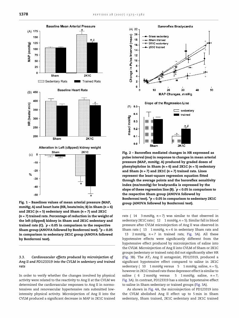
Fig. 1 – Baselines values of mean arterial pressure (MAP,
mmHg; A) and heart hate (HR, beats/min; B) in Sham (n = 6)
and 2K1C (n = 5) sedentary and Sham (n = 7) and 2K1C
(n = 7) trained rats. Percentage of reduction in the weight of
the left (clipped) kidney in Sham and 2K1C sedentary and
trained rats (C). *p < 0.05 in comparison to the respective
Sham group (ANOVA followed by Bonferroni test). #p < 0.05
in comparison to sedentary 2K1C group (ANOVA followed
by Bonferroni test).
Fig. 2 – Baroreflex mediated changes in HR expressed as
pulse interval (ms) in response to changes in mean arterial
pressure (MAP, mmHg; A) produced by graded doses of
phenylephrine in Sham (n = 6) and 2K1C (n = 5) sedentary
and Sham (n = 7) and 2K1C (n = 7) trained rats. Lines
represent the least-square regression equation fitted
through the average points and the baroreflex sensitivity
index (ms/mmHg) for bradycardia is expressed by the
slope of these regression line (B). *p < 0.05 in comparison to
the respective Sham group (ANOVA followed by
Bonferroni test). #p < 0.05 in comparison to sedentary 2K1C
group (ANOVA followed by Bonferroni test).
p e p t i d e s 2 8 ( 2 0 0 7 ) 1 3 7 5 – 1 3 8 21378
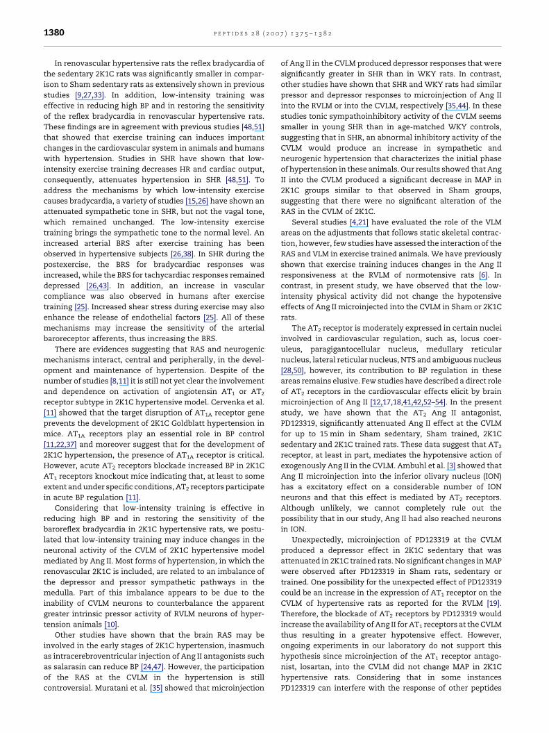
3.3. Cardiovascular effects produced by microinjection ofAng II and PD123319 into the CVLM in sedentary and trainedrats
In order to verify whether the changes involved by physical
activity were related to the reactivity to Ang II at the CVLM we
determined the cardiovascular responses to Ang II in normo-
tensives and renovascular hypertensive rats submitted low-
intensity physical activity. Microinjection of Ang II into the
CVLM produced a significant decrease in MAP in 2K1C trained
rats (�14� 3 mmHg, n = 7) was similar to that observed in
sedentary 2K1C rats (�12� 1 mmHg, n = 5). Similar fall in blood
pressure after CVLM microinjection of Ang II was observed in
Sham rats (�13� 1 mmHg, n = 6 in sedentary Sham rats and
�13� 2 mmHg, n = 7 in trained rats; Fig. 3A). All these
hypotensive effects were significantly different from the
hypotensive effect produced by microinjection of saline into
the CVLM. Microinjection of Ang II into CVLM of Sham or 2K1C
groups (sedentary or trained rats) did not significantly alter HR
(Fig. 3B). The AT2 Ang II antagonist, PD123319, produced a
significant hypotensive effect compared to saline in 2K1C
sedentary (�10� 1 mmHg versus �5� 1 mmHg; saline, n = 5),
however in 2K1C trained rats these depressor effect is similar to
saline (�6� 2 mmHg versus �5� 1 mmHg; saline, n = 7;
Fig. 3A). In contrast, PD123319 has a similar hypotensive effect
to saline in Sham sedentary or trained groups (Fig. 3A).
As shown in Fig. 4A, the microinjection of PD123319 into
the CVLM abolished Ang II effect up to 5 min in Sham
sedentary, Sham trained, 2K1C sedentary and 2K1C trained
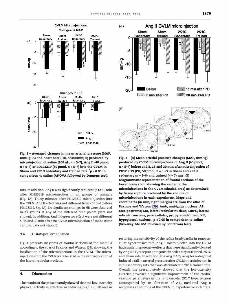
Fig. 3 – Averaged changes in mean arterial pressure (MAP,
mmHg; A) and heart hate (HR, beats/min; B) produced by
microinjection of saline (100 nL, n = 5–7), Ang II (40 pmol,
n = 5–7) or PD123319 (50 pmol, n = 5–7) into the CVLM in
Sham and 2K1C sedentary and trained rats. *p < 0.05 in
comparison to saline (ANOVA followed by Dunnets test).
Fig. 4 – (A) Mean arterial pressure changes (MAP, mmHg)
produced by CVLM microinjections of Ang II (40 pmol,
n = 5–7) before and 5, 15 and 30 min after microinjection of
PD123319 (PD, 50 pmol, n = 5–7) in Sham and 2K1C
sedentary (n = 5–6) and trained (n = 7) rats. (B)
Diagrammatic representation of frontal sections of the
lower brain stem showing the center of the
microinjections in the CVLM (shaded area) as determined
by tissue rupture produced by the volume of
microinjections in each experiment. Maps and
coordinates (in mm, right margin) are from the atlas of
Paxinos and Watson [39]. Amb, ambiguus nucleus; AP,
area postrema; LRt, lateral reticular nucleus; LRtPC, lateral
reticular nucleus, parvocellular; py, pyramidal tract; XII,
hypoglossal nucleus. *p < 0.05 in comparison to saline
(two-way ANOVA followed by Bonferroni test).
p e p t i d e s 2 8 ( 2 0 0 7 ) 1 3 7 5 – 1 3 8 2 1379
rats. In addition, Ang II was significantly reduced up to 15 min
after PD123319 microinjection in all groups of animals
(Fig. 4A). Thirty minutes after PD123319 microinjection into
the CVLM, Ang II effect was not different from control (before
PD123319, Fig. 4A). No significant changes in HR were observed
in all groups at any of the different time points (data not
shown). In addition, And II depressor effect were not different
5, 15 and 30 min after the CVLM microinjection of saline (time
control, data not shown).
3.4. Histological examination
Fig. 4 presents diagrams of frontal sections of the medulla
according to the atlas of Paxinos and Watson [39], showing the
localization of the microinjections in the CVLM. The micro-
injections into the CVLM were located in the ventral portion of
the lateral reticular nucleus.
4. Discussion
The results of the present study showed that the low-intensity
physical activity is effective in reducing high BP, HR and in
restoring the sensitivity of the reflex bradycardia in renovas-
cular hypertensive rats. Ang II microinjected into the CVLM
had similar hypotensive effects that were significantly blocked
by Ang II AT2 receptor antagonist in sedentary or trained, 2K1C
and Sham rats. In addition, the Ang II AT2 receptor antagonist
induced a fall in arterial pressure after CVLM microinjection in
2K1C sedentary rats that was attenuated in 2K1C trained rats.
Overall, the present study showed that the low-intensity
exercise provokes a significant improvement of the cardio-
vascular parameters in the renovascular 2K1C hypertension
accompanied by an alteration of AT2 mediated Ang II
responses at neurons of the CVLM in hypertensive 2K1C rats.
p e p t i d e s 2 8 ( 2 0 0 7 ) 1 3 7 5 – 1 3 8 21380
In renovascular hypertensive rats the reflex bradycardia of
the sedentary 2K1C rats was significantly smaller in compar-
ison to Sham sedentary rats as extensively shown in previous
studies [9,27,33]. In addition, low-intensity training was
effective in reducing high BP and in restoring the sensitivity
of the reflex bradycardia in renovascular hypertensive rats.
These findings are in agreement with previous studies [48,51]
that showed that exercise training can induces important
changes in the cardiovascular system in animals and humans
with hypertension. Studies in SHR have shown that low-
intensity exercise training decreases HR and cardiac output,
consequently, attenuates hypertension in SHR [48,51]. To
address the mechanisms by which low-intensity exercise
causes bradycardia, a variety of studies [15,26] have shown an
attenuated sympathetic tone in SHR, but not the vagal tone,
which remained unchanged. The low-intensity exercise
training brings the sympathetic tone to the normal level. An
increased arterial BRS after exercise training has been
observed in hypertensive subjects [26,38]. In SHR during the
postexercise, the BRS for bradycardiac responses was
increased, while the BRS for tachycardiac responses remained
depressed [26,43]. In addition, an increase in vascular
compliance was also observed in humans after exercise
training [25]. Increased shear stress during exercise may also
enhance the release of endothelial factors [25]. All of these
mechanisms may increase the sensitivity of the arterial
baroreceptor afferents, thus increasing the BRS.
There are evidences suggesting that RAS and neurogenic
mechanisms interact, central and peripherally, in the devel-
opment and maintenance of hypertension. Despite of the
number of studies [8,11] it is still not yet clear the involvement
and dependence on activation of angiotensin AT1 or AT2
receptor subtype in 2K1C hypertensive model. Cervenka et al.
[11] showed that the target disruption of AT1A receptor gene
prevents the development of 2K1C Goldblatt hypertension in
mice. AT1A receptors play an essential role in BP control
[11,22,37] and moreover suggest that for the development of
2K1C hypertension, the presence of AT1A receptor is critical.
However, acute AT2 receptors blockade increased BP in 2K1C
AT1 receptors knockout mice indicating that, at least to some
extent and under specific conditions, AT2 receptors participate
in acute BP regulation [11].
Considering that low-intensity training is effective in
reducing high BP and in restoring the sensitivity of the
baroreflex bradycardia in 2K1C hypertensive rats, we postu-
lated that low-intensity training may induce changes in the
neuronal activity of the CVLM of 2K1C hypertensive model
mediated by Ang II. Most forms of hypertension, in which the
renovascular 2K1C is included, are related to an imbalance of
the depressor and pressor sympathetic pathways in the
medulla. Part of this imbalance appears to be due to the
inability of CVLM neurons to counterbalance the apparent
greater intrinsic pressor activity of RVLM neurons of hyper-
tension animals [10].
Other studies have shown that the brain RAS may be
involved in the early stages of 2K1C hypertension, inasmuch
as intracerebroventricular injection of Ang II antagonists such
as salarasin can reduce BP [24,47]. However, the participation
of the RAS at the CVLM in the hypertension is still
controversial. Muratani et al. [35] showed that microinjection
of Ang II in the CVLM produced depressor responses that were
significantly greater in SHR than in WKY rats. In contrast,
other studies have shown that SHR and WKY rats had similar
pressor and depressor responses to microinjection of Ang II
into the RVLM or into the CVLM, respectively [35,44]. In these
studies tonic sympathoinhibitory activity of the CVLM seems
smaller in young SHR than in age-matched WKY controls,
suggesting that in SHR, an abnormal inhibitory activity of the
CVLM would produce an increase in sympathetic and
neurogenic hypertension that characterizes the initial phase
of hypertension in these animals. Our results showed that Ang
II into the CVLM produced a significant decrease in MAP in
2K1C groups similar to that observed in Sham groups,
suggesting that there were no significant alteration of the
RAS in the CVLM of 2K1C.
Several studies [4,21] have evaluated the role of the VLM
areas on the adjustments that follows static skeletal contrac-
tion, however, few studies have assessed the interaction of the
RAS and VLM in exercise trained animals. We have previously
shown that exercise training induces changes in the Ang II
responsiveness at the RVLM of normotensive rats [6]. In
contrast, in present study, we have observed that the low-
intensity physical activity did not change the hypotensive
effects of Ang II microinjected into the CVLM in Sham or 2K1C
rats.
The AT2 receptor is moderately expressed in certain nuclei
involved in cardiovascular regulation, such as, locus coer-
uleus, paragigantocellular nucleus, medullary reticular
nucleus, lateral reticular nucleus, NTS and ambiguous nucleus
[28,50], however, its contribution to BP regulation in these
areas remains elusive. Few studies have described a direct role
of AT2 receptors in the cardiovascular effects elicit by brain
microinjection of Ang II [12,17,18,41,42,52–54]. In the present
study, we have shown that the AT2 Ang II antagonist,
PD123319, significantly attenuated Ang II effect at the CVLM
for up to 15 min in Sham sedentary, Sham trained, 2K1C
sedentary and 2K1C trained rats. These data suggest that AT2
receptor, at least in part, mediates the hypotensive action of
exogenously Ang II in the CVLM. Ambuhl et al. [3] showed that
Ang II microinjection into the inferior olivary nucleus (ION)
has a excitatory effect on a considerable number of ION
neurons and that this effect is mediated by AT2 receptors.
Although unlikely, we cannot completely rule out the
possibility that in our study, Ang II had also reached neurons
in ION.
Unexpectedly, microinjection of PD123319 at the CVLM
produced a depressor effect in 2K1C sedentary that was
attenuated in 2K1C trained rats. No significant changes in MAP
were observed after PD123319 in Sham rats, sedentary or
trained. One possibility for the unexpected effect of PD123319
could be an increase in the expression of AT1 receptor on the
CVLM of hypertensive rats as reported for the RVLM [19].
Therefore, the blockade of AT2 receptors by PD123319 would
increase the availability of Ang II for AT1 receptors at the CVLM
thus resulting in a greater hypotensive effect. However,
ongoing experiments in our laboratory do not support this
hypothesis since microinjection of the AT1 receptor antago-
nist, losartan, into the CVLM did not change MAP in 2K1C
hypertensive rats. Considering that in some instances
PD123319 can interfere with the response of other peptides
p e p t i d e s 2 8 ( 2 0 0 7 ) 1 3 7 5 – 1 3 8 2 1381
or can present an agonistic effect [13,34,53]. Further studies are
necessary to clarify our present findings.
In summary, our results showed that low-intensity
physical activity that is effective in reducing high BP and in
restoring the sensitivity of the baroreflex bradycardia, does
not induce changes in the responsiveness to Ang II at CVLM of
normotensive or hypertensive, 2K1C rats. These results, on the
order hand, changes in the responsiveness to AT2 related
stimuli at the CVLM appears to be involved in the cardiovas-
cular effect of low-intensity physical activity and add new
significant insights into RAS mechanism involved in cardio-
vascular homeostasis and its adaptation to exercise in
renovascular hypertension.
Acknowledgements
This study was supported by FAPEMIG (Fundacao de Amparo a
Pesquisa do Estado de Minas Gerais), CNPq (Conselho Nacional
de Desenvolvimento Cientıfico e Tecnologico) Pronex Project
Grant (FAPEMIG/CNPq) and CAPES (Coordenacao de Aper-
feicoamento de Pessoal de Nıvel Superior). M.C. Rodrigues was
a recipient of CAPES fellowship (Master Degree) at the
‘‘Programa de Pos-graduacao Ciencias Biologicas’’, NUPEB,
UFOP. We are thankful to Dr. Claudia Martins Carneiro
Associate Professor at the ‘‘Departamento de Analises
Clınicas’’, UFOP, for histological analysis.
r e f e r e n c e s
[1] Alzamora AC, Santos RAS, Campagnole-Santos MJ.Hypotensive effect of Ang II and Ang-(1-7) at thecaudal ventrolateral medulla involves differentmechanisms. Am J Physiol (Regul Integr Comp Physiol)2002;283:R1187–95.
[2] Alzamora AC, Santos RAS, Campagnole-Santos MJ.Baroreflex modulation by angiotensins at the rat rostraland caudal ventrolateral medulla. Am J Physiol (RegulIntegr Comp Physiol) 2006;290:R1027–34.
[3] Ambuhl P, Felix D, Imboden H, Khosla MC, Ferrario CM.Effects of angiotensin analogues and angiotensin receptorantagonists on paraventricular neurones. Regul Pept1992;38(2):111–20.
[4] Ally A, Phattanarudee S, Kabadi S, Patel M, Maher TJ.Cardiovascular responses and neurotransmitter changesduring static muscle contraction following blockade ofinducible nitric oxide synthase (iNOS) within theventrolateral medulla. Brain Res 2006;1090(1):123–33.
[5] Averill DB, Diz D. Angiotensin peptides and baroreflexcontrol of sympathetic outflow: pathways and mechanismsof the medulla oblongata. Brain Res Bull 2000;51(2):119–28.
[6] Becker LK, Santos RAS, Campagnole-Santos MJ.Cardiovascular effects of angiotensin II and angiotensin-(1-7) at the RVLM of trained normotensive rats. Brain Res2005;1040(1–2):121–8.
[7] Blessing WW, Reis DJ. Inhibitory cardiovascular functin ofneurons in the caudal ventrolateral medulla of the rabbit:relationship to the area containing A1 noradrenergic cells.Brain Res 1982;253:161–71.
[8] Blume A, Kaschina E, Unger T. Angiotensin II type 2receptors: signaling and pathophysiological role. Curr OpinNephrol Hypertens 2001;10:239–46.
[9] Britto RR, Santos RAS, Fagundes-Moura CR, Khosla MC,Campagnole-Santos MJ. Role of angiotensin-(1-7) in themodulation of the baroreflex in renovascular hypertensiverats. Hypertension 1997;30:549–56.
[10] Carvalho THF, Bergamaschi CT, Lopes OU, Campos RR. Roleof endogenous angiotensin II on glutamatergic actions inthe rostral ventrolateral medulla in goudblatt hypertensiverats. Hypertension 2003;42(pt 2):707–12.
[11] Cervenka L, Horacek V, Vaneckova I, Hubacek JA, OliverioMI, Coffman TM, Navar LG. Essential role of AT1A receptorin the development of 2k1c hypertension. Hypertension2002;40:735–41.
[12] D’Amico M, Filippo C, Rossi F. Role of AT2 receptors in thecardiovascular events following microinjection ofangiotensin II into the superior colliculus of anaesthetizedrats. Naunyn-Schmiedebergs Arch Pharmacol1998;357:121–5.
[13] Gironacci MM, Vata M, Rodriguez-Fermepın M, FernandezBE, Pena C. Angiotensin-(1-7) reduces norepinephrinerelease through a nitric oxide mechanism in rathypothalamus. Hypertension 2000;35:1248–52.
[14] Dampney RAL. Functional organization of centralpathways regulating the cardiovascular system. PhysiolRev 1994;74(2):323–64.
[15] Gava NS, Veras-Silva AS, Negrao CE, Krieger EM. Low-intensity exercise training attenuates cardiac ß-adrenergictone during exercise in spontaneously hypertensive rats.Hypertension 1995;26(pt 2):1129–33.
[16] Gross V, Plehm R, Tank J, Jordan J, Diedrich A, Obst M, et al.Heart rate variability and baroreflex function in AT2receptor-disrupted mice. Hypertension 2002;40:207–13.
[17] Hein LGS, Barsh RE, Pratt VJ, Dzau BK, Kodilka. Behavioraland cardiovascular effects of disrupting the angiotensin IItype-2 receptor gene in mice. Nature 1995;377:744–7.
[18] Hogarty DC, Speakman EA, Puig V, Phillips MI. The role ofangiontensin AT1 and AT2 receptors in the pressor,drinking and vasopressin responses to the centralangiontensin. Brain Res 1992;586:289–94.
[19] Hu L, Zhu D, Yu Z, Wang JQ. Expression of angiotensin IItype 1 (AT1) receptor in the rostral ventrolateral medulla inrats. J Appl Physiol 2002;92:2153–61.
[20] Ichiki T, Labosky PA, Shiota C, Okuyama S, Imagawa Y,Fogo A, et al. Effects on blood pressure and exploratorybehavior of mice liking angiotesnin II type-2 receptor.Nature 1995;377:748–50.
[21] Ishide T, Preuss CV, Maher TJ, Ally A. Neurochemistrywithin ventrolateral medulla and cardiovascular effectsduring static exercise following eNOS antagonism.Neurosci Res 2005;52:21–30.
[22] Ito S, Komatsu K, Tsukamoto K, Kanmatsuse K, Sved AF.Ventrolateral medulla AT1 receptors support bloodpressure in hypertensive rats. Hypertension 2002;40:552.
[23] Johren O, Dendorfer A, Dominiak P. Cardiovascular andrenal function of angiotensin II type-2 receptors.Cardiovasc Res 2004;62(3):460–7.
[24] Kagiyama S, Varela A, Phillips MI, Galli SM. Antisenseinhibition of brain renin–angiotensin system decreasedblood pressure in chronic 2-kidney, 1 clip hypertensive rats.Hypertension 2001;37:371–5.
[25] Katz SD. The role of endothelium-derived vasoactivesubstances in the pathophysiology of exercise intolerancein patients with congestive heart failure. Prog CardiovascDis 1995;38:23–50.
[26] Krieger EM, Farah VMA, Moreira ED, Pires MD, IrigoyenMCC. Comparison of three methods for the determinationof baroreflex sensitivity in conscious rats. Braz J Med BiolRes 1999;32(3):361–9.
[27] Kumagai K, Suzuki H, Ryuzaki M, Matsukawa S, Saruta T.Baroreflex control of renal sympathetic nerve activity is
p e p t i d e s 2 8 ( 2 0 0 7 ) 1 3 7 5 – 1 3 8 21382
potenciated at early phase of two-kidney, one-clipGoldblatt hypertension in conscious rabbits. Circ Res1990;67:1309–22.
[28] Lenkei Z, Palkovits M, Corvol P, Llorens-Cortes C.Expression of angiotensin type-1 (AT1) and type-2 (AT2)receptor mRNAs in the adult rat brain: a functionalneuroanatomical review. Front Neuroendocrinol1997;18(14):383–439.
[29] Lin KS, Chan JYH, Chan SHH. Involvement of AT2 receptorsat NRVL in tonic baroreflex suppression by endogenousangiotensis. Am J Physiol 1997;272:2204–10.
[30] Lo M, Liu KL, Lantelme P, Sassard J. Subtype 2 ofangiotensin II receptors controls pressure-natriuresis inrats. J Clin Invest 1995;95:1394–7.
[31] Luoh HF, Chan SHH. Participation of AT1 and AT2 receptorsubtypes in the tonic inhibitory modulation of baroreceptorreflex response by endogenous angiotensins at the nucleustractus solitarii in the rat. Brain Res 1998;782(1–2):73–82.
[32] Matsumura Y, Hasser EM, Bishop VS. Central effect ofangiotensin II on baroreflex regulation in consciousrabbits. Am J Physiol (Regul Integr Comp Physiol)1989;256:R694–700.
[33] Moreira ED, Oliveira M, Krieger EM. Impaired baroreflexcontrol of heart rate in high-renin renal hypertension.J Hypertens 1988;6(8):619–25.
[34] Moreira TH, Rodrigues AL, Beirao PS, Santos RAS, Cruz JS.Angiotensin II inhibition of Ca2+ currents is independent ofATR1 angiotensin II receptor activation in rat adult vagalafferent neurons. Auton Neurosci 2005;117(2):79–86.
[35] Muratani H, Ferrario CM, Averill DB. Ventrolateral medullaof spontaneously hypertensive rats: role of angiotensin II.Am J Physiol (Regul Integr Comp Physiol) 1993;264:R388–95.
[36] Negrao CE, Krieger EM, Brum PC. Influence of exercisetraining on neurogenic control of blood pressure inspontaneously hypertensive rats. Hypertension1999;34:720–3.
[37] Oliverio MI, Best CF, Smithies O, Coffman TM. Regulation ofsodium balance and blood pressure by the AT1A receptorfor angiotensin II. Hypertension 2000;35:550–4.
[38] Pagani M, Somers V, Furlan R, Dell OS, Conway J, Baselli G,et al. Changes in autonomic regulation induced byphysical training in mild hypertension. Hypertension1988;12:600–10.
[39] Paxinos G, Watson C. The rat brain in stereotaxiccoordinates. New York: Academic Press; 1986.
[40] Sesoko S, Muratami H, Takeshita S, Teruya H, Kawazoe N,Fukuyama K. Modulation of baroreflex function byangiotensin II endogenous to the caudal ventrolateralmedulla. Brain Res 1995;671:38–44.
[41] Shi L, Yao J, Koos BJ, Xu Z. Induced fetal depressor orpressor responses associated with c-fos by intravenous orintracerebroventricular losartan. Dev Brain Res2004;(153):53–60.
[42] Shi L, Mao C, Thorton SN, Sun W, Wu J, Yao J, et al. Effectsof intracerebroventricular losartan on angiotensin II-mediated pressor responses and c-fos expression in near-term ovine fetus. J Comp Neurol 2005;493(4):571–9.
[43] Silva GJJ, Brum PC, Negrao CE, Krieger EM. Acute andchronic effects of exercise on baroreflexes inspontaneously hypertensive rats. Hypertension1997;30(2):714–9.
[44] Smith JK, Barron KW. The rostral and caudal ventrolateralmedulla in young spontaneously hypertensive rats. BrainRes 1989;506(1):153–8.
[45] Stromberg C, Naveri L, Saavedra JM. Nonpeptideangiotensin AT1 and AT2 receptors ligands modulate theupper limit of cerebral blood flow autoregulation in the rat.J Cereb Blood Flow Metab 1993;13:293–303.
[46] Sveed AF, Ito S, Madden CJ. Baroreflex dependent andindependent roles of the caudal ventrolateral medulla incardiovascular regulation. Brain Res Bul 2000;51(2):120–33.
[47] Sweet CS, Columbo JM, Gaul SL. Central antihypertensiveeffects of inhibitors of the rennin–angiotensin system inrats. Am J Physiol 1976;231:1794–9.
[48] Tipton CM. Exercise training for the treatment ofhypertension: a review. Clin J Sports Med 1999;9(2):104.
[49] Tony GM, Porter JP. Functional role of brain AT1 and AT2receptors in the central angiotensin II pressor response.Brain Res 1993;603:57–63.
[50] Veerasingham SJ, Raizada MK. Brain renin–angiotensinsystem dysfunction in hypertension: recent advances andperspectives. Br Pharmacol 2003;139:191–202.
[51] Veras-Silva AS, Mattos KC, Gava NS, Brum PC, Negrao CE,Krieger EM. Low-intensity exercise training decreasescardiac output and hypertension in spontaneouslyhypertensive rats. Am J Physiol (Heart Circ Physiol)1997;273:H2627–31.
[52] Von Bohlen U, Halbach O, Albrecht D. Angiotensin IIinhibits long-term potentiation with the lateral nucleus ofthe amydala through AT1 receptors. Peptides1998;19(6):1031–6.
[53] Walthers PE, Gaspari TA, Widdop RE. Angiotensin-(1-7) actsas a vasodepressor agent via angiotensin II type 2 receptorsin conscious rats. Hypertension 2005;45:960–6.
[54] Widdop RE, Gardiner SM, Kemp PA, Bennett T. Differentialblockade of central effects of angiotensin II by AT2 receptorantagonists. Am J Physiol (Heart Circ Physiol)1993;265:H226–31.