Evidence-based orthopaedic manual therapy for patients with nonspecific low back pain: Analysis of a...
214
EVIDENCE-BASED ORTHOPAEDIC MANUAL THERAPY FOR PATIENTS WITH NONSPECIFIC LOW BACK PAIN ANALYSIS OF A KINEMATIC MODEL OF THE SPINE Benjamin Hidalgo Thesis submitted in fulfillment of the requirements for the degree of « Docteur en Sciences de la Motricité » Supervisors: Professors Christine Detrembleur and Henri Nielens May 2015 Faculté des Sciences de la Motricité Institute of Neuroscience
Transcript of Evidence-based orthopaedic manual therapy for patients with nonspecific low back pain: Analysis of a...

EVIDENCE-BASED ORTHOPAEDIC MANUAL THERAPY FOR PATIENTS WITH NONSPECIFIC
LOW BACK PAIN
ANALYSIS OF A KINEMATIC MODEL OF THE SPINE
Benjamin Hidalgo Thesis submitted in fulfillment of the requirements for the degree of
« Docteur en Sciences de la Motricité »
Supervisors: Professors Christine Detrembleur and Henri Nielens
May 2015
Faculté des Sciences de la Motricité Institute of Neuroscience

2

3
Université Catholique de Louvain
Faculté des Sciences de la Motricité
Evidence-based orthopaedic manual therapy for patients with nonspecific
low back pain Analysis of a kinematic model of the spine
Benjamin HIDALGO
Thesis submitted in fulfillment of the requirements for the degree of “Docteur en Sciences de la Motricité”
May 2015
Supervisors: Professor Christine Detrembleur and Professor Henri Nielens (UCL)
Committee members:
Chair: Prof. Schepens Bénédicte (UCL)
Prof. Berquin Anne (UCL)
Prof. Feipel Véronique (ULB)
Prof. Vanderthommen Marc (ULG)
Prof. Thonnard Jean-Louis (UCL)
Prof. Bragard Dominique (UCL)

4

5
Benjamin Hidalgo
Pictured as speaker in 2012 during the World Musculoskeletal Congress of the International Federation of
Orthopaedic Manipulative Physical Therapists (IFOMPT), Québec, Canada
Benjamin was born in December 1975 in Belgium. He completed his master’s degrees in
Physical Education, supplemented with a pedagogical degree for teaching, in 2000 and in
Physical Therapy and Rehabilitation in 2002 at the Faculty of Motor Sciences (FSM) of the
Université Catholique de Louvain (UCL), Louvain-La-Neuve, Belgium. He completed a
degree in Osteopathy in 2007 after 5 years of continuing education (160 ECTS) at the
Sutherland College of Osteopathic Medicine, Belgium. Benjamin was certified in
Orthopaedic Manual Therapy (OMT) by the Manual Concepts Program (25 ECTS) of Curtin
University, Australia in 2011, and by UCL in 2014 after 2 years of continuing education (60
ECTS). He has fulfilled the requirements for certificates in sport physiotherapy, trigger
points therapy and dry needling, mobilisation with movement (Mulligan concept), spinal
manipulative therapy, functional movement assessment, classification-based cognitive
functional therapy for low back pain, and specific osteopathic care. He continues to learn
through continuing education courses in his field of interest, and is the co-founder and co-
head of the Certificate Program in Orthopaedic Manual Therapy (75 ECTs) at UCL.
Since 2002, Benjamin has specialised in the evaluation and treatment of
neuromusculoskeletal disorders of the whole body for elite sportsmen and the public in his
private clinical practice. In the last 6 years, he has focused his clinical and research
expertise on the spine, particularly the lumbopelvic region. He is an accomplished
practitioner, teacher and researcher in the neuromusculoskeletal field. These three areas of
expertise and skills are complementary and interrelated, providing Benjamin with a broad

6
view of OMT, great satisfaction in his work, and, he hopes, a good quality of care and
education for his patients and students.
As the historic context of this thesis, in 2008 Prof. Jean-Louis Thonnard contacted Benjamin
and encouraged him to begin a PhD in the field of OMT for patients with low back pain.
Benjamin accepted this challenge, motivated by curiosity and a desire for intellectual
development in this very interesting and complex topic of research. In the beginning, the
research was difficult because there was no background in manual therapy at UCL.
Fortunately, the advice of Benjamin’s thesis supervisors, Profs. Christine Detrembleur and
Henri Nielens (FSM-UCL), and a great meeting with an external international expert in OMT,
Dr. Toby Hall (Curtin University, Australia), combined to enable on-time completion of this
ambitious project. Regarding the picture on the cover, with this thesis, Benjamin hopes to
contribute insight to understanding the complex puzzle of low back pain, thereby improving
the management of patients with this debilitating disorder.

7
Remerciements
Entreprendre une thèse, un « PhD », n’est vraiment pas une épreuve de vie anodine… Cela
représente un défi important et même un combat qui a comme conséquence, qu’une longue
période de vie y soit entièrement dévouée. Cette quête de soi-même en ce qui me
concerne, implique que la vie privée ainsi que parfois la santé, en subissent certaines
conséquences.
Mais à travers l’effort, cette épreuve scientifique est pourtant une fantastique aventure
humaine où le dépassement de soi et la détermination règnent en maître mots en particulier
à l’époque d’un « no man’s land » en terme de thérapie manuelle orthopédique moderne au
sein de notre institution. Les conséquences positives en sont certes d’abord un
développement personnel, ensuite une ouverture et une collaboration avec la communauté
internationale en thérapie physique, ainsi qu’une grande fierté. Evidemment, la vie a rajouté
son lot de difficultés et de drames durant cette période, il n’a vraiment pas toujours été
facile de concrétiser mon « Doc »…
Mes amis me l’ont souvent dit: « Alors c’est quand que tu termines ton Doc ? » Et bien c’est
aujourd’hui ! C’est grâce à la détermination, ainsi qu’au courage que cette épreuve
académique majeure se concrétise. Mais ce travail ne se fait pas seul, je voudrais donc en
remercier les personnes suivantes :
La première personne que je veux remercier de tout mon cœur et à qui je dédie entièrement
ma thèse est mon père Daniel Hidalgo décédé prématurément en juillet 2013 à l’issue d’une
très longue hospitalisation. Tu me manques tous les jours et durant ces longs mois de soins
intensifs, tu t’es battu avec un courage forçant l’admiration pour ta vie et ta famille…
Chaque jour je t’ai vu lutter et je sais à présent de qui je tiens ma détermination. J’aurais
tellement aimé que tu sois encore là lors de la présentation publique de ma thèse pour que
tu puisses être fier de moi. Car c’est bien grâce à toi ainsi qu’à ton soutien sans faille pour
mon éducation et pour mes études depuis mon enfance jusqu’à ton départ que je suis
devenu l’homme que je suis. Je continuerai à transmettre ton héritage éducatif ainsi que ta
détermination dans mon attitude à travers et autour de moi dans l’enseignement, dans ma
pratique clinique et ma vie privée.
Bien entendu, je remercie le Professeur Christine Detrembleur, ma promotrice de thèse,
grâce à qui tout a été possible de par son intelligence et ses grandes compétences
techniques et scientifiques. De plus, je la remercie également pour sa gentillesse, sa
compréhension, son soutien, sa confiance et toutes ses grandes qualités humaines qui la
caractérise et dont la liste serait beaucoup trop longue à détailler ici. Je remercie également

8
mon autre promoteur, le Professeur Henri Nielens pour sa rigueur et son expertise
scientifique et clinique sur la lombalgie. Merci encore à vous deux pour votre expertise
commune, vos encouragements, ainsi que vos nombreuses lectures et corrections pendant
ces années de doctorat.
Parfois, la vie est faite de belles rencontres et celle-là fut cruciale pour l’aboutissement de
ma thèse, je voudrais donc très chaleureusement remercier mon ami et Professeur le
Docteur Toby Hall pour son aide si précieuse, sa gentillesse, ses corrections et ses
conseils, ses encouragements, sa rapidité et son positivisme anglo-saxon, ainsi que sa très
grande expertise. C’est vraiment un très grand honneur pour moi d’avoir été reconnu dans
mon travail ainsi que d’avoir travaillé et de continuer à collaborer avec un grand expert
mondialement connu en thérapie manuelle orthopédique.
Je remercie également l’ensemble de mon comité de thèse pour leurs idées, corrections,
suggestions lors des réunions de comité de doctorat les Professeurs : Bénédicte Schepens,
Anne Berquin, Jean-Louis Thonnard, Dominique Bragard.
Je remercie pour leur soutien le doyen du pôle COSY et de l’IoNS ; le Professeur Jean-Noël
Octave et le Doyen de la Faculté des Sciences de la Motricité (FSM); le Professeur Thierry
Zintz.
Je remercie également tous mes collègues du laboratoire, de l’IoNS et de la FSM, et en
particulier les Professeurs Philippe Mahaudens et Laurent Pitance pour leur soutien et les
échanges de conversation tout au long de ces années.
Je remercie également mes collègues de l’Université de Gand (UGent) en particulier les
Professeurs et enseignants Lieven Danneels, Barbara Cagnie, Damien Van Tiggelen, Axell
Bernaerts, Bart Vanthillo, ainsi que ceux de l’Université de Liège (ULg), Christophe
Demoulin et Sébastien Wolf pour leurs collaborations, leurs enseignements, les inspirations
et les discussions qui ont indirectement contribué à la rédaction de cette thèse.
Merci aussi à mes amis qui par leurs amitiés ont directement aidé à clôturer ce doctorat, je
souhaite en particulier remercier : Pierre et Sophie Henrich, ainsi que le Docteur Emmanuel
Gérard et Sophie Gérard-Zaczek, Bruno Lheureux, Grégoire Litt, Patrice Combe car ils ont
toujours été là pour moi dans les bons comme dans les mauvais moments.
Un énorme merci à mes nombreux étudiants-mémorants et à toutes les personnes qui ont
participé aux expériences et qui ont donc fondamentalement contribué à construire cette
thèse par leur participation à travers l’ensemble des articles scientifiques qui la compose.
« Last but not least », je remercie ma famille sans qui je n’aurai pas eu la force de terminer
ce doctorat. Grâce à eux j’ai pu trouver des ressources inattendues lors des nombreux

9
passages difficiles tout au long de ce projet professionnel et de ma vie, je remercie donc
chaleureusement et avec amour : ma mère Elyane Clesse, mon frère Pierre Hidalgo et sa
compagne Mélanie Glenda-Gillis, ma sœur Adèlanne Hidalgo, ainsi que ma petite chienne
Lana. Enfin, j’aimerais remercier ma compagne Concetta Crimi qui a su me donner le
sourire et la volonté à travers son amour et son humour, ainsi que les innombrables fous
rires dont j’avais besoin parfois pour m’échapper temporairement afin de finaliser ce travail
que vous allez j’espère lire attentivement…
A TOUS MERCI

10

11
TABLE OF CONTENTS
Summary of the thesis 13
Index of abbreviations 17
Introduction 19
Section 1: Evidence for the use of orthopaedic manual therapy for 47
patients with low back pain
Chapter I: Efficacy of manual therapy and exercises for different stages 49
of nonspecific low back pain: Current evidence from the literature
Section 2: Development of a kinematic model of the spine 79
Chapter II: Reliability and validity of a kinematic model of the spine during 81
active trunk movement in healthy subjects and patients with chronic
nonspecific low back pain
Chapter III: Use of kinematic algorithms to distinguish people with chronic 103
nonspecific low back pain from asymptomatic subjects: Validation study
Chapter IV: Effects of proprioceptive disruption on lumbar spine repositioning 117
error in a trunk forward-bending task
Section 3: Clinical physical examination and evaluation of the efficacy 133
of orthopaedic manual therapy for patients with low back pain
Chapter V: Intertester agreement and validity of identifying lumbar pain 135
provocative movement patterns using active and passive accessory movement tests
Chapter VI: Short-term effects of Mulligan mobilisation with movement on pain, 157
disability and kinematic spinal movements in patients with nonspecific low back pain:
a randomised placebo-controlled trial
General discussion and conclusions 179

12

13
Summary of the thesis
This thesis on the study of the efficacy of orthopaedic manual therapy (OMT) for patients
with nonspecific low back pain (LBP) was developed by following the steps of an
evidence-based practice process through three major sections.
The Introduction defines the debilitating disorder of LBP and OMT, and describes an
integrative approach for the stratification of care in LBP patients.
Section 1 presents a systematic review that updates the best evidence of OMT efficacy in
terms of pain, functions, activities and participation. The findings allow us: (I) to establish
different levels of evidence for this form of therapy, (II) to understand the complexity of LBP
and (III) to affirm the importance of the study design quality in OMT trials (e.g. splitting
design, complexity of the placebo procedure and integration of clinical reasoning).
Section 2, which is composed of three studies, investigates a kinematic model of the spine to help in the diagnosis of LBP patients, as well as outcome measures for future
investigations of OMT in LBP patients. This kinematic tool permits a valid assessment of
body structures (lumbopelvic and thoracic vertebral column, muscles of the trunk and pelvic
regions), body functions (mobility in a vertebral segment, control of complex voluntary
movements, proprioceptive function) and activities (bending, maintaining a body position).
Finally, Section 3 presents two clinical studies. The first is a reliability study on a
standardised and original pain provocation examination of the lumbar spine in a combined
movement fashion. This examination provides the direction and vertebral level(s) of
treatment. On the basis of this reliable objective examination and evidence described
throughout this thesis, a randomised controlled trial was conducted. This last study
questions the short-term efficacy of a novel form of OMT, namely mobilisation with
movement, on primary kinematic outcome measures (kinematic algorithms for range of
motion and speed) and secondary self-reported outcome measures (pain, function, activities
and participation) in LBP patients with a mechanical pain pattern in flexion. The results of
this investigation raise the overall level of evidence from limited to moderate in favour of
using central sustained natural apophyseal glides in LBP patients.
In conclusion, the different points and perspectives developed along this thesis contribute
towards solving the complex puzzle of LBP within a patient-centred approach. Manual
therapy is an art developed through clinical practice, as well as a science developed
through fundamental and clinical research. Clinical research is of major importance
because it directly drives clinical practice and education towards an evidence-based OMT
practice within the biopsychosocial framework, thereby aiding many patients, students and
health professionals.

14

15
Cette thèse étudie l’efficacité de la thérapie manuelle orthopédique (TMO) auprès de
patients présentant une lombalgie commune (LBP). Elle s’articule selon les différentes
étapes d’un processus de pratique fondée sur les preuves scientifiques à travers trois
sections principales. Au préalable, nous définissons la LBP ainsi que la TMO selon une
vision contemporaine. La présentation d'une approche intégrative a également été abordée
pour mieux comprendre la stratification des soins chez les patients LBP avec une TMO
adaptée à chaque sous-groupe.
Dans la section 1, qui compose la première étape de notre travail, une revue systématique
met à jour les meilleures preuves existantes sur l’efficacité de la TMO (techniques passives
et actives) sur la LBP (de la phase aiguë à chronique) pour la douleur et la fonction, ainsi
que pour les activités et la participation. Ce qui nous a permis : (I) d'établir différents
niveaux de preuve pour cette forme de thérapie avec une fluctuation assez large du niveau
de preuves (de faible à forte), (II) de mieux comprendre la complexité de la LBP, (III) de
mettre en avant l'importance de la qualité de conception des études cliniques en TMO
comme par exemple : le fractionnement en sous-groupes, la complexité de la procédure
placebo, ainsi que de l'intégration du raisonnement clinique dans les études sur la TMO et
la LBP.
La section 2, représentant la deuxième étape, se compose de trois études pour évaluer
l’intérêt d’un modèle cinématique de la colonne vertébrale pour aider dans le diagnostic des
patients LBP, ainsi que comme outil de mesure pour les futures études cliniques sur la TMO
et la colonne vertébrale. Cet outil cinématique a démontré une bonne reproductibilité et
validité pour l’analyse des mouvements du tronc, c’est-à-dire: des structures du corps
(colonne vertébrale lombo-pelvienne et thoracique, des muscles du tronc et de la région du
bassin), des fonctions du corps (les fonctions de mobilité articulaire: spécifié comme la
mobilité dans un segment vertébral, le contrôle volontaire de mouvements complexes, ainsi
que la fonction proprioceptive) et des activités (flexion du tronc et le maintien d'une position
du corps).
Enfin la section 3 de cette thèse présente deux études cliniques originales. La première est
une étude sur la fiabilité de l’examen physique en TMO par provocation de la douleur sur le
rachis lombaire selon le principe des mouvements combinés. Sur base de cet examen qui a
démontré une fiabilité suffisante nous donnant la direction et les niveaux vertébraux de
traitement, ainsi que sur base des éléments de preuve décrits le long de cette thèse, une
étude placebo-contrôlée et randomisée a été menée. Cette dernière étude interroge
l'efficacité à court terme d'une nouvelle forme de TMO très peu étudiée au niveau du rachis
lombaire, à savoir la mobilisation avec mouvement. Celle-ci a été étudiée sur des mesures
principales cinématiques (algorithmes cinématiques pour l’amplitude et la vitesse)
déterminées dans la section 2 et sur des mesures secondaires à l’aide d’échelles /

16
questionnaires (douleur, la fonction, activités et participation) chez des patients LBP avec
présence d’une douleur à comportement mécanique lors de la flexion du tronc (sous-groupe
de LBP). Les résultats de cette enquête ont élevé le niveau global de preuve qui était faible
auparavant à modéré en faveur de l'utilisation des “glissements naturels soutenus des
apophyses articulaires” (mobilisation avec mouvement au niveau du rachis) chez un sous-
groupe de patients LBP.
En conclusion, les différents points et perspectives développés tout au long de cette thèse
devraient probablement contribuer à résoudre le puzzle complexe de la LBP dans une
optique d’approche centrée sur le patient. La thérapie manuelle est un art développé par la
pratique clinique ainsi qu’une science développée par la recherche fondamentale et
clinique. Nous croyons que la recherche clinique est d'une importance capitale car elle
détermine directement la qualité de la pratique clinique, ainsi que l'éducation et
l'enseignement. Une pratique clinique qui s’appuye sur des preuves issues de la recherche
tout en s’inscrivant dans une approche biopsychosociale devrait, d'une manière générale,
aider de nombreux patients, étudiants et professionnels de la santé.

17
Index of abbreviations
CB-CFT Classification Based Cognitive Functional Therapy
CCBRG Cochrane collaboration back review group
CCR clinical classification rule
CLBP chronic low back pain
CM combined movements
CPR clinical predictive rule
CS classification system
EBP evidence-based practice
HLS high lumbar spine
ICF International Classification of Functioning, Disability and
Health
KA-R kinematic algorithm for range of motion
KA-S kinematic algorithm for speed
LLS low lumbar spine
LS logit score
LTS low thoracic spine
MD means of difference
MICS Movement Impairments Classification System
MT manual therapy
MT1 spinal manipulation
MT2 spinal mobilisation and soft-tissue-techniques
MT3 MT1 combined with MT2
MWM mobilisation with movement
NSLBP nonspecific low back pain
NS-CLBP nonspecific chronic low back pain
OMT orthopaedic manual therapy
PAIVM passive accessory intervertebral movement

18
PPIVM passive physiological intervertebral movement
QTF Quebec task force
RCTs randomised controlled trials
RE repositioning error
ROM range of motion
SBT Start Back Tool
SMD standardised means of difference
SNAGs sustained natural apophyseal glides
SR systematic review
SS shoulder segment
TLS total lumbar spine

19
INTRODUCTION

20

21
The topic of this thesis regards the efficacy of orthopaedic manual therapy (OMT) for
patients with low back pain (LBP). This thesis is organised with the presentation of different
ideas following a continuum. First, the concept of evidence-based practice (EBP) will be
described (1) in the contexts of OMT and the complex disorder of LBP (2). Second, OMT
will be defined (3), potential mechanisms of action for OMT will be described (3.1) and an
integrative approach of using validated classification systems in the care of patients with
LBP will be presented (3.2). An objective examination (4) with a clinical physical
examination of the lumbar spine will be described (4.1), together with the role of kinematic
measures in diagnosis and as primary outcome measures of the body structures, functions
and activities for patients with nonspecific low back pain (NSLBP) (4.2). Finally, the
context, objectives and organisation (5) of this thesis according to an EBP process will be
presented through three major sections:
Section 1: Evidence for the use of OMT for patients with LBP
Section 2: Development of a kinematic model of the spine
Section 3: Clinical physical examination and evaluation of the efficacy of OMT for
patients with LBP
1. EVIDENCE-BASED PRACTICE
EBP is a philosophical approach that is in opposition to medical ‘folklore’, tradition and the
random treatment of patients. An interdisciplinary approach to clinical practice, EBP
originally began as evidence-based medicine (EBM) and spread to other fields with
complex intervention, such as psychology and physical therapy. EBP recognises that care is
individualised, ever changing and sometimes involves doubts as well as probabilities.1
In OMT, EBP involves complex and conscientious decision making in applying high-quality
therapy that is based not only on the best available clinical evidence, but also on the
patient’s values (biopsychosocial and lifestyle influences) and the clinician’s clinical
expertise (basic education, experience and continuing education) and reasoning (Figure
1).1-4 EBP constitutes a dynamic integrative approach that must be adapted during
treatment according to the severity and sensitivity of the disorder, as well as the
improvements gained. Also important are the influences of the manual therapist’s practical
skills across the range of commonly used OMT techniques to manage people with LBP, as
well as the therapist’s level of clinical reasoning skills in dealing with the complexity of
LBP.4-5

22
Figure 1. EBP concept3
2. DEFINITION OF NONSPECIFIC LOW BACK PAIN
LBP is the leading cause of disability and absence from work, and its increasing prevalence
has had major socioeconomic impacts.5-6 LBP has reached epidemic proportions, with
about 80% of the population experiencing LBP at some point in their lives. Of these
sufferers, 75% are in their most productive years, between the ages of 30 and 59.7-8
In 1987, the biopsychosocial model was suggested as a theoretical framework for LBP
treatment.9 Most LBP cases are described as ‘nonspecific’, as a precisely identified cause
for pain can only be determined in a small minority of cases. Indeed, there is a poor
correlation between findings on medical images and symptoms, with a radiologic diagnosis
being clearly identified in only 15% of cases.10-11 Hence, based on imaging, NSLBP is
defined by the lack of a recognisable, specific pathology, and is usually
multifactorial/multidimensional and of unknown origin and etiology.11 In the absence of
specific diagnoses, profiling LBP patients on the basis of biological, psychological and social
prognostic factors appears relevant.12
Among the biological influences, nociceptive factors play a major role in acute and
subacute NSLBP. Various structures in the lumbar spine are recognised as possible origins
of LBP due to their innervations. In particular, the zygapophyseal joints, intervertebral discs
and sacroiliac joints, with up to 75% of involvement, have been determined as nociceptive
sources for NSLBP.11,13-14 However, an important distinction in terms of pain stages needs
to be made, as nociceptive ‘sources’ can only be clearly determined in approximately half of
subjects with chronic LBP.15
23
The topography of pain in NSLBP is generally defined as pain in the lower back between
the lowest ribs and inferior gluteal folds.5,16-17 The duration or stage of the pain disorder is
typically categorised as acute (0–6 weeks), subacute (6–12 weeks) or chronic (>12
weeks).5,16-18
It is becoming increasingly clear that the clinical evaluation of patients with LBP, particularly
in the chronic stage, should not focus solely on the pathoanatomical examination
(structural nociceptive sources).5,10-11,15 Indeed, psychological and social factors a ,
lifestyle influences b and pain mechanisms c have important roles in explaining the
development of chronic LBP.11,19-20 This assumption is particularly important when
conceiving a comprehensive management strategy. Instead, it would be better to classify
patients with LBP into distinct subgroups by developing classification systems based on
clusters of signs and symptoms relevant to physical therapy.19-23
Use of the International Classification of Functioning, Disability and Health (ICF) is of
potential interest for improving clinical practice and stimulating research. The ICF comprises
four components: body functions, body structures, activities and participation, and
environmental factors. Short and comprehensive, the ICF Core Set for LBP was developed
with ICF codes and category titles for each of the four components, to aid in defining the
multidimensional aspects of LBP.5,8 In this Core Set, body structures has 5 categoriesd,
body functions has 19 categoriese, activities and participation has 29 categoriesf and
environmental factors has 25 categoriesg.5,8
3. MODERN ORTHOPAEDIC MANUAL THERAPY OF NONSPECIFIC LOW BACK PAIN
A consensus definition of OMT was determined at a general meeting in Cape Town in
March 2004 (www.ifompt.comh):
a Fears, beliefs/attitudes, coping strategies, education, anxiety, depression, work satisfaction b Sedentary lifestyle, poor sleep, stress, nutrition, smoking, alcoholism c Peripheral/functional or central sensitisation d Such as: s7401/joints of pelvic region, s76001/thoracic vertebral column, S76002/lumbar vertebral column, s7601/muscles of trunk region, and s7402/muscles of pelvic region e Such as: b28013/pain in back, b7101/mobility of several joints, b7108/mobility of joint functions, specified as mobility in a vertebral segment, b7601/control of complex voluntary movements, b260/proprioceptive function f Such as: d4108/bending, d415/maintaining a body position, d430/lifting and carrying objects g Such as: e410/individual attitudes of immediate family members, e450/individual attitudes of health professionals h Organised in 1974, the International Federation of Orthopaedic Manipulative Physical Therapists (IFOMPT) is a subgroup of the World Confederation of Physical Therapy recognised by World Health Organisation. IFOMP represents groups of manipulative physical therapists around the world who have completed stringent specialisation programs in the field of neuromusculoskeletal disorders. IFOMPT sets educational and clinical standards in this area of physical therapy. IFOMPT actively encourages improved patient management by its standards and by endorsing EBP.
24
‘Orthopaedic Manual Therapy is a specialized area of
physiotherapy/physical therapy for the management of
neuromusculoskeletal conditions, based on clinical reasoning, using highly
specific treatment approaches including manual techniques and
therapeutic exercises. Orthopaedic Manual Therapy also encompasses,
and is driven by, the available scientific and clinical evidence and the
biopsychosocial framework of each individual patient.’
In physical therapy, various forms of OMT are currently used to manage LBP, and there is
growing evidence in favour of this kind of treatment.4-5,21 Manual therapists use a range of
treatment approaches, including various ‘hands-on’ passive techniques (e.g. lumbopelvic
spinal mobilisation/manipulation, neurodynamic mobilisation of sciatic and femoral nerves
and soft tissue techniques) i and active-passive techniques (e.g. mobilisation with
movement or muscle energy techniques of the lumbopelvic region). Use of OMT involves
‘hands-off’ active techniques, such as motor control, directional preference, core stability
of the lumbar spine and communication skillsj.4,5,21
In some OMT approaches, the frequency, intensity and, particularly, direction of treatment
are driven by the patient’s pain severity and irritability. In the presence of high irritability
(i.e. easily provoked pain that lingers at a high level), OMT in a single or combined
movement, in the direction away from the most pain-provoking movement, is recommended.
The opposite is true for a low irritability disorder (i.e. hard to provoke pain that abates
quickly).24 Use of these collective OMT approaches, along with clinical reasoning based
on the biopsychosocial model, represents the fundamentals of OMT.4-5,25
3.1. Hypothetical mechanisms of action of orthopaedic manual therapy
When managing neuromusculoskeletal disorders, the aims of OMT are to reduce pain
and improve movement and function. Potential mechanisms of action for OMT in this
context have been described.26-45 To illustrate these mechanisms, hands-on techniques of
OMT are thought to cause mechanical, neurophysiological and placebo effects, as
described below.
Mechanical effects. The key mechanism of OMT, which may underlie all others, is the
administration of objective biomechanical segmental vertebral movement26-31 from the initial
mechanical action of the therapist’s hands. (I) A slight component of the load and peak
forces is absorbed by the paraspinal soft tissues, (II) while the main component is absorbed
by the spine.31 Spinal manipulative therapy of the lower back may generate motion of the
i Facilitated positional release (strain-counterstrain), myofascial release, neuromuscular technique, muscle energy technique and trigger points therapy j Empathy, listening, motivational interview, education, positivism and reassurance

25
vertebral bodies that will, from a cavitation phenomenon, separate the articular facet joints
and increase the lumbar zygapophyseal joints space separation over several days.26-32
Separation of the joint surface may release entrapped synovial meniscus, facilitate
transsynovial fluid fluctuation in the zygapophyseal joints and stretch the intra-articular
capsular adhesions that limit movement.26,29,31 Although research in human cadavers
showed that OMT decreases the intervertebral disc pressure, one study in a living human
subject did not support this mechanism of action.33 A recent study demonstrated that spinal
manipulative therapy facilitates water diffusion in the nucleus pulposus of the lumbar
intervertebral discs.34
Neurophysiological effects. OMT can stimulate responses from mechanoreceptors of the
joints, among others, and normalise neurogenic reflex activity.35-43 OMT has been reported
to 1) reduce paraspinal muscle spasms and the gastrocnemius H-reflex, 2) increase the
central motor excitability and the isometric strength of the paraspinal or quadriceps muscles,
and 3) influence motor control and proprioception.35-43 OMT can induce hypoalgesia through
peripherally inducing pain inhibition at the spinal cord (gate control theory) and centrally
activating descending inhibitory pathways from the dorsal periaqueductal gray area, while
concurrently activating the sympathetic nervous system.35,37,42-43
Placebo effects. As in any therapeutic intervention, OMT probably generates a placebo
effect through various psychological mechanisms. Such an effect may be explained by the
patient’s and therapist’s interests, beliefs, concerns and interactions.44,45 The fact that the
OMT therapist physically interacts with the patient (‘therapeutic touch’) may reinforce this
phenomenon.
3.2. Integrative stratified care in orthopaedic manual therapy for patients with low
back pain
Stratification of treatment represents a method to apply targeted OMT to LBP patients,
with the potential for greater effectiveness and efficiency of physical therapy. LBP is an ideal
clinical condition for stratified care research, as LBP patients comprise a heterogeneous
population4-5 with variations in prognosis and treatment options. Thus, stratified care is
becoming a dominant topic in research and clinical practice for LBP.46 However, most
randomised controlled trials (RCTs) addressing the effectiveness of OMT have treated LBP
patients as a homogeneous group. Consequently, the concept of subgrouping people
with LBP is more and more common in the OMT literature and in research. Classifying
patients into subgroups and applying specific OMT interventions for each subgroup is
thought to be effective approach.5,46

26
In physical therapy, stratified care comprises three main approaches of classification
systems, with overlaps between them. These approaches are based on the patients’
treatment responsiveness, prognosis and underlying causal pain mechanisms (Figure 2).46
Figure 2. Stratified care approaches, adapted from Foster et al.46
Integrative approachk
The Treatment-Based Classification (TBC) system to identify targeted OMT interventions
for people with LBP is an example of a treatment responsiveness-based stratification
scheme.47 The main principle of the TBC system is to group patients who are likely to
respond to a well-defined OMT technique (e.g. lumbopelvic manipulation, stabilisation,
directional preference exercise and traction), rather than trying to classify patients on the
basis of their hypothesised pain mechanisms and/or prognosis. The challenge of this
approach is providing evidence of patient features that consistently identify those who will
respond to a specific treatment.46
The Start Back Tool (SBT) is a prognosis-based classification system that aims to
subclassify patients according to physical and psychosocial factors. SBT is more effective
than a non-subgrouping approach, especially when patients are fast-tracked to an
k Targeted OMT for specific subgroups using an integrative approach merging classification systems and OMT interventions should be useful for patients. This integrative approach will be discussed in detail in the General Discussion section.

27
appropriate treatment course, and is particularly designed to support care decision making
in the primary/first-contact context.46,48-49
Classification-Based Cognitive Functional Therapy (CB-CFT) is a classification system
based on the causal pain mechanism. This multidimensional approach integrates evidence
from pathoanatomical, neurophysiological, psychosocial, physical and lifestyle domains.19,49
Modifiable beliefs l and behavioursm considered to contribute to pain and disability are
identified and become targets of treatment.19,46,49
The TBC and CB-CFT systems have moderate to good reliability, with some evidence of
validity.50 It has been recommended that these classification systems be implemented in
clinical practice.50 Moreover, the patients’ beliefs and expectations regarding treatment
effects of OMT interventions have been shown to be important predictors of treatment
outcomes and should be integrated into classification systems.51
In the specific field of physical therapy, several classification systems for LBP have been
proposed. However, only four of them directly tailor OMT management to the patient and
have been evaluated scientifically: the McKenzie LBP classification system, the TBC system
(Figure 3), the Movement-System Impairment Classification for LBP, and the CB-CFT
approach.19,52-53
l Fear of movement and pain-related anxiety m Pain-provoking postures and movement patterns

28
Figure 3. Treatment-based classification algorithm57
Nevertheless, the best way to subgroup patients with LBP has not been determined.46 For
example, in the TBC system, after an objective examination based on the assumption of
mechanical NSLBP, patients may be classified in the spinal manipulation subgroup and
matched to a directional preference exercise. Some patients classified into one treatment
category may meet criteria for another treatment subgroup and benefit from either or both
treatments.54 Different stratified care approaches may not necessarily be mutually exclusive,
and could be integrated in the management of LBP patients.46,54

29
The McKenzie and TBC systems interpret the patient’s symptoms and behaviour through a
series of single standardised and repeated spinal movements and sustained postures
performed during clinical examination. The goal of the assessment is to identify the
directional patterns that worsen or improve the patient’s symptoms.54-56 The following
modalities of physical examination provide a basis for the patient’s classification and
treatment: repeated spinal movements and sustained positions in a directional preference
(Figure 4A), passive spinal mobilisation/manipulation (Figure 4B), stabilisation exercises
(Figure 4C) and traction.5,46,47 In all of these classification systems, the sagittal plane is of
major importance to determine specific patterns.55-58
Figure 4. Illustration of interventions in the TBC System
A. Example of repeated spinal movements and/or sustained positions in a directional preference of extension, which centralisen symptoms
n Centralisation is a clinical phenomenon that can be reliably detected and is associated with a good prognosis. Centralisation was first recognised by McKenzie in the 1950s and, after much experimentation and verification, was described in the literature (McKenzie, 1981). It is the process by which pain radiating from the spine is sequentially abolished, distally to proximally, in response to therapeutic positions or movements, and includes reduction and abolition of spinal pain.

30
B. Example of passive spinal mobilisation/manipulation in a combined movement position (flexion/left lateral side bending and left rotation)
C. Example of a stabilisation exercise for transversus abdominis muscle strengthening
Long-term chronic LBP is a complex multidimensional condition that represents a
management challenge. A recent review of clinical classification systems for chronic LBP
patients concluded that most systems do not consider the underlying pain mechanisms and
focus largely on biomedical (pathoanatomical and pathophysiological) assessments.59 A
multidimensional classification system for LBP has been proposed (i.e. CB-CFT)19 and will
be discussed in detail in the General Discussion of this thesis.

31
4. OBJECTIVE EXAMINATION IN ORTHOPAEDIC MANUAL THERAPY
4.1. Clinical physical examination of the lumbar spine
To be evidence-based, the objective evaluation of patients with LBP, as part of the clinical
reasoning implemented in OMT, should be based on valid and reliable tests. During
physical examination, several testing manoeuvres may be implemented, including
reproduction or abolition of movement-induced symptoms, as well as palpation to detect
hypo- or hypermobility. The most reproducible tests in clinical examination of the lumbar
spine are based on mechanically induced symptom reproduction.55 Specifically,
examination of the mechanical pain response during repeated lumbar spinal movements in
the sagittal plane (flexion/extension) is the only procedure to show moderate evidence of
high reliability (based on inter-raters agreement).55 Reliability is good when the physical
examination is based on the response to symptoms, but generally low when the
examination is based on palpation to detect mobility.55
One mechanical pain provocation test that is commonly used in OMT during objective
examination is an active movement test in single or combined planes (Figure 5A). The
concept of combined movements testing was originally developed by Edwards and is an
expansion of the routine clinical examination.24 Another form of pain provocation testing is
passive accessory intervertebral movement (PAIVM) testing (Figure 5B). In the concept
proposed by Edwards, data obtained from the single- and combined-plane active movement
examinations (Figure 5A), together with the results of the PAIVM tests (Figure 5B)
performed in different lumbar spine positions (Figure 5C), are used to determine a pain-
provoking direction that is specific to the patient’s condition.24
In OMT, LBP management is based on the identification of the pain-provoking direction. For
cases of high irritability, some authors recommend treating by a single or combined
movements in a direction away from the most pain-provoking movement. The opposite
approach is recommended for low irritability disorders, as the goal is to reduce pain by
restoring pain-free range of motion (ROM) in the specific direction.24
The presence of a painful pattern of flexion or extension, coupled with pain on PAIVM tests,
comprises a clinical classification rule (CCR), developed to identify the presence of
mechanical NSLBP. This CCR consists of three criteria (Figures 5A-C), all of which are all
required for the rule to be positive60:

32
Figure 5. Illustrations of the clinical classification rule
A. Example of a Criteria 1 manoeuvreo; combined movement assessment in flexion with left lateral flexion
B. Example of a Criteria 2 manoeuvrep; PAIVM tests at L3 in neutral position
o Active movement tests with a predominant pain-provoking movement direction (flexion or extension) during single, repeated, sustained or overpressure tests or, if required, in a combined direction (i.e. flexion or extension combined with lateral flexion right or left). The examiner should establish the most painful pattern of spinal movement direction. p Passive movement tests with at least two adjacent vertebral levels provoking pain on PAIVM tests

33
C. Example of a Criteria 3 manoeuvreq; PAIVM tests at L3 in a flexed position
There are various concepts for the management of LBP by OMT, including McKenzie,56
Maitland,61 and Mulligan,62 as well as several forms of spinal mobilisation/manipulation and
exercises with different putative mechanisms of action. One of the strongest paradigms in
OMT is that localised techniques may reduce pain and improve global ROM. Even if the
principles of treatment vary from one method to another, the underlying goal of OMT is to
reduce pain. It is essential that the treatment, regardless of the concept, be performed on
the basis of a reliable and valid physical clinical examination, such as the CCR. Pain
provocation tests within the CCR might be confidently used in clinical practice to direct the
treatment of NSLBP patients with various OMT concepts.
4.2. Analysis of a kinematic model of the spine
A diagnosis of NSLBP resulting from clinical reasoning is based on subjective and objective
examination criteria. In the context of objective examination, kinematic variables of
lumbopelvic and trunk motion/coordination are useful for identifying impairments in LBP
patients, both quantitatively (ROM, speed and acceleration, repositioning error [RE]
accuracy) and qualitatively (smoothness of speed curves, ‘motion signatures’), when single
or combined planes of trunk movement are investigated. Some of these features may
enable differentiation into treatment-specific subgroups.63-66
q Pain provoked by PAIVM made worse at the specific vertebral level, by flexing or extending the spine, with the direction in concordance with the direction of the active pain-provoking movement identified in Criteria 1

34
Studies have demonstrated the validity of movement tests to discriminate people with
NSLBP from healthy controls by using kinematic analyses of active spinal movement
during objective examination.63-66 For example, in the ICF model, kinematic analyses of the
back represent assessments of (I) body structures (e.g. s7401/joints of the pelvic region,
s76001/thoracic vertebral column, S76002/lumbar vertebral column, s7601/muscles of the
trunk region, and s7402/muscles of the pelvic region), (II) body functions (e.g.
b7101/mobility of several joints, b7108/mobility of joint functions, specified as mobility in a
vertebral segment, b7601/control of complex voluntary movements, b260/proprioceptive
function), and (III) activities (e.g. d4108/bending, d415/maintaining a body position).
Using an optoelectronic measurement system, our team developed a kinematic model of
the spine to measure, in two dimensions, trunk movements of people with NSLBP from a
sitting position in various directions (flexion, extension, rotation, side bending and combined
movements). Passive markers were placed on body landmarks to measure the
displacement of the spine, which was modelled in seven segments: shoulder and pelvic
girdles, high and low thoracic spine, high and low lumbar spine, and total lumbar spine
(Figure 6). Speed and ROM were evaluated during movement in all planes.63-65
Figure 6. Kinematic model of the spine in a trunk flexion task
Placement of markers in the sitting position, and model representation. Girdles are represented by two triangles.
The pelvis is delimited by S2 and the anterior-superior-iliac-spinous (ASIS). Shoulders are delimited by C7 and
both acromio-clavicular (Ac) joints. Low lumbar spine (LLS: S2-L3), high lumbar spine (HLS: L3-T12), total lumbar
spine (TLS: S2-T12), low thoracic spine (LTS: T12-T7), and high thoracic spine (HTS: T7-C7). Illustration of the
flexion task from the sitting position to the end of the ROM.

35
This kinematic model was designed to measure differences between people with NSLBP
and healthy people, as well as to aid in diagnosis, given the poor correlation between
medical images and symptoms in NSLBP patients. Outputs of this model could be used as
primary kinematic outcome measures in future clinical trials of OMT efficacy. Using this
model, we generated various kinematic curves and data for each spinal segment in various
movement directions for the ROM (Figure 7) and speed (Figure 8).
Figure 7. Kinematic curves from the kinematic model of the spine for ROM during a trunk
flexion task (n=10 trials) from a sitting position
A. Results for one acute NSLBP patient

36
B. Results for one chronic NSLBP patient
C. Results for one healthy subject

37
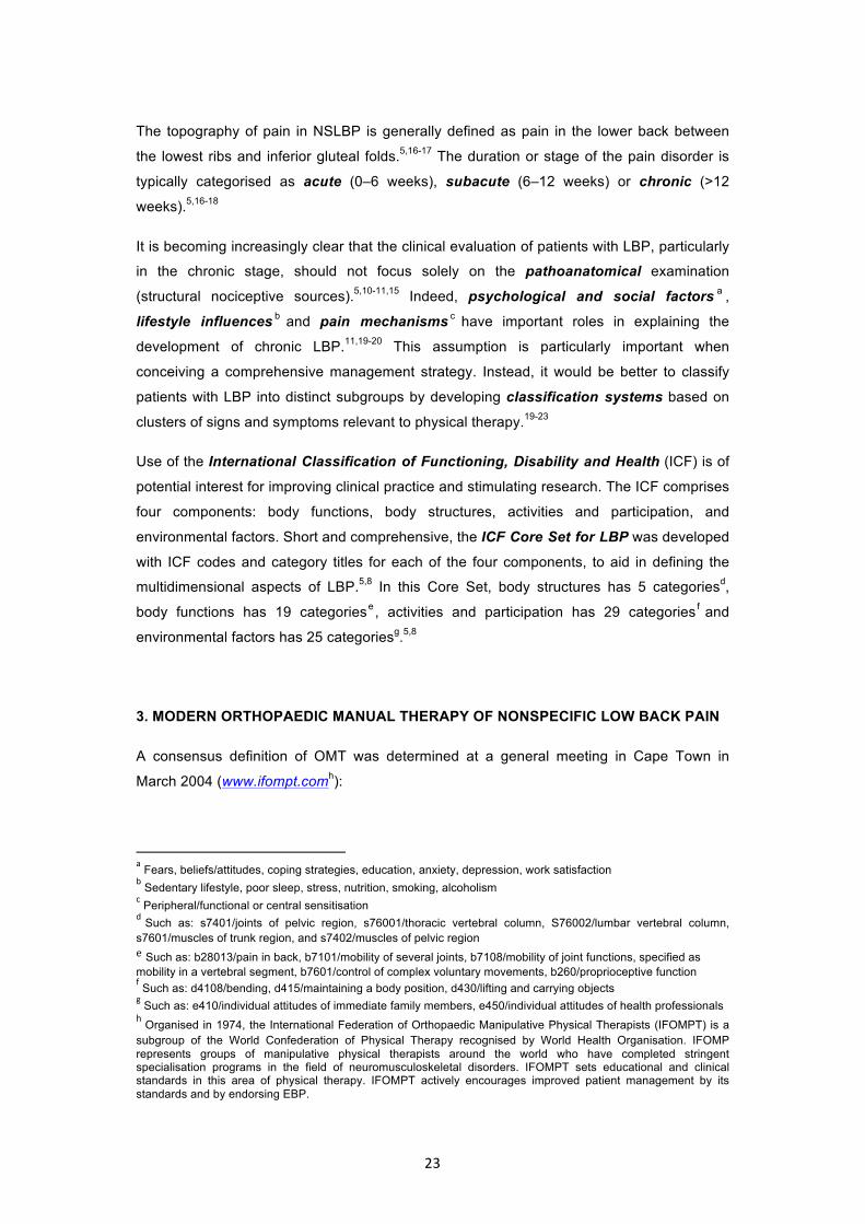
Figure 8. Kinematic curves from the kinematic model of the spine for speed during a trunk
flexion task (n=10 trials) from a sitting position
A. Results from one acute and one chronic NSLBP patient
(red curves of one acute LBP patient with mean smoothness = 0.30; blue curves of one chronic LBP patient with
mean smoothness = 0.54, see Discussion section 2.2)
38
B. Results from one healthy subject
(blue curves of one healthy subject with mean smoothness = 0.63, see Discussion section 2.2)
5. CONTEXT AND OBJECTIVES OF THE RESEARCH
LBP is a complex issue with high prevalence, and it requires a multidimensional approach
towards assessment and treatment. This Introduction has described an integrative approach
utilising a range of research tools that might help people with LBP. These tools have been
used in published studies and on-going investigations. This multidimensional approach
might contribute to the definition of new levels of evidence for specific OMT interventions for
specific subgroups of people with NSLBP.

39
The main objective of this thesis is to study the efficacy of using OMT for NSLBP, which is
addressed through the following aims:
1. To establish a background on OMT within our team, and to improve our knowledge
of the effects of OMT in NSLBP
2. To create a quantitative assessment tool for measuring kinematic patterns in
subjects with NSLBP who have mechanical pain behaviours, and for determining
the effects of OMT
3. To test the reliability of a standardised objective clinical examination aimed at
identifying the direction(s) of trunk movement impairment and spinal level(s) of
involvement; and, based on these findings, to perform a clinical study on
mobilisation with movement (MWM; a novel OMT technique) within a subgroup of
NSLBP patients.
This thesis is organised into three sections:
Section 1: Evidence for the use of orthopaedic manual therapy for patients with low back pain
The research was initiated with a systematic review of the literature to summarise existing
evidence on the efficacy of OMT in NSLBP disorders, and to evaluate the quality of the
methodology and design of previous clinical trials (Chapter I, published in the Journal of
Manual and Manipulative Therapy). This overview helped us to identify potential avenues
for future research in OMT and to elaborate the protocol of a RCT analysing the efficacy of
OMT, presented in the final section of this thesis.
Section 2: Development of a kinematic model of the spine
We developed and validated a kinematic model of the spine: (I) to analyse kinematic
variables of various spinal segments in NSLBP patients with mechanical pain behaviour
patterns compared to healthy people, and (II) as a source of objective outcome measures
for future clinical trials to evaluate OMT interventions of the spine (Chapters II and III,
published in the Journal of Rehabilitation Medicine, and Chapter IV, published in the Journal
of Back and Musculoskeletal Rehabilitation).
Section 3: Clinical physical examination and evaluation of the efficacy of orthopaedic
manual therapy for patients with low back pain

40
Clinical objective examination in OMT incorporates combined movement procedures to
identify the dominant painful pattern of movement and symptomatic spinal level(s) (Chapter
V, published in the Journal of Manipulative and Physiological Therapeutics). The purpose of
the objective evaluation is to choose the most appropriate OMT technique(s) for NSLBP
patients by using an integrative EBP approach. Effects of this treatment may be objectively
measured by an analysis of the kinematic model of the spine, as described in Section 2.
MWM is a novel-growing concept in the field of OMT and clinical practice, but it remains
sparsely studied in the literature. In the MWM concept, the sustained natural apophyseal
glide (SNAG) has become increasingly popular,62,67 despite the poor level of evidence
about the procedure, mostly due to a lack of clinical studies. Hence, this thesis includes a
randomised placebo-controlled trial on the effects of MWM in a subgroup of patients with
NSLBP of mixed stages. Outcomes of this intervention were evaluated by using the tools
developed in the previous sections of this thesis (Chapter VI, accepted for publication in the
Journal of Manipulative and Physiological Therapeutics).

41
References
1. Hjørland B. Evidence based practice: An analysis based on the philosophy of
science. J of the Amer Soc for Inform Science and Technol 2011;62:1301–10.
2. Sackett DL. Evidence-based medicine and treatment choices. Lancet
1997;349:570.
3. Spring B, Hitchcock K. Evidence-based practice in psychology. In I.B. Weiner &
W.E. Craighead (Eds.) Corsini’s Encyclopedia of Psychology, 4th edition 2009;603-
07. New York:Wiley.
4. Ford JJ and Hahne AJ. Complexity in the physiotherapy management of low back
disorders: Clinical and research implications. Man Ther 2013;18(5):438-42.
5. Delitto A, George SZ, Van Dillen LR, et al. Low back pain. J Orthop Sports Phys
Ther 2012;42:A1-57.
6. Hoy D, Bain C, Williams G, March L, Brooks P, Blyth F, et al. A systematic review of
the global prevalence of low back pain. Arthritis Rheum 2012;64:2028–37.
7. Kirschneck M, Kirchberger I, Amann E, Cieza A. Validation of the comprehensive
ICF core set for low back pain: the perspective of physical therapists. Man Ther
2011;16:364-72.
8. Cieza A, Stucki G, Weigl M, et al. ICF Core Sets for low back pain. J Rehabil Med
2004;44:69-74.
9. Waddell G. Volvo award in clinical sciences. A new clinical model for the treatment
of low back pain. Spine 1987;12:632-44.
10. McCullough BJ, Johnson GR, Martin BI, Jarvik JG. Lumbar MR imaging and
reporting epidemiology: do epidemiologic data in reports affect clinical
management? Radiology 2012;262:941-46.
11. Balague F, Mannion AF, Pellise F, Cedraschi C. Non-specific low back pain. Lancet
2012; 379:482-91.
12. Hemingway H, Croft P, Perel P, et al. Prognosis research strategy (PROGRESS) 1:
a framework for researching clinical outcomes. BMJ 2013;346:5595.
13. Hamidi-Ravari B, Tafazoli S, Chen H, Perret D. Diagnosis and Current Treatments
for Sacroiliac Joint Dysfunction: A Review. Current Phys Med Rehab Reports
2014;2:48-54.
14. Hodge JC, Bessette B. The incidence of sacroiliac joint disease in patients with low-
back pain. Can Assoc Radiol J 1999;50:321–3.
15. Laslett M, McDonald B, Tropp H, Aprill CN, Oberg B. Agreement between
diagnoses reached by clinical examination and available reference standards: a
prospective study of 216 patients with lumbopelvic pain. BMC Musculoskelet Disord
2005;9;6:28.

42
16. van Tulder MW, Assendelft WJ, Koes BW and Bouter LM. Method guidelines for
systematic reviews in the Cochrane Collaboration Back Review Group for Spinal
Disorders. Spine 1997;22: 2323-30.
17. Furlan AD, Pennick V, Bombardier C, van Tulder M and Editorial Board CBRG.
2009 updated method guidelines for systematic reviews in the Cochrane Back
Review Group. Spine 2009; 34:1929-41.
18. Aure OF, Nilsen JH and Vasseljen O. Manual therapy and exercise therapy in
patients with chronic low back pain: a randomized, controlled trial with 1-year follow-
up. Spine 2003; 28: 525-31.
19. O’Sullivan P. It’s time for change with the management of chronic non-specific low
back pain. Br J Sports Med 2013;46:224-27.
20. Waddell G. The back pain revolution. 2nd ed. ed. Edinburgh: Churchill Livingstone,
2005.
21. Delitto A, Erhard RE, Bowling RW. A treatment-based classification approach to low
back syndrome: identifying and staging patients for conservative treatment. Phys
Ther 1995;75(6):470-85.
22. Van Dillen LR, Sahrmann SA, Norton BJ, Caldwell CA, Fleming DA, McDonnell MK,
et al. Reliability of physical examination items used for classification of patients with
low back pain. Phys Ther 1998;78(9):979-88.
23. Maher C, Adams R. Reliability of pain and stiffness assessments in clinical manual
lumbar spine examination. Phys Ther 1994;74(9):801-09.
24. Edwards BC. Manual of combined movements: their use in the examination and
treatment of musculoskeletal vertebral column disorders. (2nd) Oxford, 1999;
Butterworth-Heinemann.
25. Sheeran L, Coales P, Sparkes V. Clinical challenges of classification based
targeted therapies for non-specific low back pain: What do physiotherapy
practitioners and managers think? Man Ther 2015;20:456-62.
26. Maigne JY, Vautravers P. Mechanism of action of spinal manipulative therapy. Joint
Bone Spine 2003;70:336-41.
27. Pickar JG. Neurophysiological effects of spinal manipulation. Spine J 2002;2:357-
371.
28. Colloca CJ, Keller TS, Harrison DE, Moore RJ, Gunzburg R, et al. Spinal
manipulation force and duration affect vertebral movement and neuromuscular
responses. J Clin Biomech 2006;21:254-262.
29. Keller TS, Colloca CJ, Gunzburg R. Neuromechanical characterization of in vivo
lumbar spinal manipulation. Part I. Vertebral motion. J Manipulative Physiol Ther
2003;26: 567-78

43
30. Powers CM, Kulig K, Harrison J, Bergman G. Segmental mobility of the lumbar
spine during a posterior to anterior mobilization: assessment using dynamic MRI.
Clin Biomech 2003;18: 80-3.
31. Triano JJ. Biomechanics of spinal manipulative therapy. Spine J 2001;1:121-30.
32. Cramer GD, Cambron J, Cantu JA, et al. Magnetic resonance imaging
zygapophyseal joint space changes (gapping) in low back pain patients following
spinal manipulation and side-posture positioning: a randomized controlled
mechanisms trial with blinding. J Manipulative Physiol Ther 2013; 36(4):203-17.
33. Lisi AJ, O’Neill CW, Lindsey DP, Cooperstein R, Cooperstein E, Zucherman JF.
Measurement of in vivo lumbar intervertebral disc pressure during spinal
manipulation: a feasibility study. J Appl Biomech 2006; 22:234-39.
34. Beattie PF, Butts R, Donley JW, Liuzzo DM. The within-session change in low back
pain intensity following spinal manipulative therapy is related to differences in
diffusion of water in the intervertebral discs of the upper lumbar spine and L5-S1. J
Orthop Sports Phys Ther 2014;44:19-29.
35. Soulvis T, Vincenzino B, Wright A. Neurophysiological effects of spinal manual
therapy. Modern Manual Therapy, the vertebral column. Churchill Livingstone 2004:
367-79.
36. Colloca CJ, Keller TS, Gunzburg R. Neuromechanical characterization of in vivo
lumbar spinal manipulation. Part II. Neurophysiological response. J Manipulative
Physiol Ther 2003;26:579-91.
37. Potter L, McCarthy C, Oldham J. Physiological effects of spinal manipulation: a
review of proposed theories. Phys Ther 2005;10:163-70.
38. Dishman JD, Greco DS, Burke JR. Motor-evoked potentials recorded from lumbar
erector spinae muscles: a study of corticospinal excitability changes associated with
spinal manipulation. J Manipulative Physiol Ther 2008;31:258-70.
39. Grindstaff TL, Hertel J, Beazell R, Magrum EM, Ingersoll CD. Effects of lumbopelvic
joint manipulation on quadriceps activation and strength in healthy individuals. Man
Ther 2009;14:415-20.
40. Dishman JD, Cunningham BM, Burke JR. Comparison of tibial nerve H-reflex
excitability after cervical and lumbar spine manipulation. J Manipulative Physiol
Ther 2002;25:318-25.
41. Dishman JD, Ball KA, Burke JR. First prize central motor excitability changes after
spinal manipulation: a transcranial magnetic stimulation study. J Manipulative
Physiol Ther 2002;25:1-9.
42. Vernon H. Qualitative review of studies of manipulation-induced hypoalgesia. J
Manipulative Phyiol Ther 2000;23:134-38.

44
43. Bialosky JE, Bishop MD, Robinson ME, Zeppieri G, George SZ. Spinal manipulative
therapy has an immediate effect on thermal pain sensitivity in people with low back
pain: a randomized controlled trial. Phys Ther 2009;89:1292-303.
44. Kaptchuk TJ.The placebo effect in alternative medicine: can the performance of a
healing ritual have clinical significance? Ann Intern Med 2002;136:817-25.
45. Bialosky JE, Bishop MD, George SZ, Robinson ME. Placebo response to manual
therapy: something out of nothing? J Man Manip Ther 2011;19:11-19.
46. Foster NE, Hill JC, O'Sullivan P, Hancock M. Stratified models of care. Best Pract
Res Clin Rheumatol 2013;27:649-61.
47. Fritz JM, Cleland JA, Childs JD. Subgrouping patients with low back pain: evolution
of a classification approach to physical therapy. J Orthop Sports Phys Ther
2007;37: 290-302.
48. Hill JC, Whitehurst DG, Lewis M, Bryan S, Dunn KM, et al. Comparison of stratified
primary care management for low back pain with current best practice (STarT
Back): a randomised controlled trial. Lancet 2011;378:1560-71.
49. Vibe Fersum K, O'Sullivan P, Skouen JS, Smith A and Kvale A. Efficacy of
classification-based cognitive functional therapy in patients with non-specific chronic
low back pain: A randomized controlled trial. Eur J Pain 2012;17:916-28.
50. Schäfer A, Gärtner-Tschacher N, Schöttker-Königer T. Subgroup-specific therapy of
low back pain: description and validity of two classification systems. Orthopade
2013; 42:90-2.
51. Bishop MD, Bialosky JE, Cleland JA. Patient expectations of benefit from common
interventions for low back pain and effects on outcome: secondary analysis of a
clinical trial of manual therapy interventions. J Man Manip Ther 2011;19:20-5.
52. Harris-Hayes M, Van Dillen LR. The inter-tester reliability of physical therapists
classifying low back pain problems based on the movement system impairment
classification system. PM R 2009;1(2):117-26.
53. Riddle DL. Classification and low back pain: a review of the literature and critical
analysis of selected systems. Phys Ther 1998;78(7):708-37.
54. Werneke MW, Hart D, Olivier D, McGill T, Grigsby D, et al. Prevalence of
classification methods for patients with lumbar impairments using the McKenzie
syndromes, pain pattern, manipulation, and stabilization clinical prediction rules. J
Man Manip Ther 2010;18:197-204.
55. May S, Littlewood C, Bishop A. Reliability of procedures used in the physical
examination of non-specific low back pain: a systematic review. Aust J Physiother
2006;52: 91-102.
56. McKenzie R, May S. The lumbar spine – mechanical diagnosis and therapy. (2nd)
Spinal Publications, 2003; New Zealand Ltd.

45
57. Apeldoorn A, Ostelo RW, van Helvoirt H, Fritz JM, Knol DL, van Tulder MW, de Vet
HC. A randomized controlled trial on the effectiveness of a classification-based
system for subacute and chronic low back pain. Spine 2012;37:1347-56.
58. Brennan GP, Fritz JM, Hunter SJ, Thackeray A, Delitto A, et al. Identifying
subgroups of patients with acute/subacute "nonspecific" low back pain: results of a
randomized clinical trial. Spine 2006;31:623-31.
59. Karayannis NV, Jull GA, Hodges PW. Physiotherapy movement based classification
approaches to low back pain: comparison of subgroups through review and
developer/expert survey. BMC Musculoskelet Disord 2012;13:24.
60. Hidalgo B, Hall T, Nielens H, Detrembleur C. Inter-tester agreement and validity of
identifying lumbar pain provocative movement patterns using active and passive
accessory movement tests. J Manipulative Physiol Ther 2014;37:105-15.
61. Maitland J. Spinal manipulation made simple: a manual of soft tissue techniques.
North Atlantic Books, 2000; Berkeley, Calif.
62. Vicenzino B, Hing W, Rivett D, Hall T. Mobilisation with movement: The art and the
science. Elsevier, 2011; Sydney.
63. Hidalgo B, Gilliaux M, Poncin W, Detrembleur C. Reliability and validity of a
kinematic spine model during active trunk movement in healthy subjects and
patients with chronic non-specific low back pain. J Rehabil Med 2012;44:756-63.
64. Hidalgo B, Nielens H, Gilliaux M, Hall T, Detrembleur C. The use of kinematic
algorithms to identify people with chronic non-specific low back pain from
asymptomatic subjects: a validation study. J Rehabil Med 2014;46:819-23.
65. Hidalgo B, Gobert F, Bragard D, Detrembleur C. Effects of proprioceptive disruption
on lumbar spine repositioning error in a trunk forward bending task. J Back
Musculoskelet Rehabil 2013;26:381-87.
66. Laird RA, Gilbert J, Kent P, Keating JL. Comparing lumbo-pelvic kinematics in
people with and without back pain: a systematic review and meta-analysis. BMC
Musculoskelet Disord 2014;15:229.
67. Konstantinou K, Foster N, Rushton A, Baxter D. The use and reported effects of
mobilization with movement techniques in low back pain management; a cross-
sectional descriptive survey of physiotherapists in Britain. Man Ther 2002;7:206-14.

46

47
Section 1
Evidence for the use of orthopaedic manual therapy for patients with low back pain

48

49
CHAPTER I
Efficacy of manual therapy and exercises for different stages of nonspecific low back
pain: Current evidence from the literature
Benjamin Hidalgo, Christine Detrembleur, Toby Hall, Philippe Mahaudens, Henri Nielens
J Man Manip Ther 2014 May;22(2):59-74. Doi: 10.1179/2042618613Y.0000000041
ABSTRACT
Objectives: to review and update the evidence for different forms of manual therapy (MT)
for patients with different stages of nonspecific low back pain (LBP).
Data sources: MEDLINE, Cochrane-Register-of-Controlled-Trials, PEDro, EMBASE.
Methods: two independent reviewers according to Cochrane and PRISMA guidelines
conducted a systematic review of MT with a literature search covering the period of January
2000 to April 2013. A total of 360 studies were evaluated using qualitative criteria. Two
stages of LBP were categorized; combined acute–subacute and chronic. Further sub-
classification was made according to MT intervention: MT1 (manipulation); MT2
(mobilization and soft-tissue-techniques); and MT3 (MT1 combined with MT2). In each sub-
category, MT could be combined or not with exercise or usual medical care (UMC).
Consequently, quantitative evaluation criteria were applied to 56 eligible randomised
controlled trials (RCTs), and hence 23 low-risk of bias RCTs were identified for review. Only
studies providing new updated information (11/23 RCTs) are presented here.
Results:
Acute–subacute LBP: STRONG-evidence in favor of MT1 when compared to sham for pain,
function and health improvements in the short-term (1–3 months). MODERATE-evidence to
support MT1 and MT3 combined with UMC in comparison to UMC alone for pain, function
and health improvements in the short-term.

50
Chronic LBP: MODERATE to STRONG-evidence in favor of MT1 in comparison to sham for
pain, function and overall-health in the short-term. MODERATE-evidence in favor of MT3
combined with exercise or UMC in comparison to exercise and back school was established
for pain, function and quality-of-life in the short and long-term. LIMITED-evidence in favor of
MT2 combined with exercise and UMC in comparison to UMC alone for pain and function
from short to long-term. LIMITED-evidence of no effect for MT1 with extension-exercise
compared to extension-exercise alone for pain in the short to long-term.
Conclusion: This systematic review updates the evidence for MT with exercise or UMC for
different stages of LBP and provides recommendations for future studies.

51
Introduction
After headaches and chronic fatigue, low back pain (LBP) is the most reported complaint,
with more than 80% of the population reporting LBP at some point in their life.1,2 In
developed countries, LBP has enormous and growing indirect and direct costs for society
and public health organizations.3,4
The majority of LBP cases are described as nonspecific as there is no identifiable pathology
on radiological imaging.2 Indeed there is a poor correlation between findings on radiological
imaging and symptoms, with a radiological diagnosis identified in only 15% of cases.5-9
Hence, LBP is often a symptom of unknown origin and etiology.2,5,10,11
Many factors have been identified as possible causes or contributing factors to LBP. For
example nociceptive inputs, particularly in acute-subacute conditions from various spine
structures can cause pain, including zygapophysial joints, intervertebral discs and sacro-
iliac joints.5,12-14 In chronic LBP, psychosocial factors are of prime importance in explaining
the prolongation of pain.2,15,16 Additional factors linked to chronic LBP include obesity and
physical deconditioning associated with sedentary lifestyles.2,17 Moreover, genetic factors
have been strongly linked to LBP through their influence on pain perception and
psychosocial factors.2,18
In general terms, in the case of acute LBP, reports suggest that 75-90% of cases recover
within 6 weeks irrespective of medical intervention, whereas up to 25% are at risk of
developing chronic pain and disability.1,2 Indeed, many individuals with LBP have a number
of persisting or recurring symptoms.1,5,8,19 Chronic LBP therefore represents a considerable
challenge because recovery is unlikely to occur, despite considerable medical advances.20
In physical therapy practice, various forms of manual therapy (MT) are currently used to
manage LBP.7,21-23 Manual therapists use a range of treatment approaches including
various passive techniques such as mobilisation and manipulation as well as a variety of
different forms of exercise. The use of these approaches, along with clinical reasoning
based on the bio-psycho-social model, represent the essence of MT (www.ifompt.com).24
This systematic review (SR) focuses on the effects of commonly used MT approaches
identified through a comprehensive evidence based search strategy of low-risk of bias
clinical trials. Three categories of passive MT techniques are defined; MT1 (lumbopelvic
manipulation: high-velocity-low-amplitude thrust) MT2 (non-thrust lumbo-pelvic mobilisation
and soft-tissue techniques),25-27 and MT3 (combination of MT1 and MT2). We also
considered passive MT techniques (MT1-3) combined or not with exercise (specific or
general) or combined with usual medical care (UMC) (stay active, reassurance, education
and medication).11,27,28

52
The popularity and use of MT for the management of LBP has grown, in part supported by
the inclusion of MT in various clinical practice guidelines.5,10,23,29 This is despite uncertainty
regarding the levels of evidence for the effectiveness of different approaches in MT at
different stages of LBP.5,7,10,22,29-36
Previous SRs have reported that in general terms, MT is considered better than a placebo
treatment or no treatment at all for LBP.7,30,35-40 These reviews failed to establish levels of
evidence for other forms of treatment such as UMC or exercise in comparison to
MT.35,37,39,40 In addition, previous SRs have not investigated which MT approaches “(MT1-
3)” when combined with UMC or “exercise” are more effective for LBP. The present SR
updates previous reviews, and is the first to focus specifically on different MT approaches
for different stages of LBP. New findings, as well as new evidence to inform findings from
previous systematic reviews,41-45 are presented.
Methods
This SR was conducted in accordance with the PRISMA and Cochrane-Collaboration-Back-
Review-Group (CCBRG) updated guidelines for SR.46,47
Search strategy
A literature search of RCTs published in English between 2000 and 2013, on the efficacy of
MT in the treatment of LBP was conducted independently by two reviewers in four electronic
databases: MEDLINE, Cochrane-Register-of-Controlled-Trials, PEDro, and EMBASE. The
detailed search strategy in MEDLINE is presented in Appendix 1, and was adapted to
search in the three other databases.
Based on information revealed in the titles and abstracts, a first selection of articles was
performed using the inclusion criteria described below. A final selection was conducted after
a blinded critical appraisal of the quality of the studies. A consensus was reached at each
step (Figure 1) on the studies to be included. In cases of disagreement, a third reviewer
made the necessary decision.
Inclusion criteria
Study design
RCTs from the period of January 2000 to April 2013 were included only if (I) they presented
a low-risk of bias, (II) if LBP cases treated with MT were compared to a randomised control-
group (CG) receiving either no treatment, a placebo procedure, or another effective therapy
for LBP and (III) if the randomisation methods was appropriated and clearly reported, with

53
moreover (IV) a single (assessors blinded) or quasi-double-blind design (assessors and
patients blinded).
Patients
LBP is distinguished on the basis of the duration of the pain episode: acute (< 6 weeks),
subacute (6-12 weeks) and chronic (> 12 weeks).2,29 However, this distinction may not be
satisfactory and it has been argued that categorization should be on the basis of other
factors including location, symptoms, duration, frequency, and severity.48 In this SR, we
used a combination of duration, location and symptoms to specify the study population:
- Studies were included if subjects were males and females aged between 18-60
years suffering from acute-subacute (0-12 weeks) or chronic (>12 weeks) LBP.
Acute and subacute categories were combined because of their similarities in
contrast to chronic LBP category, where psycho-social factors appear more
important.16,49,50
- LBP is defined as pain in the lower back between the lowest ribs and inferior gluteal
folds.46,51 Given that people with LBP may present with radicular pain, LBP is
defined according to the following Quebec-Task-Force (QTF) classification: (1) LBP
alone (QTF 1), (2) LBP with radiating pain into the thigh but not below the knee
(QTF 2), (3) LBP with nerve root pain without neurologic deficit (QTF 3), or (4) LBP
with nerve root pain with neurologic deficit (QTF 4).52 In the present SR, only trials
that contained patients in classes QTF 1-3 were included.
Interventions
Among the included trials, we considered 3 categories of the most common MT techniques
represented in the intervention groups. MT1 comprised high-velocity-low-amplitude thrust of
the lumbo-pelvic region with “cavitation”.7,21,22,27,37,53 MT2 comprised mobilisation and soft-
tissue-techniques including “myofascial”, “myotensive” or “harmonic” techniques on the
lumbo-pelvic region.22,27,37,54 MT3 comprised the combination of MT1 and MT2.
Furthermore, sub-categorization of groups MT1-3 was based on the addition or not of
exercises either specific (for example based on directional preference, stabilization, and
motor control) or general (for example global strengthening, cardiovascular endurance,
stretching and range-of-motion exercises) or UMC.1,21,32,55
Control groups
The control groups received no treatment, placebo, UMC, or exercise.
54
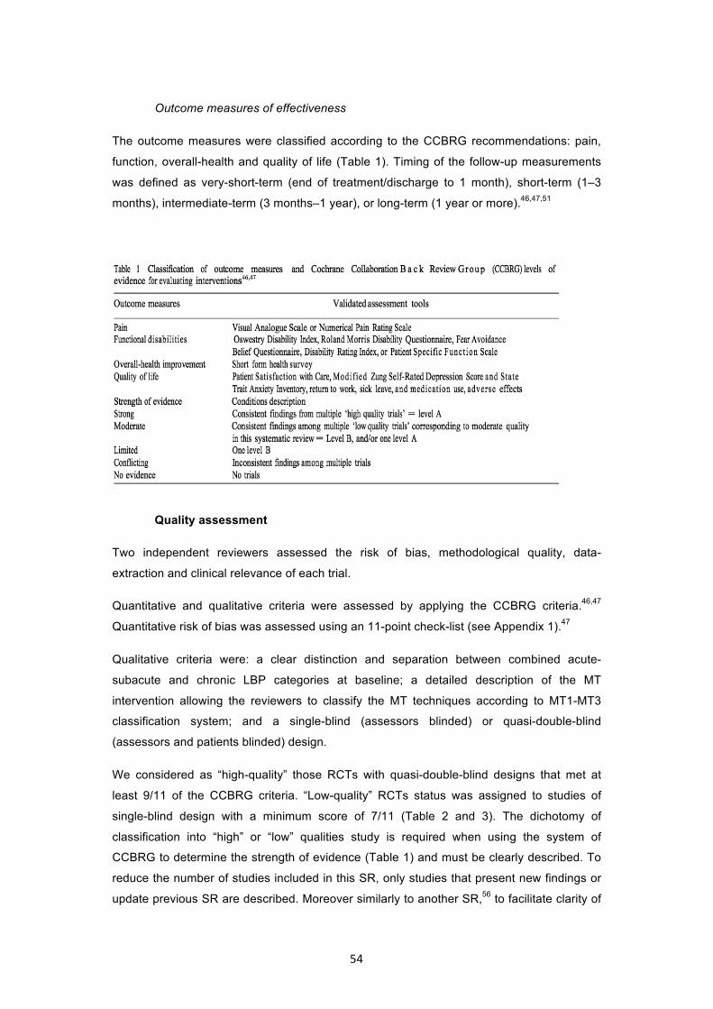
Outcome measures of effectiveness
The outcome measures were classified according to the CCBRG recommendations: pain,
function, overall-health and quality of life (Table 1). Timing of the follow-up measurements
was defined as very-short-term (end of treatment/discharge to 1 month), short-term (1–3
months), intermediate-term (3 months–1 year), or long-term (1 year or more).46,47,51
Quality assessment
Two independent reviewers assessed the risk of bias, methodological quality, data-
extraction and clinical relevance of each trial.
Quantitative and qualitative criteria were assessed by applying the CCBRG criteria.46,47
Quantitative risk of bias was assessed using an 11-point check-list (see Appendix 1).47
Qualitative criteria were: a clear distinction and separation between combined acute-
subacute and chronic LBP categories at baseline; a detailed description of the MT
intervention allowing the reviewers to classify the MT techniques according to MT1-MT3
classification system; and a single-blind (assessors blinded) or quasi-double-blind
(assessors and patients blinded) design.
We considered as “high-quality” those RCTs with quasi-double-blind designs that met at
least 9/11 of the CCBRG criteria. “Low-quality” RCTs status was assigned to studies of
single-blind design with a minimum score of 7/11 (Table 2 and 3). The dichotomy of
classification into “high” or “low” qualities study is required when using the system of
CCBRG to determine the strength of evidence (Table 1) and must be clearly described. To
reduce the number of studies included in this SR, only studies that present new findings or
update previous SR are described. Moreover similarly to another SR,56 to facilitate clarity of

55
presentation, RCTs were only included if they were of low-risk of bias, and either high
quality (indicated by a “A”) or moderate quality (indicated by a “B”).
Strength of evidence and clinical relevance
Strength of evidence was determined by grouping similar “Patients Interventions
Comparisons Outcomes Study design” (PICO) to provide an overall level of evidence (Table
1) on the efficacy of the MT techniques (Table 4). Based on CCBRG guidelines,47,51 the
effect sizes were independently collected or calculated by two authors, and used to assess
the clinical relevance of MT interventions on outcome measures. We report the between
groups means of difference (MD = mean A – mean B) or Cohen’s standardised means of
difference (SMD = mean A – mean B / mean SD). In this SR, the clinical relevance was
determined by two conditions and scored by “YES” in favor of the intervention group; if there
was significant difference between groups (p < 0.05) associated with between groups effect
sizes equal or superior to the minimal clinically important difference (MD) or moderate to
large effect (SMD) on specific outcome measure (Tables 2 and 3).
Results
Two reviewers performed the initial selection of articles based on keywords. Upon
discussion, the reviewers achieved consensus on inclusion of 56 trials that met the selection
criteria based on their titles and abstracts. After critical appraisal of these 56 studies, 23
RCTs were retained (Figure 1). Only 11/23 of these RCTs were found to have new evidence
or updated previous SRs and are fully presented here. Appendix 2 and Table 4 presents a
summary of the remaining 12 RCTs that are not detailed in this results section.
The studies’ characteristics and effect sizes on outcome measures are presented for acute-
subacute (Table 2) and chronic LBP (Table 3). A qualitative SR was undertaken on the 11
low-risk of bias RCTs, 5 studies were classified as level A quality, and 6 as level B quality

56
Figure 1. PRISMA flowchart of inclusion

57
Effects of interventions on acute and subacute LBP
MT versus sham-MT
Santilli et al,57 Hoiriis et al,58 von Heymann et al,59 (studies rated as level A quality)
assessed the effects of MT1 in comparison to sham-MT1 in patients with acute LBP.
Santilli et al57 compared lumbo-pelvic rotational manipulation toward the pain-free direction
to simulated manipulation not following any specific pattern and not involving rapid thrust.
The frequency of treatment was 5 days per week until pain relief occurred or up to a
maximum of 20 sessions of 5 minutes. For LBP up to 3 months, MT1 was more effective in
decreasing local pain, radiating pain, and the duration of pain with clinical relevance
(p<.0001 and means of difference of 1.8). No statistically significant differences were found
for overall-health improvement and psychosocial outcomes. At 6 months, the percentage of
pain-free patients was significantly higher in the MT1 group with means difference of 22%
for local pain (p<.005) and of 35% for radiating pain (p<.001). Two patients, one in MT1 and
one in sham-MT1, were dissatisfied with treatment and stopped.
Hoiriis et al58 investigated the effects of lumbo-pelvic manipulation in prone or side-lying
position combined with a drug placebo, in comparison to sham-MT1 combined with a
muscle relaxant or with a drug placebo. Sham-MT1 consisted of manual light pressure on
the lumbar spine in both positions (prone and side-lying). All groups received 8 visits over 2
weeks and showed significant improvements in pain relief and disability (p<.0001) and
depression scores (p<0001). Clinically relevant differences between groups could only be
identified in favor of the intervention group for pain relief in the very short term with p<.05
and standardised means difference of 0.70. However, further evaluation revealed that the
perception of true MT was significantly higher (p<.05) in the intervention group than in either
of the two control groups. Indeed, the sham maneuver did not closely approximate the
manipulation technique.
Von Heymann et al59 explored the efficacy of lumbo-pelvic rotational manipulation in side-
lying position and placebo-diclofenac in comparison to Sham-MT1 with diclofenac or
placebo-diclofenac. Sham MT1 was performed using real manipulation in a prone position
but at the incorrect location (i.e. on a non-dysfunctional sacro-iliac-joint) to mimic as closely
as possible the intervention being tested. This sham procedure is not supposed to have any
influence on the lumbar dysfunction and is not believed to harm the patient. All groups
received 2-3 visits over a 1-week period. There was a clear and clinically relevant difference
at very-short-term follow up (9 days) between the groups (p=0.013), the intervention group
showed a standardised means difference of 0.60 on functional improvement with similar

58
result for pain and quality of life. No adverse effects or harm were reported in this study.
These results suggested that real MT1 had clinically superior effects than NSAID and
placebo interventions.
MT with UMC versus UMC alone
Bishop et al60 and Cruser et al61 (studies rated as level B quality) compared respectively
MT1 (2-3 sessions per week over four weeks) and MT3 (1 session per week over four
weeks) combined with UMC, to UMC alone in patients with acute LBP from QTF 1-2.
Bishop et al60 reported clinically relevant differences in favor of the intervention group in
terms of functional improvement (p=.002 and mean difference of 2.6) at 16 and 24 weeks,
but there were no significant differences for pain and physical functioning. In the short-term
(4weeks), Cruser et al61 determined clinically relevant differences in favor of MT3 compared
to UMC alone for pain now (p=.025 and SMD of 1.04) and pain typical (p=.020 and SMD of
0.88) and a standardised means difference of 0.56 for function associated with significantly
greater satisfaction with treatment and overall-health improvement (p<.01). The authors
concluded that compared to UMC, MT160 and MT361 combined with UMC provides clinically
greater improvement in function and pain relief.

59
60
Effects of interventions on chronic LBP
MT versus sham-MT
Ghroubi et al62 and Senna et al63 (studies rated as level A quality) investigated, respectively, the
effectiveness of MT1 in a side-lying position (painful side-up) and MT1 in supine position
(toward the painful side), as compared to sham-MT1 (mimic of lumbo-pelvic manipulation
without final impulsion to provide minimal likelihood of therapeutic effect); on pain, function and
overall health in patients with chronic LBP from QTF 1-2. True-MT1 of 4 sessions spread over
one month for Ghroubi et al62, or 16 sessions over 1 month for Senna et al63, led to significant
improvements for pain ([Ghroubi et al62 reported standardized mean difference of 0.86 at 4-8
weeks with p<.001];[Senna et al63 reported means difference of 1.9 at 10 months with p<.005]),
for functional outcomes ([Ghroubi et al62 reported standardised means difference of 0.40 at 4-8
weeks with p<.001];[Senna et al63 reported means difference of 18.9 at 10 months with
p<.001]). Only Senna et al63 reported an overall-health improvement of means difference of 7.8
at 10 months (p<.001). The authors62,63 concluded that MT1 is clinically effective in treating
patients with chronic LBP in the short-term, but to obtain long-term benefit on all outcome
measures requires maintenance of MT1 every 2 weeks.63
MT combined with other interventions
Niemistö et al32 (rated as level B quality) investigated the effects of combined MT2 (myotensive
lumbo-pelvic mobilization techniques) with exercises (stabilising exercise to correct lumbo-pelvic
rhythm) and UMC in comparison to UMC alone (patient education, stay active approach,
ergonomic instruction, home general exercises, and educational-booklet) in patients with
chronic LBP from QTF 1-3. They found that the intervention group provided clinically relevant
improvements in pain relief (p<.001 and standardised means difference of 0.60) and function
(p=.002 and standardised means difference of 0.45) from the short to long-term (up to one
year). However, there were no significant differences between the groups in terms of the quality-
of-life and medical costs.
Aure et al49 (rated as level B quality) evaluated the effectiveness of MT3 (consisting of
mobilisation and rotational manipulation in side-lying position from T10 to the pelvis) combined
with specific and general exercise in comparison to exercises only in patients with chronic LBP
from QTF 1-3. Both groups received 16 sessions of 45 minutes over 8 weeks. The results

61
showed statistically significant improvements in terms of pain reduction and function in both
groups. However, there was a greater improvement in all outcome measures for the intervention
group leading to clinically relevant differences in the very-short to long-term on pain (at one
year: p<.05 and means difference of 1.5) and functional improvement (at one year: p<.05 and
mean difference of 9), as well as for return to work rate (at 2 months; p<.01 means difference of
40 %).
Cecchi et al34 (rated as level B quality) compared one group receiving MT3 combined with UMC,
to another group receiving back school with UMC to another group receiving individual
physiotherapy (passive and assisted mobilisation, active exercises, massage, and
proprioceptive-neuromuscular-facilitation) with UMC in patients with chronic LBP of type QTF 1-
2. The results showed that MT3 led to clinically relevant decrease in pain (at 12 months: p<.001,
standardised means of difference of 0.7 and 1.1) and a greater functional recovery (at 12
months: p<.001, standardised means of difference of 0.70 and 0.73) than the two control groups
at long term. But, the intervention group (MT3) received significantly more treatment than the
two control groups at follow-up. Pain recurrence and drug intake were also significantly reduced
in the MT3 group (p<.001).
Rasmussen et al64 (rated as a level B quality) compared the effects of combined MT1 (in a side-
lying position at the lumbar level of reduced movement) with exercises (2 different extension
exercises performed as often as possible during the day and at least once per hour), to the
extension exercises alone in patients with chronic LBP classified as QTF 1-3. Both groups
showed clinically relevant back and leg pain reduction, and no difference between the groups
could be observed at the one month and one year follow-ups. Importantly, four patients in the
intervention group and three in the control group reported worsening of back pain after 4 weeks,
3 months and one year.
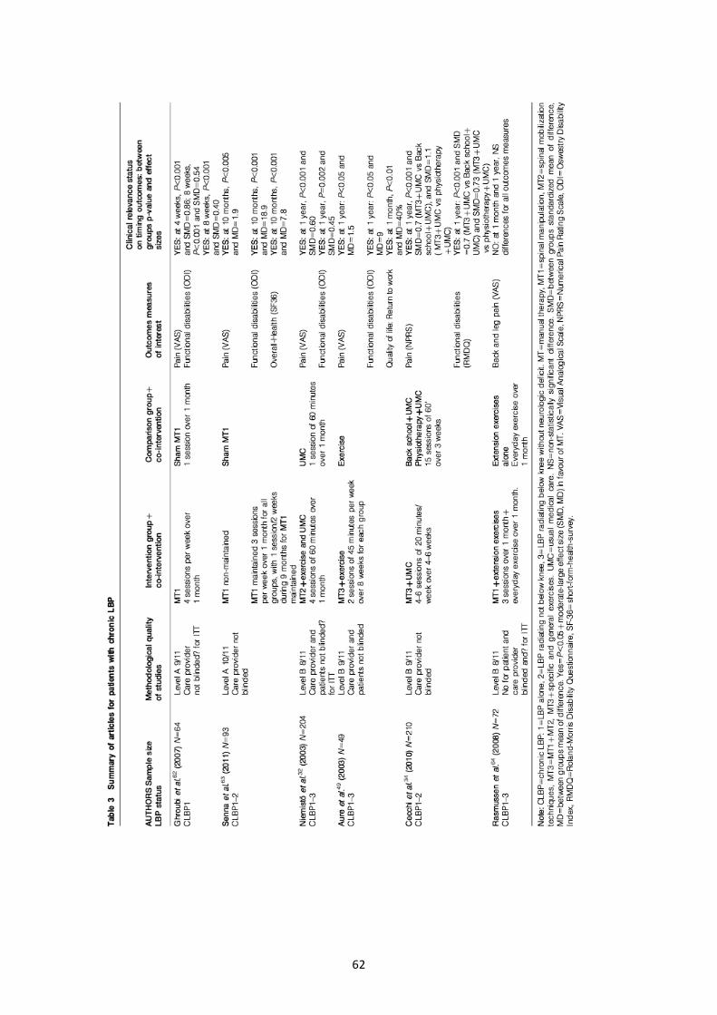
62

63

64
Discussion
The purpose of this SR was to assess and update the evidence pertaining to the effectiveness
of different MT approaches in isolation or when combined with exercise or UMC in the
management of LBP. Thus, this SR deviates and provides clinicians and researchers with new
information compared with other recent high quality SRs41,43,45 which are focused more on
manipulation. A detailed summary of these updated findings, as well as the strength of their
evidence and level of agreement with existing studies, are presented in Table 4.7,30,35-38,41,43,45
In comparison to recent SRs36,41,43,45, the present results highlight a number of new issues in the
management of LBP with MT:
Firstly, in comparison to previous reports of limited-evidence41,43 showing no-difference between
true and sham manipulation, the results of this SR show moderate to strong evidence57-59,62,63
for the beneficial effects of MT1 in comparison to sham-MT1. These differences are
demonstrated in terms of pain relief, functional improvement, and overall-health and quality of
life improvements in the short-term for all stages of LBP.
Secondly, in patients with acute-subacute LBP, in contrast to the previous reports of limited
evidence of no-difference for manipulation combined with other interventions,41 we determined
moderate-evidence60,61 to support MT1 and MT3 combined with UMC, in comparison to UMC
alone, for pain, function, overall-health and quality of life.60,61

65
Thirdly in patients with chronic LBP, in contrast to the previous reports of varying quality
evidence (ranging from limited to strong) that manipulation has short term efficacy when
combined with other interventions43, we found moderate evidence34,49 in support of the use of
MT3 combined with exercises or UMC, in comparison to exercise alone or back-school, for pain,
function and return to work from short to long-term. In addition limited evidence32 supports the
use of MT2 combined with exercises and UMC, in comparison to UMC alone, for pain and
function from short to long-term. Finally, there is limited evidence of no-difference in efficacy for
MT1 combined with extension-exercises, in comparison to extension-exercises alone for pain.64
The highest quality clinical research study is the conventional RCT. These studies have good
internal validity but at the expense of external validity. An alternative for “real world” application
is a pragmatic RCT which has good external validity but poor internal validity.65 Pragmatic
clinical trials are becoming a frequently used tool to evaluate complex interventions.66 Another
possibility is to extend the conventional RCT to retain some of its key advantages (e.g.
Cochrane criteria shown in Appendix 1), and use a “quasi-double-blind” design to make a
realistic compromise between internal and external validity. The CONSORT guidelines should
also be considered to develop high quality study designs.67
One of the key issues in MT research is developing a plausible placebo or sham technique. A
sham manipulation should be an appropriate placebo procedure because it mimics interaction
between the intervention, the patient, the practitioner and the environment. Moreover,
researchers need to conceptualize placebo not only as a comparative inert intervention, but also
as a potential mechanism to partially account for treatment effects associated with MT.68
In the present SR, only 5 studies were placebo-controlled, 4 of them using sham
adjustment,31,57,58,62,63 while 1 used a real manipulation at the incorrect spinal level to achieve an
authentic placebo response.59 Further research is required to identify a plausible placebo
response.
In the majority of RCTs addressing the effectiveness of MT, LBP patients are treated as a
homogeneous group while recent research suggets that people with LBP in fact comprise a
heterogeneous group.40,65,69 Consequently, the concept of subgrouping among people with LBP
is growing in the MT literature.65 Classification of patient into sub-groups and the application of
specific MT interventions for each sub-group have been shown to be more efficient.28,69-74 For
example, a treatment based classification system to identify MT for people with LBP is one form
of subgrouping.28 The Start-Back-Tool is another approach that aims to sub-classify according
to psychosocial issues, and has been found to be more effective than a non-subgrouping
approach.75,76 Moreover, the patients’ beliefs and expectations regarding treatment effects of
MT interventions has also shown to be an important predictor of treatment outcome.77 Targeted
MT for specific subgroups is important because of the heterogeneity of people with LBP, future
clinical trials should address the “wash-out” effect of applying treatments for un-classified LBP.78

66
In terms of quality of the MT management, MT should always be based on evidence-based-
practice, which incorporates patient values (bio-psycho-social influences), clinical expertise and
reasoning on part of the clinician, as well as the best available clinical research evidence.5,79-81 It
could also be useful to establish a minimum level of practical skills across the range of
commonly used MT techniques to manage people with LBP, and to improve clinical reasoning
skills dealing with the complexity of LBP.65 Future studies should incorporate clinical expertise
as a factor in treatment trials for LBP.
Limitations
The results of our SR should be interpreted in the light of some limitations. Firstly, there was
heterogeneity in the RCTs evaluated in this study including the data presentation and outcome
measures. Consequently, a meta-analysis enabling pooled statistics of effect was not possible.
Furthermore, the strength of evidence comprising this SR is limited (particularly for the stronger
level of evidence) due to the difficulty of a true double-blind study design and because of the
limited number of high quality studies. Finally, only studies published in English from 2000 to
2013 were reviewed, leading to the possibility of relevant articles existing in other languages or
before 2000.
Conclusions
This SR, based on low-risk of bias studies, has provided a comprehensive review of different
MT approaches in patients with different stages of LBP, informing evidence-based-practice.
Based on the results of this SR, a variety of manual procedures combined or not with other
interventions, including exercise, may improve patient management. The summary findings of
this review are both comprehensive and novel and may be used to guide clinical practice and
future studies of this topic.
Recommendations for future research to investigate MT include pragmatic high quality RCTs to
maximize the application of results to clinical practice and to reflect the complexity of clinical
reasoning and multi-modal management of MT. Future studies should also investigate targeted
MT for specific subgroups of people with LBP, and continue to address the complex issue of the
best placebo procedure in MT trials.

67
References
1. Waddell G. The back pain revolution. 2nd ed. ed. Edinburgh: Churchill Livingstone, 2005.
2. Balague F, Mannion AF, Pellise F and Cedraschi C. Non-specific low back pain. Lancet. 2012; 379: 482-91.
3. Maniadakis N and Gray A. The economic burden of back pain in the UK. Pain. 2000; 84: 95-103.
4. Dagenais S, Caro J and Haldeman S. A systematic review of low back pain cost of illness studies in the United States and internationally. The spine journal: official journal of the North American Spine Society. 2008; 8: 8-20.
5. Delitto A, George SZ, Van Dillen LR, et al. Low back pain. The Journal of orthopaedic and sports physical therapy. 2012; 42: A1-57.
6. Hidalgo B, Gilliaux M, Poncin W and Detrembleur C. Reliability and validity of a kinematic spine model during active trunk movement in healthy subjects and patients with chronic non-specific low back pain. J Rehabil Med. 2012; 44: 756-63.
7. Di Fabio RP. Efficacy of manual therapy. Physical therapy. 1992; 72: 853-64.
8. McCullough BJ, Johnson GR, Martin BI and Jarvik JG. Lumbar MR imaging and reporting epidemiology: do epidemiologic data in reports affect clinical management? Radiology. 2012; 262: 941-6.
9. Maus TP and Aprill CN. Lumbar diskogenic pain, provocation diskography, and imaging correlates. Radiologic clinics of North America. 2012; 50: 681-704.
10. Airaksinen O, Brox JI, Cedraschi C, et al. Chapter 4. European guidelines for the management of chronic nonspecific low back pain. European spine journal: official publication of the European Spine Society, the European Spinal Deformity Society, and the European Section of the Cervical Spine ResearchSociety. 2006; 15 Suppl 2: S192-300.
11. Delitto A, Erhard RE and Bowling RW. A treatment-based classification approach to low back syndrome: identifying and staging patients for conservative treatment. Physical therapy. 1995; 75: 470-85; discussion 85-9.
12. Vanelderen P, Szadek K, Cohen SP, et al. Sacroiliac joint pain. Pain practice : the official journal of World Institute of Pain. 2010; 10: 470-8.
13. Kallewaard JW, Terheggen MA, Groen GJ, et al. 15. Discogenic low back pain. Pain practice : the official journal of World Institute of Pain. 2010; 10: 560-79.
14. van Kleef M, Vanelderen P, Cohen SP, Lataster A, Van Zundert J and Mekhail N. Pain originating from the lumbar facet joints. Pain practice : the official journal of World Institute of Pain. 2010; 10: 459-69.
15. O'Sullivan P. It's time for change with the management of non-specific chronic low back pain. British journal of sports medicine. 2012; 46: 224-7.
16. Riipinen M, Niemisto L, Lindgren KA and Hurri H. Psychosocial differences as predictors for recovery from chronic low back pain following manipulation, stabilizing exercises and physician
68
consultation or physician consultation alone. Journal of rehabilitation medicine : official journal of the UEMS European Board of Physical and Rehabilitation Medicine. 2005; 37: 152-8.
17. Verbunt JA, Smeets RJ and Wittink HM. Cause or effect? Deconditioning and chronic low back pain. Pain. 2010; 149: 428-30.
18. Tegeder I and Lotsch J. Current evidence for a modulation of low back pain by human genetic variants. J Cell Mol Med. 2009; 13: 1605-19.
19. Valat JP, Goupille P and Vedere V. Low back pain: risk factors for chronicity. Rev Rhum Engl Ed. 1997; 64: 189-94.
20. Krismer M, van Tulder M, Low Back Pain Group of the B and Joint Health Strategies for Europe P. Strategies for prevention and management of musculoskeletal conditions. Low back pain (non-specific). Best Pract Res Clin Rheumatol. 2007; 21: 77-91.
21. Cleland JA, Fritz JM, Kulig K, et al. Comparison of the effectiveness of three manual physical therapy techniques in a subgroup of patients with low back pain who satisfy a clinical prediction rule: a randomized clinical trial. Spine. 2009; 34: 2720-9.
22. Harvey E, Burton AK, Moffett JK, Breen A and team UBt. Spinal manipulation for low-back pain: a treatment package agreed to by the UK chiropractic, osteopathy and physiotherapy professional associations. Manual therapy. 2003; 8: 46-51.
23. Farrell JP and Jensen GM. Manual therapy: a critical assessment of role in the profession of physical therapy. Phys Ther. 1992; 72: 843-52.
24. Zusman M. Hands on, hands off? The swings in musculoskeletal physiotherapy practice. Manual therapy. 2013.
25. Maitland J. Spinal manipulation made simple : a manual of soft tissue techniques. Berkeley, Calif.: North Atlantic Books, 2000.
26. Fryer G, Johnson JC and Fossum C. The use of spinal and sacroiliac joint procedures within the British osteopathic profession. Part 2: Treatment. Int J Osteopathic medicine. 2010; 13: 152-9.
27. Cook C. Orthopedic manual therapy : an evidence-based approach. Upper Saddle River, N.J. ; London: Pearson/Prentice Hall, 2007.
28. Fritz JM, Cleland JA and Childs JD. Subgrouping patients with low back pain: evolution of a classification approach to physical therapy. J Orthop Sports Phys Ther. 2007; 37: 290-302.
29. van Tulder M, Becker A, Bekkering T, et al. Chapter 3. European guidelines for the management of acute nonspecific low back pain in primary care. European spine journal : official publication of the European Spine Society, the European Spinal Deformity Society, and the European Section of the Cervical Spine Research Society. 2006; 15 Suppl 2: S169-91.
30. Licciardone JC, Brimhall AK and King LN. Osteopathic manipulative treatment for low back pain: a systematic review and meta-analysis of randomized controlled trials. BMC musculoskeletal disorders. 2005; 6: 43.
31. Licciardone JC, Stoll ST, Fulda KG, et al. Osteopathic manipulative treatment for chronic low back pain: a randomized controlled trial. Spine. 2003; 28: 1355-62.

69
32. Niemisto L, Lahtinen-Suopanki T, Rissanen P, Lindgren KA, Sarna S and Hurri H. A randomized trial of combined manipulation, stabilizing exercises, and physician consultation compared to physician consultation alone for chronic low back pain. Spine. 2003; 28: 2185-91.
33. Eisenberg DM, Post DE, Davis RB, et al. Addition of choice of complementary therapies to usual care for acute low back pain: a randomized controlled trial. Spine. 2007; 32: 151-8.
34. Cecchi F, Molino-Lova R, Chiti M, et al. Spinal manipulation compared with back school and with individually delivered physiotherapy for the treatment of chronic low back pain: a randomized trial with one-year follow-up. Clin Rehabil. 2010; 24: 26-36.
35. Assendelft WJ, Morton SC, Yu EI, Suttorp MJ and Shekelle PG. Spinal manipulative therapy for low back pain. A meta-analysis of effectiveness relative to other therapies. Ann Intern Med. 2003; 138: 871-81.
36. Bokarius AV and Bokarius V. Evidence-based review of manual therapy efficacy in treatment of chronic musculoskeletal pain. Pain practice : the official journal of World Institute of Pain. 2010; 10: 451-8.
37. Bronfort G, Haas M, Evans RL and Bouter LM. Efficacy of spinal manipulation and mobilization for low back pain and neck pain: a systematic review and best evidence synthesis. The spine journal : official journal of the North American Spine Society. 2004; 4: 335-56.
38. Rajadurai V and Murugan K. Spinal manipulative therapy for low back pain: a systematic review. Physical therapy. 2009; 14: 260-70.
39. Ernst E and Canter PH. A systematic review of systematic reviews of spinal manipulation. J R Soc Med. 2006; 99: 192-6.
40. Koes BW, Assendelft WJ, van der Heijden GJ and Bouter LM. Spinal manipulation for low back pain. An updated systematic review of randomized clinical trials. Spine. 1996; 21: 2860-71; discussion 72-3.
41. Rubinstein SM, Terwee CB, Assendelft WJ, de Boer MR and van Tulder MW. Spinal manipulative therapy for acute low-back pain. Cochrane database of systematic reviews. 2012; 9: CD008880.
42. Rubinstein SM, Terwee CB, Assendelft WJ, de Boer MR and van Tulder MW. Spinal manipulative therapy for acute low back pain: an update of the cochrane review. Spine. 2013; 38: E158-77.
43. Rubinstein SM, van Middelkoop M, Assendelft WJ, de Boer MR and van Tulder MW. Spinal manipulative therapy for chronic low-back pain. Cochrane database of systematic reviews. 2011: CD008112.
44. Walker BF, French SD, Grant W and Green S. A Cochrane review of combined chiropractic interventions for low-back pain. Spine. 2011; 36: 230-42.
45. Standaert CJ, Friedly J, Erwin MW, et al. Comparative effectiveness of exercise, acupuncture, and spinal manipulation for low back pain. Spine. 2011; 36: S120-30.
46. van Tulder MW, Assendelft WJ, Koes BW and Bouter LM. Method guidelines for systematic reviews in the Cochrane Collaboration Back Review Group for Spinal Disorders. Spine. 1997; 22: 2323-30.

70
47. van Tulder M, Furlan A, Bombardier C, Bouter L and Editorial Board of the Cochrane Collaboration Back Review G. Updated method guidelines for systematic reviews in the cochrane collaboration back review group. Spine. 2003; 28: 1290-9.
48. Dionne CE, Dunn KM, Croft PR, et al. A consensus approach toward the standardization of back pain definitions for use in prevalence studies. Spine. 2008; 33: 95-103.
49. Aure OF, Nilsen JH and Vasseljen O. Manual therapy and exercise therapy in patients with chronic low back pain: a randomized, controlled trial with 1-year follow-up. Spine. 2003; 28: 525-31; discussion 31-2.
50. Niemisto L, Sarna S, Lahtinen-Suopanki T, Lindgren KA and Hurri H. Predictive factors for 1-year outcome of chronic low back pain following manipulation, stabilizing exercises, and physician consultation or physician consultation alone. Journal of rehabilitation medicine : official journal of the UEMS European Board of Physical and Rehabilitation Medicine. 2004; 36: 104-9.
51. Furlan AD, Pennick V, Bombardier C, van Tulder M and Editorial Board CBRG. 2009 updated method guidelines for systematic reviews in the Cochrane Back Review Group. Spine. 2009; 34: 1929-41.
52. Loisel P, Vachon B, Lemaire J, et al. Discriminative and predictive validity assessment of the quebec task force classification. Spine. 2002; 27: 851-7.
53. Bronfort G. Spinal manipulation: current state of research and its indications. Neurol Clin. 1999; 17: 91-111.
54. Hengeveld E, Banks K and Maitland GD. Maitland's vertebral manipulation. 7th ed. / edited by Elly Hengeveld, Kevin Banks. ed. Oxford: Elsevier Butterworth-Heinemann, 2005.
55. Kent P, Mjosund HL and Petersen DH. Does targeting manual therapy and/or exercise improve patient outcomes in nonspecific low back pain? A systematic review. BMC medicine. 2010; 8: 22.
56. Hegedus EJ, Goode A, Butler RJ and Slaven E. The neurophysiological effects of a single session of spinal joint mobilization: does the effect last? J Man Manip Ther. 2011; 19: 143-51.
57. Santilli V, Beghi E and Finucci S. Chiropractic manipulation in the treatment of acute back pain and sciatica with disc protrusion: a randomized double-blind clinical trial of active and simulated spinal manipulations. The spine journal : official journal of the North American Spine Society. 2006; 6: 131-7.
58. Hoiriis KT, Pfleger B, McDuffie FC, et al. A randomized clinical trial comparing chiropractic adjustments to muscle relaxants for subacute low back pain. Journal of manipulative and physiological therapeutics. 2004; 27: 388-98.
59. von Heymann WJ, Schloemer P, Timm J and Muehlbauer B. Spinal high-velocity low amplitude manipulation in acute nonspecific low back pain: a double-blinded randomized controlled trial in comparison with diclofenac and placebo. Spine. 2013; 38: 540-8.
60. Bishop PB, Quon JA, Fisher CG and Dvorak MF. The Chiropractic Hospital-based Interventions Research Outcomes (CHIRO) study: a randomized controlled trial on the effectiveness of clinical practice guidelines in the medical and chiropractic management of

71
patients with acute mechanical low back pain. The spine journal : official journal of the North American Spine Society. 2010; 10: 1055-64.
61. Cruser d A, Maurer D, Hensel K, Brown SK, White K and Stoll ST. A randomized, controlled trial of osteopathic manipulative treatment for acute low back pain in active duty military personnel. J Man Manip Ther. 2012; 20: 5-15.
62. Ghroubi S, Elleuch H, Baklouti S and Elleuch MH. [Chronic low back pain and vertebral manipulation]. Ann Readapt Med Phys. 2007; 50: 570-6.
63. Senna MK and Machaly SA. Does maintained spinal manipulation therapy for chronic nonspecific low back pain result in better long-term outcome? Spine. 2011; 36: 1427-37.
64. Rasmussen J, Laetgaard J, Lindecrona AL, Qvistgaard E and Bliddal H. Manipulation does not add to the effect of extension exercises in chronic low-back pain (LBP). A randomized, controlled, double blind study. Joint Bone Spine. 2008; 75: 708-13.
65. Ford JJ and Hahne AJ. Complexity in the physiotherapy management of low back disorders: Clinical and research implications. Manual therapy. 2013; 18(5):438-42.
66. Hotopf M. The pragmatic randomized controlled trial. Adv Psychiat treat. 2002; 8: 326-33.
67. Moher D, Hopewell S, Schulz KF, et al. CONSORT 2010 explanation and elaboration: updated guidelines for reporting parallel group randomised trials. Int J Surg. 2012; 10: 28-55.
68. Bialosky JE, Bishop MD, George SZ and Robinson ME. Placebo response to manual therapy: something out of nothing? The Journal of manual & manipulative therapy. 2011; 19: 11-9.
69. Leboeuf-Yde C, Lauritsen JM and Lauritzen T. Why has the search for causes of low back pain largely been nonconclusive? Spine. 1997; 22: 877-81.
70. Fersum KV, Dankaerts W, O'Sullivan PB, et al. Integration of subclassification strategies in randomised controlled clinical trials evaluating manual therapy treatment and exercise therapy for non-specific chronic low back pain: a systematic review. British journal of sports medicine. 2010; 44: 1054-62.
71. Brennan GP, Fritz JM, Hunter SJ, Thackeray A, Delitto A and Erhard RE. Identifying subgroups of patients with acute/subacute "nonspecific" low back pain: results of a randomized clinical trial. Spine. 2006; 31: 623-31.
72. O'Sullivan P. Diagnosis and classification of chronic low back pain disorders: maladaptive movement and motor control impairments as underlying mechanism. Manual therapy. 2005; 10: 242-55.
73. Childs JD, Fritz JM, Piva SR and Erhard RE. Clinical decision making in the identification of patients likely to benefit from spinal manipulation: a traditional versus an evidence-based approach. The Journal of orthopaedic and sports physical therapy. 2003; 33: 259-72.
74. Vibe Fersum K, O'Sullivan P, Skouen JS, Smith A and Kvale A. Efficacy of classification-based cognitive functional therapy in patients with non-specific chronic low back pain: A randomized controlled trial. Eur J Pain. 2012.

72
75. Hill JC, Dunn KM, Main CJ and Hay EM. Subgrouping low back pain: a comparison of the STarT Back Tool with the Orebro Musculoskeletal Pain Screening Questionnaire. European journal of pain. 2010; 14: 83-9.
76. Hill JC, Whitehurst DG, Lewis M, et al. Comparison of stratified primary care management for low back pain with current best practice (STarT Back): a randomised controlled trial. Lancet. 2011; 378: 1560-71.
77. Bishop MD, Bialosky JE and Cleland JA. Patient expectations of benefit from common interventions for low back pain and effects on outcome: secondary analysis of a clinical trial of manual therapy interventions. The Journal of manual & manipulative therapy. 2011; 19: 20-5.
78. Dankaerts W and O'Sullivan P. The validity of O'Sullivan's classification system (CS) for a sub-group of NS-CLBP with motor control impairment (MCI): overview of a series of studies and review of the literature. Manual therapy. 2011; 16: 9-14.
79. Moseley AM, Herbert RD, Sherrington C and Maher CG. Evidence for physiotherapy practice: a survey of the Physiotherapy Evidence Database (PEDro). The Australian journal of physiotherapy. 2002; 48: 43-9.
80. Danneels L, Beernaert A, De Corte K, et al. A didactical approach for musculoskeletal physiotherapy: The planetary model. J Musculoskeletal Pain. 2011; 19: 218-24.
81. Sackett DL. Evidence-based medicine and treatment choices. Lancet. 1997; 349: 570; author reply 2-3.

73
Appendix 1
Search strategy in MEDLINE
In MeSH (MEDLINE), “Manual Therapy” was used as a free-term. The result of the MeSH Heading was “Musculoskeletal-Manipulations” and we added “Low-Back-Pain” to the MEDLINE search box as follows: "Musculoskeletal-Manipulations"[Mesh] AND "Low-Back-Pain"[Mesh] AND ("humans"[MeSH-Terms] AND ("male"[MeSH-Terms] OR "female"[MeSH-Terms]) AND Randomized-Controlled-Trial[ptyp] AND English[lang] AND "adult"[MeSH-Terms] AND "2000/01/01"[PDat] : "2013/04/01"[PDat])
Risk of bias assessment
Criteria list for methodological quality assessment from Cochrane Collaboration Back Review Group
A Was the method of randomization adequate? Yes/No/Don’t know
B Was the treatment allocation concealed? Yes/No/Don’t know
C Were the groups similar at baseline regarding the most important prognostic indicators? Yes/No/Don’t know
D Was the patient blinded to the intervention? Yes/No/Don’t know
E Was the care provider blinded to the intervention? Yes/No/Don’t know
F Was the outcome assessor blinded to the intervention? Yes/No/Don’t know
G Were cointerventions avoided or similar? Yes/No/Don’t know
H Was the compliance acceptable in all groups? Yes/No/Don’t know
I Was the dropout rate described and acceptable? Yes/No/Don’t know
J Was the timing of the outcome assessment in all groups similar? Yes/No/Don’t know
K Did the analysis include an intention-to-treat analysis? Yes/No/Don’t know
Operationalization of the criteria list
A: A random (unpredictable) assignment sequence. Examples of adequate methods are computer generated random number table and use of sealed opaque envelopes. Methods of allocation using date of birth, date of admission, hospital numbers, or alternation should not be regarded as appropriate.
B: Assignment generated by an independent person not responsible for determining the eligibility of the patients. This person has no information about the persons included in the trial and has no influence on the assignment sequence or on the decision about eligibility of the patient.
C: In order to receive a “yes,” groups have to be similar at baseline regarding demographic factors, duration and severity of complaints, percentage of patients with neurologic symptoms, and value of main outcome measure(s).
D: The reviewer determines if enough information about the blinding is given in order to score a “yes.”
E: The reviewer determines if enough information about the blinding is given in order to score a “yes.”

74
F: The reviewer determines if enough information about the blinding is given in order to score a “yes.”
G: Cointerventions should either be avoided in the trial design or similar between the index and control groups.
H: The reviewer determines if the compliance to the interventions is acceptable, based on the reported intensity, duration, number and frequency of sessions for both the index intervention and control intervention(s).
I: The number of participants who were included in the study but did not complete the observation period or were not included in the analysis must be described and reasons given. If the percentage of withdrawals and dropouts does not exceed 20% for short-term follow-up and 30% for long-term follow-up and does not lead to substantial bias a “yes” is scored. (N.B. these percentages are arbitrary, not supported by literature).
J: Timing of outcome assessment should be identical for all intervention groups and for all-important outcome assessments.
K: All randomized patients are reported/analyzed in the group they were allocated to by randomization for the most important moments of effect measurement (minus missing values) irrespective of noncompliance and cointerventions.

75
Appendix 2
Studies that confirmed previous evidence (1) and studies that have been excluded from the SR (2)
1. Childs JD, Fritz JM, Flynn TW, et al. A clinical prediction rule to identify patients with low back pain most likely to benefit from spinal manipulation: a validation study. Ann Intern Med. 2004; 141: 920-8.
2. Cleland JA, Fritz JM, Kulig K, et al. Comparison of the effectiveness of three manual physical therapy techniques in a subgroup of patients with low back pain who satisfy a clinical prediction rule: a randomized clinical trial. Spine. 2009; 34: 2720-9.
3. Wand BM, Bird C, McAuley JH, Dore CJ, MacDowell M and De Souza LH. Early intervention for the management of acute low back pain: a single-blind randomized controlled trial of biopsychosocial education, manual therapy, and exercise. Spine. 2004; 29: 2350-6.
76
4. Curtis P, Carey TS, Evans P, Rowane MP, Mills Garrett J and Jackman A. Training primary care physicians to give limited manual therapy for low back pain: patient outcomes. Spine. 2000; 25: 2954-60; discussion 60-1.
5. Juni P, Battaglia M, Nuesch E, et al. A randomised controlled trial of spinal manipulative therapy in acute low back pain. Ann Rheum Dis. 2009; 68: 1420-7.
6. Hay EM, Mullis R, Lewis M, et al. Comparison of physical treatments versus a brief pain-management programme for back pain in primary care: a randomised clinical trial in physiotherapy practice. Lancet. 2005; 365: 2024-30.
7. Hancock MJ, Maher CG, Latimer J, et al. Assessment of diclofenac or spinal manipulative therapy, or both, in addition to recommended first-line treatment for acute low back pain: a randomised controlled trial. Lancet. 2007; 370: 1638-43.
8. Hurley DA, McDonough SM, Dempster M, Moore AP and Baxter GD. A randomized clinical trial of manipulative therapy and interferential therapy for acute low back pain. Spine. 2004; 29: 2207-16.
9. Licciardone JC, Stoll ST, Fulda KG, et al. Osteopathic manipulative treatment for chronic low back pain: a randomized controlled trial. Spine. 2003; 28: 1355-62.
10. Cherkin DC, Eisenberg D, Sherman KJ, et al. Randomized trial comparing traditional Chinese medical acupuncture, therapeutic massage, and self-care education for chronic low back pain. Arch Intern Med. 2001; 161: 1081-8.
11. Ferreira ML, Ferreira PH, Latimer J, et al. Comparison of general exercise, motor control exercise and spinal manipulative therapy for chronic low back pain: A randomized trial. Pain. 2007; 131: 31-7.
12. Critchley DJ, Ratcliffe J, Noonan S, Jones RH and Hurley MV. Effectiveness and cost-effectiveness of three types of physiotherapy used to reduce chronic low back pain disability: a pragmatic randomized trial with economic evaluation. Spine. 2007; 32: 1474-81.
13. Bogefeldt J, Grunnesjo MI, Svardsudd K and Blomberg S. Sick leave reductions from a comprehensive manual therapy programme for low back pain: the Gotland Low Back Pain Study. Clin Rehabil. 2008; 22: 529-41.
14. Hertzman-Miller RP, Morgenstern H, Hurwitz EL, et al. Comparing the satisfaction of low back pain patients randomized to receive medical or chiropractic care: results from the UCLA low-back pain study. Am J Public Health. 2002; 92: 1628-33.
15. Cairns MC, Foster NE and Wright C. Randomized controlled trial of specific spinal stabilization exercises and conventional physiotherapy for recurrent low back pain. Spine. 2006; 31: E670-81.
16.Hondras MA, Long CR, Cao Y, Rowell RM and Meeker WC. A randomized controlled trial comparing 2 types of spinal manipulation and minimal conservative medical care for adults 55 years and older with subacute or chronic low back pain. Journal of manipulative and physiological therapeutics. 2009; 32: 330-43.
17. Chiradejnant A, Maher CG, Latimer J and Stepkovitch N. Efficacy of "therapist-selected" versus "randomly selected" mobilisation techniques for the treatment of low back pain: a randomised controlled trial. The Australian journal of physiotherapy. 2003; 49: 233-41.
18. Hsieh CY, Adams AH, Tobis J, et al. Effectiveness of four conservative treatments for subacute low back pain: a randomized clinical trial. Spine. 2002; 27: 1142-8.
19. Chown M, Whittamore L, Rush M, Allan S, Stott D and Archer M. A prospective study of patients with chronic back pain randomized to group exercise, physiotherapy or osteopathy. Physiother. 2008; 94: 21-8.
20. Hurwitz EL, Morgenstern H, Harber P, et al. A randomized trial of medical care with and without physical therapy and chiropractic care with and without physical modalities for patients with low back pain: 6-month follow-up outcomes from the UCLA low back pain study. Spine. 2002; 27: 2193-204.
21. Eisenberg DM, Post DE, Davis RB, et al. Addition of choice of complementary therapies to usual care for acute low back pain: a randomized controlled trial. Spine. 2007; 32: 151-8.
22. Kilpikoski S, Alèn M, Paatelma M, Simonen R, Heinonen A and Videman T. Outcome comparison among working adults with centralizing low back pain: secondary analysis of a randomized controlled trial with 1-year follow-up. Adv Physiother. 2009; 11: 210-7.
23. Ferreira ML, Ferreira PH, Latimer J, Herbert RD, Maher C and Refshauge K. Relationship between spinal stiffness and outcome in patients with chronic low back pain. Manual therapy. 2009; 14: 61-7.

77
24. Konstantinou K, Foster N, Rushton A, Baxter D, Wright C and Breen A. Flexion mobilizations with movement techniques: the immediate effects on range of movement and pain in subjects with low back pain. Journal of manipulative and physiological therapeutics. 2007; 30: 178-85.
25. Flynn T, Fritz J, Whitman J, et al. A clinical prediction rule for classifying patients with low back pain who demonstrate short-term improvement with spinal manipulation. Spine. 2002; 27: 2835-43.
26. Kool J, Bachmann S, Oesch P, et al. Function-centered rehabilitation increases work days in patients with nonacute nonspecific low back pain: 1-year results from a randomized controlled trial. Archives of physical medicine and rehabilitation. 2007; 88: 1089-94.
27. Geisser ME, Wiggert EA, Haig AJ and Colwell MO. A randomized, controlled trial of manual therapy and specific adjuvant exercise for chronic low back pain. Clin J Pain. 2005; 21: 463-70.
28. Miller E, Schenck R, Karnes J and Rouselle J. A comparison of a McKenzie approach to a specific spine stabilization program for chronic low back pain. J Man Manipulative Ther. 2005; 13: 103-12.
29. Giles LG and Muller R. Chronic spinal pain: a randomized clinical trial comparing medication, acupuncture, and spinal manipulation. Spine. 2003; 28: 1490-502; discussion 502-3.
30. Mohseni-Bandpei MA, Critchley J, Staunton T and Richardson B. A prospective randomized controlled trial of spinal manipulation and ultrasound in the treatment of chronic low back pain. Physical therapy. 2006; 92: 34-42.
31. Goldby LJ, Moore AP, Doust J and Trew ME. A randomized controlled trial investigating the efficiency of musculoskeletal physiotherapy on chronic low back disorder. Spine. 2006; 31: 1083-93.
32. Nagrale AV, Patil SP, Gandhi RA and Learman K. Effect of slump stretching versus lumbar mobilization with exercise in subjects with non-radicular low back pain: a randomized clinical trial. J Man Manip Ther. 2012; 20: 35-42.
33. Grunnesjo MI, Bogefeldt JP, Svardsudd KF and Blomberg SI. A randomized controlled clinical trial of stay-active care versus manual therapy in addition to stay-active care: functional variables and pain. Journal of manipulative and physiological therapeutics. 2004; 27: 431-41.
34. Niemisto L, Rissanen P, Sarna S, Lahtinen-Suopanki T, Lindgren KA and Hurri H. Cost-effectiveness of combined manipulation, stabilizing exercises, and physician consultation compared to physician consultation alone for chronic low back pain: a prospective randomized trial with 2-year follow-up. Spine. 2005; 30: 1109-15.
35. Haas M, Groupp E and Kraemer DF. Dose-response for chiropractic care of chronic low back pain. The spine journal : official journal of the North American Spine Society. 2004; 4: 574-83.
36. Paatelma M, Kilpikoski S, Simonen R, Heinonen A, Alen M and Videman T. Orthopaedic manual therapy, McKenzie method or advice only for low back pain in working adults: a randomized controlled trial with one year follow-up. Journal of rehabilitation medicine : official journal of the UEMS European Board of Physical and Rehabilitation Medicine. 2008; 40: 858-63.
37. Hagen E, Grasdal A and Eriksen H. Does early intervention with a light mobilization program reduce long-term sick leave for low back pain: a 3 year follow-up study. Spine. 2003; 28: 309-15.
38. Parkin-Smith GF, Norman IJ, Briggs E, Angier E, Wood TG and Brantingham JW. A structured protocol of evidence-based conservative care compared with usual care for acute nonspecific low back pain: a randomized clinical trial. Arch Phys Med Rehabil. 2012; 93: 11-20.
39. Hallegraeff JM, de Greef M, Winters JC and Lucas C. Manipulative therapy and clinical prediction criteria in treatment of acute nonspecific low back pain. Percept Mot Skills. 2009; 108: 196-208.
40. Rasmussen-Barr E, Nilsson-Wikmar L and Arvidsson I. Stabilizing training compared with manual treatment in sub-acute and chronic low-back pain. Manual therapy. 2003; 8: 233-41.
41. Hancock MJ, Maher CG, Latimer J, Herbert RD and McAuley JH. Independent evaluation of a clinical prediction rule for spinal manipulative therapy: a randomised controlled trial. European spine journal : official publication of the European Spine Society, the European Spinal Deformity Society, and the European Section of the Cervical Spine Research Society. 2008; 17: 936-43.
42. Riipinen M, Niemisto L, Lindgren KA and Hurri H. Psychosocial differences as predictors for recovery from chronic low back pain following manipulation, stabilizing exercises and physician consultation or physician consultation alone.

78
Journal of rehabilitation medicine : official journal of the UEMS European Board of Physical and Rehabilitation Medicine. 2005; 37: 152-8.
43. Hemmila HM, Keinanen-Kiukaanniemi SM, Levoska S and Puska P. Long-term effectiveness of bone-setting, light exercise therapy, and physiotherapy for prolonged back pain: a randomized controlled trial. Journal of manipulative and physiological therapeutics. 2002; 25: 99-104.
44. Team UBT. United Kingdom back pain exercise and manipulation (UK BEAM) randomised trial: effectiveness of physical treatments for back pain in primary care. Bmj. 2004; 329: 1377.

79
Section 2
Development of a kinematic model of the spine

80

81
CHAPTER II
Reliability and validity of a kinematic model of the spine during active trunk movements
in healthy subjects and patients with chronic nonspecific low back pain
Benjamin Hidalgo, Maxime Gilliaux, William Poncin, Christine Detrembleur
J Rehabil Med 2012 Sep;44(9):756-63. Doi: 10.2340/16501977-1015
ABSTRACT
Objectives: To develop a standardized, reliable, valid spine model of active trunk movements
that accurately discriminates kinematic patterns of patients with chronic nonspecific low back
pain from those of healthy subjects.
Design: Comparative cohort study.
Subjects: Healthy subjects (n = 25) and patients with chronic nonspecific low back pain (n =
25) aged 30–65 years.
Methods: Subjects performed 7 trunk movements from a seated position at non-imposed
speed during 2 sessions. Nine markers on bony landmarks measured range of motion and
speed of 5 spinal segments, recorded by 8 optoelectronic cameras.
Results: Both groups showed good–excellent reliability in all movements for range of motion
and speed of all spinal segments (intraclass correlation (ICC), 0.70–0.96; standard error of
measurement, expressed as a percentage, 19.4–3.3%). The minimal detectable change in
the patient group was 16.7–53.7%. Range of motion and speed in all spinal segments for
trunk flexion, rotation, and flexion with rotation differed significantly between groups (p <
0.001), with large/ very large effect sizes (Cohen’s d = 1.2–2). Binary logistic regression
yielded sensitivities/specificities of 92%/84% for range of motion and 92%/80% for speed.

82
Conclusion: Kinematic variables are valid, reliable measures and can be used clinically to
diagnose chronic nonspecific low back pain, manage treatment, and as quantitative outcome
measures for clinical trial interventions.

83
Introduction
Back disorders are the most frequently reported musculoskeletal problems and are the third
most common bodily symptom, after headache and tiredness.1-2 The annual prevalence of low
back pain (LBP) is estimated at 55% among French population aged 30-64 years.3-4
Approximately 60-80% of people in Western society will experience LBP at some stage in their
life.1,5 Indeed, LBP is a considerable public health problem, due to work absenteeism and
tremendous health care costs.5-7 From a physical medicine perspective, 55% of people suffering
from LBP consult a physiotherapist or other health care professional in France.8 In the United
States, LBP is the second highest cause of consultation among physiotherapists and the
second highest reason for work absenteeism.5,9
LBP may be due to serious spinal pathology (e.g., spinal tumor or infection), rheumatologic
disease, or true nerve root pain. These causes of low backache are referred as specific LBP
and represent less than 7% of all LBP. However, a definitive diagnosis is not possible in 80% of
cases of low backache. In such cases of nonspecific LBP (NS-LBP), the pain is caused by a
mechanical disturbance of the musculoskeletal structures or back function, or by degenerative
changes of the vertebral column.1-3 When NS-LBP is present for more 3 months, the disorder is
labeled as non-specific chronic LBP (NS-CLBP). Approximately 10% of acute NS-LBP becomes
NS-CLBP, which affects 7% of the U.S. population.1-6
Traditionally, the classification of LBP has been based on anatomopathology and the diagnosis
involves clinical examination, X-ray, and/or magnetic resonance imaging. However, weak
correlations exist between the medical imaging and symptoms of patients with LBP, with only
15% of the LBP patients showing such a correlation.10 Furthermore, in 25% of asymptomatic
subjects, real signs of herniated discs are found on medical imaging. Others authors estimate
that only 10-20% of LBP diagnoses are accurate and arise from a well-identified origin.11
Perhaps due to its diagnostic failure and the lack of sub-classifying the heterogeneous
population with NS-LBP, the best care for NS-LBP remains controversial and considerable
variability exists in its management across medical disciplines. On the contrary, it is well
established that the classification of NS-LBP disorders into homogenous subgroups is likely to
perform efficacy of adapted treatments.12-13 Therefore, the classification based on
anatomopathology may not be the most effective method of classification system for NS-CLBP
to target treatment. Consequently, the information collected from the clinical examination may
be useful in identifying subgroups of NS-LBP patients and managing specific treatment
strategies.14-15
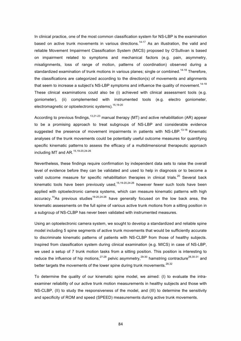
84
In clinical practice, one of the most common classification system for NS-LBP is the examination
based on active trunk movements in various directions.14-17 As an illustration, the valid and
reliable Movement Impairment Classification System (MICS) proposed by O’Sullivan is based
on impairment related to symptoms and mechanical factors (e.g. pain, asymmetry,
misalignments, loss of range of motion, patterns of coordination) observed during a
standardized examination of trunk motions in various planes; single or combined.14-18 Therefore,
the classifications are categorized according to the direction(s) of movements and alignments
that seem to increase a subject’s NS-LBP symptoms and influence the quality of movement.14-18
These clinical examinations could also be (i) achieved with clinical assessment tools (e.g.
goniometer), (ii) complemented with instrumented tools (e.g. electro goniometer,
electromagnetic or optoelectronic systems).15,19-20
According to previous findings,13,21-23 manual therapy (MT) and active rehabilitation (AR) appear
to be a promising approach to treat subgroups of NS-LBP and considerable evidence
suggested the presence of movement impairments in patients with NS-LBP.13-18 Kinematic
analyses of the trunk movements could be potentially useful outcome measures for quantifying
specific kinematic patterns to assess the efficacy of a multidimensional therapeutic approach
including MT and AR.15,19-20,24-26
Nevertheless, these findings require confirmation by independent data sets to raise the overall
level of evidence before they can be validated and used to help in diagnosis or to become a
valid outcome measure for specific rehabilitation therapies in clinical trials.20 Several back
kinematic tools have been previously used,15,19-20,24-26 however fewer such tools have been
applied with optoelectronic camera systems, which can measure kinematic patterns with high
accuracy.15As previous studies19-20,24-26 have generally focused on the low back area, the
kinematic assessments on the full spine of various active trunk motions from a sitting position in
a subgroup of NS-CLBP has never been validated with instrumented measures.
Using an optoelectronic camera system, we sought to develop a standardized and reliable spine
model including 5 spine segments of active trunk movements that would be sufficiently accurate
to discriminate kinematic patterns of patients with NS-CLBP from those of healthy subjects.
Inspired from classification system during clinical examination (e.g. MICS) in case of NS-LBP,
we used a setup of 7 trunk motion tasks from a sitting position. This position is interesting to
reduce the influence of hip motions,27-28 pelvic asymmetry,29-30 hamstring contracture28,30-31 and
better targets the movements of the lower spine during trunk movements.28,32
To determine the quality of our kinematic spine model, we aimed: (I) to evaluate the intra-
examiner reliability of our active trunk motion measurements in healthy subjects and those with
NS-CLBP, (II) to study the responsiveness of the model, and (III) to determine the sensitivity
and specificity of ROM and speed (SPEED) measurements during active trunk movements.

85
Methods
Subjects
The cohort comprised 25 healthy subjects aged 30-60 years and 25 subjects with NS-CLBP
aged 30-65 years. Anthropometrics data are shown in Table 1.
Table 1. Baseline characteristic of healthy and NS-CLBP subjects
Group n M/F Age (years) BMI (kg/m2) VAS T0 VAS T1
Healthy 25 10/15 40 (11) 23.3 (2.5) / /
NSCLBP 25 12/13 42 (9) 25.2 (3.2) 2.4 (1.7) 2.5 (1.5) NS
Data are shown as the mean (SD). VAS represents pain score according to a 10-point scale during the first (VAS T0) or second (VAS T1) session. NS = non-significant difference with t-paired test (p=0.44,); ICC (VAS T0-T1) = 0.85
The NS-CLBP group included men and women recruited from Saint-Luc University Hospital,
including patients who have been suffering from a NS-CLBP with or without radiating pain into
the thigh but not below the knee, for at least 12 weeks. Moreover, they were required to present
through the clinical examination several impairment directions (≥2 on 7 directions) during active
trunk movements from a sitting position.
The healthy subjects had no incidences of NS-LBP for 6 months prior the experiment. The Ethic
Committee of the University of Louvain approved the protocol for this study.
Protocol and Material
Seven trunk motion tasks were used that involved the whole spine and were performed at a
non-imposed speed. Eight infrared cameras (ELITE-BTS) registered the 3D positions of 9
reflective markers placed on bony landmarks. Each task consisted of 10 trials for each session
(2 sessions around 1 week apart). From the positions of the markers, customised software
calculated the range of motion (ROM) and the SPEED of each spinal segment for each subject
and each trial. The mean and standard deviation (SD) were calculated for each variable (n =
10).

86
All trunk movements were executed from a sitting position on a stool and the height of the stool
was adjusted for each subject in such a way that the angle between the hip and the trunk
reached 120°. This amplitude was chosen to help in maintaining the physiological curvature for
the starting position. From this starting position, subjects performed successively a set of 7
different direction tasks (the same as during clinical examination) described below (Tasks and
instructions).
Placement of markers
Standardised marker placements were positioned as follow: 5 markers on spinous processes of
S2, L3, T12, T7, and C7; 2 markers were placed on the anterior-superior-iliac-spinous process
(ASIS) right and left; and 2 markers were placed on the acromio-clavicular (Ac) joints right and
left. The examiner who placed the markers was an experimented manual-therapist in order to
enhance as possible palpatory accuracy. Moreover to reduce the approximation of bony
landmarks palpation described into the literature, the distance between the markers of each
subject were (I) measured from C7 as a reference (easy to find spinous process, the most
prominent in head flexion) (II) recorded and (III) reported from the first to the second session.
Kinematic spine model
A spine model was elaborated that included the pelvic and shoulder girdles (Figure 1A, B). The
whole spine was divided into various segments: high thoracic spine (HTS: C7 - T7), low thoracic
spine (LTS: T7 - T12), high lumbar spine (HLS: T12 - L3), low lumbar spine (LLS: L3 - S2), total
lumbar spine (TLS: T12 - S2), and shoulder segment (SS: AcRight - AcLeft). Each spinal
segment was considered as rigid and homogenous, delimited by proximal and distal markers as
illustrated in Figure 1. This modeling procedure is similar to that of Larivière et al.33 and
Gombatto et al.15 The selected variables are the range of motion (ROM) and the SPEED of
each spinal segment.
Tasks and instructions
Subjects were asked to fulfill conscientiously the following 4 rules, regardless of the task:
Subjects must (1) be in a physiological sitting position (with respect to curvatures) at the
beginning and end of each movement; (2) aim to make the movement at non-imposed
(spontaneous) speed and as far as possible; (3) keep the ischiatic tuberosities in contact with
the stool; (4) and strictly respect the specific plane of motion, depending of the trunk movement
task. Each movement was repeated 15 times and recorded after the fifth movement (n = 10
trials). Subjects were also instructed to perform each task according to the following
instructions:
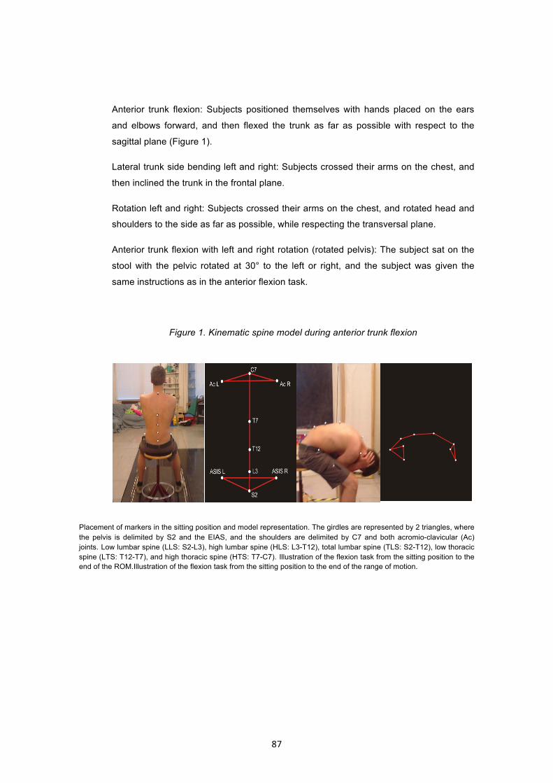
87
Anterior trunk flexion: Subjects positioned themselves with hands placed on the ears
and elbows forward, and then flexed the trunk as far as possible with respect to the
sagittal plane (Figure 1).
Lateral trunk side bending left and right: Subjects crossed their arms on the chest, and
then inclined the trunk in the frontal plane.
Rotation left and right: Subjects crossed their arms on the chest, and rotated head and
shoulders to the side as far as possible, while respecting the transversal plane.
Anterior trunk flexion with left and right rotation (rotated pelvis): The subject sat on the
stool with the pelvic rotated at 30° to the left or right, and the subject was given the
same instructions as in the anterior flexion task.
Figure 1. Kinematic spine model during anterior trunk flexion
Placement of markers in the sitting position and model representation. The girdles are represented by 2 triangles, where the pelvis is delimited by S2 and the EIAS, and the shoulders are delimited by C7 and both acromio-clavicular (Ac) joints. Low lumbar spine (LLS: S2-L3), high lumbar spine (HLS: L3-T12), total lumbar spine (TLS: S2-T12), low thoracic spine (LTS: T12-T7), and high thoracic spine (HTS: T7-C7). Illustration of the flexion task from the sitting position to the end of the ROM.Illustration of the flexion task from the sitting position to the end of the range of motion.

88
Data Analysis
Each spinal segment was analyzed according to the mean ROM and the mean SPEED. The
mean ROM (°) corresponded to the range of the angular displacement of each spinal segment
during 10 trials. At each frame, the angular displacement of motion in the sagittal plane (YZ) and
frontal plane (XZ) was calculated from the vertical axis (Z) localized on the proximal marker of
each segment, according to:
The angular displacement in the transversal plane (XY) relative to the horizontal axis (Y) was
calculated, localized on the proximal marker of each segment according to:
In these equations, Y, Z, X are the coordinates in the horizontal, vertical, and lateral directions,
respectively; p is the proximal marker of the segment; and d is the distal marker.
Figure 2 illustrates the calculation of the angular displacement in the lateral side-bending task
for one frame. The mean SPEED (° s-1) was calculated from the finite derivative of the angular
displacement of 10 trials for each task.

89
Figure 2. Illustration of angle calculation in the lateral side-bending task
Side bending is performed in the frontal plane, and the angular displacement of each segment is calculated from the Z (vertical) and Y (horizontal) axis. A zoomed image of the angular displacement of the upper thoracic segment (C7 – T7) is presented in the right part of the figure.
Statistical Analysis
To assess the reliability, subjects were invited for a second session separated from the first
session by around 1 week (6.3 ± 1.5 days). To prevent an eventual bias in the kinematic
variables due to a significant change in pain score, each patient was contacted before the
second session and it was asked if they were in a similarly state of pain before the second
acquisition of data. Moreover, the visual analog score (VAS) of back pain was measured (Table
1) for our NS-CLBP patients before starting each session, t-paired test and intraclass correlation
coefficient (ICC) was calculated showing no significant difference between both sessions
(p=0.44, ICC=0.85). The same examiner evaluated each subject during both sessions.
Reliability assessments were performed according to a method described by Wagner et al.34 by
the ICC and the standard error of the measurement (SEM).
The ICC assesses variations in the population sample within and between patients.34 The ICC
parameters used were the ICC consistency. These ICCs were calculated under a 2-way mixed
model in SPSS (SPSS v16.0 for Windows; SPSS Inc., Chicago, IL, USA). According to Shrout

90
and Fleiss,35 an ICC >0.75 indicates excellent reliability; ICC of 0.40-0.75 indicates fair to good
reliability; and ICC <0.40 indicates poor reliability.
The SEM estimates the nonsystematic variance,34 which include natural fluctuations in the
kinematic patterns in a single patient and the potential non-reproducibility of the optoelectronic
system itself. As a measure of within-subject variability across repeated trials, SEM expresses
the measurement error in the same units as those of the original measurement.
The SEM was calculated as: SEM, where SD is the standard deviation for all observations, x
expressed sessions 1 and 2 and R is the test-retest reliability coefficient (ICC) for session 1 and
2.
Measurement error also was expressed as the SEM%, the within-subject standard deviation as
a percentage of the mean, which was defined as:
Where mean is the mean for all of the observations in sessions 1 and 2. The SEM% indicates
measurement error independent of the units of measurement. The SEM% represents the limit
for the smallest change that indicates a real improvement for a group of subjects.34
Responsiveness is the sensitivity to change of outcome measures and was assessed from the
SEM using the minimal detectable change (MDC). MDC95 represents the change of the
variables falling outside of the measurement error and the magnitude of change necessary to
exceed the measurement error of 2 repeated sessions (T0 and T1) at a specified confidence
interval (CI) of 95%SEM, where 1.96 is the 2-sided table z value for the 95% CI and is used to
account for the variance of the 2 measurement sessions.34
So that the MDC could be independent of the units of measurement, it was expressed as a
percentage (MDC%), which was defined as:

91
The MDC% represents the smallest change that indicates a real change in a single individual.34
Comparison between groups (Table 3): To compare both groups, a Student’s t-tests were used
on the overall means of the ROM and SPEED of each spinal segment variables (n= 42). The
effect size with the standardised means of difference (SMD) described by Cohen was calculated
to compare the magnitude of the difference between both populations:
Where mean (A) is the mean of the healthy group, mean (B) is the mean of the NS-CLBP group,
and mean SD (standard deviation) is the mean from SD (A) and SD (B).
To obtain an index that allowed discriminate both groups, binary logistic regression analyses
(Forward Likelihood Ratio Stepwise in SPSS) were computed with the variables showing
significance in Student’s t-tests (n=26) as independent variables and as dependant variable the
status from inclusion criteria (0 = healthy, 1 = NS-CLBP). Prior, this analysis the variance
inflation factor (VIF>10) was estimate on the 26 pre-selected variables to remove variables
showing stronger correlation, 17 variables were finally selected and computed in the logistic
regression.36
Sensitivity and specificity were determined with the Receiver Operating Characteristic (ROC)
curves construction (MedCalc v.11.5 Software) with the Logit Score (LS) of ROM and SPEED
(best discriminant variables form the binary logistic regression) of each subject of the
experiment. The greatest Youden index (Y=sensitivity+specificity-1) was chosen as a decision
criterion to choose the most appropriate cut-off score for both logit score (ROM and SPEED).37
Generalization of our results for people not tested in the experiment could be assessed using
the probability method (α = .05) equation based on the logit score with any subject (Table 4).

92
Results
Reliability and responsiveness
Table 2 shows results of the ICCs, SEM% and MDC% values for each segment within each task
for ROM and SPEED variables of both groups. Means and standard deviations are presented in
Table 3.
In overall, the healthy group (n = 25) showed good to excellent reliability for all tasks and spinal
segments in terms of ROM and SPEED variables, with the worst and best ICCs values from
0.60 – 0.96; the SEM% were from 19.4% – 3.3% and the minimal and maximal values for
MDC% were from 9.3% – 53.8%.
In overall, the NS-CLBP group (n = 25) demonstrated good to excellent reliability for all tasks
and segments in terms of ROM and SPEED variables, the worst and best ICCs values were
from 0.77– 0.96; the SEM% were from 19.4% – 4.3% and the minimal and maximal values for
MDC% were from 11.8% – 53.7%.

93
Table 2. Reliability and responsiveness results of trunk movement tasks
Comparison between groups
When comparing ROM and SPEED segments variables of the healthy subjects to the NS-CLBP
patients (Table 3), all of the p-values were highly significant (p < 0.001) for the anterior flexion
test for all spinal segments, except for the SPEED of the low lumbar spine segment (p = 0.003).
Very large effect sizes were observed with SMD from 0.9 – 1.4.

94
For the trunk rotation task (left and right), the ROM variable of the shoulder segment (SS)
showed a significant p-value (p < 0.05) for the left rotation and a high significant p-value (p <
0.001) for the right rotation, with a SMD of respectively 0.6 and 1.3.
The trunk flexion with rotation task demonstrated high significant p-value (p < 0.001) for all
spinal segments and both variables, with higher effect sizes SMD from 1.2 – 2 (Figure 3).
Figure 3. Boxplots illustration for ROM and SPEED variables in both groups during trunk flexion
with rotation (right) for the total lumbar spine
CS: cut-off score
For the lateral side-bending test, the majority of ROM and SPEED variables of all spinal
segments were not normally distributed or significant but with very weak statistical power or not
significant (p > 0.05).
Results from the binary logistic regression showed that the most discriminant variables were for
ROM the low thoracic spine for flexion test (F LTS °), shoulder segment for right rotation test
(RR °), the total lumbar spine for flexion with left rotation test (FL TLS°) and for the SPEED, the
total lumbar spine segment in flexion with right rotation (FR TLS°/s) (Table 3).

95
Figure 4. Typical mean curves illustration for SPEED and ROM during trunk flexion with rotation
(10 trials) for the Low Lumbar Spine
Red curve of one NS-CLBP patient; Dark curve of one healthy subject
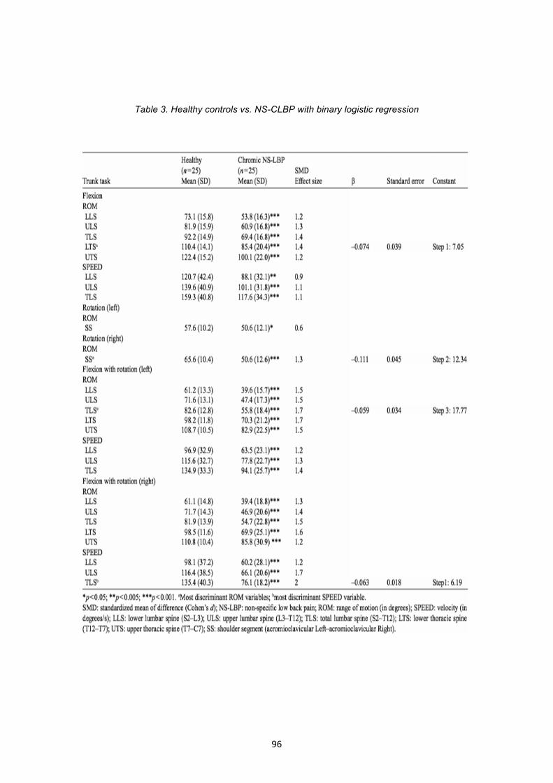
96
Table 3. Healthy controls vs. NS-CLBP with binary logistic regression

97
Sensitivity and specificity
Logit Scores (LS) from the binary logistic regression for ROM and SPEED showed a sensitivity
of 92% and specificity of 84% with a cutoff score of -0.65 for LS ROM and of 92% and 80% with
a cutoff score of -0.35 for LS SPEED (Table 4).
Table 4. Sensitivity and specificity with ROC curve analysis
Discussion
According to the literature,15,20,24-31 our kinematic spine model and trunk movements protocol
revealed good to excellent reliability and sensitivity/specificity for ROM and SPEED to
discriminate NS-CLBP patients.
It must be highlighted that only the lateral trunk side-bending task (left and right) did not reach
the statistical power of 0.8 for the majority of the variables, or are not normally distributed or are
not significantly different. As a consequence, even if reliable, we believe that this test may not
be adapted to discriminate subjects suffering from NS-CLBP and has been removed from table
3 for more simplification. Even if, differences between LBP and control groups in patterns
during trunk motion in side bending performed from a sitting position has been proved in
another study.28
It should be noted that previous studies20,24-29,31 have mainly assessed the lumbar spine
segment and only in one direction of trunk movement from a standing position. In the present
study, the decreased of ROM and SPEED observed in all tasks (7 directions) and in all spinal

98
segments were expected only for the NS-CLBP group and may reflect localized disorders of the
lumbar spine (Figure 4).
Marras et al.20,24 described speed and acceleration from a standing position as sensitive
variables to distinguish CLBP but not the ROM. Compared with healthy subjects, a specific
“motion signature” has been observed on kinematic variables during active trunk movements
from LBP patients, which revealed the musculoskeletal status of the spine with a very good
sensitivity and specificity.20,24 Our results showed similar findings for SPEED variables, however
ROM variable was also very sensitive, probably due to the sitting reference position and the use
of Logit Score from the binary logistic regression.
In the thoracic spinal segments, our results presented divergences from the results of Larivière
et al.,33 they showed an increase in mobility of the thoracic spine during trunk flexion in CLBP
patients. These authors proposed the existence of compensations as a consequence of the loss
of flexibility of the lumbar spine. However, our results demonstrated a significant stiffness of the
thoracic spine (decrease of ROM) in the NS-CLBP group compared to the healthy group. A
possible explanation for this discrepancy may be methodological differences between the
studies. The previous trials33 used a standing position, from which it is likely that subjects were
stimulated to compensate with the thoracic spine to reach the floor with their hands during
flexion.28,30-31 Moreover, the reduced flexibility of the hamstring, which is naturally encountered
with aging, may have limited the lumbar spine during the forward bending task from a standing
position.31
The tasks used in our protocol were chosen on the basis of biomechanical38-40 and classification
system evidences for NS-LBP.14-18 For example, the anterior flexion task increases the load on
the vertebral disc and may cause back pain due to the enhancement of neural compression.
Moreover, the posterior fibers of the annulus and posterior ligaments are thought to provide
resistance.38 Side-bending of the trunk decreases the ROM in subjects with LBP generated by
painful hernia and/or facet joints.15,26,28,39 Rotations are focused on the thoracic-lumbar hinge
because of the very small biomechanical motion in rotation for the lumbar spine.39 The anterior
trunk flexion task with rotated pelvis increases the pelvic constraints, due to the inferior and
superior ilio-lumbar ligaments. This scenario leads to lumbar stiffness, especially at the lower
lumbar spine, decreasing the ROM during the flexion movement.40
Initially, a trunk extension task was included in our kinematic protocol, because this task
increases the load at the facet joints in degenerative vertebrae39 and is used in MICS for NS-
LBP.14-18 However, during our preliminary study, trunk extension from a sitting position revealed
poor reliability (ICCs from 0.25 – 0.60). Furthermore, the subjects also described it as difficult or
dangerous. Therefore, this task was deleted from our protocol.

99
In a preliminary study, performance of the anterior trunk flexion at an imposed speed following a
metronome rhythm showed poor reliability (ICC < 0.40 and SEM% > 20%) of the variables for all
the spinal segments as compared to performance at a non-imposed speed. Moreover, the
reproducibility of the non-imposed speed during trunk movements has been proven in several
previous trials,20,24-31 therefore, the use of an imposed speed was removed from our protocol.
Trunk movement tasks with spinal segments variables (ROM and SPEED) that present good to
excellent reliability (ICC ≥ 0.7, SEM% ≤ 15%) with the best discrimination between both
populations in using logit scores (LS) for ROM and SPEED may be helpful as quantitative
kinematic outcome measures to support in diagnosis any patient or to measure improvement in
future clinical trials. Our results provide additional insight evidence into the future use of
kinematic spine motion models with an optoelectronic system to help in diagnose subgroups of
NS-LBP in a classification system and to better target specific treatment (e.g., MT, AR) adapted
to movement impairments and motor control as well as measure the therapeutic effects with the
MDC and/or LS in clinical practice for a single patient or in clinical studies for a sample of
patients with the SEM and/or LS.
Authors have generalised results from the sample tested to people not tested with the LS for
sensitivity and specificity and the probability equation (Table 4). Nevertheless, findings of
people not tested should be interpreted cautiously because of the small sample size of this
study. Therefore, it’s recommended for a future trial using our model to raise the sample size of
both groups and to integrate SPEED variables for the thoracic spine to improve the index of LS
speed.
Conclusion
The quantitative analysis of kinematic motion patterns in subgroups of patients with chronic
NS-LBP during trunk movements in different directions is of major importance because it can
help clinicians to identify motion patterns that may contribute to chronic NS-LBP disorders
and target interventions according to the quality of movement. The kinematic spine model and
standardised protocol including 7 trunk motion tasks demonstrated good to excellent
reliability. However, only 4 tasks were selected for inclusion in the final protocol. The LSs of
ROM and SPEED variables may be used as quantitative outcome measures to aid in the
diagnosis and assessment of patients with chronic NS-LBP before and after physical therapy
(e.g. MT) in clinical practice and research. To our knowledge, such analyses have not been
used in randomised clinical trials assessing the efficacy of physical therapies in NS-LBP
subgroups.

100
References
1. Waddell G. Low back pain revolution. Churchill Livingstone 2004:1-459.
2. Andersson GB. Epidemiological features of chronic low-back pain. Lancet 1999;354:581-5.
3. Gourmelen J, Chastang JF, Ozguler A, et al. Frequency of low back pain among men and women aged 30 to 64 years in France. Results of two national surveys. Ann Readapt Med Phys 2007;50:633-40.
4. Leclerc A, Chastang JF, Ozguler A, Ravaud JF. Chronic back problems among persons 30 to 64 years old in France. Spine 2006;31:479-84.
5. Waddell G, Burton AK. Occupational health guidelines for the management of low back pain at work: evidence review. Occup Med 2001;51:124-35.
6. Majid K, Trummees E. Epidemiology and natural history of low back pain. Seminars in spine surgery 2008;20:87-92.
7. Wynne-Jones G, Dunn KM, Main CJ. The impact of low back pain on work: a study in primary care consulters. Eur J Pain 2008;12:180-8.
8. Rossignol M, Rozenberg S, Leclerc A. Épidémiologie des lombalgies: quoi de neuf ? Joint Bone Spine 2009:76;608-13.
9. Praemera A, Furners S, Rice DP. Musculoskeletal conditions in the United States. Park Ridge, IL: American Academy of Orthopaedics Surgeons 1992.
10. Nachemson AL. Advances in low back pain. Clin Orthopedic 1985;200:266-78.
11. Spratt K, Lehman T, Weinstein J. A new approach to low back pain examination: behavorial assessment of mechanical signs. Spine 1990;15:96-102.
12. Leboeuf-Yde C, Lauritsen J, Lauritzen T. Why has the search for causes of low back pain largely been nonconclusive? Spine 1997;22:877-81.
13. Fersum KV, Dankaerts W, O’Sullivan PB, et al. Integration of subclassification strategies in randomised controlled clinical trials evaluating manual therapy treatment and exercise therapy for non-specific chronic low back pain: a systematic review. Br J Sports Med 2010;44:1054-1062.
14. Harris-Hayes M, Van Dillen LR. The inter-tester reliability of physical therapists classifying low back pain problems based on the Movement System Impairment classification system. PM R 2009;1:117-26.
15. Gombatto SP, Collins DR, Sahrmann SA, Engsberg JR, Van Dillen LR. Patterns of lumbar region movement during trunk lateral bending in 2 subgroups of people with low back pain. Phys Ther 2007;87:441-54.
16. Dankaerts W, O’Sullivan PB, Straker LM, Burnett AF, Skouen JS. The inter-examiner reliability of a classification method for non-specific low back pain patients with motor control impairement. Man Ther 2006;11:28-39.

101
17. O’Sullivan PB. Diagnosis and classification of chronic low back pain disorders: maladaptive movement and motor control impairments as underlying mechanism. Man Ther 2005;10:242-55.
18. Van Dillen LR, Sahrmann SA, Norton BJ, et al. Reliability of physical examination items used for classification of patients with low back pain. Phys Ther 1998;78:979-88.
19. Troke M, Moore AP, Cheek E. Intra-operator and inter-operator reliability of the OSI CA 6000 Spine Motion Analyzer with a new skin fixation system. Man Ther 1996;3:92-8.
20. Marras WS, Parnianpour M, Ferguson SA, et al. The classification of anatomic- and symptom-based low back disorders using motion measure models. Spine 1995;20: 2531-46.
21. Aure OF, Nilsen JH, Vasseljen O. Manual therapy and exercise therapy in patients with chronic low back pain: a randomized, controlled trial with 1-year follow-up. Spine 2003;28:525-31.
22. Niemistö L, Sarna S, Lahtinen-Suopanki T, Lindgren KA, Hurri H. Predictive factors for 1-year outcome of chronic low back pain following manipulation, stabilizing exercises, and physician consultation or physician consultation alone. J Rehabil Med 2004;36:104-109.
23. Flynn T, Fritz J, Whitman J, et al. A clinical prediction rule for classifying patients with low back pain who demonstrate short-term improvement with spinal manipulation. Spine 2002;27:2835-43.
24. Marras WS, Ferguson SA, Gupta P, et al. The quantification of low back disorder using motion measures. Methodology and validation. Spine 1999;24:2091-100.
25. Lehman GJ. Biomechanical assessments of lumbar spinal function. How low back pain sufferers differ from normals. Implications for outcome measures research. Part I: kinematic assessments of lumbar function. J Manipulative Physiol Ther 2004;27:57-62.
26. Lehman GJ, McGill SM. Spinal manipulation causes variable spine kinematic and trunk muscle electromyographic responses. Clin Biomech 2001;16:293-9.
27. Shum G, Crosbie J, Lee R. Effect of low back pain on the kinematics and joint coordination of the lumbar spine and hip during sit-to-stand and stand-to-sit. Spine 2005;30:1998-2004.
28. Al-Eisa E, Egan D, Deluzio K,Wassersug R. Effects of pelvic asymmetry on low back pain
on trunk kinematics during sitting: a comparison with standing. Spine 2006;31:135-43.
29. van Wingerden, JP, Vleeming, A, Ronchetti I. Differences in standing and forward bending in women with chronic low back or pelvic girdle pain: indications for physical compensation strategies. Spine 2008;33:E334-41.
30. Esola M, McClure P, Fitzgerald GK, et al. Analysis of lumbar spine and hip motion during forward bending in subjects with and without a history of low back pain. Spine 1996;21:71-78.
31. Johnson EN, Thomas JS. Effect of hamstring flexibility on hip and lumbar spine joint excursions during forward-reaching tasks in participants with and without low back pain. Arch Phys Med Rehabil 2010;91:1140-2.

102
32. Maigne JY, Lapeyre E, Morvan G, Chatellier G. Pain immediately upon sitting down and relieved by standing up is often associated with radiologic lumbar instability or marked anterior loss of disc space. Spine 2003;28:1327-34.
33. Lariviere C, Gagnon D, Loisel P. The effect of load on the coordination of the trunk for subjects with and without chronic low back pain during flexion-extension and lateral bending tasks. Clin Biomech 2000;15:407-16.
34. Wagner JM, Rhodes JA, Patten C. Reproducibility and minimal detectable change of three-dimensional kinematic analysis of reaching tasks in people with hemiparesis after stroke. Phys Ther 2008;88:652-63.
35. Shrout PE, Fleiss JL. Intraclass correlations: Uses in assessing rater reliability. Psychol Bull 1979;86:420-4.
36. Hosmer DW, Lemeshow S. Applied Logistic Regression. New York, NY: John Wiley and Sons 2000:1-373.
37.Bewick V, Cheek L, Ball J. Statistics review 13: Receiver operating characteristic curves. Critical Care 2004;8:508-12..
38. Goto K, Tajima N, Chosa E, et al. Mechanical analysis of the lumbar vertebrae in a three dimensional finite element method model in which intradiscal pressure in the nucleus pulposus was used to establish the model. J Orthop Sci 2002;7:243-6.
39. Kuo CS, Hu HT, Lin RM, et al. Biomechanical analysis of the lumbar spine on facet joint force and intradiscal pressure-a finite element study. BMC Musculoskelet Disord 2010;11:151.
40. Chansirinukor W, Lee M, Latimer J. Contribution of pelvic rotation to lumbar posteroanterior movement. Man Ther 2001;6:242-9.

103
CHAPTER III
Use of kinematic algorithms to identify people with chronic nonspecific low back pain
from asymptomatic subjects: validation study
Benjamin Hidalgo, Henri Nielens, Maxime Gilliaux, Toby Hall, Christine Detrembleur
J Rehabil Med. 2014 Sep;46(8):819-23. Doi: 10.2340/16501977-1836
ABSTRACT
Objective: To determine whether kinematic algorithms can distinguish subjects with chronic
nonspecific low back pain from asymptomatic subjects and subjects simulating low back pain,
during trunk motion tasks.
Design: Comparative cohort study.
Subjects: A total of 90 subjects composed 3 groups; 45 chronic nonspecific low back pain
patients in the CLBP group; 45 asymptomatic controls people in the asymptomatic controls
group. 20/45 subjects from the asymptomatic controls group composed the CLBP simulators
group as well.
Methods: During performance of 7 standardized trunk motion tasks 8 infrared cameras
recorded 6 spinal segments from the kinematic spine model. Two logit scores, for range of
motion and speed, were used to investigate differences between the groups. Group allocation
based on logit scores was also calculated, allowing the assessment of sensitivity and specificity
of the algorithms.
Results: For the 90 subjects (pooled data), the logit scores for range of motion and speed
demonstrated highly significant differences between groups (p < 0.001). The logit score means
and standard deviation (SD) values in the asymptomatic group (n = 45) and chronic nonspecific
low back pain group (n = 45), respectively, were –1.6 (SD 2.6) and 2.8 (SD 2.8) for range of
motion and –2.6 (SD 2.5) and 1.2 (SD 1.9) for speed. The sensitivity and specificity (n = 90) for

104
logit score for range of motion were 0.80/0.82 and for logit score for speed were 0.80/0.87,
respectively.
Conclusion: These results support the validity of using 2 movement algorithms, range of
motion and speed, to discriminate asymptomatic subjects from those with low back pain.
However, people simulating low back pain cannot be distinguished from those with real low back
pain using this method.

105
Introduction
Chronic nonspecific low back pain (CLBP) is a growing problem in Western industrialised
countries, which brings diagnostic and treatment challenges.1-4 From a diagnostic point of view,
in general, people with CLBP have no clear patho-anatomical features distinguishing them from
asymptomatic subjects. For example, there is a poor correlation between features seen on spinal
imaging and symptoms of low back pain (LBP).1,3,5 Thus, diagnosis of non-specific LBP is based
mainly on subjective and physical clinical examination criteria.1-3,6-9 Kinematic analysis of trunk
motion appears promising in the diagnosis and discrimination of people with non-specific
LBP.1,9-12 Some of these features may also enable differentiation into treatment specific sub-
groups, which may have value in the management of LBP.13
Our previous study12 showed that kinematic tools are useful in identifying impairments in people
with CLBP, both quantitatively (range of motion, speed and acceleration) and qualitatively
(motion signatures), when single and combined planes of movement are investigated.1,10-13 The
study identified 2 kinematic algorithms (logit scores) that could be used to distinguish people with
CLBP from asymptomatic controls on the basis of a binary logistic regression analysis.12 The
first algorithm is an index for range of motion (ROM) and the second is an index for speed of
movement (SPEED). The sensitivity and specificity of the logits for these algorithms has been
reported previously.12 To our knowledge, no studies have investigated whether movement
algorithms can correctly identify people simulating LBP from those with actual LBP in order to
address the external validity (i.e. the generalisability) of the research findings.
The aim of this study was to evaluate the external validity and generalisability of previously
reported algorithms in the quantitative assessment of spinal movement impairment in an
independent sample of asymptomatic controls, people simulating LBP, and those with CLBP.
Methods
Subjects
This study comprised 90 subjects, of whom 50 were from a previous investigation (25 healthy
controls and 25 subjects with CLBP),12 together with 40 new subjects (20 asymptomatic
subjects who represented the healthy simulators and 20 with CLBP). Anthropometric data are
shown in Table 1.

106
The CLBP group included men and women recruited from Cliniques Universitaires St-Luc
(Woluwé - Saint - Lambert, Belgium). At the time of the experiment, these subjects had CLBP
with or without radiating pain no further than the knee (Quebec Task force 1–2 categories) for
at least 12 weeks and had clinical physical examination features of spinal movement
impairment.
The asymptomatic subjects had no history of LBP for at least 6 months prior to the experiment.
The human research ethics committee of the institution approved the study protocol.
Table 1. Baseline characteristics of asymptomatic subjects and chronic nonspecific low back
pain patients (CLBP)
Asymptomatic
controls and LBP
simulators
CLBP
Asymptomatic
controls from
previous study
CLBP from
previous
study
M/F, n 11/9 (n=20) 11/9 (n=20) 10/15 (n=25) 12/13
(n=25)
Age, years,
mean (SD)
39.9 (13.5) 45.1 (11.6) 40 (11) 42 (9)
Body mass
index, mean
(SD)
23.5 (2.8) 27.4 (3.5) 23.3 (2.5) 25.2 (3.2)
Visual
analogical
scale, mean
(SD)
0.2 (0.4) 2.9 (1.7) 0 2.5 (1.5)
Oswestry
disability
index (%)
1.6 (1.8) 17.9 (8.2) 0 19.8 (8.6)
107
Protocol and material
Seven standardised trunk motion tasks, described below, were assessed in the kinematic spine
model, and recorded by 8 infrared cameras (ELITE-BTS, Milan, Italy). The kinematic spine
model, the trunk movement standardisation procedure, data collection and statistical analysis
have been described in a previous study.12
All subjects were asked to follow 4 rules during movement tasks, which were performed in a
seated position: (I) begin and end each movement with a neutral spine posture; (II) to move at
a self-determined (spontaneous) speed through the largest possible range; (III) to maintain
contact between the ischial tuberosities and the stool; (IV) and to adhere strictly to the plane of
motion specified by each task. Each movement was repeated 15 times and recorded after the
fifth movement (n=10 trials). Twenty asymptomatic subjects formed the LBP simulators group.
These subjects were asked firstly to perform all the trunk tests in a natural way (asymptomatic
controls) and, secondly, to repeat the task simulating CLBP (simulators) while carrying out
each movement task.14
The trunk motion tasks were as follows:
Anterior trunk flexion: Subjects positioned themselves with their hands over their ears
and their elbows forward, and then flexed the trunk as far as possible with respect to the
sagittal plane.
Lateral trunk side-bending left and right: Subjects crossed their arms on their chest, and
then inclined their trunk in the frontal plane.
Rotation left and right: Subjects crossed their arms on their chest, and rotated their head
and shoulders to the side as far as possible, while respecting the transverse plane.
Anterior trunk flexion with left and right rotation (rotated pelvis): The subject sat on the
stool with the pelvic rotated 30° to the left or right, and the subject was given the same
instructions as in the anterior flexion task.
Statistical analysis
Comparison of the kinematic variables between groups was carried out using 1-way analysis of
variance with pairwise multiple comparison procedures (Holm-Sidak method, factor groups)
(Sigmastat® 3.5, Systat Software, San Jose, USa) (Table 2).

108
Binary logistic regression analysis was previously used to evaluate the kinematic spine model in
each group, with 2 logit scores (LS) calculated for ROM and SPEED (see Table 3).12 For this,
we calculated an index to enable discrimination between groups using binary logistic regression
analyses (stepwise forward likelihood ratio in SPSS). These analyses were applied only to
variables found to differ significantly by Student’s t-tests (n=26). These variables were assigned
as independent variables, and group membership (0 = healthy, 1 = chronic LBP) was the
dependent variable. Before regression analyses were performed, the variance inflation factor
(VIF) was estimated for each of the 26 selected variables, in order to remove variables with
strong correlation (VIF > 10); 17 variables were finally selected and included in the logistic
regression.12
The generalisability of our previous results to correctly identify people with the use of LS was the
main goal of this study. Therefore, we investigated the sensitivity and specificity of the
previously determined algorithms using the probability equation (Table 3) on the 40 new
subjects in the present study, with the following equation:
Sensitivity = true positives / (true positives + false negatives)
Specificity = true negatives / (true negatives + false positives)
Following this, the overall sensitivity and specificity were calculated by pooling the results of LS
for ROM and SPEED from both studies (n=90 subjects) using the receiver operating
characteristic (ROC) curves (MedCalc software,version 11.5, Mariakerke, Belgium).
Results
The results of the between-groups’ comparison of kinematic variables of each spinal segment in
each task as well as for kinematic algorithms (ROM and SPEED) are shown in Table 2.
Concerning the LS for ROM (n=40), a highly significant difference was found between the
groups with p-value < 0.001 (power of 1.000 with alpha = 0.05). The mean and standard
deviation (SD) of LS for ROM was –0.47 (SD 2.5) in the asymptomatic control group (n=20); 3.2
(SD 3.4) in the CLBP group (n=20); and 5.6 (SD 3.5) in the LBP simulator group (n=20).
In addition, the LS for SPEED demonstrated highly significant differences between the groups,
with p-value < 0.001 (power of 1.000 with alpha = 0.05). The mean of LS for SPEED was –2.9
(SD 2.5) in the asymptomatic control group; 1.1 (SD 2.5) in the CLBP group; and 2.1 (SD 2.1) in
the LBP simulator group.

109
When matching subjects of both the current and previous studies (n=90), the comparison
between the asymptomatic subjects (n=45) with those with LBP (n=45) revealed highly
significant differences (p < 0.001; power of 1.000 and alpha = 0.05) for the LS’s ROM and
SPEED. The means and SD values for the combined asymptomatic group and CLBP group,
respectively, for LS ROM were –1.6 (SD 2.6) and 2.8 (SD 2.8); and for LS SPEED were –2.6
(SD 2.5) and 1.2 (SD 1.9).
Table 2. Asymptomatic controls and low back pain (LBP) simulators vs chronic nonspecific low
back pain patients, one-way analysis of variance of each spinal segment in each task

110
Using the probability equation presented in Table 3, we calculated the probability that each new
subject in this study had LBP.
Table 3. Kinematic algorithms and probability equations from previous study12
For the index of LS ROM:
• asymptomatic controls (n=20): 8 were positive and 12 were negative; 60% of healthy
controls were correctly classified.
• LBP simulators (n=20): 19 were positive and 1 was negative; 5% of healthy simulators
were correctly classified.
• CLBP (n=20): 4 were negative and 16 were positive; 80% of LBP subjects were
correctly classified.
Based on the data of asymptomatic controls and CLBP subjects, the sensitivity was 0.80, with
specificity 0.60. Positive predictive value (PPV) was 0.67 and negative predictive value (NPV)
0.75.
For the index of LS SPEED:
• asymptomatic controls (n=20): 2 were positive and 18 were negative; 90% of healthy
controls were correctly classified.
• LBP simulators (n=20): 20 were positive and 0 was negative; 0% of healthy simulators
were correctly classified.
• CLBP (n=20): 7 were negative and 13 were positive; 65% of LBP subjects were
correctly classified.

111
Based on the data of asymptomatic controls and CLBP subjects, the sensitivity was 0.65 and
specificity 0.90, with PPV 0.87 and NPV 0.72.
Using LS of ROM and SPEED with ROC curves analysis on all subjects of both studies (n=90),
we calculated an overall sensitivity/specificity:
• For LS ROM: sensitivity was 0.80 (0.65–0.90) and specificity 0.82 (0.68–0.92) with a
cut-off score of 0.77 (area under the ROC curve: 0.88 with standard error: 0.03 and
significant level p < 0.0001; youden index: 0.62).
• For LS SPEED: sensitivity was 0.80 (0.65–0.90) and specificity 0.87 (0.73–0.95) with
a cut-off score of 0.11 (area under the ROC curve: 0.88 with standard error: 0.03 and
significance level p < 0.0001; youden index: 0.66).
The LS pooled values of each subject for groups CLBP (n=45) and asymptomatic controls
(n=45) are shown in Figures 1 and 2.
Figure 1. Scatter plot graph of pooled data for logit scores of ROM of asymptomatic controls
(n=45) and chronic non-specific low back pain subjects (n=45)

112
Figure 2. Scatter plot graph of pooled data for logit score of SPEED for asymptomatic controls
(n=45) and chronic non-specific low back pain subjects (n=45)
Discussion
The kinematic algorithms ROM and SPEED clearly discriminated the groups with highly
significant differences. In comparison with people with CLBP, the kinematic values for people
voluntarily simulating LBP were lower, and the LS differed significantly (Table 2). When the
healthy subjects tried to simulate CLBP, they were unable to mimic the true kinematics of people
with chronic LBP. During each movement task the healthy subjects exaggerated the spinal
movement impairments, as if they were affected by an acute LBP.
The present findings also confirm those of the previous study concerning the data from the trunk
lateral flexion task, which did not differ significantly between the groups. This task was once
again not useful in discriminating subjects with chronic LBP, at least when carried out in a
seated position.12

113
The sensitivity and specificity of both LS (ROM and SPEED) were previously found to be
excellent in discriminating people with CLBP.12 However, when applying the previously
determined probability equation to a new independent sample of subjects, we found only a
moderate to good level of sensitivity and specificity. Despite this, when data from both studies
are pooled, the ROC curves analysis of both LS (ROM and SPEED) revealed improved overall
sensitivity and specificity. The results of the present and previous study12 indicate that the
algorithms ROM and SPEED provide useful discrimination between populations. These
kinematic measures may be helpful in sub-grouping people with movement impairment in LBP,
which may have potential benefit in future clinical trials. For example, the targeting of specific
interventions, such as manual therapy, might be better suited to people with certain movement
impairments identified through these algorithms. This requires further investigation.
The present study has several limitations. Simulating CLBP is problematic because of the
inherent multifactorial nature of CLBP, which influences the kinematic outputs during movement.
Subjects may have had difficulty imagining and carrying out the simulation of CLBP. In the case
of CLBP, pain may arise from a variety of causes. The cognitive interpretation of pain has
multiple psycho-social influences (e.g. misrepresentation of body schema, anxiety, depression,
education, beliefs, negativism, catastrophisation, lifestyle). In turn, this may influence central
sensitisation, resulting in altered “outputs”, in particular movement behaviors and patterns of
impairment seen when examining patients with CLBP.2,3,15 All these elements are very difficult, if
not impossible, to simulate in our sample of LBP simulators. Moreover, we should not forget a
possible more important Hawthorne learning effect in the subjects simulating LBP that may have
influenced the simulated patterns of movement.
In conclusion, these results validate the use and generalizability of both kinematic algorithms for
the discrimination of spinal movement impairments between healthy controls and patients with
CLBP. However, subjects who were simulating CLBP could not be correctly classified by our
method. This validation study supports the use of this method to objectively evaluate the efficacy
of physical manual therapy treatment in future clinical trials.

114
References
1. Hidalgo B, Hall T, Nielens H, Detrembleur C. Inter-tester agreement and validity of identifying lumbar pain provocative movement patterns using active and passive accessory movement tests. J Manipul Physiol Ther 2014; 37:105-15.
2. Delitto A, George SZ, Van Dillen LR, Whitman JM, Sowa G, Shekelle P, et al. Low back pain. J Orthop Sports Phys Ther 2012;42: A1-57.
3. Balague F, Mannion AF, Pellise F, Cedraschi C. Non-specific low back pain. Lancet 2012; 379:482-91.
4. Hidalgo B, Detrembleur C, Hall T, Mahaudens P, Nielens H. The efficacy of manual therapy and exercise for different stages of non-specific low back pain: an update of sytematic reviews. J Man Manip Ther 2014;22:59-74.
5. McCullough BJ, Johnson GR, Martin BI, Jarvik JG. Lumbar MR imaging and reporting epidemiology: do epidemiologic data in reports affect clinical management? Radiology 2012; 262:941-46.
6. Fritz JM, Cleland JA, Childs JD. Subgrouping patients with low back pain: evolution of a classification approach to physical therapy. J Orthop Sports Phys Ther 2007;37:290-302.
7. O'Sullivan P. Diagnosis and classification of chronic low back pain disorders: maladaptive movement and motor control impairments as underlying mechanism. Man Ther 2005;10:242-55.
8. Dankaerts W, O'Sullivan P. The validity of O'Sullivan's classification system (CS) for a sub-group of NS-CLBP with motor control impairment (MCI): overview of a series of studies and review of the literature. Man Ther 2011;16:9-14.
9. Hidalgo B, Gobert F, Bragard D, Detrembleur C. Effects of proprioceptive disruption on lumbar spine repositioning error in a trunk forward bending task. J Back Musculoskelet Rehabil 2013;26:381-387.
10. Lehman GJ. Biomechanical assessments of lumbar spinal function. How low back pain sufferers differ from normals. Implications for outcome measures research. Part I: kinematic assessments of lumbar function. J Manipulative Physiol Ther 2004;27:57-62.
11. Dankaerts W, O'Sullivan P, Burnett A, Straker L, Davey P, Gupta R. Discriminating healthy controls and two clinical subgroups of nonspecific chronic low back pain patients using trunk muscle activation and lumbosacral kinematics of postures and movements: a statistical classification model. Spine 2009;34:1610-18.
12. Hidalgo B, Gilliaux M, Poncin W, Detrembleur C. Reliability and validity of a kinematic spine model during active trunk movement in healthy subjects and patients with chronic non-specific low back pain. J Rehabil Med 2012;44:756-63.
13. Vibe Fersum K, O'Sullivan P, Skouen JS, Smith A, Kvale A. Efficacy of classification-based cognitive functional therapy in patients with non-specific chronic low back pain: A randomized controlled trial. Eur J Pain 2013;17:916-28.

115
14. Almosnino S, Stevenson JM, Day AG, Bardana DD, Diaconescu ED, Dvir Z. Differentiating between types and levels of isokinetic knee musculature efforts. J Electromyogr Kinesiol 2011; 21:974-81.
15. O'Sullivan P. It's time for change with the management of non-specific chronic low back pain. Br J Sports Med 2012;46:224-27.

116

117
CHAPTER IV
Effects of proprioceptive disruption on lumbar spine repositioning error in a trunk
forward bending task
Benjamin Hidalgo, François Gobert, Dominique Bragard, Christine Detrembleur
J Back Musculoskelet Rehabil 2013;26(4):381-7. Doi: 10.3233/BMR-130396
ABSTRACT
Background: Various inputs of proprioception have been identified and shown to influence low
back proprioception sense.
Objective: To investigate the effect of disrupting proprioception on lumbar spine repositioning
error during forward bending.
Methods: Healthy-subjects (n = 28) and patients with non-specific chronic low-back pain (n =
10) aged between 20–50 years. Subjects performed 5 repetitions of a lumbar repositioning task
targeting 30° of trunk-forward-bending from a seated-position with different proprioceptive
disturbances administered to the low back. Video analysis of skin reflective markers measured
lumbar spine range-of-motion. A control-task was performed without any proprioceptive
disturbance, while the remaining 4 tasks were electro-stimulation, vibration, taping and sitting on
an unstable surface.
Results: The healthy group showed significantly altered repositioning error when compared
with the control task (p = 0.004): control-task vs. taping-task, vibration-task and unstable-sitting.
In the NS-CLBP group, one motor-task showed significant difference in control-task vs. taping-
task (p = 0.004). Comparison between the NS-CLBP and matched-healthy groups revealed that
the NS-CLBP subjects had larger repositioning-error (p = 0.009) for control, taping and vibration
tasks.

118
Conclusion: Proprioceptive disturbances had the most significant effect in increasing
repositioning-error among healthy subjects. The between-groups analysis confirmed evidence
consistent with the literature of greater repositioning-error in people with NS-CLBP than healthy
subjects.

119
Introduction
Low-back pain is a common musculoskeletal disorder.1-3 The population most at risk is active
people in industrialized countries.2-3 Up to 75% of the working population suffers at least once in
their lives from low-back pain and approximately 14% suffer pain lasting for more than 2
weeks.1-3 In the United-States, low-back pain ranks second among the reasons for seeking
medical attention3 leading to important medical costs and economic consequences.1-5
The sources of non-specific low-back pain (NS-LBP) are multiple and lack diagnostic precision
in 80 % of cases.2-3 The Postural-structural-biomechanical model is currently used for evaluation
of low-back pain and rationalizes the application of physical therapy and/or pharmacological
treatment; however this model has recently been questioned.4 Indeed, medical images and
clinical tests do not always correlate with low-back pain status.4-7 Therefore, new assessment
tools are necessary to help in understanding the problematic of NS-LBP patients.5-7
Motor control disorders are common problems in chronic NS-LBP. The hypothetical underlying
mechanism could be a deficiency of proprioception in these patients.8-13 Proprioception is
considered as “the knowledge of the positions of body segments and the movement of body in
space”.13 Since proprioception is essential for motor control of the trunk it should be
quantitatively evaluated. Therefore, the developments of accurate tools providing a quantifiable
measure of spinal proprioception are required.6-7
Several studies have investigated motor control and lumbar proprioception.6 For example, one
study focused on the detection of trunk motion during passive movements induced by an
electromechanical device13, while a further study used a force-platform to evaluating postural-
balance with external stimulations.10-12 Another common assessment of proprioception is the
measurement of active and/or passive repositioning error (RE, i.e. the difference between a
target position and the reached position of the patient) using kinematic-tools.6,9,13-20 These
studies13-20 showed conflicting evidence about a higher RE in chronic NS-LBP patients. This
discrepancy could also be due to different protocols used to measure RE. However, the main
tendency supports the hypothesis that reposition sense is altered in patients with chronic NS-
LBP when compared to healthy subjects,13,17-20 while a minority revealed no differences.9
The aims of this study were to investigate the effects of various proprioceptive disturbances (i.e.
electro-stimulations, vibrations, taping on the low-back area or sitting on unstable support) on
RE in healthy and chronic NS-LBP subjects and to compare RE accuracy between both groups.
To our knowledge, the introduction of various proprioceptive disturbances on both these
populations while simultaneously performing RE motor tasks have never been undertaken in the
same study.

120
Methods
Subjects
The cohort was composed of 28 healthy subjects aged 26 ± 9.8 years (mean + SD) with a body
mass index (BMI) of 23.18% ± 2.64 (14 male and 14 female).
The non-specific chronic low back pain (NS-CLBP) group was composed of 10 patients aged 34
± 8.9 years with BMI of 22.34% ± 3.09 (5 male and 5 female).
To compare both groups, a matched-healthy group of 10 subjects was composed from the
healthy subjects. Anthropometric data are reported in Table 1.
Table 1. Baseline characteristics of healthy subjects and those with chronic non-specific low
back pain
Data are shown as the mean (standard deviation). NS-CLBP, non-specific chronic low back pain; M, male; F, female; BMI, body mass index; VAS, 10-point visual analogue scale pain score (present pain); NS, non-significant difference between NS-CLBP and Matched Healthy (Student t-test, p > 0.05)
Healthy subjects were recruited on a voluntary basis and had no history of NS-LBP in the 12
months prior to the experiment.
The NS-CLBP group included patients recruited from Saint-Luc University Hospital (Brussels,
Belgium) with chronic (≥ 6 months) NS-LBP without pain radiating into the leg. The Visual
Analogue Scale (VAS) score of the chronic NS-LBP group represents the pain on the day prior
to the experiment and was 3.4 ± 0.9. The mean duration of pain was 11.4 ± 4.7 months.
Group n M/F Age
(years)
BMI
(kg/m²)
VAS Pain duration
(months)
Healthy 28 14/14 27.7 (9.7) 23.1 (2.4) /
NS-CLBP
Matched
Healthy
10
10
5/5
5/5
33.8 (7.5)
30.0
(11.7)NS
22.4 (2.9)
22.9
(2.2)NS
3.4 (0.9) 11.4
(4.7)

121
Exclusion criteria for both groups were vestibular diseases, pregnancy, diabetes, neurologic
disorders, specific low-back pain and having no history of musculoskeletal system surgery in the
low-back area. The Ethics Committee of the “Université Catholique de Louvain” approved the
study protocol and informed consent was obtained from subjects prior to testing.
Protocol and material
Placement of markers and kinematic spine model: The following standardised marker
locations were used: two markers were placed on the spinous processes of S2 and T12.
The segment between S2-T12 was considered as rigid and homogenous, delimited by proximal
(S2) and distal markers (T12) as illustrated in Figure 1. The selected variable is the range of
motion (ROM) and corresponded to the range of the angular displacement of the spinal
segment during each trial. At each frame, the angular displacement of motion in the sagittal
plane (XZ) was calculated from the vertical (Z) and lateral axis (X) located on the proximal
marker of the segment, according to:
Similarly to Wilson et al.,23 only the low-back segments (S2-T12) from the spine model7 were
used to calculate the torso angle in the sagittal plane given the trunk position in flexion.23
Placements of markers on bony landmarks, the spine model (Figure 1) and method of angles
calculation are well described in details elsewhere in Hidalgo et al.7
The testing protocol included five trunk repositioning error tasks and was performed at a non-
imposed speed (spontaneous speed). All trunk repositioning error tasks were executed from a
seated position on a stool; the height of the stool was adjusted for each subject to create a 130°
angle between the thigh and trunk, allowing the maintenance of normal physiological curvature
by anterior pelvic tilt in the starting position (corrected position) (Figure1A). From this position,
subjects successively performed the five tasks described below.

122
Figure 1. Illustration of the repositioning error in control and TENS tasks conditions
A: starting position of control task, B: spine model, C: target position at 30° of control task, D: spine model, E: illustration of electrodes placement for TENS task. The acquisitions and calculation of torso-angle were made only on two markers of the spine model on S2 (proximal) and T12 (distal) spinous process.
Tasks and instructions:
To minimise proprioceptive feedback from the lower limbs and pelvis,9,24 the subjects were
sitting in a standardised position described here above. Both feet were placed on marks to keep
the knees and feet apart in standardized positions and both upper limbs were crossed in front of
the chest with the hands on the contralateral shoulder.
Subjects were asked to follow the following five rules during each task: (1) begin each
movement in a seated position with corrected spine posture (2) maintain this curvature while
moving (3) move at their own pace (4) aim for the target position of 30° and (5) keep the eyes
closed except for the initial warm-up trial.
T12
S2
A B C D
E

123
The repositioning error task (RE): As shown in Wilson et al.,23 subjects were instructed and
trained to bend forward while trying to hold the spine physiological position to the target position
of 30° ROM indicated by an audio-signal. The subjects paused for 3 seconds, to memorize the
target position. The subjects were instructed to move to the target position and return to the
starting position 10 times as precisely as possible (Figures 1 and 2).
Figure 2. The repositioning error in control and vibration tasks, illustration of one healthy subject
The RE and proprioception disturbance tasks:
Five RE tasks were carried out. The first task was the control task (CT) with the eyes
closed/blindfolded and 4 other tasks with eyes closed/blindfolded and standardised
proprioceptive perturbation inputs. To limit bias due to a training phenomenon,16,20 the order of
the motor tasks, with the various proprioception disturbances, was randomised for each patient.
The control-task (CT): The RE was estimated without external perturbation and with the
eyes closed/blindfolded to exclude visual inputs (Fig.1A, C).
The vibration- task (VT): Prior to the vibration RE task, vibrations were applied at 50 Hz
for 3 minutes on the skin in the L3 region over the paravertebral muscles. Vibration was applied
with a vibrating massage device.

124
The electro-stimulation task (TENS T) (Figure 1E): During the repositioning test, electro-
stimulations were conducted with four electrodes placed around L3 marker. The device used
was a Compex Mi Sport© and the electro-stimulation program was Rehabilitation
Reinforcement. This program alternated two types of contractions: isometric and shock
contractions. Intensities of stimulation were the same for each subject corresponding to 15 units
on the screen of the apparatus for isometric contractions and 30 units for shock contractions.
The taping task (TT): Two bands of elastic tape (Elastaband©, width: one and a half
inches) were placed on both sides over and along the paravertebral muscles (from T12 to S2)
and two other bands were placed in a cross over L3.
The unstable sitting task (UT): This task was carried out on an unstable surface,
comprising of a swissball © of 38 inches of diameter.
Data and kinematic recording analysis
The Elite 3D track-system (BTS, Italy) was used to record the positions of the reflective markers
by eight infrared cameras recording at a frequency of 200 Hz. Based on the positions of the
markers (proximal = S2 and distal = T12), a customised program established the displacement
of the lower-back segment (between S2 and T12 spinous process) as a function of time.7
Repositioning error for each trial was evaluated from lower-back displacements according to the
equation:15,23
RE = RPi – TP
Where i represents the number of trials (n = 10), “RP” the reached position, “TP” the target
position at 30° ROM and finally “RE” is the repositioning error (Figure 2). Mean value of
absolute algebraic RE, representing the mean of deviation between reached and target-
positions, was calculated for every task as well as standard deviation (SD), representing the
variability of reached positions.
Statistical analysis
To assess the reliability of the repositioning error task (CT) at target position of 30° ROM, 15
subjects from our cohort executed the repositioning error tasks without perturbation and with
eyes closed. The tasks were performed three times with an interval of 5 minutes between
sessions.

125
Intraclass correlation coefficient (ICC) was used to measure reproducibility of intra- and inter-
subject variability7 during tasks (SPSS software).
To assess the effect of disturbance on RE, within-group comparisons were made between the 4
proprioceptive disturbances and the CT and were calculated with one-way repeated measures
ANOVA (Table 2).
Between-groups comparisons (matched-healthy and NS-CLBP groups) on RE motor tasks were
estimated with two-way repeated measures ANOVA (Table 3). To compare both groups, the
subjects of the NS-CLBP group (n=10) were matched with 10 subjects from the healthy group
according to gender, BMI and age (Table 1).
Results
Reliability
The reliability of the measurement of RE was excellent (ICC = 0.94) during the control task.
Within-group difference in motor tasks
The healthy group showed significantly altered RE measurement between motor tasks (p-value
= 0.004, statistical-power = 0.8): Multiple Comparisons versus Control-Group (Holm-Sidak
method) determined differences for: CT vs. taping-task (p-value = 0.003; standardized mean of
difference [SMD] = 0.8), CT vs. vibrations-task (p-value = 0.01; SMD = 0.6), CT vs. unstable-
sitting-task (p-value = 0.001; SMD = 0.7).
In the NS-CLBP group, RE was slightly affected by proprioceptive disturbances (p-value =
0.047, statistical-power = 0.5), the 4 proprioceptive disturbances had almost no significant
effects except that post-hoc analysis with Multiple Comparisons versus Control Group (Holm-
Sidak method) determined a significant difference for CT vs. taping-task (p-value = 0.004, SMD
= 0.5).

126
Table 2. Within group comparison with one-way repeated measures ANOVA
Motor tasks Healthy (n=28)
Mean (SD)
in degrees
NS-CLBP (n=10)
Mean (SD)
in degrees
Control task 2.8 (2.0) 4.3 (2.6)
Taping task 4.4 (2.4) ** 7.9 (5.0) **
Vibration task 4.2 (2.8) * 5.1 (2.8)
TENS task 3.3 (2.1) 5.5 (4.3)
Unstable sitting task 4.6 (3.5) ** 5.8 (2.5)
Between tasks
p = 0.004; P = 0.8
p < 0.05; P = 0.5
*p < 0.05; **p < 0.005 within group post hoc analysis in comparison to control task (Holm-Sidak method)
P = power; SD = standard deviation
Between-groups differences in motor tasks
Between-groups comparison showed that NS-CLBP subjects had larger RE in the tasks from
those of the matched healthy group (p-value = 0.009, statistical-power = 0.8), multiple
comparison procedures (Holm-Sidak method) determined that these differences between
groups were for control (p-value = 0.03), taping (p-value = 0.006) and vibrations-tasks (p-value
= 0.03).

127
Table 3: Between groups comparisons with two-way repeated measures ANOVA
Between matched healthy (n=10) and NS-
CLBP (n=10)
Difference of means
in degrees
p-value
Control task 2.5 0.03*
Taping task 3.2 0.006*
Vibration task 2.3 0.03*
TENS task 1.7 0.14
Unstable sitting task 1.9 0.08
Between groups
2.4
0.009 * P = 0.8
*p < 0.05 between groups comparison post hoc analysis within tasks (Holm-Sidak method); P = power
Discussion
The major goal of this study was to measure the effect of various forms of perturbations on the
lower back “proprioceptive system” during RE tasks. The design of this study concerning
proprioceptive inputs during RE tasks was carried out in a fashion consistent with Stillman, who
described five inputs of proprioception; from cutaneous tissues, articulations, muscle, tendon
and visual inferences.25 Mann et al., studied the effect of visual privation on postural stability.
Their results showed that NS-CLBP patients are better able to compensate proprioception
deficiency using vision to perform postural stability.26 Therefore, each test was performed with
the eyes closed to exclude bias related to vision. Newcomer et al.,15 and McNair et al.,27
evaluated the effect of a lumbar support (an elastic lumbar brace) on repositioning error in a
standing position. They observed that CLBP subjects who wore a brace had decreased RE.
Theoretically, the brace stiffens the lumbar spine and decreases all movements from this portion
of the spine. Moreover, no high quality evidence has tested the effect of taping on
proprioception and stabilisation.28 In our study, tape showed that it had a significant influence on
RE in both samples.

128
It was hypothetically assumed that there would be greater perturbation on RE tasks with electro-
stimulation, but no significant effect on either population were established. As described by
Paillard et al.,29 Golgi-tendinous-organ activity could be modified and neuromuscular activity
increased, with the aim of disturbing the length/tension relationship of paravertebral muscles
and therefore the real position of the spine. Grunnesjö et al.30 and many other experts8-31 agree
that CLBP can be caused by a deficit of proprioception. The deficit of proprioception could be
affected by an increase in muscle spindle sensitivity, producing an erroneous signal of spinal
position.30 Despite any significant effects of electro-stimulation on RE, our results supports the
hypothetical mechanism described above and reflect that the low level of change demonstrated
in the present study are probably due to lower intensity levels of the electro-stimulation used in
this studies protocol.
Li et al., studied whole-body vibration at 5Hz applied to a healthy population for 20 minutes prior
to measurement of RE in trunk flexion executed from a sitting position with a target position of
30° ROM. Their results were in concordance with our study showing a significantly larger mean
RE after vibration application in a healthy population.32 Differences between both studies arose
from the application of vibrations on lumbar paravertebral-muscles only, with a frequency of 50
Hz for 3 minutes. Moreover, we also studied the effect of vibration perturbation on NS-CLBP
patients and no significant effect on RE was found. Brumagne et al., demonstrated that vibration
applied to paravertebral muscle led to an increase of RE in healthy subjects and therefore
provided evidence that muscle spindles are major elements of lumbar proprioceptive ability.33
On the other hand, Brumagne et al., found that muscle vibration in LBP subjects decreased the
RE. Previous and present evidence suggested that LBP and healthy subjects are different in the
way they process spindle information.33 For Hill et al., the effects of vibration on the spine is a
very complex issue depending on the axis, frequency, amplitude, duration, and soft-tissue
health that could influence the spine’s response to vibration.34 There is a long history of
investigation to determine the effect of vibration on the spine. Nevertheless, clinical data shows
mixed effects and conflicting-evidence.34
To our knowledge, no study has directly examined active RE while sitting on an unstable
surface, such as the Swissball. Some authors have, however, examined trunk muscular35 or re-
equilibration14 in healthy-subjects during an equilibration task on a rocker-board. The first
study,35 described above, showed that trunk muscle activation was more important during
unstable sitting position. The second study14 described an effect of gender and age on
equilibration, but in the present study we did not find any effect for gender. The unstable sitting
task on the Swissball showed a significantly larger mean RE when compared to the CT in
healthy subjects. Moreover, we could again observed that for the NS-CLBP group; unstable
sitting did not increase RE.

129
Proprioceptive disturbances clearly raise doubt about the accuracy of RE tasks in healthy
subjects, and to a much lesser extent in the NS-CLBP group. This can perhaps be explained by
the fact that proprioception is already disturbed in the LBP patients and it is not possible to add
further disruptive effects artificially.
In accordance with the literature,15-22 there was significant difference for active RE tasks
between both populations, the NS-CLBP group showed larger RE than healthy subjects.
Literature reports that pain-free subjects have a RE of around 1-2 degrees, while LBP patients
have an error about twice as great, probably due to altered proprioceptive input from the lumbar
spine.21 Impaired proprioception may contribute to the worse RE accuracy in patients with
LBP.33 Moreover, previous work on peripheral joints has revealed that proprioception is affected
by muscular or joint injuries or degeneration.17
Conversely, two other studies9,13 also using active RE, have found no differences between both
populations. This conflicting evidence between studies is probably due to protocol and design
variations. As an illustration, Assel et al., studied active RE in healthy and CLBP subjects using
a longer segment between S2 and T7,9 thus including the low-thoracic spine for evaluation from
a sitting position but in a physiological curvature repositioning task. Lee et al., used a similar
active RE but from a side-lying position.13 Results showed no difference between groups. These
studies assessed spinal proprioception with major differences between patient positions and
task from our protocol.
The small sample in NS-CLBP group, in comparison to healthy subjects, could, within NS-CLBP
group, slightly bias outcome measures, but between-group comparisons showed good statistical
power.
Conclusion
Artificial proprioceptive perturbations had effects on the RE sense of the lumbar spine in healthy
subjects, increasing RE during trunk forward bending. In contrast, subjects with NS-CLBP
seemed to be unaffected by almost all perturbations on RE tasks, probably because
proprioceptive alterations resulting from LBP cannot be further influenced by external
perturbations or could be dependent on stimulation intensities. Between-group comparisons
showed larger RE for the NS-CLBP group in 3 RE tasks.
The present study confirms evidence that patients with CLBP have larger active RE than
healthy subjects. Further studies are necessary to evaluate the impact of different intensities of
proprioceptive disturbance on RE, to investigate RE in different sub-groups of NS-CLBP such
as motor control impairment or instability. Indirectly, these results may also have clinical
implications and confirm the importance of RE in people with LBP.

130
References
1. Waddel G. Low back pain revolution. Churchill Livingstone 2004:1–6.
2. Rossignol M, Rozenberg S, Leclerc A. Epidemiology of low back pain: what’s new? Joint Bone Spine 2009: 76;608–13.
3. Krismer M, van Tulder M. Low back pain (non-specific). Best Pract Res Clin Rheumatol 2007;21(1):77-91.
4. Lederman E. The fall of the postural-structural-biomechanical model in manual and physical therapies: Exemplified by lower back pain. J Body W Mov Ther 2011;15(2):131-8.
5. Marras WS, Parnianpour M, Ferguson SA, Kim JY, Crowell RR, Bose S, et al. The classification of anatomic- and symptom-based low back disorders using motion measure models. Spine 1995; 20:2531–46.
6. Lehman GJ. Biomechanical assessments of lumbar spinal function. How low back pain sufferers differ from normals. Implications for outcome measures research. Part I: kinematic assessments of lumbar function. J Manipulative Physiol Ther 2004;27: 57–62.
7. Hidalgo B, Gilliaux M, Poncin W, Detrembleur C. Reliability and validity of a kinematic spine model during active trunk movement in healthy subjects and patients with chronic non-specific low back pain. J Rehabil Med 2012;44:756-63.
8. O’Sullivan PB. Diagnosis and classification of chronic low back pain disorders: maladaptive movement and motor control impairments as underlying mechanism. Man Ther 2005;10:242–55.
9. Asell M, Sjölander P, Kershbaumer H, et al. Are Lumbar repositioning errors larger among patients with chronic low back pain compared with asymptomatic subjects ? Arch Phys Med Rehabil 2006;87:1170-76.
10. Brumagne S, Janssens L, Goddyn L. Altered postural control in anticipation of postural instability in persons with recurrent low back pain. Gait Posture 2008;28:657-662.
11. Demoulin C, Distrée V, Tomasella M, Crielaard JM, et al. Lumbar functional instability: a critical appraisal of the literature. Ann Read Med Phys 2007;50:677-84.
12. Della Volpe R, Popa T, Ginanneschi F, et al. Changes in coordination of postural control during dynamic stance in chronic low back pain patients. Gait Posture 2006;24:349-55.
13. Lee, AS, Cholewicki J, Reeves NP, et al. Comparison of trunk proprioception between patients with low back pain and healthy controls. Phys Med Rehabil 2010;91:1327-31.
14. Feipel V, Parent C, Dugailly PM, Brassine E, Salvia P, Rooze M. Development of kinematics tests for the evaluation of lumbar proprioception and equilibration. Clin Biomech 2003;18:612-18.
15. Newcomer K, Laskowski ER, Yu B, et al. The effects of a lumbar support on repositioning error in subjects with low back pain. Arch Phys Med Rehabil 2001;82:906-10.
16. Petersen CM, Zimmermann CL, Cope S, Bulow ME, Ewers-Panveno E. A new measurement method for spine reposition sense. J Neuroeng Rehabil 2008;5:1-9.
131
17. Gill KP, Callaghan MJ. The measurement of lumbar proprioception in individuals with and without low back pain. Spine 1998;23(3):371-77.
18. O’Sullivan PB, Burnett A, Floyd AN, et al. Lumbar repositioning deficit in a specific low back pain population. Spine 2003;28:1074-9.
19. Newcomer KL, Laskowski ER, Yu B, Johnson JC. Differences in repositioning error among patients with low back pain compared with control subjects. Spine 2000;25(19):2488-93.
20. Descarreaux M, Blouin JS, Teasdale N. Repositioning accuracy and movements parameters in low back pain subjects and healthy control subjects. Eur Spine J 2005;14(2):185-91.
21. Wilder DG, Vining RD, Pohlman KA, Meeker WC, Xia T, DeVocht JW. Effect of spinal manipulation on sensorimotor functions in back pain patients: study protocol of a randomized controlled trial. Trials 2011;12(161):1-15.
22. Maduri A, Wilson SE. Lumbar position sense with extreme lumbar angle. J Electromyogr kinesiol 2009;19:607-613.
23. Wilson SE, Granata KP. Reposition sense of lumbar curvature with flexed and asymmetric lifting postures. Spine 2003,28(5):513-18.
24. Pal P, Milosavljevic S, Sole G, et al. Hip and lumbar continuous motion characteristics during flexion and return in young healthy males. Eur Spine J 2007;16:741-7.
25. Stillman BC. Making sense of proprioception : The meaning of proprioception, kinaesthesia and related terms. Physiotherapy 2002;88(11):646-67.
26. Mann L, Kleinpaul JF, Pereira Moro AR, et al. Effect of low back pain on postural stability in younger women: influence of visual deprivation. J Body W Mov Ther 2009;14(4):361-6.
27. Mc Nair P J, Heine PJ. Trunk proprioception: enhancement through lumbar bracing. Arch Phys Med Rehabil 1999;80:96-9.
28. Hughes T, Rochester P. The effects of proprioceptive exercise and taping on proprioception in subjects with functional ankle instability: A review of the literature. Phys Ther Sport 2008;9:136-47.
29. Paillard T, Noé F, Edeline O. Neuromuscular effects of superimposed and combined transcutaneous electrical stimulation with voluntary activity: a review. Ann Readapt Med Phys 2005;48:126-37.
30. Grunnesjö M, Bogefeldt JP, Svärdsudd KF, et al. A randomized controlled clinical trial of stay-active care versus manual therapy in addition to stay-active care: functional variables and pain. J Manipulative Physiol Ther 2004;27(7):431-41.
31. Johanson H, Sojka P. Pathophysiological mechanism involved in genesis and spread of muscular tension in occupational muscle pain and in chronic musculoskeletal pain syndromes: a hypothesis. Med Hypotheses 1991;35:196-203.
32. Li L, Lamis F, Wilson SE. Whole-body vibration alters proprioception in the trunk. Int J Ind Ergon 2008;38:792-800.

132
33. Brumagne S, Cordo P, Lysens R, Verschueren S, Swinnen S. The role of paraspinal muscle spindles in lumbosacral position sense in individuals with and without low back pain. Spine 2000,25(8):989-94.
34. Hill TE, Desmoulin GT, Hunter CJ. Is vibration truly an injurious stimulus in the human spine ? J Biomech 2009;42:2631-35.
35. Preuss RA, Grenier SG, McGill ST. Postural control of the lumbar spine in unstable sitting. Arch Phys Med Rehabil 2005;86:2309-15.
133
Section 3
Clinical physical examination and evaluation of the efficacy of orthopaedic manual therapy for patients with low back pain

134

135
CHAPTER V
Inter-tester agreement and validity of identifying lumbar pain provocative movement
patterns using active and passive accessory movement tests
Benjamin Hidalgo, Toby Hall, Henri Nielens, Christine Detrembleur
J Manipulative Physiol Ther 2014 Feb;37(2):105-15. Doi: 10.1016/j.jmpt.2013.09.006
ABSTRACT
Objective: The purpose of this study was to evaluate the interexaminer agreement and validity
of active and passive pain provocation tests in the lumbar spine.
Methods: Two blinded raters examined 36 participants, 18 of whom were asymptomatic and 18
reported subacute nonspecific low back pain (LBP). Two types of pain provocation tests were
performed: (1) physiological movements in single (flexion/extension) and, when necessary,
combined planes and (2) passive accessory intervertebral movement tests of each lumbar
vertebra in prone with the lumbar spine in neutral, flexion, and extension position.
Results: The interobserver agreement in both groups was good to excellent for the
identification of flexion (κ = 0.87-1) or extension (κ = 0.65-0.74) as the most painful pattern of
spinal movement. In healthy participants, 0% was identified as having a flexion provocative
pattern and 8.8% were identified as having an extension provocative pattern. In the LBP group,
20% were identified as having a flexion provocative pattern vs 60% with an extension
provocative pattern. The average interexaminer agreement for passive accessory intervertebral
movement tests in both groups was moderate to excellent (κ = 0.42-0.83). The examiners
showed good sensitivity (0.67-0.87) and specificity (0.82-0.85) to distinguish participants with
LBP using this combined examination procedure.

136
Conclusion: The use of a combination of pain provocative tests was found to have acceptable
interexaminer reliability and good validity in identifying the main pain provocative movement
pattern and the lumbar segmental level of involvement. These pain provocation tests were able
to distinguish participants with LBP from asymptomatic participants and may help clinicians in
directing manual therapy treatment.

137
Introduction
Low back pain (LBP) has a high prevalence in Western societies. It is estimated that up to 84%
of the European population will experience, at least once in a lifetime, an episode of LBP, with
the prevalence of chronic LBP approximately 23%.1-3
Most of LBP is described as nonspecific because a radiologically identified cause for pain can
only be determined in a small minority of cases. Indeed, there is a poor correlation between
findings on radiologic imaging and symptoms, with a radiologic diagnosis identified in only 15%
of cases.2,4,5
Hence, based on imaging, nonspecific LBP is defined by the lack of a recognizable, specific
pathology and is usually of unknown origin and etiology.1-3,6 However, despite this evidence,
nociceptive factors have a major role in acute and subacute nonspecific LBP conditions. For
example, various structures in the lumbar spine are recognized as causative of LBP due to their
innervation.3 In particular the zygapophysial joints, intervertebral disks, and sacroiliac joints have
been determined as nociceptive sources in 15%,7 40%,8 and 30% of LBP2,9,10 cases,
respectively. However, the clinical evaluation of patients with LBP should not focus on
pathoanatomical data alone.3 For example, psychosocial factors play a major role in explaining
the development of chronic back pain.3,11 Therefore, generally speaking, in the case of
nonspecific LBP, determining a pathoanatomical diagnosis for pain is unhelpful, particularly
when used to drive the management strategy.12 As a consequence, there has been a call to
better define LBP into distinct subgroups by the development of classification systems based on
clusters of signs and symptoms relevant to physical therapy.6,12,13
Several classification systems for LBP have been proposed, but only 4 systems meet the
criteria for tailoring directly manual therapy management and which have been evaluated
scientifically.12,14,15 These 4 systems are theMcKenzie (MK) LBP classification system,16-18
the Treatment-Based Classification system,6,19,20 the movement system impairment
classification for LBP,14,21 and the motor control impairment or the classification-based cognitive
functional approach.11,22-24 Nevertheless, the best system for sub classification of people with
LBP has not yet been determined.
The MK and treatment-based classification systems interpret the patient's symptom behavior
with a series of single and repeated spinal movements and sustained postures performed
during clinical examination. The goal of the assessment is to identify the directional pattern that
worsens and improves the patient's symptoms. These modalities of physical examination
provide a basis for the patient’s LBP classification and treatment (e.g., repeated spinal
movements and sustained positions or passive spinal mobilization and manipulation,

138
stabilisation exercises, or traction).14 In all of these classification systems, the sagittal plane is
of major importance to determine specific patterns.
Orthopedic manual therapy (OMT) management for an individual patient is driven by evidence-
based practice and the results obtained from the clinical examination of the patient together with
clinical reasoning.2,6,25,26 Therefore, the clinical examination should have evidence of sufficient
reliability and validity. However, there are few physical assessments that demonstrate evidence
of such qualities.2,4
Furthermore, there is a generally considered poor correlation between movement impairment
and the presence and severity of LBP.11,24
There are at least 3 general domains in the clinical assessment of articular dysfunction in LBP:
observation of movement and posture, motion palpation for spinal segmental mobility, and pain
provocation tests.4 Investigations of the reliability of these procedures indicate greater reliability
for tests of pain or symptom provocation rather than observation or motion palpation.2,4,12,13,27
For example, a systematic review reported moderate evidence regarding the identification of
bony landmarks by palpation, and weak evidence for the evaluation of segmental mobility and
segmental dysfunction requiring treatment in the lumbar spine.4
The literature suggests that those tests that are the most reproducible, in clinical examination of
the lumbar spine, are those that are based on symptom reproduction.2,4,17,19,27 More specifically,
the interexaminer reliability of pain response during repeated lumbar spinal movements (in
flexion/extension) is the only procedure to show moderate evidence of high reliability.4
Therefore, when a physical examination is based on the response to symptoms, reliability is
good, whereas when it is based on palpation to detect mobility, reliability is generally low.4,27
Moreover, several studies of good methodological quality have demonstrated the validity of
movement tests to discriminate people with LBP from healthy asymptomatic participants using
tests of active spinal movement.5,22,24,28
One form of pain provocation testing that is commonly used in OMT clinical examination is
active movement tests in single or combined planes. The concept of combined movements
(CM) testing was originally developed by Edwards29 and is an expansion of the routine clinical
examination. Another form of pain provocation testing is passive accessory intervertebral
movement (PAIVM) testing. In the concept proposed by Edwards, information gained from
single and combined planes active movement examination is used together with PAIVM tests
performed in different lumbar spine positions to determine a pain provocative direction that is
more specific to the patient’s problem and is also more functional.2,25,29-31

139
This pain provocative direction directs manual therapy management, which aims to reduce pain
through restoring pain-free range of motion in the specific direction. There have been no studies
to date that have investigated the interexaminer agreement or validity of this approach to
examination of LBP.
Therefore, the purpose of this study was to investigate the reliability and validity of pain
provocation tests to identify a pain provocative direction. Specifically, we examined the
combination of active trunk movements with PAIVM of the lumbar spine to determine a pain
provocative direction of flexion and/or extension and the involved lumbar levels. We also sought
to investigate whether these tests of pain provocation could be used to distinguish participants
with LBP from asymptomatic participants.
Methods
Study Population
Two groups of participants were investigated (Table 1): a group of healthy asymptomatic
participants (n = 18) and a group of patients with subacute nonspecific LBP (n = 18). The
inclusion criteria for the asymptomatic group were as follows: aged between 20 and 65 years,
body mass index (BMI) less than 30 kg m-2 and no back pain for at least 6 months.
Asymptomatic participants were recruited on a voluntary basis in response to posters placed
around the hospital. The inclusion criteria for the LBP group were as follows: aged between 20
and 65 years, BMI less than 30 kg m-2, and the presence of nonspecific LBP for at least 4
weeks.32 A medical doctor confirmed the diagnosis of nonspecific LBP. The Roland Morris
Disability Questionnaire was used to measure disability. Exclusion criteria included the
presence of red flags, rheumatologic diseases, neurologic deficits, and a history of spinal
surgery. Patients were recruited from outpatient clinics at the Saint-Luc University Hospital
(Belgium) and had symptoms in the low back area and/or irradiation into the lower limb but not
below the knee. Patients were excluded from the study if they had a visual analog scale score
for pain greater than or equal to 7/10 on the day of the experiment. Each patient participated in
the study on a voluntary basis and provided written informed consent, following the principles of
the Declaration of Helsinki. This study had ethical approval from the Commission d’éthique
Hospitalo-Facultaire de l'Université de Louvain-La-Neuve, Brussels, Belgium.

140
Table 1. Baseline characteristics of healthy subjects and those with nonspecific low back pain
Assessment Procedures
Two blinded observers with postgraduate qualifications in manual therapy performed the
evaluation. One examiner had 10-year postgraduate clinical expertise in OMT, and the other
was a novice (degree in OMT but without clinical experience). A third observer recruited the
participants, confirmed participant eligibility for each group, and was the only person aware of
the participant's group status.
Each participant was examined on a single occasion by both examiners who were blind to each
other. During this assessment, 2 types of pain provocation tests were performed: first, active
movement pain provocation tests in standing and, second, PAIVM tests in prone. Active
movement comprised single-plane repeated active trunk movements including sustained
positioning and overpressure in flexion and extension to reproduce or increase pain. If there
were no pain during these spinal movements, then CMs of the trunk were performed.2,20,25,29,31,33
Passive accessory intervertebral movement tests were applied to each lumbar vertebrae (from
L1 to L5) in prone position with the lumbar spine in neutral, flexion, and extension.27,29,31,33,34
Both examiners performed all tests successively on the same day, with a short break for the
participant between examiners (10 minutes). The following 2 factors were randomised: order of
examiner, and the order of PAIVM tests or active movement tests.
Battery of Tests
Participants were asked not to inform the examiner of their group allocation. In addition,
examiners used standardised communication to question participants in a similar manner on the
presence of pain.

141
Single and combined active trunk movements
The aim of the active movement examination protocol was to provoke pain (and therefore
identify the pain provocative direction), in such a way that progressive strains were placed
on the lumbar spine as follows: the patient started in a standing position and performed a
maximum of 10 trunk flexion movements, maintaining the knees in extension (Figure 1A). The
participant determined a comfortable movement velocity. During the repetition until the 10th, the
examiner asked the participant about the presence of pain onset or pain increase. If no pain
was provoked, then the participant's spine was sustained in an end-range flexion position for a
maximum of 10 seconds, or until pain was provoked.2,16,17,19,33 In the absence of pain onset or
pain increase, the examiner then applied overpressure (Figure 1B) and again seeking the status
of pain. The same procedure was repeated for the movement of trunk extension (Figures 1D,
E). If these procedures did not influence pain, then CM were evaluated.29,31,33,35 The participant
was directed to move to a position of flexion or extension, combined with assisted lateral flexion
to the left and right (Figures 1C, F). The CM used were as follows: flexion with lateral flexion left,
flexion with lateral flexion right, extension with lateral flexion left, and extension with lateral
flexion right.29,33
Following these routine clinical examination procedures, the examiners were required to identify
the most pain provocative direction (flexion or extension) for each participant.

142
Figure 1. Single and Combined Active Trunk Movements
A: Active flexion with repeated movements (maximum 10 repetitions) and sustained position; B: Sustained flexion with overpressure; C: Combined movements in primary flexion then secondary lateral-flexion left; D: Active extension with repeated movements (maximum 10 repetitions) and sustained position; E: Sustained extension with overpressure; F: Combined movements in primary extension then secondary lateral-flexion right.
Passive Accessory Intervertebral Movement
Prior to the examination, the skin overlying each lumbar spinous process was marked with a
visible dermographic pencil using a previously developed method.27 After this, 5 types of
oscillatory PAIVM were applied to each lumbar vertebra: posteroanterior (PA) pressure on each
spinous process (Figure 2A), as well as PA pressure on the left and right zygoapophyseal joints
(Figure 2B) and lateral pressures applied on the left and right sides of each spinous process
(Figure 2C). The method of application has been previously described.16,20,27,31,33,34 All
accessory motion tests were applied in 3 different prone positions: neutral (e.g., Figure 2A),
flexed over a 20-cm cushion cylinder (e.g., Figure 2D), and an extended position achieved
through the patient resting on their elbows (e.g., Figure 2E).29,31

143
To improve the standardisation of force applied by the examiners,36 each PAIVM test was
standardized according to the grades of Maitland.33,34 The grades applied were progressive
oscillatory pressure from grades III to IV. The end point for each test was either pain or end-
range resistance with a grade IV pressure, whichever came first. Although these tests are used
to assess for hypomobility or hypermobility, this aspect of testing was not included and we only
assessed for pain provocation. Any pain response was recorded as a positive response. The
examiner recorded the dichotomous pain response (present or not), vertebral level at which
pain was provoked, and type of accessory movement that provoked pain. The manual
examination by PAIVM tests when accompanied by a verbal participant response had
previously been demonstrated to be highly accurate in detecting the lumbar segmental level
responsible for a participant complaint.37,38 All vertebrae were tested from L1 to L5, and all
accessory movements performed on each vertebral level.
Figure 2. Passive accessory intervertebral movements
A: Postero-anterior pressure on spinous process in neutral prone position; B: Lateral pressure on the spinous process in neutral prone position; C: Postero-anterior pressure on the zygoapophyseal joint in neutral position; D: Postero-anterior pressure on a spinous process in flexion prone position; E: Lateral pressure on the spinous process in extension.

144
Clinical classification rule
Both examining therapists were required to state whether the participant they had tested had
LBP or not, determined by the presence of a painful pattern of flexion or extension coupled with
pain on PAIVM tests.
Hence, a clinical classification rule (CCR) was developed to identify the presence of LBP. This
consisted of 3 criteria that were all required to be positive:
Criteria 1: active movement tests. A predominant pain provocative movement direction (flexion
or extension) during single, repeated, sustained, or overpressure tests (Figures 1A, B, D, E), or
if required in a CM direction (flexion or extension combined then with lateral flexion right or left;
Figures 1C, F). After these single or CM tests, the assessors had to establish the most painful
pattern of spinal movement: that is, positive = flexion or extension and negative = no painful
pattern.
Criteria 2: passive movement tests. At least 2 adjacent vertebral levels provoked pain on PAIVM
tests (Figures 2A- C): that is, positive = 2 painful adjacent vertebral levels and negative = 0 or 1
painful vertebral level.
Criteria 3: pain provoked by PAIVM was made worse at the specific vertebral level, by flexing or
extending the spine (Figures 2D, E), with the direction in concordance with the direction of
active pain provocative movement previously identified in criteria 1: that is, positive =
concordance and negative = no concordance
Statistical analysis
Interexaminer agreement for single and CMs and the identification of the pain provocative
direction (Table 2) as well as PAIVM tests (Table 3) were calculated by using the percentage of
agreement (%A) and κ test (MedCalc software, version 11.5; MedCalc, Mariakerke, Belgium). In
some situations, when the prevalence of a given response to a test is either very high or very
low, the interpretation of the κ statistic does not satisfactorily reflect the true level of
agreement.39 Other statistical tools have been developed to account for this, such as
“prevalence-adjusted bias-adjusted κ” (PABAK),40 which corrects for this type of bias. κ and
PABAK were interpreted according to the classification of Blum et al41 (Table 4).

145
The validity of our CCR for each examiner was determined by assessing sensitivity and
specificity using the following equations:
Sensitivity = true positives / (true positives + false negatives)
Specificity = true negatives / (true negatives + false positives)
Sensitivity refers to the ability of the CCR to correctly identify those patients with LBP.
Specificity refers to the ability of the CCR to correctly identify those patients without LBP.37
One-way analysis of variance (ANOVA) with all pairwise multiple comparison procedures (Holm-
Sidak method) (Sigmastat 3.5; Systat Software, Inc, San Jose, CA) was performed in each
group on the prevalence of positive responses during PAIVM with comparison for factor 2:
levels of vertebra (L1, L2, L3, L4, L5). Two-way ANOVA with all pairwise comparison
procedures (Holm-Sidak method) was performed as well with comparison for factor 1: groups
(Healthy and LBP) and for factor 2: levels of vertebra (Table 5). The first author performed all
statistical analyses.
Results
Interobserver agreement
Single and combined active trunk movement tests in standing
In healthy participants, the interexaminer agreement of classification of flexion or extension
pattern was good to excellent, with PABAK values of 0.65 to 1.00 and %A between examiners
ranging from 82.4% to 100%. Moreover, 0% of healthy participants were identified as having a
flexion pattern, whereas 8.8% were identified as having an extension pattern. In participants
with LBP, the interexaminer agreement of classification of flexion or extension pattern was
good, with PABAK values of 0.73 to 0.87 and %A ranging from 86.7% to 93.3%. In the LBP
group, 20% were identified as having a flexion pattern, whereas 60% had an extension pattern
(Table 2).

146
Table 2: Reliability of pain provocation direction during trunk movements
Passive accessory intervertebral movements
Interexaminer agreement for PAIVM from L1 to L5 in healthy participants (neutral, flexion, and
extension positions) was good for PA pressure on the spinous process, with PABAK of 0.76 and
a %A of 87.9%; for PA pressure on the right zygoapophyseal joint, agreement was good, with
PABAK of 0.72 and 85.9 %A; for PA pressure on left zygoapophyseal joint, agreement was
good, with PABAK of 0.75 and 87.9%A; and for lateral pressure on the right and left sides of the
spinous process, agreement was excellent, with PABAK of 0.83 and 91.4%A, respectively
(Table 3).
The interexaminer agreement for PAIVM from L1 to L5 in participants with LBP (neutral, flexion,
and extension positions) for PA pressure on the spinous process was moderate, with PABAK of
0.45 and 72.5 %A; for PA pressure on the right zygoapophyseal joint, agreement was
moderate, with PABAK of 0.48 and 73.8%A; for PA pressure on left zygoapophyseal joint,
agreement was moderate with PABAK of 0.46 and 73.4 %A; for lateral pressure on the right
side of the spinous process, agreement was moderate, with PABAK of 0.45 and 77.1 %A; and
for lateral pressure on the left sides of the spinous process, agreement was moderate, with
PABAK of 0.52 and 76.0 %A (Table 3).
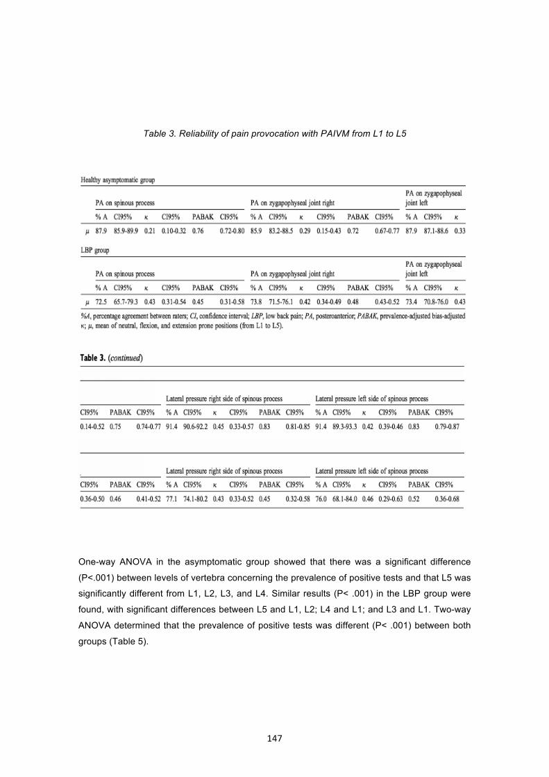
147
Table 3. Reliability of pain provocation with PAIVM from L1 to L5
One-way ANOVA in the asymptomatic group showed that there was a significant difference
(P<.001) between levels of vertebra concerning the prevalence of positive tests and that L5 was
significantly different from L1, L2, L3, and L4. Similar results (P< .001) in the LBP group were
found, with significant differences between L5 and L1, L2; L4 and L1; and L3 and L1. Two-way
ANOVA determined that the prevalence of positive tests was different (P< .001) between both
groups (Table 5).

148
The detailed values of PAIVM, interexaminer agreement, and prevalence of positive tests by
vertebra are respectively presented in Tables 3 and 5.
Table 4. Interpretation of the κ according to the classification of Blum et al41
Table 5: Prevalence of positive responses during pain provocation with passive accessory
intervertebral movement tests

149
Diagnostic accuracy of LBP classification
The sensitivity and specificity of identifying a person with LBP using the proposed CCR were,
respectively, 0.87 and 0.82 for the experienced examiner and 0.67 and 0.85 for the novice. The
combined sensitivity and specificity were, respectively, 0.77 and 0.84.
Discussion
In accordance with previous studies,2,4,5,17,19,22,23 this study found good interexaminer agreement
for active movement tests of the trunk in asymptomatic participants and patients with LBP. The
reliability of identification of the most painful pattern (flexion or extension) was also found to be
at least good, which is comparable with other studies investigating movement classification
systems in people with LBP.5,17,19,20,22,23
In the present study, CM tests were applied in a higher percentage of asymptomatic participants
than in patients with LBP. In our protocol, we decided to stop the progressive strain of spinal
movement at pain onset or pain increase. Most participants with LBP experienced pain during
single, repeated, or sustained active movement tests or with the application of overpressure.
Hence, CM tests were less frequently required in participants with LBP. The reproducibility of
CM testing has been studied by Haswell et al.35 Paradoxically, the results showed poor
agreement between raters, which is in contrast to our results. The reason for this difference
remains unclear and may be related to differences in the frequency that CM tests were
performed or alternatively in differences in examiners training and participants characteristics.
In accordance with a previous study,42 the prevalence during clinical examination of positive
responses to PAIVM tests was higher for the lower lumbar vertebrae than for the upper lumbar
vertebrae in both groups. However, the results of a 2-way ANOVA demonstrated that the
prevalence of positive responses from testing each lumbar vertebra was more frequent in
participants with LBP, probably because of specific underlying pain mechanisms in this
population. The topographical differences in pain responses in both groups could perhaps be
explained by the increased predominance of biomechanical strain and pathoanatomical features
in the lower lumbar area when compared with upper lumbar levels.43,44 Another explanation
might be due to the transition from the mobile lumbar spine to relatively rigid pelvis, placing
physiological stress on the lower lumbar segments, sensitizing those segments. The presence
of positive PAIVM tests in asymptomatic people highlights the importance of identifying more
than 1 vertebral level as symptomatic, as adopted in this study. In addition, this highlights the
importance of using a combination of factors such as the CCR when distinguishing people with
LBP.
150
There are few studies, to our knowledge, that have reported on the relative frequency to which
each vertebral level contributes to LBP. However, based on the clinical observation of vertebral
levels that commonly receive surgery, zygoapophyseal injections, or intervertebral discography,
the lower lumbar spine would appear to be the more common source of symptoms.42 The
precise location of pain origin in the spine of patients with LBP is of major importance to manual
therapists. The L4 and L5 vertebral levels, which are frequently found to have pathology in LBP
people, have also been reported to be the most common vertebral levels to provoke concordant
pain during epiduroscopy.42
The results for interexaminer agreement for tests of PAIVM are at least comparable with or
better than those obtained in previous studies.27,36-38,45 Previous studies reported either weak
to moderate or moderate to good agreement for intraobserver reliability when testing PA
pressures only on the spinous process (“spring test”). Schneider et al27 also reported similar
levels of interexaminer agreement for palpation of the zygoapophyseal joints, although they
grouped levels into upper and lower lumbar levels rather than a specific vertebral level.
Similarly to Phillips and Twomey37 and in terms of manual diagnostic accuracy, the validity
(sensitivity and specificity) of our CCR to identify people with LBP can be rated as good,
especially for the evaluator with better clinical experience. The sensitivity for the novice
examiner was slightly less than that found for the experienced examiner. Therefore, the novice
examiner should be more careful to interpret a negative result (because of a higher chance of
false-negative findings) when testing LBP people with the CCR. The results of this study
suggest that pain provocation using the CCR, when accompanied by a verbal participant
response,37 is an important component of physical examination to distinguish people with LBP
from healthy participants. This information may be important when one considers the poor
correlation between medical imaging and LBP, and the poor correlation between information
gained from magnetic resonance imaging and clinical examination in people who have
LBP.5,42,46
Limitations and futures studies
There is a major limitation of our report. Validity is usually determined by comparing the results
of a new test against a criterion standard. Unfortunately, there is no pathoanatomical criterion
standard in the case of nonspecific LBP.3 Furthermore, in the present study, the patient's own
report of pain was used during pain provocation tests because verbal response during manual
diagnosis has previously been validated in unilevel lumbar spinal block procedures.37 This
approach may constitute an important limitation of internal validity, but because the method is
easily transferable to a clinical context, it provides substantial external application.
Nevertheless, a recent study highlights the interest of epiduroscopy as an external reference to
help diagnose the vertebral level of pain in people with LBP, but this invasive investigation may
be beyond the reach of most manual therapy research.42

151
A further limitation of our study was that we mainly assessed for flexion and extension pain
provocation patterns. Combined movements evaluation typically seek to identify other patterns
of pain provocation (i.e., lateral flexion left and right combined with flexion or extension).
Future studies could investigate the reliability of these patterns. Moreover, owing to the
impossibility of blinding patients to the clinical examination and the consequential potential
Hawthorne effect, patients’ verbal response to pain provocative tests may have been
influenced.
In manual therapy, there are various concepts for the management of LBP including MK,16
Maitland,30,34 Mulligan,47 and various forms of spinal manipulation among others with different
mechanisms of action.26,30,48-52 Even if the principles of treatment vary from one method to
another, the underlying principles of manual therapy are to reduce pain. It is essential that the
treatment, regardless of the concept, is performed on the basis of a reliable and valid clinical
examination protocol aimed to correctly classify LBP. The results of the current study using pain
provocative tests provide confidence that aspects of CCR examination used in this study are
valid and reliable and can therefore be used in clinical practice to direct patient management.
Nevertheless, future studies are needed to confirm the value of this examination protocol.
Future studies should integrate more patients with different LBP disorders (e.g., in acute and
chronic phases) and with more examiners.
Conclusion
This study demonstrated that the use of the CCR (3 positive criteria arising from active and
passive pain provocative tests) was found to have good interexaminer reliability and validity to
identify the most provocative lumbar spine movement direction as well as the lumbar segmental
levels of involvement.

152
References
1. Airaksinen O, Brox JI, Cedraschi C, Hildebrandt J, Klaber-Moffett J, Kovacs F, et al. Chapter 4. European guidelines for the management of chronic nonspecific low back pain. Eur Spine J 2006;15 Suppl 2:S192-300.
2. Delitto A, George SZ, Van Dillen LR, Whitman JM, Sowa G, Shekelle P, et al. Low back pain. J Orthop Sports Phys Ther 2012;42(4):A1-57.
3. Balague F, Mannion AF, Pellise F, Cedraschi C. Non-specific low back pain. Lancet 2012;379(9814):482-91.
4. May S, Littlewood C, Bishop A. Reliability of procedures used in the physical examination of non-specific low back pain: a systematic review. Aust J Physiother 2006;52(2):91-102.
5. Hidalgo B, Gilliaux M, Poncin W, Detrembleur C. Reliability and validity of a kinematic spine model during active trunk movement in healthy subjects and patients with chronic non-specific low back pain. J Rehabil Med 2012;44(9):756-63.
6. Delitto A, Erhard RE, Bowling RW. A treatment-based classification approach to low back syndrome: identifying and staging patients for conservative treatment. Phys Ther 1995;75(6):470-85.
7. van Kleef M, Vanelderen P, Cohen SP, Lataster A, Van Zundert J, Mekhail N. 12. Pain originating from the lumbar facet joints. Pain Pract 2010;10(5):459-69.
8. Kallewaard JW, Terheggen MA, Groen GJ, Sluijter ME, Derby R, Kapural L, et al. 15. Discogenic low back pain. Pain Pract 2010;10(6):560-79.
9. Zusman M. Hands on, hands off? The swings in musculoskeletal physiotherapy practice. Man Ther 2013;18(3):e13.
10. Vanelderen P, Szadek K, Cohen SP, De Witte J, Lataster A, Patijn J, et al. 13. Sacroiliac joint pain. Pain Pract 2010;10(5):470-78.
11. O'Sullivan P. It's time for change with the management of non-specific chronic low back pain. Br J Sports Med 2012;46(4):224-27.
12. Van Dillen LR, Sahrmann SA, Norton BJ, Caldwell CA, Fleming DA, McDonnell MK, et al. Reliability of physical examination items used for classification of patients with low back pain. Phys Ther 1998;78(9):979-88.
13. Maher C, Adams R. Reliability of pain and stiffness assessments in clinical manual lumbar spine examination. Phys Ther 1994;74(9):801-809.
14. Harris-Hayes M, Van Dillen LR. The inter-tester reliability of physical therapists classifying low back pain problems based on the movement system impairment classification system. PM R 2009;1(2):117-26.
15. Riddle DL. Classification and low back pain: a review of the literature and critical analysis of selected systems. Phys Ther 1998;78(7):708-37.

153
16. McKenzie R, May S. The lumbar spine – mechanical diagnosis and therapy. Spinal Publications, New Zealand Ltd 2003(2nd):732.
17. Kilpikoski S, Airaksinen O, Kankaanpaa M, Leminen P, Videman T, Alen M. Interexaminer reliability of low back pain assessment using the McKenzie method. Spine (Phila Pa 1976) 2002;27(8):E207-14.
18. Clare HA, Adams R, Maher CG. A systematic review of efficacy of McKenzie therapy for spinal pain. Aust J Physiother 2004;50(4):209-16.
19. Fritz JM, Delitto A, Vignovic M, Busse RG. Interrater reliability of judgments of the centralization phenomenon and status change during movement testing in patients with low back pain. Arch Phys Med Rehabil 2000;81(1):57-61.
20. Fritz JM, George S. The use of a classification approach to identify subgroups of patients with acute low back pain. Interrater reliability and short-term treatment outcomes. Spine (Phila Pa 1976) 2000;25(1):106-14.
21. Van Dillen LR, Sahrmann SA, Norton BJ, Caldwell CA, McDonnell MK, Bloom NJ. Movement system impairment-based categories for low back pain: stage 1 validation. J Orthop Sports Phys Ther 2003;33(3):126-42.
22. Dankaerts W, O'Sullivan P. The validity of O'Sullivan's classification system (CS) for a sub-group of NS-CLBP with motor control impairment (MCI): overview of a series of studies and review of the literature. Man Ther 2011;16(1):9-14.
23. Dankaerts W, O'Sullivan PB, Straker LM, Burnett AF, Skouen JS. The inter-examiner reliability of a classification method for non-specific chronic low back pain patients with motor control impairment. Man Ther 2006;11(1):28-39.
24. O'Sullivan P. Diagnosis and classification of chronic low back pain disorders: maladaptive movement and motor control impairments as underlying mechanism. Man Ther 2005;10(4):242-55.
25. El Avd. Orthopaedic manual therapy diagnosis : spine and temporomandibular joints. 1st English ed. ed. Sudbury, Mass. ; London: Jones and Bartlett; 2010.
26. Hidalgo B, Detrembleur C, Hall T, Mahaudens P, Nielens H. The efficacy of manual therapy and exercise for different stages of non-specific low back pain: an update of systematic reviews. J Man Manip Ther 2014;22(2):59-74.
27. Schneider M, Erhard R, Brach J, Tellin W, Imbarlina F, Delitto A. Spinal palpation for lumbar segmental mobility and pain provocation: an interexaminer reliability study. J Manipulative Physiol Ther 2008;31(6):465-73.
28. Dankaerts W, O'Sullivan P, Burnett A, Straker L, Davey P, Gupta R. Discriminating healthy controls and two clinical subgroups of nonspecific chronic low back pain patients using trunk muscle activation and lumbosacral kinematics of postures and movements: a statistical classification model. Spine (Phila Pa 1976) 2009;34(15):1610-18.
29. Edwards BC. Manual of combined movements : their use in the examination and treatment of musculoskeletal vertebral column disorders. 2nd ed. ed. Oxford: Butterworth-Heinemann; 1999.
154
30. Maitland J. Spinal manipulation made simple : a manual of soft tissue techniques. Berkeley, Calif.: North Atlantic Books; 2000.
31. McCarthy C. Combined movement theory : rational mobilization and manipulation of the vertebral column. Edinburgh: Churchill Livingstone; 2010.
32. Rubinstein SM, van Tulder M. A best-evidence review of diagnostic procedures for neck and low-back pain. Best Pract Res Clin Rheumatol 2008;22(3):471-82.
33. Petty NJ. Neuromusculoskeletal examination and assessment : a handbook for therapists. 3rd ed. ed. Edinburgh: Elsevier Churchill Livingstone; 2006.
34. Hengeveld E, Banks K, Maitland GD. Maitland's vertebral manipulation. 7th ed. / edited by Elly Hengeveld, Kevin Banks. ed. Oxford: Elsevier Butterworth-Heinemann; 2005.
35. Haswell K, Williams M, Hing M. Interexaminer reliability of symptom-provoking active side-bend, rotation and combined movement assessments of patients with low back pain. J Manual Manipulative Ther 2004;12(1):11-20.
36. Horneij E, Hemborg B, Johnsson B, Ekdahl C. Clinical tests on impairment level related to low back pain: a study of test reliability. J Rehabil Med 2002;34(4):176-82.
37. Phillips DR, Twomey LT. A comparison of manual diagnosis with a diagnosis established by a uni-level lumbar spinal block procedure. Man Ther 1996;1(2):82-87.
38. Abbott JH, Flynn TW, Fritz JM, Hing WA, Reid D, Whitman JM. Manual physical assessment of spinal segmental motion: intent and validity. Man Ther 2009;14(1):36-44.
39. Sim J, Wright CC. The kappa statistic in reliability studies: use, interpretation, and sample size requirements. Phys Ther 2005;85(3):257-68.
40. Chen G, Faris P, Hemmelgarn B, Walker RL, Quan H. Measuring agreement of administrative data with chart data using prevalence unadjusted and adjusted kappa. BMC Med Res Methodol 2009;9:5.
41. Blum A, Feldmann L, Bresler F, Jouanny P, Briancon S, Regent D. Value of calculation of the kappa coefficient in the evaluation of an imaging method. J Radiol 1995;76(7):441-3.
42. Bosscher HA, Heavner JE. Diagnosis of the vertebral level from which low back or leg pain originates. A comparison of clinical evaluation, MRI and epiduroscopy. Pain Pract 2012;12(7):506-12.
43. Kalichman L, Li L, Kim DH, Guermazi A, Berkin V, O'Donnell CJ, et al. Facet joint osteoarthritis and low back pain in the community-based population. Spine (Phila Pa 1976) 2008;33(23):2560-5.
44. Maus TP, Aprill CN. Lumbar diskogenic pain, provocation diskography, and imaging correlates. Radiol Clin North Am 2012;50(4):681-704.
45. Hicks GE, Fritz JM, Delitto A, Mishock J. Interrater reliability of clinical examination measures for identification of lumbar segmental instability. Arch Phys Med Rehabil 2003;84(12):1858-64.

155
46. McCullough BJ, Johnson GR, Martin BI, Jarvik JG. Lumbar MR imaging and reporting epidemiology: do epidemiologic data in reports affect clinical management? Radiology 2012;262(3):941-6.
47. Exelby L. The Mulligan concept: its application in the management of spinal conditions. Man Ther 2002;7(2):64-70.
48. Maigne JY, Vautravers P. Mechanism of action of spinal manipulative therapy. Joint Bone Spine 2003;70(5):336-41.
49. Licciardone JC, Brimhall AK, King LN. Osteopathic manipulative treatment for low back pain: a systematic review and meta-analysis of randomized controlled trials. BMC Musculoskelet Disord 2005;6:43.
50. Soulvis T, Vincenzino B, Wright A. Neurophysiological effects of spinal manual therapy. Modern manual therapy, the vertebral column. 3rd ed. / edited by Jeffrey D. Boyling, Gwendolen A. Jull ; foreword by Lance T. Twomey. ed. Edinburgh: Churchill Livingstone; 2004:367-379.
51. Bronfort G. Spinal manipulation: current state of research and its indications. Neurol Clin 1999;17(1):91-111.
52. Harvey E, Burton AK, Moffett JK, Breen A, team UBt. Spinal manipulation for low-back pain: a treatment package agreed to by the UK chiropractic, osteopathy and physiotherapy professional associations. Man Ther 2003;8(1):46-51.

156

157
CHAPTER VI
Short-term effects of Mulligan mobilisation with movement on pain, disability and kinematic spinal movements in patients with nonspecific low back pain: A randomised
placebo controlled trial
Benjamin Hidalgo, Laurent Pitance, Toby Hall, Christine Detrembleur, Henri Nielens
Accepted for publication in J Manipulative Physiol Ther 2015
ABSTRACT
Objective: To determine the efficacy of lumbar Mulligan sustained natural apophyseal glides
(SNAGs) in patients with nonspecific low back pain (LBP) with respect to two new kinematic
algorithms (KA) for range of motion (KA-R) and speed (KA-S) as well as pain, functional
disability, and kinesiophobia.
Methods: This was a randomized placebo controlled trial with two arms in accordance with
CONSORT-guidelines. 87 subjects with nonspecific LBP were assessed and 32 fulfilled criteria
for the application of lumbar SNAGs. Subjects, blinded to allocation, were randomized to 2
groups; real-SNAG (n = 16) and sham-SNAG (n = 16). All patients were treated during a single-
session of real/sham SNAG (3 X 6 repetitions) to the lumbar spine in a sitting position in a
flexion direction. Two new KA from a validated kinematic spine model were used and recorded
with an optoelectronic device. Pain at rest and during flexion, as well as functional disability and
kinesiophobia were recorded by self-reported measures. These outcomes were blindly
evaluated before, after treatment, and at 2-week follow-up in both groups.
Results: 4 of 6 variables demonstrated significant improvement with moderate to large effect-
sizes (ES) in favor of the Real-SNAG group: KA-R (p=.014; between groups ES Cliff’s delta=-
.52), pain at rest and during flexion (VAS: p<.001; ES=-.73/-.75), functional-disability (Oswestry
Disability Index: p=.003 and ES=-.61). Kinesiophobia was not considered to be significant
(Tampa scale: p=.03) but presented moderate effect size ES=-.46. KA-S was not significantly
different between groups (p=.12) with a small ES of -.33. All the 6 outcome measures were

158
significantly different (p≤.008) during within group analysis (before and after treatment) only in
the Real-SNAG group. No serious or moderate adverse events were reported.
Conclusion: This study provides evidence that lumbar spine‘s SNAGs have a short-term
favorable effect on KA-R, pain and function in a targeted group of patients with nonspecific LBP.
Further studies are required to validate these findings and to further investigate kinesiophobia
and KA-S, as well as the long-term effects of SNAGs for LBP.
159
Introduction
Low back pain (LBP) is one of the most common musculoskeletal disorders for which patients
consult medical care.1 It is also the most important cause of disability and absenteeism with
increasing prevalence leading to a major socio-economic impact on society.2-4 These facts
highlight the importance of finding effective and validated treatments for this disabling condition.
Two broad categories of LBP are recognised. When a specific patho-anatomical origin is
identified such as a tumor or fracture, LBP is labelled as specific and requires appropriate
medical care such as specific medication or surgery. On the other hand and more commonly, in
up to 90% of cases no precise specific origin for pain can be identified; such LBP is
consequently described as nonspecific.1
LBP is managed by a variety of treatment modalities5,6 including Orthopaedic Manual therapy
(OMT). This form of treatment has been recommended in national guidelines, for example in the
United States,7 and is also frequently used in clinical practice in various countries.8,9 As
demonstrated by recent systematic reviews, OMT management combined with usual medical
care provides better results as compared to usual medical care alone for all stages (acute/sub
acute or chronic) of LBP.7,10
A novel growing concept in the field of OMT and clinical practice, which remains sparsely
studied in the literature, is "Mobilisation With Movement" (MWM),11-13 originally developed by
Mulligan.11 The main indication for MWM is movement impairment due to pain and/or stiffness.
The therapeutic goal is to rapidly reduce pain and to increase range of motion (ROM). The
principle of this treatment is simple, in that the manual therapist performs a sustained passive
segmental glide of the involved joint, while the patient actively moves in the impaired
direction.11-13 Mulligan11,12 purported a biomechanical basis for the efficacy of MWM in reducing
pain and improving ROM, but there may be other explanations for their effects including
neurophysiological mechanisms. Mulligan MWM techniques can be applied to both peripheral
and spinal joints. When applied to the spine, MWM are called Sustained Natural Apophyseal
Glides (SNAGs).11-13 The current study focuses on SNAGs and their effects on the lumbar
spine.
It has been reported that many physical therapists in the UK manage their patients with LBP by
using SNAGs as a part of their physical intervention.14 This is despite the poor level of evidence,
through lack of clinical studies, for the efficacy of lumbar SNAGs for LBP.15 Indeed, only three
studies reported on the effects of lumbar SNAGs,16-18 with only 2 investigating the
biomechanical effects.16,17 The first, a placebo controlled trial16 was carried out on 49
asymptomatic subjects. SNAGs were applied during flexion in sitting at 2 lumbar levels by an
experienced examiner in a single session and failed to demonstrate an increase in lumbar ROM

160
measured by a 3-dimensional electro goniometer. In contrast, the second placebo controlled
trial17 investigated 26 people with LBP during flexion and who were suitable for SNAGs,
recording ROM using double inclinometry. A single session of SNAGs demonstrated a
significant increase of 7˚ lumbar flexion ROM greater than placebo, but no change in pain
scores. Obviously, in view of the paucity of literature regarding lumbar SNAGs, and in
comparison with its widespread clinical use, further investigations are necessary to study lumbar
SNAGs’ efficacy, as well as indications when used for people with LBP.
Recent studies from our research team3,19,20 have investigated spine kinematics in people with
LBP using an optoelectronic measurement system. A kinematic spine model was developed
where the shoulder girdle and spine were divided into 6 segments: shoulder girdle, upper
thoracic and lower thoracic spine, upper and lower lumbar spine, and the last segment
comprising the whole lumbar spine (combining the upper and lower lumbar spine segments).
Each segment was considered to be rigid and homogenous. Kinematic variables speed and
ROM were evaluated during movement in all planes. ROM and speed variables showed a
highly significant difference (p < 0.001) between healthy subjects and those with chronic non-
specific LBP in all spinal segments during flexion and combined movements. These studies
provided evidence for the validity of the kinematic spine model in distinguishing people with
LBP.
From our previous studies, two new kinematic algorithms ROM (KA-R) and speed (KA-S) were
identified as having a potential interest in future clinical studies addressing the effectiveness of
OMT interventions applied to the spine in ways other than simply looking at the effects on pain
and disability.19,20 Moreover, it has been proposed that future clinical studies should target their
interventions on a more homogeneous subgroup of patients with LBP to improve clinical
outcomes, as well as effect sizes for outcome measures.10
Based on these findings, we used the kinematic spine model to assess whether lumbar SNAGs
were able to improve the kinematic features of trunk movement in a targeted group of subjects
with LBP. The main goal of this clinical study was to compare the immediate and short-term
effects of a single session of SNAG to a sham SNAG (placebo) treatment applied to the lumbar
region, on 2 primary outcome measures, kinematics (KA-R and KA-S), and 3 secondary
outcome measures; pain, function, and kinesiophobia in a subgroup of people with LBP. The
hypothesis were that outcomes would be more favorable in the real SNAG intervention for
primary and secondary outcome measures during between groups analyses, with additional
improvements expected within each group over time.

161
Method
Design
This study was a single-center (Cliniques Universitaires Saint-Luc, Brussels, Belgium),
prospective, randomised and placebo-controlled trial with two arms and with blinded patients
and evaluator. The design of this clinical trial followed the recommendations of the CONSORT
statement.21 The study was approved by the local ethic committee board of the University of
Louvain (UCL) and was registered in ClinicalTrials.gov : NCT02128607.
Subjects
Eighty-seven people with LBP were initially recruited from “Cliniques Universitaires Saint-Luc”.
Of these, thirty-two were included in the study based on specific criteria. Stratification based on
pain mechanisms has been previously recommended.22 These criteria were combined with
indications for the application of lumbar SNAGs.11,12 The inclusion criteria were subjects aged
between 20 and 55 years, who complained of LBP mostly provoked by trunk flexion at any
stage (acute to chronic), which did not radiate lower than the knee. The subjects were selected
where lumbar flexion was the most provocative movement using a standardized physical
examination method aimed to identify lumbar pain provocative movement patterns using active
and passive accessory movement tests.23 Finally, the pain associated with trunk flexion had to
be reduced by the application of a central lumbar SNAG applied through the spinous process.
Patients were excluded if they presented with any known contraindication to OMT (e.g. tumor,
fracture, osteoporosis, infection, rheumatic diseases, or herniated disk).
Thirty-two people with LBP were included in the trial and were randomly distributed in two arms:
one group receiving the lumbar SNAG treatment (n = 16) and the other receiving a sham lumbar
SNAG (n = 16). Randomization was performed by stratified randomization with blocks of
random numbers under sealed opaque envelopes previously prepared, in a fashion of 4
subsets, each subset containing 8 envelopes, aimed to balance the stages of LBP for each
group during the process of the study.
Material and outcome measures
The outcome measures were trunk ROM and speed, as well as pain at rest and during trunk
flexion just before and just after a single session of treatment. The impact of the intervention at
very short-term (2 weeks) on functional disability and kinesiophobia was also evaluated. Six
variables were assessed before (T0) and after treatment (T1) (Figure 1). The same examiner
blindly assessed all the following outcome measures.

162
Figure 1. Flow chart of the study process

163
Kinematic measures
Kinematic variables were the primary outcome measures and were evaluated using an
optoelectronic device (Elite-BTS) composed of eight infrared cameras capable of recording the
3D-positions of 9 reflective markers placed on bony landmarks on the trunk according to a
validated kinematic spine model,19,20 at a frequency of 200 Hz and accuracy of 0.1 mm. This
model (Figure 2) sub-divides the shoulder girdle, spine and pelvic girdle into various segments.
The test procedure and recording conditions have been described previously.19,20 Briefly, trunk
movements were assessed in a sitting position; trunk flexion, left and right rotation, and
combined movement of trunk flexion associated with left and right rotation of the pelvis (Figure
3). Each trunk movement was performed and recorded 10 times.
Figure 2. The kinematic model of the spine
(A): Position of the patient and the nine reflective markers, (B): kinematic spine model and segments: pelvic girdle (ASIS L-S2-ASIS R); low lumbar spine (S2-L3); high lumbar spine (L3-T12); total lumbar spine (S2-T12); low thoracic spine (T12-T7); high thoracic spine (T7-C7); shoulder girdle (Ac L-C7-Ac R)

164
Figure 3. Trunk motion tasks
(A) starting position and trunk flexion, (B) starting position and Left / Right rotation, (C) starting position and combined movement of trunk flexion with pelvic rotation to the left / right.
A binary logistic regression analysis had previously determined segments and trunk movements
of the kinematic spine model that were the most discriminant for LBP.19,20 The final results were
two kinematic algorithms, one for ROM (KA-R) and one for speed (KA-S) according to the
following equations (see19,20 for more information):
KA-R = 17.77 – (0.074 × LTS°) – (0.11 × SS°) – (0.059 × TLS°)
KA-S = 6.19 – (0.063 × TLS°/s)
Where KA-R, kinematic algorithm for ROM; LTS°, lower thoracic spine ROM in flexion; SS°,
shoulder segment ROM in right rotation; TLS°, total lumbar spine ROM in flexion with left
rotation; KA-S, kinematic algorithm for speed; TLS°/s, total lumbar spine speed in flexion with
right rotation.
Self-reported measures
Self-reported measures were the secondary outcome measures. Pain at rest (present pain), as
well as pain during trunk forward bending from a standing position was recorded using a 10 cm
visual analogue scale (VAS) just before and just after the intervention. Functional disability was

165
assessed with the use of the Oswestry Disability Index (ODI) before intervention and 2 weeks
after. The score was expressed in percentage terms (%). Kinesiophobia was assessed with the
Tampa scale.
Intervention
First through a standardized clinical examination incorporating combined movements
evaluation,23 the examiner determined if the patient had greater pain during active trunk flexion
than extension, as well as the most painful vertebral level (with passive accessory intervertebral
movements). This combined movements examination procedure has previously been described
and validated to identify the most painful pattern of trunk movement, as well as the lumbar
segmental level(s) involved.23
Secondly, the evaluator determined whether the patient responded positively to a seated lumbar
SNAG applied through the spinous process of the involved vertebra.13 To do this, the examiner
had four attempts to increase ROM and reduce pain by at least 2/10 on the VAS. As
recommended,13 the evaluator applied the SNAG on the spinous process of the vertebra that
was the most painful during combined movements examination. Glide force was applied parallel
to the apophyseal articular surface (cranial direction). If the effect obtained was not sufficient,
the examiner was allowed to vary the intensity and/or direction (vector of applied force) of the
SNAG. In addition, the evaluator could change the central vertebral level of lumbar SNAG
application.13 If, after four trials, the SNAG application did not provide the desired effect, the
patient was excluded from the study (Figure 1).
In both groups, during the treatment, the patient was placed in a standardized seated position
(hips and knees in 90˚ flexion) on a table with feet supported, stabilized with a belt around the
waist (Figure 4).11,16 Three sets of six repetitions were performed in the Real-SNAG and sham
(placebo) intervention. A single inexperienced physiotherapist (novice in the use of SNAGs)
applied the treatment procedure in both groups and was therefore not blind to the patient’s
group allocation. Both the treating therapist and evaluator were trained for 16 hours to ensure
correct application of the study protocol by two experienced manual therapists.
In the Real-SNAG group, the therapist followed published guidelines for SNAG application.11-13
The therapist applied a gliding force with the hypothenar eminence placed on the spinous
process of appropriate lumbar vertebral level while the patient performed the limited trunk
flexion movement until onset of pain before returning to the starting position (Figure 4). The
cranial glide force was maintained throughout all the movement in both direction (forward

166
bending and back from bending) and with each repetition. Communication was maintained with
the patient to ensure that no pain was felt during the treatment.
In the Placebo group, the Sham-SNAG intervention replicated the same procedure used in a
previous study.16 The technique mimicked the Real-SNAG, only with two differences: the
therapist placed his hypothenar eminence on the spinous process of the above vertebral level
and applied minimal glide force in a caudal direction.
Figure 4. SNAGs application from a sitting position
Standardised position of the patient and therapist showing the belt placement during the application of the real and sham lumbar SNAG
Statistical analysis
Statistical analysis was carried out using SigmaStat 3.5. Estimation of the required sample size
was calculated on the basis of the Minimal Detectable Change 95% (MDC95) of the primary
outcome measure (KA-R and KA-S)20 with a desired power of 0.80 and an alpha level of 0.05;
we obtained an estimation of the required sample size for each group to be 16 patients.
Similarity of baseline measures between groups (T0) was assessed using a Student T-test. Our
main hypothesis was the comparison between the groups for primary kinematic outcome

167
measures and for self-reported outcome measures. We used Mann-Whitney Rank Sum Test on
the means of difference (T0-T1) of the Sham and Real group for statistical evaluation as the
majority of the variables failed to demonstrate a normal distribution. We performed a specific
alpha correction for inflated type-1 error with null hypothesis rejection using a Bonferroni
correction. For primary outcome kinematic measures (KA-R and KA-S), this correction was
0.05/2, indicating p<0.025 was the required level for significance. For the secondary self-
reported measures (VAS rest, VAS flexion, ODI, TAMPA) the correction was O.05/4, indicating
p<0.0125 was the required level for significance. The clinical effect size for between groups
analysis was evaluated with a non–parametric effect size, Cliff’s delta.24 This score can range
from -1 to 1; where 1 indicates all observations from the sham-SNAG group are greater than all
observations from the real-SNAG group. Conversely, -1 indicates that all observations from the
sham-SNAG group are less than all observations from the real-SNAG group. Finally, 0 indicates
perfect overlap, with equality of observations between the groups.24 Cliff’s delta is calculated
with R software and are presented with a confidence interval (CI) of 95% and categorized in
small, moderate, large, and very large effect sizes.24 We also ran an exploratory analysis for the
secondary within-group hypothesis (between baseline and final evaluation) in the sham and
real-SNAG group following the same statistical method described above but with a Wilcoxon
Signed Rank Test.
Results
The number of patients included and excluded, as well as the reasons of exclusion during the
process of the study is reported in Figure 1. Anthropometric data and variable outcomes at
baseline of included patients are described in Table 1. The period of participants enrollment was
from February 2014 until June 2014, the end of follow-up was July 2014. The trial was ended in
July 2014 because the required sample size was reached.

168
Table 1. Anthropometric data and outcome variables at baseline
BMI: body mass index; KA-R: kinematic algorithm for range of motion; KA-S: kinematic algorithm for speed; T0: baseline; VAS: visual analogue 10 cm scale; ODI: Oswestry disability index; NS: non-
significant difference between groups with Student T-test.
Subjects with non-specific LBP included in this study had a mixed pain history: 63% were
chronic, 21% acute, and 16% sub acute. No significant differences on outcome measures were
present at baseline between groups (Table 1). No serious or moderate adverse events were
reported in either group during the study.
The graph of speed curves (°/s) of the lower lumbar spine segment during trunk forward
bending in one typical acute LBP patient and one typical chronic LBP patient from the Real-
SNAG (Figure 5A) and Sham-SNAG (Figure 5B) group is presented in figure 5.
Male / Female
Age (y years
BMI (kg/m2)
LBP duration (months)
KA-R (T0)
KA-S (T0)
VAS at rest (present pain) T0
VAS flexion (pain in trunk flexion)
T0
ODI (T0)
Tampa scale (T0)
Sham-SNAG (n=16)
7/9
Means (SD)
40.7 (10.2) NS
25.1 (3.3) NS
19.7 (19.4) NS
3.5 (2.9) NS
2.1 (1.4) NS
2.5 (1.7) NS
5.1 (1.6) NS
22.9 (10.7) NS
42.1 (6.2) NS
Real-SNAG (n=16)
9/7
Means (SD)
37.8 (9.7)
24.1 (2.6)
21.0 (21.2)
4.9 (3.2)
2.4 (1.9)
3.0 (1.8)
5.6 (1.8)
22.4 (12.2)
43.7 (6.3)

169
Figure 5. Typical speed curves before and after intervention
A: Speed curves of the lower lumbar spine segment (S2-L3) during trunk forward bending before (baseline) and after (final evaluation) Real-SNAG for one typical (best responder) acute LBP patient (red curve) and one typical chronic LBP patient.
B: Speed curves of the lower lumbar spine segment (S2-L3) during trunk forward bending before (baseline) and after
(final evaluation) Sham-SNAG for one typical (best non-responder) acute LBP patient (red curve) and one typical
chronic LBP patient.

170
Between groups comparison
Primary kinematic outcome measures
KA-R demonstrated a significant difference (p<.025) in favor of the Real-SNAG group with large
clinical effect size (p=.014 and ES=-.52). In contrast, KA-S demonstrated no significant
difference (p>.025) with only small clinical effect size (p=.118 and ES=-.33).
Secondary self-reported outcome measures
Pain (VAS) at rest and during lumbar spine flexion demonstrated a significant difference
(p<.0125) in favor of the Real-SNAG group with large clinical effect size (p=.001 and ES=-.73;-
.75). Functional disability (ODI) also demonstrated a significant difference (p<.0125) in favor of
the Real-SNAG group with large clinical effect size (p=.003 and ES=-.61). In contrast, there was
no significant difference between groups for Kinesiophobia (Tampa scale) (p>.0125), with only a
moderate clinical effect size favoring the real-SNAG group (p=.03 and ES=-.46).
Table 2: Between groups analysis on primary kinematic and secondary self-reported outcome
measures
KA-R: kinematic algorithm for range of motion. KA-S: kinematic algorithm for speed. VAS rest: visual analogue scale (pain) at rest. VAS flexion: visual analogue scale (pain) during trunk flexion. ODI: Oswestry Disability Index (functional disability). TAMPA: TAMPA scale for kinesiophobia. * Significant difference between groups, corrected level of p<0.025 for primary kinematic outcome measures (KA-R and KA-S). # Significant difference between groups, corrected level of p<0.0125 for secondary self-reported outcome measures.

171
Within group comparison (secondary explanatory hypothesis)
Primary kinematic outcome measures
KA-R and KA-S before and after the intervention improved significantly in the Real-SNAG group
(respectively: p=.001; p=.008) but not in the Sham-SNAG group (respectively: p=.86; p=.63).
Secondary self-reported outcome measures
There were significant improvements in the real-SNAG group for all secondary outcome
measures following the intervention. Pain (VAS) at rest and during lumbar spine flexion before
and after intervention improved significantly in the Real-SNAG group (p<.001) but not in the
Sham-SNAG group (respectively p=.56; p=.15). Functional disability (ODI) before and 2-weeks
after the intervention improved significantly in the Real-SNAG group (p=.002) but not in the
Sham-SNAG group (p=.84). Kinesiophobia (Tampa scale) before and 2-weeks after the
intervention improved significantly in the Real-SNAG group (p=.004) but not in the Sham-SNAG
group (p=.23).
Table 3: Within group analysis before and after treatment
KA-R: kinematic algorithm for range of motion. KA-S: kinematic algorithm for speed. VAS rest: visual analogue scale (pain) at rest. VAS flexion: visual analogue scale (pain) during trunk flexion. ODI: Oswestry Disability Index (functional disability). TAMPA: TAMPA scale for kinesiophobia. * Significant difference between baseline and final evaluation within groups, corrected level of p<0.025 for primary kinematic outcome measures (KA-R and KA-S). # Significant between baseline and final evaluation, corrected level of p<0.0125 for secondary self-reported outcome measures.

172
Discussion
Our results suggest substantial improvements favoring lumbar SNAG’s as compared to placebo
for KA-R, pain at rest and during trunk flexion, as well as for functional disability. In contrast, KA-
S and kinesiophobia showed no significant difference between groups. Despite this, within
group explanatory analysis demonstrated highly significant differences in all outcome measures
before and after intervention only in the real-SNAG group.
It may be hypothesised that a larger sample size may have resulted in significant differences
between groups for KA-S also. However, our prospective calculation of sample size provided an
estimate of 16 patients within each group for kinematic outcome measures. With such a small
sample, the statistical effect of possible atypical responses is greater. Indeed, almost all patients
from our sample (80%) improved their speed (KA-S) in both groups after the intervention.
However, a small percentage (20%) of subjects demonstrated the opposite response, and
decreased speed during trunk-movements after lumbar real and sham SNAG therapy.
Moreover, the mix of different stages of LBP included in our sample, from acute to chronic, is
another factor that may explain the observed non significant between groups effect on KA-S.
When comparing the current results to previous studies reporting on the effects of Mulligan
techniques, most of them have investigated the effects of MWM on peripheral joints25-30 or on
the cervical spine.31-33 However, there are few published reports investigating effects with
respect to the lumbar spine. Indeed, only two studies have addressed the effects of lumbar
SNAGs on ROM and pain. The first study,16 a placebo controlled trial, showed no-significant
improvement in active trunk flexion ROM after lumbar SNAGs in asymptomatic people.
However, it is problematic to compare those results in asymptomatic people (where the SNAG
technique could not be applied according to the technique guidelines11-13) with the present study
on people with LBP. The second study,17 investigated patients with LBP and showed a
significant increase in trunk flexion ROM, but no significant reduction in pain after the application
of lumbar SNAGs. Our results show that lumbar SNAGs reduced pain at both rest and during
active trunk flexion, and also increased trunk ROM. The effectiveness of SNAGs was not limited
to just pain reduction and improved ROM, but also to improved functional disability and
kinesiophobia in people with LBP. However, long-term effects were not analyzed in this study. It
would be interesting to analyze long-term effects of lumbar SNAGs in future studies.
In the study of Konstantinou et al.,17 the placebo was a passive modality (patient lying on the
table). The authors made this choice in order to avoid the influence of an active placebo on the
quality of trunk movement probably because repeated active spinal movements could be
considered as a self-treatment for LBP.34,35 However, the authors could not distinguish the

173
possibility that patients in the SNAG group have improved only through repetition of movements
rather than SNAG technique application. Moreover, an active placebo as in our study mimicked
as closely as possible a real treatment. In our protocol, the same total number of active trunk
movements was performed in both groups, in order to distinguish the effect of real and sham
SNAGs from the simple effect of repeated active trunk movements.
The exact mechanism of potential action for lumbar SNAGs is not known as no studies have yet
investigated this. However, there are proposed biomechanical and neurophysiological
mechanisms. Biomechanically, there are some similarities between postero-anterior
mobilisation (PA) undertaken in prone lying and a SNAG. Lee and Evans36 reported that a PA
on the L5 spinous process induced anterior translation of the L5 vertebra and flexion at the L5-
S1 segment. The biomechanical effects of a lumbar SNAG may be enhanced by the cranial
direction of the glide along the facet joint plane, together with the active trunk movement.
Another proposed mechanism of action may be through correction of a positional fault. Mulligan
hypothesized that lack of normal facet gliding in flexion may distort the disc11,12 and provoke
pain. Hence improving facet gliding may normalize forces on the disc, relieving pain.
Zusman37,38 has described a rationale for the pain relief provided by manual therapy based on
the theory of extinction and habituation. Pain may be considered as a form of aversive memory
that once present could be more and more easily recalled. Behaviorally, a conditioned fear
response may be reduced in intensity through extinction, a form of learning characterized by a
decrease in a conditioned response when the conditioned stimulus that elicits it is repeatedly
non-reinforced37,38 such as might occur during SNAGs. In our sample of people with LBP, trunk
flexion was the most painful movement. The real SNAG intervention provided exposure to the
painful movement in the absence of any overt danger, which is fundamental to interventions
used in the extinction of aversive memories,37,38 but this was not the case for the sham SNAG
intervention. Progressive mobilization may also desensitize the nervous system through
habituation. The mechanism involves a progressive decline in the ability of the presynaptic
nerve terminal to transmit impulses. In the subjects from this study, non-noxious sensory input
from the repeated real lumbar SNAG may have competed with and replaced pain sensitization,
returning the nervous system to a normal state.38
There may be various mechanisms of action for lumbar SNAGs at different stages of LBP. In
our sample there was a mix of stages, with the majority being chronic in nature. As we have
discussed, SNAGs may have neurophysiological as well as mechanical effects, which may have
implications for acute and chronic LBP. However, it is beyond the scope of this study to identify
the mechanisms underscoring the positive changes seen from SNAGs.
These proposed mechanisms of action described here might explain the significant difference
observed on outcome measures in favor of real SNAG interventions. However, for

174
Kinesiophobia there was no significant difference for between groups analysis after Bonferroni
correction despite significant improvement in the real-SNAG group for within group analysis.
This might be explained by the nature of the sham intervention in which the subjects were still
exposed to the painful stimulus during trunk flexion, and thus, may maintain a conditioned fear
response. Moreover, another often-debated issue is the quality of the placebo procedure used
in physical therapy trials because that might explain the results in favor of the real intervention.
Placebo in manual therapy and in non-pharmacological trials is still a very complex issue to
address since a good quality placebo needs to mimic as closely as possible the real intervention
without its specific effect with patients still believing that they have received the real treatment.10
Limitations and futures studies
There are several potential limitations to this study’s findings. One is the limited clinical
experience of the treating therapist in the use of SNAGs that may have influenced the
effectiveness of the intervention.39,40 However, SNAGs are simple techniques that require
minimal training, so this is not believed to be a substantial factor in the outcome of the
technique. Moreover, some caution is required when interpreting the outcome measures in
favor of the real SNAG group, as the 95% CI covers a wide range of possibilities in terms of
effect size. Finally, a potential bias could be present during the initial selection of patients as
they were required to respond positively to the SNAG application, before inclusion and
randomization to one of the groups. This procedure may have the potential to subconsciously
inform the patients of the real SNAG effects during the selection. However, this procedure is
consistent with the widespread recommendations of stratification of care for LBP patients,10,22
as well as the integration of the clinical reasoning in manual therapy trials.10,15,22,37,39
To corroborate the positive changes of lumbar SNAGs seen in this study, future studies should
further investigate the effects on speed of trunk-movements and kinesiophobia, long-term
efficacy, and possible mechanisms of action. Moreover, correlations between primary kinematic
outcomes measures and secondary clinical outcome measures should be investigated. Finally,
more studies are required to identify potential responders to validate the clinical application of
this form of manual therapy.
Conclusion
This is the first randomised placebo controlled trial that has investigated the short-term effects of
lumbar SNAGs on two new kinematic algorithms of trunk movements (KA-R and KA-S), as well
as pain, functional disability and kinesiophobia in patients with non-specific LBP. While the
results show a significant improvement in KA-R, pain, and functional disability in favor of lumbar
SNAGs, some caution is required when interpreting these data, as the 95% CI covers a wide
range of possibilities in terms of clinical effect size. Hence this study provides preliminary

175
evidence that lumbar SNAGs have immediate and short-term efficacy in the treatment of a
targeted group of patients with nonspecific LBP.

176
References
1. Manek NJ, MacGregor AJ. Epidemiology of back disorders: prevalence, risk factors, and prognosis. Curr Opin Rheumatol 2005;17:134-40.
2. Hoy D, Brooks P, Blyth F, Buchbinder R. The Epidemiology of low back pain. Best Pract
Res Clin Rheumatol 2010;24:769-81. 3. Hidalgo B, Gobert F, Bragard D, Detrembleur C. Effects of proprioceptive disruption on
lumbar spine repositioning error in a trunk forward bending task. J Back Musculoskelet Rehabil 2013;26:381-7.
4. Hoy D, Bain C, Williams G, March L, Brooks P, Blyth F, et al. A systematic review of the
global prevalence of low back pain. Arthritis Rheum 2012;64:2028–37. 5. Koes BW, van Tulder MW, Thomas S. Diagnosis and treatment of low back pain. BMJ
2006;332:1430-4. 6. Krismer M, van Tulder M. Low back pain (non-specific). Best Pract Res Clin Rheumatol
2007;21:77-91. 7. Delitto A, George SZ, Van Dillen L, Whitman JM, Sowa G, Shekelle P, et al. Low back
pain. J Orthop Sports Phys Ther 2012;42:A1-A57. 8. Li LC, Bombardier C. Physical Therapy Management of Low Back Pain: An Exploratory
Survey of Therapist Approaches. Phys Ther 2001; 81:1018-28. 9. Liddle SD, David Baxter G, Gracey JH. Physiotherapists' use of advice and exercise for the
management of chronic low back pain: a national survey. Man Ther 2009;14:189-96. 10. Hidalgo B, Detrembleur C, Hall T, Mahaudens P, Nielens H. The efficacy of manual
therapy and exercice for different stages of non-specific low back pain: an update of systematic reviews. J Man Manip Ther 2014;22:59-74.
11. Mulligan B. Manual Therapy: "Nags", Snags", "MWMS" etc. 5th edn. Welligton, New
Zealand: Plane View Services Ltd; 2004. 12. Mulligan B. SNAGS: mobilisations of the spine with active movement. In: Boyling J,
Palastanga N (eds) Grieves' Modern Manual Therapy : The Vertebral Column (2nd edn). Edinburgh: Churchill Livingstone 1994.
13. Vicenzino B, Hing W, Rivett D, Hall T. Mobilisation with Movement : The Art and the
Science. Churchill livingstone: Elsevier; 2011. 14. Konstantinou K, Foster N, Rushton A, Baxter D. The use and reported effects of
mobilization with movement techniques in low back pain management; a cross-sectional descriptive survey of physiotherapists in Britain. Man Ther 2002;7:206-14.
15. Vicenzino B, Paungmali A, Teys P. Mulligan's mobilization-with-movement, positional faults
and pain relief: Current concepts from a critical review of literature. Man Ther 2007;12:98-108.
16. Moutzouri M, Billis E, Strimpakos N, Kottika P, Oldham JA. The effects of the Mulligan
Sustained Natural Apophyseal Glide (SNAG) mobilisation in the lumbar flexion range of

177
asymptomatic subjects as measured by the Zebris CMS20 3-D motion analysis system. BMC Musculoskelet Disord 2008;9:131.
17. Konstantinou K, Foster N, Rushton A, Baxter D, Wright C, Breen A. Flexion Mobilizations
With Movement Techniques: the Immediate Effects on Range of Movement and Pain in Subjects With Low Back Pain. J Manipulative Physiol Ther 2007;30:178-85.
18. Moutzouri M, Perry J, Billis E. Investigation of the effects of a centrally applied lumbar
sustained natural apophyseal glide mobilization on lower limb sympathetic nervous system activity in asymptomatic subjects. J Manipulative Physiol Ther 2012;35:286-94.
19. Hidalgo B, Gilliaux M, Poncin W, Detrembleur C. Reliability and validity of a kinematic
spine model during active trunk movement in healthy subjects and patients with chronic non-specific low back pain. J Rehabil Med 2012;44:756-63.
20. Hidalgo B, Nielens H, Gillaux M, Hall T, Detrembleur C. The use of kinematic algorithms to
identify people with chronic non-specific low back pain from asymptomatic subjects a validation study. J Rehabil Med 2014;46:819-23.
21. Schulz KF, Altman DG, Moher D. CONSORT 2010 statement: updated guidelines for
reporting parallel group randomised trials. BMJ 2010;340:c332. 22. Foster NE, Hill JC, O'Sullivan P, Hancock M. Stratified models of care. Best Pract Res Clin
Rheumatol 2013;27:649-61. 23. Hidalgo B, Hall T, Nielens H, Detrembleur C. Intertester agreement and validity of
identifying lumbar pain provocative movement patterns using active and passive accessory movement tests. J Manipulative Physiol Ther 2014;37:105-15.
24. Macbeth G, Razumiejczyk E, Ledesma RD. Cliff's Delta Calculator: A non-parametric effect
size program for two groups of observations. Univ Psychol 2011 10:545-55. 25. Amro A, Diener I, Bdair WO, Hameda IM, Shalabi AI, Ilyyan DI. The effects of Mulligan
mobilisation with movement and taping techniques on pain, grip strength, and function in patients with lateral epicondylitis. Hong Kong Physiother J 2010;28:19-23.
26. Collins N, Teys P, Vicenzino B. The initial effects of a Mulligan's mobilization with
movement technique on dorsiflexion and pain in subacute ankle sprains. Man Ther 2004;9:77-82.
27. Gilbreath JP, Gaven SL, Van Lunen BL, Hoch MC. The effects of Mobilization with
Movement on dorsiflexion range of motion, dynamic balance, and self-reported function in individuals with chronic ankle instability. Man Ther 2014;19:152-7.
28. Kochar M, Dogra A. Effectiveness of a Specific Physiotherapy Regimen on Patients with
Tennis Elbow: Clinical study. Physiotherapy 2002;88:333-41. 29. Marrón-Gómez D, Rodríguez-Fernández ÁL, Martín-Urrialde JA. The effect of two
mobilization techniques on dorsiflexion in people with chronic ankle instability. Phys Ther in Sport 2014.
30. Teys P, Bisset L, Vicenzino B. The initial effects of a Mulligan's mobilization with movement
technique on range of movement and pressure pain threshold in pain-limited shoulders. Man Ther 2008;13:37-42.

178
31. Izquierdo Pérez H, Alonso Perez JL, Gil Martinez A, La Touche R, Lerma-Lara S, Commeaux Gonzalez N, et al. Is one better than another?: A randomized clinical trial of manual therapy for patients with chronic neck pain. Man Ther 2014;19:215-21.
32. Moulson A, Watson T. A preliminary investigation into the relationship between cervical
snags and sympathetic nervous system activity in the upper limbs of an asymptomatic population. Man Ther 2006;11:214-24.
33. Reid SA, Rivett DA, Katekar MG, Callister R. Sustained natural apophyseal glides
(SNAGs) are an effective treatment for cervicogenic dizziness. Man Ther 2008;13:357-66. 34. Purepong N, Jitvimonrat A, Boonyong S, Thaveeratitham P, Pensri P. Effect of flexibility
exercise on lumbar angle: A study among non-specific low back pain patients. J Bodyw MovTher 2012;16:236-43.
35. van Middelkoop M, Rubinstein SM, Verhagen AP, Ostelo RW, Koes BW, van Tulder MW.
Exercise therapy for chronic nonspecific low-back pain. Best Pract Res Clin rheumatol 2010;24:193-204.
36. Lee R, Evans J. An in vivo study of the intervertebral movements induced by posteroanterior mobilisation. Clin Biomech 1997;12:400-8.
37. Zusman M. Mechanisms of musculoskeletal phystiotherapy. Phys Ther Rev 2004;9:39-49.
38. Zusman M. Forebrain-mediated sensitization of central pain pathways: 'non-specific' pain and new image for MT. Man Ther 2002;7:80-8.
39. Jensen GM, Gwyer J, Shepard KF, Hack LM. Expert Practice in Physical Therapy. Phys Ther 2000;80:28-43.
40. Jensen GM, Shepard KF, Gwyer J, Hack LM. Attribute Dimensions that Distinguish Master and Novice Physical Therapy Clinicians in Orthopedic Settings. Phys Ther 1992;72:711-22.

179
GENERAL DISCUSSION AND CONCLUSIONS

180

181
The evidence-based model and practice of patient care have received much attention.
However, there are some criticisms of EBM use by healthcare clinicians, teachers and
researchers around the world. For example, the authors of the paper ‘Evidence based medicine:
a movement in crisis?’1 argue that it is now more than 20 years since the EBM Working Group
announced a new paradigm for teaching and practicing medicine. It was suggested that
tradition and theoretical reasoning from basic sciences would be replaced by evidence from
high-quality RCTs and observational studies, in combination with clinical expertise and the
needs of patients. Many people who supported EBM in principle at that time have argued that
the movement is currently facing a serious crisis. Several reasons have been cited for this crisis,
including the following: the evidence-based quality mark has been misappropriated by conflicts
of interestr, the volume of evidence and clinical guidelines have become unmanageables,
statistically significant benefits may be marginal in clinical practice, inflexible rules and
technology-driven prompts may produce care that is management-driven rather than patient-
centredt, and evidence-based guidelines often map poorly to complex multimorbidityu.1
Some authors have proposed changes to the EBM paradigm to create ‘real EBM’, in which the
ethical care of the patient is a top priority, evidence is individualised in a format that clinicians
and patients can understand (based on expert judgment rather than mechanical rules), and
decisions are shared through meaningful conversations in the context of a humanistic and
professional strong clinician-patient relationship.1 Moreover, in all educational fields,
including university settings and postgraduate continuing education, the EBM model should be
incorporated into an integrated curriculum that promotes reflection, criticism and clinical
reasoning of cases alongside the application of evidence to fit an EBP approach.
1. FROM SECTION 1
Section 1 presents the results of a systematic review of available evidence regarding the
efficacy of OMT in patients with NSLBP, in terms of pain, function, disability and overall health.
This review covers a wide range of the ICF domain for LBP in terms of body structures, body
r Such as: bias from the authors due to financial supports and interests and/or philosophical beliefs s Particularly for LBP, as it is a multidimensional and a complex disorder t Application of clinical prediction rules for spinal manipulation and stabilisation for LBP patients may not correspond to patients’ expectations u Complex chronic NSLBP patients, who often have comorbidities, e.g. depression, obesity, fibromyalgia, irritable bowel syndrome and urinary incontinence

182
functions, activities and participation. Previous systematic reviews have frequently
recommended studies of the cost-effectiveness (an environmental factor in the ICF model) of
the use of OMT to manage musculoskeletal disorders. A recent systematic review addressed
this topic and found limited evidence in favour of the economic advantage of passive OMT
techniques, combined or not with other interventions, compared to classical unidimensional
interventions (i.e. usual medical care, spinal stabilisation and advice to stay active).2
1.1. Design of clinical studies in an EBM model
Lumping v and splitting w research designs are two important processes for improving the
methodology of RCTs. The splitting design is a patient-centred approach that closely follows
the normal clinical practice of complex intervention within an EBP approach. This design is
recommended because the way by which different therapies (e.g. general exercise vs. specific
exercise in patients with chronic LBP) obtain similar results is not always understandable.
Moreover, there is growing evidence of the considerable heterogeneity among patients
diagnosed with the same medical condition (i.e. NSLBP).3
Interest in and evidence for subdividing/splitting patients into meaningful groups has been
discussed with the presentation of an integrative approach to care for LBP. Future observational
or clinical studies should present data based on the identification of patient subgroups, using
stratification of care with classification systems (i.e. based on risk, pain mechanisms and
treatment responsiveness). All classification systems from the integrative approach have
demonstrated sufficient reliability and validity to be used in future studies of physical therapy.
Targeted OMT for specific subgroups using merged classification systems should be useful for
patients (Table 1). Such an integrative approach is consistent with the latest evidence. As
developed in the first chapter, there is a moderate level of evidence regarding the short-term
efficacy of combining spinal mobilisation/manipulation with directional exercises compared to
traditional medical care for pain, function and health improvements in acute-subacute NSLBP.4
In the case of chronic NSLBP, there is moderate evidence that spinal mobilisation/manipulation
combined with exercises or usual medical care is superior to exercises and back-school for
short- and long-term pain relief, function and quality of life improvements.4
v Put LBP patients in an indiscriminate group and treat as alike without regard for particularity w Divide LBP patients based, e.g. on classification systems and match treatments to patient subgroups

183
Table 1: Integrative approach with an example of merging stratification classification systems in OMT to treat one patient with acute NSLBPA and one patient with chronic NSLBPB
Stratified
care approach
Research setting and
patient characteristics
Key stratification
method
Patients groups and matched treatment
Flexibility of the concept and evidence
Prognosis Start Back Tool (SBT)
Primary care/first contact care patients with LBP of all duration
Self-reported brief screening tool (SBT) based on physical and psychosocial features
1.LOW RISK= patient has a good prognosis with UMC (reassurance, medication, education, advice to stay active) 2.MEDIUM RISKA: patient has a possible poor prognosis (physical issues forming an obstacle for recovery) without additional physical therapy (OMT), treatment as for low risk + evidence based OMT 3.HIGH RISKB: patient has a probable poor prognosis because of psycho-social issues forming an obstacle for recovery, treatment as for low and moderate risk with deeper bio-psycho-social assessment and treatment (e.g. CB-CFT)
Guidance for all LBP patients in 3 subgroups to receive adapted types and intensity of treatments. Derivation studies, validation study (one high quality RCT), impact analysis (one high quality implementation study), external validation of SBT [Foster et al. 2013].
Treatment responsiveness Treatment Based Classification (TBC)
Patients attending physical therapy for LBP of all duration (except for spinal manipulation subgroup)
Based on standardized physical evaluation process and updated criteria, mechanical responses (clinical signs and symptoms) observed during assessment
1.SPINAL MANIPULATIONA: positively respond to thrust manipulation (no evidence of superiority of one manipulation technique over another) following two criteria (pragmatic application of clinical prediction rule [CPR]), that includes no symptoms below the knee and recent onset of symptoms (<16 days), prevalence of CPR in LBP population ranged between 29-48% [Werneke et al. 2010; Brennan et al. 2006]. 2.DIRECTIONAL EXERCISEA: Positively respond to repeated movements or sustained postures when centralization is present specifically for LBP patients with referred pain in the lower limb. Prevalence in LBP population ranged between 31-87% [Werneke et al 2010]. 3.STABILISATIONB: CPR was developed for this kind of
Guidance for acute NSLBP, derivation, validation studies of high quality but no cost-effectiveness and implementation studies [Foster et al 2013; Delitto et al. 2012]. Moderate to strong evidence for spinal manipulation within this subgroup of LBP patients has been determined in a systematic reviews on manual therapy [Delitto et al. 2012; Hidalgo et al. 2014]. Guidance for acute and chronic NSLBP, numerous studies support centralisation (directional preference) as an important prognostic factor and classification category for identifying patients who respond favorably to specific centralisation treatment [Werneke et al. 2010].
Guidance for acute and chronic NSLBP, several high-quality

184
exercise according to 3 or 4 positive criteria (age<40 years old, average left and right SLR > 91°, positive aberrant trunk movements, positive prone instability test). The prevalence in acute/subacute LBP population is 24% [Werneke et al. 2010; Brennan et al. 2006]. 4.TRACTION: signs and symptoms of nerve root compression and no movements centralize symptoms [Fritz et al. 2007].
reviews indicated some benefit of specific stabilisation exercise programs for patients with chronic NSLBP [Haladay et al. 2013]. But there is only one derivation study for the CPR for stabilization and no validation study.
Guidance for acute and chronic LBP, but results from a SR established no impact of traction on pain intensity, functional status, global improvement and return to work among people with LBP [Wegner et al. 2013].
Underlying mechanisms Classification based cognitive functional therapy (CB-CFT)
Primary and secondary care patients with recurrent, persistent chronic LBP
A mix of clinical assessment and self-report questionnaires to identify symptom-provoking and modifiable cognitions, movement and lifestyle behaviors
Three subgroups are proposed:
1.DOMINANT PSYCHOSOCIAL FACTORSB: Patient centered bio psychosocial education, body relaxation, active coping strategies and graded exposure integrated with treatment 2 and 3
2.SPECIFIC PATHOLOGIES: (e.g. spinal stenosis), education, reassurance, beliefs and evidence concerning the pathology and targeted functional training, specific to symptom provoking functional characteristics and pain behaviors identified 3.MALADAPTIVE MOTOR CONTROL PATTERNSB (either movement impairments or motor control impairments) Placed physical activation directed by patient preferences and integrated with treatment 1 and 2
Guidance for chronic LBP, with derivation and validation studies (one RCT), but no cost-effectiveness or implementation studies [Foster et al 2013; Vibe Fersum et al. 2013].

185
Beliefs and expectations regarding treatment used Patient expectations
Primary and secondary care patients with LBP of all duration
Patient’s expectation is related to a variety of common interventions for LBP and determines the influence that specific expectations about an OMT treatment might have on self-report of disability.
BELIEFS that the proposed treatment may help for the problem:
-‐ Spinal mobilization and manipulationA
-‐ Directional exerciseA
-‐ StabilizationB -‐ Traction -‐ CB-CFTB
Guidance for spinal manipulation and exercises, derivation study: the findings indicate that patients seeking intervention for LBP expect active interventions and manual therapy to significantly help improve their pain more than interventions like traction, rest, surgery, or medication [Bishop et al. 2011].
1.2. Limitations of stratification systems of care for LBP
Although stratified care for LBP is increasingly recognised in research and clinical practice,5 and
although chronic NSLBP is understood to be a multidimensional biopsychosocial disorder,6 no
more than 10% of validated classification systems include a biopsychosocial framework.7 Most
classification systems do not consider the multiple dimensions of interaction essential for a
comprehensive classification of chronic NSLBP.8-9 Early classification systems focused on
pathoanatomy (the biomedical model) to determine a structure contributing to the peripheral
nociceptive inputs, using anaesthetic diagnostic blocks. Poor agreement between the clinical
identification of nociceptive inputs and diagnostic injections was found. Moreover, this approach
is limited in its ability to direct NSLBP management because it does not consider contributors to
pain persistence. Furthermore, interventions based on the hypothesis that persistent peripheral
sensitisation of spinal structures is the cause of chronic NSLBP have been associated with poor
outcomes.9
Because of these aforementioned limitations, classification systems in physical therapy have
progressed to include pain responses to movement and tissue loading, such as are used in the
TBC system10-15 described in the Introduction. Although treatments matched to the TBC system
in the acute-subacute stages of NSLBP offer better results than unmatched treatments,14 one
study found that the TBC system produced results in chronic NSLBP that were only equivalent
to those achieved by guideline-based care.15 To facilitate the management of complex chronic
NSLBP, the TBC system evaluates the psychosocial dimension, but only in terms of fear-
186
avoidance.16 In this regard, the TBC approach is unlikely to be adequate in driving the
management of patients with more complex multidimensional profiles of LBP.9
McKenzie’s movement-based classification system17-18 bases its assessment and treatment of
LBP on pain responses to lumbar mechanical loading, including the effects of different postures
and repeated movements. Examination determines if a directional preference (flexion or
extension) occurs with a centralisation phenomenonx. Patients with NSLBP are classified as
‘mechanical responders’ or ‘not-responders’.17,18 However, it is difficult to classify all NSLBP
patients by this method because centralisation occurs in only 52% of people with chronic
NSLBP and 70% of people with acute-subacute NSLBP.19
The TBC and McKenzie classification systems are both based on pain responses to movement
and tissue loading, and both assume that dominant peripheral nociception inputs are the cause
of LBP. Therefore, these systems are unidimensional in their method.9
Improved understanding of the psychosocial contributions to pain and disability has resulted in
classification systems that target prognostic risk factors of poor outcome in LBP. For example,
the SBT is a screening method that is based on pain characteristics, functional impairment and
psychosocial factors (Table 1).20 There is strong evidence to support its use to target care, even
if the tool does not always address broader multidimensional contributors to chronic NSLBP,9,20
does not consider movement, and does not include quantitative sensory testingy. Despite its
limitations in terms of tailoring treatment for individual complex presentations of LBP, there is
the potential to integrate the SBT with other classification systems in the presented integrative
approach (Table 1) to enhance the targeted care of NSLBP.9
In parallel with the development of other classification systems for NSLBP, there has been an
interest in determining the neurophysiological mechanisms underlying pain.21 Initial work, which
accredited nociceptive aspects of chronic NSLBP, has expanded to consider neuropathic and
central nervous system mechanisms underlying persistent nociception. Smart et al.22 proposed
a neurophysiological classification system that categorises subjects’ pain as nociceptive,
peripheral neuropathic, or central sensitisation. However, this system has not been validated
x Distal to proximal reduction in pain or symptoms y Altered pressure pain threshold, heat and cold pain threshold, pinprick and nylon monofilament stimulations

187
alongside quantitative sensory testing and, although it considers psychosocial dimensions, it
fails to consider the lifestyle and movement dimensions.9
Finally, as previously described (Table 1), O’Sullivan23-25 described CB-CFT as a flexible
biopsychosocial classification system for profiling across multiple relevant dimensionsz , to
facilitate targeted care based on the dominant factors in individual profiles. However, to date,
only one RCT on patients with chronic LBP has demonstrated superior long-term clinical
outcomes for CB-CFT compared to standard OMT.26 The CB-CFT process constitutes a first-
level selection of people with LBP to identify red flags and specific or nonspecific disorders.
Identified chronic NSLBP disorders are further differentiated on the basis of their pain characteristics reflecting either mechanical or nonmechanical pain. This differentiation is made
during routine clinical examination, where patients report their pain characteristics linked to
aggravating and easing factors during movements and loading tests.9,23-25 Some patients may
have a mixed pain profile, but for others the clinical distinction is very clear. It is postulated that
the groups may have different underlying neurophysiological mechanisms. Mechanical pain
may be more related to processes of major peripheral sensitisation of inputs of persistent
nociceptive structures and some degree of central sensitisation-dependent activity. In contrast,
nonmechanical pain may be due to more extensive changes in pain processing by the central
nervous system. The CB-CFT also considers other dimensions, such as psychosocial (Table 2),
pain type (Table 3), lifestyle and movement-related factors.25
A major aspect that is often not routinely included in these classification systems and in
research is the pain stage of nonspecific LBP (i.e. acute-subacute/chronic). Most RCTs and
classification systems presented above include mixed LBP of different pain durations (Table 1),
although the pain mechanisms, as described below, may be different between the categories of
NSLBP.
1.3. Clinical reasoning in orthopaedic manual therapy
It is increasingly being recommended that clinical reasoning be integrated into OMT clinical
studies.27 Clinical reasoning in OMT must involve a multidimensional patient-centred approach,
rather than a disease-centred one.
z Pain characteristics, psychophysical, psychological, social, lifestyle, movement, comorbidities

188
The planetary model is a didactic representation model of pain drivers. This model integrates,
in a vertical plane, the ICF components (blue arrow in Figure 1), psychosocial factors and pain
mechanisms surrounding the vertical structure, reflecting their continuous interaction with
different components of the vertical plane. This model could help manual therapists to obtain a
comprehensive overview and build clinical reasoning and decision-making skills while managing
a patient’s musculoskeletal disorders. It also aims at developing a more efficient method to treat
patients.28
Figure 1: Planetary model, adapted from Danneels et al.28
Clinical reasoning is one part of the expert component from an EBP approach. This concept is
composed of subjective and objective examinations to determine the main hypothesis and plan
OMT after discussion with the patient. The initial subjective examination (anamnesis) and
observation could help to: (I) identify the stage of the LBP (acute-subacute/chronic), (II) classify

189
the pain severity and irritabilityaa, and (III) exclude serious spinal pathologies (red flags).29-
31 People with major psychosocial issues (yellow flags) and activity avoiders are identified
(Figures 1, 2, Table 2). These two steps may help to enhance safety before the objective
(physical) examination29-31 and comprise a first screening to understand if the NSLBP patient
needs a physical therapy approach with the application of TBC or a multidisciplinary functional
cognitive-behavioural approach.
Table 2: Clinical assessment of psychosocial factors associated with LBPbb
Attitudes and Beliefs about Back Pain Belief that pain is harmful or disabling resulting in fear-avoidance behavior, e.g. the development of guarding and fear of movement Belief that all pain must be abolished before attempting to return to work or normal activity Expectation of increased pain with activity or work, lack of ability to predict capability Catastrophising, thinking the worst, misinterpreting bodily symptoms Belief that pain is uncontrollable Passive attitude to rehabilitation Behaviors Use of extended rest, disproportionate ‘downtime’ Reduced activity level with significant withdrawal from activities of daily living Irregular participation or poor compliance with physical exercise Avoidance of normal activity and progressive substitution of lifestyle away from productive activity Report of extremely high intensity of pain, e.g. above 10, on a 0 to 10 Visual Analogue Scale Excessive reliance on use of aids or appliances Sleep quality reduced since onset of back pain High intake of alcohol or other substances (possibly as self-medication), with an increase since onset of back pain Smoking Compensation Issues Lack of financial incentive to return to work History of claim(s) due to other injuries or pain problems History of extended time off work due to injury or other pain problem (e.g. more than 12 weeks) History of previous back pain, with a previous claim(s) and time off work Health professional sanctioning disability, not providing interventions that will improve function Experience of conflicting diagnoses or explanations for back pain, resulting in confusion Diagnostic language leading to catastrophising and fear (e.g. fear of ending up in a wheelchair) Dramatization of back pain by health professional producing dependency on treatments, and continuation of passive treatment Number of times visited health professional in last year (excluding the present episode of back pain) Expectation of a ‘techno-fix’, e.g. requests to treat as if body were a machine Lack of satisfaction with previous treatment for back pain Advice to withdraw from job Emotions Fear of increased pain with activity or work
aa High or low irritability, as it could influence the starting position/direction of treatment bb http://www.chiro.org/LINKS/GUIDELINES/FULL/NEW_ZEALAND/Guide_to_Assessing/full_text.html

190
Depression (especially long-term low mood), loss of sense of enjoyment More irritable than usual Anxiety about and heightened awareness of body sensations (includes sympathetic nervous system arousal) Feeling under stress and unable to maintain sense of control Presence of social anxiety or disinterested in social activity Feeling useless and not needed Family Over-protective partner/spouse, emphasizing fear of harm or encouraging catastrophising (usually well-intentioned) Solicitous behavior from spouse (e.g. taking over tasks) Socially punitive responses from spouse (e.g. ignoring, expressing frustration) Extent to which family members support any attempt to return to work Lack of support person to talk about problems Work History of manual work, notably from the following occupational groups: fishing, forestry and farming workers; construction including carpenters and builders; nurses; truck drivers; laborers Work history, including patterns of frequent job changes, experiencing stress at work, job dissatisfaction, poor relationships with peers or supervisors, lack of vocational direction Belief that work is harmful; that it will do damage or be dangerous Unsupportive or unhappy current work environment Low educational background, low socioeconomic status Job involves significant bio-mechanical demands, such as lifting, manual handling heavy items, extended sitting, extended standing, driving, vibration, maintenance of constrained or sustained postures, inflexible work schedule preventing appropriate breaks Job involves shift work or working ‘unsociable hours’ Absence of interest from employer
Next, the dominant pain type/mechanism should be identified9,29-32 (Figure 1, Table 3), as
follows:
- Inputs: nociceptive, inflammatory, neuropathic pain
- Processing: functional (peripheral) or central sensitisation and the cognitive-affective
mechanisms of pain, including: catastrophisation, fear-avoidance, psychological stress,
beliefs, work status and satisfaction
- Outputs: autonomic (dermatome, scleratome, myotome), motor (adaptive or
maladaptive patterns), mechanical and nonmechanical pain behaviour, neuroendocrine
and immune systems
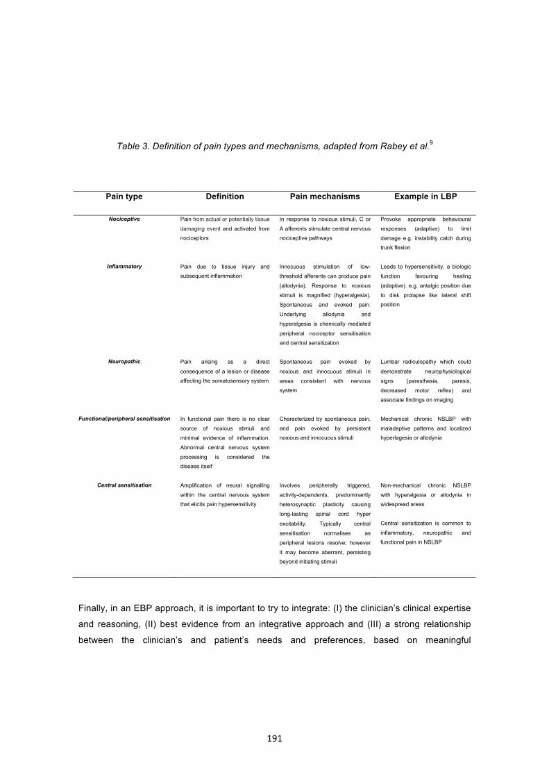
191
Table 3. Definition of pain types and mechanisms, adapted from Rabey et al.9
Pain type Definition Pain mechanisms Example in LBP
Nociceptive Pain from actual or potentially tissue
damaging event and activated from
nociceptors
In response to noxious stimuli, C or
A afferents stimulate central nervous
nociceptive pathways
Provoke appropriate behavioural
responses (adaptive) to limit
damage e.g. instability catch during
trunk flexion
Inflammatory Pain due to tissue injury and
subsequent inflammation
Innocuous stimulation of low-
threshold afferents can produce pain
(allodynia). Response to noxious
stimuli is magnified (hyperalgesia).
Spontaneous and evoked pain.
Underlying allodynia and
hyperalgesia is chemically mediated
peripheral nociceptor sensitisation
and central sensitization
Leads to hypersensitivity, a biologic
function favouring healing
(adaptive). e.g. antalgic position due
to disk prolapse like lateral shift
position
Neuropathic Pain arising as a direct
consequence of a lesion or disease
affecting the somatosensory system
Spontaneous pain evoked by
noxious and innocuous stimuli in
areas consistent with nervous
system
Lumbar radiculopathy which could
demonstrate neurophysiological
signs (paresthesia, paresis,
decreased motor reflex) and
associate findings on imaging
Functional/peripheral sensitisation In functional pain there is no clear
source of noxious stimuli and
minimal evidence of inflammation.
Abnormal central nervous system
processing is considered the
disease itself
Characterized by spontaneous pain,
and pain evoked by persistent
noxious and innocuous stimuli
Mechanical chronic NSLBP with
maladaptive patterns and localized
hyperlagesia or allodynia
Central sensitisation Amplification of neural signalling
within the central nervous system
that elicits pain hypersensitivity
Involves peripherally triggered,
activity-dependents, predominantly
heterosynaptic plasticity causing
long-lasting spinal cord hyper
excitability. Typically central
sensitisation normalises as
peripheral lesions resolve; however
it may become aberrant, persisting
beyond initiating stimuli
Non-mechanical chronic NSLBP
with hyperalgesia or allodynia in
widespread areas
Central sensitization is common to
inflammatory, neuropathic and
functional pain in NSLBP
Finally, in an EBP approach, it is important to try to integrate: (I) the clinician’s clinical expertise
and reasoning, (II) best evidence from an integrative approach and (III) a strong relationship
between the clinician’s and patient’s needs and preferences, based on meaningful

192
communication. This integrative process enables determination of the therapeutic goals,30-32
which likely will be further adapted during the treatment sessions.
In the case that dominant input components are responsible for NSLBP, hypotheses about the
possible pain types and symptoms (e.g. nociceptive sources with mechanical behaviour) can be
formulated.30-31 Most conditions presented by people with acute-subacute NSLBP are
mechanical in origin (i.e. nociceptive sources from zygapophyseal joints, disks and sacroiliac
joints).12,33 In contrast, in patients with chronic NSLBP, an identifiable nociceptive source of pain
can be determined in only 50% of cases.31
Mechanical LBP, which is the predominant form of NSLBP, has been defined as having a clear
and consistent anatomical focus, with pain proportionate to a mechanical behaviour (i.e.
consistently provoked and relieved with specific activities, movements and postures).25 Under
these circumstances, it is hypothesised that predominant nociceptive inputs preside (Table 3).
Mechanical nociceptive pain is thought to arise from articular structures, which are likely to
respond to techniques that are passive (spinal mobilisation/manipulation), active/passive
(mobilisation with movement or muscle energy technique) or active (directional exercise).12,30-31
Most of these interventions are present in the TBC system. It is also likely that muscular or
neurological structures contribute to the nociceptive sources (Figure 1) and may need
appropriate management.12,30-31
To determine whether the patient has a dominant processing component, various tools
addressing psychosocial influences (Table 2) and pain mechanisms could be usedcc. These
features are evaluated concurrently during the subjective examination. Management for people
in this category should consist of a functional cognitive-behavioural therapy approach
incorporating a substantial pain education component. Failure to identify a processing
component in the patient’s presentation is likely to lead to treatment failure, as dominant
processing mechanisms have been shown to be important predictors of risk of chronicity in
people with LBP.12,25-31
cc Orebrö or SBT, Fear Avoidance Beliefs, Tampa Scale (kinesiophobia), Catastrophizing Scale questionnaires, Central Sensitisation Inventory

193
In some individuals, LBP is driven by dominant output mechanisms, such as functional
maladaptive patterns during flexion (Table 1) (i.e. a passive flexiondd or active extensionee
pattern).23-26,29-32,34 The goal of OMT in the first case is to improve motor control and cognitive
postural education with a guided exercises approach. In the second case, the goal is to promote
functional paravertebral muscle relaxation by decreasing cognitive physical hypervigilance.23-26
Hands-on treatment techniques may be helpful in both patterns, but only as adjunctive therapy
to relieve any nociceptive symptoms.12,30-31 In the case of adaptive patterns resulting from
inputs (i.e. pain and restricted ROM in a combined movement direction, antalgic position or
instability catch during trunk movement), TBC or active/passive OMT techniques could be
applied as in the case of dominant input components (Tables 1 and 3, Figure 1).12
In clinical practice, each person complaining of NSLBP typically presents with a combination of
the above three categories of pain mechanisms. However, there is often a dominant component
that can be identified to enable more targeted OMT intervention for the patient.
1.4. Best practice for nonspecific low back pain
One potential reason for failure of clinical practice to manage LBP patients effectively may be a
lack of adherence to current evidence. This oversight might be due to a dominant biomedical
(anatomopathological) approach to the care of people with LBP, with a failure to consider and
manage LBP from a biopsychosocial perspective.12,29,35
Best practice for the management of acute LBP should involve an initial diagnosis based on
a triage process to screen for serious pathology (specific LBP and red flags) and consideration
of the disorder from a biopsychosocial perspective, including assessment for psychosocial risk
factors for chronic LBP. Tailored OMT management should then be applied, according to the
presentation. This approach may empower patients as active participants in their recovery and
discourage inappropriate radiologic investigation (Figure 3).12,29,31
In his discussion of the best practice for management of chronic LBP, Peter O’Sullivan23-25
proposed a shift from biomedical beliefs to a new paradigm in which the therapist has greater
skills and knowledge across several domains. Skills and knowledge should include, for dd Localised hypermobility of the lumbar spine during trunk flexion and sitting posture ee Localised hypomobility of the lumbar spine due to paravetebral muscle guarding during trunk flexion and sitting posture

194
example: better understanding of the complex multidimensional nature of chronic NSLBP;
development of effective communication skillsff to explore the patient’s story, pain behaviour,
pain beliefs, fears, life stresses, coping strategies and psychosocial factors; development of an
effective patient-centred therapeutic relationship that is within a biopsychosocial framework, to
identify the primary drivers of pain and disability; identification of maladaptive cognitive
behavioursgg; identification of neurophysiological processes, such as central and peripheral
sensitisation; development of a broad categorisation of chronic NSLBP disorders based on the
presence of dominant psychosocial, neurophysiological, lifestyle and movement behaviours that
act as drivers for the disorder; and development of multidimensional and multidisciplinary
flexible interventions that target maladaptive cognitive, lifestyle, pain and movement behaviours
in an integrated manner.24 The goal of this approach is to focus less on treating only structures
or signs/symptoms, particularly for chronic NSLBP disorders, and to improve targeting of the
different combinations of attitudes, beliefs, and cognitive, pain, lifestyle and movement
behaviours that underlie and drive disorders (Figure 2, Tables 1, 2).24,26
Figure 2: Biopsychosocial model of the clinical presentation and assessment of LBP and
disability35
ff Such as empathy, reflective questioning and motivational interview techniques gg Negative beliefs, coping, stress responsiveness, hypervigilance, catastrophising, anxiety, depression

195
Figure 3. Proposed comprehensive biopsychosocial integrative approach for screening and
targeting management of LBP patients, adapted from O’Sullivan25,29

196
In conclusion, following the development of this major part of the thesis, we are able to make
methodological recommendations to improve future RCTs.
(1) Homogeneous subgroups of LBP patients should be used, with the application of an
integrative approach (based on validated classification systems) and more accurately
targeted OMT. Patient samples should be better described in terms of duration (acute-
subacute/chronic), symptom location and mechanical vs. nonmechanical pain
behaviours.
(2) The best practice of OMT intervention(s) should be described in detail, with justification
from a clinical reasoning perspective.
(3) The active OMT intervention should be compared to: sham OMT; an ideal or plausible
placebo procedure (e.g. similar procedure without an active effect and with patient
blinding); the same OMT intervention with a nontargeted group; or another usual or
novel intervention. Blinding should be used in the assessment of biopsychosocial
features (e.g. ICF Core Set for LBP). Moreover, validated quantitative outcome
197
measures (e.g. kinematic analysis) should complement the usual self-reported
measures.
In general, clinical trials must be of good methodological quality but also patient-centred for
direct applicability to routine clinical practice, which will require improved education of health
professionals who will use this treatment approach.
2. FROM SECTION 2
Through three observational studies, a kinematic model of the spine was validated. This model
addressed: (I) trunk ROM, and (II) speed of various spinal segments, (III) RE in NSLBP patients
with mechanical pain behaviours when performing trunk movements from a sitting position
compared to healthy subjects. This tool is an objective movement-based analysis that
generates kinematic outputs in terms of movement quality (‘motion signature’ graphs of speed,
ROM curves of the lumbar spinal segments and RE curves) and movement quantity, including
RE and kinematic algorithms for the ROM (KA-R) and speed (KA-S) of trunk movements. These
three kinematic variables (RE, KA-R, KA-S) may be used in clinical trials as quantitative
outcome measures for the evaluation of various OMT interventions.
2.1. Kinematic variables as outcome measures
During the objective examination of LBP patients, an important part of the clinical reasoning is
the visual assessment of posture and spinal curves, lumbopelvic and trunk movement
coordination during functional movements, as well as the clinical measurement of ROM to assist
in identifying patterns of dysfunction.36 These patterns could be adaptive or maladaptive and are
associated with pain, disability, beliefs and fear of movement.23-29 Most clinicians, probably due
to a common paradigm, think that identifying and correcting coordination of maladaptive
movement, combined or not with maladaptive posture, can improve pain, function and activity
limitations.36-37 Fortunately, advances in technology are creating opportunities to quantify the

198
relationship between posture/movement coordination and pain behaviours, as well as the
influences of cognitive and psychological factors on movementhh.36-37
The most frequently used kinematic variables for LBP are the lordosis angle, ROM of the lumbar
spine, lumbar vs. hip contribution to flexion/extension, pelvic tilt angle, speed of lumbar flexion
and lumbopelvic or lumbar spine proprioception (position/reposition accuracy).36 A recent meta-
analysis identified lumbopelvic kinematic analysis as a useful measurement approach in people
with LBP. On average, LBP patients had significantly large to very large effect sizes for reduced
lumbar ROM in all directions (except extension) and proprioception on position-reposition
accuracy. They also moved more slowly than healthy people. However, there were no
significant differences in variability of the lordosis angles (lumbar posture), extension ROM or
lumbar vs. hip contribution to movement (i.e. lumbopelvic rhythm). However, a nonsignificant
but consistent effect favoured reduced lumbar compared to hip contribution to flexion for those
with LBP has been determined.36-37
The reduced speed of lumbar movement has been linked to fear of movement and shown to
persist after recovery in a subgroup of LBP patients with a persistent fear of movement.38 This
last point might partially explain the nonsignificant difference for KA-S in our RCT on MWM in
NSLBP people (Section 3, Chapter VI).
Future clinical studies should consider kinematic variables as interesting outcomes for
specifically addressing localised functional movement patterns of LBP patients (i.e. specific
body structures, body functions and activities of the ICF model, described in Introduction section
4.2) to complement classical self-reported measures of other features of the biopsychosocial
model (activities and participation in ICF model), such as pain scalesii and questionnairesjj.
2.2. Perspectives for kinematic analyses
The KA-S is a comprehensive quantitative variable of speed indicating whether patients are
moving slowly or quickly. However, the quality of speed curves/forms is another movement
hh Beliefs regarding posture and movement, kinesiophobia associated with protective muscle guarding ii For example, the visual analogue numeric pain-rating scale (VAS) jj For example, SBT, Fear Avoidance Beliefs questionnaire, Tampa Scale for Kinesiophobia, Patient Specific Functional Scale, Catastrophising Scale, Central Sensitisation Inventory, Hamilton Anxiety Rating Scale, Hamilton Depression Rating Scale, Oswestry Disability Index, Roland-Morris Disability Questionnaire

199
characteristic that may be objectively addressed by the calculation of smoothness. The
smoothness corresponds to the ratio between the speed and peak speed, with ratios closer to 0
indicating less smooth movements. It is a measure of parametric continuity along the speed
curve.39 Parametric continuity is distinct from geometric continuity (i.e. targeting variations of
speed on a time parameter trace curve). A speed curve describes the motion of an object with a
parameter of time, and must have continuity for the object to have a finite acceleration.40 An
observational study based on retrospective data for 60 subjects is currently in progress to
compare the smoothness of lumbar spine speed curves (motion signature) between patients
with acute and chronic NSLBP and healthy people (Figure 4).
Figure 4: Speed curves and smoothness of the lower lumbar spine in healthy and patients with
acute / chronic NSLBP (n=60) during trunk flexion with left pelvic rotation
LLS: lower lumbar spine (S2-L3); red curve: 20 acute LBP patients (smoothness = 0.37); blue curve: 20 chronic LBP patients (smoothness = 0.43) and green curve: 20 healthy people (smoothness = 0.49). ANOVA one way determined
significant differences between groups (p<.05,p<.005,p<.001) (preliminary results)
Time normalised (%)
0 10 20 30 40 50 60 70 80 90 100
SPEE
D (∞
/s) i
n LL
S
-60
-40
-20
0
20
40
60

200
Another interesting method of kinematic analysis that has recently appeared in the literature
used a dynamical system approach to address coordination and neuromuscular control
between segments in LBP patients. This approach, developed by Spinelli et al. (2015),37
analyses movement control and coordination. Using this method, clinicians and researchers can
determine aberrant movements by people with LBP, helping them to categorise functional
movement patterns and monitor changes before/after therapy.37
This method combines ‘continuous angular displacement motion curves’ (Figure 5A) to generate
‘angle-angle’kk (Figure 5B) and ‘coupling angle-movement cycle’ graphsll (Figure 5C), which
provide information about coordinated movement between body segments. ‘Phase-plane’mm
graphs provide information about the neuromuscular control of the segment. An increasing or
decreasing smooth period represents typical neuromuscular control, whereas a sudden
increase or decrease in angular velocity with resulting ‘cusps’nn is probably indicative of aberrant
movement and poor control.37
kk Angular movement of one body segment against another ll Quantifying the relative change in motion between data points in a standardised manner over the movement task mm Use of angular displacement and speed of the same segment nn Pointed end where two curves meet

201
Figure 5: Kinematic graphs in a dynamical system approach for one healthy subject37
Using our kinematic model of the spine, we can easily generate angle-angle graphs between
the low (S2-L3) and high (L3-T12) lumbar spine segments. These data could be used to
address coordination37 during trunk flexion (Figures 6, 7), as these segments may differ
biomechanically.36,41

202
Figure 6: Angle-angle graph between ROM of the low (LLS) and high (HLS) lumbar spine during
trunk flexion in 20 acute NSLBP patients, 20 chronic NSLBP patients and 20 healthy subjects
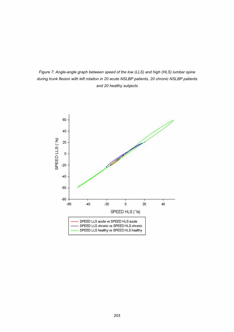
203
Figure 7: Angle-angle graph between speed of the low (LLS) and high (HLS) lumbar spine
during trunk flexion with left rotation in 20 acute NSLBP patients, 20 chronic NSLBP patients
and 20 healthy subjects

204
We are also able to generate phase-plane graphs, to analyse the neuromuscular control37 of
the low, high or total lumbar spine segments. These graphs can be created for the entire
duration of trunk flexion (Figure 8) or only in the forward/backward bending phase, as specific
patterns could appear during forward or backward movement from flexion due to pain
provocation in one or both directions.
Figure 8: Phase-plane graph of the total lumbar spine segment during trunk flexion with left
rotation in 20 acute NSLBP patients, 20 chronic NSLBP patients and 20 healthy subjects

205
This method of kinematic analysis can be used to measure OMT effects and to monitor changes
before/after spinal manipulation, as proposed in Figure 9.
Figure 9: Illustration of a phase-plane graph of the low lumbar spine segment during trunk
flexion for one acute mechanical NSLBP patientoo before and after two sessions of spinal
manipulation
The patient bent (forward and backward) in flexion five times from a sitting position. Red and green circles represent neuromuscular control of the low lumbar spine (S2-L3) before and after OMT intervention, respectively.
oo This acute NSLBP patient (a 45-year-old man) was profiled as moderate risk by SBT and positive on CCR (4/5) for spinal manipulation, with a dominant articular input nociceptive pain mechanism identified by combined movement assessment. An active flexion (primary) and left side-bending (secondary) pain pattern (confirmed by PAIVM on L4-L5 vertebral segments) was determined by the CCR. The patient was treated in accordance with his expectations during two sessions over 1 week of combined (flexion/left side-bending and rotation) lumbar spinal manipulation (grade 5) in a side-lying position on the L4-L5 intervertebral segment. VAS score at rest was 6/10 before and 2/10 after OMT.

206
The kinematic model of the spine looks promising and could be adapted from two to three
dimensions (e.g. by using an electromagnetic device, such as Fast-track) to analyse movement
compensation in all planes during movement in a given direction. This model can be applied to
analyse patients in clinical practice, as well as to aid in objective examinations and in following
the patient’s evolution during OMT interventions. Future studies in people with LBP could
integrate the hip/pelvis contribution, to generate coupling-angle graphs to study lumbopelvic
rhythm coordination, and could be combined with paravertebral muscle electromyography.
Other functional tasks could be studied, such as sitting, standing forward bending, sit-to-stand,
lifting, squatting, and walking. However, a key question remains in studies reporting the
quantitative kinematic analysis of people with LBP: Is the reduction in proprioception, ROM,
speed of movement or maladaptive lumbopelvic patterns the result or the cause of LBP?36
In conclusion, the kinematic data highlight the importance of using a patient-centred approach
within a biopsychosocial framework. Kinematic variations probably arise from multidimensional
and individual origins, and they could be consequential (adaptive patterns), causative
(maladaptive patterns) or even mixed. Variables derived from kinematic analyses are interesting
outcomes for addressing the body functions and activities of LBP patients. These outcomes can
be used to complement classical self-reported measures on other features of the
biopsychosocial model, such as pain scales or questionnaires. Future research might include
measurement of the correlations between kinematic measures and other tools addressing
components of the ICF Core Set for LBP, such as the Oswestry Disability Index, Patient Specific
Functional Scale, Tampa Scale (Kinesiophobia) or the Fear Avoidance Beliefs Questionnaire,
Central Sensitisation Inventory, in specific subgroups of LBP patients classified with an
integrative approach.

207
3. FROM SECTION 3
Two clinical studies are presented in this thesis. One reports the reliability and validity of
combined movement procedures, which extend the routine objective examination of the
lumbar spine. The other study is a randomised, placebo-controlled trial of good methodological
quality (8-9/11 on the Cochrane Collaboration Back Review Group checklist) that investigated
MWM in a subgrouppp of patients with LBP.
3.1. Evaluation of combined movements
The first study aimed to determine the most painful direction of movement (flexion or extension)
that needed to be improved by OMT, as well as the vertebral level(s) that may contribute to the
specific direction of impairment. Once the direction and level are determined, various forms of
OMT could be applied to improve findings determined during the combined movement
assessment. Information gained from this assessment may contribute to the therapist’s clinical
reasoning. This procedure is an evaluation of the articular planet (Figure 1) of the structure and
function of the lumbar spine, adapted for people with mechanical LBP disorders.
After the first and second triage processes (Figure 3) are completed, the combined movement
test results must be clinically reasoned within TBC, to select an adapted and targeted therapy or
MWM. Findings from the manual assessment may also help in directing treatment. For example,
the end-feel sensation during PAIVM testing of hypomobile vertebral segments may indicate a
need for more passive techniques, whereas hypermobility may indicate a need for more active
techniques.42
3.2. Mobilisation with movement
MWM is a relatively new active/passive technique in OMT. MWM on the lumbar spine is
innovating by the application of a passive vertebral glide, or SNAG, while the patient actively
moves in the impairment direction, which should be rendered pain-free. MWM may progress
depending on the functional or specific comparable signs of the LBP patient. This technique
might act on various pain mechanisms, resulting in less pain in the impairment direction.
pp Based on pain mechanism with a predominant flexion pattern released during an application of central SNAG (see inclusion criteria of Chapter VI)

208
However, as previously discussed (Section 3, Discussion of Chapter VI), the biomechanics and
neuropsychophysiological mechanisms of action of MWM require further research. There are
published guidelines but no classification systems for the application of MWM,43 partly because
this is a new concept.
During this RCT, variables from a developed kinematic model of the spine were used as the
primary outcome measures to address specific components of the ICF domain for LBP (see
Introduction section 4.2). The kinematic analysis was complemented by secondary self-reported
outcome measures addressing other factors of the ICF domain for LBP, such as the VAS score,
as well as functions, activities and participation on the Oswestry Disability Index and Tampa
Scale.
In conclusion, the combined movement procedure and MMW might be easily integrated into
the clinical reasoning during the objective examination for every patient with mechanical
NSLBP (Figure 3). If the CCR is positive and the response is positiveqq during the application of
SNAGs according to findings of the combined movement procedure, then MWM is probably
indicated, either in isolation or complemented with other OMT interventions from the TBC if
needed.
4. THESIS CONCLUSIONS
In summary, this thesis questioned the efficacy of OMT for LBP patients following the stepsrr of
the EBP process44 through three major sections.
Section 1 presented the results of a systematic review of evidence for OMT: (I) in acute-
subacute LBP, with strong evidence in favour of spinal manipulation vs. sham for pain, function
and health improvements in the short term, as well as moderate evidence in favour of spinal
mobilisation/manipulation combined with usual medical care vs. usual medical care alone for
qq Pain during movement is reduced and/or ROM in the impaired direction is improved rr Namely, ask an important question about the care (OMT efficacy) of LBP people; acquire the best available evidence regarding the question and critically appraise the evidence for validity and applicability of OMT to the LBP problem; apply the evidence by engaging in collaborative health decision making with the affected LBP individual(s) and/or group(s); appropriate decision making integrates the context, values and preferences of the care recipient, as well as available resources, including professional expertise; assess the outcome and disseminate the results.
209
pain, function and health improvements in the short term; (II) in chronic LBP, with moderate to
strong evidence in favour of spinal manipulation vs. sham for pain, function and overall health in
the short term; moderate evidence in favour of spinal mobilisation/manipulation combined with
exercise or usual medical care vs. exercise and back-school for pain, function and quality-of-life
in the short and long terms; limited evidence in favour of spinal mobilisation combined with
exercise and usual medical care vs. usual medical care alone for pain and function from the
short to long term; and limited evidence of no effect for spinal manipulation with extension-
exercise vs. extension-exercise alone for pain in the short to long term. This section highlighted
the importance of the quality of the clinical study design, including classification into subgroups
with targeted OMT interventions (splitting design) and the complex issue of the placebo
procedure in OMT trials.
Section 2 investigated a kinematic model of the spine in LBP patients during various trunk
movements from a sitting position. Kinematic variables were valid and reliable measures, with
the tasks of trunk flexion, rotation and flexion with rotation being the most discriminant. Effects
of disrupting proprioception on lumbar spine RE during forward bending were studied.
Proprioceptive disturbances had the most significant effect in increasing RE among healthy
people. Consistent with the literature, greater RE was observed in people with chronic LBP
compared to healthy subjects. Three kinematic variables were established: algorithms for ROM
(KA-R) and speed (KA-S), as well as RE. These variables could be used to aid in diagnosis
and/or to monitor changes during physical therapy programs in LBP patients, and as
quantitative outcome measures for OMT interventions in clinical trials.
Section 3 presented an original and standardised clinical pain provocation examination of the
lumbar spine in a combined movements fashion, aimed at reliably finding the direction and
vertebral level(s) of treatment. On the basis of this examination and evidence described
throughout the thesis, a RCT was conducted to analyse the efficacy of a novel specific method
of OMT, namely MWM, in NSLBP patients with a mechanical pain flexion pattern. This clinical
study aimed to raise the level of evidence from limited to moderate for the use of central SNAG
in a subgroup of NSLBP patients.

210
In summary, this thesis provides information to help unravel the complex puzzle of LBP. This
project has brought me great personal and professional satisfaction, as well as improved
knowledge in the assessment and management of patients with LBP. Manual therapy is an art
developed through clinical practice, as well as a science developed through fundamental and
clinical research. All of the knowledge acquired during my PhD is combined with my previous
experience and will directly impact my clinical practice, research and teaching, which will
become even more focused on a biopsychosocial patient-centred approach to LBP
management. In the future, I hope to continue to write scientific articles, contributing further
towards solving this puzzle. Clinical research is of major importance because it directly drives
clinical practice and education towards evidence-based OMT practice in a way that helps many
patients, students and health professionals.

211
References
1. Greenhalgh T, Howick J, Maskrey N. Evidence based medicine: a movement in crisis?
BMJ 2014;13:348:3725.
2. Tsertsvadze A, Clar C, Court R, Clarke A, Mistry H, Sutcliffe P. Cost-effectiveness of
manual therapy for the management of musculoskeletal conditions: a systematic review
and narrative synthesis of evidence from randomized controlled trials. J Manipulative
Physiol Ther 2014; 37:343-62.
3. Turk DC. The potential of treatment matching for subgroups of patients with chronic pain:
lumping versus splitting. Clin J Pain. 2005; 21:44-55.
4. Hidalgo B, Detrembleur C, Hall T, Mahaudens P, Nielens H. The efficacy of manual
therapy and exercise for different stages of non-specific low back pain: an update of
systematic reviews. J Man Manip Ther 2014;22:59-74.
5. Foster NE, Hill JC, O'Sullivan P, Hancock M. Stratified models of care. Best Pract Res
Clin Rheumatol 2013; 27:649-61.
6. Waddell G. The back pain revolution. 2nd ed. ed. Edinburgh: Churchill Livingstone, 2005.
7. Billis EV, McCarthy CJ, Oldham JA. Subclassification of low back pain: a cross-country
comparison. Eur Spine J 2007;16:865-79.
8. Rusu A. C., Boersma, K., & Turk, D. C. (2012). Reviewing the Concept of Subgroups in
Subacute and Chronic Pain and Potential of Customizing Treatment. In M. I. Hasenbring,
A. C. Rusu, & D. C. Turk (Eds.), From acute to chronic back pain. Risk factors,
mechanisms, and clinical implications (pp. 485–512). New York NY: Oxford University
Press.
9. Rabey M, Beales D, Slater H, O'Sullivan P. Multidimensional pain profiles in four cases of
chronic non-specific axial low back pain: An examination of the limitations of contemporary
classification systems. Man Ther 2015;20:138-47.
10. Delitto A, Erhard RE, Bowling RW. A treatment-based classification approach to low back
syndrome: identifying and staging patients for conservative treatment. Phys Ther
1995;75(6):470-485.
11. Fritz JM, Cleland JA, Childs JD. Subgrouping patients with low back pain: evolution of a
classification approach to physical therapy. J Orthop Sports Phys Ther 2007;37:290-302.
12. Delitto A, George SZ, Van Dillen LR, et al. Low back pain. J Orthop Sports Phys Ther
2012;42: A1-57.
13. Werneke MW, Hart D, Olivier D, McGill T, Grigsby D, et al. Prevalence of classification
methods for patients with lumbar impairments using the McKenzie syndromes, pain
pattern, manipulation, and stabilization clinical prediction rules. J Man Manipul Ther
2010;18:197-204.
14. Brennan GP, Fritz JM, Hunter SJ, Thackeray A, Delitto A, Erhard RE. Identifying
subgroups of patients with acute/subacute "nonspecific" low back pain: results of a

212
randomized clinical trial. Spine 2006;31:623-31.
15. Apeldoorn A, Ostelo RW, van Helvoirt H, Fritz JM, Knol DL, van Tulder MW, de Vet HC. A
randomized controlled trial on the effectiveness of a classification-based system for
subacute and chronic low back pain. Spine 2012; 37:1347-56.
16. Karayannis NV, Jull GA, Hodges PW. Physiotherapy movement based classification
approaches to low back pain: comparison of subgroups through review and
developer/expert survey. BMC Musculoskelet Disord 2012;13:24.
17. May S, Littlewood C, Bishop A. Reliability of procedures used in the physical examination
of non-specific low back pain: a systematic review. Aust J Physiother 2006;52:91-102.
18. McKenzie R, May S. The lumbar spine – mechanical diagnosis and therapy. (2nd) Spinal
Publications, 2003; New Zealand Ltd.
19. Aina A, May S, Clare H. The centralization phenomenon of spinal symptoms: a systematic
review. Man Ther 2004;9:134-43.
20. Hill JC, Whitehurst DG, Lewis M, Bryan S, Dunn KM, et al. Comparison of stratified
primary care management for low back pain with current best practice (STarT Back): a
randomised controlled trial. Lancet 2011;378:1560-71.
21. Woolf CJ. Central sensitization: implications for the diagnosis and treatment of pain. Pain
2011;152:2-15.
22. Smart KM, Blake C, Staines A, Doody C. The Discriminative validity of "nociceptive,"
"peripheral neuropathic," and "central sensitization" as mechanisms-based classifications
of musculoskeletal pain. Clin J Pain 2011;27:655-63.
23. O’Sullivan P. Classification-based cognitive functional approach for the management of
back pain. J Orthop Sports Phys Ther 2012;42:17-21.
24. O'Sullivan P. It's time for change with the management of non-specific chronic low back
pain. Br J Sports Med 2012;46:224-7.
25. O'Sullivan P, Waller R, Wright A, Gardner J, Johnston R, Payne C, Shannon A, Ware B,
Smith A. Sensory characteristics of chronic non-specific low back pain: a subgroup
investigation. Man Ther 2014;19:311-8.
26. Vibe Fersum K, O'Sullivan P, Skouen JS, Smith A and Kvale A. Efficacy of classification-
based cognitive functional therapy in patients with non-specific chronic low back pain: A
randomized controlled trial. Eur J Pain 2012;17: 916-928.
27. Ford JJ and Hahne AJ. Complexity in the physiotherapy management of low back
disorders: Clinical and research implications. Man Ther 2013;18(5):438-42.
28. Danneels L, Beernaert A, De Corte K, Descheemaeker F, Vanthillo B, Van Tiggelen D,
Cagnie B. A didactical approach for musculoskeletal physiotherapy: the planetary model. J
of Musculoskeletal pain 2011;19:218-224.
29. O’Sullivan P. Acute low back pain, beyond drug therapies. Pain Management Today
2014;1:8-13.
30. Dewitte V, Beernaert A, Vanthillo B, Barbe T, Danneels L, Cagnie B. Articular dysfunction

213
patterns in patients with mechanical neck pain: a clinical algorithm to guide specific
mobilization and manipulation techniques. Man Ther 2014;19:2-9.
31. Dewitte V, Cagnie B, Barbe T, Beernaert A, Vanthillo B, Danneels L. Articular dysfunction
patterns in patients with mechanical low back pain: A clinical algorithm to guide specific
mobilization and manipulation techniques. Man Ther 2014; 20 [Epub ahead of print].
32. Jones LE, O'Shaughnessy DF. The pain and movement-reasoning model: introduction to
a simple tool for integrated pain assessment. Man Ther 2014;19:270-6.
33. Balagué F, Mannion AF, Pellisé F, Cedraschi C. Non-specific low back pain. Lancet 2012;
379:482-91.
34. O'Sullivan K, Verschueren S, Van Hoof W, Ertanir F, Martens L, Dankaerts W. Lumbar
repositioning error in sitting: healthy controls versus people with sitting-related non-
specific chronic low back pain (flexion pattern). Man Ther 2013;18:526-32.
35. Main CJ, Williams AC. Musculoskeletal pain. BMJ 2002;325:534-7.
36. Laird RA, Gilbert J, Kent P, Keating JL. Comparing lumbo-pelvic kinematics in people with
and without back pain: a systematic review and meta-analysis. BMC Musculoskeletal
Disord 2014;15:229.
37. Spinelli BA, Wattananon P, Silfies S, Talaty M, Ebaugh D. Using kinematics and a
dynamical systems approach to enhance understanding of clinically observed aberrant
movement patterns. Man Ther 2015;20:221-6.
38. Thomas JS, France CR, Lavender SA, Johnson MR. Effects of fear of movement on spine
velocity and acceleration after recovery from low back pain. Spine 2008;33:564-70.
39. Gilliaux M, Lejeune T, Detrembleur C, Sapin J, Dehez B, Stoquart G. A robotic device as a
sensitive quantitative tool to assess upper limb impairments in stroke patients: a
preliminary prospective cohort study. J Rehabil Med 2012;44:210-7.
40. Richard H. Bartels; John C. Beatty; Brian A. Barsky. An Introduction to Splines for Use in
Computer Graphics and Geometric Modeling. Morgan Kaufmann. Chapter 13. Parametric
vs. Geometric Continuity 1987.
41. O'Sullivan K, O'Keeffe M, O'Sullivan L, O'Sullivan P, Dankaerts W. Perceptions of sitting
posture among members of the community, both with and without non-specific chronic low
back pain. Man Ther 2013;18:551-6.
42. Maher C, Adams R. Reliability of pain and stiffness assessments in clinical manual lumbar
spine examination. Phys Ther 1994;74(9):801-809.
43. Hing W, Hall T, Rivett D, Vicenzino B, Mulligan B. The Mulligan Concept of Manual
Therapy, 2015, Churchill Livingstone: 500p.
44. Spring B, Hitchcock K. Evidence-based practice in psychology. In I.B. Weiner & W.E.
Craighead (Eds.) Corsini’s Encyclopedia of Psychology, 4th edition 2009; 603-607. New
York:Wiley.

214
Summary of the thesis This thesis on the study of the efficacy of orthopaedic manual therapy (OMT) for patients with nonspecific low back pain (LBP) was developed by following the steps of an evidence-based practice process through three major sections. The Introduction defines the debilitating disorder of LBP and OMT, and describes an integrative approach for the stratification of care in LBP patients. Section 1 presents a systematic review that updates the best evidence of OMT efficacy in terms of pain, functions, activities and participation. The findings allow us: (I) to establish different levels of evidence for this form of therapy, (II) to understand the complexity of LBP and (III) to affirm the importance of the study design quality in OMT trials (e.g. splitting design, complexity of the placebo procedure and integration of clinical reasoning). Section 2, which is composed of three studies, investigates a kinematic model of the spine to help in the diagnosis of LBP patients, as well as outcome measures for future investigations of OMT in LBP patients. This kinematic tool permits a valid assessment of body structures (lumbopelvic and thoracic vertebral column, muscles of the trunk and pelvic regions), body functions (mobility in a vertebral segment, control of complex voluntary movements, proprioceptive function) and activities (bending, maintaining a body position). Finally, Section 3 presents two clinical studies. The first is a reliability study on a standardised and original pain provocation examination of the lumbar spine in a combined movement fashion. This examination provides the direction and vertebral level(s) of treatment. On the basis of this reliable objective examination and evidence described throughout this thesis, a randomised controlled trial was conducted. This last study questions the short-term efficacy of a novel form of OMT, namely mobilisation with movement, on primary kinematic outcome measures (kinematic algorithms for range of motion and speed) and secondary self-reported outcome measures (pain, function, activities and participation) in LBP patients with a mechanical pain pattern in flexion. The results of this investigation raise the overall level of evidence from limited to moderate in favour of using central sustained natural apophyseal glides in LBP patients. In conclusion, the different points and perspectives developed along this thesis contribute towards solving the complex puzzle of LBP within a patient-centred approach. Manual therapy is an art developed through clinical practice, as well as a science developed through fundamental and clinical research. Clinical research is of major importance because it directly drives clinical practice and education towards an evidence-based OMT practice within the biopsychosocial framework, thereby aiding many patients, students and health professionals.
Benjamin Hidalgo