Extremos geográficos de la distribución natural de Austrocedrus chilensis (Cupressaceae
Upload
independentCategory
view
2download
0
American Journal of Phytomedicine and Clinical Therapeutics www.ajpct.org
Original Article
Evaluation of the Acute Toxicity of the Hydroalcoholic Extract of Solidago chilensis Meyen (Arnica Do Campo) in Mice
Lyvia I.G. Paula-Freire1, Elena L.A. Malpezzi-Marinho2, Graziela R. Molska1, Daniele O. Kohn4, Luciana Correa5 and Eduardo A.V. Marinho*3
1Faculdade de Ciências da Saúde, Universidade Braz Cubas, São Paulo, Brasil. 2Departamento de Ciências Biológicas, Universidade Estadual de Santa Cruz, Ilhéus, Bahia, Brasil 3Departamento de Ciências da Saúde, Universidade Estadual de Santa Cruz, Ilhéus, Bahia, Brasil 4Departamento de Psicobiologia, Universidade Federal de São Paulo, São Paulo, Brasil 5Departamento de Patologia Geral, Faculdade de Odontologia, Universidade de São Paulo, São Paulo, Brasil.
ABSTRACT
Solidago chilensis is a plant with therapeutic properties that is widely used in folk Brazilian medicine. In this study, we evaluated the possible acute toxicity and determined the LD50 of the hydroalcoholic extract of Solidago chilensis. Swiss mice of both sexes received intraperitoneal injections of Solidago chilensis hydroalcoholic extract (ScHE) at the doses of 30, 100, 300, 500, 750 and 1000 mg/kg. Pharmacological screening, LD50 determination, food intake evaluation, body and target organ weight measurements and histopathological analyses were performed. The LD50 value was determined to be 512.5 mg/kg. Pharmacological screening revealed central nervous system depressant activity, as shown by ptosis and decreases in general activity and motor activity. Food intake and body weight were not affected by extract administration. Macroscopic analysis of organs indicated splenomegaly and changes in the color of the liver in animals treated with ScHE; both effects were dose dependent. Significant macroscopic changes in the kidneys were not observed. Histopathological analysis showed significant morphological alterations in the livers of animals treated with high doses of the extract. Only animals treated with 1000 mg/kg had hemorrhagic foci in the kidneys. Our findings indicate that the intraperitoneal injection of ScHE causes relevant toxicity, with a relatively high LD50 (512.5 mg/kg), and affects the histopathological parameters analyzed. Further toxicological tests should be performed using oral administration to compare the effects of different routes of administration and to simulate traditional use.
Keywords: Solidago chilensis, Toxicity, LD50, Mice.
Address for
Correspondence
BR-415, Rodovia Ilhéus- Itabuna, Km-16, Salobrinho, Ilhéus - Bahia, 45662-000, Brazil
E-mail: edumarinho @hotmail.com

Paula-Freire et al____________________________________________ ISSN 2321 – 2748
AJPCT[2][2][2014]217-228
INTRODUCTION
The genus Solidago is considered as one of the largest genera of the Asteraceae family, comprising more than 120 species, most of which are found in North and South America. This genus comprises a great number of medicinal plants with therapeutic properties, including S. virgaurea and S. gigantea, the aerial parts of which are widely used as anti-inflammatory and diuretic agents. Solidago chilensis (S. microglossa), known in Brazil as arnica-do-campo or arnica-brasileira, is a plant found throughout the country, especially in the South and Southeast regions. This plant, high 80-120 cm, can be found in wastelands, roadsides, pastures, orchards and coffee plantations1.
Ethnopharmacological data indicate that S. chilensis is broadly used in Brazil. At Rio de Janeiro, patches from leaves and stems are used on contusions2, in many towns from Minas Gerais State S. chilensis is indicated to skin diseases, wound healings and boils, purulent infections, inflammation, edema, body pain, chill, rheumatism, diuretic, using leaves, flowers or the whole plant, by maceration, infusion, bath, decoction and tincture with topic and oral administration3-5. In Santa Catarina State it is used to skin lesions6. S. chilensis is in the Medicinal Plant Program of the Health Municipal Secretariat (Fitoviva) indicated to contusion, trauma and boils, as tincture and poultice from flowers and leaves7. Since 2009 S. microglossa is in the list of native medicinal plants used by Health Brazilian Program (SUS) and in the National List of Medicinal Plant (Renisus) that guides the scientific studies8. According to Corrêa et al.9 it is toxic, should not be used in wounds, but without scientific evidences.
Several studies have demonstrated that both the extract and essential oil of S. chilensis contain compounds that have relevant biological activities. S. chilensis
flower extracts have been shown to exhibit outstanding antioxidant activity and to be more effective than butylhydroxytoluene (an antioxidant used in food industry)10. Aqueous and hydroalcoholic extracts of S. chilensis exhibit topical and systemic anti-inflammatory activities similar to those of dexamethasone in rats and mice. This anti-inflammatory activity is not only due to the inhibition of pro-inflammatory mediators but also to reduce lymphocyte migration11-13. Other demonstrated activities include antifungal14, anti-microbial15, gastro-protective16, hypo-glycemic17, healing18 and analgesic activities19.
Despite the known pharmacological effects of S. chilensis and its traditional use as an alternative treatment for health problems, there have been few data about the possible toxic effects of this plant16. Therefore, we aimed to investigate the acute toxicity and to determine the LD50 of the hydroalcoholic extract of Solidago chilensis when administered intraperitoneally to mice. Possible histopathological alterations in target organs were also evaluated. Material and Methods Botanical material
Solidago chilensis was collected in the city of Guararema, São Paulo, Brazil. Botanical identification and authentication were performed by Dr. Lucia Rossi from the Instituto Botânico de São Paulo, São Paulo, Brazil, and one specimen was deposited in the herbarium under the number SP 397.047 (Figure 1). Hydroalcoholic extract preparation and analysis
The aerial parts of the plant (flowers, leaves and stalks) were immersed in 93% ethanol (100 g/L) for one month. After this period, the hydroalcoholic extract was

Paula-Freire et al____________________________________________ ISSN 2321 – 2748
AJPCT[2][2][2014]217-228
filtered, concentrated using a rotary evaporator and then lyophilized. The dried material was referred to as the Solidago chilensis hydroalcoholic extract (ScHE) and was stored at 4°C until use. The dried extract was diluted in distilled water immediately prior to analysis. The Solidago chilensis hidroalcoolic extract was previously analyzed in its major constituents on work previously published by the group. Briefly, have been identified constituents as the 5-O-E-, 3,4-, 4,5-di-OE-caffeoylquinic acids and rutin13. Animals
Male and female Swiss albino mice (3 months old) obtained from the Braz Cubas University vivarium, were used. The animals were kept in a room with a controlled temperature of 21±1°C, a light/dark cycle of 12 hours (lights on at 7:00 A.M.) and free access to food and water. The study was conducted according to ethical principles20
following institutional protocols and was approved by the local Committee for Ethical Surveillance in Animal Experimentation (SBCAL) for the proper use and care of experimental animals. Acute toxicity study
Mice were equally separated into seven groups of four males and four females each. A range of doses of ScHE (30, 100, 300, 500, 750 and 1000 mg/kg) were administered once intraperitoneally. The vehicle control group received 0.3 mL of distillated water. The animals were observed for 14 days. The number of dead animals was recorded for each tested dose, and the lethal dose that killed 50% of animals (LD50) was calculated according to the method of Carvalho et al21. LD50 = Df – Z (a.b) / n Df = lowest dose able to kill all animals;
a = difference between 2 consecutive doses administered; b = average number of animals killed between 2 consecutive doses; n = number of mice by group; z = sum of (ax b) / n. Physiological and anatomopathological parameters
Pharmacological screening was also performed. The animals were observed continuously during the first hour after extract administration and at 2, 4, 12, 24 and 48 hours after extract administration22. The animals were also observed once per day for 14 days to check for residual effects. Food intake and body weight were analyzed daily. After 14 days, the animals were euthanized (cervical dislocation), and the liver, kidneys and spleen were removed, weighed and fixed in formalin (10%) for subsequent histological analysis. The organ weights were corrected based on the last body weight of the animals. Animals that died before the 14th day were autopsied, and the same parameters were analyzed. Histopathology
Fixed material was embedded in paraffin, and sections (thickness: 5μm) were stained with hematoxylin/eosin. Histological analysis was performed using conventional microscopy by one pathologist who described the morphological alterations due to toxic effects. Statistical analysis
Parametric tests were performed, taking into account variable characteristics. The data obtained from the food intake and body weight evaluations were analyzed by ANOVA with repeated measures, followed by Tukey’s post hoc test. The organ weights were analyzed by one-way ANOVA, followed by Tukey’s post hoc test. The values were presented as the mean ± standard error
Paula-Freire et al____________________________________________ ISSN 2321 – 2748
AJPCT[2][2][2014]217-228
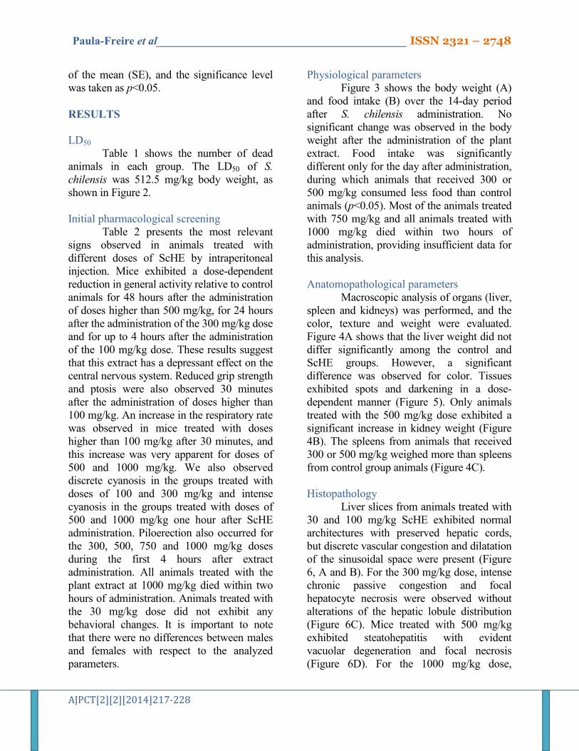
of the mean (SE), and the significance level was taken as p<0.05. RESULTS
LD50
Table 1 shows the number of dead animals in each group. The LD50 of S. chilensis was 512.5 mg/kg body weight, as shown in Figure 2.
Initial pharmacological screening
Table 2 presents the most relevant signs observed in animals treated with different doses of ScHE by intraperitoneal injection. Mice exhibited a dose-dependent reduction in general activity relative to control animals for 48 hours after the administration of doses higher than 500 mg/kg, for 24 hours after the administration of the 300 mg/kg dose and for up to 4 hours after the administration of the 100 mg/kg dose. These results suggest that this extract has a depressant effect on the central nervous system. Reduced grip strength and ptosis were also observed 30 minutes after the administration of doses higher than 100 mg/kg. An increase in the respiratory rate was observed in mice treated with doses higher than 100 mg/kg after 30 minutes, and this increase was very apparent for doses of 500 and 1000 mg/kg. We also observed discrete cyanosis in the groups treated with doses of 100 and 300 mg/kg and intense cyanosis in the groups treated with doses of 500 and 1000 mg/kg one hour after ScHE administration. Piloerection also occurred for the 300, 500, 750 and 1000 mg/kg doses during the first 4 hours after extract administration. All animals treated with the plant extract at 1000 mg/kg died within two hours of administration. Animals treated with the 30 mg/kg dose did not exhibit any behavioral changes. It is important to note that there were no differences between males and females with respect to the analyzed parameters.
Physiological parameters Figure 3 shows the body weight (A)
and food intake (B) over the 14-day period after S. chilensis administration. No significant change was observed in the body weight after the administration of the plant extract. Food intake was significantly different only for the day after administration, during which animals that received 300 or 500 mg/kg consumed less food than control animals (p<0.05). Most of the animals treated with 750 mg/kg and all animals treated with 1000 mg/kg died within two hours of administration, providing insufficient data for this analysis.
Anatomopathological parameters
Macroscopic analysis of organs (liver, spleen and kidneys) was performed, and the color, texture and weight were evaluated. Figure 4A shows that the liver weight did not differ significantly among the control and ScHE groups. However, a significant difference was observed for color. Tissues exhibited spots and darkening in a dose-dependent manner (Figure 5). Only animals treated with the 500 mg/kg dose exhibited a significant increase in kidney weight (Figure 4B). The spleens from animals that received 300 or 500 mg/kg weighed more than spleens from control group animals (Figure 4C).
Histopathology
Liver slices from animals treated with 30 and 100 mg/kg ScHE exhibited normal architectures with preserved hepatic cords, but discrete vascular congestion and dilatation of the sinusoidal space were present (Figure 6, A and B). For the 300 mg/kg dose, intense chronic passive congestion and focal hepatocyte necrosis were observed without alterations of the hepatic lobule distribution (Figure 6C). Mice treated with 500 mg/kg exhibited steatohepatitis with evident vacuolar degeneration and focal necrosis (Figure 6D). For the 1000 mg/kg dose,

Paula-Freire et al____________________________________________ ISSN 2321 – 2748
AJPCT[2][2][2014]217-228
microscopic alterations due to toxicity were not observed, but generalized vascular congestion was present (most likely due the early death of these animals).
Only the animals treated with 1000 mg/kg exhibited intense vascular congestion in kidney cortex, with small hemorrhagic areas. The spleens did not exhibit morphological alterations that could indicate toxicity and did not differ from the control spleens at any of the tested doses.
DISCUSSION
Solidago chilensis is a plant that is
widely used in folk medicine due to its well- known therapeutic properties10,23,18,13. However, there have been few toxicological studies performed on plants from this genus.
Our results suggest that acute treatment with Solidago chilensis hydro-alcoholic extract at a dose ranging from 30 to 500 mg/kg has little effect on the animal’s body weight over a 14-day period (in the higher doses, the animals death and was not possible evaluate the weight). Food intake was decreased only on the day after extract administration for the 300 and 500 mg/kg groups. Despite this small change, animals treated with ScHE did not exhibit significant body weight changes.
The oral administration of aqueous extract of S. chilensis did not cause signs of toxicity in a previous study16. In the present study, however, the LD50 of the hydroalcoholic extract of this plant was determined to be 512.5 mg/kg, that this extract has high toxicity when administered intraperitoneally. Nevertheless, the doses used in the studies published in the literature were usually much lower than the LD50 that we determined in this study.
Based on the results obtained from the initial pharmacological screening, the Solidago chilensis extract exhibited a central effect at doses higher than 100 mg/kg. This
effect manifested as ptosis and decreases in general activity and motor activity. These signs strongly suggest that the plant has a sedative effect when administered at high doses22,24,25. Symptoms such as cyanosis, respiratory rate alterations and piloerection were also observed for lethal doses before death, indicative of the pronounced toxic effect22. All animals that received the 1000 mg/kg dose died within two hours of administration, again indicating that this plant extract is toxic when administered intraperitoneally. It is important to note that the results obtained for the initial screening represent only a subjective impression of the drug’s likely effect, but this screening is essential to determine the appropriate research direction.
The anatomopathological data indicate that there were significant changes in the livers and spleens of animals treated with ScHE and that these effects were dose dependent. The macroscopic observation of organs showed that livers from animals treated with arnica do campo exhibited darkened coloration, the extent of which depended on the administered dose, relative to the control animals. Additionally, animals treated with the plant extract exhibited splenomegaly (increases in spleen weight and volume). This response can occur due the presence of compounds with injurious activity. This activity can induce an inflammatory response, promoting organ enlargement (edema)26.
The liver is the organ most affected by toxic substances due to its intense participation in biotransformation reactions27. It is known that splenomegaly can also occur due to liver failure, which promotes the congestion of the liver’s blood vessels. This congestion is caused by the decrease in activity and by the increase in hydrostatic pressure. Organ enlargement causes blood to reflux into spleen through the splenic vein,

Paula-Freire et al____________________________________________ ISSN 2321 – 2748
AJPCT[2][2][2014]217-228
causing the spleen to swell28. No macroscopic alterations were observed in the kidneys.
The histopathological data show that the extract seems to affect the liver more intensely, and significant cellular alterations (such as hepatic steatosis, intense vacuolization and hepatocytes necrosis) were observed in mice treated with 300 mg/kg and higher doses of the extract. We also observed vessel congestion and hemorrhagic areas. Fat infiltration, blood congestion and hepatocyte necrosis are common signs of hepatitis and may cause death29,30. These data confirm the harmful effect of the extract on the liver that was observed in the macroscopic analyses.
Tests to evaluate the sub chronic toxicity (due to the repeated administration of lower doses) of ScHE are needed because the population uses this extract for extended periods of time1. Detailed analyses are also needed to determine which of the component compounds are toxic and if it is possible to remove them before use. Were commend that ScHE not be used by parenteral and enteral means.
CONCLUSION
In conclusion, Solidago chilensis
hydroalcoholic extract exhibits significant toxicity when administered intraperitoneally, with a relatively high LD50, and it affects anatomopathological and histopathological parameters. More studies should be performed because the important therapeutic effects of this plant. ACKNOWLEDGMENTS
The authors are grateful to Dr. Lucia Rossi from Instituto de Botânica de São Paulo, São Paulo, Brazil, for identifying the Solidago chilensis plant used to prepare the extract and to Dr. José Gregório Cabrera Gomes from Instituto de Ciências Biomédicas da Universidade de São Paulo, São Paulo,
Brazil, for assistance in the preparation of the hydroalcoholic extract of this plant. Authors’ contributions
Lyvia I.G. Paula-Freire contributed in running the laboratory work, analysis of the data and drafted the paper. Elena L. A. Malpezzi-Marinho designed the study, contributed in collecting plant sample and identification, confection of herbarium, running the laboratory work, drafted the paper and supervised the laboratory work. Graziela R. Molska contributed in running the laboratory work. Daniele O. Kohn contributed to drafted of the paper. Luciana Corrêa contributed to anatomopathological and histopathological studies and drafted of the paper. Eduardo A.V. Marinho (PhD) designed the study, contributed in collecting plant sample and identification, confection of herbarium, running the laboratory work, analysis of the data and drafted the paper and supervised the laboratory work. All the authors have read the final manuscript and approved the submission. REFERENCES 1. Lorenzi H, Matos FJA. Plantas medicinais
no Brasil – nativas e exóticas. 1st ed., São Paulo: Instituto Plantarum de Estudos da Flora LTDA; 2002: 544p.
2. Posse JC. Medicinal plants used by SUS users in Paquetá and Santa Teresa neighborhoods. An ethnobotany approach. Rio de Janeiro. Dissertation. Post-Graduate Program in Pharmaceutical Sciences, Faculty of Pharmacy, Federal University of Rio de Janeiro, Rio de Janeiro (in Portuguese), 2007, 115p.
3. Calábria L, Cuba GT, Hwang SM, Marra JCF, Mendonça MF, Nascimento RC, Oliveira MR, Porto JPM, Santos DF, Silva BL, Soares TF, Xavier EM, Damasceno AA, Milani JF, Rezende CHA, Barbosa AAA, Canabrava HAN. Ethnobotanical and Ethnopharmacological survey of medicinal

Paula-Freire et al____________________________________________ ISSN 2321 – 2748
AJPCT[2][2][2014]217-228
plants in Indianopolis, Minas Gerais, Brazil. Rev Bras Plantas Med 2008, 10: 49-63.
4. Kffuri CW. Ethnobotany of medicinal plants in Senador Firmino (Minas Gerais State). Dissertation, Federal University of Viçosa, 2008, 88p.
5. Oliveira HB, Kffuri CW, Casali VWD. Ethnopharmacological study of medicinal plants used in Rosário da Limeira, Minas Gerais, Brazil. Rev Bras Farmacogn 2010, 20: 256-60.
6. Amorim CC. Ethnobotany of land medicine in the Coxilha Rica region, SC. Rev Bras Agroecol 2009, 4: 1596-99.
7. Jorge SSA. Plantas Medicinais: Coletânea de Saberes: www.agronomiaufs.com.br/index. php/downloads-e-videos/category/75-downloads (47825636 Plantas Medicinais. pdf) 2009.
8. Brasil.http://portal.saude.gov.br/portal/saude/area.cfm?id_area=1336; 2012.
9. Corrêa AD, Siqueira-Batista R, Quintas LEM. Plantas medicinais do cultivo à terapêutica, 7th ed., Petrópolis:Editora Vozes, 2008: 1247p.
10. Güntner C, Barra C, Cesio MV, Dellacassa E, Ferrando L, Ferreira F, Garcia C, González G, Heinzen H, Lloret A, Lorenzo D, Menendez P, Paz D, Soule S, Vasquez A, Moyna P. Antioxidant properties of Solidago chilensis L. flavonoids. Acta Hortic 1999, 501: 159-63.
11. Goulart S, Moritz MI, Lang KL, Liz R, Schenkel EP, Fröde TS. Anti-inflammatory evaluation of Solidago chilensis Meyen in a murine model of pleurisy. J Ethnopharmacol 2007, 113: 346-53.
12. Liz R, Vigil SV, Goulart S, Moritz MI, Schenkel EP, Fröde TS. The anti-inflammatory modulatory role of Solidago chilensis Meyen in the murine model of the air pouch. J Pharm Pharmacol 2008, 60: 515-21.
13. Tamura EK, Jimenez RS, Waismam K, Gobbo-Neto L, Lopes NP, Malpezzi-Marinho ELA, Marinho EA, Farsky SH. Inhibitory effects of Solidago chilensis Meyen hydroalcoholic extract on acute inflammation. J Ethnopharmacol 2009, 122: 478-85.
14. Duarte MC, Figueira GM, Sartoratto A, Rehder VL, Delarmelina C. Anti-Candida activity of Brazilian medicinal plants. J Ethnopharmacol 2005, 97: 305-11.
15. Morel AF, Dias GO, Porto C, Simionatto E, Stuker CZ, Dalcol II. Antimicrobial activity of extractives of Solidago microglossa. Fitoterapia 2006, 77: 453-55.
16. Bucciarelli A, Minetti A, Milczakowskyg C, Skliar M. Evaluation of gastroprotective activity and acute toxicity of Solidago chilensis Meyen (Asteraceae). Pharm Biol 2010, 48: 1025-30.
17. Melo AM, Bittencourt P, Nakutis FS, Silva AP, Cursino J, Santos GA, Ashino NG, Velloso LA, Torsoni AS, Torsoni MA. Solidago chilensis Meyen hydroalcoholic extract reduces JNK/IκB pathway activation and ameliorates insulin resistance in diet-induced obesity mice. Exp Biol Med 2011, 236: 1147-55.
18. Facury Neto MA, Fagundes DJ, Beletti ME, Novo NF, Juliano Y, Penha-Silva N. Systemic use of Solidago microglossa DC in the cicatrization of open cutaneous wounds in rats. Braz J morphol Sci 2004, 21: 207-10.
19. Silva AG, Sousa CP, Koehler J, Fontana J, Christo AG, Guedes-Bruni RR. Evaluation of an extract of Brazilian arnica (Solidago chilensis Meyen, Asteraceae) in treating lumbago. Phytother Res 2010, 24: 283-87.
20. Andersen ML, Tufik S. Animal models as ethical tools in biomedical research. 1st ed São Paulo: CLR Balieiro Editores, 2010: 563p.
21. Carvalho AAT, Sampaio MCC, Melo AFM, Souza IA, Higino JS. Estudos toxicológicos do extrato hidroalcoólico de Psidium guajava Linn. Rev Bras Ciênc Saúde 2002, 6: 43-50.
22. Carlini EA. “Screening” farmacológico de plantas brasileiras. Rev Bras Biol 1972, 32: 265-74.
23. Vila R, Mundina M, Tomi F, Furlán R, Zacchino S, Casanova J, Cañigueral S. Composition and antifungal activity of the essential oil of Solidago chilensis. Planta Med 2001, 68: 164-67.
24. Tabach R, Rodrigues E, Carlini EA. Preclinical toxicological assessment of a phytotherapeutic product--CPV (based on

Paula-Freire et al____________________________________________ ISSN 2321 – 2748
AJPCT[2][2][2014]217-228
dry extracts of Crataegus oxyacantha L., Passiflora incarnata L., and Valeriana officinalis L.). Phytother Res 2009, 23: 33-40.
25. Carlini EA. Plants and the central nervous system. Pharmacol Biochem Behav 2003, 75: 501-12.
26. Porfírio Z, Ribeiro MP, Estevam CS, Houly RLS, Sant’Ana AEG. Hepatosplenomegaly caused by an extract os cyanobacterium Microcystis aeruginosa bloom collected in the Manguaba lagoon, Alagoas, Brazil. Rev Microbiol 1999, 30: 278-85.
27. Matos LC, Martins B. Hepatites tóxicas: Revisão da literatura. Med Interna 2005, 12: 239-58.
28. Petroianu A. Drug-induced splenomegaly. Acta Med Port 2011, 4: 977-82.
29. Kim ST, Kim JD, Ahn SH, Ahn GS, Lee YI, Jeong YS. Hepatoprotective and antioxidant effects of Alnus japonica extracts on acetaminophen-induced hepatotoxicity in rats. Phytother Res 2004, 18: 971-75.
30. Begriche K, Massart J, Robin MA, Borgne-Sanchez A, Fromenty B. Drug-induced toxicity on mitochondria and lipid metabolism: mechanistic diversity and deleterious consequences for the liver. J Hepatol;2011,54:773-94.
Table 1. Number of deaths caused by the administration of Solidago chilensis hydroalcoholic extract at different doses
Groups Number of Deaths
30 mg/kg 0/8
100 mg/kg 0/8
300 mg/kg 2/8
500 mg/kg 5/8
750 mg/kg 6/8
1000 mg/kg 8/8
Table 2. Summary of the observations during the initial pharmacological screening of mice treated with Solidago chilensis hydroalcoholic extract
Observed effects Dose (mg/kg – I. p.)
30 100 300 500 750 1000
General activity ↓ ↓ ↓↓ ↓↓↓ †
Ptosis + + ++ †
Grip strength ↓ ↓ ↓ †
Cyanosis + ++ +++ †
Respiratory rate + ++ +++ †
Piloerection + + + †
Writhing †
(+) presence of effect; (↓) decrease; † death. Blanks represent no effect.

Paula-Freire et al____________________________________________ ISSN 2321 – 2748
AJPCT[2][2][2014]217-228
Figure 1. Solidago chilensis Meyen (photograph Eduardo A.V. Marinho)
Figure 2. Graphic representation of the determination of the LD50 of the Solidago chilensis hydroalcoholic
extract (512.5 mg/kg). The black line represents the linear regression of the data points in gray

Paula-Freire et al____________________________________________ ISSN 2321 – 2748
AJPCT[2][2][2014]217-228
Figure 3. Body weight (A) and food intake (B) of the animals treated by intraperitoneal injection with increasing doses of Solidago
chilensis hydroalcoholic extract. The values are expressed as the mean ± standard error of the mean. (*) Significantly different from
the control group at the respective time (ANOVA with repeated measures followed by Tukey’s post hoc test, p<0.05; n=8/group)

Paula-Freire et al____________________________________________ ISSN 2321 – 2748
AJPCT[2][2][2014]217-228
Figure 4. Weights of the liver (A), kidneys (B) and spleen (C), in grams, of the animals treated with water (control group) and
Solidago chilensis hydroalcoholic extract at different doses. The values are expressed as the mean ± standard error of the mean. (*) Significantly different from the control group (ANOVA followed by
Tukey’s post hoc test, p<0.05, n=8/group)

Paula-Freire et al____________________________________________ ISSN 2321 – 2748
AJPCT[2][2][2014]217-228
Figure 5. Photographs of the livers from animals treated with increasing doses of Solidago chilensis hydroalcoholic extract and from the control group
Figure 6. Liver slices from animals treated with the hydroalcoholic extract of Solidago chilensis at different doses: (A-30 mg/kg) Hepatocytes cords exhibited
intercellular spaces that suggested dilatation of the sinusoidal space. The centrilobular veins exhibited slight passive congestion. (B–100 mg/kg) Hepatic
lobules with stromal and parenchymal integrity. The centrilobular veins exhibited evident passive congestion. (C-300 mg/kg) Hepatic lobules with a
centrilobular vein (c) exhibiting congestion and necrotic hepatocytes (arrow). (D-500 mg/kg) Steatohepatitis, with loss of the normal architecture of the
hepatic lobules, vacuolar degeneration, and focal necrosis (HE, 100X)
Copyright © 2022 FDOKUMEN