Evaluation of Protective Efficacy of Viral Vector Based Swine ...
79
University of Nebraska - Lincoln University of Nebraska - Lincoln DigitalCommons@University of Nebraska - Lincoln DigitalCommons@University of Nebraska - Lincoln Dissertations & Theses in Veterinary and Biomedical Science Veterinary and Biomedical Sciences, Department of Spring 4-29-2022 Evaluation of Protective Efficacy of Viral Vector Based Swine Evaluation of Protective Efficacy of Viral Vector Based Swine Influenza Vaccine and a Method for B Cell Culture for Monoclonal Influenza Vaccine and a Method for B Cell Culture for Monoclonal Antibody Generation Antibody Generation Sushmita Kumari University of Nebraska-Lincoln, [email protected] Follow this and additional works at: https://digitalcommons.unl.edu/vetscidiss Part of the Veterinary Infectious Diseases Commons Kumari, Sushmita, "Evaluation of Protective Efficacy of Viral Vector Based Swine Influenza Vaccine and a Method for B Cell Culture for Monoclonal Antibody Generation" (2022). Dissertations & Theses in Veterinary and Biomedical Science. 32. https://digitalcommons.unl.edu/vetscidiss/32 This Article is brought to you for free and open access by the Veterinary and Biomedical Sciences, Department of at DigitalCommons@University of Nebraska - Lincoln. It has been accepted for inclusion in Dissertations & Theses in Veterinary and Biomedical Science by an authorized administrator of DigitalCommons@University of Nebraska - Lincoln.
-
Upload
khangminh22 -
Category
Documents
-
view
4 -
download
0
Transcript of Evaluation of Protective Efficacy of Viral Vector Based Swine ...
University of Nebraska - Lincoln University of Nebraska - Lincoln
DigitalCommons@University of Nebraska - Lincoln DigitalCommons@University of Nebraska - Lincoln
Dissertations & Theses in Veterinary and Biomedical Science
Veterinary and Biomedical Sciences, Department of
Spring 4-29-2022
Evaluation of Protective Efficacy of Viral Vector Based Swine Evaluation of Protective Efficacy of Viral Vector Based Swine
Influenza Vaccine and a Method for B Cell Culture for Monoclonal Influenza Vaccine and a Method for B Cell Culture for Monoclonal
Antibody Generation Antibody Generation
Sushmita Kumari University of Nebraska-Lincoln, [email protected]
Follow this and additional works at: https://digitalcommons.unl.edu/vetscidiss
Part of the Veterinary Infectious Diseases Commons
Kumari, Sushmita, "Evaluation of Protective Efficacy of Viral Vector Based Swine Influenza Vaccine and a Method for B Cell Culture for Monoclonal Antibody Generation" (2022). Dissertations & Theses in Veterinary and Biomedical Science. 32. https://digitalcommons.unl.edu/vetscidiss/32
This Article is brought to you for free and open access by the Veterinary and Biomedical Sciences, Department of at DigitalCommons@University of Nebraska - Lincoln. It has been accepted for inclusion in Dissertations & Theses in Veterinary and Biomedical Science by an authorized administrator of DigitalCommons@University of Nebraska - Lincoln.

EVALUATION OF PROTECTIVE EFFICACY OF VIRAL VECTOR BASED SWINE
INFLUENZA VACCINE AND A METHOD FOR B CELL CULTURE FOR MONOCLONAL
ANTIBODY GENERATION
BY
SUSHMITA KUMARI
A THESIS
Presented to the Faculty of
The Graduate College at the University of Nebraska
In Partial Fulfillment of Requirements
For the Degree of Master of Science
Major: Veterinary Sciences
Under the Supervision of Professor Hiep L.X. Vu
Lincoln, Nebraska
April, 2022

EVALUATION OF PROTECTIVE EFFICACY OF VIRAL VECTOR BASED SWINE
INFLUENZA VACCINE AND A METHOD FOR B CELL CULTURE FOR
MONOCLONAL ANTIBODY GENERATION
Sushmita Kumari, M.S.
University of Nebraska, 2022
Advisor: Hiep L.X. Vu
Influenza A virus of the swine (IAV-S) is an economically important swine pathogen that
has the potential to spread to humans, thus posing an ongoing public health concern due to its
zoonotic potential. Hemagglutinin (HA), the most abundant viral envelope protein, is known to be
the key protective antigen. Anti-HA antibodies alone, have been shown to prevent IAV infection.
In this study we evaluated the feasibility of a recombinant tri segmented Pichinde virus (PICV) as
a viral vector to deliver IAV-S hemagglutinin antigen in pigs. Four groups of weaned pigs (T01-
04) were immunized twice with PBS, rPICV-GFP as a vector control, rPICV-H3 and H3-protein,
respectively, at day 0 and 21 followed by an intra-tracheal challenge with a wild-type H3N2 IAV-
S strain on day 42. T03 and T04 groups seroconverted and exhibited high titers of plasma
neutralizing antibodies after vaccination. Post challenge infection, undetectable or minimal levels
of influenza virus shedding, and lung lesions were observed in T03 and T04 groups whereas high
levels of shedding and lung lesions were observed in T01 and T02 groups. Overall, both rPICV-
H3 and H3-protein were able to confer solid protection in pigs and no significant difference in
terms of protection was observed between these two vaccine types. These results provide the basis
for the continuous development of the rPICV to use as a potential viral vector vaccine in pigs.

While the vaccines act as a preventive measurement against infectious diseases,
monoclonal antibodies (mAb) act as a therapeutic tool for treating already infected subjects. In the
second part of the study, a new method for gene isolation and in vitro mAb generation was
evaluated. Unlike the conventional hybridoma method where all the B cells are immortalized to
continuously express antibodies, in this method antigen specific murine B cells were sorted and
cultured in vitro to isolate variable region genes of the antibody and then cloned in a plasmid vector
to transfect mammalian cells for expressing that particular antibody. By using this method, a
complete functional murine mAb was successfully generated that was detect and bind specifically
to the antigen. In future the same method can be employed to generate mAbs from swine and other
systems as well.

Dedicated to my beloved PARENTS and BROTHER
For all their sacrifices, enormous love, encouragement, continuous support and for always
having my back.

i
ACKNOWLEDGEMENT
I am grateful to the Lord for everything in my life.
I would sincerely thank my advisor Dr. Hiep L.X. Vu for providing me this lifetime opportunity
of joining his lab and expanding the horizon of my scientific knowledge, his guidance and
support in not just lab work but through all the difficult situations that I faced academically.
I thank all my committee members, Dr. John Dustin Loy and Dr. Sarah Sillman for being
understanding, their kind support, insightful advice and suggestions. I thank Dr. Hinh Ly and
Yuying Liang for providing the recombinant pichinde virus constructs. I thank Dr. Phillip
Gauger for helping us with the pathological examination and his advice for the animal study.
I would like to thank all my supportive lab members, Jayeshbhai Chaudhari, Kassandra
Durazo, The Nguyen, Bikash Ranjan Sahoo and Hung Luong for helping and teaching me in all
the lab work whenever I needed and making this place feel like another home far away from my
family.

ii
TABLE OF CONTENTS ACKNOWLEDGEMENT .................................................................................................................................... i
LIST OF FIGURES ........................................................................................................................................... iii
ABBREVIATIONS ........................................................................................................................................... iv
GENERAL INTRODUCTION .......................................................................................................................... viii
CHAPTER 1: LITERATURE REVIEW ................................................................................................................. 1
INFLUENZA A VIRUS ...................................................................................................................................... 1
1.1 Structure of the virus .................................................................................................................... 1
1.2 Hemagglutinin protein .................................................................................................................. 1
1.3 Genomic mutation ........................................................................................................................ 2
1.4 IAV-S variants ................................................................................................................................ 3
1.5 Current vaccine strategies ............................................................................................................ 3
1.6 Vector based Vaccine strategies against Influenza viruses........................................................... 5
PICHINDE VIRUS ............................................................................................................................................ 7
2.1 General overview .......................................................................................................................... 7
2.2 Recombinant PICV as viral vectors ................................................................................................ 7
CHAPTER 2: EVALUATION OF PROTECTIVE EFFICACY OF A VIRAL VECTOR BASED SWINE INFLUENZA VACCINE ........................................................................................................................................................ 9
INTRODUCTION ............................................................................................................................................. 9
MATERIALS AND METHODS ........................................................................................................................ 12
1. Cells, Reagents and Viruses used ........................................................................................................ 12
2. Detection of antigen expression in vitro ............................................................................................. 14
3. Animal study ....................................................................................................................................... 14
4. ELISA .................................................................................................................................................... 15
5. Virus neutralization assay ................................................................................................................... 16
6. Hemagglutination inhibition assay ..................................................................................................... 17
7. Quantification of viral load ................................................................................................................. 17
8. Pathologic evaluation .......................................................................................................................... 18
9. Statistical analysis ............................................................................................................................... 19
RESULTS ...................................................................................................................................................... 21
1. rPICV vector expressed high levels of H3 antigen in pig cells ............................................................. 21
2. Antibody responses against the viral NP protein ................................................................................ 23

iii
3. Antibody responses against GFP ......................................................................................................... 25
4. Antibody against H3 protein ............................................................................................................... 26
5. Virus neutralization and hemagglutination inhibition antibody responses ....................................... 28
6. Detection of IAV-S RNA in nasal swab samples .................................................................................. 30
7. Detection of viral RNA and infectious virus in BALF ........................................................................... 31
8. Lung pathology .................................................................................................................................... 33
9. Detection of virus infected cells in lung and trachea .......................................................................... 37
DISCUSSION ................................................................................................................................................. 40
CHAPTER 3: OPTIMIZATION AND EVALUATION OF A METHOD FOR MONOCLONAL ANTIBODY GENERATION ............................................................................................................................................... 43
ABSTRACT .................................................................................................................................................... 43
DISCUSSION ................................................................................................................................................. 58
REFERENCES ................................................................................................................................................ 59
LIST OF FIGURES
Figure 2.1. Genomic organization ............................................................................................................... 13
Figure 2.2. Antigen expression in swine cells. ............................................................................................ 22
Figure 2.3. Antibody responses against the viral NP protein. ..................................................................... 24
Figure 2.4. Antibody response against GFP. ............................................................................................... 25
Figure 2.5. H3-specific antibody responses. ............................................................................................... 27
Figure 2.6. Virus neutralizing (VN) and hemagglutination inhibition (HI) antibody responses. ................. 29
Figure 2.7. Detection of IAV-S RNA in nasal swab samples ........................................................................ 30
Figure 2.8. Detection of viral RNA and infectious virus in BALF. ................................................................ 32
Figure 2.9. Macroscopic lung lesion scores. ............................................................................................... 34
Figure 2.10. (A) representative images of H&E-stained lung tissue samples (B) composite score comparing microscopic lesion scores between all the groups. .................................................................. 36
Figure 2.11. Representative images of lungs .............................................................................................. 39
Figure 3.1. Labelling of antigen with fluorophore. ..................................................................................... 48
Figure 3.2. Immunization of mice. .............................................................................................................. 50
Figure 3.3. Antigen specific memory B cell sorting. .................................................................................... 52
Figure 3.4. B cell co-culture......................................................................................................................... 54
Figure 3.5. Frequency of B cell clones expressing anti-NSP7 antibodies. ................................................... 55
Figure 3.6 Expression of functional antibody ............................................................................................. 57
iv
ABBREVIATIONS
IAV-S Influenza A Virus-Swine
PICV Pichinde virus
RNA Ribonucleic acid
H3 Hemagglutinin protein from H3N2 IAV-S subtype-3
GFP Green fluorescent protein
PRDC porcine respiratory disease complex
PRRSV Porcine reproductive and respiratory syndrome virus
PCV Porcine circovirus
NSP7 PRRSV non-structural protein 7
IgG Immunoglobulin isotype G
FACS Fluorescence assisted cell sorting
IL Interleukin

v
cDNA complementary Deoxyribonucleic acid
CD40L Cluster of differentiation 40 ligands
HEK Human embryonic kidney cells
HA hemagglutinin protein
NA Neuraminidase protein
M Matrix protein
PB Polymerase basic
NP Nucleoprotein
mRNA messenger RNA
S Small
Z Zinc finger domain
GPC Glycoprotein
ORF Open reading frame
GOI Gene of interest
BHK-21 Baby hamster kidney cell line
CTL Cytotoxic T lymphocytes
TARV Turkey arthritis reovirus
PFU Plaque forming units

vi
EDTA Ethylenediamine tetraacetic acid
DPC Days post Challenge
PK-15 Porcine kidney cell line
FBS Fetal bovine serum
P/S Penicillin/streptomycin
PAM Porcine alveolar macrophages
RPMI Roswell Park Memorial Institute
MDCK Madin-Darby Canine Kidney cell line
BSA Bovine serum albumin
HEPES 4-(2-hydroxyethyl)-1-piperazineethanesulfonic acid
MCS Multiple cloning site
MOI Multiplicity of infection
PBS Phosphate buffered saline
hr Hour
IFA Immunofluorescence
BALF Bronchioalveolar lavage fluid
Mins Minutes
RDE Receptor destroying enzyme

vii
qPCR Quantitative PCR
RBC Red blood cells
CT Cycle threshold
H2O2 Hydrogen peroxide
DPV Days post vaccination
ELISA Enzyme linked immunosorbent assay
RT-PCR reverse transcription – polymerase chain reaction
qPCR Quantitative PCR
MVA Modified vaccinia virus Ankara PRV Pseudorabies virus
rAd recombinant Adenovirus
SIV Swine influenza virus
L Large

viii
GENERAL INTRODUCTION
According to the National Animal Health Monitoring System (NAHMS), respiratory
diseases are the most important cause of economical loss to swine producers. The primary agents
involved in the porcine respiratory disease complex (PRDC) includes Porcine reproductive and
respiratory syndrome virus (PRRSV), swine influenza virus, porcine circovirus (PCV) and other
opportunistic bacterial agents Mycoplasma hyopneumoniae, Bordetella bronchiseptica, and
Actinobacillus pleuropneumoniae. Influenza A virus of the swine (IAV-S) and PRRSV are known
to be the two major viral contributors to PRDC. Influenza virus carries a segmented, negative sense
RNA genome. The viral RNA-dependent RNA polymerase lacks exonuclease proof reading
capacity leading to high genomic mutation rates. Vaccination is the main tool to control the spread
of the disease. This thesis consists of two chapters. In chapter 2, the main objective was to evaluate
the immunogenicity and protective efficacy of a recombinant pichinde virus as a live viral vector
to deliver the hemagglutinin antigen of IAV-S in pigs. In chapter 3, the main objective was to
optimize a method for cloning and expression of functional IgG antibodies in mouse model from
antigen specific B cells. Briefly, B cells specific to NSP7 protein of PRRSV were sorted using
ix
fluorophore tagged NSP7 protein. The cells were expanded ex vivo by co-culture with CD40L
expressing feeder cells in the medium supplemented with recombinant murine IL-2 and IL-21.
Total RNA was extracted from the B cell clones that were expressing antibody against NSP7 in
the supernatant. The variable regions of the heavy and light chains were PCR amplified and cloned
into expression vectors containing constant regions of the mouse IgG genes. The resulting
expression plasmids were transfected to HEK-293-T cells to produce a NSP7-specific monoclonal
antibody.

1
CHAPTER 1: LITERATURE REVIEW
INFLUENZA A VIRUS
1.1 Structure of the virus
Swine Influenza is a very important respiratory disease of the pigs. The causative agent of
the disease, Influenza A virus, belongs to the Orthomyxoviridae family [1]. The virus genome
consists of negative sense RNA genome. A total of eight RNA segments encodes 11 proteins [1].
The host derived, lipid envelope membrane surrounding the virus particle presents two main
antigenic determinants of the virus; hemagglutinin (HA), neuraminidase (NA) and a third integral
membrane protein M2 [2]. Just beneath the lipid bilayer lies a layer of matrix protein M1. The core
of the virus particle consists of eight RNA segments complexed with polymerase proteins PB1,
PB2 and PA and a nucleoprotein (NP) that has RNA-dependent RNA polymerase activity [3]. The
negative sense genomic RNA is noninfectious and needs viral transcriptase to first transcribe into
the positive sense complimentary mRNA for protein translation [4].
1.2 Hemagglutinin protein
The surface of IAV consists of two important antigenic determinants: the glycoprotein HA
and NA; of which HA plays a crucial role in mediating viral entry into the cells [5]. HA is a trimeric
glycoprotein composed of a membrane-distal globular head domain and a membrane proximal
stalk domain [6, 7]. HA recognizes and binds to the sialic acid residues present on the surface of
the host cells. Following viral endocytosis, in the acidic condition HA undergoes a conformational
change that fuses the viral membrane and the endosomal membrane of the host cell [8]. This
phenomenon facilitates the virus genome release. Anti-HA antibodies neutralize virus infection by
blocking the viral attachment to the host cells [9, 10]. However, the major challenge for IAV

2
vaccine development is that HA antigenic epitopes change constantly, which allows the virus to
escape the pre-existing antibodies induced by previous vaccination [11].
1.3 Genomic mutation
Unlike many other RNA viruses, the influenza virus genome replication and transcription
are carried out in the host cell’s nucleus [12]. The viral polymerase of influenza virus lacks
exonuclease proofreading capacity, making the virus a dynamic population with high gene
mutation rates [3]. Every mutation that helps the virus to evade the host’s immune response may
be positively selected and passed on to the next generation. There can be mainly two different
mechanisms by which the influenza virus can evolve: point mutation (antigenic drift) and gene
reassortment (antigenic shift). In rare cases recombination in the genomic RNA can also take place
[13, 14].
Point Mutation (antigenic drift) is defined as accumulation of random mutations at the
antigenic sites in the surface glycoproteins: either HA and/or NA leading to gradual evolution of
the virus. Hence, previously acquired antibodies can no longer interact with the antigenic domain
allowing the virus to escape from the pre-existing immunity [13, 15].
Reassortment, or antigenic shift, occurs when the cells are simultaneously infected with
two genotypically different influenza viruses [16]. Due to the segmented nature of the viral
genome, exchange of RNA segments between the two viruses might occur which will result in an
entirely new virus variant [17]. The pandemic of 1957 was caused by reassortant virus containing
HA, NA and PB1 from avian origin and remaining genes from the 1918 strain, and 1968 pandemic
was caused by the reassortant virus containing HA and PB1 from avian origin and remaining genes
from the 1918 strain [18].

3
1.4 IAV-S variants
Isolated from pigs, first in 1931 [19], the classical H1N1 IAV-S was found to have evolved
from the 1918 Spanish flu human strain [20]. In 1998, a new triple reassortant H3N2 virus was
introduced into the swine population that contained HA, NA, and PB1 RNA segments from
seasonal human H3N2 influenza, PB2 and PA from avian influenza, and NP, M and NS from the
classical H1N1 virus [21]. These triple-reassortant viruses started co-circulating with the classical
H1N1 viruses and resulted in reassortant viruses of new lineages of H1N1 and H1N2 [22, 23]. The
introduction of H1N1pdm09 into the swine population lead to another lineage of reassortant
viruses between this subtype and the locally endemic strains [24, 25].
Currently there are three major subtypes that are co-circulating in the swine population
worldwide: H1N1, H1N2, and H3N2 [20]. Though it might be just three different subtypes, the
origin, genetic background, and the antigenic properties of these IAV-S vary considerably. Among
all the co-circulating IAV-S strains in the swine population of the United States, there are at least
10 antigenically different HA lineages- classical swine lineage: H1α, H1β, H1γ [26, 27]; human
seasonal H1 lineage: H1δ1, H1δ2 [28]; H1N1pdm09 lineage [29]; and H3 cluster I-IV [21, 30, 31].
The subsequent increment in the generation of new antigenic variants and the global rise in number
of infected populations indicates the importance of controlling the disease [32]. The most effective
way of controlling IAV-S is by vaccinating the animals to prevent them from getting infected [33-
35].
1.5 Current vaccine strategies
Most current vaccines are based on the whole inactivated viruses [34, 36, 37]. A few of
them are based on replicating live attenuated viruses (LAIV) or RNA target delivery platform.

4
Whole inactivated viruses (WIV) are usually locally produced, and they contain a mix of
different strains circulating within that region [38]. They are administered intramuscularly along
with an adjuvant [39, 40]. Whole inactivated viruses are safe to use and provide robust protection
in case of homologous infection. But they fail to show any efficacy in case of heterologous
infection [41, 42]. It has been shown that the immunization of pigs with WIV in the presence of
maternally derived antibodies, enhances the disease pathology in case the pigs get infected by an
antigenically or genetically distinct virus strain [43].
Live attenuated viruses (LAIV) are replicating influenza viruses with a mutation or a set of
mutations in the genome that reduces their pathogenicity, one example is the truncation of H3N2
IAV-S NS1 protein that impairs the virus from antagonizing the IFN response and they replicate
poorly in the pig cells. LAIV provides good antibody as well as cellular immune response in the
vaccinated subject. Even in the case of heterologous infection the LAIV vaccination can provide
partial protection [43-46]. But we cannot eliminate the fact that these are replicating viruses and
they can reassort with the cocirculating virus to become virulent any time [47].
The third type of vaccine depends on a foreign RNA particle to deliver the HA antigen
gene to the dendritic cells in the host system for effective antigen presentation and induction of
immune response (Sequivity, Merck). The veterinarians help in analyzing and selecting the strain
that needs to be included for a specific locality or a particular farm, based on that the HA genes
of those selective strains are inserted into the RNA particle.
Other than these three licensed commercial vaccines, there are many ongoing research
studies, that are exploring other modes of antigen delivery for the development of vaccines for
pigs.

5
1.6 Vector based Vaccine strategies against Influenza viruses
To date, several reported studies have been performed by using different virus-based
vectors as a carrier for delivering influenza antigens [15, 48-55]. Viral vectors have been shown
to induce both cell mediated and humoral immune responses [55]. The following paragraphs will
summarize different types of viral vectors that have been used to deliver IAV antigens.
MVA is a highly attenuated strain of vaccinia virus that was serially passaged in chicken
embryo fibroblasts and lost its ability to replicate in any other cell type than avian cells [56-59].
Unlike wild-type vaccinia strain, the MVA has lost the capacity to evade host’s immune system
[56, 58]. One advantage of the MVA vector is that it can accommodate the insertion of multiple
foreign antigenic genes thus making it suitable for developing multivalent vaccines [56].
Recombinant MVA expressing the HA genes from H1N1pdm09 tested for their immunogenicity
have shown to induce virus neutralizing antibody and T cell response, protection against
homologous challenge, and some level of protection even in case of heterosubtypic challenge with
other H1N1 strain [60]. Even though swine influenza viruses were used to test the vector, most of
the studies have been performed in mice and ferrets [60].
Orf virus (ORFV), a member of Poxviridae family, is a ubiquitous virus that causes
mucocutaneous infections in sheep, goats, and wild ruminants [61, 62]. Several features like
restricted host range [63], ability to induce both humoral and cellular immune response, its cellular
tropism (restricted to the skin keratinocytes) [62], lack of vector specific neutralizing antibodies,
and its large genome size capable to of accommodating multiple gene of interests (GOI) makes it
a potential vaccine delivery platform. The ORFV carrying HA or HA and NP gene together from
H1N1 IAV-S were tested for their efficacy elicited robust antibody response in the pigs, and the
HA-NP construct was shown to induce higher antibody as well as CD8+T cell response in the pigs.

6
Overall, there was complete protection against the homologous virus challenge. The serum isolated
from the animals also showed cross neutralization activity against panel of IAV-S isolates
belonging to the major genetic clades of current circulating virus in swine, demonstrating that the
ORFV can be a potential vector for the swine system [64].
Alphavirus replicon particle is a propagation defective virus which enters inside the cells
in the first round of infection and delivers the gene into the cytoplasm but cannot spread from
one cell to another [65-67]. Alphavirus replicon particle vectored influenza virus vaccine is the
first recombinant product to be approved for use as a vaccine in pigs in the United States
[Center for Veterinary Biologics. Notice N. 17–01. 2017]. The vaccine is made of an
attenuated Venezuelan-equine encephalitis virus, that is replication defective because of HA
gene substitution from North American cluster-IV H3N2 in place of viral structural genes [68].
The viral structural genes are provided as a helper RNA. With the assistance from helper RNA,
replicon particle RNA is efficiently packed and is indistinguishable from the native alphavirus
particle [69]. The vaccine was administered in a prime-boost schedule with 2-3 weeks gap
between each dose. The pigs showed reduced influenza virus RNA in the nasal swab and BALF
samples, and considerable protection from pathological damage in case of homologous challenge
but failed to show efficacy in the presence of maternally derived antibodies [67, 68].

7
PICHINDE VIRUS
2.1 General overview
Pichinde virus (PICV) is an enveloped RNA virus, belonging to the family of Arenaviridae
[70]. PICV has a negative sense, bisegmented RNA genome, which is very well conserved among
all the family members [71, 72]. Using a unique ambisense coding strategy, the two viral RNA
segments, Large (L) and Small (S), encode for four viral proteins [73]. The L segment is ~7.2 kb
in length and encodes for the RNA-dependent RNA polymerase (L polymerase) and the small ring
finger protein Z. The S RNA segment encodes for glycoprotein (GPC) and nucleoprotein (NP)
[72]. The Z protein is indicated to be involved in viral assembly, budding, and regulation of viral
RNA synthesis [74]. The viral polymerase L protein is required for viral RNA transcription and
translation in the cytoplasm of host cell [75, 76]. The post translationally activated proteins,
derived from GPC, are known to mediate receptor binding and membrane fusion [77, 78]. The NP
and Z protein have been shown to be a type I-IFN antagonists, contributing to modulation of host’s
innate immune responses [79, 80]. Even though rodents are the natural host for PICV, the virus
can replicate in a wide range of hosts like macaques, humans, birds, and mouse.
2.2 Recombinant PICV as viral vectors
An attractive reason for selecting the recombinant pichinde virus as a vector is that they
mainly target the antigen presenting cells that are macrophages and dendritic cells [81] in vivo.
There have been no known cases of pathogenicity caused by the PICV in any other organism than
the rodents, even though they can infect both human as well as porcine cells. A recombinant
trisegmented PICV (rPICVtri) was generated to be used as a viral vector for vaccine development
in which, the two S RNA segments were incorporated into the recombinant virus. S1 segment

8
carries the viral NP gene and a multiple cloning site for insertion of a gene of interest (GOI) [81].
Similarly, the S2 RNA segment carries the viral GPC gene and another multiple cloning site which
allows the insertion of a second GOI. With this design, two different GOIs can be inserted into the
triPCIV vector. The recombinant, trisegmented PICV replicates less efficiently in cell culture as it
has smaller plaque size than its parental strain and yield about 1-1.5 log lower in viral titers [82].
But in terms of genetic stability, The triPICV seems to be working well, as the expression of GOI
were maintained even after the virus was serially passaged in BHK-21 and Vero cells several times
[83]. Importantly, the triPICV can deliver vaccine antigens in multiple animal species. In one of
the studies, the HA and NP antigens of the IAV H1N1 was inserted into the triPICV and
immunization of mice and guinea pigs with the triPICV-HA resulted in high titers of neutralizing
antibody and CTL response. Mice injected with just a single dose of immunization were
completely protected from a lethal challenge with homologous influenza virus strain [83]. The
triPICV vector has also been tested in turkey. In this study, a triPICV expressing (TARV) antigen
of Turkey Arthritis Reovirus was constructed, which when used to vaccinate turkeys, with two
doses of triPICV vaccine at two-week interval displayed high titer of serum neutralizing antibody
[84].

9
CHAPTER 2: EVALUATION OF PROTECTIVE EFFICACY OF A VIRAL VECTOR
BASED SWINE INFLUENZA VACCINE
INTRODUCTION
IAV-S is a highly contagious, acute respiratory infection-causing pathogen that
results in significant economic losses for the global pig productions [85]. IAV-S by itself is known
to have high morbidity and low mortality rate (ranging from 1-4 %) [20], but in cases where
opportunistic pathogens like the PRRSV, PCV, and different bacterial agents coinfect the pigs can
result in significant enhanced mortality [86].
IAV genome is composed of eight negative sense segmented RNA. Two of these
segments encode the immunogenically important surface protein HA and NA [87], required for
viral attachment and release from the infected cells [12, 88, 89]. IAV carries its own RNA
dependent RNA polymerase that lacks the exonuclease proof reading capacity making its genome
prone to frequent variations [3, 15]. It can be minor changes due to point mutations like insertions
or deletions in the antigen domain, referred to as antigenic drift or major antigenic change due to
swapping of gene segments between two antigenically different viruses called antigenic shift [90].
Current subtypes of Influenza A virus of the swine circulating all over the world are H1N1, H3N2
and H1N2 [20]. Pigs consists of both sialic acid α2,3Gal (avian receptor) and sialic acid α2,6Gal
(mammalian receptor) HA binding receptor that allows them to be infected by both the viruses
[18] and thus making them susceptible to infection by both avian and human flu viruses [91].
Which means, when both the viruses infect an individual pig at the same time, genetic reassortment
can take place giving rise to a novel strain with a broader host tropism [18, 32, 92]. There have
been multiple cases where these viruses have jumped from the swine into the human population.

10
Repeated outbreaks rapidly spreading genetically, and antigenically distinct IAV-S poses an
ongoing threat for swine production and public health.
The most effective measure to control IAV-S infection is by vaccinating the pigs to prevent
the spread of the virus [34]. Most of the current vaccines are based on the whole inactivated viruses
(WIV), whereas some of them are replicating modified live vaccine (MLV), or RNA vector
expressing HA [15]. However, current vaccines are safe and provide good protection against
homologous infection, but in case of heterologous infection the vaccine efficacy reduces
significantly with low or no protection [39]. In certain cases, the WIV have been found to enhance
the disease pathology if animal gets infected by an antigenically different strain known as vaccine
associated enhanced respiratory disease (VAERD) [41, 93]. Though the vaccines available at
present can limit disease progression up to certain level, they cannot consistently prevent virus
shedding or transmission and provide protection against multiple strains of influenza virus [20].
Thus, there is an urgent need for evaluating other tools for vaccine development against the swine
influenza A virus which either has the potential of being a vaccine production platform, that can
be quicky updated with current circulating strains and/or is universal.
In this study we are evaluating a new pichinde virus (PICV) as a vector for IAV-S vaccine
in the pigs. Pichinde virus belongs the family Arenaviridae. PICV is an enveloped virus with
bisegmented, negative sense single-stranded RNA genome. Each genome segment (S and L)
separated by a noncoding intergenic region, uses an ambisense coding strategy to express two
polypeptides. While the S segment encodes GPC and N, the L segment encodes the L polymerase
(viral RdRp) and Z protein. Although, PICV is naturally known to cause pathogenesis in rodents,
in which antigen presenting cells- macrophages and dendritic cells are the primary targets, PICV
is also permissive to other cell types like kidney cells, epithelial cells from a wide range of species

11
including macaques, humans, and pigs without causing any disease [18]. The wild type PICV, was
modified to carry three gene segments with one L and two S RNA, where each S RNA segment
can carry a gene of interest along with one of the viral genes. Which makes it capable of carrying
two GOI altogether other than the four self-genes [16]. With all these advantageous features of
pichinde virus: to target the antigen presenting cells, a better safety profile, lack of strong anti-
vector immune response and antigen encoding capacity in such diverse range of host species [78,
84], we wanted to explore whether the recombinant PICV can serve as an efficient viral vector in
swine too.

12
MATERIALS AND METHODS
1. Cells, Reagents and Viruses used
Baby hamster kidney (BHK-21) and Porcine kidney-15 (PK-15) cell line were cultured in
complete Dulbecco’s Modified Eagle Medium (DMEM, Gibco #12100-061) supplemented with
10% fetal bovine serum (FBS, SAFC Biosciences #12103C) and 1% of 100U Penicillin-
Streptomycin (pen/strep, Sigma #P4333). Primary porcine alveolar macrophages (PAM) isolated
and crypreserved from previous study pigs were cultured in complete Roswell Park Memorial
Institute 1640 (RPMI-1640, Gibco #11835-0) supplemented with 10% FBS and 1% pen/strep.
Madin-Darby Canine Kidney (MDCK) cell line was cultured in DMEM supplemented with 10%
FBS, 1% pen/strep, 0.2% Bovine serum albumin (BSA, Sigma Cat. # A8412), and 25mM HEPES
(Hyclone, Cat. # SH3023701). All the cell cultures and infections were incubated in the incubator
at 37°C with 5% CO2, in a humidified condition.
Recombinant H3 protein from IAV-S strain H3N2 was expressed using the baculovirus
expression system via a contract with GenScript. IAV-S H3N2 strain TX98 was obtained from
USDA National Veterinary Services Laboratories (NVSL). The virus was propagated in the
MDCK cells in infection medium containing DMEM supplemented with 1% pen/strep, 0.2% BSA,
and 25mM HEPES and 1ug/ml of Trypsin-TPCK treated (Thermofisher #20233).
Recombinant Pichinde virus constructs expressing H3 antigen of the H3N2 strain TX98
and green fluorescent protein (herein designated as rPICV-H3 and rPICV-GFP, respectively) was
generated by our collaborators Dr. Ly at University of Minnesota. The HA gene was inserted into
both S1 and S2 segment of the recombinant, trisegmented PICV (Figure 2.1).

13
A
B
Figure 2.1. Genomic organization
(A) Large (L) and Small (S) RNA segments of wild type PICV carrying two genes on each segment
in opposite orientation separated by an intergenic region. (B) The recombinant PICV construct
with IAV-S subtype 3 hemagglutinin gene (H3), rPICV-H3 has GPC gene on S1 and NP on S2.
The gene for IAV-S subtype 3 hemagglutinin (H3) antigen is inserted in MCS of both S1 and S2.

14
2. Detection of antigen expression in vitro
Antigen expression of rPICV-H3 and rPICV-GFP was evaluated in three different cell
types: BHK-21, PK-15, and PAM. BHK-21 and PK-15 cells were seeded in 24-well plates at
density of 1x10⁵ cells per well in 500µl of complete DMEM. PAM cells were seeded in 24 well
plates at density of 5x10⁵ cells per well in 500µl of complete RPMI- 1640. After reaching the
confluency of about 80%, the cells were infected with either rPICV-H3 or rPICV-GFP at
multiplicity of infection (MOI) of 1. After 1h adsorption in a 37°C, 5% CO2 incubator the virus
inoculum was removed, and fresh medium was added to the wells. At 48 hrs post infection,
expression of H3 antigen was evaluated by indirect immunofluorescence assay (IFA) using an
inhouse monoclonal antibody (Vmab5) specific to the H3 antigen. For IFA, the cells were fixed
for 10 minutes with 350 µl cold solution of methanol/acetone (1:1 v/v). After methanol/acetone
was removed, the cell monolayer was allowed to air dry completely. The cells were rehydrated
with 350µl of PBS followed by an incubation with a in house purified monoclonal antibody
(Vmab5) specific for H3 protein for 1 hr at room temperature. After three washes in PBS, the cells
were incubated with Alexa flour 488 labeled donkey anti-mouse IgG antibody (Invitrogen, Cat. #
A21202) for 1 hr at room temperature. The cell monolayer was washed three times with PBS and
observed under an inverted fluorescence microscope. Cells that were infected with rPICV-GFP
were directly visualized under a fluorescence microscope at 48 hrs post infection. All the images
were taken at 20x magnification.
3. Animal study
A total of 16 IAV-S seronegative 3-week-old pigs were randomly assigned into 4 different
groups (T01-T04) accordingly as mentioned later. Two pigs died at the beginning of the study due
to unknown reasons and were eliminated from the study. After 2 weeks of acclimatization, group

15
T01 (n=3) was injected with PBS to serve as a non-vaccinated control. Group T02 (n=3) was
inoculated intramuscularly (IM) with 1x106.0 PFU of rPICV-GFP diluted in 2 ml of DMEM.
Group T03 (n=4) was inoculated IM with 1x106.0 PFU of rPICV-H3 in 2ml of DMEM. Group
T04 (n=4) was inoculated IM with 2mL solution containing 100µg recombinant H3 protein
emulsified in 20% (V/V) Emulsigen-DL 90 (Phibro Animal Health Corporation, Omaha, NE). The
immunization was administered once on day 0 and boosted again on day 21 intramuscularly. Group
T01 and T02 were commingled in the same room. Nasal swabs were taken from all pigs on day -
10 and -2, and 42 days post immunization to check for the presence of IAV-S RNA by commercial
qPCR. Whole blood samples with anticoagulant EDTA were collected from all pigs at different
time points (-10, 0, 14, 21, 28, 35 and 42-days post immunization) for the isolation of plasma
samples to measure humoral immune response. On day 42 all the animals were challenged with
2mL inoculum containing 105.0 TCID50/ml wild type H3N2 IAV-S strain by intra-tracheal route.
Nasal swabs were collected from all the animals everyday post challenge infection to evaluate
influenza virus shedding by qPCR. At day 5 post challenge (DPC) all the pigs were humanely
euthanized. Bronchioalveolar lavage fluid (BALF) samples were collected in cold PBS to measure
viral titers. Samples of lung and trachea were collected in 10% neutral buffered formalin for
pathological evaluation by a board-certified pathologist blinded to the treatment groups. The
experiment was approved by the University of Nebraska-Lincoln Institutional Animal Care and
Use Committee under the protocol number 1789.
4. ELISA
Enzyme-linked immunosorbent assay was performed with the plasma samples collected at
different time points before the challenge to detect the presence of antibody against antigen
delivered by the vector. Purified H3 protein was diluted to a concentration of 2µg/ml in PBS (pH-

16
7.4) and100µl was added to coat each well of a 96 well Immulon 2HB flat Bottom plate
(Immunochemistry technologies, Cat. # 4073273) and incubated overnight at 4°C. After that, the
coating solution was removed, and the wells were blocked for 2 hrs with 250µl/well of blocking
buffer (10% skim milk in PBS with 1% Tween-20). The plasma samples were serially diluted two
times in dilution buffer (5% skim milk in PBS with 1% Tween-20) and 100µl of each sample were
added to the wells, followed by 1hr incubation at room temperature. After five washes with 300µl
wash buffer per well (PBS with 1 % tween-20), goat anti-pig IgG antibody labelled with HRP
(0.5mg/ml- KPL #5220-0363) was diluted 5,000 times in dilution buffer and 100µl was added to
each well, followed by 30 mins incubation at room temperature. After five washes with the wash
buffer, 100µl of ABTS substrate (Sera Care, KPL #5120-0042) was added to each well and the
plate was incubated at room temperature for 5-25 mins. The reaction was stopped by adding 100µl
of 1% Sodium dodecyl sulfate diluted in distilled water. Absorbance was read at 405nm. A similar
ELISA procedure was used to measure antibody against GFP. In this case, ELISA plate was coated
with 100 µl of 5µg/ml GFP protein (Sino Biologics, Cat # 13105-S07E).
5. Virus neutralization assay
Virus neutralization assay was performed in MDCK cells. Cells were seeded into 96 well
plates at the density of 1.5x103.0 cells per well and cultured for 48 hrs. Plasma samples were
incubated at 56°C for 30 mins to inactivate complement. Samples were diluted 2-fold serially in
50µl virus inoculation medium as mentioned in cells and reagents. An equal volume (50µl)
containing 100 TCID50 of IAV-S H3N2 TX98 was added to each well. After 1hr incubation at
37°C, the total mixture (100 µl/well) was transferred to another 96-well plate containing confluent
MDCK cells which had been seeded 48h earlier. The plate was further incubated for 48 hrs at 37°C
in a humidified atmosphere containing 5% CO2. After incubation, presence of virus infection was

17
determined by using an IFA assay as described previously in section 2. Neutralization titers were
expressed as the reciprocal of the highest dilution that completely inhibited virus replication.
6. Hemagglutination inhibition assay
The plasma samples were first treated with receptor destroying enzyme (RDE) over night
at 37°C, followed by an incubation at 56°C for 1hr to inactivate the RDE activity. The treated
samples were then incubated with 50% turkey red blood cells (RBC, Lampire Biological
Laboratories, Cat # 7209403) to remove non-specific binding proteins. Treated samples were
diluted 2-fold serially in 25µl of PBS in V-bottom 96-well plates. Twenty- five µl of virus
inoculum containing 8 HA units was added to all the wells except first column followed by 30
mins incubation at room temperature. After that 50µl of 0.5% RBC was added to all the wells,
followed by 30 min incubation at room temperature. Hemagglutination inhibition (HI) titers were
determined as the reciprocal of the highest sample dilution that completely inhibit
hemagglutination.
7. Quantification of viral load
Total RNA was extracted from nasal swab and BALF samples by using the RNA extraction
kit (Zymo research, Cat. # 1154G46), according to the manufacturer’s protocol. The viral load was
quantified using real-time reverse transcription PCR kit (RT-PCR, Life Technologies, VetMax-
Gold SIV Detection Kit, Cat. #4415200) following the manufacturer’s instruction in QuantStudio3
(Appliedbiosystems). A set of synthesized RNA template with known copy numbers was used to
establish a standard curve from which the genomic copy number of the samples were extrapolated.
Virus titration assay was used to measure viral infectious units in BALF. MDCK cells were
seeded in 96-well plates at density of 1.5x103.0 cells per well and cultured for 48 hrs. BALF
samples were diluted 10-fold serially in the virus inoculation medium. After that, 100 µl of each

18
dilution were inoculated into each well of the 96-well plates containing MDCK cells cultured 48
hrs prior. After 1 hr adsorption, the BALF samples were removed, and the wells were replenished
with 200µl of virus inoculation medium. After 48 hrs of incubation at 37°C, IFA in section 2 was
performed to visualize the infected cells. Virus titers were calculated using a method described by
Reed and Muench [22] and expressed as Tissue Culture Infectious Dose 50 (TCID50) per milliliter.
8. Pathologic evaluation
Gross macroscopic lung lesions were scored at the time of necropsy by a board-certified
pathologist who was blinded to the experimental design. The score was assigned based on the
average percentage of each lung lobe affected by pneumonia with respect to the total lung volume
[23, 24].
Sections of lung lobes and trachea were H&E (hematoxylin and eosin) stained and
evaluated for microscopic lesion. Each lung section was evaluated and scored using 6 different
parameters including interstitial pneumonia (0-4), peribronchiolar lymphocytic cuffing (0- 4),
alveolar and interlobular edema (0-4), epithelial exocytosis (1-3), bronchi and bronchiolar
epithelial changes like loss of cilia, necrosis, and proliferation (0-4), and bronchitis and
bronchiolitis (0-4) with a score of 0 representing no change whereas a score of 4 representing
severely affected [44, 93]. A composite score (sum of 6 individual scores) was reported for each
section.
To look for the presence of viral RNA in the tissue samples, RNA in-situ hybridization was
performed on formalin-fixed, paraffin-embedded (FFPE) lung and trachea sections as described
previously [25]. Briefly, 4μm tissue section on the slides were baked for 1 h at 60 °C, then
immersed in xylene for deparaffinizing, followed by dehydration in 100 % ethanol. Afterwards,
tissue sections were treated with RNAscope® Hydrogen peroxide reagent (H2O2, ACD, Cat. #

19
322330) for 10 min at room temperature and washed twice with distilled water. The slides were
immersed in boiling RNAscope® target retrieval reagent (ACD, Cat. # 322000) for 15 min and
washed in distilled water followed by 100% ethanol. A hydrophobic barrier was drawn around the
tissue sections. Protease plus reagent (ACD, Cat. # 322330) was added on to each tissue sections
and incubated for 30 min at 40°C in a HybEZ hybridization oven. The sections were then incubated
with a specific probe for IAV-S NP (V-InfluenzaA-H3N2-NP) for 2 h at 40 °C in a HybEZ
hybridization oven. Signal amplification and detection reagents were applied sequentially using
RNAscope® 2.5 HD Detection Reagent-Brown (ACD, Cat. # 322310) and incubated in AMP 1,
AMP 2, AMP 3, AMP 4, AMP 5, and AMP 6 reagents, for 30, 15, 30, 15, 30, 15 min, respectively
with two times wash in wash buffer reagent (ACD, Cat. # 310091) between each amplification
step. Hybridization was revealed by using diaminobenzidine (DAB), followed by Gill´s
hematoxylin counterstaining. The Ss-PPIB (Sus Scrofa Peptidylprolyl Isomerase B (cyclophilin
B) probe the dapB probe were used as the positive and negative control, respectively. The
frequency of IAV-S infected cells was scored by a board-certified pathologist using the score
system ranging from 0 to 4 with 0 representing no signal and 4 represent the highest possible
signal. The hybridization signals were evaluated in two different areas: airway epithelium and
pulmonary parenchyma and a composite score (sum of scores from these two areas) was reported.
9. Statistical analysis
All the statistical analysis were carried out using the GraphPad Prism 9.3. Log2 titer for
the H3 ELISA was analyzed using mixed-effects analysis multiple comparison. qPCR for viral
RNA copy number in BALF, viral titer in BALF, macroscopic lung lesion score, in situ composite
score for lungs and trachea and microscopic lesion score were analyzed by ordinary one-way
analysis of variance (ANOVA). OD values for GFP ELISA, log10 viral titer in nasal swab, log2

20
virus neutralization titer, and HAI log2 titer were analyzed using 2way ANOVA. All the groups
were compared with each other at different time points. p value <0.05 were considered statistically
significant. Asterisk was used to denote statistically significant: * p <0.05, ** p ≤0.01, *** p
≤0.001, and **** p≤0.0001.
21
RESULTS
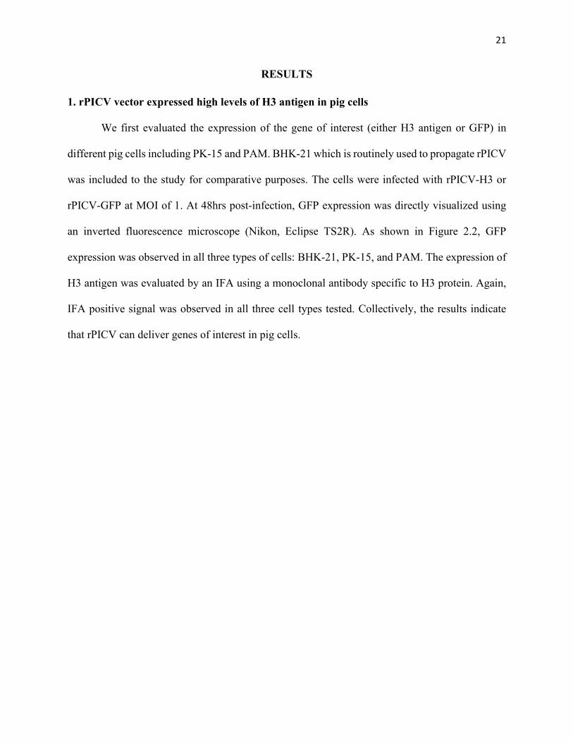
1. rPICV vector expressed high levels of H3 antigen in pig cells
We first evaluated the expression of the gene of interest (either H3 antigen or GFP) in
different pig cells including PK-15 and PAM. BHK-21 which is routinely used to propagate rPICV
was included to the study for comparative purposes. The cells were infected with rPICV-H3 or
rPICV-GFP at MOI of 1. At 48hrs post-infection, GFP expression was directly visualized using
an inverted fluorescence microscope (Nikon, Eclipse TS2R). As shown in Figure 2.2, GFP
expression was observed in all three types of cells: BHK-21, PK-15, and PAM. The expression of
H3 antigen was evaluated by an IFA using a monoclonal antibody specific to H3 protein. Again,
IFA positive signal was observed in all three cell types tested. Collectively, the results indicate
that rPICV can deliver genes of interest in pig cells.

22
Figure 2.2. Antigen expression in swine cells.
Expression of GFP and H3 protein in the three different cell types BHK-21, PK-15, and PAM
infected by rPICV-GFP and rPICV-H3 respectively.
23
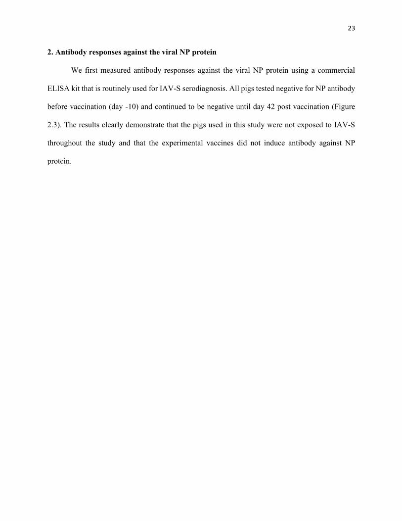
2. Antibody responses against the viral NP protein
We first measured antibody responses against the viral NP protein using a commercial
ELISA kit that is routinely used for IAV-S serodiagnosis. All pigs tested negative for NP antibody
before vaccination (day -10) and continued to be negative until day 42 post vaccination (Figure
2.3). The results clearly demonstrate that the pigs used in this study were not exposed to IAV-S
throughout the study and that the experimental vaccines did not induce antibody against NP
protein.

24
-10 28 420.00.20.40.60.81.01.2
Days post Vaccination
S/N
ratio
H3-ProteinrPICV-H3rPICV-GFPPBS
Figure 2.3. Antibody responses against the viral NP protein.
Serum samples collected before and after vaccination were used for a commercial inhibition
ELISA that detects antibody against IAV-S NP protein. Data are express as the sample to
negative ration (S/N ratio). The horizontal dotted line at S/N 0.6 indicates the assay cut-off.
Samples with S/N above this cutoff are consider negative. T01, T02, T03 and T04 refers to group
immunized with Mock, rPICV-GFP, rPICV-H3 and purified H3 protein respectively.

25
3. Antibody responses against GFP
To determine if rPICV was shedding and spreading from infected pigs, groups T01
(injected with PBS) and T02 (inoculated with rPICV-GFP) were commingled in the same room
throughout the course of this study. Antibodies against GFP were measured in plasma samples
collected at multiple time points before and after inoculation using an indirect ELISA. As shown
in Figure 2.4, pigs inoculated with rPICV-GFP (T02) had GFP-specific antibodies whereas pigs
inoculated with PBS did not. The results suggest that pigs inoculated with rPICV did not transmit
the virus to the contact pigs.
0 14 21 28 35 420.0
0.2
0.4
0.6
0.8
1.0
Days post Vaccination
OD
at 4
05nm
PBSrPICV-GFP
Figure 2.4. Antibody response against GFP.
Plasma samples collected at different time points after vaccination were subjected to an indirect
ELISA that detect antibodies against GFP. Data expressed as mean and standard error of mean of
optical density (OD) at 405nm.

26
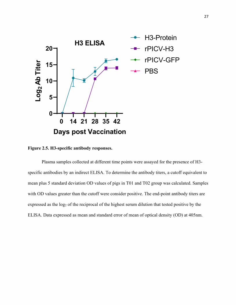
4. Antibody against H3 protein
Antibodies specific to H3 antigen were measured by an indirect ELISA. H3-specific
antibodies were not detected in pigs in T01 and T02 until day 42, the day of challenge infection
with a live H3N2 virus. In contrast, pigs in T03 and T04 groups exhibited high titers of H3-
specific antibodies (Figure 2.5). H3-specific antibodies could be detected in T04 group
(immunized with recombinant H3 protein with adjuvant) at day 14 after the first dose of
immunization whereas these antibodies were not detected in T03 group (immunized with rPICV-
H3) until day 28, corresponding to day 7 after the second immunization. However, no significant
difference in the antibody titer between T03 and T04 was observed one week after the second
immunization. The results indicate that immunization of pigs with recombinant H3 protein
together with adjuvant elicited faster antibody responses than immunization with the rPICV-H3
viral vector.
27
0 14 21 28 35 420
5
10
15
20
Days post Vaccination
Log 2
AbTi
ter
H3-ProteinrPICV-H3rPICV-GFPPBS
H3 ELISA
Figure 2.5. H3-specific antibody responses.
Plasma samples collected at different time points were assayed for the presence of H3-
specific antibodies by an indirect ELISA. To determine the antibody titers, a cutoff equivalent to
mean plus 5 standard deviation OD values of pigs in T01 and T02 group was calculated. Samples
with OD values greater than the cutoff were consider positive. The end-point antibody titers are
expressed as the log2 of the reciprocal of the highest serum dilution that tested positive by the
ELISA. Data expressed as mean and standard error of mean of optical density (OD) at 405nm.

28
5. Virus neutralization and hemagglutination inhibition antibody responses
Next, virus neutralization (VN) and hemagglutination inhibition (HI) antibody titers were
evaluated. Background levels of VN and HI antibody titers (approximately 1:40) were observed
in all pigs before immunization. VN and HI antibody titers did not increase in T01 and T02 group
whereas these antibody titers significantly increased in T03 and T04 at 21 DPV and increased even
further at 42 DPV. No significant difference in VN and HI titers between T03 and T04 groups
were observed (Figure 2.6). The results indicate that both rPICV-H3 and recombinant H3 vaccine
were able to elicit functional antibodies that block IAV-S infection.
A
0 21 420
5
10
15
Days post VaccinationLog 2
Nut
raliz
ing
Antib
ody
Tite
r
Virus Neutralization
H3-ProteinrPICV-H3rPICV-GFPPBS

29
B
0 21 420
5
10
15
Days post VaccinationLog 2
Nut
raliz
ing
Antib
ody
Tite
rHAI
H3-ProteinrPICV-H3rPICV-GFPPBS
Figure 2.6. Virus neutralizing (VN) and hemagglutination inhibition (HI) antibody
responses.
(A) Plasma samples from 0, 21 and 42 days post vaccination (DPV) were used to determine the
virus neutralization titers against H3N2 TX98 in the MDCK cells. (B) Plasma samples from 0, 21
and 42 DPV were used to determine the hemagglutination inhibition titer against H3N2 virus.
Horizontal dotted lines indicate the cutoff of the assays at 5.32 Log2 neutralizing antibody titer
equivalent of 1:40 times sample dilution. Data expressed as mean and standard error of mean of
neutralizing antibody titer.

30
6. Detection of IAV-S RNA in nasal swab samples
All animals were challenged by an intra-tracheal inoculation with the H3N2 TX98 strain
at 42 DPV. Nasal swabs were collected daily from all animals to measure the IAV-S shedding.
Viral RNA was detected from all pigs in T01 and T02 groups starting from day 1 post-challenge.
On the other hand, viral RNA was not detected in any of the pigs from T04 group at any time-
points post challenge (Figure 2.7). For T03 group, two pigs did not have any detectable levels of
viral RNA at any time points post challenge. The other two pigs had detectable viral RNA at only
one time on day 1 and day 4 post challenge.
0 1 2 3 4 5-5
0
5
10
15
Days post ChallengeLog 1
0 Vira
l cop
y nu
m/ μ
l of R
NA
H3-ProteinrPICV-H3rPICV-GFPPBS
Figure 2.7. Detection of IAV-S NP RNA in nasal swab samples
Nasal swabs collected daily after challenge infection were subjected to a quantitative real-time
PCR to determine the amount of viral RNA in the samples. Data are expressed as mean and
standard error of mean of the viral RNA copy number per µl of RNA sample from all animals of
one group.

31
7. Detection of viral RNA and infectious virus in BALF
All pigs were humanely sacrificed at 5 DPC and BALF was collected to measure viral
RNA by using a real-time PCR. High levels of viral RNA were detected in BALF from all pigs
in T01 and T02 groups (Figure 2.8A). In contrast, viral RNA was not detected in BALF from any
pigs in T04 group. Whereas one of the pigs in T03 group had IAV-S NP RNA. This pig also had
viral RNA in its nasal swab collected at 4 DPC.
To determine viral infectivity, samples of BALF were titrated in MDCK cells. All BALF
samples collected from pigs in T01 and T02 groups had infectious virus with the titer ranging
from 104-105.75 TCID50/ml (figure 2.8 B). On the other hand, infectious virus was not detected in
BALF of any pigs in T03 and T04 group. Even though BALF from one pig in T03 group
exhibited detectable level of viral RNA, this sample was not infectious when inoculated onto
MDCK.
A H3-P
rotein
rPICV-H
3
rPICV-G
FPPBS
-2
0
2
4
6
8
Log 1
0 Vira
l cop
y nu
m/ μ
l of R
NA
ns
ns
✱✱✱
✱✱

32
B H3-P
rotein
rPICV-H
3
rPICV-G
FPPBS
0
2
4
6
8
10Lo
g 10 T
CID
50/m
l
ns
ns
✱✱✱✱
Figure 2.8. Detection of viral RNA and infectious virus in BALF.
(A) RNA was extracted from BALF and used for quantitative real-time PCR. Data are expressed
as log10 viral genome copy number per µl of RNA sample. (B) Infectious virus in BALF were
titrated in MDCK cells. Data are expressed as log10 TCID50 per mL of sample and Error bars
indicate the standard error of mean value. ns-No significance, ** p≤0.01, *** p≤0.001, ****
p≤0.0001

33
8. Lung pathology
Typical macroscopic lung lesions were observed in pigs T01 and T02 groups with the
mean lung consolidation ranging from 2% to 4% (Figure 2.9). The lesions were more profound
on the left cardiac lung lobe. On the other hand, lung consolidation was not observed in pigs
from T03 and T04 groups.
A

34
B H3-P
rotein
rPICV-H
3
rPICV-G
FPPBS
0
2
4
6
8
10%
Lun
g C
onso
lidat
ion
ns
ns
✱✱
Figure 2.9. Macroscopic lung lesion scores.
(A) representative images for presence of lung lesion indicated by the black arrows pointing at
red depression in the lung of pigs from different groups. (B) percentage lung consolidation
quantifying the scoring of macroscopic lesion in lungs for all the groups. Data expressed as mean
and standard error of mean % lung consolidation for all the animals in each group. ns-No
significance, ** p≤0.01

35
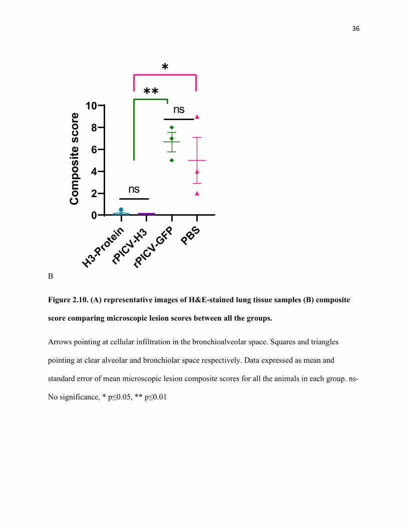
Microscopic lung lesion scores followed a similar trend as the macroscopic scores. Pigs
in T01 and T02 groups exhibited significantly high microscopic lesions characterized by
necrotizing bronchiolitis with peribronchiolar lymphocytic cuffing and interstitial pneumonia.
The mean composite microscopic scores for pigs in T01 and T02 groups varied from 5 to 7
(Figure 2.10). On the other hand, no microscopic lesions were observed in T03 and T04 groups.
A
36
B H3-P
rotein
rPICV-H
3
rPICV-G
FPPBS
0
2
4
6
8
10
Com
posi
te s
core
ns
ns✱✱
✱
Figure 2.10. (A) representative images of H&E-stained lung tissue samples (B) composite
score comparing microscopic lesion scores between all the groups.
Arrows pointing at cellular infiltration in the bronchioalveolar space. Squares and triangles
pointing at clear alveolar and bronchiolar space respectively. Data expressed as mean and
standard error of mean microscopic lesion composite scores for all the animals in each group. ns-
No significance, * p≤0.05, ** p≤0.01

37
9. Detection of virus infected cells in lung and trachea
RNA in situ hybridization was used to detect virus infected cells from lung and trachea
sections. As shown in Figure 2.11, large number of virus infected cells were observed in both
lung and tracheal sections of pigs in T01 and T02 groups. In contrast, no virus infected cells
were observed in lung and trachea section of pigs in T03 and T03 groups
A

38
B H3-P
rotein
rPICV-H
3
rPICV-G
FPPBS
0
2
4
6
8
10 C
ompo
site
sco
re
ns
ns
✱✱
C

39
D H3-P
rotein
rPICV-H
3
rPICV-G
FPPBS
0.0
0.5
1.0
1.5
2.0
2.5
Com
posi
te s
core
ns
ns✱✱✱
✱✱
Figure 2.11. In situ for IAV-S RNA in the tissue
Lung (A) and trachea (C) tissue section showing presence of IAV-NP RNA. Quantitative
comparison of presence of viral RNA in the lung(B) and trachea (D) samples. Data expressed as
mean and standard error of mean composite scores for all the animals in each group. ns-No
significance, ** p≤0.01, *** p<0.001

40
DISCUSSION
Swine influenza virus, first isolated from pigs in 1931 [19], were found to have negative
sense segmented RNA genome. This inherent property of the flu virus genome allows it to
frequently mutate and generate new variants either by exchanging RNA segments between
different antigenic strain or due to accumulation of point mutations because of error prone
replication. Most efficient way of protecting the pigs is by preventing the spread of the virus by
vaccinating the animals against influenza virus [34].
Majority of the licensed vaccines for pigs are based on the whole inactivated viruses, which
are safe and provides robust protection against the homologous infection but fails to show any
efficacy when a heterologous strain of virus infects the animals. In the presence of maternally
derived antibodies, vaccination of piglets with WIV enhances the respiratory disease pathology
rather than protecting them [45]. The live attenuated or modified live vaccine for influenza A virus
protects the pigs from homologous infection and shows some level of efficacy against heterologous
infection as well. But it must be noted that the LAIV is a replicating virus and the fact that it may
reassort with the wild type infectious IAV co-circulating in the field and revert to being infectious
cannot be eliminated [47]. The vaccines based on RNA particle (Sequivity, Merck) technology is
a good candidate for frequently updating the vaccine with HA genes from current circulating strain
from a locality. But the challenge with the RNA particle technology is the requirement of large
amount of recombinant RNA expression. Expressing large amount RNA can be difficult,
expensive and time consuming, limiting the availability of the vaccine. The poor stability of the
RNA particle is also another issue [94].
Using a recombinant virus that can infect the porcine cells and deliver the HA antigen into
the system without causing any disease can be a useful alternative in influenza virus vaccine

41
development. Recombinant Pichinde virus is one such viral vector that can infect the porcine cells
and express antigen very well, as shown by PK-15 and primary PAM cells infected with rPICV-
GFP and rPICV-H3. One of the concerns associated with live viruses is that they can replicate and
spread from one animal to another [95]. The PBS group pigs even after comingling with rPICV-
GFP infected pigs in the same room for over 42 days, did not have any anti-GFP antibodies in the
plasma sample as detected by ELISA. If the GFP protein expressing pichinde virus was shedding,
pigs from PBS group would have picked them up thereby express antibodies against the protein.
This indicated that the pichinde virus does not shed or spread from spread from one animal to
another.
Interestingly, for humoral immune response, while the H3-protein immunized pigs
expressed high titer of anti-HA antibodies after first vaccination, the rPICV-H3 immunized pigs
expressed significant levels of anti-H3 antibodies only after the second immunization. On
determining the functional response of these Anti-HA antibodies by virus neutralization and
hemagglutination inhibition assay it was observed that even though the H3-protein group had
presence of high level of antibodies against the HA protein starting from the first dose itself,
significant levels of neutralizing antibody titer was found only after the booster immunization, just
like that of the rPICV-H3 immunized pigs. This can be attributed to the fact that the antibodies
expressed in H3-protein injected animals was not specific to the neutralizing epitope of the H3
protein and may be binding to some other region of the full length H3 protein, that was used for
performing ELISA. This indicated that the early antibody response seen in the ELISA for H3
treated animals did not have a protective role. The high concentration of H3 protein used to
vaccinate the pigs might have also contributed to the high levels of early antibodies.

42
When the vaccinated animals were challenged with homologous influenza virus strain,
none of the pigs from H3 protein vaccinated group showed any viral RNA in the nasal swab or
BAL fluid sample, whereas two of the four pigs from rPICV-H3 vaccinated group showed the
presence of viral RNA at two different time points in the nasal swab sample and only one animal
in the BAL fluid. When the same BAL fluid samples were used to detect the presence of infectious
virus particle, it was observed that even though some of the rPICV-H3 vaccinated pigs had viral
RNA they were not from infectious virus particle as none of the sample could infect or replicate
in the MDCK cells compared to which virus in the BALF sample from PBS and rPICV-GFP group
replicated in the MDCK cells with high viral titer. Along with these results the absence of any
macroscopic or microscopic lung lesion and viral RNA in tissue of the infected animals indicated
that the vaccination of rPICV-H3 completely protected all the pigs just like the H3 protein
immunization.
Overall, it was concluded that recombinant Pichinde virus can be a potential viral vector
for the development of swine influenza vaccines to administer in the pigs, providing solid
protection against the homologous swine influenza challenge. In future rPICV construct with HA
from other circulating subtypes of swine influenza virus belonging to H1N1, H1N2 and H3N2 [20]
can be used to vaccinate the pigs and determine if the animals are protected and whether equal
level of protective immune response is elicited against each subtype to eventually develop a
broadly protecting vaccine against Influenza A viruses of swine.

43
CHAPTER 3: OPTIMIZATION AND EVALUATION OF A METHOD FOR
MONOCLONAL ANTIBODY GENERATION
ABSTRACT
A new method for generation of monoclonal antibody (mAb) was evaluated in mice model
for optimizing the method to express mAb from pigs. Antigen specific memory B cells were sorted
out and cultured in-vitro with feeder cells expressing CD40L and exogenous recombinant
cytokines IL-2 and IL-21. Variable genes were isolated from the cultured B cell clones expressing
antibodies against the antigen and cloned in expression vectors containing constant region of
mouse IgG heavy chain and light chain separately. The vectors containing full length heavy and
light chain of the antibody were transfected into HEK-293T cells. Finally, ELISA performed for
NSP7 protein with the transfected cell culture supernatant detected the presence of functional anti-
NSP7 antibodies, indicating that the method used in this study for monoclonal antibody generation
worked. In future this same technique can be applied to express monoclonal antibodies for species
of other origin like pigs.
Expressed by single B cell clones, monoclonal antibodies are a homogenous mix of
immunoglobulins targeting a specific epitope of an antigen [96]. Each antibody consists of a pair
of identical heavy and light chains. The light chain weighing ~22kDa [97], has an amino terminal
and a carboxy terminal. The amino terminal half consists of the variable region (VL) and carboxy
terminal possess the constant region (CL). There are two major types of light chain constant
sequences- κ and λ [98]. In humans there are 60% of light chains that are κ and 40% that belong
to λ. But unlike humans, in mouse only 5% of the light chains are of λ-type [99]. The heavy chain
constant region is larger when compared to the light chain, consisting of three different constant
regions CH1, CH2, and CH3 at the carboxy terminal and a variable region VH at the amino

44
terminal. The two heavy and light chain dimers are held together by a flexible hinge region. The
heavy chain sequences can be of five different types referred to as - µ, δ, γ, ε, and α isotype, each
isotype giving rise to a specific class of antibodies- IgM, IgD, IgG, IgE and IgA respectively. The
molecular weight of heavy chain is approximately 55kDa [97] with 20% higher for IgM and IgE
molecule. While the sequence of the variable region varies from one antibody to another within
the same organism, constant domain sequence remains the same for a particular species [100]. It
is to be noted that both the heavy and light chains are encoded by two different mRNAs.
Monoclonal antibodies are of great importance as a diagnostic tool, where they can be used
to detect the presence of a pathogen, toxin, drug, hormone or they can be used to detect a certain
cell type or molecule to determine its function [101]. There are many mAbs that has been used as
therapeutic interventions too [102, 103]. The first FDA approved murine mAb OKT3 mAb targets
human CD3 receptor on T cell surface to prevent the graft rejection in individuals who undergo
organ transplants [104]. A mouse chimeric antibody rituximab is used to treat non-Hodgkin B cell
lymphoma [105, 106]. Rituximab binds to the CD20 receptor present on tumor B cells and elicits
an immune response against the malignant cells to kill them [107]. Humanized mAb palivizumab
targeted against respiratory syncytial virus is approved by the FDA for the treatment of the infected
children [108]. Apart from being a diagnostic or therapeutic tool, recently the usage of monoclonal
antibody to guide the designing of structure-based vaccine has seen significant advancement.
There are different ways in which the antibody can guide vaccine development. They can be either
conformation-based designing, or epitope-based designing [109]. Immunogenic antigens can take
more than one conformation [110], antibodies can be used to identify which conformation induces
a protective immune response [111]. Epitopes, the sites present on an antigen that can be detected
by an antibody or supersites consisting of multiple overlapping epitopes that can be recognized by

45
broadly protective antibodies can help in development of multivariant vaccines. In case of
Influenza, broadly recognizing antibodies induced by vaccination with diverse hemagglutinin were
shown to be binding to the hemagglutinin stem region [112]. New vaccines are already being
studied using these supersites for the development of multivalent vaccines against influenza virus
[113, 114].
To generate optimum number of monoclonal antibodies for all these applications, they are
expressed and purified in vitro. Monoclonal antibodies can be generated by different techniques.
The classical method involves hybridoma technique [115]. In this method lymphocytes from the
antigen stimulated animal are isolated from spleen and fused with the same species immortal
myeloma cells. The fused cells are cultured in a hypoxanthine-aminopterin-thymidine (HAT)
selection media. The aminopterin in the HAT media blocks the cells from synthesizing nucleotides
by the de novo pathway. Hypoxanthine and deoxythymidine allows cells with functional
hypoxanthine-guanine phosphoribosyltransferase (HGPRT) to synthesize nucleotides via the
salvage pathway [115]. Even though myeloma cell is immortal, they will dye in the HAT media,
as it does not have HGPRT. B cells have short life span, so they eventually die too. Only the fused
myeloma and B cells called hybridomas will be able to survive as combining immortality from
myeloma cells and presence of HGPRT in B cells [116]. Successful hybridoma colonies are later
screened for antigen specificity. The hybridoma cells can be cryopreserved for using any time in
future [117]. But the challenge with hybridoma technique is that it requires a suitable fusion
partner. For mouse, SP2/0 cells are the myeloma cell line that serve as the fusion partner [118].
But for pigs until now there is no known myeloma cell line that can be used to fuse with the B
cells [119]. Fusion efficiency of mouse myeloma cells and swine B cells is very low and the few
cells that successfully fuse to form hetero-hybridomas are highly unstable and lose antibody

46
expressing capacity after some time (as observed by us in our work), and thus makes it challenging
to generate antibodies of swine origin using hybridoma technology. Another method to generate
mAb depends on the separation of antigen specific single B cells from the immunized or infected
donor’s blood or splenocytes Antibody genes from the single B cells are isolated and PCR
amplified to clone into expression vectors [120]. But isolation of RNA from the single B cell limits
the amount of template available for PCR, making it difficult to amplify the antibody genes. To
overcome this issue of limited template, another method of monoclonal antibody generation
depends on growing the B cells for a short time span. Normally, in vivo, once the antigen
displaying B cell encounters the antigen specific T cell, the T cell recognizes the MHC-II
complexed antigenic peptides on the B cell’s surface and secretes CD40L [121], costimulatory
cytokines like IFN-γ, IL-4, IL-21 [122] and IL-2 [123]. The CD40L binds with the CD40 receptor
present on the surface of the B cell, leading to proliferation and differentiation of the B cells.
Interleukin-2 also aids the B cell proliferation and differentiation [124]. After the initial secretion
of IgM, cytokines released by the T cells, especially interleukin-21 stimulates these plasma B cells
expressing IgM to undergo Ig isotype switching [125], and produce IgG, in some cases IgA or IgE
rather than IgM [126]. Following the same principal antigen specific B cells are cultured
transiently in an in vitro setup along with feeder cells that express CD40L and co stimulatory
cytokines IL-2 and IL-21, increasing the amount of template availability for PCR amplification
[127, 128]. From this culture, B cell clones that are expressing antibodies targeting the immunogen
of interest can be selected for isolation and cloning of antibody genes to generate monoclonal
antibodies [129].
This study was performed for the optimization of B cell co-culture procedure as mentioned
before [20, 129], that can be applied to generate monoclonal antibodies from pigs by eliminating

47
the need of any suitable fusion partner or low amount of antibody gene PCR templates. Due to
ease of animal handling, antigen immunization, and sample processing we first evaluated the
feasibility of the B cell co-culture method in mice. As purified recombinant PRRSV-NSP7 protein
was already available with us, therefore the study was performed with this protein.
Firstly, NSP7 protein was labelled with a fluorophore to detect and separate antigen
specific B cells in Fluorescent activated cell sorting (FACS). 1µg of NSP7 protein was incubated
with Alexaflour-546 (AF546, Invitrogen, Cat#A20183) and conjugation was performed as per
manufacturer’s instructions. Labelling of NSP7 was confirmed by running SDS-PAGE for the
labelled protein. Un-tagged NSP7 protein was run along with the tagged protein as a positive
control. After the protein bands were transferred on to polyvinylidene difluoride (PVDF)
membrane, it was observed under ChemDoc (Figure 3.1 A). While unlabeled NSP7 protein
showed no bands in ChemDoc, the NSP7-AF546 showed bright band near 35kDa, same size as
that of NSP7 protein. The same membrane was then probed with anti-His tagged primary antibody
to detect the NSP7 protein with 6X His tag. After probing the membrane with HRP-labelled goat
anti-mouse IgG (KPL, Cat#5220-0360) secondary antibody, membrane was incubated with HRP-
substrate and bands were developed on a sheet of X ray film. It was observed that both untagged
NSP7 and labelled NSP7-AF546 had distinct bands near 35kDa (Figure 3.1 B), implying that the
image from ChemDoc where no bands for untagged NSP7 was observed was because of no AF546
labelling and not due to the absence of NSP7 protein.

48
A B
Figure 3.1. Labelling of antigen with fluorophore.
(A) untagged NSP7 protein and AF546 tagged NSP7 protein bands after running SDS-PAGE as
observed under ChemDoc. (B) His tag probed protein bands as observed on the X-ray film.
(Where NSP7 is untagged protein and NSP7-AF546 is protein labelled with the fluorophore.

49
After the confirmation of successful antigen labelling, a group of 8 weeks old BALB/C
mice were immunized with 10µg of NSP7 protein first emulsified in Freund’s complete adjuvant,
followed by boosting with same amount of protein emulsified in Freund’s incomplete adjuvant
(Figure 3.2 A). Serum samples were collected from the mice to perform ELISA against NSP7
protein in order to determine whether the mice were seroconverted or not (Figure 3.2 B). High
level of absorbance detected in ELISA clearly indicated that the animals were well seroconverted
and anti-NSP7 antibody was being expressed.

50
A
B
50 100
200
400
80016
0032
0064
0012
80025
60051
200
1024
000
1
2
3
4
Dilution factor
OD
at 4
50nm
Day 14Day 21Naive
Figure 3.2. Immunization of mice.
(A) schedule of mice immunization and spleen collection. (B) graphic representing OD values
from the ELISA against NSP7 using sera samples, showing seroconversion of mice. The data are
represented as mean and standard error of mean for each OD value at 450nm.

51
One week after the booster immunization, splenocytes from an immunized mouse and
another from a naïve mouse was isolated.
Both the samples were stained with four different cell surface markers: Zombie aqua
(Biolegend, Cat#423101), anti-mouse CD19 APC (Biolegend, Cat#115512), IgM AF488
(Biolegend, Cat#406522) and IgD Brilliant violet 711 (Biolegend, Cat#405731) along with NSP7-
AF546 all together and sorted by FACS instrument to obtain IgG expressing memory B cells.
Zombie Aqua was used to separate the live cells from total lymphocytes. CD19, a pan B cell
marker, was used to gate B cell population from live cells [130]. For the isolation of memory B
cells, IgM and IgD double negative population was selected from CD19+ cells [131]. Finally, to
detect the antigen specificity, IgM, IgD double negative (DN) cells were sorted for NSP7-AF546
stained cells (Figure 3.3 A).
A

52
B
C
Figure 3.3. Antigen specific memory B cell sorting.
(A) gating strategy for selecting NSP7 specific IgG expressing memory B cells. (B) Naïve cell
population stained with all the markers showing lymphocytes (lymphs) gated population, live
cells followed by single cell population gating. Below CD19+ population, followed by gating of

53
IgM, IgD DN cells. lastly gating for NSP7 stained cells. (C) Test immunized population stained
with all the markers in the same order as that of naïve sample.
Cell sorting by FACS indicated that although, there was no significant difference in the
CD19+ or DN population between naïve and immunized samples, cells from naïve mouse did not
show any staining for NSP7-546, whereas in test sample, 1-2% of DN population were positive
for NSP7-AF546 staining, confirming that the antigen staining was selective, and no non-specific
binding of NSP7-AF546 with memory B cells of naïve mice took place. For side-by-side
comparison of the effect of antigen specific enrichment of memory B cells, two sets of cell
population were sorted out from the immunized test sample. One that was total memory B cell
population-CD19+ and DN, the other, which was NSP7 specific memory B cells- CD19+, DN as
well as NSP7+. These sorted B cells were transiently cultured for 13 days with CD40L expressing
recombinant 3T3-msCD40L fibroblast cells and 50ng/ml of recombinant mouse IL-2 (Sigma,
cat#I0523-20UG) and IL-21 (Biolegend, cat#574504) in 96 well flat bottom plate. Each well was
seeded with 25 NSP7+ B cells. For the culture of total memory B cells, 100 cells were added to
each 96 well in order to increase the chance of each well obtaining a NSP7 specific cell. Few wells
were seeded with B cells along with just IL-2 and IL-21 and no 3T3-msCD40L feeder cells
(Figure3.4 A) to determine if the cytokines alone were enough or not to grow B cells in the absence
of the feeder cells. At the end of 13 day, it was observed that B cells only grew and proliferated
when both CD40L as well as cytokines were present (Figure 3.4 B), just the presence of cytokines
alone was not sufficient.

54
A
B
Figure 3.4. B cell co-culture.
(A) B cell cultured in medium with cytokine and no CD40L expressing cells. (B) B cells co-
cultured with CD40L expressing cells and cytokines. Small dots indicated by the arrow are the
expanding B cells, and apoptotic cells represented by the square box are the feeder cell. Images
were taken at 10X magnification on day 13 after seeding the cells.

55
Supernatant from the cell cultures were taken to detect the presence of antibodies against
NSP 7 by ELISA. It was observed that out of 30 different wells that were seeded with NSP7
specific memory B cells, except for one well, all of them were expressing high level of anti-NSP7
antibodies represented by most of the absorbance values above 4 OD as obtained in ELISA (figure
3.5). Compared to which, total memory B cell culture had only 6 wells positive for anti-NSP7 from
a total of 180 wells. Indicating, that just by enriching the B cell population for antigen, the
frequency of having positive B cell clones secreting antigen specific antibody was increased from
3.3% in total memory B cell population to 96.6% in NSP7 specific population.
NSP7+ Ig
G B
Total Ig
G B0
1
2
3
4
5
Abso
rban
ce a
t 450
nm
Figure 3.5. Frequency of B cell clones expressing anti-NSP7 antibodies.
Dotted line at 0.5 OD indicates the cutoff of absorbance value for a sample to be positive.

56
One of the positive wells from NSP7 specific sorted cell culture was selected to isolate the
antibody genes. Cells were lysed and total RNA was extracted from the sample. RNA was used to
obtain cDNA for the variable region of both the heavy and light chain IgG gene. The cDNA was
further PCR amplified and cloned into Topo pJet1.2 blunt end vector and six different colonies
each for heavy and light chain were send for sequencing. After confirming the sequence and
performing multiple sequence alignment by an online tool MUSCLE, it was concluded that the
variable regions of heavy and light chains were from two different antibodies. The sequences were
BLAST searched to annotate that the genes belonged to mouse IgG, following which variable
regions genes were further cloned into two different pCI vectors containing constant region for
heavy chain and light chain respectively. A total of four different full-length gene pCI constructs
named as H1-heavy from clone 1, L1-light from clone 1, H2-heavy from clone 2 and L2-light from
clone 2 were obtained. These vectors with full length heavy and light chain IgG sequences were
transfected into HEK-293 T cells for expressing the whole antibody. To maintain the functionality
of the antibody proper pairing of heavy chain with its cognate light chain is important, therefore
four different combinations of heavy and light chains were tried as shown in Figure 3.6 A.
Supernatant from the transfected cells were collected from day 1 until day 5. On performing
ELISA against NSP7 with the cell culture supernatants, it was observed that out of all the
combinations, H1L2 was the only one expressing a functional antibody that detected and bound to
NSP7 protein (figure 3.6). The level of antibody expression peaked on day 2 and 3 and decreased
by day 5. Compared to which H1L1, H2L1, H2L2 or mock transfected cells had no signal at all,
implying that overall, the procedure worked in mice. Functional antibodies specific to an antigen
were expressed that detected the NSP7 protein.

57
combinations Days post transfection (OD at 450nm)
1 2 3 4 5
H1L1 0.066 0.057 0.057 0.052 0.059
H1L2 0.499 0.873 0.780 0.723 0.592
H2L1 0.061 0.084 0.075 0.081 0.079
H2L2 0.103 0.092 0.082 0.075 0.084
Mock 0.089 0.082 0.082 0.065 0.057
A
B
1 2 3 4 5 0.0
0.2
0.4
0.6
0.8
1.0
Days post transfection
OD
at 4
50nm
H1L1 H1L2 H2L1H2L2 Mock
Figure 3.6 Expression of functional antibody.
(A) table indicating OD values of different combinations of antibody gene transfected cell
supernatant and mock supernatant from ELISA (B) graphic indicating the dynamics of antibody
expression at different time point from Transfected or mock cells.

58
DISCUSSION
Monoclonal antibody generation by the process of antigen specific B cell co-culture was
successfully optimized in the mice. The labeling of antigen showed specific activity and no naïve
cells were stained for NSP7, which when used to enrich memory B cell population binding to
NSP7 protein, enhanced the efficiency of anti-NSP7 expressing B cells in the culture. This shows
that the simple method of incubating the protein with a commercially available fluorophore
conjugation kit like that of AF-546 used in this study, is sufficient to label the protein. Even without
the need of other complex methods like labelling using a biotin-streptavidin tetramer molecule
[132], recombinant virus particles expressing fluorophore genes [133], or expression of
recombinant protein together with the fluorophore [134] that sometimes can result in the change
of antigen conformation and becoming unable to bind to the antibody or B cell receptor that
otherwise would have detected. By using the feeder cells expressing CD40L and the costimulatory
cytokines, the requirement of species-specific myeloma cell line was eliminated. The ligand and
cytokines also enabled single B cells to proliferate and undergo clonal expansion [127] which
increased the genomic content for the same monoclonal antibody. The increased amount of
template obtained facilitated better amplification of the antibody genes. In future, same
methodology can be used to try to make stable porcine monoclonal antibodies.

59
REFERENCES
1. Cheung, T.K. and L.L. Poon, Biology of influenza a virus. Ann N Y Acad Sci, 2007. 1102: p. 1-25.
2. Rossman, J.S. and R.A. Lamb, Influenza virus assembly and budding. Virology, 2011. 411(2): p. 229-36.
3. Boivin, S., et al., Influenza A virus polymerase: structural insights into replication and host adaptation mechanisms. J Biol Chem, 2010. 285(37): p. 28411-7.
4. Lamb, R.A. and P.W. Choppin, The gene structure and replication of influenza virus. Annu Rev Biochem, 1983. 52: p. 467-506.
5. Russell, C.J., Hemagglutinin Stability and Its Impact on Influenza A Virus Infectivity, Pathogenicity, and Transmissibility in Avians, Mice, Swine, Seals, Ferrets, and Humans. Viruses, 2021. 13(5).
6. Jin, H., et al., Influenza virus hemagglutinin and neuraminidase cytoplasmic tails control particle shape. EMBO J, 1997. 16(6): p. 1236-47.
7. Zhang, Y., et al., Targeting Hemagglutinin: Approaches for Broad Protection against the Influenza A Virus. Viruses, 2019. 11(5).
8. Dou, D., et al., Influenza A Virus Cell Entry, Replication, Virion Assembly and Movement. Front Immunol, 2018. 9: p. 1581.
9. Brandenburg, B., et al., Mechanisms of hemagglutinin targeted influenza virus neutralization. PLoS One, 2013. 8(12): p. e80034.
10. Coudeville, L., et al., Relationship between haemagglutination-inhibiting antibody titres and clinical protection against influenza: development and application of a bayesian random-effects model. BMC Med Res Methodol, 2010. 10: p. 18.
11. Zost, S.J., et al., Immunodominance and Antigenic Variation of Influenza Virus Hemagglutinin: Implications for Design of Universal Vaccine Immunogens. J Infect Dis, 2019. 219(Suppl_1): p. S38-S45.
12. Samji, T., Influenza A: understanding the viral life cycle. Yale J Biol Med, 2009. 82(4): p. 153-9.
13. Shao, W., et al., Evolution of Influenza A Virus by Mutation and Re-Assortment. Int J Mol Sci, 2017. 18(8).
14. Kim, H., R.G. Webster, and R.J. Webby, Influenza Virus: Dealing with a Drifting and Shifting Pathogen. Viral Immunol, 2018. 31(2): p. 174-183.
15. Mancera Gracia, J.C., et al., Influenza A Virus in Swine: Epidemiology, Challenges and Vaccination Strategies. Front Vet Sci, 2020. 7: p. 647.
16. Ryt-Hansen, P., et al., Co-circulation of multiple influenza A reassortants in swine harboring genes from seasonal human and swine influenza viruses. Elife, 2021. 10.
17. Krammer, F., et al., Influenza. Nat Rev Dis Primers, 2018. 4(1): p. 3. 18. Ma, W., R.E. Kahn, and J.A. Richt, The pig as a mixing vessel for influenza viruses:
Human and veterinary implications. J Mol Genet Med, 2008. 3(1): p. 158-66. 19. Shope, R.E., Swine Influenza : I. Experimental Transmission and Pathology. J Exp Med,
1931. 54(3): p. 349-59. 20. Anderson, T.K., et al., Population dynamics of cocirculating swine influenza A viruses in
the United States from 2009 to 2012. Influenza Other Respir Viruses, 2013. 7 Suppl 4: p. 42-51.
21. Webby, R.J., et al., Evolution of swine H3N2 influenza viruses in the United States. J

60
Virol, 2000. 74(18): p. 8243-51. 22. Ma, W., et al., Viral reassortment and transmission after co-infection of pigs with
classical H1N1 and triple-reassortant H3N2 swine influenza viruses. J Gen Virol, 2010. 91(Pt 9): p. 2314-21.
23. Rajao, D.S., et al., Antigenic and genetic evolution of contemporary swine H1 influenza viruses in the United States. Virology, 2018. 518: p. 45-54.
24. Gibbs, A.J., J.S. Armstrong, and J.C. Downie, From where did the 2009 'swine-origin' influenza A virus (H1N1) emerge? Virol J, 2009. 6: p. 207.
25. Guo, F., et al., Origin and Evolution of H1N1/pdm2009: A Codon Usage Perspective. Front Microbiol, 2020. 11: p. 1615.
26. Song, Y., et al., Identification, Genetic Analysis, and Pathogenicity of Classical Swine H1N1 and Human-Swine Reassortant H1N1 Influenza Viruses from Pigs in China. Viruses, 2020. 12(1).
27. Neumann, G., T. Noda, and Y. Kawaoka, Emergence and pandemic potential of swine-origin H1N1 influenza virus. Nature, 2009. 459(7249): p. 931-9.
28. Nelson, M.I., et al., Global migration of influenza A viruses in swine. Nat Commun, 2015. 6: p. 6696.
29. Vijaykrishna, D., et al., The contrasting phylodynamics of human influenza B viruses. Elife, 2015. 4: p. e05055.
30. Bolton, M.J., et al., Antigenic evolution of H3N2 influenza A viruses in swine in the United States from 2012 to 2016. Influenza Other Respir Viruses, 2019. 13(1): p. 83-90.
31. Zell, R., et al., Novel reassortant swine H3N2 influenza A viruses in Germany. Sci Rep, 2020. 10(1): p. 14296.
32. Chauhan, R.P. and M.L. Gordon, A Systematic Review Analyzing the Prevalence and Circulation of Influenza Viruses in Swine Population Worldwide. Pathogens, 2020. 9(5).
33. Lopez, C.E. and K.L. Legge, Influenza A Virus Vaccination: Immunity, Protection, and Recent Advances Toward A Universal Vaccine. Vaccines (Basel), 2020. 8(3).
34. Salvesen, H.A. and C.B.A. Whitelaw, Current and prospective control strategies of influenza A virus in swine. Porcine Health Manag, 2021. 7(1): p. 23.
35. Ma, W., Swine influenza virus: Current status and challenge. Virus Res, 2020. 288: p. 198118.
36. Rajao, D.S., et al., Pathogenesis and vaccination of influenza A virus in swine. Curr Top Microbiol Immunol, 2014. 385: p. 307-26.
37. Vincent, A.L., et al., Influenza A virus vaccines for swine. Vet Microbiol, 2017. 206: p. 35-44.
38. Nuwarda, R.F., A.A. Alharbi, and V. Kayser, An Overview of Influenza Viruses and Vaccines. Vaccines (Basel), 2021. 9(9).
39. Abente, E.J., et al., Comparison of Adjuvanted-Whole Inactivated Virus and Live-Attenuated Virus Vaccines against Challenge with Contemporary, Antigenically Distinct H3N2 Influenza A Viruses. J Virol, 2018. 92(22).
40. Ainai, A., et al., Intranasal Administration of Whole Inactivated Influenza Virus Vaccine as a Promising Influenza Vaccine Candidate. Viral Immunol, 2017. 30(6): p. 451-462.
41. Kimble, J.B., et al., Vaccine-Associated Enhanced Respiratory Disease following Influenza Virus Infection in Ferrets Recapitulates the Model in Pigs. J Virol, 2022. 96(5): p. e0172521.
42. Souza, C.K., et al., The type of adjuvant in whole inactivated influenza a virus vaccines

61
impacts vaccine-associated enhanced respiratory disease. Vaccine, 2018. 36(41): p. 6103-6110.
43. Richt, J.A., et al., Vaccination of pigs against swine influenza viruses by using an NS1-truncated modified live-virus vaccine. J Virol, 2006. 80(22): p. 11009-18.
44. Gauger, P.C., et al., Live attenuated influenza A virus vaccine protects against A(H1N1)pdm09 heterologous challenge without vaccine associated enhanced respiratory disease. Virology, 2014. 471-473: p. 93-104.
45. Vincent, A.L., et al., Live attenuated influenza vaccine provides superior protection from heterologous infection in pigs with maternal antibodies without inducing vaccine-associated enhanced respiratory disease. J Virol, 2012. 86(19): p. 10597-605.
46. Lee, J., et al., Bat influenza vectored NS1-truncated live vaccine protects pigs against heterologous virus challenge. Vaccine, 2021. 39(14): p. 1943-1950.
47. Sharma, A., et al., Detection of live attenuated influenza vaccine virus and evidence of reassortment in the U.S. swine population. J Vet Diagn Invest, 2020. 32(2): p. 301-311.
48. Sakurai, F., M. Tachibana, and H. Mizuguchi, Adenovirus vector-based vaccine for infectious diseases. Drug Metab Pharmacokinet, 2022. 42: p. 100432.
49. Roberts, A., et al., Attenuated vesicular stomatitis viruses as vaccine vectors. J Virol, 1999. 73(5): p. 3723-32.
50. Weaver, E.A. and M.A. Barry, Low seroprevalent species D adenovirus vectors as influenza vaccines. PLoS One, 2013. 8(8): p. e73313.
51. Wesley, R.D. and K.M. Lager, Overcoming maternal antibody interference by vaccination with human adenovirus 5 recombinant viruses expressing the hemagglutinin and the nucleoprotein of swine influenza virus. Vet Microbiol, 2006. 118(1-2): p. 67-75.
52. Tang, M., J.A. Harp, and R.D. Wesley, Recombinant adenovirus encoding the HA gene from swine H3N2 influenza virus partially protects mice from challenge with heterologous virus: A/HK/1/68 (H3N2). Arch Virol, 2002. 147(11): p. 2125-41.
53. Barefoot, B.E., C.J. Sample, and E.A. Ramsburg, Recombinant vesicular stomatitis virus expressing influenza nucleoprotein induces CD8 T-cell responses that enhance antibody-mediated protection after lethal challenge with influenza virus. Clin Vaccine Immunol, 2009. 16(4): p. 488-98.
54. Roberts, A., et al., Vaccination with a recombinant vesicular stomatitis virus expressing an influenza virus hemagglutinin provides complete protection from influenza virus challenge. J Virol, 1998. 72(6): p. 4704-11.
55. de Vries, R.D. and G.F. Rimmelzwaan, Viral vector-based influenza vaccines. Hum Vaccin Immunother, 2016. 12(11): p. 2881-2901.
56. Altenburg, A.F., et al., Modified vaccinia virus ankara (MVA) as production platform for vaccines against influenza and other viral respiratory diseases. Viruses, 2014. 6(7): p. 2735-61.
57. Volz, A. and G. Sutter, Modified Vaccinia Virus Ankara: History, Value in Basic Research, and Current Perspectives for Vaccine Development. Adv Virus Res, 2017. 97: p. 187-243.
58. Price, P.J., et al., Modified Vaccinia virus Ankara: innate immune activation and induction of cellular signalling. Vaccine, 2013. 31(39): p. 4231-4.
59. Hessel, A., et al., Vectors based on modified vaccinia Ankara expressing influenza H5N1 hemagglutinin induce substantial cross-clade protective immunity. PLoS One, 2011. 6(1): p. e16247.

62
60. Castrucci, M.R., et al., Modified vaccinia virus Ankara expressing the hemagglutinin of pandemic (H1N1) 2009 virus induces cross-protective immunity against Eurasian 'avian-like' H1N1 swine viruses in mice. Influenza Other Respir Viruses, 2014. 8(3): p. 367-75.
61. Bergqvist, C., M. Kurban, and O. Abbas, Orf virus infection. Rev Med Virol, 2017. 27(4).
62. Spyrou, V. and G. Valiakos, Orf virus infection in sheep or goats. Vet Microbiol, 2015. 181(1-2): p. 178-82.
63. Wang, R., et al., Orf virus: A promising new therapeutic agent. Rev Med Virol, 2019. 29(1): p. e2013.
64. Joshi, L.R., et al., Protective Efficacy of an Orf Virus-Vector Encoding the Hemagglutinin and the Nucleoprotein of Influenza A Virus in Swine. Front Immunol, 2021. 12: p. 747574.
65. Vander Veen, R.L., D.L. Harris, and K.I. Kamrud, Alphavirus replicon vaccines. Anim Health Res Rev, 2012. 13(1): p. 1-9.
66. Perri, S., et al., An alphavirus replicon particle chimera derived from venezuelan equine encephalitis and sindbis viruses is a potent gene-based vaccine delivery vector. J Virol, 2003. 77(19): p. 10394-403.
67. Erdman, M.M., et al., Alphavirus replicon particle vaccines developed for use in humans induce high levels of antibodies to influenza virus hemagglutinin in swine: proof of concept. Vaccine, 2010. 28(3): p. 594-6.
68. Bosworth, B., et al., Replicon particle vaccine protects swine against influenza. Comp Immunol Microbiol Infect Dis, 2010. 33(6): p. e99-e103.
69. Sanchez-Puig, J.M., M.M. Lorenzo, and R. Blasco, A vaccinia virus recombinant transcribing an alphavirus replicon and expressing alphavirus structural proteins leads to packaging of alphavirus infectious single cycle particles. PLoS One, 2013. 8(10): p. e75574.
70. Trapido, H. and C. Sanmartin, Pichinde virus, a new virus of the Tacaribe group from Colombia. Am J Trop Med Hyg, 1971. 20(4): p. 631-41.
71. Bishop, D.H. and D.D. Auperin, Arenavirus gene structure and organization. Curr Top Microbiol Immunol, 1987. 133: p. 5-17.
72. Gonzalez, J.P., et al., Arenaviruses. Curr Top Microbiol Immunol, 2007. 315: p. 253-88. 73. Leung, W.C., et al., Pichinde virus L and S RNAs contain unique sequences. J Virol,
1981. 37(1): p. 48-54. 74. Shao, J., Y. Liang, and H. Ly, Roles of Arenavirus Z Protein in Mediating Virion
Budding, Viral Transcription-Inhibition and Interferon-Beta Suppression. Methods Mol Biol, 2018. 1604: p. 217-227.
75. Ferron, F., et al., Transcription and replication mechanisms of Bunyaviridae and Arenaviridae L proteins. Virus Res, 2017. 234: p. 118-134.
76. Salvato, M.S., The Arenaviridae. Viruses. 1993, New York: Plenum Press. xxvi, 401 pages.
77. Shao, J., et al., Characterization of the Glycoprotein Stable Signal Peptide in Mediating Pichinde Virus Replication and Virulence. J Virol, 2016. 90(22): p. 10390-10397.
78. Dhanwani, R., et al., Establishment of Bisegmented and Trisegmented Reverse Genetics Systems to Generate Recombinant Pichinde Viruses. Methods Mol Biol, 2018. 1604: p. 247-253.
79. Brisse, M., et al., RIG-I and MDA5 Protect Mice From Pichinde Virus Infection by

63
Controlling Viral Replication and Regulating Immune Responses to the Infection. Front Immunol, 2021. 12: p. 801811.
80. Meyer, B. and H. Ly, Inhibition of Innate Immune Responses Is Key to Pathogenesis by Arenaviruses. J Virol, 2016. 90(8): p. 3810-3818.
81. Dhanwani, R., H. Ly, and Y. Liang, Recombinant Tri-Segmented Pichinde Virus as a Novel Live Viral Vaccine Platform. Methods Mol Biol, 2017. 1581: p. 169-179.
82. Lan, S., et al., Development of infectious clones for virulent and avirulent pichinde viruses: a model virus to study arenavirus-induced hemorrhagic fevers. J Virol, 2009. 83(13): p. 6357-62.
83. Dhanwani, R., et al., A Novel Live Pichinde Virus-Based Vaccine Vector Induces Enhanced Humoral and Cellular Immunity after a Booster Dose. J Virol, 2015. 90(5): p. 2551-60.
84. Kumar, P., et al., Development of a Recombinant Pichinde Virus-Vectored Vaccine against Turkey Arthritis Reovirus and Its Immunological Response Characterization in Vaccinated Animals. Pathogens, 2021. 10(2).
85. Li, Y. and I. Robertson, The epidemiology of swine influenza. Anim Dis, 2021. 1(1): p. 21.
86. Pomorska-Mol, M., et al., Coinfection modulates inflammatory responses, clinical outcome and pathogen load of H1N1 swine influenza virus and Haemophilus parasuis infections in pigs. BMC Vet Res, 2017. 13(1): p. 376.
87. Kosik, I. and J.W. Yewdell, Influenza Hemagglutinin and Neuraminidase: Yin(-)Yang Proteins Coevolving to Thwart Immunity. Viruses, 2019. 11(4).
88. Chu, V.C. and G.R. Whittaker, Influenza virus entry and infection require host cell N-linked glycoprotein. Proc Natl Acad Sci U S A, 2004. 101(52): p. 18153-8.
89. Yang, J., et al., A new role of neuraminidase (NA) in the influenza virus life cycle: implication for developing NA inhibitors with novel mechanism of action. Rev Med Virol, 2016. 26(4): p. 242-50.
90. van de Sandt, C.E., J.H. Kreijtz, and G.F. Rimmelzwaan, Evasion of influenza A viruses from innate and adaptive immune responses. Viruses, 2012. 4(9): p. 1438-76.
91. Zhang, H., et al., A unique feature of swine ANP32A provides susceptibility to avian influenza virus infection in pigs. PLoS Pathog, 2020. 16(2): p. e1008330.
92. Castrucci, M.R., et al., Genetic reassortment between avian and human influenza A viruses in Italian pigs. Virology, 1993. 193(1): p. 503-6.
93. Gauger, P.C., et al., Kinetics of lung lesion development and pro-inflammatory cytokine response in pigs with vaccine-associated enhanced respiratory disease induced by challenge with pandemic (2009) A/H1N1 influenza virus. Vet Pathol, 2012. 49(6): p. 900-12.
94. Wadhwa, A., et al., Opportunities and Challenges in the Delivery of mRNA-based Vaccines. Pharmaceutics, 2020. 12(2).
95. Robert-Guroff, M., Replicating and non-replicating viral vectors for vaccine development. Curr Opin Biotechnol, 2007. 18(6): p. 546-56.
96. Zhang, C., Hybridoma technology for the generation of monoclonal antibodies. Methods Mol Biol, 2012. 901: p. 117-35.
97. Ma, H. and R. O'Kennedy, The Structure of Natural and Recombinant Antibodies. Methods Mol Biol, 2015. 1348: p. 7-11.
98. Haughton, G., L.L. Lanier, and G.F. Babcock, The murine kappa light chain shift. Nature,

64
1978. 275(5676): p. 154-7. 99. Liu, T., et al., Frequency of lambda light chain subtypes in mouse antibodies to the 2,4-
dinitrophenyl (DNP) group. Eur J Immunol, 1984. 14(7): p. 667-72. 100. Stanfield, R.L. and I.A. Wilson, Antibody Structure. Microbiol Spectr, 2014. 2(2). 101. Siddiqui, M.Z., Monoclonal antibodies as diagnostics; an appraisal. Indian J Pharm Sci,
2010. 72(1): p. 12-7. 102. Nelson, P.N., et al., Monoclonal antibodies. Mol Pathol, 2000. 53(3): p. 111-7. 103. Berger, M., V. Shankar, and A. Vafai, Therapeutic applications of monoclonal
antibodies. Am J Med Sci, 2002. 324(1): p. 14-30. 104. Norman, D.J., Mechanisms of action and overview of OKT3. Ther Drug Monit, 1995.
17(6): p. 615-20. 105. Maloney, D.G., Advances in immunotherapy of hematologic malignancies. Curr Opin
Hematol, 1998. 5(4): p. 237-43. 106. Ockert, D., et al., Advances in cancer immunotherapy. Immunol Today, 1999. 20(2): p.
63-5. 107. Cerny, T., et al., Mechanism of action of rituximab. Anticancer Drugs, 2002. 13 Suppl 2:
p. S3-10. 108. Garegnani, L., et al., Palivizumab for preventing severe respiratory syncytial virus (RSV)
infection in children. Cochrane Database Syst Rev, 2021. 11: p. CD013757. 109. Kwong, P.D., B.J. DeKosky, and J.B. Ulmer, Antibody-guided structure-based vaccines.
Semin Immunol, 2020. 50: p. 101428. 110. Nestorowicz, A.N., D.O. White, and D.C. Jackson, Conformational changes in influenza
virus haemagglutinin and its monomer detected by monoclonal antibodies. Vaccine, 1985. 3(3 Suppl): p. 175-81.
111. Zhang, B., et al., Protection of calves by a prefusion-stabilized bovine RSV F vaccine. NPJ Vaccines, 2017. 2: p. 7.
112. Joyce, M.G., et al., Vaccine-Induced Antibodies that Neutralize Group 1 and Group 2 Influenza A Viruses. Cell, 2016. 166(3): p. 609-623.
113. Krammer, F. and P. Palese, Universal Influenza Virus Vaccines That Target the Conserved Hemagglutinin Stalk and Conserved Sites in the Head Domain. J Infect Dis, 2019. 219(Suppl_1): p. S62-S67.
114. Impagliazzo, A., et al., A stable trimeric influenza hemagglutinin stem as a broadly protective immunogen. Science, 2015. 349(6254): p. 1301-6.
115. Parray, H.A., et al., Hybridoma technology a versatile method for isolation of monoclonal antibodies, its applicability across species, limitations, advancement and future perspectives. Int Immunopharmacol, 2020. 85: p. 106639.
116. Tomita, M. and K. Tsumoto, Hybridoma technologies for antibody production. Immunotherapy, 2011. 3(3): p. 371-80.
117. Greenfield, E.A., Liquid Nitrogen Storage of Hybridoma Cells. Cold Spring Harb Protoc, 2020. 2020(10).
118. Melixetian, M.B., et al., Mouse myeloma cell line Sp2/0 multidrug-resistant variant as parental cell line for hybridoma construction. Hybrid Hybridomics, 2003. 22(5): p. 321-7.
119. Puckette, M.C., et al., Generation and characterization of genetically stable heterohybridomas producing foot-and-mouth disease virus-specific porcine monoclonal antibodies. J Immunol Methods, 2020. 487: p. 112873.

65
120. Li, K., et al., Generation of porcine monoclonal antibodies based on single cell technologies. Vet Immunol Immunopathol, 2019. 215: p. 109913.
121. Parker, D.C., T cell-dependent B cell activation. Annu Rev Immunol, 1993. 11: p. 331-60.
122. Doherty, D.G., et al., Activation and Regulation of B Cell Responses by Invariant Natural Killer T Cells. Front Immunol, 2018. 9: p. 1360.
123. Mingari, M.C., et al., Human interleukin-2 promotes proliferation of activated B cells via surface receptors similar to those of activated T cells. Nature, 1984. 312(5995): p. 641-3.
124. Owens, T., Requirement for noncognate interaction with T cells for the activation of B cell immunoglobulin secretion by IL-2. Cell Immunol, 1991. 133(2): p. 352-66.
125. Ozaki, K., et al., A critical role for IL-21 in regulating immunoglobulin production. Science, 2002. 298(5598): p. 1630-4.
126. Senger, K., et al., Antibody Isotype Switching in Vertebrates. Results Probl Cell Differ, 2015. 57: p. 295-324.
127. Huang, J., et al., Isolation of human monoclonal antibodies from peripheral blood B cells. Nat Protoc, 2013. 8(10): p. 1907-15.
128. Carbonetti, S., et al., A method for the isolation and characterization of functional murine monoclonal antibodies by single B cell cloning. J Immunol Methods, 2017. 448: p. 66-73.
129. Meyer, L., et al., A simplified workflow for monoclonal antibody sequencing. PLoS One, 2019. 14(6): p. e0218717.
130. Depoil, D., et al., CD19 is essential for B cell activation by promoting B cell receptor-antigen microcluster formation in response to membrane-bound ligand. Nat Immunol, 2008. 9(1): p. 63-72.
131. Weisel, F. and M. Shlomchik, Memory B Cells of Mice and Humans. Annu Rev Immunol, 2017. 35: p. 255-284.
132. Rahe, M.C., K.L. Gustafson, and M.P. Murtaugh, B Cell Tetramer Development for Veterinary Vaccinology. Viral Immunol, 2018. 31(1): p. 1-10.
133. Liu, S.L., et al., Single-Virus Tracking: From Imaging Methodologies to Virological Applications. Chem Rev, 2020. 120(3): p. 1936-1979.
134. Rucker, E., et al., Rapid evaluation and optimization of recombinant protein production using GFP tagging. Protein Expr Purif, 2001. 21(1): p. 220-3.