Étude des mouvements oculaires au cours de l'imagerie
141
HAL Id: tel-01616846 https://tel.archives-ouvertes.fr/tel-01616846 Submitted on 15 Oct 2017 HAL is a multi-disciplinary open access archive for the deposit and dissemination of sci- entific research documents, whether they are pub- lished or not. The documents may come from teaching and research institutions in France or abroad, or from public or private research centers. L’archive ouverte pluridisciplinaire HAL, est destinée au dépôt et à la diffusion de documents scientifiques de niveau recherche, publiés ou non, émanant des établissements d’enseignement et de recherche français ou étrangers, des laboratoires publics ou privés. Étude des mouvements oculaires au cours de l’imagerie mentale visuelle, chez les sujets sains et chez ceux atteints d’une négligence représentationnelle ou d’une hémianopsie latérale homonyme Maryam Fourtassi To cite this version: Maryam Fourtassi. Étude des mouvements oculaires au cours de l’imagerie mentale visuelle, chez les sujets sains et chez ceux atteints d’une négligence représentationnelle ou d’une hémianopsie latérale homonyme. Neurosciences. Université de Lyon, 2016. Français. NNT : 2016LYSE1287. tel- 01616846
-
Upload
khangminh22 -
Category
Documents
-
view
3 -
download
0
Transcript of Étude des mouvements oculaires au cours de l'imagerie
HAL Id: tel-01616846https://tel.archives-ouvertes.fr/tel-01616846
Submitted on 15 Oct 2017
HAL is a multi-disciplinary open accessarchive for the deposit and dissemination of sci-entific research documents, whether they are pub-lished or not. The documents may come fromteaching and research institutions in France orabroad, or from public or private research centers.
L’archive ouverte pluridisciplinaire HAL, estdestinée au dépôt et à la diffusion de documentsscientifiques de niveau recherche, publiés ou non,émanant des établissements d’enseignement et derecherche français ou étrangers, des laboratoirespublics ou privés.
Étude des mouvements oculaires au cours de l’imageriementale visuelle, chez les sujets sains et chez ceux
atteints d’une négligence représentationnelle ou d’unehémianopsie latérale homonyme
Maryam Fourtassi
To cite this version:Maryam Fourtassi. Étude des mouvements oculaires au cours de l’imagerie mentale visuelle, chez lessujets sains et chez ceux atteints d’une négligence représentationnelle ou d’une hémianopsie latéralehomonyme. Neurosciences. Université de Lyon, 2016. Français. �NNT : 2016LYSE1287�. �tel-01616846�
N°d’ordre NNT :
THESE de DOCTORAT DE L’UNIVERSITE DE LYON opérée au sein de
l’Université Claude Bernard Lyon 1
Ecole Doctorale (Arrêté du 7 Aout 2006) Neurosciences et Cognition
Spécialité de doctorat :Discipline : Neurosciences
Soutenue publiquement le 14/12/2016 par :
Maryam FOURTASSI
Etude des mouvements oculaires au cours de l’imagerie mentale visuelle, chez les sujets sains et chez ceux
atteints d’une négligence représentationnelle ou d’une hémianopsie latérale homonyme
Devant le jury composé de :
Professeur Sophie Jacquin-Courtois Présidente
Professeur Giuseppe Vallar Rapporteur
Docteur Paolo Bartolomeo Rapporteur
Docteur Stephane Rousset Examinateur
Docteur Laure Pisella Directrice de thèse
Professeur Gilles Rode Co-directeur de thèse
TITRE DE THESE
ETUDE DES MOUVEMENTS OCULAIRES AU
COURS DE L’IMAGERIE MENTALE
VISUELLE,
CHEZ LES SUJETS SAINS
ET CHEZ CEUX ATTEINTS D’UNE
NEGLIGENCE REPRESENTATIONNELLE
OU D’UNE HEMIANOPSIE LATERALE
HOMONYME
RESUME
L’imagerie mentale visuelle est généralement accompagnée de mouvements spontanés des
yeux qui ne sont pas arbitraires mais reflètent le contenu spatial de cette imagerie. Ce travail de thèse
avait pour principal objectif l’utilisation de l’enregistrement des mouvements oculaires afin d’étudier
les représentations mentales chez les sujets sains et les sujets atteints de lésions cérébrales et ainsi d’en
explorer les mécanismes, la dynamique, les référentiels et les substrats neuronaux.
Nous avons enregistré les mouvements des yeux pendant le rappel des villes de France à partir
de la mémoire à long terme, soit en ayant recours à l’imagerie mentale de la carte de France, soit avec
un accès sémantique (tâche de fluence verbale). Ce paradigme a été réalisé dans 3 situations
différentes : chez les sujets sains avec le regard libre, chez les sujets sains avec le regard fixé et chez
les sujets atteints de négligence spatiale unilatérale et/ou hémianopsie latérale homonyme (HLH) avec
le regard libre. En utilisant la corrélation bi-dimensionelle (BDR) entre les positions oculaires et les
positions GPS des villes évoquées par le sujet, nous avons pu réaliser dans ces trois situations une
analyse individuelle.
Chez les sujets sains en regard libre, nous avons démontré que l’imagerie mentale se construit
de façon séquentielle, et fragmentée, et que la corrélation significative est une signature individuelle
de l’utilisation de l’imagerie visuelle. Chez les sujets sains avec le regard fixe, nous avons mis en
évidence l’existence de microsaccades qui reflètent toujours le contenu spatial de l’imagerie mentale
pour la plupart des individus. Chez les négligents, la représentation mentale de la carte de France était
perturbée aussi bien dans le référentiel allocentrique (absence de cohérence spatiale pour les villes de
la moitié gauche) que dans le référentiel égocentrique (carte mentale décalée du côté ipsilésionnel).
Chez les patients avec HLH, la représentation mentale était décalée du côté contra-lésionnel, mais était
spatialement cohérente dans le référentiel allocentrique.
Cette nouvelle approche méthodologique et statistique a permis de discuter les différentes
interprétations théoriques de la littérature concernant les liens entre mouvements des yeux et imagerie
mentale, et d’étudier les troubles de représentation spatiale faisant suite à la lésion du cortex visuel
primaire et du réseau pariéto-frontal droit, substrats respectifs du tampon visuel et de la fenêtre
attentionnelle / analyse des relations spatiales dans le modèle d’imagerie de Kosslyn.
Mots clés : Imagerie mentale visuelle, mouvements des yeux, négligence, hémianopsie
latérale homonyme, corrélation bi-dimensionelle
ABSTRACT
Visual mental imagery is usually accompanied by spontaneous eye movements that
are not random but reflect the spatial content of the imagery. The main objective of this thesis
was to use eye movements recording in order to explore the mechanisms, the dynamics, the
reference frames and the neural processes of spatial representations in healthy subjects and
brain damaged patients.
We recorded eye movements during the verbal recall of french cities from long-term
memory, either through mental imagery of the map of France, or through a semantic access
(verbal fluency task). This paradigm was carried out in three different situations: In healthy
subjects with free gaze, in healthy subjects with fixed gaze and in patients with unilateral
spatial neglect and / or homonymous hemianopia with free gaze. Using bi-dimensional
regression (BDR) between ocular positions when cities were evoked and GPS positions of
these cities, we could provide an individual analysis in each of these three situations.
In healthy subjects with free gaze, we demonstrated that mental imagery is built
sequentially and fragmented, and that significant correlation is a signature, at an individual
level, of the use of visual imagery. In healthy subjects with central gaze fixation, we have
demonstrated that the remaining microsaccades still reflect the spatial content of the imagery
in most individuals. In patients with hemineglect, the mental representation of the map of
France was disturbed both in the allocentric reference frame (lack of spatial coherence for
cities of the left side) and in the egocentric reference frame (mental map shifted
ipsilesionally). In subjects with hemianopia, the mental image was shifted contralesionally but
was spatially coherent in the allocentric frame.
These results are discussed in the light of the different theories on eye movements and
mental imagery found in the literature and in particular with respect to the Kosslyn model of
imagery in which the primary visual cortex (damaged in hemianopia) corresponds to the
substratum of the visual buffer and the right parieto-frontal network to the substratum of the
the attentional window and the spatial properties processing system.
Key words: Visual mental imagery, eye movements, neglect, homonymous hemianopia,
Bi-Dimensional regression
REMERCIEMENTS
Tous d’abord, je tiens à adresser mes chaleureux remerciements à tous les membres du
Jury ayant accepté de lire ce manuscrit et de juger mon travail. Je remercie particulièrement
Dr. Paolo Bartoloméo, Dr. Giuseppe Vallar et Dr. Stéphane Rousset pour avoir accepté de
faire le trajet jusqu’à Lyon pour siéger dans mon Jury de thèse, ainsi que le Pr. Sophie
Jacquin-Courtois pour le plaisir qu’elle me fait en acceptant de présider ce jury.
Ensuite j’aimerai adresser un grand hommage à deux personnes sans lesquelles je ne
serais jamais arrivée là où je suis aujourd’hui. Il s’agit d’abord de ma directrice de thèse le Dr.
Laure Pisella, qui a complètement modifié mon concept de ce que pourrait être un directeur
de thèse !! Laure, je ne sais quels mots choisir pour te dire merci. Alors, merci tout court..
Merci pour tout ce que tu as été pour moi ; le guide, la conseillère, l’encadrante mais aussi
l’amie. Jamais je n’oublierai nos longues discussions pour trouver l’histoire à raconter dans
chaque article, nos interminables échanges d’emails de part et d’autres de la méditerranée, ta
façon de me convaincre sans jamais rien m’imposer, tes conseils qui m’ont été d’une immense
utilité aussi bien dans la recherche que dans la vie. Et je pense que moi aussi j’ai un peu
modifié ta conception de ce que pourrait être un thésard, notamment en terme de respect des
deadlines ;).
La deuxième personne à laquelle je dois encore beaucoup plus est mon maître et co-
directeur de thèse le Pr. Gilles RODE. Cher maître, si j’ai réussi à parcourir ce long chemin
jusqu’à ce jour, c’est essentiellement grâce à vous qui avez cru en moi et qui m’avez offert
l’opportunité d’emprunter cette voie dans le monde de la recherche. Depuis que j’ai atterri
dans votre service en tant que « Faisant fonction d’interne » en 2009, vous n’avez cessé de
m’encourager et de m’ouvrir les portes qu’il m’était impossible de franchir sans votre aide.
Grâce à votre soutien, j’ai pu m’inscrire et réussir mon master 2 et par la suite entamer ce
travail de thèse. Vous avez toujours vu en moi celle qui sera professeur et chef de service,
vous avez cru en moi plus que je ne croyais en moi même. En vous côtoyant tous les jours à
l’hôpital, je vous observais dans votre consultation avec les patients, dans vos visites avec les
étudiants, dans vos réunions avec le personnel et les autres médecins, et je me suis fait la
promesse qu’un jour je serai comme vous. Aujourd’hui, je suis enfin professeur et chef de
service dans mon Pays, et je sais que la seule façon de vous remercier, c’est de faire de mes
étudiants ce que vous avez fait de moi.. et ça je m’y engage et vous en donne ma parole!!
J’aimerai aussi remercier tous mes collaborateurs, dans les différents projets entrepris
le long de cette thèse :
Merci Caroline pour tes nombreuses contributions et tes encouragements
Merci Karène pour tes relectures de mes manuscrits et tes conseils intéressants
Merci Yves pour tes brillantes idées pour mes cours de physiologie eti pour mes manips
Merci Christian, pour tout le mal que je t’ai donné pour trouver les meilleurs combines à
mes problèmes d’analyse des données, et toutes les fois que tu as du me guider à réutiliser
les scripts de matlab
Merci Eric, et Roméo pour votre disponibilité
Merci Serge, pour ta gentillesse, et pour m’avoir si souvent prêté ton bureau
Merci Denis, Claude, et Pierre, pour les moments agréables et les discussions partagées
pendant les repas.
Merci Jean-Louis, pour l’organisation de mes déplacements mais surtout pour ta si bonne
connaissance de la carte de France
Merci Michelle, pour ton aide avec la photocopieuse et tout le reste..
Merci Delphine, pour ton café chaud le matin et les bruits sur ton clavier qui me tenaient
compagnie
Merci Ouazna, Dolliane, Laure H, Myriam, et tous les autres pour les conseils et les
rires partagés, et surtout merci d’avoir rendu mes séjours parmi vous si agréables.
Enfin, je tiens à remercier mes parents pour leurs prières qi m’ont toujours accompagnées,
et mon mari Abderrazak pour son soutien inconditionnel et sa grande patience lors de mes
moments de faiblesse.
Et j’aimerai dédier cette thèse à mes petits cœurs d’amour..
A mes enfants Noor (4 ans) et Taha (3 mois),
sans qui ce travail se serait achevé deux années plus tôt !!!
LISTE DES ABREVIATIONS
BDR : Bidimensional regression
EOG : Electro-oculogramme
GPS : Global positioning system
HLH : Hémianopsie latérale homonyme
MVPC : Multivariate pattern classifiers
QMI : Questionnaire upon mental imagery
VVIQ : Vividness of visual imagery questionnaire
TABLE DES MATIERES
Préambule…………………………………………………………………………...………9
Partie 1 : Introduction Générale……………………………………………..12
Chapitre 1 : L’imagerie mentale visuelle…………………………………………..13
1. Définitions et rôle…………………………………………..……………………………13
2. Contexte historique et évolution des idées…………………………………...………...14
2.1. De l’antiquité aux temps modernes…………………………………………………..14
2.2. L’imagerie mentale à l’ère de la Psychologie…………………………………….….15
2.3. Entre Comportementalistes et Cognitivistes…………………………………...…….16
2.4. Le débat « Analogistes » Vs « Propositionnels »……………………………………18
3. Relation perception-imagerie mentale…………...…………………………………….20
3.1. Arguments neuro-anatomiques…………………………………………………...….21
3.2. Arguments neuropsychologiques…………………………………………………….25
3.3. Arguments oculaires……………………………………………………………...….30
4. Modèle de Kosslyn……………………………………….………………………..…….31
4.1. Processus mis en jeu dans la perception visuelle…………………………………….31
4.2. Processus mis en jeu dans l’imagerie mentale…………………………………...…..34
5. Différences individuelles…………………………………………….…………………..36
Chapitre 2 : Les mouvements des yeux en imagerie mentale visuelle…….…38
1. Mouvements des yeux et contenu spatial de l’imagerie mentale……………..………39
2. Théories explicatives des mouvements des yeux en imagerie mentale……...………..44
2.1. La théorie du Scanpath…………………………………………………………….....44
2.2. Les mouvements des yeux dans le modèle de Kosslyn…………………………...…46
2.3. Les théories de simulation mentale………………………………………………..…47
2.4. La théorie des indices spatiaux………………………………………………………48
3. Mouvements des yeux en condition de fixation……………………………….……….49
Chapitre 3 : Les mouvements des yeux dans l’étude des représentations
mentales en situation pathologique…………………………………………….…….51
1. Intérêt de l’analyse individuelle…………………………………………………..…….51
2. Chez les sujets avec HLH…………………………………………………………...…..52
3. Chez les sujets avec négligence spatiale unilatérale………………………………...…56
3.1. La négligence perceptive………………………………………………………...…..56
3.2. La négligence représentationnelle……………………………………………………57
4. Représentations mentales et référentiels spatiaux………………………………….…57
Partie 2 : Contribution expérimentale…………………………...…………..61
Article 1………………………………………………………………………………………62
Article 2………………………………………………………………………………..……..71
Article 3…………………………………………………...………………………………….84
Article 4……………………………………………………………………………………..100
Partie 3 : Discussion générale…………………………………………….…105
Références bibliographiques………………………………………………...121
PREAMBULE
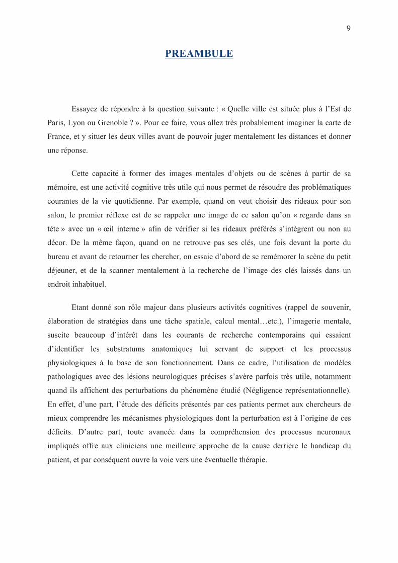
Essayez de répondre à la question suivante : « Quelle ville est située plus à l’Est de
Paris, Lyon ou Grenoble ? ». Pour ce faire, vous allez très probablement imaginer la carte de
France, et y situer les deux villes avant de pouvoir juger mentalement les distances et donner
une réponse.
Cette capacité à former des images mentales d’objets ou de scènes à partir de sa
mémoire, est une activité cognitive très utile qui nous permet de résoudre des problématiques
courantes de la vie quotidienne. Par exemple, quand on veut choisir des rideaux pour son
salon, le premier réflexe est de se rappeler une image de ce salon qu’on « regarde dans sa
tête » avec un « œil interne » afin de vérifier si les rideaux préférés s’intègrent ou non au
décor. De la même façon, quand on ne retrouve pas ses clés, une fois devant la porte du
bureau et avant de retourner les chercher, on essaie d’abord de se remémorer la scène du petit
déjeuner, et de la scanner mentalement à la recherche de l’image des clés laissés dans un
endroit inhabituel.
Etant donné son rôle majeur dans plusieurs activités cognitives (rappel de souvenir,
élaboration de stratégies dans une tâche spatiale, calcul mental…etc.), l’imagerie mentale,
suscite beaucoup d’intérêt dans les courants de recherche contemporains qui essaient
d’identifier les substratums anatomiques lui servant de support et les processus
physiologiques à la base de son fonctionnement. Dans ce cadre, l’utilisation de modèles
pathologiques avec des lésions neurologiques précises s’avère parfois très utile, notamment
quand ils affichent des perturbations du phénomène étudié (Négligence représentationnelle).
En effet, d’une part, l’étude des déficits présentés par ces patients permet aux chercheurs de
mieux comprendre les mécanismes physiologiques dont la perturbation est à l’origine de ces
déficits. D’autre part, toute avancée dans la compréhension des processus neuronaux
impliqués offre aux cliniciens une meilleure approche de la cause derrière le handicap du
patient, et par conséquent ouvre la voie vers une éventuelle thérapie.
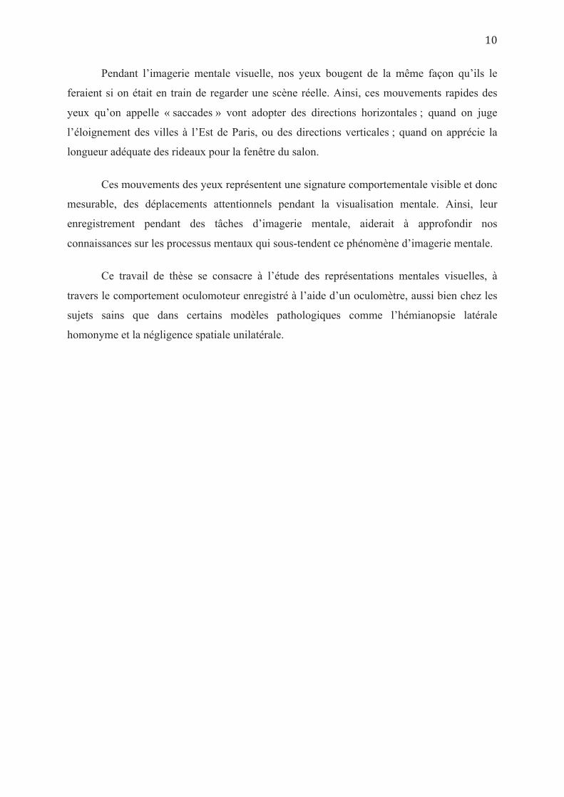
Pendant l’imagerie mentale visuelle, nos yeux bougent de la même façon qu’ils le
feraient si on était en train de regarder une scène réelle. Ainsi, ces mouvements rapides des
yeux qu’on appelle « saccades » vont adopter des directions horizontales ; quand on juge
l’éloignement des villes à l’Est de Paris, ou des directions verticales ; quand on apprécie la
longueur adéquate des rideaux pour la fenêtre du salon.
Ces mouvements des yeux représentent une signature comportementale visible et donc
mesurable, des déplacements attentionnels pendant la visualisation mentale. Ainsi, leur
enregistrement pendant des tâches d’imagerie mentale, aiderait à approfondir nos
connaissances sur les processus mentaux qui sous-tendent ce phénomène d’imagerie mentale.
Ce travail de thèse se consacre à l’étude des représentations mentales visuelles, à
travers le comportement oculomoteur enregistré à l’aide d’un oculomètre, aussi bien chez les
sujets sains que dans certains modèles pathologiques comme l’hémianopsie latérale
homonyme et la négligence spatiale unilatérale.


« A Great thought begins by seeing something differently with a shift of the Mind's Eye »
Chapitre 1
L’IMAGERIE MENTALE VISUELLE
« Thoughts are made of pictures » John Ciardi (1959)
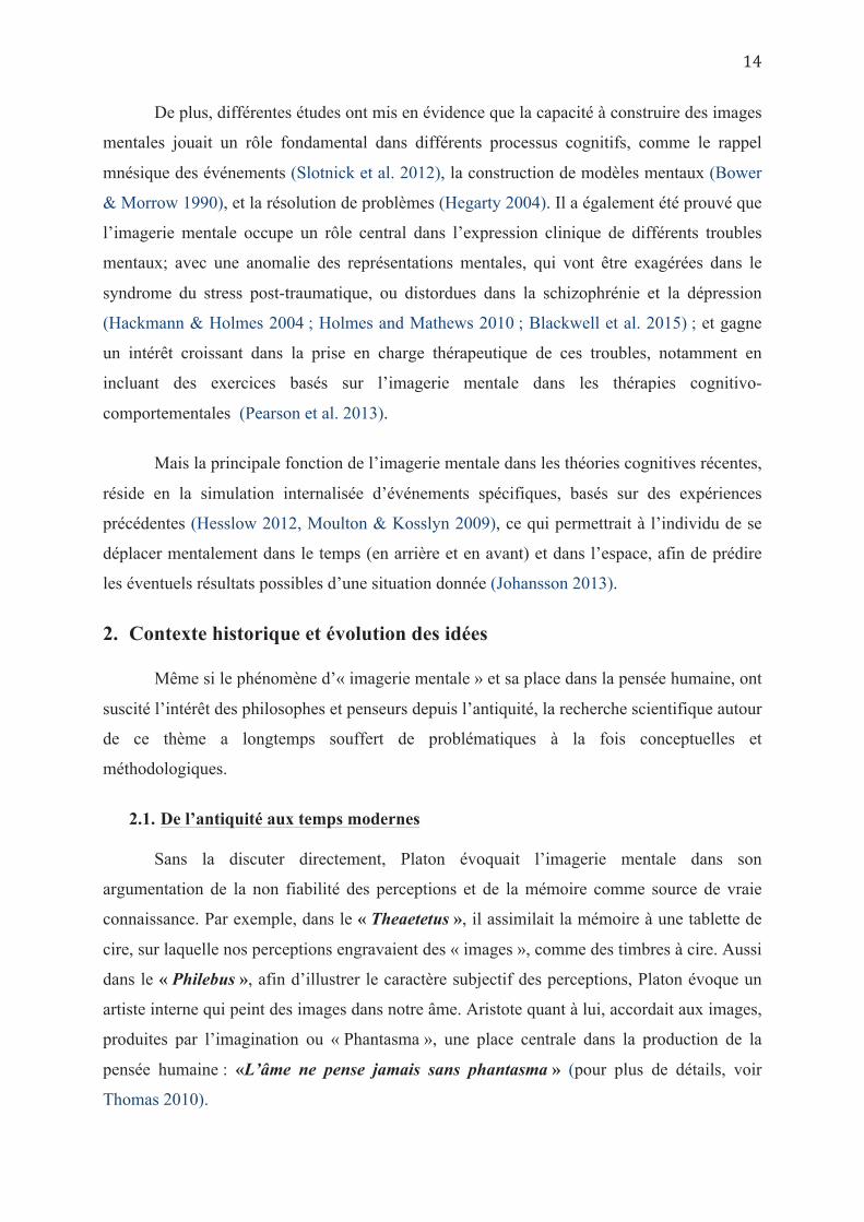
1. Définition et rôles
L’espace physique est accessible au système cognitif à travers les organes de sens, qui
collectent les informations de l’environnement permettant de construire une ou plusieurs
représentations ou images mentales de cet espace (Gibson, 1966). L’imagerie mentale est un
processus cognitif pouvant être défini comme « la création mentale d’une expérience qui
ressemble, au moins dans certains aspects, à l’expérience perceptive d’objets ou
d’événements, mais en l’absence de stimulation sensorielle directe » (Finke, 1989). On en
distingue différentes modalités selon la nature du stimulus virtuel impliqué, comme l’imagerie
mentale visuelle, auditive ou encore olfactive. Dans ce travail de thèse, nous nous limiterons à
l’étude de l’imagerie mentale visuelle.
L’imagerie mentale visuelle, souvent rapportée comme étant l’expérience de voir ou
de revoir une image dans sa tête avec un œil interne (« Seeing with the mind’s eye »), survient
fréquemment dans différentes situations de la vie quotidienne. Par exemple, pour se rappeler
le nombre de fenêtres dans une maison, il suffit de visualiser une image mentale de la façade
et de compter les fenêtres dessus (Johansson et al. 2006). De la même façon, pour décider
quel animal (le cheval ou le zèbre) à les oreilles les plus grandes ou quel fruit (fraises ou
groseilles) à la couleur plus sombre, il suffit de visualiser une image mentale des animaux ou
fruits concernés et comparer leurs formes ou leurs couleurs.
L’imagerie mentale visuelle comporte à la fois, une dimension non spatiale
(représentation des couleurs, formes, textures…etc) et une dimension spatiale, permettant à
l’individu de se représenter mentalement la localisation des objets les uns par rapport aux
autres, ou par rapport à lui-même, et d’identifier leur orientation dans l’espace. Cette
représentation spatialisée semble très utile dans certaines tâches de la vie quotidienne comme
reproduire un dessin, tracer un plan, ou indiquer le chemin à quelqu’un.
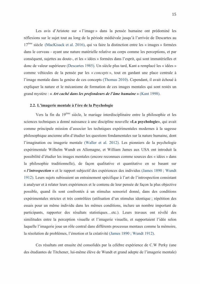
De plus, différentes études ont mis en évidence que la capacité à construire des images
mentales jouait un rôle fondamental dans différents processus cognitifs, comme le rappel
mnésique des événements (Slotnick et al. 2012), la construction de modèles mentaux (Bower
& Morrow 1990), et la résolution de problèmes (Hegarty 2004). Il a également été prouvé que
l’imagerie mentale occupe un rôle central dans l’expression clinique de différents troubles
mentaux; avec une anomalie des représentations mentales, qui vont être exagérées dans le
syndrome du stress post-traumatique, ou distordues dans la schizophrénie et la dépression
(Hackmann & Holmes 2004 ; Holmes and Mathews 2010 ; Blackwell et al. 2015) ; et gagne
un intérêt croissant dans la prise en charge thérapeutique de ces troubles, notamment en
incluant des exercices basés sur l’imagerie mentale dans les thérapies cognitivo-
comportementales (Pearson et al. 2013).
Mais la principale fonction de l’imagerie mentale dans les théories cognitives récentes,
réside en la simulation internalisée d’événements spécifiques, basés sur des expériences
précédentes (Hesslow 2012, Moulton & Kosslyn 2009), ce qui permettrait à l’individu de se
déplacer mentalement dans le temps (en arrière et en avant) et dans l’espace, afin de prédire
les éventuels résultats possibles d’une situation donnée (Johansson 2013).
2. Contexte historique et évolution des idées
Même si le phénomène d’« imagerie mentale » et sa place dans la pensée humaine, ont
suscité l’intérêt des philosophes et penseurs depuis l’antiquité, la recherche scientifique autour
de ce thème a longtemps souffert de problématiques à la fois conceptuelles et
méthodologiques.
2.1. De l’antiquité aux temps modernes
Sans la discuter directement, Platon évoquait l’imagerie mentale dans son
argumentation de la non fiabilité des perceptions et de la mémoire comme source de vraie
connaissance. Par exemple, dans le « Theaetetus », il assimilait la mémoire à une tablette de
cire, sur laquelle nos perceptions engravaient des « images », comme des timbres à cire. Aussi
dans le « Philebus », afin d’illustrer le caractère subjectif des perceptions, Platon évoque un
artiste interne qui peint des images dans notre âme. Aristote quant à lui, accordait aux images,
produites par l’imagination ou « Phantasma », une place centrale dans la production de la
pensée humaine : «L’âme ne pense jamais sans phantasma » (pour plus de détails, voir
Thomas 2010).
Les avis d’Aristote sur « l’image » dans la pensée humaine ont prédominé les
réflexions sur le sujet tout au long de la période médiévale jusqu’à l’arrivée de Descartes au
17ème siècle (MacKisack et al. 2016), qui va faire la distinction entre les « images » formées
dans le cerveau - ayant une nature matérielle relative au corps comme les perceptions, et par
conséquent, sujettes au doute-, et les « idées » formées dans l’esprit, qui sont immatérielles et
donc de valeur supérieure (Descartes 1985). Un siècle plus tard, Kant a remplacé les « idées »
comme véhicules de la pensée par les « concepts », tout en gardant une place centrale à
l’image mentale dans la genèse de ces concepts (Thomas 2010). Cependant, il avait échoué à
expliquer la nature et le mécanisme de formation de ces images mentales qui sont restés un
grand mystère : « Art caché dans les profondeurs de l’âme humaine » (Kant 1998).
2.2. L’imagerie mentale à l’ère de la Psychologie
Vers la fin du 19ème siècle, le mariage interdisciplinaire entre la philosophie et les
sciences techniques a donné naissance à une discipline nouvelle «La psychologie», qui avait
comme principale mission d’associer les techniques expérimentales modernes à la sagesse
philosophique ancienne afin d’étudier les questions fondamentales sur la nature humaine, dont
l’imagination ou imagerie mentale (Waller et al. 2012). Les pionniers de la psychologie
expérimentale Wilhelm Wundt en Allemagne, et William James aux USA ont introduit la
possibilité d’étudier les images mentales (encore reconnues comme sources des « idées » dans
la philosophie traditionnelle), de façon qualitative et quantitative en se basant sur
« l’introspection » et le rapport subjectif des expériences des individus (James 1890 ; Wundt
1912). Leurs sujets subissaient un entrainement spécifique à l’art de l’introspection consistant
à analyser et à relater leurs expériences et le contenu de leur pensée de façon la plus objective
possible, quand ils sont confrontés à un stimulus sensoriel donné, dans des conditions
expérimentales strictes et très contrôlées (utilisation d’un stimulus identique ; répétition des
essais pour un même individu dans les mêmes conditions, inclure un nombre important de
participants, rapporter des résultats statistiques…etc.). Leurs travaux ont révélé des
similitudes entre la perception visuelle et l’imagerie visuelle, et supportaient l’idée selon
laquelle l’imagerie joue un rôle central dans différents processus mentaux comme la mémoire,
la résolution de problèmes, l’émotion et la créativité (James 1890 ; Wundt 1912).
Ces résultats ont ensuite été consolidés par la célèbre expérience de C.W Perky (une
des étudiantes de Titchener, lui-même élève de Wundt et grand adepte de l’imagerie mentale)
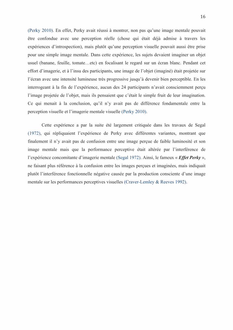
(Perky 2010). En effet, Perky avait réussi à montrer, non pas qu’une image mentale pouvait
être confondue avec une perception réelle (chose qui était déjà admise à travers les
expériences d’introspection), mais plutôt qu’une perception visuelle pouvait aussi être prise
pour une simple image mentale. Dans cette expérience, les sujets devaient imaginer un objet
usuel (banane, feuille, tomate…etc) en focalisant le regard sur un écran blanc. Pendant cet
effort d’imagerie, et à l’insu des participants, une image de l’objet (imaginé) était projetée sur
l’écran avec une intensité lumineuse très progressive jusqu’à devenir bien perceptible. En les
interrogeant à la fin de l’expérience, aucun des 24 participants n’avait consciemment perçu
l’image projetée de l’objet, mais ils pensaient que c’était le simple fruit de leur imagination.
Ce qui menait à la conclusion, qu’il n’y avait pas de différence fondamentale entre la
perception visuelle et l’imagerie mentale visuelle (Perky 2010).
Cette expérience a par la suite été largement critiquée dans les travaux de Segal
(1972), qui répliquaient l’expérience de Perky avec différentes variantes, montrant que
finalement il n’y avait pas de confusion entre une image perçue de faible luminosité et son
image mentale mais que la performance perceptive était altérée par l’interférence de
l’expérience concomitante d’imagerie mentale (Segal 1972). Ainsi, le fameux « Effet Perky »,
ne faisant plus référence à la confusion entre les images perçues et imaginées, mais indiquait
plutôt l’interférence fonctionnelle négative causée par la production consciente d’une image
mentale sur les performances perceptives visuelles (Craver-Lemley & Reeves 1992).
2.3. Entre « Comportementalistes » et « Cognitivistes »
Le début du 20ème siècle a été marqué par la montée en puissance du courant
comportementaliste qui dominait la psychologie expérimentale, notamment en Amérique du
Nord. Ce courant qui a transformé la psychologie d’une science étudiant l’esprit en une
science étudiant le comportement, réfutait tout ce qui ne pouvait être observable ni
objectivement mesurable, et avait remis en doute les précédentes expérimentations sur
l’imagerie mentale (Johansson, 2013). Pour les adeptes de ce courant, les méthodes
introspectives étaient considérées comme non-scientifiques et leurs résultats n’étaient pas
admissibles. Certains comportementalistes radicaux comme J.B. Watson allaient jusqu’à
réfuter l’idée même d’expériences sur l’imagerie mentale et recommandait de bannir ces dites
expérimentations de toute discussion scientifique (Watson, 1913).
Ainsi, durant l’ère comportementaliste, l’imagerie mentale était quasi-absente dans la
recherche en psychologie expérimentale, jusqu’aux années soixante, avec l’émergence de la
« Psychologie Cognitive » qui a écarté les comportementalistes de leur position dominante et
recentré l’attention des chercheurs sur l’étude de « l’esprit (the Mind) » comme étant le
microprocesseur du cerveau (Miller, 2003). Grace à l’utilisation de techniques expérimentales
modernes, ne se basant pas sur des méthodes subjectives ou introspectives, les premières
expériences en psychologie cognitive ont remis sous les projecteurs, l’imagerie mentale
comme l’un des phénomènes cognitifs dont dépend la pensée humaine, et l’ont restaurée
comme un sujet principal d’étude en psychologie expérimentale (Paivio 1971 ; Hebb 1968 ;
Shepard & Chipman 1970).
Ensuite, la multiplication des expériences sur le sujet dans les années 80 et 90 ont
nourri les théories cognitives naissantes soutenant un mode de fonctionnement
computationnel du cerveau, et ont allumé les premières étincelles du célèbre débat sur
l’imagerie mentale entre le courant « Analogiste » ou « Imagiste » (Kosslyn et al. 2006) et le
courant « Propositionnel » (Pylyshyn 2002).
2.4. Le débat « Analogistes » Vs « Propositionnels »
Le débat « Analogiste-Propositionnel » encore connu sous le nom de « The Mental
Imagery Debate » a dévié l’attention des spécialistes de l’imagerie mentale de l’étude de son
rôle fonctionnel dans la cognition humaine, pour se focaliser sur la nature et le format des
représentations mentales. La principale question soulevée par ce débat consistait à préciser si
les représentations mentales visuelles avaient une forme « d’image dépictive » -partageant
certaines propriétés des images réelles comme les formes, les couleurs et les relations
spatiales- (Kosslyn et al. 2006), ou étaient de nature purement propositionnelle (amodale)
comme le langage, sans caractéristiques imagées ou dépictives (Pylyshyn 2002).
La théorie analogique s’était initialement appuyée sur les premières expériences en
psychologie cognitive, basées sur la mesure des temps de réaction pendant des tâches
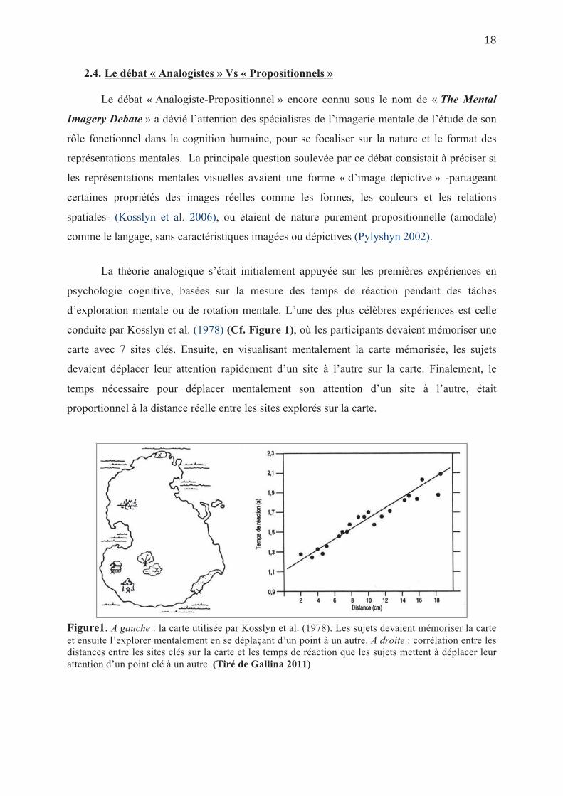
d’exploration mentale ou de rotation mentale. L’une des plus célèbres expériences est celle
conduite par Kosslyn et al. (1978) (Cf. Figure 1), où les participants devaient mémoriser une
carte avec 7 sites clés. Ensuite, en visualisant mentalement la carte mémorisée, les sujets
devaient déplacer leur attention rapidement d’un site à l’autre sur la carte. Finalement, le
temps nécessaire pour déplacer mentalement son attention d’un site à l’autre, était
proportionnel à la distance réelle entre les sites explorés sur la carte.
Figure1. A gauche : la carte utilisée par Kosslyn et al. (1978). Les sujets devaient mémoriser la carte et ensuite l’explorer mentalement en se déplaçant d’un point à un autre. A droite : corrélation entre les distances entre les sites clés sur la carte et les temps de réaction que les sujets mettent à déplacer leur attention d’un point clé à un autre. (Tiré de Gallina 2011)
D’autres expériences clés basées sur la mesure du temps de réaction, avaient montré
que le temps nécessaire pour faire une rotation mentale d’un objet visualisé augmentait avec
l’angle ou le degré de rotation exigé (Shepard & Metzler 1971 ; Cooper & Shepard, 1973).
Ces résultats et d’autres encore étaient interprétés comme une preuve solide que les
représentations mentales visuelles avaient un format analogique dépictif, avec ses
caractéristiques spatiales inhérentes.
Cependant, cette interprétation a été fortement critiquée par le courant propositionnel
qui attribuait les résultats suscités aux « connaissances tacites (Tacit Knowledge) » et non pas
à la nature « imagée » des représentations mentales (Pylyshyn 1981). Selon la théorie
propositionnelle, on dispose tous de connaissances tacites ou implicites (expérience, savoir-
faire, compétences…etc.) qui nous guident vers un certain comportement dans une situation
donnée. Dans ce contexte, Pylyshyn avait soutenu que lors d’une expérience d’imagerie
mentale, les sujets simulent un comportement de visualisation mentale en se basant sur leurs
connaissances tacites, issues des expériences visuelles perceptives (Pylyshyn 2002, 2003).
Donc les résultats issus des expériences d’exploration mentale sont de nature épi-
phénoménale et ne doivent en aucun cas être interprétés comme liés à une exploration mentale
(mental-scanning), qui n’est pas réelle, mais seulement simulée par les participants.
Ce principe de connaissances tacites pouvait s’appliquer pour offrir une explication
alternative, et donc une critique d’interprétation, à toute expérience en psychologie cognitive
dont les résultats étaient en faveur de la théorie imagiste, jusqu’à l’arrivée de l’imagerie
fonctionnelle et son utilisation dans l’exploration des mécanismes d’imagerie mentale, dans le
domaine des neurosciences (Johansson 2013). Ces nouvelles techniques de neuro-imagerie
ont finalement permis de grandes avancées dans la compréhension des bases anatomiques et
processus neuronaux soutenant le phénomène d’imagerie mentale, sans forcément
définitivement clore le fameux débat sur sa nature picturale ou non.
3. Relation perception visuelle - imagerie mentale visuelle
Depuis bien longtemps, la ressemblance ressentie entre l’expérience perceptive d’un objet
et l’expérience de sa visualisation mentale, suggérait une relation étroite entre les deux
phénomènes. La reconnaissance de cette relation intime entre perception et imagerie, se
déclinait dans la définition même que les chercheurs contemporains ont essayé de donner au
concept « Imagerie mentale » en utilisant toujours le terme « perception » dans leurs
définitions (Waller et al. 2012). A titre d’exemple, pour Wraga et Kosslyn (2003) une image
mentale est « une représentation interne qui reproduit l’expérience perceptive en l’absence
du stimulus sensoriel approprié ». De même, Ishai et Sagi (1995) ont défini l’imagerie
visuelle comme « l’invention ou la re-création d’une expérience perceptive en l’absence
d’afférences rétiniennes ».
Comme nous l’avons déjà signalé, les toutes premières études en Psychologie
expérimentale allaient jusqu’à affirmer une relation d’interchangeabilité ou de
superposablilité entre les deux processus mentaux, en soutenant qu’une information
sensorielle faible ou subliminale pouvait être confondue avec une image mentale (Perky,
1901). Les études ayant répliqué l’expérience princeps de Perky, ont souligné de façon encore
plus diversifiée l’existence d’interférences facilitatrices (Ishai & Sagi, 1995) ou inhibitrices
(Segal, 1972) entre la perception et l’imagerie visuelle selon la nature de la tâche, suggérant
que ces deux processus mentaux partageaient les mêmes structures neuronales (Waller et al.
2012).
Finke (1986) avait soutenu que si l’on supposait que l’imagerie visuelle était supportée par
les mêmes processus de traitement de l’information, impliqués dans la perception visuelle, on
devait vérifier que ces processus étaient partagés dans chacun des différents niveaux de leur
organisation hiérarchique jusqu’aux niveaux les plus primaires. En effet, nous disposons
aujourd’hui d’un corps solide de recherche et de nombreuses preuves empiriques attestant du
fait que la perception et l’imagerie mentale semblent partager une architecture neuronale
semblable, quoi que non exactement superposable.
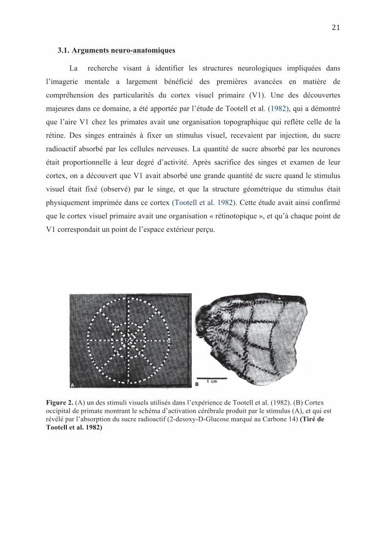
3.1. Arguments neuro-anatomiques
La recherche visant à identifier les structures neurologiques impliquées dans
l’imagerie mentale a largement bénéficié des premières avancées en matière de
compréhension des particularités du cortex visuel primaire (V1). Une des découvertes
majeures dans ce domaine, a été apportée par l’étude de Tootell et al. (1982), qui a démontré
que l’aire V1 chez les primates avait une organisation topographique qui reflète celle de la
rétine. Des singes entrainés à fixer un stimulus visuel, recevaient par injection, du sucre
radioactif absorbé par les cellules nerveuses. La quantité de sucre absorbé par les neurones
était proportionnelle à leur degré d’activité. Après sacrifice des singes et examen de leur
cortex, on a découvert que V1 avait absorbé une grande quantité de sucre quand le stimulus
visuel était fixé (observé) par le singe, et que la structure géométrique du stimulus était
physiquement imprimée dans ce cortex (Tootell et al. 1982). Cette étude avait ainsi confirmé
que le cortex visuel primaire avait une organisation « rétinotopique », et qu’à chaque point de
V1 correspondait un point de l’espace extérieur perçu.
Figure 2. (A) un des stimuli visuels utilisés dans l’expérience de Tootell et al. (1982). (B) Cortex occipital de primate montrant le schéma d’activation cérébrale produit par le stimulus (A), et qui est révélé par l’absorption du sucre radioactif (2-desoxy-D-Glucose marqué au Carbone 14) (Tiré de Tootell et al. 1982)
Quelques années plus tard, Fox et al. (1986) ont utilisé la tomographie par émission de
positrons (PET) pour démontrer que l’aire V1 avait aussi une organisation topographique chez
les humains, et conservait la structure géométrique de la rétine. A partir de là, plusieurs
chercheurs ont essayé de tester si les aires visuelles primaires étaient directement activées lors
des tâches d’imagerie mentale, si cette activation avait aussi des caractéristiques
topographiques ou spatiales, et à quel point ces patrons d’activation étaient semblables à ceux
observés dans les tâches visuelles perceptives. Autrement dit, dans une approche où
l’imagerie mentale est considérée comme une « perception à sens inverse», quel rôle jouait le
cortex visuel primaire dans l’affichage des détails visuels du contenu des représentations
mentales ?
Les premières études (entre 1993 et 2010) avaient apporté des résultats contradictoires
sur l’activation de V1 pendant l’imagerie mentale, avec la quasi-absence de toute activation
au-dessus du seuil critique pour certaines (D’Esposito et al. 1997 ; Knauff et al. 2000 ;
Daselaar et al. 2010), et la présence d’une activité significative dans d’autres (Le Bihan et al.
1993 ; Sabbah et al. 1995 ; Ganis et al 2004 ; Slotnick et al. 2005). Ces discordances ont été
essentiellement interprétées par la différence des procédés expérimentaux (Kosslyn &
Thompson 2003), et par les différences individuelles en matière de vivacité des images
mentales (Cui et al. 2007 ; Pearson et al. 2015). Par ailleurs, cette activation controversée de
V1 pendant l’imagerie mentale, n’est pas forcément synonyme de l’implication fonctionnelle
obligatoire des aires visuelles primaires dans le processus d’imagerie à l’instar de la
perception visuelle, comme le montrent les études des cas de lésions cérébrales (Voir chapitre
suivant 3.2)
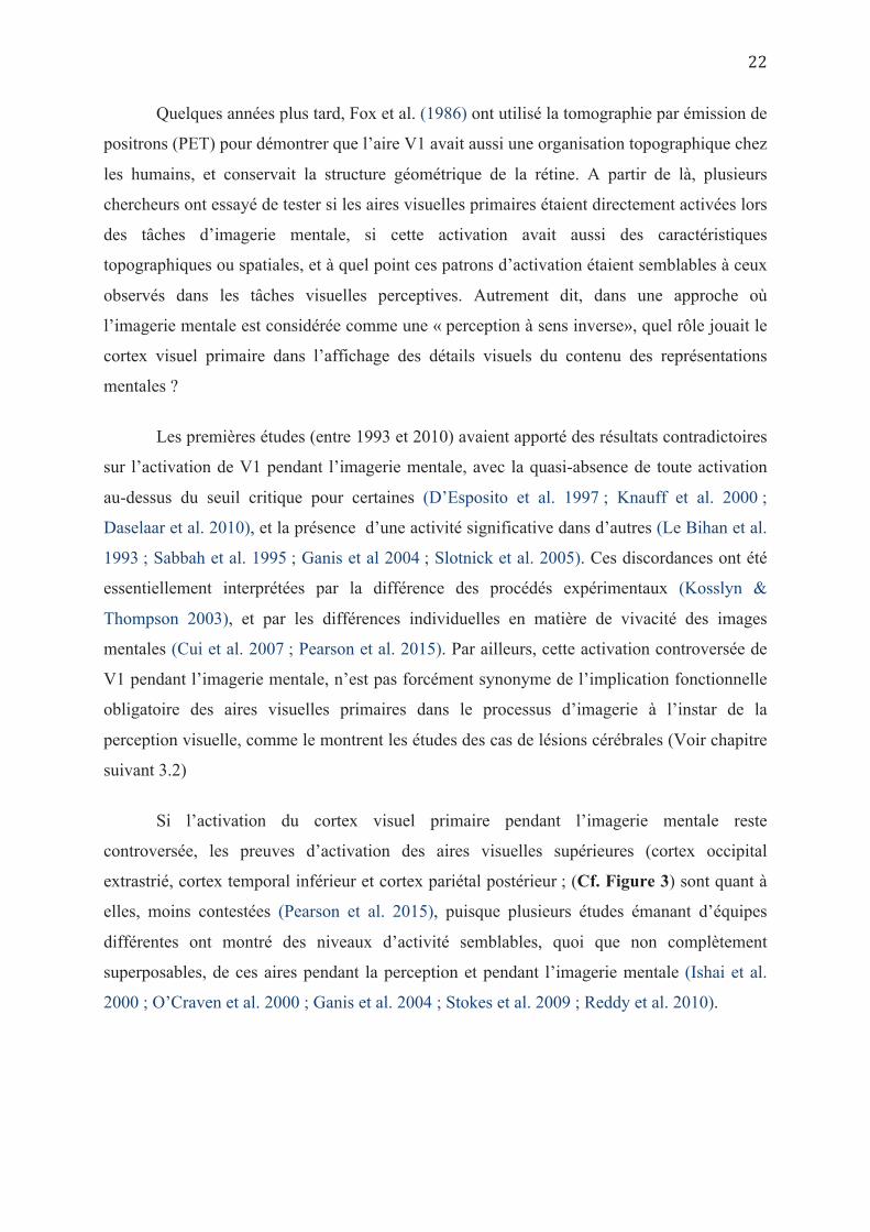
Si l’activation du cortex visuel primaire pendant l’imagerie mentale reste
controversée, les preuves d’activation des aires visuelles supérieures (cortex occipital
extrastrié, cortex temporal inférieur et cortex pariétal postérieur ; (Cf. Figure 3) sont quant à
elles, moins contestées (Pearson et al. 2015), puisque plusieurs études émanant d’équipes
différentes ont montré des niveaux d’activité semblables, quoi que non complètement
superposables, de ces aires pendant la perception et pendant l’imagerie mentale (Ishai et al.
2000 ; O’Craven et al. 2000 ; Ganis et al. 2004 ; Stokes et al. 2009 ; Reddy et al. 2010).
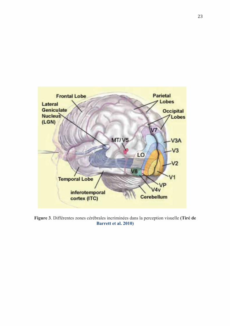
Figure 3. Différentes zones cérébrales incriminées dans la perception visuelle (Tiré de Barrett et al. 2010)
Au-delà de la simple détection d’activation des aires corticales, des études plus
récentes sont allées plus loin en utilisant une technique d’analyse d’image et de décodage
photographique en IRM fonctionnelle ; La MVPCs « Multivariate pattern classifiers » se
basant sur l’analyse des patterns d’activation au niveau du cortex visuel primaire durant la
perception de stimuli visuels (Pearson et al. 2015). Ces mêmes patterns sont ensuite utilisés
pour identifier les mêmes stimuli lors de leur évocation mentale visuelle, prouvant ainsi une
similarité des schémas d’activation de V1 en perception et en imagerie mentale d’un même
stimulus, même si le degré d’activation reste plus faible en cas d’imagerie (Albers et al.
2013 ; Cichy et al. 2012 ; Lee et al. 2012). Cette similitude des schémas d’activation entre
perception et imagerie a également été retrouvée et de façon encore plus nette et plus robuste
au niveau des aires visuelles supérieures ou associatives (Stokes et al. 2009 ; Reddy et al.
2010 ; Johnson & Johnson 2014), soulignant ainsi avec les études d’activation suscitées que
les processus neuronaux de la perception visuelle et de l’imagerie mentale deviennent de plus
en plus similaires en escaladant les niveaux d’analyse hiérarchiques (Pearson et al. 2015).
Dans une étude récente utilisant cette technique de « MVPC », les chercheurs ont pu
décoder le contenu de l’imagerie mentale, sur une tâche d’orientation spatiale, en appliquant
un logarithme de décodage de voxels au niveau des aires visuelles primaires (Naselaris et al.
2015). Ce logarithme a été défini sur la base de l’orientation spatiale des lignes au niveau des
aires visuelles, qui se fait de façon topographiquement dépictive, et ne peut fonctionner pour
détecter la bonne orientation du stimuli imaginé à travers l’analyse des signaux corticaux
pendant la tâche d’imagerie que si celle-ci se fait de la même façon dépictive que pendant la
perception (Naselaris et al. 2015). Ces résultats ont été interprétés par les « imagistes »
comme la preuve incontestable confirmant l’hypothèse que les caractéristiques visuelles
primaires d’un stimulus (position, orientation, forme) sont explicitement représentées au
niveau des aires visuelles primaires, pendant l’imagerie mentale, les amenant à considérer le
fameux débat autour de la nature des représentations mentales, clos en leur faveur (Person et
al. 2015).
3.2. Arguments neuropsychologiques :
D’autres arguments sur l’identification des substratums anatomiques impliqués dans
l’imagerie mentale visuelle sont issus des études neuropsychologiques chez des patients
souffrant de lésions cérébrales. Malgré leurs différences méthodologiques, ces études de cas,
ont mis en évidence la possibilité de toutes les dissociations imaginables entre les
performances en perception visuelle et en imagerie mentale. Sans prétendre être exhaustif
(pour revue détaillée, voir Farah 1988, et Bartolomeo 2002), nous regroupons ci-dessous
certaines des études les plus rapportées dans ce domaine, en fonction des structures
anatomiques impliquées.
3.2.1. Cortex occipital (notamment aires visuelles primaires)
Farah et al. (1992), avaient comparé la taille maximale d’objets imaginés avant et
après lobectomie occipitale unilatérale chez une patiente qui souffrait d’épilepsie. Ils avaient
rapporté qu’en post-opératoire, et en plus de la réduction du champ visuel perceptif de la
patiente (hémianopsie latérale homonyme : HLH), la taille des objets imaginés était aussi
réduite dans l’axe horizontal. Ce cas et d’autres cas de cécité corticale qui présentaient un
déficit concomitant de leurs capacités d’imagerie mentale (Farah, 1988), étaient en faveur
d’une implication fonctionnelle du cortex visuel primaire dans la génération et/ou
l’exploration d’images mentales.
Cependant, les cas de patients présentant une dissociation des troubles avec altération
de la perception visuelle (cécité corticale, suite à une lésion bilatérale du lobe occipital), mais
conservation des capacités d’imagerie mentale ne sont pas rares dans la littérature (Chatterjee
& Southwood 1995 ; Goldenberg & Nowak 1995 ; Bridge et al. 2012), soulignant au contraire
que les aires visuelles primaires ne sont pas nécessaires à la production d’images mentales
visuelles.
Par ailleurs, Butter et al (1997) avaient conduit une expérience chez 8 cas souffrant
d’hémianopsie latérale homonyme (Cf. Figure 4), et qui consistait à présenter un ensemble de
points agencés de façon arbitraire pour une courte durée. Ensuite, les points disparaissaient et
étaient remplacés par une flèche, dont les participants devaient juger si la direction pointait
vers une zone occupée par l’un des points projetés antérieurement. Les résultats avaient
montré que les patients avaient plus de mal à imaginer les points situés dans leur champ visuel
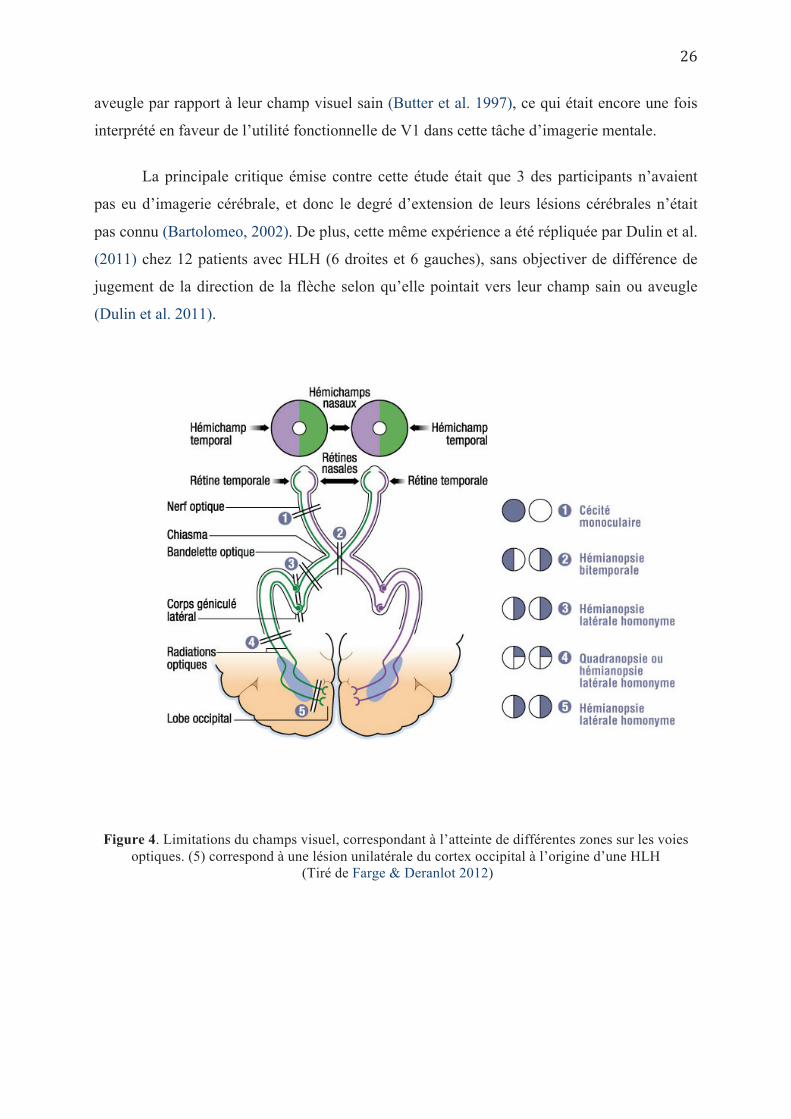
aveugle par rapport à leur champ visuel sain (Butter et al. 1997), ce qui était encore une fois
interprété en faveur de l’utilité fonctionnelle de V1 dans cette tâche d’imagerie mentale.
La principale critique émise contre cette étude était que 3 des participants n’avaient
pas eu d’imagerie cérébrale, et donc le degré d’extension de leurs lésions cérébrales n’était
pas connu (Bartolomeo, 2002). De plus, cette même expérience a été répliquée par Dulin et al.
(2011) chez 12 patients avec HLH (6 droites et 6 gauches), sans objectiver de différence de
jugement de la direction de la flèche selon qu’elle pointait vers leur champ sain ou aveugle
(Dulin et al. 2011).
Figure 4. Limitations du champs visuel, correspondant à l’atteinte de différentes zones sur les voies optiques. (5) correspond à une lésion unilatérale du cortex occipital à l’origine d’une HLH
(Tiré de Farge & Deranlot 2012)
Cette discordance des données pathologiques rejoint les données controversées sur
l’activation des aires visuelles primaires lors des tâches d’imagerie mentale, et pourrait être
expliquée, entre autres, par les différences individuelles ou de stratégies quant à la mise en jeu
des mécanismes d’imagerie mentale.
3.2.2. Lobe temporal
L’altération des capacités d’imagerie mentale (notamment pour la forme et les
couleurs) décrite chez des patients présentant des lésions spécifiques du lobe temporal,
notamment dans sa partie mésiale (Riddoch 1990 ; Manning 2000), suggère l’importance de
cette zone dans l’identification des propriétés de l’objet visualisé mentalement, comme c’est
le cas dans la perception visuelle (voie visuelle ventrale de Goodale & Milner 1992).
Cependant, l’existence de doubles dissociations avec à la fois altération des capacités
d’imagerie mentale et préservation des capacités perceptives de reconnaissance de l’objet
(Goldenberg 1992 ; Sirigu & Duhamel 2001 ; Moro et al. 2008), mais aussi la préservation
d’une imagerie mentale de bonne qualité coexistant avec un déficit perceptif important
(anosognosie, alexie, prosopagnosie) (Bartolomeo et al. 1998 ; Jankowiak et al. 1992),
souligne le fait que malgré la similarité des processus impliqués dans l’imagerie et la
perception au niveau des aires supérieures ou associatives (ici temporales inférieures), il n’est
pas question d’une superposition parfaite des structures anatomiques impliquées dans l’un et
l’autre de ces deux processus (Bartolomeo 2000).
Dans notre Article (2), nous explorons les capacités d’imagerie mentale
spatiale, chez les individus présentant des lésions unilatérales du lobe occipital,
en utilisant un nouveau procédé expérimental (mouvements des yeux), afin de
rechercher les points communs et les différences individuelles dans les
stratégies d’exploration mentale dans cette population de patients HLH.
Da
3.2.3. Lobe pariétal
Dans leur observation princeps, Bisiach & Luzzatti (1978) avaient rapporté le cas de deux
patients souffrant de négligence spatiale unilatérale gauche, qui avaient du mal à décrire de
mémoire la partie gauche d’un endroit familier (La Place du Dôme à Milan). Pour s’assurer
qu’il ne s’agissait pas d’omissions en rapport avec un trouble mnésique ou une
méconnaissance de cette partie de la place, ils ont demandé aux patients de décrire la même
place en se plaçant à l’autre bout géographique (rotation mentale de 180°), et là encore les
patients ont eu du mal à décrire avec détail la (nouvelle) partie gauche de la place qui était
bien décrite du premier point de vue (Bisiach & Luzzatti 1978). Ce déficit d’imagerie mentale
touchant la moitié gauche de l’espace imaginé était baptisé « négligence représentationnelle ».
Sa présence concomitante avec la négligence perceptive était interprétée comme preuve que la
perception et l’imagerie partageaient les mêmes processus neurologiques (Pearson et al. 2015)
impliqués dans l’attention et les relations spatiales à savoir les réseaux fronto-pariétaux et la
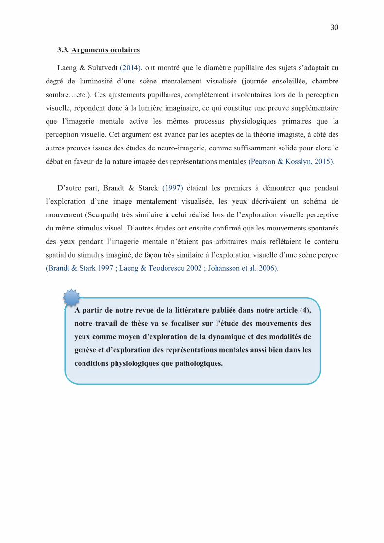
voie visuelle dorsale (Ungerleider & Mishkin 1982), respectivement. D’autres études ultérieures avaient confirmé ce constat, notamment celle conduite par
Rode et al (1995), rapportant 8 cas de patients avec négligence spatiale qui omettaient de
décrire de mémoire la partie gauche de la carte de France, quel que soit le point de vue sur la
carte. Dans une publication ultérieure, Rode et al. (2004), met en évidence chez un patient
atteint d’une lésion cérébrale droite, que les données géographiques en rapport avec la moitié
gauche de la carte de France n’étaient négligées qu’une fois spatialisées alors que l’individu
était capable d’évoquer des villes de l’ensemble du territoire Français s’il n’utilisait pas
l’imagerie mentale pour les rappeler de la mémoire à long terme.
Cependant, des cas isolés de dissociation entre négligence perceptive et
représentationnelle décrits ultérieurement ont semé le doute quant au partage strict de
processus neuronaux communs en matière de traitement spatial de l’information visuelle entre
la perception et l’imagerie. Il est à noter que la négligence visuelle non accompagnée de
négligence représentationnelle était plus fréquente (Bartolomeo et al. 1994), mais que des cas
de négligence représentationnelle sans négligence perceptive étaient aussi rapportés (Beschin
et al. 1997 ; Coslett 1997 ; Guariglia et al. 1993).
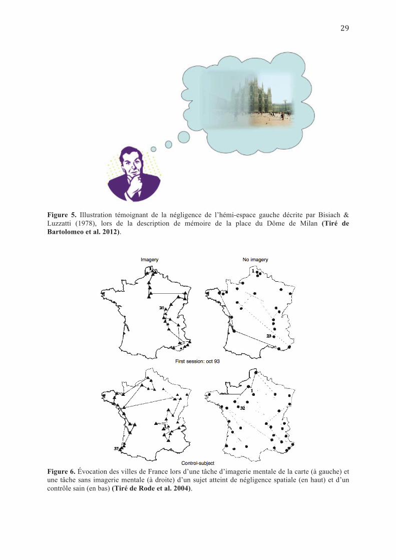
Figure 5. Illustration témoignant de la négligence de l’hémi-espace gauche décrite par Bisiach & Luzzatti (1978), lors de la description de mémoire de la place du Dôme de Milan (Tiré de Bartolomeo et al. 2012).
Figure 6. Évocation des villes de France lors d’une tâche d’imagerie mentale de la carte (à gauche) et une tâche sans imagerie mentale (à droite) d’un sujet atteint de négligence spatiale (en haut) et d’un contrôle sain (en bas) (Tiré de Rode et al. 2004).
3.3. Arguments oculaires
Laeng & Sulutvedt (2014), ont montré que le diamètre pupillaire des sujets s’adaptait au
degré de luminosité d’une scène mentalement visualisée (journée ensoleillée, chambre
sombre…etc.). Ces ajustements pupillaires, complètement involontaires lors de la perception
visuelle, répondent donc à la lumière imaginaire, ce qui constitue une preuve supplémentaire
que l’imagerie mentale active les mêmes processus physiologiques primaires que la
perception visuelle. Cet argument est avancé par les adeptes de la théorie imagiste, à côté des
autres preuves issues des études de neuro-imagerie, comme suffisamment solide pour clore le
débat en faveur de la nature imagée des représentations mentales (Pearson & Kosslyn, 2015).
D’autre part, Brandt & Starck (1997) étaient les premiers à démontrer que pendant
l’exploration d’une image mentalement visualisée, les yeux décrivaient un schéma de
mouvement (Scanpath) très similaire à celui réalisé lors de l’exploration visuelle perceptive
du même stimulus visuel. D’autres études ont ensuite confirmé que les mouvements spontanés
des yeux pendant l’imagerie mentale n’étaient pas arbitraires mais reflétaient le contenu
spatial du stimulus imaginé, de façon très similaire à l’exploration visuelle d’une scène perçue
(Brandt & Stark 1997 ; Laeng & Teodorescu 2002 ; Johansson et al. 2006).
A partir de notre revue de la littérature publiée dans notre article (4),
notre travail de thèse va se focaliser sur l’étude des mouvements des
yeux comme moyen d’exploration de la dynamique et des modalités de
genèse et d’exploration des représentations mentales aussi bien dans les
conditions physiologiques que pathologiques.
A
4. Modèle de Kosslyn de l’imagerie mentale
La théorie de l’imagerie mentale visuelle selon Kosslyn, représente le modèle type qui
suggère une quasi-superposition entre l’imagerie mentale et la perception visuelle, où les
représentations mentales ont une forme imagée ; dépictive (Bartolomeo 2002). Ce modèle est
basé sur la présomption que l’imagerie mentale visuelle partage les mêmes processus
neuronaux que la perception visuelle mais où les informations empruntent une voie de retour
« Backwards » (Farah, 2000). En effet, le point clé du modèle de Kosslyn réside dans le fait
que la perception et l’imagerie visuelles partageraient le même écran ; « Tampon visuel/
Visual Buffer », pour afficher les informations visuelles, soit transmises par la rétine et
empruntant une voie ascendante (bottom-up), soit générées de façon interne (représentations
visuelles) et transmises par voie descendante (top-down) provenant des structures
neuroanatomiques de la mémoire (Cf. Figure 3). Sur le plan anatomique, ce tampon visuel
correspondrait au cortex visuel primaire (V1) (Kosslyn 1994 ; Kosslyn et al. 1995, 1999),
situé au niveau du lobe occipital et qui se caractérise par une organisation rétinotopique
(Tootell et al. 1982).
Ce modèle peut ainsi expliquer les processus neuronaux mis en jeu à la fois dans la
perception visuelle et dans l’imagerie mentale visuelle.
4.1. Processus mis en jeu dans la perception visuelle (Kosslyn, 2006):
Etant donné que le tampon visuel reçoit plus d’informations que le système perceptif
puisse traiter en détail, une fenêtre attentionnelle « Attention Window » va se déplacer pour
sélectionner les zones d’intérêt qui vont subir une analyse plus poussée (Cave & Kosslyn
1989 ; Posner & Peterson 1990). A partir de là, l’information emprunte deux voies différentes
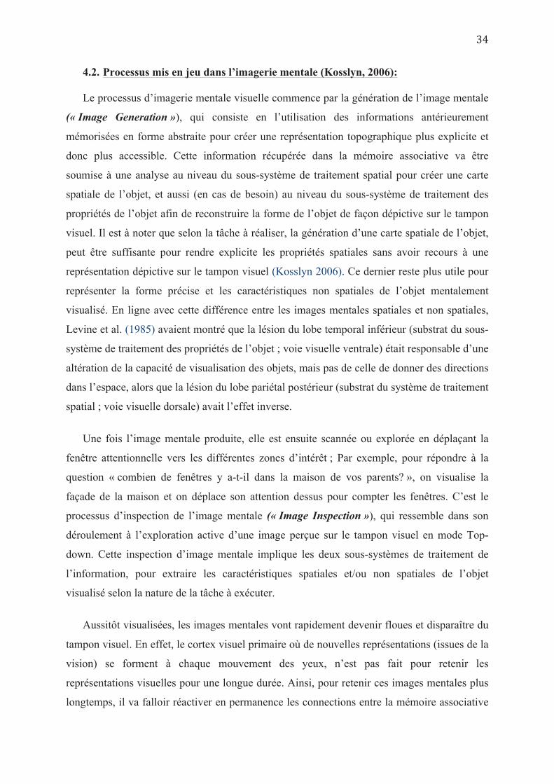
pour un traitement plus spécifique ; une voie ventrale, vers le lobe temporal inférieur qui traite
les informations relatives à la forme et à la couleur de l’objet (« Object Properties
Processing sub-System») et une voie dorsale, vers le lobe pariétal postérieur qui traite les
informations relatives à la position et l’orientation spatiale de l’objet (« Spatial Properties
Processing sub-System ») (Goodale & Milner 1992 ; Haxby et al. 1991 ; Ungerleider &
Mishkin 1982 ; Kosslyn et al. 1998, (Cf. Figure 7 et 8)). Ce système d’analyse des propriétés
spatiales se caractérise par une organisation spatio-topique lui permettant de générer une carte
de localisation des différents objets dans l’espace perçu (Sereno et al. 2001). Cette carte
spatio-topique est importante car dès que les yeux bougent, une nouvelle représentation est
affichée sur le tampon visuel selon les nouvelles coordonnées rétinotopiques. La carte spatio-
topique conserve un indice de localisation des représentations successivement projetées sur le
tampon visuel les unes par rapport aux autres (référentiel allocentré) et/ou par rapport au
corps (référentiel égocentré): on sait où un objet (qui n’est plus vu) est situé dans l’espace, ce
qui nous permet d’y retourner pour un second regard (c’est le phénomène de remise à jour
spatiale trans-saccadique ou « remapping » expliqué dans Pisella & Mattingley 2004). Les
informations traitées par les deux sous-systèmes, sont ensuite comparées avec les
représentations stockées en mémoire associative (« Associative Memories ») permettant ainsi
une identification de l’objet.
Quand l’objet n’est pas clairement identifié, il y a mise en jeu d’un travail actif de
comparaison entre les principales caractéristiques de l’objet perçu et les informations
antérieurement mémorisées grâce à un sous-système d’échange d’information (« Information
Shunting ») (Gregory 1970), afin de confirmer ou infirmer les hypothèses formulées à propos
de l’objet perçu. Ce travail de reconnaissance peut nécessiter un déplacement de l’attention
(« Attention Shift ») entre les différentes parties d’intérêt ou caractéristiques clés de l’objet à
identifier. Ce déplacement attentionnel implique un système complexe mettant en jeu des
structures comme le lobule pariétal supérieur (SPL : Superior parietal lobule), les champs
visuels frontaux (FEF : Frontal eye fields), le colliculus supérieur, le thalamus, le cortex
cingulaire antérieur (Corbetta et al 1993, Posner & Peterson 1990, Peterson & Posner 2012).
Ce système peut déplacer la fenêtre attentionnelle de façon discrète « Covert attention », mais
peut aussi entrainer un déplacement manifeste « Overt attention » des yeux, voire de la tête à
la recherche d’une nouvelle information à scruter.
En conclusion, le processus perceptif peut être complètement passif pour un objet familier
observé d’un angle de vue habituel. L’objet vu peut ainsi être identifié (quand il correspond à
une ancienne représentation stockée en mémoire associative à long terme) par un mécanisme
principalement ascendant « bottom-up ». Par contre, si l’objet n’est pas reconnu à première
vue, un processus d’identification active descendant « top-down » est mis en jeu à la
recherche d’autres indices d’identification. Après chaque déplacement de l’attention, un
nouveau détail est encodé sur le tampon visuel et un nouveau cycle d’analyse et de
comparaison est enclenché jusqu’à identification de l’objet. Ce modèle est utilisé en bottom-
up et top-down aussi lorsque l’on cherche un objet dans l’environnement parmi des
distracteurs et qu’il faut donc sélectionner l’information visuelle pertinente (processus
d’attention sélective).
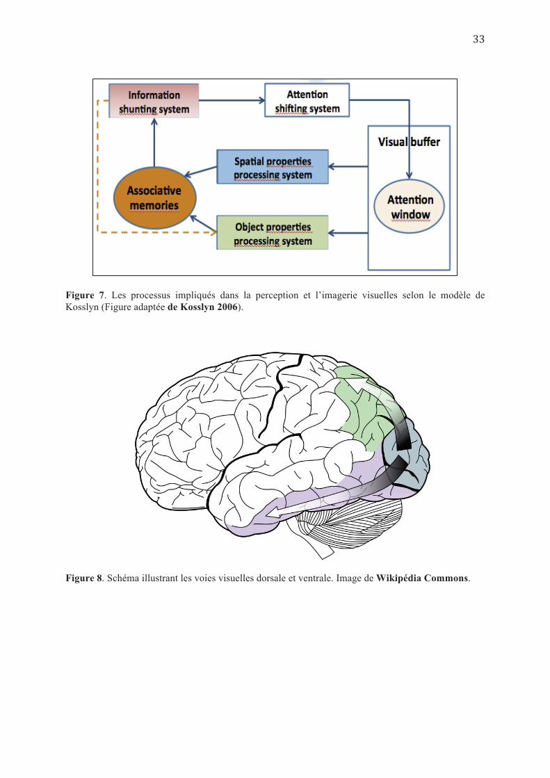
Figure 7. Les processus impliqués dans la perception et l’imagerie visuelles selon le modèle de Kosslyn (Figure adaptée de Kosslyn 2006).
Figure 8. Schéma illustrant les voies visuelles dorsale et ventrale. Image de Wikipédia Commons.
4.2. Processus mis en jeu dans l’imagerie mentale (Kosslyn, 2006):
Le processus d’imagerie mentale visuelle commence par la génération de l’image mentale
(« Image Generation »), qui consiste en l’utilisation des informations antérieurement
mémorisées en forme abstraite pour créer une représentation topographique plus explicite et
donc plus accessible. Cette information récupérée dans la mémoire associative va être
soumise à une analyse au niveau du sous-système de traitement spatial pour créer une carte
spatiale de l’objet, et aussi (en cas de besoin) au niveau du sous-système de traitement des
propriétés de l’objet afin de reconstruire la forme de l’objet de façon dépictive sur le tampon
visuel. Il est à noter que selon la tâche à réaliser, la génération d’une carte spatiale de l’objet,
peut être suffisante pour rendre explicite les propriétés spatiales sans avoir recours à une
représentation dépictive sur le tampon visuel (Kosslyn 2006). Ce dernier reste plus utile pour
représenter la forme précise et les caractéristiques non spatiales de l’objet mentalement
visualisé. En ligne avec cette différence entre les images mentales spatiales et non spatiales,
Levine et al. (1985) avaient montré que la lésion du lobe temporal inférieur (substrat du sous-
système de traitement des propriétés de l’objet ; voie visuelle ventrale) était responsable d’une
altération de la capacité de visualisation des objets, mais pas de celle de donner des directions
dans l’espace, alors que la lésion du lobe pariétal postérieur (substrat du système de traitement
spatial ; voie visuelle dorsale) avait l’effet inverse.
Une fois l’image mentale produite, elle est ensuite scannée ou explorée en déplaçant la
fenêtre attentionnelle vers les différentes zones d’intérêt ; Par exemple, pour répondre à la
question « combien de fenêtres y a-t-il dans la maison de vos parents? », on visualise la
façade de la maison et on déplace son attention dessus pour compter les fenêtres. C’est le
processus d’inspection de l’image mentale (« Image Inspection »), qui ressemble dans son
déroulement à l’exploration active d’une image perçue sur le tampon visuel en mode Top-
down. Cette inspection d’image mentale implique les deux sous-systèmes de traitement de
l’information, pour extraire les caractéristiques spatiales et/ou non spatiales de l’objet
visualisé selon la nature de la tâche à exécuter.
Aussitôt visualisées, les images mentales vont rapidement devenir floues et disparaître du
tampon visuel. En effet, le cortex visuel primaire où de nouvelles représentations (issues de la
vision) se forment à chaque mouvement des yeux, n’est pas fait pour retenir les
représentations visuelles pour une longue durée. Ainsi, pour retenir ces images mentales plus
longtemps, il va falloir réactiver en permanence les connections entre la mémoire associative
et le tampon visuel à travers le système de traitement des propriétés de l’objet ; C’est le
processus de maintien de l’image mentale (« Image maintenance »).
Enfin, une transformation de l’objet visualisé est aussi possible, en anticipant sa nouvelle
forme s’il était manipulé d’une certaine façon (« Image transformation »). Ceci se produit
par la modification volontaire de la carte spatiale de l’objet (Object map), et par la modulation
de la fonction de représentation de la forme de l’objet par le sous-système de traitement des
propriétés de l’objet sur le tampon visuel. C’est le principe de base des tâches de rotation
mentale.
Le modèle de Kosslyn représente la conception la plus poussée, de notre compréhension
actuelle de l’imagerie mentale visuelle, et offre une opportunité inégalée de tests empiriques
aussi bien en neuroscience qu’en neuropsychologie, car il apporte en plus d’explications
hypothétiques des mécanismes et processus fonctionnels, une proposition des substratums
anatomiques mis en jeu.
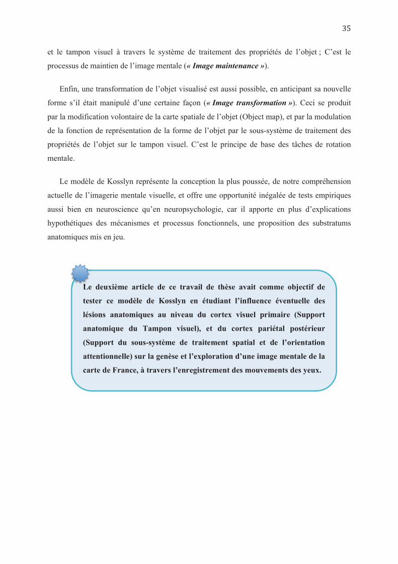
Le deuxième article de ce travail de thèse avait comme objectif de
tester ce modèle de Kosslyn en étudiant l’influence éventuelle des
lésions anatomiques au niveau du cortex visuel primaire (Support
anatomique du Tampon visuel), et du cortex pariétal postérieur
(Support du sous-système de traitement spatial et de l’orientation
attentionnelle) sur la genèse et l’exploration d’une image mentale de la
carte de France, à travers l’enregistrement des mouvements des yeux.
Le
5. Différences individuelles en imagerie mentale
Les différences individuelles représentent un aspect important de la recherche dans le
domaine de l’imagerie mentale, notamment dans sa relation avec la perception visuelle. En
effet, étant donné le caractère subjectif de ces deux activités mentales, il est logique de penser
que les différents individus n’ont pas forcément les mêmes stratégies d’acquisition et de
traitement de l’information visuelle, ni les mêmes capacités à générer et à analyser des images
mentales. Ainsi, pouvoir étudier ces différences à l’échelle de l’individu représenterait un
apport important dans la compréhension de la discordance de certains résultats obtenus dans
différentes études.
Galton (1880) était le premier à soulever la question d’inégalité des personnes en terme de
capacité d’imagerie mentale. Ses recherches étaient motivées par une constatation que
l’expérience même d’imagerie mentale, assez habituelle dans la population générale, était un
phénomène quasi-inconnu dans la communauté scientifique. Il avait alors conçu le premier
questionnaire « On visualizing and other allied activities » permettant de quantifier la qualité
des images mentales (en terme de clarté, de précision, d’éclat de coloration…etc.), et avait
mis en évidence, en s’appuyant sur des données statistiques, une grande variabilité et diversité
des expériences d’imagerie mentale chez les différents individus (Galton 1880).
D’autres questionnaires visant à quantifier les capacités individuelles d’imagerie mentale,
moyennant des scores de qualité de l’imagerie ont ensuite été développés et utilisés dans
différentes études, comme le « QMI : Questionnaire upon Mental Imagery » (Betts 1909 ;
Sheehan 1967), or the « VVIQ : Vividness of Visual Imagery Questionnaire » (Marks 1973).
En utilisant ces questionnaires et d’autres, plusieurs études ont mis en évidence des
différences majeures entre des groupes d’individus concernant leurs capacités d’imagerie
mentale. Ainsi, on distinguait les visualiseurs « Visualisers » -qui s’appuient principalement
sur des stratégies d’imagerie mentale dans la résolution des tâches cognitives- des
verbalisateurs « Verbalisers » -qui ont davantage recours à des moyens verbaux-logiques dans
le traitement de l’information et la résolution des problèmes (Jonassen & Grabowski 1993). A
travers une série d’expériences récentes Kozhevnikov et al. (2005) ont montré qu’il existait
non pas une seule mais deux catégories de visualiseurs selon leur utilisation préférentielle de
l’imagerie « spatiale » ou « iconique », liée à l’objet. Ceux utilisant une imagerie liée à
l’objet, analysaient les images de façon plutôt globale et holistique, alors que ceux préférant
l’imagerie spatiale se caractérisaient par un traitement plus analytique des images (partie par
partie), et utilisaient les relations spatiales entre les différentes composantes d’une image pour
la reconstituer (Kozhevnikov 2005). Dans une étude ultérieure, Kozhevnikov et al. (2010)
soutiennent l’existence d’un compromis plutôt qu’une indépendance entre les capacités
d’imagerie spatiale et d’imagerie iconique, en démontrant chez les artistes un niveau
supérieur à la moyenne uniquement dans les scores d’imagerie iconique, alors que les
scientifiques avait des scores supérieurs à la moyenne uniquement en imagerie spatiale et
qu’aucun groupe n’avait des scores supérieurs à la moyenne dans les deux types d’imagerie
(iconique et spatiale). Les auteurs relient ce compromis au fait que le traitement des
informations dans les deux situations (imagerie spatiale et imagerie iconique) fait appel à des
ressources limitées en terme de capacités attentionnelles. Ainsi, il est possible que durant
l’intégration fonctionnelle des voies dorsale et ventrale pendant la petite enfance, l’imagerie
spatiale et iconique se développeraient l’une au dépend de l’autre (Kozhevnikov et al. 2010 ;
Johansson 2013)
Etant données toutes ces différences individuelles, nous avons opté
dans ce travail de thèse, pour une méthode d’analyse que nous avons
validé à l’échelle de l’individu au niveau de l’article (1).
L’intérêt de cette méthode dans l’exploration des représentations
mentales aussi bien chez les sujets sains que chez les sujets
pathologiques est exposé et discuté au niveau de l’article (4).
E
Chapitre 2
LES MOUVEMENTS DES YEUX EN IMAGERIE MENTALE
VISUELLE
« Quand le regard parle.., la parole se tait» Henri Frédéric Amiel (1854)
Les mouvements des yeux représentent un des aspects communs entre la perception
visuelle et l’imagerie mentale. Cependant, si le rôle des mouvements oculaires est
actuellement bien établi dans la perception visuelle, leur survenue pendant les autres tâches
cognitives en dehors de toute activité perceptive, comme l’imagerie mentale, reste l’un des
comportements humains les moins élucidés.
Les mouvements des yeux jouent un rôle fondamental dans la perception visuelle. En
effet, seule la partie centrale de la rétine « fovéa », très riche en cônes, est dotée de la
meilleure résolution ou acuité visuelle. Cependant, elle ne couvre pas plus de 2° du champ
visuel soit l’équivalent de la taille du pouce au bout du bras. En déplaçant le globe oculaire,
les mouvements rapides des yeux dits «saccades» déplacent la fovéa vers différentes zones
d’intérêt de l’environnement visuel. Ces zones sont analysées successivement mais
séparément pendant un bref moment de « fixation » oculaire. Ce n’est que l’intégration de ces
différentes parties de l’image, au niveau cortical, qui nous donne l’illusion de percevoir une
scène dans sa totalité (Yarbus 1967). Dans ce processus perceptif, les mouvements oculaires
sont entrainés soit par des facteurs « bottom-up » comme les régions les plus saillantes de la
scène qui attirent notre attention, ou par des facteurs « top-down » comme notre connaissance
sur la façon de regarder un certain type de scène (un visage, un paysage…etc) (Noton & Stark
1997).
Par contre, quand on est engagé dans une activité d’imagerie mentale, il n’y a pas de
problème d’acuité visuelle puisqu’il n’y a pas d’information visuelle réelle à regarder. Par
conséquent, il n’y a ni facteurs « bottom-up » pour diriger les mouvements des yeux, ni de
scène physiquement réelle à inspecter visuellement par des mécanismes « top-down ». Il
semblerait donc inutile que des mouvements des yeux viennent accompagner cette tâche
mentale. Pourtant, il a été montré que des mouvements spontanés des yeux accompagnent
souvent, les activités d’imagerie mentale et sont spatialement cohérents avec le contenu de
cette imagerie (Brandt & Stark 1997 ; Johansson et al. 2006 ; Laeng & Teodorescu 2002 ;
Spivey & Geng 2001).
1. Mouvements des yeux et contenu spatial de l’imagerie mentale
Dès le début du 20ème siècle, les premières études expérimentales avaient mis en évidence
une augmentation de l’activité oculomotrice pendant des tâches de visualisation mentale
(Jacobson 1932 ; Stoy 1930). Dans une étude descriptive, Totten (1935) avait utilisé une
technique photographique pour étudier les mouvements des yeux, et avait suggéré que ces
mouvements reflétaient souvent la forme de l’objet visualisé.
Le concept selon lequel les mouvements oculaires auraient une signification dans
l’imagerie mentale a longtemps été alimenté par la théorie de « regarder ses rêves », avançant
l’hypothèse que la direction des mouvements rapides des yeux (REM) pendant la phase du
sommeil paradoxal, correspondrait aux caractéristiques spatiales du contenu des rêves (Ladd
1892 ; Dement & Kleitman 1957). En se basant sur des enregistrements d’électro-
oculographie (EOG), Dement & Kleitman (1957) avaient montré que des mouvements
verticaux des yeux pendant la période de REM, étaient plus souvent liés à une activité
nécessitant un déplacement vertical du regard (un des sujets rêvait qu’il montait le long d’une
échelle en regardant en haut et en bas), et les déplacements horizontaux étaient liés avec un
activité où le regard se déplacerait horizontalement (un des sujets rêvait qu’il observait deux
personnes se jetant des tomates l’un sur l’autre). Des études ultérieures avaient confirmé ces
données expérimentales (Dement 1964 ; Herman et al. 1984 ; Doricchi et al. 2007), alors que
d’autres étaient plutôt en défaveur, alimentant ainsi la controverse (Jacobs et al. 1972 ;
Moskowitz & Berger 1969). Par ailleurs, étant donné le degré de subjectivité dans ce genre
d’expériences, liée à la fiabilité non vérifiable du récit que fait l’individu de son propre rêve
(Kamiya 1961), il paraissait plus intéressant d’étudier les mouvements des yeux survenant
pendant l’imagerie mentale dans un état d’éveil.
En 1997, Brandt et Stark ont publié la première étude ayant comparé la distribution
spatiale des mouvements oculaires pendant la perception visuelle et pendant l’imagerie
mentale. Les sujets devaient regarder une grille (6X6) comportant quelques carrés sombres,
puis ils devaient visualiser mentalement cette même grille qui aura disparu du champ visuel.
Pendant ce temps, leurs mouvements oculaires étaient enregistrés au moyen d’un « Eye-
tracker ». Les auteurs ont retrouvé une similitude statistiquement significative du schéma de
déplacement oculaire (scanpath) pour chaque participant, entre la perception visuelle et
l’imagerie mentale visuelle d’une même grille (Brandt & Stark 1997 ; Cf. Figure 9). Laeng et
Teodorescu (2002) ont répliqué l’étude de Brandt & Stark (1997), confirmant la similitude
des « scanpaths » entre la perception et l’imagerie d’un même stimulus. Ils avaient en plus
montré que les individus, à qui on demandait de fixer le regard au centre de la scène pendant
la tâche perceptive, adoptaient spontanément le même comportement de fixation oculaire
pendant la tâche d’imagerie mentale du même stimulus (Laeng & Teodorescu 2002).
Une autre série d’études a ensuite franchi une étape supérieure en montrant que pendant
l’imagerie mentale, les mouvements des yeux n’étaient pas seulement similaires à ceux
réalisés pendant la perception de la même scène imaginée, mais que les yeux bougeaient en
direction de la localisation de chacun des objets constituant la scène visualisée mentalement.
Spivey & Geng (2001) avaient utilisé comme stimulus une grille à 4 quadrants, comportant un
objet simple chacun, que les participants étaient amenés à regarder pendant une courte durée.
Ensuite, les objets disparaissent et les participants devaient répondre à des questions relatives
à l’un des objets de la grille (sa forme, sa couleur…etc.). Pendant la réponse aux questions,
les participants regardaient spécifiquement l’endroit sur la grille où figurait préalablement
l’objet au sujet duquel ils étaient questionnés (Spivey & Geng 2001). Ces résultats ont ensuite
été confirmés dans plusieurs autres études similaires (Martarelli & Mast 2011 ; Altmann
2004 ; Knoferle & Croquer 2007).
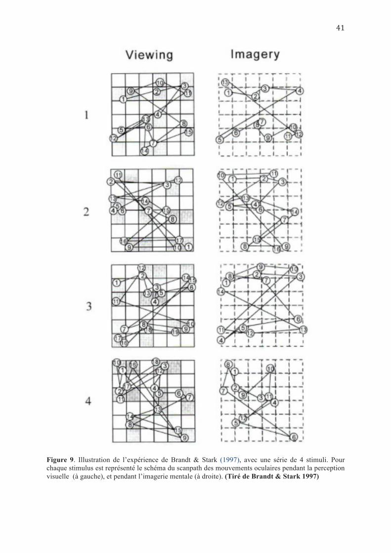
Figure 9. Illustration de l’expérience de Brandt & Stark (1997), avec une série de 4 stimuli. Pour chaque stimulus est représenté le schéma du scanpath des mouvements oculaires pendant la perception visuelle (à gauche), et pendant l’imagerie mentale (à droite). (Tiré de Brandt & Stark 1997)
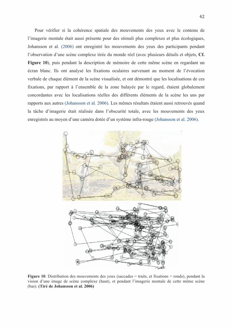
Pour vérifier si la cohérence spatiale des mouvements des yeux avec le contenu de
l’imagerie mentale était aussi présente pour des stimuli plus complexes et plus écologiques,
Johansson et al. (2006) ont enregistré les mouvements des yeux des participants pendant
l’observation d’une scène complexe tirée du monde réel (avec plusieurs détails et objets, Cf.
Figure 10), puis pendant la description de mémoire de cette même scène en regardant un
écran blanc. Ils ont analysé les fixations oculaires survenant au moment de l’évocation
verbale de chaque élément de la scène visualisée, et ont démontré que les localisations de ces
fixations, par rapport à l’ensemble de la zone balayée par le regard, étaient globalement
concordantes avec les localisations réelles des différents éléments de la scène les uns par
rapports aux autres (Johansson et al. 2006). Les mêmes résultats étaient aussi retrouvés quand
la tâche d’imagerie était réalisée dans l’obscurité totale, avec les mouvements des yeux
enregistrés au moyen d’une caméra dotée d’un système infra-rouge (Johansson et al. 2006).
Figure 10. Distribution des mouvements des yeux (saccades = traits, et fixations = ronds), pendant la vision d’une image de scène complexe (haut), et pendant l’imagerie mentale de cette même scène (bas). (Tiré de Johansson et al. 2006)
Ces déplacements oculaires spontanés vers les localisations spatiales (approximatives) des
objets évoqués dans une scène mentalement visualisée, étaient encore retrouvés quand
l’épreuve d’imagerie se déroulait quelques jours après l’encodage perceptif (Humphrey &
Underwood 2008 ; Martarelli & Mast 2013).
D’autres expériences ont adopté une approche différente dans l’exploration des
mouvements des yeux pendant l’imagerie mentale de façon à supprimer la phase préalable
d’encodage visuel. Ces expériences étaient essentiellement basées sur l’enregistrement des
mouvements oculaires pendant que les sujets écoutaient une description verbale d’une scène
qui contenait des mots indicateurs spatiaux, et ont confirmé que ces sujets réalisaient des
mouvements spontanés des yeux dans les mêmes directions indiquées dans cette description
pendant l’encodage et pendant l’imagerie mentale (Demarais & Cohen 1998 ; Spivey et al.
2000 ; Spivey & Geng 2001 ; Johansson et al. 2006). Des résultats similaires ont été retrouvés
dans une étude récente ayant analysé les mouvements des yeux lors de l’évocation de la carte
de France à partir de la mémoire à long terme, sans tâche de perception préalable. Les
individus devaient décider si les villes, qu’on leur annonçait verbalement, étaient situées à
droite ou à gauche de la capitale (Paris). Les auteurs ont montré que la direction horizontale
des premières saccades et la localisation des premières fixations observées suite à l’annonce
d’une ville donnée, étaient concordantes avec la localisation de la ville évoquée sur la carte
(Bourlon et al. 2011).
Dans notre Article (1), nous étendons ces résultats à la situation où l’on
utilise un stimulus complexe (carte de France), issu de la mémoire à long
terme (pas de phase d’encodage préalable), imaginé et exploré
mentalement de façon purement spontanée pour son évocation verbale,
et dont les résultats peuvent être analysés statistiquement de façon
individuelle.
D
2. Les théories explicatives des mouvements des yeux en imagerie mentale
Toutes ces études citées plus haut ont confirmé de façon claire que les mouvements
spontanés des yeux pendant l’imagerie mentale ne sont pas arbitraires, mais ont une
signification spatiale qui reflète le contenu de l’imagerie mentale. Cependant, la question qui
se pose c’est pourquoi ces mouvements surviennent-ils en l’absence d’un stimulus visuel
réel ? Sont-ils vraiment indispensables pour la génération ou/et l’exploration des images
mentales ou s’agit-il d’un simple épiphénomène sans véritable importance fonctionnelle ?
Plusieurs théories plus ou moins appuyées par des données empiriques ont été avancées
pour expliquer la signification fonctionnelle ou non de ces mouvements oculaires réalisant le
curieux phénomène de « regarder le vide » («Looking at nothing»). Il est à souligner que ces
différentes théories n’ont pas été développées comme des alternatives. Certaines comportent
des éléments communs et d’autres peuvent se compléter (Johansson 2013). Ainsi, il ne serait
pas judicieux de les considérer comme mutuellement exclusives quand elles sont comparées.
2.1. La théorie du « Scanpath », ou réactivation de la trace oculomotrice :
Hebb (1968) avait fait le rapprochement entre les mouvements des yeux dans la
perception visuelle et dans l’imagerie mentale en supposant que si ces mouvements étaient
mécaniquement indispensables pour scanner les objets lors de la perception, ce même
processus moteur oculaire pourrait également avoir une fonction d’assemblage et
d’organisation des différentes parties de l’image mentale ; «Si l’image est une réintégration
du processus perceptif, elle devrait inclure les mouvements oculaires» (Hebb 1968).
Cette hypothèse selon laquelle les mouvements des yeux auraient un rôle fonctionnel dans
l’imagerie mentale était ensuite reprise dans la « théorie du Scanpath » développée par Noton
& Stark (1971). Cette théorie suppose que les mouvements oculaires sont nécessaires pour
l’imagerie mentale visuelle et prédit que ces mouvements seraient une véritable reproduction
de ceux survenus pendant la perception visuelle avec des Scanpaths qui suivraient le même
ordre séquentiel (Noton et Stark 1971). La théorie du Scanpath a d’abord été supportée par les
données empiriques issues des expériences de la même équipe et retrouvant une grande
similitude du comportement oculomoteur pendant la perception et la visualisation mentale
d’un même stimulus (Brandt & Stark 1997 ; Cf. Figure 9). Plus tard, Laeng et al. (2014) ont
montré qu’il y avait une corrélation significative entre le degré de similarité des fixations
oculaires entre perception et imagerie mentale (en % d’occupation des zones d’intérêt), et la
qualité de la mémoire visuo-spatiale des individus (capacité à comparer la taille d’un stimulus
mémorisé à celui présenté).
Mais le rôle fonctionnel de ces scanpaths proposé par la théorie initiale de Noton et Stark
(1971) a surtout été confirmé par les quelques études ayant manipulé les mouvements des
yeux comme variable indépendante dans des expériences de perception et d’imagerie mentale,
c’est-à-dire en imposant parfois la fixation du regard (Laeng & Teodorescu 2002 ; Laeng et
al. 2014 ; Johansson et al. 2012). En effet, les sujets qui pouvaient inspecter librement un
stimulus visuel pendant la phase perceptive, mais qui étaient contraints à fixer le regard en
position centrale pendant la phase de mentalisation faisaient plus d’erreurs sur les
caractéristiques du stimulus, que les sujets qui avaient leur regard libre aussi bien pendant la
perception que pendant l’imagerie (Laeng & Teodorescu 2002 ; Laeng et al. 2014). A la
lumière de ces résultats, les auteurs avaient conclu que les mouvements des yeux pendant
l’imagerie mentale étaient une sorte de réactivation de la trace oculomotrice qui permettait un
meilleur rappel mnésique de l’objet (Laeng et Teodorescu 2002), donc un rôle fonctionnel
exactement comme le prédisait la théorie du Scanpath de Noton et Stark (1971). Pendant la
perception, la localisation de l’objet était encodée en parallèle avec les autres informations
visuelles (couleur, forme…etc.), et en revisitant leurs localisations spatiales lors de l’imagerie,
le rappel de l’ensemble de la trace mnésique était réactivé.
Johansson et al. (2012) ont également testé cette théorie en utilisant la fixation contrainte
du regard mais pendant la phase perceptive et/ou pendant la phase d’imagerie. Ils ont
confirmé que les sujets qui avaient le regard libre pendant la phase perceptive mais qui étaient
contraints à maintenir une fixation centrale pendant la phase d’imagerie avaient plus de mal à
décrire la scène de mémoire (moins de détails). Ils ont également montré que les sujets
contraints à fixer le regard pendant la phase de perception avaient un regard plutôt dispersé
pendant la phase d’imagerie mentale. Cependant leurs mouvements correspondaient
globalement au contenu spatial de la scène visualisée et décrite de mémoire, suggérant que les
mouvements des yeux pendant l’imagerie mentale ne sont pas forcément une réactivation de
ceux réalisés pendant la perception visuelle du même stimulus. Ces résultats étaient donc
toujours interprétés en faveur d’un rôle fonctionnel des mouvements des yeux dans l’activité
de visualisation mentale, mais en désaccord avec la théorie du Scanpath qui relie ce rôle
fonctionnel à une réactivation (re-enactment) du processus perceptif incluant les mouvements
oculaires (Johansson et al. 2012).
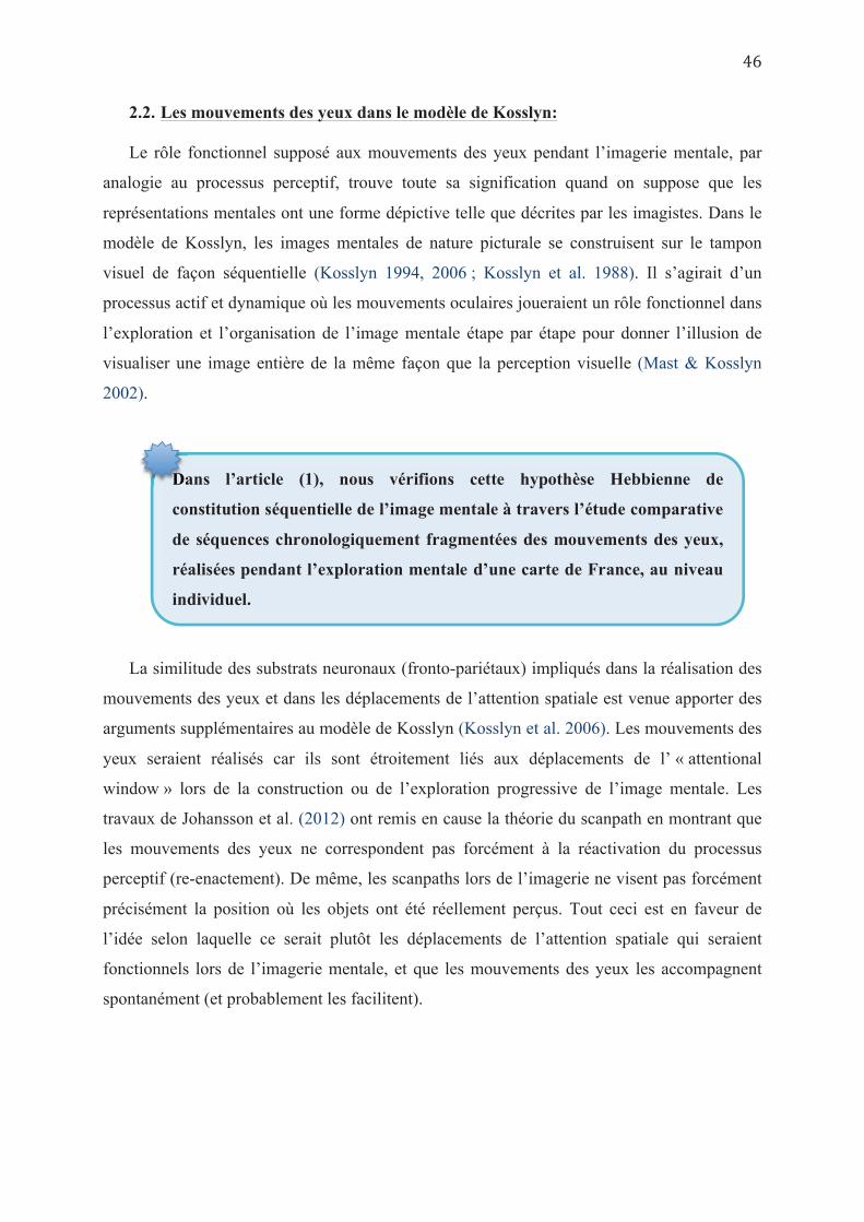
2.2. Les mouvements des yeux dans le modèle de Kosslyn:
Le rôle fonctionnel supposé aux mouvements des yeux pendant l’imagerie mentale, par
analogie au processus perceptif, trouve toute sa signification quand on suppose que les
représentations mentales ont une forme dépictive telle que décrites par les imagistes. Dans le
modèle de Kosslyn, les images mentales de nature picturale se construisent sur le tampon
visuel de façon séquentielle (Kosslyn 1994, 2006 ; Kosslyn et al. 1988). Il s’agirait d’un
processus actif et dynamique où les mouvements oculaires joueraient un rôle fonctionnel dans
l’exploration et l’organisation de l’image mentale étape par étape pour donner l’illusion de
visualiser une image entière de la même façon que la perception visuelle (Mast & Kosslyn
2002).
La similitude des substrats neuronaux (fronto-pariétaux) impliqués dans la réalisation des
mouvements des yeux et dans les déplacements de l’attention spatiale est venue apporter des
arguments supplémentaires au modèle de Kosslyn (Kosslyn et al. 2006). Les mouvements des
yeux seraient réalisés car ils sont étroitement liés aux déplacements de l’ « attentional
window » lors de la construction ou de l’exploration progressive de l’image mentale. Les
travaux de Johansson et al. (2012) ont remis en cause la théorie du scanpath en montrant que
les mouvements des yeux ne correspondent pas forcément à la réactivation du processus
perceptif (re-enactement). De même, les scanpaths lors de l’imagerie ne visent pas forcément
précisément la position où les objets ont été réellement perçus. Tout ceci est en faveur de
l’idée selon laquelle ce serait plutôt les déplacements de l’attention spatiale qui seraient
fonctionnels lors de l’imagerie mentale, et que les mouvements des yeux les accompagnent
spontanément (et probablement les facilitent).
Dans l’article (1), nous vérifions cette hypothèse Hebbienne de
constitution séquentielle de l’image mentale à travers l’étude comparative
de séquences chronologiquement fragmentées des mouvements des yeux,
réalisées pendant l’exploration mentale d’une carte de France, au niveau
individuel.
Da
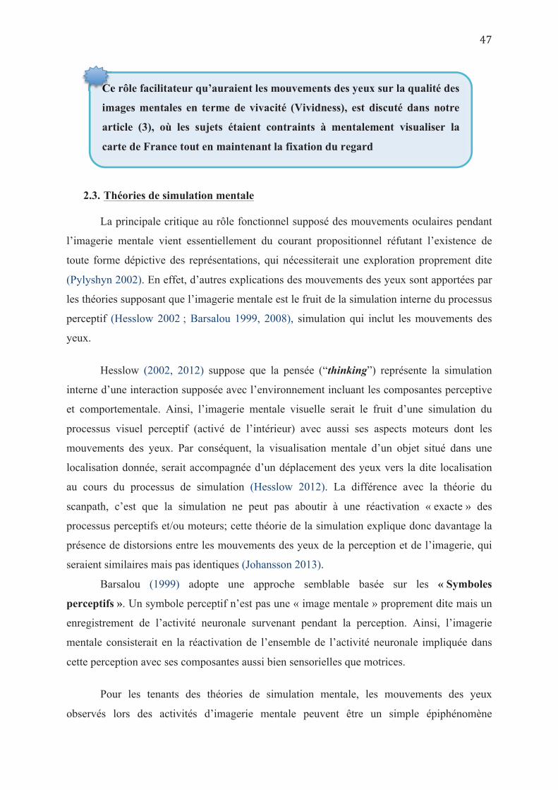
2.3. Théories de simulation mentale
La principale critique au rôle fonctionnel supposé des mouvements oculaires pendant
l’imagerie mentale vient essentiellement du courant propositionnel réfutant l’existence de
toute forme dépictive des représentations, qui nécessiterait une exploration proprement dite
(Pylyshyn 2002). En effet, d’autres explications des mouvements des yeux sont apportées par
les théories supposant que l’imagerie mentale est le fruit de la simulation interne du processus
perceptif (Hesslow 2002 ; Barsalou 1999, 2008), simulation qui inclut les mouvements des
yeux.
Hesslow (2002, 2012) suppose que la pensée (“thinking”) représente la simulation
interne d’une interaction supposée avec l’environnement incluant les composantes perceptive
et comportementale. Ainsi, l’imagerie mentale visuelle serait le fruit d’une simulation du
processus visuel perceptif (activé de l’intérieur) avec aussi ses aspects moteurs dont les
mouvements des yeux. Par conséquent, la visualisation mentale d’un objet situé dans une
localisation donnée, serait accompagnée d’un déplacement des yeux vers la dite localisation
au cours du processus de simulation (Hesslow 2012). La différence avec la théorie du
scanpath, c’est que la simulation ne peut pas aboutir à une réactivation « exacte » des
processus perceptifs et/ou moteurs; cette théorie de la simulation explique donc davantage la
présence de distorsions entre les mouvements des yeux de la perception et de l’imagerie, qui
seraient similaires mais pas identiques (Johansson 2013).
Barsalou (1999) adopte une approche semblable basée sur les « Symboles
perceptifs ». Un symbole perceptif n’est pas une « image mentale » proprement dite mais un
enregistrement de l’activité neuronale survenant pendant la perception. Ainsi, l’imagerie
mentale consisterait en la réactivation de l’ensemble de l’activité neuronale impliquée dans
cette perception avec ses composantes aussi bien sensorielles que motrices.
Pour les tenants des théories de simulation mentale, les mouvements des yeux
observés lors des activités d’imagerie mentale peuvent être un simple épiphénomène
Ce rôle facilitateur qu’auraient les mouvements des yeux sur la qualité des
images mentales en terme de vivacité (Vividness), est discuté dans notre
article (3), où les sujets étaient contraints à mentalement visualiser la
carte de France tout en maintenant la fixation du regard
Ce
automatiquement activé, qui n’aurait pas forcément un rôle fonctionnel sur le processus de
visualisation mentale ou de rappel mnésique.
2.4. Théorie des indices spatiaux
O’Regan & Noë (2001) attribuent aux mouvements des yeux une signification d’indices
spatiaux, permettant l’utilisation de l’environnement comme un support extérieur de notre
mémoire. Selon ce modèle, le sujet relierait différents éléments de la scène observée à des
indicateurs spatiaux extérieurs ou « marqueurs de position » auxquels il pourrait accéder
ultérieurement lors de la tâche d’imagerie. Il y aurait alors un échange permanent entre
l’information visuelle maintenue dans la mémoire de travail et les informations spatiales
revisitées par les mouvements oculaires permettant ainsi de minimiser la charge de stockage
interne de l’information (O’Regan & Noë 2001). Cette théorie des indices spatiaux reflète une
sorte d’incorporation de l’activité cognitive (Embodied cognition) pendant laquelle la
recherche mentale (abstraite) est couplée à une recherche oculomotrice (concrète) dans
l’espace extérieur. Il s’agit donc d’une interaction entre les processus abstraits de la cognition
et leur expression physique ou corporelle représentée par les mouvements oculaires (Spivey &
Geng 2001). Selon cette théorie des indices spatiaux, les déplacements du regard en l’absence
de stimulus visuel ne seraient pas liés à une représentation interne imagée de l’espace mais
traduiraient un mécanisme d’association inconsciente et automatique reliant chaque objet
évoqué à la place qu’il occupait dans le monde réel environnant, comme si sa position
correspondait à un élément de la « proposition » caractérisant cet objet (Pylyshyn 2002). En
d’autres termes, quand l’individu se rappelle un objet de mémoire, les yeux se déplacent
systématiquement vers l’endroit où l’objet a été initialement perçu ou simplement vers
l’endroit où le sujet « imagine » que l’objet est localisé.
La théorie des indices spatiaux comme moyen de diminuer la charge d’informations
supportée par la mémoire de travail, impliquerait un effet facilitateur des mouvements des
yeux sur les performances mnésiques. Si cet effet facilitateur des mouvements des yeux sur
l’amélioration des performances mnésiques a été vérifié dans certaines études (Laeng &
Teodorescu 2002 ; Laeng & al. 2014 ; Johansson et al. 2012), il n’a pas été retrouvé dans
d’autres (Richardson & Kirkham 2004 ; Richardson & Spivey 2000 ; Spivey & Geng 2001).
Pour expliquer ces discordances, Richardson et al. (2009) ont supposé que la mémoire
externe -représentée par les indices spatiaux- n’était pas le seul stock mnésique disponible, et
que les représentations visuo-spatiales internes représentent une partie importante des traces
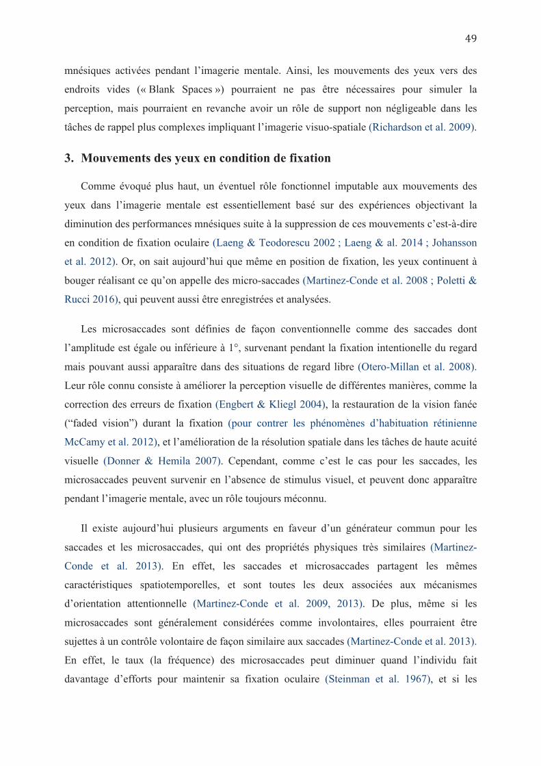
mnésiques activées pendant l’imagerie mentale. Ainsi, les mouvements des yeux vers des
endroits vides (« Blank Spaces ») pourraient ne pas être nécessaires pour simuler la
perception, mais pourraient en revanche avoir un rôle de support non négligeable dans les
tâches de rappel plus complexes impliquant l’imagerie visuo-spatiale (Richardson et al. 2009).
3. Mouvements des yeux en condition de fixation
Comme évoqué plus haut, un éventuel rôle fonctionnel imputable aux mouvements des
yeux dans l’imagerie mentale est essentiellement basé sur des expériences objectivant la
diminution des performances mnésiques suite à la suppression de ces mouvements c’est-à-dire
en condition de fixation oculaire (Laeng & Teodorescu 2002 ; Laeng & al. 2014 ; Johansson
et al. 2012). Or, on sait aujourd’hui que même en position de fixation, les yeux continuent à
bouger réalisant ce qu’on appelle des micro-saccades (Martinez-Conde et al. 2008 ; Poletti &
Rucci 2016), qui peuvent aussi être enregistrées et analysées.
Les microsaccades sont définies de façon conventionnelle comme des saccades dont
l’amplitude est égale ou inférieure à 1°, survenant pendant la fixation intentionelle du regard
mais pouvant aussi apparaître dans des situations de regard libre (Otero-Millan et al. 2008).
Leur rôle connu consiste à améliorer la perception visuelle de différentes manières, comme la
correction des erreurs de fixation (Engbert & Kliegl 2004), la restauration de la vision fanée
(“faded vision”) durant la fixation (pour contrer les phénomènes d’habituation rétinienne
McCamy et al. 2012), et l’amélioration de la résolution spatiale dans les tâches de haute acuité
visuelle (Donner & Hemila 2007). Cependant, comme c’est le cas pour les saccades, les
microsaccades peuvent survenir en l’absence de stimulus visuel, et peuvent donc apparaître
pendant l’imagerie mentale, avec un rôle toujours méconnu.
Il existe aujourd’hui plusieurs arguments en faveur d’un générateur commun pour les
saccades et les microsaccades, qui ont des propriétés physiques très similaires (Martinez-
Conde et al. 2013). En effet, les saccades et microsaccades partagent les mêmes
caractéristiques spatiotemporelles, et sont toutes les deux associées aux mécanismes
d’orientation attentionnelle (Martinez-Conde et al. 2009, 2013). De plus, même si les
microsaccades sont généralement considérées comme involontaires, elles pourraient être
sujettes à un contrôle volontaire de façon similaire aux saccades (Martinez-Conde et al. 2013).
En effet, le taux (la fréquence) des microsaccades peut diminuer quand l’individu fait
davantage d’efforts pour maintenir sa fixation oculaire (Steinman et al. 1967), et si les

individus ne sont pas conscients de leurs microsaccades, ils ne sont généralement pas
conscients de leurs saccades exploratoires non plus (Yarbus 1967), lesquelles peuvent avoir
de petites amplitudes comme les microsaccades (Steinman et al. 1973).
Ainsi, dans les tâches d’imagerie mentale, on peut supposer que les microsaccades
joueraient, dans les conditions de fixation du regard, un rôle similaire à celui des saccades
dans les conditions de regard libre.
Notre article (3) étudie les micro-saccades pendant une tâche d’imagerie
mentale réalisée en fixation oculaire et leurs corrélations spatiales avec le
contenu de l’imagerie mentale.
No

Chapitre 3
LES MOUVEMENTS DES YEUX DANS L’ETUDE DES
REPRÉSENTATIONS MENTALES EN SITUATION
PATHOLOGIQUE
« When the neglect is severe, the patient may behave almost as if one half of the universe had abruptly ceased to exist in any meaningful form.... Patients with unilateral neglect behave not only as if nothing were actually happening in the left hemispace, but also as if nothing of any importance could be expected to occur there »
Mesulam M.M. (1985)
1. Intérêt de l’analyse individuelle
Selon le modèle de Kosslyn détaillé plus haut (Chapitre 1.4), l’imagerie mentale visuelle
consiste en la projection d’informations visuelles issues de la mémoire, sur un tampon visuel
(correspondant au cortex visuel primaire, à organisation rétinotopique), et son exploration par
le déplacement d’une fenêtre attentionnelle (supportée par des structures fronto-pariétales)
afin de sélectionner les zones d’intérêt nécessaires ou utiles pour la résolution d’une tâche
donnée (Kosslyn 1994 ; Kosslyn et al. 2006).
Comme il a été dit plus haut (Chapitre 1.3), les études aussi bien en neuro-imagerie qu’en
neuropsychologie avaient apporté des données controversées quant à l’implication des cortex
fronto-pariétal et occipital dans l’imagerie mentale visuelle (D’Esposito et al. 1997 ; Daselaar
et al. 2010 ; Le Bihan et al. 1993 ; Ganis et al 2004 ; Slotnick et al. 2005 ; voir Farah 1988,
Bartolomeo 2002). Ceci pourrait être expliqué par les différences individuelles dans l’aptitude
à faire de l’imagerie mentale et dans le type préférentiel de stratégie mentale qui diffère d’un
individu à un autre (Voir plus haut, Chapitre 1.5), mais aussi par l’utilisation de stratégies de
compensation comme c’est le cas dans la perception visuelle (par exemple le positionnement
atypique des yeux ou d’autres parties du corps pour limiter l’utilisation des zones aveugles
(Meinberg et al. 1981 ; Ishiaï et al. 1987 ; Zangemeister et al. 1982, 1986, 1995) , ou encore
par la plasticité cérébrale qui pourrait être mise en jeu chez certains patients et pas chez
d’autres selon les caractéristiques de leur lésion (Silvanto & Rees 2011 ; Rode et al. 2003).
Une analyse individuelle des capacités d’imagerie des patients est donc justifiée plutôt que
des études de groupes.
Par ailleurs, que les mouvements des yeux aient ou non un rôle fonctionnel dans
l’imagerie mentale visuelle -notamment pour le rappel des caractéristiques non spatiales
(forme, couleur…etc) de l’objet, il est actuellement admis qu’ils reflètent le contenu spatial de
cette imagerie. Par conséquent, enregistrer les mouvements spontanés des yeux pendant
l’imagerie mentale constituerait un moyen fiable pour tester la capacité d’un sujet à construire
des représentations mentales spatialement cohérentes, et pourrait apporter une percée
considérable dans la « boîte noire » des représentations mentales des patients.
C’est également une méthodologie qui, si elle est exploitable à un niveau individuel, peut
permettre d’accéder à la compréhension des stratégies individuelles de compensation
développées par les sujets souffrant d’altération des structures neuronales supposées
impliquées dans l’imagerie mentale spatiale; à savoir le cortex visuel primaire (en cas d’HLH)
et le réseau fronto-pariétal (en cas de négligence spatiale). Cependant, à l’exception d’une
seule étude chez des sujets atteints d’hémianopsie (Gbadamosi & Zangemeister 2001), nous
n’avons pas trouvé d’autres études utilisant l’enregistrement des mouvements oculaires pour
explorer l’imagerie mentale visuelle chez des patients cérébro-lésés.
2. Chez les sujets avec HLH:
L’hémianopsie latérale homonyme est une amputation unilatérale du champ visuel,
secondaire à une lésion directe ou indirecte du cortex visuel primaire, notamment en cas
d’accident vasculaire cérébral postérieur. Elle est responsable d’un trouble complet de la
perception visuelle au niveau de l’hémi-champ aveugle, que les patients surmontent en
Pour combler ce manque, dans l’article (2) nous avons appliqué notre
paradigme d’analyse du comportement oculaire pendant la mentalisation
de la carte de France (utilisé dans l’article 1) à une population de sujets
atteints de négligence spatiale et/ou hémianopsie, afin de mieux
comprendre leurs stratégies de réalisation de cette tâche mentale.
Po
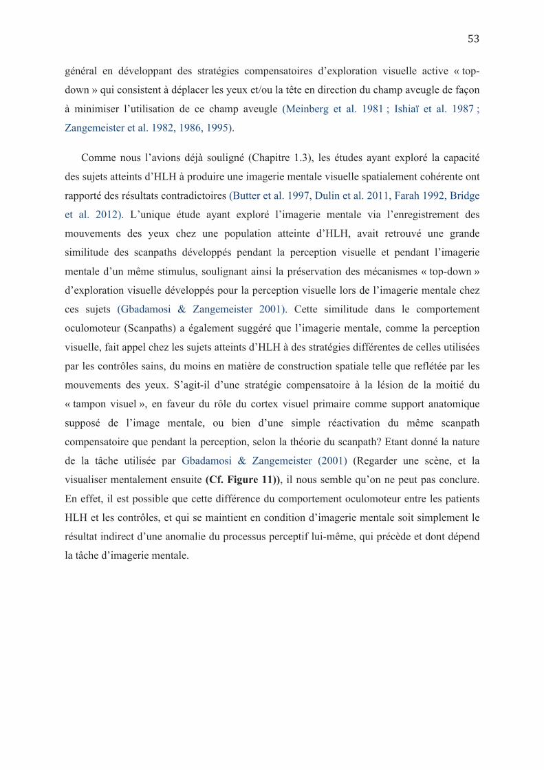
général en développant des stratégies compensatoires d’exploration visuelle active « top-
down » qui consistent à déplacer les yeux et/ou la tête en direction du champ aveugle de façon
à minimiser l’utilisation de ce champ aveugle (Meinberg et al. 1981 ; Ishiaï et al. 1987 ;
Zangemeister et al. 1982, 1986, 1995).
Comme nous l’avions déjà souligné (Chapitre 1.3), les études ayant exploré la capacité
des sujets atteints d’HLH à produire une imagerie mentale visuelle spatialement cohérente ont
rapporté des résultats contradictoires (Butter et al. 1997, Dulin et al. 2011, Farah 1992, Bridge
et al. 2012). L’unique étude ayant exploré l’imagerie mentale via l’enregistrement des
mouvements des yeux chez une population atteinte d’HLH, avait retrouvé une grande
similitude des scanpaths développés pendant la perception visuelle et pendant l’imagerie
mentale d’un même stimulus, soulignant ainsi la préservation des mécanismes « top-down »
d’exploration visuelle développés pour la perception visuelle lors de l’imagerie mentale chez
ces sujets (Gbadamosi & Zangemeister 2001). Cette similitude dans le comportement
oculomoteur (Scanpaths) a également suggéré que l’imagerie mentale, comme la perception
visuelle, fait appel chez les sujets atteints d’HLH à des stratégies différentes de celles utilisées
par les contrôles sains, du moins en matière de construction spatiale telle que reflétée par les
mouvements des yeux. S’agit-il d’une stratégie compensatoire à la lésion de la moitié du
« tampon visuel », en faveur du rôle du cortex visuel primaire comme support anatomique
supposé de l’image mentale, ou bien d’une simple réactivation du même scanpath
compensatoire que pendant la perception, selon la théorie du scanpath? Etant donné la nature
de la tâche utilisée par Gbadamosi & Zangemeister (2001) (Regarder une scène, et la
visualiser mentalement ensuite (Cf. Figure 11)), il nous semble qu’on ne peut pas conclure.
En effet, il est possible que cette différence du comportement oculomoteur entre les patients
HLH et les contrôles, et qui se maintient en condition d’imagerie mentale soit simplement le
résultat indirect d’une anomalie du processus perceptif lui-même, qui précède et dont dépend
la tâche d’imagerie mentale.
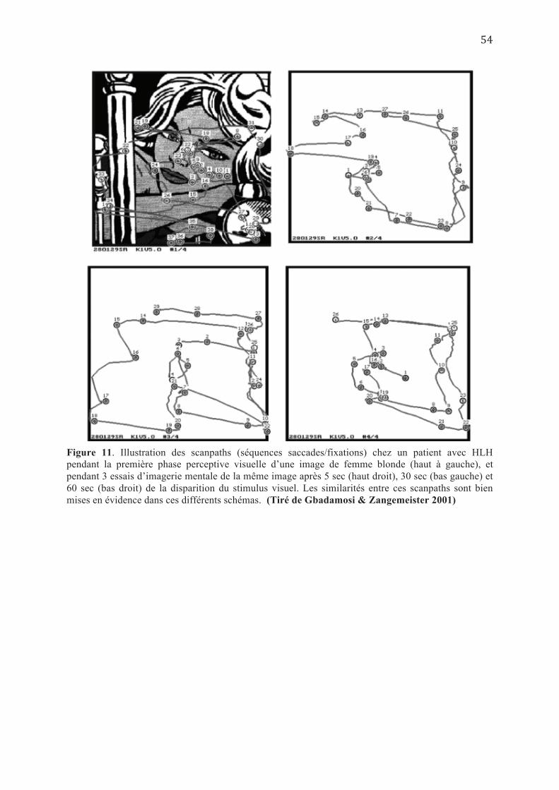
Figure 11. Illustration des scanpaths (séquences saccades/fixations) chez un patient avec HLH pendant la première phase perceptive visuelle d’une image de femme blonde (haut à gauche), et pendant 3 essais d’imagerie mentale de la même image après 5 sec (haut droit), 30 sec (bas gauche) et 60 sec (bas droit) de la disparition du stimulus visuel. Les similarités entre ces scanpaths sont bien mises en évidence dans ces différents schémas. (Tiré de Gbadamosi & Zangemeister 2001)
En concordance avec la dernière supposition, Liman & Zangemeister (2012) avaient
montré, à travers un paradigme d’hémianopsie simulée chez des sujets sains, une perturbation
des scanpaths pendant la perception et pendant l’imagerie mentale en dehors de toute lésion
cérébrale.
Ainsi, la simple comparaison des mouvements oculaires pendant la perception et
l’imagerie mentale d’un même stimulus nouveau (encodé après la lésion cérébrale), n’est pas
suffisante pour juger de la cohérence spatiale de toute imagerie mentale chez les patients, car
les anomalies perceptives peuvent se répercuter sur la qualité de l’imagerie mentale
subséquente. En effet, une amputation artificielle partielle du champ visuel qui inclut la fovéa
(dans un paradigme d’hémianopsie simulée), lors de l’exploration visuelle d’une scène,
engendre une baisse des performances en imagerie mentale de cette même scène (Liman &
Zangemeister 2011).
Pour éviter que ces difficultés perceptives des HLH retentissent sur le
processus d’imagerie, nous avons utilisé pour la tâche d’imagerie
mentale un stimulus extrait de la mémoire à long terme (Rode et al.
2004) et dont l’encodage a été effectué antérieurement à la lésion
cérébrale. Cette expérience nous permettrait de répondre à deux
questions principales : Les sujets présentant une lésion unilatérale de V1
sont-ils capables d’imagerie mentale spatialement cohérente ? Et si c’est
le cas, adoptent-ils une stratégie compensatoire à la lésion cérébrale qui
ne soit pas le simple reflet de celle adoptée en phase perceptive ?
P
3. Chez les sujets avec Négligence spatiale unilatérale:
3.1. La négligence perceptive :
La négligence spatiale résulte généralement d’une lésion de l’hémisphère cérébral droit, et
se caractérise par un trouble de l’orientation de l’attention vers l’hémi-espace gauche et par un
défaut de détection consciente des événements (ou objets) qui s’y situent (Bartolomeo 2006).
Cette négligence de l’hémi-espace gauche va avec un biais attentionnel vers le côté
ipsilésionnel (hyper-attention droite) qui a été mis en évidence dans plusieurs études où le
sujet reconnaît d’abord et considère essentiellement dans son comportement les cibles
visuelles de l’espace ipsi-lésionnel (Gainotti et al. 1991, Azouvi et al. 2002, Mattingley et al.
1994, Bartolomeo 1997), avec une difficulté à extraire son attention de ces cibles là ; ou
déficit de désengagement de l’attention du côté ipsilésionel : Posner et al. 1984, Rastelli et al.
2008). Ce biais attentionnel chez les patients négligents prédit un biais directionnel au niveau
des mouvements des yeux, car les saccades oculaires vers une localisation donnée sont
généralement précédées par un déplacement de l’attention vers cette même cible (Deubel &
Schneider 1996), cependant les enregistrements oculaires lors de l’exploration de scènes
visuelles chez ces patients ont rapporté des résultats controversés avec davantage de saccades
orientées vers la droite dans certaines études (Bourgeois et al. 2014, Cazolli et al. 2011,
Gainotti et al. 2009), et autant de saccades vers la droite que de saccades vers la gauche,
même si le patient n’explore pas la partie de l’espace égocentré la plus à gauche, dans d’autres
(Niemeier & Karnath 2000, 2003 ; Ro et al. 2001). De plus, une étude récente a décrit chez un
patient négligent, une dissociation où la réalisation de saccades vers l’espace gauche n’était
pas accompagnée d’un déplacement attentionnel, de façon à ce que la saccade vers la gauche
est réalisable mais le sujet n’arrive pas à identifier ce qu’il fixe avec le regard dans son hémi-
champ contralésionnel (Benson et al. 2012, voir aussi Khan et al. 2009).
Bartolomeo & Chokron (2002) ont montré que chez les négligents le biais attentionnel
vers le champ ipsilesionel est prédominant sur l’attention exogène (bottom-up) par rapport à
endogène (top-down). Etant donné que imagerie mentale repose sur des mécanismes top-
down, on peut prédire que la génération de l’image mentale ne devrait pas souffrir de biais
attentionnel ipsilésionnel, c’est-à-dire qu’elle ne devrait pas être aussi sévère que la
négligence perceptive qui combine l’hyper-attention vers les éléments visuels droits et la
négligence de l’espace gauche. Ceci constitue un possible élément de compréhension des
dissociations observées entre négligence perceptive et négligence représentationnelle.
3.2. La négligence représentationnelle :
La négligence spatiale peut aussi concerner l’imagerie mentale avec une difficulté à
décrire de mémoire un espace imaginé (exemple : Place du dôme de milan dans l’étude de
Bisiach & Luzzati (1978) ; Cf. Figure 5). Bisiach et al. (1981) attribuaient ceci soit à un
défaut de génération (image mentale amputée de sa moitié gauche), soit à un trouble
d’exploration de l’hémi-espace gauche imaginé. L’étude de ces déficits d’imagerie mentale
encore connus sous le nom de « négligence représentationnelle », se fait généralement en
clinique soit par des tâches d’exploration mentale d’une carte (Rode et al 2004), à la
recherche d’un biais de latéralisation des éléments évoqués (les sujets décrivent plus
d’éléments à droite qu’à gauche), mais ce genre de tâches ne renseigne pas sur le degré de
distorsions spatiales entre les éléments évoqués, soit par le dessin, mais dans ce cas l’hyper-
attention visuelle vers les éléments de droite (du modèle ou dessinés en premier par le patient)
vient perturber l’exercice (Bartolomeo & Chokron 2002).
Il nous semble alors intéressant d’utiliser l’enregistrement des mouvements oculaires
comme outil d’exploration des représentations mentales chez les sujets négligents, car leur
caractère inconscient, ne laissant pas de trace perceptible éviterait les éventuels biais
attentionnels de type Bottom-up.
4. Représentations mentales et référentiels spatiaux:
La localisation d’un objet peut généralement être considérée selon deux principaux cadres
de références spatiales ; egocentrique et allocentrique (Galati et al. 2000). Dans le cadre
egocentrique, la position de l’objet est codée par rapport à la position du corps de
l’observateur ou plus spécifiquement par rapport à une partie de son corps, on distingue ainsi
parmi les cadres égocentriques, le cadre centré sur la rétine, centré sur l’œil (Howard 1991),
centré sur la tête (Karn et al. 1997), centré sur le tronc (Darling & Miller 1995), ou sur un
Dans l’article (2), nous rapportons pour la première fois, les résultats de
l’analyse des mouvements spontanés des yeux dans une tâche d’imagerie
mentale libre chez des sujets atteints de négligence spatiale avec ou sans
HLH
Dan
autre segment du corps selon la tâche demandée comme le membre supérieur (Soechting et al.
1990).
Ce référentiel égocentré est généralement utilisé pour l’organisation des mouvements
orientés vers l’espace extracorporel (Jeannerod & Biguer 1989), comme pour atteindre une
cible. Dans le cadre allocentrique, les objets sont représentés selon leur propre configuration
spatiale, tenant compte des relations entre leurs différents constituants et avec les objets
environnants (Galati et al 2000).
Chez les primates, les études neurophysiologiques ont identifié des neurones codant pour
l’information visuelle dans un référentiel égocentrique au niveau du cortex pariétal postérieur
et plus précisément au niveau des aires 7a, LIP, VIP, PO et au niveau du cortex pré-moteur
(Andersen et al. 1985 ; Andersen et al. 1990 ; Duhamel et al. 1997, Galletti et al. 1993 ;
Graziano et al. 1994), alors que d’autres neurones dont le taux d’activation est modulé par des
modifications spatiales à l’intérieur de l’objet, quel que soit sa position par rapport au corps
(et donc codant l’information selon un référentiel allocentré) ont été retrouvés également au
niveau cortex pariétal postérieur, plus particulièrement dans LIP (Breznen et al. 1999) et au
niveau du lobe frontal dans le Supplementary Eye Field (Olson et Gettner 1995).
Chez l’Homme, et en cas de négligence spatiale unilatérale, un de ces référentiel ou les
deux peuvent être altérés. Le déficit d’orientation de l’attention vers le côté opposé à la lésion
observés chez les négligents, résulterait d’un trouble de l’élaboration de la référence
égocentrique en se référant à l’axe sagittal du corps (Karnath et al., 1991). Cette référence
peut se concevoir comme une position d’équilibre entre les informations issues des deux côtés
de l’espace et gouvernant les actions dirigées vers celles-ci. Chez les patients négligents, la
transformation des coordonnées sensorielles en coordonnées égocentrées fonctionnerait avec
une erreur systématique résultant de la déviation de la référence égocentrique du côté de la
lésion (Ventre et al. 1984). En concordance avec cette hypothèse, un décalage ipsilésionnel du
droit-devant chez les patients négligents a été observé (Heilman 1983, Perenin, 1992,
Chokron & Imbert 1995). Cette déviation était supposée responsable d’une altération du
système de coordonnées égocentrées nécessaire pour la coordination visuomotrice et
l’exploration de l’espace, ce qui expliquerait le comportement de Négligence. Karnath et al.
(1991) ont proposé que la négligence soit causée par une rotation ipsilésionnelle du cadre de
référence tandis que Vallar et al. (1995) ont plaidé pour une translation ispsilésionnelle.
Cependant, la relation de cause à effet entre cette déviation ipsilésionelle dans la
perception de l’axe egocentrique et les conséquences comportementales de négligence
spatiale a ensuite été critiquée notamment suite à la mise en évidence d’une dissociation entre
la présence d’un comportement de négligence avec l’absence de décalage de la référence
égocentrique chez certains patients d’une part (Hasselbach and Butter 1997, Farné et al.
1998), et d’autre part, par l’absence de corrélation entre l’existence effective de ce décalage
de l’axe égocentrique et la présence de comportements de négligence perceptive et/ou visuo-
motrice chez les patients présentant des lésions cérébrales droites (Bartolomeo & Chokron
1999).
Le déficit peut aussi concerner la moitié contro-lésionnelle de chaque objet quelle que soit
sa position dans le référentiel égocentré, c’est ce qu’on appelle « Négligence centrée sur
l’objet » (Walker 1995, Bisiach 1997). Ces deux troubles (Négligence centrée sur le corps
suite à une perturbation du référentiel egocentrique, et Négligence centrée sur l’objet suite à
une perturbation du référentiel allocentrique) peuvent être concomitants (Gainotti et al. 1972)
ou être observés séparément (Humphrey & Riddoch 1994). Ils ont été décrits après lésion du
lobe pariétal inférieur droit, ou du cortex prémoteur dorsolatéral droit (Vallar 1993, Husain &
Karnath 1996 ; Bisiach & Vallar 2000).
En utilisant l’IRM fonctionnelle chez les sujets sains, plusieurs études en mis en
évidence une activation large des réseaux temporo-pariéto-occipitaux dans les tâches de
navigation spatiale mettant en jeu à la fois les référentiels égocentriques et allocentriques
(Ghaem et al. 1997 ; Gomez et al. 2014). Galati et al. (2000), a mis en évidence une activation
des réseaux fronto-pariétaux à savoir le cortex pariétal postérieur et le cortex frontal pré-
moteur aussi bien dans les taches faisant appel aux références égocentriques
qu’allocentriques, avec une latéralisation évidente pour le côté droit, et avec des zones
d’activations plus étendues pour les tâches égocentriques.
L’implication spécifique des réseaux fronto-pariétaux s’explique par leur rôle dans la
préparation du mouvement orienté vers un but (comme l’orientation de la tête ou des yeux,
l’approche et la saisie d’un objet), nécessitant la détermination de la localisation de la cible
par rapport aux effecteurs moteurs (Galati et al. 2000), mais aussi par leur rôle dans le
traitement visuel des relations spatiales allocentrées (voie visuelle dorsale, Landmark task,
(Ungerleider & Mishkin 1982)).
Des troubles de représentation spatiale notamment au niveau de l’axe horizontal, ont été
décrits chez les sujets ayant une HLH, avec un biais contra-lésionnel lors de la bissection de
lignes (Barton & Black 1998) et une exagération dans l’estimation des distances dans l’espace
visuel contra-lesionnel (Doricchi et al. 2002). Ce trouble serait la conséquence de l’altération
des coordonnées spatiales rétinotopiques (centré sur la rétine, amputation du champ visuel
contra-lésionnel) au niveau supérieur de codage spatiotopique (centré sur la tête) par les
réseaux pariéto-occipitaux (Galletti and Battaglini, 1989; Galletti et al, 1993, 1995; Andersen,
1995). La création d’un décalage entre les deux cadres de référence, par l’orientation de la tête
vers le côté contralésionnel (en direction du champ aveugle) permet une réduction de ces
troubles de représentation spatiale chez les hémianopsiques (Doricchi et al. 2002).
Nous pensons qu’étudier les mouvements des yeux pendant une tâche
d’imagerie mentale chez des sujets négligents, pourrait participer à mieux
comprendre la négligence représentationnelle et son approche vis à vis des
différents cadres de référence. D’une part, l’analyse des positions oculaires
absolues au moment de l’évocation des différents éléments d’une carte
visualisée, permettrait de situer l’image mentale selon l’axe egocentrique
(le sujet génère-t-il sa carte mentale de façon centrée sur son corps ou
complètement déviée vers le côté ipsi-lésionnel ?). D’autre part, l’analyse
spatiale des fixations oculaires les unes par rapport aux autres, permettrait
de vérifier la cohérence spatiale globale de la carte mentale (spatialement
cohérente dans un cadre allocentrique ? distorsions spatiales plus marquées
à gauche ?).
Nous p
Dans l’article (2), nous discutons les caractéristiques spatiales des
représentations mentales chez les HLH en référence d’abord à un
cadre de référence allocentrique (cohérence spatiale globale de la carte
mentale), et à un cadre de référence égocentrique non rétinotopique en
évaluant la projection de la carte mentale par rapport à l’axe du corps
Dan


Article (1)
Publié dans
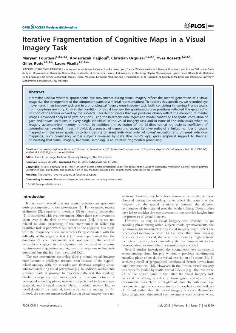
Iterative Fragmentation of Cognitive Maps in a VisualImagery TaskMaryam Fourtassi1,2,3,4,5, Abderrazak Hajjioui6, Christian Urquizar1,2,3,4, Yves Rossetti1,2,3,4,
Gilles Rode1,2,3,4, Laure Pisella1,2,3,4*
1 INSERM, U1028; CNRS, UMR5292; Lyon Neuroscience Research Center, ImpAct team, Lyon, France, 2Universite Lyon 1, Biologie Humaine, Lyon, France, 3Hospices Civils
de Lyon, Mouvement et Handicap, Hopital Henry Gabrielle, St-Genis-Laval, France, 4Mouvement et Handicap, Hopital Neurologique, Lyon, France, 5 Faculte de Medecine
et de pharmacie, Universite Mohamed Premier, Oujda, Morocco, 6 Physical Medicine and Rehabilitation, CHU Hassan II Fez Faculty of Medicine and Pharmacy, University
Mohammed Benabdellah, Fez, Morocco
Abstract
It remains unclear whether spontaneous eye movements during visual imagery reflect the mental generation of a visualimage (i.e. the arrangement of the component parts of a mental representation). To address this specificity, we recorded eyemovements in an imagery task and in a phonological fluency (non-imagery) task, both consisting in naming French townsfrom long-term memory. Only in the condition of visual imagery the spontaneous eye positions reflected the geographicposition of the towns evoked by the subjects. This demonstrates that eye positions closely reflect the mapping of mentalimages. Advanced analysis of gaze positions using the bi-dimensional regression model confirmed the spatial correlation ofgaze and towns’ locations in every single individual in the visual imagery task and in none of the individuals when noimagery accompanied memory retrieval. In addition, the evolution of the bi-dimensional regression’s coefficient ofdetermination revealed, in each individual, a process of generating several iterative series of a limited number of townsmapped with the same spatial distortion, despite different individual order of towns’ evocation and different individualmappings. Such consistency across subjects revealed by gaze (the mind’s eye) gives empirical support to theoriespostulating that visual imagery, like visual sampling, is an iterative fragmented processing.
Citation: Fourtassi M, Hajjioui A, Urquizar C, Rossetti Y, Rode G, et al. (2013) Iterative Fragmentation of Cognitive Maps in a Visual Imagery Task. PLoS ONE 8(7):e68560. doi:10.1371/journal.pone.0068560
Editor: Floris P. de Lange, Radboud University Nijmegen, The Netherlands
Received January 28, 2013; Accepted May 30, 2013; Published July 17, 2013
Copyright: � 2013 Fourtassi et al. This is an open-access article distributed under the terms of the Creative Commons Attribution License, which permitsunrestricted use, distribution, and reproduction in any medium, provided the original author and source are credited.
Funding: The authors have no support or funding to report.
Competing Interests: The authors have declared that no competing interests exist.
* E-mail: [email protected]
Introduction
It has been observed that any mental activities are spontane-
ously accompanied by eye movements [1]. For example, mental
arithmetic [2], response to questions [3] or memory recollection
[1] is associated with eye movements. Since these eye movements
occur even in the dark or with closed eyes [2,4], they are not
related to visual processing of the environment in which the
cognitive task is performed but rather to the cognitive task itself,
with the frequency of eye movements being correlated with the
difficulty of the cognitive task [1]. It was hypothesized that the
direction of eye movements was opposite to the cortical
hemisphere engaged in the cognitive task (leftward in response
to visuo-spatial questions and rightward in response to linguistic
questions) but this has been discarded [5,6].
The eye movements occurring during mental visual imagery
have become a privileged research area because of the hypoth-
esized analogy with the saccades and fixations sampling visual
information during visual perception [7]. In addition, oculometric
technics made it possible to experimentally test this analogy.
Studies comparing eye movements or fixations between a
perceptual encoding phase, in which subjects had to learn a new
material, and a visual imagery phase, in which subjects had to
recall details of this material, have confirmed this analogy [8–15].
Indeed, the eye movements evoked during visual imagery were not
arbitrary. Instead, they have been shown to be similar to those
observed during the encoding, or to reflect the content of the
imagery, i.e. the spatial relationship between the different
components of the material provided to the subject. These studies
have led to the idea that eye movements may provide insights into
the processes of visual imagery.
However, as long as visual imagery was preceded by an
encoding phase during which subjects made eye movements, the
eye movements measured during visual imagery might reflect the
processes of memory retrieval [15–17] rather than visual imagery
processes per se. Indeed, the recall from memory might activate
the whole memory trace, including the eye movements to the
corresponding location where a stimulus was encoded.
Several studies investigated the spontaneous eye movements
accompanying visual imagery without a previous experimental
encoding phase either during verbal description of a scene [10,11]
or during recall of geographical locations of French towns from
long-term memory [18]. However, in the former, visual imagery
was explicitly guided by spatial verbal indexes (e.g. ‘‘the tree to the
left of the house’’) and in the latter the visual imagery task
consisted in stating whether a town given verbally by the
experimenter was ‘‘left’’ or ‘‘right’’ of Paris. In both cases eye
movements might reflect a reaction to the explicit spatial indexes
of the task rather than the visual imagery processes themselves.
Accordingly, such directional eye movements were observed when
PLOS ONE | www.plosone.org 1 July 2013 | Volume 8 | Issue 7 | e68560
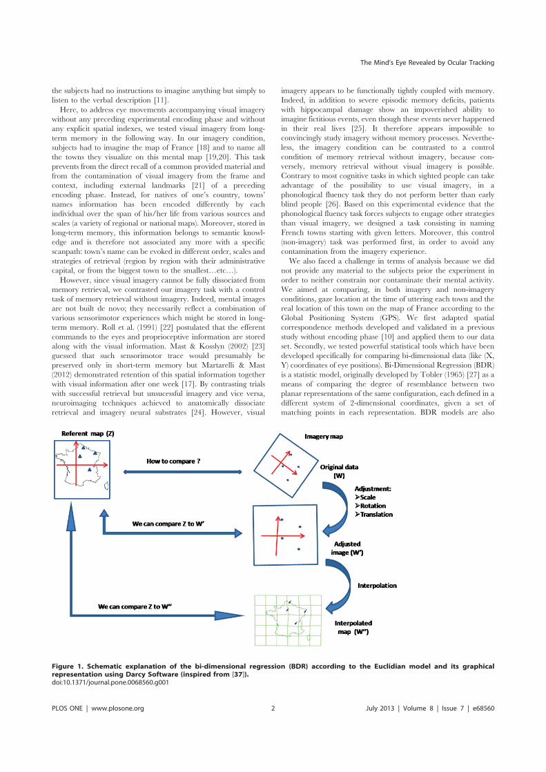
the subjects had no instructions to imagine anything but simply to
listen to the verbal description [11].
Here, to address eye movements accompanying visual imagery
without any preceding experimental encoding phase and without
any explicit spatial indexes, we tested visual imagery from long-
term memory in the following way. In our imagery condition,
subjects had to imagine the map of France [18] and to name all
the towns they visualize on this mental map [19,20]. This task
prevents from the direct recall of a common provided material and
from the contamination of visual imagery from the frame and
context, including external landmarks [21] of a preceding
encoding phase. Instead, for natives of one’s country, towns’
names information has been encoded differently by each
individual over the span of his/her life from various sources and
scales (a variety of regional or national maps). Moreover, stored in
long-term memory, this information belongs to semantic knowl-
edge and is therefore not associated any more with a specific
scanpath: town’s name can be evoked in different order, scales and
strategies of retrieval (region by region with their administrative
capital, or from the biggest town to the smallest…etc…).
However, since visual imagery cannot be fully dissociated from
memory retrieval, we contrasted our imagery task with a control
task of memory retrieval without imagery. Indeed, mental images
are not built de novo; they necessarily reflect a combination of
various sensorimotor experiences which might be stored in long-
term memory. Roll et al. (1991) [22] postulated that the efferent
commands to the eyes and proprioceptive information are stored
along with the visual information. Mast & Kosslyn (2002) [23]
guessed that such sensorimotor trace would presumably be
preserved only in short-term memory but Martarelli & Mast
(2012) demonstrated retention of this spatial information together
with visual information after one week [17]. By contrasting trials
with successful retrieval but unsucessful imagery and vice versa,
neuroimaging techniques achieved to anatomically dissociate
retrieval and imagery neural substrates [24]. However, visual
imagery appears to be functionally tightly coupled with memory.
Indeed, in addition to severe episodic memory deficits, patients
with hippocampal damage show an impoverished ability to
imagine fictitious events, even though these events never happened
in their real lives [25]. It therefore appears impossible to
convincingly study imagery without memory processes. Neverthe-
less, the imagery condition can be contrasted to a control
condition of memory retrieval without imagery, because con-
versely, memory retrieval without visual imagery is possible.
Contrary to most cognitive tasks in which sighted people can take
advantage of the possibility to use visual imagery, in a
phonological fluency task they do not perform better than early
blind people [26]. Based on this experimental evidence that the
phonological fluency task forces subjects to engage other strategies
than visual imagery, we designed a task consisting in naming
French towns starting with given letters. Moreover, this control
(non-imagery) task was performed first, in order to avoid any
contamination from the imagery experience.
We also faced a challenge in terms of analysis because we did
not provide any material to the subjects prior the experiment in
order to neither constrain nor contaminate their mental activity.
We aimed at comparing, in both imagery and non-imagery
conditions, gaze location at the time of uttering each town and the
real location of this town on the map of France according to the
Global Positioning System (GPS). We first adapted spatial
correspondence methods developed and validated in a previous
study without encoding phase [10] and applied them to our data
set. Secondly, we tested powerful statistical tools which have been
developed specifically for comparing bi-dimensional data (like (X,
Y) coordinates of eye positions). Bi-Dimensional Regression (BDR)
is a statistic model, originally developed by Tobler (1965) [27] as a
means of comparing the degree of resemblance between two
planar representations of the same configuration, each defined in a
different system of 2-dimensional coordinates, given a set of
matching points in each representation. BDR models are also
Figure 1. Schematic explanation of the bi-dimensional regression (BDR) according to the Euclidian model and its graphicalrepresentation using Darcy Software (inspired from [37]).doi:10.1371/journal.pone.0068560.g001
The Mind’s Eye Revealed by Ocular Tracking
PLOS ONE | www.plosone.org 2 July 2013 | Volume 8 | Issue 7 | e68560
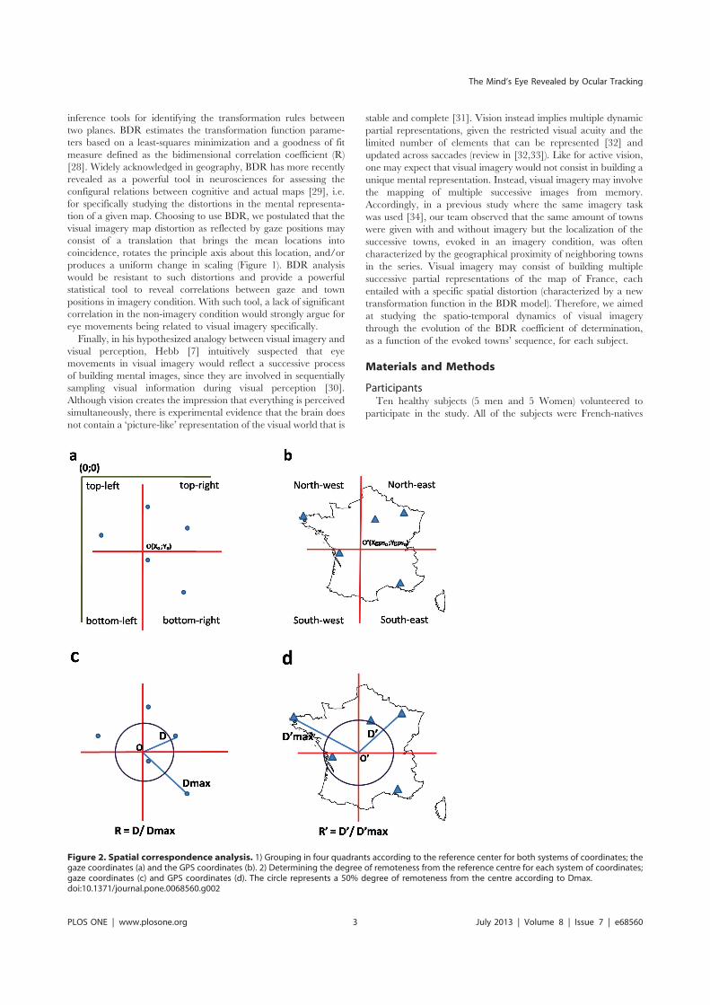
inference tools for identifying the transformation rules between
two planes. BDR estimates the transformation function parame-
ters based on a least-squares minimization and a goodness of fit
measure defined as the bidimensional correlation coefficient (R)
[28]. Widely acknowledged in geography, BDR has more recently
revealed as a powerful tool in neurosciences for assessing the
configural relations between cognitive and actual maps [29], i.e.
for specifically studying the distortions in the mental representa-
tion of a given map. Choosing to use BDR, we postulated that the
visual imagery map distortion as reflected by gaze positions may
consist of a translation that brings the mean locations into
coincidence, rotates the principle axis about this location, and/or
produces a uniform change in scaling (Figure 1). BDR analysis
would be resistant to such distortions and provide a powerful
statistical tool to reveal correlations between gaze and town
positions in imagery condition. With such tool, a lack of significant
correlation in the non-imagery condition would strongly argue for
eye movements being related to visual imagery specifically.
Finally, in his hypothesized analogy between visual imagery and
visual perception, Hebb [7] intuitively suspected that eye
movements in visual imagery would reflect a successive process
of building mental images, since they are involved in sequentially
sampling visual information during visual perception [30].
Although vision creates the impression that everything is perceived
simultaneously, there is experimental evidence that the brain does
not contain a ‘picture-like’ representation of the visual world that is
stable and complete [31]. Vision instead implies multiple dynamic
partial representations, given the restricted visual acuity and the
limited number of elements that can be represented [32] and
updated across saccades (review in [32,33]). Like for active vision,
one may expect that visual imagery would not consist in building a
unique mental representation. Instead, visual imagery may involve
the mapping of multiple successive images from memory.
Accordingly, in a previous study where the same imagery task
was used [34], our team observed that the same amount of towns
were given with and without imagery but the localization of the
successive towns, evoked in an imagery condition, was often
characterized by the geographical proximity of neighboring towns
in the series. Visual imagery may consist of building multiple
successive partial representations of the map of France, each
entailed with a specific spatial distortion (characterized by a new
transformation function in the BDR model). Therefore, we aimed
at studying the spatio-temporal dynamics of visual imagery
through the evolution of the BDR coefficient of determination,
as a function of the evoked towns’ sequence, for each subject.
Materials and Methods
ParticipantsTen healthy subjects (5 men and 5 Women) volunteered to
participate in the study. All of the subjects were French-natives
Figure 2. Spatial correspondence analysis. 1) Grouping in four quadrants according to the reference center for both systems of coordinates; thegaze coordinates (a) and the GPS coordinates (b). 2) Determining the degree of remoteness from the reference centre for each system of coordinates;gaze coordinates (c) and GPS coordinates (d). The circle represents a 50% degree of remoteness from the centre according to Dmax.doi:10.1371/journal.pone.0068560.g002
The Mind’s Eye Revealed by Ocular Tracking
PLOS ONE | www.plosone.org 3 July 2013 | Volume 8 | Issue 7 | e68560
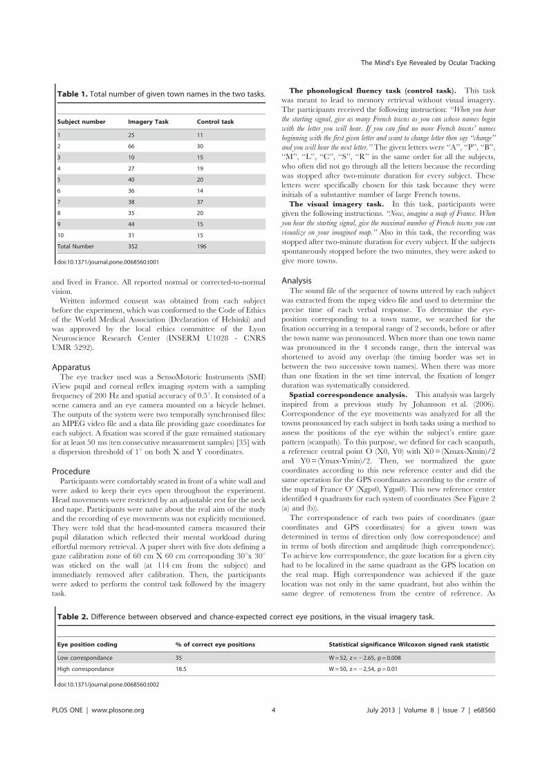
and lived in France. All reported normal or corrected-to-normal
vision.
Written informed consent was obtained from each subject
before the experiment, which was conformed to the Code of Ethics
of the World Medical Association (Declaration of Helsinki) and
was approved by the local ethics committee of the Lyon
Neuroscience Research Center (INSERM U1028 - CNRS
UMR 5292).
ApparatusThe eye tracker used was a SensoMotoric Instruments (SMI)
iView pupil and corneal reflex imaging system with a sampling
frequency of 200 Hz and spatial accuracy of 0.5u. It consisted of a
scene camera and an eye camera mounted on a bicycle helmet.
The outputs of the system were two temporally synchronised files:
an MPEG video file and a data file providing gaze coordinates for
each subject. A fixation was scored if the gaze remained stationary
for at least 50 ms (ten consecutive measurement samples) [35] with
a dispersion threshold of 1u on both X and Y coordinates.
ProcedureParticipants were comfortably seated in front of a white wall and
were asked to keep their eyes open throughout the experiment.
Head movements were restricted by an adjustable rest for the neck
and nape. Participants were naıve about the real aim of the study
and the recording of eye movements was not explicitly mentioned.
They were told that the head-mounted camera measured their
pupil dilatation which reflected their mental workload during
effortful memory retrieval. A paper sheet with five dots defining a
gaze calibration zone of 60 cm X 60 cm corresponding 30ux 30uwas sticked on the wall (at 114 cm from the subject) and
immediately removed after calibration. Then, the participants
were asked to perform the control task followed by the imagery
task.
The phonological fluency task (control task). This task
was meant to lead to memory retrieval without visual imagery.
The participants received the following instruction: ‘‘When you hear
the starting signal, give as many French towns as you can whose names begin
with the letter you will hear. If you can find no more French towns’ names
beginning with the first given letter and want to change letter then say ‘‘change’’
and you will hear the next letter.’’ The given letters were ‘‘A’’, ‘‘P’’, ‘‘B’’,
‘‘M’’, ‘‘L’’, ‘‘C’’, ‘‘S’’, ‘‘R’’ in the same order for all the subjects,
who often did not go through all the letters because the recording
was stopped after two-minute duration for every subject. These
letters were specifically chosen for this task because they were
initials of a substantive number of large French towns.
The visual imagery task. In this task, participants were
given the following instructions. ‘‘Now, imagine a map of France. When
you hear the starting signal, give the maximal number of French towns you can
visualize on your imagined map.’’ Also in this task, the recording was
stopped after two-minute duration for every subject. If the subjects
spontaneously stopped before the two minutes, they were asked to
give more towns.
AnalysisThe sound file of the sequence of towns uttered by each subject
was extracted from the mpeg video file and used to determine the
precise time of each verbal response. To determine the eye-
position corresponding to a town name, we searched for the
fixation occurring in a temporal range of 2 seconds, before or after
the town name was pronounced. When more than one town name
was pronounced in the 4 seconds range, then the interval was
shortened to avoid any overlap (the timing border was set in
between the two successive town names). When there was more
than one fixation in the set time interval, the fixation of longer
duration was systematically considered.
Spatial correspondence analysis. This analysis was largely
inspired from a previous study by Johansson et al. (2006).
Correspondence of the eye movements was analyzed for all the
towns pronounced by each subject in both tasks using a method to
assess the positions of the eye within the subject’s entire gaze
pattern (scanpath). To this purpose, we defined for each scanpath,
a reference central point O (X0, Y0) with X0= (Xmax-Xmin)/2
and Y0= (Ymax-Ymin)/2. Then, we normalized the gaze
coordinates according to this new reference center and did the
same operation for the GPS coordinates according to the centre of
the map of France O’ (Xgps0, Ygps0). This new reference center
identified 4 quadrants for each system of coordinates (See Figure 2
(a) and (b)).
The correspondence of each two pairs of coordinates (gaze
coordinates and GPS coordinates) for a given town was
determined in terms of direction only (low correspondence) and
in terms of both direction and amplitude (high correspondence).
To achieve low correspondence, the gaze location for a given city
had to be localized in the same quadrant as the GPS location on
the real map. High correspondence was achieved if the gaze
location was not only in the same quadrant, but also within the
same degree of remoteness from the centre of reference. As
Table 1. Total number of given town names in the two tasks.
Subject number Imagery Task Control task
1 25 11
2 66 30
3 10 15
4 27 19
5 40 20
6 36 14
7 38 37
8 35 20
9 44 15
10 31 15
Total Number 352 196
doi:10.1371/journal.pone.0068560.t001
Table 2. Difference between observed and chance-expected correct eye positions, in the visual imagery task.
Eye position coding % of correct eye positions Statistical significance Wilcoxon signed rank statistic
Low correspondance 35 W=52, z =22.65, p = 0.008
High correspondance 18.5 W=50, z =22,54, p = 0.01
doi:10.1371/journal.pone.0068560.t002
The Mind’s Eye Revealed by Ocular Tracking
PLOS ONE | www.plosone.org 4 July 2013 | Volume 8 | Issue 7 | e68560
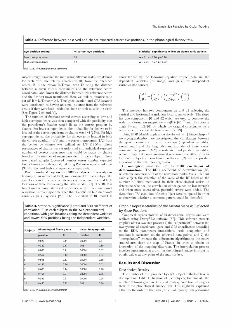
subjects might visualize the map using different scales, we defined
for each town the relative remoteness (R) from the reference
centre. R is the ration D/Dmax, with D being the distance
between a given town’s coordinates and the reference centre
coordinates, and Dmax the distance between this reference centre
and the furthest town mentioned. Here we took as distance ratio
cut-off R=D/Dmax= 0.5. Thus gaze location and GPS location
were considered as having an equal distance from the reference
centre if they were both inside the circle or both outside the circle
(See Figure 2 (c) and (d)).
The number of fixations scored correct according to low and
high correspondence was then compared with the possibility that
the participant’s fixation would lie at the correct position by
chance. For low correspondence, the probability for the eye to be
located in the correct quadrant by chance was 1/4 (25%). For high
correspondence, the probability for the eye to be located in both
the correct quadrant (1/4) and the correct remoteness (1/2) from
the center by chance was defined as 1/8 (12.5%). These
percentages of chance were transformed into individual expected
number of correct occurrence that would be made by chance
based on the number of towns provided for each subject. These
two paired samples (observed number versus number expected
from chance) were then analyzed using Wilcoxon signed-ranks test
[36] for low and high correspondence separately.
Bi-dimensional regression (BDR) analysis. To verify our
findings at an individual level, we compared for each subject the
gaze locations at the time of uttering each town and the real GPS
locations of these towns using the BDR model [27]. The BDR is
based on the same statistical principles as the one-dimensional
regression with a major difference that it applies to bi-dimensional
variables (X;Y) systems [29]. The Eucledian BDR model is
characterized by the following equation where (A;B) are the
dependent variables (the image) and (X;Y) the independent
variables (the source).
A
B
� �~
a1
a2
� �z
b1{b2
b2zb1
� �: X
Y
� �
The intercept has two components a2 and a1 reflecting the
vertical and horizontal translation factors, respectively. The slope
has two components b1 and b2 which are used to compute the
scale transformation magnitude = (b1+ b2)1/2 and the rotation
angle h= tan21(b2/b1) by which the original coordinates were
transformed to derive the least square fit [29].
Using BDR Matlab application developed by TJ Pingel (http://
www.geog.ucsb.edu/), we investigated the correlations between
the gaze locations at towns’ evocation (dependent variables,
variant map) and the longitudes and latitudes of these towns,
converted to planar (X;Y) coordinates (independent variables,
referent map). Like uni-dimensional regression, the BDR provides
for each subject a correlation coefficient (R) and a p-value
according to the test F for regression.
Chronological evolution of the BDR coefficient of
determination. The BDR coefficient of determination (R2)
reflects the goodness of fit of the regression model. We studied for
each subject, the evolution of the value of the R2 based on the
number of cities mentioned in their chronological order, to
determine whether the correlation either gained or lost strength
and when more towns (data, potential errors) were added. The
dynamics of R2 evolution of each subject were compared in order
to determine whether a common pattern could be identified.
Graphic Representations of the Mental Maps as Reflectedby Gaze PositionsGraphical representations of bi-dimensional regressions were
realized using DarcyH2.0 software [37]. This software extracts
graphics after a two-step process: 1) the ‘‘adjustment’’ between the
two systems of coordinates (gaze and GPS coordinates) according
to the BDR parameters (translations, scale adaptation and
rotation) is calculated on the observed data points, and 2) the
‘‘interpolation’’ extends the adjustment algorithms to the entire
studied area (here the map of France) in order to obtain an
illustration of the mapping distortion. The interpolation process
involves superimposing a grid on the adjusted image in order to
obtain values at any point of the map surface.
Results and Discussion
Descriptive ResultsThe number of town provided by each subject in the two tasks is
displayed on Table 1. In most of the subjects, but not all, the
number of towns given in the visual imagery condition was higher
than in the phonological fluency task. This might be explained
either by the order of the tasks (the visual imagery task performed
Table 3. Difference between observed and chance-expected correct eye positions, in the phonological fluency task.
Eye position coding % correct eye positions Statistical significance Wilcoxon signed rank statistic
Low correspondance 25 W=2, z =20.10, p = 0.20
High correspondance 9.5 W=5, z =21.27, p = 0.92
doi:10.1371/journal.pone.0068560.t003
Table 4. Statistical significance (F test) and BDR coefficient ofcorrelation (R) in each subject, in the two experimentalconditions, with gaze locations being the dependent variablesand towns’ GPS positions being the independent variables.
Subjects Phonological fluency task Visual imagery task
p-value R p-value R
1 0.823 0.14 0.0001 0.61
2 0.322 0.17 0.05 0.39
3 0.803 0.1 0.0001 0.87
4 0.562 0.17 0.0001 0.67
5 0.503 0.17 0.0001 0.55
6 0.185 0.36 0.0001 0.51
7 0.685 0.14 0.0001 0.48
8 0.461 0.2 0.0001 0.85
9 0.546 0.2 0.0001 0.68
10 0.450 0.22 0.01 0.36
doi:10.1371/journal.pone.0068560.t004
The Mind’s Eye Revealed by Ocular Tracking
PLOS ONE | www.plosone.org 5 July 2013 | Volume 8 | Issue 7 | e68560
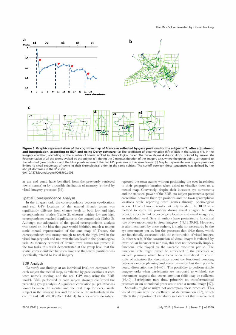
at the end could have benefited from the previously retrieved
towns’ names) or by a possible facilitation of memory retrieval by
visual imagery processes [38].
Spatial Correspondence AnalysisIn the imagery task, the correspondence between eye-fixations
and real GPS locations of the uttered French towns was
significantly different from chance levels in both low and high
correspondence models (Table 2), whereas neither low nor high
correspondence reached significance in the control task (Table 3).
Although our adaptation of the spatial correspondence analysis
was based on the idea that gaze would faithfully match a unique
static mental representation of the true map of France, the
correspondence was strong enough to reach the high level in the
visual imagery task and not even the low level in the phonological
task. As memory retrieval of French town names was present in
the two tasks, this result demonstrated at the group level that the
spatial correspondence between gaze and the towns’ positions was
specifically related to visual imagery.
BDR AnalysisTo verify our findings at an individual level, we compared for
each subject the mental map, as reflected by gaze locations at each
town name’s uttering, and the real GPS map using the BDR
model. BDR performed in each subject strongly confirmed the
preceding group analysis. A significant correlation (all p,0.05) was
found between the mental and the real map for every single
subject in the imagery task and for none of the individuals in the
control task (all p.0.05) (See Table 4). In other words, no subject
reported the town names without positioning the eyes in relation
to their geographic location when asked to visualize them on a
mental map. Conversely, despite their incessant eye movements
and the statistical power of the BDR, no subject presented a spatial
correlation between their eye positions and the town geographical
locations while reporting town names through phonological
access. These clear-cut results not only validate the BDR as a
method to study eye positions during visual imagery but also
provide a specific link between gaze location and visual imagery at
an individual level. Several authors have postulated a functional
role of eye movements in visual imagery [7,9,18,39,40]. However,
as also mentioned by these authors, it might not necessarily be the
eye movements per se, but the processes that drive them, which
are functionally associated with the construction of visual images.
In other words, if the construction of visual images is reflected by
overt ocular behavior in our task, this does not necessarily imply a
functional role played by the saccadic execution per se. The
functional role might rather be attributed to the processes of
saccade planning which have been often assimilated to covert
shifts of attention (for discussions about the functional coupling
between saccade planning and covert attention but their possible
neural dissociation see [41–45]). The possibility to perform simple
imagery tasks when participants are instructed to withhold eye
movements suggests that covert attention shifts may be sufficient
[46,40]. Participants may draw primarily on transformational
processes or on attentional processes to scan a mental image [47].
Saccades might or might not accompany these processes. This
would explain why the coefficient of determination (R2), which
reflects the proportion of variability in a data set that is accounted
Figure 3. Graphic representation of the cognitive map of France as reflected by gaze positions for the subject n61, after adjustmentand interpolation, according to BDR and using Darcy software. (a) The coefficient of determination (R2) of BDR in the subject nu1, in theimagery condition, according to the number of towns evoked in chronological order. The curve shows 4 drastic drops pointed by arrows. (b)Representation of all the towns evoked by the subject nu1 during the 2 minutes duration of the imagery task, where the green points correspond tothe adjusted gaze positions and the blue points represent the real GPS positions of the same towns. (c) Graphic representations of gaze positions,limited to small sequences of towns in their chronological order, in the same subject. The cut-off between these sequences was defined by theabrupt decreases in the R2 curve.doi:10.1371/journal.pone.0068560.g003
The Mind’s Eye Revealed by Ocular Tracking
PLOS ONE | www.plosone.org 6 July 2013 | Volume 8 | Issue 7 | e68560
for by the statistical Euclidian model, remained relatively low in
most subjects even if the correlations were significant.
An additional explanation would be that a unique Euclidian
transformation function was calculated for the entire duration of
the visual imagery task. The analysis can be further improved if we
consider that visual imagery may not consist in building a unique
mental representation. Instead, it may consist of building multiple
successive partial representations of the map of France, each
entailed with a specific spatial distortion (characterized by a new
transformation function in the BDR model).
Fragmentation of Visual ImageryCongruent with the above prediction, we observed that R2
values decreased as a function of the evoked towns’ sequence, for
each subject with a stereotyped pattern. This diminution was not
progressive but rather characterized by plateaus and drastic drops
(red arrows on Figure 3a for a typical subject; see other subjects on
Figures S1, S2, S3, S4, S5, S6, S7, S8, S9). The drops occurred
about every 6 successive towns on average, defining series of towns
mapped with similar spatial distortion (plateau in R2 values) in-
between. This finding reveals that imagery of the map of France
was not generated at once as a global and unique mental image.
Instead, imagery appears to be made up of a succession of partial
mental images. The transition between two partial images may be
revealed objectively by the drops in the R2 function across the
order of towns evoked. Each drop would represent a change in one
or more of the BDR adjustment parameters, i.e. a translation, a
rotation or a scale variation at each new partial image generation
rather than a decrease of spatial correlation. Each successive
partial mapping can be objectified by illustrations provided by
DarcyH 2.0 Software [37]. The variations of scales and orienta-
tions between successive maps are illustrated on Figure 2c for a
typical subject (see other subjects on Figures S1–S9). The
illustration of a unique visual representation produced for the
entire sequence of towns is provided for a typical subject on
Figure 3b. Even if the spatial correlation is significant for each
subject, the graphic does not appear visually consistent with the
real map of France. For comparison, Figure 3c (see other subjects
on Figures S1–S9) presents the successive representations separat-
ed by the R2 drops, which clearly show an improved visual
consistency with the real map of France. Since this improved
consistency was found for each subject, it provides a strong
argument for a common fragmentation procedure of visual
imagery, in which eye movements allow one to visualize a
montage, a composite created from multiple and various
memories [23].
ConclusionTo sum up, spatial correspondence between the sequence of
gaze and town locations was revealed only when visual imagery
accompanied memory retrieval. As reflected by the evolution of
the BDR coefficient of determination with the number of towns
reported, all subjects used a common formula, i.e. iteratively
generated a series of partial mental images, each of them
representing a limited number of towns. BDR graphical repre-
sentations revealed that this common sequential procedure of
visual imagery between individuals did not prevent each individual
from exhibiting different spatial strategies of town evocation and
different spatial distortions. Therefore, gaze recording and BDR
analysis are powerful tools to both reveal the common dynamics
and procedures of visual imagery and study specific individual
imagery distortions, which could be interesting, especially follow-
ing brain damage (e.g. representational neglect: [34]).
Supporting Information
Figure S1 Graphic representation of the cognitive map of
France as reflected by gaze positions, in the imagery task, for the
subject nu2.(TIF)
Figure S2 Graphic representation of the cognitive map of
France as reflected by gaze positions, in the imagery task, for the
subject nu3.(TIF)
Figure S3 Graphic representation of the cognitive map of
France as reflected by gaze positions, in the imagery task, for the
subject nu4.(TIF)
Figure S4 Graphic representation of the cognitive map of
France as reflected by gaze positions, in the imagery task, for the
subject nu5.(TIF)
Figure S5 Graphic representation of the cognitive map of
France as reflected by gaze positions, in the imagery task, for the
subject nu6.(TIF)
Figure S6 Graphic representation of the cognitive map of
France as reflected by gaze positions, in the imagery task, for the
subject nu7.(TIF)
Figure S7 Graphic representation of the cognitive map of
France as reflected by gaze positions, in the imagery task, for the
subject nu8.(TIF)
Figure S8 Graphic representation of the cognitive map of
France as reflected by gaze positions, in the imagery task, for the
subject nu9.(TIF)
Figure S9 Graphic representation of the cognitive map of
France as reflected by gaze positions, in the imagery task, for the
subject nu10.(TIF)
Acknowledgments
The authors are grateful to Colette Cauvin for her help in the use of the
Darcy software, and to Aarlenne Z. Khan for her comments on an earlier
version of the manuscript. This study has been performed at the Platform
‘‘Movement and Handicap’’ (Hospices Civils de Lyon and Centre de
Recherche en Neurosciences de Lyon).
Author Contributions
Conceived and designed the experiments: LP MF GR. Performed the
experiments: LP MF CU. Analyzed the data: MF AH LP. Contributed
reagents/materials/analysis tools: MF LP CU YR. Wrote the paper: MF
LP GR.
The Mind’s Eye Revealed by Ocular Tracking
PLOS ONE | www.plosone.org 7 July 2013 | Volume 8 | Issue 7 | e68560

References
1. Ehrlichman H, Micic D, Sousa A, Zhu J (2007) Looking for answers: eye
movements in non-visual cognitive tasks. Brain Cogn 64: 7–20.
2. Lorens SA, Darrow CW (1962) Eye movements, EEG and EKG during mental
multiplication. Electroencephalogr Clin Neurophysiol 14 : 739–746.
3. Ehrlichman H, Barrett J (1983) ‘‘Random’’ saccadic eye movements during
verbal-linguistic and visual-imaginal tasks. Acta Psycologia 53: 9–26.
4. Gurevitch B (1959) Possible role of higher proprioceptive centres in the
perception of visual space and in the control of motor behaviour. Nature 184 :
1219–1220.
5. Ehrlichman H, Weinberger A (1978) Lateral eye movements and hemispheric
asymmetry: a critical review. Psychol Bull 85: 1080–1101.
6. Raine A (1991) Are lateral eye movements a valid index of functional
hemispheric asymmetries? Br J Psychol 82: 129–153.
7. Hebb DO (1968) Concerning imagery. Psychol Rev 6: 466–477.
8. Brandt SA, Stark LWJ (1997) Spontaneous eye movements during visual
imagery reflect the content of the visual scene. J Cogn Neurosc 26 : 207–231.
9. Laeng B, Teodorescu DS (2002) Eye scanpaths during imagery reenact those of
perception of the same visual scene. Cogn Sci 26: 207–231.
10. Johansson R, Holsanova J, Holmqvist K (2006) Pictures and spoken descriptions
elicit similar eye movements during mental imagery, in light and complete
darkness. Cogn Sci 30: 1053–1079.
11. Spivey MJ, Geng JJ (2001) Oculomotor mechanisms activated by imagery and
memory: eye movements to absent objects. Psychol Res 65: 235–241.
12. Richardson DC, Kirkham NZ (2004) Multimodal events and moving locations:
eye movements of adults and six-month-olds, reveal dynamic spatial indexing.
J Exp Psychol Gen 133: 46–62.
13. Altmann GT, Kamide Y (2009) Discourse-mediation of the mapping between
language and the visual world: eye movements and mental representations.
Cognition 111: 55–71.
14. Martarelli CS, Mast FW (2011) Preschool children’s eye-movements during
pictorial recall. Br J Dev Psychol 29: 425–436.
15. Altmann GT (2004) Language mediated eye movements in the absence of a
visual world: ‘‘the blank screen paradigm.’’ Cognition 93: B79–B87.
16. Richardson DC, Altmann GT, Spivey MJ, Hoover MA (2009) Much do about
eye movements to nothing: a response to Ferreira et al.: taking a new look at
looking at nothing. Trends Cong Sci 13: 235–236.
17. Martarelli CS, Mast FW (2013) Eye movements during long-term pictorial
recall. Psychol Res 77(3): 303–309.
18. Bourlon C, Oliviero B, Wattiez N, Pouget P, Bartolomeo P (2011) Visual mental
imagery: What the head’s eye tells the mind’s eye. Brain Res 1367: 287–297.
19. Rode G, Perenin MD, Boisson D (1995) Neglect of the representational space:
demonstration by mental evocation of the map of France. Rev Neurol 151: 161–
164.
20. Rode G, Rossetti Y, Perenin MT, Boisson D (2004) Geographic information has
to be spatialized to be neglected: a representational neglect case. Cortex 40:
391–7.
21. O’Regan JK, Noe A (2001) A sensorimotor account of vision and visual
consciousness. Behav Brain Sci 24: 939–973.
22. Roll R, Velay JL, Roll JP (1991) Eye and neck proprioceptive massages
contribute to the spatial coding of retinal input in visually oriented activities. Exp
Brain Res 85: 423–431.
23. Mast FW, Kosslyn SM (2002) Eye movements during visual mental imagery.
Trends Cogn Sci 6: 271–272.
24. Huijbers W, Pennartz CM, Rubin DC, Daselaar SM (2011) Imagery and
retrieval of auditory and visual information : Neural correlates of successful and
unsuccessful performance. Neuropsychologia 49 : 1730–1740.
25. Hassabis D, Kumaran D, Vann SD, Maquire EA (2007) Patients with
hippocampal amnesia cannot imagine new experiences. Proc Natl Sci U S A104 : 1726–1731.
26. Wakefield CE, Homewood J, Taylor AJ (2006) Early Blindness May BeAssociated with Changes in Performance on Verbal Fluency Tasks. J Visual
Impairment and Blindness 100 : 306–310.
27. Tobler W (1965) Computation of the corresponding geographical patterns.Papers of the regional science association 15: 131–139.
28. Hessler J (2006) Warping Waldseemuller: A Phenomenological and GeometricStudy of the 1507 World Map. Cartographia 4: 101–114.
29. Friedman A, Kohler B (2003) Bidimensional regression: assessing the configural
similarity and accuracy of cognitive maps and other two-dimensional data sets.Psychol Methods 8: 468–491.
30. Yarbus AL (1967) Eye movements and vision. New York; Plenum Press.31. Rensink RA (2000) The dynamic representation of scenes. Vis Cognition 7: 17–
42.32. Pisella L, Alahyane N, Blangero A, Thery F, Blanc S et al. (2011) Right-
hemispheric dominance for visual remapping in humans. Philos Trans R Soc B
366(1564): 572–85.33. Pisella L, Mattingley JB (2004) The contribution of spatial remapping
impairments to unilateral visual neglect. Neurosci Behav Rev 28: 181–200.34. Rode G, Revol P, Rossetti Y, Boisson D, Bartolomeo P (2007) Looking while
imagining: the influence of visual input on representational neglect. Neurology
68: 432–437.35. Van Reekum CM, Jonstone T, Urry HL, Thurrow ME, Schaefer HS et al.
(2007) Gaze fixations predict brain activation during the voluntary regulation ofpicture-induced negative affect. Neuroimage 36 : 1041–1055.
36. Wilcoxon F (1945) Individual comprison by ranking methods. Biometrics Bull 1 :80–83.
37. Cauvin C, Vuidel G (2009) Logiciel de comparaison spatiale Darcy 2.0 d’apres
les travaux originaux de W. Tobler. Spatial-modelling.info Website (InteractivePlatform for Geography and Spatial Modelling). Available: http://www.spatial-
modelling.info/Darcy-2-module-de-comparaison. Accessed 2013 Jun 10.38. Ferreira F, Apel J, Handerson JM (2008) Taking a new look at looking at
nothing. Trends Cog Sci 12 : 405–410.
39. Neisser U (1967) Cognitive Psychology. New York : Appleton-Century-Crofts.40. Johansson R, Holsanova J, Dewhurst R, Holmqvist K (2012) Eye movements
during scene recollection have a functional role, but they are not reinstatementsof those produced during encoding. J Exp Psychol Hum Percept Perform 38 :
1289–1314.41. Rizzolatti G, Riggio L, Dascola I, Umilta C (1987) Reorienting attention across
the horizontal and vertical meridians – Evidence in favor of a premotor theory of
attention. Neuropsychologia 25 : 31–40.42. Deubel H, Schneider WX (1996). Saccade target selection and object
recognition: Evidence for a common attentional mechanism. Vis Research 36: 1827–1837.
43. Blangero A, Khan AZ, Salemme R, Deubel H, Schneider WX et al. (2010) Pre-
saccadic perceptual facilitation can occur without covert orienting of attention.Cortex 46 : 1132–1137.
44. Khan AZ, Blangero A, Rossetti Y, Salemme R, Luaute J et al. (2009) Parietaldamage dissociates saccade planning from presaccadic perceptual facilitation.
Cereb Cortex 19 : 383–387.45. Smith DT, Schenk T (2012) The premotor theory of attention : time to move on
? Neuropsychologia 50 : 1104–1114.
46. Thomas LE, Lleras A (2009) Covert shifts of attention function as an implicit aidto insight. Cognition 111 : 168–174.
47. Borst G, Kosslyn SM, Denis M (2006) Different cognitive processes in twoimage-scanning paradigms. Mem Cognit 34(3): 475–90.
The Mind’s Eye Revealed by Ocular Tracking
PLOS ONE | www.plosone.org 8 July 2013 | Volume 8 | Issue 7 | e68560

Article (2)
Publié dans
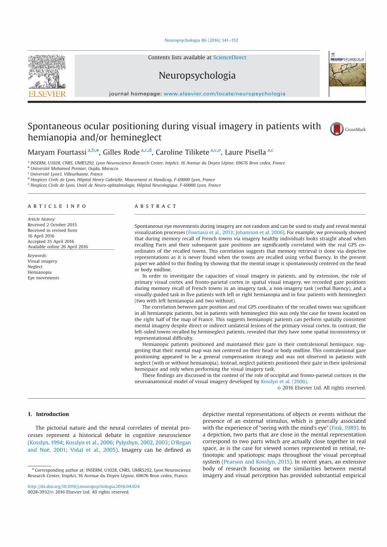
Spontaneous ocular positioning during visual imagery in patients withhemianopia and/or hemineglect
Maryam Fourtassi a,b,n, Gilles Rode a,c,d, Caroline Tilikete a,c,e, Laure Pisella a,c
a INSERM, U1028, CNRS, UMR5292, Lyon Neuroscience Research Center, ImpAct, 16 Avenue du Doyen Lépine, 69676 Bron cedex, Franceb Université Mohamed Premier, Oujda, Moroccoc Université Lyon1, Villeurbanne, Franced Hospices Civils de Lyon, Hôpital Henry Gabrielle, Mouvement et Handicap, F-69000 Lyon, Francee Hospices Civils de Lyon, Unité de Neuro-ophtalmologie, Hôpital Neurologique, F-69000 Lyon, France
a r t i c l e i n f o
Article history:Received 2 October 2015Received in revised form16 April 2016Accepted 25 April 2016Available online 26 April 2016
Keywords:Visual imageryNeglectHemianopiaEye movements
a b s t r a c t
Spontaneous eye movements during imagery are not random and can be used to study and reveal mentalvisualization processes (Fourtassi et al., 2013; Johansson et al. 2006). For example, we previously showedthat during memory recall of French towns via imagery healthy individuals looks straight ahead whenrecalling Paris and their subsequent gaze positions are significantly correlated with the real GPS co-ordinates of the recalled towns. This correlation suggests that memory retrieval is done via depictiverepresentations as it is never found when the towns are recalled using verbal fluency. In the presentpaper we added to this finding by showing that the mental image is spontaneously centered on the heador body midline.
In order to investigate the capacities of visual imagery in patients, and by extension, the role ofprimary visual cortex and fronto-parietal cortex in spatial visual imagery, we recorded gaze positionsduring memory recall of French towns in an imagery task, a non-imagery task (verbal fluency), and avisually-guided task in five patients with left or right hemianopia and in four patients with hemineglect(two with left hemianopia and two without).
The correlation between gaze position and real GPS coordinates of the recalled towns was significantin all hemianopic patients, but in patients with hemineglect this was only the case for towns located onthe right half of the map of France. This suggests hemianopic patients can perform spatially consistentmental imagery despite direct or indirect unilateral lesions of the primary visual cortex. In contrast, theleft-sided towns recalled by hemineglect patients, revealed that they have some spatial inconsistency orrepresentational difficulty.
Hemianopic patients positioned and maintained their gaze in their contralesional hemispace, sug-gesting that their mental map was not centered on their head or body midline. This contralesional gazepositioning appeared to be a general compensation strategy and was not observed in patients withneglect (with or without hemianopia). Instead, neglect patients positioned their gaze in their ipsilesionalhemispace and only when performing the visual imagery task.
These findings are discussed in the context of the role of occipital and fronto-parietal cortices in theneuroanatomical model of visual imagery developed by Kosslyn et al. (2006).
& 2016 Elsevier Ltd. All rights reserved.
1. Introduction
The pictorial nature and the neural correlates of mental pro-cesses represent a historical debate in cognitive neuroscience(Kosslyn, 1994; Kosslyn et al., 2006; Pylyshyn, 2002, 2003; O’Reganand Noë, 2001; Vidal et al., 2005). Imagery can be defined as
depictive mental representations of objects or events without thepresence of an external stimulus, which is generally associatedwith the experience of “seeing with the mind's eye” (Fink, 1989). Ina depiction, two parts that are close in the mental representationcorrespond to two parts which are actually close together in realspace, as is the case for viewed scenes represented in retinal, re-tinotopic and spatiotopic maps throughout the visual perceptualsystem (Pearson and Kosslyn, 2015). In recent years, an extensivebody of research focusing on the similarities between mentalimagery and visual perception has provided substantial empirical
Contents lists available at ScienceDirect
journal homepage: www.elsevier.com/locate/neuropsychologia
Neuropsychologia
http://dx.doi.org/10.1016/j.neuropsychologia.2016.04.0240028-3932/& 2016 Elsevier Ltd. All rights reserved.
n Corresponding author at: INSERM, U1028, CNRS, UMR5292, Lyon NeuroscienceResearch Center, ImpAct, 16 Avenue du Doyen Lépine, 69676 Bron cedex, France.
Neuropsychologia 86 (2016) 141–152
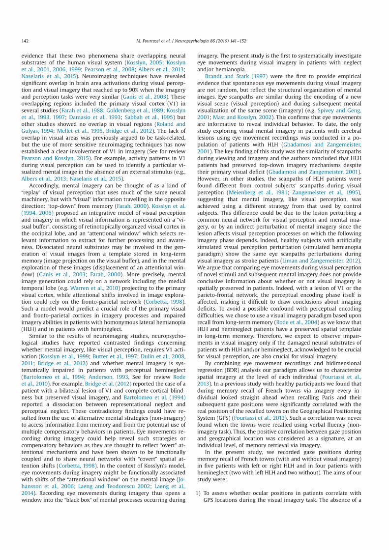
evidence that these two phenomena share overlapping neuralsubstrates of the human visual system (Kosslyn, 2005; Kosslynet al., 2001, 2006, 1999; Pearson et al., 2008; Albers et al., 2013;Naselaris et al., 2015). Neuroimaging techniques have revealedsignificant overlap in brain area activations during visual percep-tion and visual imagery that reached up to 90% when the imageryand perception tasks were very similar (Ganis et al., 2003). Theseoverlapping regions included the primary visual cortex (V1) inseveral studies (Farah et al., 1988; Goldenberg et al., 1989; Kosslynet al., 1993, 1997; Damasio et al., 1993; Sabbah et al., 1995) butother studies showed no overlap in visual regions (Roland andGulyas, 1994; Mellet et al., 1995, Bridge et al., 2012). The lack ofoverlap in visual areas was previously argued to be task-related,but the use of more sensitive neuroimaging techniques has nowestablished a clear involvement of V1 in imagery (See for reviewPearson and Kosslyn, 2015). For example, activity patterns in V1during visual perception can be used to identify a particular vi-sualized mental image in the absence of an external stimulus (e.g.,Albers et al., 2013; Naselaris et al., 2015).
Accordingly, mental imagery can be thought of as a kind of“replay” of visual perception that uses much of the same neuralmachinery, but with “visual” information travelling in the oppositedirection: “top-down” from memory (Farah, 2000). Kosslyn et al.(1994, 2006) proposed an integrative model of visual perceptionand imagery in which visual information is represented on a “vi-sual buffer”, consisting of retinotopically organized visual cortex inthe occipital lobe, and an “attentional window” which selects re-levant information to extract for further processing and aware-ness. Dissociated neural substrates may be involved in the gen-eration of visual images from a template stored in long-termmemory (image projection on the visual buffer), and in the mentalexploration of these images (displacement of an attentional win-dow) (Ganis et al., 2003; Farah, 2000). More precisely, mentalimage generation could rely on a network including the medialtemporal lobe (e.g. Warren et al., 2010) projecting to the primaryvisual cortex, while attentional shifts involved in image explora-tion could rely on the fronto-parietal network (Corbetta, 1998).Such a model would predict a crucial role of the primary visualand fronto-parietal cortices in imagery processes and impairedimagery abilities in patients with homonymous lateral hemianopia(HLH) and in patients with hemineglect.
Similar to the results of neuroimaging studies, neuropsycho-logical studies have reported contrasted findings concerningwhether mental imagery, like visual perception, requires V1 acti-vation (Kosslyn et al., 1999; Butter et al., 1997; Dulin et al., 2008,2011; Bridge et al., 2012) and whether mental imagery is sys-tematically impaired in patients with perceptual hemineglect(Bartolomeo et al., 1994; Anderson, 1993, See for review Rodeet al., 2010). For example, Bridge et al. (2012) reported the case of apatient with a bilateral lesion of V1 and complete cortical blind-ness but preserved visual imagery, and Bartolomeo et al. (1994)reported a dissociation between representational neglect andperceptual neglect. These contradictory findings could have re-sulted from the use of alternative mental strategies (non-imagery)to access information from memory and from the potential use ofmultiple compensatory behaviors in patients. Eye movements re-cording during imagery could help reveal such strategies orcompensatory behaviors as they are thought to reflect “overt” at-tentional mechanisms and have been shown to be functionallycoupled and to share neural networks with “covert” spatial at-tention shifts (Corbetta, 1998). In the context of Kosslyn's model,eye movements during imagery might be functionally associatedwith shifts of the “attentional window” on the mental image (Jo-hansson et al., 2006; Laeng and Teodorescu 2002; Laeng et al.,2014). Recording eye movements during imagery thus opens awindow into the “black box” of mental processes occurring during
imagery. The present study is the first to systematically investigateeye movements during visual imagery in patients with neglectand/or hemianopia.
Brandt and Stark (1997) were the first to provide empiricalevidence that spontaneous eye movements during visual imageryare not random, but reflect the structural organization of mentalimages. Eye scanpaths are similar during the encoding of a newvisual scene (visual perception) and during subsequent mentalvisualization of the same scene (imagery) (e.g. Spivey and Geng,2001; Mast and Kosslyn, 2002). This confirms that eye movementsare informative to reveal individual behavior. To date, the onlystudy exploring visual mental imagery in patients with cerebrallesions using eye movement recordings was conducted in a po-pulation of patients with HLH (Gbadamosi and Zangemeister,2001). The key finding of this study was the similarity of scanpathsduring viewing and imagery and the authors concluded that HLHpatients had preserved top-down imagery mechanisms despitetheir primary visual deficit (Gbadamosi and Zangemeister, 2001).However, in other studies, the scanpaths of HLH patients werefound different from control subjects’ scanpaths during visualperception (Meienberg et al., 1981; Zangemeister et al., 1995),suggesting that mental imagery, like visual perception, wasachieved using a different strategy from that used by controlsubjects. This difference could be due to the lesion perturbing acommon neural network for visual perception and mental ima-gery, or by an indirect perturbation of mental imagery since thelesion affects visual perception processes on which the followingimagery phase depends. Indeed, healthy subjects with artificiallysimulated visual perception perturbation (simulated hemianopiaparadigm) show the same eye scanpaths perturbations duringvisual imagery as stroke patients (Liman and Zangemeister, 2012).We argue that comparing eye movements during visual perceptionof novel stimuli and subsequent mental imagery does not provideconclusive information about whether or not visual imagery isspatially preserved in patients. Indeed, with a lesion of V1 or theparieto-frontal network, the perceptual encoding phase itself isaffected, making it difficult to draw conclusions about imagingdeficits. To avoid a possible confound with perceptual encodingdifficulties, we chose to use a visual imagery paradigm based uponrecall from long-term memory (Rode et al., 2004) as we know thatHLH and hemineglect patients have a preserved spatial templatein long-term memory. Therefore, we expect to observe impair-ments in visual imagery only if the damaged neural substrates ofpatients with HLH and/or hemineglect, acknowledged to be crucialfor visual perception, are also crucial for visual imagery.
By combining eye movement recordings and bidimensionalregression (BDR) analysis our paradigm allows us to characterizespatial imagery at the level of each individual (Fourtassi et al.,2013). In a previous study with healthy participants we found thatduring memory recall of French towns via imagery every in-dividual looked straight ahead when recalling Paris and theirsubsequent gaze positions were significantly correlated with thereal position of the recalled towns on the Geographical PositioningSystem (GPS) (Fourtassi et al., 2013). Such a correlation was neverfound when the towns were recalled using verbal fluency (non-imagery task). Thus, the positive correlation between gaze positionand geographical location was considered as a signature, at anindividual level, of memory retrieval via imagery.
In the present study, we recorded gaze positions duringmemory recall of French towns (with and without visual imagery)in five patients with left or right HLH and in four patients withhemineglect (two with left HLH and two without). The aims of ourstudy were:
1) To assess whether ocular positions in patients correlate withGPS locations during the visual imagery task. The absence of a
M. Fourtassi et al. / Neuropsychologia 86 (2016) 141–152142
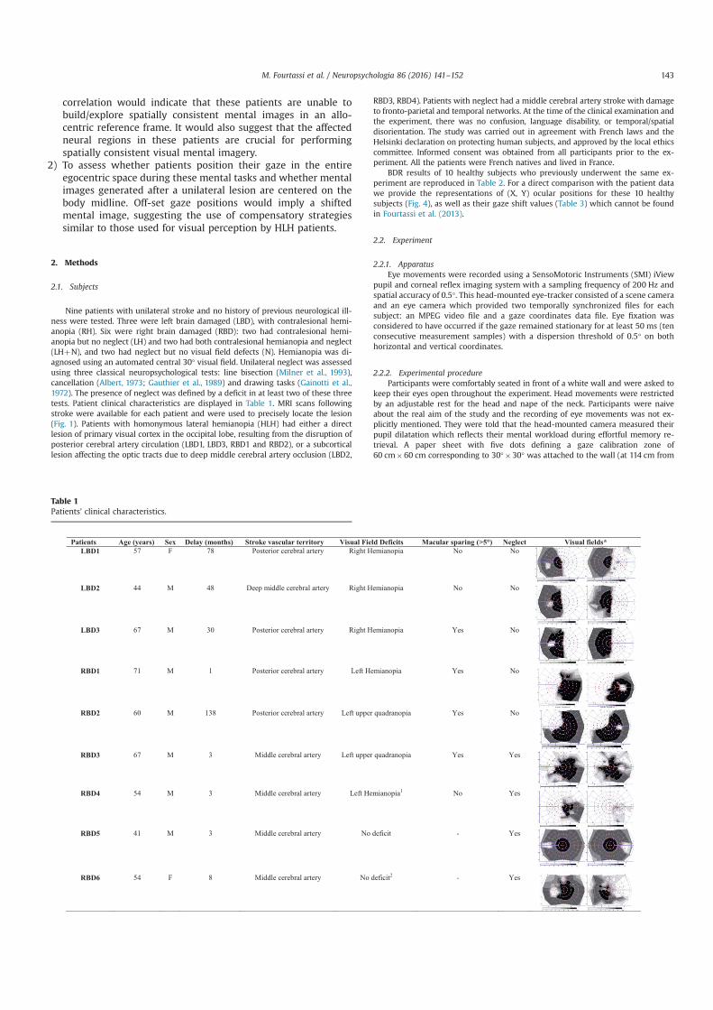
correlation would indicate that these patients are unable tobuild/explore spatially consistent mental images in an allo-centric reference frame. It would also suggest that the affectedneural regions in these patients are crucial for performingspatially consistent visual mental imagery.
2) To assess whether patients position their gaze in the entireegocentric space during these mental tasks and whether mentalimages generated after a unilateral lesion are centered on thebody midline. Off-set gaze positions would imply a shiftedmental image, suggesting the use of compensatory strategiessimilar to those used for visual perception by HLH patients.
2. Methods
2.1. Subjects
Nine patients with unilateral stroke and no history of previous neurological ill-ness were tested. Three were left brain damaged (LBD), with contralesional hemi-anopia (RH). Six were right brain damaged (RBD): two had contralesional hemi-anopia but no neglect (LH) and two had both contralesional hemianopia and neglect(LHþN), and two had neglect but no visual field defects (N). Hemianopia was di-agnosed using an automated central 30° visual field. Unilateral neglect was assessedusing three classical neuropsychological tests: line bisection (Milner et al., 1993),cancellation (Albert, 1973; Gauthier et al., 1989) and drawing tasks (Gainotti et al.,1972). The presence of neglect was defined by a deficit in at least two of these threetests. Patient clinical characteristics are displayed in Table 1. MRI scans followingstroke were available for each patient and were used to precisely locate the lesion(Fig. 1). Patients with homonymous lateral hemianopia (HLH) had either a directlesion of primary visual cortex in the occipital lobe, resulting from the disruption ofposterior cerebral artery circulation (LBD1, LBD3, RBD1 and RBD2), or a subcorticallesion affecting the optic tracts due to deep middle cerebral artery occlusion (LBD2,
RBD3, RBD4). Patients with neglect had a middle cerebral artery stroke with damageto fronto-parietal and temporal networks. At the time of the clinical examination andthe experiment, there was no confusion, language disability, or temporal/spatialdisorientation. The study was carried out in agreement with French laws and theHelsinki declaration on protecting human subjects, and approved by the local ethicscommittee. Informed consent was obtained from all participants prior to the ex-periment. All the patients were French natives and lived in France.
BDR results of 10 healthy subjects who previously underwent the same ex-periment are reproduced in Table 2. For a direct comparison with the patient datawe provide the representations of (X, Y) ocular positions for these 10 healthysubjects (Fig. 4), as well as their gaze shift values (Table 3) which cannot be foundin Fourtassi et al. (2013).
2.2. Experiment
2.2.1. ApparatusEye movements were recorded using a SensoMotoric Instruments (SMI) iView
pupil and corneal reflex imaging system with a sampling frequency of 200 Hz andspatial accuracy of 0.5°. This head-mounted eye-tracker consisted of a scene cameraand an eye camera which provided two temporally synchronized files for eachsubject: an MPEG video file and a gaze coordinates data file. Eye fixation wasconsidered to have occurred if the gaze remained stationary for at least 50 ms (tenconsecutive measurement samples) with a dispersion threshold of 0.5° on bothhorizontal and vertical coordinates.
2.2.2. Experimental procedureParticipants were comfortably seated in front of a white wall and were asked to
keep their eyes open throughout the experiment. Head movements were restrictedby an adjustable rest for the head and nape of the neck. Participants were naiveabout the real aim of the study and the recording of eye movements was not ex-plicitly mentioned. They were told that the head-mounted camera measured theirpupil dilatation which reflects their mental workload during effortful memory re-trieval. A paper sheet with five dots defining a gaze calibration zone of60 cm�60 cm corresponding to 30°�30° was attached to the wall (at 114 cm from
Table 1Patients' clinical characteristics.
M. Fourtassi et al. / Neuropsychologia 86 (2016) 141–152 143
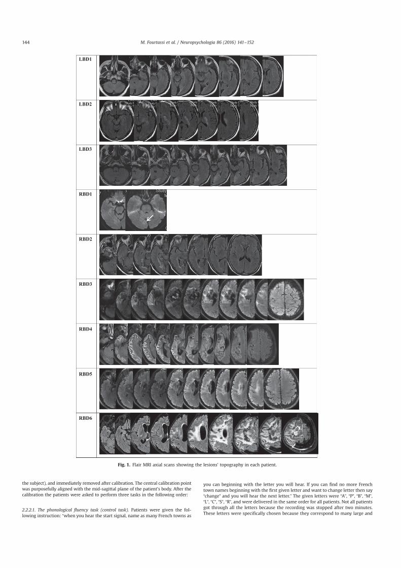
the subject), and immediately removed after calibration. The central calibration pointwas purposefully aligned with the mid-sagittal plane of the patient's body. After thecalibration the patients were asked to perform three tasks in the following order:
2.2.2.1. The phonological fluency task (control task). Patients were given the fol-lowing instruction: “when you hear the start signal, name as many French towns as
you can beginning with the letter you will hear. If you can find no more Frenchtown names beginning with the first given letter and want to change letter then say“change” and you will hear the next letter.” The given letters were “A”, “P”, “B”, “M”,“L”, “C”, “S”, “R”, and were delivered in the same order for all patients. Not all patientsgot through all the letters because the recording was stopped after two minutes.These letters were specifically chosen because they correspond to many large and
LBD1
LBD2
LBD3
RBD1
RBD2
RBD3
RBD4
RBD5
RBD6
Fig. 1. Flair MRI axial scans showing the lesions' topography in each patient.
M. Fourtassi et al. / Neuropsychologia 86 (2016) 141–152144
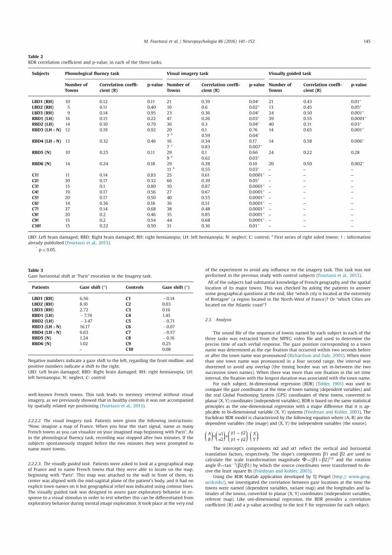
well-known French towns. This task leads to memory retrieval without visualimagery, as we previously showed that in healthy controls it was not accompaniedby spatially related eye positioning (Fourtassi et al., 2013).
2.2.2.2. The visual imagery task. Patients were given the following instructions:“Now, imagine a map of France. When you hear the start signal, name as manyFrench towns as you can visualize on your imagined map beginning with Paris”. Asin the phonological fluency task, recording was stopped after two minutes. If thesubjects spontaneously stopped before the two minutes they were prompted toname more towns.
2.2.2.3. The visually guided task. Patients were asked to look at a geographical mapof France and to name French towns that they were able to locate on the map,beginning with “Paris”. This map was attached to the wall in front of them, itscenter was aligned with the mid-sagittal plane of the patient's body, and it had noexplicit town names on it but geographical relief was indicated using contour lines.The visually guided task was designed to assess gaze exploratory behavior in re-sponse to a visual stimulus in order to test whether this can be differentiated fromexploratory behavior during mental image exploration. It took place at the very end
of the experiment to avoid any influence on the imagery task. This task was notperformed in the previous study with control subjects (Fourtassi et al., 2013).
All of the subjects had substantial knowledge of French geography and the spatiallocation of its major towns. This was checked by asking the patients to answersome geographical questions at the end, like “which city is located at the extremityof Bretagne” (a region located in the North-West of France)? Or “which Cities arelocated on the Atlantic coast”?
2.3. Analysis
The sound file of the sequence of towns named by each subject in each of thethree tasks was extracted from the MPEG video file and used to determine theprecise time of each verbal response. The gaze position corresponding to a townname was determined as the eye fixation that occurred within two seconds beforeor after the town name was pronounced (Richardson and Dale, 2005). When morethan one town name was pronounced in a four second range, the interval wasshortened to avoid any overlap (the timing border was set in-between the twosuccessive town names). When there was more than one fixation in the set timeinterval, the fixation with the longest duration was associated with the town name.
For each subject, bi-dimensional regression (BDR) (Tobler, 1965) was used tocompare the gaze coordinates at the time of town naming (dependent variables) andthe real Global Positioning System (GPS) coordinates of these towns, converted toplanar (X, Y) coordinates (independent variables). BDR is based on the same statisticalprinciples as the one-dimensional regression with a major difference that it is ap-plicable to bi-dimensional variable (X, Y) systems (Friedman and Kohler, 2003). TheEuclidean BDR model is characterized by the following equation where (A, B) are thedependent variables (the image) and (X, Y) the independent variables (the source).
= αα
+ β − ββ + β
⎛⎝⎜
⎞⎠⎟
⎛⎝⎜
⎞⎠⎟
⎛⎝⎜
⎞⎠⎟
⎛⎝⎜
⎞⎠⎟
AB
XY
12
1 2
1 2.
The intercept's components α2 and α1 reflect the vertical and horizontaltranslation factors, respectively. The slope's components β1 and β2 are used tocalculate the scale transformation magnitude Φ¼(β1þβ2)1/2 and the rotationangle θ¼tan�1(β2/β1) by which the source coordinates were transformed to de-rive the least square fit (Friedman and Kohler, 2003).
Using the BDR Matlab application developed by TJ Pingel (http:// www.geog.ucsb.edu/), we investigated the correlation between gaze locations at the time thetowns were named (dependent variables, variant map) and the longitudes and la-titudes of the towns, converted to planar (X, Y) coordinates (independent variables,referent map). Like uni-dimensional regression, the BDR provides a correlationcoefficient (R) and a p-value according to the test F for regression for each subject.
Table 2BDR correlation coefficient and p-value, in each of the three tasks.
Subjects Phonological fluency task Visual imagery task Visually guided task
Number ofTowns
Correlation coeffi-cient (R)
p-value Number ofTowns
Correlation coeffi-cient (R)
p-value Number ofTowns
Correlation coeffi-cient (R)
p-value
LBD1 (RH) 10 0.12 0.11 21 0.39 0.04* 21 0.43 0.01*LBD2 (RH) 5 0.11 0.40 10 0.6 0.02* 13 0.45 0.05*LBD3 (RH) 9 0.14 0.95 23 0.36 0.04* 24 0.50 0.001*RBD1 (LH) 16 0.15 0.22 47 0.26 0.03* 39 0.55 0.0001*RBD2 (LH) 14 0.10 0.79 36 0.3 0.04* 40 0.31 0.03*RBD3 (LHþN) 12 0.19 0.92 20 0.1 0.76 14 0.65 0.001*
7 δ 0.59 0.04*RBD4 (LHþN) 13 0.32 0.46 16 0.34 0.17 14 0.58 0.006*
7 δ 0.83 0.007*RBD5 (N) 10 0.25 0.11 29 0.1 0.66 24 0.22 0.28
9 δ 0.62 0.03*RBD6 (N) 14 0.24 0.18 29 0.28 0.10 20 0.50 0.002*
11 δ 0.55 0.03* – – –
C1! 11 0.14 0.83 25 0.61 0.0001* – – –
C2! 30 0.17 0.32 66 0.39 0.05* – – –
C3! 15 0.1 0.80 10 0.87 0.0001* – – –
C4! 19 0.17 0.56 27 0.67 0.0001* – – –
C5! 20 0.17 0.50 40 0.55 0.0001* – – –
C6! 14 0.36 0.18 36 0.51 0.0001* – – –
C7! 37 0.14 0.68 38 0.48 0.0001* – – –
C8! 20 0.2 0.46 35 0.85 0.0001* – – –
C9! 15 0.2 0.54 44 0.68 0.0001* – – –
C10! 15 0.22 0.50 31 0.36 0.01* – – –
LBD: Left brain damaged; RBD: Right brain damaged; RH: right hemianopia; LH: left hemianopia; N: neglect; C: control; δ First series of right sided towns; ! : informationalready published (Fourtassi et al., 2013).
* pr0.05.
Table 3Gaze horizontal shift at “Paris” evocation in the Imagery task.
Patients Gaze shift (°) Controls Gaze shift (°)
LBD1 (RH) 6.56 C1 �0.14LBD2 (RH) 8.10 C2 0.03LBD3 (RH) 2.72 C3 0.16RBD1 (LH) �7.74 C4 1.45RBD2 (LH) �3.47 C5 �0.71RBD3 (LHþN) 16.17 C6 �0.07RBD4 (LHþN) 6.63 C7 �0.57RBD5 (N) 1.24 C8 �0.16RBD6 (N) 1.02 C9 0.25
– C10 0.08
Negative numbers indicate a gaze shift to the left, regarding the front midline, andpositive numbers indicate a shift to the right.LBD: Left brain damaged; RBD: Right brain damaged; RH: right hemianopia; LH:left hemianopia; N: neglect, C: control
M. Fourtassi et al. / Neuropsychologia 86 (2016) 141–152 145
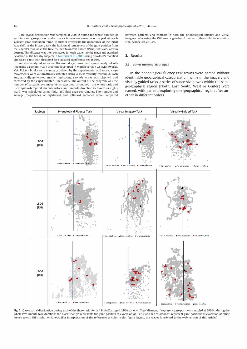
Gaze spatial distribution was sampled at 200 Hz during the whole duration ofeach task and gaze position at the time each townwas named was mapped into eachsubject's gaze calibration frame. To further investigate the importance of the initialgaze shift in the imagery task the horizontal remoteness of the gaze position fromthe subject's midline at the time the first town was named (Paris), was calculated indegrees. This distance was then compared for each patient to the mean and standarddeviation of the healthy subjects in Fourtassi et al. (2013) using Crawford's modifiedone-tailed t-test with threshold for statistical significance set at 0.05.
We also analyzed saccades. Horizontal eye movements were analyzed off-line using a custom-made program developed in Matlab version 7.8 (Mathworks,MA., U.S.A.). Blinks were manually deleted by the experimenter and saccadic eyemovements were automatically detected using a 15°/s velocity threshold. Eachautomatically-generated marker indicating saccade onset was checked andcorrected by the experimenter if necessary. The output of the program was thenumber of saccadic eye movements executed throughout the whole task andtheir spatio-temporal characteristics, and saccade direction (leftward or right-ward) was calculated using initial and final gaze coordinates. The number andaverage magnitudes of rightward and leftward saccades were compared
between patients and controls in both the phonological fluency and visualimagery tasks using the Wilcoxon signed-rank test with threshold for statisticalsignificance set at 0.05.
3. Results
3.1. Town naming strategies
In the phonological fluency task towns were named withoutidentifiable geographical categorization, while in the imagery andvisually guided tasks, a series of successive towns within the samegeographical region (North, East, South, West or Center) werenamed, with patients exploring one geographical region after an-other in different orders.
Fig. 2. Gaze spatial distribution during each of the three tasks for Left Brain Damaged (LBD) patients. Grey “diamonds” represent gaze positions sampled at 200 Hz during thewhole two-minute task duration; the black triangle represents the gaze position at evocation of “Paris” and red “diamonds” represent gaze positions at evocation of otherFrench towns. RH¼right hemianopia.(For interpretation of the references to color in this figure legend, the reader is referred to the web version of this article.)
M. Fourtassi et al. / Neuropsychologia 86 (2016) 141–152146
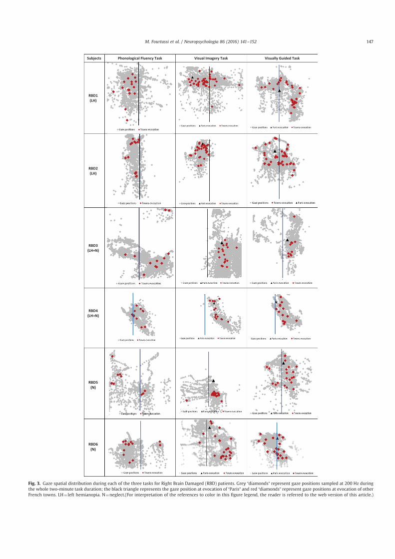
Fig. 3. Gaze spatial distribution during each of the three tasks for Right Brain Damaged (RBD) patients. Grey “diamonds” represent gaze positions sampled at 200 Hz duringthe whole two-minute task duration; the black triangle represents the gaze position at evocation of “Paris” and red “diamonds” represent gaze positions at evocation of otherFrench towns. LH¼ left hemianopia. N¼neglect.(For interpretation of the references to color in this figure legend, the reader is referred to the web version of this article.)
M. Fourtassi et al. / Neuropsychologia 86 (2016) 141–152 147
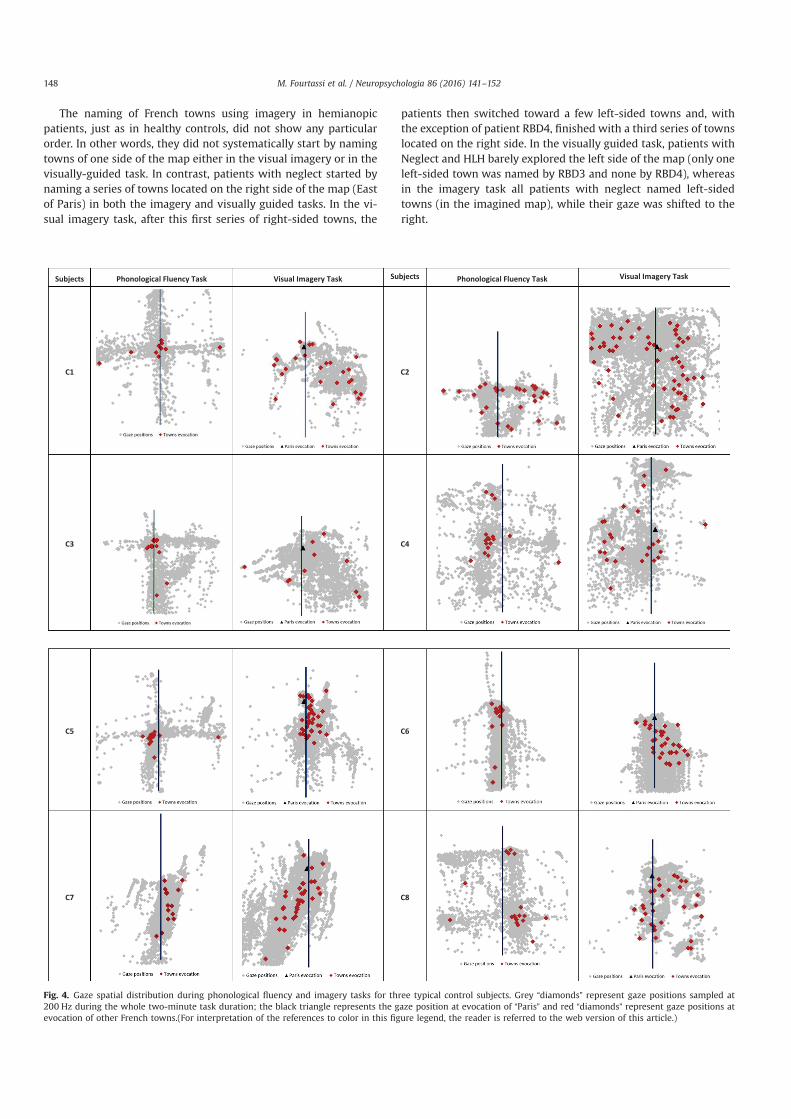
The naming of French towns using imagery in hemianopicpatients, just as in healthy controls, did not show any particularorder. In other words, they did not systematically start by namingtowns of one side of the map either in the visual imagery or in thevisually-guided task. In contrast, patients with neglect started bynaming a series of towns located on the right side of the map (Eastof Paris) in both the imagery and visually guided tasks. In the vi-sual imagery task, after this first series of right-sided towns, the
patients then switched toward a few left-sided towns and, withthe exception of patient RBD4, finished with a third series of townslocated on the right side. In the visually guided task, patients withNeglect and HLH barely explored the left side of the map (only oneleft-sided town was named by RBD3 and none by RBD4), whereasin the imagery task all patients with neglect named left-sidedtowns (in the imagined map), while their gaze was shifted to theright.
Fig. 4. Gaze spatial distribution during phonological fluency and imagery tasks for three typical control subjects. Grey “diamonds” represent gaze positions sampled at200 Hz during the whole two-minute task duration; the black triangle represents the gaze position at evocation of “Paris” and red “diamonds” represent gaze positions atevocation of other French towns.(For interpretation of the references to color in this figure legend, the reader is referred to the web version of this article.)
M. Fourtassi et al. / Neuropsychologia 86 (2016) 141–152148
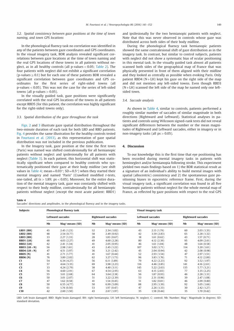
3.2. Spatial consistency between gaze positions at the time of townnaming, and town GPS locations
In the phonological fluency task no correlationwas identified inany of the patients between gaze coordinates and GPS coordinates.In the visual imagery task, BDR analysis revealed significant cor-relations between gaze locations at the time of town naming andthe real GPS locations of these towns in all patients without ne-glect, as in all healthy controls (all p-valueso0.05; Table 2). Thefour patients with neglect did not exhibit a significant correlation(p-valuesZ0.1) but for each one of these patients BDR revealed asignificant correlation between gaze coordinates and GPS co-ordinates for the first series of right-sided towns (allp-valueso0.05). This was not the case for the series of left-sidedtowns (all p-values40.05).
In the visually guided task, gaze positions were significantlycorrelated with the real GPS locations of the towns in all patientsexcept RBD5 (for this patient, the correlationwas highly significantfor the right-sided towns only).
3.3. Spatial distribution of the gaze throughout the task
Figs. 2 and 3 illustrate gaze spatial distribution throughout thetwo-minute duration of each task for both LBD and RBD patients.Fig. 4 provides the same illustration for the healthy controls testedin Fourtassi et al. (2013), as this representation of gaze spatialdistribution was not included in the previous publication.
In the Imagery task, gaze position at the time the first town(Paris) was named was shifted contralesionally for all hemianopicpatients without neglect and ipsilesionally for all patients withneglect (Table 3). In each patient, this horizontal shift was statis-tically significant when compared to healthy controls who sys-tematically positioned their gaze at their body midline (see shiftvalues in Table 4; mean¼0.03°; SD¼0.5°) when they started theirmental imagery and named “Paris” (Crawford modified t-tests,one-tailed, all ts41.89; pso0.05). Moreover, for the entire dura-tion of the mental imagery task, gaze was constantly shifted withrespect to their body midline, contralesionally for all hemianopicpatients without neglect (except the most acute patient: RBD1)
and ipsilesionally for the two hemianopic patients with neglect.Note that this was never observed in controls whose gaze wasdistributed across both sides of their body midline.
During the phonological fluency task hemianopic patientsshowed the same contralesional shift of gaze distribution as in theimagery task. In contrast, but similar to control subjects, patientswith neglect did not show a systematic bias of ocular positioningin this mental task. In the visually guided task almost all patientsscanned both sides of the geographical map of France that wasphysically presented in front of them aligned with their midlineand they looked as centrally as possible when evoking Paris. Onlypatient RBD4 (NþLH) kept his gaze on the right side of the mapand did not mention any left-sided towns. Even though RBD3(NþLH) scanned the left side of the map he named only one left-sided town.
3.4. Saccade analysis
As shown in Table 4, similar to controls, patients performed aroughly similar number of saccades of similar magnitude in bothdirections (Rightward and Leftward). Statistical analyses in pa-tients and controls using Wilcoxon signed-rank tests did not revealsignificant differences between the number or the mean magni-tudes of Rightward and Leftward saccades, either in imagery or innon-imagery tasks (all ps40.05).
4. Discussion
To our knowledge this is the first time that eye positioning hasbeen recorded during mental imagery tasks in patients withhemineglect and/or hemianopia following stroke. This experimentyielded two main findings based on 1) the BDR statistical result asa signature of an individual's ability to build mental images withspatial (allocentric) consistency and 2) the spontaneous gaze po-sitioning biases in egocentric reference frame. First, during thevisual imagery task, a significant correlation was found in all fivehemianopic patients without neglect for the whole mental map ofFrance, as reflected by gaze positions with respect to the real GPS
Table 4Saccades' directions and amplitudes, in the phonological fluency and in the imagery tasks.
Subjects Phonological fluency task Visual imagery task
Leftward saccades Rightward saccades Leftward saccades Rightward saccades
Nb Mag°:means (SD) Nb Mag°:means (SD) Nb Mag°:means (SD) Nb Mag°:means (SD)
LBD1 (RH) 45 2.45 (1.23) 52 2.34 (1.02) 43 2.13 (1.79) 60 2.03 (1.35)LBD2 (RH) 48 2.54 (0.77) 58 2.49 (0.92) 42 3.59 (2.83) 65 2.28 (1.32)LBD3 (RH) 33 2.27 (1.33) 28 1.83 (0.67) 18 1.61 (0.62) 43 1.57 (0.71)RBD1 (LH) 48 4.03 (2.27) 49 4.60 (2.28) 80 4.12 (2.39) 95 4.09 (2.61)RBD2 (LH) 42 2.41 (1.34) 41 2.05 (0.95) 46 1.61 (1.04) 48 1.64 (0.58)RBD3 (LHþN) 56 2.98 (1.61) 43 2.45 (1.22) 107 3.02 (1.71) 114 3.20 (1.61)RBD4 (LHþN) 47 4.51 (3.97) 36 3.21 (2.42) 45 2.04 (0.94) 42 2.08 (0.98)RBD5 (N) 41 2.71 (1.77) 41 1.67 (0.44) 52 2.93 (1.54) 47 2.97 (1.51)RBD6 (N) 76 3.89 (2.02) 62 3.27 (1.73) 96 3.83 (1.76) 71 4.12 (2.00)C1 54 6.34 (4.27) 56 6.11 (3.89) 79 4.22 (2.23) 92 3.52 (1.97)C2 57 4.64 (3.35) 69 3.98 (2.23) 116 4.46 (2.85) 141 4.16 (2.61)C3 33 4.24 (2.79) 38 4.40 (3.11) 145 5.22 (2.63) 135 5.77 (3.21)C4 56 4.60 (2.91) 67 4.54 (2.95) 63 4.15 (2.83) 77 3.35 (2.25)C5 55 3.61 (2.68) 64 3.64 (2.18) 56 1.87 (0.93) 46 2.28 (1.31)C6 50 3.01 (2.07) 51 3.22 (2.39) 53 2.31 (0.96) 39 2.47 (1.08)C7 47 1.62 (0.58) 41 1.54 (0.45) 51 1.82 (0.81) 46 2.09 (0.80)C8 50 6.55 (4.77) 50 6.99 (5.09) 88 2.95 (1.39) 92 3.05 (1.66)C9 61 1.74 (0.50) 53 1.97 (0.47) 47 2.26 (1.33) 30 2.42 (1.27)C10 42 2.60 (1.50) 43 2.67 (1.97) 35 1.58 (0.44) 35 1.70 (0.42)
LBD: Left brain damaged; RBD: Right brain damaged; RH: right hemianopia; LH: left hemianopia; N: neglect; C: control; Nb: Number; Mag°: Magnitude in degrees; SD:standard deviation.
M. Fourtassi et al. / Neuropsychologia 86 (2016) 141–152 149
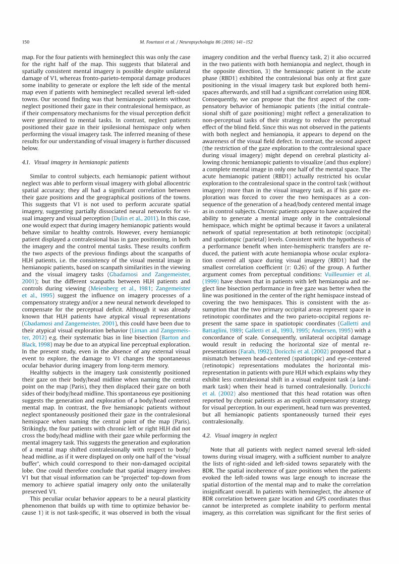
map. For the four patients with hemineglect this was only the casefor the right half of the map. This suggests that bilateral andspatially consistent mental imagery is possible despite unilateraldamage of V1, whereas fronto-parieto-temporal damage producessome inability to generate or explore the left side of the mentalmap even if patients with hemineglect recalled several left-sidedtowns. Our second finding was that hemianopic patients withoutneglect positioned their gaze in their contralesional hemispace, asif their compensatory mechanisms for the visual perception deficitwere generalized to mental tasks. In contrast, neglect patientspositioned their gaze in their ipsilesional hemispace only whenperforming the visual imagery task. The inferred meaning of theseresults for our understanding of visual imagery is further discussedbelow.
4.1. Visual imagery in hemianopic patients
Similar to control subjects, each hemianopic patient withoutneglect was able to perform visual imagery with global allocentricspatial accuracy; they all had a significant correlation betweentheir gaze positions and the geographical positions of the towns.This suggests that V1 is not used to perform accurate spatialimagery, suggesting partially dissociated neural networks for vi-sual imagery and visual perception (Dulin et al., 2011). In this case,one would expect that during imagery hemianopic patients wouldbehave similar to healthy controls. However, every hemianopicpatient displayed a contralesional bias in gaze positioning, in boththe imagery and the control mental tasks. These results confirmthe two aspects of the previous findings about the scanpaths ofHLH patients, i.e. the consistency of the visual mental image inhemianopic patients, based on scanpath similarities in the viewingand the visual imagery tasks (Gbadamosi and Zangemeister,2001); but the different scanpaths between HLH patients andcontrols during viewing (Meienberg et al., 1981; Zangemeisteret al., 1995) suggest the influence on imagery processes of acompensatory strategy and/or a new neural network developed tocompensate for the perceptual deficit. Although it was alreadyknown that HLH patients have atypical visual representations(Gbadamosi and Zangemeister, 2001), this could have been due totheir atypical visual exploration behavior (Liman and Zangemeis-ter, 2012) e.g. their systematic bias in line bisection (Barton andBlack, 1998) may be due to an atypical line perceptual exploration.In the present study, even in the absence of any external visualevent to explore, the damage to V1 changes the spontaneousocular behavior during imagery from long-term memory.
Healthy subjects in the imagery task consistently positionedtheir gaze on their body/head midline when naming the centralpoint on the map (Paris), they then displaced their gaze on bothsides of their body/head midline. This spontaneous eye positioningsuggests the generation and exploration of a body/head centeredmental map. In contrast, the five hemianopic patients withoutneglect spontaneously positioned their gaze in the contralesionalhemispace when naming the central point of the map (Paris).Strikingly, the four patients with chronic left or right HLH did notcross the body/head midline with their gaze while performing themental imagery task. This suggests the generation and explorationof a mental map shifted contralesionally with respect to body/head midline, as if it were displayed on only one half of the “visualbuffer”, which could correspond to their non-damaged occipitallobe. One could therefore conclude that spatial imagery involvesV1 but that visual information can be “projected” top-down frommemory to achieve spatial imagery only onto the unilaterallypreserved V1.
This peculiar ocular behavior appears to be a neural plasticityphenomenon that builds up with time to optimize behavior be-cause 1) it is not task-specific, it was observed in both the visual
imagery condition and the verbal fluency task, 2) it also occurredin the two patients with both hemianopia and neglect, though inthe opposite direction, 3) the hemianopic patient in the acutephase (RBD1) exhibited the contralesional bias only at first gazepositioning in the visual imagery task but explored both hemi-spaces afterwards, and still had a significant correlation using BDR.Consequently, we can propose that the first aspect of the com-pensatory behavior of hemianopic patients (the initial contrale-sional shift of gaze positioning) might reflect a generalization tonon-perceptual tasks of their strategy to reduce the perceptualeffect of the blind field. Since this was not observed in the patientswith both neglect and hemianopia, it appears to depend on theawareness of the visual field defect. In contrast, the second aspect(the restriction of the gaze exploration to the contralesional spaceduring visual imagery) might depend on cerebral plasticity al-lowing chronic hemianopic patients to visualize (and thus explore)a complete mental image in only one half of the mental space. Theacute hemianopic patient (RBD1) actually restricted his ocularexploration to the contralesional space in the control task (withoutimagery) more than in the visual imagery task, as if his gaze ex-ploration was forced to cover the two hemispaces as a con-sequence of the generation of a head/body centered mental imageas in control subjects. Chronic patients appear to have acquired theability to generate a mental image only in the contralesionalhemispace, which might be optimal because it favors a unilateralnetwork of spatial representation at both retinotopic (occipital)and spatiotopic (parietal) levels. Consistent with the hypothesis ofa performance benefit when inter-hemispheric transfers are re-duced, the patient with acute hemianopia whose ocular explora-tion covered all space during visual imagery (RBD1) had thesmallest correlation coefficient (r: 0.26) of the group. A furtherargument comes from perceptual conditions: Vuilleumier et al.(1999) have shown that in patients with left hemianopia and ne-glect line bisection performance in free gaze was better when theline was positioned in the center of the right hemispace instead ofcovering the two hemispaces. This is consistent with the as-sumption that the two primary occipital areas represent space inretinotopic coordinates and the two parieto-occipital regions re-present the same space in spatiotopic coordinates (Galletti andBattaglini, 1989; Galletti et al., 1993, 1995; Andersen, 1995) with aconcordance of scale. Consequently, unilateral occipital damagewould result in reducing the horizontal size of mental re-presentations (Farah, 1992). Doricchi et al. (2002) proposed that amismatch between head-centered (spatiotopic) and eye-centered(retinotopic) representations modulates the horizontal mis-representation in patients with pure HLH which explains why theyexhibit less contralesional shift in a visual endpoint task (a land-mark task) when their head is turned contralesionally. Doricchiet al. (2002) also mentioned that this head rotation was oftenreported by chronic patients as an explicit compensatory strategyfor visual perception. In our experiment, head turn was prevented,but all hemianopic patients spontaneously turned their eyescontralesionally.
4.2. Visual imagery in neglect
Note that all patients with neglect named several left-sidedtowns during visual imagery, with a sufficient number to analyzethe lists of right-sided and left-sided towns separately with theBDR. The spatial incoherence of gaze positions when the patientsevoked the left-sided towns was large enough to increase thespatial distortion of the mental map and to make the correlationinsignificant overall. In patients with hemineglect, the absence ofBDR correlation between gaze location and GPS coordinates thuscannot be interpreted as complete inability to perform mentalimagery, as this correlation was significant for the first series of
M. Fourtassi et al. / Neuropsychologia 86 (2016) 141–152150
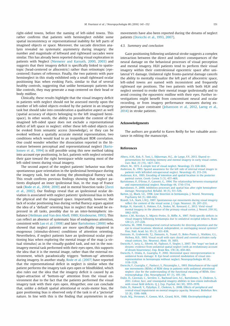
right-sided towns, before the naming of left-sided towns. Thisrather confirms that patients with hemineglect exhibit somespatial inconsistency or representational inability for left parts ofimagined objects or space. Moreover, the saccade direction ana-lysis revealed no systematic asymmetry during imagery: thenumber and magnitude of leftward and rightward saccades weresimilar. This has already been reported during visual exploration inpatients with Neglect (Niemeier and Karnath, 2000, 2003) andsuggests that their imagery deficit is specifically linked to spatio-topic (head-centered or allocentric) rather than retinotopic (eye-centered) frames of reference. Finally, the two patients with purehemineglect in this study exhibited only a small rightward ocularpositioning bias when evoking Paris, similar to that of severalhealthy controls, suggesting that unlike hemianopic patients butlike controls, they may generate a map centered on their head orbody midline.
Clinically, these results highlight that the visual imagery deficitin patients with neglect should not be assessed merely upon thenumber of left-sided objects evoked by the patient in an imagerytask but should take into consideration a qualitative aspect as well(spatial accuracy of objects belonging to the left imagined hemi-space). In other words, the ability to provide the content of theimagined left-sided space does not exclude a representationaldeficit of left space in neglect: either these left-sided objects canbe evoked from semantic access (knowledge), or they can beevoked without a spatially accurate mental representation, twoconditions which would lead to an insignificant BDR correlation.One could wonder whether the dissociation reported in the lit-erature between perceptual and representational neglect (Barto-lomeo et al., 1994) is still possible using this new method takinginto account eye positioning. In fact, patients with neglect orientedtheir gaze toward the right hemispace while naming most of theleft-sided towns during visual imagery.
The second aspect of the neglect patients’ behavior was theirspontaneous gaze orientation in the ipsilesional hemispace duringthe imagery task, but not during the phonological fluency task.This result confirms previous findings showing that neglect pa-tients displayed an inner ipsilesional bias, in the map of Francetask (Rode et al., 2004; 2010) and in mental bisection tasks (Zorziet al., 2002). Our findings reveal that an ipsilesional ocular de-viation is associated with neglect patients’ ipsilesional bias in boththe physical and the imagined space. Importantly, however, thelack of ocular positioning bias during verbal fluency argues againstthe idea of a “default” orienting bias in neglect that would be ex-pressed in all tasks, possibly due to an inter-hemispheric im-balance (Heilman and Van den Abell, 1980; Kinsbourne, 1993). Thiscan reflect an absence of systematic bias of endogenous attention,consistent with Luo et al. (1998) and later Bartolomeo (2000) whoshowed that neglect patients are more specifically impaired inexogenous (stimulus-driven) conditions of attention orienting.Nevertheless, if neglect patients have an ipsilesional ocular posi-tioning bias when exploring the mental image of the map (a vir-tual stimulus) as in the visually-guided task, and not in the non-imagery mental task performed with their eyes open, this supportsthe idea that it is the mental image, rather than the external en-vironment, which paradoxically triggers “bottom-up” attentionduring imagery. In another study, Rode et al. (2007) have reportedthat the representational deficit in neglect is similar when thepatient performs the imagery task eyes open or blindfolded, whichalso rules out the idea that the imagery deficit is caused by anhyper-attraction of “bottom-up” attention from the visual en-vironment due to the fact that the patients performed the visualimagery task with their eyes open. Altogether, one can concludethat, unlike a default spatial attentional or oculo-motor bias, thegaze positioning bias is observed only if the task is of a “pictorial”nature. In line with this is the finding that asymmetries in eye
movements have also been reported during the dreams of neglectpatients (Doricchi et al., 1993, 2007).
4.3. Summary and conclusion
Gaze positioning following unilateral stroke suggests a complexinteraction between the direct and indirect consequences of theneural damage on the behavioral processes of visual perceptionand mental imagery. HLH patients tend to perform their visualimagery within their controlesional egocentric space after uni-lateral V1 damage. Unilateral right fronto-parietal damage cancelsthe ability to mentally visualize the left part of allocentric space,left-sided towns are named with inconsistent and frequentlyrightward eye positions. The two patients with both HLH andneglect seemed to evoke their mental image ipsilesionally and toavoid crossing the egocentric midline with their eyes. Further in-vestigations might benefit from concomitant neural and ocularrecording, or from imagery performance measures during ex-perimental gaze constraint (Johansson et al., 2012, Laeng et al.,2014) in stroke patients.
Acknowledgments
The authors are grateful to Karen Reilly for her valuable assis-tance in editing the manuscript.
References
Albers, A.M., Kok, P., Toni, I., Dijkerman, H.C., de Lange, F.P., 2013. Shared re-presentations for working memory and mental imagery in early visual cortex.Curr. Biol. 23 (15), 1427–1431.
Albert, M., 1973. A simple test of visual neglect. Neurology 23, 658–664.Anderson, B., 1993. Spared awareness for the left side of internal visual images in
patients with leftsided extrapersonal neglect. Neurology 43, 213–216.Andersen, R.A., 1995. Encoding of intention and spatial location in the posterior
parietal cortex. Cereb. Cortex 5 (5), 457–469.Bartolomeo, P., D’Erme, P., Gainotti, G., 1994. The relationship between visuospatial
and representational neglect. Neurology 44, 1710–1714.Bartolomeo, P., 2000. Inhibitory processes and spatial bias after right hemisphere
damage. Neuropsychol. Rehabil. 10 (5), 511–526.Barton, J.J., Black, S.E., 1998. Line bisection in hemianopia. J. Neurol. Neurosurg.
Psychiatry 64 (5), 660–662.Brandt, S.A., Stark, L.W.J., 1997. Spontaneous eye movements during visual imagery
reflect the content of the visual scene. J. Cogn. Neurosci. 26, 207–231.Bridge, H., Harrold, S., Holmes, E.A., Stokes, M., Kennard, C., 2012. Vivid visual
mental imagery in the absence of the primary visual cortex. J. Neurol. 259 (6),1062–1070.
Butter, C.M., Kosslyn, S., Mijovic-Prelec, D., Riffle, A., 1997. Field-specific deficits invisual imagery following hemianopia due to unilateral occipital infarcts. Brain120 (2), 217–228.
Corbetta, M., 1998. Frontoparietal cortical networks for directing attention and theeye to visual locations: Identical, independent, or overlapping neural systems?Proc. Natl. Acad. Sci. 95 (3), 831–838.
Damasio, H., Grabowski, T.J., Damasio, A., Tranel, D., Boles-Ponto, L., Watkins, G.L.,Hichwa, R.D., 1993. Visual recall with eyes closed and covered activates earlyvisual cortices. Soc. Neurosci. Abstr. 19, 1603.
Doricchi, F., Iaria, G., Silvetti, M., Figliozzi, F., Siegler, I., 2007. The “ways” we look atdreams: evidence from unilateral spatial neglect (with an evolutionary accountof dream bizarreness). Exp. Brain Res. 178 (4), 450–461.
Doricchi, F., Onida, A., Guariglia, P., 2002. Horizontal space misrepresentation inunilateral brain damage: II. Eye-head centered modulation of visual mis-representation in hemianopia without neglect. Neuropsychologia 40 (8),1118–1128.
Doricchi, F., Guariglia, C., Paolucci, S., Pizzamiglio, L., 1993. Disturbances of the rapideye movements (REMs) of REM sleep in patients with unilateral attentionalneglect: clue for the understanding of the functional meaning of REMs. Elec-troencephalogr. Clin. Neurophysiol. 87 (3), 105–116.
Dulin, D., Cavezian, C., Serrière, C., Bachoud-Levi, A.C., Bartolomeo, P., Chokron, S.,2011. Colour, face, and visuospatial imagery abilities in low-vision individualswith visual field deficits. Q. J. Exp. Psychol. 64 (10), 1955–1970.
Dulin, D., Hatwell, Y., Pylyshyn, Z., Chokron, S., 2008. Effects of peripheral andcentral visual impairment on mental imagery capacity. Neurosci. Biobehav. Rev.32 (8), 1396–1408.
Farah, M.J., Peronnet, F., Gonon, M.A., Girard, M.H., 1988. Electrophysiological
M. Fourtassi et al. / Neuropsychologia 86 (2016) 141–152 151

evidence for a shared representational medium for visual images and visualpercepts. J. Exp. Psychology: Gen. 117, 248–257.
Farah, M.J., 2000. The neural basis of mental imagery. In: Gazzaniga, M. (Ed.), TheNew Cognitive Neuroscience. MIT Press, pp. 965–974.
Farah, M.J., Soso, M.J., Dasheiff, R.M., 1992. Visual angle of the mind's eye before andafter unilateral occipital lobectomy. J. Exp. Psychology: Hum. Percept. Perform.18, 241–246.
Finke, R.A., 1989. Principles of Mental Imagery. MIT Press, Cambridge, MA.Fourtassi, M., Hajjioui, A., Urquizar, C., Rossetti, Y., Rode, G., et al., 2013. Iterative
fragmentation of cognitive maps in a visual imagery task. PLoS. ONE 8 (7),e68560.
Friedman, A., Kohler, B., 2003. Bidimensional regression: assessing the configuralsimilarity and accuracy of cognitive maps and other two-dimensional data sets.Psychol. Methods 8, 468–491.
Gainotti, G., Messerli, P., Tissot, R., 1972. Qualitative analysis of unilateral spatialneglect in relation to laterality of cerebral lesions. J. Neurol. Neurosurg. Psy-chiatry 35, 545–550.
Galletti, C., Battaglini, P.P., 1989. Gaze-dependent visual neurons in area V3A ofmonkey prestriate cortex. J. Neurosci. 9 (4), 1112–1125.
Galletti, C., Battaglini, P.P., Fattori, P., 1993. Parietal neurons encoding spatial loca-tions in craniotopic coordinates. Exp. Brain Res. 96 (2), 221–229.
Galletti, C., Battaglini, P.P., Fattori, P., 1995. Eye position influence on the parieto‐occipital area PO (V6) of the macaque monkey. Eur. J. Neurosci. 7 (12),2486–2501.
Ganis, G., Thompson, W.L., Mast, F.W., Kosslyn, S.M., 2003. Visual imagery in cer-ebral visual dysfunction. Neurol. Clin. 21 (3), 631–646.
Gauthier, L., Dehaut, F., Joanette, Y., 1989. The Bells test - a quantitative and qua-litative test for visual neglect. Int. J. Clin. Neuropsychol. 11, 49–54.
Gbadamosi, J., Zangemeister, W.H., 2001. Visual imagery in hemianopic patients. J.Cognit. Neurosci. 13 (7), 855–866.
Goldenberg, G., Podreka, I., Steiner, M., Willmes, K., Suess, E., Deecke, L., 1989.Regional cerebral blood flow patterns in visual imagery. Neuropsychologia 27,641–664.
Heilman, K.M., Van den Abell, T., 1980. Right hemisphere dominance for attention:the mechanism underlying hemispheric asymmetries of inattention (neglect).Neurology 30, 327–330.
Johansson, R., Holsanova, J., Holmqvist, K., 2006. Pictures and spoken descriptionselicit similar eye movements during mental imagery, in light and completedarkness. Cogn. Sci. 30, 1053–1079.
Johansson, R., Holsanova, J., Dewhurst, R., Holmqvist, K., 2012. Eye movementsduring scene recollection have a functional role, but they are not reinstate-ments of those produced during encoding. J. Exp. Psychology: Hum. Percept.Perform 38 (5), 1289.
Kinsbourne, M., 1993. Orientational bias model of unilateral neglect: evidence fromattentional gradients within hemispace. In: Robertson, I.H., Marschall, J.C.(Eds.), Unilateral Neglect: Clinical and Experimental Studies. Lawrence ErlbaumAssociates, Hove (UK), pp. 63–86.
Kosslyn, S.M., Alpert, N.M., Thompson, W.L., Maljkovic, V., Weise, S.B., Chabris, C.F.,Hamilton, S.E., Buonano, F.S., 1993. Visual mental imagery activates topo-graphically organized visual cortex: PET investigations. J. Cognit. Neurosci. 5,263–287.
Kosslyn, S.M., 1994. Image and Brain: The Resolution of the Imagery Debate. TheMIT Press, Cambridge, Mass.
Kosslyn, S.M., Thompson, W.L., Alpert, N.M., 1997. Neural systems shared by visualimagery and visual perception: a positron emission tomography study. Neu-roImage 6, 320–334.
Kosslyn, S.M., et al., 1999. The role of area 17 in visual imagery: convergent evi-dence from PET and rTMS. Science 284 (5411), 167–170.
Kosslyn, S.M., Ganis, G., Thompson, W.L., 2001. Neural foundations of imagery. Nat.Rev. Neurosci. 2, 635–642.
Kosslyn, S.M., 2005. Mental images and the brain. Cogn. Neuropsychol. 22 (3),333–347.
Kosslyn, S.M., Thompson, W.L., Ganis, G., 2006. The Case for Mental Imagery. OxfordUniversity Press, New York.
Liman, T.G., Zangemeister, W.H., 2012. Scanpath eye movements during visualmental imagery in a simulated hemianopia paradigm. J Eye Mov Res 5 (1), 2.
Laeng, B., Bloem, I.M., D’Ascenzo, S., Tommasi, L., 2014. Scrutinizing visual images:the role of gaze in mental imagery and memory. Cognition 131 (2), 263–283.
Laeng, B., Teodorescu, D.S., 2002. Eye scanpaths during visual imagery reenact thoseof perception of the same visual scene. Cogn. Sci. 26 (2), 207–231.
Luo, C.R., Anderson, J.M., Caramazza, A., 1998. Impaired stimulus-driven orientingof attention and preserved goal-directed orienting of attention in unilateralvisual neglect. Am. J. Psychol. 111 (4), 487.
Mast, F.W., Kosslyn, S.M., 2002. Eye movements during visual mental imagery.Trends Cognit. Sci. 6 (7), 271–272.
Meienberg, O., Zangemeister, W.H., Rosenberg, M., Hoyt, W.F., Stark, L., 1981. Sac-cadic eye movement strategies in patients with homonymous hemianopia. Ann.Neurol. 9 (6), 537–544.
Mellet, E., Tzourio, N., Denis, M., Mazoyer, B., 1995. A positron emission tomographystudy of visual and mental spatial exploration. J. Cognit. Neurosci. 7, 433–445.
Milner, A.D., Harvey, M., Roberts, R.C., Forster, S.V., 1993. Line bisection errors invisual neglect: misguided action or size distortion? Neuropsychologia 31 (1),39–49.
Naselaris, T., Olman, C.A., Stansbury, D.E., Ugurbil, K., Gallant, J.L., 2015. A voxel-wiseencoding model for early visual areas decodes mental images of rememberedscenes. Neuroimage 105, 215–228.
Niemeier, M., Karnath, H.O., 2000. Exploratory saccades show no direction-specificdeficit in neglect. Neurology 54 (2), 515–518.
Niemeier, M., Karnath, H.O., 2003. Stimulus-driven and voluntary saccades arecoded in different coordinate systems. Curr. Biol. 13 (7), 585–589.
O’Regan, J.K., Noë, A., 2001. A sensorimotor account of vision and visual con-sciousness. Behav. Brain Sci. 24 (05), 939–973.
Pearson, J., Kosslyn, S.M., 2015. The heterogeneity of mental representation: endingthe imagery debate. Proc. Natl. Acad. Sci. 112 (33), 10089–10092.
Pearson, J., Clifford, C.W.G., Tong, F., 2008. The functional impact of mental imageryon conscious perception. Curr. Biol. 18 (13), 982–986.
Pylyshyn, Z.W., 2002. Mental imagery: In search of a theory. Behav. Brain Sci. 25,157–238.
Pylyshyn, Z., 2003. Return of the mental image: are there pictures in the brain?Trends Cognit. Sci. 7, 113–118.
Richardson, D.C., Dale, R., 2005. Looking to understand: the coupling betweenspeakers' and listeners' eye movements and its relationship to discoursecomprehension. Cognit. Sci. 29 (6), 1045–1060.
Rode, G., Rossetti, Y., Perenin, M.T., Boisson, D., 2004. Geographic information has tobe spatialized to be neglected: a representational neglect case. Cortex 40 (2),391–397.
Rode, G., Revol, P., Rossetti, Y., Boisson, D., Bartolomeo, P.A.O.L.O., 2007. Lookingwhile imagining The influence of visual input on representational neglect.Neurology 68 (6), 432–437.
Rode, G., Cotton, F., Revol, P., Jacquin-Courtois, S., Rossetti, Y., Bartolomeo, P., 2010.Representation and disconnection in imaginal neglect. Neuropsychologia 48(10), 2903–2911.
Roland, P.E., Gulyas, B., 1994. Visual imagery and visual representation. TrendsNeurosci. 17, 281–296 (with commentaries).
Sabbah, P., Simond, G., Levrier, O., Habib, M., Trabaud, V., Murayama, N., Mazoyer, B.M., Briant, J.F., Raybaud, C., Salamon, G., 1995. Functional magnetic resonanceimaging at 1.5T during sensorimotor and cognitive task. Eur. Neurol. 35,131–136.
Spivey, M.J., Geng, J.J., 2001. Oculomotor mechanisms activated by imagery andmemory: eye movements to absent objects. Psychol. Res. 65, 235–241.
Tobler, W., 1965. Computation of the corresponding geographical patterns. Pap.Reg. Sci. Assoc. 15, 131–139.
Vidal, J.R., Gauchou, H.L., Tallon-Baudry, C., O’Regan, J.K., 2005. Relational in-formation in visual short-term memory: the structural gist. J. Vis. 5 (3),244–256.
Vuilleumier, P., Valenza, N., Mayer, E., Perrig, S., Landis, T., 1999. To see better to theleft when looking more to the right: effects of gaze direction and frames ofspatial coordinates in unilateral neglect. J. Int. Neuropsychol. Soc. 5 (01), 75–82.
Warren, D.E., Duff, M.C., Tranel, D., Cohen, N.J., 2010. Medial temporal lobe damageimpairs representation of simple stimuli. Front. Hum. Neurosci., 4.
Zangemeister, W.H., Oechsner, U., Freksa, C., 1995. Short-term adaptation of eyemovements in patients with visual hemifield defects indicates high level con-trol of human scanpath. Optom. Vis. Sci.: Off. Publ. Am. Acad. Optom. 72 (7),467–477.
Zorzi, M., Priftis, K., Umiltà, C., 2002. Brain damage: neglect disrupts the mentalnumber line. Nature 417 (6885) 138–9.
M. Fourtassi et al. / Neuropsychologia 86 (2016) 141–152152

Article (3)
« En préparation pour»
Title:
Eye positions, during maintained fixation, still reveal the content of visual
mental imagery
Authors:
Maryam Fourtassia,b,*; Gilles Rodea,c,d; Laure Pisellaa,c
a) INSERM, U1028, CNRS, UMR5292, Lyon Neuroscience Research Center, ImpAct, 16
Avenue du Doyen Lépine, 69676 Bron cedex, France
b) Université Mohamed Premier, Oujda, Morocco
c) Université Lyon1, Villeurbanne, France
d) Hospices Civils de Lyon, Hôpital Henry Gabrielle, Mouvement et Handicap, F-69000
Lyon, France
*Corresponding Author
Abstract
It is still debated whether or not eye movements play a functional role in visual mental
imagery during memory retrieval. In previous studies, eye movements were assumed to have
a functional role in visual imagery, based on poorer memory retrieval performance in central
fixation condition with respect to free viewing. However, contradictory results have been
reported with similar performance in free and fixed gaze conditions. We propose that this may
be due to the fact that when a subject intentionally attempts to maintain gaze fixation, the eyes
are not completely still but rather perform subtle displacements or microsaccades, which were
found to share common spatiotemporal characteristics with exploratory saccades. We argue
that these fixational eye-movements (microsaccades) might play a similar role as saccades in
visual mental imagery. We asked 9 healthy subjects to recall, French towns from long-term
memory using visual imagery of the map of France while maintaining central fixation. In 6
subjects, we demonstrated a significant correlation between eye positions at times of towns’
names evocation and the GPS locations of these towns, suggesting a similar role of saccades
and microsaccades in imagery-based memory recall. These results are further discussed, with
regard to imagery vividness, attention displacement mechanisms, double-task effects and
individual differences in imagery capacities and strategies.
Key words:
Visual imagery, gaze fixation, microsaccades
Introduction:
Visual imagery can be defined as mental representations of objects or events without
the presence of an external stimulus, which is generally associated to the experience of
“seeing with the mind’s eye” (Fink 1989). Many studies have established that eye movements
during visual imagery are not random, but do reflect the spatial content of the visual image
(Brandt & Stark 1997; Spivey & Geng 2001; Laeng & Teodorescu 2002; Johansson et al.
2006). Imagery is hypothesized to be a potential mental support for memory-retrieval and the
content of visual images is assessed through memory recall. In a recent study (Fourtassi et al.
2013), we asked our subjects to recall French towns, either by a phonological fluency task
(retrieval without imagery) or by visualizing mentally the map of France (retrieval with visual
imagery). During these two conditions of recall, we recorded eye positions and performed a
bi-dimensional regression (BDR) analysis between the spontaneous eye positions when
subjects recalled the French towns’ names and the real positions of these same French towns
on the Geographical Positioning System (GPS). In every single individual, we found a
significant spatial correlation between ocular positioning and geographical positioning only in
the condition of recall involving visual imagery. We concluded that this BDR correlation is an
ocular signature, at an individual level, of the use of visual imagery for memory-retrieval
(Fourtassi et al. 2013).
It is still debated however, whether or not eye movements have a functional role in
generating and/or exploring the visual mental image during memory-retrieval. In their attempt
to confirm the functionality of eye movements during imagery several researchers have used,
in their experimental designs, a condition of “eye fixation” as an effective way to suppress
eye movements. Thus, eye movements were assumed to have a functional role, based on
poorer memory retrieval performance in central fixation condition with respect to free
viewing (Laeng & Teodoscu 2002; Johansson et al. 2012 Lang et al. 2014). However, other
studies failed to demonstrate any memory facilitation effect of permitting free viewing during
recall (Richardson & Spivey 2000; Spivey & Geng 2001; Richardson & Kirkham 2004;
Martarelli & Mast 2013). These controversial results might be explained by individual
differences in the ability of performing visual imagery (Kozehnikov et al. 2005, 2010), or in
the ability of inhibiting eye movements. Indeed, top-down inhibitory control of eye
movements can act as a second cognitive task, which can more or less interfere with memory
recall and visual imagery. Here, we ask whether the instruction to keep the eyes on fixation
changes the nature of memory recall, i.e. does it prevent from recalling information via visual
imagery? To address this issue of the functionality of eye movements, we propose to use the
ocular signature as in the previous study (Fourtassi et al. 2013). Because gaze fixation never
fully suppresses eye movements, we aim to apply to the fixation condition the BDR analysis
between the spontaneous eye positions when subjects recalled the French towns’ names and
the real positions of these same French towns on the GPS.
When a subject intentionally attempts to maintain gaze fixation, the eyes are not
completely still but rather perform subtle displacements including drifts, tremor and
microsaccades (Martinez-Conde et al. 2009). Among these “fixational” eye movements,
microsaccades are the most studied and their significance or functional status is still largely
debated, but the recent body of research is leading towards a unified theory of saccadic and
microsaccadic function (Martinez-conde et al. 2013). Microsaccades are conventionally
defined as saccades smaller than 1 degree occurring during gaze fixation, but which can also
occur during non-fixation conditions (Otero-Millan et al. 2008). Their role consists of
improving visual perception in different ways, as correcting fixation errors (Engbert & Kliegl
2004), restoring faded vision during fixation for both foveal and peripheral targets (McCamy
et al. 2012), and improving spatial resolution in high acuity-tasks (Donner & Hemila 2007).
However, like saccades, microsaccades are produced even when there is nothing external to
explore, so they should be produced during visual imagery. There is mounting evidence for a
common generator for microsaccades and saccades as these two phenomena share most
physical properties (Martinez-Conde et al. 2013). Indeed, microsaccades and saccades share
the same spatiotemporal characteristics, and have been both related to the orienting of
attention (Martinez-Conde et al. 2009, 2013); covertly shifting attention away from central
fixation resulting in a bias of microsaccade direction towards the peripheral target location
(Yuval-Greenberg et al. 2014). Moreover, even if microsaccades are often referred to as
“involuntary”, they can be subjects to volitional control in a comparable way as saccades
(Martinez-Conde et al. 2013). Indeed, the rate of microsaccades can be reduced with marked
efforts to maintain eye fixation (Steinman et al. 1967), and if subjects are not aware of their
microsaccades, they are not usually aware of exploratory saccades either (Yarbus 1967),
which can be of the same size as microsaccades (Steinman et al. 1973).
Consequently, in studies, which have shown that visual imagery performance was not
impaired by gaze fixation, micro-saccades might have endorsed the role of the regular
saccades, which accompany the task in free viewing. In order to test for this assumption, the
method developed by Fourtassi et al (2013) appears suitable since 1) the eyes continue to
displace during attempted fixation, and the gaze locations coordinates can still be determined
with high precision during these eye movements of small amplitude; and 2) BDR correlation
can be performed at an individual level.
The current investigation was therefore designed as a follow-up to the study of
Fourtassi et al. (2013), in which we asked the subjects to perform an additional condition of
recall of French towns through visual imagery while maintaining ocular fixation. Results and
data from that study will therefore be considered in several comparisons. In particular, we will
explore whether eye movements during gaze attempted-fixation (microsaccades) still reflect
the content of mental imagery at an individual level, as it was already shown during free
viewing (Fourtassi et al. 2013). We will also investigate the relationships between the strength
of BDR correlation, and the vividness and size of the mental image evoked by the participants
both in the gaze fixation and free-viewing conditions.
Methods
1. Participants
All of the subjects who had participated in our previous study (Fourtassi et al. 2013)
were asked to take part of the present follow-up experimental condition. 9 of them (4 men and
5 women) were available to be tested. They were healthy French-natives, living in France.
All reported normal or corrected-to-normal vision. Informed consent was obtained from
each subject before the experiment, which was conformed to the Code of Ethics of the
World Medical Association (Declaration of Helsinki).
2. Apparatus
Eye movements were recorded using a SensoMotoric Instruments (SMI) iView pupil
and corneal reflex imaging system with a sampling frequency of 200 Hz and spatial accuracy
of 0.5°. This head-mounted eye-tracker consisted of a scene camera and an eye camera,
providing for each subject, two temporally synchronized files: an MPEG video file and a gaze
coordinates data file.
3. Procedure
As in Fourtassi et al. (2013), participants were comfortably seated in front of a white
wall and were asked to keep their eyes open throughout the experiment. Head movements
were restricted by an adjustable rest for the neck and nape. Participants were still naive about
the real aim of the study and the recording of eye movements was not explicitly mentioned.
Instead, they were told that the head-mounted camera measured their pupil dilatation
reflecting their mental workload during effortful memory retrieval. A paper sheet with five
dots defining a gaze calibration zone of 60 cm X 60 cm corresponding to 30°x 30° was
attached on the wall (at 114 cm from the subject), and immediately removed after calibration.
Then, the subjects were asked to fixate their gaze on a dot of 1 cm (0.5°) diameter placed on
the opposite wall at the exact position of the central calibration point, while performing the
visual imagery task. They were given the following instruction “Imagine a map of France.
When you hear the starting signal, give the maximal number of French towns you can
visualize on your imagined map, while keeping your gaze continuously on the fixation dot”.
The imagery task lasted for 2 minutes. At the end, the subjects were asked to assess on a five-
point scale the vividness of their visual mental image of the map of France during this fixation
condition. This same subjective evaluation was also asked previously after the free-viewing
condition. We also asked to the participants whether the visual image evoked in the fixation
condition was of similar size as the one previously evoked during free gaze.
Besides, to assess individual differences in general visual imagery ability, the subjects
were asked to complete the “Visual Vividness Imagery Questionnaire” (VVIQ) (Marks 1973),
which provided a vividness score based on ratings in a five-point scale for 16 imagery items.
We used the French version of the VVIQ (Santarpia et al. 2008) with two scores (eyes open,
and eyes closed) ranging from 16 to 80.
4. Analysis
a. Fixations’ analysis
The sound file of the sequence of towns uttered by each subject was extracted from the
MPEG video file and used to determine the precise time of each verbal response. The gaze
position corresponding to a town name was determined as the eye position coordinates exactly
found at this precise time when the town name was pronounced.
For each subject, bi-dimensional regression (BDR) (Tobler 1965) was used to compare
the gaze coordinates at the time of uttering town names (dependent variables) and the real
Global Positioning System (GPS) coordinates of these towns, converted to planar (X, Y)
coordinates (independent variables). (For more details, see Fourtassi et al., 2013).
Because the experiment aim was rather descriptive, and the data analysis was done
individually, no subject was excluded from the study even if they couldn’t maintain their gaze
within a fixation zone threshold of 3° (Johansson et al. 2012) throughout the whole task
duration. However, the distances between gaze positions at the time of town names verbal
evocation and the central fixation dot were calculated in degrees and classified as within the
fixation zone threshold of 3° or not.
b. Eye movements’ analysis
Horizontal eye movements were analyzed off-line using a home-developed program
under Matlab version 7.8 (Mathworks, MA., U.S.A.). Saccadic eye movements were
automatically detected on the basis of 15°/s velocity threshold. Each marker was then checked
by the experimenter and could be manually displaced. The home-developed software also
allowed the experimenter to cancel the blinks manually, and provided, at the end, the number
of saccadic eye movement executed throughout the whole task duration and their spatio-
temporal characteristics. The gaze data from the free-viewing condition (Fourtassi et al. 2013)
were also analyzed using the same software.
c. Visual imagery performance
Participants’ imagery performance in the fixation condition (current study) and in the
free viewing condition (previous study Fourtassi et al. 2013) were compared with regard to
the imagery vividness scores and the BDR coefficient values (R), using paired-sample
Wilcoxon signed rank tests with threshold for statistical significance set at 0.05.
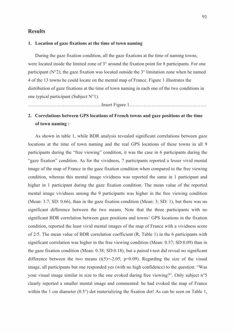
Results
1. Location of gaze fixations at the time of town naming
During the gaze fixation condition, all the gaze fixations at the time of naming towns,
were located inside the limited zone of 3° around the fixation point for 8 participants. For one
participant (N°2), the gaze fixation was located outside the 3° limitation zone when he named
4 of the 13 towns he could locate on the mental map of France. Figure 1 illustrates the
distribution of gaze fixations at the time of town naming in each one of the two conditions in
one typical participant (Subject N°1).
………………………………………..Insert Figure 1…………………………………………. 2. Correlations between GPS locations of French towns and gaze positions at the time
of town naming :
As shown in table 1, while BDR analysis revealed significant correlations between gaze
locations at the time of town naming and the real GPS locations of these towns in all 9
participants during the “free viewing” condition, it was the case in 6 participants during the
“gaze fixation” condition. As for the vividness, 7 participants reported a lesser vivid mental
image of the map of France in the gaze fixation condition when compared to the free viewing
condition, whereas this mental image vividness was reported the same in 1 participant and
higher in 1 participant during the gaze fixation condition. The mean value of the reported
mental image vividness among the 9 participants was higher in the free viewing condition
(Mean: 3.7; SD: 0.66), than in the gaze fixation condition (Mean: 3; SD: 1), but there was no
significant difference between the two means. Note that the three participants with no
significant BDR correlation between gaze positions and towns’ GPS locations in the fixation
condition, reported the least vivid mental images of the map of France with a vividness score
of 2/5. The mean value of BDR correlation coefficient (R, Table 1) in the 6 participants with
significant correlation was higher in the free viewing condition (Mean: 0.57; SD:0.09) than in
the gaze fixation condition (Mean: 0.38; SD:0.18), but a paired t-test did reveal no significant
difference between the two means (t(5)=-2.05; p=0.09). Regarding the size of the visual
image, all participants but one responded yes (with no high confidence) to the question: “Was
your visual image similar in size to the one evoked during free viewing?”. Only subject n°5
clearly reported a smaller mental image and commented: he had evoked the map of France
within the 1 cm diameter (0.5°) dot materializing the fixation dot! As can be seen on Table 1,
this subject still reported a lower vividness (3/5) during fixation than during free viewing
(4/5).
3. Comparison of saccades’ parameters in each participant between the gaze fixation
condition and the free viewing condition:
Saccades analysis revealed that during the gaze fixation condition, all participants
performed saccades of lesser magnitude, velocity and duration, when compared to the
saccades performed during the free viewing condition (Cf. Table 2 & Figure 2).
…………………………………Insert Table 2 And Figure 2…………………………………..
Discussion
The present study yielded four main results: 1) Performing mental imagery is
accompanied with eye movements even when the participants are asked to maintain gaze
fixation on a central point. 2) These fixational eye micro-movements are also, as in the free
viewing condition, not random but reflect the spatial content of the visual mental imagery. 3)
However, gaze fixation altered the quality, as reflected by the reported vividness, of the
mental image in a majority of participants. 4) The three participants exhibiting no significant
correlation between Gaze positions and towns GPS locations in the fixation condition were
also the ones reporting the least vivid mental image of the map of France during the gaze
fixation task, with a vividness score of 2/5. On the whole, these results argue in favour of a
functional relationship between eye movements (whether fixational or not) and visual
imagery. We discuss below more specifically what step, i.e. eye movement planning or
execution, may be necessary for visual imagery.
There is a recent and consistent body of research, associating saccades and microsaccades
in different aspects, arguing that these two allegedly different types of eye movements
actually share common spatiotemporal characteristics, roles, and neural substrates (common
generator) (Martinez Conde et al. 2009, 2013). Indeed, microsaccades are not exclusively
observed during eye fixation, as very small magnitude saccades (less than 1°) also appear
during free viewing, in both perception (Otero-Millan et al. 2008) and imagery as shown in
the present study (Figure 2). This supports the possibility that fixational microsaccades might
play a functional role in exploring mental images in the same way that saccades do when the
gaze is not restricted. Accordingly, BDR analysis revealed significant correlations between
gaze positions at the time of town naming and GPS locations of these towns in 6 out of the 9
participants during the gaze fixation condition. In these 6 subjects, the fixational eye
movements may have indeed endorsed the functional role of regular saccades in
accompanying visual imagery. However, let’s further speculate about the two alternative ways
saccades and microsaccades can play a functional role in imagery. More specifically, these
spatially consistent eye movements during gaze fixation could be explained in two different
ways depending on the size of the visualized map of France.
On one hand, if what matters to mentally organize and explore a mental image is where
eye movement ends (eye positions per se), then maintaining gaze in a fixation zone would
lead to perform visual imagery on a smaller scale. In perfect accordance with this hypothesis,
one participant (Subject n°5) spontaneously mentioned imagining a very small map of France
during the gaze fixation condition, so that it could grossly fit into the 1 cm diameter fixation
point. However, when they were explicitly asked for it, all other participants reported that
“yes”, they did evoke a visual image of approximately the same size as in free gaze. The fact
that participants may have been unable to efficiently manipulate the size of their mental
image, and shrink it enough to be explored with small magnitude saccades, could be seen as a
reason why at the level of the group the quality of the visual image was reported to be lower
during gaze fixation. Unlike subject n°5, they may have evoked a mental image of large size
that could only be explored by displacing attention covertly. However, subject n°5 does not
report the highest vividness of the group. So other manipulations of the visual image might
have been performed by other subjects like displacing successively the part of the visual
image that is currently reported toward gaze fixation, instead of displacing the gaze on the
visual image. As shown in Fourtassi et al. (2013), subjects use multiple and various dynamic
visual representations of the map of France rather than a unique and static visual image.
Manipulating a novel (just encoded) visual scene might be more difficult than manipulating
the map of France, seen from different points of view and at different scales, stored in long-
term memory and manipulated mentally many times. This could explain why maintaining
fixation during an imagery task has been reported to have a deleterious effect on imagery
performance in three previous studies (Johansson et al. 2012; Laeng & Teodorescu 2002,
Laeng et al. 2014), in which participants had to visualize a stimulus, seen and encoded a few
seconds before the imagery task was performed.
Alternatively, what matters may not be where eye movements actually ends (where they
are executed) but instead their underlying shifts of attention which allow the subject to
mentally organize and explore his mental image; in such case, micro-saccades could represent
a mere expression of covert attention shifts while exploring even a mental image of large size.
In the Kosslyn model (Kosslyn 1994, 2006), saccadic eye movements during imagery are
functionally associated to shifts of the “attentional window” on the mental image (Kosslyn &
Mast 2002). In a gaze fixation task, we could assume that subjects explore their mental image
using covert attention, which are overtly reflected either by saccades in free gaze, or by
microsaccades during gaze fixation. It can be noticed that the two subjects who have a mean
of saccade magnitude superior to 1° (Table 2) also have no significant BDR correlation and
low vividness score. One can assume that in these subjects the overt manifestation of covert
attention reflects an inability to efficiently control fixation. If inhibiting saccades requires too
much effort in these subjects, it might act as a complex double-task, which does not leave
enough resources for the main task of recall via visual imagery. We could thus postulate that
these participants were not able to use visual imagery to perform the task, but instead non-
imaging strategy (e.g. access to French towns through semantic or episodic knowledge). This
may also have been the case for the subject n°6 whose eye positions also did not correlate
significantly with GPS coordinates of towns. In these three subjects, we hypothesize that the
task was not performed through imagery as revealed by the low vividness score they reported
(2/5), and we maintain that our BDR analysis represents an “ocular signature” for the use of
visual imagery, even during fixation condition. For the 6 subjects who presented with a
significant ocular signature, the lower vividness suggests either that attentional shifts might
need to be expressed overtly at a large scale to produce vivid and fine-grained visual imagery
support for memory-retrieval in most individual subjects, while others might be able to either
build or explore mental images covertly with a performance not suffering from the effort to
control eye movements, or manipulate or reduce the size of their mental image.
To conclude, these findings might be explained by the use of different individual
strategies. Even if the instruction was to visualize the map of France, subjects may have used
semantic or episodic knowledge to recall French towns, Others may have evoked either a very
small mental image (like subject n°5 clearly reported to do) or a large size mental image of
the map that they have explored by displacing attention covertly, with micro-saccades
reflecting these shifts of attention. All these individual strategies might explain the
controversial results in previous studies performed either on patients or on healthy individuals

attempting to perform visual imagery in fixation condition. These individual differences
should be taken into consideration when interpreting grouped analyses. We are confident that
the BDR analysis is a useful tool to assess these strategies at individual level, and thus to
better assess patients’ mental space (Fourtassi et al. 2016) or better test for theoretical models
about the role of eye movements in imagery.

Figure 1: Gaze fixations’ distribution, in the gaze fixation condition (A) and in the free viewing condition (B), in participant N°1. Legend: black circle: fixation point; purple triangles: gaze locations at the time of naming towns in the fixation condition; black triangle: gaze location at the time of naming “Paris”; red diamonds: gaze fixations at the time of naming towns in the free viewing condition.
Figure 2: Total number of saccades, according to their magnitude in visual degrees, in the central fixation and in the free viewing conditions, in participant N°1.

Tab
le 1
: V
VIQ
scor
es, B
DR
cor
rela
tion
coef
ficie
nt (R
) and
p-v
alue
in e
ach
of th
e tw
o co
nditi
ons
Nb
: Num
ber o
f. *p
<0.0
5 T
able
2: C
ompa
riso
n of
sacc
ades
’ par
amet
ers
betw
een
the
gaze
fixa
tion
cond
ition
and
the
free
vi
ewin
g co
nditi
on
ISI:
Inte
r-sa
ccad
ic in
terv
al; G
F: G
aze
fixat
ion;
FV
: Fre
e vi
ewin
g
References
1. Brandt, S. A., & Stark, L. W. (1997). Spontaneous eye movements during visual imagery reflect the content of the visual scene. Journal of cognitive neuroscience, 9(1), 27-38.
2. Donner, K. & Hemila, S. Modelling the effect of microsaccades on retinal responses to stationary contrast patterns. Vision Res. 47, 1166–1177 (2007). Engbert, R. and Kliegl, R. (2004) Microsaccades keep the eyes’ balance during fixation. Psychol. Sci. 15, 431–436
3. Finke, R. A. (1986). Mental imagery and the visual system. Scientific American, 254(3), 88-95.
4. Fourtassi, M., Hajjioui, A., Urquizar, C., Rossetti, Y., Rode, G., & Pisella, L. (2013). Iterative fragmentation of cognitive maps in a visual imagery task. PloS one, 8(7), e68560.
5. Johansson R, Holsanova J, Holmqvist K. (2006). Pictures and spoken descriptions elicit similar eye movements during mental imagery, in light and complete darkness. Cogn Sci; 30:1053-79
6. Kosslyn, S. M., Thompson, W. L., & Ganis, G. (2006). The case for mental imagery. Oxford University Press.
7. Kosslyn, S. M. (1996). Image and brain: The resolution of the imagery debate. MIT press.
8. Kozhevnikov, M., Blazhenkova, O., & Becker, M. (2010). Trade-off in object versus spatial visualization abilities: Restriction in the development of visual-processing resources. Psychonomic Bulletin & Review, 17(1), 29-35.
9. Kozhevnikov, M., Kosslyn, S., & Shephard, J. (2005). Spatial versus object visualizers: A new characterization of visual cognitive style. Memory & cognition, 33(4), 710-726.
10. Johansson, R., Holsanova, J., Dewhurst, R., & Holmqvist, K. (2012). Eye movements during scene recollection have a functional role, but they are not reinstatements of those produced during encoding. Journal of Experimental Psychology: Human Perception and Performance, 38(5), 1289.
11. Laeng, B., & Teodorescu, D. S. (2002). Eye scanpaths during visual imagery reenact those of perception of the same visual scene. Cognitive Science, 26(2), 207-231.
12. Laeng, B., & Sulutvedt, U. (2014). The eye pupil adjusts to imaginary light. Psychological science, 25(1), 188-197.
13. Marks, D. F. (1973). Visual imagery differences in the recall of pictures. British journal of Psychology, 64(1), 17-24.
14. Martarelli, C. S., & Mast, F. W. (2013). Eye movements during long-term pictorial recall. Psychological research, 77(3), 303-309.
15. Martinez-Conde, S., Macknik, S. L., Troncoso, X. G., & Hubel, D. H. (2009). Microsaccades: a neurophysiological analysis. Trends in neurosciences, 32(9), 463-475.
16. Martinez-Conde, S., Otero-Millan, J., & Macknik, S. L. (2013). The impact of microsaccades on vision: towards a unified theory of saccadic function. Nature Reviews Neuroscience, 14(2), 83-96.
17. Mast, F. W., & Kosslyn, S. M. (2002). Eye movements during visual mental imagery. Trends in Cognitive Sciences, 6(7), 271-272.
18. McCamy, M. B. et al. Microsaccadic efficacy and contribution to foveal and peripheral vision. J. Neurosci. 32, 9194–9204 (2012).
19. Otero-Millan, J., Troncoso, X. G., Macknik, S. L., Serrano-Pedraza, I., & Martinez-Conde, S. (2008). Saccades and microsaccades during visual fixation, exploration, and search: foundations for a common saccadic generator. Journal of Vision, 8(14), 21-21.
20. Richardson, D. C., & Kirkham, N. Z. (2004). Multimodal events and moving locations: eye movements of adults and 6-month-olds reveal dynamic spatial indexing. Journal of Experimental Psychology: General, 133(1), 46.
21. Richardson, D. C., & Spivey, M. J. (2000). Representation, space and Hollywood Squares: Looking at things that aren't there anymore. Cognition, 76(3), 269-295.
22. Santarpia, A., Blanchet, A., Poinsot, R., Lambert, J. F., Mininni, G., & Thizon-Vidal, S. (2008). Évaluer la vivacité des images mentales dans différentes populations françaises. Pratiques psychologiques, 14(3), 421-441.
23. Spivey, M. J., & Geng, J. J. (2001). Oculomotor mechanisms activated by imagery and memory: Eye movements to absent objects. Psychological research, 65(4), 235-241.
24. Steinman, R. M., Cunitz, R. J., Timberlake, G. T. & Herman, M. Voluntary control of microsaccades during maintained monocular fixation. Science 155, 1577–1579 (1967).
25. Steinman, R. M., Haddad, G. M., Skavenski, A. A. & Wyman, D. Miniature eye movement. Science 181, 810–819 (1973).
26. Tobler W (1965) Computation of the corresponding geographical patterns. Papers of the regional science association 15: 131–139. doi: 10.1007/bf01947869
27. Yarbus, A. L. Eye Movements and Vision. Plenum Press, 1967 28. Yuval-Greenberg, S., Merriam, E. P., & Heeger, D. J. (2014). Spontaneous
microsaccades reflect shifts in covert attention. The Journal of Neuroscience, 34(41), 13693-13700.

Article (4)
Publié dans..
Update article
Using eye movements to explore mental representations of space
Maryam Fourtassi a,b,*, Gilles Rode a,c,d, Laure Pisella a,c
a Inserm, U1028, CNRS, UMR5292, Lyon Neuroscience Research Center, ImpAct, 16, avenue du Doyen-Lepine, 69676 Bron cedex, FrancebUniversite Mohamed Premier, Faculte de medecine et de Pharmacie d’Oujda, BP 724 Hay Al Quods, 60000 Oujda, MoroccocUniversite Claude-Bernard Lyon 1, 69100 Villeurbanne, FrancedHospices Civils de Lyon, Hopital Henry-Gabrielle, Mouvement et Handicap, 69000 Lyon, France
1. Introduction
Physical space is accessed by the cognitive system by way of thesensory organs, mainly sight, which collect information on theenvironment, enabling the construction of one or severalrepresentations or mental images of the space in question[1]. Mental imagery can be defined as the mental creation of anexperience that resembles the perceptual experience of objects orevents, at least for certain aspects, but in the absence of directsensory stimulation [2]. Different modes can be distinguishedaccording to the nature of the virtual stimulus involved, forinstance mental imagery arising from visual, auditory or olfactorystimuli.
Visual mental imagery is a cognitive experience that sharesseveral similarities with visual perception, among which are ocularmovements. Even in the absence of any real visual stimulus, thedescription of a scene or object from memory is accompanied byeye movements that are not arbitrary, but reflect the spatialcontent of the mental image [3–6]. For instance, in the course ofmental exploration of the map of France, the spontaneous
positioning of the eyes when mention is made of cities in theeast, on the west coast, or in the south is correlated to thegeographical location of these elements [7].
While the functional significance of the eye movementsaccompanying visual mental imagery is still the subject of debate[5,8–11], their concordance with the spatial content of mentalimagery has been evidenced among groups of individuals [3–7,10]and on individual scale [7]. Thus the exploration of ocularorientation in mental imagery tasks could be a valuable, objectivemeans of gaining better understanding of the spatial structure ofvisual mental representations, in particular in certain pathologicalconditions where the mental representation of space is altered, forinstance in the case of hemispatial neglect.
2. Visual mental imagery and representations of space
Visual mental imagery, often reported as being the experienceof seeing an image inside one’s head (‘‘seeing with the mind’seye’’), often occurs in different situations of daily life. For instance,to recall the number of windows in a house, it is sufficient to call upa mental image of the house frontage and count the windows[4]. Likewise, to decide which of the cities of Rennes or Le Mans isthe further west in France, it is enough to visualise a map of Franceand position the cities on it. Visual mental imagery comprises both
Annals of Physical and Rehabilitation Medicine xxx (2016) xxx–xxx
A R T I C L E I N F O
Article history:
Received 31 May 2015
Accepted 8 March 2016
Keywords:
Visual imagery
Ocular-tracking
Bi-dimensional regression
Mental representation of space
A B S T R A C T
Visual mental imagery is a cognitive experience characterised by the activation of the mental
representation of an object or scene in the absence of the corresponding stimulus. According to the
analogical theory, mental representations have a pictorial nature that preserves the spatial
characteristics of the environment that is mentally represented. This cognitive experience shares
many similarities with the experience of visual perception, including eye movements. The mental
visualisation of a scene is accompanied by eye movements that reflect the spatial content of the mental
image, and which can mirror the deformations of this mental image with respect to the real image, such
as asymmetries or size reduction. The present article offers a concise overview of the main theories
explaining the interactions between eye movements and mental representations, with some examples of
the studies supporting them. It also aims to explain how ocular-tracking could be a useful tool in
exploring the dynamics of spatial mental representations, especially in pathological situations where
these representations can be altered, for instance in unilateral spatial neglect.
� 2016 Elsevier Masson SAS. All rights reserved.
* Corresponding author. Universite Mohamed Premier, Faculte de medecine et de
Pharmacie d’Oujda, BP 724 Hay Al Quods, 60000 Oujda, Morocco.
E-mail address: [email protected] (M. Fourtassi).
G Model
REHAB-969; No. of Pages 4
Please cite this article in press as: Fourtassi M, et al. Using eye movements to explore mental representations of space. Ann Phys RehabilMed (2016), http://dx.doi.org/10.1016/j.rehab.2016.03.001
Available online at
ScienceDirectwww.sciencedirect.com
http://dx.doi.org/10.1016/j.rehab.2016.03.001
1877-0657/� 2016 Elsevier Masson SAS. All rights reserved.
a non-spatial dimension (representation of colours, shapes,textures, etc.) and a spatial dimension, enabling individuals toconstruct a mental representation of the localisation of objects onein relation to the other, or in relation to themselves, and identifytheir orientation in space. This spatialised representation appearsas very useful in certain day-to-day tasks, such as reproducing adrawing, drawing a plan, or showing someone an itinerary. Inaddition, different studies have shown that the ability to constructvisual mental images plays a fundamental role in differentcognitive processes, such as recalling events [12], the constructionof mental models [13], problem-solving [14], or mental trainingamong athletes [15].
The nature of mental representations is the subject ofconsiderable debate between two main trends in neuropsychology[16–19]. While the ‘‘propositionalists’’ support the notion that allmental representations are of amodal nature and solely proposi-tional, like language [18,19], the proponents of the ‘‘analogical’’theory support the idea that visual mental representations arepictorial in nature, gathering different components of the imagethat is represented, among which its spatial characteristics[17]. The analogical theory draws support from several recentexperimental studies that claim that mental imagery involves thesame neurone networks and underpinnings as visual perception[20]. Indeed, functional MRI studies have shown that a taskinvolving visual perception of a stimulus and mental imagery ofthe same stimulus involved activation of neighbouring or evensuperimposed cortical areas of the brain, with maintenance of theretinal-cortical topographical organisation [20–22]. Thus visualmental imagery could be explained by way of a reactivation or‘‘replay’’ of the areas coming into play in visual perception of thebottom-up type, but with the visual information travelling in theopposite direction (top-down) from the substrates of memorytowards the areas of visual perception [23].
Kosslyn et al. proposed a model for perception and mentalvisual imagery that remains coherent with the main results ofexperimental studies on mental imagery [16,17]. According to thismodel, the visual information transmitted by the retina in thecourse of seeing an object or a scene is projected onto a ‘‘visualbuffer’’, corresponding to the primary visual cortex located in theoccipital lobes, and possessing a retino-topic organisation. Fromthis visual buffer, the attentional window shifts to select relevantinformation, which is then analysed in two distinct systems: thesystem relating to the properties of the object (or the ‘‘what’’system) located in the temporal lobe via the ventral visualpathway, and the system relating to spatial properties (the‘‘where’’ system) located in the parietal lobe, via the dorsal visualpathway [17]. In similar manner, in the course of mental imagery,the pictorial characteristics of the object visualised are projectedonto this same visual buffer, across which the attentional windowmoves for better inspection and identification of the spatialproperties and relationships of the mental image [17].
3. Perceived images and mental representations of space
Eye movements are one of the aspects in common betweenvisual perception and mental imagery. However, while the role ofocular movements is at present well-established in visualperception, their occurrence in the course of other cognitive taskssuch as mental imagery, disconnected from any perceptualactivity, is one of the least well elucidated human behaviours.
3.1. Eye movements in visual perception
Eye movements have a fundamental role in visual perception.Indeed, it is the fovea, the central part of the retina, with its denselypacked cones, that provides the best visual resolution. It however
concerns no more than 28 of the visual field, about equivalent to thesize of the thumb held at arm’s length. By rapid movements of theeyeball known as ‘‘saccades’’, the fovea is displaced to differentzones of interest in the visual environment. These zones areanalysed separately in a brief moment of ocular fixation. It is onlythe integration of these different parts of the image in the cortexthat gives us the illusion of seeing a scene clearly in its entirety[24].
3.2. Eye movements in mental imagery
In 1968, Hebb noted similarities between eye movementsduring visual perception and during mental representation,suggesting that while these movements are mechanically essentialto scan objects in the course of perception, the same eye movementprocess could also have an organising function for the mentalimage. Hebb held that if the image is a reintegration of theperceptual process, it should include the eye movements [9]. It wasonly 30 years after Hebb’s hypothesis that the first experimentaldata to confirm it was published. Brandt and Stark published thefirst study to compare the spatial distribution of eye movementsduring visual perception and during mental imagery. They showedstatistically significant similarities in the scanpaths for eachparticipant between the visual perception of black squares on agrid background and mental visual imagery of the same grid [3]. Inthe course of the perceptive phase and in the imagery phase alike,the eyes shifted to the positions in the grid with black squares.Since then several studies have confirmed that spontaneous eyemovements accompanying a mental imagery activity are notarbitrary, but reflect the content of the imagery [4–7]. Johanssonet al. [4] recorded eye movements in subjects looking at a blankscreen while they were calling to mind a scene that had previouslybeen presented, either visually (image of the scene) or verbally(oral description of the scene with spatial indications). By ananalysis of eye fixations occurring at the time of the recall of eachelement in the scene they showed that their localisations inrelation to the overall zone scanned were globally concordant withthe actual localisation of the different elements in the scene, one inrelation to another [4].
Several theories, variously supported by empirical data, havebeen put forward to explain the functional role of these eyemovements ‘‘looking at nothing’’. Certain authors, in particularauthors in the ‘‘propositional’’ current, consider that these eyemovements are spatial pointers [11,18]. According to this theory,the shifting of gaze in the absence of visual stimulus might not belinked to an image-based internal representation of space, butcould result from a mechanism of unconscious, automaticassociation linking each object mentioned to the place it occupiesin the real environment, as if its position corresponded to anelement in the ‘‘proposition’’ characterising the object [11]. Inother words, when the individual recalls an object from memory,his eyes move systematically towards the place where the objectwas initially perceived or encoded in memory, as if this positionwas included in the mnesic trace of the object. In Kosslyn’sanalogist model, however, these eye movements have beeninterpreted as reflecting the shift in the attentional window tothe visual buffer when the mental image is generated or explored[4]. Thus the debate continues as to what this association mightreflect: according to the propositional theory, the positioning of theeyes is an epiphenomenon because recalling the object automati-cally reactivates its entire mnesic trace, while according to thepictorial representational theory [16] the positioning of the eyesreflects a necessary orientation of our attention towards the object(i.e. towards the position it occupies in our mental image) in orderto recall its other characteristics. According to this second theory,the positioning of the eyes at the moment of the mental recall of
M. Fourtassi et al. / Annals of Physical and Rehabilitation Medicine xxx (2016) xxx–xxx2
G Model
REHAB-969; No. of Pages 4
Please cite this article in press as: Fourtassi M, et al. Using eye movements to explore mental representations of space. Ann Phys RehabilMed (2016), http://dx.doi.org/10.1016/j.rehab.2016.03.001
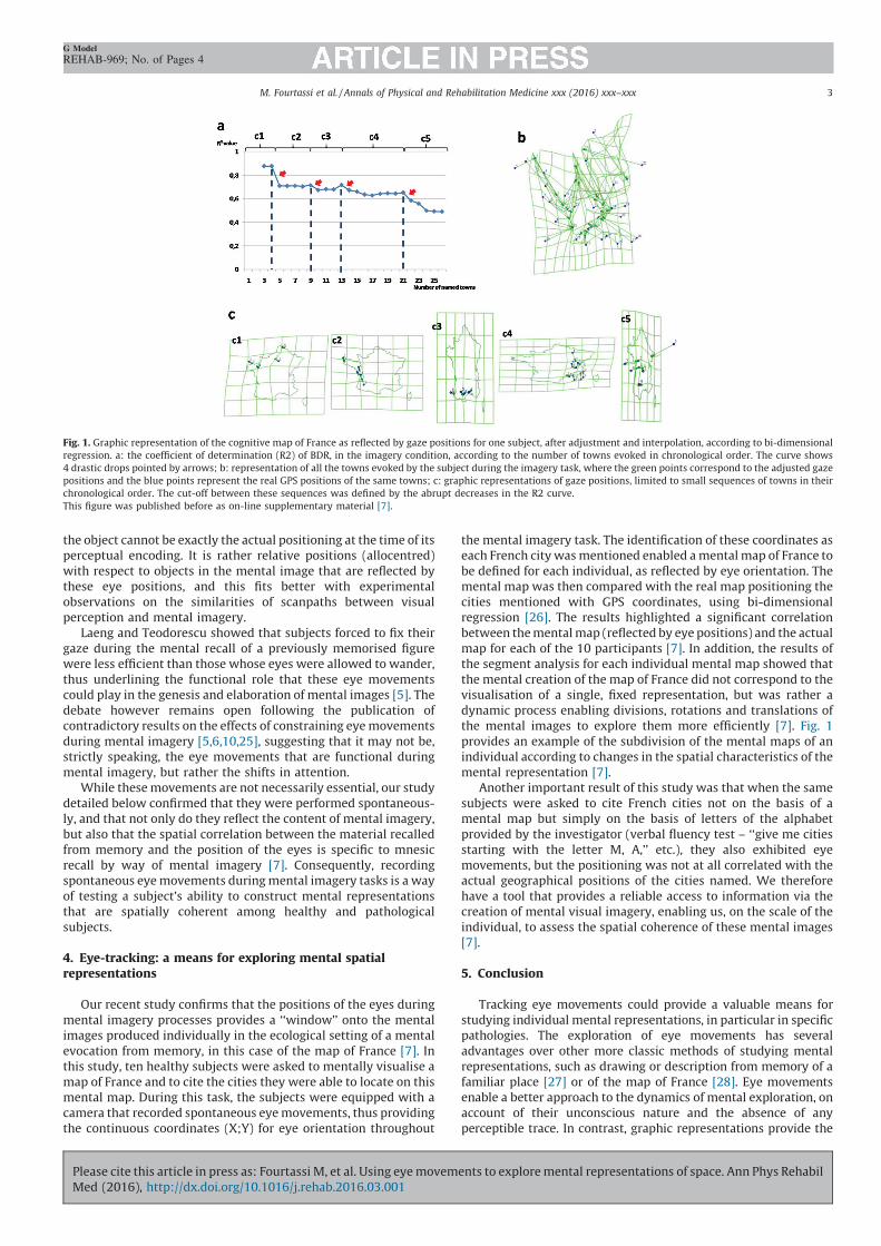
the object cannot be exactly the actual positioning at the time of itsperceptual encoding. It is rather relative positions (allocentred)with respect to objects in the mental image that are reflected bythese eye positions, and this fits better with experimentalobservations on the similarities of scanpaths between visualperception and mental imagery.
Laeng and Teodorescu showed that subjects forced to fix theirgaze during the mental recall of a previously memorised figurewere less efficient than those whose eyes were allowed to wander,thus underlining the functional role that these eye movementscould play in the genesis and elaboration of mental images [5]. Thedebate however remains open following the publication ofcontradictory results on the effects of constraining eye movementsduring mental imagery [5,6,10,25], suggesting that it may not be,strictly speaking, the eye movements that are functional duringmental imagery, but rather the shifts in attention.
While these movements are not necessarily essential, our studydetailed below confirmed that they were performed spontaneous-ly, and that not only do they reflect the content of mental imagery,but also that the spatial correlation between the material recalledfrom memory and the position of the eyes is specific to mnesicrecall by way of mental imagery [7]. Consequently, recordingspontaneous eye movements during mental imagery tasks is a wayof testing a subject’s ability to construct mental representationsthat are spatially coherent among healthy and pathologicalsubjects.
4. Eye-tracking: a means for exploring mental spatialrepresentations
Our recent study confirms that the positions of the eyes duringmental imagery processes provides a ‘‘window’’ onto the mentalimages produced individually in the ecological setting of a mentalevocation from memory, in this case of the map of France [7]. Inthis study, ten healthy subjects were asked to mentally visualise amap of France and to cite the cities they were able to locate on thismental map. During this task, the subjects were equipped with acamera that recorded spontaneous eye movements, thus providingthe continuous coordinates (X;Y) for eye orientation throughout
the mental imagery task. The identification of these coordinates aseach French city was mentioned enabled a mental map of France tobe defined for each individual, as reflected by eye orientation. Themental map was then compared with the real map positioning thecities mentioned with GPS coordinates, using bi-dimensionalregression [26]. The results highlighted a significant correlationbetween the mental map (reflected by eye positions) and the actualmap for each of the 10 participants [7]. In addition, the results ofthe segment analysis for each individual mental map showed thatthe mental creation of the map of France did not correspond to thevisualisation of a single, fixed representation, but was rather adynamic process enabling divisions, rotations and translations ofthe mental images to explore them more efficiently [7]. Fig. 1provides an example of the subdivision of the mental maps of anindividual according to changes in the spatial characteristics of themental representation [7].
Another important result of this study was that when the samesubjects were asked to cite French cities not on the basis of amental map but simply on the basis of letters of the alphabetprovided by the investigator (verbal fluency test – ‘‘give me citiesstarting with the letter M, A,’’ etc.), they also exhibited eyemovements, but the positioning was not at all correlated with theactual geographical positions of the cities named. We thereforehave a tool that provides a reliable access to information via thecreation of mental visual imagery, enabling us, on the scale of theindividual, to assess the spatial coherence of these mental images[7].
5. Conclusion
Tracking eye movements could provide a valuable means forstudying individual mental representations, in particular in specificpathologies. The exploration of eye movements has severaladvantages over other more classic methods of studying mentalrepresentations, such as drawing or description from memory of afamiliar place [27] or of the map of France [28]. Eye movementsenable a better approach to the dynamics of mental exploration, onaccount of their unconscious nature and the absence of anyperceptible trace. In contrast, graphic representations provide the
Fig. 1. Graphic representation of the cognitive map of France as reflected by gaze positions for one subject, after adjustment and interpolation, according to bi-dimensional
regression. a: the coefficient of determination (R2) of BDR, in the imagery condition, according to the number of towns evoked in chronological order. The curve shows
4 drastic drops pointed by arrows; b: representation of all the towns evoked by the subject during the imagery task, where the green points correspond to the adjusted gaze
positions and the blue points represent the real GPS positions of the same towns; c: graphic representations of gaze positions, limited to small sequences of towns in their
chronological order. The cut-off between these sequences was defined by the abrupt decreases in the R2 curve.
This figure was published before as on-line supplementary material [7].
M. Fourtassi et al. / Annals of Physical and Rehabilitation Medicine xxx (2016) xxx–xxx 3
G Model
REHAB-969; No. of Pages 4
Please cite this article in press as: Fourtassi M, et al. Using eye movements to explore mental representations of space. Ann Phys RehabilMed (2016), http://dx.doi.org/10.1016/j.rehab.2016.03.001

subject with a sensory/visual feedback that can interact bothpositively or negatively with the performance of the drawing [29,30]obliging the subject to retain the same representational referencesand allowing for self-correction. In addition, recording eyemovements enables an assessment of the spatial coherence ofelements represented mentally when a patient with hemispatialneglect calls them to mind.
Disclosure of interest
The authors declare that they have no competing interest.
References
[1] Gibson JJ. The senses considered as perceptual systems. Boston: Houghton;1966.
[2] Finke RA. Principles of mental imagery. Cambridge: MIT Press; 1989.[3] Brandt SA, Stark LW. Spontaneous eye movements during visual imagery
reflect the content of the visual scene. J Cogn Neurosci 1997;9:27–38.[4] Johansson R, Holsanova J, Holmqvist K. Pictures and spoken descriptions elicit
similar eye movements during mental imagery, in light and complete dark-ness. Cogn Sci 2006;30:1053–79.
[5] Laeng B, Teodorescu DS. Eye scanpaths during imagery reenact those ofperception of the same visual scene. Cogn Sci 2002;26:207–31.
[6] Spivey MJ, Geng JJ. Oculomotor mechanisms activated by imagery and mem-ory: eye movements to absent objects. Psycho Res 2001;65:235–41.
[7] Fourtassi M, Hajjioui A, Urquizar C, Rossetti Y, Rode G, Pisella L. Iterativefragmentation of cognitive maps in a visual imagery task. PLoS ONE 2013;8:e68560.
[8] Barsalou LW. Perceptual symbol systems. Behav Brain Sci 1999;22:577–609.[9] Hebb DO. Concerning imagery. Psychol Rev 1968;6:466–77.
[10] Johansson R, Holsanova J, Dewhurst R, Holmqvist K. Eye movements duringscene recollection have a functional role, but they are not reinstatements ofthose produced during encoding. J Exp Psychol Hum Percept Perform 2012;38:1289–314.
[11] O’Regan JK, Noe A. A sensorimotor account of vision and visual consciousness.Behav Brain Sci 2001;24:939–73.
[12] Slotnick SD, Thompson WL, Kosslyn SM. Visual memory and visual mentalimagery recruit common control and sensory regions of the brain. CognNeurosci 2012;3:14–20.
[13] Bower GH, Morrow DG. Mental models in narrative comprehension. Science1990;247:44–8.
[14] Hegarty M. Mechanical reasoning by mental stimulation. Trends Cogn Sci2004;8:280–5.
[15] Olsson CJ, Nyberg L. Motor imagery: if you cannot do it, you will not think it.Scand J Med Sci Sports 2010;20:711–5.
[16] Kosslyn SM. Image and brain. Cambridge: MIT Press; 1994.[17] Kosslyn SM, Thompson WL, Ganis G. The case for mental imagery. New York:
Oxford University Press; 2006.[18] Pylyshyn ZW. Mental imagery: in search of a theory. Behav Brain Sci 2002;
25:157–82.[19] Pylyshyn ZW. Return of the mental image: are there really pictures in the
brain? Trends Cogn Sci 2003;7:113–8.[20] Ganis G, Thompson WL, Kosslyn SM. Brain areas underlying visual mental
imagery and visual perception: an fMRI study. Cogn Brain Res 2004;20:226–41.[21] Kosslyn SM, Thompson WL, Kim IJ, Alpert NM. Topographical representations
of mental images in primary visual cortex. Nature 1995;378:496–8.[22] Kosslyn SM, Ganis G, Thompson WL. Neural foundations of imagery. Nat Rev
Neurosci 2001;2:635–42.[23] Farah MJ. The neural basis of mental imagery. In: Gazzaniga MS, editor. The
new cognitive neuroscience. Cambridge: MIT Press; 2000. p. 965–74.[24] Yarbus AL. Eye movement and vision. New York: Plenum Press; 1967.[25] Richardson DC, Spivey MJ. Representation, space and Hollywood squares:
looking at things that are not there anymore. Cognition 2000;76:269–95.[26] Tobler W. Bidimensional regression. Geogr Anal 1994;26:187–212.[27] Bisiach E, Luzzatti C. Unilateral neglect of representational space. Cortex
1978;14:129–33.[28] Rode G, Perenin MT, Boisson D. Negligence de l’espace represente : mise en
evidence par l’evocation mentale de la carte de France. Rev Neurol1995;151:161–4.
[29] Cristinzio C, Bourlon C, Pradat-Diehl P, Trojano L, Grossi D, Chokron S, et al.Representational neglect in ‘‘invisible’’ drawing from memory. Cortex 2009;45:313–7.
[30] Chokron S, Colliot P, Bartolomeo P. The role of vision in spatial representation.Cortex 2004;40:281–90.
M. Fourtassi et al. / Annals of Physical and Rehabilitation Medicine xxx (2016) xxx–xxx4
G Model
REHAB-969; No. of Pages 4
Please cite this article in press as: Fourtassi M, et al. Using eye movements to explore mental representations of space. Ann Phys RehabilMed (2016), http://dx.doi.org/10.1016/j.rehab.2016.03.001

“Every act of perception, is to some degree an act of creation, and every act of
memory is to some degree an act of imagination.” Oliver Sacks (2009)
A travers ce travail de thèse, nous avons essayé d’utiliser les mouvements des
yeux comme outil d’exploration de l’imagerie mentale visuelle, dans l’objectif
d’approfondir la compréhension de ses mécanismes physiologiques aussi bien chez
les sujets sains qu’à travers de modèles pathologiques. L’ensemble de notre
contribution expérimentale (articles joints à ce manuscrit), sera discutée de façon à
identifier ses différents apports dans les domaines de recherche autour de l’imagerie
mentale visuelle que nous avions pu approcher et que nous avons exposés en
introduction.
Mais avant d’exposer les résultats de nos expériences et les interprétations
théoriques que nous en faisons à la lumière des données de la littérature, il nous paraît
important de justifier le choix du paradigme expérimental de la carte de France et de
la méthode d’analyse par régression bidimensionnelle, utilisés dans toutes les
expériences entreprises. En effet, ce développement méthodologique nous apparaît
comme un premier apport important de cette thèse pour l’étude des représentations
mentales.
1. Apport méthodologique : notre méthode expérimentale constitue
une signature oculaire de l’utilisation de l’imagerie mentale à
l’échelle individuelle
Dans le cadre de ce travail de thèse, nous avons développé un modèle
expérimental qui devait à la fois contourner les critiques que nous avions faites des
études antérieures et être adapté aux sujets sains comme aux patients.
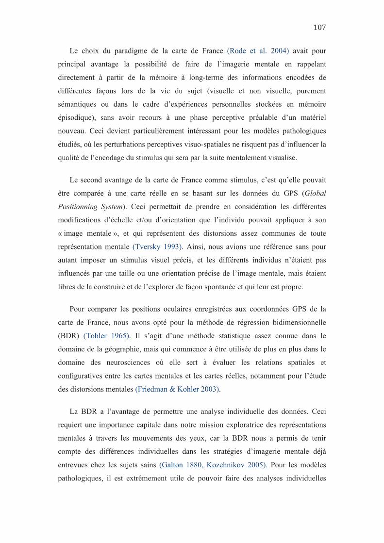
Le choix du paradigme de la carte de France (Rode et al. 2004) avait pour
principal avantage la possibilité de faire de l’imagerie mentale en rappelant
directement à partir de la mémoire à long-terme des informations encodées de
différentes façons lors de la vie du sujet (visuelle et non visuelle, purement
sémantiques ou dans le cadre d’expériences personnelles stockées en mémoire
épisodique), sans avoir recours à une phase perceptive préalable d’un matériel
nouveau. Ceci devient particulièrement intéressant pour les modèles pathologiques
étudiés, où les perturbations perceptives visuo-spatiales ne risquent pas d’influencer la
qualité de l’encodage du stimulus qui sera par la suite mentalement visualisé.
Le second avantage de la carte de France comme stimulus, c’est qu’elle pouvait
être comparée à une carte réelle en se basant sur les données du GPS (Global
Positionning System). Ceci permettait de prendre en considération les différentes
modifications d’échelle et/ou d’orientation que l’individu pouvait appliquer à son
« image mentale », et qui représentent des distorsions assez communes de toute
représentation mentale (Tversky 1993). Ainsi, nous avions une référence sans pour
autant imposer un stimulus visuel précis, et les différents individus n’étaient pas
influencés par une taille ou une orientation précise de l’image mentale, mais étaient
libres de la construire et de l’explorer de façon spontanée et qui leur est propre.
Pour comparer les positions oculaires enregistrées aux coordonnées GPS de la
carte de France, nous avons opté pour la méthode de régression bidimensionnelle
(BDR) (Tobler 1965). Il s’agit d’une méthode statistique assez connue dans le
domaine de la géographie, mais qui commence à être utilisée de plus en plus dans le
domaine des neurosciences où elle sert à évaluer les relations spatiales et
configuratives entre les cartes mentales et les cartes réelles, notamment pour l’étude
des distorsions mentales (Friedman & Kohler 2003).
La BDR a l’avantage de permettre une analyse individuelle des données. Ceci
requiert une importance capitale dans notre mission exploratrice des représentations
mentales à travers les mouvements des yeux, car la BDR nous a permis de tenir
compte des différences individuelles dans les stratégies d’imagerie mentale déjà
entrevues chez les sujets sains (Galton 1880, Kozehnikov 2005). Pour les modèles
pathologiques, il est extrêmement utile de pouvoir faire des analyses individuelles
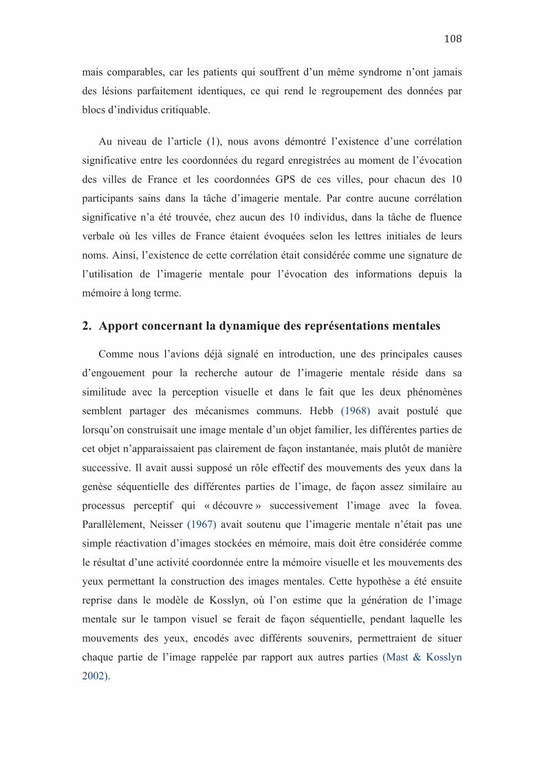
mais comparables, car les patients qui souffrent d’un même syndrome n’ont jamais
des lésions parfaitement identiques, ce qui rend le regroupement des données par
blocs d’individus critiquable.
Au niveau de l’article (1), nous avons démontré l’existence d’une corrélation
significative entre les coordonnées du regard enregistrées au moment de l’évocation
des villes de France et les coordonnées GPS de ces villes, pour chacun des 10
participants sains dans la tâche d’imagerie mentale. Par contre aucune corrélation
significative n’a été trouvée, chez aucun des 10 individus, dans la tâche de fluence
verbale où les villes de France étaient évoquées selon les lettres initiales de leurs
noms. Ainsi, l’existence de cette corrélation était considérée comme une signature de
l’utilisation de l’imagerie mentale pour l’évocation des informations depuis la
mémoire à long terme.
2. Apport concernant la dynamique des représentations mentales
Comme nous l’avions déjà signalé en introduction, une des principales causes
d’engouement pour la recherche autour de l’imagerie mentale réside dans sa
similitude avec la perception visuelle et dans le fait que les deux phénomènes
semblent partager des mécanismes communs. Hebb (1968) avait postulé que
lorsqu’on construisait une image mentale d’un objet familier, les différentes parties de
cet objet n’apparaissaient pas clairement de façon instantanée, mais plutôt de manière
successive. Il avait aussi supposé un rôle effectif des mouvements des yeux dans la
genèse séquentielle des différentes parties de l’image, de façon assez similaire au
processus perceptif qui « découvre » successivement l’image avec la fovea.
Parallèlement, Neisser (1967) avait soutenu que l’imagerie mentale n’était pas une
simple réactivation d’images stockées en mémoire, mais doit être considérée comme
le résultat d’une activité coordonnée entre la mémoire visuelle et les mouvements des
yeux permettant la construction des images mentales. Cette hypothèse a été ensuite
reprise dans le modèle de Kosslyn, où l’on estime que la génération de l’image
mentale sur le tampon visuel se ferait de façon séquentielle, pendant laquelle les
mouvements des yeux, encodés avec différents souvenirs, permettraient de situer
chaque partie de l’image rappelée par rapport aux autres parties (Mast & Kosslyn
2002).
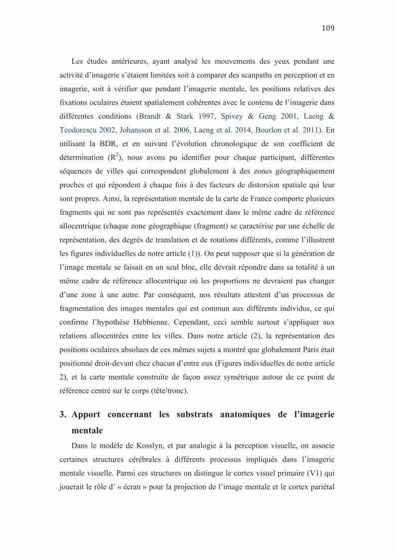
Les études antérieures, ayant analysé les mouvements des yeux pendant une
activité d’imagerie s’étaient limitées soit à comparer des scanpaths en perception et en
imagerie, soit à vérifier que pendant l’imagerie mentale, les positions relatives des
fixations oculaires étaient spatialement cohérentes avec le contenu de l’imagerie dans
différentes conditions (Brandt & Stark 1997, Spivey & Geng 2001, Laeng &
Teodorescu 2002, Johansson et al. 2006, Laeng et al. 2014, Bourlon et al. 2011). En
utilisant la BDR, et en suivant l’évolution chronologique de son coefficient de
détermination (R2), nous avons pu identifier pour chaque participant, différentes
séquences de villes qui correspondent globalement à des zones géographiquement
proches et qui répondent à chaque fois à des facteurs de distorsion spatiale qui leur
sont propres. Ainsi, la représentation mentale de la carte de France comporte plusieurs
fragments qui ne sont pas représentés exactement dans le même cadre de référence
allocentrique (chaque zone géographique (fragment) se caractérise par une échelle de
représentation, des degrés de translation et de rotations différents, comme l’illustrent
les figures individuelles de notre article (1)). On peut supposer que si la génération de
l’image mentale se faisait en un seul bloc, elle devrait répondre dans sa totalité à un
même cadre de référence allocentrique où les proportions ne devraient pas changer
d’une zone à une autre. Par conséquent, nos résultats attestent d’un processus de
fragmentation des images mentales qui est commun aux différents individus, ce qui
confirme l’hypothèse Hebbienne. Cependant, ceci semble surtout s’appliquer aux
relations allocentrées entre les villes. Dans notre article (2), la représentation des
positions oculaires absolues de ces mêmes sujets a montré que globalement Paris était
positionné droit-devant chez chacun d’entre eux (Figures individuelles de notre article
2), et la carte mentale construite de façon assez symétrique autour de ce point de
référence centré sur le corps (tête/tronc).
3. Apport concernant les substrats anatomiques de l’imagerie
mentale Dans le modèle de Kosslyn, et par analogie à la perception visuelle, on associe
certaines structures cérébrales à différents processus impliqués dans l’imagerie
mentale visuelle. Parmi ces structures on distingue le cortex visuel primaire (V1) qui
jouerait le rôle d’ « écran » pour la projection de l’image mentale et le cortex pariétal
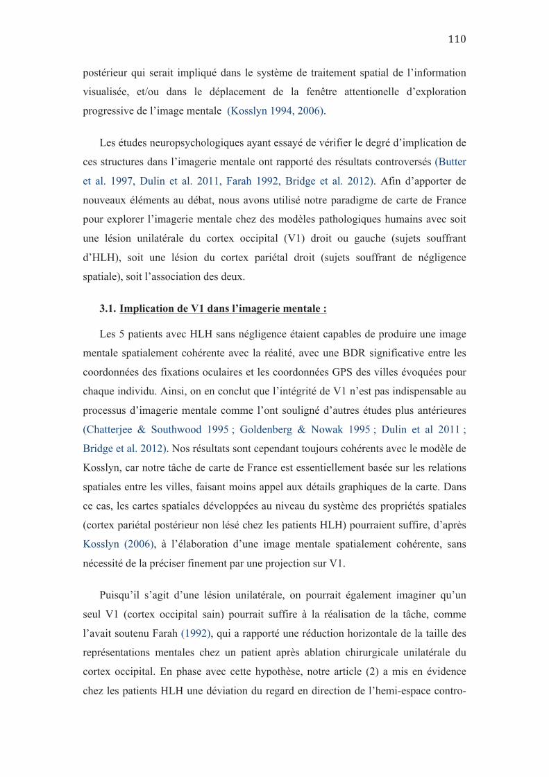
postérieur qui serait impliqué dans le système de traitement spatial de l’information
visualisée, et/ou dans le déplacement de la fenêtre attentionelle d’exploration
progressive de l’image mentale (Kosslyn 1994, 2006).
Les études neuropsychologiques ayant essayé de vérifier le degré d’implication de
ces structures dans l’imagerie mentale ont rapporté des résultats controversés (Butter
et al. 1997, Dulin et al. 2011, Farah 1992, Bridge et al. 2012). Afin d’apporter de
nouveaux éléments au débat, nous avons utilisé notre paradigme de carte de France
pour explorer l’imagerie mentale chez des modèles pathologiques humains avec soit
une lésion unilatérale du cortex occipital (V1) droit ou gauche (sujets souffrant
d’HLH), soit une lésion du cortex pariétal droit (sujets souffrant de négligence
spatiale), soit l’association des deux.
3.1. Implication de V1 dans l’imagerie mentale :
Les 5 patients avec HLH sans négligence étaient capables de produire une image
mentale spatialement cohérente avec la réalité, avec une BDR significative entre les
coordonnées des fixations oculaires et les coordonnées GPS des villes évoquées pour
chaque individu. Ainsi, on en conclut que l’intégrité de V1 n’est pas indispensable au
processus d’imagerie mentale comme l’ont souligné d’autres études plus antérieures
(Chatterjee & Southwood 1995 ; Goldenberg & Nowak 1995 ; Dulin et al 2011 ;
Bridge et al. 2012). Nos résultats sont cependant toujours cohérents avec le modèle de
Kosslyn, car notre tâche de carte de France est essentiellement basée sur les relations
spatiales entre les villes, faisant moins appel aux détails graphiques de la carte. Dans
ce cas, les cartes spatiales développées au niveau du système des propriétés spatiales
(cortex pariétal postérieur non lésé chez les patients HLH) pourraient suffire, d’après
Kosslyn (2006), à l’élaboration d’une image mentale spatialement cohérente, sans
nécessité de la préciser finement par une projection sur V1.
Puisqu’il s’agit d’une lésion unilatérale, on pourrait également imaginer qu’un
seul V1 (cortex occipital sain) pourrait suffire à la réalisation de la tâche, comme
l’avait soutenu Farah (1992), qui a rapporté une réduction horizontale de la taille des
représentations mentales chez un patient après ablation chirurgicale unilatérale du
cortex occipital. En phase avec cette hypothèse, notre article (2) a mis en évidence
chez les patients HLH une déviation du regard en direction de l’hemi-espace contro-

lésionnel (notamment au moment de positionner Paris), qui reste maintenue pendant
toute la durée de la tâche d’imagerie mentale. Nos résultats ont montré que nos
patients HLH semblent éviter spontanément de traverser avec leur regard la ligne
médiane du corps (le droit-devant tête/tronc), qui est un référentiel spatiotopique
égocentré différent de celui (rétinotopique) affecté chez ces patients. Il pourrait s’agir
d’un comportement compensatoire permettant aux HLH de projeter et d’explorer leur
image mentale sur la moitié seulement de l’espace mental de façon à recruter un seul
système pariéto-occipital, codant l’espace en coordonnées spatiotopiques (Galletti and
Battaglini, 1989; Galletti et al., 1993, 1995; Andersen, 1995). Cependant, cette
stratégie compensatoire n’est pas spécifique à la tâche d’imagerie puisqu’elle se
manifeste aussi dans la tâche de fluence verbale n’impliquant aucune représentation
spatiale, suggérant plutôt une généralisation d’un comportement compensatoire
perceptif à toute tâche cognitive, même non dépictive et en l’absence de tout stimulus
visuel.
3.2. Implication des réseaux fronto-pariétaux dans l’imagerie mentale :
Chez les patients négligents, la corrélation n’était significative qu’au niveau de la
moitié droite de la carte mentale ce qui souligne que les réseaux fronto-pariétaux
(sièges de lésions chez ces patients) sont impliqués dans l’élaboration des relations
spatiales allocentrées de l’image mentale. L’absence de corrélation spatiale sur la
moitié gauche de la carte, même avec une bonne série évoquée de villes de l’Ouest,
peut s’expliquer soit par une absence de visualisation mentale de cette moitié (et un
accès sémantique, non imagé, aux villes de l’Ouest), soit par un trouble de
représentation spatiale de cette moitié gauche. En effet, notre article (2) a montré qu’il
n’y a pas chez ces patients d’impossibilité à orienter le regard vers la gauche puisque
leur positionnements oculaires lors de la tâche de fluence verbale couvraient tout
l’espace. Or, lors de la tâche d’imagerie visuelle, les villes de gauche étaient citées
alors que le regard était localisé à droite. Ceci rappelle le phénomène de transposition
à droite ou d’allochirie, souvent rapporté chez les sujets atteints de négligence spatiale
et qui consiste à percevoir les éléments contro-lésionels au niveau de l’espace
ipsilésionnel (Halligan et al. 1992). Par exemple, lors d’une épreuve de dessin de
mémoire, le patient va placer tous les constituants gauches de l’objet à droite
(marguerite avec toutes les pétales condensées à droite, ou l’horloge avec tous les
chiffres placés à droite).
Une des applications importantes en clinique de ce résultat de distorsion spatiale
de la moitié gauche de la carte imaginée, c’est qu’il ne suffit pas que le sujet rapporte
verbalement plusieurs éléments de l’hémi-espace gauche imaginé pour dire qu’il
n’accuse aucun trouble de représentation spatiale de cet espace. En effet, les éléments
de l’espace gauche évoqués peuvent être très mal situés témoignant d’un trouble
représentationnel ou être évoqués sur la base d’un accès sémantique du fait de
l’impossibilité de les représenter mentalement. Cette sous-estimation de l’occurrence
des troubles de représentation spatiale gauche sur la base de l’évocation verbale de
lieux et places, pourrait expliquer certains cas de dissociation, où la négligence
perceptive a été rapportée non associée à une négligence représentationnelle.
4. Apport dans le débat « Imagistes Vs Propositionnels »
Même si les adeptes du courant imagiste, défendant une nature picturale des
images mentales, clament que le débat sur la question est maintenant tranché
notamment avec l’arrivée de nouvelles techniques d’imagerie cérébrale fonctionnelle
pouvant utiliser des logarithmes pour reconnaître les patterns imaginés à travers
l’analyse des signaux cérébraux (MVPC, Naselaris et al. 2015), la clôture du débat est
loin d’être consensuelle.
A travers nos résultats, nous apportons une observation qui soutiendrait la thèse
des imagistes. Chez les patients négligents, nous avons remarqué un comportement
oculaire particulier avec un regard qui balaye l’ensemble de l’espace egocentrique
dans la tâche de fluence verbale n’impliquant pas d’imagerie mentale, alors que ce
regard était préférentiellement situé au niveau de l’hémi-espace droit aussi bien dans
la tâche perceptive que dans la tâche d’imagerie mentale. Or, il a été montré que le
biais ipsilésionnel d’orientation attentionnelle était plus marqué dans les taches
bottom-up (perception) que dans les tâches top-down (recherche visuelle volontaire)
(Bartolomeo & Chokron 2002). Ce biais ipsilesionnel dans la tâche d’imagerie
mentale supposée être purement top-down devrait être moins marqué par rapport à la
tâche perceptive sauf s’il existait un stimulus « virtuel » qui aurait un effet attractif sur
l’attention du patient, et ceci ne pourrait s’expliquer que par une forme dépictive de
l’image mentale au niveau de l’ « écran de projection » occipital. Cette hypothèse
pourrait également expliquer les anomalies des mouvements des yeux vers la gauche
chez les sujets négligents pendant le sommeil paradoxal où se produit une certaine
forme d’images mentales vécues comme dépictives appelées « rêves » (Doricchi et al.
1993).
5. Apport concernant les référentiels spatiaux de l’imagerie visuelle
5.1. Au niveau des sujets sains
Comme souligné au niveau de l’article (2), la distribution des positions absolues
des fixations oculaires de part et d’autre de l’axe sagittal médian du corps (référentiel
tête/tronc), rend compte de la construction de l’image mentale dans le cadre de
référence égocentrique. Mais comme la représentation mentale de la carte change
souvent de caractéristiques spatiales (échelle, orientation…etc.) lorsque le sujet passe
d’une région géographique à une autre, nous avons évalué la projection mentale de la
carte imaginée dans le référentiel égocentrique en mesurant la distance entre les
coordonnées du regard au moment de l’évocation de la première ville (Paris), qui
dispose d’une situation centrale sur l’axe vertical médian de la carte de France.
Chez les sujets sains, il y a une quasi-projection des coordonnées du regard au
moment de l’évocation de (Paris) au niveau de l’axe sagittal médian du corps,
soulignant une génération d’une carte mentale centrée sur l’axe égocentrique.
De plus la corrélation significative en BDR entre les coordonnées du regard et les
coordonnées GPS des villes évoquées témoigne d’une cohérence spatiale de la carte
mentale sur son ensemble. Par conséquent, la carte mentale est représentée
correctement selon le référentiel allocentrique.
5.2. En cas de Négligence spatiale
Chez les sujets atteints de Négligence spatiale, on observe une génération de carte
mentale avec un décalage ipsilésionnel dans le référentiel égocentrique surtout en cas
de HLH associée. Cette observation expliquerait les résultats de Doricchi & Angelelli
(1999), où seuls les patients négligents avec HLH présentaient un trouble de
représentation des distances horizontales avec une sous-estimation des distances
contro-lesionnelles et une surestimation des distances ipsi-lesionnelles, alors que les
patients négligents sans HLH avaient une estimation des distances horizontales
comparable aux sujets ne souffrant pas de négligence. Ainsi, l’influence de l’HLH sur
l’exagération du comportement de négligence au niveau des tâches perceptives, se
reproduit également au niveau des tâches purement top-down.
D’autre part, au niveau du référentiel allocentrique, il existerait une incapacité à se
représenter spatialement l’hémi-espace gauche (puisque la BDR ne corrèle pas sur la
moitié gauche de la carte). Il ne peut pas s’agir seulement d’une transposition sur la
droite de cet espace puisque la BDR accepterait cette distorsion « cohérente » de
l’espace réel. Cette « perte de repère » suite à l’orientation vers la gauche de la carte
de France est à rapprocher de l’impossibilité totale de remise à jour des cartes de
représentation spatiotopique (remapping) observée suite à une saccade dirigée vers la
gauche chez les patients héminégligents (Duhamel et al. 1992, Heide et al. 1995,
revue dans Pisella & Mattingley 2004).
5.3. En cas de HLH
Chez les HLH pures, la projection de la carte mentale se fait avec une translation
contro-lésionnelle par rapport au référentiel egocentrique. Nous soutenons qu’il s’agit
là d’un mécanisme compensatoire développé avec le temps grâce à des processus de
plasticité cérébrale. En effet, cette translation n’est observable que chez les patients
chroniques ayant une lésion ancienne, et pas chez le patient aigu dont la lésion
remonte à moins de 3 mois. Ce comportement est également retrouvé dans les
conditions perceptives où les sujets avec HLH compensent leur déficit en orientant le
regard vers l’hémiespace controlésionnel afin de minimiser l’étendue du champ
aveugle, et de resituer la cible visuelle au centre du champ perçu (Doricchi et al. 2002,
Vuillemier et al. 1999).
Le transfert de ce comportement compensatoire au niveau des tâches d’imagerie
mentale (en dehors de tout stimulus visuel) pourrait avoir un rôle fonctionnel
d’optimisation de la tâche. En effet, la génération d’une carte mentale décalée aurait
pour effet de maintenir la représentation mentale à explorer au niveau d’un seul
hémiespace afin de réduire les transferts inter-hémisphériques avec une utilisation
unilatérale de l’hémisphère sain, aussi bien dans le codage rétinotopique (V1 sain),
que spatiotopique (réseau fronto-pariétal homolatéral). Cette hypothèse est supportée
par le fait que le patient qui n’adopte pas encore cette stratégie et génère une carte
mentale centrée dans le référentiel égocentrique sans déplacer le regard vers le côté
ipsilésionel, dispose de la moins bonne cohérence spatiale de l’image mentale parmi
les HLH (le plus bas coefficient de détermination de BDR). Cependant, la survenue
de ce comportement également dans la tâche de fluence verbale ne faisant appel à
aucune imagerie mentale, met le doute sur son éventuelle utilité fonctionnelle dans
l’optimisation des processus de représentations mentales.
Par ailleurs, la corrélation significative entre les coordonnées du regard et les
coordonnées GPS des villes évoquées au niveau de chacun des patients avec HLH
témoigne d’une bonne cohérence spatiale de la carte mentale au niveau du référentiel
allocentrique.
6. Positionnement par rapport aux théories explicatives des
mouvements oculaires en imagerie mentale :
6.1. La théorie du Scanpath
La théorie du Scanpath prédit que les mouvements des yeux durant le rappel de
mémoire d’une scène, se feront vers les mêmes endroits et suivant le même ordre
séquentiel que durant l’encodage de cette même scène (Brandt & Stark 1997).
La principale critique antérieure à cette théorie, consiste en la survenue de
saccades spatialement cohérentes pendant l’imagerie mentale, même lorsque
l’encodage s’est fait en condition de fixation, soulignant le fait que les mouvements
des yeux lors du rappel imagé ne sont pas une simple réactivation à l’identique (re-
enactment) de ceux produits lors de l’encodage (Johansson et al. 2012). Cependant,
dans notre article 3, nous montrons que même en condition de fixation, il se produit
des micro-saccades qui partagent les caractéristiques spatiotemporelles des saccades
exploratrices et qui sont également spatialement cohérentes avec le contenu de
l’imagerie. Ainsi, les saccades survenant en imagerie mentales pourraient être une
réactivation (mais dans un mode plus ample) de la trace oculomotrice survenue
pendant la fixation en encodage dans l’expérience de Johansson et al. (2012).
Cependant, dans notre paradigme expérimental le rappel de mémoire par imagerie
mentale visuelle n’est pas précédé d’une phase d’encodage qui nous permettrait de
comparer les mouvements des yeux entre encodage et rappel. De plus, l’encodage des
localisations géographiques des villes de France, chez chacun de nos participants s’est
fait à plusieurs reprises dans des conditions variables et avec des stimuli différents et
même des modalités sensorielles différentes (visuelles, auditives…etc.). Ainsi, cette
théorie du scanpath nous semble insuffisante pour rendre part de cette diversité,
d’autant plus qu’elle ne peut pas expliquer également la survenue des mouvements
des yeux spatialement cohérents lorsque le sujet écoute une description verbale d’une
scène (Spivey and Geng 2001, Johansson et al. 2006).
6.2. Le modèle de Kosslyn
Dans le modèle de Kosslyn, les images mentales auraient une forme dépictive au
niveau du tampon visuel, et les mouvements des yeux joueraient un rôle fonctionnel
dans la genèse et l’exploration de ces images (Kosslyn 1994, 2006), en permettant le
déplacement de la fenêtre attentionnelle et en permettant l’agencement des différentes
parties de l’image qui se construit de façon séquentielle sur ce tampon.
Nous avons pu démontrer dans l’article 1, que les mouvements des yeux en
imagerie mentale ont révélé une dynamique séquentielle fragmentée dans la
génération et l’exploration de l’image mentale de la carte de France ce qui confirme
la théorie de Hebb, intégrée au modèle de Kosslyn (Mast & Kosslyn 2002). D’autre
part, les résultats de l’expérience d’imagerie mentale en condition de fixation, exposés
dans notre article 3, soutiendraient un rôle fonctionnel ou du moins, facilitateur des
mouvements des yeux dans le processus d’imagerie mentale. En effet, à l’échelle du
groupe la vivacité de l’imagerie était plus faible en condition de fixation qu’en regard
libre. Néanmoins, l’explication alternative de ces résultats serait que seuls les
déplacements attentionnels seraient indispensables à la réalisation de la tâche
d’imagerie mentale. Ces shifts attentionnels peuvent être soit explicites (« Overt ») et
auquel cas, accompagnés de saccades, ou bien implicites (« Covert ») permettant une
exploration mentale de la carte en condition de fixation oculaire.
6.3. Les théories de simulation mentale
Dans les théories de simulation mentale, l’imagerie mentale est considérée comme
une simulation du processus perceptif (Hesslow 2012, Barsalou 2008). Ainsi le
comportement pendant l’imagerie va être très similaire à celui pendant la perception y
compris les mouvements des yeux qui vont s’activer comme si la personne était en
train de réellement voir ce qu’elle est en train d’imaginer.
Nos expériences chez les sujets sains (article 1) ont révélé un comportement
oculomoteur pendant la tâche d’imagerie qui reproduit de façon assez similaire ce qui
se passerait en cas de perception visuelle, avec une correspondance spatiale au
contenu de l’imagerie et une exploration séquentielle fragmentée. Même en condition
de fixation (article 3), les microsaccades joueraient un rôle facilitateur dans
l’augmentation de la vivacité de l’image mentale ce qui rappelle un peu leur rôle
d’augmentation de la résolution spatiale en perception visuelle. De plus, même dans
les conditions pathologiques (article 2), nous avons mis en évidence pendant
l’imagerie mentale des comportements oculomoteurs très similaires à ceux
développés dans le cadre de stratégies de compensation en perception visuelle,
notamment en cas d’HLH. Chez les négligents, l’exploration de l’hémiespace gauche
imaginé, avec absence de corrélation spatiale, rappelle les troubles exploratoires de
l’espace gauche en perception visuelle.
Ainsi, tous ces résultats sont facilement explicables en se basant sur les théories de
simulation mentale qui restent des théories assez larges avec un grand pouvoir
explicatif mais peu de pouvoir prédictif (Johansson 2013).
6.4. La théorie des indices spatiaux
La théorie des indices spatiaux permet de relier chaque objet mémorisé à des
éléments de l’environnement extérieur de façon à faciliter sa récupération de mémoire
en activant ces traces mnésiques grâce aux mouvements des yeux qui vont aller
revisiter ces éléments extérieurs (Pylyshyn 2002). Ainsi ces indices spatiaux
serviraient de support à une « mémoire externe » qui soulagerait la charge de la
mémoire de travail, notamment dans les tâches visuo-spatiales complexes (O’Regan
& Noë 2001, Richardson et al. 2009).
Selon la théorie des indices spatiaux, les mouvements des yeux en imagerie ne
seraient pas indispensables mais pourrait avoir un rôle facilitateur. En effet, même
dans l’obscurité absolue (et donc en l’absence de tout indice extérieur), Johansson et
al. (2006) avaient retrouvé une correspondance spatiale des mouvements des yeux
avec le contenu de l’imagerie mentale, mais la force de cette correspondance était plus
faible que lorsque l’expérience se réalisait les yeux ouverts à la lumière du jour.
Dans notre paradigme, nous avons veillé à conduire l’expérience dans un
environnement très pauvre en éléments qui accrocheraient le regard, face à un mur
blanc, et avons quand même trouvé des corrélations spatiales très significatives à
l’échelle individuelle. Ces résultats peuvent toujours s’expliquer dans le cadre de cette
théorie dans la mesure où ces indices environnementaux auraient un rôle non pas
indispensable mais simplement potentiellement facilitateur.
7. Applications et Perspectives:
Les résultats et conclusions issues de ce travail de thèse peuvent être exploités
dans différentes applications cliniques, comme elles peuvent être utilisées pour
alimenter de nouvelles pistes de recherches fondamentales autour de l’imagerie
mentale et sa relation avec les mouvements des yeux.
Nous avons montré (article 2), que les mouvements des yeux représentent un
reflet fidèle de l’imagerie mentale visuelle dans son aspect spatial, aussi bien chez les
sujets sains que dans les situations pathologiques. Leur enregistrement constitue dès
lors, un moyen intéressant pour l’évaluation clinique des patients atteints de
négligence spatiale dès le début des symptômes. L’adaptation prismatique représente
une des méthodes rééducatives les plus prometteuses visant à moduler le biais
attentionnel ipsilesionnel chez les négligents afin d’améliorer leurs capacités visuo-
percetives et représentationnelles (Jacquin-Courtois et al. 2013). L’évaluation de
l’efficacité d’une telle méthode repose encore aujourd’hui largement sur les épreuves
papier-crayon avec toutes leurs limites dans l’évaluation des représentations mentales
chez les négligents. Ainsi, un enregistrement des mouvements des yeux lors d’une
tâche standardisée d’imagerie mentale pourrait être d’une grande valeur dans les
bilans d’évaluation.
L’enregistrement des mouvements des yeux pourrait également être appliqué dans
le domaine de l’entrainement mental par le biais de l’imagerie motrice. L’intérêt de
l’entrainement mental des capacités motrices et visuo-spatiales est actuellement admis
pour l’amélioration de la performance en sport (Olsson & Nyberg 2010) ou en
rééducation fonctionnelle avec un réapprentissage moteur (Malouin & Richards,
2010 ; Mateo et al. 2015). Des études récentes ont mis en évidence que des
mouvements oculaires spontanés non orientés vers une cible perceptive (« dans le
vide ») surviennent pendant des tâches d’imagerie motrice. Par conséquent,
l’enregistrement des mouvements des yeux dans ces conditions, pourrait également
renseigner sur les stratégies d’entrainement mental par imagerie motrice, d’autant
plus, que l’évaluation de ces stratégies repose essentiellement aujourd’hui sur le récit
subjectif qu’en fait le sujet (sportif ou patient), en l’absence d’une méthode
d’évaluation objective et valide (Johansson 2013). Il convient de noter que certains
auteurs commencent à s’intéresser au sujet avec déjà la mise en évidence d’une
similarité entre le comportement oculomoteur lors d’un mouvement orienté vers une
cible du bras ou de la main, et pendant la réalisation effective de cette action
(Gueugneau et al. 2008, Heremans et al. 2012).
Enfin, dans leur rapport à la perception d’une part (vision), à la cognition
(imagerie) et à la motricité, les mouvements des yeux représentent un des exemples
les plus illustratifs de l’approche incorporée (embodied) du fonctionnement cérébral,
où la perception, la cognition et l’action constitueraient un seul continuum dynamique
interagissant l’un avec l’autre (Wilson 2002). Il s’agit d’une approche plus écologique
de la cognition humaine rendant compte de notre façon d’interagir intérieurement
(avec notre corps) et extérieurement (avec l’environnement) et tenant compte des
limites neurobiologiques de l’organisme humain (Wilson 2002). Cette approche
incorporée est en train de gagner du terrain dans la compréhension du phénomène
« d’imagerie mentale » en faisant face à l’approche computationnelle plus classique,
avec l’émergence d’un nouveau débat de l’imagerie autour de ces questions (Foglia &
O’Regan 2016).

Ces nouvelles tendances dans le domaine des sciences cognitives visant à
réconcilier différentes approches, profiteraient pleinement de la recherche déjà établie
et à venir sur les processus et mécanismes de l’imagerie mentale à travers
l’enregistrement des mouvements des yeux.
RÉFÉRENCES BIBLIOGRAPHIQUES
Engbert, R. and Kliegl, R. (2004) Microsaccades keep the eyes’ balance during fixation. Psychol. Sci. 15, 431–436
.