Ethylene perception and NEP-like protein production by ...
102
Ethylene perception and NEP-like protein production by Botrytis cinerea Yaite Cuesta Arenas
-
Upload
khangminh22 -
Category
Documents
-
view
5 -
download
0
Transcript of Ethylene perception and NEP-like protein production by ...

Ethylene perception and NEP-like protein production by
Botrytis cinerea
Yaite Cuesta Arenas

Thesis committee Supervisor: Prof. Dr. P.J.G.M. de Wit Professor of Phytopathology Wageningen University Co-supervisor Dr. J.A.L. van Kan Universitair Docent Phytopathology Wageningen University Other members: Prof. Dr. E.J. Woltering, Wageningen University Prof. Dr. B.J.C. Cornelissen, University of Amsterdam Dr. A.W. van Heusden, Wageningen UR
Prof. Dr. R.P. Oliver, Murdoch University, Perth, Australia
This research was conducted under the auspices of the Graduate School of Experimental Plant Sciences.

Ethylene perception and NEP-like protein production by Botrytis cinerea
Yaite Cuesta Arenas
Thesis Submitted in fulfilment of the requirements for the degree of doctor
at Wageningen University by the authority of the Rector Magnificus
Prof. Dr. M. J. Kropff, in the presence of the
Thesis Committee appointed by the Doctorate Board to be defended in public
on Monday 17 May 2010 at 16.00 in the Aula.

Ethylene perception and NEP-like protein production by Botrytis cinerea. Y. Cuesta Arenas Thesis Wageningen University, the Netherlands, 2010 With references, with summaries in Dutch and English ISBN: 978-90-8585-634-4

Table of contents
Chapter 1 General Introduction 7
Chapter 2 Ethylene perception by Botrytis cinerea does not affect pathogenesis
on tomato 21
Chapter 3 Expression and functional analysis of NLP-encoding genes of Botrytis
cinerea 37
Chapter 4 Site-directed mutagenesis of Botrytis cinerea NLPs to identified amio
acids that are essential for necrosis-inducing activity 53
Chapter 5 Dissecting the mode of action phytotoxic proteins of Botrytis cinerea 63
Chapter 6 General discussion 83
Summary 93
Samenvatting 95
Acknowledgements 97
Curriculum vitae 99
Education statement graduate school of Experimental Plant Sciences 101


Chapter 1
General introduction


Chapter 1
9
Chapter 1
General introduction
The necrotrophic fungus Botrytis cinerea, commonly known as grey mould, is one of the
world’s most devastating plant pathogens. It colonizes a wide range of economically
important crops including fruits, vegetables, ornamental plants and cut flowers, and causes
major postharvest losses (Jarvis, 1977). B. cinerea belongs to the class of Deuteromycetes
(imperfect stage), order Moniliales and family Moniliaceae. The fungus predominantly
disperses by massive production of grey brown clusters of macroconidia. The perfect stage,
found quite rarely in nature, is known as Botryotinia fuckeliana (de Bary), belonging to the
Ascomycetes, order Helotiales and the family Sclerotiniaceae (Jarvis, 1977).
The fungus requires high relative humidity on the plant surface for germination, temperatures
between 15°C and 25°C, damp weather for optimal infection, growth, sporulation and spore
release. B. cinerea is also active at low temperatures, and can cause problems on vegetables
stored for weeks or months at temperatures ranging from 0 - 10°C. Infection rarely occurs at
temperatures above 25°C but once the infection occurs, the fungus can grow over a range of
0-35°C (Babadoost, 2000).
The symptoms of Botrytis diseases vary greatly depending on the host and plant part
attacked. General symptoms include a grey to brown discoloration, water soaking, and a fuzzy
whitish grey to tan mould (mycelium and spores) growing on the surface of affected areas.
The host tissue becomes soft and rots. The conidiospores produced are the inoculum for the
next round of infection. One infection cycle may be completed in 3 to 4 days, depending on
the environmental conditions and type of host tissue attacked.
Enzymes and metabolites involved in disease development
Virulence of B. cinerea requires a set of enzymes and (secondary) metabolites that serve as
weapons for attack, as well as mechanisms to overcome the host defence responses. The
pathogen penetrates into the host via wounds, natural openings or even intact surfaces, and
rapidly colonizes dead or senescing tissues. To penetrate intact host surface, the fungus has to
deal with physical barriers including the cuticle and the epidermal cell wall. During
penetration, B. cinerea produces and secretes enzymes like cutinases and lipases (Salinas and
Verhoeff, 1995; van Kan et al., 1997; Comménil et al., 1998; Gindro and Pezet, 1999; Reis et
al., 2005), however, none of these have been shown to be essential in the penetration process.

General introduction
10
Cell wall degrading enzymes such as pectinases and cellulases are suggested to act in the
breakdown of the cell wall, resulting in tissue destruction and the release of nutrients to the
fungus. B. cinerea possesses multiple copies of genes encoding these enzymes. At least three
genes encoding pectin methylesterases are present in the B. cinerea genome. Replacement of
the Bcpme1 and Bcpme2 genes, either separately or in combination, in the strain B05.10,
yielded mutants that were fully virulent on tomato and grapevine leaves. In vitro growth
assays demonstrated that pectin demethylation by these two enzymes is not important for
growth on highly methylated pectin (Kars et al., 2005b). There are six endopolygalacturonase
(Bcpg) genes in the B. cinerea genome (Wubben et al., 1999) and these genes are expressed
differentially during infection in several hosts (ten Have et al., 2001). Five BcPG enzymes
were produced in a heterologous host and shown to possess macerating and necrosis-inducing
activity (Kars et al., 2005). Deletion of Bcpg1 and Bcpg2 resulted in strong reduction in
virulence on different hosts (ten Have et al., 2001; Kars et al., 2005a).
Once B. cinerea has penetrated the plant surface, it kills the underlying epidermal host cells
before the hyphae invade (Clark and Lorbeer, 1976). B. cinerea culture filtrates contain low
molecular weight compounds with phytotoxic activity, named botrydial and botcinic acid
(Calvo et al., 2002; Pinedo et al., 2008). Botrydial has been demonstrated to accumulate in B.
cinerea-infected leaves (Deighton et al., 2001). The Bcbot1 gene encoding a P450
monooxygenase is essential in the biosynthesis of this metabolite. Mutants of three different
B. cinerea strains lacking a functional copy of the Bcbot1 gene did not produce any botrydial
(Siewers et al., 2005). Bcbot1 deletion mutants were tested for their virulence on different
host plants. Bcbot1 deletion mutants in strains ATCC 58025 and SAS56 did not show any
alteration in virulence, because they were still producing botcinic acid. By contrast, mutants
in strain T4, which is not able to produce botcinic acid, displayed a significant reduction in
virulence on bean leaves, tomato leaves and tomato fruits. Therefore, botrydial must be
regarded as a strain-dependent virulence factor (Siewers et al., 2005).
Necrotrophs by definition cause plant cell death, which triggers a spectrum of local and
systemic defense responses in the host plant, some of which include an oxidative burst and the
production of antifungal metabolites and proteins. B. cinerea must be able to evade such plant
defences to successfully colonize the host. The presence of an array of enzymes capable of
metabolizing hydrogen peroxide such as intra- and extracellular peroxidases, superoxide
dismutase (SOD) and catalase (Gil-ad et al., 2000), shows that B. cinerea is equipped to cope
with active oxygen species produced by the host during early stages of infection (Schouten et

Chapter 1
11
al., 2002a). A BcSOD1-deficient mutant showed reduced virulence on French bean and an
enhanced accumulation of extracellular hydrogen peroxide was observed at the plant-
pathogen interface (Rolke et al., 2004). Furthermore, enzymes like laccases have been
reported to be involved in the detoxification of phenolic compounds (Adrian et al., 1998;
Mayer et al., 2001). B cinerea is able to protect itself from the action of resveratrol, a
phytoalexin from grapevine leaves, by its detoxification through laccases (Adrian et al.,
1998). Schouten (2002a) however reported that resveratrol is not toxic to B. cinerea by itself,
but the resveratrol oxidation product (named viniferin) generated by the action of laccase
BcLCC2 is the active compound that causes the toxicity. However, deletion of the laccase
genes Bclcc1 and Bclcc2 in strain B05.10 did not affect the virulence on several hosts
(Schouten et al., 2002b). Recently it became clear that the B cinerea genome contains eleven
laccase-encoding genes (www.broad.mit.edu), suggesting that they may have overlapping
functions (Schouten et al., 2008). Another mechanism by which B. cinerea manages to deal
with growth-inhibiting compounds is by the activity of ATP-binding cassette (ABC)
transporters and Major Facilitator (MFS) proteins that mediate the efflux of a range of
antifungal compounds. Several studies have been performed on the role of transporter proteins
in conferring resistance against industrial fungicides, plant defense compounds and a natural
antibiotic produced by Pseudomonas fluorescens (Vermeulen et al., 2001; Schoonbeek et al.,
2003; Schouten et al., 2008a).
NLPs
The Necrosis and Ethylene-inducing Protein Nep1 was purified from culture filtrates of
Fusarium oxysporum f.sp. erythroxyli (Bailey, 1995) and was the first representative of what
is currently known to be a large family of necrosis-inducing proteins. Nep1-Like Proteins
(NLPs) are present in a spectrum of microorganisms including bacteria, actinomycetes,
oomycetes and fungi but not in higher organisms (Pemberton and Salmond, 2004). NLPs are
present in Gram-negative and Gram-positive bacteria with saprophytic or pathogenic life
styles. In fungi and oomycetes, NLPs are especially present in species interacting with plants,
and predominantly in species that display a hemibiotrophic or necrotrophic life style on plants
(Qutob et al., 2006). NLPs are secreted proteins that share a conserved heptapeptide motif in
the central region of the protein, but they do not contain any previously recognized enzymatic
domain. The presence of two or more conserved cysteine residues, predicted to form disulfide
bridges, has been used to classify the NLPs in two subgroups. Group I comprises proteins that

General introduction
12
harbour only the two first conserved cysteines and group II proteins that contain four
cysteines (Gijzen and Nürnberger, 2006). The molecular weight of NLPs ranges from 24 to 26
kDa and they can occur as monomers as well as dimers (Garcia et al., 2007).
Responses to NLPs are restricted to dicotyledonous plants; all monocotyledonous plants or
other organisms tested so far are insensitive to NLPs (Bailey, 1995; Keates et al., 1998; Staats
et al., 2007; Schouten et al., 2008b). Sensitive plants exposed to NLPs respond by production
of H2O2, nitric oxide, ethylene, accumulation of transcripts encoding pathogenesis-related
proteins, calcium influx, release of phytoalexins, activation of MAP kinases and necrotic
lesion formation (Keates et al., 1998; Fellbrich et al., 2002; Bae et al., 2006). Qutob et al
(2006) demonstrated that NLPs have affinity for lipid bilayers and their phytotoxic activity
and specificity for dicots do not require the presence of a cell wall. The mode of action and
target site of NLPs in plants however are unknown.
The role of host plant processes in B. cinerea pathogenesis
Ethylene is present in trace amounts in nature and it is produced by almost all living
organisms, either biologically or chemically by the incomplete combustion of hydrocarbons.
The activity of ethylene as a plant hormone was discovered in the nineteenth century (Abeles,
1992). The plant physiologist Nelbujov (1879-1926) observed that etiolated pea seedlings
grew horizontally in the laboratory but upright in outside air and he showed that the abnormal
growth habit was caused by contaminating illuminating gas. In 1901, he proved that the active
principle in illuminating gas was ethylene (Bleecker and Kende, 2000). Since then, the study
of ethylene as a plant hormone has been extensive. Ethylene is now known to regulate seed
germination, seedling growth, leaf and floral abscission, ripening in climacteric fruits,
senescence of plant organs, plant growth. Furthermore it acts as a stress hormone during
biotic (pathogen infection) and abiotic (wounding, hypoxia, freezing) stress conditions
(Mattoo and Suttle, 1991).
In higher plants, ethylene biosynthesis occurs through the conversion of methionine via S-
Adenosyl-L-Methionine, into 1-aminocyclopropane-1-carboxylic acid (ACC) and proceeds to
ethylene by oxidation (Fig.1). This process is tightly regulated at the level of ACC synthase
(ACS). Both positive and negative feedback regulation of ethylene biosynthesis have been
reported in different plant species (Kende, 1993; Nakatsuka et al., 1998; Barry et al., 2000).
For example, in tomato, Le-ACS2 and Le-ACS4 are positively regulated, and Le-ACS6 is
negatively regulated by ethylene synthesized during fruit ripening (Nakatsuka et al., 1998).

Chapter 1
13
Once ethylene is synthesized, it is perceived by membrane-localized receptors that are
homologous to the bacterial two-component histidine kinases (Bleecker and Kende, 2000).
Arabidopsis thaliana contains five receptors (ETR1, ETR2, ERS1, ERS2 and EIN4) that act
as negative regulators of ethylene responses and they are inactivated by ethylene binding
(Guo and Ecker, 2004). Transduction of the ethylene signal is achieved through a series of
phosphorylations that are carried out by a cascade of MAP kinases, resulting in the activation
of transcription factors that bind to promoters of ethylene-responsive genes.
An increase in ethylene production by plants occurs as an early response to pathogen
infection (Boller, 1991). Ethylene treatment of plants increases either susceptibility or
resistance, depending on the plant-pathogen interaction and environmental conditions. Elad
(1993) showed that covering pre-inoculated tomato plants with polyethylene bags in order to
accumulate ethylene, promoted B. cinerea infection. On the other hand, pre-treatment of
tomato plants with exogenous ethylene prior to inoculation, resulted in an increased, partial,
resistance to B. cinerea (Díaz et al., 2002). The complex physiological responses to ethylene
in different tissues and during development, as well as the positive and negative feedback
mechanisms make it difficult to generalise. For example, ripening makes tomato fruit more
susceptible to B. cinerea and the ripening is stimulated by ethylene (Cantu et al., 2008).
However, ethylene is not required for the increased susceptibility to B. cinerea during fruit
ripening (Cantu et al., 2009). Thus, ethylene stimulates B. cinerea disease in an indirect way.
Cell death in plant-pathogen interactions
Plant cell death can occur during the interaction with pathogens that attempt to colonize the
host (Greenberg, 1997). Cell death processes may occur by at least two distinct pathways: one
that resembles apoptosis that is observed in programmed cell death (PCD) in animal systems
and one that resembles necrotic cell death.
Apoptosis is an active process that includes the fragmentation of DNA into so-called DNA
ladders, membrane blebbing, cell shrinkage, chromatin condensation, nuclear fragmentation
with nucleic acids found in membrane-bound vessels (apoptotic bodies) and cytoplasmic
condensation. Some of the hallmarks for apoptotic cell death have been observed in plant-
pathogen interactions during the occurrence of the hypersensitive response (Heath, 1998). The
PCD-eliciting mycotoxins AAL toxin or fumonisin B1 (FB1) (Gilchrist et al., 1995; Gilchrist,
1997; 1998) have been used as models for the study of plant cell death in pathogen response
pathways. AAL toxin is secreted by the tomato pathogen Alternaria alternata f. sp.

General introduction
14
lycopersici, while the structurally related toxin FB1 is secreted by the maize pathogen
Fusarium moniliforme. These toxins induce apoptosis-like cell death in tomato. DNA ladders
were observed during cell death in toxin-treated tomato protoplasts and leaflets.
Necrosis in plants is caused by extrinsic factors, such as mechanical damage or phytotoxic
accumulation of specific molecules after a stress event. Thus, plant cell death through necrosis
is considered a “passive” process. Swelling is the defining feature of the morphological
changes during necrosis. Swelling is due to the cell losing the ability to osmoregulate,
resulting in water and ions flooding into the cell and culminating in cellular lysis (Lennon et
al., 1991). Generation of ROS, ATP depletion and mitochondrial and chloroplast dysfunction
are also characteristic of necrosis. Several fungal toxins are known to cause necrosis in plant.
For example two naphthazarin phytotoxins (dihydrofusarubin and isomarticin) produced by
Fusarium solani caused cell necrosis in veins, plasmolysis or collapse of spongy mesophyll
cells, collapse of phloem, depletion of starch, swelling of chloroplasts and disruption of
cellular organization in leaves of rough lemon seedlings (Achor et al., 1993).
The most widely studied cell death response in plants is the hypersensitive response (HR),
which occurs when a biotrophic pathogen produces an effector that is recognized by a host
plant with a corresponding resistance gene, resulting in Effector-Triggered Immunity (ETI).
HR resembles PCD in animals and results in the killing of host cells at the site of attempted
infection. Cells are sacrificed at the point of pathogen entry to avoid further spreading of
(hemi)-biotrophic microbes; however this strategy is not effective against necrotrophic
microbes since cell death facilitates the infection by necrotrophs (Govrin and Levine, 2000).
Many studies have been performed on the role of plant genes in HR and disease symptom
development. Such studies were supported by the finding of lesion mimic mutants, such as
Arabidopsis mutant acd5, that spontaneously forms disease-like lesions (Greenberg et al.,
2000; Pilloff et al., 2002), and shows more severe disease symptoms and allows increased
growth of P. syringae. The Acd5 gene encodes a ceramide kinase (CERK) that is induced
during P. syringae infection (Liang et al., 2003). Ceramides are bioactive lipids that activate
apoptosis in animals (Hannun and Obeid, 2002), while its phosphorylated derivative (the
product of the CERK reaction) can partially block PCD in Arabidopsis protoplasts (Greenberg
and Yao, 2004). Ceramides are part of the sphingolipid family of bioactive lipids (Hannun
and Obeid, 2002). Some fungal pathogens secrete cerebrosides, which are derived from
sphingolipids that induce HR-like cell death in rice (Koga et al., 1999). Additionally, a
number of fungal pathogens secrete related mycotoxins that cause PCD and disrupt

Chapter 1
15
sphingolipid metabolism (Abbas et al., 1994). Transient expression of cpr22 (constitutive
expresser of PR 22) induces cell death in Nicotiana benthamiana leaves (Yoshioka et al.,
2006). This cell death resembles the HR seen in an incompatible plant-pathogen interaction. A
microscopic analysis of cpr22-induced cell death revealed strong similarities to pathogen-
induced as well as developmental PCD (Fukuda, 2000; Lam, 2004) including retraction of the
plasma membrane from the cell wall, vesicle formation and degradation of the tonoplast.
Anti-apoptotic genes have been identified in viruses, animals and humans. The transgenic
expression of several of these genes (Op-IAP, CED-9, Bcl-2, Bcl-xL,) in tobacco plants
conferred heritable disease resistance to necrotrophic fungi, including B. cinerea and
Sclerotinia sclerotiorum (Dickman et al., 2001).
Ethylene production and perception by microorganisms
Microorganisms can synthesize ethylene by one of three possible pathways. A few fungi
produce ethylene from methionine via ACC, alike the pathway described for plants (Figure
1A) (Sharon et al., 2004). In the second pathway, α-keto-γ-(methylthio)butyric acid (KMBA),
resulting from deamination of L-methionine, is converted to ethylene by enzymatic
conversion (Figure 1B) or by spontaneous oxidation of KMBA in the presence of light (Yang,
1969). In the third pathway, 2-oxoglutarate is converted to ethylene by the ethylene-forming-
enzyme (Figure 1C) (Fukuda et al., 1986). The second and third pathways occur much more
frequently in microorganisms than the first one.
Fig. 1. Biosynthetic pathways in microorganisms for synthesis of ethylene using either methionine (A), KMBA (B) or 2-oxoglutarate (C) as precursors.
ACCsynthaseS C C C C
NH3+
O
O-
H3C S C C C C
NH3+
O
O-
H3C
CH2
O
HO
HO
Ade
+ CC
C C
NH3+
O
O-
CO2 HCN+ +ATP ADP
Methionine
SAM
ACC
C
C
ACCoxidase
Ethylene
C C C C
NH3+ O
O-C
HO
OC C C C
O O
OHC
HO
O
Glutamic acid2-oxoglutarate
amino-transferase
Methionine KMBA
S C C C C
O O
O-H3CS C C C C
NH3+ O
O-H3C
2CO2
+1/2 CH3SSH3C
hv
Ethylene
C
C
Dimethyl disulfide
CO2 H O+ +C
C
EFE
Fe2+
Ethylene
2C C C C
O O
OHC
HO
O
2-oxoglutarate
3O2+
A
B
C
ACCsynthaseS C C C C
NH3+
O
O-
H3C S C C C C
NH3+
O
O-
H3C
CH2
O
HO
HO
Ade
+ CC
C C
NH3+
O
O-
CO2 HCN+ +ATP ADP
Methionine
SAM
ACC
C
C
ACCoxidase
Ethylene
C C C C
NH3+ O
O-C
HO
OC C C C
O O
OHC
HO
O
Glutamic acid2-oxoglutarate
amino-transferase
Methionine KMBA
S C C C C
O O
O-H3CS C C C C
NH3+ O
O-H3C
2CO2
+1/2 CH3SSH3C
hv
Ethylene
C
C
Dimethyl disulfide
CO2 H O+ +C
C
EFE
Fe2+
Ethylene
2C C C C
O O
OHC
HO
O
2-oxoglutarate
3O2O2+
A
B
C

General introduction
16
Microorganisms cannot only produce ethylene, they can also respond to it. It has been
reported for several fungi that ethylene may stimulate conidial germination, germ tube
elongation and appressorium formation (Kepczynska, 1994; Sharon et al., 2004). The
phenomenon of ethylene perception by microorganisms has been study mainly in fungi but
there is no description of the molecular mechanisms involved in the perception.
Outline of the thesis
The aim of the research presented in this thesis was to obtain insight in the roles that ethylene
production and perception, both by the pathogen and the plant, plays in the interaction
between B. cinerea and crop plants, using tomato as a model. Furthermore, functional analysis
was performed of two B. cinerea necrosis-inducing NLPs, called BcNEP1 and BcNEP2, with
emphasis on their role in virulence and their mode of action.
Chapter 2 describes studies on the effects of ethylene on B. cinerea either in vitro or during
pathogenesis. Lesion development on tomato genotypes with a reduced or an enhanced
ethylene production level was monitored. Furthermore, reports that the transcript levels of
certain B. cinerea genes are induced by exposure to ethylene (Chagué et al., 2006), led us to
characterize the role of the fungal histidine kinase BcHHK5, which structurally resembles
plant ethylene receptors.
Chapter 3 describes the functional analysis of the two NLPs from B. cinerea. Expression of
Bcnep genes and production of the corresponding proteins during infection was investigated.
Single knock out mutants were made for Bcnep1 and Bcnep2 and their role in virulence was
tested. Additionally, experiments were conducted to study whether ethylene induced in plants
during B. cinerea infection results from a response to BcNEP proteins.
Chapter 4 investigates whether the conserved heptapeptide motif, the disulfide bonds, as well
as posttranslational modification motifs in the BcNEP proteins influence their necrosis-
inducing activity. Transient agro-infiltration assays were performed in Nicotiana benthamiana
and N. tabacum to express site-directed mutant proteins.
Chapter 5 describes a study on the role of the plant in the response to BcNEP proteins. The
inhibition of necrosis-inducing activity by polyclonal antibodies raised against BcNEP
proteins was analysed. Genetic tools and pharmacological inhibitors were used to elucidate
plant pathways required for the necrosis-inducing activity of BcNEP proteins.
Chapter 6 provides a general discussion of the thesis.

Chapter 1
17
REFERENCE LIST
Abbas, H. K., Tanaka, T., Duke, S. O., Porter, J. K., Wray, E. M., Hodges, L., Sessions, A. E., Wang, E., Merrill Jnr, A. H. and Riley, R. T. (1994). "Fumonisin- and AAL-toxin-induced disruption of sphingolipid metabolism with accumulation of free sphingoid bases." Plant Physiology 106(3): 1085-1093.
Abeles, F. B. (1992). Ethylene in Plant Biology. New York, New York Academic Press. Achor, D. S., Nemec, S. and Baker, R. A. (1993). "Effects of Fusarium solani naphthazarin toxins on the
cytology and ultrastructure of rough lemon seedlings." Mycopathologia 123(2): 117-126. Adrian, M., Rajaei, H., Jeandet, P., Veneau, J. and Bessis, R. (1998). "Resveratrol oxidation in Botrytis cinerea
conidia." Phytopathology 88(5): 472-4776. Babadoost, M. (2000). Gray mold rot or Botrytis blight of vegetables Plant disease., Department of Crop
Sciences, University of Illinois: 5. Bae, H., Kim, M. S., Sicher, R. C., Bae, H. J. and Bailey, B. A. (2006). "Necrosis- and ethylene-inducing peptide
from Fusarium oxysporum induces a complex cascade of transcripts associated with signal transduction and cell death in Arabidopsis." Plant Physiology 141(3): 1056-1067.
Bailey, B. A. (1995). "Purification of a protein from culture filtrates of Fusarium oxysporum that induces ethylene and necrosis in leaves of Erythroxylum coca." Phytopathology 85(10): 1250-1255.
Barry, C. S., Llop-Tous, M. I. and Grierson, D. (2000). "The regulation of 1-aminocyclopropane-1-carboxylic acid synthase gene expression during the transition from system-1 to system-2 ethylene synthesis in tomato." Plant Physiology 123(3): 979-986.
Bleecker, A. B. and Kende, H. (2000). "Ethylene: A gaseous signal molecule in plant." Annual Review of Cell and Developmental Biology 16: 1-18.
Boller, T. (1991). Ethylene in pathogenesis and disease resistance. The plant hormone ethylene. Mattoo, A. K. and Suttle, J. C. Boca Raton, Florida, CRC Press: 293-314.
Calvo, A. M., Wilson, R. A., Bok, J. W. and Keller, N. P. (2002). "Relationship between secondary metabolism and fungal development." Microbiology and Molecular Biology Reviews 66(3): 447-459.
Cantu, D., Vicente, A. R., Greve, L. C., Dewey, F. M., Bennett, A. B., Labavitch, J. M. and Powell, A. L. T. (2008). "The intersection between cell wall disassembly, ripening, and fruit susceptibility to Botrytis cinerea." Proceedings National Academy of Sciences United States of America 105(3): 859-864.
Cantu, D., Blanco-Ulate, B., Yang, L., Labavitch, J. M., Bennett, A. B. and Powell, A. L. T. (2009). "Ripening-regulated susceptibility of tomato fruit to Botrytis cinerea requires NOR but not RIN or Ethylene." Plant Physiology 150(3): 1434-1449.
Chagué, V., Danit, L. V., Siewers, V., Schulze-Gronover, C., Tudzynski, P., Tudzynski, B. and Sharon, A. (2006). "Ethylene sensing and gene activation in Botrytis cinerea: a missing link in ethylene regulation of fungus-plant interactions?" Molecular Plant-Microbe Interaction 19: 33-42.
Clark, C. A. and Lorbeer, J. W. (1976). "Comparative histopathology of Botrytis squamosa and B. cinerea on onion leaves." Phytopathology 66: 1279-1289.
Comménil, P., Belingheri, L. and Dehorter, B. (1998). "Antilipase antibodies prevent infection of tomato leaves by Botrytis cinerea." Physiological and Molecular Plant Pathology 52(1): 1-14.
Deighton, N., Muckenschnabel, I., Colmenares, A. J., Collado, I. G. and Williamson, B. (2001). "Botrydial is produced in plant tissues infected by Botrytis cinerea." Phytochemistry 57(5): 689-692.
Díaz, J., ten Have, A. and van Kan, J. A. L. (2002). "The role of ethylene and wound signaling in resistance of tomato to Botrytis cinerea." Plant Physiology 129(3): 1341-1351.
Elad, Y. (1993). "Regulators of ethylene biosynthesis or activity as a tool for reducing susceptibility of host plant tissues to infection by Botrytis cinerea." Netherlands Journal of Plant Pathology 99(3): 105-113.
Fellbrich, G., Romanski, A., Varet, A., Blume, B., Brunner, F., Engelhardt, S., Felix, G., Kemmerling, B., Krzymowska, M. and Nürnberger, T. (2002). "NPP1, a Phytophthora-associated trigger of plant defense in parsley and Arabidopsis." Plant Journal 32(3): 375-390.
Fukuda, H. (2000). "Programmed cell death of tracheary elements as a paradigm in plants." Plant Molecular Biology 44(3): 245-253.
Fukuda, H., Fujii, T. and Ogawa, T. (1986). "Preparation of a cell-free ethylene forming system from Penicillium digitatum." Agricultural Biology and Chemistry. 50: 977-981.
Garcia, O., Macedo, J. A. N., Tibúrcio, R., Zaparoli, G., Rincones, J., Bittencourt, L. M. C., Ceita, G. O., Micheli, F., Gesteira, A., Mariano, A. C., Schiavinato, M. A., Medrano, F. J., Meinhardt, L. W., Pereira, G. A. G. and Cascardo, J. C. M. (2007). "Characterization of necrosis and ethylene-inducing proteins (NEP) in the basidiomycete Moniliophthora perniciosa, the causal agent of witches' broom in Theobroma cacao." Mycological Research 111(4): 443-455.
Gijzen, M. and Nürnberger, T. (2006). "Nep1-like proteins from plant pathogens: recruitment and diversification of the NPP1 domain across taxa." Phytochemistry 67(16): 1800-1807.

General introduction
18
Gil-ad, N. L., Bar-Nun, N., Noy, T. and Mayer, A. M. (2000). "Enzymes of Botrytis cinerea capable of breaking down hydrogen peroxide." FEMS Microbiology Letters 190(1): 121-126.
Gilchrist, D. G. (1997). "Mycotoxins reveal connections between plants and animals in apoptosis and ceramide signaling." Cell Death and Differentiation 4(8): 689-698.
Gilchrist, D. G. (1998). "Programmed cell death in plant disease: The purpose and promise of cellular suicide." Annual Review of Phytopathology 36: 393-414.
Gilchrist, A., Wang, H. and Bostock, R. M. (1995). "Sphingosine-related mycotoxins in plant and animal diseases." Canadian Journal of Botany 73: 459-467Gindro, K. and Pezet, R. (1999). "Purification and characterization of a 40.8-kDa cutinase in ungerminated conidia of Botrytis cinerea Pers.: Fr." FEMS Microbiology Letters 171(2): 239-243.
Govrin, E. M. and Levine, A. (2000). "The hypersensitive response facilitates plant infection by the necrotrophic pathogen Botrytis cinerea." Current Biology 10(13): 751-757.
Greenberg, J. T. (1997). "Programmed cell death in plant-pathogen interactions." Annual Review of Plant Biology 48: 525-545.
Greenberg, J. T. and Yao, N. (2004). "The role of regulation of programmed cell death in plant-pathogen interactions." Cellular Microbiology 6(3): 201-211.
Greenberg, J. T., Silverman, F. P. and Liang, H. (2000). "Uncoupling salicylic acid-dependent cell death and defense-related responses from disease resistance in the arabidopsis mutant acd5." Genetics 156: 341-350.
Guo, H. and Ecker, J. R. (2004). "The ethylene signaling pathway: new insights." Current Opinion in Plant Biology 7(1): 40-49.
Hannun, Y. A. and Obeid, L. M. (2002). "The ceramide-centric universe of lipid-mediated cell regulation: Stress encounters of the lipid kind." Journal of Biological Chemistry 277(29): 25847-25850.
Heath, M. C. (1998). "Apoptosis, programmed cell death and the hypersensitive response " European Journal of Plant Pathology 104(2): 117-124.
Jarvis, W. R. (1977). Botryotinia and Botrytis species: taxonomy, physiology, and pathogenicity. . Otawa, Research Branch Canada Department of Agriculture.
Kars, I., Krooshof, G. H., Wagemakers, L., Joosten, R., Benen, J. A. E. and van Kan, J. A. L. (2005a). "Necrotizing activity of five Botrytis cinerea endopolygalacturonases produced in Pichia pastoris." Plant Journal 43(2): 213-225.
Kars, I., McCalman, M., Wagemakers, L. and van Kan, J. A. L. (2005b). "Functional analysis of Botrytis cinerea pectin methylesterase genes by PCR-based targeted mutagenesis: Bcpme1 and Bcpme2 are dispensable for virulence of strain B05.10." Molecular Plant Pathology 6(6): 641-652.
Keates, S. E., Loewus, F. A., Helms, G. L. and Zink, D. L. (1998). "5-O-(α-D-galactopyranosyl)-D-glycero-pent-2-enono-1,4-lactone: Characterization in the oxalate-producing fungus, Sclerotinia sclerotiorum." Phytochemistry 49(8): 2397-2401.
Kende, H. (1993). "Ethylene biosynthesis." Annual Review of Plant Physiology and Plant Molecular Biology 44(1): 283-307.
Kepczynska, E. (1994). "Involvement of ethylene in spore germination and mycelial growth of Alternaria alternata." Mycological Research 98(1): 118-120.
Koga, J., Yamauchi, T., Shimura, M., Ogawa, N., Oshima, K., Umemura, K., Kikuchi, M. and Ogasawara, N. (1999). "Cerebrosides A and C, sphingolipid elicitors of hypersensitive cell death and phytoalexin accumulation in rice plants." Journal of Biological Chemistry 273(48): 31985-31991.
Lam, E. (2004). "Controlled cell death, plant survival and development." Nature Reviews Molecular Cell Biology 5(4): 305-315.
Lennon, S. V., Martin, S. J. and Cotter, T. G. (1991). "Dose-dependent induction of apoptosis in human tumour cell lines by widely diverging stimuli." Cell Proliferation 24(2): 203-214.
Liang, H., Yao, N., Song, J. T., Luo, S., Lu, H. and Greenberg, J. T. (2003). "Ceramides modulate programmed cell death in plants." Genes and Development 17(21): 2636-2641.
Mattoo, A. K. and Suttle, J. C. (1991). The Plant Hormone Ethylene. Boca Raton, Florida, CRC Press. Mayer, A. M., Staples, R. C. and Gil-ad, N. L. (2001). "Mechanisms of survival of necrotrophic fungal plant
pathogens in hosts expressing the hypersensitive response." Phytochemistry 58(1): 33-41. Nakatsuka, A., Murachi, S., Okunishi, H., Shiomi, S., Nakano, R., Kubo, Y. and Inaba, A. (1998). "Differential
expression and internal feedback regulation of 1-aminocyclopropane-1-carboxylate synthase, l-aminocyclopropane-1-carboxylate oxidase, and ethylene receptor genes in tomato fruit during development and ripening." Plant Physiology 118(4): 1295-1305.
Pemberton, C. L. and Salmond, G. P. C. (2004). "The Nep1-like proteins - A growing family of microbial elicitors of plant necrosis." Molecular Plant Pathology 5(4): 353-359.
Pilloff, R. K., Devadas, S. K., Enyedi, A. and Raina, R. (2002). "The Arabidopsis gain-of-function mutant dll1 spontaneously develops lesions mimicking cell death associated with disease." Plant Journal 30: 61-70.

Chapter 1
19
Pinedo, C., Wang, C. M., Pradier, J. M., Dalmais, B., Choquer, M., Le Pêcheur, P., Morgant, G., Collado, I. G., Cane, D. E. and Viaud, M. (2008). "Sesquiterpene synthase from the botrydial biosynthetic gene cluster of the phytopathogen Botrytis cinerea." ACS Chemical Biology 3(12): 791-801.
Qutob, D., Kemmerling, B., Brunner, F., Kufner, I., Engelhardt, S., Gust, A. A., Luberacki, B., Seitz, H. U., Stahl, D., Rauhut, T., Glawischnig, E., Schween, G., Lacombe, B., Watanabe, N., Lam, E., Schlichting, R., Scheel, D., Nau, K., Dodt, G., Hubert, D., Gijzen, M. and Nürnberger, T. (2006). "Phytotoxicity and innate immune responses induced by Nep1-like proteins." Plant Cell 18(12): 3721-3744.
Reis, H., Pfiff, S. and Hahn, M. (2005). "Molecular and functional characterization of a secreted lipase from Botrytis cinerea." Molecular Plant Pathology 6(3): 257-267.
Rolke, Y., Liu, S., Quidde, T., Williamson, B., Schouten, A., Weltring, K. M., Siewers, V., Tenberge, K. B., Tudzynski, B. and Tudzynski, P. (2004). "Functional analysis of H2O2-generating systems in Botrytis cinerea: The major Cu-Zn-superoxide dismutase (BCSOD1) contributes to virulence on French bean, whereas a glucose oxidase (BCGOD1) is dispensable." Molecular Plant Pathology 5(1): 17-27.
Salinas, J. and Verhoeff, K. (1995). "Microscopical studies of the infection of gerbera flowers by Botrytis cinerea." European Journal of Plant Pathology 101(4): 377-386.
Schoonbeek, H. J., van Nistelrooy, J. G. M. and de Waard, M. A. (2003). "Functional analysis of ABC transporter genes from Botrytis cinerea identifies BcatrB as a transporter of eugenol." European Journal of Plant Pathology 109(9): 1003-1011.
Schouten, A., Tenberge, K. B., Vermeer, J., Stewart, J., Wagemakers, L., Williamson, B. and van Kan, J. A. L. (2002a). "Functional analysis of an extracellular catalase of Botrytis cinerea." Molecular Plant Pathology 3(4): 227-238.
Schouten, A., Wagemakers, L., Stefanato, F. L., van der Kaaij, R. M. and van Kan, J. A. L. (2002b). "Resveratrol acts as a natural profungicide and induces self-intoxication by a specific laccase." Molecular Microbiology 43(4): 883-894.
Schouten, A., Maksimova, O., Cuesta-Arenas, Y., van den Berg, G. and Raaijmakers, J. M. (2008a). "Involvement of the ABC transporter BcAtrB and the laccase BcLCC2 in defence of Botrytis cinerea against the broad-spectrum antibiotic 2,4-diacetylphloroglucinol." Environmental Microbiology 10(5): 1145-1157.
Schouten, A., van Baarlen, P. and van Kan, J. A. L. (2008b). "Phytotoxic Nep1-like proteins from the necrotrophic fungus Botrytis cinerea associate with membranes and the nucleus of plant cells." New Phytologist 177(2): 493-505.
Sharon, A., Elad, Y., Barakat, R. and Tudzynski, P. (2004). Phytohormones and Botrytis. Botrytis: Biology, Pathology and Control. Elad, Y., Williamson, B., Tudzynski, P. and Delen, N. Dordrecht, the Netherlands, Kluwer Academic Publishers: 163-179.
Siewers, V., Viaud, M., Jimenez-Teja, D., Collado, I. G., Gronover, C. S., Pradier, J. M., Tudzynski, B. and Tudzynski, P. (2005). "Functional analysis of the cytochrome P450 monooxygenase gene bcbot1 of Botrytis cinerea indicates that botrydial is a strain-specific virulence factor." Molecular Plant-Microbe Interactions 18(6): 602-612.
Staats, M., van Baarlen, P., Schouten, A., van Kan, J. A. L. and Bakker, F. T. (2007). "Positive selection in phytotoxic protein-encoding genes of Botrytis species." Fungal Genetics and Biology 44(1): 52-63.
Stergiopoulos, I. and de Waard, M. A. (2002). "Activity of azole fungicides and ABC transporter modulators on Mycosphaerella graminicola." Journal of Phytopathology 150(6): 313-320.
ten Have, A., Breuil, W. O., Wubben, J. P., Visser, J. and van Kan, J. A. L. (2001). "Botrytis cinerea endopolygalacturonase genes are differentially expressed in various plant tissues." Fungal Genetics and Biology 33(2): 97-105.
van Kan, J. A. L., van't Klooster, J. W., Wagemakers, C. A. M., Dees, D. C. T. and van der Vlugt-Bergmans, C. J. B. (1997). "Cutinase A of Botrytis cinerea is expressed, but not essential, during penetration of gerbera and tomato." Molecular Plant-Microbe Interactions 10(1): 30-38.
Vermeulen, T., Schoonbeek, H. and de Waard, M. A. (2001). "The ABC transporter BcatrB from Botrytis cinerea is a determinant of the activity of the phenylpyrrole fungicide fludioxonil." Pest Management Science 57(5): 393-402.
Wubben, J. P., Mulder, W., ten Have, A., van Kan, J. A. and Visser, J. (1999). "Cloning and partial characterization of endopolygalacturonase genes from Botrytis cinerea." Applied and Environmental Microbiology 65(4): 1596-1602.
Yang, S. F. (1969). "Further studies on ethylene formation from alpha-keto-gamma-methylthiobutyric acid or beta-methylthiopropionaldehyde by peroxidase in the presence of sulfite and oxygen." Journal of Biological Chemistry 244(16): 4360-4365.
Yoshioka, K., Moeder, W., Kang, H. G., Kachroo, P., Masmoudi, K., Berkowitz, G. and Klessig, D. F. (2006). "The chimeric Arabidopsis CYCLIC NUCLEOTIDE-GATED ION CHANNEL11/12 activates multiple pathogen resistance responses." Plant Cell 18(3): 747-763.


Chapter 2
Ethylene perception by Botrytis cinerea does not affect pathogenesis on tomato


Ethylene perception by Botrytis cinerea
23
Chapter 2
Ethylene perception by Botrytis cinerea does not affect pathogenesis on tomato
Yaite Cuesta, Ester Dekkers and Jan van Kan
SUMMARY
Ethylene regulates several developmental processes in plants and plays an important role in
plant-pathogen interactions. Chagué et al. (2006) reported that exposure of Botrytis cinerea to
ethylene in vitro leads to reduction of growth and the induction of expression of two genes,
one of which encodes a phytotoxic protein. These results led to the hypothesis that B. cinerea
for its infection takes advantage of ethylene released by the plant. We did not observe the
previously reported growth reduction upon exposure to ethylene. We investigated possible
effects of ethylene on B. cinerea during pathogenesis by monitoring lesion development on
tomato genotypes with either a reduced or an enhanced ethylene production level. The lesion
sizes on mutant genotypes and their respective wild type progenitors did not differ.
The genome of B. cinerea contains a gene encoding a histidine kinase receptor, BcHHK5,
which structurally resembles ETR1, the best characterized ethylene receptor in plants. Gene
replacement mutants were made to study the function of the Bchhk5 gene. The mutants were
neither affected in growth in vitro nor in virulence. Furthermore, the expression of the two
genes reported by Chagué et al. (2006) to be ethylene-responsive was not influenced by
exposure to ethylene in the B. cinerea wild type strain nor in a ΔBchhk5 mutant. Altogether
we obtained no evidence that B. cinerea indeed senses ethylene, nor that the Bchhk5 gene
product acts as an ethylene receptor.

Chapter 2
24
INTRODUCTION
Ethylene perception has been studied in detail in plants. In Arabidopsis thaliana, ethylene
sensing and signaling occurs in the endoplasmic reticulum through a family of five receptors
(ETR1, ETR2, ERS1, ERS2 and EIN4) that share similarity with bacterial two component
phosphorelay signaling systems (Chang and Stadler, 2001). The receptors function as
negative regulators of ethylene responses and are inactivated by ethylene binding (Guo and
Ecker, 2004). ETR1 is the best characterized ethylene receptor; mutations in the A. thaliana
etr1 gene result in an ethylene-insensitive phenotype, indicating that ETR1 must be a
dominant receptor (Bleecker et al., 1988). Other proteins downstream in the signaling cascade
can integrate signals from other plant hormones, like jasmonic acid, to modulate the response
to biotic and abiotic stress. Hundreds of A. thaliana genes are induced or repressed by
ethylene (Alonso et al., 2003).
A well known effect of ethylene on plant growth is the triple response of etiolated
dicotyledonous seedlings. This response is characterized by the inhibition of hypocotyl and
root cell elongation, radial swelling and exaggerated curvature of the apical hook (Ecker,
2004). This morphological response allowed rapid screening of mutant populations based on
ethylene response defects. Multiple mutations affecting ethylene perception have been
identified. In A. thaliana, a series of mutants (etr1, ein1, ein2, ein3, ctr1) has been obtained
that are characterized by an altered ethylene response, sometimes leading to changes in
developmental processes (Bleecker et al., 1988; Gúzman and Ecker, 1990; Kieber et al.,
1993). In tomato, mutants affected in ethylene responses have been identified but their
phenotypes only become evident during fruit ripening and flower abscission. The natural
tomato mutant Never Ripe (Nr) is insensitive to ethylene (Lanahan et al., 1994) and
presumably lacks the feedback mechanism that normally abolishes ethylene production once
high concentrations are reached. As a consequence, the Nr mutant overproduces ethylene. The
tomato Nr mutant does not display a triple response and shows only little differences in
morphology and development, other than a late flower abscission and an incomplete fruit
ripening (Lanahan et al., 1994). The severity of the fruit ripening defect caused by the Nr
mutation in tomato varies between genetic backgrounds (J. Díaz & J. van Kan, unpublished).
In order to block or reduce ethylene production, different approaches have been reported. The
expression in a transgenic plant of an antisense construct of the tomato ACC oxidase gene
reduced ethylene production and extended the shelf life of tomato fruit after harvest
(Hamilton et al., 1990). Tomato plants expressing a bacterial ACC deaminase, which

Ethylene perception by Botrytis cinerea
25
degrades the ethylene precursor ACC, showed a reduced ethylene biosynthesis throughout the
vegetative parts of the plant and the fruit (Klee et al., 1991).
In fungi, there are few reports of ethylene perception. Ethylene produced by ripening
climacteric fruit induces spore germination and appressorium formation in Colletotrichum
gloeosporioides and C. musae, but not in other Colletotrichum species that normally infect
non-climacteric fruit (Flaishman and Kolattukudy, 1994). In Aspergillus nidulans, aflatoxin
production and ascus development are inhibited by ethylene in a dose-dependent manner
(Roze et al., 2004). Chagué et al. (2006) have previously reported that the radial growth of
Botrytis cinerea in vitro is affected by ethylene and the transcript levels of two genes, Bcspl1
and Bchsp30, are induced at 24 h after exogenous ethylene application. The Bcspl1 gene
(Kunz et al., 2006) is homologous to a gene from Phaeosphaeria nodorum that encodes a
phytotoxic protein produced during infection on wheat (Hall et al., 1999). The Bchsp30 gene
encodes a heat shock protein (Chagué et al., 2006), which in S. cerevisiae is induced by heat
shock, ethanol and weak acid (Panaretou and Piper, 1992; Piper et al., 1997).
B. cinerea infection induces the expression in the host plant of ethylene biosynthetic genes,
ACC synthase and ACC oxidase (Benito et al., 1998; Diaz et al., 2002). A correlation was
reported between the level of ethylene produced by flower petals and leaves from different
rose cultivars and the severity of grey mould symptoms (Elad and Volpin, 1988). In
strawberry, B. cinerea establishes a primary infection that stays quiescent until the initiation
of fruit ripening, which coincides with a burst of ethylene synthesis (Williamson, 1994).
Effects of ethylene on disease development can be caused by direct action of the gas on the
pathogen or by indirect action via modifications in the host metabolism, which predispose the
plant to fungal invasion (Brown and Burns, 1998). We set out to evaluate the hypothesis by
Chagué et al. (2006) that ethylene may act as a signal for B. cinerea to begin the colonization
on weakened and senescent tissues. Responses to ethylene were studied in vitro and B.
cinerea transformants were generated in which we attempted to alter ethylene perception.
Furthermore tomato mutant lines with altered ethylene production levels were inoculated with
B. cinerea. We found no evidence that ethylene is important for B. cinerea to colonize tomato.
RESULTS
Ethylene does not affect B. cinerea in vitro growth
The effect of ethylene on the growth of B. cinerea strain B05.10 was determined by
inoculating mycelium plugs on agar plates. The cultures were grown in sealed desiccators

Chapter 2
26
either in ethylene-free atmosphere or in the presence of ethylene at a concentration of 200
ppm. The radial growth of mycelia in the presence of ethylene was similar to that of the
control (Table 1). The experiment was repeated several times, in the presence of different
concentrations of ethylene ranging from 0.1 to 200 ppm, and in three different media. In all
experiments, growth rates in the presence of ethylene did not differ from that in ethylene-free
air (data not shown).
Table 1. Effect of ethylene on radial growth of B. cinerea strain B05.10.
Colony radius (mm ± standard deviation; n=3)
24 h 65 h 90 h
Ethylene (200 ppm) 6 ± 1.4 26.5 ± 3.5 37.5 ± 0.7
Control 6 ± 0 24.5± 0.7 35.5± 2.1
Ethylene production by plants does not affect B. cinerea lesion size
To investigate whether ethylene produced by the host plant affects disease development, B.
cinerea was inoculated on detached leaves from mutant tomato lines that have altered levels
of ethylene production, i.e. the transgenic line UC8338, an ethylene non-producer and Nr, a
natural ethylene-insensitive mutant that overproduces ethylene. After 72 h, the lesion sizes on
line UC8338 and Nr were similar and did not differ from the lesion sizes in the corresponding
wild type control lines (Figure 1). These observations suggest that the levels of ethylene
produced by the host plant do not affect B. cinerea lesion size.
0
2
4
6
8
10
12
14
16
UC82B (control) UC8338 Pearson (control) Never Ripe
Les
ion
dia
met
er (
mm
)
Fig. 1. Lesion diameters of B. cinerea inoculated on detached leaves from mutant and wild type tomato lines at 72 hpi. Diameters (given in mm) are the average of 20 inoculation points per genotype; the standard deviation is indicated by an error bar. The experiment was repeated 3 times with similar results.

Ethylene perception by Botrytis cinerea
27
The Bchhk5 gene encodes a protein homologous to the ethylene receptor ETR1
The genome of B. cinerea contains a family of 20 genes encoding histidine kinase receptors,
belonging to two-component phosphorelay signaling systems (Catlett et al., 2003). One of the
B. cinerea histidine kinases, named BcHHK5 (BC1G_08461), is the only family member that
contains transmembrane domains (Catlett et al., 2003) and shares structural features with
Arabidopsis thaliana ETR1, the best characterized ethylene receptor in plants (Figure 2).
Most notable structural similarities of the BcHHK5 protein to ETR1 are the presence of an N-
terminal hydrophobic region with three predicted membrane-spanning domains, followed by a
histidine kinase domain and a C-terminal response regulator domain (Figure 2). Furthermore,
the second membrane-spanning domain of ETR1 contains a Cysteine residue at position 65
which is essential for ethylene binding (Bleecker et al., 1988; Chang et al., 1993). A cysteine
residue is also present in the second membrane-spanning domain of BcHHK5 at position 66.
These features would make BcHHK5 a candidate to be involved in ethylene binding.
Fig. 2. Structural similarity of A. thaliana ETR1 and BcHHK5. The black boxes are membrane-spanning domains. The conserved Cysteine (C) residue in the second membrane-spanning domain is indicated. Conserved Histidine (H) residues in the kinase domain and aspartic acid (D) residues involved in phosphorelay signaling in the regulatory domain are indicated. The GAF domain, which in some proteins is involved in cGMP binding (Aravind and Ponting, 1997) has an unknown function in ETR1 (Chang and Stadler, 2001). The HAMP domain is found in bacterial sensor and chemotaxis proteins, and in eukaryotic histidine kinases (Aravind and Ponting, 1999).
In order to study the role of the Bchhk5 gene in virulence and in ethylene perception, gene
replacement mutants were made by homologous integration of a hygromycin resistance
cassette (Figure 3A). Three independent mutants were obtained. Southern analysis after
digestion with BsrDI showed that BsrDI fragments of 1.2 kb and 1.7 kb in the wild type were
replaced by a 3.1 kb fragment in the mutant, as expected when correct homologous
recombination has occurred (Figure 3B).
Histidine kinase Response regulator
C65 D H
GAF
BcHHK5
ETR1
Response regulator
C66 D H
Histidine kinase HAMP

Chapter 2
28
1.7
1.2
WT 1 2
The phenotypic behavior of knock out mutants was analyzed. The growth rate and
appearance of the ΔBchhk5 mutants were indistinguishable from that of the wild type B05.10
and were not affected by the presence of ethylene (not shown). Virulence assays of ΔBchhk5
mutants on detached tomato leaves did not show any difference in lesion sizes (Table 2) or in
the appearance of symptoms when compared with wild type B05.10 (not shown).
Table 2. Lesion diameter by B.cinerea B05.10 and ΔBchhk5 mutants on tomato genotypes at 72 hpi.
Lesion diameter (mm ± standard deviation, n=32)
Control Ethylene non-producer mutant
Control Ethylene over-producer mutant
UC82B UC8338 Pearson Never Ripe
B05.10 17.8 ± 1.4 17.5 ± 1.6 17.3 ± 2 17.9 ± 1.5
ΔBchhk5 17.1 ± 1.5 17.3 ± 1.4 16 ± 0.8 16.9 ± 1.3
3.1
Fig. 3. Gene replacement of the Bchhk5 gene. A: Organization of the Bchhk5 locus before and after integration of a hygromycin selection marker cassette by homologous recombination. The translation start (arrow) and stop codons (diamond), the introns (grey boxes) and restriction sites used for Southern analysis are indicated. The dotted box represents the hygromycin selection cassette. B. Southern analysis of DNA from the wild type B05.10 (WT) and two independent ΔBchhk5 transformants (1, 2) digested with BsrDI. The large arrow indicates the recombinant fragment. The two small arrows indicate the BsrDI fragments of 2.2 kb and 3.6 kb, which border the Bchhk5 gene.
A
B
BsrDI BsrDI BsrDI
1 kb
BsrDI BsrDI
2.2
3.6

Ethylene perception by Botrytis cinerea
29
Expression analysis of Bcspl1 and Bchsp30 in wild type and ΔBchhk5 mutants
The expression of the ethylene-inducible genes Bcspl1 and Bchsp30 (Chagué et al., 2006) was
analyzed in wild type strain B05.10 and in one of the ΔBchhk5 mutants in the absence or
presence of ethylene during 48 h (Figure 4). The level of actin (Bcact1) transcript in the wild
type and the ΔBchhk5 mutant strain decreased between 24 h and 48 h in both cultures,
presumably related to reduced growth of the fungus due to medium depletion. Transcript
levels of Bcspl1 increased very strongly between 24 and 48 h both in the wild type and the
ΔBchhk5 mutant. At 24 h, Bcspl1 transcript levels in the ΔBchhk5 mutant culture grown in the
presence of ethylene were slightly lower as compared with the culture grown in air. This
decrease was not observed in the wild type culture in this experiment, but it was observed in
at least one biological replication (not shown). The Bchsp30 transcript level in the wild type
strain slightly decreased between 24 and 48 h in absence of ethylene, but slightly increased in
the presence of ethylene. By contrast the Bchsp30 transcript level in the ΔBchhk5 mutant
slightly decreased between 24 and 48 h in the presence of ethylene, but remained constant in
the absence of ethylene. The Bchsp30 transcript profiles were consistently observed in other
experiments (not shown).
Fig. 4. Bcspl1 and Bchsp30 expression in B. cinerea wild type B05.10 and a ΔBchhk5-mutant during growth in ethylene-free atmosphere (-) or in the presence of 200 ppm ethylene (+). Hybridization intensity with the actin probe Bcact1 is a measure for the amount of mRNA in the total RNA sample. Hybridization with an rDNA probe was used as measure for equal lading.

Chapter 2
30
DISCUSSION We aimed to unravel the role of ethylene during the interaction of B. cinerea with tomato
plants, and specifically to elucidate whether ethylene influences the fungus. Our results
showed that ethylene, in a range of concentrations and on different media, did not affect to
any discernible degree the radial growth or the mycelium morphology of B. cinerea strain
B05.10 in vitro. This result contrasted with the report by Chagué et al. (2006), that the
application of 200 ppm ethylene to the same strain reduced the mycelium surface by 24-30 %.
The experiment was repeated several times in the same way as described by Chagué et al.
(2006) and we never observed any growth reduction.
Experiments were conducted in planta to study whether ethylene production by the host plant
affects disease development, by inoculating detached leaves from tomato mutant plants with
altered ethylene production with B. cinerea. The lesion diameters on the ethylene over-
producing mutant Nr were similar to those on the ethylene non-producing mutant UC8338
and on their respective wild type lines. These results are in agreement with those reported by
Diaz et al. (2002), who used the same tomato genotypes, but inoculated intact 3 week-old
plants instead of detached leaves. We conclude that even if B. cinerea were able to sense
ethylene produced by the host, it does not affect disease development to any detectable
degree. Interestingly pre-treatment of tomato plants with exogenous ethylene prior to
inoculation with B. cinerea partially reduced their susceptibility (Diaz et al., 2002),
suggesting that ethylene responses affect predisposition of tomato to B. cinerea infection.
The genome of B. cinerea contains the Bchhk5 gene, encoding a protein that shows structural
similarity to A. thaliana ETR1, the best characterized ethylene receptor in plants. The
BcHHK5 protein is the only of the 20 B. cinerea histidine kinases that contains membrane-
spanning regions in the N-terminal part of the protein. Furthermore BcHHK5 contains a
cysteine residue in the second membrane-spanning domain, in a position that is very similar
to the cysteine residue which, in ETR1, is essential for ethylene binding (Bleecker et al.,
1988; Chang et al., 1993). Virulence assays and gene expression studies in ΔBchhk5 mutant
did not provide any indication that the gene product is involved in ethylene perception or
important for virulence of B. cinerea. Lesion diameters caused by ΔBchhk5 mutants were
similar to those of the wild type recipient strain and there were no differences in lesion
diameters between the tomato genotypes tested, regardless of whether or not they differed in
their capacity to produce ethylene.

Ethylene perception by Botrytis cinerea
31
The observed expression profiles of the presumed ethylene-inducible genes Bchsp30 and
Bcspl1 (Figure 4) were not in agreement with the profiles previously published by Chagué et
al. (2006), who reported a strong (at least 10-fold at 24 h) increase in Bchsp30 transcript in
ethylene-treated cultures of strain B05.10 and a moderate (approx. 2- to 5-fold) induction of
Bcspl1 transcript at 24 and 48 h. In our experiments, the effects of ethylene treatment were
small and in some cases a decrease rather than an increase was observed. The Bcspl1
transcript level dramatically increased between 24 and 48 h, regardless of the presence of
ethylene. The concomitant reduction of actin mRNA between 24 and 48 h was indicative of
cessation of growth, possibly related to starvation caused by nutrient depletion in the medium.
Taken together, our results do not support the earlier report by Chagué et al. (2006) that B.
cinerea is able to sense ethylene and induce the expression of Bchsp30 and Bcspl1. The
reasons for this discrepancy remain unclear. Secondly, we have not found any indications that
the Bchhk5 gene product acts as an ethylene receptor, in spite of its structural similarity to the
A. thaliana ethylene receptor ETR1. Thirdly, inoculations of B. cinerea on various tomato
genotypes with altered ethylene production levels have not provided any indication that
ethylene production by the plant influences disease development to any visually detectable
degree. Most reported effects of ethylene on grey mould development are related to flowers or
fruit (El Kazzaz et al., 1983; Elad, 1988; Kepczynska, 1993; Govrin and Levine, 2000). Much
more than leaves, flowers and fruit undergo tremendous physiological changes by exposure to
ethylene. Disassembly of tomato fruit cell walls during ripening by a cooperative action of
expansins and polygalacturonases contributes to fruit softening, thereby increasing the fruit
susceptibility to B. cinerea (Cantu et al., 2008). We propose that the stimulation of grey
mould disease by ethylene treatment exclusively results from ethylene-induced senescence
and ripening processes in the host and not from effects of ethylene on the pathogen.
MATERIALS AND METHODS
Plant material
The tomato (Solanum lycopersicum) lines used in this experiment were cv. Moneymaker, cv.
Pearson, the homozygous mutant Nr/Nr in the cv. Pearson background, the transgenic line
UC8338, expressing a bacterial ACC deaminase and its non-transgenic progenitor UC82B
(Klee et al., 1991). All plants were grown in potting soil for 6 weeks in the greenhouse at
23°C with a 16 h photoperiod, in soil supplemented with nutrients (ten Have et al., 1998).

Chapter 2
32
Fungal growth conditions
Botrytis cinerea wild-type haploid strain B05.10 was used for all experiments. To grow
mycelium or isolate conidia, malt extract agar plates (Difco) were inoculated with conidia and
incubated at 20°C. Plates, which were completely covered with mycelium, were placed under
near-UV light for 16 h to induce sporulation. Conidia were harvested from sporulating plates
one week later, using sterile water. The suspension was filtered through glasswool, conidia
were pelleted by centrifugation at 800 rpm during 5 minutes and re-suspended in sterile water.
Culture growth for RNA analysis
5x10⁶ conidia of wild type and transformed B. cinerea isolates were inoculated in 25 ml
potato dextrose broth medium in 150 ml bottles that were sealed with an aluminium lid with
rubber septum. Ethylene was injected through the septum to a final concentration of 200 ppm
and the culture was incubated for 24 h at 20°C in a rotary shaker at 180 rpm. A sample was
taken; ethylene was added again to 200 ppm. Incubation was continued for 24 h and another
sample was taken. The mycelium was separated from culture filtrate over Miracloth
(Calbiochem), collected in tubes and rapidly submerged in liquid nitrogen and freeze dried.
Ethylene treatment
Mycelia plugs of 3 mm in diameter from wild type strain B05.10 and ΔBchhk5 mutants were
inoculated on plates containing malt extract agar (Oxoid), potato dextrose agar (Oxoid) or
oatmeal agar (Sigma) and placed in a 10 L desiccator. Ethylene was injected through a sealed
rubber lid to a final concentration of 200 ppm. The control was placed under the same
conditions in a desiccator in ethylene-free atmosphere. The colony diameter was measured
every day over four days, until the colony reached the edge of the Petri dish. After opening
the desiccator for colony diameter measurement, ethylene was readjusted to 200 ppm.
Construction of the gene replacement cassette
Three fragments were generated (5’-HHK5, 3’-HHK5 and the hygromycin selection marker
cassette) and individual fragments were joint by a single step overlap-extension PCR. Based
on genomic DNA sequences, two sets of primers were designed to generate 5’HHK5 and 3’
HHK5 fragments (Table 3). The 5’ HHK5 fragment (500 bp) was amplified using primers
HHK5_5for and HHK5_5SOE. The 3’ HHK5 fragment (500 bp) was amplified using primers
HHK5_3SOE and HHK5_3rev. The HHK5_5SOE and HHK5_3SOE primers contain an

Ethylene perception by Botrytis cinerea
33
extension of around 20 nucleotides (underlined in Table 3) which are complementary to the
primers that amplified the hygromycin cassette from vector pLOB1. Approximately 50-100
ng of genomic DNA from the strain B05.10 was used as template in 50 μl PCR reactions.
The hygromycin cassette (2700 bp) - abbreviated as HYG- consists of the hygromycin B
phosphotransferase (hph) gene from E. coli under control of the OliC promoter from
Aspergillus nidulans and a terminator fragment from B. cinerea. HYG was amplified using
primers 20 and 21 (Table 3) and as template 20 ng of a HYG-containing plasmid. The PCR
conditions were as described by Kars et al. (2005). The three PCR products were analyzed by
gel electrophoresis and joint to a single fragment by overlap extension PCR using nested
primers HHK5-5Nfor and HHK5-3Nrev. The resulting fragment was cloned into PCR-BluntII
TOPO® vector and transformed in One Shot®TOP10 competent E. coli (Invitrogen).
Table 3. Primers used to generate the gene replacement fragments.
Gene Fragment Name Sequence
HHK5_5for GGCCTGGTTGTCCTGGTGGCTG 5’HHK5
HHK5_5SOE TACTAACAGATATCAAGCTTCGCCTAGGAAAGCGGGCTGG
HHK5_3SOE GGGTACCGAGCTCGAATTCCTCCCAGTTAGCAGGCCTTATGGG3’HHK5
HHK5_3rev GCCTCCTGCCCATCCTTGGCGACC
HHK5_N5 CCATTGATTGTGCTCGCTCTCGCTACC
Bchhk5
Construct HHK5_N3 GGCGACCACCACGTCATACACGTCTTC
20 GAATTCGAGCTCGGTACCC cassette HYG
21 AAGCTTGATATCTGTTAGTA
Underlined sequences indicate primer extensions complementary to the primers that amplified the hygromycin cassette.
Transformation of B. cinerea
Protoplast preparation, transformation, selection of transformants and single spore
purification of homokaryotic mutants were performed as described (Kars et al., 2005).
Southern analysis
Genomic DNA from 4-day-old cultures was isolated with a Puregene DNA purification kit
(Gentra systems) from monospore isolates of transformants and the control B05.10. The DNA
yield was determined by electrophoresis. 1.5 µg of genomic DNA from ΔBchhk5 mutants and
B05.10 was digested with BsrDI, size separated on a 1% agarose gel and blotted onto
Hybond-N+ (Amersham) as described (Sambrook et al., 1989). Blots were hybridized in

Chapter 2
34
Church buffer at 65°C for 48 h in the presence of probe labeled with α-32P-dCTP (Amersham)
and a Prime-a-Gene labeling kit according to manufacturer’s manual (Promega). The blots
were washed twice in 2X SSC/0.5 % (w/v) SDS and once in 0.5XSSC/0.5% (w/v) SDS at
65°C. Autoradiograms were made using Kodak Scientific Imaging film X-OMAT AR with an
intensifying screen at -80°C overnight. The probe used for Southern blots was a 3.5 kb
fragment containing the hygromycin cassette flanked by 500 bp from the Bchhk5 gene.
RNA analysis
Total RNA was isolated using TRIzol (LifeTechnologies) according to the manufacturer’s
recommendations. Electrophoresis under denaturing conditions, blotting and hybridization
were performed as described previously (Prins et al., 2000). Fragments of the Bcspl1,
Bchsp30 and BcactA genes, used as probes in RNA hybridization, were obtained by PCR
amplification from B. cinerea genomic DNA using primer combinations listed in Table 4.
Table 4. Primers used to generate Bcspl1, Bchsp30 and BcactA fragments used as probes.
Gene Name Sequence
Bcspl1- for ATGCAATTCCCAACTCTCGC Bcspl1
Bcspl1- rev AAGCACTCTTATCGACTTGGGCG
Bchsp30- for GTCTTTCTTCCCACGACACTACA Bchsp30
Bchsp30- rev GGTGATTTTGCGGCCTTCTTGC
BcactA- for CCCAATCAACCCAAAGTCCAACAG BcactA
BcactA- rev CCACCGCTCTCAAGACCCAAGA
Infection assay
Conidia of sporulating B. cinerea cultures, wild-type strain B05.10 and ΔBchhk5 mutants
were harvested, resuspended in Gamborg’s B5 medium (Duchefa), supplemented with 10 mM
glucose and 10 mM potassium phosphate, pH 6.0 (106 conidia/ml). The conidial suspension
was applied in 3 droplets of 2 µl each onto tomato leaflets on both sides of the central vein.
The compound leaves were incubated with their stem inserted in wet florist’s foam oasis, in
closed plastic boxes with a transparent lid to obtain a humidity of 100 %. The boxes were
placed at 20°C with a diurnal cycle of 16 h light and 8 h darkness. At 72 h post-inoculation,
the diameter of the spreading lesions was measured. Statistical analysis was performed using
Student’s t-test (two-tailed distribution, two-sample unequal variance) on each leaf.

Ethylene perception by Botrytis cinerea
35
REFERENCE LIST
Alonso, J. M., Stepanova, A. N., Solano, R., Wisman, E., Ferrari, S., Ausubel, F. M. and Ecker, J. R. (2003). "Five components of the ethylene-response pathway identified in a screen for weak ethylene-insensitive mutants in Arabidopsis." Proceedings of the Natural Academy of Sciences of the United States of America 100: 2992-7.
Aravind, L. and Ponting, C. P. (1997). "The GAF domain: an evolutionary link between diverse phototransducing proteins." Trends in Biochemical Sciences 22: 458-9.
Aravind, L. and Ponting, C. P. (1999). "The cytoplasmic helical linker domain of receptor histidine kinase and methyl-accepting proteins is common to many prokaryotic signalling proteins." FEMS Microbiology Letters 176: 111-6.
Benito, E. P., ten Have, A., van 't Klooster, J. W. and van Kan, J. A. L. (1998). "Fungal and plant gene expression during synchronized infection of tomato leaves by Botrytis cinerea " European Journal of Plant Pathology 104: 207-220.
Bleecker, A. B., Estelle, M. A., Somerville, C. and Kende, H. (1988). "Insensitivity to Ethylene Conferred by a Dominant Mutation in Arabidopsis thaliana." Science 241: 1086-1089.
Brown, E. G. and Burns, J. K. (1998). "Enhanced activity of abscission enzymes predisposes oranges to invasion by Diplodia natalensis during ethylene degreening." Postharvest Biology and Technology 14: 217-227.
Cantu, D., Vicente, A. R., Greve, L. C., Dewey, F. M., Bennett, A. B., Labavitch, J. M. and Powell, A. L. (2008). "The intersection between cell wall disassembly, ripening, and fruit susceptibility to Botrytis cinerea." Proceedings of Natural Academy of Sciences of the United States of America 105: 859-64.
Catlett, N. L., Yoder, O. C. and Turgeon, B. G. (2003). "Whole-genome analysis of two-component signal transduction genes in fungal pathogens." Eukaryotic Cell 2: 1151-61.
Chagué, V., Danit, L. V., Siewers, V., Schulze-Gronover, C., Tudzynski, P., Tudzynski, B. and Sharon, A. (2006). "Ethylene sensing and gene activation in Botrytis cinerea: a missing link in ethylene regulation of fungus-plant interactions?" Molecular Plant-Microbe Interaction 19: 33-42.
Chang, C., Kwok, S. F., Bleecker, A. B. and Meyerowitz, E. M. (1993). "Arabidopsis ethylene-response gene ETR1: similarity of product to two-component regulators." Science 262: 539-44.
Chang, C. and Stadler, R. (2001). "Ethylene hormone receptor action in Arabidopsis." Bioessays 23: 619-27. Diaz, J., ten Have, A. and van Kan, J. A. (2002). "The role of ethylene and wound signaling in resistance of
tomato to Botrytis cinerea." Plant Physiology 129: 1341-51. El Kazzaz, M. K., Sommer, N. F. and Kader, A. A. (1983). "Ethylene effects on in vitro and in vivo growth of
certain postharvest fruit-infecting fungi." Phytopathology 73: 998-1001. Elad, Y. (1988). "Involvement of ethylene in the disease caused by Botrytis cinerea on rose and carnation
flowers and the possibility of control." Annals of Applied Biology 113: 589-598. Elad, Y. and Volpin, H. (1988). "The involvement of ethylene, and calcium in gray mold of pelargonium,
Ruscus, and rose plants." Phytoparasitica 16: 119-132. Flaishman, M. A. and Kolattukudy, P. E. (1994). "Timing of fungal invasion using host's ripening hormone as a
signal." Proceedings of the Natural Academy of Sciences of the United States of America 91: 6579-83. Govrin, E. M. and Levine, A. (2000). "The hypersensitive response facilitates plant infection by the necrotrophic
pathogen Botrytis cinerea." Current Biology 10: 751-7. Guo, H. and Ecker, J. R. (2004). "The ethylene signaling pathway: new insights." Current Opinion in Plant
Biology 7: 40-9. Gúzman, P. and Ecker, J. R. (1990). "Exploiting the triple response of Arabidopsis to identify ethylene-related
mutants." Plant Cell 2: 513-23. Hall, N., Keon, J. P. R. and Hargreaves, J. A. (1999). "A homologue of a gene implicated in the virulence of
human fungal diseases is present in a plant fungal pathogen and is expressed during infection " Physiological and Molecular Plant Pathology 55: 69-73.
Hamilton, A. J., Lycett, G. W. and D., G. (1990). "Antisense gene that inhibits synthesis of the hormone ethylene in transgenic plants " Nature 346: 284-286.
Kars, I., McCalman, M., Wagemakers, L. and van Kan, J. A. L. (2005). "Functional analysis of Botrytis cinerea pectin methylesterase genes by PCR-based targeted mutagenesis: Bcpme1 and Bcpme2 are dispensable for virulence of strain B05.10." Molecular Plant Pathology 6: 641-652.
Kepczynska, E. (1993). "Involvement of ethylene in the regulation of growth and development of the fungus Botrytis cinerea Pers. ex. Fr." Plant Growth Regulation 13: 65-69.
Kieber, J. J., Rothenberg, M., Roman, G., Feldmann, K. A. and Ecker, J. R. (1993). "CTR1, a negative regulator of the ethylene response pathway in Arabidopsis, encodes a member of the raf family of protein kinases." Cell 72: 427-41.
Klee, H. J., Hayford, M. B., Kretzmer, K. A., Barry, G. F. and Kishore, G. M. (1991). "Control of ethylene synthesis by expression of a bacterial enzyme in transgenic tomato plants." Plant Cell 3: 1187-93.

Chapter 2
36
Kunz, C., Vandelle, E., Rolland, S., Poinssot, B., Bruel, C., Cimerman, A., Zotti, C., Moreau, E., Vedel, R., Pugin, A. and Boccara, M. (2006). "Characterization of a new, nonpathogenic mutant of Botrytis cinerea with impaired plant colonization capacity." New Phytologist 170: 537-50.
Lanahan, M. B., Yen, H. C., Giovannoni, J. J. and Klee, H. J. (1994). "The never ripe mutation blocks ethylene perception in tomato." Plant Cell 6: 521-30.
Panaretou, B. and Piper, P. W. (1992). "The plasma membrane of yeast acquires a novel heat-shock protein (hsp30) and displays a decline in proton-pumping ATPase levels in response to both heat shock and the entry to stationary phase." European Journal of Biochemistry 206: 635-40.
Piper, P. W., Ortiz-Calderon, C., Holyoak, C., Coote, P. and Cole, M. (1997). "Hsp30, the integral plasma membrane heat shock protein of Saccharomyces cerevisiae, is a stress-inducible regulator of plasma membrane H(+)-ATPase." Cell Stress Chaperones 2: 12-24.
Prins, T. W., Wagemakers, L., Schouten, A. and van Kan, J. A. L. (2000). "Cloning and characterization of a glutathione S-transferase homologue from the plant pathogenic fungus Botrytis cinerea." Molecular Plant Pathology 1(3): 169-178.
Roze, L. V., Calvo, A. M., Gunterus, A., Beaudry, R., Kall, M. and Linz, J. E. (2004). "Ethylene modulates development and toxin biosynthesis in aspergillus possibly via an ethylene sensor-mediated signaling pathway." Journal of Food Protection 67: 438-47.
Sambrook, J., Fritsch, E. F. and Maniatis, T. (1989). Molecular Cloning: a laboratory manual. Cold Spring Harbor, Cold Spring Harbor Press.
ten Have, A., Mulder, W., Visser, J. and van Kan, J. A. L. (1998). "The endopolygalacturonase gene Bcpg1 is required for full virulence of Botrytis cinerea." Molecular Plant-Microbe Interaction 11: 1009-1016.
Williamson, B. (1994). Latency and quiescence in survival and success of fungal plant pathogens. Ecology of plant pathogens. J. P. Blakeman and B. Williamson. Oxford, UK, CAB International: 187-207.

Chapter 3
Expression and functional analysis of NLP-encoding genes of Botrytis cinerea

38

Functional analysis of BcNEP proteins
39
Chapter 3
Expression and functional analysis of NLP-encoding genes of Botrytis cinerea
Yaite Cuesta, Alexander Schouten, Beatrice Uwumukiza and Jan van Kan
SUMMARY
Nep1-like proteins (NLPs) have been described in bacteria, oomycetes and fungi and have
been proposed to act as phytotoxins in dicotyledonous plants. The Botrytis cinerea genome
contains two genes encoding NLPs, named Bcnep1 and Bcnep2. The genes are differentially
expressed during infection of tomato and Nicotiana benthamiana leaves. Bcnep1 is
predominantly expressed during the formation of primary lesions, whereas the expression of
Bcnep2 starts at the onset of lesion expansion and increases with the fungal biomass. Single
knock-out mutants of either Bcnep1 or Bcnep2 gene showed no reduction of virulence on
tomato or N. benthamiana. Ethylene emission by leaves inoculated with ΔBcnep mutants was
not significantly different from the parental wild type strain. From this study we conclude that
BcNEP proteins are not essential in the infection process of B. cinerea.

Chapter 3
40
INTRODUCTION
The main determinant of Botrytis cinerea to be a successful necrotroph is the production of
secreted metabolites and proteins, able to kill the host (Durán-Patrón et al., 2000; Colmenares
et al., 2002; Kars and van Kan, 2004; Reino et al., 2004). B. cinerea isolates can produce two
chemically different phytotoxic metabolites (botrydial and botcinic acid) and there are
differences in the types and amounts of phytotoxins produced by individual isolates (Reino et
al., 2004). Deletion of a cytochrome P450 gene, named Bcbot1, led to a deficiency in
botrydial production. The mutation resulted in a reduction in virulence in strain T4, but not in
strains B05.10 and ATCC 58025 (Siewers et al., 2005). This was explained by the
observation that strains B05.10 and ATCC 58025 still produce botcinolide whereas T4 does
not produce botcinolide, suggesting functional overlap between the phytotoxins.
The B. cinerea genome sequence revealed the presence of a number of genes encoding
phytotoxic proteins, including a protein designated as SPL1 (Kunz et al., 2006) and two
paralogous proteins, BcNEP1 and BcNEP2, members of the NLP protein family (Staats et al.,
2007; Schouten et al., 2008). Both proteins induce necrosis when infiltrated into leaves of
dicot plants (Figure 1), and when expressed by agro-infiltration (Schouten et al., 2008).
NLPs, first discovered in Fusarium oxysporum f.sp. erythroxyli, are present strictly in
bacterial, fungal and oomycete microbes, including pathogens and non-pathogens (Bailey,
1995; Gijzen and Nürnberger, 2006). Purified NLPs in low concentrations can induce callose
apposition, accumulation of reactive oxygen species and ethylene, activation of genes
involved in stress and defence responses (Veit et al., 2001; Qutob et al., 2002; Pemberton and
Salmond, 2004) and at higher concentrations induce localized cell death. NLPs are only
phytotoxic to dicotyledonous plant cells (Gijzen and Nürnberger, 2006; Staats et al., 2007). In
spite of all these observations the importance of NLPs in pathogenesis remains elusive.
Disruption of nep1 in F. oxysporum f.sp. erythroxyli did not affect its ability to cause wilting
on coca plants (Bailey et al., 2002). The only evidence of a role of NLPs in virulence is in the
soft-rot bacteria Erwinia carotovora subsp. carotovora (syn. Pectobacterium carotovorum)
and E. carotovora subsp. atroseptica. Disruption of nipecc and nipeca genes reduced virulence
Fig. 1. Response of Nicotiana benthamiana leaf infiltrated with purified BcNEP1 (left hand side of the leaf) and BcNEP2(right hand side of the leaf), expressed in Pichia pastoris. The concentrations applied range from 0.4 to 4µM for BcNEP1 and from 4 to 40 µM for BcNEP2 as indicated. Control leaves infiltrated with 10 mM phosphate buffer, pH 7, showed no symptoms. (From Schouten et al., 2008)

Functional analysis of BcNEP proteins
41
on potato (Pemberton et al., 2005). Virulence of P. carotovorum mutants could partially be
restored by complementation with NLPs from the oomycetes Phytophthora parasitica and
Pythium aphanidermatum (Ottmann et al., 2009). Gene expression studies have demonstrated
that NLP genes are differentially expressed during infection. In Phytophthora sojae, a
hemibiotroph that causes root and stem rot on soybean, the gene encoding PsojNIP is
expressed exclusively during late stages of the infection, corresponding with the transition to
the necrotrophic phase (Qutob et al., 2002). Moniliophthora perniciosa, a basidiomycete that
causes witches broom disease of cacao, possesses two NLP genes. Mpnep1 is expressed in the
biotrophic and saprophytic phases whereas Mpnep2 is expressed only in the biotrophic phase
(Garcia et al., 2007). The aim of this study was to investigate the expression pattern of Bcnep
genes during colonization and their contribution to the virulence of B. cinerea on different
hosts. We generated mutant strains deficient in Bcnep1 and Bcnep2 and tested the virulence of
mutants on tomato and Nicotiana benthamiana leaves.
RESULTS BcNEP1 and BcNEP2 are differentially expressed during infection
Bcnep transcripts and the release of BcNEP proteins in culture medium could not be detected
during growth of B. cinerea B05.10 in vitro in any medium tested (not shown). Expression of
Bcnep genes during infection was investigated on detached tomato leaves. Leaflets inoculated
with B. cinerea were collected in a time course. Total RNA was extracted, separated on gel,
blotted onto a nylon membrane and hybridized with Bcnep1 or Bcnep2 cDNA as probes
(Figure 2). Bcnep1 transcript was detected as early as 8 h post inoculation (hpi) and its level
increased until 48 hpi, corresponding with the formation of primary lesions and the
subsequent lag period that occurs in tomato leaf infection (Benito et al., 1998). Bcnep1
transcript levels declined from 56 hpi onwards. Transcripts of Bcnep2 were first detected
around 40 hpi and increased over time with lesion expansion, which starts between 40 and 64
hpi (Benito et al., 1998). The transcript profile of Bcnep2 was similar to that of Bcact1 (not
shown), used as a measure of B. cinerea biomass during infection (Benito et al., 1998).
Bcnep1
Bcnep2
RNA loading
0 8 16 32 40 48 56 64 72 96 120 14424h.p.i.
Bcnep1Bcnep1
Bcnep2Bcnep2
RNA loadingRNA loading
0 8 16 32 40 48 56 64 72 96 120 14424h.p.i. 0 8 16 32 40 48 56 64 72 96 120 14424h.p.i. Fig. 2. Bcnep1 and Bcnep2 expression during tomato leaf infection by B. cinerea. Samples were taken at different times until 144 hpi. Blots were hybridized with Bcnep1 or Bcnep2 cDNA. The lower panel shows an image of one of the rRNA bands in the ethidium bromide-stained gel.

Chapter 3
42
Gene replacement
Single gene knock-out mutants were generated by transforming B. cinerea with PCR
fragments containing a hygromycin cassette (2.4 kb) flanked at both sides by ± 400 bp from
the target gene. A total of five and nine transformants were obtained for Bcnep1 and Bcnep2,
respectively. Hygromycin-resistant colonies were screened by PCR to detect transformants in
which homologous recombination had occurred. Recombinants were subjected to monospore
isolation to resolve heterokaryons and Southern hybridization performed on genomic DNA
from single-spore transformants. For each gene, one independent mutant was obtained in
which homologous recombination had occurred and no additional ectopic integrations were
detected. A 5.8 kb EcoRI fragment, containing the Bcnep1 gene in wild type strain B05.10,
was replaced by 5.8 kb and 2.4 kb fragments in the mutant. A 5.9 kb EcoRI fragment in the
wild type strain, containing the Bcnep2 gene, was replaced by 5.3 kb and 3.0 kb fragments in
the mutant (Figure 3). A 2.1 kb HindIII fragment, containing the Bcnep1 gene in wild type
strain B05.10, was replaced by 1.4 kb and 3.1 kb fragments in the mutant (not shown). A 5.5
kb HindIII fragment in the wild type strain, containing the Bcnep2 gene, was replaced in the
mutant by 0.8 kb and 7.1 kb fragments (not shown). An additional ectopic insertion was
detected in mutants ΔBcnep1-2 and ΔBcnep2-1 after digestion with HindIII (not shown).
Fig. 3. A: Organization of the Bcnep1 and Bcnep2 locus before and after disruption by integration of a hygromycin selection marker by homologous recombination. The translation start codons (arrow) and stop codons (diamond), as well as the EcoRI and HindIII restriction sites used for Southern analysis are indicated. The dotted box represents the hygromycin marker cassette. B. Southern analysis of the ΔBcnep1 and ΔBcnep2 mutants. Genomic DNA from the wild type
strain B05.10 and the mutants (ΔBcnep1-2, ΔBcnep1-4, ΔBcnep2-1 and ΔBcnep2-4) was digested with EcoRI, and hybridized with cDNA from Bcnep1 or Bcnep2 genes.

Functional analysis of BcNEP proteins
43
Protein detection
Total protein was extracted from N. benthamiana leaves inoculated with B05.10, ΔBcnep1 or
ΔBcnep2 mutant strains at 24, 48 and 72 hpi. Infection on N.benthamiana progressed faster
than on tomato leaves; the lag phase reported by Benito et al. (1998) to occur on tomato
between 16 and 48 hpi was not observed on N. benthamiana, and the lesions expanded from
16 hpi onwards. Proteins were concentrated and analyzed by Western blot using polyclonal
antibodies against BcNEP1 and BcNEP2. Purified BcNEP proteins (200 ng) and total protein
from non-inoculated leaves were used as controls. BcNEP1 was detected only in the samples
collected at 24 hpi from leaves inoculated with the wild type strain B05.10 and the ΔBcnep2
mutant. The protein band detected in these samples had an apparent molecular weight of 75
kDa, whereas BcNEP1 produced in P. pastoris has an apparent molecular weight of 27 kDa.
No BcNEP1 protein was detected at any time point in the samples isolated from leaves
inoculated with the ΔBcnep1 mutant. BcNEP2 was detected in samples collected at 48 and 72
hpi from leaves inoculated with B05.10 and the ΔBcnep1 mutant and this protein had the
same apparent molecular weight as the purified BcNEP2, produced in P. pastoris (Figure 4).
No BcNEP2 protein was detected at any time point in the samples isolated from leaves
inoculated with the ΔBcnep2 mutant.
Fig. 4. Immunological detection of BcNEP proteins during colonization of Nicotiana benthamiana leaves
(indicated by the arrow). Total protein was extracted from N. benthamiana leaves at 1, 2 and 3 dpi with a conidial suspension from B05.10, ΔBcnep1-4 and ΔBcnep2-4 mutant strains. Control lanes marked as C1 contain purified protein (200 ng), while control lanes marked C2 contain total protein from uninoculated leaves. The lanes marked as M contain a molecular size standard. Arrows indicate positions of proteins detected on the blot.

Chapter 3
44
Virulence assay
Conidial suspensions of B05.10, ΔBcnep1 or ΔBcnep2 strains were inoculated on detached
tomato and N. benthamiana leaves and infection progress was monitored over several days.
Both mutants formed primary lesions at the same rate as the wild type strain B05.10. Lesion
diameters caused by the mutants were not significantly different from those caused by the
wild type in both plants at 72 hpi (Figure 5, Table 1) or any other time point (not shown).
Table 1. Diameter of lesions by B. cinerea wild type and mutant strains on tomato and Nicotiana benthamiana leaves (sizes given in mm ± standard deviation).
B05.10 ΔBcnep1-4 B05.10 ΔBcnep2-4
Tomato (n= 26) 14.5 ± 1.3 15.1± 1.3 14.8± 1.4 15.6 ± 1.3
N. benthamiana (n= 14) 12.4 ± 1.9 12.4 ± 2.7 13.2 ± 1.8 13.1 ± 2.6
Ethylene production by leaves inoculated by ΔBcnep mutant strains
B. cinerea-infected plants release large amounts of the plant hormone ethylene. In order to
evaluate whether ethylene induced during B. cinerea infection is a consequence of a response
to BcNEP proteins, detached N. benthamiana leaves were inoculated either with the wild type
strain B05.10, ΔBcnep1 or ΔBcnep2 mutants and placed in bottles at high humidity. At
various times the bottles were closed for 1 h and ethylene production was measured, over a
Fig. 5. Virulence assays. The wild type strain B05.10 and the mutants ΔBcnep1-4 and ΔBcnep2-4 were inoculated on detached tomato leaves (Moneymaker Cf4) (A), or on N. benthamiana (B).
A
B

Functional analysis of BcNEP proteins
45
time span of five days (Figure 5, Table 2). At 16 hpi, ethylene was detected even though no
disease symptoms were visible yet; at this time point, the ethylene production following
inoculation with wild type strain B05.10 was twice as high as the level following inoculation
with the mutants. By 24 hpi, the leaves inoculated with all three strains produced similar
amounts of ethylene. At 48 hpi, disease symptoms became evident and ethylene production
increased rapidly, reaching maximum levels at 72 hpi. At this time point, more than 50% of
the leaf surface was colonized and lesions started to converge. At later time points, lesion
growth rate declined and ethylene production decreased. At 120 hpi, the leaf surfaces were
completely colonized and only low levels of ethylene were produced. Leaves inoculated only
with PDB showed a transient ethylene production and never reached levels above 2 nl/ h/ leaf.
Fig. 5. Ethylene production induced by B. cinerea wild type strain and mutants on N. benthamiana leaves. Five droplets of 2 µl of conidial suspension from each strain (wild type B05.10, mutants ΔBcnep1-2 and ΔBcnep2-4) were inoculated on N. benthamiana leaves. Leaves inoculated with PDB medium were used as control. The amount of ethylene produced in one hour was measured at different time points using gas chromatography. The means of four replicates are shown. Error bars represent standard errors of the means. On the fifth day, leaf surfaces were almost completely colonized by B. cinerea.
Table 2. Ethylene production rates in N. benthamiana inoculated with B. cinerea wild type and mutant strains (given in nl per hour per infiltrated leaf).
Ethylene (nl/ h/leaf, n=4) Strain
hpi 0 16 24 48 72 96 120
Controla 0 0.8 1.4 0 0 0 0
B05.10 0 2.5 4.1 15.8 20.2 9.8 7.0
ΔBcnep1 0 1.2 3.3 16.1 21.1 9.1 6.9
ΔBcnep2 0 1.3 3.8 13.7 18.3 9.3 5.3
a leaves inoculated with PDB medium.

Chapter 3
46
DISCUSSION
The presence of NLP-encoding gene families in phytopathogenic fungi and oomycetes
(Qutob et al., 2006) suggests that NLPs may play a role in the plant–pathogen interaction,
especially for pathogens that have a hemibiotrophic or necrotrophic lifestyle. In this study we
report the expression and functional analysis of two NLP-encoding genes in B. cinerea,
designated Bcnep1 and Bcnep2. The expression patterns of Bcnep genes suggest that they may
play different roles during pathogenesis. Transcripts of Bcnep1 were detected very early after
inoculation, coinciding with the formation of primary lesions whereas transcripts of Bcnep2
were detected from the onset of lesion expansion and increased with the fungal biomass.
Interestingly, the Bcnep1 promoter region contains multiple sequence motifs that, in
Aspergillus nidulans, act as binding sites for transcription factor AbaA, which is crucial for
the initiation of conidia development in this fungus (Andrianopoulos and Timberlake, 1994).
The genome of B. cinerea contains a gene homologous to A. nidulans AbaA, but its role in
regulating the expression of the Bcnep genes remains to be studied.
Immunological detection demonstrated the transient presence of BcNEP1 in B. cinerea-
infected N. benthamiana leaves at 1 dpi, whereas BcNEP2 accumulated during disease
progression at 2 and 3 dpi (Figure 4). The timing of protein accumulation in N. benthamiana
(Fig. 4) slightly differed from the timing of mRNA accumulation in tomato (Fig. 2) due to
differences in disease progress between the hosts used. B. cinerea infection in N. benthamiana
appears to skip the lag phase that was observed in tomato (Benito et al., 1998) and
immediately proceeds from primary lesion induction to the stage of lesion expansion.
Remarkably, in protein samples collected from B. cinerea-infected N. benthamiana, BcNEP1
migrated as a protein with an apparent molecular weight of 75 kDa, while BcNEP1 produced
in P. pastoris has an apparent molecular weight of 27 kDa, which is in agreement with the
amino acid sequence. One of the two NLPs from Moniliophthora perniciosa, MpNLP1, was
also reported to show aberrant migration in non-denaturing protein gels due to its ability to
form dimers or trimers (Garcia et al., 2007). Oligomerization was suggested to be mediated
by intermolecular cysteine bridges as the MpNLP1 protein migrated as a 25 kDa protein in
denaturing gels (Garcia et al., 2007). The aberrant migration of BcNEP1 cannot be due to
intermolecular cysteine bridges, as protein samples from B. cinerea-infected N. benthamiana
were denatured prior to electrophoresis and separated on denaturing gels. It is unknown
whether the aberrant migration of BcNEP1 is due to a posttranslational modification that
specifically occurs in B. cinerea but not in P. pastoris, or by a plant-mediated modification.

Functional analysis of BcNEP proteins
47
The fact that Bcnep1 was expressed very early during infection could indicate that the
BcNEP1 protein is involved in the initial breakdown of plant membranes and in triggering
plant cell death during primary lesion development (Schouten et al., 2008). Bcnep2 transcript
levels increased from the initiation of the lesion expansion phase onwards and followed the
increase of fungal biomass, suggesting that BcNEP2 could be responsible for killing plant
cells at the plant-fungus interface in the expanding lesions. The observed expression profile of
the Bcnep genes during pathogenesis would predict that ΔBcnep1 mutants might possibly be
disturbed or delayed in primary lesion formation, whereas ΔBcnep2 mutants might be affected
in the lesion expansion rate. Gene replacement of Bcnep1 or Bcnep2, however, showed that
these genes are not important for either early or late stages of the infection process. Single
mutants did not show any reduction in virulence when compared with the parental strain on N.
benthamiana and tomato plants. Attempts to generate double mutants were unsuccessful for
unknown reasons. The loss of BcNEP-mediated necrotizing activity in the mutants could be
compensated by the production of phytotoxic secondary metabolites (e.g. botrydial and
botcinic acid) or other phytotoxic proteins (Deighton et al., 2001; Brito et al., 2006; Tani et
al., 2006), which could be masking any phenotype that ΔBcnep mutants might display.
Ethylene emission by leaves inoculated with ΔBcnep mutants or the wild type strain was not
significantly different. Therefore, we conclude that BcNEP1 and BcNEP2 do not significantly
contribute to the induction of ethylene production during B. cinerea infection.
MATERIAL AND METHODS
Northern blot analysis
Total RNA was extracted from B. cinerea-inoculated tomato leaves that were harvested at
different intervals following inoculation by spraying a suspension of conidia (106/ml) of B.
cinerea strain B05.10 onto detached leaves as described (Benito et al., 1998). RNA from
uninoculated leaves was used as control. Leaves frozen in liquid nitrogen were disintegrated
in a 12 ml centrifuge tube by vortexing with a pre-cooled spatula in liquid nitrogen at high
speed. To the powder, 1 ml Trizol was added and incubated for 1 h at room temperature and
inverted regularly. The mixtures were centrifuged at 14000 rpm for 10 min to precipitate non-
dissolved material and polysaccharides. The supernatant was transferred to clean tubes, mixed
with 0.2 ml chloroform, shaken for 2 to 3 min at room temperature and centrifuged at 14000
rpm for 15 min. After centrifugation the aqueous phase containing total RNA was transferred
to a 1.5 ml tube and 0.5 ml iso-propanol was added to precipitate RNA. The tubes were

Chapter 3
48
inverted several times, incubated 10 min at room temperature and centrifuged at 14000 rpm.
Pellets were washed twice with 1 ml 75% (v/v) ethanol. The centrifugation between washes
was at 8000 rpm for 5 min. The pellets were air dried and dissolved in 0.2 ml RNAse free
water and incubated for 10 min at 55°C. All centrifugation steps were performed at 4°C.
Northern blotting and hybridization were performed as described (Prins et al., 2000). cDNA
fragments from Bcnep1 and Bcnep2 genes were used as probes.
Generation of gene replacement mutants
B. cinerea mutants in which the Bcnep1 or the Bcnep2 gene was disrupted were generated as
described by Kars et al. (2005) with some modifications. Three fragments were generated
(5’NEP, 3’NEP and the hygromycin selection marker cassette), and the individual fragments
were joint by a single step overlap-extension PCR. Two sets of primers were designed to
generate 5’NEP and 3’NEP fragments (Table 3). The 5’NEP1 fragment (807 bp) was
amplified using NEP1_5for and NEP1_5SOE primers. The 3’NEP1 fragment (791 bp) was
amplified using NEP1_3SOE and NEP1_3rev. The 5’NEP2 fragment (850 bp) was amplified
using the primer pair NEP2_5for and NEP2_5SOE and the 3’NEP2 fragment (964 bp) was
amplified using the primer pair NEP2_3SOE and NEP2_3rev. The NEP1_5SOE,
NEP1_3SOE, NEP2_5SOE and NEP2_3SOE primers contain an extension of around 20 nt
(underlined in Table 3) complementary to primers that amplified the hygromycin cassette.
Approximately 50-100 ng of genomic DNA from B. cinerea strain B05.10 was used as
template in 50 μl PCR reactions, using 2.6 U Expand High Fidelity polymerase (Roche), 1X
Expand High Fidelity buffer, 0.2 mM of each dNTP, and 1 μmol of each specific primer
(Sigma Aldrich). The hygromycin cassette (2403 bp) - abbreviated as HYG- used as selection
marker contains the hygromycin B phosphotransferase (hph) gene from Escherichia coli
under the control of the OliC promoter and the TrpC terminator from Aspergillus nidulans.
HYG was amplified in a similar reaction using primers 20a and 30 (Table 3) and as template
20 ng of a HYG-containing plasmid. The PCR conditions for all reactions were as follows: 1
cycle of 94ºC for 2 min; 10 cycles of 94 ºC for 15 sec, 50 ºC for 30 sec and 68 ºC for 3 min;
20 cycles of 94 ºC for 15 sec, 50 ºC for 30 sec and 68 ºC for 3 min increasing with 5 sec/
cycle, and a final extension at 68 ºC for 7 min. The PCR products were analyzed by agarose
gel electrophoresis and purified using GFX PCR purification kit (Amersham Pharmacia).
The respective 5’NEP and 3’NEP fragments were joint with HYG by overlap extension PCR
using nested primers (Table 3). The PCR conditions were as described above, and 40 ng of

Functional analysis of BcNEP proteins
49
each purified product were used as templates. The resulting amplified fragments were 3210
bp and 3188 bp respectively and were cloned into PCR-Blunt II TOPO® vector and
transformed in One Shot®TOP10 chemically competent E. coli (Invitrogen).
Botrytis cinerea protoplast preparation, transformation, selection of transformants and single
spore purification of heterokaryotic transformants to obtain homokaryotic mutants were
performed as described by Kars et al. (2005).
Table 3. Primers used to generate the gene replacement fragments.
Gene Fragment Primer name Primer sequence
NEP1_5for GGTCCATCGCATGTCGTATTCGGA 5’NEP1
NEP1_5SOE GCGCGCCGAGAGAGAAGCTTCATAGGTTTGGCCCTTGCTC
NEP1_3SOE TCCCCGGGTACCGAGCTCGAATTCGGCTGGTCTGGAGGTCGCTATGG 3’NEP1
NEP1_3rev CAATGCGGAGGACCGCGCGTGGTG
NEP1_n5 CTATCCTCGCTGCTGCTGCAGCAG
Bcnep1
Construct NEP1_n3 GAGGTTGTTGTTGAAGTTTGCGTC
NEP2_5for GGACCCGCTTGCAATCTATTGTCTCC 5’NEP2
NEP2 5-SOE GCGCGCCGAGAGAGAAGCTTGACCGGTACTGCTGCTGCAACC
NEP2_3SOE TCCCCGGGTACCGAGCTCGAATTCGGATCAAACTACGCCATCATGTAC3’NEP2
NEP2_3rev GATCTGAATTGCGGTAGCAGGGGG
NEP2_n5 GGTCTTGGCATCTACAGTCATTGCC
Bcnep2
Construct NEP2_n3 GTAGCCTTCGCAAGATTGTCTGTG
20a GAATTCGAGCTCGGTACCCGGGGA Cassette HYG
30 AAGCTTCTCTCTCGGCGCG
Underlined sequences are primer extensions complementary to primers that amplified the hygromycin cassette.
Southern analysis
Genomic DNA was isolated with GenElute™ Plant Genomic DNA Miniprep Kit (Sigma)
from transformants and wild type strain B05.10. 1.5 µg genomic DNA was digested with
EcoRI, separated on a 1% agarose gel and blotted onto Hybond-N+ (Amersham) as described
(Sambrook et al., 1989). Blots were hybridized in 0.25M Na-phosphate, pH 7.2, 1 mM
EDTA, 7% SDS, 1% BSA (Church and Gilbert, 1984) at 65°C for 48 h in the presence of a
probe, radioactively labeled with α-32P-dCTP (Amersham) and a from Bcnep1 and Bcnep2
genes were used as probes. The blots were washed twice in 2X SSC/0.5 % (w/v) SDS and
once in 0.5XSSC/0.5% (w/v) SDS at 65°C. Autoradiograms were made using Kodak
Scientific Imaging film X-OMAT AR with intensifying screen at -80°C overnight.

Chapter 3
50
Virulence assay of mutants
Conidia of sporulating B. cinerea cultures of wild type strain B05.10, ΔBcnep1 and ΔBcnep2
mutants were harvested and suspended in 12 g/l of Potato Dextrose Broth medium (106
conidia/ml). The conidial suspension was applied in 3 droplets of 2 µl each onto both halves
of the tomato leaflets on opposite sides of the central vein. The compound leaves were
incubated with their stem inserted in wet florist’s foam oasis, in closed plastic boxes with a
transparent lid to generate high humidity. The boxes were placed at 20°C with a diurnal cycle
of 16 h light and 8 h darkness. The disease development was followed every day. At 72 h
post-inoculation, the diameter of the spreading lesions was measured. Statistical analysis was
done by Student’s t-test (two-tailed distribution, two-sample unequal variance).
Ethylene induction in Nicotiana benthamiana
Four detached leaves of N. benthamiana (5 week-old) were inoculated each with five droplets
of 2 μl of conidia suspended in 12 g/l PDB (106 conidia/ml) of B05.10, ΔBcnep1 or ΔBcnep2
mutants. Leaves inoculated with PDB medium were used as control. Every inoculated leaf
was dried at room temperature and subsequently inserted into opened 60 ml bottles. Bottles
were placed into boxes containing wet filter paper and covered with plastic lids to generate
high relative humidity. Air samples were taken at 0, 16, 24, 48, 72, 96 and 120 hpi for
ethylene measurements. At each time point, the bottles were taken from the boxes and closed
with rubber tight caps for exactly 1 h. Subsequently, 2 ml air samples were taken from each
bottle and injected with a syringe into a gas chromatograph (Shimadzu 17A). Then, bottles
were opened and placed back into the boxes until the next sampling.
To calculate the amount of ethylene produced per hour per leaf in each treatment, the average
of ethylene concentrations from each treatment was multiplied by a calibration factor and by
the volume of the bottles. The calibration factor was calculated from the average of several
ethylene measurements from pure ethylene of known concentration.
Acknowledgements
Prof. Ernst Woltering and Dianne Somhorst (Food & Biobased Research Institute,
Wageningen UR, The Netherlands) are acknowledged for supplying ethylene and for
providing the GC facilities for ethylene measurements.

Functional analysis of BcNEP proteins
51
REFERENCE LIST Andrianopoulos, A. and Timberlake, W. E. (1994). "The Aspergillus nidulans abaA gene encodes a
transcriptional activator that acts as a genetic switch to control development." Molecular Cell Biology 14(4): 2503-2515.
Bae, H., Bowers, J. H., Tooley, P. W. and Bailey, B. A. (2005). "NEP1 orthologs encoding necrosis and ethylene inducing proteins exist as a multigene family in Phytophthora megakarya, causal agent of black pod disease on cacao." Mycological Research 109: 1373-1385.
Bailey, B. A. (1995). "Purification of a protein from culture filtrates of Fusarium oxysporum that induces ethylene and necrosis in leaves of erythroxylum-coca." Phytopathology 85: 1250-1125.
Bailey, B. A., Apel-Birkhold, P. C. and Luster, D. G. (2002). "Expression of Nep1 by Fusarium oxysporum f.sp. erythroxyli after gene replacement and overexpression using polyethylene glycol-mediated transformation." Phytopathology 92: 833-841.
Benito, E. P., ten Have, A., van 't Klooster, J. W. and van Kan, J. A. L. (1998). "Fungal and plant gene expression during synchronized infection of tomato leaves by Botrytis cinerea " European Journal of Plant Pathology 104: 207-220.
Brito, N., Espino, J. J. and Gonzalez, C. (2006). "The endo-beta-1,4-xylanase xyn11A is required for virulence in Botrytis cinerea." Molecular Plant-Microbe Interaction 19(1): 25-32.
Church, G. M. and Gilbert, W. (1984). "Genomic sequencing." Proceedings of Natural Academy of Sciences of the United States of America 81(7): 1991-1995.
Colmenares, A. J., Aleu, J., Duran-Patron, R., Collado, I. G. and Hernandez-Galan, R. (2002). "The putative role of botrydial and related metabolites in the infection mechanism of Botrytis cinerea." Journal of Chemical Ecology 28(5): 997-1005.
Deighton, N., Muckenschnabel, I., Colmenares, A. J., Collado, I. G. and Williamson, B. (2001). "Botrydial is produced in plant tissues infected by Botrytis cinerea." Phytochemistry 57(5): 689-692.
Durán-Patrón, R., Hernández-Galán, R. and Collado, I. G. (2000). "Secobotrytriendiol and Related Sesquiterpenoids: New Phytotoxic Metabolites from Botrytis cinerea." Journal of Natural Products 63: 182-184.
Garcia, O., Macedo, J. A., Tiburcio, R., Zaparoli, G., Rincones, J., Bittencourt, L. M., Ceita, G. O., Micheli, F., Gesteira, A., Mariano, A. C., Schiavinato, M. A., Medrano, F. J., Meinhardt, L. W., Pereira, G. A. and Cascardo, J. C. (2007). "Characterization of necrosis and ethylene-inducing proteins (NEP) in the basidiomycete Moniliophthora perniciosa, the causal agent of witches' broom in Theobroma cacao." Mycological Research 111: 443-455.
Gijzen, M. and Nürnberger, T. (2006). "Nep1-like proteins from plant pathogens: recruitment and diversification of the NPP1 domain across taxa." Phytochemistry 67(16): 1800-1807.
Kars, I. and van Kan, J. A. L. (2004). Extracellular Enzymes and Metabolites Involved in Pathogenesis of Botrytis. In". Botrytis: Biology, Pathology and Control". Elad, Y., Williamson, B., Tudzynski, P. and Delen, N. Dordrecht, the Netherlands, Kluwer Academic Press: 99-118.
Kars, I., McCalman, M., Wagemakers, L. and van Kan, J. A. L. (2005). "Functional analysis of Botrytis cinerea pectin methylesterase genes by PCR-based targeted mutagenesis: Bcpme1 and Bcpme2 are dispensable for virulence of strain B05.10." Molecular Plant Pathology 6(6 ): 641-652.
Kunz, C., Vandelle, E., Rolland, S., Poinssot, B., Bruel, C., Cimerman, A., Zotti, C., Moreau, E., Vedel, R., Pugin, A. and Boccara, M. (2006). "Characterization of a new, nonpathogenic mutant of Botrytis cinerea with impaired plant colonization capacity." New Phytologist 170(3): 537-550.
Pemberton, C. L. and Salmond, G. P. C. (2004). "The Nep1-like proteins - a growing family of microbial elicitors of plant necrosis." Molecular Plant Pathology 5: 353-359.
Pemberton, C. L., Whitehead, N. A., Sebaihia, M., Bell, K. S., Hyman, L. J., Harris, S. J., Matlin, A. J., Robson, N. D., Birch, P. R., Carr, J. P., Toth, I. K. and Salmond, G. P. (2005). "Novel quorum-sensing-controlled genes in Erwinia carotovora subsp. carotovora: identification of a fungal elicitor homologue in a soft-rotting bacterium." Molecular Plant-Microbe Interaction 18(4): 343-353.
Prins, T. W., Wagemakers, L., Schouten, A. and van Kan, J. A. L. (2000). "Cloning and characterization of a glutathione S-transferase homologue from the plant pathogenic fungus Botrytis cinerea." Molecular Plant Pathology 1(3): 169-178.
Qutob, D., Kamoun, S. and Gijzen, M. (2002). "Expression of a Phytophthora sojae necrosis-inducing protein occurs during transition from biotrophy to necrotrophy." Plant Journal 32(3): 361-373.
Qutob, D., Kemmerling, B., Brunner, F., Kufner, I., Engelhardt, S., Gust, A. A., Luberacki, B., Seitz, H. U., Stahl, D., Rauhut, T., Glawischnig, E., Schween, G., Lacombe, B., Watanabe, N., Lam, E., Schlichting, R., Scheel, D., Nau, K., Dodt, G., Hubert, D., Gijzen, M. and Nürnberger, T. (2006). "Phytotoxicity and innate immune responses induced by Nep1-like proteins." Plant Cell 18(12): 3721-3744.

Chapter 3
52
Reino, J. L., Hernández-Galán, R., Durán-Patrón, R. and Collado, I. G. (2004). "Virulence–toxin production relationship in isolates of the plant pathogenic fungus Botrytis cinerea." Phytopathology 152: 563-566.
Sambrook, J., Fritsch, E. F. and Maniatis, T. (1989). Molecular Cloning: a laboratory manual. Cold Spring Harbor, Cold Spring Harbor Press.
Schouten, A., van Baarlen, P. and van Kan, J. A. (2008). "Phytotoxic Nep1-like proteins from the necrotrophic fungus Botrytis cinerea associate with membranes and the nucleus of plant cells." New Phytologist 177(2): 493-505.
Staats, M., van Baarlen, P., Schouten, A., van Kan, J. A. and Bakker, F. T. (2007). "Positive selection in phytotoxic protein-encoding genes of Botrytis species." Fungal Genetics and Biology 44(1): 52-63.
Tani, H., Koshino, H., Sakuno, E., Cutler, H. G. and Nakajima, H. (2006). "Botcinins E and F and Botcinolide from Botrytis cinerea and structural revision of botcinolides." Journal of Natural Products 69(4): 722-725.
Veit, S., Worle, J. M., Nürnberger, T., Koch, W. and Seitz, H. U. (2001). "A novel protein elicitor (PaNie) from Pythium aphanidermatum induces multiple defense responses in carrot, arabidopsis, and tobacco." Plant Physiology 127: 832-841.

Chapter 4
Site-directed mutagenesis of Botrytis cinerea NLPs to identify amino acids that are essential for necrosis-inducing activity


Site-directed mutagenesis in BcNEP proteins
55
Chapter 4
Site-directed mutagenesis of Botrytis cinerea NLPs to identify amino acids that are
essential for necrosis-inducing activity
Yaite Cuesta, Mirjam Dieho, Peter Vredenbregt, Alexander Schouten and Jan van Kan
SUMMARY
Botrytis cinerea produces two phytotoxic proteins that are members of the family of Nep1-
Like Proteins (NLPs). These proteins are designated BcNEP1 and BcNEP2, and contain a
conserved heptapeptide motif, typical for NLPs, as well as a number of cysteine residues,
which are predicted to form disulfide bridges. They also possess several potential
posttranslational modification motifs for phosphorylation, N-glycosylation, O-glycosylation.
Furthermore there is a potential signal for endocytosis at the C-terminus of BcNEP1. In order
to study which amino acid residues and motifs are important for the necrosis-inducing activity
of B. cinerea NLPs, site directed mutagenesis was performed. Wild type and mutant proteins
were expressed in Nicotiana tabacum and N. benthamiana by means of Agrobacterium-
mediated transient transformation. Substitution of the heptapeptide motif, or of either of the
two cysteine residues in the N-terminal part of the protein completely abolished the necrosis-
inducing activity of both BcNEP proteins. Substitution of cysteine residues in the C-terminal
part of the protein (one in BcNEP1, two in BcNEP2), as well as the substitution of
posttranslational modification motifs or endocytosis signals did not lead to loss of function.

Chapter 4
56
INTRODUCTION
NEP1 like proteins (NLPs) are 24-26 kDa proteins that have been identified in a wide range
of microorganisms, including pathogenic and non pathogenic species (Bailey, 1995) All these
proteins share a conserved hepta-peptide (‘GHRHDWE’) in the central region that is not
found in other proteins (Pemberton and Salmond, 2004). NLPs are divided into two
subgroups, called type I and type II, based on the presence of either two or four conserved
cysteine residues (Gijzen and Nürnberger, 2006). Both types may occur in a single species.
Botrytis cinerea contains two genes, representative of each type, called BcNEP1 and
BcNEP2. The overall sequence similarity between both proteins is 39% (Schouten et al.,
2008). BcNEP1 contains three cysteine residues and it is predicted that the two most N-
terminal cysteine residues, which are also conserved in BcNEP2, form a disulfide bridge
(Staats et al., 2007b). Both BcNEP proteins contain a secretion signal peptide and several
potential posttranslational modification motifs, but these motifs differ between the two
proteins. BcNEP1 contains several N-glycosylation motifs but no O-glycosylation motifs,
whereas BcNEP2 contains exclusively O-glycosylation motifs.
The focus of this study was to elucidate whether the heptapeptide motif, the disulfide bonds
and posttranslational modifications in the BcNEP proteins are important for their necrosis-
inducing activity. Transient agro-infiltration assays were performed in Nicotiana benthamiana
and N. tabacum to express site-directed mutant proteins. The use of Agrobacterium
tumefaciens infiltration for transient assays (ATTA) has become an established method for
studying processes related to gene function, regulation and promoter element analysis. The
majority of experiments have been conducted in N. benthamiana and N. tabacum, which are
particularly suited to this method. ATTA has recently been optimized for other species,
including, Lactuca sativa (lettuce), L. serriola (wild lettuce), Solanum lycopersicum (tomato)
and Arabidopsis thaliana. Vegetative tissues have typically been used for agro-infiltration,
although tomato fruit and hairy root cultures have also been used (Shang et al., 2007).
ATTA was used to test the effects of amino acid substitutions in BcNEP proteins on their
necrosis-inducing activity.
RESULTS
To determine which amino acids are essential for the necrosis-inducing activity of the
BcNEP proteins, several constructs were generated to introduce amino acid substitutions in
the heptapeptide motif, in the cysteine residues or in potential glycosylation sites (Figure 1).

Site-directed mutagenesis in BcNEP proteins
57
Signal sequence
BcNEP1246 aa
Conserved regionN79
N89C68
C94
C230
BcNEP2244 aa
Signal sequence
Conserved regionT80
T87
K241C68
C94
C157C166
GHRHDWE
GHRHDWE
K243Signal sequence
BcNEP1246 aa
Conserved regionN79
N89C68
C94
C230
BcNEP2244 aa
Signal sequence
Conserved regionT80
T87
K241C68
C94
C157C166
GHRHDWE
GHRHDWE
K243
Fig.1. Schematic representation of BcNEP1 and BcNEP2; the amino acid residues targeted for substitution are indicated, dashed lines represent the predicted disulfide bonds.
Two amino acids (DW) in the heptapeptide motif were substituted by alanine residues (AA).
Each cysteine residue was individually substituted by serine, which is chemically most
closely related to cysteine. Furthermore, constructs were generated in which several cysteines
were substituted. The N-glycosylation motif NXS/T was present twice at positions N79 and
N89 in BcNEP1 while no N-glycosylation motif was present in BcNEP2. These two motifs
were mutated by substituting the asparagine residue by glutamine. No O-glycosylation motif
was detected in BcNEP1 while in BcNEP2, O-glycosylation motifs are present at T80, T87
and T243. The first two motifs were mutated by substituting residues T80 or T87 by valine.
BcNEP1 and BcNEP2 can enter plant cells and accumulate in the nuclear envelope (Schouten
et al., 2008). The sequence motif KARI, at the C-terminus of BcNEP1, was predicted to act as
a potential motif for endocytosis (MotifScan) and thus might be important in uptake of the
protein into the plant cell. The sequence motif KATF, at the C-terminus of BcNEP2, was not
predicted to act as endocytosis motif. We deleted the C-terminal four amino acids from both
proteins by introducing stop codons at the positions of residue K243 (BcNEP1) or K241
(BcNEP2). The latter mutation also removed the O-glycosylation motif T243 in BcNEP2.
Agrobacterium tumefaciens strains containing constructs encoding wild type or mutant
BcNEP1 or BcNEP2 proteins were infiltrated in N. benthamiana and N. tabacum. As negative
controls, A. tumefaciens containing the empty vector pMOG800 were infiltrated. The results
are presented in Table 1. The wild type BcNEP1-encoding construct caused cell collapse and
chlorosis between 24 and 48 h progressing to a severe necrosis at 72 h post-infiltration.

Chapter 4
58
Infiltration of the wild type BcNEP2-encoding construct needed more than 48 h to cause cell
collapse which progressed in a patchy necrosis (Schouten et al., 2008). Constructs encoding
BcNEP1 and BcNEP2 proteins carrying amino acid substitutions in the conserved
heptapeptide region, in the C68 residue or the C94 residue did not cause necrotic symptoms in
the infiltrated leaves. Constructs encoding BcNEP1 protein carrying a substitution in the
C230 residue, or encoding BcNEP2 proteins carrying a substitution in the C157 or C166
residues caused necrotic symptoms similar to the wild type constructs. Constructs encoding
BcNEP1 proteins carrying a substitution in the N-glycosylation motifs (N79 and N89), or
encoding BcNEP2 proteins carrying a substitution in the O-glycosylation motifs (T80, T87)
caused necrotic symptoms similar to the wild type constructs (Figure 2). Constructs encoding
truncated proteins in which the four C-terminal amino acids were lacking (KARI in BcNEP1,
KATF in BcNEP2) caused necrotic symptoms similar to the wild type constructs.
Table 1. Necrosis on N. benthamiana and N. tabacum after agro-infiltration of BcNEP constructs.
BcNEP Substitution Feature Necrosis formation
none Wild type +
GHRHDWE → GHRHAAE Heptapeptide motif -
C68S -
C94S -
C230S +
C68S C94S -
C68S C230S -
C94S C230S -
C68S C94S C230S
Disulfide bonds
-
N79Q +
N89Q N-glycosylation
+
BcNEP1
K243stop Endocytosis motif +
none Wild type +
GHRHDWE → GHRHAAE Heptapeptide motif -
C68S -
C94S -
C157S +
C166S +
C68S C94S
Disulfide bonds
-
T80A +
T87A +
BcNEP2
K241stop
O-glycosylation
+

Site-directed mutagenesis in BcNEP proteins
59
DISCUSSION
The transient expression of site-directed mutant forms of BcNEP proteins by agroinfiltration
in N. benthamiana and N. tabacum was used to determine whether specific amino acids are
essential for the necrosis-inducing activity of these proteins. We confirmed that the conserved
heptapeptide motif is essential for necrosis-inducing activity, as was also reported for other
NLPs (Ottmann et al., 2009). Furthermore we analysed the role of cysteine residues which are
potentially involved in disulfide bond formation, and of asparagine and threonine residues
that may be subject to post-translational N- and O- glycosylation, respectively. Finally, we
studied the importance of the four C-terminal amino acids, that in BcNEP1 were predicted to
act as a potential signal for endocytosis.
Recently, the crystal structure of NPP1 has been elucidated (Ottmann et al., 2009). The
conserved motif GHRHDWE in NLPs was shown to be important for the necrosis-inducing
activity and is exposed to the protein surface where it presumably binds Ca2+. Scavenging
extracellular calcium by a membrane-impermeable Ca2+ chelator, BAPTA, abolished the
plasma membrane-disintegrating activity of NLPPP and NLPPcc from Phytophthora parasitica
and Pectobacterium carotovorum, respectively. Substitution of three amino acid residues in
this motif (H101, D104, E106) and another highly conserved residue (D93) in NLPPP and
NLPPcc suggested that these residues are required for the binding of a divalent cation within
this cavity (Ottmann et al., 2009). We tested the effects of the ion chelator EDTA (in 1000-
fold molar excess) on the necrosis-inducing activity of BcNEP1 and BcNEP2, but observed
no effect (not shown). Simultaneous site-directed mutagenesis in BcNEP1 and BcNEP2 on
two residues (DW) in the heptapeptide motif, showed that this core is indeed essential for
necrosis-inducing activity. Interestingly, non-pathogenic microbes like Streptomyces
coelicolor and Bacillus halodurans produce NEP-like proteins with an intact heptapeptide
domain, that nevertheless do not show cytolytic activity (Qutob et al., 2002).
Fig. 2. ATTA to study the effect of amino acid substitutions on the necrosis-inducing activity of BcNEP2. N. tabacum leaves were agro-infiltrated, as indicated, with BcNEP2 constructs carrying a substitution in O-glycosylation motifs (T80, T87) or in the conserved heptapeptide region GHRHDWE. Symptoms were observed after 2 days.

Chapter 4
60
Disulfide bonds stabilize the native conformation of a protein and maintain protein stability
by protecting it from oxidative and proteolytic attack in the extracellular environment. The
function of some secreted soluble proteins and cell-surface receptors is controlled by cleavage
of one or more of their disulfide bonds (Hogg, 2003). The two N-terminal cysteines are
conserved among all NLPs (Gijzen, 2006) and have been shown to form a disulfide bond in
NLPPya from Pythium aphanidermatum (Ottmann et al., 2009). Moreover, they are essential
for necrosis-inducing activity of NLPPP from P. parasitica. Substitution of either of the two
cysteine residues impaired the ability of NLPPP to trigger phytoalexin production and cell
death in parsley protoplasts and necrotic lesion formation in tobacco leaves (Fellbrich et al.,
2002). The substitution of BcNEP1 and BcNEP2 residues C68 and C94, or substitution of any
combination of cysteine residues that included either C68 or C94, resulted in the loss of
necrosis-inducing activity (Table 1). It remains to be determined whether the loss of disulfide
bridge formation merely reduces protein stability and accumulation, or whether it affects the
protein conformation required for recognition by the plant. The cysteine residues C230 (in
BcNEP1), and C157 and C166 (in BcNEP2) are not required for necrosis-inducing activity.
Glycosylation has been suggested to play a role in modulating the function of secreted
proteins by enhancing stability and solubility, or by protecting against proteolytic attack
(Goto, 2007). The predicted structures of BcNEP1 and BcNEP2 (Staats et al., 2007b) were
compared to the crystal structure of NLPPya (Ottmann et al. 2009). The sequence elements of
BcNEP1 or BcNEP2 that contain the glycosylation motifs were predicted to be located on the
protein surface and are potentially accessible for glycosylation. Transient expression of
BcNEP1, carrying a substitution in the N-glycosylation motifs or BcNEP2 carrying a
substitution in the O-glycosylation motifs, did not show any alteration of necrosis-inducing
activity compared with the wild type protein. If BcNEP proteins are glycosylated following
agroinfiltration, such a modification seems not to be essential for their biological activity.
In conclusion, besides the conserved heptapeptide domain and the two N-terminal cysteine
residues, no residues could be identified that are crucial for activity of BcNEP1 or BcNEP2.
MATERIALS AND METHODS
Plant material
N. benthamiana and N. tabacum plants were grown from seeds in a peat-moss mixture
(Jiffy) and maintained in a greenhouse at 21°C (day), 19°C (night), 75% humidity and 16h/8h
day/night photoperiod under natural light for 4-6 weeks.

Site-directed mutagenesis in BcNEP proteins
61
Site-directed mutagenesis of Bcnep cDNAs and agroinfiltration
Site-directed mutagenesis was performed using as template Bcnep1 or Bcnep2 cDNA,
lacking the signal peptide region (Schouten et al., 2008). cDNA constructs containing the
respective mutation were generated by PCR. Forward primers NEP1+65for or Nep2+68for
were combined with the corresponding reverse primers that contain the nucleotide substitution
to generate the N-terminal fragments. Analogously, C-terminal fragments were generated
through the combination of forward primers that contain the nucleotide substitution(s) and
reverse primer NEP1+907rev or Nep2+898rev (Table 2).
Table 2. Primers used in site-directed mutagenesis.
The underlined sequences correspond to the restriction sites SalI and SacII introduced for cloning purposes. Sequences in bold represent the nucleotides that generate the mutations.
Gene Feature
Mutation Primer name
Primer sequence 5’ → 3’
Nep1+65for ACGCGTCGACAATTGAGGAGAGCACCATTCAAGCTCGCGCC Nep1+907rev TCCCCGCGGCTGACAGGCCAAACTTCCAGATTCTCC DWfor CCGTCACGCTGCAGAGTATGTCGTCGCTTGGGTC Heptapeptide
DW→ AA DWrev CATACTCTGCAGCGTGACGGTGACCTCCGACGAC C68for CACATTGCTCATGGTTCTCAACCTACAGTGCCGTTGATGG C68rev CTGTATGGTTGAGAACCATGAGCAATGTGAAGATATGG C94for GTCTCAGCCGGCTCCCGTGATCAGAGCAAGGGCCAAACC C94rev CTTGCTCTGATCACGGGAGCCGGCTGAGACATTGCCAGTATCC230for GGAAAGGCAAACTCCCCATTCAATGACGCAAACTTC
Disulfide bridge C→ S
C230rev GCGTCATTGAATGGGGAGTTTGCCTTTCCAAAGTTGGTG N79for GGTAATGGTCAGACCAGTGGTG GACTCCAAG N79rev CACCACTGGTCTGACCATTACCATCAACG N89for GATACTGGCCAGGTCTCAGCCG GCTGCCGTG
Bcnep1
N-glycosylation N→ Q
N89rev CGGCTGAGACCTGGCCAGTATCTTGGAGTC Truncation
endocytosis motif, ΔKARI
K243stop TCCCCGCGGCTAGGCGAGGTTGTTGTTGAAGTTTGCGTC
Nep2+68for ACGCGTCGACTACACCATCACAACTTGAGTCTCGGG Nep2+898rev TCCCCGCGGCCAATAGACTCCCAGAATATAGCCCCT DW2for CACCGTCACGCTGCAGAAGGTGTAATTGTCTGGCT Heptapeptide
DW→ AA DW2rev CCTTCTGCAGCGTGACGGTGACCAATACCG C68for CGTAAATGGATCCGTACCATTCCCTGCCGTCGA C68rev GGGAATGGTACGGATCCATTTACGACTTTAAGGA C94for GTGGTTCCAGCAGCAGTACCGGTCAAGTA C94rev GGTACTGCTGCTGGAACCACCATTGCTACTGCCAG C157for CTTAGCCGTTTCTCCTTCCGCCCACGGAGGCTGG C157rev GCGGAAGGAGAAACGGCTAAGATGTTGTCGG C166for GGCTGGGATTCTTCCACGGATGGCTATTCCCT
Disulfide bridge C→ S
C166rev TCCGTGGAAGAATCCCAGCCTCCGTGGGCGGA T80for CGGGTAACGTAGGTGGTGGTTTGTCACCAAC T80rev ACCACCACCTACGTTACCCGATGCATCGACGG T87for GTCACCAGTAGGCAGTAGCAATGGTGGTT
Bcnep2
O-glycosylation T→ V
T87rev GCTACTGCCTACTGGTGACAAACCACCACCTG Truncation K241stop TCCCCGCGGCTAGGCGAGGTTGTTGTTGAAGTTTGCGTC

Chapter 4
62
PCR was carried out in volumes of 50 µl containing 1U Taq polymerase (Roche), 0.2 mM of
each dNTP, 1X Taq PCR buffer, 1µM of specific primers (Sigma–Aldrich) and 20-50 ng
DNA. PCR conditions were: 1 cycle of 95°C for 4 min; 10 cycles at 95°C for 1 min, 50°C for
2 min, 72°C for 4 min; followed by 20 cycles of 95°C for 1 min, 60°C for 2 min, 72°C for 5
min. Finally, each pair of fragments was fused using a single-step overlap extension PCR with
the conditions described above but using the primer combination NEP1+65for/ NEP1+907rev
or Nep2+68for/ NEP1+907rev. Constructs in which the last four codons are deleted were
generated in one step using PCR primers NEP1+65for/K243stop or Nep2+68for/K241 stop.
Fragments were extracted from agarose gel with GFX PCR purification kit (Amersham
Pharmacia), cloned and characterized as described by Schouten et al. (2008). Validated
mutant constructs were cloned into binary expression vector pMOG800, transformed into A.
tumefaciens and used in agroinfiltration as described by Schouten et al. (2008).
REFERENCE LIST
Bailey, B. A. (1995). "Purification of a protein from culture filtrates of Fusarium oxysporum that induces
ethylene and necrosis in leaves of Erythroxylum coca." Phytopathology 85(10): 1250-1255. Fellbrich, G., Romanski, A., Varet, A., Blume, B., Brunner, F., Engelhardt, S., Felix, G., Kemmerling, B.,
Krzymowska, M. and Nürnberger, T. (2002). "NPP1, a Phytophthora-associated trigger of plant defense in parsley and Arabidopsis." Plant Journal 32(3): 375-390.
Garcia, O., Macedo, J. A. N., Tibúrcio, R., Zaparoli, G., Rincones, J., Bittencourt, L. M. C., Ceita, G. O., Micheli, F., Gesteira, A., Mariano, A. C., Schiavinato, M. A., Medrano, F. J., Meinhardt, L. W., Pereira, G. A. G. and Cascardo, J. C. M. (2007). "Characterization of necrosis and ethylene-inducing proteins (NEP) in the basidiomycete Moniliophthora perniciosa, the causal agent of witches' broom in Theobroma cacao." Mycological Research 111(4): 443-455.
Gijzen, M. and Nürnberger, T. (2006). "Nep1-like proteins from plant pathogens: recruitment and diversification of the NPP1 domain across taxa." Phytochemistry 67(16): 1800-1807.
Goto, M. (2007). "Protein O-glycosylation in fungi: Diverse structures and multiple functions." Bioscience, Biotechnology and Biochemistry 71(6): 1415-1427.
Hogg, P. J. (2003). "Disulfide bonds as switches for protein function." Trends in Biochemical Sciences 28: 210-214.
Ottmann, C., Luberacki, B., Küfner, I., Koch, W., Brunner, F., Weyand, M., Mattinen, L., Pirhonen, M., Anderluh, G., Seitz, H. U., Nürnberger, T. and Oecking, C. (2009). "A common toxin fold mediates microbial attack and plant defense." Proceedings of the National Academy of Sciences of the United States of America 106(25): 10359-10364.
Pemberton, C. L. and Salmond, G. P. C. (2004). "The Nep1-like proteins - A growing family of microbial elicitors of plant necrosis." Molecular Plant Pathology 5(4): 353-359.
Qutob, D., Kamoun, S. and Gijzen, M. (2002). "Expression of a Phytophthora sojae necrosis-inducing protein occurs during transition from biotrophy to necrotrophy." Plant Journal 32(3): 361-373.
Schouten, A., van Baarlen, P. and van Kan, J. A. L. (2008). "Phytotoxic Nep1-like proteins from the necrotrophic fungus Botrytis cinerea associate with membranes and the nucleus of plant cells." New Phytologist 177(2): 493-505.
Shang, Y., Schwinn, K. E., Bennett, M. J., Hunter, D. A., Waugh, T. L., Pathirana, N. N., Brummell, D. A., Jameson, P. E. and Davies, K. M. (2007). "Methods for transient assay of gene function in floral tissues." Plant Methods 3(1).
Staats, M., van Baarlen, P., Schouten, A. and van Kan, J. A. L. (2007a). "Functional analysis of NLP genes from Botrytis elliptica" Molecular Plant Pathology 8(2): 209-214.
Staats, M., van Baarlen, P., Schouten, A., van Kan, J. A. L. and Bakker, F. T. (2007b). "Positive selection in phytotoxic protein-encoding genes of Botrytis species." Fungal Genetics and Biology 44(1): 52-63.

Chapter 5
Dissecting the mode of action of phytotoxic proteins of Botrytis cinerea


Mode of action of BcNEP proteins
65
Chapter 5
Dissecting the mode of action of phytotoxic proteins of Botrytis cinerea
Yaite Cuesta, Eric Kalkman, Miriam Oses Ruiz, Beatrice Uwumukiza, Alexander
Schouten and Jan van Kan
SUMMARY
The contribution of the plant to the necrosis-inducing activity of Botrytis cinerea NLPs was
investigated. Infiltration of purified BcNEP1 and BcNEP2 into Nicotiana benthamiana leaves
induced ethylene emission and necrosis in a dose-dependent manner. For BcNEP2 a higher
protein concentration was required to induce ethylene emission as compared to BcNEP1.
Necrotic symptom development in response to BcNEP1 was generally faster than in response
to BcNEP2, indicating that BcNEP1 is more active than BcNEP2. The necrosis-inducing
activity of BcNEP1 was independent of light, whereas necrosis-inducing activity of BcNEP2
was transiently compromised when plants were incubated in darkness following infiltration.
Arabidopsis thaliana mutants altered in ethylene, jasmonate or salicylate production or in
signalling, as well as tomato plants affected in ethylene production or signalling, developed
normal necrotic lesions following infiltration with BcNEP proteins, demonstrating that
phytohormone production and hormone signalling pathways are not required for responses to
these proteins. Virus-induced gene silencing in Nicotiana benthamiana of genes that are
required for the necrotic response to an effector from Phytopthora infestans did not affect the
necrotic response to BcNEP1 or BcNEP2. Pharmacological inhibitors that have been reported
to interfere with various cellular processes (endocytosis, cytoskeleton function, vesicle
trafficking, apoptotic cell death, necrotic cell death, protein kinase activity and cell energy
supply) did not affect the necrotic response of plants to BcNEP1 or BcNEP2. We have not
been able to identify any cellular process or pathway in plants that is required for the necrosis-
inducing activity of B. cinerea NLPs.

Chapter 5
66
INTRODUCTION
The induction by Botrytis cinerea of cell death in host plants constitutes an important
component of its virulence (van Kan, 2006; Choquer et al., 2007) and has been proposed to
represent a type of hypersensitive response (HR) (Govrin and Levine, 2000). HR has been
extensively studied in the context of resistance responses to biotrophic pathogens triggered by
pathogen effectors (Kliebenstein and Rowe, 2008). Plant genes involved in effector
recognition and the activation of downstream HR-induced defence responses have been
studied in many plant–pathogen interactions (Chisholm et al., 2006). HR is accompanied by
an oxidative burst, membrane perturbation leading to calcium influx, pH changes, phytoalexin
biosynthesis, the induction of pathogenesis-related protein synthesis, and reinforcement of
cell walls, all of which processes occur in B. cinerea-plant interactions (e.g. van Baarlen et
al., 2007). HR also leads to an increase in phytohormone biosynthesis. A surge of ethylene
production occurs at the onset of tissue necrosis in B. cinerea-infected tomato plants
(Cristescu et al., 2002). Enzymes secreted by B. cinerea may directly generate reactive
oxygen species (Rolke et al., 2004) and the fungus also secretes phytotoxic metabolites and
proteins that induce an oxidative burst in host plants, factors that collectively have been
implicated in the initiation of plant cell death by B. cinerea (Choquer et al., 2007). Among the
phytotoxic, necrosis-inducing proteins produced by B. cinerea are Nep1-like proteins (NLPs),
named BcNEP1 and BcNEP2 (Staats et al., 2007; Schouten et al., 2008). Bcnep1 and Bcnep2
genes are expressed during infection of tomato and Nicotiana benthamiana leaves, albeit with
different temporal expression patterns (Chapter 3). Production of BcNEP1 and BcNEP2
coincides with ethylene emission in B. cinerea-infected N. benthamiana leaves (Chapter 3).
Microbial effectors that either induce ethylene emission, such as the xylanase EIX from
Trichoderma viride (Avni et al., 1994), or induce HR, such as the AVR proteins of
Cladosporium fulvum (Stergiopoulos and de Wit, 2009) are proposed to interact with plant
receptors and activate signalling cascades that trigger the downstream responses in the host
(Ron and Avni, 2004; Chisholm et al., 2006). Whether microbial NLPs also act as effectors
that are recognized by a receptor and activate a signal transduction pathway that culminates in
the activation of cell death pathways, however, was unknown. We aimed to investigate the
contribution of the plant to the necrosis- and ethylene-inducing activity of purified B. cinerea
NLPs. We used tomato, Arabidopsis and Nicotiana benthamiana and performed
physiological, genetic and pharmacological studies to unravel the contribution of cellular
processes in the plant in the response to BcNEP proteins.

Mode of action of BcNEP proteins
67
RESULTS
Dose-dependent induction of necrosis and ethylene production
Nicotiana benthamiana leaves were infiltrated with BcNEP1 in a concentration range from
0.04 to 1.2 μM, and BcNEP2 from 0.4 to 4 μM. The appearance of symptoms was evident
after a few hours and was monitored during the following three days. For BcNEP1, a
concentration as low as 0.04 μM was able to induce discoloration in the infiltrated zone after
8 h, whereas BcNEP2 required 24 h and at least 0.4 μM to induce the same type of symptoms
(Figure 2). Three days after infiltration, the tissues infiltrated either with BcNEP1 (0.4 μM) or
BcNEP2 (4 μM) were completely necrotic. Control leaves infiltrated with phosphate buffer
did not show any symptom during the three days (data not shown).
NLPs have been reported to induce expression of genes encoding ethylene biosynthetic
enzymes (Qutob et al., 2006). In order to quantify the amount of ethylene that can be induced
by BcNEP proteins in plants, different concentrations of BcNEP1 and BcNEP2 were
infiltrated into individual N. benthamiana leaves. The leaves were subsequently enclosed in
bottles with rubber caps and the ethylene concentration in the bottle was monitored every 30
minutes. After each sampling, bottles were opened to release the remnant ethylene and refresh
the air. BcNEP1 and BcNEP2 induced ethylene production in a dose-dependent manner
(Figure 1). Both BcNEP proteins induced a sharp increase in ethylene production that peaked
at 1.5 h after infiltration. BcNEP1 induced ethylene production at the lowest concentration
tested (0.04 μM). Interestingly, this concentration of BcNEP1 did not induce any visible
symptom in this experiment (not shown). For BcNEP2 the highest concentration tested (4
μM) induced an ethylene production comparable with the highest concentration tested for
BcNEP1 (1.2 μM), showing that BcNEP2 is less active or phytotoxic than BcNEP1.
Fig. 1: Ethylene production in N. benthamiana following infiltration with BcNEP1 (A) or BcNEP2 (B) at different concentrations (µM). Proteins were infiltrated (100 µl of each concentration) in N. benthamiana leaves. 10 mM phosphate buffer, pH 7 was used as control. The amount of ethylene produced during 30 min intervals was measured over a period of 3.5 h. The means of three replicates are shown. Error bars represent standard errors of the means.

Chapter 5
68
Inhibition of necrosis-inducing activity by antibodies
Polyclonal antibodies raised against purified BcNEP1 and BcNEP2 were tested for their
ability to inhibit BcNEP-induced necrosis. The antibodies against BcNEP1 showed slight
cross-reactivity towards BcNEP2 and vice-versa (not shown). When antibodies were co-
infiltrated with BcNEP proteins in different ratios, BcNEP1- and BcNEP2-induced necrosis
was reduced when the corresponding antibodies were added at equimolar concentration and
abolished when 3-fold or 10-fold excess of antibody was added (Figure 2). The antibody
raised against BcNEP1 could not reduce necrosis-inducing activity of BcNEP2 or vice versa.
Fig. 2. Effect of co-infiltration of BcNEP proteins with polyclonal antibodies. N. benthamiana leaves were infiltrated on the left hand-side of each leaf with different concentrations of the BcNEP protein as indicated (μM). The right hand side of each leaf was infiltrated with a mixture of BcNEP protein (in the same concentration as on the left hand side) and a polyclonal antibody raised either against BcNEP1 or BcNEP2, as indicated at the top of the image. The BcNEP protein and the antibodies were mixed at different molar ratios (1:10, 1:3 or 1:1) as indicated on the right hand side of each leaf.

Mode of action of BcNEP proteins
69
Necrosis-inducing activity of BcNEP2 is light-dependent
Based on the observation that the induction of necrosis by NLPPP from Phytophthora
parasitica is an active light-dependent process (Qutob et al., 2006), we also tested whether
BcNEP-induced necrosis would be affected by light. N. benthamiana plants were placed in
total darkness for one day and subsequently infiltrated in a dark room with BcNEP1 or
BcNEP2. Immediately after infiltration, one plant infiltrated with each protein was transferred
to a growth chamber with light (16 h/day). Other plants were transferred to the growth
chamber at one day intervals over a week. Symptoms were evaluated 8 days post infiltration
(dpi). As controls, one plant infiltrated with each protein was kept in the growth chamber
before and after infiltration, while one plant infiltrated with each protein was kept in darkness
before and after infiltration. All the plants infiltrated with BcNEP1 showed similar necrotic
symptoms, regardless how long they had been incubated in darkness after infiltration (Figure
3A). In contrast, the response to BcNEP2 was affected by the duration of the incubation in
darkness following infiltration. In plants that were transferred from darkness to light at 1, 2
and 3 dpi, the symptoms at 8 dpi were clearly less pronounced (Figure 3B). Remarkably,
plants that were transferred from darkness to light at 4 and 5 dpi showed necrotic symptoms
comparable to the control plant that was transferred to light immediately after infiltration.
Fig. 3. Effect of light on the necrosis-inducing activity of BcNEP1 (A) and BcNEP2 (B). N. benthamiana plants were preconditioned in total darkness for one day and subsequently infiltrated in a dark room with BcNEP1 or BcNEP2. One plant infiltrated with each protein was immediately transferred to a growth chamber. Other plants were transferred to the growth chamber at one day intervals. As a control, one plant infiltrated with each protein was kept in permanent light before and after infiltration.
This experiment was repeated two more times; for each time point, BcNEP proteins were
infiltrated in three plants and in several leaves per plant. The results were not always
consistent between leaves and plants. The trend was in all cases that the response to BcNEP2
was reduced in the first days of incubation in darkness, whereas plants that were incubated in
darkness for longer times showed typical necrotic responses similar to the controls.

Chapter 5
70
Is ethylene required for the response to BcNEP proteins?
In order to determine whether ethylene production or perception by the plant is required for
the necrosis-inducing activity of BcNEP proteins, tomato mutants that are altered in ethylene
biosynthesis or perception were tested (Figure 4). Different concentrations of BcNEP1 were
infiltrated into the leaves of UC8338, an ethylene non-producing transgenic tomato that
expresses ACC deaminase. The non-transgenic progenitor UC82B was tested as control.
BcNEP1 was infiltrated into the leaves in a concentration range of 0.04-0.4 μM. BcNEP1
induced necrosis in a dose-dependent manner in both tomato lines. At the lowest
concentration, the response was restricted to the zone where the syringe was placed on the
leaf, even though the area that was infiltrated with the protein sample extended well beyond
this zone. No important difference was observed between mutants and wild type in severity of
symptom development (Figure 4), nor in timing (not shown).
A similar experiment was conducted to study whether ethylene perception is required for the
necrosis-inducing activity. In this case, the tested plants were Never Ripe (Nr) and its wild
type progenitor Pearson. Nr is a natural mutant that carries a mutation in the ethylene receptor
LeETR1, making the plant less sensitive to ethylene (Lanahan et al., 1994). No important
differences were observed between Nr and its wild type progenitor Pearson (Figure 4).
In addition, 1-methylcyclopropene (MCP), which irreversibly binds to the ethylene-binding
domain of ethylene receptors (Blankenship and Dole, 2003) was used to inactivate the entire
set of (five) ethylene receptors present in tomato (Bleecker et al., 1998).
Half of the leaves were pre-treated with 1-methylcyclopropene (MCP), while the other half
of the leaves remained untreated. MCP-treated and untreated leaves were infiltrated with
BcNEP1 at concentrations of 0.04 and 0.12 μM. No differences were observed between
treatments or between plant genotypes. All leaves showed very similar necrotic symptoms as
observed in the previous experiment with the transgenic UC8338 genotype.
Fig. 4. Role of ethylene in the necrosis-inducingactivity of BcNEP1. Tomato mutants that are altered in ethylene biosynthesis (UC8338) or perception (Never Ripe) and their respective wild types were infiltrated with BcNEP1 at different concentrationsas indicated (µM). Symptoms were recorded after 5 days.

Mode of action of BcNEP proteins
71
Plant pathways involved in the response to BcNEP proteins
In order to investigate whether different defense-related pathways play a role in the necrosis-
inducing activity of BcNEP proteins, leaves of Arabidopsis mutant genotypes were infiltrated
with BcNEP1 (0.04 and 0.12 μM) or BcNEP2 (1.2 μM). Genotypes tested included the
ethylene-insensitive mutants ein2, ein3, etr1; the jasmonate-insensitive mutant jin4; the
salicylate-dependent defense pathway mutants mpk4, npr1 and NahG; the camalexin-deficient
mutant pad3. All mutant plants tested developed necrotic lesions similar to the progenitor line
Col-0 at all NLP concentrations tested (0.04 and 0.12 BcNEP1; 1.2 μM BcNEP2).
Gabriëls et al. (2006) described tomato genes that are involved in responses to effectors.
Silencing these genes by Tobacco Rattle Virus-based VIGS constructs severely compromised
the necrotic response to Phytopthora infestans INF1, as well as the resistance gene-mediated
hypersensitive response to Cladosporium fulvum effector AVR4 (Gabriels et al., 2006). To
study whether the plant genes required for these responses are also involved in the necrotic
response to BcNEP1, VIGS constructs were introduced into N. benthamiana using TRV-
based vectors in A. tumefaciens along with three control constructs (Table 1). Symptoms of
viral infection were observed 12 days post agroinfiltration and resembled phenotypes
described by Gabriels et al. (2006). At this time, the plants were either infiltrated with A.
tumefaciens containing a construct encoding the P. infestans protein INF1 or with purified
BcNEP1 protein. Plants that were first inoculated with TRV:LeHsp90-1, TRV:nGTPase and
TRV:rL19 displayed a strongly compromised necrotic response to INF1 (score= 1). Plants
infected with TRV:EDS1, TRV:NRC1, TRV:MEK2, TRV:RAR1, TRV:SGT1 and
TRV:NDR1 showed less pronounced symptom development in response to INF1 (score= 2).
BcNEP1 induced severe necrosis in the entire infiltrated area in leaves of all plants in 3-5
days (Score =3; Figure 5). No differences were observed between BcNEP1 concentrations
used, 0.04 and 0.12 µM (not shown).
Fig. 5. Responses of TRV-inoculated (silenced) Nicotiana benthamiana plants to phytotoxic proteins were scored from 3 to 1, with 3 = full necrosis, 2= chlorosis with mild necrosis, 1 = chlorosis.

Chapter 5
72
Table 1. Response in TRV-inoculated plants to INF1 and BcNEP1.
Construct a Phenotype b INF1 c BcNEP1 d
TRV:EDS1 Severe viral symptoms 2 3
TRV:NRC1 Compact/ Fragile 2 3
TRV:MEK2 Severe viral symptoms 2 3
TRV:RAR1 Severe viral symptoms 2 3
TRV:SGT1 Stunted/ branched 2 3
TRV:NDR1 None 2 3
TRV:LeHsp90-1 Stunted 1 3
TRV:nGTPase Compact/ Curly leaves 1 3
TRV:rL19 None 1 3
TRV:Cf4 None 3 3
TRV:00 None 3 3
a VIGS construct inoculated in young plants b Phenotype in TRV-inoculated plants at moment of effector infiltration c Response to INF1 in TRV-inoculated plants, scored on scale 1-3 as shown in Fig. 5
d Response to BcNEP1 in TRV-inoculated plants, scored on scale 1-3 as shown in Fig. 5
Pharmacological analysis of processes involved in the response to BcNEP proteins
To determine which cellular processes are required for the necrosis-inducing activity of
BcNEP proteins, N. benthamiana leaves were pre-treated with several inhibitors and then
infiltrated with BcNEP1 protein. Inhibitors (Table 4) were selected that have been reported to
interfere in endocytosis, cytoskeleton function, vesicle trafficking, apoptotic cell death,
necrotic cell death, protein kinase activity and cell energy supply. Of the inhibitors tested, all
except dynasore and ikarugamycin (both inhibitors of endocytosis in mammals) were tested
and confirmed to be effective in plants. A concentration range of each inhibitor was infiltrated
in N. benthamiana leaves to determine whether they caused any visible phytotoxic effect in
the leaves. For each inhibitor, the highest concentration that did not cause any visible damage
was either infiltrated simultaneously with BcNEP1 (0.04 μM or 0.12 μM) into the same area,
or the inhibitor was infiltrated 3 h or 8 h prior to infiltration with BcNEP1. Of all inhibitors
tested, only dynasore (>100 µM) was able to reduce the necrosis-inducing activity of BcNEP1
(0.04 μM) when compared with the control (Figure 6). Dynasore was not able to reduce the
necrosis-inducing activity of recombinant NLPPP from Phytopthora parasitica (Figure 6), nor
was it able to reduce the necrosis-inducing activity of BcNEP1 at 0.12 μM or of BcNEP2 at
any concentration tested (up to 4 μM, not shown).

Mode of action of BcNEP proteins
73
Fig 6. Effects of the dynamin-mediated endocytosis inhibitor Dynasore on response of Nicotiana benthamiana leaves to NLPs. BcNEP1 was infiltrated on both sides of the central vein at 0.04 μM in phosphate buffer, pH 7. Recombinant NLPPP was infiltrated on both sides of the central vein at 0.1 μM in phosphate buffer, pH 7. In the left side of the leaf, Dynasore (100 μM) was co-infiltrated with the protein. The dotted circle marks the area of the leaf which was infiltrated.
DISCUSSION
Here, we studied the contribution of the plant in the necrosis-inducing activity of BcNEP
proteins in an attempt to unravel their mode of action and/or molecular target(s). Both
BcNEP1 and BcNEP2 were able to induce necrosis and ethylene production in dose-
dependent manner, but their dose-response profiles were different. The fact that BcNEP1 is
able to cause necrosis in shorter time than BcNEP2, and the differences in the amount of
protein that is required to induce ethylene production, lead us to conclude that BcNEP1 is
more phytotoxic than BcNEP2. The transcription profile described in Chapter 3 shows that
Bcnep1 is transiently expressed during the first 40 h of the infection process, while Bcnep2
expression is first detected when the infection is already established and the lesion is
expanding. This observation suggested that these proteins may have different functions or
have a similar function albeit at different stages of the infection.
Remarkably, necrosis-inducing activity of BcNEP1 was independent of light, whereas
necrosis-inducing activity of BcNEP2 was compromised when the protein was infiltrated in
leaves of dark-adapted plants and the infiltrated plants were kept in darkness. Light-dependent
necrosis-inducing activity was also reported for other NLPs (Qutob et al., 2006), the ToxA
+ Dynasore (100 μM) - Dynasore
BcNEP1 (0.04 μM)
NLPPP (0.1 μM)

Chapter 5
74
protein from Pyrenophora tritici (Sarpeleh et al., 2008), as well as the sphingoid-like AAL
toxin (Gechev et al., 2004). All these toxins may require an active photosynthetic apparatus to
cause necrosis. Chloroplasts play a major role in ROS-dependent lipid peroxidation occurring
during the HR (Mur et al., 2009). Liu et al. (2007) reported that the pathogen-responsive
NtMEK2-SIPK/Ntf4/WIPK cascade plays an active role in promoting ROS generation in
chloroplasts by inhibiting the carbon fixation reaction, which can create a situation of excess
excitation energy in plants under illumination. In the absence of light, chloroplast-generated
ROS is inhibited and cell death delayed. It remains unclear why the reduced response to
BcNEP2 in dark-adapted plants was transient.
Plant hormone signaling pathways are important key regulators in plant cell death processes
and defense against microbial pathogens. Ethylene and jasmonate (JA) are important in
determining disease severity caused by a range of necrotrophic microorganisms, while
salicylate (SA) is important for resistance against biotrophs (Glazebrook et al., 1996).
Protoplasts from Arabidopsis mutants in phytohormone biosynthesis or signaling pathways
were less responsive to the toxin Fumonisin B1 (Asai et al., 2000). Arabidopsis mutants
altered in ethylene, JA and SA production or signalling, as well as tomato mutants altered in
ethylene production or signalling, when infiltrated with BcNEP proteins, showed necrosis
development indistinguishable from the wild type progenitor. Moreover plants pre-treated
with MCP in which all the ethylene receptors are irreversibly inactivated, developed necrosis,
demonstrating that ethylene perception is not required for responses to BcNEP proteins.
None of the Arabidopsis mutants tested showed any alteration in sensitivity to BcNEP1 or
BcNEP2 as compared to the wild type, as was also reported by Qutob et al. (2006) for
responses to NLPPP. Also the silencing of several genes, previously shown in N. benthamiana
to be required for the necrotic response to P. infestans protein INF1 (Gabriels et al., 2006) did
not affect the response to BcNEP1. Silencing of hsp90, mek2, nrc1, rar1 and sgt1 in N.
benthamiana compromised the necrosis-inducing activity of the P.infestans elicitor INF1, but
the plants still displayed an unaltered response to BcNEP1.
Previously, it was reported that BcNEP proteins can induce a combination of apoptotic and
necrotic cell death mechanisms in tomato cell cultures (Schouten et al., 2008). BcNEP1 and
BcNEP2 have been found to associate in vivo with cell membranes and even accumulate in
the nuclear envelope (Schouten et al., 2008). To further study their mode of action,
pharmacological inhibitors were co-infiltrated with BcNEP proteins. Of all tested compounds,
only dynasore was able to consistently reduce the phytotoxic effect of BcNEP1. Dynasore

Mode of action of BcNEP proteins
75
acts by blocking coated vesicle formation and thereby inhibits endocytotic pathways known to
depend on dynamin. Dynasore was reported to partially protect human cells against the action
of Shiga-toxin (Romer et al., 2007). The observed partial inhibition of BcNEP1-mediated
necrosis induction suggested that BcNEP1 may enter the plant cell by a dynamin-mediated
endocytosis pathway. However, dynasore was not effective when higher concentrations of
BcNEP1 were applied and it was totally ineffective against BcNEP2 and NLPPP. Moreover, a
different endocytosis inhibitor, ikarugamycin, did not influence necrosis-inducing activity of
BcNEP1. It should be noted that dynasore and ikarugamycin have been successfully used in
animal systems, but have not yet been reported to be functional in plants. We have not
verified that these compounds indeed affect the same process(es) in plants as they were
reported to inhibit in the organism for which they were previously developed.
In conclusion, the mode of action of BcNEP proteins remains unresolved. We were not able
to identify any cellular process or pathway that is required for their necrosis-inducing activity.
Our results are consistent with the mechanism recently proposed by Küfner et al. (2009), that
NLPs act as pore-forming cytolytic toxins that permeate and disintegrate membranes without
the need for active recognition by putative receptors in the plant cell.
MATERIALS AND METHODS
Plant material
N benthamiana plants were grown from seeds in a peat-moss mixture (Jiffy) and maintained
in a greenhouse at 21°C (day), 19°C (night), 75% humidity and 16h/8h day/night photoperiod
under natural light for 4-6 weeks. Transgenic N. benthamiana plants containing the Cf-4
resistance gene from tomato against C. fulvum were grown for four weeks under greenhouse
conditions at 22 °C under a 12/12 h light/ dark regime with 200 watt mercury vapor lamps.
The plants were agroinfiltrated and kept in a climate chamber at 22 °C and 70% of RH.
The tomato (Solanum lycopersicum) lines used were cv. Moneymaker, cv. Pearson, the
homozygous mutant Nr in the cv. Pearson background, the transgenic line UC8338,
expressing a bacterial ACC deaminase and its non-transgenic progenitor UC82B (Klee et al.,
1991). All plants were grown in potting soil as described by ten Have et al. (1998).
Eight mutant genotypes of A. thaliana were used to investigate whether these genes were
involved in the necrotic response of the plant to BcNEP proteins (Table 2). Ten 4-5 week old
plants of each mutant were tested. As controls wild type Columbia (Col-0) and Landsberg
erecta (Ler) were used, both of which are susceptible to B. cinerea (Thomma et al., 1998).

Chapter 5
76
Table 2. Arabidopsis mutant genotypes used in this study
Plants Feature Reference
Col-0
Ein2
Ein3
Etr1
Jin4
Mpk4
NahG
Npr1
Pad3
Wild type
Ethylene signaling pathway
Ethylene signaling pathway
Ethylene signaling pathway
Jasmonate insensitive
MAP kinase involved in SA pathway
SA depletion
No PR protein accumulation
Camalexin synthesis
Alonso (1999)
Chao et al.(1997)
Chang et al.(1993)
Berger et al.(1996)
Sundaresan et al.(1995)
Delaney et al.(1994)
Chao et al.(1997)
Zhou et al.(1999)
Protein preparation and infiltration
Heterologous expression, purification and quantification of BcNEP1 and BcNEP2 were
described by Schouten et al. (2008). From a BcNEP1 (4 μM) stock solution, dilutions (1.2,
0.4, 0.12, 0.04 µM) were prepared in 10mM KPi buffer, pH 7. From BcNEP2 (40 μM),
dilutions (4, 1.2, 0.4, 0.12 µM) were prepared in the same buffer. Tomato, Nicotiana
benthamiana and Arabidopsis thaliana leaves were infiltrated with a 1 ml needle-less syringe
on their abaxial side with protein dilutions mentioned above or with 10mM KPi buffer, pH 7.
Measurement of ethylene production
Detached N. benthamiana leaves were infiltrated each with 100 µl of either BcNEP1 or
BcNEP2 at the concentrations mentioned above or with 10mM KPi buffer, pH 7 using a 1 ml
syringe on the abaxial surface. Leaves infiltrated with the buffer were used as control.
Subsequently each leaf was placed into a 30 ml bottle containing 2ml tap water (to avoid
wilting) and the bottles were sealed with rubber caps. Ethylene-air samples were extracted
every 30 minutes with a 2 ml syringe and injected in a gas chromatograph (Shimadzu 17A).
After each sampling, bottles were opened during 5 minutes to release the remnant ethylene
and closed again. The experiment was terminated after 3.5 h. Each experiment contained three
biological replicates per treatment and was performed three times. To calculate the amount of
ethylene produced per leaf in each treatment (nl/ l/ leaf), the average of ethylene
concentrations from each treatment was multiplied by a calibration factor and by the volume
of the bottles. The calibration factor was calculated from the average of several ethylene
measurements from pure ethylene of known concentration.

Mode of action of BcNEP proteins
77
Antibody inhibition
From a BcNEP1 (4μM) stock solution, dilutions (0.04, 0.12, 0.4 μM) were prepared in
10mM KPi buffer, pH 7. For BcNEP2 (40 μM) the dilutions (0.4, 1.2, 4 μM) were prepared in
the same buffer. Purified IgG fractions of polyclonal antisera (made in rabbits) against
purified recombinant BcNEP1 and BcNEP2 (Schouten et al., 2008) were prepared by
Eurogentec (Maastricht, The Netherlands). BcNEP protein dilutions were mixed with
antibody in a molar ratio of 1:10, 1:3, 1:1 in 10mM KPi buffer, pH 7, and incubated at 37 ºC
for 15 min. The mixture was cooled at room temperature for 5 min and infiltrated into N.
benthamiana leaves. BcNEP dilutions without antibody served as controls.
Gene silencing
Tobacco rattle virus (TRV) was used for silencing genes involved in HR (Gabriels et al.,
2006). cDNA fragments of the genes tested originated from different plants (Table 3).
Transgenic Cf4-N. benthamiana plants were infiltrated as described (van der Hoorn et al.,
2001) with A. tumefaciens strain MOG101 carrying binary vector pBintra6 and the TRV
recombinants in 1:1 ratio. Ten days post-infiltration, purified BcNEP1 protein (0.04 µM) was
infiltrated in the lower side of the 5th or 6th leaf. A. tumefaciens strain MOG101 containing
binary plasmid pInf1 (Kamoun et al., 2003) was used to transiently express P. infestans INF1.
Table 3. Plasmid constructs used for Virus-Induced Gene Silencing (VIGS)
Construct Viral vector Origin of insert Reference
TRV:EDS1 pYL156 N. tabacum Ekengren et al. (2003)
TRV:NRC1 pTV00 Tomato Gabriels et al. (2006)
TRV:MEK2 pTV00 N. tabacum Abd-el-Haliem (unpublished)
TRV:RAR1 pTV00 N. tabacum Abd-el-Haliem (unpublished)
TRV:SGT1 pTV00 N. benthamiana Peart et al. (2002)
TRV:NDR1 pYL156 N. benthamiana Ekengren et al. (2003)
TRV:LeHSP90-1 pTV00 Tomato Gabriels et al. (2006)
TRV:nGTPase pTV00 Tomato Gabriels et al. (2006)
TRV:rL19 pYL156 Tomato Abd-el-Haliem (unpublished)
TRV:Cf4 pTV00 Tomato van der Hoorn et al (2000)
TRV:00 pTV00 - Ratcliff et al.(2001)
TRV:1 pBintra6 - Ratcliff et al.(2001)

Chapter 5
78
Dark treatment
N. benthamiana plants (6 weeks-old) were placed in a closed cabinet inside a dark room for
24 h. Subsequently, leaves were infiltrated with BcNEP1 (0.4 μM) or BcNEP2 (4μM) in the
dark room under a red candescent lamp (15 W). The infiltration was performed in multiple
leaves of each plant. One plant infiltrated with each protein was taken out immediately and
placed in a growth chamber. Other plants were transferred to the growth chamber at one day
intervals. As a control, one plant infiltrated with each protein was kept in constant light before
and after infiltration. Symptoms were scored 8 days after infiltration.
Pharmacological inhibition assays
All inhibitors (Table 4) were purchased from Sigma, with exception of Ac-DEVD-CHO
(Promega) and Ikarugamycin (BioRes Labs). The dilutions were infiltrated in the abaxial side
of the leaves from N. benthamiana plants (4 week-old). The highest inhibitor concentration, at
which no phytotoxic effects were observed at 24 h after infiltration, was selected for further
experiments. A new set of leaves were infiltrated with the selected dilutions and 3 h later,
BcNEP1 (0.04 or 0.12 μM) was infiltrated, partially overlapping the inhibitor infiltrated zone.
Table 4. Pharmacological inhibitors used in this study
Inhibitor Target Stock Concentrations tested Solvent
BDM Cytoskeleton function 0.5 M 20, 30, 40, 60 mM Water
Brefeldin A Vesicle trafficking 10mM 10,36 μM DMSO
Staurosporine Protein kinase 1mM 0.5, 1, 1.5, 2 μM DMSO
Ac-DEVD-CHO Apoptotic cell death 10 mM 1 μM DMSO
E-64 Apoptotic cell death 10 mM 25 μM DMSO
Ikarugamycin Endocytosis 10 mM 10, 20, 50, 100 μM DMSO
LaCl3 Apoptotic cell death 1M 2, 5, 10, 20, 30, 40 mM Water
NEC-1 Necrotic cell death 300 mM 10, 20, 30 μM DMSO
Oligomycin Cell energy supply 20 mM 50, 200, 500 nM, 1, 2, 5, 10, 100, 200 μM CH3OH
Na-Vanadate Cell energy supply 0.5 M 50, 100, 200, 300, 400μM Water
Latrunculin B Cytoskeleton function 2.5 mM 10, 20, 50 μM DMSO
Dynasore Endocytosis 29.4 mM 10, 80, 200, 300 μM DMSO
MCP treatment
Leaves from different tomato genotypes were detached and their petioles inserted in wet
florist foam. The leaves were placed in sealed glass containers (1 l) with a rubber septum,
through which gaseous MCP was injected to a final concentration of 100 nl/l. After 4 h

Mode of action of BcNEP proteins
79
exposure to MCP, the leaves were placed into open plastic containers for 1 h to remove excess
MCP and then infiltrated with 100 µl BcNEP1 of each concentration mentioned above. As
control, detached leaves were incubated in closed containers without addition of MCP and
treated as above. The experiment was performed twice.
Acknowledgements
Dr. Isabell Küfner and Prof. Thorsten Nürnberger (ZPMB, Univerisyt of Göttingen,
Germany) are acknowledged for providing the recombinant NLPPP produced in E. coli. Prof.
Ernst Woltering and Dianne Somhorst (Food & Biobased Research Institute, Wageningen
UR, The Netherlands) are acknowledged for supplying ethylene and MCP, and for providing
the GC facilities for ethylene measurements. Ahmed Abd-el-Haliem (Laboratory of
Phytopathology, Wageningen University) is acknowledged for providing the Agrobacterium
tumefaciens strains with gene silencing constructs.
REFERENCE LIST
Able, A. J. (2003). "Role of reactive oxygen species in the response of barley to necrotrophic pathogens." Protoplasma 221(1-2): 137-143.
Alonso, J. M., Hirayama, T., Roman, G., Nourizadeh, S. and Ecker, J. R. (1999). "EIN2, a bifunctional transducer of ethylene and stress responses in Arabidopsis." Science 284(5423): 2148-2152.
Asai, T., Stone, J. M., Heard, J. E., Kovtun, Y., Yorgey, P., Sheen, J. and Ausubel, F. M. (2000). "Fumonisin B1-induced cell death in arabidopsis protoplasts requires jasmonate-, ethylene-, and salicylate-dependent signaling pathways." Plant Cell 12(10): 1823-1836.
Avni A, Bailey B. A., Mattoo, A. K. and Anderson, J. D. (1994) "Induction of ethylene biosynthesis in Nicotiana tabacum by a Trichoderma viride xylanase is correlated to the accumulation of 1-aminocyclopropane-1-carboxylic acid (ACC) synthase and ACC oxidase transcripts." Plant Physiology 106: 1049-1055.
Bailey, B. A. (1995). "Purification of a protein from culture filtrates of Fusarium oxysporum that induces ethylene and necrosis in leaves of Erythroxylum coca." Phytopathology 85(10): 1250-1255.
Berger, S., Bell, E. and Mullet, J. E. (1996). "Two Methyl Jasmonate-Insensitive Mutants Show Altered Expression of AtVsp in Response to Methyl Jasmonate and Wounding." Plant Physiology 111(2): 525-531.
Blankenship, S. M. and Dole, J. M. (2003). "1-Methylcyclopropene: A review." Postharvest Biology and Technology 28(1): 1-25.
Bleecker, A. B., Esch, J. J., Hall, A. E., Rodriguez, F. I. and Binder, B. M. (1998). "The ethylene-receptor family from Arabidopsis: structure and function." Philosophical Transactions of the Royal Society B Biological Sciences 353(1374): 1405-1412.
Chang, C., Kwok, S. F., Bleecker, A. B. and Meyerowitz, E. M. (1993). "Arabidopsis ethylene-response gene ETR1: similarity of product to two-component regulators." Science 262(5133): 539-544.
Chao, Q., Rothenberg, M., Solano, R., Roman, G., Terzaghi, W. and Ecker, J. R. (1997). "Activation of the ethylene gas response pathway in Arabidopsis by the nuclear protein ETHYLENE-INSENSITIVE3 and related proteins." Cell 89(7): 1133-1144.
Chisholm, S. T., Coaker, G., Day, B. and Staskawicz, B.J. (2006) “Host-microbe interactions: Shaping the evolution of the plant immune response.” Cell 124: 803-814.
Choquer, M., Fournier, E., Kunz, C., Levis, C., Pradier, J. M., Simon, A. and Viaud, M. (2007). "Botrytis cinerea virulence factors: new insights into a necrotrophic and polyphageous pathogen." FEMS Microbiology Letters 277(1): 1-10.

Chapter 5
80
Cristescu, S. M., De Martinis, D., Te Lintel Hekkert, S., Parker, D. H. and Harren, F. J. (2002). "Ethylene production by Botrytis cinerea in vitro and in tomatoes." Applied and Environmental Microbiology 68(11): 5342-5350.
Delaney, T. P., Uknes, S., Vernooij, B., Friedrich, L., Weymann, K., Negrotto, D., Gaffney, T., Gut-Rella, M., Kessmann, H., Ward, E. and Ryals, J. (1994). "A central role of salicylic acid in plant disease resistance." Science 266(5188): 1247-1250.
Ekengren, S. K., Liu, Y., Schiff, M., Dinesh-Kumar, S. P. and Martin, G. B. (2003). "Two MAPK cascades, NPR1, and TGA transcription factors play a role in Pto-mediated disease resistance in tomato." Plant Journal 36(6): 905-917.
Furman-Matarasso, N., Cohen, E., Du, Q., Chejanovsky, N., Hanania, U. and Avni, A. (1999). "A point mutation in the ethylene-inducing xylanase elicitor inhibits the β-1-4-endoxylanase activity but not the elicitation activity." Plant Physiology 121(2): 345-351.
Gabriels, S. H., Takken, F. L., Vossen, J. H., de Jong, C. F., Liu, Q., Turk, S. C., Wachowski, L. K., Peters, J., Witsenboer, H. M., de Wit, P. J. and Joosten, M. H. (2006). "CDNA-AFLP combined with functional analysis reveals novel genes involved in the hypersensitive response." Molecular Plant Microbe Interactions 19(6): 567-576.
Garcia, O., Macedo, J. A. N., Tibúrcio, R., Zaparoli, G., Rincones, J., Bittencourt, L. M. C., Ceita, G. O., Micheli, F., Gesteira, A., Mariano, A. C., Schiavinato, M. A., Medrano, F. J., Meinhardt, L. W., Pereira, G. A. G. and Cascardo, J. C. M. (2007). "Characterization of necrosis and ethylene-inducing proteins (NEP) in the basidiomycete Moniliophthora perniciosa, the causal agent of witches' broom in Theobroma cacao." Mycological Research 111(4): 443-455.
Gechev, T. S., Gadjev, I. Z. and Hille, J. (2004). "An extensive microarray analysis of AAL-toxin-induced cell death in Arabidopsis thaliana brings new insights into the complexity of programmed cell death in plants." Cellular and Molecular Life Sciences 61(10): 1185-1197.
Gilchrist, D. G. and Grogan, R. G. (1976). "Production and nature of a host-specifc toxin from Alternaria alternata f. sp. lycopersici." Phytopathology 66: 165-171.
Glazebrook, J., Rogers, E. E. and Ausubel, F. M. (1996). "Isolation of Arabidopsis mutants with enhanced disease susceptibility by direct screening." Genetics 143(2): 973-982.
Govrin, E. M. and Levine, A. (2000). "The hypersensitive response facilitates plant infection by the necrotrophic pathogen Botrytis cinerea." Current Biology 10(13): 751-757.
Kamoun, S., Hamada, W. and Huitema, E. (2003). "Agrosuppression: a bioassay for the hypersensitive response suited to high-throughput screening." Molecular Plant Microbe Interactions 16(1): 7-13.
Klee, H. J., Hayford, M. B., Kretzmer, K. A., Barry, G. F. and Kishore, G. M. (1991). "Control of ethylene synthesis by expression of a bacterial enzyme in transgenic tomato plants." Plant Cell 3(11): 1187-1193.
Kliebenstein, D. J. and Rowe, H. C. (2008). “Ecological costs of biotrophic versus necrotrophic pathogen resistance, the hypersensitive response and signal transduction.” Plant Science 174: 551-556.
Küfner, I., Ottmann, C., Oecking, C. and Nürnberger, T. (2009) "Cytolytic toxins as triggers of plant immune response." Plant Signalling and Behaviour 4: 977-979.
Lanahan, M. B., Yen Hsiao, C., Giovannoni, J. J. and Klee, H. J. (1994). "The Never ripe mutation blocks ethylene perception in tomato." Plant Cell 6(4): 521-530.
Liu, Y., Ren, D., Pike, S., Pallardy, S., Gassmann, W. and Zhang, S. (2007). "Chloroplast-generated reactive oxygen species are involved in hypersensitive response-like cell death mediated by a mitogen-activated protein kinase cascade." Plant Journal 51(6): 941-954.
Lund, S. T., Stall, R. E. and Klee, H. J. (1998). "Ethylene regulates the susceptible response to pathogen infection in tomato." Plant Cell 10(3): 371-382.
Markham, J. E. and Hille, J. (2001). "Host-selective toxins as agents of cell death in plant-fungus interactions." Molecular Plant Pathology 2(4): 229-239.
Mur, L. A., Lloyd, A. J., Cristescu, S. M., Harren, F. J., Hall, M. A. and Smith, A. R. (2009). "Biphasic ethylene production during the hypersensitive response in Arabidopsis: a window into defense priming mechanisms?" Plant Signal Behaviour 4(7): 610-613.
Peart, J. R., Lu, R., Sadanandom, A., Malcuit, I., Moffett, P., Brice, D. C., Schauser, L., Jaggard, D. A., Xiao, S., Coleman, M. J., Dow, M., Jones, J. D., Shirasu, K. and Baulcombe, D. C. (2002). "Ubiquitin ligase-associated protein SGT1 is required for host and nonhost disease resistance in plants." Proceedings of Natural Academy of Sciences of the United States of America 99(16): 10865-10869.
Qutob, D., Kemmerling, B., Brunner, F., Kufner, I., Engelhardt, S., Gust, A. A., Luberacki, B., Seitz, H. U., Stahl, D., Rauhut, T., Glawischnig, E., Schween, G., Lacombe, B., Watanabe, N., Lam, E., Schlichting, R., Scheel, D., Nau, K., Dodt, G., Hubert, D., Gijzen, M. and Nürnberger, T. (2006). "Phytotoxicity and innate immune responses induced by Nep1-like proteins." Plant Cell 18(12): 3721-3744.
Ratcliff, F., Martin-Hernandez, A. M. and Baulcombe, D. C. (2001). "Tobacco rattle virus as a vector for analysis of gene function by silencing." Plant Journal 25(2): 237-245.

Mode of action of BcNEP proteins
81
Rolke, Y., Liu, S., Quidde, T., Williamson, B., Schouten, A., Weltring, K. M., Siewers, V., Tenberge, K. B., Tudzynski, B. and Tudzynski, P. (2004). "Functional analysis of H2O2-generating systems in Botrytis cinerea: The major Cu-Zn-superoxide dismutase (BCSOD1) contributes to virulence on French bean, whereas a glucose oxidase (BCGOD1) is dispensable." Molecular Plant Pathology 5(1): 17-27.
Romer, W., Berland, L., Chambon, V., Gaus, K., Windschiegl, B., Tenza, D., Aly, M. R., Fraisier, V., Florent, J. C., Perrais, D., Lamaze, C., Raposo, G., Steinem, C., Sens, P., Bassereau, P. and Johannes, L. (2007). "Shiga toxin induces tubular membrane invaginations for its uptake into cells." Nature 450: 670-675.
Ron, M. and Avni, A. (2004) “The receptor for the fungal elicitor ethylene-inducing xylanase is a member of a resistance-like gene family in tomato.” Plant Cell 16: 1604-1615.
Sarpeleh, A., Wallwork, H., Tate, M. E., Catcheside, D. E. A. and Able, A. J. (2008). "Initial characterisation of phytotoxic proteins isolated from Pyrenophora teres." Physiological and Molecular Plant Pathology 72(1-3): 73-79.
Schouten, A., van Baarlen, P. and van Kan, J. A. L. (2008). "Phytotoxic Nep1-like proteins from the necrotrophic fungus Botrytis cinerea associate with membranes and the nucleus of plant cells." New Phytologist 177(2): 493-505.
Staats, M., van Baarlen, P., Schouten, A., van Kan, J. A. L. and Bakker, F. T. (2007). "Positive selection in phytotoxic protein-encoding genes of Botrytis species." Fungal Genetics and Biology 44(1): 52-63.
Stergiopoulos, I., and de Wit P. J. G. M. (2009). “Fungal effectors.” Annual Review of Phytopathology 47: 233-263.
Stone, J. M., Heard, J. E., Asai, T. and Ausubel, F. M. (2000). "Simulation of fungal-mediated cell death by fumonisin B1 and selection of fumonisin B1-resistant (fbr) Arabidopsis mutants." Plant Cell 12(10): 1811-1822.
Sundaresan, V., Springer, P., Volpe, T., Haward, S., Jones, J. D., Dean, C., Ma, H. and Martienssen, R. (1995). "Patterns of gene action in plant development revealed by enhancer trap and gene trap transposable elements." Genes Development 9(14): 1797-1810.
ten Have, A., Mulder, W., Visser, J. and van Kan, J. A. L. (1998). "The endopolygalacturonase gene Bcpg1 is required to full virulence of Botrytis cinerea." Molecular Plant-Microbe Interactions 11(10): 1009-1016.
Thomma, B. P., Eggermont, K., Penninckx, I. A., Mauch-Mani, B., Vogelsang, R., Cammue, B. P. and Broekaert, W. F. (1998). "Separate jasmonate-dependent and salicylate-dependent defense-response pathways in Arabidopsis are essential for resistance to distinct microbial pathogens." Proceeding of Natural Academy of Sciences of the United States of america 95(25): 15107-15111.
van der Hoorn, R. A., Laurent, F., Roth, R. and de Wit, P. J. (2000). "Agroinfiltration is a versatile tool that facilitates comparative analyses of Avr9/Cf-9-induced and Avr4/Cf-4-induced necrosis." Molecular Plant Microbe Interactions 13(4): 439-446.
van Kan J. A. L (2006) “Licensed to kill: the lifestyle of a necrotrophic plant pathogen.” Trends in Plant Sciences 11: 247-253.
Zhou, N., Tootle, T. L. and Glazebrook, J. (1999). "Arabidopsis PAD3, a gene required for camalexin biosynthesis, encodes a putative cytochrome P450 monooxygenase." Plant Cell 11(12): 2419-2428.


Chapter 6
General discussion


General discussion
85
Chapter 6 General discussion
Senescence and ripening processes in plant tissues are often stimulated by ethylene, and may
result in an increase of susceptibility to pathogens, which may cause enormous losses in the
post-harvest chain of a wide variety of horticultural products. Total post-harvest losses of
fresh produce world-wide are on average estimated to exceed 30%. After harvest, fruits as
well as flowers become increasingly sensitive to ethylene. Low exogenous ethylene
concentrations or stress conditions (dark storage, desiccation, pathogen attack) can induce
auto-catalytic ethylene production and senescence that results in product loss. Ethylene-
induced plant defence is hardly active at this stage since microbial attack is no longer
threatening to the survival of the individual, as long as seeds remain unaffected. Harvested
products can be highly susceptible to a range of post-harvest pathogens. Among the most
important post-harvest problems are diseases caused by the fungal plant pathogen Botrytis
cinerea. This fungus can infect at least 235 plant species, including a range of economically
important crops (Jarvis, 1977). The aim of this research was to obtain insight in the role of the
phytohormone ethylene in the interaction of plants with B. cinerea, using tomato as a model.
Furthermore, functional analysis of B. cinerea Nep1-Like Proteins (NLPs) was performed.
At the onset of this research there were reports that B. cinerea can produce ethylene, as well
as perceive and respond to ethylene. B. cinerea can produce ethylene in culture from α-keto-γ-
(methylthio)butyric acid (KMBA), produced by deamination of L-methionine (Qadir et al.,
1997; Chagué et al., 2002), however the amount of ethylene produced by the fungus in planta
was reported to be below the detection level (Cristescu et al., 2002). It was postulated that
production of ethylene by B. cinerea might weaken plant tissue and thereby predispose it for
invasion, but no experimental data were provided to support this hypothesis. We attempted to
abolish the ethylene biosynthetic pathway in B. cinerea. Catabolism of L-methionine and
other amino acids involves bacterial branched-chain aminotransferases (BCAs) (Engels et al.,
2000; Thage et al., 2004), some of which accept a specific amino acid as substrate while
others have broad substrate preference. It was proposed that conversion of L-methionine into
KMBA in the yeast Yarrowia lipolytica is catalyzed by a BCA (Bondar et al., 2005). We
considered the possibility that a (possibly methionine-specific) BCA in B. cinerea might be
essential in production of KMBA, which would subsequently decompose into ethylene. The
B. cinerea genome contains at least 4 genes that encode predicted proteins with homology to
bacterial BCAs. One of the B. cinerea genes (designated Bcat1) showed 37% identity to a
BCA from Lactococcus lactis (accession number AF164204). Although we were aware that

Chapter 6
86
the presence of other putative BCA genes in the B. cinerea genome might provide functional
redundancy, the Bcat1 gene was selected for functional analysis. Deletion mutants in this gene
were generated in an attempt to abolish ethylene synthesis in B. cinerea. Cultures of Bcat1-
deficient mutants grown in presence of methionine showed reduction in KMBA production
but no notable reduction in ethylene production when compared with the parental strain
B05.10 (F. Borges & J. van Kan, unpublished). Bcat1-deficient mutants were equally virulent
as the wild type. A concentration of at least 5 mM methionine is required to achieve
detectable levels of ethylene production by B. cinerea (A. ten Have & J. van Kan,
unpublished). This concentration is well beyond the amount of free methionine available in
plant tissue. We consider it unlikely that B. cinerea produces ethylene in plant tissue and
ethylene produced by B. cinerea is thus unlikely to play any role in the infection process.
Besides the ability to produce ethylene, there were reports that B. cinerea is able to respond
to exogenously applied ethylene. Ethylene treatment stimulated germination of B. cinerea
conidia and growth of germ tubes (Kepczynska, 1993). Moreover, the rates of conidial
germination and germ tube growth on glass, tomato and bean leaf surfaces were enhanced in
the presence of ethylene (Elad et al., 2002). Chagué et al. (2006), however, reported that
ethylene inhibited mycelial growth in cultures. Thus, ethylene may have different effects on
the fungus at different developmental stages and in different experimental systems (Sharon et
al., 2004). Taken together, these observations suggested that an ethylene receptor and
signalling cascade may be present in B. cinerea. In order to search for a possible ethylene
receptor in B. cinerea, we first verified that ethylene indeed affects mycelium growth either in
vitro or during plant colonization (Chapter 2). The fungus was cultured in different media and
exposed to a range of ethylene concentrations. In sharp contrast to previous reports of others,
no differences were observed between cultures in the morphology or growth rates in the
presence or absence of ethylene. It should be noted that Kepczynska (1993) used ethephon as
ethylene releasing agent, instead of pure ethylene. Ethephon decomposes into ethylene,
phosphonic acid and hydrochloric acid (Lawton et al., 1994; van Kan et al., 1995). Effects of
ethephon on B. cinerea may not necessarily be attributed only to a response to ethylene. Our
experiments were performed several times using the same strain and conditions as reported by
Chagué et al. (2006), however, effects of ethylene on fungal growth were never observed. A
concentration of 200 μl/l is very high considering that the amount of ethylene released in red
tomato fruit is 5 nl.h-1.gFW-1 (E.J. Woltering, pers. comm.) and the ethylene released by B.
cinerea-infected tomato fruit ranges from 28 to 60 nl.h-1.gFW-1 (Cristescu et al., 2002).

General discussion
87
Experiments were conducted in planta to study whether ethylene production by the host plant
affects disease development. Detached leaves from tomato mutant plants with altered ethylene
production were inoculated with B. cinerea. The results showed that even if B. cinerea were
able to sense ethylene produced by the host, it did not affect disease development. It should be
noted that experiments were conducted in leaves and not in fruits, since it was difficult to get
uniformity in the ripening stage of fruits. Processes that facilitate infections in fruit may differ
from those in vegetative organs. Cantu et al., (2008) demonstrated that simultaneous
suppression of expansin and polygalacturonase in ripening tomato fruit reduced wall
disassembly, and slowed fruit softening in vivo, which decreased susceptibility to B. cinerea.
Infection of a range of plant species and tissues by B. cinerea, as well as other pathogens,
results in a large increase in ethylene production in the infected tissue (Elad, 1988a, 1990).
Cantu et al. (2009) observed that the transcriptional changes that occur following B. cinerea
infection of mature green tomato fruit resemble, in qualitative terms, the changes that were
observed during normal ripening in uninfected fruit. In mature green fruit, B. cinerea
infection caused 5- and 7-fold up-regulation of ACS2 and ACS4, respectively, key enzymes in
the ripening-associated ethylene biosynthetic pathway. Expression of ACS2 and ACS4
increased 31- and 139- fold, respectively, in healthy red ripe fruits compared to healthy
mature green fruit. B. cinerea appears to exploit endogenous developmental programs and
actively induce, in unripe tomato fruit, some of the processes that normally occur during fruit
ripening, possibly to predispose the host tissue to fungal colonization (Cantu et al., 2009).
Reports that the transcript levels of certain B. cinerea genes are induced by exposure to
ethylene (Chagué et al., 2006), led us to characterize the role of the fungal histidine kinase
BcHHK5, which structurally resembles plant ethylene receptors. Gene replacement mutants in
the Bchhk5 gene were neither affected in growth in vitro nor in virulence. The expression of
the two genes, Bchsp30 and Bcspl1, reported by Chagué et al. (2006) to be ethylene-
responsive were not significantly and consistently altered upon exposure to ethylene in the B.
cinerea wild type strain nor in the mutant. In conclusion, our experiments did not provide any
evidence that B. cinerea senses ethylene, nor that the BcHHK5 protein acts as an ethylene
receptor. We propose that the stimulation of disease development by ethylene is exclusively
the result of the ethylene-induced senescence and ripening processes in the host. These
processes provoke softening and disintegration of tissues that facilitate the entry and
proliferation of the pathogen. The effects of ethylene are not a direct consequence of an
ethylene response in the pathogen.

Chapter 6
88
Functional analysis of B. cinerea NLPs
NLPs constitute a protein family that is produced by a wide range of microbes, including
both prokaryotic and eukaryotic organisms (Bailey, 1995; Fellbrich et al., 2002; Gijzen and
Nürnberger, 2006). Most of the plant pathogenic microbes that produce NLPs exhibit a
hemibiotrophic or necrotrophic life style. NLPs are considered proteinaceous toxins that
trigger necrosis and ethylene production in plants. Besides their stimulation of immune-
associated defenses specifically in dicotyledonous plants, little is known about their mode of
action. Ottmann et al. (2009) demonstrated that an NLP from Pythium aphanidermatum is
able to disintegrate plant plasma membranes and provoke subsequent cell lysis.
Many fungi and oomycetes posses several copies of NLPs in their genomes. Fungi can posses
from one to four copies, whereas oomycetes like Phytophthora infestans possess up to 60
copies (Gijzen and Nürnberger, 2006; Garcia et al., 2007; Motteram et al., 2009). B. cinerea
possesses two NLP proteins, named BcNEP1 and BcNEP2 and these have orthologs in all
other Botrytis species (Staats et al., 2007). BcNEP1 is the ortholog of all other NLPs
identified thus far. No ortholog of BcNEP2 was detected in other species, except the closely
related pathogen Sclerotinia sclerotiorum (Schouten et al., 2008). The two proteins have low
sequence similarity (39 %) and possess different post-transcriptional modification motifs
which, however, do not contribute to the necrosis-inducing activity of the proteins. Amino
acid substitutions in the conserved hepta-peptide GHRHWDE abolish the necrosis-inducing
activity on N. benthamiana and N. tabacum (Chapter 4). This region is part of a negatively
charged cavity exposed at the protein surface and is implicated in coordination of a divalent
cation within this cavity (Ottmann et al., 2009).
Although both BcNEP1 and BcNEP2 are able to induce necrosis and ethylene production in a
dose-dependent manner, BcNEP1 is more phytotoxic than BcNEP2. BcNEP1 is able to cause
necrosis more rapidly and the protein concentration needed to induce ethylene production is
lower. Other features of these proteins suggested that they may have different functions or
have similar functions at different stages of the infection. Bcnep1 is transiently expressed
during the formation of primary lesions in early stages of infection, while Bcnep2 expression
is detected when the infection is established and the lesions are expanding (Chapter 3).
Activity of BcNEP1 was independent of light, whereas the necrosis-inducing activity of
BcNEP2 was compromised when the protein was infiltrated in leaves of dark-adapted plants
and the infiltrated plants were kept in darkness (Chapter 5). The mechanism underlying light-
dependent activity of BcNEP2 remains to be resolved.

General discussion
89
Gene replacement of Bcnep1 or Bcnep2 showed that these genes are dispensable for
virulence (Chapter 3). Disease development by the mutant strains on tomato and N.
benthamiana was similar to the wild type strain B05.10. The lack of BcNEP1 or BcNEP2 in
single mutants was not compensated by the overexpression of the other gene since the genes
are differentially expressed during infection. The fact that these genes are not essential in the
infection process is in agreement with several reports. Single deletion of the two NLP genes
from B. elliptica had no effect on virulence on lily (Staats et al., 2007). Likewise, deletion of
an NLP gene in Mycosphaerella graminicola, present in a single copy in the genome, had no
effect on virulence of this fungus on wheat, and the MgNLP protein, produced in Pichia
pastoris, failed to induce necrosis on wheat leaves (Motteram et al., 2009). Both B. elliptica
and M. graminicola are pathogens of monocots, and it is well established that NLPs are not
able to cause necrosis in monocots (Gijzen and Nürnberger, 2006; Staats et al., 2007;
Schouten et al., 2008; Motteram et al., 2009). So why would pathogens that attack monocot
plants contain NLP genes? Also non-pathogenic microbes like Neurospora crassa, Bacillus
halodurans, Streptomyces coelicolor posses NLP genes (Pemberton and Salmond, 2004). It is
tempting to speculate that NLPs have a structural role or a developmental role in microbes
instead of a role in pathogenesis. Their phytotoxic activity may be an inadvertent side effect
of a different function.
We attempted to determine which cellular processes and pathways in plants are required for
the necrosis-inducing activity of BcNEP proteins. N. benthamiana leaves were pre-treated
with several inhibitors of cellular processes and then infiltrated with BcNEP1 protein
(Chapter 5). Only dynasore was able to reduce the necrosis-inducing activity of BcNEP1 but
not of BcNEP2. Dynasore blocks coated vesicle formation and thereby inhibits endocytotic
pathways known to depend on dynein (Macia et al., 2006). Does this suggest that BcNEP1
enters plant cells through endocytosis whereas BcNEP2 uses a different strategy for entry?
Previous reports established that BcNEP1 and BcNEP2 are associated with cell and nuclear
membranes and may act as membrane-altering toxins that can penetrate deeply into the cell
(Schouten et al., 2008). Schouten et al. (2008) proposed that NLPs might bind to plant lectins.
Interestingly, there is a NLP in Bacillus thuringiensis serovar israelensis, annotated as having
insecticidal activity but of which further details have remained unpublished. This protein
contains a NLP domain fused to a lectin domain homologous to ricin B. The ricin B domain
binds to galactose-containing receptors in (mammalian) cell membranes (Fu et al., 1996).
Bacteria produce several toxins, whose actions often depend on glycan-binding subunits that

Chapter 6
90
allow the toxin to interact with membrane glyco-conjugates and deliver the active toxic
subunit across the plasma membrane. Binding of a toxin or bacterium to a glycolipid also may
increase the likelihood of further interactions with membranes (Esko and Sharon, 2009).
Ottmann et al. (2009) and Küfner et al. (2009) proposed that NLPPya, from the plant
pathogenic oomycete Pythium aphanidermatum, targets specific components of membrane
bilayers via a surface-exposed cavity and that it is distantly related to actinoporins and lectins.
Actinoporins are cytolytic toxins that form transmembrane pores via their flexible N-terminal
regions (Mancheño et al., 2003). The N-terminal region of NLPPya is required for NLP-
induced necrosis and plant defense activation and it was proposed that NLPs and actinoporins
share a cytolytic, membrane-disintegrating mode of action (Ottmann et al., 2009).
Pathogen-derived compounds like toxins trigger a complex spectrum of plant defence
responses requiring activation of distinct signalling pathways. The phytohormones ethylene,
jasmonic acid (JA) and salicylic acid (SA) have been implicated in plant defence signalling
(Thomma et al., 1998; van Loon et al., 1998; Petersen et al., 2000). Arabidopsis mutants
altered in ethylene, JA and SA production or signalling, as well as tomato mutants altered in
ethylene production or signalling, were infiltrated with BcNEP proteins, and showed necrotic
lesion development indistinguishable from the wild type progenitor (Chapter 5). Similar
results were found for NLPPP, which induced lesions in SA-deficient nahG-expressing
Arabidopsis plants, suggesting that SA is not required for this response (Qutob et al., 2006).
Various experiments using genetic and pharmacological approaches (Chapter 5) have not
identifed any target or cellular process that may be essential for the mode of action of B.
cinerea NLPs. BcNEP1 and BcNEP2, as well as other NLPs, may indeed act as pore-forming
toxins and permeate membranes without any active participation by the plant cell, as proposed
by Küfner et al. (2009). Their role as virulence factors for B. cinerea appears to be marginal
and may be overruled by two phytotoxic metabolites (botrydial and botcinic acid) that induce
chlorosis and cell collapse. These toxic metabolites have very distinct chemical structures, yet
seem to display functional overlap. The production of at least one of these two phytotoxic
metabolites is essential for virulence of B. cinerea (Siewers et al., 2005; Pinedo et al., 2008;
Paul and Bettina Tudzynski, pers. comm.). B. cinerea field isolates that do not produce these
toxins are significantly less virulent as compared to isolates that produce either one or both of
these toxins (Reino et al.,2004). The lack of any detectable role of BcNEP1 and BcNEP2 in
virulence of B. cinerea and the difficulty in unravelling their mode of action will lead to
discontinuation of research on these proteins.

General discussion
91
REFERENCE LIST
Bailey, B. A. (1995). "Purification of a protein from culture filtrates of Fusarium oxysporum that induces
ethylene and necrosis in leaves of Erythroxylum coca." Phytopathology 85(10): 1250-1255. Bondar, D. C., J. M. Beckerich and P. Bonnarme (2005). "Involvement of a branched-chain aminotransferase in
production of volatile sulfur compounds in Yarrowia lipolytica." Applied and Environmental Microbiology 71(8): 4585-4591.
Cantu, D., B. Blanco-Ulate, L. Yang, J. M. Labavitch, A. B. Bennett and A. L. T. Powell (2009). "Ripening-regulated susceptibility of tomato fruit to Botrytis cinerea requires NOR but not RIN or Ethylene." Plant Physiology 150(3): 1434-1449.
Chagué, V., L. V. Danit, V. Siewers, C. S. Gronover, P. Tudzynski, B. Tudzynski and A. Sharon (2006). "Ethylene sensing and gene activation in Botrytis cinerea: A missing link in ethylene regulation of fungus-plant interactions?" Molecular Plant-Microbe Interactions 19(1): 33-42.
Chague, V., Y. Elad, R. Barakat, P. Tudzynski and A. Sharon (2002). "Ethylene biosynthesis in Botrytis cinerea." FEMS Microbiology Ecology 40(2): 143-149.
Cristescu, S. M., D. De Martinis, S. Te Lintel Hekkert, D. H. Parker and F. J. Harren (2002). "Ethylene production by Botrytis cinerea in vitro and in tomatoes." Applied and Environmental Microbiology 68(11): 5342-5350.
Elad, Y., Z. Lapsker, I. Kolesnik and B. Kirshner (2002). Involvement of ethylene in plant Botrytis cinerea interaction. 7th International Mycological Congress, Oslo.
Engels, W. J. M., A. C. Alting, M. M. T. G. Arntz, H. Gruppen, A. G. J. Voragen, G. Smit and S. Visser (2000). "Partial purification and characterization of two aminotransferases from Lactococcus lactis subsp. cremoris B78 involved in the catabolism of methionine and branched-chain amino acids." International Dairy Journal 10(7): 443-452.
Esko, J. D. and N. Sharon (2009). Microbial Lectins: Hemagglutinins, Adhesins, and Toxins. Essentials of Glycobiology. A. Varki, R. D. Cummings, J. D. Esko, H. H. Freeze, P. Stanley, C. R. Bertozzi, G. W. Hart and M. E. Etzler. Cold Spring Harbor, New York, Cold Spring Harbor Laboratory Press: 784.
Fellbrich, G., A. Romanski, A. Varet, B. Blume, F. Brunner, S. Engelhardt, G. Felix, B. Kemmerling, M. Krzymowska and T. Nürnberger (2002). "NPP1, a Phytophthora-associated trigger of plant defense in parsley and Arabidopsis." Plant Journal 32(3): 375-390.
Fu, T., C. Burbage, E. Tagge, J. Chandler, M. Willingham and A. Frankel (1996). "Double-lectin site ricin B chain mutants expressed in insect cells have residual galactose binding: evidence for more than two lectin sites on the ricin toxin B chain." Bioconjugate Chemistry 7(6): 651-658.
Garcia, O., J. A. N. Macedo, R., Tibúrcio, G., Zaparoli, J., Rincones, L. M. C., Bittencourt, G. O., Ceita, F., Micheli, A., Gesteira, A. C., Mariano, M. A., Schiavinato, F. J. Medrano, L. W. Meinhardt, G. A. G. Pereira and J. C. M. Cascardo (2007). "Characterization of necrosis and ethylene-inducing proteins (NEP) in the basidiomycete Moniliophthora perniciosa, the causal agent of witches' broom in Theobroma cacao." Mycological Research 111(4): 443-455.
Gijzen, M. and T. Nürnberger (2006). "Nep1-like proteins from plant pathogens: recruitment and diversification of the NPP1 domain across taxa." Phytochemistry 67(16): 1800-1807.
Jarvis, W. R. (1977). Botryotinia and Botrytis species: taxonomy, physiology, and pathogenicity. . Otawa, Research Branch Canada Department of Agriculture.
Kepczynska, E. (1993). "Involvement of ethylene in the regulation of growth and development of the fungus Botrytis cinerea Pers. ex. Fr." Plant Growth Regulation 13: 65-69.
Klee, H. J., M. B. Hayford, K. A. Kretzmer, G. F. Barry and G. M. Kishore (1991). "Control of ethylene synthesis by expression of a bacterial enzyme in transgenic tomato plants." Plant Cell 3(11): 1187-1193.
Küfner, I., Ottmann, C., Oecking, C. and Nürnberger, T. (2009) "Cytolytic toxins as triggers of plant immune response." Plant Signalling and Behaviour 4: 977-979.
Lawton, K. A., S. L. Potter, S. Uknes and J. Ryals (1994). "Acquired Resistance Signal Transduction in Arabidopsis Is Ethylene Independent." Plant Cell 6(5): 581-588.
Macia, E., M. Ehrlich, R. Massol, E. Boucrot, C. Brunner and T. Kirchhausen (2006). "Dynasore, a Cell-Permeable Inhibitor of Dynamin." Developmental Cell 10(6): 839-850.
Mancheño, J. M., J. Martín-Benito, M. Martínez-Ripoll, J. G. Gavilanes and J. A. Hermoso (2003). "Crystal and electron microscopy structures of sticholysin II actinoporin reveal insights into the mechanism of membrane pore formation." Structure 11(11): 1319-1328.
Motteram, J., I. Kufner, S. Deller, F. Brunner, K. E. Hammond-Kosack, T. Nürnberger and J. J. Rudd (2009). "Molecular characterization and functional analysis of MgNLP, the sole NPP1 domain-containing protein, from the fungal wheat leaf pathogen Mycosphaerella graminicola." Molecular Plant Microbe Interactions 22(7): 790-799.

Chapter 6
92
Ottmann, C., B. Luberacki, I. Küfner, W. Koch, F. Brunner, M. Weyand, L. Mattinen, M. Pirhonen, G. Anderluh, H. U. Seitz, T. Nürnberger and C. Oecking (2009). "A common toxin fold mediates microbial attack and plant defense." Proceedings of the National Academy of Sciences of the United States of America 106(25): 10359-10364.
Pemberton, C. L. and G. P. C. Salmond (2004). "The Nep1-like proteins - A growing family of microbial elicitors of plant necrosis." Molecular Plant Pathology 5(4): 353-359.
Petersen, M., P. Brodersen, H. Naested, E. Andreasson, U. Lindhart, B. Johansen, H. B. Nielsen, M. Lacy, M. J. Austin, J. E. Parker, S. B. Sharma, D. F. Klessig, R. Martienssen, O. Mattsson, A. B. Jensen and J. Mundy (2000). "Arabidopsis map kinase 4 negatively regulates systemic acquired resistance." Cell 103(7): 1111-1120.
Pinedo, C., C. M. Wang, J. M. Pradier, B. Dalmais, M. Choquer, P. Le Pecheur, G. Morgant, I. G. Collado, D. E. Cane and M. Viaud (2008). "Sesquiterpene synthase from the botrydial biosynthetic gene cluster of the phytopathogen Botrytis cinerea." ACS Chemical Biology 3(12): 791-801.
Qadir, A., E. W. Hewett and P. G. Long (1997). "Ethylene production by Botrytis cinerea." Postharvest Biology and Technology 11(2): 85-91.
Qutob, D., B. Kemmerling, F. Brunner, I. Kufner, S. Engelhardt, A. A. Gust, B. Luberacki, H. U. Seitz, D. Stahl, T. Rauhut, E. Glawischnig, G. Schween, B. Lacombe, N. Watanabe, E. Lam, R. Schlichting, D. Scheel, K. Nau, G. Dodt, D. Hubert, M. Gijzen and T. Nürnberger (2006). "Phytotoxicity and innate immune responses induced by Nep1-like proteins." Plant Cell 18(12): 3721-3744.
Reino, J. L., Hernández-Galán, R., Durán-Patrón, R. and Collado, I. G. (2004). "Virulence–toxin production relationship in isolates of the plant pathogenic fungus Botrytis cinerea." Phytopathology 152: 563-566.
Schouten, A., P. Van Baarlen and J. A. L. Van Kan (2008). "Phytotoxic Nep1-like proteins from the necrotrophic fungus Botrytis cinerea associate with membranes and the nucleus of plant cells." New Phytologist 177(2): 493-505.
Sharon, A., Y. Elad, R. Barakat and P. Tudzynski (2004). Phytohormones and Botrytis. Botrytis: Biology, Pathology and Control. Y. Elad, B. Williamson, P. Tudzynski and N. Delen. Dordrecht, the Netherlands, Kluwer Academic Publishers: 163-179.
Staats, M., P. Van Baarlen, A. Schouten and J. A. L. Van Kan (2007). "Functional analysis of NLP genes from Botrytis elliptica: Short communication." Molecular Plant Pathology 8(2): 209-214.
Thage, B. V., F. P. Rattray, M. W. Laustsen, Y. Ardo, V. Barkholt and U. Houlberg (2004). "Purification and characterization of a branched-chain amino acid aminotransferase from Lactobacillus paracasei subsp. paracasei CHCC 2115." Journal of Applied Microbiology 96(3): 593-602.
Thomma, B. P., K. Eggermont, I. A. Penninckx, B. Mauch-Mani, R. Vogelsang, B. P. Cammue and W. F. Broekaert (1998). "Separate jasmonate-dependent and salicylate-dependent defense-response pathways in arabidopsis are essential for resistance to distinct microbial pathogens." Proceedings of Natural Academy of Science of the United States of America 95(25): 15107-15111.
van Kan, J. A., T. Cozijnsen, N. Danhash and P. J. De Wit (1995). "Induction of tomato stress protein mRNAs by ethephon, 2,6-dichloroisonicotinic acid and salicylate." Plant Molecular Biology 27(6): 1205-1213.
van Loon, L. C., P. A. Bakker and C. M. Pieterse (1998). "Systemic resistance induced by rhizosphere bacteria." Annual Review of Phytopathology 36: 453-483.

93
Summary
Botrytis cinerea can infect more than 200 plant species, including a wide range of
economically important crops. During pathogen infection, plants release ethylene and it has
been hypothesized that ethylene may predispose host tissue for infection by inducing
senescence and ripening. This thesis focused on the roles that ethylene production and
perception, both by the pathogen and the plant, play in the interaction between B. cinerea and
crops, using tomato as a model. Furthermore, functional analysis was performed of B. cinerea
Nep1-Like Proteins (NLPs), called BcNEP1 and BcNEP2, with emphasis on their role in
virulence and mode of action.
Ethylene regulates several developmental processes in plants and plays an important role in
plant-pathogen interactions. We investigated possible effects of ethylene on B. cinerea during
infection of tomato Solanum lycopersicum (Chapter 2). There were previous reports that
ethylene released by the plant could stimulate germination of B. cinerea conidia and affect
germ tube growth and infection structure differentiation. Based on growth experiments in
vitro in the presence of ethylene, we conclude that ethylene does not affect hyphal
development of the fungus. Also the virulence of B. cinerea on tomato genotypes with a
reduced or an enhanced ethylene production level was unaltered. Neither did ethylene induce
fungal gene expression as was previously reported. We studied a B. cinerea gene encoding a
histidine kinase (BcHHK5) with strong structural similarity to plant ethylene receptors.
Mutants in which the Bchhk5 was deleted were neither affected in growth in vitro nor in
virulence. We propose that the effects of ethylene on B. cinerea disease development are not a
direct consequence of an ethylene response in the pathogen, but rather a consequence of
induced senescence and ripening processes in the host. These processes provoke softening and
disintegration of tissues that facilitate the entry and proliferation of the pathogen.
Functional analysis was performed of two B. cinerea NLPs, named BcNEP1 and BcNEP2,
produced in Pichia pastoris (Chapter 3). Infiltration of purified proteins into N. benthamiana
leads to induction of ethylene in a dose-dependent manner. BcNEP1 was able to induce
ethylene and necrosis at lower concentrations as compared to BcNEP2. Transcriptional
studies (Chapter 3) showed that Bcnep1 is transiently expressed during early stages of
infection when primary lesions develop, while Bcnep2 is expressed when the infection is
established and lesions are expanding. Altogether these results suggested that BcNEP1 and
BcNEP2 may have different functions or they have a similar function at different stages of the

94
infection process. Single knock-out mutants of either Bcnep1 or Bcnep2 gene showed no
reduction of virulence on tomato or N. benthamiana. Ethylene emitted by leaves inoculated
with Bcnep mutants was not significantly different from leaves inoculated with the parental
wild type strain B05.10. These results demonstrate that BcNEP proteins are not essential in
the infection process of B. cinerea and that ethylene produced in B. cinerea-infected tissue
does not result from a response to BcNEP proteins (Chapter 3).
By transiently expressing site-directed mutant BcNEP proteins in N. benthamiana and N.
tabacum through Agrobacterium tumefaciens, we could study structure-function relationships
(Chapter 4). The conserved hepta-peptide GHRHWDE, in the central part of the protein
sequence, was shown to be essential for the necrosis-inducing activity. Also the first two
cysteine residues, C68 and C94, which are predicted to form a disulfide bridge, are important
for necrosis-inducing activity. The two proteins contain different post-transcriptional
modification motifs, however, none of these motifs is essential for necrosis-inducing activity.
Necrosis-inducing activity of BcNEP1 was independent of light, whereas the activity of
BcNEP2 was compromised when the protein was infiltrated in leaves of dark-adapted plants
and the infiltrated plants were kept in darkness (Chapter 5). We studied the role of the plant in
the mode of action of BcNEP proteins using genetic and pharmacological approaches
(Chapter 5). In spite of several efforts, we were not able to identify any cellular process or
signaling pathway in plants that is required for the necrosis-inducing activity of BcNEP
proteins. The target(s) and mode(s) of action of BcNEP proteins remain unresolved.

95
Samenvatting
De schimmel Botrytis cinerea kan meer dan 200 plantensoorten infecteren, waaronder een
groot aantal gewassen van grote economische betekenis. Tijdens infectie door
ziekteverwekkers produceren planten ethyleen. Er is gepostuleerd dat ethyleen in een
waardplant de rijping en veroudering van weefsels induceert, waardoor de plant vatbaarder
wordt voor ziekteverwekkers. Dit proefschrift richtte zich op het bestuderen van de rol van
ethyleenproductie en -perceptie, zowel door de ziekteverwekker als door de plant, in de
interactie tussen B. cinerea en planten, waarbij tomaat als modelgewas werd gebruikt.
Bovendien werd een functionele analyse uitgevoerd aan twee fytotoxische ‘Nep1-Like
Proteins’ van B. cinerea, genaamd BcNEP1 and BcNEP2, met bijzondere aandacht voor de
rol van deze eiwitten in virulentie en voor hun werkingsmechanisme.
Ethyleen reguleert in planten verschillende ontwikkelingsprocessen en het speelt een
belangrijke rol in plant-pathogeen interacties. We bestudeerden de mogelijke effecten van
ethyleen op B. cinerea tijdens de infectie van tomaat, Solanum lycopersicum (Hoofdstuk 2).
Er waren eerdere publikaties die beschreven dat ethyleen, geproduceerd door een plant, in
staat was om de kieming van B. cinerea conidia te stimuleren, en ook de kiembuisgroei en
differentiatie van infectie structuren van de schimmel te beïnvloeden. Op basis van in vitro
groeiproeven in aanwezigheid van ethyleen, konden wij concluderen dat ethyleen de
ontwikkeling van hyfen van de schimmel niet beïnvloedt. Ook de ziekteontwikkeling van B.
cinerea op tomaat genotypes met verminderde of verhoogde ethyleen productie was
onveranderd. Ethyleen kon evenmin de expressie induceren van twee schimmelgenen, zoals
door anderen werd gerapporteerd. We bestudeerden een B. cinerea gen dat codeert voor een
histidine kinase (BcHHK5), dat structurele gelijkenis vertoont met ethyleenreceptoren van
planten. B. cinerea mutanten waarin het Bchhk5 gen was uitgeschakeld vertoonden geen
veranderde groei in vitro en ook geen verminderde virulentie. We concluderen dat de effecten
van ethyleen op de ziekteontwikkeling van B. cinerea geen direct gevolg kunnen zijn van een
ethyleenrespons in de schimmel, maar resulteren van de inductie van veroudering en rijping in
de waardplant. Deze processen veroorzaken plantenweefselafbraak en vergemakkelijken de
toegang en proliferatie van B. cinerea (Hoofdstuk 2).
Eveneens werd een functionele analyse uitgevoerd aan twee fytotoxische eiwitten van B.
cinerea, genaamd BcNEP1 en BcNEP2 die werden geproduceerd in de gist Pichia pastoris
(Hoofdstuk 3). Infiltratie van de eiwitten in Nicotiana benthamiana leidde op een

96
concentratie-afhankelijke wijze tot inductie van ethyleen productie. BcNEP1 induceerde
ethyleen en necrose bij lagere concentraties dan BcNEP2. Transcript analyse (Hoofdstuk 3)
toonde aan dat Bcnep1 tot expressie komt in de vroege infectiestadia, terwijl Bcnep2 tot
expressie komt als de B. cinerea lesies uitgroeien. Alles overziend suggereren deze resultaten
dat BcNEP1 en BcNEP2 verschillende functies hebben, of ze hebben dezelfde functie op
verschillende tijdstippen van de infectie door B. cinerea. Deletiemutanten in het Bcnep1 of
Bcnep2 gen vertoonden geen verminderde virulentie op tomaat of N. benthamiana. Ethyleen
productie in bladeren die waren geïnoculeerd met Bcnep mutanten was niet lager dan in
bladeren die waren geïnoculeerd met de wild type B. cinerea stam B05.10. BcNEP eiwitten
zijn dus niet essentieel voor virulentie van B. cinerea en ethyleen productie in B. cinerea-
geïnfecteerd weefsel is niet het gevolg van respons op BcNEP eiwitten (Hoofdstuk 3).
Door transiënte expressie van gemuteerde BcNEP eiwitten in N. benthamiana en N. tabacum
door middel van Agrobacterium tumefaciens, konden we structuur-functie relaties bestuderen
(Hoofdstuk 4). Het geconserveerde hepta-peptide domein, GHRHWDE, in het centrale deel
van de eiwitsequentie, was essentieel voor de necrose-inducerende activiteit. Ook de eerste
twee cysteine residuen, C68 en C94, die volgens structuurvoorspellingen een zwavelbrug
vormen, zijn belangrijk voor necrose-inducerende activiteit. De twee eiwitten bevatten geheel
verschillende motieven voor post-translationele modificaties, maar deze motieven zijn niet
essentieel voor necrose-inducerende activiteit (Hoofdstuk 4).
Necrose-inducerende activiteit van BcNEP1 was niet afhankelijk van licht. Daarentegen nam
de necrotische reactie op BcNEP2 af als het eiwit werd geïnfiltreerd in bladeren van in het
donker geplaatste planten, en de geïnfiltreerde planten vervolgens in het donker bleven
(Hoofdstuk 5). We bestudeerden de rol van de plant in het werkingsmechanisme van BcNEP
eiwitten door genetische en farmacologische benaderingen (Hoofdstuk 5). In verschillende
typen experimenten werden geen processen geïdentificeerd die van belang zijn voor necrose-
inducerende activiteit van BcNEP eiwitten. Het werkingsmechanisme en de moleculaire
targets van BcNEP eiwitten blijven onopgehelderd.

97
Acknowledgements The completion of this project has been possible thanks to the support and contribution of
several people.
Jan, my appreciation and esteem for your guidance and the commitment to bring me to the
completion of this thesis. Without you I would not have been able to come to these final
pages. I don’t have words to thank you for the efforts you made and for encouraging me.
When you and Sander gave me the possibility to start this project, you rescued me from a
difficult situation, I am also thankful for that. Since then, you and Sander have taught me
almost everything that I know in Molecular Biology and helped me to grow as a scientist.
In 2001, I arrived in the Laboratory of Phytopathology to do my MSc research with Sander
and Jos. Since then, it has been a long journey. Sander, you have been my daily supervisor for
the MSc and PhD research; you were very patient and kind with me. Thank you for all your
teaching and guidance. You also brought a lot of enthusiasm to the Botrytis group. I would
like to thank also Martijn, Ilona, Lia and Peter for the time that we spent together in and out of
the lab as members of the Botrytis group
The help of Ester at the end of the project was decisive. You are an excellent technician and I
learnt a lot from you including keeping the lab in order ☺. I would like to thank as well Eric
for his input in the research and being a funny roommate.
Beatrice, Filipe, Miriam O., Mirjam D., and Peter V., all of you as students contributed to
experiments that are described in this thesis. I cannot forget José, who made me visit his home
country with his nice stories during lab work. I enjoyed a lot the company of all you guys.
I would like to express my gratitude to Paul and Bettina Tudzynski for their advices during
the Dutch-German Botrytis meetings and more importantly letting me stay in their labs for a
while. Thanks also to Verena for helping me out with the experiments.
I was very lucky to be member of Phytopathology. My friends were always complaining that
Dutch people are very tough, direct… In my case it was not true, as my colleagues were very
kind and always ready to help even when you don’t ask. I appreciate a lot the friendly
environment and of course the phyto-excursions without biking. I enjoyed during all those
years having nice chats and sometimes very funny ones with John, Harold, Rob, Ioannis,
Marco, Peter or Koste. Harold, I was delighted to be your co-pilot while benefiting from your
advice for writing this thesis and to be a good mother. Thanks again for being such a
gentleman. Every time I got trapped in technical matters, I always looked for Klaas who was

98
always ready to help me with troubleshooting. Thank you. Emilie: you were my special
colleague, I will never forget your inestimable friendship and you named my first child☺.
I will take as nice memories our girls dinner that allowed me to enjoy the sympathy and the
delicious food that we shared during very long evenings. Thanks to all phyto girls.
I would like to thank also the ones that were involved in my research indirectly as their help
has been crucial. My gratitude to Bert, André and Henk for taking care of the plants and their
good advices. Ali, you were always ready to help me with administrative stuff even when I
was not longer part of the staff, I appreciate your efficiency a lot. Diane, I enjoyed a lot our
times measuring ethylene and talking about Dutch and Cuban culture. Ernst, thanks you for
the supervision of the ethylene experiments and the advices during STW user committee
meetings. I would also like to acknowledge STW for financing this project and the rest of the
user STW committee members as well as Productschap Tuinbouw
Wageningen is a small city where you can make great friends.
I would like to thank to the French connection: Stephanie Pringent, Stephanie Guillotin
Laurice and Natalie, for the very good moments that we spent together. Maelle, thanks to you
I came back to Holland to pursue the PhD title. You also make me easy the communication
with your compatriots, thanks for the French lessons!! I appreciate you a lot. Thierry and Hoa,
thank you for everything that you have done for me and my little family. Merci à tous
Douglas, Berna, Lourdes, Sjaak, Rosita y Norka a todos muchas gracias por su incondicional
amistad, les agradeceré eternamente haber formado parte de esta etapa de mi vida.
Arantza, ni volviendo a nacer podría agradecerte todo lo que has hecho por mi durante todo
este tiempo. Tu has sido mas que mi amiga y tu entusiasmo y determinación han sido muy
importante para ponerle punto final a este proyecto.
Me gustaría agradecer en especial a Ester y sus “amigos” por sus predicciones y consejos.
Me gustaría agradecer a mi extensa familia, que desde la distancia me ha estimulado y
motivado. A mi abuela, por su amor incondicional que me ha ayudado en los momentos mas
difíciles sin mirar atrás y gracias a ti pude terminar mis estudios universitarios. En especial mi
tia Sara, que es la loca mas cuerda que conozco y me ha apoyado y ayudado en esta aventura
científica. Mi profunda gratutid a mi madre que ha venido desde tan lejos para cuidar de mis
hijos para poder redactar estas últimas líneas.
William, tu amor| y sacrificios han sido un pilar en donde guarecerme durante todo este
trayecto, te doy mil gracias.
...ah se me olvidaba... GRACIAS TIO ☻

99
Curriculum vitae
Yaite Cuesta Arenas was born on September 20, 1974 in Havana City, Cuba. In 1992, she
received her high school diploma at Pre- university Institute of Exact Sciences “Heroes of
Humboldt 7” with specialization in Mathematics. Then, she pursued studies at Havana
University during 5 years. After graduating as BSc in Microbiology in 1997, she began to
work as junior scientist at the laboratory of microorganism collection in the Institute for
Fundamental Research on Tropical Agriculture “Alejandro de Humboldt” (INIFAT) in
Havana City. In 2000, she started an MSc in Biotechnology at Wageningen University thanks
to a fellowship granted by this university. During her MSc project she worked on resistance
mechanisms in Botrytis cinerea against antibiotics produced by the biocontrol agent
Pseudomonas fluorescens, under the supervision of Dr. Alexander Schouten and Dr. Jos
Raaijmakers at the Laboratory of Phytopathology. In 2002, she returned to Cuba to work in an
international project to obtain secondary metabolites from fungi to use in agriculture and
medicine at INIFAT. From 2004 to 2008, she conducted her PhD thesis research entitled “The
role of ethylene production and perception, by both pathogen and plant, in the interaction of
Botrytis cinerea and tomato” under the supervision of Dr. Jan van Kan at the Laboratory of
Phytopathology, Wageningen University.
PUBLICATIONS
Alexander Schouten, Olesya Maksimova, Yaite Cuesta-Arenas, Grardy van den Berg and Jos M. Raaijmakers (2008).”Involvement of the ABC transporter BcAtrB and the laccase BcLCC2 in defence of Botrytis cinerea against the broad-spectrum antibiotic 2,4-diacetylphloroglucinol” Enviromental Microbiology. 10 (5); 1145-1157 Yaite Cuesta, Ester Dekkers and Jan A.L. van Kan (2010) Ethylene perception by Botrytis cinerea does not affect pathogenesis on tomato. Molecular Plant Pathology. Submitted
Yaite Cuesta Arenas, Ester Dekkers, Alexander Schouten, Eric Kalkman, Miriam Oses Ruiz, Beatrice Uwumukiza, Mirjam Dieho, Peter Vredenbregt, Jan A.L. van Kan “Functional analysis and mode of action of Botrytis cinerea NEP-like proteins.” In preparation

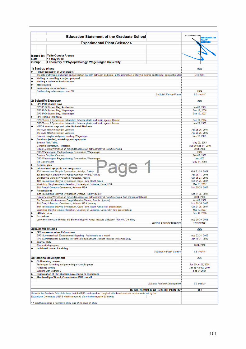
101

This research was performed at the Laboratory of Phytopathology, Binnenhaven 5, 6709 PD
Wageningen, the Netherlands, and within the graduate school for Experimental Plant Sciences
(EPS).
This research was funded by the Dutch Technology Foundation STW (project WEB6084)
with financial support by the Horticultural Product Board (Productschap Tuinbouw).
Cover: a tomato leaflet inoculated with Botrytis cinerea Printed at GVO B.V. /Ponsen & Looijen, Ede