Establishment of an infectious RNA transcription system for ...
10
Journal of General Virology (2001), 82, 2653–2662. Printed in Great Britain ................................................................................................................................................................................................................................................................................... Establishment of an infectious RNA transcription system for Striped jack nervous necrosis virus, the type species of the betanodaviruses Tokinori Iwamoto, 1 † Kazuyuki Mise, 2 Koh-ichiro Mori, 3 Misao Arimoto, 3 Toshihiro Nakai 1 and Tetsuro Okuno 2 1 Faculty of Applied Biological Science, Hiroshima University, Higashihiroshima, 739-8528, Japan 2 Laboratory of Plant Pathology, Graduate School of Agriculture, Kyoto University, Kyoto 606-8502, Japan 3 Kamiura Station, Japan Sea-Farming Association, Oita 879-2602, Japan A system has been established to produce infectious RNA transcripts for Striped jack nervous necrosis virus (SJNNV), the type species of the betanodaviruses, which infect fish. An enzymological analysis suggested that both RNA1 and RNA2 of SJNNV have a 5« cap. Both RNAs were largely resistant to 3« polyadenylation and ligation, suggesting the presence of an interfering 3« structure, while a small quantity of viral RNAs were polyadenylated in vitro. The complete 5« and 3« non- coding sequences of both segments were determined using the rapid amplification of cDNA ends method. Based on the terminal sequences obtained, RT–PCR was carried out and plasmid clones containing full-length cDNA copies of both RNAs, positioned downstream of a T7 promoter, were constructed. These plasmids were cleaved at a unique restriction site just downstream of the 3« terminus of each SJNNV sequence and were transcribed in vitro into RNA with a cap structure analogue. A mixture of the transcripts was transfected into the fish cell line E-11. Using indirect immunofluorescence staining with anti-SJNNV serum, fluorescence was observed specifically in these transfected cells ; this culture supernatant exhibited pathogenicity to striped jack larvae. Northern blot analysis of E-11 cells infected with the recombinant virus or SJNNV showed small RNA (ca. 0–4 kb) that was newly synthesized and corresponded to the 3«-terminal region of RNA1. Finally, the complete nucleotide sequences of these functional cDNAs (RNA1, 3107 nt ; RNA2, 1421 nt) were determined. This is the first report of betanodavirus cDNA clones from which infectious genomic RNAs can be transcribed. Introduction Betanodaviruses, members of the family Nodaviridae, cause highly destructive diseases in hatchery-reared larvae and juveniles of a variety of marine fish species. Since its first description in 1990 (Glazebrook et al., 1990 ; Yoshikoshi & Inoue, 1990), the disease, named viral nervous necrosis or viral Author for correspondence : Kazuyuki Mise. Fax ›81 75 753 6131. e-mail kmise!kais.kyoto-u.ac.jp † Present address : Kamiura Station, Japan Sea-Farming Association, Oita 879-2602, Japan. The DDBJ accession nos of the sequences reported in this paper are AB056571 and AB056572. encephalopathy and retinopathy, has spread to 19 or more marine fish species of 10 families in the Indo-Pacific region, Mediterranean and Scandinavia (Munday & Nakai, 1997 ; Office International des Epizooties, 2000). Recently, the virus has been proven to have existed in North America (Curtis et al., 2001). Affected fish exhibit a range of neurological abnor- malities, which are characterized by vacuolization and cellular necrosis in the central nervous system and retina. Nodaviruses have a bipartite genome of positive-sense RNA, with RNA1 encoding the RNA-dependent RNA polymerase and RNA2 encoding the coat protein (CP). Both RNAs are capped, but not polyadenylated. During RNA replication, a subgenomic RNA3, which is co-terminal with RNA1 and encodes small proteins, is synthesized. The family Nodaviridae comprise two genera : Alphanodavirus and Beta- 0001-7873 # 2001 SGM CGFD
-
Upload
khangminh22 -
Category
Documents
-
view
2 -
download
0
Transcript of Establishment of an infectious RNA transcription system for ...

Journal of General Virology (2001), 82, 2653–2662. Printed in Great Britain. . . . . . . . . . . . . . . . . . . . . . . . . . . . . . . . . . . . . . . . . . . . . . . . . . . . . . . . . . . . . . . . . . . . . . . . . . . . . . . . . . . . . . . . . . . . . . . . . . . . . . . . . . . . . . . . . . . . . . . . . . . . . . . . . . . . . . . . . . . . . . . . . . . . . . . . . . . . . . . . . . . . . . . . . . . . . . . . . . . . . . . . . . . . . . . . . . . . . . . . . . . . . . . . . . . . . . . . . . . . . . . . . . . . . . . . . . . . . . . . . . . . . . . . . . . . . . . . . . . . . . . . . . . . . . . . . . .
Establishment of an infectious RNA transcription system forStriped jack nervous necrosis virus, the type species of thebetanodaviruses
Tokinori Iwamoto,1† Kazuyuki Mise,2 Koh-ichiro Mori,3 Misao Arimoto,3 Toshihiro Nakai1
and Tetsuro Okuno2
1 Faculty of Applied Biological Science, Hiroshima University, Higashihiroshima, 739-8528, Japan2 Laboratory of Plant Pathology, Graduate School of Agriculture, Kyoto University, Kyoto 606-8502, Japan3 Kamiura Station, Japan Sea-Farming Association, Oita 879-2602, Japan
A system has been established to produce infectious RNA transcripts for Striped jack nervousnecrosis virus (SJNNV), the type species of the betanodaviruses, which infect fish. An enzymologicalanalysis suggested that both RNA1 and RNA2 of SJNNV have a 5« cap. Both RNAs were largelyresistant to 3« polyadenylation and ligation, suggesting the presence of an interfering 3« structure,while a small quantity of viral RNAs were polyadenylated in vitro. The complete 5« and 3« non-coding sequences of both segments were determined using the rapid amplification of cDNA endsmethod. Based on the terminal sequences obtained, RT–PCR was carried out and plasmid clonescontaining full-length cDNA copies of both RNAs, positioned downstream of a T7 promoter, wereconstructed. These plasmids were cleaved at a unique restriction site just downstream of the 3«terminus of each SJNNV sequence and were transcribed in vitro into RNA with a cap structureanalogue. A mixture of the transcripts was transfected into the fish cell line E-11. Using indirectimmunofluorescence staining with anti-SJNNV serum, fluorescence was observed specifically inthese transfected cells ; this culture supernatant exhibited pathogenicity to striped jack larvae.Northern blot analysis of E-11 cells infected with the recombinant virus or SJNNV showed smallRNA (ca. 0±4 kb) that was newly synthesized and corresponded to the 3«-terminal region of RNA1.Finally, the complete nucleotide sequences of these functional cDNAs (RNA1, 3107 nt; RNA2,1421 nt) were determined. This is the first report of betanodavirus cDNA clones from whichinfectious genomic RNAs can be transcribed.
IntroductionBetanodaviruses, members of the family Nodaviridae, cause
highly destructive diseases in hatchery-reared larvae andjuveniles of a variety of marine fish species. Since its firstdescription in 1990 (Glazebrook et al., 1990 ; Yoshikoshi &Inoue, 1990), the disease, named viral nervous necrosis or viral
Author for correspondence: Kazuyuki Mise.
Fax 81 75 753 6131. e-mail kmise!kais.kyoto-u.ac.jp
† Present address: Kamiura Station, Japan Sea-Farming Association,
Oita 879-2602, Japan.
The DDBJ accession nos of the sequences reported in this paper are
AB056571 and AB056572.
encephalopathy and retinopathy, has spread to 19 or moremarine fish species of 10 families in the Indo-Pacific region,Mediterranean and Scandinavia (Munday & Nakai, 1997 ;Office International des Epizooties, 2000). Recently, the virushas been proven to have existed in North America (Curtis et al.,2001). Affected fish exhibit a range of neurological abnor-malities, which are characterized by vacuolization and cellularnecrosis in the central nervous system and retina.
Nodaviruses have a bipartite genome of positive-senseRNA, with RNA1 encoding the RNA-dependent RNApolymerase and RNA2 encoding the coat protein (CP). BothRNAs are capped, but not polyadenylated. During RNAreplication, a subgenomic RNA3, which is co-terminal withRNA1 and encodes small proteins, is synthesized. The familyNodaviridae comprise two genera : Alphanodavirus and Beta-
0001-7873 # 2001 SGM CGFD

T. Iwamoto and othersT. Iwamoto and others
novirus, members of which primarily infect insects and fish,respectively (van Regenmortel et al., 2000). Striped jack nervousnecrosis virus (SJNNV), which had been purified from diseasedlarvae of the striped jack Pseudocaranx dentex, was first identifiedas a betanodavirus (Mori et al., 1992). RNA1 (3±1 kb) andRNA2 (1±4 kb) of SJNNV encode a 100 kDa protein (pre-sumably RNA-dependent RNA polymerase) and a major CPof 42 kDa, respectively (Mori et al., 1992 ; Nagai & Nishizawa,1999). The sequence similarities of RNA2, about 870 bases inopen reading frame (ORF), were less than 29% at the nucleotidelevel and less than 11% at the amino acid level betweenSJNNV and four representative insect nodaviruses, Nodamuravirus (NoV), Black beetle virus (BBV), Flock house virus (FHV) andBoolarra virus (Dasgupta et al., 1984 ; Dasgupta & Sgro, 1989),whereas they were 70% or higher among four piscinenodavirus isolates (Nishizawa et al., 1995). On the other hand,the RNA1 sequence similarities between SJNNV and thealphanodaviruses were 28% at the nucleotide and amino acidlevels (Nagai & Nishizawa, 1999).
With the progress of recombinant DNA technology,single- or double-stranded RNA viruses have been geneticallyanalysed and infectious RNA transcripts or cDNA clones havebeen produced from a variety of RNA viruses (Boyer &Haenni, 1994). The alphanodaviruses can be propagated in awide range of cultured cells, such as insect, plant, vertebrateand yeast cells, and their infectious RNA transcripts have beenused frequently for RNA transfection into these permissivecells. This has led to studies of their RNA replication, geneexpression and virion assembly (reviewed by Ball & Johnson,1998). In contrast, the establishment of such a reverse geneticssystem for betanodaviruses has long been hampered by thelack of an appropriate cell culture system (Breuil et al.,1991 ; Mori et al., 1991 ; Munday et al., 1992 ; Nguyen et al.,1994 ; Grotmol et al., 1995). The studies of Frerichs et al. (1996)and our own group (Iwamoto et al., 1999, 2000) have revealed,however, that the striped snakehead cell line (SSN-1) (Frerichset al., 1991) and the clonal cell line E-11 from the SSN-1 cellsare useful for qualitative and quantitative analyses for all of thebetanodaviruses, including SJNNV. Furthermore, it has beenconfirmed that the SJNNV genomic RNA is infectious whentransfected into E-11 cells and that the progeny virus recoveredfrom the cells is virulent to striped jack larvae (Iwamoto et al.,2001).
Recently, a reverse genetics system was developed forInfectious pancreatic necrosis virus, a double-stranded RNA virusof fish (Yao & Vakharia, 1998). To date, however, there is noreverse genetics system for positive-sense single-strandedRNA viruses that infect fish or aquatic animals. In general,cDNA containing entire viral genome sequences is required toobtain infectious in vitro RNA transcripts. Although thenucleotide sequences for RNA1 and RNA2 of SJNNV andother betanodaviruses have been published (Nishizawa et al.,1995, 1997 ; Delsert et al., 1997a ; Sideris, 1997 ; Aspehauget al., 1999 ; Nagai & Nishizawa, 1999 ; Thiery et al., 1999 ;
Grotmol et al., 2000 ; Starkey et al., 2000), precise sequences oftheir 5« and 3« non-coding regions have not been determined.In this report, we describe the construction and sequencing offull-length cDNA clones of SJNNV and the recovery ofinfectious SJNNV from E-11 cells transfected with RNAtranscripts that, in turn, were synthesized in vitro from theircDNAs. This is the first report of the production of cDNAclones of a betanodavirus from which infectious genomicRNAs can be transcribed.
Methods+ Cells. The E-11 cell line (Iwamoto et al., 2000) was grown at 25 °Cin Leibovit’s L-15 medium (Gibco BRL) supplemented with 5% foetalbovine serum.
+ SJNNV purification. Naturally infected striped jack larvae that hadbeen collected at the Nagasaki prefecture in Japan in 1993 were used asthe source of SJNNV. SJNNV was purified as described by Mori et al.(1992) and stored at ®80 °C.
+ Confirmation of the 5« and 3« end structures of the SJNNVgenome. SJNNV virion RNA was extracted from the purified virus asdescribed previously (Kroner & Ahlquist, 1992). The 5« terminus of thevirion RNA was treated with bacterial alkaline phosphatase fromEscherichia coli C75 (Takara) and then labelled with T4 polynucleotidekinase (Toyobo) in the presence of [γ-$#P]ATP (Amersham) either with orwithout prior decapping treatment with tobacco acid pyrophosphatase(TAP) (Nippon gene) under the conditions recommended by themanufacturer. The 3« terminus of each strand was treated with T4 RNAligase (Takara) and poly(A) polymerase (Takara) in the presence of[$#P]pCp (Amersham) and [α-$#P]ATP (Amersham), respectively, ac-cording to the manufacturer’s recommendations. The treated RNAs wereseparated by electrophoresis in 1% agarose gels and the signal was thendetected by autoradiography.
+ Determination of the 5«- and 3«-terminal sequences of theSJNNV genome. The rapid amplification of cDNA ends (RACE)method (Frohman et al., 1988) was used to determine the completenucleotide sequences of the 5« and 3« termini of the SJNNV genome. For5« RACE, virion RNA was reverse-transcribed with SuperScript II (GibcoBRL) using the synthetic oligonucleotide primers SJ1R1 or SJ2R1 (Table1). The first-strand cDNAs were polyadenylated with terminal deoxy-nucleotidyltransferase (Takara) and then the second-strand cDNAs weresynthesized using the primer ANCH (Table 1), after purification throughthe GLASS MAX Column (Pharmacia), according to the manufacturer’sinstructions. The double-stranded cDNAs were amplified using theprimers AUAP and either SJ1R2 or SJ2R2 (Table 1). Decapped RNA,prepared as described above, was also used in 5« RACE for the detectionof cap structure. For 3« RACE, viral RNA was polyadenylated withpoly(A) polymerase in the presence of ATP and reverse-transcribed usingthe primer ANCH, as described above, and then amplified with theprimers AUAP and either SJ1F1 or SJ2F1 (Table 1). These amplifiedproducts were purified by 1% low-melting-point (LMP) agarose gelelectrophoresis and then further amplified by nested PCR using theprimers AUAP and either SJ1F2 or SJ2F2. The nested PCR products werepurified and used to determine the terminal sequences. Sequencingreactions were performed using the BigDye Deoxy Terminator CycleSequencing kit (PE Applied Biosystems), according to the manufacturer’sinstruction, and nucleotide sequences were determined using theautomated sequencer ABI Prism 310 (PE Applied Biosystems).
CGFE

Infectious RNA transcription system for SJNNVInfectious RNA transcription system for SJNNV
Table 1. Oligonucleotide primers for the construction and determination of full-length cDNA clones of SJNNV RNAs
Oligonucleotide sequences used for both 5« and 3« RACE and for the synthesis of full-length SJNNV cDNA are shown. SJNNV-specificnucleotides are shown in bold and the T7 promoter sequence is underlined. Restriction sites are indicated in lowercase. The oligonucleotidepositions correspond to the SJNNV full-length cDNA within pSJ1TK19 and pSJ2TK30, the sequences of which were deposited into the DNAData Bank of Japan (DDBJ) under accession nos AB056571 and AB056572, respectively.
Purpose Primer Sequence (5«!3«) Position
ANCHAUAP*
GGCCACGCGTCGACTAGTACTTTTTTTTTTTTTTTTTTTTpGGCCACGCGTCGACTAGTAC
––
5« RACE
1
23
4
SJ1R1†SJ1R2SJ2R1†SJ2R2
GAGATAATGTATGGCTCGTAGCCAAATGCGCACTAGTTCCATCTGCACATTGGCTGAATTTCGAACTCGAACGATTGTGGAATCGACGAC
370–392262–283349–371262–283
3« RACE
1
23
4
SJ1F1SJ1F2SJ2F1SJ2F2
GAGCTAGAACAGCTCTTCAAGGTAGTCCAGGTAAACGAGATGTTGATTACGGCACTAACCACTCAACAAGAGCGAAATTGAAGC
2795–28152855–28751101–11211142–1162
SJNNV cDNAsynthesis
1
23
4
SJ1-5HdT7SJ1-3EcSJ2-5HdT7SJ2-3Ec
CCCCaagcttAATACGACTCACTATAGTAACATCAGCTCTTGCTCTGACCGgaattcGCCGAAGCGTAGGACAGCACCCCaagcttAATACGACTCACTATAGTAATCTAACACCGCTTTGCAACCGgaattcGCCGAGTAATGTGGCGATC
1–203088–3107
1–201402–1421
* Primer AUAP is phosphorylated at the 5« end.† These primers were also used for direct sequencing.
+ Construction of full-length cDNA and cloning into a tran-scription vector. Full-length cDNA copies of genomic RNA1 and
RNA2 were synthesized by RT–PCR using specific primers for the 5« and3« termini of each RNA. First-strand cDNA copies of RNA1 and RNA2
were synthesized, as described above, using virion RNA as a template
and the oligonucleotide primers SJ1-3Ec and SJ2-3Ec, respectively (Table
1). These oligonucleotides were complementary to the 3« end sequences
of the SJNNV RNA1 and RNA2 and contained an EcoRI site to facilitate
cloning and to linearize the plasmids. The single-stranded cDNAs were
then used as templates for PCR using the above-mentioned oligo-
nucleotides as reverse primers and SJ1-5HdT7 (for RNA1) or SJ2-5HdT7
(for RNA2) as forward primers (Table 1). These primer sequences
correspond to the 5« ends of RNA1 and RNA2 and contain the T7
promoter sequence and a HindIII site to facilitate cloning. PCR was
performed using the high fidelity DNA polymerase KOD plus (Toyobo).
Thermocycling was carried out for 20 (RNA1) or 15 (RNA2) cycles of
40 s at 94 °C, 60 s at 55 °C and 90 s at 72 °C, with a final extension of
10 min at 72 °C. Amplified full-length cDNAs of each RNA were purified
using 1% LMP agarose gel electrophoresis and digested with EcoRI}HindIII. These fragments were cloned into the HindIII and EcoRI sites of
pUC118 (Takara), according to the standard protocol (Sambrook et al.,
1989). Plasmid DNA was maintained in E. coli DH5α cells and extracted
using the Plasmid Midi kit (Qiagen), according to the manufacturer’s
protocol. The resulting plasmids were named according to the format
pSJxTKy, where x is the SJNNV component number and y is an arbitrary
isolation number of the pUC118 recombinant plasmid containing the
SJNNV cDNA insert. Full-length cDNA inserts within the representative
plasmids pSJ1TK19 and pSJ2TK30 were sequenced using synthetic
oligonucleotides designed after the known sequences (Nishizawa et al.,
1995 ; Nagai & Nishizawa, 1999).
+ In vitro transcription. Plasmids containing full-length SJNNV
cDNA were linearized with EcoRI and then used for in vitro transcription
with T7 RNA polymerase (Takara) in the presence of synthetic cap
analogue [m(G(5«)ppp(5«)G] (New England Biolabs), as described pre-
viously (Kroner & Ahlquist, 1992). After being treated with RQ1 DNase
I (Promega), transcripts were purified through a Sephadex G-50 column
(Pharmacia) and their concentrations were quantified spectrophoto-
metrically before transfection into cells. The RNAproducts were analysed
by agarose gel electrophoresis in TBE buffer (Sambrook et al., 1989).
+ Inoculation of cultured cells and infection assay. The
infectivity of transcripts was examined by transfection with lipofectin
reagent (Gibco BRL) into E-11 cells followed by 24 h of incubation at
25 °C, as described previously (Iwamoto et al., 2001). For infection
analysis of progeny virus, media cultured in the same manner as above for
72 h were collected, inoculated onto fresh E-11 cells and incubated at
25 °C for 24 h. Cell infectivity of the transcripts and their progeny was
examined by immunofluorescence staining using anti-SJNNV rabbit
polyclonal antibody and FITC-conjugated swine immunoglobulin (Ig) to
rabbit Ig (Dako), as described previously (Nguyen et al., 1996). The
fluorescent cells were then counted.
+ Northern blot analysis. Total RNA was extracted from E-11 cells
infected with progeny viruses using ISOGEN (Nippon gene), according
to the manufacturer’s instructions. The RNA was subjected to Northern
blot analysis, essentially as described by Damayanti et al. (1999), except
that the DIG Labelling and Detection kit (Roche) was used, according to
the supplier’s instructions.
DIG-labelled RNA probes specific for the positive- and negative-
sense strand of SJNNV RNA1 and RNA2 were prepared as follows. For
RNA1, a PCR product was amplified from pSJ1TK19 using the M13
CGFF
T. Iwamoto and othersT. Iwamoto and others
primers M4 and RV (Takara) and digested with ClaI}EcoRI and theresulting 0±3 kb fragment was inserted into the transcription vectorpBluescript II KS(®) (Stratagene) to create pSJ1BS1. For RNA2,pSJ2TK30 was digested with BamHI}EcoRI and the resulting 0±4 kbfragment was ligated into pBluescript II SK(®) to create pSJ2BS2. Toprepare probes for positive-sense RNA1 and RNA2, pSJ1BS1 andpSJ2BS2 were linearized with SalI and BamHI, respectively, andtranscribed with T7 RNA polymerase (Takara). To prepare probes fornegative-sense RNA1 or RNA2, either pSJ1BS1 or pSJ2BS2 waslinearized with EcoRI and then transcribed with T3 RNA polymerase(Gibco BRL).
+ Western blot analysis. Infected fish cells were suspended inLaemmli’s sample buffer and subjected to SDS–PAGE. Western blotanalysis was carried out as described previously (Damayanti et al., 1999)using an Immobilon-P transfer membrane (Millipore). SJNNV CP wasdetected using anti-SJNNV rabbit polyclonal antibody and alkalinephosphatase-conjugated goat anti-rabbit secondary antibody (Bio-Rad)followed by incubation with BCIP}NBT for colour development.
+ Electron microscopy of progeny. Cell culture supernatantscontaining progeny virus were layered onto 10–40% sucrose gradientsin TE buffer (10 mM Tris–HCl, 1 mM EDTA, pH 7±2) and centrifuged at80000 g for 2 h at 16 °C. Each fraction was collected, analysed byWestern blotting and concentrated using the Centricon centrifugal filterunit (Millipore), according to the manufacturer’s instructions. Virussuspensions were stained with 1% uranyl acetate. Simultaneously,progeny virus was inoculated onto freshly prepared E-11 cells andincubated at 25 °C. After incubation for 3 days, the cells were fixed in2±5% glutaraldehyde and post-fixed with 1% osmium tetroxide. Ultra-thin sections were stained with 1% uranyl acetate and 1% lead citrate.These samples were examined under a Hitachi (Model H-7100FA)electron microscope.
+ Virulence assay for fish larvae. One-day-old striped jack larvaereared at Kamiura Station of the Japan Sea-Farming Association wereused for a virulence assay of the progeny virus produced in cells infectedwith the in vitro RNA transcripts. Before the infection experiment, theselarvae were confirmed to be SJNNV-free by RT–PCR (Nishizawa et al.,1994). Duplicate groups of 250 larvae were kept at 23 °C in glass beakerscontaining 1 litre of sea water to which kanamycin was added to a finalconcentration of 5 µg}ml. The 96 h culture supernatant of E-11 cells(50 µl) infected with progeny viruses was inoculated into the beakers andmoribund fish were collected daily. The homogenate (progenitor virus) ofstriped jack larvae naturally infected with SJNNV and the culturesupernatant of uninfected E-11 cells were used as positive and negativecontrols, respectively. Virus titres of the inocula used were determined, asdescribed previously (Iwamoto et al., 2000), to be 10*±
' TCID&!
}ml for theprogeny and 10"!±
' TCID&!
}ml for the progenitor. Fish were fixed with10% formalin and embedded in paraffin. Sections were subjected toimmunofluorescence staining as described above.
ResultsTerminal sequences of SJNNV RNA1 and RNA2
SJNNV RNAs were successfully labelled with [γ-$#P]ATP atthe 5« end only after decapping treatment with TAP (data notshown). When we performed 5« RACE using virion RNAs ofBrome mosaic virus (BMV), a cytidine residue complementary tothe m(G in the cap structure was incorporated into the RACEproducts (K. Mise, unpublished data). Consistent with thisobservation, when SJNNV RNAs were used as a template for
5« RACE after prior treatment with TAP, the sequence of theRACE products showed that the signal for such a cytidineresidue was reduced compared with that of the capped,untreated RNA (data not shown). Polyadenylation of the 3«end of alphanodavirus RNAs was unsuccessful, probablybecause of the modification of their ends by an unidentified‘blocking group ’ (Dasmahapatra et al., 1985). Although the 3«end of SJNNV RNAs does not have a poly(A) structure (Moriet al., 1992), it remains unknown whether such a blockingstructure exists. This was examined by labelling SJNNV RNAswith poly(A) polymerase or T4 RNA ligase ; BMV RNAs wereused as a positive control as these RNAs have 3«-OH groupsthat are reactive with these particular enzymes (Ahlquist et al.,1981). Autoradiography showed that SJNNV RNAs werelabelled at their 3« ends, but the efficiency of labelling wasconsiderably less than that of BMV RNAs (data not shown).These results indicate that the 5« ends of both SJNNV RNAsare capped and that a blocking group rather than a hydroxylgroup would thus probably modify most of their 3« ends.
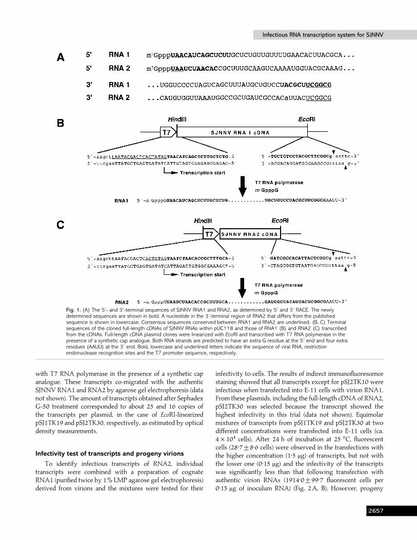
We then used the sample containing a small quantity of invitro polyadenylated virion RNAs for the determination of the3«-terminal sequence. Both the 5« and 3« termini of SJNNVRNA1 or RNA2 were amplified by RACE using SJNNV-specific primers that were synthesized according to the knownsequence (Nishizawa et al., 1995 ; Nagai & Nishizawa, 1999).We found 14 additional bases at the 5« terminus and 12additional bases at the 3« terminus on RNA1 (Fig. 1A). RNA2had another 11 bases at the 5« terminus and the 3«-terminalsequence corresponded to the published sequence, althoughone nucleotide had been substituted (Fig. 1A).
Construction and sequence of full-length cDNA clonesand in vitro transcription
To synthesize full-length cDNA copies of SJNNV RNA1and RNA2, we designed synthetic oligonucleotides based onthe 5«- and 3«-terminal sequences of the SJNNV RNAsdetermined in this study (Fig. 1B, C). The reverse primersincluded a unique EcoRI site for efficient cloning and subsequentplasmid linearization before in vitro transcription. The forwardprimers contained both a HindIII site and a T7 promotersequence. HindIII and EcoRI sites were chosen because theserecognition sites were not found in the known internal orterminal sequences of the SJNNV genome. PCR amplificationwas carried out with few (15 or 20) cycles to avoid theincorporation of undesired mutations into the RT–PCRproducts. cDNA clones containing the entire coding and non-coding regions of SJNNV RNA1 and RNA2 were prepared,the terminal regions of these complete cDNA copies weresequenced and the clones containing full-length sequenceswere selected. Finally, seven clones were obtained : one(pSJ1TK19) for RNA1 and the others (pSJ2TK9, -10, -22, -28,-29 and -30) for RNA2. Plasmids containing full-length SJNNVcDNA were linearized by cleavage with EcoRI and transcribed
CGFG
Infectious RNA transcription system for SJNNVInfectious RNA transcription system for SJNNV
Fig. 1. (A) The 5«- and 3«-terminal sequences of SJNNV RNA1 and RNA2, as determined by 5« and 3« RACE. The newlydetermined sequences are shown in bold. A nucleotide in the 3«-terminal region of RNA2 that differs from the publishedsequence is shown in lowercase. Consensus sequences conserved between RNA1 and RNA2 are underlined. (B, C) Terminalsequences of the cloned full-length cDNAs of SJNNV RNAs within pUC118 and those of RNA1 (B) and RNA2 (C) transcribedfrom the cDNAs. Full-length cDNA plasmid clones were linearized with EcoRI and transcribed with T7 RNA polymerase in thepresence of a synthetic cap analogue. Both RNA strands are predicted to have an extra G residue at the 5« end and four extraresidues (AAUU) at the 3« end. Bold, lowercase and underlined letters indicate the sequence of viral RNA, restrictionendonuclease recognition sites and the T7 promoter sequence, respectively.
with T7 RNA polymerase in the presence of a synthetic capanalogue. These transcripts co-migrated with the authenticSJNNV RNA1 and RNA2 by agarose gel electrophoresis (datanot shown). The amount of transcripts obtained after SephadexG-50 treatment corresponded to about 25 and 16 copies ofthe transcripts per plasmid, in the case of EcoRI-linearizedpSJ1TK19 and pSJ2TK30, respectively, as estimated by opticaldensity measurements.
Infectivity test of transcripts and progeny virions
To identify infectious transcripts of RNA2, individualtranscripts were combined with a preparation of cognateRNA1 (purified twice by 1% LMP agarose gel electrophoresis)derived from virions and the mixtures were tested for their
infectivity to cells. The results of indirect immunofluorescencestaining showed that all transcripts except for pSJ2TK10 wereinfectious when transfected into E-11 cells with virion RNA1.From these plasmids, including the full-length cDNA of RNA2,pSJ2TK30 was selected because the transcript showed thehighest infectivity in this trial (data not shown). Equimolarmixtures of transcripts from pSJ1TK19 and pSJ2TK30 at twodifferent concentrations were transfected into E-11 cells (ca.4¬10% cells). After 24 h of incubation at 25 °C, fluorescentcells (28±7³8±6 cells) were observed in the transfections withthe higher concentration (1±5 µg) of transcripts, but not withthe lower one (0±15 µg) and the infectivity of the transcriptswas significantly less than that following transfection withauthentic virion RNAs (1914±0³99±7 fluorescent cells per0±15 µg of inoculum RNA) (Fig. 2A, B). However, progeny
CGFH

T. Iwamoto and othersT. Iwamoto and others
Fig. 2. (A)–(D) Detection of SJNNV antigen in E-11 cells at 24 h post-transfection or -infection by immunofluorescence staining. Specificfluorescent cells transfected with a mixture of transcripts from pSJ1TK19and pSJ2TK30 (A) or virion RNAs (B), or (C) mock-transfected with DEPCwater are shown. Fluorescent cells were detected by an anti-SJNNV rabbitpolyclonal antibody. (D) Cells infected by a 72 h culture supernatant from(A). Bar, 50 µm. (E)–(F) Electron micrographs of rSJ. (E) Negativelystained virions purified from the supernatant of cultured cells transfectedwith a mixture of transcripts from pSJ1TK19 and pSJ2TK30. Bar, 50 nm.(F) Virions assembled in a crystalline array in an E-11 cell infected with rSJand incubated at 25 °C for 3 days. Bar, 200 nm.
viruses (recombinant SJNNV, rSJ) in the culture supernatants oftransfected cells were highly infectious to fresh E-11 cells (Fig.2D).
To identify rSJ, we performed observations by electronmicroscopy. From the supernatant of cultured cells transfectedwith a mixture of transcripts from pSJ1TK19 and pSJ2TK30,we detected rSJ particles approximately 25 nm in diameter(Fig. 2E), similar to the size reported previously (Mori et al.,1992). Furthermore, we also observed a crystalline array ofvirions in the cytoplasm of E-11 cells infected with rSJ (Fig. 2F).
Northern blot analysis of total RNA extracted from infectedE-11 cells was performed using DIG-labelled riboprobesspecific for positive- or negative-sense RNA. In the sampletaken 24 h post-inoculation (p.i.), we detected strong signalsthat hybridized to SJNNV positive-sense-specific probes andco-migrated with original virion RNAs (Fig. 3A). Negative-sense RNA1 and RNA2 were also detected from cells at24 h p.i. (Fig. 3B). However, the signals for RNA2, especiallyfor the negative-sense one, were significantly lower than thoseof RNA1 (Fig. 3A, B). These differences were due to thedifference in hybridization efficiency between the probe for() RNA1 and that for () RNA2 as well as between theprobe for (®) RNA1 and that for (®) RNA2, which was
Fig. 3. (A, B) Northern blot analyses of progeny virus RNA in E-11 cellsinoculated with rSJ or wild-type SJNNV. Total RNA was extracted frominfected cells at 1 and 24 h p.i., separated by electrophoresis in a 1±0%agarose–formaldehyde gel and transferred onto a nylon membrane. (A)Viral RNAs were detected with a mixture of DIG-labelled positive-strand-specific probes, each complementary to the 3«-proximal region of SJNNVRNA1 or RNA2. (B) Viral RNAs were detected with a mixture of DIG-labelled negative-strand-specific probes, each corresponding to the 3«-proximal region of SJNNV RNA1 or RNA2. The mock controls contain cellsinoculated with the culture supernatant of uninfected E-11 cells. Note thatthe RNA2 signals are weaker than those of RNA1, probably because of thelow hybridization efficiency of RNA2-specific probes. The positions ofRNA1 and RNA2 are indicated at the right. Asterisks indicate bands relatedto subgenomic RNA3, similar to that of alphanodaviruses. Blots wereexposed to films for 10 s (A) and 5 min (B). (C) Western blot analysis ofthe accumulation of SJNNV CP in E-11 cells inoculated with rSJ or wild-type SJNNV. Proteins were extracted from the infected cells at 1 and24 h p.i., separated by 12±5% SDS–PAGE and electroblotted onto a PVDFmembrane. CP was detected with anti-SJNNV polyclonal antibody. Themock control is as above.
confirmed by hybridizing those probes with known amountsof RNAs transcribed in vitro (data not shown). In addition toRNA1 and RNA2, bands showing faster migration were alsodetected in both positive- and negative-sense hybridizations(Fig. 3A, B). In parallel hybridization analyses using eachsegment-specific probe, these extra bands reacted with theRNA1-specific probes, but not with the RNA2-specific probes(data not shown). Meanwhile, Western blot analysis of thesample of cells at 24 h p.i. showed an obvious increase in CP
CGFI

Infectious RNA transcription system for SJNNVInfectious RNA transcription system for SJNNV
Fig. 4. Cumulative mortality of 1-day-old striped jack larvae exposed toSJNNV. Larvae were inoculated with the homogenate of naturally infectedstriped jack larvae (+), supernatant of E-11 cells containing rSJ (D) orthe culture supernatant of uninfected E-11 cells (_) in a 1 litre beaker.Data are represented as the mean³SD from two separate experiments.
accumulation when compared with that of cells at 1 h p.i. (Fig.3C).
rSJ was virulent to 1-day-old striped jack larvae and thevirulence of rSJ was similar to that of the progenitor virus. Allthe larvae exposed to these virus inocula died at 2–4 days p.i.(Fig. 4) and SJNNV antigen was detected from the brain, spinalcord and retina of affected larvae (Fig. 5). RT–PCR analysis ofthe dead larvae were all positive (data not shown). Neithersignificant mortality nor SJNNV antigens were observed in thelarvae exposed to the supernatant of uninfected E-11 cellculture.
Sequence of functional cDNA clones
As described above, in vitro transcripts synthesized fromthe plasmid clones pSJ1TK19 and pSJ2TK30 successfullyinitiated SJNNV amplification in transfected cells. Both cDNAinserts were then fully sequenced to establish definitive,functional nucleotide sequences of the two viral genomesegments. Based on the published SJNNV sequences, severaloligonucleotide primers were synthesized and used for thedetermination of full-length cDNA sequences. The cDNAs inpSJ1TK19 and pSJ2TK30 contained 3107 and 1421 nucleo-tides, respectively. The full-length cDNA sequence ofpSJ1TK19 differed from the published sequence (accession no.AB025018) (Nagai & Nishizawa, 1999) by three nucleotides :position 378 was G (A), 1692 was C (T) and 2148 was T (C) inpSJ1TK19 (published nucleotides are in parentheses). The full-length cDNA sequence of pSJ2TK30 also differed from thepublished sequence (accession no. D30814) (Nishizawa et al.,1995) by four nucleotides : position 766 was G (T), 881 was G(A), 969 was A (G) and 1411 was A (G) in pSJ2TK30 (publishednucleotides are in parentheses). Although the nucleotidedifferences in pSJ1TK19 do not lead to amino acid changes, the
Fig. 5. Virus antigen from striped jack larvae infected with rSJ detected byimmunofluorescence staining. (A) Larvae were inoculated with the culturesupernatant containing rSJ, fixed at 3 days p.i., sectioned and stained withanti-SJNNV polyclonal antibody. (B) Larvae were inoculated with theculture supernatant of uninfected E-11 cells. Bar, 100 µm.
pSJ2TK30 sequence encodes an A at position 247 and an R atposition 285 in the ORF rather than an S and a Q in thepublished sequence, respectively. These nucleotide differenceswere also found in the other functional cDNA clones pSJ2TK22and pSJ2TK29 and were also confirmed by direct sequencingof the RT–PCR products from RNA2 (data not shown).
DiscussionIn the present study, we completely determined the 5«- and
3«-terminal sequences of SJNNV genomic RNAs by poly(A)-tailing and RACE and then constructed full-length cDNAclones that can serve as templates for the production of RNAtranscripts infectious to cultured fish cells. This is the firstreport of infectious RNA transcripts derived from clonedcDNA of betanodavirus genomic RNAs.
Single-stranded RNA genomes of many plant and animalviruses have unique structures at their 5« and 3« termini : the capor the VPg structure at the 5« end and the tRNA-like structureor the poly(A) tract at the 3« end (Goldbach, 1987). The 3« endsof alphanodavirus RNAs lack a poly(A) tail (Newman &Brown, 1976 ; Scotti et al., 1983) and are not reactive with RNAligase or poly(A) polymerase (Dasgupta et al., 1984 ; Guarino etal., 1984 ; Dasmahapatra et al., 1985 ; Kaesberg et al., 1990),suggesting that the 3« end is modified by an unidentifiedblocking group. Our results suggest that SJNNV also has the
CGFJ

T. Iwamoto and othersT. Iwamoto and others
cap structure at the 5« end and the blocking group at the 3« end.Because the 3« end of alphanodavirus RNA is blocked in, thedouble-stranded RNAs or head-to-tail homodimers in infectedcultured cells have been used to determine the 3«-terminalsequences (Guarino et al., 1984 ; Johnson et al., 2000). Althoughthis was a significant obstacle to conquer for the sequencing ofthe 3« end of SJNNV, a small quantity of viral RNAs were,fortunately, polyadenylated in vitro and hence we were able toobtain the 3«-terminal cDNA fragments of SJNNV genomicRNAs by 3« RACE. Those minor unmodified RNAs mightexist because nascent viral RNAs were packaged beforemodification and}or because the blocking structure waseventually removed from the 3« end during the RNA extractionprocedure. Consistent with our results, the 3«-terminalsequences of genomic RNAs of the betanodaviruses Greasygrouper nervous necrosis virus (GGNNV) and Dicentrarchuslabrax encephalitis virus (DlEV) have been determined after invitro genomic RNA self-ligation (Tan et al., 2001) and in vitropolyadenylation (Delsert et al., 1997a), respectively. Conse-quently, we completely determined the 5« and 3« non-codingsequences of both RNAs in which 11–14 nucleotides werefound at both termini for RNA1 and at the 5« terminus forRNA2, in addition to the published sequences of SJNNV(Nishizawa et al., 1995 ; Nagai & Nishizawa, 1999). Comparisonof the 5«- and 3«-terminal sequences between both RNAspecies demonstrated that each 5« terminus begins with 5«UAA… 3« and each 3« terminus ends with 5«…UCGGCG 3«.The identical sequences are also present at the 5« end of RNA1and RNA2 of GGNNV (accession nos AF318942 andAF319555, respectively) (Tan et al., 2001) and at the 3« end ofRNA2 of DlEV (accession no. U39876) (Delsert et al., 1997a).Such sequences, conserved between two genomic RNAs butdifferent from those of SJNNV, were found among thealphanodaviruses BBV [accession nos K02560 (Dasmahapatraet al., 1985) and X00956 (Dasgupta et al., 1984)], FHV (accessionnos X77156 and X15959) (Dasgupta & Sgro, 1989) and NoV(accession nos AF174533 and AF174534), in which thesequence starts with 5« GU… 3« and ends with 5«…GGU 3«.These results might suggest that other betanodaviruses alsohave consensus sequences such as those found in SJNNV.
The transcript RNAs were less infectious than their SJNNVvirion RNA counterparts. This may be caused by the absenceof a peculiar, unknown blocking structure at the 3« end and}orby the presence of extra non-viral nucleotides at both terminiof the transcripts. At the 5« end of the transcripts, a non-viralextra G residue that originated from the T7 promoter sequencewas added. It has been reported that transcription efficiency isincreased when G residues are inserted between the T7promoter and viral cDNA sequences, although the amplifi-cation efficiency in cells is lost to some extent (Janda et al.,1987). Our previous experiments suggest that the poly(A)tract was not entirely present or was not long in SJNNV viralRNAs, because they were not trapped with an oligo(dT)column (Mori et al., 1992). However, we could not rule out the
possibility that the 3« ends contain an oligo(A) tract that wasshort enough for the RNAs to pass through the column andcould not be distinguished from the poly(A) added in vitro withpoly(A) polymerase during 3« RACE. Alternatively, if the 3«terminus ending with oligo(A) is important for the infectivityof SJNNV, UU residues within the extra non-viral sequenceAAUU at the 3« end might have lowered the infectivity ofthese transcripts.
It has been known that subgenomic RNA3 (0±4 kb), derivedfrom RNA1 of alphanodavirus, can only be detected frominfected cells (Guarino et al., 1984 ; Dasmahapatra et al.,1985 ; Johnson et al., 2000). In this study, we also detectedsignals for RNA with faster migration by Northern blotanalysis from cells infected with wild-type or rSJ. Although asimilar phenomenon has been reported for the betanodavirusDlEV (Delsert et al., 1997b), detailed analysis was notperformed. In this study, we detected the extra bands onlyfrom infected cells and verified that the bands reacted withboth positive- and negative-strand-specific probes for the 3«-proximal region of SJNNV RNA1. Furthermore, the molecularsize for these bands was estimated to be approximately 0±4 kb.These results strongly suggest that RNA3 is generated fromRNA1 during SJNNV RNA replication.
Previously, we demonstrated the pathogenicity to stripedjack larvae of the progeny that was generated by transfectionof SJNNV virion RNAs into E-11 cells (Iwamoto et al., 2001).rSJ obtained from in vitro transcripts in this study waspathogenic to striped jack larvae. In regard to fish viruses, thisis the first known instance in which a recombinant virus hasthe ability to kill the original hosts. As mentioned before,betanodaviruses can be classified into four genotypes, desig-nated SJNNV, Barfin flounder nervous necrosis virus (BFNNV),Tiger puffer nervous necrosis virus and Redspotted grouper nervousnecrosis virus, based on the RNA2 partial sequences (Nishizawaet al., 1997). We have reported recently that the optimalgrowth temperature for virus growth in cultured cells differsamong the genotypes. Furthermore, because SJNNV genotypevirus was not infectious to the Atlantic halibut Hippoglossushippoglossus, which BFNNV genotype virus can infect, it hasbeen suggested that host-specificity might be different amongsome betanodaviruses (Totland et al., 1999). A reverse geneticssystem for SJNNV, as reported here, will open the way formolecular studies directed at virus multiplication and patho-genesis of the betanodaviruses. In particular, the relationshipbetween genetic variations and host specificities in betanoda-viruses and comparative studies with alphanodaviruses will beof great interest.
We wish to thank Dr Iwao Furusawa for his valuable advice on thisstudy and for critical reading of the manuscript. This work was supportedin part by a grant from the Japan Sea-Farming Association and by a grant-in-aid (12052201) for Scientific Research on Priority Area (A), a grant-in-aid (09NP1501) for Creative Basic Research from the Ministry ofEducation, Culture, Sports, Science and Technology, Japan and a grant-in-
CGGA
Infectious RNA transcription system for SJNNVInfectious RNA transcription system for SJNNV
aid (JSPS-RFTF96L00603) from the ‘Research for the Future ’ program ofthe Japan Society for the Promotion of Science.
ReferencesAhlquist, P., Dasgupta, R. & Kaesberg, P. (1981). Near identity of 3«RNA secondary structure in bromoviruses and cucumber mosaic virus.Cell 23, 183–189.
Aspehaug, V., Devold, M. & Nylund, A. (1999). The phylogeneticrelationship of nervous necrosis virus from halibut (Hippoglossus hippo-glossus). Bulletin of the European Association of Fish Pathologists 19,196–202.
Ball, L. A. & Johnson, K. L. (1998). Nodaviruses of insects. In The InsectViruses, pp. 225–267. Edited by L. K. Miller & L. A. Ball. New York :Plenum.
Boyer, J. C. & Haenni, A. L. (1994). Infectious transcripts and cDNAclones of RNA viruses. Virology 198, 415–426.
Breuil, G., Bonami, J. R., Pepin, J. F. & Pichot, Y. (1991). Viral infection(picorna-like virus) associated with mass mortalities in hatchery-rearedsea bass (Dicentrarchus labrax) larvae and juveniles. Aquaculture 97,109–116.
Curtis, P. A., Drawbridge, M., Iwamoto, T., Nakai, T., Hedrick, R. P. &Gendron, A. P. (2001). Nodavirus infection of juvenile white sea bass,Atractoscion nobilis, cultured in southern California : first record of viralnervous necrosis (VNN) in North America. Journal of Fish Diseases 24,263–271.
Damayanti, T. A., Nagano, H., Mise, K., Furusawa, I. & Okuno, T.(1999). Brome mosaic virus defective RNAs generated during infectionof barley plants. Journal of General Virology 80, 2511–2518.
Dasgupta, R. & Sgro, J.-Y. (1989). Nucleotide sequences of threenodavirus RNA2s : the messengers for their coat protein precursors.Nucleic Acids Research 17, 7525–7526.
Dasgupta, R., Ghosh, A., Dasmahapatra, B., Guarino, L. A. & Kaesberg,P. (1984). Primary and secondary structure of black beetle virus RNA2,the genomic messenger for BBV coat protein precursor. Nucleic AcidsResearch 12, 7215–7223.
Dasmahapatra, B., Dasgupta, R., Ghosh, A. & Kaesberg, P. (1985).Structure of the black beetle virus genome and its functional implications.Journal of Molecular Biology 182, 183–189.
Delsert, C., Morin, N. & Comps, M. (1997a). A fish encephalitis virusthat differs from other nodaviruses by its capsid protein processing.Archives of Virology 142, 2359–2371.
Delsert, C., Morin, N. & Comps, M. (1997b). Fish nodavirus lytic cycleand semipermissive expression in mammalian and fish cell cultures.Journal of Virology 71, 5673–5677.
Frerichs, G. N., Morgan, D., Hart, D., Skerrow, C., Roberts, R. J. &Onions, D. E. (1991). Spontaneously productive C-type retrovirusinfection of fish cell lines. Journal of General Virology 72, 2537–2539.
Frerichs, G. N., Rodger, H. D. & Peric, Z. (1996). Cell culture isolationof piscine neuropathy nodavirus from juvenile sea bass, Dicentrarchuslabrax. Journal of General Virology 77, 2067–2071.
Frohman, M. A., Dush, M. K. & Martin, G. R. (1988). Rapid productionof full-length cDNAs from rare transcripts : amplification using a singlegene-specific oligonucleotide primer. Proceedings of the National Academyof Sciences, USA 85, 8998–9002.
Glazebrook, J. S., Heasman, M. P. & der Beer, S. W. (1990). Picorna-like viral particles associated with mass mortalities in larval barramundi,Lates calcarifer (Bloch). Journal of Fish Diseases 13, 245–249.
Goldbach, R. (1987). Genome similarities between plant and animalRNA viruses. Microbiological Sciences 4, 197–202.
Grotmol, S., Totland, G. K., Kvellestad, A., Fjell, K. & Olsen, A. B.(1995). Mass mortality of larval and juvenile hatchery-reared halibut(Hippoglossus hippoglossus L.) associated with the presence of virus-likeparticles in vacuolated lesions in the central nervous system and retina.Bulletin of the European Association of Fish Pathologists 15, 176–180.
Grotmol, S., Nerland, A. H., Biering, E., Totland, G. K. & Nishizawa, T.(2000). Characterization of the capsid protein gene from a nodavirusstrain affecting the Atlantic halibut Hippoglossus hippoglossus and designof an optimal reverse-transcriptase polymerase chain reaction (RT–PCR)detection assay. Diseases of Aquatic Organisms 39, 79–88.
Guarino, L. A., Ghosh, A., Dasmahapatra, B., Dasgupta, R. & Kaesberg,P. (1984). Sequence of the black beetle virus subgenomic RNA and itslocation in the viral genome. Virology 139, 199–203.
Iwamoto, T., Mori, K., Arimoto, M. & Nakai, T. (1999). High permissivityof the fish cell line SSN-1 for piscine nodaviruses. Diseases of AquaticOrganisms 39, 37–47.
Iwamoto, T., Nakai, T., Mori, K., Arimoto, M. & Furusawa, I. (2000).Cloning of the fish cell line SSN-1 for piscine nodaviruses. Diseases ofAquatic Organisms 43, 81–89.
Iwamoto, T., Nakai, T., Mori, K., Arimoto, M., Mise, K. & Furusawa, I.(2001). Transfection of striped jack nervous necrosis virus (SJNNV)RNA into fish cells. Journal of Fish Diseases 24, 185–188.
Janda, M., French, R. & Ahlquist, P. (1987). High efficiency T7polymerase synthesis of infectious RNA from cloned brome mosaic viruscDNA and effects of 5« extensions on transcript infectivity. Virology 158,259–262.
Johnson, K. N., Zeddam, J.-L. & Ball, L. A. (2000). Characterization andconstruction of functional cDNA clones of Pariacoto virus, the firstAlphanodavirus isolated outside Australasia. Journal of Virology 74,5123–5132.
Kaesberg, P., Dasgupta, R., Sgro, J.-Y., Wery, J.-P., Selling, B. H.,Hosur, M. V. & Johnson, J. E. (1990). Structural homology among fournodaviruses as deduced by sequencing and X-ray crystallography. Journalof Molecular Biology 28, 423–435.
Kroner, P. & Ahlquist, P. (1992). RNA-based viruses. In Molecular PlantPathology : A Practical Approach, vol. I, pp. 23–34. Edited by S. J. Gurr,M. J. McPherson & D. J. Bowles. Oxford : IRL Press.
Mori, K., Nakai, T., Nagahara, M., Muroga, K., Mekuchi, T. & Kanno, T.(1991). A viral disease in hatchery-reared larvae and juveniles ofredspotted grouper. Fish Pathology 26, 209–210.
Mori, K., Nakai, T., Muroga, K., Arimoto, M., Mushiake, K. & Furusawa,I. (1992). Properties of a new virus belonging to Nodaviridae found inlarval striped jack (Pseudocaranx dentex) with nervous necrosis. Virology187, 368–371.
Munday, B. L. & Nakai, T. (1997). Special topic review: nodaviruses aspathogens in larval and juvenile marine fish. World Journal of Microbiology& Biotechnology 13, 375–381.
Munday, B. L., Langdon, J. S., Hyatt, A. & Humphrey, J. D. (1992).Mass mortality associated with a viral-induced vacuolating encepha-lopathy and retinopathy of larval and juvenile barramundi, Lates calcariferBloch. Aquaculture 103, 197–211.
Nagai, T. & Nishizawa, T. (1999). Sequence of the non-structural proteingene encoded by RNA1 of striped jack nervous necrosis virus. Journal ofGeneral Virology 80, 3019–3022.
Newman, J. F. E. & Brown, F. (1976). Absence of poly(A) from theinfective RNA of Nodamura virus. Journal of General Virology 30,137–140.
Nguyen, H. D., Mekuchi, T., Imura, K., Nakai, T., Nishizawa, T. &Muroga, K. (1994). Occurrence of viral nervous necrosis (VNN) in
CGGB

T. Iwamoto and othersT. Iwamoto and others
hatchery-reared juvenile Japanese flounder Paralichthys olivaceus. FisheriesScience 60, 551–554.
Nguyen, H. D., Nakai, T. & Muroga, K. (1996). Progression of stripedjack nervous necrosis virus (SJNNV) infection in naturally and ex-perimentally infected striped jack Pseudocaranx dentex larvae. Diseases ofAquatic Organisms 24, 99–105.
Nishizawa, T., Mori, K., Nakai, T., Furusawa, I. & Muroga, K. (1994).Polymerase chain reaction (PCR) amplification of RNA of striped jacknervous necrosis virus (SJNNV). Diseases of Aquatic Organisms 18,103–107.
Nishizawa, T., Mori, K., Furuhashi, M., Nakai, T., Furusawa, I. &Muroga, K. (1995). Comparison of the coat protein genes of five fishnodaviruses, the causative agents of viral nervous necrosis in marine fish.Journal of General Virology 76, 1563–1569.
Nishizawa, T., Furuhashi, M., Nagai, T., Nakai, T. & Muroga, K. (1997).Genomic classification of fish nodaviruses by molecular phylogeneticanalysis of the coat protein gene. Applied and Environmental Microbiology63, 1633–1636.
Office International des Epizooties (OIE) (2000). Viral encephalopathyand retinopathy. In Diagnostic Manual for Aquatic Animal Diseases, 3rdedn, pp. 69–73. Paris : OIE.
Sambrook, J., Fritsch, E. F. & Maniatis, T. (1989). Molecular Cloning : ALaboratory Manual, 2nd edn. Cold Spring Harbor, NY: Cold SpringHarbor Laboratory.
Scotti, P. D., Dearing, S. & Mossop, D. W. (1983). Flock house virus : anodavirus isolated from Costelytra zealandica (White) (Coleoptera :Scarabaeidae). Archives of Virology 75, 181–189.
Sideris, D. C. (1997). Cloning, expression and purification of the coat
protein of encephalitis virus (DlEV) infecting Dicentrarchus labrax.Biochemistry and Molecular Biology International 42, 409–417.
Starkey, W. G., Ireland, J. H., Muir, K. F., Shinn, A. P., Richards, R. H.& Ferguson, H. W. (2000). Isolation of nodavirus from Scottish farmedhalibut, Hippoglossus hippoglossus (L.). Journal of Fish Diseases 23, 419–422.
Tan, C., Huang, B., Chang, S. F., Ngoh, G. H., Mundy, B., Chen, S. C. &Kwang, J. (2001). Determination of the complete nucleotide sequencesof RNA1 and RNA2 from greasy grouper (Epinephelus tauvina) nervousnecrosis virus, Singapore strain. Journal of General Virology 82, 647–653.
Thiery, R., Arnauld, C. & Delsert, C. (1999). Two isolates of sea bass,Dicentrarchus labrax L., nervous necrosis virus with distinct genomes.Journal of Fish Diseases 22, 201–207.
Totland, G. K., Grotmol, S., Morita, Y., Nishioka, T. & Nakai, T. (1999).Pathogenicity of nodavirus strains from striped jack Pseudocaranx dentexand Atlantic halibut Hippoglossus hippoglossus, studied by waterbornechallenge of yolk-sac larvae of both teleost species. Diseases of AquaticOrganisms 38, 169–175.
van Regenmortel, M. H. V., Fauquet, C. M., Bishop, D. H. L., Carstens,E. B., Estes, M. K., Lemon, S. M., Maniloff, J., Mayo, M. A., McGeoch,D. J., Pringle, C. R. & Wickner, R. B. (editors) (2000). Virus Taxonomy.Seventh Report of the International Committee on Taxonomy of Viruses. SanDiego : Academic Press.
Yao, K. & Vakharia, V. N. (1998). Generation of infectious pancreaticnecrosis virus from cloned cDNA. Journal of Virology 72, 8913–8920.
Yoshikoshi, K. & Inoue, K. (1990). Viral nervous necrosis in hatchery-reared larvae and juveniles of Japanese parrotfish, Oplegnathus fasciatus.Journal of Fish Diseases 13, 69–77.
Received 22 May 2001; Accepted 4 July 2001
CGGC