Molar Distalization With the Herbst Appliance - Moro Ortodontia
Upload
khangminh22Category
view
2download
0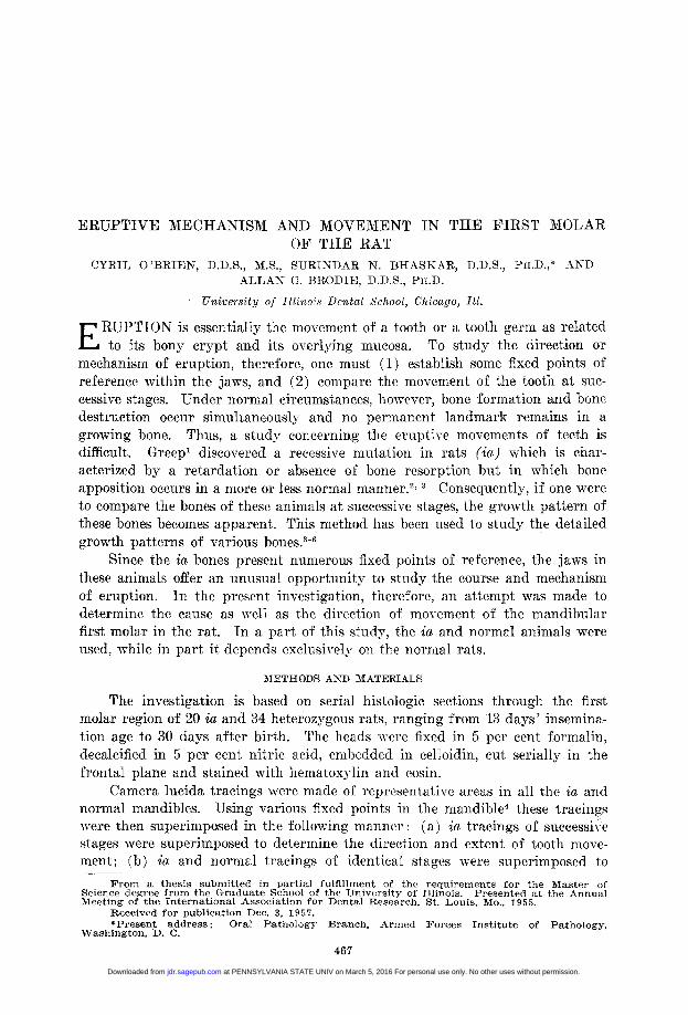
ERUPTIVE MECHANISM AND MOVEMENT IN THE FIRST MOLAROF THE RAT
CYRIL O 'BRIEN. D.D.S.. M.S.. SURINDAR N. BHASKAR. D.D.S., PH.D.,* ANDALLAN G. BRODIE, D.D.S., PH.D.
University of Ilitnos Dental School, Chicago, Ill.
E RUPTION is essentially the movement of a tooth or a tooth germ as relatedto its bony crypt and its overlying mucosa. To study the direction or
mechanism of eruption, therefore, one must (1) establish some fixed points ofreference within the jaws, and (2) compare the movement of the tooth at suc-cessive stages. Under normal circumstances, however, bone formation and bonedestruction occur simultaneously and no permanent landmark remains in agrowing bone. Thus, a study concerning the eruptive movements of teeth isdifficult. Greepl discovered a recessive mutation in rats (ia) which is char-acterized by a retardation or absence of bone resorption but in which boneapposition occurs in a more or less normal manner.2 Consequently, if one wereto compare the bones of these animals at successive stages, the growth pattern ofthese bones becomes apparent. This method has been used to study the detailedgrowth patterns of various bones.3-6
Since the ia bones present numerous fixed points of reference, the jaws inthese animals offer an unusual opportunity to study the course and mechanismof eruption. In the present investigation, therefore, an attempt was made todetermine the cause as well as the direction of movement of the mandibularfirst molar in the rat. In a part of this study, the ia and normal animals wereused, while in part it depends exclusively on the normal rats.
METHODS AND MATERIALS
The investigation is based on serial histologic sections through the firstmolar region of 20 ia and 34 heterozygous rats, ranging from 13 days' insemina-tion age to 30 days after birth. The heads were fixed in 5 per cent formalin,decalcified in 5 per cent nitric acid, embedded in celloidin, cut serially in thefrontal plane and stained with hematoxylin and eosin.
Camera lucida tracings were made of representative areas in all the ia andnormal mandibles. Using various fixed points in the mandible4 these tracingswere then superimposed in the following manner: (a) ia tracings of successivestages were superimposed to determine the direction and extent of tooth move-ment; (b) ita and normal tracings of identical stages were superimposed to
From a thesis submitted in partial fulfillment of the requirements for the Master ofScience degree from the Graduate School of the University of Illinois. Presented at the AnnualMeeting of the International Association for Dental Research, St. Louis, Mo., 1955.
Received for publication Dec. 3, 1957.*Present address: Oral Pathology Branch, Armed Forces Institute of Pathology,
Washington, D. C.
467
at PENNSYLVANIA STATE UNIV on March 5, 2016 For personal use only. No other uses without permission.jdr.sagepub.comDownloaded from

468 O'BRIEN, BHASKAR AND BRODIE J. D. Res.June. 1958
determine the role of bone resorption in tooth movement; and (c) since from13 days' insemination age to 5 days after birth the ia first molar tooth germ isrelatively undisturbed, up to this stage the ia jaws could be used to advantage.From 5 days to 30 days after birth, tracing of successive normal jaws were super-imposed to determine the extent and direction of movement. In part, this studyconsists of a histologic analysis of the eruptive changes in and around the firstmolar tooth germ.
REVIEW OF LITERATURE
Literature concerning the eruptive movement of teeth is voluminous andcontradictory. Since in different studies different aspects of eruption have beenemphasized, the information on this subject may be grouped under the followingmain headings.
A. Movement of the Tooth Germ and Tooth.-The movement of developingteeth prior to the formation of the root was first recognized by Brash.7 Brodie8suggested that prior to eruption the developing and growing teeth move toadjust their position in the growing jaws. Weinmann9' 10 reported that duringthe pre-eruptive phase (prior to the beginning of root formation) the toothgerms move bodily as well as by excentric growth. These movements are asso-ciated with apposition in the area of the crypt from which the tooth migratesand resorption of bone on the side of migration. In contrast, Landsberger,"Diamond and Applebaum,'2 and Diamond,'3 reported that the developing crowndid not leave its original initiation site until root formation began and that itsvertical height increased by shifting of cervical loops in the direction of thefundus. According to these authors, the only ocelusal displacement prior toroot formation was the addition of enamel increments. Sicher'4' 15 proposedthat the developing root was prevented from growing into the alveolus by thehammock ligament and that the epithelial diaphragm was a fixed point.
The continuous eruption of human teeth is recognized and has been sub-divided into the active and passive phases. 18 19
B. Changes in the Crypt.-That the crypt undergoes resorption during thegrowth of the tooth germ has been shown repeatedly.9' 20, 21 During the stagefollowing the start of root formation, bone apposition occurs rapidly at thefundus of the crypt.9' 16 The apposition and resorption pattern of bone in thecrypt has been related to the rate of growth and the direction of eruption of thetooth. In addition, Brash7 showed, in madder-fed pigs, that during the activeeruption extensive growth is seen on the crests of alveolar crypts.
C. Tooth Movement and Growth of Alveolar Process.-Landsberger"l foundthat the removal of tooth germs from the maxilla of dogs led to an absence of thealveolar process, shortening of the maxilla, and failure of the floor of the nose todescend normally. Further, it has been shown that in cases of anodontia, al-though the mandible develops normally, its alveolar process is completelymiSSing.22, 23
D. Tooth Eruption and the Follicle.-Orban24 described the primitive perio-dontal membrane as having 3 distinct layers, 2 outer layers adjacent to thebone and the tooth germ, respectively, and a central zone. The outer zones
at PENNSYLVANIA STATE UNIV on March 5, 2016 For personal use only. No other uses without permission.jdr.sagepub.comDownloaded from
Volume 37 ERUPTIVE MECHANISM AND MOVEMENT IN MOLAR 469Number 3
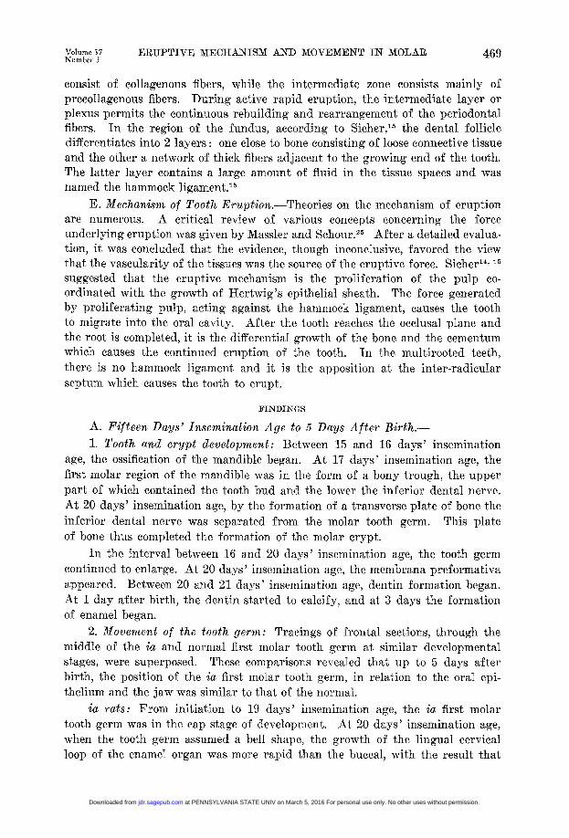
consist of collagenous fibers, while the intermediate zone consists mainly ofprecollagenous fibers. During active rapid eruption, the intermediate layer orplexus permits the continuous rebuilding and rearrangement of the periodontalfibers. In the region of the fundus, according to Sicher,15 the dental follicledifferentiates into 2 layers: one close to bone consisting of loose connective tissueand the other a network of thick fibers adjacent to the growing end of the tooth.The latter layer contains a large amount of fluid in the tissue spaces and wasnamed the hammock ligament.'5
E. Mechanism of Tooth Eruption.-Theories on the mechanism of eruptionare numerous. A critical review of various concepts concerning the forceunderlying eruption was given by Massler and Schour.25 After a detailed evalua-tion, it was concluded that the evidence, though inconclusive, favored the viewthat the vascularity of the tissues was the source of the eruptive force. Sicher'4' 15suggested that the eruptive mechanism is the proliferation of the pulp co-ordinated with the growth of Hertwig's epithelial sheath. The force generatedby proliferating pulp, acting against the hammock ligament, causes the toothto migrate into the oral cavity. After the tooth reaches the occlusal plane andthe root is completed, it is the differential growth of the bone and the cementumwhich causes the continued eruption of the tooth. In the multirooted teeth,there is no hammock ligament and it is the apposition at the inter-radicularseptum which causes the tooth to erupt.
FINDINGS
A. Fifteen Days' Insemination Age to 5 Days After Birth.-1. Tooth and crypt development: Between 15 and 16 days' insemination
age, the ossification of the mandible began. At 17 days' insemination age, thefirst molar region of the mandible was in the form of a bony trough, the upperpart of which contained the tooth bud and the lower the inferior dental nerve.At 20 days' insemination age, by the formation of a transverse plate of bone theinferior dental nerve was separated from the molar tooth germ. This plateof bone thus completed the formation of the molar crypt.
In the interval between 16 and 20 days' insemination age, the tooth germcontinued to enlarge. At 20 days' insemination age, the membrana preformativaappeared. Between 20 and 21 days' insemination age, dentin formation began.At 1 day after birth, the dentin started to calcify, and at 3 days the formationof enamel began.
2. Movement of the tooth germ: Tracings of frontal sections, through themiddle of the ia and normal first molar tooth germ at similar developmentalstages, were superposed. These comparisons revealed that up to 5 days afterbirth, the position of the ia first molar tooth germ, in relation to the oral epi-thelium and the jaw was similar to that of the normal.
ia rats: From initiation to 19 days' insemination age, the ia first molartooth germ was in the cap stage of development. At 20 days' insemination age,when the tooth germ assumed a bell shape, the growth of the lingual cervicalloop of the enamel organ was more rapid than the buccal, with the result that
at PENNSYLVANIA STATE UNIV on March 5, 2016 For personal use only. No other uses without permission.jdr.sagepub.comDownloaded from
470 O'BlRIEN, 1 IkASIKAR, ANI) IBUOI)IEK 1). Ics.lI i'. 19;s
thel tooth germi. Cassurlneli a tiltt(ed position w- within the erypt. T7is positioii wa85suit that tlhe future e occhisal. plane of thle tooth wN-as inelnedl inwcaird aind upwarda11nd thle cervicial- openlincg Ax-as diieted dowlwvaral and outward (Fig. 1).
From 20 daxs' insemnination nage to 5) (la-s after lirth, the ia first molar gcrew,considerablxtIii size 1ut 111(inaiuaiit l tl1si incliialiion. A superpstii of cornparatible trlacings of the ia mtandihie at snecessixe stages, indiealted thait! thedentiliioeiilae Junictionl progrec-,ssively shi-ifts iipwaid, and that thle most ii-jloi'tait;11shift of, thle tool-th1(elylg was ini a s-pteroi}lateral1 diet iou as iliiiealtd I)n>lie aironms inl Pligs. 2, 3, anl 4. 1 )iirin tliiS pei9'iod, al111t0ilol tIlic w as ait
ilnlce(ase ill size (of tilem i tlootil gen11,1 ther-eAwaNf1s n1o dlistoi't loll ol its iiiol'pliolog-except ili tile 1a'ea of tI lie 'eixv ica loops. The -facet thiat ill tlie w ia Is. (ieSiIt
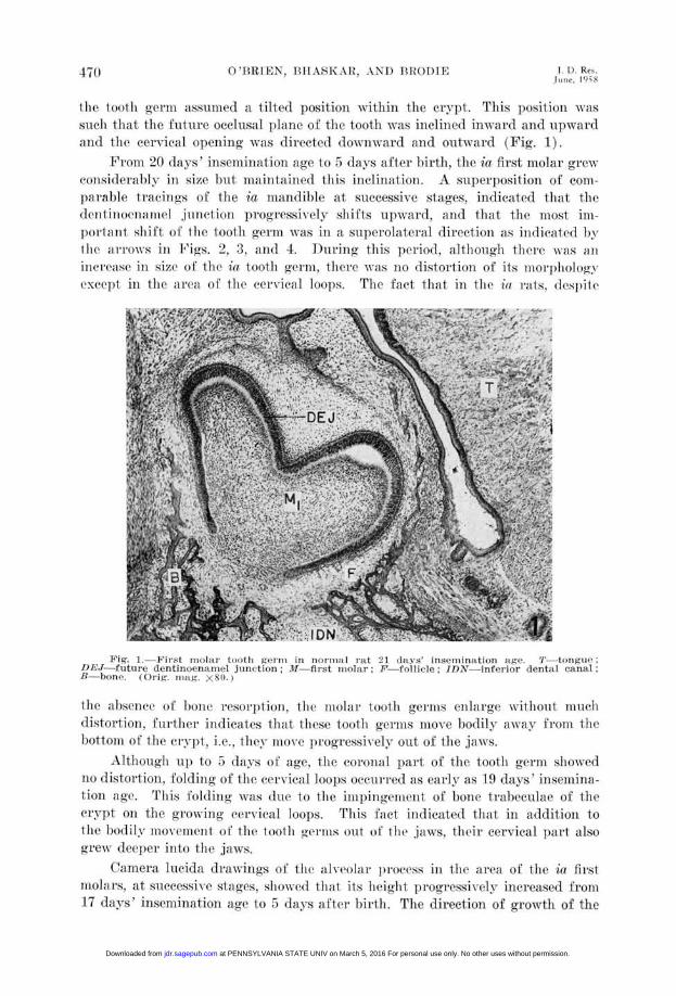
ig. 1 -First riiolar tooth genii in nflornitl rtt ':1 (laxs' inseiiiination age. TI torigus':I)EJ future dentinoenamiel junction; .1--first molar;F-, follicle; IDN-inferior dental canal;B-bone. (Orig. mtg. X8S1.
hi' ahlselice)8(81] of' [)Otifbloei'eso^i'ptiovii the> mlolarl too~th germsbS enlage xx'itlionlt. ronchmlg w
distortion, 1itlier inLlicates tltat I- ie>se tooth germs move l)oilyofl a ay fiotltleliottoll 1of t1e erypt, i.e.. they mlo\ve progressively out of the jaw-S.
Altlioglloi- up to 5 dlays of age, tie coi'onlt . part of tle tootl gerii slowed10) dli;Stort loll, folding of tle cr,(v'cal lOO])5;I Ioos occi'i'el' as earIl' as 19 da >s ' insemina-iou age. 'P'llis foldinl wal"s due to the imii)iiglemliit of boet tIrabeculae of thecript ol le grox il1i ervvieald loops. T1iS iact indicated thlat ii at-itlitioll to
thet Io of1 thovement()ltletooth gelilts out of, tlet, jsaws. their cervical part .dsogre'-dnAV'7epel' ilnto tilt' jaw's.
Camera 1lteida1 drawings of tilI ola I})Iovtopress i t 1 et of' thfl1e ia firstuohai's, at sliuect'ssix'e Staes, Sllj1Ox,id tiht its height progr'essivey inc>rluiased front17 days' iniselnination ag(e to 5 days3s a(ftei birth. The direction of gi'owth of the
at PENNSYLVANIA STATE UNIV on March 5, 2016 For personal use only. No other uses without permission.jdr.sagepub.comDownloaded from

Volume 37Number 3
ERUPTIVE MECHANISM AND MOVEMENT IN MOLAR 471
alveolar process, as seen by the arrangement of the bony plates, was upwardand outward. However, the tooth germ maintained a constant relationship withthe medial and lateral alveolar crests, indicating once again its upward andoutward migration. Since the alveolar crest was always below the widest partof the tooth germ (Fig. 1), it is obvious that the upward and outward migrationof the tooth germ was faster than the growth of the crest. If the growth ofthe alveolar process had occurred independently, or prior to the upward bodilymovement of the tooth germ, the latter would have shown gross distortion ofshape.
MG
DEtjDEJ
4PSC
PSA
3Figs. 2, 3, and 4.-Superimposed camera lucida tracings of the first molar area in ia
animals at successive stages to show an occlusal and lateral migration of the tooth germ.Broken line is the older stage. Fig. 2 represents 20 days' insemination age and birth. Fig. 3.1 and 2 days after birth, and Fig. 4 represents 1 and 5 days after birth. T-tongue; M1 molartooth germ; DEJ-future dentinoenamel junction; PS perichondral splint of condylarcartilage; PSA-perichondral splint of angular cartilage. (Orig. mag. X18.)
Normal rats: The superposed tracings of sections through first molar regionof ia and normal rats at comparable stages, showed identical position of theteeth. The movements of the normal first molar tooth germ, therefore, were thesame as described in the ia rats.
Study of serial sections of the bony crypt of the first molar tooth germshowed that, up to 20 days' insemination age, resorption occurred on all surfaces
at PENNSYLVANIA STATE UNIV on March 5, 2016 For personal use only. No other uses without permission.jdr.sagepub.comDownloaded from
472 O 'BRTEN, 13ITASKAR, AND BROI)1E J. 1). Res.Juille, 1958
of the erypt. Frvo2m21 days' insemination age to 5 days after lirth, inactivity,or a varying degree of 1)1neopposition, could be seen on the wall of the cryptwhich faced the inferolinguall part of thie tooth germ, while elsewhere honeresorption eoltilnel. The areca ot bone) poapsition was opposite to the directionin which the tooth. germn wals migrating.
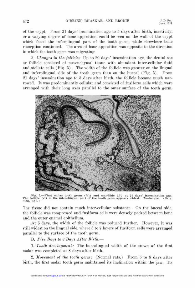
3. Changes in the folliele: lip to 20 (lays' insemination age, the dental sacor follicle coiisisted of- ImeCseOnIChxnilfX.1 tissue, xitli a-lllndant inter-cellular fluidand stellate (clls (Fig. 5). The width of the 'follicle was greater on the lingualalnd inferolingual side of the tooth germ than on the buecal (Fig. 5). From21 days' insemination age to 3 d(lys after 1)irthl, the follicle became much nar-rowed. It xas predominantly cellular and consisted of fusiform cells which werearrianged xxitli their long axes parallel to the oiter surface of the tooth germ.
Fig. 5a. 'irst miolar tooth germ (Mi) and miandible (K) ait 20 days' insemination age.Tlin follicle (Fh) in the inferolingual part of the tootlh germ appears xw iflest. T tongue. (Orig.moI:Ig. X80.)
The tissue dlid not contain much inter-celluhlar substance. On the buccal side,thlie follicle vfas compr.-essed and fusiformn cells were densely packed between b)onend the outer enamel epitheliuin.
At 5 davs, the width of the follicle was1 reduced further. HIowever, it wasstill widest on the ling-ual side, where 6 to 7 la-yers of fusiforin cells xvere arrangedparallel to the surface of the tooth germ.
B. Five Days to 8 Days After Birth.-1. Tooth development: Thie buecolingtualwidthh of the crown of the first
molar wvas completed at 8 days (after birth.2. lMorentent of the tooth gcrm: (Nornal rats.) From 5 t o 8 days after
birth, the first molar tooth gerin maintained its inclination within the jaw. Its
at PENNSYLVANIA STATE UNIV on March 5, 2016 For personal use only. No other uses without permission.jdr.sagepub.comDownloaded from

Volume 37Numbler
ERUPTrIVE MECHANISM ANT) MOVEMENT -IN MOLAkR 473
ocelusal surface still faeed upward and ilnward(, arid its cervical suIrface faced(lowllward and outward. At 5 (lays the relationship between the tooth gelrnand the alveolar crests changed. Whereas tip to tlis time thle tooth extendedbeyond the alveolar crests now the medial and lateral Jhlates of the alveolarprocess surrounded it and extended to its ocelusa] surface. Tilus the alxcolarprocess, during this timne, grew faster than the upward migration of the toothgerm.
The part of the crypt which faced the infer-olingual surfaces of the toothgerm showed bone apposition, while elsewhere resorption of bonle occurred.Bone resorption in the funduis of the crypt led to the perfora(tin of the hateralLate of the alvolar process (Fig. 6). This pattern of b)one apposition ilndresorlption in the Clv,)t indicated (a) an upward and outw aird migration of thetooth gerin, and (b) a downward alnd outwaild growth of' thle eerxical 1001) regionof the toothl gerlIml.
Fig. 6.-First molar tooth germ (Ml) in normal andibleb at 6 lays. Note completeabsence of bone on the lateral buccall) surface. Follicle is seen only on the lingual part ofthe tooth bud. B- bony plates of mandible; IDN inferior dental nerve. and canal. (Orig.nag. X80.)
3. Changes in the follicle: At 6 and 8 days, a (lefilite follicle could be seenonly on tire lingual side. At 8 days, the inter-cellular fluid decrea-csed, the fibro-blasts increased in number, and a few collagen fibers appealed, which, like thecells were oriented parallel- to the surface of tire tooth gerirn.
C'. Ten to 20 Days After Birth.-I. Tooth development: At 10 days after birth, lfrtwig's epitlhelial root
sheath appeared and the formation of the root began. Formation of the rootbifurcation, inter-ra-ldicular bony septa, and the maturation of enamel began at12 days (Figs. 7 and 9). At 17 days after birth, the enamel, with the exception
at PENNSYLVANIA STATE UNIV on March 5, 2016 For personal use only. No other uses without permission.jdr.sagepub.comDownloaded from
474 (0) IEIN. P11A;\SKAR, ANI)AD OI)RIE i. D. Res.bllue, 198t8
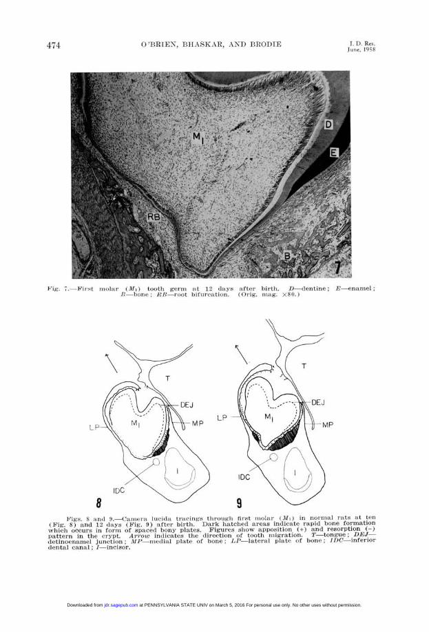
Fig. 7.Fiut nllar (0lI) tooth gci ni at 12 dlaiis n ftrI birth. D dentine -enanmel- hone FL Iot htbifurcation ((trig. n1mg. X80i)
LP-MP
8Pig-S. S 1l 9.(9.itiml-ra 1(1j(11t tracings tiirougli tirst miolii (31, ) in nloliltill ruts At ten
(Fig. 8) and 12 (lays (Fig. 9) after birth. Dark hatched areas indicate rapid bone formation-v lhi( occurs in formi of spaced bony plates. Figures show apposition (+) and resorption (-)pattern in the cry pt. Arrowv indicates the directionn of tooth muig ration. T-tongue, DEJdletinoenanIel junction .lIP medial plate of' hone; LP-lateral plaIte 4f hone; lDC-inferior(dental canal ; I-incisor.
at PENNSYLVANIA STATE UNIV on March 5, 2016 For personal use only. No other uses without permission.jdr.sagepub.comDownloaded from
V u1noe :>Nuill l,iC
ER I7IVE'y MEA\lINISMI ANDMOVE.MENT IN- MOLAR 4T5
of a, slighit (eicl'.1Pllo0tion, wo.85 co1mIletely ilaluttiiedl, 8d tile11( l isial ('lis1(s ofthe toot1l 'lbroke through' the0l181 11111COS.. -At 19(lXa's aliterllthl. the rew11ill-ill, eiillet in tile Ce'lrvical 81108l mIa-ltureld .11(1 tIle, (list at c0s1)s also(ilt eted thleof.'.al cavity.
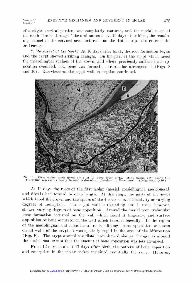
2. Moloccnifnt of thI tooth1: At 1(0 d(188 alloter 1)1itl thle o(ot lformlntioi Ilglll.11(1 tilhe rtx]t sholell striking elmiaigs. Oti the p)nt of tile (11)t1wlich facedthe in1-felorlingluail silrfaIce of tI-le ('lOW,('11d1(1 where previously sura-.ce 1)01bn 11p-fPOsitioll oee(nil'ed, 11now 1)o110 AN as folre1(1 ill t lallellar anu.angtrelill ( Fjicrs. Sand 10). E]sewx lw1r' 0o1 tile erviAt N'.11i, resorptio111 o011ti1le0d.
Jig. 11.,-leFirst moa;lar tootlh germw (TI0) at 15 lax aaftes r birth. Bone tissue (H) above) thofli;oc8 line represents neN -ly formed trabeeulWe. I) dentin; E-enamiel. (() ig. maig. X'N.)
-At -12 (>d-s the loots of- the first- nIolar (mesial, iiesioliiigtial. aIn'ito)ltlccal,81n(1 htistal)had foriited to 11-Il n A s the pl1)8118 of the Cryp1>tw hiel) laced tle roPwn 1(1d the pf)1s(.)of' the 4 roots showed irtiadfxit> 01' Avxn-igtgdegr-ees of iresoirption. Thoe el,-pt xVal.l surroumidiii-n the 4 i'oots, hoxVcx or,showed valyvig (legilees (If 1)o0e appositioii. A'vound the iesial root,t tl'rahecular'bonc folilmation oceurtied on the Avail AN lielt J.laeed it, 1lloguahly, arind surifaear)1)osition of,' hone ocur'r'1d toil thle xxall whlli('1 fa(aed it uL('(ally. In. the rcoioof- thle lIlesiolillgual a(l1d mlilesi'ollleal roots, hth'1olloil bone ah)iosit loll xxv85 8001
()I1 all xxalls of tlle. civilt it INs88 ec('ially 183)4d iii the areaof, the 1(iflll'tiollPigi') The ervpl)t arotn(Id the (1di8t1 11 oot sIhowN ed simlliri' changes as alonild
tfle s;al151 oot-, except that till aiimoiiiit of' 1)10le ap}}ositioii xxas less dvalldeed.F'omia 12 (das 1(o 8 T1011117 (lavs 8 ftc 1'h, i ithi 1 lie pat 1-0d11 of 11(111e 81ppositioll
aid1 rtesorptiolu in the molar soklet 1ellLaillcd essemitially the s8811e. I owexvei,
at PENNSYLVANIA STATE UNIV on March 5, 2016 For personal use only. No other uses without permission.jdr.sagepub.comDownloaded from
476 O'BRIEN, BHASKAR, AND BRODIE 1. D. Res.June, 1958
rapid bone growth, as indicated by its trabecular arrangement, could now beseen on the walls of the socket facing the buccal surfaces of the mesial, mesio-buccal and distal roots (Fig. 11). As the roots lengthened and the tooth migratedocelusally, the rapid formation of trabecular bone on the walls of the socketcontinued. In addition, trabecular bone formation could also be seen in theareas of the bifurcation of roots. During these stages, the upper part of thesocket wall facing the buccal surface of the crown consistently showed areasof resorption.
At 19 days after birth, rapid bone apposition in the molar socket ceased.At this time slow surface apposition occurred only on walls which faced theapical parts of the roots, while the upper portion of the buccal alveolar plateshowed continued resorption.
TDEJ
M1
LP-- 1=7 ~~~LPMP ~~~~~MP
IDC~~~~~~D12
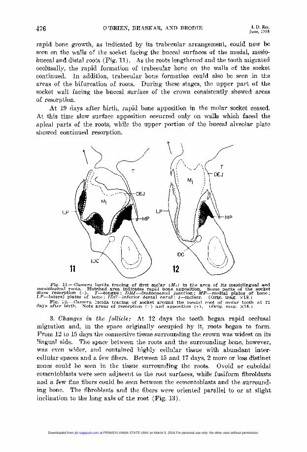
Fig. 11.-Camera lucida tracing of first molar (Mi) in the area of its mesiolingual andmesiobuccal roots. Hatched area indicates rapid bone apposition. Some parts of the socketshow resorption (-). T-tongue; DEJ-dentoenamel junction; MP-medial plates of bone;LP-lateral plates of bone; IDC-inferior dental canal; I-incisor. (Orig. mag. X18.)
Fig. 12.-Camera lucida tracing of socket around the mesial root of molar tooth at 21days after birth. Note areas of resorption (-) and apposition (+). (Orig. mag. X18.)
3. Changes in the follicle: At 12 days the tooth began rapid occlusalmigration and, in the space originally occupied by it, roots began to form.From 12 to 15 days the connective tissue surrounding the crown was widest on itslingual side. The space between the roots and the surrounding bone, however,was even wider, and contained highly cellular tissue with abundant inter-cellular spaces and a few fibers. Between 15 and 17 days, 2 more or less distinctzones could be seen in the tissue surrounding the roots. Ovoid or cuboidalcementoblasts were seen adjacent to the root surfaces, while fusiform fibroblastsand a few fine fibers could be seen between the cementoblasts and the surround-ing bone. The fibroblasts and the fibers were oriented parallel to or at slightinclination to the long axis of the root (Fig. 13).
at PENNSYLVANIA STATE UNIV on March 5, 2016 For personal use only. No other uses without permission.jdr.sagepub.comDownloaded from

Fig1. Ievloing peridna ninbrn (P1)11)(at 1 safe irh ), tn.(rg
#.*0. ia . )
ffE # jERE ._ : :.^' ffi t - E q~~~~~A
Fixg. 13.-De(velopinlg periodountal mlemlbrane (PDM) at 15 (Iaysfcter birth. D -dentin. (COrig.Illag. X300 ?
'7T±. W - UtFlig. 14. Frontal section through the mesiolingtlal and mesiobuccal roots at 21 days after birth.
The periodontal membrane is wider on the lingual than on the buccal sides.
at PENNSYLVANIA STATE UNIV on March 5, 2016 For personal use only. No other uses without permission.jdr.sagepub.comDownloaded from
478 O'BRIEN, BHASKAR, AND BRODIE JunD. Res.
At 17 and 19 days, respectively, the periodontal space around the mesialand the other 3 roots began to widen (Fig. 12). This was correlated to thepattern in which the occlusal surface of the tooth erupted into the oral cavity.The periodontal space was wider on the lingual sides of the roots than on theirbuccal sides (Fig. 14). The general orientation of the fibers was from theregion of the cementoenamel junction to the alveolar crests, and from the boneof the socket to directly across or to slightly apical areas on the root (Fig. 14).On the buccal side of the mesiolingual root, and on the lingual of the mesiobuccalroot, however, the collagen fibers ran parallel to the long axis of the roots (Fig.14).
D. Twenty-one to 30 Days After Birth.-1. Tooth development: The first molar tooth germ came into functional
occlusion at about 23 days of age. Thirty days after birth the root formationwas completed and their apices began to show apposition of cellular cementum.
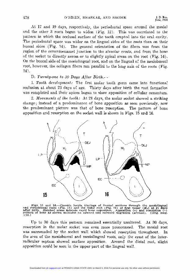
2. Movements of the tooth: At 21 days, the molar socket showed a strikingchange; instead of a predominance of bone apposition as seen previously, nowthe predominant picture was that of bone resorption. The pattern of boneapposition and resorption on the socket wall is shown in Figs. 15 and 16.
TT
DEJ --
LP
- ~~MP
IDC
15 1
Figs. 13 and 16.-Camera lucida tracings of frontal sections through the mesiolingualand mesiobuccal roots (Fig. 15) and the distal root (Fig. 16) of first molar (Mi) at 21 daysafter birth. Hatched areas indicate rapid bone apposition. Apposition (+) and resorption (-)pattern of bone as shown indicates an upward and outward migration (arrows). (Orig. mag.X18.)
Up to 30 days this pattern remained essentially unaltered. At 30 days,resorption in the molar socket was even more pronounced. The mesial rootwas surrounded by the socket wall which showed resorption throughout. Inthe area of the mesiobuccal and mesiolingual roots, only the crest of the inter-radicular septum showed surface apposition. Around the distal root, slightapposition could be seen in the upper part of the lingual wall.
at PENNSYLVANIA STATE UNIV on March 5, 2016 For personal use only. No other uses without permission.jdr.sagepub.comDownloaded from
Volume 37 ERUPTIVE MECHANISM AND MOVEMENT IN MOLAR 479Number 3
3. Changes in the follicle: From 21 to 30 days, the periodontal membranespace continued to widen. At 23 days, the connective tissue of the periodontalmembrane consisted of densely collagenized, well-oriented fibers. They extendedfrom the bone to slightly apical areas on the root. The only exception wasthe buccal side of the mesiolingual root, where the fibers were oriented parallelto the long axis of the root. At 30 days, however, even in this area the fibersextended from the alveolus to the root surface.
DISCUSSION
A. Eruptive Movement of the First Lower Molar of the Rat.-Weinmann10 divided the movement of a tooth into 3 distinct phases, pre-
eruptive, pre-functional, and functional. The pre-eruptive phase was designatedas the movement of a tooth prior to root formation. By such movement, whichoccurs mainly as excentric growth, the teeth adjust their position within thejaws. The pre-functional phase begins with root formation and ends when thetooth reaches occlusion while the functional phase begins when the teeth reachocclusion and ends with tooth loss.
The present investigation further confirms the view that the movementof the tooth germ begins in the cap or bell stage and from then on continues atvarying rates. From the time the first molar tooth is in cap or early bell stage,the mandible grows rapidly at its crest as well as in thickness by appositionon its lateral surface.' To keep up with this mandibular growth, the toothgerm moves in an upward and outward direction. If the tooth germ did notmove in this manner it would soon become embedded within the jaw.
The superposition of tracings of the ia and normal molars shows that,up to about 5 days, the molar moves and grows in a more or less identicalmanner. If one superimposes tracings of ia molars at successive stages, it isseen that the dentinoenamel junction of this tooth progressively shifts in anupward and outward direction. Since the dentinoenamel junction is a fixedpoint in a tooth, its shift implies a corresponding shift of the tooth. Thatthe tooth germ moves bodily at this early stage is illustrated further by thefact that, on the inferolingual wall of the crypt, apposition of bone or restinglines can be seen, whereas elsewhere, on the surface of the crypt, resorptionis in progress. These resting lines, or bone apposition, indicate the movementof the tooth germ away from this wall. It has also been shown that on thelingual and inferolingual sides of the tooth germ the follicle is always widerthan on the lateral side. This observation further indicates the bodily move-ment of the tooth germ.
A study of the resorption and apposition pattern of the crypt from 5 to Sdays in the normal rats indicates that the movement of the tooth remains es-sentially unchanged.
At 10 days, the root formation begins and the movement of the toothbecomes more rapid. From 10 to 21 days after birth, the relationship betweenthe alveolar crest and the crown progressively changes so that the crown movestoward the oral cavity at a faster rate than the growth of the alveolar crests.
at PENNSYLVANIA STATE UNIV on March 5, 2016 For personal use only. No other uses without permission.jdr.sagepub.comDownloaded from

480 06'BkIEN, BHASKAR, AND BRODIE i. D. Res.June, 1958
In other words, the crown progressively moves out of the jaws. During thisphase, the apposition of bone is fastest on those walls of the socket which facethe inferolingual and lingual surfaces of the roots. Also, during this periodthe newly formed periodontal membrane tends to be widest in these areas.These observations indicate that while the tooth moves axially toward the oralcavity, it also tilts in an outward direction. Since, during development, themolar crown is steeply inclined within the jaws, this outward movement ofthe tooth during eruption is a necessary adjustment for its normal positioningwithin the jaw.
From 21 days to 30 days of life, the movement of the tooth is considerablyslowed. This is due to the fact that the first molar has already reached itsocclusal plane.
B. Downward Growth of the Tooth Germ.-Whether a tooth germ during its development grows down into the jaws has
been a point of controversy.12' 13, 24 The present investigation shows that themolar tooth germ of the rat does grow into the jaws for a short distance. Astudy of the ia first molar revealed that, up to 5 days, the molar crown is notdistorted on its occlusal, lingual, or buccal surfaces. However, slight impinge-ments by bone occurred in the area of the cervical loop, and they led to pro-gressive distortion of the tooth. Since in ia jaws, resorption is absent,2' 4 itis obvious that these cervical areas of distortion are correlated with the downgrowth of the cervical loop region of the tooth germ. Between 10 and 12 days,the bottom of the crypt showed beginning of bone apposition, which indicatedthe cessation of the down growth of the tooth germ.
C. Correlation Between Tooth and Jaw Growth.-It has been shown that the molar crown is so inclined within the jaw that
its occlusal plane faces inward and upward. Later when the tooth begins itsmovement toward the oral cavity, the crown straightens its position. Theinclination of the molar crown during development is an adaptation of thetooth growth to the jaw growth. The rat has a single set of molars. The basicsize of a tooth crown does not change after the establishment of the dentino-enamel junction, while the mandible of the rat grows almost continuously. Therat molar, which attains its permanent adult size very early, develops in amandible which is comparatively much smaller. Since the transverse width ofthe rat molar crown in a horizontal position is much larger than in a tiltedposition, the development of the molars in this position makes it possible forthese teeth to grow to adult dimensions in a relatively small jaw.
D. The Follicle.-This study has shown further that the follicle undergoes changes which
are correlated to tooth movement and function. Until the root formationbegins, the follicle consists of loosely arranged stellate and fusiform cells, a fewfibrils, and abundant interfibrillar substance. The width of the follicle tendsto be greater on the sides from which the tooth germ is migrating than on theside to which it migrates.
at PENNSYLVANIA STATE UNIV on March 5, 2016 For personal use only. No other uses without permission.jdr.sagepub.comDownloaded from
Volume 37 ERUPTIVE MECHANISM AND MOVEMENT IN MOLAR 481Number 3
As the root formation begins and the tooth moves rapidly toward the oralcavity, the connective tissue around the crown appears unchanged, but surround-ing the roots there is abundant formation of new connective tissue, whichis the precursor of the periodontal membrane. This tissue is at first mainlycellular, but gradually collagen fibers appear in it. In the beginning, the cellsand few fibers are oriented parallel to the root surface, but as soon as thetooth reaches its occlusal plane (21 days) the fibers and cells become arrangedso that they run from the bone to the cementum. Also, at this time, the boneapposition on the walls of the socket is replaced by bone resorption. This leadsto a widening of the periodontal membrane. The collagen bundles of theperiodontal membrane also progressively increase. These changes are correlatedto the establishment of the masticatory function, and are first noted around themesial root, then around the mesiobuccal and mesiolingual roots, and finallyaround the distal root. Since the eruption of the molar tooth has an anteriorposterior gradient, the order in which these changes occur is not unexpected.
E. Mechanism of Eruption.-The present investigation has shown that the tooth eruption begins almost
simultaneously with the formation of the tooth bud. Since this is the case, thismovement cannot be dependent upon the elongation of root, formation of dentin,or the apposition of enamel. Further, since the teeth erupt farther than thelengthening of their roots, one can conclude that root formation is not thecause of eruption. It was demonstrated in the present study that duringsome stages the eruption of the molar tooth germ was associated with inactivityor resorption on the walls of the crypt. It may be therefore assumed that thebone formation alone cannot cause eruption.
Essentially, eruption is the movement of one organ (tooth) in relation tothe other (the bony crypt or socket). This phenomenon is not unique to theteeth but is commonplace in ontogenesis. The "movement' of the heart fromthe visceral arch region to the thorax, of the thyroid anlage from the base ofthe tongue to the neck, of the eyes from the lateral to the anterior aspects ofthe face are all examples of organ migration or movement. These shifts inorgan position are undoubtedly a result of differential growth and directionalgrowth. Eruption, like other organ movements, appears to be the result ofdifferent rates of growth of the dental pulp, dental follicle, and the bony crypt.To simplify the mechanism of tooth eruption, one may compare it with thegrowth or "movement" of 2 bones as a result of sutural growth. The growthof connective tissue of a suture leads to a "pushing apart" of the participatingbones. Following this, bone apposition occurs on the opposing surfaces so thatthe suture is maintained at a more or less constant width.
From the present investigation it would appear that the eruption of teethis probably similar to the sutural growth and the connective tissue of the dentalfollicle and periodontal membrane is analogous to the connective tissue of thesuture. The growth of the follicular connective tissue leads to the migrationof the tooth germ or tooth (comparable to one of the bones) from the bottom
at PENNSYLVANIA STATE UNIV on March 5, 2016 For personal use only. No other uses without permission.jdr.sagepub.comDownloaded from
482 O'BRIEN, BHASKAR, AND BRODIE JunD. Res.
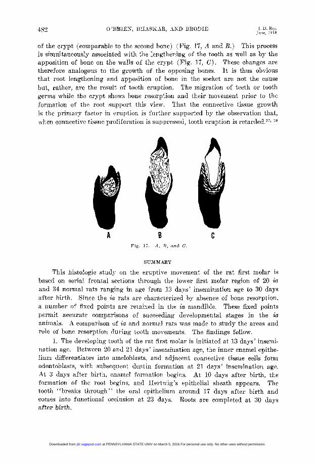
of the crypt (comparable to the second bone) (Fig. 17, A and B.) This processis simultaneously associated with the lengthening of the tooth as well as by theapposition of bone on the walls of the crypt (Fig. 17, C). These changes aretherefore analogous to the growth of the opposing bones. It is thus obviousthat root lengthening and apposition of bone in the socket are not the causebut, rather, are the result of tooth eruption. The migration of teeth or toothgerms while the crypt shows bone resorption and their movement prior to theformation of the root support this view. That the connective tissue growthis the primary factor in eruption is further supported by the observation that,when connective tissue proliferation is suppressed, tooth eruption is retarded.27' 28
A B CFig. 17. A, B. and C.
SUMMARY
This histologic study on the eruptive movement of the rat first molar isbased on serial frontal sections through the lower first molar region of 20 iaand 34 normal rats ranging in age from 13 days' insemination age to 30 daysafter birth. Since the ia rats are characterized by absence of bone resorption,a number of fixed points are retained in the ia mandible. These fixed pointspermit accurate comparisons of succeeding developmental stages in the icanimals. A comparison of ia and normal rats was made to study the areas androle of bone resorption during tooth movements. The findings follow.
1. The developing tooth of the rat first molar is initiated at 13 days' insemi-nation age. Between 20 and 21 days' insemination age, the inner enamel epithe-lium differentiates into ameloblasts, and adjacent connective tissue cells formodontoblasts, with subsequent dentin formation at 21 days' insemination age.At 3 days after birth, enamel formation begins. At 10 days after birth, theformation of the root begins, and Hertwig 's epithelial sheath appears. Thetooth "breaks through" the oral epithelium around 17 days after birth andcomes into functional occlusion at 23 days. Roots are completed at 30 daysafter birth.
at PENNSYLVANIA STATE UNIV on March 5, 2016 For personal use only. No other uses without permission.jdr.sagepub.comDownloaded from
Volume 37Number 3
ERUPTIVE MECHANISM AND MOVEMENT IN MOLAR 483
2. The movement of the tooth germ (eruption) begins soon after its initia-tion and continues until 30 days of age. Prior to the beginning of root forma-tion, the tooth germ moves upward and outward and thus keeps a constantrelation with the growing alveolar process. Associated with the beginning ofroot formation, the tooth moves more rapidly toward the oral cavity and tiltsoutward. This movement is accompanied by selective, rapid bone formationon crypt wall. After the tooth comes into functional occlusion the appositionof bone in the crypt ceases.
3. The cervical loop region of the tooth germ grows deeper into the jaws fora short distance.
4. During development the crown of the first molar is inclined in the jawsin such a way that its ocelusal surface faces upward and inward. This tiltedposition of the tooth is corrected during eruption. The tilted position of thedeveloping crown is due to the fact that the molar teeth of "adult size" developin a relatively smaller jaw.
5. In early stages of development, the dental follicle consists of looselyarranged mesenchymal cells with abundant intercellular fluid. With age thefollicle changes to a highly cellular structure with fusiform cells arrangedparallel to the tooth surface. During these stages, the follicle is wider on thelingual and inferolingual sides of the tooth germ. These are the sites fromwhich the tooth germ migrates. Still later, collagen fibers appear among thefusiform cells. When the tooth erupts into the oral cavity, further organizationof periodontal fibers occur, and they extend from the bone to the cementum.When the tooth comes into functional occlusion, the periodontal membranebecomes densely collagenized and the periodontal space widens. The wideningis a result of resorption on the socket wall.
6. The eruption is not a result of root elongation, bone apposition, dentin orenamel formation, but probably is a result of growth of the follicle. Thisgrowth is analogous to the growth in a suture. The connective tissue growthin the follicle leads to migration of tooth from the crypt. It is suggested thatas in the bones at a suture, the growth of the root and apposition at the bottomof the crypt are the results of tooth migration and not its cause.
REFERENCES
1. Greep, R. O.: An Hereditary Absence of the Incisor Tooth, J. Hered. 32: 397, 1941.2. Schour, I., Bhaskar, S. N., Greep, R. O., and Weinmann, J. P.: Odontome-like Formation
in a Mutant Strain of Rats, Am. J. Anat. 85: 73, 1949.3. Bhaskar, S. N., Weinmann, J. P., Schour, I., and Greep, R. O.: The Growth Pattern of
the Tibia in Normal and ia Rats, lAm. J. Anat. 86: 439, 1950.4. Bhaskar, S. N.: Growth Pattern of the Rat Mandible From 13 Days Insemination Age
to 30 Days After Birth, Am. J. Anat. 92: 1, 1953.5. Bhaskar, S. N., Weinmann, J. P., and Schour, I.: Role of Meekel 's Cartilage in the
Development and Growth of the Rat Mandible, J. D. Res. 32: 398, 1953.6. Idem: The Growth Rate of the Tibia of the Rat From 17 Days Insemination Age to
30 Days After Birth, A nat. Rec. 119: 231, 1954.7. Brash, J. C.: The Growth of the Alveolar Bone and Its Relation to the Movements of
the Teeth, Including Eruption, Internat. J. Orthodontia 14: 196; 283; 398; 487;1928.
8. Brodie, A. G.: Present Status of Our Knowledge Concerning Movement of the ToothGerm Through the Jaw, J. A. D. A. 24: 1830, 1934.
at PENNSYLVANIA STATE UNIV on March 5, 2016 For personal use only. No other uses without permission.jdr.sagepub.comDownloaded from

484 O'BRIEN, BHASKAR AND BRODIE J. D. Res.
9. Weinmann, J. P.: Bone Changes Related to Eruption of the Teeth, Angle Orthodont.11: 83, 1941.
10. Idem: Eruption of the Teeth in Oral Histology and Embryology, edited by B. J.Orhan, ed. 1, St. Louis, 1944, The C. V. Mosby Company.
11. Landsberger, R.: Histologic Research Concerning the Growth of the Alveolar Processin Its Relation to the Development of the Tooth Germ, D. Cosmos 66: 1334, 1924.
12. Diamond, M., and Applebaum, E.: The Epithelial Sheath: Histogenesis and Function,J. D. Res. 21: 403, 1942.
13. Diamond, M.: The Patterns of Growth and Development of the Human Teeth and Jaws,J. D. Res. 23: 273, 1944.
14. Sicher, H.: Tooth Eruption: The Axial Movement of Continuously Growing Teeth,J. D. Res. 21: 201, 1942.
15. Idem: Tooth Eruption: The Axial Movement of Teeth With Limited Growth, J. D. Res.21: 395, 1942.
16. Noyes, F. B., Sehour, I., and Noyes, J. J.: Dental Histology and Embryology, Phila-delphia, 1938, Lea & Febiger.
17. Gottlieb, B.: Der Epithelansatz am Zahne, Deutsche Monatschr. Zahnh. 39: 142, 1921.18. Gottlieb, B.: E-n Fall von Scheinbarer Verkun zung eines Oberen Seitlichen Schnieder-
zahnes, Ztschr. f. Stomatol. 22: 510, 1924.19. Weinmann, J. P., and Sicher, H.: Correlation of Active and Passive Eruption, Bur,
November, 1946, pp. 3-7.20. Bodecker, C. F.: Concerning Organ Affecting the Eruption of Human Teeth, Internat. J.
Orthodontia 14: 657, 1928.21. Hoffman, M. M., and Schour, I.: Quantitative Studies in the Development of the Rat
Molar. II. Alveolar Bone, Cementum and Eruption (From Birth to 500 Days),Am. J. Orthodont. 26: 854, 1940.
22. Brodie, A. G., and Sarnat, B. G.: Ectodermal Dysplasia (Anhidrotic Type) With Com-plete Anodontia, Am. J. Dis. Child. 64: 1046, 1942.
23. Cohen, M. M., and Wagner, R.: Ectodermal Dysplasia With Partial Anodontia, Am. J.Dis. Child. 68: 333, 1944.
24. Orban, B.: Growth and Movement of the Tooth Germs and Teeth, J. A. D. A. 15:1004, 1928.
25. Massler, M., and Schour, I.: Studies in Tooth Development: Theories of Eruption, Am.J. Orthodont. 27: 552, 1941.
26. Gans, B. J., and Sarnat, B. G.: Sutural Facial Growth of the Macaca Rhesus Monkey:A Gross and Serial Roentgenographic Study by Means of Metallic Implants, Am.J. Orthodont. 37: 827, 1951.
27. Schour, I.: The Hypophysis and the Teeth. I. Changes in the Rat Incisor FollowingHypophysectomy, Angle Orthodont. 4: 3, 1934.
28. Bhaskar, S. N., Schour, I., MacDowell, E. C., and Weinmann, J. P.: The Skull and theDentition of Screw Tail Mice, Anat. Bee. 110: 199-229, 1951.
at PENNSYLVANIA STATE UNIV on March 5, 2016 For personal use only. No other uses without permission.jdr.sagepub.comDownloaded from
Copyright © 2022 FDOKUMEN