Epilepsies as Dynamical Diseases of Brain Systems: Basic Models of the Transition Between Normal and...
12
Epilepsia, 44(Suppl. 12):72–83, 2003 Blackwell Publishing, Inc. C International League Against Epilepsy Epilepsies as Dynamical Diseases of Brain Systems: Basic Models of the Transition Between Normal and Epileptic Activity ∗ Fernando Lopes da Silva, ∗ Wouter Blanes, ∗ Stiliyan N. Kalitzin, ∗ Jaime Parra, ∗ †Piotr Suffczynski, and ∗ Demetrios N. Velis ∗ SEIN, Special Centre for Epilepsy in the Netherlands, “Meer en Bosch,” Heemstede, The Netherlands; and †Laboratory of Medical Physics, University of Warsaw, Warsaw, Poland Summary: Purpose: The occurrence of abnormal dynamics in a physiological system can become manifest as a sudden quali- tative change in the behavior of characteristic physiologic vari- ables. We assume that this is what happens in the brain with regard to epilepsy. We consider that neuronal networks involved in epilepsy possess multistable dynamics (i.e., they may dis- play several dynamic states). To illustrate this concept, we may assume, for simplicity, that at least two states are possible: an interictal one characterized by a normal, apparently random, steady-state of ongoing activity, and another one that is charac- terized by the paroxysmal occurrence of a synchronous oscilla- tions (seizure). Methods: By using the terminology of the mathematics of nonlinear systems, we can say that such a bistable system has two attractors, to which the trajectories describing the system’s output converge, depending on initial conditions and on the sys- tem’s parameters. In phase-space, the basins of attraction cor- responding to the two states are separated by what is called a “separatrix.” We propose, schematically, that the transition be- tween the normal ongoing and the seizure activity can take place according to three basic models: Model I: In certain epileptic brains (e.g., in absence seizures of idiopathic primary generalized epilepsies), the distance between “normal steady-state” and “paroxysmal” attractors is very small in contrast to that of a normal brain (possibly due to genetic and/or developmental factors). In the former, discrete random fluctuations of some variables can be sufficient for the occur- rence of a transition to the paroxysmal state. In this case, such seizures are not predictable. Model II and model III: In other kinds of epileptic brains (e.g., limbic cortex epilepsies), the distance between “normal steady- state” and “paroxysmal” attractors is, in general, rather large, such that random fluctuations, of themselves, are commonly not capable of triggering a seizure. However, in these brains, neu- ronal networks have abnormal features characterized by unstable parameters that are very vulnerable to the influence of endoge- nous (model II) and/or exogenous (model III) factors. In these cases, these critical parameters may gradually change with time, in such a way that the attractor can deform either gradually or suddenly, with the consequence that the distance between the basin of attraction of the normal state and the separatrix tends to zero. This can lead, eventually, to a transition to a seizure. Results: The changes of the system’s dynamics preceding a seizure in these models either may be detectable in the EEG and thus the route to the seizure may be predictable, or may be unob- servable by using only measurements of the dynamical state. It is thinkable, however, that in some cases, changes in the excitabil- ity state of the underlying networks may be uncovered by using appropriate stimuli configurations before changes in the dynam- ics of the ongoing EEG activity are evident. A typical example of model III that we discuss here is photosensitive epilepsy. Conclusions: We present an overview of these basic models, based on neurophysiologic recordings combined with signal analysis and on simulations performed by using computational models of neuronal networks. We pay especial attention to recent model studies and to novel experimental results obtained while analyzing EEG features preceding limbic seizures and during intermittent photic stimulation that precedes the transition to paroxysmal epileptic activity. Key Words: Epilepsy— Electroencephalography—Nonlinear dynamics—Seizures— Phase-coherency index—Models—Signal processing, computer assisted. THE CONCEPT OF EPILEPSY AS A DYNAMICAL DISEASE In essence the sudden occurrence of an increase in synchronous activity within relatively large neuronal net- works underlies epileptic manifestations. This widespread synchronous state disturbs the normal working of the Address correspondence and reprint requests to Dr. F. Lopes de Silva at Dutch Epilepsy Clinics Foundation, “Meer en Bosch,” Heemstede, The Netherlands. brain. It may be triggered by some changes in network’s parameters and/or inputs, although this may not be evident to an observer. In a normal brain, such changes would not cause more than a transient and harmless modification of brain activity; but in the epileptic brain, they can cause disastrous massive synchronous discharges. This is the essence of a paroxysmal disorder. Why and how paroxys- mal episodes occur is difficult to apprehend, based only on current knowledge of pathophysiology, because of the complexity of the factors that jointly are responsible for 72
-
Upload
independent -
Category
Documents
-
view
0 -
download
0
Transcript of Epilepsies as Dynamical Diseases of Brain Systems: Basic Models of the Transition Between Normal and...

Epilepsia, 44(Suppl. 12):72–83, 2003Blackwell Publishing, Inc.C© International League Against Epilepsy
Epilepsies as Dynamical Diseases of Brain Systems: Basic Modelsof the Transition Between Normal and Epileptic Activity
∗Fernando Lopes da Silva, ∗Wouter Blanes, ∗Stiliyan N. Kalitzin, ∗Jaime Parra,∗†Piotr Suffczynski, and ∗Demetrios N. Velis
∗SEIN, Special Centre for Epilepsy in the Netherlands, “Meer en Bosch,” Heemstede, The Netherlands;and †Laboratory of Medical Physics, University of Warsaw, Warsaw, Poland
Summary: Purpose: The occurrence of abnormal dynamics ina physiological system can become manifest as a sudden quali-tative change in the behavior of characteristic physiologic vari-ables. We assume that this is what happens in the brain withregard to epilepsy. We consider that neuronal networks involvedin epilepsy possess multistable dynamics (i.e., they may dis-play several dynamic states). To illustrate this concept, we mayassume, for simplicity, that at least two states are possible: aninterictal one characterized by a normal, apparently random,steady-state of ongoing activity, and another one that is charac-terized by the paroxysmal occurrence of a synchronous oscilla-tions (seizure).
Methods: By using the terminology of the mathematics ofnonlinear systems, we can say that such a bistable system hastwo attractors, to which the trajectories describing the system’soutput converge, depending on initial conditions and on the sys-tem’s parameters. In phase-space, the basins of attraction cor-responding to the two states are separated by what is called a“separatrix.” We propose, schematically, that the transition be-tween the normal ongoing and the seizure activity can take placeaccording to three basic models:
Model I: In certain epileptic brains (e.g., in absence seizures ofidiopathic primary generalized epilepsies), the distance between“normal steady-state” and “paroxysmal” attractors is very smallin contrast to that of a normal brain (possibly due to geneticand/or developmental factors). In the former, discrete randomfluctuations of some variables can be sufficient for the occur-rence of a transition to the paroxysmal state. In this case, suchseizures are not predictable.
Model II and model III: In other kinds of epileptic brains (e.g.,limbic cortex epilepsies), the distance between “normal steady-
state” and “paroxysmal” attractors is, in general, rather large,such that random fluctuations, of themselves, are commonly notcapable of triggering a seizure. However, in these brains, neu-ronal networks have abnormal features characterized by unstableparameters that are very vulnerable to the influence of endoge-nous (model II) and/or exogenous (model III) factors. In thesecases, these critical parameters may gradually change with time,in such a way that the attractor can deform either gradually orsuddenly, with the consequence that the distance between thebasin of attraction of the normal state and the separatrix tends tozero. This can lead, eventually, to a transition to a seizure.
Results: The changes of the system’s dynamics preceding aseizure in these models either may be detectable in the EEG andthus the route to the seizure may be predictable, or may be unob-servable by using only measurements of the dynamical state. It isthinkable, however, that in some cases, changes in the excitabil-ity state of the underlying networks may be uncovered by usingappropriate stimuli configurations before changes in the dynam-ics of the ongoing EEG activity are evident. A typical exampleof model III that we discuss here is photosensitive epilepsy.
Conclusions: We present an overview of these basic models,based on neurophysiologic recordings combined with signalanalysis and on simulations performed by using computationalmodels of neuronal networks. We pay especial attention to recentmodel studies and to novel experimental results obtained whileanalyzing EEG features preceding limbic seizures and duringintermittent photic stimulation that precedes the transitionto paroxysmal epileptic activity. Key Words: Epilepsy—Electroencephalography—Nonlinear dynamics—Seizures—Phase-coherency index—Models—Signal processing, computerassisted.
THE CONCEPT OF EPILEPSYAS A DYNAMICAL DISEASE
In essence the sudden occurrence of an increase insynchronous activity within relatively large neuronal net-works underlies epileptic manifestations. This widespreadsynchronous state disturbs the normal working of the
Address correspondence and reprint requests to Dr. F. Lopes de Silvaat Dutch Epilepsy Clinics Foundation, “Meer en Bosch,” Heemstede,The Netherlands.
brain. It may be triggered by some changes in network’sparameters and/or inputs, although this may not be evidentto an observer. In a normal brain, such changes would notcause more than a transient and harmless modificationof brain activity; but in the epileptic brain, they can causedisastrous massive synchronous discharges. This is theessence of a paroxysmal disorder. Why and how paroxys-mal episodes occur is difficult to apprehend, based onlyon current knowledge of pathophysiology, because of thecomplexity of the factors that jointly are responsible for
72

CROSSLINKS IN NEUROPHYSIOLOGY AND EPILEPSY 73
their occurrence. The main purpose of this article is toshow that to understand this kind of phenomenon, it is use-ful to apply concepts derived from the mathematics of non-linear complex systems to the analysis of the working ofneuronal networks. Likewise such concepts are necessaryto understand other complex phenomena of nature, suchas in hydrodynamics, in meteorology, or in the physics ofplasmas (1).
In this context, we assume that in the epileptic brain,some neuronal networks can display different kinds ofdynamical states because they possess an abnormal setof control parameters. In other words, they may havebi(multi) stable properties. This means that, in addition toa normal steady state, they also have an abnormal one char-acterized by widespread synchronous activity, and that thetransition between these two states may occur abruptly.This accounts for the two main characteristics of epilepsy:(a) that an epileptic brain can function apparently normallybetween seizures (i.e., during the interictal state; and (b)that the seizures occur in a paroxysmal way, thereby im-pairing brain functioning to a lesser or greater extent. Inthis sense, epileptic disorders may be considered especialcases of the large class of dynamical diseases, meaningthose pathophysiologic states characterized by the occur-rence of abnormal dynamics, a theoretical concept pro-posed by Glass and Mackey (2) that we have used in thecontext of epilepsy (3) and others thereafter (4).
The theory of nonlinear dynamics offers the possibilityto understand, in formal terms, how the occurrence of themanifestations of dynamical diseases takes place. In thecase of epilepsy, the basic question is how changes inthe dynamics of a neuronal network may occur such thatparoxysmal widespread synchronous oscillations abruptlyemerge.
NONLINEAR DYNAMICS, ATTRACTORS,AND EPILEPSY
We briefly present some general notions with respect tothe dynamics of complex nonlinear systems that are usefulto better understand the models that we propose here. Animportant notion in the dynamics of such cases is the factthat systems are characterized by the presence of attractingsets, or attractors, in the phase space. An attractor may beseen as a state toward which a system tends to evolve overtime. For example, in a damped harmonic oscillator, a typ-ical trajectory in phase space spirals into its point of origin,as illustrated in the examples of Fig. 1A. In this case, thesystem has the origin as attractor (i.e., it has a point attrac-tor). In the example of Fig. 1B, the attractor is a closedcurve, called the limit cycle. In these cases, the attractorshave simple forms. However, in more complex systems,the attractor has an intricate geometric structure, called amanifold. The latter are characteristic of high-dimensionalsystems with so-called chaotic dynamics. These complex
manifolds are commonly called fractals, if they consistof noninteger dimensions. An attractor that is a fractal iscalled a strange attractor. These systems display sensitivedependence on initial conditions. As time evolves, smallfluctuations in some parameters may drastically changethe behavior of the system. One calls the part of the phasespace within which the characteristic trajectories of thesystem converge to the attractor the basin of attraction.A complex nonlinear system may have more than one at-tractor, and therefore more than one basin of attraction.In phase space, the basins of attraction occupy distinctspaces and are separated by a closed curve, the separa-trix. Examples obtained by using our model are shown inFig. 2.
The transition from one to another kind of attractor ina nonlinear complex system may not occur abruptly, butit may show an intermittent character when an approxi-mately periodic behavior is intermittently interrupted bybursts in which the system’s trajectory behaves in a differ-ent manner. Several forms of intermittency transition froma stable periodic attractor to chaos may occur (1). Transi-tions from interictal EEG activity to seizure activity oftenoccur in ways that are reminiscent of such intermittencybehavior. In general, a dynamical system has a relativelysmall number of parameters that can modify its overalldynamical structure, such that the system may make atransition from one to another attractor. We then say thata bifurcation has taken place (for details of physiologi-cal applications, see refs. 5 and 6). Thus, a bifurcationrepresents a qualitative change and depends on a set ofcritical parameters that define the operating regime of thesystem. An epileptic seizure may occur when some crit-ical parameters of a neuronal network change in such away that a bifurcation to a low-dimensional attractor oc-curs. Whether the latter is a chaotic attractor is difficult todetermine by using real EEG signals in most cases. Theneuronal network’s behavior may often approach a limitcycle, as illustrated in Fig. 1B. The basic set of criticalparameters of such a neuronal network is reflected in thebalance between excitatory and inhibitory processes, bothintrinsic and synaptic.
NEURONAL NETWORKS, SYNCHRONY,AND OSCILLATORY BEHAVIOR
The main factors that condition, in general, the basicphenomenon of synchronous oscillations in neuronal as-semblies are (a) the intrinsic membrane properties of theneurons, (b) the structure of the interconnectivity betweenthe network elements, (c) the synaptic processes relatednot only to specific inputs but also to the existence of feed-back and feedforward connections, and (d) the modulatinginfluences from neurotransmitter systems.
It has been frequently assumed that cellular “pacemak-ers” might determine oscillations in neuronal networks.
Epilepsia, Vol. 44, Suppl. 12, 2003

74 F. LOPES DA SILVA ET AL.
FIG. 1. Examples of attractors. A: Response of a damped linear oscillator on the right and phase plane showing the convergence ofthe trajectory to a point attractor. B: Stable limit cycle in the phase space of a neural computer simulation. The limit cycle represents theattractor as a function of the firing rates of the excitatory and the inhibitory populations (adapted from 70).
Epilepsia, Vol. 44, Suppl. 12, 2003

CROSSLINKS IN NEUROPHYSIOLOGY AND EPILEPSY 75
FIG. 2. Phase-plane representa-tion of the attractors obtained byway of computer simulations withtwo models. Right: This model hasparameters representing a “normalbrain.” Left: This model has param-eters of an “epileptic brain.” In the“normal brain,” a large distance ex-ists between the “normal attractor,”having a concentrated basin of at-traction and the possible “seizureattractor,” so that a transition tothe latter will practically never oc-cur. Conversely, the “epileptic brain”(right) shows a very small distancebetween the two attractors, suchthat any fluctuation of critical pa-rameters can give rise to a transi-tion to the “seizure attractor.” Theseparatrix between the two basinsof attraction is represented by thedotted line (adapted from Suffczyn-ski et al., unpublished data).
In this context, it has been shown that some types of tha-lamic neurons may display oscillatory behavior in vitro,even after blocking synaptic transmission (7,8). Given theappropriate initial conditions, such neurons can generateintrinsic membrane oscillations mainly in the frequencyrange of 6–10 Hz. The question is whether these oscil-lations are really autonomous, as in the case of a gen-uine “pacemaker” in some populations of heart cells. Thisis difficult to prove. Studies of the ionic conductancesof thalamic neurons, both thalamocortical and reticularnucleus neurons (9–11), showed that these neurons maypresent oscillations, but only under specific initial condi-tions. Such neurons may switch from a nonoscillatory toan oscillatory mode, and even from one to another pre-ferred frequency within the latter, depending on the levelof membrane potential. Thus, these neurons tend to os-cillate at 10 Hz when their membrane potential is moredepolarized than at rest, and at ∼6 Hz when they are rel-atively more hyperpolarized than the resting potential. Inbetween, they do not appear to behave in a continuous os-cillatory state. This is why they do not behave as true (i.e.,autonomous and continuous) “pacemaker” cells. They re-act to the input conditions that modulate their membranepotential by shifting from one mode to another. For in-stance, the 10-Hz oscillatory mode requires that the mem-brane potential shift in the depolarizing direction, whichhas to be achieved by an appropriate synaptic input. Simi-larly the 6-Hz mode requires that the membrane be first hy-perpolarized by an inhibitory γ -aminobutyric acid-ergic(GABAergic) synaptic input. The same authors showedthat the thalamic nuclei do not generate spindle oscilla-tions after being disconnected from the reticular nucleus(RE) of the thalamus (12). Nevertheless, although the neu-rons of the latter may show oscillatory spindles on theirown, the mechanism underlying these oscillations proba-bly depends on a change in driving forces (13,14). This
means that in the intact brain in vivo, the initial conditionsand control parameters that are responsible for differentbehavioral modes are supplied by specific and modulat-ing synaptic inputs. Therefore, these initial conditions andcontrol parameters depend on the activity of other neu-ronal elements of the local network and/or on that of dis-tant neuronal populations (e.g., cholinergic, monoamin-ergic, and/or peptidergic) that act as modulating systems.This implies that when discussing the mechanisms respon-sible for rhythmic behavior in neuronal networks, we mustemphasize the dynamics of synaptic interactions (feedfor-ward and feedback connections), taking into considerationthe intrinsic membrane properties of the different neuronaltypes (15,16). The latter are certainly important in settingthe initial conditions that are necessary for the occurrenceof specific oscillations. We next present a general model ofhow oscillations of large amplitude and relatively low fre-quency (3–4 Hz), typical of some forms of epilepsy, mayemerge from a state characterized by low-amplitude oscil-lations around 7–14 Hz, as seen during alpha rhythms andsleep spindles, that in a first approximation may be takentogether, although these two kinds of rhythmic activitiesdiffer in a number of properties.
In addition to the relatively low-frequency oscillationsdescribed earlier, remarkable high-frequency oscillationsalso can be recorded in the EEG or magnetoencephalo-gram (MEG). During visual stimulation, high-frequencysynchrony between series of action potentials is evidentduring specific behavioral states (17–19). The discharge ofthese neurons is typically oscillatory in the high-frequencyrange (20–70 Hz, usually called beta and gamma bands).In some cases, phase-locking of oscillatory trains of ac-tion potentials occurs at a distance as great as 7 mm overthe cortical surface (20,21). Most interestingly, in catstrained to pay attention to a visual stimulus cue, precisesynchronization occurred between populations of neurons
Epilepsia, Vol. 44, Suppl. 12, 2003

76 F. LOPES DA SILVA ET AL.
of different cortical areas: the visual, parietal, and motorcortex, with a millisecond precision (22). At the sametime, although oscillatory bursts with a frequency of ∼20Hz were found in area 5, the oscillatory bursts in area 7had a lower frequency. These relatively high frequencysynchronous oscillations at the neuronal level are likely tocorrespond to the local cortical beta/gamma EEG rhyth-mic activities recorded in freely behaving animals, partic-ularly during attentive visual states (23–25) and in humans(26,27). Both neurophysiologic and computational stud-ies showed that oscillations in the gamma frequency rangein hippocampal and neocortical networks may be causedby changes in the dynamics of inhibitory neuronal popu-lations (28–33).
From these experimental findings, we may concludethat neuronal networks can display different states of syn-chrony, with oscillations at different frequencies with spe-cific dynamics. We described (24) that it is possible torecord alpha or beta/gamma oscillations from the samecortical areas, depending on the level of alertness, but thatthe low-frequency rhythms are much more generalized inspace, whereas the higher frequency oscillations are local-ized to restricted cortical areas and can vary in dominantfrequency among closely spaced areas. In this respect, italso has been shown that oscillations in the beta frequencyrange (12–29 Hz) have a different dynamical structure thangamma oscillations (30–70 Hz) and that the former involvecortical areas at longer distances than the latter (30). Thetransition between both types of oscillations depends onthe system’s parameters (on the strength of excitatory re-current synapses and of intrinsic slow K+ conductances).Thus, when neuronal populations display resonant behav-ior (i.e., they present oscillations), the latter tend to recruitneurons in larger cortical areas in the case of low fre-quencies and to be more spatially restricted in the case ofhigher frequencies. Under these circumstances, the cortexappears to be functionally organized as a mosaic of differ-ently active neuronal assemblies that may display a largevariety of oscillations with distinct dominant frequencies.
Basic mechanisms of thalamocortical oscillationsand paroxysmal spike-and-waves
The basic mechanisms of oscillations, of the ∼3-Hzspike-and-wave kind, can best be understood with the helpof computational models of the electrical activity of tha-lamocortical networks. In these networks, two main typesof oscillations can occur, depending on specific condi-tions: the spindles found in certain stages of sleep andthe ∼3-Hz spike-and-wave (SW) oscillations character-istic of absence seizures of idiopathic primary general-ized epilepsy. Spindles are defined as waxing and waningwaves between 7 and 14 Hz, grouped in sequences thatlast for 1.5–2 s and that recur periodically with a slowrhythm of 0.1–0.2 Hz (10). The thalamic origin of thesespindle waves is well known (34). Experimental studies in
vivo (9), in vitro (35–37), as well as computational mod-eling (38) clarified the cellular and network mechanismsunderlying spindle rhythmicity. It is currently consideredthat spindle oscillations result from reciprocal interactionsbetween thalamocortical relay (TCR) and thalamic REcells (Fig. 3). The RE cells receive excitatory input fromTCR cells and project back to relay nuclei via inhibitorysynapses. The TCR cells can fire occasionally reboundbursts of spikes after recovery from hyperpolarization in-duced by inhibitory postsynaptic potentials (IPSPs) of REorigin. RE cells tend to fire bursts of action potentialsin response to excitation from thalamocortical and corti-cothalamic cells. In both types of cells, the ability to gen-erate bursts is provided by a low-threshold (IT) calciumcurrent (7,39–43) that needs a period of membrane hyper-polarization to deinactivate it. This hyperpolarization canbe caused by GABAergic inhibition. The cellular activityduring the alpha rhythms recorded during wakefulness isstill unknown, but it is hypothesized that it might resem-ble some general mechanisms responsible for the 7- to14-Hz spindle activity, although alpha rhythms and sleepspindles differ quantitatively and qualitatively in severalrespects (44).
The cellular mechanisms of the generation of spindleoscillations during sleep appear to be related to those forthe generation of the ∼3-Hz SW complexes (45) that areassociated with classic absence seizures in idiopathic pri-marily generalized epilepsy. This was clearly in evidencein ferret geniculate slices. In this preparation, the pharma-cologic block of GABAA can result in the paroxysmallyoccurring transformation of spindle waves into ∼3-Hz SWictal activity. A remarkable property of these paroxysmalSW oscillations is that they are suppressed by GABAB-receptor antagonists (35–37). Although this SW activityin vitro may differ from that observed during absenceseizures in patients (46,47), the activation of GABAB re-ceptors in the thalamic relay nuclei seems to be essential inboth cases. In animals with genetic absence-type seizures,thalamic injection of selective agonists of GABAB recep-tors results in SW discharges, whereas administration ofGABAB-receptor antagonists diminishes the occurrenceof SW in a dose-dependent manner. The long duration ofGABAB receptor–mediated hyperpolarization is effectivein removing the inactivation of the low-threshold calciumcurrent. Therefore, activation of the GABAB receptors re-sults in rebound bursts of action potentials in a large pro-portion of thalamocortical neurons. These facilitated TCRcell discharges strongly excite RE cells, which can resultin the generalization of paroxysmal activity. A major roleof the low-threshold Ca2+ currents in the pathophysiologyof absence-type seizures is suggested by the observationthat the “antiabsence seizure” drugs ethosuximide (ESM)and trimethadione (TMO) exert their therapeutic effect byantagonizing low-threshold calcium currents in the thala-mus (48–50).
Epilepsia, Vol. 44, Suppl. 12, 2003

CROSSLINKS IN NEUROPHYSIOLOGY AND EPILEPSY 77
FIG. 3. Schematic diagram showing two simplified thalamocortical modules. Each module consists of a thalamocortical neuronal pop-ulation (TCR), the corresponding population of reticular neurons (RE), and the cortical pyramidal neurons. The TCR neurons receiveafferents from specific sensory sources as well as modulating inputs from the brainstem and basal forebrain. Note the existence of feed-back loops between TCR and RE neurons and between the thalamic and the cortical neurons. The two modules are interconnected bylateral connections between neighboring RE neurons.
Thus, it is clear that both GABA synaptic transmis-sion, mediated by A or B receptors, and low-thresholdCa2+ currents play a role in the transition from the alphaspindle activity mode to the 3-Hz SW bursts mode. Theinterplay between these factors is complex, and they can-not be considered isolated from another important controlfactor, the level of the membrane potential of the mainneuronal population. The latter is modulated by a numberof inputs (cholinergic, monoaminergic, peptidergic) aris-ing from the brainstem and forebrain. To obtain a betterunderstanding of how these different factors condition thetwo main modes of activity in this neuronal network, andthe transition between both, we constructed a computa-tional model (Fig. 4). In this way, it is possible to analyzein a quantitative way, albeit by means of computer simu-lations, the conditions by which the thalamocortical net-works display different dynamical states that characterizethe normal oscillatory activity in the alpha frequency rangeand the transition to the paroxysmal SW oscillations.
Model I: how bifurcations between distinct oscillatorystates can take place in a thalamocortical network
A number of detailed, distributed models of thalamicand thalamocortical networks were recently developed
(51–53). These models can give insight into basic neuronalmechanisms. However, 3-Hz SW absence-type seizuresreflect the dynamical properties of neuronal populationsat the macroscopic level. Therefore, in a previous study(54), we approached this problem at an intermediate level(i.e., we did not simulate the explicit behavior of individualneurons but rather modeled the populations of interactingneurons lumped together). With this approach, we wereable to simulate that thalamocortical networks can dis-play distinct oscillatory modes (Fig. 5). This means thatthese networks have distinct attractors. This can best bevisualized by way of phase-space plots, as illustrated inFig. 2. A separatrix between the two basins of attraction inphase space can be defined. It also can be shown that thisdynamical system presents hysteresis and jump phenom-ena (i.e., the system’s dynamics may jump abruptly fromone oscillatory mode to another). One mode correspondsto the normal oscillatory state, the typical alpha rhythm,whereas the other corresponds to the SW mode charac-teristic of the absence-type seizure of idiopathic primarygeneralized epilepsy.
In analogy, the basic difference between a normalbrain and that of an epilepsy patient with idiopathic pri-mary generalized epilepsy during absence seizures is that,
Epilepsia, Vol. 44, Suppl. 12, 2003
78 F. LOPES DA SILVA ET AL.
FIG. 4. A: Block scheme of the lumped model used to simulate the transition between normal ongoing EEG activity and spike-and-wave seizures. Each module represents a TCR population with three inputs: a one glutamatergic (AMPA) and two γ -aminobutyric acid(GABA)ergic (A, B), with the corresponding synaptic transfer functions. The summed activity is the input to a nonlinear transfer function thatrepresents the generation of impulses, including the low-threshold spikes (LTSs), as shown in B. C: The two modules are interconnected(54).
in the former, the basin of attraction under all circum-stances of everyday life is very distant from the sepa-ratrix, and thus from the attractor corresponding to the∼3-Hz SW oscillatory mode, whereas in the latter, thisdistance is very small (Fig. 2). This feature of this kindof epileptic brains is likely determined by the existenceof abnormal neuronal parameters, affecting for exam-ple the low-threshold Ca2+ channels and/or the GABAB
receptors, because of genetic and/or developmentaldefects.
According to this model, discrete random fluctuationsof some variables can be sufficient for the occurrence ofa transition to the ictal state. Therefore, in this patho-physiologic case, any small fluctuation of parameters orinputs may flip the system’s trajectory over the separa-trix such that it can enter the basin of attraction of the∼3-Hz SW paroxysmal mode. If random fluctuations ina bistable network are responsible for the sudden onsetof the absence seizures in idiopathic primary generalizedepilepsy, it seems reasonable to assume that occurrenceof those seizures cannot be predicted, as fluctuations areby definition unpredictable. This conclusion is consistentwith the long-standing axiomatic clinical observation, “If
warning occurs, the diagnosis of petit mal may be ques-tioned” (55).
Model II: Experimental evidence for a gradualchange in network parameters and EEG dynamicspreceding limbic epileptic seizures
We may assume that in a class of epileptic seizures(e.g., limbic cortex epilepsies), the pathophysiology ofthe epileptogenic network is characterized by a set ofcellular/molecular changes rendering certain control pa-rameters (deemed essential in maintaining stability of theneuronal networks) extremely vulnerable to the influenceof exogenous and/or endogenous factors. In these cases,the distance between the basins of attraction of the nor-mal steady-state oscillatory behavior of the interictal stateand the separatrix to the ictal oscillations is commonlylarge enough such that random fluctuations do not lead toa seizure. However, this distance may gradually becomesmaller because of certain changes of some critical unsta-ble parameters, in such a way that a transition to a seizureeventually occurs. Accordingly we may assume that inthis case, changes of dynamics preceding the seizure maybe detectable in the EEG. The question is whether such
Epilepsia, Vol. 44, Suppl. 12, 2003

CROSSLINKS IN NEUROPHYSIOLOGY AND EPILEPSY 79
FIG. 5. Above: Result of the simulation of an EEG from a network that is in a state close to the separatrix between the “normal” ongoingEEG activity attractor, characterized by a relatively low-amplitude predominant alpha activity, and the “seizure” attractor with the typical3-Hz spike-and-wave oscillations of large amplitude. Below: Two epochs of real EEG signals recorded from a patient with absence-typeseizures.
changes in the dynamical properties of the underlying sys-tem may be detected by using the current methods ofsignal analysis, even before the seizure becomes man-ifest. Should such changes occur and be detectable byusing the mathematical tools derived from the theory ofnonlinear dynamical systems, one would be able to predictthe occurrence of seizures. This assumption would be ofpotential clinical significance. An affirmative answer tothis question was obtained in a number of studies show-ing decreased values of correlation dimension in interictalEEGs preceding epileptic seizures (56). Others describedthat a decrease of the value of the largest Lyapunov expo-nent occurring simultaneously in a number of EEG chan-nels appears to precede an epileptic seizure (57,58). Inyet other studies, a method based on the correlation di-mension and surrogate signals was reported to anticipateseizures several minutes before seizure onset (59). In afollow-up study (60), the same group was able to antic-ipate epileptic seizures on both scalp and intracerebrallyrecorded EEG signals by using a measure of nonlinearsimilarity. In our experience with a modified method ofcomputing the correlation dimension, we also found thatchanges of this statistic may precede seizures by severalminutes. However, these methods appear to have a ratherweak specificity (i.e., similar changes may be detected
although no seizure occurs within a reasonable interval).Nevertheless we cannot simply consider such events “falsepositives,” because they may just correspond to changesin the dynamical state of neuronal networks that are notdirectly reflected in electrographic seizures and/or elec-troclinical ictal events. Recent reports in the literature in-dicate that use of more traditional signal-analysis methodsmay identify changes in interictal intracranial EEG record-ings preceding seizure occurrence in TLE patients (61).This raises the intriguing question, which of all measur-able changes in the dynamical state of neuronal networksdo actually lead to an epileptic seizure? Is this a questionof the duration and magnitude of the dynamical change,and/or of the extent of the neuronal networks involved?Of course, current methods of analysis may still be toolimited to capture all relevant features of the dynamicalchanges reflected in the EEG signals that precede epilep-tic seizures. A precise answer to these questions requiresa profound analysis of electroclinical seizures along withmore comprehensive experimental and theoretical mod-els of the dynamics of neuronal networks. In any case, therelation between EEG statistical measures, like the cor-relation dimension, and the neurophysiological substratemust be better understood to be able to grasp mechanismsresponsible for the transition from the ongoing interictal
Epilepsia, Vol. 44, Suppl. 12, 2003

80 F. LOPES DA SILVA ET AL.
to the seizure activity. In this respect, some preliminarydata of our group appear relevant because they show thatchanges in the excitatory/inhibitory balance within neu-ronal networks of the hippocampal formation occurringminutes before a limbic epileptic seizure can be put inevidence by probing the status of the neuronal networkswith appropriate stimulations and recording the resultinglocal field potentials.
Model III: Experimental evidence for the existence ofspecific features of EEG/MEG signals preceding thetransition to SW discharges in photosensitive epilepsy
In a mixed model, the distance between the normalsteady state and the separatrix may be very small, as inmodel I, but in addition, the network’s parameters alsocan change gradually as in model II, but now under theinfluence of specific external stimuli. This may occur ingeneral in reflex epilepsies. Recently we found experi-mental evidence for such a possibility in the photosen-sitive absence type of epilepsy. The observation that inphotosensitive epilepsies, the intermittent light at a givenfrequency after a number of stimuli can elicit the transi-tion to the paroxysmal SW oscillations characteristic ofclassic absence-type seizures led us to search for featuresof the neural activity that would indicate the change innetwork parameters. Without entering into methodologicdetails published elsewhere (62), we note that we wereable to find features in the MEG/EEG signals of patientsbefore the transition to the paroxysmal epileptiform ac-tivity mode that appear to be significantly associated withthe probability that such a transition will really occur some
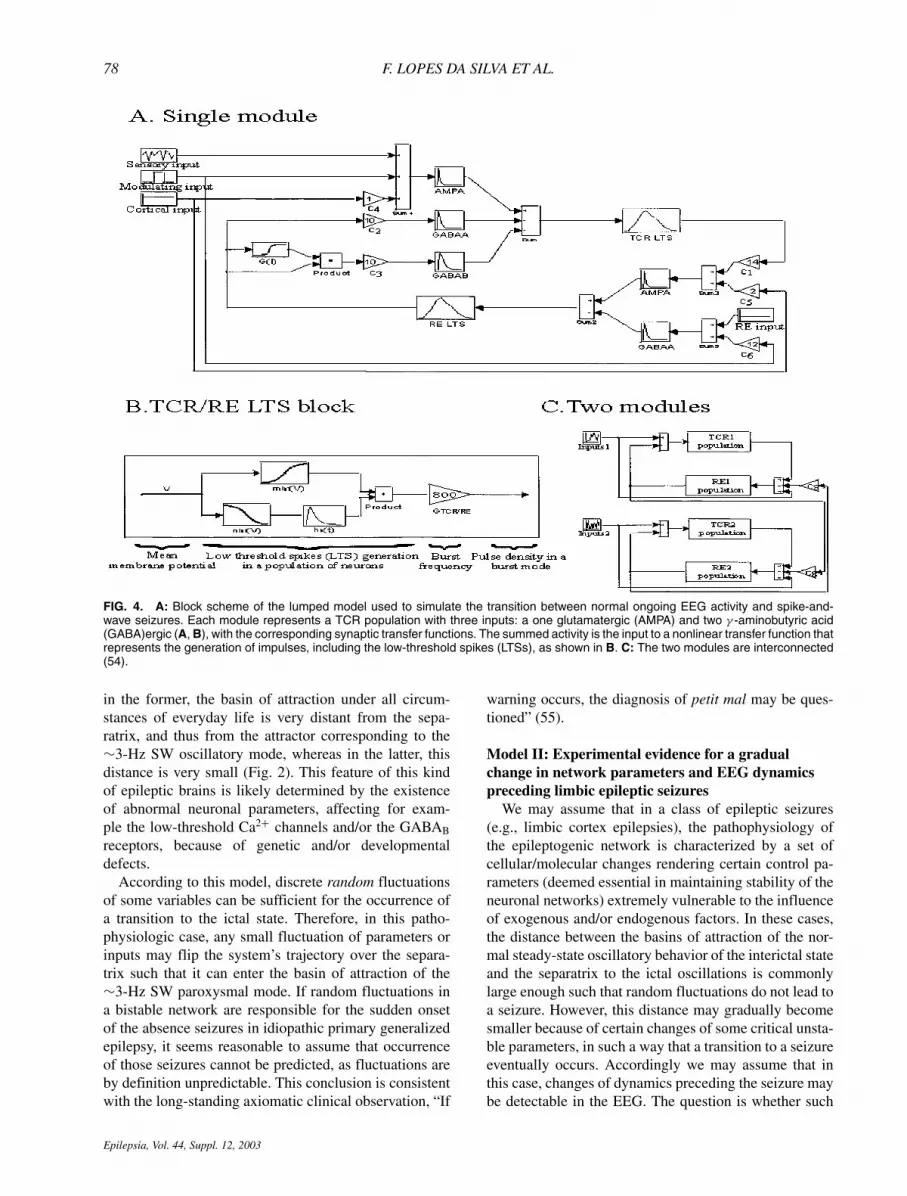
FIG. 6. Histograms showing the value ofphase coherency index (PCI) of one MEG(above) and one EEG (below) channelas function of frequency. Trials performedwith intermittent photic stimulation (IPS) at10 Hz. The PCI values are shown forthis frequency and higher harmonics. Widegrey bars: PCI values in the condition thatno seizure occurred. Thin black bars: PCIvalues in the case a seizure occurred. ThePCI values were averaged over a time win-dow of 5 s during IPS and before the tran-sition to seizure was detected. Note themuch larger PCI values (both in MEG andEEG), particularly in the gamma frequencyrange (30 to 90 Hz), in the case in which aseizure followed the IPS, as compared withthe case in which this did not occur.
seconds later. The most significant feature in this respect isa decrease of the phase dispersion, or increase of the phasecoherency index, of frequency components in the gammafrequency range that are harmonically related to the fun-damental frequency of the intermittent light stimulation(Fig. 6). It should be noted that in the normal functioningof the brain, the formation of dynamic links mediated bysynchrony over multiple frequency bands has been pro-posed (63) as the mechanism involved in large-scale in-tegration of distributed anatomic and functional domainsof brain activity to enable the emergence of coherent be-havior and cognition. We may hypothesize that the mech-anisms involved in this large-scale synchronization maybe disturbed in some brains such that they became unsta-ble and, eventually, may undergo a transition to anotherpathophysiologic oscillatory state, resulting in a seizure.
How can these MEG/EEG-evoked activities with an en-hanced phase synchrony at specific high-frequency bandsbe generated? To understand these phenomena, we shouldtake into consideration that a flickering light of the kindused to trigger epileptiform paroxysms in photosensitiveepilepsy patients causes the generation of higher harmon-ics, and sometimes also subharmonics, of the fundamentalstimulation frequency. These nonlinear properties of thevisual pathways are well documented (64,65). In addi-tion we note that during visual stimulation, high-frequencysynchrony between series of action potentials is evident,as indicated earlier (17–21), and that in the awake atten-tive state, domains of beta and gamma oscillations arepresent in the neocortex of animals (22–25), and humans(27). Furthermore it has been demonstrated that cortical
Epilepsia, Vol. 44, Suppl. 12, 2003

CROSSLINKS IN NEUROPHYSIOLOGY AND EPILEPSY 81
networks may display gamma oscillations even in vitro,and the neurophysiologic conditions that may be respon-sible for the generation of such oscillations have beenput in evidence both experimentally (29,31,66) and inmodel studies (67). Taking these different observationstogether, we may assume that cortical populations of neu-rons may display an intrinsic tendency to oscillate in thebeta/gamma frequency range under appropriate behav-ioral conditions. These populations may oscillate at dif-ferent dominant frequencies, although within the samefrequency range, as encountered experimentally. Inter-mittent photic stimulation that causes the occurrence ofhigher harmonics within the beta/gamma frequency rangeappears to lead to the entrainment of such intrinsic oscilla-tory populations. Thus, the finding of the enhancement ofphase coherency within this same frequency range in thephotosensitive epilepsy patients (68) may be interpretedas evidence for such an entrainment. The observation thatthis increased phase coherency is much enhanced in thecases in which the intermittent light stimulation leads tothe transition to SW dynamical state implies that, in thesecases, a stronger tendency exists for the occurrence of theentrainment of beta/gamma oscillators. Experimental data(69) show that human subjects stimulated with flickeringlight at frequencies from 1 to 100 Hz exhibit event-relatedpotentials with steady-state oscillations at all frequenciesup to ≥90 Hz. Interestingly, the steady-state potentialsexhibited clear resonance phenomena around 10, 20, 40,and 80 Hz. How these physiologic properties relate tothe pathophysiologic enhancement that we found in pa-tients, as described earlier, is discussed elsewhere in moredetail (62).
CONCLUSIONS
We present our view of what we may call the ba-sic mechanisms of the routes to epileptic seizures. Ourmain assumption is that these processes cannot be under-stood just on the basis of currently accepted pathophys-iologic concepts, or even stronger, of current neurobio-logic knowledge. To achieve this aim, it is necessary tocombine concepts of the neurophysiology of neuronal net-works with those of the mathematics of nonlinear systems.The reason is that neuronal networks, in general, behaveas nonlinear systems with complex dynamics. This es-sential feature must be taken into account to understandhow neuronal networks can have bi(multi)-stable statesand can display bifurcations between such states, some-times displaying intermittency, depending on changes ofthe values of some critical parameters. The latter, even ifminute, may have enormous consequences. Such dynam-ical features are characteristic of how epileptic seizuresmay occur. We developed a framework to account for dif-ferent routes that can lead a neuronal network to changefrom its normal mode of activity to a seizure mode within
the context of these theoretical considerations. We wereable to construct three basic models of routes to epilep-tic seizures and to define under which circumstances thetransition from the ongoing (interictal) activity mode to theictal (seizure) mode may or may not be predictable. Wedraw the conclusion that either situation is possible, de-pending on the dynamical state of a given neuronal system.Whether in some cases seizures may be essentially unpre-dictable, as most often in absence-type seizures of idio-pathic primary generalized epilepsy, in others, it is likelythat the actual seizure is preceded by a gradual change indynamics that, in principle, may be detectable some timebefore the seizure becomes manifest. This has been al-ready demonstrated in a number of studies, mostly withanalysis methods derived from the theory of nonlineardynamics. In addition we stress the need for a more com-prehensive analysis of the dynamical states of neuronalnetworks based on a combination of basic neurophysiol-ogy and computer model studies.
REFERENCES
1. Ott E. Chaos in dynamical systems. Cambridge: Cambridge Univer-sity Press, 1993.
2. Glass L, Mackey MC. The rhythms of life. New Jersey: PrincetonUniversity Press, 1988:248.
3. Lopes da Silva FH, Pijn JP, Wadman WJ. Dynamics of local neuronalnetworks: control parameters and state bifurcations in epileptogen-esis. Prog Brain Res 1994;102:359–70.
4. Andrzejak RG, Widman G, Lehnertz K, et al. The epileptic pro-cess as nonlinear deterministic dynamics in a stochastic environ-ment: an evaluation on mesial temporal lobe epilepsy. Epilepsy Res2001;44:129–40.
5. Elbert T, Ray WJ, Kowalik ZJ, et al. Chaos and physiology: deter-ministic chaos in excitable cell assemblies. Physiol Rev 1994;74:1–47.
6. Freeman WJ, Skarda CA. Spatial EEG patterns, nonlinear dy-namics and perception: the neo-sherringtonian view. Brain Res1985;357:147–75.
7. Jahnsen H, Llinas RR. Electrophysiological properties of guinea-pigthalamic neurons, an in vitro study. J Physiol 1984;349:205–26.
8. Jahnsen H, Llinas RR. Ionic basis for the electroresponsivenessand oscillatory properties of guinea-pig thalamic neurones in vitro.J Physiol 1984;349:227–47.
9. Steriade M, Llinas RR. The functional states of the thalamus andthe associated neuronal interplay. Physiol Rev 1988;68:649–742.
10. Steriade M, Datta S, Pare D, et al. Neuronal activities in brain-stemcholinergic nuclei related to tonic activation processes in thalamo-cortical systems. J Neurosci 1990;10:2541–59.
11. Steriade M, Gloor P, Llinas RR, et al. Basic mechanism of cere-bral rhythmic activities. Electroencephalogr Clin Neurophysiol1990;76:481–508.
12. Steriade M, Deschenes M, Domich L, et al. Abolition of spindle os-cillations in thalamic neurons disconnected from nucleus reticularisthalami. J Neurophysiol 1985;54:1473–97.
13. Steriade M. Central core modulation of spontaneous oscillationsand sensory transmission in thalamocortical systems. Curr OpinNeurobiol 1993;3:619–25.
14. Steriade M, McCormick DA, Sejnowski TJ. Thalamocortical oscil-lations in the sleeping and aroused brain. Science 1993;262:679–85.
15. Freeman WJ. Characterization of state transitions in spatially dis-tributed, chaotic, nonlinear, dynamical systems in cerebral cortex.Integr Physiol Behav Sci 1994;29:294–306.
16. Lopes da Silva FH, Kamphuis W, Titulaer M, et al. An ex-perimental model of progressive epilepsy: the development of
Epilepsia, Vol. 44, Suppl. 12, 2003

82 F. LOPES DA SILVA ET AL.
kindling of the hippocampus of the rat. Ital J Neurol Sci 1995;16:45–57.
17. Eckhorn R, Bauer R, Jordan W, et al. Coherent oscillations: a mech-anism of feature linking in the visual cortex? Multiple electrode andcorrelation analyses in the cat. Biol Cybern 1988;60:121–30.
18. Gray CM, Singer W. Stimulus-specific neuronal oscillations in ori-entation columns of cat visual cortex. Proc Natl Acad Sci U S A1989;86:1698–702.
19. Gray CM, Konig P, Engel AK, et al. Oscillatory responses in catvisual cortex exhibit inter-columnar synchronization which reflectsglobal stimulus properties. Nature 1989;338:334–7.
20. Engel J Jr, Henry TR, Risinger MW, et al. Presurgical evaluationfor partial epilepsy: relative contributions of chronic depth-electroderecordings versus FDG-PET and scalp-sphenoidal ictal EEG. Neu-rology 1990;40:1670–7.
21. Gray CM, Engel AK, Konig P, et al. Synchronization of oscillatoryneuronal responses in cat striate cortex: temporal properties. VisNeurosci 1992;8:337–47.
22. Roelfsema PR, Engel AK, Konig P, et al. Visuomotor integration isassociated with zero time-lag synchronization among cortical areas.Nature 1997;385:157–61.
23. Bouyer JJ, Montaron MF, Rougeul A. Fast fronto-parietal rhythmsduring combined focused attentive behaviour and immobility in cat:cortical and thalamic localizations. Electroencephalogr Clin Neu-rophysiol 1981;51:244–52.
24. Lopes da Silva FH, van Rotterdam A, Storm van Leeuwen W,et al. Dynamic characteristics of visual evoked potentials in the dog,II: beta frequency selectivity in evoked potentials and backgroundactivity. Electroencephalogr Clin Neurophysiol 1970;29:260–8.
25. Rougeul A, Bouyer JJ, Dedet L, et al. Fast somato-parietal rhythmsduring combined focal attention and immobility in baboon and squir-rel monkey. Electroencephalogr Clin Neurophysiol 1979;46:310–9.
26. Bird BL, Newton FA, Sheer DE, et al. Behavioral and electroen-cephalographic correlates of 40-Hz EEG biofeedback training inhumans. Biofeedback Self Regul 1978;3:13–28.
27. Sheer DE, Grandstaff NW, Benignus VA. Behavior and 40-c-sec.electrical activity in the brain. Psychol Rep 1966;19:1333–4.
28. Bragin A, Engel J Jr, Wilson CL, et al. High-frequency oscillationsin human brain. Hippocampus 1999;9:137–42.
29. Jefferys JG, Traub RD, Whittington MA. Neuronal networks forinduced “40 Hz” rhythms. Trends Neurosci 1996;19:202–8.
30. Kopell N, Ermentrout GB, Whittington MA, et al. Gamma rhythmsand beta rhythms have different synchronization properties. ProcNatl Acad Sci U S A 2000;97:1867–72.
31. Whittington MA, Traub RD, Jefferys JG. Synchronized oscillationsin interneuron networks driven by metabotropic glutamate receptoractivation. Nature 1995;373:612–5.
32. Whittington MA, Traub RD, Kopell N, et al. Inhibition-basedrhythms: experimental and mathematical observations on networkdynamics. Int J Psychophysiol 2000;38:315–36.
33. Whittington MA, Doheny HC, Traub RD, et al. Differential expres-sion of synaptic and nonsynaptic mechanisms underlying stimulus-induced gamma oscillations in vitro. J Neurosci 2001;21:1727–38.
34. Steriade M, Deschenes M. The thalamus as a neuronal oscillator.Brain Res Rev 1984;8:l–63.
35. Bal T, von Krosigk M, McCormick DA. Synaptic and mem-brane mechanisms underlying synchronized oscillations in the ferretLGNd in vitro. J Physiol 1995;483:641–63.
36. Bal T, von Krosigk M, McCormick DA. Role of the ferret perigenic-ulate nucleus in the generation of synchronized oscillations in vitro.J Physiol 1995;483:665–85.
37. von Krosigk M, Bal T, McCormick DA. Cellular mechanisms of asynchronized oscillation in the thalamus. Science 1993;261:361–4.
38. Destexhe A, Sejnowski TJ. Synchronized oscillations in thalamicnetworks: insight from modeling studies. In: Steriade M, Jones EG,McCormick DA, eds. Thalamus. Amsterdam: Elsevier, 1996.
39. Avanzini G, de Curtis M, Panzica F, et al. Intrinsic properties ofnucleus reticularis thalami neurones of the rat studied in vitro.J Physiol 1989;416:111–22.
40. Bal T, McCormick DA. Ionic mechanisms of rhythmic burst firingand tonic activity in the nucleus reticularis thalami, a mammalianpacemaker. J Physiol 1993;486:669–91.
41. Contreras D, Curr’o Dosi R, Steriade M. Electrophysiological
properties of cat reticular thalamic neurones in vivo. J Physiol1993;470:273–94.
42. Huguenard JR, Prince DA. A novel T-type current underlies pro-longed Ca(2+)-dependent burst firing GABAergic neurons of ratthalamic reticular nucleus. J Neurosci 1992;12:3804–17.
43. Mulle C, Madariage A, Deschenes M. Morphology and electrophys-iological properties of reticularis thalamic neurones in cat, in vivostudy of a thalamic pacemaker. J Neurosci 1986;6:2134–45.
44. Lopes da Silva FH, Vos JE, Mooibroek J, et al. Relative contributionsof intracortical and thalamo-cortical processes in the generation ofalpha rhythms, revealed by partial coherence analysis. Electroen-cephalogr Clin Neurophysiol 1980;50:449–56.
45. Amzica F, Steriade M. Neuronal and glial membrane potentials dur-ing sleep and paroxysmal oscillations in the neocortex. J Neurosci2000;20:6648–65.
46. Castro-Alamancos MA. Neocortical synchronized oscillations in-duced by thalamic disinhibition in vivo. J Neurosci 1999;19:RC27.
47. Steriade M, Contreras D. Spike-wave complexes and fast compo-nents of cortically generated seizures, I: role of neocortex and tha-lamus. J Neurophysiol 1998;80:1439–55.
48. Coulter DA, Huguenard JR, Prince DA. Specific petit mal anticon-vulsants reduce calcium currents in thalamic neurons. Neurosci Lett1989;98:74–8.
49. Coulter DA, Huguenard JR, Prince DA. Differential effects of petitmal anticonvulsants and convulsants on thalamic neurones: calciumcurrent reduction. Br J Pharmacol 1990;100:800–6.
50. Coulter DA, Huguenard JR, Prince DA. Differential effects of petitmal anticonvulsants and convulsants on thalamic neurones GABAcurrent blockade. Br J Pharmacol 1990;100:807–13.
51. Destexhe A, Contreras D, Steriade M. Mechanisms underlying thesynchronizing action of corticothalamic feedback through inhibitionof thalamic relay cells. J Neurophysiol 1998;79:999–1016.
52. Golomb D, Wang XJ, Rinzel J. Propagation of spindle waves in athalamic slice model. J Neurophysiol 1996;75:750–69.
53. Wang XJ. Multiple dynamical modes of thalamic relay neurons:rhythmic bursting and intermittent phase-locking. Neuroscience1994;59:21–31.
54. Suffczynski P, Pijn JP, Pfurtscheller G, et al. Event-related dy-namics of alpha band rhythms: a neuronal network model of fo-cal ERD/surround ERS. In: Pfurtscheller G, Lopes da Silva FH,eds. Event-related desynchronization, handbook of EEG and clin-ical neurophysiology rev series. Amsterdam: Elsevier, 1999:67–88.
55. Lennox WG. Epilepsy and related disorders. Boston: Little, Brown,1960.
56. Lehnertz K, Elger CE. Spatio-temporal dynamics of the primaryepileptogenic area in temporal lobe epilepsy characterized byneuronal complexity loss. Electroencephalogr Clin Neurophysiol1995;95:108–17.
57. Iasemidis LD, Sackellares JC, Zaveri HP, et al. Phase space topog-raphy and the Lyapunov exponent of electrocorticograms in partialseizures. Brain Topogr 1990;2:187–201
58. Iasemidis LD, Olson LD, Savit RS, et al. Time dependencies in theoccurrences of epileptic seizures. Epilepsy Res 1994;17:81–94.
59. Martinerie J, Adam C, Le Van Quyen M, et al. Epileptic seizurescan be anticipated by nonlinear analysis. Nat Med 1998;4:1173–6.
60. Le Van Quyen M, Martinerie J, Navarro V, et al. Anticipa-tion of epileptic seizures from standard EEG recordings. Lancet2001;357:183–8.
61. Litt B, Esteller R, Echauz J, D’Alessandro M, et al. Epileptic seizuresmay begin hours in advance of clinical onset: a report of five patients.Neuron 2001;30:51–64.
62. Kalitzin S, Parra J, Velis D, Lopes da Silva FH. Enhancement ofphase clustering in the EEG/MEG gamma frequency band antic-ipates transitions to paroxysmal epileptiform activity in epilepticpatients with known visual sensitivity. IEEE Trans Biomed Eng2002;49:1279–86.
63. Varela F, Lachaux JP, Rodriguez E, et al. The brainweb: phasesynchronization and large-scale integration. Nat Rev Neurosci2001;2:229–39.
64. Regan D, Spekreijse H. Evoked potentials in vision research 1961–86. Vision Res 1986;26:1461–80.
Epilepsia, Vol. 44, Suppl. 12, 2003

CROSSLINKS IN NEUROPHYSIOLOGY AND EPILEPSY 83
65. Spekreijse H, Reits D. Sequential analysis of the visual evoked po-tential system in man: nonlinear analysis of a sandwich system. AnnN Y Acad Sci 1982;388:72–97.
66. Buhl EH, Tamas G, Fisahn A. Cholinergic activation and tonic exci-tation induce persistent gamma oscillations in mouse somatosensorycortex in vitro. J Physiol 1998;513:117–26.
67. Traub RD, Jefferys JG, Whittington MA. Simulation of gammarhythms in networks of interneurons and pyramidal cells. J ComputNeurosci 1997;4:141–50.
68. Parra J, Meeren HK, Kalitzin S, et al. Magnetic source imaging
in fixation-off sensitivity: relationship with alpha rhythm. J ClinNeurophysiol 2000;17:212–23.
69. Herrmann CS. Human EEG responses to 1-100 Hz flicker: reso-nance phenomena in visual cortex and their potential correlation tocognitive phenomena. Exp Brain Res 2001;137:346–53
70. Thompson JMT, Stewart HB. Nonlinear dynamics and chaos.Chicester: John Wiley and Sons, 1986:376.
71. Sheer DE, Grandstaff N. Computer-analysis of electrical activity inthe brain and its relation to behavior. Bibl Psychiatry 1970;143:160–72.
Epilepsia, Vol. 44, Suppl. 12, 2003