Epigenetics of Host–Pathogen Interactions: The Road Ahead and the Road Behind
17
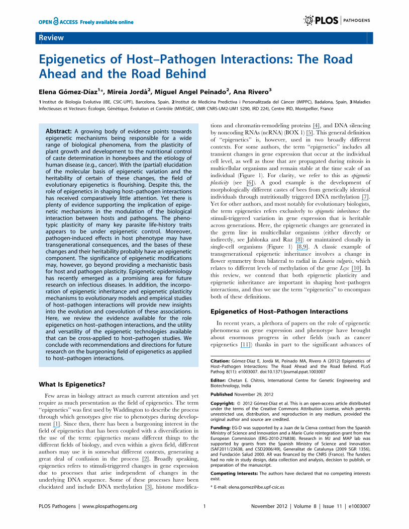
Review Epigenetics of Host–Pathogen Interactions: The Road Ahead and the Road Behind Elena Go ´ mez-Dı´az 1 *, Mireia Jorda ` 2 , Miguel Angel Peinado 2 , Ana Rivero 3 1 Institut de Biologia Evolutiva (IBE, CSIC-UPF), Barcelona, Spain, 2 Institut de Medicina Predictiva i Personalitzada del Ca ` ncer (IMPPC), Badalona, Spain, 3 Maladies Infectieuses et Vecteurs: E ´ cologie, Ge ´ne ´ tique, E ´ volution et Contro ˆ le (MIVEGEC, UMR CNRS-UM2-UM1 5290, IRD 224), Centre IRD, Montpellier, France Abstract: A growing body of evidence points towards epigenetic mechanisms being responsible for a wide range of biological phenomena, from the plasticity of plant growth and development to the nutritional control of caste determination in honeybees and the etiology of human disease (e.g., cancer). With the (partial) elucidation of the molecular basis of epigenetic variation and the heritability of certain of these changes, the field of evolutionary epigenetics is flourishing. Despite this, the role of epigenetics in shaping host–pathogen interactions has received comparatively little attention. Yet there is plenty of evidence supporting the implication of epige- netic mechanisms in the modulation of the biological interaction between hosts and pathogens. The pheno- typic plasticity of many key parasite life-history traits appears to be under epigenetic control. Moreover, pathogen-induced effects in host phenotype may have transgenerational consequences, and the bases of these changes and their heritability probably have an epigenetic component. The significance of epigenetic modifications may, however, go beyond providing a mechanistic basis for host and pathogen plasticity. Epigenetic epidemiology has recently emerged as a promising area for future research on infectious diseases. In addition, the incorpo- ration of epigenetic inheritance and epigenetic plasticity mechanisms to evolutionary models and empirical studies of host–pathogen interactions will provide new insights into the evolution and coevolution of these associations. Here, we review the evidence available for the role epigenetics on host–pathogen interactions, and the utility and versatility of the epigenetic technologies available that can be cross-applied to host–pathogen studies. We conclude with recommendations and directions for future research on the burgeoning field of epigenetics as applied to host–pathogen interactions. What Is Epigenetics? Few areas in biology attract as much current attention and yet require as much presentation as the field of epigenetics. The term ‘‘epigenetics’’ was first used by Waddington to describe the process through which genotypes give rise to phenotypes during develop- ment [1]. Since then, there has been a burgeoning interest in the field of epigenetics that has been coupled with a diversification in the use of the term: epigenetics means different things to the different fields of biology, and even within a given field, different authors may use it in somewhat different contexts, generating a great deal of confusion in the process [2]. Broadly speaking, epigenetics refers to stimuli-triggered changes in gene expression due to processes that arise independent of changes in the underlying DNA sequence. Some of these processes have been elucidated and include DNA methylation [3], histone modifica- tions and chromatin-remodeling proteins [4], and DNA silencing by noncoding RNAs (ncRNA) (BOX 1) [5]. This general definition of ‘‘epigenetics’’ is, however, used in two broadly different contexts. For some authors, the term ‘‘epigenetics’’ includes all transient changes in gene expression that occur at the individual cell level, as well as those that are propagated during mitosis in multicellular organisms and remain stable at the time scale of an individual (Figure 1). For clarity, we refer to this as epigenetic plasticity (see [6]). A good example is the development of morphologically different castes of bees from genetically identical individuals through nutritionally triggered DNA methylation [7]. Yet for other authors, and most notably for evolutionary biologists, the term epigenetics refers exclusively to epigenetic inheritance: the stimuli-triggered variation in gene expression that is heritable across generations. Here, the epigenetic changes are generated in the germ line in multicellular organisms (either directly or indirectly, see Jablonka and Raz [8]) or maintained clonally in single-cell organisms (Figure 1) [8,9]. A classic example of transgenerational epigenetic inheritance involves a change in flower symmetry from bilateral to radial in Linaria vulgaris, which relates to different levels of methylation of the gene Lcyc [10]. In this review, we contend that both epigenetic plasticity and epigenetic inheritance are important in shaping host–pathogen interactions, and thus we use the term ‘‘epigenetics’’ to encompass both of these definitions. Epigenetics of Host–Pathogen Interactions In recent years, a plethora of papers on the role of epigenetic phenomena on gene expression and phenotype have brought about enormous progress in other fields (such as cancer epigenetics [11]) thanks in part to the significant advances of Citation: Go ´ mez-Dı ´az E, Jorda ` M, Peinado MA, Rivero A (2012) Epigenetics of Host–Pathogen Interactions: The Road Ahead and the Road Behind. PLoS Pathog 8(11): e1003007. doi:10.1371/journal.ppat.1003007 Editor: Chetan E. Chitnis, International Centre for Genetic Engineering and Biotechnology, India Published November 29, 2012 Copyright: ß 2012 Go ´ mez-Dı ´az et al. This is an open-access article distributed under the terms of the Creative Commons Attribution License, which permits unrestricted use, distribution, and reproduction in any medium, provided the original author and source are credited. Funding: EG-D was supported by a Juan de la Cierva contract from the Spanish Ministry of Science and Innovation and a Marie Curie reintegration grant from the European Commission (ERG-2010-276838). Research in MJ and MAP lab was supported by grants from the Spanish Ministry of Science and Innovation (SAF2011/23638, and CSD2006/49), Generalitat de Catalunya (2009 SGR 1356), and Fundacio ´ n Salud 2000. AR was financed by the CNRS (France). The funders had no role in study design, data collection and analysis, decision to publish, or preparation of the manuscript. Competing Interests: The authors have declared that no competing interests exist. * E-mail: [email protected] PLOS Pathogens | www.plospathogens.org 1 November 2012 | Volume 8 | Issue 11 | e1003007
-
Upload
independent -
Category
Documents
-
view
2 -
download
0
Transcript of Epigenetics of Host–Pathogen Interactions: The Road Ahead and the Road Behind
Review
Epigenetics of Host–Pathogen Interactions: The RoadAhead and the Road BehindElena Gomez-Dıaz1*, Mireia Jorda2, Miguel Angel Peinado2, Ana Rivero3
1 Institut de Biologia Evolutiva (IBE, CSIC-UPF), Barcelona, Spain, 2 Institut de Medicina Predictiva i Personalitzada del Cancer (IMPPC), Badalona, Spain, 3 Maladies
Infectieuses et Vecteurs: Ecologie, Genetique, Evolution et Controle (MIVEGEC, UMR CNRS-UM2-UM1 5290, IRD 224), Centre IRD, Montpellier, France
Abstract: A growing body of evidence points towardsepigenetic mechanisms being responsible for a widerange of biological phenomena, from the plasticity ofplant growth and development to the nutritional controlof caste determination in honeybees and the etiology ofhuman disease (e.g., cancer). With the (partial) elucidationof the molecular basis of epigenetic variation and theheritability of certain of these changes, the field ofevolutionary epigenetics is flourishing. Despite this, therole of epigenetics in shaping host–pathogen interactionshas received comparatively little attention. Yet there isplenty of evidence supporting the implication of epige-netic mechanisms in the modulation of the biologicalinteraction between hosts and pathogens. The pheno-typic plasticity of many key parasite life-history traitsappears to be under epigenetic control. Moreover,pathogen-induced effects in host phenotype may havetransgenerational consequences, and the bases of thesechanges and their heritability probably have an epigeneticcomponent. The significance of epigenetic modificationsmay, however, go beyond providing a mechanistic basisfor host and pathogen plasticity. Epigenetic epidemiologyhas recently emerged as a promising area for futureresearch on infectious diseases. In addition, the incorpo-ration of epigenetic inheritance and epigenetic plasticitymechanisms to evolutionary models and empirical studiesof host–pathogen interactions will provide new insightsinto the evolution and coevolution of these associations.Here, we review the evidence available for the roleepigenetics on host–pathogen interactions, and the utilityand versatility of the epigenetic technologies availablethat can be cross-applied to host–pathogen studies. Weconclude with recommendations and directions for futureresearch on the burgeoning field of epigenetics as appliedto host–pathogen interactions.
What Is Epigenetics?
Few areas in biology attract as much current attention and yet
require as much presentation as the field of epigenetics. The term
‘‘epigenetics’’ was first used by Waddington to describe the process
through which genotypes give rise to phenotypes during develop-
ment [1]. Since then, there has been a burgeoning interest in the
field of epigenetics that has been coupled with a diversification in
the use of the term: epigenetics means different things to the
different fields of biology, and even within a given field, different
authors may use it in somewhat different contexts, generating a
great deal of confusion in the process [2]. Broadly speaking,
epigenetics refers to stimuli-triggered changes in gene expression
due to processes that arise independent of changes in the
underlying DNA sequence. Some of these processes have been
elucidated and include DNA methylation [3], histone modifica-
tions and chromatin-remodeling proteins [4], and DNA silencing
by noncoding RNAs (ncRNA) (BOX 1) [5]. This general definition
of ‘‘epigenetics’’ is, however, used in two broadly different
contexts. For some authors, the term ‘‘epigenetics’’ includes all
transient changes in gene expression that occur at the individual
cell level, as well as those that are propagated during mitosis in
multicellular organisms and remain stable at the time scale of an
individual (Figure 1). For clarity, we refer to this as epigenetic
plasticity (see [6]). A good example is the development of
morphologically different castes of bees from genetically identical
individuals through nutritionally triggered DNA methylation [7].
Yet for other authors, and most notably for evolutionary biologists,
the term epigenetics refers exclusively to epigenetic inheritance: the
stimuli-triggered variation in gene expression that is heritable
across generations. Here, the epigenetic changes are generated in
the germ line in multicellular organisms (either directly or
indirectly, see Jablonka and Raz [8]) or maintained clonally in
single-cell organisms (Figure 1) [8,9]. A classic example of
transgenerational epigenetic inheritance involves a change in
flower symmetry from bilateral to radial in Linaria vulgaris, which
relates to different levels of methylation of the gene Lcyc [10]. In
this review, we contend that both epigenetic plasticity and
epigenetic inheritance are important in shaping host–pathogen
interactions, and thus we use the term ‘‘epigenetics’’ to encompass
both of these definitions.
Epigenetics of Host–Pathogen Interactions
In recent years, a plethora of papers on the role of epigenetic
phenomena on gene expression and phenotype have brought
about enormous progress in other fields (such as cancer
epigenetics [11]) thanks in part to the significant advances of
Citation: Gomez-Dıaz E, Jorda M, Peinado MA, Rivero A (2012) Epigenetics ofHost–Pathogen Interactions: The Road Ahead and the Road Behind. PLoSPathog 8(11): e1003007. doi:10.1371/journal.ppat.1003007
Editor: Chetan E. Chitnis, International Centre for Genetic Engineering andBiotechnology, India
Published November 29, 2012
Copyright: � 2012 Gomez-Dıaz et al. This is an open-access article distributedunder the terms of the Creative Commons Attribution License, which permitsunrestricted use, distribution, and reproduction in any medium, provided theoriginal author and source are credited.
Funding: EG-D was supported by a Juan de la Cierva contract from the SpanishMinistry of Science and Innovation and a Marie Curie reintegration grant from theEuropean Commission (ERG-2010-276838). Research in MJ and MAP lab wassupported by grants from the Spanish Ministry of Science and Innovation(SAF2011/23638, and CSD2006/49), Generalitat de Catalunya (2009 SGR 1356),and Fundacion Salud 2000. AR was financed by the CNRS (France). The fundershad no role in study design, data collection and analysis, decision to publish, orpreparation of the manuscript.
Competing Interests: The authors have declared that no competing interestsexist.
* E-mail: [email protected]
PLOS Pathogens | www.plospathogens.org 1 November 2012 | Volume 8 | Issue 11 | e1003007

Box 1. The Epigenetic Code
Specific combinations of epigenetic modifications constitute what has been called the epigenetic code, determining thefunctional (gene regulation, replication, repair, etc.) and structural features of each genomic region [68]. Histonemodifications: A widely studied epigenetic mark is constituted by the set of posttranslational modifications (PTMs) onhistones, which consist in the covalent addition of different chemical groups to particular residues, and that take place mostly inthe tails of histones (see figure box). The association between different histone marks or variants and distinct chromatin andfunctional states (or histone code [69]) is well established. For instance, trimethylation of the histone 3 lysine 4 residue(H3K4me3) is usually linked to active genes, while trimethylation in lysine 9 residue (H3K9me3) is characteristic of repressedchromatin. DNA methylation: The DNA of most species is methylated and this modification takes place postreplicatively. Ineukaryotes the modified base is 5-methylcytosine (5 mC) whereas in prokaryotes is mostly N6-methyladenine (6 mA) [70]. DNAmethylation has a role in silencing gene expression and heterochromatin remodeling, among other functions [3]. DNAmethylation patterns are dynamic and have changed several times through the tree of life, exhibiting a considerable structural,functional,and mechanistic diversity [71]. Hence, while in plants and vertebrates, DNA methylation occurs widely at CpG (C—phosphate—G) dinucleotide sites, regions of DNA where a cytosine nucleotide occurs next to a guanine nucleotide in the linearsequence of bases, and appear preferentially associated with transposons and silenced DNA; in invertebrates, DNA methylationis mainly found in gene bodies, but its regulatory function is only partially understood [72]. Interestingly, DNA methylation isnot ubiquitous across the tree of life. Several species seem to have undergone loss of DNA methylation to a large degree,including model-species such as the nematode C. elegans, the insect D. melanogaster, and the yeast S. cerevisiae. RNA-mediated silencing: A variety of noncoding RNAs (ncRNA) have been shown to act in concert with the cell’s epigeneticmachinery, for example by establishing DNA methylation and by regulating histone modifiers [5]. Among those, the bestcharacterized are the so-called microRNAs (miRNA), small ncRNAs of 19 to 24 nucleotides that bind target messenger RNAs andinduce their translational repression, cleavage, or accelerated decay [73]. Yet the nature and function of this class of moleculesare poorly understood, as well as the degree to which they contribute to epigenetic phenomena.
Figure BOX 1. Types of epigenetic modifications. (A) Histones can undergo phosphorylation (Ph), methylation (Me), and acetylation (Ac), among otherchemical modifications. These modifications are involved in chromatin remodeling and transcriptional regulation. (B) DNA molecules are methylatedby the addition of a methyl group to carbon position 5 on cytosine bases, a reaction catalyzed by DNA methyltransferase enzymes, which maintainsrepressed gene activity. (C) mRNA is translated into a protein product, but this process can be repressed by binding of microRNAs (miRNA), a class ofnoncoding RNA (ncRNA). Figure adapted with permission from [45].
PLOS Pathogens | www.plospathogens.org 2 November 2012 | Volume 8 | Issue 11 | e1003007

epigenetic technologies (see Box 2 and Table S1). Conversely,
we still know comparatively little about the extent and
significance of epigenetic variation in host–pathogen interactions
(Figure 2).
Host–pathogen interactions are amongst the most plastic and
dynamic systems in nature. To cope with the selective
constraints imposed by their hosts, many pathogens have
evolved an unparalleled level of phenotypic plasticity in their
life history traits [12]. Likewise, the host phenotype is drastically
and rapidly altered by the presence of a pathogen, and in some
cases, the parasitized phenotype is inherited across host
generations (see [13] for a review). In addition, co-adaptations
between hosts and pathogens often occur over such short
evolutionary time scales as to call into question the sole role of
genetic modifications (i.e., mutation and/or recombination) as
an underlying mechanism [14]. In this sense, epigenetic
modifications may provide an accessory source of fast-acting,
reversible, and readily available phenotypic variation that can
be directly shaped by both host and pathogen selection pressures
(Figure 3) [9,14].
We describe herein recent examples of host–pathogen studies
where epigenetic processes have already been shown to play a
role and which can be broadly classified into (1) pathogen
plasticity and (2) pathogen-induced alterations of the host
(Table 1).
Pathogen Plasticity
One of the most notorious aspects of pathogens is the
morphological and developmental plasticity they exhibit, which
is intimately linked to their survival and transmission in the host.
Complex life-history transitions that occur in response to the
changing host environment require rapid and profound alterations
of their gene expression profiles. Take, for example, the malaria
parasites in the genus Plasmodium. In the vertebrate host, the
parasite has distinct hepatocytic and erythrocytic stages, and it
forms sexually differentiated gametocytes in the blood that are
taken up by the mosquito, where these gametocytes mate, then
migrate through the midgut to form oocysts and from there to the
salivary glands as sporozoites. Previous studies have revealed
distinct gene expression profiles in all of these phases (reviewed in
[15]), suggesting that developmental switches are transcriptionally
regulated. However, apicomplexan parasites such as Plasmodium
are notoriously poor in transcription factors [16]. In contrast, these
parasites contain a rich repertoire of histone variants, chromatin
and histone modifying enzymes, and RNA-mediated silencing
mechanisms [17,18]. In Toxoplasma gondii, histone acetylation has
been shown to be responsible for the switch between the
replicative and nonreplicative stages of the pathogen [19,20].
Similar mechanisms of epigenetic regulation have been charac-
terized in other protists (Table 1). Although less studied,
Figure 1. Mechanisms of epigenetic plasticity and inheritance. In single-cell organisms, epimutations induced by environmental stimuli (i.e.,host) propagate in daughter cells by mitosis and result in transient or stable epigenetic states. In multicellular, sexually reproducing, organisms thezygote (F1) differentiates into germinal and somatic cells. Epimutations can be originated directly in the germline and propagated by mitosis(‘‘germline induction’’) (A), or they can arise and propagate as a consequence of interactions with the soma (‘‘somatic induction’’) (B). In the soma,after several rounds of cell divisions, epimutations tend to accumulate during cell and tissue differentiation processes (C). Only those epimutationsgenerated in the germline that escape meiotic resetting during gametogenesis and oogenesis are expected to have transgenerational consequences(i.e., epigenetic inheritance) (F2).doi:10.1371/journal.ppat.1003007.g001
PLOS Pathogens | www.plospathogens.org 3 November 2012 | Volume 8 | Issue 11 | e1003007
Trypanosoma brucei is the only Apicomplexa where DNA methyl-
ation has been detected, but the significance of these epigenetic
modifications in parasite cell-cycle regulation remains unexplored
[18]. More recently, DNA methylation has been shown to be
responsible for the transition between the yeast and hyphal forms
of the polymorphic yeast Candida albicans [21].
The second striking aspect of pathogen plasticity concerns their
ability to alter the expression of genes linked to virulence processes,
which allows them to colonize, replicate, and/or disseminate
within the host. Within the Apicomplexa, Plasmodium falciparum
switches its variant surface proteins during its erythrocytic stage to
avoid the host’s immune system (antigenic variation). These
Box 2. Methods of Epigenetic Analysis
Over the last decade, numerous techniques have beendeveloped to analyze epigenetic marks at both genome-wide and sequence-specific levels. Here we summarize noveland cutting-edge methodologies that due to their versatileand straightforward nature can be cross-applied to host–parasite studies (refer to Table S1). A more comprehensivelist of available technologies may be found elsewhere[11,74].DNA methylationAs a first step in any epigenetic study, global DNAmethylation analyses allow the detection and identifica-tion of DNA methylation (either C and/or A methylnucleo-tides) and measure its frequency throughout the genome.These approaches do not require previous knowledge of thegenome of reference, and most rely on a prior enzymatic/chemical hydrolysis of DNA to obtain the 29-deoxymononu-cleosides, followed by the subsequent separation bychromatographic means such as High Performance LiquidChromatography (HPLC) [75] or High Performance CapillaryElectrophoresis (HPCE) [76], and a final detection step by UVspectroscopy or mass spectrometry. Alternatively, the globalcontent of DNA methylation can also be quantified byenzymatic approaches such as the Luminometric MethylationAssay (LUMA) [77]. This technique is based on the digestionof DNA by methylation-sensitive and -insensitive isoschizo-mers (HpaII/MspI) and followed by pyrosequencing [78] tomeasure the extent of endonucleases cleavage.Once the type of DNA methylation is determined, the nextstep is to study the distribution and extent of DNAmethylation. The majority of methods are based on threestrategies: DNA digestion by methylation-sensitive restrictionenzymes, DNA bisulphite conversion, and affinity enrichmentof methylated DNA using specific antibodies. The combina-tion of these techniques with different molecular andanalytical procedures has resulted in a plethora of approach-es for determining DNA methylation patterns both at thespecific and the genomic scales. At the scale of specificsequences, the bisulphite sequencing has become thegold-standard in mapping m5C sites at single base-pairresolution [79]. Following the bisulphite DNA treatment,cytosines in single-stranded DNA are deaminated to giveuracil. After PCR amplification and DNA sequencing usingprimers that do not contain any CpG site, nonmethylatedcytosines are recognized as thymines, while methylatedcytosines remain as cytosines. This way, any cytosine thatremains in bisulphite-treated DNA must have been methyl-ated. But in recent years there have been major advances atthe level of whole methylomes, and numerous techniqueshave been developed that now allow the study of DNAmethylation at a genome-wide scale. The Amplification ofInter-Methylated Sites (AIMS) [80] is based on thedifferential enzymatic digestion of genomic DNA withmethylation-sensitive and -insensitive isoschizomers (SmaI/XmaI) followed by the ligation of specific adapters and theamplification by PCR of the methylated sequences. Ampli-cons are resolved in denaturing polyacrylamide-sequencinggels, resulting in readable fingerprints that represent the
organismal cell’s DNA methylation profile. It has been widelyapplied to study DNA methylation in cancer [81], and morerecently, to the discovery of DNA methylation in a socialinsect (Apis mellifera) [82]. Another straightforward approachis Methylated DNA Immunoprecipitation (MeDIP) [83],which is based on the isolation of methylated DNAfragments using an antibody specific for 5-methylcytosines.The utility of this technique depends upon the quality of theavailable antibodies, which at present limits the MeDIPanalysis to 5 mC. Among the newest genome-wide technol-ogies that can be applied to host–parasite studies, micro-array technology provides a good resolution DNA meth-ylation profiling, but its use is restricted to the availability ofspecific probes. In addition, in nonmodel organisms, acustom array must be designed. In recent years, fastadvances in Next Generation Sequencing (NGS) havebeen successfully incorporated to analyze DNA methylationin a cost-effective manner, particularly when combined withenrichment techniques like the MeDIP-seq [84]. Nowadays,complete methylomes can be obtained at single-baseresolution by sequencing bisulphite converted wholegenomes [85]. This approach requires, however, complexbioinformatic analysis because bisulphite conversion signif-icantly reduces the complexity of the genome by convertingCs into Ts, thus complicating the alignment of short reads toreference genomes. More recently, several new technologiesunder development that will reach the market during 2012appear to be able to detect different DNA modificationsdirectly without the bisulphite transformation. These tech-nologies include the nanopore-based methods [86] andsingle molecule real time (SMRT) DNA sequencing [87].Modifications and variants of histonesThe identification and quantification of the posttranslationalmodifications (PTMs) and histone variants is an essential firstcharacterization step, especially in nonorganisms models.Mass spectrometry is the gold standard in terms ofaccuracy [88]. However, most epigenetic research in this fieldfocuses on detecting the association of individual proteinsand histones with specific genomic regions. At present, themost powerful technique is Chromatin ImmunoPrecipi-tation (ChIP) [89]. After cross-linking DNA-binding proteinsto DNA with formaldehyde in vivo, the chromatin is isolatedand the DNA along with its associated proteins are shearedinto small fragments. The DNA binding protein of interest isthen precipitated using specific antibodies to isolate thecomplex, and as a final step, the cross-link is reverted torelease the DNA. This method also relies on the availabilityand quality of antibodies. The immunoprecipitated DNA canbe then analyzed by conventional or real-time PCR (ChIP-PCR) [90]. For genome-wide analyses, ChIP is followed bymicroarray hybridization (ChIP-on-chip) [91] or next-genera-tion sequencing (ChIP-seq) [92]. ChIP-seq has become thestate-of-the-art technology for mapping protein–DNA inter-actions in a genome-wide fashion, but data analysis is time-consuming and its application to nonmodel organisms is stilllimited.
PLOS Pathogens | www.plospathogens.org 4 November 2012 | Volume 8 | Issue 11 | e1003007

surface proteins are encoded by highly polymorphic gene families
(var, rif, stevor, and pfmc-2tm, among others). In the case of the var
family, the ability of the parasite to express only one of the 60
genes that encode for these proteins (called PfEMP1) is epigenet-
ically regulated through histone modifications [22]. In a very
recent study, Rovira-Graells et al. [23] have reported a more
general association between these histone and chromatin marks
and clonally variant expression, extending previous results on
Plasmodium var genes to all but two of the 28 variantly expressed
gene families. In addition, recent work has shown that the
epigenetic state of the parasite is maintained during several rounds
of cell division [24]. Epigenetic control of virulence factors is well
demonstrated in several microbial pathogens. In Entamoeba
histolitica, for example, histone methylation and demethylation
regulate the expression of the amoebapore protein (a protein
responsible for the cytotoxicity of the pathogen [25]). DNA
methylation is also an essential regulatory mechanism of virulence
in several pathogenic bacteria [26,27]. In Salmonella enterica, for
example, lack of Dam (DNA adenine methyltransferase) methyl-
ation causes, amongst other things, envelope instability, reduced
motility, and an impaired ability to invade the intestinal
epithelium [28].
Given the importance of epigenetics for pathogen biology,
understanding how the host environment cues the epigenetic
transition between the replicative and transmission stages and the
virulence factors of morbid and deadly parasites such as Plasmodium
is not only an academic exercise, but it will also provide novel
targets for drug development; an option that has been termed
‘‘epigenetic therapy’’ is currently being tested in clinical trials for other
(noninfectious) diseases. These prevention and treatment strategies
translated to the field of host–parasite interactions could be aimed
at arresting the developmental switches of parasites within the host
or at blocking or limiting their virulence. This could be achieved
by using chemical inhibitors, gene knockout, and RNA interfer-
ence (RNAi) approaches, designed to target the epigenetic
machinery of the parasite such as the DNA methyltransferases
or the chromatin and histone modifying enzymes (see Table 1,
[29]).
Pathogen-Induced Alterations of the Host
Pathogen-induced alterations of host physiology, morphology,
and behavior are widely documented in the scientific literature.
Perhaps the most fascinating examples of these changes are those
that have been shown to be the result of a manipulative strategy of
the pathogen aimed at maximizing its survival and transmission.
Although some of the mechanisms underlying such pathogen
manipulation have been unraveled [30–32], by and large, we
know startlingly little of the strategies used by pathogens to achieve
this end. In the last few years, however, evidence has accumulated
that histone modifications and chromatin remodeling regulate
gene expression and are thus key targets for pathogen manipu-
Figure 2. Comparison between the overall number of science citation-indexed publications in the field of epigenetics (black dots)and the number of such publications in the field of host–pathogen interactions (grey dots) over the last 30 years (1980 to 2011).Search carried out on the Web of Science (Thompson Reuters) on June 2012 using a date-restricted search (1980–2011) and ‘‘epigenet*’’ or‘‘epigenet* and (parasite* or pathogen* or microbe* or bacter* or virus*)’’ as topic search terms.doi:10.1371/journal.ppat.1003007.g002
PLOS Pathogens | www.plospathogens.org 5 November 2012 | Volume 8 | Issue 11 | e1003007

lation during an infection [33]. One such obvious target is the
host’s immune system. In recent years, the epigenetic modulation
of host’s transcriptional program linked to host defense genes has
emerged as a relatively common occurrence of pathogenic viral
and bacterial infections [33,34]. Bacteria are the hallmark of
epigenetic studies on microbes and provide several pioneer
examples on infection-induced host gene reprogramming [32]. A
diverse array of bacterial effectors has been identified that either
mimic or inhibit the host cellular machinery, thus facilitating the
pathogen’s life-cycle. MAPK (mitogen-activated protein kinase),
Interferon (IFN), and transcription factor NF-kB signaling
pathways are common targets of bacterial-induced post-transla-
tional modifications, acetylation, ubiquitylation, and phosphory-
lation on histones and chromatin-associated proteins [35]. Within
the alveolar macrophages, Mycobacterium tuberculosis, for example,
inhibits interferon-c-induced expression of several immune genes
through histone acetylation [36], which explains the persistence of
long-term chronic tuberculosis infections in some patients. This
mechanism is not restricted to bacteria but appears rather
ubiquitous among intracellular pathogens (Table 1). Influenza
viruses go a step further at circumventing host immune defenses.
In a recent study, Marazzi et al. [37] report an influenza protein
called NS1 that contains an amino-acid sequence (ARTK) very
similar to the host’s H3 histone tail. The authors provide
compelling evidence of how using this histone mimic sequence,
the viral NS1 protein hijacks a host transcription elongation factor
(hPAF1), selectively suppressing the cell’s production of antiviral
proteins. This work is a good example of how a molecule of
pathogen origin can directly induce an epigenetic modification in
the host. More studies are needed that establish causative
relationships between the pathogen infection and host epigenetic
modifications such as DNA methylation and posttranslational
histone modifications. Indeed, most of the evidence currently
available is correlational (but see Table 1), and cases where
proteins of pathogen origin have been shown to interact directly
with the host epigenetic machinery are still scarce.
An additional characteristic of many pathogens is their ability to
manipulate the reproductive biology of their hosts. The endo-
bacteria Wolbachia pipientis is the archetypal example of such
reproductive manipulations. Wolbachia is the most common
parasitic microorganism in insects. Its maternal inheritance has
selected for a variety of phenotypes associated with manipulating
the reproduction of its hosts: forcing asexuality, feminizing hosts,
killing males, and inducing incompatibility between infected males
and uninfected (or differently infected) females [38]. Negri et al.
[39] have provided the first evidence that a feminizing strain of
Wolbachia interferes with the genetic imprinting of its host (the
leafhopper Zyginidia pullula) by altering the host’s methylation
pattern. Recently, the widespread existence of putative DNA-
methyltransferases in the prophage of the Wolbachia infecting
several Drosophila species [40] has raised the possibility that this
may be a widespread mechanism of epigenetic interference in this
endosymbiotic bacteria. The link is, however, unclear since these
enzymes have been identified as adenine methyltransferases, a
family of prokaryotic enzymes that methylate the amino group at
the C-6 position of adenines, whereas in the example reported
Figure 3. Schematic representation of the interrelations between epigenetic variation, phenotypic variation, and host–pathogeninteractions. The infection phenotype, which varies between host and pathogen phenotypes and is environmentally dependent, can inducechanges at both the genomic and epigenomic levels. These changes can in turn alter gene expression patterns. Apart from these direct effects ofepigenetic variation on host and pathogen phenotypes, epigenetic variation can also have indirect, and transgenerational, phenotypic effects byinfluencing the probability of mutation, transposition, and/or recombination of the DNA sequence, as well as the predisposition of a gene with aparticular epigenetic mark to be selected. See text for further explanation. Red arrows indicate action routes with potential inherited effects (seeFigure 1).doi:10.1371/journal.ppat.1003007.g003
PLOS Pathogens | www.plospathogens.org 6 November 2012 | Volume 8 | Issue 11 | e1003007

above, genetic imprinting of the invertebrate host seems to occur
at the C-5 carbon of CpG cytosines.
Not all modifications that take place in the infected host are,
however, adaptive for the pathogen. Some of them are adaptive
strategies of the host aimed to compensate or minimize the
effects of the infection. In vertebrates, invertebrates, and plants,
individuals that have recovered from certain infectious diseases
are protected against later infection with those same diseases
(immune priming). While the mechanistic basis of immune
priming in invertebrates is still unresolved, in vertebrates,
histone modifications may be associated with immune memory
following a viral infection in CD8 T cells (reviewed in [41]).
Histone modifications, DNA methylation, and other chromatin
remodeling mechanisms, including deposition of histone vari-
ants and ATP-dependent chromatin remodelers, also seem to
serve as a memory for priming in plant immunity [42,43]. In
some cases, acquired immunity can be passed from mother to
offspring, endowing the offspring with improved defense against
infection (transgenerational immune priming). For instance, a
very recent paper has shown that in Arabidopsis thaliana, immune
priming to Pseudomonas syringae is transmitted between plant
generations through the hypomethylation of defense-related
genes [44].
Future Directions
In this review, we have concentrated our attention on the
current evidence available for the role of epigenetic mechanisms in
pathogens’ life cycle and pathogen-induced modification of host
phenotype. However, epigenetics not only represents a paradigm
shift in our understanding of host and pathogen phenotypic
plasticity. We believe that in the next few years, perhaps the most
exciting developments in the field of epigenetics will come by
linking epigenetic variation and inheritance to the epidemiology
and evolution of infectious diseases.
Epigenetic epidemiology has recently emerged as a promising
area for future research on infectious diseases [29,45]. In recent
years, disease association studies based on epigenomic mapping
have arisen as a powerful tool for disease risk prediction in
humans. But these studies typically face the ‘‘chicken-and-egg’’
causality problem: there is an association between a particular
disease phenotype and the epigenome, but it is not easy to establish
whether it is the disease which is causing the epigenetic changes or
whether the epigenetic changes are the ones causing the disease
pathogenesis [29]. New epidemiological approaches are, however,
being developed in epigenetic disease studies to control for such
cause–effect relationships [46]. However, the reversible and
context-dependent nature of epigenetic changes poses serious
caveats to epidemiological studies. For example, many epigenetic
changes linked to disease risk can be lost after one generation,
change from tissue to tissue, or be differentially expressed in an
age-dependent, sex-, and parent-of-origin-specific manner [47–
49]. To overcome these difficulties, epigenetic studies of disease
must be accompanied by comprehensive longitudinal (multistage
and multi-individual) and transgenerational data. Although there
is a lot of effort to bring epigenetics into epidemiological research
in several noninfectious human diseases (see examples reviewed by
[50,51]), we still know very little about the consequences of
epigenetic processes in the emergence and epidemiology of
infectious diseases. Therefore, a comprehensive survey of epige-
netic determinants of pathogenesis coupled with population-level
epigenetic diversity studies in host–pathogen systems is needed
before any disease prediction and prevention strategies can
become a reality [52].
The second area of research is the role of epigenetic variation in
host and pathogen coevolution and evolution. Since the incorporation of
epigenetic inheritance and epigenetic plasticity mechanisms to
evolutionary models and empirical studies of host–pathogen
interactions is still lacking, our discussion is necessarily speculative.
However, we envisage several areas that are ripe for future
research. Models of host–pathogen co-evolution assume the
presence of genetic variation for host resistance and pathogen
infectivity, as well as genotype-specific interactions [53]. Mecha-
nisms of genetic variation alone are, however, often unsatisfactory
to explain the compatibility between host and pathogen pheno-
types [54], and nongenetic inheritance mechanisms may have an
important role to play (e.g., [55–57]). In addition, host–pathogen
co-evolutionary interactions are often context-dependent (i.e.,
spatially and temporally variable), and the output of infection often
depends upon several environmental factors such as temperature
or nutrition [58]. Given the prominent role of epigenetic processes
in environmentally induced phenotypic plasticity and adaptation
[59], the fact that genotype-by-environment interactions in host–
pathogen systems are epigenetically regulated seems a reasonable
assumption. The work by Laine et al. [60] on a fungal pathogen
and its host plant has, for example, demonstrated a temperature-
dependent effect on pathogen performance on local versus foreign
hosts. Multiple cases of environmentally triggered co-adaptations
have been reported in several other host–pathogen systems
(reviewed by [61]), which we contend will provide the raw
material for future epigenetic studies.
A further unresolved matter is to establish the extent, nature,
and significance of epigenetic inheritance in host–pathogen interac-
tions. For example, transgenerational immune priming in inver-
tebrates [62–64] and plants [42] is likely to have an epigenetic
component, but the actual mechanism of inheritance is not known.
Other transgenerational infection effects on host behavior and
physiology, often so-called maternal effects, still need to be
investigated (reviewed by [13]). Several authors (Bonduariansky
and Day [9], Bossdorf et al. [65], Ho and Burggren [66]) have
provided recommendations for testing epigenetic inheritance
experimentally, which could be cross-applied to host–pathogen
studies (Figure S1). In addition, there is an urgent need for
epigenetic studies to develop solid theoretical evolutionary models
[67]. Bonduriansky and Day [9] have suggested that epigenetic
inheritance allows us to overcome three major limitations of
genetic inheritance on phenotypic evolution: (1) It allows for traits
acquired during the lifetime of an individual to be directly
transmitted to the offspring, (2) it allows the transmission of
favorable trait combinations across generations (genetic recombi-
nation tends to break such combinations), and (3) it provides an
additional source of phenotypic variation for selection to act upon.
At present, however, we know startlingly little about how these
phenomena may impact the evolution of host–pathogen interac-
tions. In this sense, the collaboration between molecular
epigeneticists, functional and experimental parasitologists, and
theoretical evolutionary biologists is needed to extend the current
gene-based view of host–pathogen interactions into a more
integrated one that includes both genetic and epigenetic dimen-
sions.
Concluding Remarks
In recent years there has been an explosion in the number of
epigenetics papers across biological disciplines (Figure 2), a
progression that has been accompanied by technological break-
throughs that now make it possible to undertake sophisticated
epigenomic studies across a range of organisms (Box 2 and Table
PLOS Pathogens | www.plospathogens.org 7 November 2012 | Volume 8 | Issue 11 | e1003007

S1). However, studies on the complex and multifaceted co-
evolutionary interactions between hosts and pathogens have
received comparatively little attention (Figure 2), and this in spite
of their potentially evolutionary and epidemiological implications.
In this review, we have concentrated our attention on the
current evidence available for those few cases in which an
epigenetic mechanism has been described (i.e., see Table 1), but
we lack evidence on the evolutionary and epidemiological
significance of these changes. Conversely, there are many
pathogen and host traits of key epidemiological importance that
may be epigenetically controlled, some of which may have
transgenerational consequences and whose mechanistic basis
would merit further investigation.
In conclusion, the future is bright for the epigenetics of host–
pathogen interactions. We are confident that in the next few years,
cutting-edge epigenomic techniques combined with experimental
(whole organism), functional, and theoretical (modeling) approach-
es will provide fascinating insights into the interrelations between
genetic, epigenetic, and phenotypic variation in the complex world
of host–pathogen relationships (Figure S2).
Supporting Information
Figure S1 Experimental approach to detect transge-nerational epigenetic and phenotypic changes of infec-tion in a model study involving mosquitoes. Starting from
Table 1. Summary table of some of the best characterized epigenetic modifications (DNA methylation and histoneposttranslational modifications [hPTM]) in host–pathogen interactions.
Topic/Organism Epigenetic Mechanism (E): Effectors, (T): Targets Phenotype/Functions Refs
Parasite plasticity
Plasmodium falciparum hPTM (E): protein families ApiAP2,PfPuf2, PfGCN5, PfSET1, PfSET2,PfCARM1 & others (T): histonesHP1, H3, H4, H2A, H2B, & others
Sexual & morphologicaldifferentiation (Transmission)
[15] [93] [22,94][18] [23,24]
(E): HACs & HMTs (T): H3, Variantsurface antigen families
Virulence (Antigenic variation)
Toxoplasma gondii hPTM & chromatin-modifying proteins
(E): HDAC, PRMT, MYST, GCN5,SET, ATP-dependent remodelingfactors, (T): histones H3, H4,H2A, H2B
Sexual & morphologicaldifferentiation (Transmission)
[18,20]
Entamoeba histolytica hPTM (E): nd, (T): Histones H3, H3K4,ap-a, cpA5 & lgl1 genes
Virulence (Cytotoxicity) [25]
Salmonella enterica,Escherichia coli, and others
DNA adeninemethylation
(E): CcrM and DAM, (T):pathogenicity island I (SPI-1),lppB gene,std & spv operon
Virulence (Motility, Celladhesion & Invasion, Cytotoxicity)
[28]
Candida albicans DNA methylation (E): nd, (T): RPD3, PBI2,FOX2 genes
Morphological differentiation(Transmission)
[21]
Giardia lamblia hPTM (E): HAC & HDAC, (T): nd Morphological differentiation(Transmission)
[95]
Schistosoma mansoni DNA methylation (E): DNMT2, (T): Smp155010 protein Development (Oviposition) [96]
Epstein-Barr Virus (EBV)and others
DNA methylation (E): Human cellular DNMTs, (T): Viralmethylome
Virulence [97]
Pathogen-induced hostalterations
Wolbachia pipientis DNA methylation (E): prophage DNMTs?, (T): nd Male feminization [39,40]
Pseudomonas syringae hPTM & DNAmethylation
(E): HAC & HMT, (T): WRKY6 &WRKY53 genes; DEFENSIN1.2promoter
Host immune priming [44]
Influenza virus hPTM & DNAmethylation
(E): Viral NS1 (ARSK sequence),(T): transcription factor PAF1
Host immunosuppression [37]
Human adenovirus (HAdV) hPTM (E): Viral E1A protein (T): IFNsignaling, Histone H2B
Host immunosuppression [98]
Mycobacterium tuberculosis hPTM & chromatin-modifying proteins
(E): SWI/SNF protein complex(T): IFN signaling, CIITA promoter
Host immunosuppression [36]
Listeria monocytogenes hPTM & chromatin-modifying proteins
(E): Listeriolysin O, (T): MAPKsignaling, Histone H4 and H3
Host immunosuppression [33]
Toxoplasma gondii hPTM & chromatin-modifying proteins
(E): nd, (T): IFN signaling,transcription factor STAT1
Host immunosuppression [99]
Anaplasma phagocytophilum hPTM & chromatin-modifying proteins
(E): HDAC1, (T): Histone H3 Host immunosuppression [100]
IFN, Interferon; MAPK, mitogen-activated protein kinase; HAC/HDAC, Histone acetylase/deacetylase; HMTs, Histone methyltransferase; DAM, DNA AdenineMethyltransferase; DNMT, DNA methyltransferase.doi:10.1371/journal.ppat.1003007.t001
PLOS Pathogens | www.plospathogens.org 8 November 2012 | Volume 8 | Issue 11 | e1003007

isogenic lines and controlled environmental conditions, female
mosquitoes are experimentally infected for successive generations
to detect adaptive traits in response to a continuous selection
pressure (i.e., infection). Phenotype (behavior, immune response,
and physiology), epigenotype, and fitness (i.e., fecundity, longevity,
and survival) are then quantified and statistically compared. In F1,
two groups of females, either infected or noninfected, are back-
crossed with noninfected mosquito control males (NI(C)). If the
descendants of infected (I6NI(C)) versus noninfected lines
(NI(C)6NI) are phenotypically different but show significant
divergence in epigenetic profiles, gene or protein expression—in
spite of being still identical at the DNA level—this will be evidence
for epigenetically based phenotypic change. In subsequent
generations, the comparison of infected versus noninfected
mosquito groups that descend of infected mosquito females will
allow us to test transient versus stable changes (i.e., adaptive traits)
as well as cumulative effects of infection (we may expect them to be
greater in V than in III). In addition, differences between the
descendants of IV–V in Fx will be indicative of maternal effects.
(PDF)
Figure S2 Workflow on research strategies in host–parasite epigenetics. First, a phenomic (experimental) ap-
proach in laboratory or field settings can be designed to establish
transgenerational phenotypic effects and fitness consequences of
host or pathogen evolutionary-relevant traits for infection. Second,
epigenetic and functional approaches can then be conducted to
examine the mechanistic basis, regulatory pathways, and func-
tional significance of these effects. Third, modeling approaches
can be used to model the long-term consequences of the observed
transgenerational changes, the dynamics and persistence of
different types of epigenetic variation, and the interplay between
epigenetic and genetic variation. Arrows indicate interrelationships
among the different approaches. Feedback among the different
stages (dashed arrows) can serve to generate new hypotheses and
test model predictions.
(PDF)
Table S1 Methodologies for epigenetic analyses.
(DOCX)
Acknowledgments
We thank S. Reece and two anonymous referees for insightful comments.
References
1. Waddington CH (1942) The epigenome. Endevour 1: 18–20.
2. Jablonka E, Lamb MJ (2002) The changing concept of epigenetics. In:
VanSpeybroeck L, VandeVijver G, DeWaele D, editors. From epigenesis to
epigenetics: the genome in context. New York: New York Academy of Sciences.
pp. 82–96.
3. Suzuki MM, Bird A (2008) DNA methylation landscapes: provocative insights
from epigenomics. Nature Reviews in Genetics 9: 465–476.
4. Bannister AJ, Kouzarides T (2011) Regulation of chromatin by histone
modifications. Cell Research 21: 381–395.
5. Storz G (2002) An expanding universe of noncoding RNAs. Science 296: 1260–
1263.
6. Moczek AP, Snell-Rood EC (2008) The basis of bee-ing different: the role of
gene silencing in plasticity. Evolution & Development 10: 511–513.
7. Kucharski R, Maleszka J, Foret S, Maleszka R (2008) Nutritional control of
reproductive status in honeybees via DNA methylation. Science 319: 1827–
1830.
8. Jablonka E, Raz G (2009) Transgenerational epigenetic inheritance: prevalence,
mechanisms, and implications for the study of heredity and evolution. The
Quarterly Review of Biology 84: 131–176.
9. Bonduriansky R, Day T (2009) Nongenetic inheritance and its evolutionary
implications. Annual Review of Ecology, Evolution, and Systematics 40: 103–
125.
10. Cubas P, Vincent C, Coen E (1999) An epigenetic mutation responsible for
natural variation in floral symmetry. Nature 401: 157–161.
11. Esteller M (2007) Cancer epigenomics: DNA methylomes and histone-
modification maps. Nature Reviews in Genetics 8: 286–298.
12. Reece SE, Ricardo SR, Daniel HN (2009) Plastic parasites: sophisticated
strategies for survival and reproduction? Evolutionary Applications 2: 11–23.
13. Poulin R, Thomas F (2008) Epigenetic effects of infection on the phenotype of
host offspring: parasites reaching across host generations. Oikos 117: 331–
335.
14. Rando OJ, Verstrepen KJ (2007) Timescales of genetic and epigenetic
inheritance. Cell 128: 655–668.
15. Merrick CJ, Duraisingh MT (2010) Epigenetics in Plasmodium: what do we really
know? Eukaryotic Cell 9: 1150–1158.
16. Meissner M, Soldati D (2005) The transcription machinery and the molecular
toolbox to control gene expression in Toxoplasma gondii and other protozoan
parasites. Microbes and Infection 7: 1376–1384.
17. Hakimi M-A, Deitsch KW (2007) Epigenetics in Apicomplexa: control of gene
expression during cell cycle progression, differentiation and antigenic variation.
Current Opinion in Microbiology 10: 357–362.
18. Croken MM, Nardelli SC, Kim K (2012) Chromatin modifications, epigenetics,
and how protozoan parasites regulate their lives. Trends in Parasitology 28: 202–
213.
19. Bougdour A, Maubon D, Baldacci P, Ortet P, Bastien O, et al. (2009) Drug
inhibition of HDAC3 and epigenetic control of differentiation in Apicomplexa
parasites. Journal of Experimental Medicine 206: 953–966.
20. Dixon SE, Stilger KL, Elias EV, Naguleswaran A, Sullivan Jr WJ (2010) A
decade of epigenetic research in Toxoplasma gondii. Molecular and Biochemical
Parasitology 173: 1–9.
21. Mishra PK, Baum M, Carbon J (2011) DNA methylation regulates phenotype-
dependent transcriptional activity in Candida albicans. Proc Natl Acad Sci 108:
11965–11970.
22. Chookajorn T, Dzikowski R, Frank M, Li F, Jiwani AZ, et al. (2007) Epigeneticmemory at malaria virulence genes. Proc Natl Acad Sci 104: 899–902.
23. Rovira-Graells N, Gupta AP, Planet E, Crowley VM, Mok S, et al. (in press)Transcriptional variation in the malaria parasite Plasmodium falciparum. Genome
Research.
24. Volz JC, Bartfai R, Petter M, Langer C, Josling GA, et al. (2012) PfSET10, aPlasmodium falciparum methyltransferase, maintains the active var gene in a poised
state during parasite division. Cell Host & Microbe 11: 7–18.
25. Huguenin M, Bracha R, Chookajorn T, Mirelman D (2010) Epigenetic
transcriptional gene silencing in Entamoeba histolytica: insight into histone andchromatin modifications. Parasitology 137: 619–627.
26. Løbner-Olesen A, Skovgaard O, Marinus MG (2005) Dam methylation:coordinating cellular processes. Current Opinion in Microbiology 8: 154–160.
27. Heithoff DM, Sinsheimer RL, Low DA, Mahan MJ (1999) An essential role for
DNA adenine methylation in bacterial virulence. Science 284: 967–970.
28. Marinus MG, Casadesus J (2009) Roles of DNA adenine methylation in host–
pathogen interactions: mismatch repair, transcriptional regulation, and more.FEMS Microbiology Reviews 33: 488–503.
29. Handel A, Ebers G, Ramagopalan S (2010) Epigenetics: molecular mechanismsand implications for disease. Trends in Molecular Medicine 16: 7–23.
30. Lefevre T, Lebarbenchon C, Gauthier-Clerc M, Misse D, Poulin R, et al. (2009)
The ecological significance of manipulative parasites. Trends in Ecology &Evolution 24: 41–48.
31. Hurd H (2009) Chapter 4 evolutionary drivers of parasite-induced changes ininsect life-history traits: from theory to underlying mechanisms. In: Joanne PW,
editor. Advances in parasitology. Academic Press. pp. 85–110.
32. Bhavsar A, Guttman J, Finlay B (2007) Manipulation of host-cell pathways by
bacterial pathogens. Nature 449: 827–861.
33. Hamon MA, Cossart P (2008) Histone modifications and chromatin remodelingduring bacterial infections. Cell Host & Microbe 4: 100–109.
34. Paschos K, Allday MJ (2010) Epigenetic reprogramming of host genes in viraland microbial pathogenesis. Trends in Microbiology 18: 439–447.
35. Ribet D, Cossart P (2010) Post-translational modifications in host cells during
bacterial infection. FEBS Letters 584: 2748–2806.
36. Pennini ME, Pai RK, Schultz DC, Boom WH, Harding CV (2006) Mycobacterium
tuberculosis 19-kDa lipoprotein inhibits IFN-c-induced chromatin remodeling ofMHC2TA by TLR2 and MAPK signaling. The Journal of Immunology 176:
4323–4330.
37. Marazzi I, Ho JSY, Kim J, Manicassamy B, Dewell S, et al. (2012) Suppression
of the antiviral response by an influenza histone mimic. Nature 483: 428–433.
38. Werren JH, Baldo L, Clark ME (2008) Wolbachia: master manipulators ofinvertebrate biology. Nature Review in Microbiology 6: 741–751.
39. Negri I, Franchini A, Gonella E, Daffonchio D, Mazzoglio PJ, et al. (2009)Unravelling the Wolbachia evolutionary role: the reprogramming of the host
genomic imprinting. Proc Biol Sci 276(1666): 2485–2491.
40. Saridaki A, Sapountzis P, Harris HL, Batista PD, Biliske JA, et al. (2011)Wolbachia prophage DNA adenine methyltransferase genes in different
PLOS Pathogens | www.plospathogens.org 9 November 2012 | Volume 8 | Issue 11 | e1003007

Drosophila-Wolbachia associations. PLoS ONE 6: e19708. doi:10.1371/journal.-
pone.001970841. Youngblood B, Davis CW, Ahmed R (2010) Making memories that last a
lifetime: heritable functions of self-renewing memory CD8 T cells. International
Immunology 22: 797–803.42. Conrath U (2011) Molecular aspects of defence priming. Trends in Plant Science
16: 524–531.43. Boyko A, Kovalchuk I (2011) Genetic and epigenetic effects of plant–pathogen
interactions: an evolutionary perspective. Molecular Plant 4: 1014–1023.
44. Luna E, Bruce TJA, Roberts MR, Flors V, Ton J (2012) Next-generationsystemic acquired resistance. Plant Physiology 158: 844–853.
45. Relton CL, Davey Smith G (2010) Epigenetic epidemiology of common complexdisease: prospects for prediction, prevention, and treatment. PLoS Med 7:
e1000356. doi:10.1371/journal.pmed.100035646. Relton CL, Davey Smith G (2012) Two-step epigenetic Mendelian randomi-
zation: a strategy for establishing the causal role of epigenetic processes in
pathways to disease. International Journal of Epidemiology 41: 161–176.47. Fraga MF, Esteller M (2007) Epigenetics and aging: the targets and the marks.
Trends in Genetics 23: 413–418.48. Hemberger M, Dean W, Reik W (2009) Epigenetic dynamics of stem cells and
cell lineage commitment: digging Waddington’s canal. Nature Reviews in
Molecular Cell Biology 10: 526–537.49. Ferguson-Smith AC, Surani MA (2001) Imprinting and the epigenetic
asymmetry between parental genomes. Science 293: 1086–1089.50. Relton CL, Davey Smith G (2012) Is epidemiology ready for epigenetics?
International Journal of Epidemiology 41: 5–9.51. Baccarelli A, Ghosh S (2012) Environmental exposures, epigenetics and
cardiovascular disease. Current Opinion in Clinical Nutrition & Metabolic
Care 15: 323–329.52. Richards E (2008) Population epigenetics. Current Opinion in Genetics &
Development 18: 221–227.53. Carius HJ, Little TJ, Ebert D (2001) Genetic variation in a host-parasite
association: potential for coevolution and frequency-dependent selection.
Evolution 55: 1136–1145.54. Lambrechts L, Fellous S, Koella JC (2006) Coevolutionary interactions between
host and parasite genotypes. Trends in Parasitology 22: 12–16.55. Stjernman M, Little TJ (2011) Genetic variation for maternal effects on parasite
susceptibility. Journal of Evolutionary Biology 24: 2357–2363.56. Cosseau C, Azzi A, Rognon A, Boissier J, Gourbiere S, et al. (2010) Epigenetic
and phenotypic variability in populations of Schistosoma mansoni—a possible kick-
off for adaptive host/parasite evolution. Oikos 119: 669–678.57. Ebert D (2008) Host–parasite coevolution: insights from the daphnia–parasite
model system. Current Opinion in Microbiology 11: 290–301.58. Vale PF, Wilson AJ, Best A, Boots M, Little TJ (2011) Epidemiological,
evolutionary, and coevolutionary implications of context-dependent parasitism.
The American Naturalist 177: 510–521.59. Feil R, Fraga MF (2012) Epigenetics and the environment: emerging patterns
and implications. Nature Reviews in Genetics 13: 97–109.60. Laine A-L (2008) Temperature-mediated patterns of local adaptation in a
natural plant–pathogen metapopulation. Ecology Letters 11: 327–337.61. Wolinska J, King KC (2009) Environment can alter selection in host–parasite
interactions. Trends in Parasitology 25: 236–244.
62. Moret Y, Schmid-Hempel P (2001) Entomology—immune defence in bumble-bee offspring. Nature 414: 506–506.
63. Little TJ, O’Connor B, Colegrave N, Watt K, Read AF (2003) Maternal transferof strain-specific immunity in an invertebrate. Current Biology 13: 489–492.
64. Kurtz J, Franz K (2003) Innate defence: evidence for memory in invertebrate
immunity. Nature 425: 37–38.65. Bossdorf O, Richards CL, Pigliucci M (2008) Epigenetics for ecologists. Ecology
Letters 11: 106–115.66. Ho D, Burggren W (2010) Epigenetics and transgenerational transfer: a
physiological perspective. The Journal of Experimental Biology 213: 3–19.
67. Hunter B, Hollister J, Bomblies K (2012) Epigenetic inheritance: what news forevolution? Current Biology 22: R54–R56.
68. Kouzarides T (2007) Chromatin modifications and their function. Cell 128:693–705.
69. Jenuwein T, Allis CD (2001) Translating the histone code. Science 293: 1074–1080.
70. Wion D, Casadesus J (2006) N6-methyl-adenine: an epigenetic signal for DNA-
protein interactions. Nature Reviews in Microbiology 4: 183–192.71. Jeltsch A (2010) Phylogeny of methylomes. Science 328: 837–838.
72. Zemach A, McDaniel IE, Silva P, Zilberman D (2010) Genome-wideevolutionary analysis of eukaryotic DNA methylation. Science 328: 916–919.
73. Guil S, Esteller M (2009) DNA methylomes, histone codes and miRNAs: tying it
all together. The International Journal of Biochemistry & Cell Biology 41: 87–95.
74. Laird PW (2010) Principles and challenges of genomewide DNA methylationanalysis. Nature Reviews in Genetics 11: 191–203.
75. Eick D, Fritz HJ, Doerfler W (1983) Quantitative determination of 5-
methylcytosine in DNA by reverse-phase high-performance liquid chromatog-raphy. Analytical Biochemistry 135: 165–171.
76. Fraga MF, Rodriguez R, Canal MJ (2000) Rapid quantification of DNA
methylation by high performance capillary electrophoresis. Electrophoresis 21:2990–2994.
77. Karimi M, Johansson S, Stach D, Corcoran M, Grander D, et al. (2006) LUMA(LUminometric Methylation Assay)–a high throughput method to the analysis of
genomic DNA methylation. Experimental Cell Research 312: 1989–1995.
78. Ronaghi M, Uhlen M, Nyren P (1998) A sequencing method based on real-timepyrophosphate. Science 281: 363, 365.
79. Clark SJ, Harrison J, Paul CL, Frommer M (1994) High sensitivity mapping of
methylated cytosines. Nucleic Acids Research 22: 2990–2997.
80. Jorda M, Rodrıguez J, Frigola J, Peinado MA (2009) Analysis of DNA
Methylation by Amplification of Intermethylated Sites (AIMS). In: Tost J, editor.DNA methylation: methods and protocols, second edition. Humana Press. pp.
107–116.
81. Frigola J, Sole X, Paz MF, Moreno V, Esteller M, et al. (2005) Differential DNA
hypermethylation and hypomethylation signatures in colorectal cancer. HumanMolecular Genetics 14: 319–326.
82. Wang Y, Jorda M, Jones PL, Maleszka R, Ling X, et al. (2006) Functional CpG
methylation system in a social insect. Science 314: 645–647.
83. Weber M, Davies JJ, Wittig D, Oakeley EJ, Haase M, et al. (2005)
Chromosome-wide and promoter-specific analyses identify sites of differentialDNA methylation in normal and transformed human cells. Nature Genetics 37:
853–862.
84. Down TA, Rakyan VK, Turner DJ, Flicek P, Li H, et al. (2008) A Bayesiandeconvolution strategy for immunoprecipitation-based DNA methylome anal-
ysis. Nature Biotechnology 26: 779–785.
85. Lister R, Pelizzola M, Dowen RH, Hawkins RD, Hon G, et al. (2009) Human
DNA methylomes at base resolution show widespread epigenomic differences.Nature 462: 315–322.
86. Clarke J, Wu HC, Jayasinghe L, Patel A, Reid S, et al. (2009) Continuous base
identification for single-molecule nanopore DNA sequencing. Nature Nano-
technology 4: 265–270.
87. Flusberg BA, Webster DR, Lee JH, Travers KJ, Olivares EC, et al. (2010) Directdetection of DNA methylation during single-molecule, real-time sequencing.
Nature Methods 7: 461–465.
88. Fraga MF, Ballestar E, Villar-Garea A, Boix-Chornet M, Espada J, et al. (2005)Loss of acetylation at Lys16 and trimethylation at Lys20 of histone H4 is a
common hallmark of human cancer. Nature Genetics 37: 391–400.
89. Carey MF, Peterson CL, Smale ST (2009) Chromatin immunoprecipitation
(ChIP). Cold Spring Harbor Protocols 2009: pdb prot5279.
90. Solomon MJ, Larsen PL, Varshavsky A (1988) Mapping protein-DNAinteractions in vivo with formaldehyde: evidence that histone H4 is retained
on a highly transcribed gene. Cell 53: 937–947.
91. Kurdistani SK, Tavazoie S, Grunstein M (2004) Mapping global histone
acetylation patterns to gene expression. Cell 117: 721–733.
92. Barski A, Cuddapah S, Cui K, Roh TY, Schones DE, et al. (2007) High-resolution profiling of histone methylations in the human genome. Cell 129:
823–837.
93. Cui L, Miao J (2010) Chromatin-mediated epigenetic regulation in the malaria
parasite Plasmodium falciparum. Eukaryotic Cell 9: 1138–1149.
94. Chookajorn T, Ponsuwanna P, Cui L (2008) Mutually exclusive var geneexpression in the malaria parasite: multiple layers of regulation. Trends in
Parasitology 24: 455–461.
95. Sonda S, Morf L, Bottova I, Baetschmann H, Rehrauer H, et al. (2010)
Epigenetic mechanisms regulate stage differentiation in the minimizedprotozoan Giardia lamblia. Molecular Microbiology 76: 48–67.
96. Geyer KK, Rodriguez Lopez CM, Chalmers IW, Munshi SE, Truscott M, et al.
(2011) Cytosine methylation regulates oviposition in the pathogenic blood flukeSchistosoma mansoni. Nature Communications 2: 424.
97. Fernandez AF, Rosales C, Lopez-Nieva P, Grana O, Ballestar E, et al. (2009)
The dynamic DNA methylomes of double-stranded DNA viruses associated with
human cancer. Genome Research 19: 438–451.
98. Fonseca GJ, Thillainadesan G, Yousef AF, Ablack JN, Mossman KL, et al.(2012) Adenovirus evasion of interferon-mediated innate immunity by direct
antagonism of a cellular histone posttranslational modification. Cell Host &
Microbe 11: 597–606.
99. Lang C, Hildebrandt A, Brand F, Opitz L, Dihazi H, et al. (2012) Impairedchromatin remodelling at STAT1-regulated promoters leads to global
unresponsiveness of Toxoplasma gondii infected macrophages to IFN-c. PLoSPathog 8: e1002483. doi:10.1371/journal.ppat.1002483
100. Garcia-Garcia JC, Barat NC, Trembley SJ, Dumler JS (2009) Epigenetic
silencing of host cell defense genes enhances intracellular survival of the
rickettsial pathogen Anaplasma phagocytophilum. PLoS Pathog 5: e1000488.doi:10.1371/journal.ppat.1000488
PLOS Pathogens | www.plospathogens.org 10 November 2012 | Volume 8 | Issue 11 | e1003007

Table S1. Methodologies for epigenetic analyses1
Method Discrimination principle Detection technique Resolution References
DN
A M
ethy
latio
n
Quantification of global 5-methylcytosines HPLC, HPCE Chromatography Absorbance 5-mC genomic content [1,2] HPLC/EI-MS Chromatography EI-MS 5-mC genomic content [3] LUMA Methylation-sensitive RE Pyrosequencing RE recognition sites within the genome [4]
Sequence specific analyses
Southern blot Methylation-sensitive RE Hybridization Single RE recognition site [5] PCR amplification Methylation-sensitive RE PCR Single RE recognition site [6] Bisulphite sequencing Bisulphite conversion Sanger sequencing Single nucleotide within the amplicon [7,8] PyroMeth Bisulphite conversion Pyrosequencing Single nucleotide within the amplicon [9] MSP Bisulphite conversion PCR Few CpGs inside the primers [10] MethyLight Bisulphite conversion Fluorescence real-time PCR Few CpGs inside the primers [11] Headloop-PCR Bisulphite conversion PCR Few CpGs inside the primers [12] In-tube melting curves analysis Bisulphite conversion Fluorescence real-time PCR Average methylation of amplicon [13] COBRA Bisulphite conversion + Methylation-sensitive RE PCR Single RE recognition site within the amplicon [14] Bisulphite-Sequenom Bisulphite conversion MALDITOF-MS Single nucleotide within the amplicon [15] MS-SnuPE Bisulphite conversion SnuPE Few CpGs within the amplicon [16] MS-SSCA Bisulphite conversion SSCA Relative average methylation of amplicon [17]
Genome-wide analyses RLGS Methylation-sensitive RE 2D-electrophoresis RE recognition sites within the genome [18] AP-MS-PCR Methylation sensitive RE Southern blot RE recognition sites within the genome [19] MS-AFLP Methylation sensitive RE Fingerprinting RE recognition sites within the genome [20] MCA Methylation sensitive RE RDA RE recognition sites within the genome [21] AIMS Methylation sensitive RE Fingerprinting RE recognition sites within the genome [22]
MeDIP-chip 5mC antibody Microarray Relative average methylation of the immunoprecipitated sequences that hybridize to microarrayed probes
[23]
MeDIP-seq 5mC antibody NGS Relative average methylation of the immunoprecipitated sequences
[24]
Bisulphite-chip Bisulphite conversion Microarray Few CpGs within the probes of the microarray [25] Bisulphite-seq Bisulphite conversion NGS Single nucleotide within the genome [26]
Nanopore sequencing Conductivity changes in grapheme nanopores when DNA strands pass through
NGS Single nucleotide within the genome [27]
SMRT SMRT NGS Single nucleotide within the genome [28]

H
isto
ne m
odifi
catio
ns
and
vari
ants
Quantification of histone modifications/variants
Mass spectrometry Mass spectrometry Mass spectrometry Global content of a specific histone mark/variant [29] Western Blot Antibody Western Blot Global content of a specific histone mark/variant [30] Sequence specific analyses
ChIP-PCR Antibody Conventional PCR or fluorescence real-time PCR
Relative average of the histone mark/variant content within the amplicon
[31]
Genome-wide analyses
ChIP-on-chip Antibody Microarray Relative average of the histone mark/variant content of the immunoprecipitated sequences that hybridize on the microarray
[32]
ChIP-seq Antibody NGS Relative average of the histone mark/variant content of the immunoprecipitated sequences
[33]
Bot
h2
ChIP-BS-seq Antibody + Bisulphite conversion NGS Single nucleotide within the immunoprecipitated sequences [34,35]
1 Abbreviations: HPLC, High Performance Liquid Chromatography; EI, Electrospray Ionization; MS, Mass Spectrometry; HPCE, High Performance Capillary Electrophoresis; LUMA, LUminometric Methylation Assay; RE, restriction enzyme; 5mC, 5-methylcytosine; PCR, Polymerase Chain Reaction; MSP: Methylation Specific PCR; COBRA, COmbined Bisulphite Restriction Analysis; MS-SnuPE, Methylation Sensitive-Single nucleotide Primer Extension; MS-SSCA, Methylation Sensitive-Single Conformational Analysis; RLGS, Restriction Landmark Genomic Scanning; AP-MS-PCR, Arbitrarily Primed Methylation Sensitive-PCR; MS-AFLP, Methylation-Sensitive Amplified Fragment Length Polymorphism; MCA, Methylated CpG island Amplification; AIMS, Amplification of Inter-Methylated Sites; chip, microarray; MeDIP, Methyl-DNA Immunoprecipitation; RDA, Representational Difference Analysis; SPM, Segregation of Partially Melted molecules; NGS, Next Generation Sequencing; SMRT, Single Molecule Real Time; ChIP: Chromatin ImmunoPrecipitation. 2 DNA methylation + Histone modifications and variants 1. Eick D, Fritz HJ, Doerfler W (1983) Quantitative determination of 5-methylcytosine in DNA by reverse-phase high-performance liquid chromatography. Anal Biochem
135: 165-171. 2. Fraga MF, Rodriguez R, Canal MJ (2000) Rapid quantification of DNA methylation by high performance capillary electrophoresis. Electrophoresis 21: 2990-2994. 3. Friso S, Choi SW, Dolnikowski GG, Selhub J (2002) A method to assess genomic DNA methylation using high-performance liquid chromatography/electrospray ionization
mass spectrometry. Anal Chem 74: 4526-4531. 4. Karimi M, Johansson S, Stach D, Corcoran M, Grander D, et al. (2006) LUMA (LUminometric Methylation Assay)--a high throughput method to the analysis of genomic
DNA methylation. Exp Cell Res 312: 1989-1995.

5. Gounari F, Banks GR, Khazaie K, Jeggo PA, Holliday R (1987) Gene reactivation: a tool for the isolation of mammalian DNA methylation mutants. Genes Dev 1: 899-912.
6. Kutueva LI, Ashapkin VV, Vanyushin BF (1996) The methylation pattern of a cytosine DNA-methyltransferase gene in Arabidopsis thaliana plants. Biochem Mol Biol Int 40: 347-353.
7. Frommer M, McDonald LE, Millar DS, Collis CM, Watt F, et al. (1992) A genomic sequencing protocol that yields a positive display of 5-methylcytosine residues in individual DNA strands. Proc Natl Acad Sci U S A 89: 1827-1831.
8. Clark SJ, Harrison J, Paul CL, Frommer M (1994) High sensitivity mapping of methylated cytosines. Nucleic Acids Res 22: 2990-2997. 9. Uhlmann K, Brinckmann A, Toliat MR, Ritter H, Nurnberg P (2002) Evaluation of a potential epigenetic biomarker by quantitative methyl-single nucleotide polymorphism
analysis. Electrophoresis 23: 4072-4079. 10. Herman JG, Graff JR, Myohanen S, Nelkin BD, Baylin SB (1996) Methylation-specific PCR: a novel PCR assay for methylation status of CpG islands. Proc Natl Acad
Sci U S A 93: 9821-9826. 11. Eads CA, Danenberg KD, Kawakami K, Saltz LB, Blake C, et al. (2000) MethyLight: a high-throughput assay to measure DNA methylation. Nucleic Acids Res 28: E32. 12. Rand KN, Ho T, Qu W, Mitchell SM, White R, et al. (2005) Headloop suppression PCR and its application to selective amplification of methylated DNA sequences.
Nucleic Acids Res 33: e127. 13. Worm J, Aggerholm A, Guldberg P (2001) In-tube DNA methylation profiling by fluorescence melting curve analysis. Clin Chem 47: 1183-1189. 14. Xiong Z, Laird PW (1997) COBRA: a sensitive and quantitative DNA methylation assay. Nucleic Acids Res 25: 2532-2534. 15. Ehrich M, Nelson MR, Stanssens P, Zabeau M, Liloglou T, et al. (2005) Quantitative high-throughput analysis of DNA methylation patterns by base-specific cleavage and
mass spectrometry. Proc Natl Acad Sci U S A 102: 15785-15790. 16. Gonzalgo ML, Jones PA (2002) Quantitative methylation analysis using methylation-sensitive single-nucleotide primer extension (Ms-SNuPE). Methods 27: 128-133. 17. Bianco T, Hussey D, Dobrovic A (1999) Methylation-sensitive, single-strand conformation analysis (MS-SSCA): A rapid method to screen for and analyze methylation.
Hum Mutat 14: 289-293. 18. Hatada I, Hayashizaki Y, Hirotsune S, Komatsubara H, Mukai T (1991) A genomic scanning method for higher organisms using restriction sites as landmarks. Proc Natl
Acad Sci U S A 88: 9523-9527. 19. Gonzalgo ML, Liang G, Spruck CH, 3rd, Zingg JM, Rideout WM, 3rd, et al. (1997) Identification and characterization of differentially methylated regions of genomic
DNA by methylation-sensitive arbitrarily primed PCR. Cancer Res 57: 594-599. 20. Yamamoto F, Yamamoto M, Soto JL, Kojima E, Wang EN, et al. (2001) Notl-Msell methylation-sensitive amplied fragment length polymorhism for DNA methylation
analysis of human cancers. Electrophoresis 22: 1946-1956. 21. Toyota M, Ho C, Ahuja N, Jair KW, Li Q, et al. (1999) Identification of differentially methylated sequences in colorectal cancer by methylated CpG island amplification.
Cancer Res 59: 2307-2312. 22. Frigola J, Ribas M, Risques RA, Peinado MA (2002) Methylome profiling of cancer cells by amplification of inter-methylated sites (AIMS). Nucleic Acids Res 30: e28. 23. Weber M, Davies JJ, Wittig D, Oakeley EJ, Haase M, et al. (2005) Chromosome-wide and promoter-specific analyses identify sites of differential DNA methylation in
normal and transformed human cells. Nat Genet 37: 853-862. 24. Down TA, Rakyan VK, Turner DJ, Flicek P, Li H, et al. (2008) A Bayesian deconvolution strategy for immunoprecipitation-based DNA methylome analysis. Nat
Biotechnol 26: 779-785. 25. Bibikova M, Le J, Barnes B, Saedinia-Melnyk S, Zhou L, et al. (2009) Genome-wide DNA methylation profiling using Infinium(R) assay. Epigenomics 1: 177-200. 26. Lister R, Pelizzola M, Dowen RH, Hawkins RD, Hon G, et al. (2009) Human DNA methylomes at base resolution show widespread epigenomic differences. Nature. 27. Clarke J, Wu HC, Jayasinghe L, Patel A, Reid S, et al. (2009) Continuous base identification for single-molecule nanopore DNA sequencing. Nat Nanotechnol 4: 265-270. 28. Flusberg BA, Webster DR, Lee JH, Travers KJ, Olivares EC, et al. (2010) Direct detection of DNA methylation during single-molecule, real-time sequencing. Nat
Methods 7: 461-465.

29. Fraga MF, Ballestar E, Villar-Garea A, Boix-Chornet M, Espada J, et al. (2005) Loss of acetylation at Lys16 and trimethylation at Lys20 of histone H4 is a common hallmark of human cancer. Nat Genet 37: 391-400.
30. Egelhofer TA, Minoda A, Klugman S, Lee K, Kolasinska-Zwierz P, et al. (2011) An assessment of histone-modification antibody quality. Nat Struct Mol Biol 18: 91-93. 31. Solomon MJ, Larsen PL, Varshavsky A (1988) Mapping protein-DNA interactions in vivo with formaldehyde: evidence that histone H4 is retained on a highly transcribed
gene. Cell 53: 937-947. 32. Kurdistani SK, Tavazoie S, Grunstein M (2004) Mapping global histone acetylation patterns to gene expression. Cell 117: 721-733. 33. Barski A, Cuddapah S, Cui K, Roh TY, Schones DE, et al. (2007) High-resolution profiling of histone methylations in the human genome. Cell 129: 823-837. 34. Statham AL, Robinson MD, Song JZ, Coolen MW, Stirzaker C, et al. (2012) Bisulphite-sequencing of chromatin immunoprecipitated DNA (BisChIP-seq) directly
informs methylation status of histone-modified DNA. Genome Res. 35. Brinkman AB, Gu H, Bartels SJ, Zhang Y, Matarese F, et al. (2012) Sequential ChIP-bisulfite sequencing enables direct genome-scale investigation of chromatin and
DNA methylation cross-talk. Genome Res.

Supporting Information Figure S1. Experimental approach to detect transgenerational epigenetic and phenotypic changes of infection in a model study involving mosquitoes. Starting from isogenic lines and controlled environmental conditions, female mosquitoes are experimentally infected for successive generations to detect adaptive traits in response to a continuous selection pressure (i.e., infection). Phenotype (behavior, immune response, and physiology), epigenotype, and fitness (i.e., fecundity, longevity, and survival) are then quantified and statistically compared. In F1, two groups of females, either infected or noninfected, are back-crossed with noninfected mosquito control males (NI(C)). If the descendants of infected (I×NI(C)) versus noninfected lines (NI(C)×NI) are phenotypically different but show significant divergence in epigenetic profiles, gene or protein expression—in spite of being still identical at the DNA level—this will be evidence for epigenetically based phenotypic change. In subsequent generations, the comparison of infected versus noninfected mosquito groups that descend of infected mosquito females will allow us to test transient versus stable changes (i.e., adaptive traits) as well as cumulative effects of infection (we may expect them to be greater in V than in III). In addition, differences between the descendants of IV–V in Fx will be indicative of maternal effects. Figure S2. Workflow on research strategies in host–parasite epigenetics. First, a phenomic (experimental) approach in laboratory or field settings can be designed to establish transgenerational phenotypic effects and fitness consequences of host or pathogen evolutionary-relevant traits for infection. Second, epigenetic and functional approaches can then be conducted to examine the mechanistic basis, regulatory pathways, and functional significance of these effects. Third, modeling approaches can be used to model the long-term consequences of the observed transgenerational changes, the dynamics and persistence of different types of epigenetic variation, and the interplay between epigenetic and genetic variation. Arrows indicate interrelationships among the different approaches. Feedback among the different stages (dashed arrows) can serve to generate new hypotheses and test model predictions.

Isogenic lines!
NI (C) x NI! NI(C) x I !
F0!
F1!
F2!
FX!
Transcriptome!
Epigenome!
Phenotype & Fitness!
I! II! III! IV! V!
I! II! III!....!

EPIGENOMIC !APPROACH!
Regulatory pathways!
FUNCTIONAL !APPROACH!
Genes/Marks/Function!
MODELLING!APPROACH!
Evolutionary significance!
Epigenomic profiling!
Epigenetic variation!
Gene expression!
Phenotypic variation!
PHENOMIC !APPROACH!
Heritability/ Fitness!
Candidate genes!