Epicardium-Derived Cells Contribute a Novel Population to the Myocardial Wall and the...
11
Epicardium-Derived Cells Contribute a Novel Population to the Myocardial Wall and the Atrioventricular Cushions Adriana C. Gittenberger-de Groot, Mark-Paul F.M. Vrancken Peeters, Monica M.T. Mentink, Robert G. Gourdie, Robert E. Poelmann Abstract—The epicardium and dorsal mesocardium are known to be the source of structures that form the wall of the coronary vessels. Because mouse knockout studies have shown that proper epicardial formation is also essential for myocardial development, we have studied in detail the migration and differentiation of epicardium-derived cells (EPDCs) within the developing heart. We constructed chicken-quail chimeras by grafting the quail epicardial organ, including a piece of primordial liver, at essentially stages 16 and 17. The embryos were studied at stages 25 to 43. To detect quail-derived EPDCs, an anti-quail nucleus antibody was used in combination with several differentiation markers, eg, for muscle actin, for vascular smooth muscle cells, for procollagen-I, for quail endothelium, and for Purkinje fibers. At stages 25 to 31, EPDCs are encountered in the myocardial wall and the subendocardial region. The latter deposition is spatially facilitated as the endocardium protrudes through transient discontinuities in the myocardium to contact the subepicardial layer. Later on, at stages 32 to 43, EPDCs invaded, by way of the atrioventricular sulcus, the atrioventricular cushion tissue. The localization is apparent at the interface with the myocardium, as well as subendocardially, but never within the endocardial lining. The origin of endothelium, smooth muscle cells, and fibroblasts of the coronary vessel wall from the epicardial graft were confirmed in accordance with already published data. The functional role of the novel EPDCs in the subendocardium, myocardium, and atrioventricular cushions remains to be investigated. A close positional relationship is found with the differentiating Purkinje fibers. Furthermore, a regulatory role is postulated in the process of endocardial-mesenchymal transformation. The ultimate fate of EPDCs seems to be a cardiac fibroblast cell line involved in the formation of the fibrous heart skeleton. (Circ Res. 1998;82:1043-1052.) Key Words: atrioventricular cushion n cardiac development n epicardium n myocardial fibroblast n Purkinje fiber I nitially, the wall of the heart tube consists of myocardium and endocardium with cardiac jelly sandwiched in be- tween. The myocardium and part of the endocardium have been previously described as originating from the bilateral cardiogenic plates. 1,2 The endocardium is also postulated to originate from the visceral yolk sac mesoderm that gives rise to the vascular endothelium. 3,4 To enable proper cardiac function, other cellular compo- nents and the formation of a myocardial interstitium are essential. 5 For this, it is important to consider a third cell layer (which develops over the myocardium), the epicardium. 6–9 When this epicardial layer does not develop, as in VCAM- 1– deficient mice, 10 the myocardium is thin and not well organized, which leads to embryonic death. In addition to myocytes, cardiac fibroblasts are essential for formation of a normal myocardial wall. One of the functions of fibroblasts is to produce part of the collagen-rich matrix of the heart. The origin of these cardiac fibroblasts is still unclear. The cell lineage studies of Mikawa and cowork- ers 11,12 show the epicardium to be the source of coronary endothelium, smooth muscle cells, and fibroblasts. Studies from our own group using chicken-quail epicardium chime- ras 13 correlate with those of the retroviral tracing studies, 11,12 in that coronary endothelium reaches the heart via the subepicardial layer. The endothelial cells, however, are not epicardium-derived. 8 Our present data from chicken-quail chimeras confirm that smooth muscle and adventitial fibro- blasts are derived from the epicardium. A number of proepicardial cells migrating to the myocar- dial wall, however, were unrelated to the coronary vessels. In the present study, we describe the origin of this novel EPDC population and its migration into the myocardial interstitium and AV endocardial cushion tissue and discuss the possible function in cardiogenesis and heart pathology. Materials and Methods For the present study, we used embryos of the White Leghorn chick (Gallus domesticus) and embryos of the Japanese quail (Coturnix coturnix japonica). We staged the embryos according to the criteria Received October 17, 1997; accepted March 19, 1998. From the Department of Anatomy and Embryology (A.C.G.-de G., M.-P.F.M.V.P., M.M.T.M., R.E.P.), Leiden University Medical Center, Leiden, Netherlands, and the Department of Cell Biology and Anatomy (R.G.G.), Cardiovascular Developmental Biology Center, Medical University of South Carolina, Charleston, SC. Correspondence to Prof Dr A.C. Gittenberger-de Groot, Department of Anatomy and Embryology, Leiden University Medical Center, PO Box 9602, 2300 RC Leiden, Netherlands. E-mail [email protected] © 1998 American Heart Association, Inc. 1043 by guest on March 16, 2015 http://circres.ahajournals.org/ Downloaded from
Transcript of Epicardium-Derived Cells Contribute a Novel Population to the Myocardial Wall and the...

Epicardium-Derived Cells Contribute a Novel Population tothe Myocardial Wall and the Atrioventricular CushionsAdriana C. Gittenberger-de Groot, Mark-Paul F.M. Vrancken Peeters, Monica M.T. Mentink,
Robert G. Gourdie, Robert E. Poelmann
Abstract—The epicardium and dorsal mesocardium are known to be the source of structures that form the wall of the coronaryvessels. Because mouse knockout studies have shown that proper epicardial formation is also essential for myocardialdevelopment, we have studied in detail the migration and differentiation of epicardium-derived cells (EPDCs) within thedeveloping heart. We constructed chicken-quail chimeras by grafting the quail epicardial organ, including a piece ofprimordial liver, at essentially stages 16 and 17. The embryos were studied at stages 25 to 43. To detect quail-derived EPDCs,an anti-quail nucleus antibody was used in combination with several differentiation markers, eg, for muscle actin, for vascularsmooth muscle cells, for procollagen-I, for quail endothelium, and for Purkinje fibers. At stages 25 to 31, EPDCs areencountered in the myocardial wall and the subendocardial region. The latter deposition is spatially facilitated as theendocardium protrudes through transient discontinuities in the myocardium to contact the subepicardial layer. Later on, atstages 32 to 43, EPDCs invaded, by way of the atrioventricular sulcus, the atrioventricular cushion tissue. The localization isapparent at the interface with the myocardium, as well as subendocardially, but never within the endocardial lining. The originof endothelium, smooth muscle cells, and fibroblasts of the coronary vessel wall from the epicardial graft were confirmed inaccordance with already published data. The functional role of the novel EPDCs in the subendocardium, myocardium, andatrioventricular cushions remains to be investigated. A close positional relationship is found with the differentiating Purkinjefibers. Furthermore, a regulatory role is postulated in the process of endocardial-mesenchymal transformation. The ultimatefate of EPDCs seems to be a cardiac fibroblast cell line involved in the formation of the fibrous heart skeleton.(Circ Res.1998;82:1043-1052.)
Key Words: atrioventricular cushionn cardiac developmentn epicardiumn myocardial fibroblastn Purkinje fiber
I nitially, the wall of the heart tube consists of myocardiumand endocardium with cardiac jelly sandwiched in be-
tween. The myocardium and part of the endocardium havebeen previously described as originating from the bilateralcardiogenic plates.1,2 The endocardium is also postulated tooriginate from the visceral yolk sac mesoderm that gives riseto the vascular endothelium.3,4
To enable proper cardiac function, other cellular compo-nents and the formation of a myocardial interstitium areessential.5 For this, it is important to consider a third cell layer(which develops over the myocardium), the epicardium.6–9
When this epicardial layer does not develop, as in VCAM-1–deficient mice,10 the myocardium is thin and not wellorganized, which leads to embryonic death.
In addition to myocytes, cardiac fibroblasts are essentialfor formation of a normal myocardial wall. One of thefunctions of fibroblasts is to produce part of the collagen-richmatrix of the heart. The origin of these cardiac fibroblasts isstill unclear. The cell lineage studies of Mikawa and cowork-
ers11,12 show the epicardium to be the source of coronaryendothelium, smooth muscle cells, and fibroblasts. Studiesfrom our own group using chicken-quail epicardium chime-ras13 correlate with those of the retroviral tracing studies,11,12
in that coronary endothelium reaches the heart via thesubepicardial layer. The endothelial cells, however, are notepicardium-derived.8 Our present data from chicken-quailchimeras confirm that smooth muscle and adventitial fibro-blasts are derived from the epicardium.
A number of proepicardial cells migrating to the myocar-dial wall, however, were unrelated to the coronary vessels. Inthe present study, we describe the origin of this novel EPDCpopulation and its migration into the myocardial interstitiumand AV endocardial cushion tissue and discuss the possiblefunction in cardiogenesis and heart pathology.
Materials and MethodsFor the present study, we used embryos of the White Leghorn chick(Gallus domesticus) and embryos of the Japanese quail (Coturnixcoturnix japonica). We staged the embryos according to the criteria
Received October 17, 1997; accepted March 19, 1998.From the Department of Anatomy and Embryology (A.C.G.-de G., M.-P.F.M.V.P., M.M.T.M., R.E.P.), Leiden University Medical Center, Leiden,
Netherlands, and the Department of Cell Biology and Anatomy (R.G.G.), Cardiovascular Developmental Biology Center, Medical University of SouthCarolina, Charleston, SC.
Correspondence to Prof Dr A.C. Gittenberger-de Groot, Department of Anatomy and Embryology, Leiden University Medical Center, PO Box 9602,2300 RC Leiden, Netherlands.
E-mail [email protected]© 1998 American Heart Association, Inc.
1043 by guest on March 16, 2015http://circres.ahajournals.org/Downloaded from
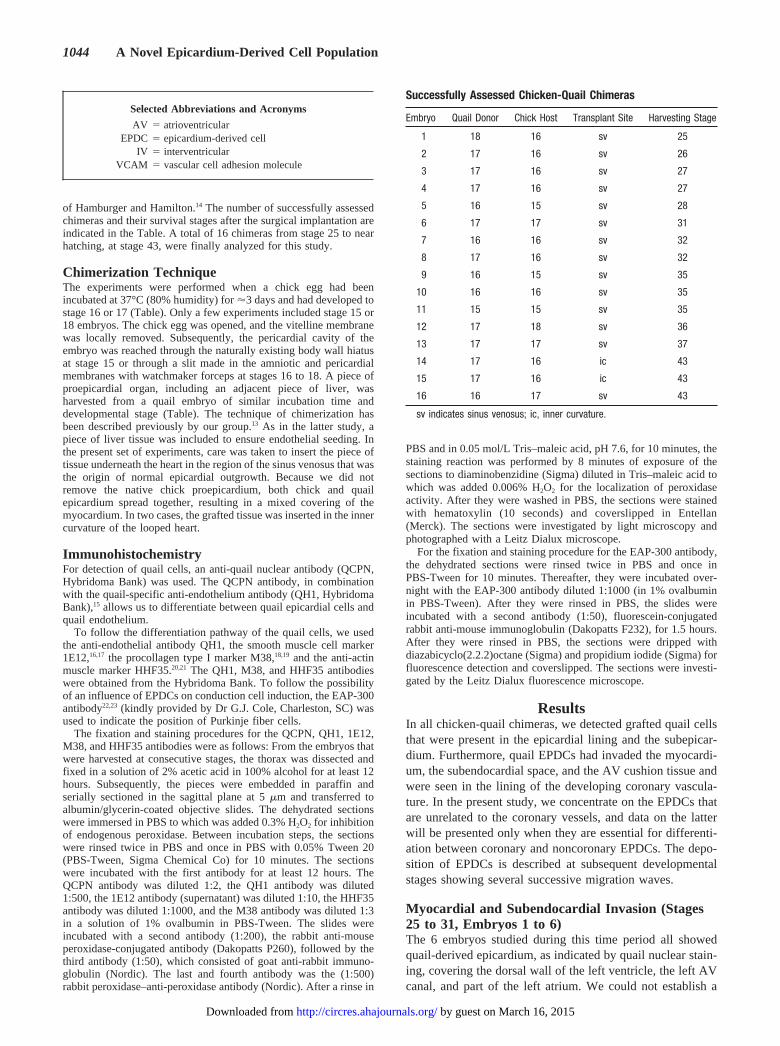
of Hamburger and Hamilton.14 The number of successfully assessedchimeras and their survival stages after the surgical implantation areindicated in the Table. A total of 16 chimeras from stage 25 to nearhatching, at stage 43, were finally analyzed for this study.
Chimerization TechniqueThe experiments were performed when a chick egg had beenincubated at 37°C (80% humidity) for'3 days and had developed tostage 16 or 17 (Table). Only a few experiments included stage 15 or18 embryos. The chick egg was opened, and the vitelline membranewas locally removed. Subsequently, the pericardial cavity of theembryo was reached through the naturally existing body wall hiatusat stage 15 or through a slit made in the amniotic and pericardialmembranes with watchmaker forceps at stages 16 to 18. A piece ofproepicardial organ, including an adjacent piece of liver, washarvested from a quail embryo of similar incubation time anddevelopmental stage (Table). The technique of chimerization hasbeen described previously by our group.13 As in the latter study, apiece of liver tissue was included to ensure endothelial seeding. Inthe present set of experiments, care was taken to insert the piece oftissue underneath the heart in the region of the sinus venosus that wasthe origin of normal epicardial outgrowth. Because we did notremove the native chick proepicardium, both chick and quailepicardium spread together, resulting in a mixed covering of themyocardium. In two cases, the grafted tissue was inserted in the innercurvature of the looped heart.
ImmunohistochemistryFor detection of quail cells, an anti-quail nuclear antibody (QCPN,Hybridoma Bank) was used. The QCPN antibody, in combinationwith the quail-specific anti-endothelium antibody (QH1, HybridomaBank),15 allows us to differentiate between quail epicardial cells andquail endothelium.
To follow the differentiation pathway of the quail cells, we usedthe anti-endothelial antibody QH1, the smooth muscle cell marker1E12,16,17 the procollagen type I marker M38,18,19 and the anti-actinmuscle marker HHF35.20,21 The QH1, M38, and HHF35 antibodieswere obtained from the Hybridoma Bank. To follow the possibilityof an influence of EPDCs on conduction cell induction, the EAP-300antibody22,23 (kindly provided by Dr G.J. Cole, Charleston, SC) wasused to indicate the position of Purkinje fiber cells.
The fixation and staining procedures for the QCPN, QH1, 1E12,M38, and HHF35 antibodies were as follows: From the embryos thatwere harvested at consecutive stages, the thorax was dissected andfixed in a solution of 2% acetic acid in 100% alcohol for at least 12hours. Subsequently, the pieces were embedded in paraffin andserially sectioned in the sagittal plane at 5mm and transferred toalbumin/glycerin-coated objective slides. The dehydrated sectionswere immersed in PBS to which was added 0.3% H2O2 for inhibitionof endogenous peroxidase. Between incubation steps, the sectionswere rinsed twice in PBS and once in PBS with 0.05% Tween 20(PBS-Tween, Sigma Chemical Co) for 10 minutes. The sectionswere incubated with the first antibody for at least 12 hours. TheQCPN antibody was diluted 1:2, the QH1 antibody was diluted1:500, the 1E12 antibody (supernatant) was diluted 1:10, the HHF35antibody was diluted 1:1000, and the M38 antibody was diluted 1:3in a solution of 1% ovalbumin in PBS-Tween. The slides wereincubated with a second antibody (1:200), the rabbit anti-mouseperoxidase-conjugated antibody (Dakopatts P260), followed by thethird antibody (1:50), which consisted of goat anti-rabbit immuno-globulin (Nordic). The last and fourth antibody was the (1:500)rabbit peroxidase–anti-peroxidase antibody (Nordic). After a rinse in
PBS and in 0.05 mol/L Tris–maleic acid, pH 7.6, for 10 minutes, thestaining reaction was performed by 8 minutes of exposure of thesections to diaminobenzidine (Sigma) diluted in Tris–maleic acid towhich was added 0.006% H2O2 for the localization of peroxidaseactivity. After they were washed in PBS, the sections were stainedwith hematoxylin (10 seconds) and coverslipped in Entellan(Merck). The sections were investigated by light microscopy andphotographed with a Leitz Dialux microscope.
For the fixation and staining procedure for the EAP-300 antibody,the dehydrated sections were rinsed twice in PBS and once inPBS-Tween for 10 minutes. Thereafter, they were incubated over-night with the EAP-300 antibody diluted 1:1000 (in 1% ovalbuminin PBS-Tween). After they were rinsed in PBS, the slides wereincubated with a second antibody (1:50), fluorescein-conjugatedrabbit anti-mouse immunoglobulin (Dakopatts F232), for 1.5 hours.After they were rinsed in PBS, the sections were dripped withdiazabicyclo(2.2.2)octane (Sigma) and propidium iodide (Sigma) forfluorescence detection and coverslipped. The sections were investi-gated by the Leitz Dialux fluorescence microscope.
ResultsIn all chicken-quail chimeras, we detected grafted quail cellsthat were present in the epicardial lining and the subepicar-dium. Furthermore, quail EPDCs had invaded the myocardi-um, the subendocardial space, and the AV cushion tissue andwere seen in the lining of the developing coronary vascula-ture. In the present study, we concentrate on the EPDCs thatare unrelated to the coronary vessels, and data on the latterwill be presented only when they are essential for differenti-ation between coronary and noncoronary EPDCs. The depo-sition of EPDCs is described at subsequent developmentalstages showing several successive migration waves.
Myocardial and Subendocardial Invasion (Stages25 to 31, Embryos 1 to 6)The 6 embryos studied during this time period all showedquail-derived epicardium, as indicated by quail nuclear stain-ing, covering the dorsal wall of the left ventricle, the left AVcanal, and part of the left atrium. We could not establish a
Selected Abbreviations and Acronyms
AV 5 atrioventricularEPDC 5 epicardium-derived cell
IV 5 interventricularVCAM 5 vascular cell adhesion molecule
Successfully Assessed Chicken-Quail Chimeras
Embryo Quail Donor Chick Host Transplant Site Harvesting Stage
1 18 16 sv 25
2 17 16 sv 26
3 17 16 sv 27
4 17 16 sv 27
5 16 15 sv 28
6 17 17 sv 31
7 16 16 sv 32
8 17 16 sv 32
9 16 15 sv 35
10 16 16 sv 35
11 15 15 sv 35
12 17 18 sv 36
13 17 17 sv 37
14 17 16 ic 43
15 17 16 ic 43
16 16 17 sv 43
sv indicates sinus venosus; ic, inner curvature.
1044 A Novel Epicardium-Derived Cell Population
by guest on March 16, 2015http://circres.ahajournals.org/Downloaded from

pattern of outgrowth that related to the stage of harvesting ofthe graft or stage of implanting in the host, and we havetherefore provided general data. In one embryo (Figure 1Aand 1B and embryo No. 5 in the Table), the major part of theepicardium was quail-derived, except for the right atrium andthe outflow tract. Using the quail anti-endothelial antibody,we confirmed in embryo 3 that the endothelium-lined tubes of
the developing coronary vasculature were of mixed quail andchick origin (Figure 1C).
Apart from the quail cells that take part in formation of thevasculature, there is a distinct subpopulation of scattered cellsthat invades the myocardium. These EPDCs are usually isolatedand do not stain for endothelium (compare panels A and B ofFigure 2), M38, and HHF35 (not shown). There is a marked
Figure 1. A, Section of the AV sulcus interposed between atrium (A) and ventricle (V). The epicardium (E) covers the myocardium (M)that is still continuous between atrium and ventricle. The dark QCPN-stained quail cells form the epicardial lining and also form the lin-ing of a coronary vessel (CV). B, Section subsequent to panel A showing quail-specific anti-endothelial antibody QH1 in the quail-de-rived endothelium. C, Distribution of EPDCs at stage 27 (embryo 3). EPDCs, with darkly stained nuclei, are present within the epicar-dium (E) and the myocardium (M) as isolated cells (arrowheads) and in the chicken (pale) and quail (dark) mixed lining of a coronaryvessel (CV). Bar550 mm.
Gittenberger-de Groot et al June 1, 1998 1045
by guest on March 16, 2015http://circres.ahajournals.org/Downloaded from

preferential localization of these quail cells in the subendocardialregion (Figure 2C). The way in which they reach the subendo-cardial position is remarkable in that there exist invaginations ofendocardium through discontinuities in the thin-walled myocar-dium. In this way, the subepicardial and subendocardial spaceare in contact, allowing for a direct route of EPDCs to asubendocardial position (Figure 2D and 2E). Only occasionallydoes the endocardial lining of the ventricles and atria harborquail endothelial cells. In one embryo (No. 6), there is colocal-ization of procollagen (M38)-positive cells and of quail-derivedcells in the subendocardial region (not shown).
Endocardial Cushion Invasion (Stages 32 to 43,Embryos 7 to 16)The distribution of the quail-derived epicardium variedfrom the right ventricular dorsal wall and part of the IVseptum and right AV transition (embryos 7, 9, and 10) to
a mainly left ventricular orientation (embryo 8). In onechimera, there was almost a complete quail-derived cov-ering of the heart, including the outflow tract (embryo 11).In another heart, the covering epicardium was chick-derived, and the subepicardium was filled with quail cells(embryo 14). In the remaining chimeras, only parts of theheart were covered with quail cells, eg, the outflow tract(embryos 13 and 15), the IV septum (embryo 12), and theright ventricular apex (embryo 16). The distribution of themigrated EPDCs is tangential to the quail-derived areas ofepicardial covering. In this way, we encountered twoembryos (Nos. 11 and 14) with atrial myocardial invasion(Figure 3A).
The quail EPDCs were found inside the compact myo-cardium, the trabeculae, and the subendocardium. Themyocardial discontinuities were not seen anymore. In threeembryos (Nos. 9, 11, and 14), we found cells positive for
Figure 2. A, Section of the ventricular wall of embryo 3. Dark EPDCs are seen scattered through the myocardium (M) and in a suben-docardial position (arrowheads) along the trabeculae of the ventricle (V). E indicates epicardium. B, Section consecutive to panel Ashowing the EPDCs (indicated by arrows in panel A) that have not differentiated into endothelial cells. The quail-derived endothelialcells participating in coronary vessel formation have been stained with QH1. C, Detail of the subendocardial region (SE) overlying themyocardium in embryo 6. EPDCs (arrowheads) are markedly present in this area. D, Detail of embryo 3 of a trabecular space (T) linedby endocardium (arrows) that breaks through a myocardial discontinuity to contact the SE (arrowheads). E, Detail of embryo 1 showingthe close relationship of subepicardium (E) and subendocardium (SE) through a myocardial discontinuity (arrowhead). Bar550 mm.
1046 A Novel Epicardium-Derived Cell Population
by guest on March 16, 2015http://circres.ahajournals.org/Downloaded from

the procollagen marker M38 in the same region as EPDCs(not shown).
A remarkable observation was the presence of a largenumber of quail EPDCs in the AV endocardial cushions. Inembryos 7, 9, and 10, the cushions of the developing tricuspidvalve had been invaded (Figures 3B, 4A to 4C, and 5B), andin embryos 11 and 14, the developing mitral valve had beeninvaded as well (Figure 5A). The location correlated with theoverlying extent of quail-derived epicardium. In this region,there was a direct contact between the subepicardium and theendocardial cushion tissue as the atrial and ventricular myo-cardial connection was interrupted, enabling a relatively shortmigration route (Figures 3B, 4A, 4B, and 5A). The quailEPDCs in the endocardial cushions favored two positions.One was at the myocardial/endocardial cushion interface(Figures 3B, 4B, and 5B). The second site of accumulatedquail EPDCs was found subendocardially at the luminal faceof the AV cushion (Figures 3B, 4A to 4C, 5A, and 5B). In theAV cushions, we were never able to demonstrate incorpora-tion of grafted cells into the endocardial lining that we hadseen occasionally in endocardium lining the ventricular myo-cardium (Figure 4C). Once again, the scattered EPDCs thathad gained an intramyocardial and subendocardial positioncould easily be distinguished from those quail cells that had
differentiated into the endothelial lining of the coronaryvessels (Figure 5B and 5C). A clear correlation was shownbetween procollagen formation and EPDCs in the developingannulus fibrosis (Figure 6A and 6B) as well as in thesubendocardial region (Figure 6C and 6D).
In 4 embryos (Nos. 11, 13, 14, and 15), in which theepicardium also covered the outflow tract, we found scatteredquail EPDCs in the muscular outflow tract septum. Themesenchyme of the endocardial outflow tract cushions had
Figure 3. A, Section of the wall of the right atrium (RA) inembryo 14 showing the subendocardial (SE)-positioned QCPN-stained EPDCs (arrowheads). EPDCs are also scatteredthroughout the myocardium (M). B, At stage 35 (embryo 9),atrial and ventricular myocardium has become discontinuous,and EPDCs are found spreading from the epicardium (E) towardthe cushion tissue of the tricuspid valve (TV) overlying the myo-cardium of the right ventricle (RV). Bar550 mm.
Figure 4. A, Overview of the developing tricuspid valvebetween the right atrium (RA) and the right ventricle (RV) inembryo 11. An extensive deposition of EPDCs (dark QCPN-stained nuclei) is present in the epicardium (E), the myocardium(M), and the AV cushion (AC). B and C, Micrographs showingdetails of the EPDCs in the AV cushion of embryo 9, revealingits mainly subendocardial, but not endocardial, position.Bar5100 mm.
Gittenberger-de Groot et al June 1, 1998 1047
by guest on March 16, 2015http://circres.ahajournals.org/Downloaded from

disappeared at this time. The semilunar valves were negativefor EPDCs.
Spatial Correlation of EPDC Distribution WithPurkinje FibersWhen the EAP-300 antibody was used for detection of differ-entiating Purkinje fibers, a remarkable colocalization was ob-served. The Purkinje fibers were localized subendocardially(Figure 7A) and around coronary arteries in a later stage ofdevelopment (Figure 7C). This staining correlated very closelywith the subendocardially positioned quail EPDCs (Figure 7B)as well as with the coronary arterial adventitial quail-derivedfibroblasts (Figure 7D). There was also a colocalization ofclusters of myocardial cells that stained with the EAP-300marker in the AV cushions (Figure 7E), and EPDCs werepresent in the adjacent section (Figure 7F).
DiscussionThe present study shows for the first time that there is anextensive contribution of epicardial cells not only to thecoronary vasculature but also to the myocardial wall, thesubendocardial region, the AV cushions and valves, and thefibrous heart skeleton.
The importance of an epicardial covering for cardiacsurvival is substantiated by several experimental models inwhich epicardial formation is hampered. An example is givenin the VCAM-1–deficient mice.10 These mice die at embry-onic days 11.5 to 12.5. A somewhat comparable model, butwith a less severely affected myocardium, is given in thea4knockout mice that also die between embryonic days 12 and12.5.24 In these embryos, the coronary vessels normallyresiding within the AV sulcus, together with the completeepicardial covering, are missing. Epicardial attachment is
believed to be established by thea4 integrin/VCAM-1interaction between epicardial cells and the outermyocardium.
It remains to be investigated what the actual triggeringeffect for the embryo lethality is. We postulate that in allmodels in which the epicardial outgrowth is disturbed notonly is coronary vascular formation hampered but also anearly important population of EPDCs does not enter themyocardium, the subendocardial layer, or, later on, the AVcushions.
There is evidence that we are dealing with precursors of thecardiac fibroblasts. In a number of cases, we could demon-strate costaining with the procollagen marker M38, especiallyin the older stages (stage 43), where formation of the fibrousheart skeleton between the atria and the ventricles takes place.Furthermore, the localization of the scattered EPDCs in themyocardium, in the subendocardial region, and, later on, inthe AV cushion tissue complies with the presence of afibroblastic cell lineage.
In the present study, we have identified a close positionalrelationship between subendocardial and periarterial EPDCsand the differentiation of Purkinje fibers. Our data implicateEPDCs in the development of conductive cells and resolve aninteresting enigma that has emerged in recent years concern-ing the differentiation of this specialized myocardial lineage.It has been established that intramural Purkinje fibers ramifyin close association with coronary arteries in the chickheart.25,26 In cell lineage studies, Gourdie et al23 have shownthat perivascular Purkinje cells share common progenitorswith working myocytes. They hypothesized that cells notfurther defined but associated with the arterial bed haveinduced the differentiation of myocytes into conductive cells.A caveat of this model has been that Purkinje fiber cells occur
Figure 5. A, Longitudinal section through the AV transition between the left atrium (LA) and the left ventricle (LV) in embryo 11. TheQCPN-stained EPDCs are clustered in the endocardial cushion tissue of the developing mitral valve (MV). E indicates epicardium; M,myocardium. B, Section of AV cushion tissue of the developing tricuspid valve in embryo 14 showing EPDCs in the subendocardium(SE) and at the endocardial cushion/myocardium interface (arrowheads). C, EPDCs separated from the quail-derived endothelial cellslining the coronary vasculature (CV) as shown in a subsequent section. Bar5100 mm.
1048 A Novel Epicardium-Derived Cell Population
by guest on March 16, 2015http://circres.ahajournals.org/Downloaded from

at subendocardial locations far from arteries as well as atlocations adjacent to arteries in birds. Indeed, in mostmammalian species, including humans, the peripheral con-ductive network is confined exclusively to the subendocar-dium.27 The migration patterns of EPDCs described in the
present study may provide the basis for an explanation of thisinteresting difference. We hypothesize that the specific dis-tribution pattern of EPDCs implicates this extracardiac pop-ulation in the induction and organization of Purkinje fibers atboth subendocardial and periarterial sites in birds.
Figure 6. A, Section through an already well-differentiated mitral valve leaflet (MV) between the left atrium (LA) and left ventricle (LV) inembryo 14. At the site of the formation of the fibrous annulus (asterisk), there is marked procollagen-I staining, as indicated by the M38antibody. B, Section subsequent to panel A showing the QCPN-stained EPDCs (asterisk) in annulus fibrosis formation. C and D, Twosubsequent sections of the wall of the LA of embryo 14. The arrowheads in panel C show the procollagen-I–positive cells in the suben-docardial region (SE). In panel D, the dark QCPN-stained EPDCs are in the same area. E indicates epicardium; M, myocardium.Bar5100 mm.
Gittenberger-de Groot et al June 1, 1998 1049
by guest on March 16, 2015http://circres.ahajournals.org/Downloaded from

From stage 32 onward, we have detected EPDCs in the AVcushions and in later stages in the AV valves. This is the firsttime that such a population is described. The presence of theEPDCs is particularly prominent in the subendocardium,where endocardial cells have been reported to transform intoAV cushion mesenchymes.2 This suggests competition for thesame localization in the subendocardial position of theendocardium-derived cells and the EPDCs. It is, however, not
possible to mix up populations, because the EPDCs areclearly quail-derived (QCPN-positive) and, in this location,nonendothelial (QH1-negative).
Another site of EPDCs is between cardiomyocytes that arefound at the endocardial cushion/myocardium interface be-fore valve formation. At this site, the cushion tissue has todelaminate from the underlying myocardium to form a freevalve leaflet.4 Transformation of myocardial cells to fibro-
Figure 7. A, The Purkinje fiber (P) within a ventricular trabecula (T) of embryo 14 is positive for EAP-300. RV indicates right ventricle;M, myocardium. B, There is a colocalization with EPDCs (arrowheads), as shown in a consecutive section. C, Purkinje fibers (arrow-heads) are localized within the myocardium surrounding the coronary arteries (CA) of embryo 14. E indicates epicardium. D, Colocaliza-tion with the adjacent coronary arterial adventitia containing quail fibroblasts (darkly stained) occurs. E, Myocardial cells present withinthe AV cushion tissue (AC) of embryo 14 are positive for EAP-300. LA indicates left atrium; LV, left ventricle. F, Again, many EPDCs(darkly stained) are lying in between the myocardial cell clusters. Bar5100 mm.
1050 A Novel Epicardium-Derived Cell Population
by guest on March 16, 2015http://circres.ahajournals.org/Downloaded from
blasts has been postulated to be essential for this process.28 Inthe AV cushions, there were clusters of myocardial cells,intermingled with EPDCs, that showed Purkinje cell charac-teristics (EAP-300 staining). It remains to be investigatedwhat the fate of the latter cells is and whether they might bethe precursors of the described occasional nerve fibers in AVvalves.29
The EPDCs are also located at the site of the fibrousannulus between atrium and ventricle, being positive forprocollagen-I. It is evident that migration of EPDCs isnecessary for the formation of the fibrous heart skeleton andthe AV valves. This is not achieved by infolding of theepicardium, as was postulated by Wenink et al,30 but bymigration of individual EPDCs. Our findings do not contra-dict the results of Wessels et al,31 who showed differentimmunohistochemical characteristics of epicardium and AVcushions.
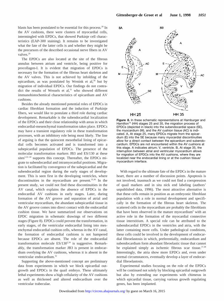
Besides the already mentioned potential roles of EPDCs incardiac fibroblast formation and the induction of Purkinjefibers, we would like to postulate a third role during cardiacdevelopment. Remarkable is the subendocardial localizationof the EPDCs and their close relationship with areas in whichendocardial-mesenchymal transformation takes place. EPDCsmay have a transient regulatory role in these transformationprocesses, with an inhibitory role being most likely. The lineof arguing is that the quiescent mesothelial lining of epicar-dial cells becomes activated and is transformed into asubepicardial population of EPDCs. The presence of themolecular transformation markers JB3 and ES/130 at thesesites2,32–34 supports this concept. Thereafter, the EPDCs mi-grate to subendocardial and intramyocardial positions. Migra-tion is facilitated by convergence of the subepicardial and thesubendocardial region during the early stages of develop-ment. This is seen first in the developing ventricles, wherediscontinuities in the myocardium are present.35,36 In thepresent study, we could not find these discontinuities in theAV canal, which explains the absence of EPDCs in theendocardial AV cushions in early stages. Later, duringformation of the AV groove and separation of atrial andventricular myocardium, the abundant subepicardial tissue inthe AV groove comes into direct contact with the endocardialcushion tissue. We have summarized our observations onEPDC migration in schematic drawings of two differentstages (Figure 8). EPDCs could prevent the transformation, inearly stages, of the ventricular endocardial lining into mes-enchymal endocardial cushion cells, whereas in the AV canal,the formation of endocardial cushions is not hamperedbecause EPDCs are absent. The role of the endocardialtransformation molecule ES/1302,34 is suggestive. Remark-ably, the transformation marker JB3 is present in endocar-dium overlying the AV cushions, whereas it is absent in theventricular endocardium.32
Supporting the above-mentioned concept are preliminarydata from experiments in which we block epicardial out-growth and EPDCs in the quail embryo. These ultimatelylethal experiments show a high cellularity of the AV cushionsas well as thickened and altered endocardium over theventricular trabeculae.
With regard to the ultimate fate of the EPDCs in the matureheart, there are a number of discussion points. Apoptosis isnot involved, inasmuch as we could not find a coexpressionof quail markers and in situ nick end labeling (authors’unpublished data, 1996). The most attractive alternative isthat these cells remain in position as an embryonic fibroblastpopulation with a role in normal development and specifi-cally in the formation of the fibrous heart skeleton. Thescattered cells in the myocardium are probably the fibroblaststhat have been observed in the mature myocardium5 with anactive role in the formation of the myocardial connectivetissue interstitium. A special role can be attributed to thesubendocardial EPDCs in the ventricles and atria, with thelatter containing more cells. Under pathological conditions,these cells could be involved in the development of endocar-dial fibroelastosis in which, preferentially, endocardium andsubendocardium form abundant fibroelastic tissue that cannotbe explained simply as ischemic fibrous scar tissue.37,38
Interestingly, the atria show more EPDCs and, even undernormal circumstances, eventually develop a layer of endocar-dial fibroelastosis.
Experimental studies focusing on the role of the EPDCswill be continued not solely by blocking epicardial outgrowthbut also by extending our experiments with chimeras inwhich epicardial tissue, carrying various growth regulatinggenes, has been implanted.
Figure 8. In these schematic representations at Hamburger andHamilton14 (HH) stages 25 and 35, the migration process ofEPDCs (depicted in black) into the subendocardial space (SE),the myocardium (M), and the AV cushion tissue (AC) is indi-cated. A, At stage 25, many EPDCs migrate from the epicar-dium (E) into the SE because many myocardial discontinuitiesallow for a direct contact between the epicardium and subendo-cardium. EPDCs are not encountered within the AV cushions atthis stage. A indicates atrium; V, ventricle. B, At stage 35, theinterruption between atrial and ventricular myocardium allowsfor migration of EPDCs into the AV cushions, where they arelocalized near the endocardial lining or at the cushion tissue/myocardium interface.
Gittenberger-de Groot et al June 1, 1998 1051
by guest on March 16, 2015http://circres.ahajournals.org/Downloaded from

AcknowledgmentsDr Gourdie is a Basil O’Conner Scholar of the March of Dimes BirthDefects Foundation (Fy95 1145). Dr Vrancken Peeters is supportedby the Netherlands Heart Foundation (No. D96.017).
References1. Laverriere AC, MacNeill C, Mueller C, Poelmann RE, Burch JBE, Evans
T. GATA-4/5/6, a subfamily of three transcription factors transcribed indeveloping heart and gut.J Biol Chem. 1994;269:23177–23184.
2. Eisenberg LM, Markwald RR. Molecular regulation of atrioventricularvalvuloseptal morphogenesis.Circ Res. 1995;77:1–6.
3. DeRuiter MC, Poelmann RE, VanderPlas-de Vries I, Mentink MMT,Gittenberger-de Groot AC. The development of the myocardium andendocardium in mouse embryos: fusion of two heart tubes?AnatEmbryol. 1992;185:461–473.
4. Gittenberger-de Groot AC, Poelmann RE, Bartelings MM. Embryologyof congenital heart disease. In: Braunwald E, ed.Atlas of Heart Diseases:Congenital Heart Disease.Philadelphia, Pa: Current Medicine; 1997;12:3.1–3.10.
5. Borg TK, Rubin K, Carver W, Samarel A, Terracio L. The cell biologyof the cardiac interstitium.Trends Cardiovasc Med. 1996;6:65–70.
6. Viragh S, Challice CE. The origin of the epicardium and the embryonicmyocardial circulation in the mouse.Anat Rec. 1981;201:157–168.
7. Hiruma T, Hirakow R. Epicardial formation in embryonic chick heart:computer-aided reconstruction, scanning and transmission electronmicroscopic studies.Am J Anat. 1989;184:129–138.
8. Viragh S, Gittenberger-de Groot AC, Poelmann RE, Kalman F. Earlydevelopment of quail heart epicardium and associated vascular andglandular structures.Anat Embryol. 1993;188:381–393.
9. Vrancken Peeters M-PFM, Mentink MMT, Poelmann RE,Gittenberger-de Groot AC. Cytokeratins as a marker for epicardial for-mation in the quail embryo.Anat Embryol. 1995;191:503–508.
10. Kwee L, Baldwin HS, Min Shen H, Stewart CL, Buck C, Buck CA,Labow MA. Defective development of the embryonic and extraem-bryonic circulatory systems in vascular cell adhesion molecule(VCAM-1) deficient mice.Development. 1995;121:489–503.
11. Mikawa T, Fischman DA. Retroviral analysis of cardiac morphogenesis:discontinuous formation of coronary vessels.Proc Natl Acad Sci U S A.1992;89:9504–9508.
12. Mikawa T, Gourdie RG. Pericardial mesoderm generates a population ofcoronary smooth muscle cells migrating into the heart along withingrowth of the epicardial organ.Dev Biol. 1996;174:221–232.
13. Poelmann RE, Gittenberger-de Groot AC, Mentink MMT, Bokenkamp R,Hogers B. Development of the cardiac coronary vascular endothelium,studied with antiendothelial antibodies, in chicken-quail chimeras.CircRes. 1993;73:559–568.
14. Hamburger V, Hamilton HL. A series of normal stages in development ofthe chick embryo.J Morphol. 1951;88:49–92.
15. Pardanaud L, Altmann C, Kitos P, Dieterlen-Lievre F, Buck CA. Vascu-logenesis in the early quail blastodisc as studied with a monoclonalantibody recognizing endothelial cells.Development. 1987;100:339–349.
16. Hungerford JE, Owens GK, Argraves WS, Little CD. Development of theaortic vessel wall as defined by vascular smooth muscle and extracellularmatrix markers.Dev Biol. 1996;178:375–392.
17. Vrancken Peeters M-PFM, Gittenberger-de Groot AC, Mentink MMT,Hungerford JE, Little CD, Poelmann RE. The development of the coro-nary vessels and their differentiation into arteries and veins in theembryonic quail heart.Dev Dyn. 1997;208:338–348.
18. McDonald JA, Broekelmann TJ, Matheke ML, Crouch E, Koo M, KuhnC III. A monoclonal antibody to the carboxyterminal domain of pro-
collagen type I visualizes collagen-synthesizing fibroblasts.J Clin Invest.1986;78:1237–1244.
19. Sinning AR, Lepera RC, Markwald RR. Initial expression of type Iprocollagen in chick cardiac mesenchyme is dependent upon myocardialstimulation.Dev Biol. 1988;130:167–174.
20. Tsukada T, Tippens D, Gordon D, Ross R, Gown AM. HHF35, a muscle-actin-specific monoclonal antibody, I: immunocytochemical and bio-chemical characterization.Am J Pathol. 1987;126:51–60.
21. Tsukada T, McNutt MA, Ross R, Gown AM. HHF35, a muscle actin-specific monoclonal antibody, II: reactivity in normal, reactive, and neo-plastic human tissues.Am J Pathol. 1987;127:389–402.
22. McCabe CF, Gourdie RG, Thompson RP, Cole GJ. Developmentallyregulated neural protein EAP-300 is expressed by myocardium andcardiac neural crest during chick embryogenesis.Dev Dyn. 1995;203:51–60.
23. Gourdie RG, Mima T, Thompson RP, Mikawa T. Terminal diversifi-cation of the myocyte lineage generates Purkinje fibers of the cardiacconduction system.Development. 1995;121:1423–1431.
24. Yang JT, Rayburn H, Hynes RO. Cell adhesion events mediated bya4integrins are essential in placental and cardiac development.Devel-opment. 1995;121:549–560.
25. Davies F. Conducting system of bird’s heart.J Anat. 1930;64:129–146.26. Gourdie RG. A map of the heart: gap junctions, connexin diversity and
retroviral studies of conduction myocyte lineage.Clin Sci. 1995;88:257–262.
27. Schiaffino S. Protean patterns of gene expression in the heart conductionsystem.Circ Res. 1997;80:749–750.
28. Lamers WH, Viragh S, Wessels A, Moorman AFM, Anderson RH.Formation of the tricuspid valve in the human heart.Circulation. 1995;91:111–121.
29. Tsumori T, Domoto T. Ultrastructural evidence for innervation of theendothelium and interstitial cells in the atrioventricular valves of theJapanese monkey.Anat Rec. 1994;240:157–166.
30. Wenink ACG, Gittenberger-de Groot AC, Brom AG. Developmentalconsiderations of mitral valve anomalies.Int J Cardiol. 1986;11:85–98.
31. Wessels A, Markman MWM, Vermeulen JLM, Anderson RH, MoormanAFM, Lamers WH. The development of the atrioventricular junction inthe human heart.Circ Res. 1996;78:110–117.
32. Wunsch AM, Little CD, Markwald RR. Cardiac endothelial heterogeneitydefines valvular development as demonstrated by the diverse expressionof JB3, an antigen of the endocardial cushion tissue.Dev Biol. 1994;165:585–601.
33. Rezaee M, Isokawa K, Halligan N, Markwald RR, Krug EL. Identifi-cation of an extracellular 130-kDa protein involved in early cardiacmorphogenesis.J Biol Chem. 1993;268:14404–14411.
34. Krug EL, Rezaee M, Isokawa K, Turner DK, Litke LL, Wunsch AM,Bain JL, Riley DA, Capehart AA, Markwald RR. Transformation ofcardiac endothelium into cushion mesenchyme is dependent on ES/130:temporal, spatial, and functional studies in the early chick embryo.CellMol Biol Res. 1995;41:263–277.
35. Rychter Z, Os˘tadal B. Mechanism of the development of coronary arteriesin chick embryo.Folia Morphol (Warsz). 1971;19:113–124.
36. Munoz-Chapuli R, MacRas D, Ramos C, Gallego A, de Andre´s V.Development of the subepicardial mesenchyme and the early cardiacvessels in the dogfish (Scyliorhinus canicula).J Exp Zool. 1996;275:95–111.
37. Essed CE, Klein HW, Krediet P. Coronary and endocardial fibroelastosisof the ventricles in the hypoplastic left and right heart syndromes.Virchows Arch A Pathol Anat Histol. 1975;368:87–97.
38. Esterly JR, Oppenheimer EH. Some aspects of cardiac pathology ininfancy and childhood, IV: myocardial and coronary lesions and cardiacmalformations.Pediatrics. 1967;39:896–903.
1052 A Novel Epicardium-Derived Cell Population
by guest on March 16, 2015http://circres.ahajournals.org/Downloaded from

Robert G. Gourdie and Robert E. PoelmannAdriana C. Gittenberger-de Groot, Mark-Paul F.M. Vrancken Peeters, Monica M.T. Mentink,
Atrioventricular CushionsEpicardium-Derived Cells Contribute a Novel Population to the Myocardial Wall and the
Print ISSN: 0009-7330. Online ISSN: 1524-4571 Copyright © 1998 American Heart Association, Inc. All rights reserved.is published by the American Heart Association, 7272 Greenville Avenue, Dallas, TX 75231Circulation Research
doi: 10.1161/01.RES.82.10.10431998;82:1043-1052Circ Res.
http://circres.ahajournals.org/content/82/10/1043World Wide Web at:
The online version of this article, along with updated information and services, is located on the
http://circres.ahajournals.org//subscriptions/
is online at: Circulation Research Information about subscribing to Subscriptions:
http://www.lww.com/reprints Information about reprints can be found online at: Reprints:
document. Permissions and Rights Question and Answer about this process is available in the
located, click Request Permissions in the middle column of the Web page under Services. Further informationEditorial Office. Once the online version of the published article for which permission is being requested is
can be obtained via RightsLink, a service of the Copyright Clearance Center, not theCirculation Researchin Requests for permissions to reproduce figures, tables, or portions of articles originally publishedPermissions:
by guest on March 16, 2015http://circres.ahajournals.org/Downloaded from