Enzootics of visceral granulomas associated with Francisella-like organism infection in tilapia (...
10
Enzootics of visceral granulomas associated with Francisella-like organism infection in tilapia (Oreochromis spp.) C.Y. Hsieh a , M.C. Tung a , C. Tu b , C.D. Chang a , S.S. Tsai a, ⁎ a Department of Veterinary Medicine, National Pingtung University of Science and Technology, No. 1, Shen-hu Rd., Neipu, Pingtung 912, Taiwan b National Animal Health Institute, Taipei, Taiwan Received 18 June 2005; received in revised form 29 March 2006; accepted 29 March 2006 Abstract Between 2001 and 2004, 10 strains of Francisella species were isolated from visceral granulomas of diseased tilapia in Taiwan. In a comparison of nucleotide sequences for whole 16S rRNA gene with those of reference bacteria, the isolated strains had a high sequence similarity to Francisella philomiragia (98.6%), Francisella tularensis subsp. novicida (97.4%) and F. tularensis subsp. tularensis (96.1%). On the basis of electron microscopic examination, phenotypic characteristics and PCR assays for the 16S rRNA gene sequence, the causative agent clearly belongs to the genus Francisella. © 2006 Published by Elsevier B.V. Keywords: Tilapia; Granuloma; Intracellular organism; Polymorphic cocco-bacillus; Francisella sp. 1. Introduction In Taiwan, tilapias infected with an unknown intracellular organism were initially found in October 1992. Now, the disease is widespread in fresh-, brackish- and salt pond-cultured tilapia, involving six species and causing high mortality, up to 95% in some cases (Chen et al., 1994; Chern and Chao, 1994). Prominent features are skin ulceration and the presence of multiple whitish nodules on most visceral organs, especially the kidney and spleen. The agent is a cocco- bacillus with a polymorphous shape, which can be cultured in CHSE-214 and TO 2 cells, but does not grow on artificial synthetic media. Therefore, it was provi- sionally called a ‘rickettsia-like organism’ (RLO) (Chen et al., 1994; Chern and Chao, 1994). Francisella spp. are facultative intracellular organ- isms, capable of penetrating intact skin or penetrating via skin injury to elicit a local inflammatory response with formation of papules and ulcers in situ. Following lymphohematogenous dispersion, the agents can invade parenchymal tissue resulting in the formation of granulomas and abscesses. Francisella tularensis and Francisella philomiragia are the most prevalent agents associated with chronic and necrotizing granulomas (Quan et al., 1956; Mörner, 1992; Sicherer et al., 1997; Polack et al., 1998). Waterborne transmission of Francisella spp. is well known for causing disease in both humans and aquatic animals (Mignani et al., 1988; Hoel et al., 1991; Polack et al., 1998). These organisms have been identified by both culture and PCR assay from crayfish (Anda et al., 2001), muskrats (Jensen et al., 1969) and water (Forsman et al., 1995). Previous research on outbreaks of RLO in Taiwan had been limited to southern Taiwan (Chen et al., 1994; Aquaculture 254 (2006) 129 – 138 www.elsevier.com/locate/aqua-online ⁎ Corresponding author. Fax: +886 8 7740295. E-mail address: [email protected] (S.S. Tsai). 0044-8486/$ - see front matter © 2006 Published by Elsevier B.V. doi:10.1016/j.aquaculture.2006.03.044
-
Upload
independent -
Category
Documents
-
view
3 -
download
0
Transcript of Enzootics of visceral granulomas associated with Francisella-like organism infection in tilapia (...

2006) 129–138www.elsevier.com/locate/aqua-online
Aquaculture 254 (
Enzootics of visceral granulomas associated with Francisella-likeorganism infection in tilapia (Oreochromis spp.)
C.Y. Hsieh a, M.C. Tung a, C. Tu b, C.D. Chang a, S.S. Tsai a,⁎
a Department of Veterinary Medicine, National Pingtung University of Science and Technology, No. 1, Shen-hu Rd., Neipu, Pingtung 912, Taiwanb National Animal Health Institute, Taipei, Taiwan
Received 18 June 2005; received in revised form 29 March 2006; accepted 29 March 2006
Abstract
Between 2001 and 2004, 10 strains of Francisella species were isolated from visceral granulomas of diseased tilapia in Taiwan.In a comparison of nucleotide sequences for whole 16S rRNA gene with those of reference bacteria, the isolated strains had a highsequence similarity to Francisella philomiragia (98.6%), Francisella tularensis subsp. novicida (97.4%) and F. tularensis subsp.tularensis (96.1%). On the basis of electron microscopic examination, phenotypic characteristics and PCR assays for the 16S rRNAgene sequence, the causative agent clearly belongs to the genus Francisella.© 2006 Published by Elsevier B.V.
Keywords: Tilapia; Granuloma; Intracellular organism; Polymorphic cocco-bacillus; Francisella sp.
1. Introduction
In Taiwan, tilapias infected with an unknownintracellular organism were initially found in October1992. Now, the disease is widespread in fresh-,brackish- and salt pond-cultured tilapia, involving sixspecies and causing high mortality, up to 95% in somecases (Chen et al., 1994; Chern and Chao, 1994).Prominent features are skin ulceration and the presenceof multiple whitish nodules on most visceral organs,especially the kidney and spleen. The agent is a cocco-bacillus with a polymorphous shape, which can becultured in CHSE-214 and TO2 cells, but does not growon artificial synthetic media. Therefore, it was provi-sionally called a ‘rickettsia-like organism’ (RLO) (Chenet al., 1994; Chern and Chao, 1994).
⁎ Corresponding author. Fax: +886 8 7740295.E-mail address: [email protected] (S.S. Tsai).
0044-8486/$ - see front matter © 2006 Published by Elsevier B.V.doi:10.1016/j.aquaculture.2006.03.044
Francisella spp. are facultative intracellular organ-isms, capable of penetrating intact skin or penetratingvia skin injury to elicit a local inflammatory responsewith formation of papules and ulcers in situ. Followinglymphohematogenous dispersion, the agents can invadeparenchymal tissue resulting in the formation ofgranulomas and abscesses. Francisella tularensis andFrancisella philomiragia are the most prevalent agentsassociated with chronic and necrotizing granulomas(Quan et al., 1956; Mörner, 1992; Sicherer et al., 1997;Polack et al., 1998). Waterborne transmission ofFrancisella spp. is well known for causing disease inboth humans and aquatic animals (Mignani et al., 1988;Hoel et al., 1991; Polack et al., 1998). These organismshave been identified by both culture and PCR assayfrom crayfish (Anda et al., 2001), muskrats (Jensen etal., 1969) and water (Forsman et al., 1995).
Previous research on outbreaks of RLO in Taiwanhad been limited to southern Taiwan (Chen et al., 1994;

130 C.Y. Hsieh et al. / Aquaculture 254 (2006) 129–138
Chern and Chao, 1994). In the present study, wecollected 120 diseased tilapia from northern, central andsouthern Taiwan. These fish had similar pathology tothose with RLO infections, but these were found to beassociated with a different agent, a Francisella species,based on electron microscopic, bacteriological andmolecular examinations.
2. Materials and methods
2.1. Cytological and pathological examinations
Duplications of organ smears were made from thespleen and kidney of the diseased fish. The smears werestained with Liu's and Gram methods, respectively. Fishtissues were fixed in 10% neutral buffered formalin forat least 16 h and processed for routine paraffin histology,sectioned at 3–4 μm, and stained with haematoxylin andeosin.
2.2. Isolation and bacteriological examinations
Both kidney and spleen were homogenized in aphosphate buffer solution using a Ten-Broeck tissuegrinder and centrifuged at 500×g for 2 min at 20 °C. Theupper layer was collected and spread over Thayer–Martin agar (Anda et al., 2001). Blood agar, brain–heartinfusion agar (containing 5% sheep red blood cells) andblood–cystine–glucose agar were used simultaneouslyfor bacterial isolation. Inoculated media were incubatedat 23, 30 and 35 °C with 5% CO2 for a minimum of10 days, respectively. At the same time, the kidney andspleen emulsions were inoculated on CHSE-214(chinook salmon embryo) cells according to the methoddescribed previously (Fournier et al., 1998), andincubated with 5% CO2 at 23 °C for observation ofcytopathic effect (CPE). The infected cells wereharvested for light and electron microscopic examina-
Table 1Location for sampling, 10 clinical cases
Samples no. Exposure date Location
1. AF-01-02 01/04/2001 Kaohsiu2. AF-01-06 01/25/2001 Kaohsiu3. AF-01-22 02/14/2001 Kaohsiu4. AF-01-23 02/25/2001 Kaohsiu5. AF-01-27 02/26/2001 Pingtung6. AF-01-28 03/15/2001 Hsinchu7. AF-03-27 03/17/2003 Chia-I C8. AF-03-28 03/17/2003 Chia-I C9. AF-04-15 03/01/2004 Pingtung10. AF-04-405 12/05/2004 Pingtung
F=fresh water; B=brackish.
tions. Biochemical tests were performed by convention-al methods (Hollis et al., 1989; Clarridge et al., 1996)and using RapID NH and RapID ANA (Remel, Lenexa,KS, USA) commercial identification kits, according tothe manufacturer's directions.
2.3. Electron microscopic examinations
For ultrastructural examination, samples of kidneyand spleen were fixed in 2.5% glutaraldehyde, post-fixed in osmium tetroxide, dehydrated in a series ofgraded alcohols and embedded in Supper's resin. Thinsections were stained with uranyl acetate and leadcitrate, and observed with a Hitachi H-600 electronmicroscope. For scanning electron microscopy, aFrancisella-like organism (FLO) was collected fromThayer–Martin agar plates and centrifuged at 2000×gfor 15 min at 4 °C, washed with PBS, fixed in 3%glutaraldehyde for 2 h at room temperature and smalldrops of the samples were placed on a presterilized 0.22-μmmembrane of a vacuum-driven disposable bottle-topfilter (Millipore, Bedford, MA, USA) and allowed toadhere. Thereafter, the samples were post-fixed with 1%osmium tetroxide plus 1% tannic acid, dehydrated ingraded ethanol, critical point-dried in CO2, coated with a3.5-nm-thick chromium layer using a penning sputtersystem in a high-vacuum chamber and viewed with aHitachi S3000N scanning electron microscope. Imageswere obtained using secondary and backscatteredelectrons.
2.4. DNA extraction
The isolates used for molecular assay included AF-01-2, AF-01-6, AF-01-22, AF-01-23, AF-01-27, AF-01-28, AF-03-27, AF-03-28, AF-04-15 and AF-04-405(Table 1). A loop of bacterium was transferred into anEppendorf tube containing 200 μl of lysozyme reaction
Fish species Water
ng City O. mossambica Bng City O. mossambica Bng City O. niloticus Bng County O. niloticus BCounty O. mossambica FCounty O. mossambica Founty O. mossambica Founty O. mossambica FCounty O. niloticus FCity O. niloticus F

131C.Y. Hsieh et al. / Aquaculture 254 (2006) 129–138
solution (20 mM Tris–HCl (pH 8.0), 2 mM EDTA,20 μg of lysozyme/ml). It was then suspended throughvigorous vortexing and incubated at 37 °C for 30 min.The cells were lysed by 2.5 mg of proteinase K(Clontech) per ml and incubated at 60 °C for 1 h. Thelysate was subjected to DNA extraction with acommercial kit (Blood and Tissue Genomic DNAExtraction Miniprep System; Viogene, Taipei, Taiwan).DNAwas finally eluted with 100 μl of TE (Tris–EDTA)buffer and stored at −4 °C before use.
2.5. Polymerase chain reaction (PCR) assay andnucleotide sequencing of whole 16S rRNA gene
Eubacteria universal primers (forward, 27f, 5′-AGAGTT TGA TCM TGG CTC AG-3′ and reverse, 1525r,5′-AAG GAG GTG WTC CAR CC-3′) were specifi-cally designed for the amplification of most eubacterial
Fig. 1. Pathological, cytological and ultrastructural examinations of Francisshows white nodules of varying sizes in the enlarged spleen (S), kidney (K) awithin the cytoplasmic vacuoles of a phagocyte on the smear made from kimultiple granulomatous formations with a necrotic center, encapsulated by m(H&E stain). (D) Ultrastructural examination of the kidney reveals that theshape, of 1.45±0.35×0.35±0.15 μm in size, and appear in the cytoplasmic vonly by a narrow electron-lucent space (arrows).
16S rRNA genes (Lane, 1991). Specific primers(forward, F11, 5′-TAC CAG TTG GAA ACG ACTGT-3′ and reverse, F5, 5′-CCT TTT TGA GTT TCGCTC C-3′) were used for the detection of Francisellagenus (Forsman et al., 1994). The PCR buffer (pH 8.5)contained 60 mM Tris–HCl, 15 mM (NH4)2SO4, and1.5 mM MgCl2. Control reactions without templatewere included. After purification of PCR products(QIAquick spin columns; Qiagen, Hilden, Germany),they were cloned into T vectors using a TA cloning kit(YT&A; Yeastern Biotech, Taipei, Taiwan), accordingto the manufacturer's instructions, and sequenced, usinga 373A automatic sequencer and a BigDye Terminatorcycle sequencing kit (Mission Biotech, Taipei, Taiwan).Both strands were sequenced as a crosscheck. Thesequences determined (1520 bp) were aligned andcompared with the sequences in GenBank by using themultiple alignment algorithms in the MegAlign package
ella-like organism (FLO)-infected tilapia. (A) Grossly, infected tilapiand gill (G). (B) Many Gram-negative organisms (arrows) are observeddney (Gram stain). (C) Histopathological lesions of the kidney revealultiple layers of epithelioid or foamy cells and fibrous tissues (arrows)intracellular organisms (⁎) are extremely irregular and pleomorphic inacuoles of a phagocyte. The organisms are separated from the cytosol

Fig. 2. Francisella-like organisms inoculated on CHSE-214 cell lineand Thayer–Martin agar. (A) The agents induce CPE in the CHSE-214cell culture at approximately 7 days post-inoculation. These arefrequently seen within the cytoplasmic vacuoles of CHSE-214 cell. (B)Francisella-like organisms collected from Thayer–Martin agar platesand examined by scanning electron microscope. The morphology ofthe agents is extremely irregular and pleomorphic.
132 C.Y. Hsieh et al. / Aquaculture 254 (2006) 129–138
(Windows version 5.0.221; DNASTAR, Madison, WI,USA).
2.6. Preparation of nucleic acid probe from 16S rRNAgene of FLO
The DIG-labeled FLO 16S rRNA gene probe wasgenerated with the PCR DIG Probe Synthesis kit (RocheMolecular Biochemicals, Mannheim, Germany), fol-lowing the manufacturer's instructions. The DNA probewas synthesized by incorporation of digoxigenein-11-dUTP during PCR using Francisella-specific primers,F5/F11 (Forsman et al., 1994). Briefly, a 200-μl reactiontube was added to the following final concentration ofreagents: 1× PCR buffer (10 mM Tris–HCl, 50 mMKCl, 2.5 mM MgCl2), 200 μM PCR DIG ProbeSynthesis Mix, 200 pM each of forward and reverseprimers, 1.5 U Taq DNA polymerase, 100 pg plasmidtemplates and distilled water with a final volume of100 μl. The PCR was carried out following cyclingconditions of 35 cycles of 94 °C for 30 s, 60 °C for 30 sand 72 °C for 1 min. The resulting DIG-labeled FLO16S rRNA gene probe was quantified further with DIGQuantitation Teststrips, following the manufacturer'sinstructions (Roche Molecular Biochemicals, Man-nheim, Germany).
2.7. In situ hybridization
By using PCR assay, 10 FLO-infected fish and threeFLO-free fish were selected for in situ hybridization.The organs of the fish were placed in Bouin's fixativesolution (10% formalin and 5% glacial acetic acid) at a10:1 (fixative/tissue by volume) for 16 h. The fixedsamples were dehydrated through an alcohol series andembedded in paraffin, according to standard laboratoryprocedures. Sections were cut into 3-μm-thick serialsections, floated on a water bath and mounted on silane-coated slides (Muto Pure Chemicals, Tokyo, Japan) forin situ hybridization. Sections were heated at 60 °C for30 min, deparaffinized in xylene and subsequentlyrehydrated in PBS (pH 7.4 and 0.01 M) for 5 min. De-proteinization was carried out in 0.2 N HCl for 20 min at37 °C in proteinase K (Clontech) 100 μg/ml in PBS in ahumid chamber. After digestion, tissues were fixed in0.4% cold formaldehyde in PBS for 5 min. After rinsingtwice with a 2× saline sodium citrate (SSC) (1× SSCcontains 50 mM NaCl and 15 mM sodium citrate, pH7.0), the slides were allowed to equilibrate for 60 min at42 °C in a humid chamber in a standard hybridizationbuffer consisting of: 4× SSC with 50% deionizedformamide, 1× Denhardt's (20× Denhardt's contains
0.4% BSA, Ficoll, PVP360, respectively, in dd H2O),5% Dextran sulfate and 0.5 mg/ml sheared salmonsperm DNA. Hybridization was done overnight at42 °C. The digoxigenin-labeled probe (1 ng μl in thestandard hybridization buffer) and DNA in the tissueslides (tissues were covered with the standard hybrid-ization buffer) were heated for 6 min at 95 °C on aheating block and quenched on ice for 1 min. The tissueslides were then replaced with 1 ml fresh standardhybridization buffer containing approximately 50 ng ofthe digoxigenin-labeled probe, and hybridization wasperformed at 42 °C overnight in a humid chamber.Subsequently, sections were thoroughly washed twice in2× SSC for 5 min at 20 °C, once in 0.1× SSC for 10 minat 42 °C, and once in maleic acid buffer (100 mMmaleicacid and 150 mMNaCl, pH 7.5) for 15 min at 20 °C. Fordetection of hybridization, sections were incubated withanti-digoxigenin conjugated with alkaline phosphatase(Roche Molecular Biochemicals) diluted 1:500 in 0.1 M
133C.Y. Hsieh et al. / Aquaculture 254 (2006) 129–138
Tris–HCl (pH 7.4) and 0.1 M NaCl, with 1% blockingreagent (Roche Molecular Biochemicals). After threewashes in maleic acid buffer, the substrate consisting ofnitroblue tetrazolium (NBT) and 5-bromocresyl-3-indolyl phosphate (BCIP) was layered over the sections,covered with a coverslip and left overnight in the dark.The slides were then washed with distilled water for1 min and stained a few seconds with 0.5%methyl green(Sigma-Aldrich, St. Louis, MO, USA). The slides weremounted with Eukitt mounting medium (ElectronMicroscopy Sciences, Hatfield, PA, USA).
3. Results
3.1. Pathological and cytological examination
Grossly, the fish had or had no skin ulcerations.Whitish nodules with multifocal distribution were foundon most organs, including the kidney, spleen and liver(Fig. 1A). Large amounts of Gram-negative cocco-bacilli with polymorphic shapes were found intracellu-larly on organ smears stained with the Liu's and Gramstain methods (Fig. 1B). Based on histopathologicalexamination of 120 tilapia, granulomas were found inthe spleen (100%), kidneys (100%), liver (85.8%), gills(72.5%), gonads (63.3%), gastrointestine (56.7%), heart(47.5%), swim bladder (40%) and eyes (30.8%). Theinitial lesions were multi-focal aggregations of largefoamy cells containing cocco-bacilli in their cytoplasm.Necrosis gradually developed in the central area and,finally, fibrous encapsulation formed in the outermost
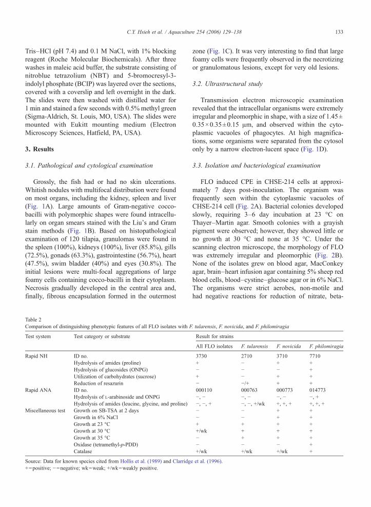
Table 2Comparison of distinguishing phenotypic features of all FLO isolates with F
Test system Test category or substrate
Rapid NH ID no.Hydrolysis of amides (proline)Hydrolysis of glucosides (ONPG)Utilization of carbohydrates (sucrose)Reduction of resazurin
Rapid ANA ID no.Hydrolysis of L-arabinoside and ONPGHydrolysis of amides (leucine, glycine, and proline)
Miscellaneous test Growth on SB-TSA at 2 daysGrowth in 6% NaClGrowth at 23 °CGrowth at 30 °CGrowth at 35 °COxidase (tetramethyl-p-PDD)Catalase
Source: Data for known species cited from Hollis et al. (1989) and Clarridg+=positive; −=negative; wk=weak; +/wk=weakly positive.
zone (Fig. 1C). It was very interesting to find that largefoamy cells were frequently observed in the necrotizingor granulomatous lesions, except for very old lesions.
3.2. Ultrastructural study
Transmission electron microscopic examinationrevealed that the intracellular organisms were extremelyirregular and pleomorphic in shape, with a size of 1.45±0.35×0.35±0.15 μm, and observed within the cyto-plasmic vacuoles of phagocytes. At high magnifica-tions, some organisms were separated from the cytosolonly by a narrow electron-lucent space (Fig. 1D).
3.3. Isolation and bacteriological examination
FLO induced CPE in CHSE-214 cells at approxi-mately 7 days post-inoculation. The organism wasfrequently seen within the cytoplasmic vacuoles ofCHSE-214 cell (Fig. 2A). Bacterial colonies developedslowly, requiring 3–6 day incubation at 23 °C onThayer–Martin agar. Smooth colonies with a grayishpigment were observed; however, they showed little orno growth at 30 °C and none at 35 °C. Under thescanning electron microscope, the morphology of FLOwas extremely irregular and pleomorphic (Fig. 2B).None of the isolates grew on blood agar, MacConkeyagar, brain–heart infusion agar containing 5% sheep redblood cells, blood–cystine–glucose agar or in 6% NaCl.The organisms were strict aerobes, non-motile andhad negative reactions for reduction of nitrate, beta-
. tularensis, F. novicida, and F. philomiragia
Result for strains
All FLO isolates F. tularensis F. novicida F. philomiragia
3730 2710 3710 7710+ − + +− − − ++ − + +− −/+ + +000110 000763 000773 014773−, − −, − −, − −, +−, −, + −, −, +/wk +, +, + +, +, +− − + +− − + ++ + + ++/wk + + +− + + +− − − ++/wk +/wk +/wk +
e et al. (1996).

Fig. 3. Results of PCR amplification. (A) The expected PCR products of all FLO isolates are obtained using 27f/1525r primers complementary toconserved regions in all eubacterial 16S rRNA gene, and (B) using a Francisella-specific F5/F11 primer pair. Lane M: 100 bp DNAmolecular weightmarker. Lanes 1–10, DNA extraction from isolates: AF-01-2, AF-01-6, AF-01-22, AF-01-23, AF-01-27, AF-01-28, AF-03-27, AF-03-28, AF-04-15and AF-04-405, respectively. Lanes 11 and 12, DNA extraction from F. tularensis subsp. tularensis (NCTC 10857) and F. tularensis subsp. novicida(CIP 56.12), respectively. Lanes 13 and 14, F. philomiragia (ATCC 25015) and F. philomiragia (ATCC 25017), respectively. Lane 15, tissuesgenomic DNA extraction from FLO-free tilapia. Lane 16, no DNA template; negative control.
Fig. 4. Level of sequence similarities and evolutionary distances, based on alignment of 1439 nucleotides of 16S rRNAs from Francisella species,and some reference species belonging to the γ subclass of the Proteobacteria. Construction of the evolutionary distance tree is carried out using themultiple alignment algorithms in the MegAlign package (Windows version 5.0.221; DNASTAR), and shows the relationships among the organismsused in this study and members of the γ subclass of the Proteobacteria that exhibit the highest levels of similarity to members of the genusFrancisella. Piscireckettsia salmonis is included as an outgroup. The unrooted tree is constructed by the Clustal algorithm after evolutionary distanceshave been calculated from nucleotide substitution values. The NCBI accession numbers for sequences used in the alignment are shown as follows:Francisella sp. CYH-2002, AF385857; Francisella philomiragia, AY928394; Francisella novicida, AY928396; Francisella tularensis, Z21931;Wolbachia persica, M21292; Ornithodoros moubata symbiote B, AB001522; Piscireckettsia salmonis EM-90, U36940; Piscireckettsia salmonisNOR-92, U36942; and E. coli, J01695.
134 C.Y. Hsieh et al. / Aquaculture 254 (2006) 129–138

135C.Y. Hsieh et al. / Aquaculture 254 (2006) 129–138
galactosidase and resazurin, and hydrolysis of gluco-side, ONPG, L-arabinoside, leucine and glycine. Theywere weakly positive for catalase, but strongly positivefor β-lactamase, proline and sucrose (Table 2).
Table 3Sequence signatures for the genus Francisella and FLO
Position a Nucleotide in: Francisella strains b
All FLO isolates F. philomiragia F. n
140 C U U141 U U U143 U A A189 C U U207 C – U208 U – U210 U U C211 G A G213 C G G214 G G G215 C C C219 G G G220 C C U271 U U C383 A A C421 U U C460 G A C461 U U A462 G A/G A463 A A G473 U U C475 G G U476 C U G477 A A G480 A A G489 G A A503 G G C546 C C G548 G G A648 A G G661 U U C673 A G G679 C U U748 A A G840 U U G853 A A C866 G G G867 C C C875 U U A999 A A G1033 C C C1035 U U U1036 C C C1037 G G G1053 G G G1292 G G A1488 A G G1524 G G Ga Escherichia coli numbering.b Bases found in Francisella species, including P. salmonis.
3.4. PCR assays
In our isolates, the sequences of PCR productsamplified by eubacterial universal primers 27f/1525r for
ovicida F. tularensis P. salmonis E. coli
U U UU A GA U AU A AU U CU A UC A CG U GG A GC G CG C CG C CU U UC U CC A/G CC C CC G UA C AA U AG A AC U UU G UG C GG U CG A AA A AC C CG G GA G AG G GC U UG G GU U UG A AG U UC G GC G GG C CA A UA A AU C CC U U– C C– G G– G GA G UG G G– G G

136 C.Y. Hsieh et al. / Aquaculture 254 (2006) 129–138
16S rRNA gene were 1520 nt long. All isolates yieldedappropriate amplified PCR products (∼1140 bp) usingFrancisella genus-specific primers F11/F5 (Fig. 3).These sequences were deposited in GenBank and weregiven accession numbers from AY928388–AY928393,and from DQ007453–DQ007456. Blast searches ofGenBank yielded high sequence similarities of 98.6%for F. philomiragia, 97.4% for Francisella novicida, and96.1% for F. tularensis (Fig. 4). Compared to referencestrains, the nucleotide differences are shown in Table 3.Based on the high sequence similarities of whole 16SrRNA genes and phenotypic characteristics, all ourisolates certainly belonged to the genus Francisella.
3.5. In situ hybridization
In order to investigate the relationship betweengranulomas and FLO, FLO-infected and FLO-freetilapia were processed for in situ hybridization using
Fig. 5. In situ hybridization of tilapia (Oreochromis spp.) kidney withdigoxigenin-labeled oligonucleotides complementary for Francisella-like organisms 16S rRNA sequences. (A) Positive reactions usingDIG-labeled probe are localized in the granulomas of tilapia kidneywith natural FLO infection; brown pigment signals (arrows) areobserved mainly in the foamy cells. (B) The intracellular organisms,FLO, (arrowheads) appear in the cytoplasmic vacuoles of phagocytes.
FLO 16S rRNA DIG-labelled DNA probes. Positivereactions, characterized by a brown precipitate, weredetected in FLO-infected tilapia and none in FLO-freefish. The brown pigments were localized mainly in thelarge foamy cells of the granulomas (Fig. 5A and B).
4. Discussion
Between 1994 and 2003, a Hawaiian tilapia Piscir-ickettsia-like organism (HTPLO) was reported ascausing significant losses on certain farms (Mauel etal., 2003). Multiple granulomas were observed in thegills, spleen, kidney, choroid gland and testes, but not inthe liver. This organism also caused enzootic diseases inJamaica, Indonesia, Florida and southern California.However, the agent cannot be isolated by cell culture.Pathologically, HTPLO is similar to RLO and our casesdescribed from Taiwan. We also used the conservedprimers PS2S and PS2AS of Piscirickettsia salmonis(Mauel et al., 1996) to amplify our samples, but noamplicon was obtained (data not shown).
Microbial pathogens of fish are routinely diagnosedvia isolation and identification methods. However, anumber of fish pathogens could be consistentlyvisualized, but failed to grow. Although the intracellularcocco-bacillus in tilapia was successfully cultivated inCHSE-214 cells and on Thayer–Martin agar in Taiwan,its isolation was difficult and time-consuming, due tothe lack of convenient culture systems and the presenceof bacterial contamination in the inoculum. All ourisolates belonged to a unique group of low-temperaturegrowing organisms. They showed similar morphologi-cal and biochemical characteristics to F. tularensis, F.novicida, and F. philomiragia (Table 2).
Universal primers for the bacterial 16S rRNA genehave been used to detect the presence or absence ofeubacteria (Lane, 1991), Rickettseae (Roux and Raoult,1995), bacterial endosymbionts (O'Neill et al., 1992;Schröder et al., 1996), P. salmonis (Mauel et al., 1996,1999), and uncultured bacillus of Whipple's disease(Relman et al., 1990, 1992). We used broad-rangeeubacterial primers to amplify a novel 16S rRNA genefrom both our tissue specimens and bacterial isolates.Sequences obtained were identical. In a comparison ofthe 16S rRNA gene sequences of our isolates with somereference strains of Proteobacteria associated withintracellular pathogens, very high levels of sequencesimilarity were found, but it was quite different fromthose of P. salmonis (Fig. 4).
Despite the high sequence similarities between ourisolates and reference strains, the nucleotide sequencesof our isolates were different from F. philomiragia by

137C.Y. Hsieh et al. / Aquaculture 254 (2006) 129–138
14 nt (2 insertions and 12 substitutions—one base of theF. philomiragia sequence in GenBank is AY928394), F.novocida by 34 nt (0 insertions, 34 substitutions—onebase of the F. novicida sequence in GenBank isAY928396), and F. tularensis sequence by 43 nt (4insertions, 39 substitutions—one base of the F.tularensis in GenBank is Z21931) (Table 3).
By using nonradioactive-labeled DNA probes, theFLO nucleic acids could be detected in formalin-fixed,paraffin-embedded tissue specimens, such as the spleen,kidney, liver and gill of tilapia naturally infected withFLO. Detection of hybridization signals providedscientific evidence that the cells were certainly infectedwith FLO. The results also indicated that the majorcellular sites of infection were mononuclear cells. Anexclusive cytoplasmic localization of FLO in mononu-clear cells suggested that FLO could survive in theircytoplasm. This finding was similar to Francisella spp.present in the cytoplasm of macrophage cell lines ofmouse and human origin (Fortier et al., 1995; Golovliovet al., 2003.).
Based on histopathological and cytological exam-inations, ultrastructural study, the sequences of the16S rRNA gene, in situ hybridization and phyloge-netic analysis, we proposed that our isolates mightrepresent a species of Francisella. According to ourdata, FLO from diseased tilapia was closely related toF. philomiragia, which was isolated from water(Jensen et al., 1969; Hollis et al., 1989) and inducedchronic granulomatous disease in humans (Sicherer etal., 1997; Polack et al., 1998). Whether our FLOisolates also play an important role in zoonotic diseaseneeds further study.
Acknowledgments
We thank the Bureau of Animal and Plant HealthInspection and Quarantine (BAPHIQ) Council ofAgriculture, Executive Yuan for the financial supportof this research (Grant no. 90AS-6.3.1-BQ-B1). We alsothank Mr. S.P. Chen (Animal Technology InstituteTaiwan) for EM technical assistance.
References
Anda, P., Segura del Pozo, J., Díaz García, J.M., Escudero, R., GarcíaPeña, F.J., López Velasco, M.C., Selleck, R.E., Jiménez Chillarón,M.R., Sañchez Serrano, L.P., Martínez Navarro, J.F., 2001.Waterborne outbreak of tularemia associated with crayfish fishing.Emerging Infect. Dis. 7, 575–582.
Chen, S.C., Tung, M.C., Chen, S.P., Tsai, J.F., Wang, P.C., Chen, R.S.,Lin, S.C., Adams, A., 1994. Systematic granulomas caused by a
rickettsia-like organism in Nile tilapia, Oreochronuics niloticus(L.), from southern Taiwan. J. Fish Dis. 17, 591–599.
Chern, R.S., Chao, C.B., 1994. Outbreak of a disease caused byrickettsia-like organism in cultured tilapias in Taiwan. Fish Pathol.29, 61–71.
Clarridge, J.E., Raich Jr., T.J., Sjosted, A., Sandstrom, G., Darouiche,R.O., Shawar, R.M., Georghiou, P.R., Osting, C., Vo, L., 1996.Characterization of two unusual clinically significant Francisellastrains. J. Clin. Microbiol. 34, 1995–2000.
Forsman, M., Sandström, G., Sjstedt, A., 1994. Analysis of 16S rDNAFrancisella strains and utilization for determination of thephylogeny of the genus and for identification of strains by PCR.Int. J. Syst. Bacterial. 44, 38–46.
Forsman, M., Nyren, A., Sjostedt, A., Sjokvist, L., Sandstrom, G.,1995. Identification of Francisella tularensis in natural watersamples by PCR. Fed. Eur. Microbiol. Ecol. 16, 83–92.
Fortier, A.H., Leiby, D.A., Narayanan, R.B., Asafoadjei, E., Crawford,R.M., Nacy, C.A., Meltzer, M.S., 1995. Growth of Francisellatularensis LVS in macrophages: the acidic intracellular compart-ment provides essential iron required for growth. Infect. Immun.63, 1478–1483.
Fournier, P.E., Bernabeu, L., Schubert, B., Mutillod, M., Roux, V.,Raoult, D., 1998. Isolation of Francisella tularensis by centrifu-gation of shell vial cell culture from an inoculation Escher. J. Clin.Microbiol. 36, 2782–2783.
Golovliov, I., Baranov, V., Krocova, Z., Kovarova, H., Sjöstedt, A.,2003. An attenuated strain of the Facultative IntracellularBacterium Francisella tularensis can escape the phagosome ofmonocytic cells. Infect. Immun. 71, 5940–5950.
Hoel, T., Scheel, O., Nordahl, S.H., Sandvik, T., 1991. Water- andairborne Francisella tularensis biovar palaearctica isolated fromhuman blood. Infection 19, 348–350.
Hollis, D.G., Weaver, R.E., Steigerwalt, A.G., Wenger, J.D., Moss,C.W., Brenner, D.J., 1989. Francisella philomiragia comb. nov.(formerly Yersinia philomiragia) and Francisella tularensisbiogroup Novicida (formerly Yersinia novicida) associated withhuman disease. J. Clin. Microbiol. 27, 1601–1608.
Jensen, W.I., Owen, C.R., Jellison, W.L., 1969. Yersinia philomiragiasp. n., a new member of the Pasteurella group of bacteria, naturallypathogenic for the muskrat (Ondatra zibethica). J. Bacteriol. 100,1237–1241.
Lane, D.J., 1991. 16S/23S rRNA sequencing. In: Stackebrandt, E.,Goodfellow, M. (Eds.), Nucleic Acid Techniques in BacterialSystematics. John Wiley & Sons, Inc., New York, pp. 115–175.
Mauel, M.J., Giovannoni, S.J., Fryer, J.L., 1996. Development ofpolymerase chain reaction assays for detection, identification, anddifferentiation of Piscirickettsia salmonis. Dis. Aquat. Org. 26,189–195.
Mauel, M.J., Giovannoni, S.J., Fryer, J.L., 1999. Phylogeneticanalysis of Piscirickettsia salmonis by 16S, internal transcribedspacer (ITS) and 23S r DNA sequencing. Dis. Aquat. Org. 35,115–123.
Mauel, M.J., Miller, D.L., Frazier, K., Liggeet, A.D., Styer, L.,Montgomery-Brock, D., Brock, J., 2003. Characterization of apiscirickettsiosis-like disease in Hawaiian tilapia. Dis. Aquat. Org.53, 249–255.
Mignani, E., Palmieri, F., Fontana, M., Marigo, S., 1988. Italianepidemic of waterborne tularaemia. Lancet (Letter) 2, 1423.
Mörner, T., 1992. The ecology of tularemia. Rev. Sci. Tech.-Off. Int.Epizoot. 11, 1123–1130.
O'Neill, S.L., Giordano, R., Colbert, A.M.E., Karr, T.L., Robertson, H.M., 1992. 16S r RNA phylogenetic analysis of the bacterial

138 C.Y. Hsieh et al. / Aquaculture 254 (2006) 129–138
endosymbiont associated with cytoplasmic incompatibility ininsects. Proc. Natl. Acad. Sci. U. S. A. 89, 2699–2702.
Polack, F.P., Harrington, S.M., Winkelstein, J.A., Merz, W.G.,Willoughby, R.E., 1998. Recurrent Francisella philomiragiasepsis in chronic granulomatous disease. Pediatr. Infect. Dis. J.17, 442–443.
Quan, S.F., McManus, A.G., von Fintel, H., 1956. Infectivity oftularemia applied to intact skin and ingested drinking water.Science 123, 942–943.
Relman, D.A., Loutit, J.S., Schmidt, T.M., Falkow, S., Tompkins, L.S.,1990. The agent of bacillary angiomatosis. An approach to theidentification of uncultured pathogens. N. Engl. J. Med. 323,1573–1580.
Relman, D.A., Schmidt, T.M., MacDermott, R.P., Falkow, S., 1992.Identification of the uncultured bacillus of Whipple's disease. N.Engl. J. Med. 327, 293–301.
Roux, V., Raoult, D., 1995. Phylogenetic analysis of the genusrickettsia by 16S rDNA sequencing. Res.Microbiol. 146, 385–396.
Sicherer, S.H., Asturias, E.J., Winkelstein, J.A., Dick, J.D., Wil-loughby, R.E., 1997. Francisella philomiragia sepsis in chronicgranulomatous disease. Pediatr. Infect. Dis. J. 16, 420–422.
Schröder, D., Deppisch, H., Obermayer, M., Krohne, G., Stackebrandt,E., Hölldobler, B., Goebel, W., Gross, R., 1996. Intracellularendosymbiotic bacteria of Camponotus species (carpenter ants):systematics, evolution and ultrastructural characterization. Mol.Microbiol. 21, 479–489.