H48 - Determination of Coagulation Factor Activities Using the ...
Upload
independentCategory
view
1download
0
Enhancement of Pig Embryonic Implants in Factor VIII KOMice: A Novel Role for the Coagulation Cascade in OrganSize ControlAnna Aronovich1, Dalit Tchorsh1, Elias Shezen1, Chava Rosen1, Yael Klionsky1, Sivan Cohen1, Orna Tal1,
Uri Martinowitz2, Helena Katchman1, Smadar Eventov-Friedman1, Ninette Amariglio3, Jasmine
Jacob-Hirsch3, Gideon Rechavi3, Yair Reisner1*
1 Department of Immunology, Weizmann Institute of Science, Rehovot, Israel, 2 The Israel National Hemophilia Center, Sheba Medical Center, Tel Hashomer, Israel,
3 Cancer Research Center, Sheba Medical Center, Tel Hashomer, Israel
Abstract
Very little is known about the mechanisms that contribute to organ size differences between species. In the presentstudy, we used a mouse model of embryonic pig tissue implantation to define the role of host Factor VIII in controllingthe final size attained by the implant. We show here that pig embryonic spleen, pancreas, and liver all grow to anincreased size in mice that are deficient in the Factor VIII clotting cascade. Similar results were obtained using thetransplantation model after treatment with the low molecular weight heparin derivative Clexane which markedlyenhanced transplant size. Likewise, enhanced size was found upon treatment with the direct thrombin inhibitorDabigatran, suggesting that organ size regulation might be mediated by thrombin, downstream of Factor VIII.Considering that thrombin was shown to mediate various functions unrelated to blood clotting, either directly bycleavage of protease-activated receptors (PARs) or indirectly by cleaving osteopontin (OPN) on stroma cells, the role ofPAR1 and PAR4 antagonists as well as treatment with cleaved form of OPN (tcOPN) were tested. While the former wasnot found to have an impact on overgrowth of embryonic pig spleen implants, marked reduction of size was noted upontreatment with the (tcOPN). Collectively, our surprising set of observations suggests that factors of the coagulationcascade have a novel role in organ size control.
Citation: Aronovich A, Tchorsh D, Shezen E, Rosen C, Klionsky Y, et al. (2009) Enhancement of Pig Embryonic Implants in Factor VIII KO Mice: A Novel Role for theCoagulation Cascade in Organ Size Control. PLoS ONE 4(12): e8362. doi:10.1371/journal.pone.0008362
Editor: Martin Pera, University of Southern California, United States of America
Received September 22, 2009; Accepted November 23, 2009; Published December 21, 2009
Copyright: � 2009 Aronovich et al. This is an open-access article distributed under the terms of the Creative Commons Attribution License, which permitsunrestricted use, distribution, and reproduction in any medium, provided the original author and source are credited.
Funding: This work was supported by Mrs. Erica Drake and the Gabriella Rich Center for Transplantation Biology Research. The funding supported mice andother supplies. There was no other funding. The funders had no role in study design, data collection and analysis, decision to publish, or preparation of themanuscript.
Competing Interests: The authors have declared that no competing interests exist.
* E-mail: [email protected]
Introduction
Despite great progress in the molecular dissection of embryo-
genesis, the mechanisms controlling the size of tissues and organs
remain a mystery. In general, it is thought that extrinsic control
mechanisms are associated with nutrition or systemic growth
signaling factors, such as those operating through the insulin/
PI3K or the TOR pathways, while intrinsic mechanisms are likely
linked to patterning morphogens and apoptosis-signaling com-
plexes, as well as to stem cell numbers[1–4]. Some of the early
insights in this area of investigation came from transplantation
experiments in which infant rat hearts or kidneys transplanted into
adult rats grew and attained the size of an adult organ[5]. Similar
results were also found by Metcalf et al. in early transplantation
experiments with fetal mouse organs. For example, when multiple
fetal mouse thymus glands were transplanted into an adult mouse,
each was found to grow to its normal adult size[6]. However, when
the same experiment was performed using fetal spleens, the total
mass of the transplanted spleen tissue attained the mass of only a
single normal adult spleen, suggesting that the growth of these fetal
tissues is limited by extrinsic factors, outside the spleen[7].
Very recently, we defined optimal ‘windows’ of gestation of
human and pig embryonic precursor tissues that afford optimal
growth and development of liver, pancreas and spleen upon
implantation into NOD-SCID mice[8]. Our studies enabled not
only a rational selection of fetal tissue for transplantation but,
among many parameters, also provide a system in which to
manipulate and study organ size control. Using this system, we
have now found, surprisingly, that a difference in the functionality
of one gene in the recipients, namely coagulation Factor VIII, has
a major impact on the final size attained by different embryonic
pig tissues upon transplantation into mice. Interrogation of other
factors along the coagulation cascade that are triggered by Factor
VIII, such as Factor Xa and thrombin, suggests that thrombin,
which is the terminal enzyme of the hemostatic system, is likely to
be the actual mediator of this novel checkpoint of organ size
control. Furthermore, our ability to reverse the enhancement of
implant size by treatment with the cleaved form of OPN that is
released upon treatment with thrombin, strongly indicates that
thrombin likely exerts its regulatory activity through interaction
with OPN on stroma cells, as previously shown for maintenance of
the blood tissue size under normal steady state hoemostasis[9,10].
PLoS ONE | www.plosone.org 1 December 2009 | Volume 4 | Issue 12 | e8362
Results
The Role of Factor VIII in Defining Organ Size, FollowingImplantation of Pig Embryonic Tissues
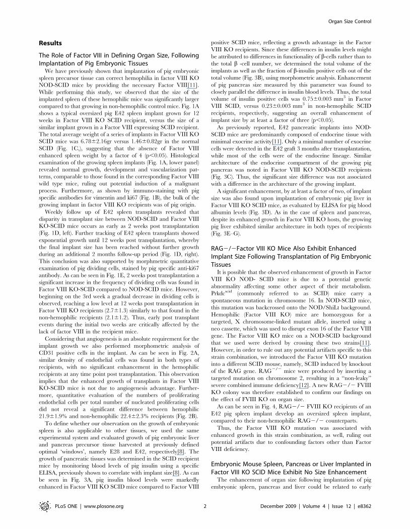
We have previously shown that implantation of pig embryonic
spleen precursor tissue can correct hemophilia in factor VIII KO
NOD-SCID mice by providing the necessary Factor VIII[11].
While performing this study, we observed that the size of the
implanted spleen of these hemophilic mice was significantly larger
compared to that growing in non-hemophilic control mice. Fig. 1A
shows a typical oversized pig E42 spleen implant grown for 12
weeks in Factor VIII KO SCID recipient, versus the size of a
similar implant grown in a Factor VIII expressing SCID recipient.
The total average weight of a series of implants in Factor VIII KO
SCID mice was 6.7862.16gr versus 1.4660.82gr in the normal
SCID (Fig. 1C,), suggesting that the absence of Factor VIII
enhanced spleen weight by a factor of 4 (p,0.05). Histological
examination of the growing spleen implants (Fig. 1A, lower panel)
revealed normal growth, development and vascularization pat-
terns, comparable to those found in the corresponding Factor VIII
wild type mice, ruling out potential induction of a malignant
process. Furthermore, as shown by immuno-staining with pig
specific antibodies for vimentin and ki67 (Fig. 1B), the bulk of the
growing implant in factor VIII KO recipients was of pig origin.
Weekly follow up of E42 spleen transplants revealed that
disparity in transplant size between NOD-SCID and Factor VIII
KO-SCID mice occurs as early as 2 weeks post transplantation
(Fig. 1D, left). Further tracking of E42 spleen transplants showed
exponential growth until 12 weeks post transplantation, whereby
the final implant size has been reached without further growth
during an additional 2 months follow-up period (Fig. 1D, right).
This conclusion was also supported by morphmetric quantitative
examination of pig dividing cells, stained by pig specific anti-ki67
antibody. As can be seen in Fig. 1E, 2 weeks post transplantation a
significant increase in the frequency of dividing cells was found in
Factor VIII KO-SCID compared to NOD-SCID mice. However,
beginning on the 3rd week a gradual decrease in dividing cells is
observed, reaching a low level at 12 weeks post transplantation in
Factor VIII KO recipients (2.761.3) similarly to that found in the
non-hemophilic recipients (2.161.2). Thus, early post transplant
events during the initial two weeks are critically affected by the
lack of factor VIII in the recipient mice.
Considering that angiogenesis is an absolute requirement for the
implant growth we also performed morphometric analysis of
CD31 positive cells in the implant. As can be seen in Fig. 2A,
similar density of endothelial cells was found in both types of
recipients, with no significant enhancement in the hemophilic
recipients at any time point post transplantation. This observation
implies that the enhanced growth of transplants in Factor VIII
KO-SCID mice is not due to angiogenesis advantage. Further-
more, quantitative evaluation of the numbers of proliferating
endothelial cells per total number of nucleated proliferating cells
did not reveal a significant difference between hemophilic
21.961.9% and non-hemophilic 22.462.3% recipients (Fig. 2B).
To define whether our observation on the growth of embryonic
spleen is also applicable to other tissues, we used the same
experimental system and evaluated growth of pig embryonic liver
and pancreas precursor tissue harvested at previously defined
optimal ‘windows’, namely E28 and E42, respectively[8]. The
growth of pancreatic tissues was determined in the SCID recipient
mice by monitoring blood levels of pig insulin using a specific
ELISA, previously shown to correlate with implant size[8]. As can
be seen in Fig. 3A, pig insulin blood levels were markedly
enhanced in Factor VIII KO SCID mice compared to Factor VIII
positive SCID mice, reflecting a growth advantage in the Factor
VIII KO recipients. Since these differences in insulin levels might
be attributed to differences in functionality of b-cells rather than to
the total b -cell number, we determined the total volume of the
implants as well as the fraction of b-insulin positive cells out of the
total volume (Fig. 3B), using morphometric analysis. Enhancement
of pig pancreas size measured by this parameter was found to
closely parallel the difference in insulin blood levels. Thus, the total
volume of insulin positive cells was 0.7560.003 mm3 in Factor
VIII SCID, versus 0.2360.003 mm3 in non-hemophilic SCID
recipients, respectively, suggesting an overall enhancement of
implant size by at least a factor of three (p,0.05).
As previously reported, E42 pancreatic implants into NOD-
SCID mice are predominantly composed of endocrine tissue with
minimal exocrine activity[11]. Only a minimal number of exocrine
cells were detected in the E42 graft 3 months after transplantation,
while most of the cells were of the endocrine lineage. Similar
architecture of the endocrine compartment of the growing pig
pancreas was noted in Factor VIII KO NOD-SCID recipients
(Fig. 3C). Thus, the significant size difference was not associated
with a difference in the architecture of the growing implant.
A significant enhancement, by at least a factor of two, of implant
size was also found upon implantation of embryonic pig liver in
Factor VIII KO SCID mice, as evaluated by ELISA for pig blood
albumin levels (Fig. 3D). As in the case of spleen and pancreas,
despite its enhanced growth in Factor VIII KO hosts, the growing
pig liver exhibited similar architecture in both types of recipients
(Fig. 3E–G).
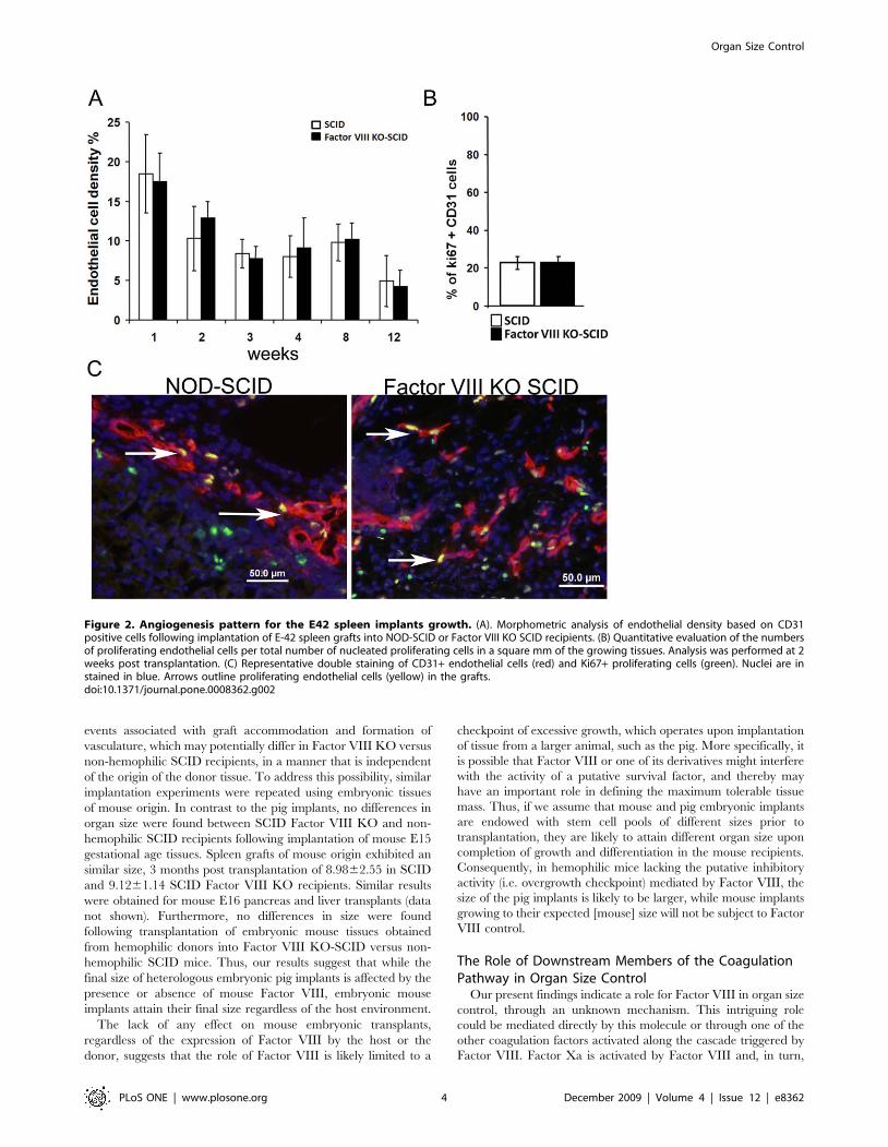
RAG2/2Factor VIII KO Mice Also Exhibit EnhancedImplant Size Following Transplantation of Pig EmbryonicTissues
It is possible that the observed enhancement of growth in Factor
VIII KO NOD- SCID mice is due to a potential genetic
abnormality affecting some other aspect of their metabolism.
Prkdcscid (commonly referred to as SCID) mice carry a
spontaneous mutation in chromosome 16. In NOD-SCID mice,
this mutation was backcrossed onto the NOD/ShiLt background.
Hemophilic (Factor VIII KO) mice are homozygous for a
targeted, X chromosome-linked mutant allele, inserted using a
neo cassette, which was used to disrupt exon 16 of the Factor VIII
gene. The Factor VIII KO mice on a NOD-SCID background
that we used were derived by crossing these two strains[11].
However, in order to rule out any potential artifacts specific to this
strain combination, we introduced the Factor VIII KO mutation
into a different SCID mouse, namely, SCID induced by knockout
of the RAG gene. RAG2/2 mice were produced by inserting a
targeted mutation on chromosome 2, resulting in a ‘‘non-leaky’’
severe combined immune deficiency[12]. A new RAG2/2 FVIII
KO colony was therefore established to confirm our findings on
the effect of FVIII KO on organ size.
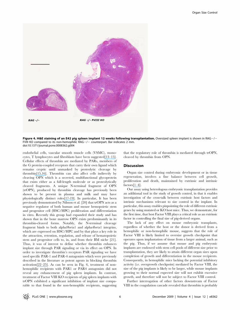
As can be seen in Fig. 4, RAG2/2 FVIII KO recipients of an
E42 pig spleen implant develop an oversized spleen implant,
compared to their non-hemophilic RAG2/2 counterparts.
Thus, the Factor VIII KO mutation was associated with
enhanced growth in this strain combination, as well, ruling out
potential artifacts due to confounding factors other than Factor
VIII deficiency.
Embryonic Mouse Spleen, Pancreas or Liver Implanted inFactor VIII KO SCID Mice Exhibit No Size Enhancement
The enhancement of organ size following implantation of pig
embryonic spleen, pancreas and liver could be related to early
Organ Size Control
PLoS ONE | www.plosone.org 2 December 2009 | Volume 4 | Issue 12 | e8362
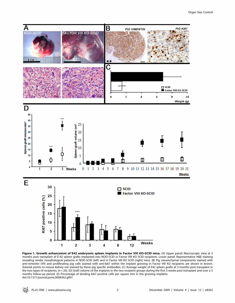
Figure 1. Growth enhancemnt of E42 embryonic spleen implants in Factor VIII KO-SCID mice. (A) Upper panel: Macroscopic view at 3months post transplant of E-42 spleen grafts implanted into NOD-SCID or Factor VIII KO SCID recipients. Lower panel: Representative H&E stainingrevealing similar morphological patterns in NOD-SCID (left) and in Factor VIII KO SCID (right) mice. (B) Pig mesenchymal components stained withanti-vimentin (V9) and proliferating pig cells stained with anti-ki67 within the implant growing in Factor VIII KO recipients are shown in brown.Asterisk points to mouse kidney not stained by these pig specific antibodies. (C) Average weight of E42 spleen grafts at 3 months post transplant inthe two types of recipients, (n = 20). (D) Graft volume of the implants in the two recipient groups during the first 3 weeks post transplant and over a 5months follow-up period. (E) Percentage of dividing ki67 positive cells per square mm in the growing implants.doi:10.1371/journal.pone.0008362.g001
Organ Size Control
PLoS ONE | www.plosone.org 3 December 2009 | Volume 4 | Issue 12 | e8362
events associated with graft accommodation and formation of
vasculature, which may potentially differ in Factor VIII KO versus
non-hemophilic SCID recipients, in a manner that is independent
of the origin of the donor tissue. To address this possibility, similar
implantation experiments were repeated using embryonic tissues
of mouse origin. In contrast to the pig implants, no differences in
organ size were found between SCID Factor VIII KO and non-
hemophilic SCID recipients following implantation of mouse E15
gestational age tissues. Spleen grafts of mouse origin exhibited an
similar size, 3 months post transplantation of 8.9862.55 in SCID
and 9.1261.14 SCID Factor VIII KO recipients. Similar results
were obtained for mouse E16 pancreas and liver transplants (data
not shown). Furthermore, no differences in size were found
following transplantation of embryonic mouse tissues obtained
from hemophilic donors into Factor VIII KO-SCID versus non-
hemophilic SCID mice. Thus, our results suggest that while the
final size of heterologous embryonic pig implants is affected by the
presence or absence of mouse Factor VIII, embryonic mouse
implants attain their final size regardless of the host environment.
The lack of any effect on mouse embryonic transplants,
regardless of the expression of Factor VIII by the host or the
donor, suggests that the role of Factor VIII is likely limited to a
checkpoint of excessive growth, which operates upon implantation
of tissue from a larger animal, such as the pig. More specifically, it
is possible that Factor VIII or one of its derivatives might interfere
with the activity of a putative survival factor, and thereby may
have an important role in defining the maximum tolerable tissue
mass. Thus, if we assume that mouse and pig embryonic implants
are endowed with stem cell pools of different sizes prior to
transplantation, they are likely to attain different organ size upon
completion of growth and differentiation in the mouse recipients.
Consequently, in hemophilic mice lacking the putative inhibitory
activity (i.e. overgrowth checkpoint) mediated by Factor VIII, the
size of the pig implants is likely to be larger, while mouse implants
growing to their expected [mouse] size will not be subject to Factor
VIII control.
The Role of Downstream Members of the CoagulationPathway in Organ Size Control
Our present findings indicate a role for Factor VIII in organ size
control, through an unknown mechanism. This intriguing role
could be mediated directly by this molecule or through one of the
other coagulation factors activated along the cascade triggered by
Factor VIII. Factor Xa is activated by Factor VIII and, in turn,
Figure 2. Angiogenesis pattern for the E42 spleen implants growth. (A). Morphometric analysis of endothelial density based on CD31positive cells following implantation of E-42 spleen grafts into NOD-SCID or Factor VIII KO SCID recipients. (B) Quantitative evaluation of the numbersof proliferating endothelial cells per total number of nucleated proliferating cells in a square mm of the growing tissues. Analysis was performed at 2weeks post transplantation. (C) Representative double staining of CD31+ endothelial cells (red) and Ki67+ proliferating cells (green). Nuclei are instained in blue. Arrows outline proliferating endothelial cells (yellow) in the grafts.doi:10.1371/journal.pone.0008362.g002
Organ Size Control
PLoS ONE | www.plosone.org 4 December 2009 | Volume 4 | Issue 12 | e8362
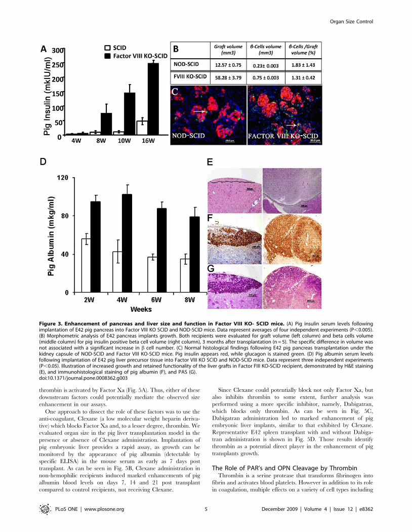
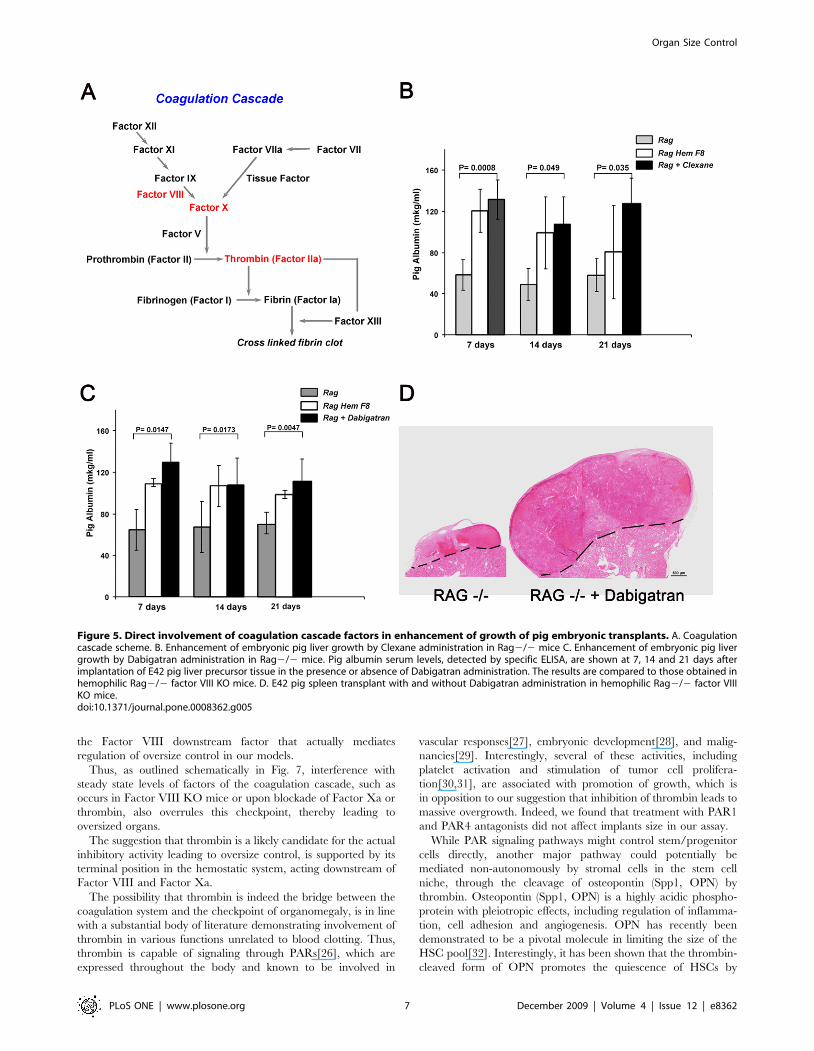
thrombin is activated by Factor Xa (Fig. 5A). Thus, either of these
downstream factors could potentially mediate the observed size
enhancement in our assays.
One approach to dissect the role of these factors was to use the
anti-coagulant, Clexane (a low molecular weight heparin deriva-
tive) which blocks Factor Xa and, to a lesser degree, thrombin. We
evaluated organ size in the pig liver transplantation model in the
presence or absence of Clexane administration. Implantation of
pig embryonic liver provides a rapid assay, as growth can be
monitored by the appearance of pig albumin (detectable by
specific ELISA) in the mouse serum as early as 7 days post
transplant. As can be seen in Fig. 5B, Clexane administration in
non-hemophilic recipients induced marked enhancements of pig
albumin blood levels on days 7, 14 and 21 post transplant
compared to control recipients, not receiving Clexane.
Since Clexane could potentially block not only Factor Xa, but
also inhibits thrombin to some extent, further analysis was
performed using a more specific inhibitor, namely, Dabigatran,
which blocks only thrombin. As can be seen in Fig. 5C,
Dabigatran administration led to marked enhancement of pig
embryonic liver implants, similar to that exhibited by Clexane.
Representative E42 spleen transplant with and without Dabiga-
tran administration is shown in Fig. 5D. Those results identify
thrombin as a potential direct player in the enhancement of pig
transplants growth.
The Role of PAR’s and OPN Cleavage by ThrombinThrombin is a serine protease that transforms fibrinogen into
fibrin and activates blood platelets. However in addition to its role
in coagulation, multiple effects on a variety of cell types including
Figure 3. Enhancement of pancreas and liver size and function in Factor VIII KO- SCID mice. (A) Pig insulin serum levels followingimplantation of E42 pig pancreas into Factor VIII KO SCID and NOD-SCID mice. Data represent averages of four independent experiments (P,0.005).(B) Morphometric analysis of E42 pancreas implants growth. Both recipients were evaluated for graft volume (left column) and beta cells volume(middle column) for pig insulin positive beta cell volume (right column), 3 months after transplantation (n = 5). The specific difference in volume wasnot associated with a significant increase in b cell number. (C) Normal histological findings following E42 pig pancreas transplantation under thekidney capsule of NOD-SCID and Factor VIII KO-SCID mice. Pig insulin appears red, while glucagon is stained green. (D) Pig albumin serum levelsfollowing implantation of E42 pig liver precursor tissue into Factor VIII KO SCID and NOD-SCID mice. Data represent three independent experiments(P,0.05). Illustration of increased growth and retained functionality of the liver grafts in Factor FIII KO-SCID recipient, demonstrated by H&E staining(E), and immunohistological staining of pig albumin (F), and PAS (G).doi:10.1371/journal.pone.0008362.g003
Organ Size Control
PLoS ONE | www.plosone.org 5 December 2009 | Volume 4 | Issue 12 | e8362
endothelial cells, vascular smooth muscle cells (VSMC), mono-
cytes, T lymphocytes and fibroblasts have been suggested[13–15].
Cellular effects of thrombin are mediated by PARs, members of
the G protein-coupled receptors that carry their own ligand which
remains cryptic until unmasked by proteolytic cleavage by
thrombin[15,16]. Thrombin can also affect cells indirectly by
cleaving OPN which is a secreted, multifunctional glycoprotein
that exists either as a full-length molecule or as proteolytically
cleaved fragments. A unique N-terminal fragment of OPN
(trOPN), produced by thrombin cleavage has previously been
shown to be present in plasma and milk and may have
physiologically distinct roles[17-19]. In particular, It has been
previously demonstrated by Nilsson et al. [20] that trOPN acts as a
negative regulator of both human and mouse hemopoietic stem
and progenitor cell (HSC/HPC) proliferation and differentiation
in vitro. Recently this group had expanded their study and has
shown that in the bone marrow OPN exists predominantly in its
thrombin-cleaved forms. Notably, the N-terminal cleavage
fragment binds to both alpha9beta1 and alpha4beta1 integrins,
which are expressed on HSC/HPC and by that plays a key role in
the attraction, retention, regulation, and release of hematopoietic
stem and progenitor cells to, in, and from their BM niche [21].
Thus, it was of interest to define whether thrombin enhances
implant size through PAR signaling or via its effect on OPN. In
order to investigate thrombin’s receptors PAR signaling we have
used specific PAR-1 and PAR-4 antagonists which were previously
described in the literature as potent agents in blocking thrombin
activation[22–25]. As can be seen in Fig. 6, treatment of non-
hemophilic recipients with PAR1 or PAR4 antagonists did not
reveal any enhancement of pig spleen implants. In contrast,
treatment of Factor VIII KO recipients of pig spleen implants with
trOPN exhibited a significant inhibition of implant size compa-
rable to that found in the non-hemophlic recipients, suggesting
that the regulatory role of thrombin is mediated through trOPN,
cleaved by thrombin from OPN.
Discussion
Organ size control during embryonic development or in tissue
regeneration, involves a fine balance between cell growth,
proliferation and death, maintained by extrinsic and intrinsic
factors[1–4].
Our assay using heterologous embryonic transplantation provides
an additional tool in the study of growth control, in that it enables
investigation of the cross-talk between extrinsic host factors and
intrinsic mechanisms relevant to size control in the implant. In
particular, this assay enables pinpointing the role of different extrinsic
genes by using mutated or KO host mice. Thus, we demonstrate, for
the first time, that host Factor VIII plays a critical role as an extrinsic
factor in controlling the final size of pig-derived organs.
The lack of any effect on mouse embryonic transplants,
regardless of whether the host or the donor is derived from a
hemophilic or non-hemophilic mouse, suggests that the role of
Factor VIII is likely limited to oversize growth checkpoint that
operates upon implantation of tissue from a larger animal, such as
the pig. Thus, if we assume that mouse and pig embryonic
implants are endowed with stem cell pools of different size prior to
transplantation, they are likely to attain different organ sizes upon
completion of growth and differentiation in the mouse recipients.
Consequently, in hemophilic mice lacking the potential inhibitory
activity (i.e. overgrowth checkpoint) mediated by Factor VIII, the
size of the pig implants is likely to be larger, while mouse implants
growing to their normal expected size will not exhibit excessive
growth, and therefore will not be subject to Factor VIII control.
Further interrogation of other factors downstream of Factor
VIII in the coagulation cascade revealed that thrombin is probably
Figure 4. H&E staining of an E42 pig spleen implant 12 weeks following transplantation. Oversized spleen implant is shown in RAG2/2FVIII KO compared to its non-hemophilic RAG2/2 counterpart. Bar indicates 2 mm.doi:10.1371/journal.pone.0008362.g004
Organ Size Control
PLoS ONE | www.plosone.org 6 December 2009 | Volume 4 | Issue 12 | e8362
the Factor VIII downstream factor that actually mediates
regulation of oversize control in our models.
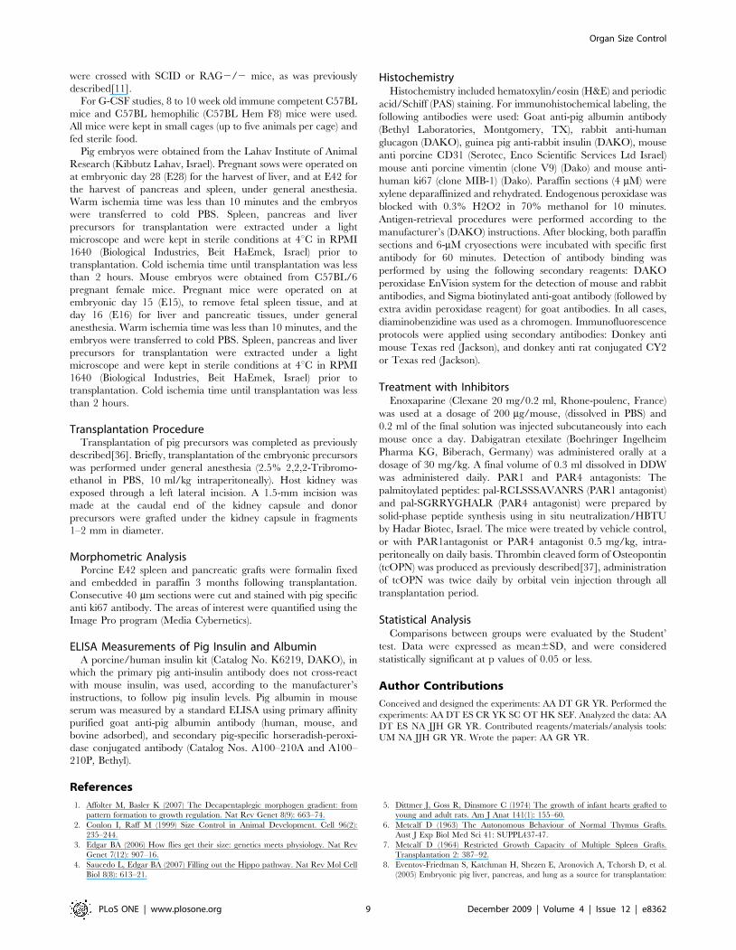
Thus, as outlined schematically in Fig. 7, interference with
steady state levels of factors of the coagulation cascade, such as
occurs in Factor VIII KO mice or upon blockade of Factor Xa or
thrombin, also overrules this checkpoint, thereby leading to
oversized organs.
The suggestion that thrombin is a likely candidate for the actual
inhibitory activity leading to oversize control, is supported by its
terminal position in the hemostatic system, acting downstream of
Factor VIII and Factor Xa.
The possibility that thrombin is indeed the bridge between the
coagulation system and the checkpoint of organomegaly, is in line
with a substantial body of literature demonstrating involvement of
thrombin in various functions unrelated to blood clotting. Thus,
thrombin is capable of signaling through PARs[26], which are
expressed throughout the body and known to be involved in
vascular responses[27], embryonic development[28], and malig-
nancies[29]. Interestingly, several of these activities, including
platelet activation and stimulation of tumor cell prolifera-
tion[30,31], are associated with promotion of growth, which is
in opposition to our suggestion that inhibition of thrombin leads to
massive overgrowth. Indeed, we found that treatment with PAR1
and PAR4 antagonists did not affect implants size in our assay.
While PAR signaling pathways might control stem/progenitor
cells directly, another major pathway could potentially be
mediated non-autonomously by stromal cells in the stem cell
niche, through the cleavage of osteopontin (Spp1, OPN) by
thrombin. Osteopontin (Spp1, OPN) is a highly acidic phospho-
protein with pleiotropic effects, including regulation of inflamma-
tion, cell adhesion and angiogenesis. OPN has recently been
demonstrated to be a pivotal molecule in limiting the size of the
HSC pool[32]. Interestingly, it has been shown that the thrombin-
cleaved form of OPN promotes the quiescence of HSCs by
Figure 5. Direct involvement of coagulation cascade factors in enhancement of growth of pig embryonic transplants. A. Coagulationcascade scheme. B. Enhancement of embryonic pig liver growth by Clexane administration in Rag2/2 mice C. Enhancement of embryonic pig livergrowth by Dabigatran administration in Rag2/2 mice. Pig albumin serum levels, detected by specific ELISA, are shown at 7, 14 and 21 days afterimplantation of E42 pig liver precursor tissue in the presence or absence of Dabigatran administration. The results are compared to those obtained inhemophilic Rag2/2 factor VIII KO mice. D. E42 pig spleen transplant with and without Dabigatran administration in hemophilic Rag2/2 factor VIIIKO mice.doi:10.1371/journal.pone.0008362.g005
Organ Size Control
PLoS ONE | www.plosone.org 7 December 2009 | Volume 4 | Issue 12 | e8362
exerting a profound suppression of proliferation of HSCs, without
inducing apoptosis[9]. OPN is a negative regulatory element of the
stem cell niche that limits the size of the stem cell pool and may
provide a mechanism for restricting excess organ growth.
Thrombin is the major serine protease that cleaves OPN, giving
rise to a 24-kDa and a 45-kDa fragment. The 45-kDa fragment
has multiple functional advantages in processes such as cell
attachment, migration, and spreading, through binding to a9b1
and a4b11. Our present finding that treatment of factor VIII KO
recipients of pig embryonic spleen exhibit a deuced implant size,
similarly to that found in the non-hemophilic recipients, strongly
indicates that thrombin likely exerts its regulatory effect through
cleavage of OPN .
While studies on the HSC niche in the bone marrow are
relatively well advanced[33-35]very little is known regarding the
stem cell niche in the embryonic pig tissue used for transplantation
in our assays. The new insights on the role of thrombin and
tcOPN as well as the well characterized binding of the latter to the
integrins a9b1 and a4b11, could facilitate our ability to identify
and localize stem cell - niche interactions which might occur in
non –hematopoietic organs and tissues.
However, other possibilities that might underline the inhibitory
activity exerted by tcOPN, including potential effect on the
vasculature should be considered.
In conclusion, our data suggest a novel role for factors of the
coagulation cascade in organ size control. In particular, we were
able to pinpoint thrombin and its substrate OPN as the direct
players.
This surprising finding may not only offer new means to
enhance or reduce the final size of implanted xenogeneic
embryonic tissues, but might also adds a novel insight into the
mysterious question of organ size control in mammals. Clearly,
further studies investigating the role of the coagulation cascade
factors in more physiological settings, and in particular their
potential ability to affect normal tissue stem cells under extreme
growth stimuli, are warranted.
Materials and Methods
AnimalsAll animals were maintained under conditions approved by the
Institutional Animal Care and Use Committee at the Weizmann
Institute. The study protocol was approved by the ethics
committees at Kibbutz Lahav and the Weizmann Institute. In
these experiments, 8–10 week old immune deficient NOD-SCID
mice and NOD-SCID Factor VIII KO mice (Weizmann Institute
Animal Breeding Center, Rehovot, Israel) or RAG2/2 mice and
RAG2/2 FVIII KO mice were used as hosts for the
transplantation studies. To obtain immunodeficient hemophilic
mice (designated Factor VIII KO–SCID), FVIII-deficient mice
Figure 6. The role og PARS and OPN cleavage by thrombin indefining size of E42 pig embryonic spleen implants. E42 pigspleen tissue was implanted into NOD-SCID and FactoorVIII KO NOD -SCID mice and final size of the implant was defined 4 weeks aftertransplantation. To define the effect of PARs cleavage by thrombin,PAR-1 and PAR-4 antagonists were administered to NOD-SCIDrecipients as described in Methods. To evaluate the role of OPNcleavage, trOPN was administered to Factor VIII NOD-SCID recipients asdescribed in Methods. Each group comprised 7 mice. Enhancement ofgrowth of E42 spleen tissue in Factor VIII NOD-SCID mice compared toNOD-SCID mice was statistically significant (t test, p,0.005)). Thisenhancement was significantly inhibited upon treatment with trOPN(p,0.05).doi:10.1371/journal.pone.0008362.g006
Figure 7. Overgrowth stimulus regulation by factors in the coagulation cascade.doi:10.1371/journal.pone.0008362.g007
Organ Size Control
PLoS ONE | www.plosone.org 8 December 2009 | Volume 4 | Issue 12 | e8362
were crossed with SCID or RAG2/2 mice, as was previously
described[11].
For G-CSF studies, 8 to 10 week old immune competent C57BL
mice and C57BL hemophilic (C57BL Hem F8) mice were used.
All mice were kept in small cages (up to five animals per cage) and
fed sterile food.
Pig embryos were obtained from the Lahav Institute of Animal
Research (Kibbutz Lahav, Israel). Pregnant sows were operated on
at embryonic day 28 (E28) for the harvest of liver, and at E42 for
the harvest of pancreas and spleen, under general anesthesia.
Warm ischemia time was less than 10 minutes and the embryos
were transferred to cold PBS. Spleen, pancreas and liver
precursors for transplantation were extracted under a light
microscope and were kept in sterile conditions at 4uC in RPMI
1640 (Biological Industries, Beit HaEmek, Israel) prior to
transplantation. Cold ischemia time until transplantation was less
than 2 hours. Mouse embryos were obtained from C57BL/6
pregnant female mice. Pregnant mice were operated on at
embryonic day 15 (E15), to remove fetal spleen tissue, and at
day 16 (E16) for liver and pancreatic tissues, under general
anesthesia. Warm ischemia time was less than 10 minutes, and the
embryos were transferred to cold PBS. Spleen, pancreas and liver
precursors for transplantation were extracted under a light
microscope and were kept in sterile conditions at 4uC in RPMI
1640 (Biological Industries, Beit HaEmek, Israel) prior to
transplantation. Cold ischemia time until transplantation was less
than 2 hours.
Transplantation ProcedureTransplantation of pig precursors was completed as previously
described[36]. Briefly, transplantation of the embryonic precursors
was performed under general anesthesia (2.5% 2,2,2-Tribromo-
ethanol in PBS, 10 ml/kg intraperitoneally). Host kidney was
exposed through a left lateral incision. A 1.5-mm incision was
made at the caudal end of the kidney capsule and donor
precursors were grafted under the kidney capsule in fragments
1–2 mm in diameter.
Morphometric AnalysisPorcine E42 spleen and pancreatic grafts were formalin fixed
and embedded in paraffin 3 months following transplantation.
Consecutive 40 mm sections were cut and stained with pig specific
anti ki67 antibody. The areas of interest were quantified using the
Image Pro program (Media Cybernetics).
ELISA Measurements of Pig Insulin and AlbuminA porcine/human insulin kit (Catalog No. K6219, DAKO), in
which the primary pig anti-insulin antibody does not cross-react
with mouse insulin, was used, according to the manufacturer’s
instructions, to follow pig insulin levels. Pig albumin in mouse
serum was measured by a standard ELISA using primary affinity
purified goat anti-pig albumin antibody (human, mouse, and
bovine adsorbed), and secondary pig-specific horseradish-peroxi-
dase conjugated antibody (Catalog Nos. A100–210A and A100–
210P, Bethyl).
HistochemistryHistochemistry included hematoxylin/eosin (H&E) and periodic
acid/Schiff (PAS) staining. For immunohistochemical labeling, the
following antibodies were used: Goat anti-pig albumin antibody
(Bethyl Laboratories, Montgomery, TX), rabbit anti-human
glucagon (DAKO), guinea pig anti-rabbit insulin (DAKO), mouse
anti porcine CD31 (Serotec, Enco Scientific Services Ltd Israel)
mouse anti porcine vimentin (clone V9) (Dako) and mouse anti-
human ki67 (clone MIB-1) (Dako). Paraffin sections (4 mM) were
xylene deparaffinized and rehydrated. Endogenous peroxidase was
blocked with 0.3% H2O2 in 70% methanol for 10 minutes.
Antigen-retrieval procedures were performed according to the
manufacturer’s (DAKO) instructions. After blocking, both paraffin
sections and 6-mM cryosections were incubated with specific first
antibody for 60 minutes. Detection of antibody binding was
performed by using the following secondary reagents: DAKO
peroxidase EnVision system for the detection of mouse and rabbit
antibodies, and Sigma biotinylated anti-goat antibody (followed by
extra avidin peroxidase reagent) for goat antibodies. In all cases,
diaminobenzidine was used as a chromogen. Immunofluorescence
protocols were applied using secondary antibodies: Donkey anti
mouse Texas red (Jackson), and donkey anti rat conjugated CY2
or Texas red (Jackson).
Treatment with InhibitorsEnoxaparine (Clexane 20 mg/0.2 ml, Rhone-poulenc, France)
was used at a dosage of 200 mg/mouse, (dissolved in PBS) and
0.2 ml of the final solution was injected subcutaneously into each
mouse once a day. Dabigatran etexilate (Boehringer Ingelheim
Pharma KG, Biberach, Germany) was administered orally at a
dosage of 30 mg/kg. A final volume of 0.3 ml dissolved in DDW
was administered daily. PAR1 and PAR4 antagonists: The
palmitoylated peptides: pal-RCLSSSAVANRS (PAR1 antagonist)
and pal-SGRRYGHALR (PAR4 antagonist) were prepared by
solid-phase peptide synthesis using in situ neutralization/HBTU
by Hadar Biotec, Israel. The mice were treated by vehicle control,
or with PAR1antagonist or PAR4 antagonist 0.5 mg/kg, intra-
peritoneally on daily basis. Thrombin cleaved form of Osteopontin
(tcOPN) was produced as previously described[37], administration
of tcOPN was twice daily by orbital vein injection through all
transplantation period.
Statistical AnalysisComparisons between groups were evaluated by the Student’
test. Data were expressed as mean6SD, and were considered
statistically significant at p values of 0.05 or less.
Author Contributions
Conceived and designed the experiments: AA DT GR YR. Performed the
experiments: AA DT ES CR YK SC OT HK SEF. Analyzed the data: AA
DT ES NA JJH GR YR. Contributed reagents/materials/analysis tools:
UM NA JJH GR YR. Wrote the paper: AA GR YR.
References
1. Affolter M, Basler K (2007) The Decapentaplegic morphogen gradient: from
pattern formation to growth regulation. Nat Rev Genet 8(9): 663–74.
2. Conlon I, Raff M (1999) Size Control in Animal Development. Cell 96(2):
235–244.
3. Edgar BA (2006) How flies get their size: genetics meets physiology. Nat Rev
Genet 7(12): 907–16.
4. Saucedo L, Edgar BA (2007) Filling out the Hippo pathway. Nat Rev Mol Cell
Biol 8(8): 613–21.
5. Dittmer J, Goss R, Dinsmore C (1974) The growth of infant hearts grafted to
young and adult rats. Am J Anat 141(1): 155–60.
6. Metcalf D (1963) The Autonomous Behaviour of Normal Thymus Grafts.
Aust J Exp Biol Med Sci 41: SUPPL437-47.
7. Metcalf D (1964) Restricted Growth Capacity of Multiple Spleen Grafts.
Transplantation 2: 387–92.
8. Eventov-Friedman S, Katchman H, Shezen E, Aronovich A, Tchorsh D, et al.
(2005) Embryonic pig liver, pancreas, and lung as a source for transplantation:
Organ Size Control
PLoS ONE | www.plosone.org 9 December 2009 | Volume 4 | Issue 12 | e8362
Optimal organogenesis without teratoma depends on distinct time windows.
Proceedings of the National Academy of Sciences 102(8): 2928–2933.
9. Haylock D, Nilsson S (2005) Stem cell regulation by the hematopoietic stem cell
niche. Cell Cycle 4(10): 1353–5.
10. Haylock D, Nilsson S (2006) Osteopontin: a bridge between bone and blood.
Br J Haematol 134(5): 467–74.
11. Aronovich A, Tchorsh D, Katchman H, Eventov-Friedman S, Shezen E, et al.
(2006) Correction of hemophilia as a proof of concept for treatment of
monogenic diseases by fetal spleen transplantation. Proceedings of the National
Academy of Sciences 103(50): 19075–19080.
12. Shinkai Y, Rathbun G, Lam KP, Oltz EM, Stewart V, et al. (1992) RAG-2-
deficient mice lack mature lymphocytes owing to inability to initiate V(D)J
rearrangement. Cell 68(5): 855–67.
13. Croce K, Libby P (2007) Intertwining of thrombosis and inflammation in
atherosclerosis. Curr Opin Hematol 14(1): 55–61.
14. Martin K, Weiss S, Metharom P, Schmeckpeper J, Hynes B, et al. (2009)
Thrombin stimulates smooth muscle cell differentiation from peripheral blood
mononuclear cells via protease-activated receptor-1, RhoA, and myocardin. Circ
Res 105(3): 214–8.
15. Martorell L, Martınez-Gonzalez J, Rodrıguez C, Gentile M, Calvayrac O, et al.
(2008) Thrombin and protease-activated receptors (PARs) in atherothrombosis.
Thromb Haemost 99(2): 305–15.
16. Hirano K (2007) The roles of proteinase-activated receptors in the vascular
physiology and pathophysiology. Arterioscler Thromb Vasc Biol 27(1): 27–36.
17. Senger DR, Perruzzi CA, Papadopoulos-Sergiou A, Van de Water L (1994)
Adhesive properties of osteopontin: regulation by a naturally occurring
thrombin-cleavage in close proximity to the GRGDS cell-binding domain.
Mol Biol Cell 5(5): 565–74.
18. Senger DR, Perruzzi CA (1996) Cell migration promoted by a potent GRGDS-
containing thrombin-cleavage fragment of osteopontin. Biochim Biophys Acta
1314(1–2): 13–24.
19. Senger DR, Brown LF, Perruzzi CA, Papadopoulos-Sergiou A, Van de Water L
(1995) Osteopontin at the tumor/host interface.Functional regulation by
thrombin-cleavage and consequences for cell adhesion. Ann N Y Acad Sci
760: 83–100.
20. Nilsson SK, Johnston HM, Whitty GA, Williams B, Webb RJ, et al. (2005)
Osteopontin, a key component of the hematopoietic stem cell niche and
regulator of primitive hematopoietic progenitor cells. Blood 106(4): 1232–9.
21. Grassinger J, Haylock DN, Storan MJ, Haines GO, Williams B, et al. (2009)
Thrombin-cleaved osteopontin regulates hemopoietic stem and progenitor cell
functions through interactions with alpha9beta1 and alpha4beta1 integrins.
Blood 114(1): 49–59.
22. Agarwal A, Covic L, Sevigny LM, Kaneider NC, Lazarides K, et al. (2008)
Targeting a metalloprotease-PAR1 signaling system with cell-penetratingpepducins inhibits angiogenesis, ascites, and progression of ovarian cancer.
Mol Cancer Ther 7(9): 2746–57.
23. Ahn HS, Chackalamannil S, Boykow G, Graziano MP, Foster C (2003)Development of proteinase-activated receptor 1 antagonists as therapeutic
agents for thrombosis, restenosis and inflammatory diseases. Curr Pharm Des9(28): 2349–65.
24. Slofstra SH, Bijlsma MF, Groot AP, Reitsma PH, Lindhout T, et al. (2007)
Protease-activated receptor-4 inhibition protects from multiorgan failure in amurine model of systemic inflammation. Blood 110(9): 3176–82.
25. Wielders SJ, Bennaghmouch A, Reutelingsperger CP, Bevers EM, Lindhout T(2007) Anticoagulant and antithrombotic properties of intracellular protease-
activated receptor antagonists. J Thromb Haemost 5(3): 571–6.26. Coughlin S (2000) Thrombin signalling and protease-activated receptors. Nature
407(6801): 258–64.
27. Landis R (2007) Protease activated receptors: clinical relevance to hemostasisand inflammation. Hematol Oncol Clin North Am 21(1): 103–13.
28. Kelleher FC, Fennelly D, Rafferty M (2006) Common critical pathways inembryogenesis and cancer. Acta Oncol 45(4): 375–88.
29. Coughlin S (2005) Protease-activated receptors in hemostasis, thrombosis and
vascular biology. J Thromb Haemost 3(8): 1800–14.30. Nierodzik ML, Karpatkin S (2006) Thrombin induces tumor growth, metastasis,
and angiogenesis: Evidence for a thrombin-regulated dormant tumor phenotype.Cancer Cell 10(5): 355–62.
31. Ruf W, Mueller BM (2006) Thrombin generation and the pathogenesis ofcancer. Semin Thromb Hemost 32 Suppl 1: 61–8.
32. Stier S, Ko Y, Forkert R, Lutz C, Neuhaus T, Grunewald E, et al. (2005)
Osteopontin is a hematopoietic stem cell niche component that negativelyregulates stem cell pool size. J Exp Med 201(11): 1781–91.
33. Forsberg EC, Bhattacharya D, Weissman IL (2006) Hematopoietic stem cells:expression profiling and beyond. Stem Cell Rev 2(1): 23–30.
34. Papayannopoulou T, Scadden DT (2008) Stem-cell ecology and stem cells in
motion. Blood 111(8): 3923–30.35. Roobrouck VD, Ulloa-Montoya F, Verfaillie CM (2008) Self-renewal and
differentiation capacity of young and aged stem cells. Exp Cell Res 314(9):1937–44.
36. Dekel B, Burakova T, Arditti FD, Reich-Zeliger S, Milstein O, et al. (2003)Human and porcine early kidney precursors as a new source for transplantation.
Nature Medicine 9(1): 53–60.
37. Doyle KP, Yang T, Lessov NS, Ciesielski TM, Stevens SL, et al. (2008) Nasaladministration of osteopontin peptide mimetics confers neuroprotection in
stroke. J Cereb Blood Flow Metab 28(6): 1235–48.
Organ Size Control
PLoS ONE | www.plosone.org 10 December 2009 | Volume 4 | Issue 12 | e8362
Copyright © 2022 FDOKUMEN