Endothelial heterogeneity in the umbilico-placental unit: DNA methylation as an innuendo of...
9
REVIEW ARTICLE published: 27 March 2014 doi: 10.3389/fphar.2014.00049 Endothelial heterogeneity in the umbilico-placental unit: DNA methylation as an innuendo of epigenetic diversity Paola Casanello 1,2 , Daniela Schneider 1 , Emilio A. Herrera 3 , Ricardo Uauy 2 and Bernardo J. Krause 1 * 1 Division of Obstetrics and Gynaecology, School of Medicine, Faculty of Medicine, Pontificia Universidad Católica de Chile, Santiago, Chile 2 Division of Paediatrics, School of Medicine, Pontificia Universidad Católica de Chile, Santiago, Chile 3 Programa de Fisiopatología, Laboratorio de Función y ReactividadVascular, Instituto de Ciencias Biomédicas, Facultad de Medicina, Universidad de Chile, Santiago, Chile Edited by: Carlos Alonso Escudero, Universidad del Bio Bio, Chile Reviewed by: Norma Beatriz Ojeda, University of Mississippi Medical Center, USA Carlos Alonso Escudero, Universidad del Bio Bio, Chile *Correspondence: Bernardo J. Krause, Division of Obstetrics and Gynaecology, School of Medicine, Faculty of Medicine, Pontificia Universidad Católica de Chile, Marcoleta 391, 8330024 Santiago, Chile e-mail: [email protected] The endothelium is a multifunctional heterogeneous tissue playing a key role in the physiology of every organ.To accomplish this role the endothelium presents a phenotypic diversity that is early prompted during vascular development, allowing it to cope with specific requirements in a time- and site-specific manner. During the last decade several reports show that endothelial diversity is also present in the umbilico-placental vasculature, with differences between macro- and microvascular vessels as well as arterial and venous endothelium. This diversity is evidenced in vitro as a higher angiogenic capacity in the microcirculation; or disparity in the levels of several molecules that control endothelial function (i.e., receptor for growth factors, vasoactive mediators, and adhesion molecules) which frequently are differentially expressed between arterial and venous endothelium. Emerging evidence suggests that endothelial diversity would be prominently driven by epigenetic mechanisms which also control the basal expression of endothelial-specific genes.This review outlines evidence for endothelial diversity since early stages of vascular development and how this heterogeneity is expressed in the umbilico-placental vascula- ture. Furthermore a brief picture of epigenetic mechanisms and their role on endothelial physiology emphasizing new data on umbilical and placental endothelial cells is presented. Unraveling the role of epigenetic mechanisms on long term endothelial physiology and its functional diversity would contribute to develop more accurate therapeutic interventions. Altogether these data show that micro- versus macro-vascular, or artery versus vein comparisons are an oversimplification of the complexity occurring in the endothelium at different levels, and the necessity for the future research to establish the precise source of cells which are under study. Keywords: endothelial, epigenetics, artery, vein, placenta, umbilical INTRODUCTION Since the discovery of the role of endothelium on vascular tone regulation at the beginning of 1980s, a countless num- ber of studies have shown the plethora of remarkable functions that this tissue has in vascular physiology. Notably significant advances in understanding the role of endothelium have used human umbilical and placental vessels as experimental models, which is also applied to the knowledge regarding endothelial diversity. The diversity of functions that the endothelium exerts (i.e., regulation of vessel tone, angiogenesis, immune cell adhe- sion and migration, exchange, and haemostasis) associates with specific “zones” of the vasculature, suggesting that endothelial cells present a phenotypic heterogeneity that supports this func- tional diversity (Atkins et al., 2011). From the molecular point of view endothelial cells in vivo express several proteins which allow to distinguish between arterial and venous endothelial cells and some of these patterns are preserved in vitro, suggest- ing that long term endothelial physiology is importantly influ- enced by epigenetic mechanisms (Matouk and Marsden, 2008; Aird, 2012). ORIGINS OF ENDOTHELIAL CELLS Vasculogenesis is the process by which vessels are formed from mesenchymal-derived hemangioblasts which differentiate into endothelial cells (Demir et al., 2007). Current evidence shows that initial stages of vascular development are determined by genetic factors (le Noble et al., 2008; Atkins et al., 2011). These processes require the expression of VEGF (Shalaby et al., 1995) and acti- vation of downstream mitogenic effectors (Parenti et al., 1998; Shizukuda et al., 1999). However, the site from which the vas- cular progenitors for placental and embryo vasculogenesis emerge is still debated. It is accepted that in the embryo vascular progeni- tors emerge from intra- and extra-embryonic mesodermal tissues (Jin and Patterson, 2009), whilst in the placenta they arise from the extra-embryonic mesoderm (Chaddha et al., 2004). However, there is growing evidence for a crucial role of the yolk sac in embryo and placental vascular development (Freyer and Ren- free, 2009). Indeed, using a sodium-calcium exchanger (Ncx-1) knockout mice which fails to initiate cardiac contraction Lux et al. (2008) showed that all the hematopoietic progenitor cells emerge from the yolk sac. The origin of placental endothelial cells www.frontiersin.org March 2014 | Volume 5 | Article 49 | 1
Transcript of Endothelial heterogeneity in the umbilico-placental unit: DNA methylation as an innuendo of...
REVIEW ARTICLEpublished: 27 March 2014
doi: 10.3389/fphar.2014.00049
Endothelial heterogeneity in the umbilico-placental unit:DNA methylation as an innuendo of epigenetic diversityPaola Casanello1,2 , Daniela Schneider1, Emilio A. Herrera 3, Ricardo Uauy 2 and Bernardo J. Krause1*
1 Division of Obstetrics and Gynaecology, School of Medicine, Faculty of Medicine, Pontificia Universidad Católica de Chile, Santiago, Chile2 Division of Paediatrics, School of Medicine, Pontificia Universidad Católica de Chile, Santiago, Chile3 Programa de Fisiopatología, Laboratorio de Función y Reactividad Vascular, Instituto de Ciencias Biomédicas, Facultad de Medicina, Universidad de Chile,
Santiago, Chile
Edited by:
Carlos Alonso Escudero, Universidaddel Bio Bio, Chile
Reviewed by:
Norma Beatriz Ojeda, University ofMississippi Medical Center, USACarlos Alonso Escudero, Universidaddel Bio Bio, Chile
*Correspondence:
Bernardo J. Krause, Division ofObstetrics and Gynaecology, Schoolof Medicine, Faculty of Medicine,Pontificia Universidad Católica deChile, Marcoleta 391, 8330024Santiago, Chilee-mail: [email protected]
The endothelium is a multifunctional heterogeneous tissue playing a key role in thephysiology of every organ. To accomplish this role the endothelium presents a phenotypicdiversity that is early prompted during vascular development, allowing it to cope withspecific requirements in a time- and site-specific manner. During the last decade severalreports show that endothelial diversity is also present in the umbilico-placental vasculature,with differences between macro- and microvascular vessels as well as arterial and venousendothelium. This diversity is evidenced in vitro as a higher angiogenic capacity in themicrocirculation; or disparity in the levels of several molecules that control endothelialfunction (i.e., receptor for growth factors, vasoactive mediators, and adhesion molecules)which frequently are differentially expressed between arterial and venous endothelium.Emerging evidence suggests that endothelial diversity would be prominently driven byepigenetic mechanisms which also control the basal expression of endothelial-specificgenes.This review outlines evidence for endothelial diversity since early stages of vasculardevelopment and how this heterogeneity is expressed in the umbilico-placental vascula-ture. Furthermore a brief picture of epigenetic mechanisms and their role on endothelialphysiology emphasizing new data on umbilical and placental endothelial cells is presented.Unraveling the role of epigenetic mechanisms on long term endothelial physiology and itsfunctional diversity would contribute to develop more accurate therapeutic interventions.Altogether these data show that micro- versus macro-vascular, or artery versus veincomparisons are an oversimplification of the complexity occurring in the endothelium atdifferent levels, and the necessity for the future research to establish the precise sourceof cells which are under study.
Keywords: endothelial, epigenetics, artery, vein, placenta, umbilical
INTRODUCTIONSince the discovery of the role of endothelium on vasculartone regulation at the beginning of 1980s, a countless num-ber of studies have shown the plethora of remarkable functionsthat this tissue has in vascular physiology. Notably significantadvances in understanding the role of endothelium have usedhuman umbilical and placental vessels as experimental models,which is also applied to the knowledge regarding endothelialdiversity. The diversity of functions that the endothelium exerts(i.e., regulation of vessel tone, angiogenesis, immune cell adhe-sion and migration, exchange, and haemostasis) associates withspecific “zones” of the vasculature, suggesting that endothelialcells present a phenotypic heterogeneity that supports this func-tional diversity (Atkins et al., 2011). From the molecular pointof view endothelial cells in vivo express several proteins whichallow to distinguish between arterial and venous endothelialcells and some of these patterns are preserved in vitro, suggest-ing that long term endothelial physiology is importantly influ-enced by epigenetic mechanisms (Matouk and Marsden, 2008;Aird, 2012).
ORIGINS OF ENDOTHELIAL CELLSVasculogenesis is the process by which vessels are formed frommesenchymal-derived hemangioblasts which differentiate intoendothelial cells (Demir et al., 2007). Current evidence shows thatinitial stages of vascular development are determined by geneticfactors (le Noble et al., 2008; Atkins et al., 2011). These processesrequire the expression of VEGF (Shalaby et al., 1995) and acti-vation of downstream mitogenic effectors (Parenti et al., 1998;Shizukuda et al., 1999). However, the site from which the vas-cular progenitors for placental and embryo vasculogenesis emergeis still debated. It is accepted that in the embryo vascular progeni-tors emerge from intra- and extra-embryonic mesodermal tissues(Jin and Patterson, 2009), whilst in the placenta they arise fromthe extra-embryonic mesoderm (Chaddha et al., 2004). However,there is growing evidence for a crucial role of the yolk sac inembryo and placental vascular development (Freyer and Ren-free, 2009). Indeed, using a sodium-calcium exchanger (Ncx-1)knockout mice which fails to initiate cardiac contraction Luxet al. (2008) showed that all the hematopoietic progenitor cellsemerge from the yolk sac. The origin of placental endothelial cells
www.frontiersin.org March 2014 | Volume 5 | Article 49 | 1
Casanello et al. Placental endothelial diversity and epigenetics
could have an important impact on its vascular physiology becausearterial-venous identity is early established by environmental cueswhich could have diverse effects depending on the localization inthe embryo.
Growth and consolidation of the placental vascular treeoccurs by angiogenesis. In this process single vessels are formedby endothelial precursor cells (EPCs) which differentiate intoendothelial cells, and/or proliferate from endothelial cells. Thesevessels can spread in two ways, (1) non-branching angiogen-esis, which implies an increase in the length of the villousvessels, and (2) branching angiogenesis, in which multiple shortcapillary loops are formed (Demir et al., 2007), increasing the vas-cular surface area. After these processes have taken place, thevessels mature and their structures stabilize. Additional matu-ration and specialization in the vascular system are influencedby environmental signals, such as blood flow, oxygen tension,oxidative stress, and epigenetic factors (le Noble et al., 2008;Atkins et al., 2011). All these factors have been implicated inthe development and function of the human placenta (Fowdenet al., 2008; Burton, 2009; Dennery, 2010). Thus, angiogenesis is acomplex process which involves genetic, epigenetic and environ-mental commands in the development and establishment of thevasculature.
EPIGENETICS OVERVIEWDuring the last decade, the study of genome-environment inter-actions has revealed a plethora of mechanisms that modulate
short and long term cellular physiology. These mechanismsinvolve mainly epigenetic processes which control chromatinaccessibility in a gene- and cell-specific manner. Definitionof epigenetics is still under debate mainly due to the severalmolecular mechanisms that it comprises and the heritabilityof these changes in an organism and its progeny; however, asimple and broad definition considers epigenetic mechanismsas “chromosome-based mechanisms that change the pheno-typic plasticity in a cell or organism” (Krause et al., 2009;Gibney and Nolan, 2010).
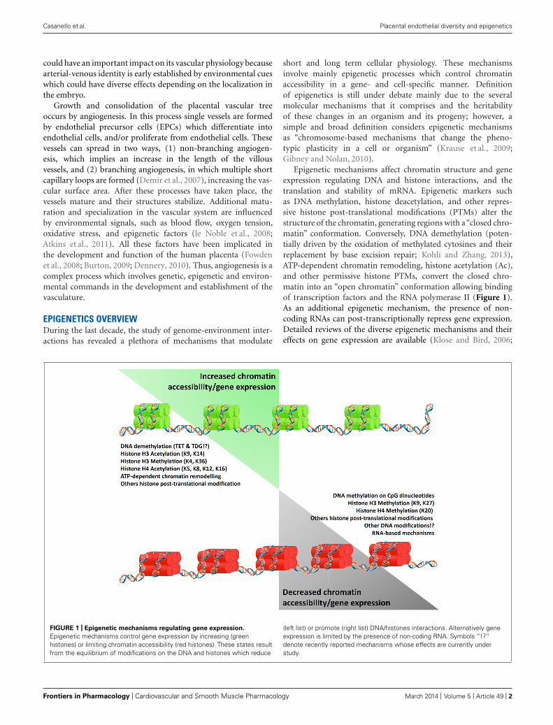
Epigenetic mechanisms affect chromatin structure and geneexpression regulating DNA and histone interactions, and thetranslation and stability of mRNA. Epigenetic markers suchas DNA methylation, histone deacetylation, and other repres-sive histone post-translational modifications (PTMs) alter thestructure of the chromatin, generating regions with a“closed chro-matin” conformation. Conversely, DNA demethylation (poten-tially driven by the oxidation of methylated cytosines and theirreplacement by base excision repair; Kohli and Zhang, 2013),ATP-dependent chromatin remodeling, histone acetylation (Ac),and other permissive histone PTMs, convert the closed chro-matin into an “open chromatin” conformation allowing bindingof transcription factors and the RNA polymerase II (Figure 1).As an additional epigenetic mechanism, the presence of non-coding RNAs can post-transcriptionally repress gene expression.Detailed reviews of the diverse epigenetic mechanisms and theireffects on gene expression are available (Klose and Bird, 2006;
FIGURE 1 | Epigenetic mechanisms regulating gene expression.
Epigenetic mechanisms control gene expression by increasing (greenhistones) or limiting chromatin accessibility (red histones). These states resultfrom the equilibrium of modifications on the DNA and histones which reduce
(left list) or promote (right list) DNA/histones interactions. Alternatively geneexpression is limited by the presence of non-coding RNA. Symbols “!?”denote recently reported mechanisms whose effects are currently understudy.
Frontiers in Pharmacology | Cardiovascular and Smooth Muscle Pharmacology March 2014 | Volume 5 | Article 49 | 2
Casanello et al. Placental endothelial diversity and epigenetics
Kouzarides, 2007; Wang et al., 2007a,b; Kaikkonen et al., 2011;Kohli and Zhang, 2013).
From a developmental perspective epigenetic mechanismsallow the generation of diverse cell phenotypes and functionsof an organism from a single genome, and respond to a rangeof environmental fluctuations. This issue is especially evident inorgans and tissues whose structure and function are under con-stant change across lifespan, such as the cardiovascular system(Aird, 2012). Nonetheless, placental vasculature may also be pro-grammed by epigenetic mechanisms, which are currently underrestless research.
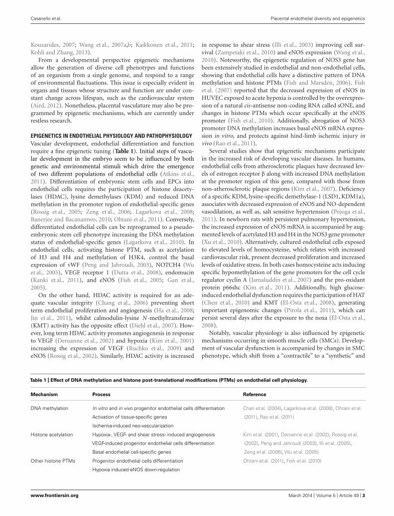
EPIGENETICS IN ENDOTHELIAL PHYSIOLOGY AND PATHOPHYSIOLOGYVascular development, endothelial differentiation and functionrequire a fine epigenetic tuning (Table 1). Initial steps of vascu-lar development in the embryo seem to be influenced by bothgenetic and environmental stimuli which drive the emergenceof two different populations of endothelial cells (Atkins et al.,2011). Differentiation of embryonic stem cells and EPCs intoendothelial cells requires the participation of histone deacety-lases (HDAC), lysine demethylases (KDM) and reduced DNAmethylation in the promoter region of endothelial-specific genes(Rossig et al., 2005; Zeng et al., 2006; Lagarkova et al., 2008;Banerjee and Bacanamwo, 2010; Ohtani et al., 2011). Conversely,differentiated endothelial cells can be reprogramed to a pseudo-embryonic stem cell phenotype increasing the DNA methylationstatus of endothelial-specific genes (Lagarkova et al., 2010). Inendothelial cells, activating histone PTM, such as acetylationof H3 and H4 and methylation of H3K4, control the basalexpression of vWF (Peng and Jahroudi, 2003), NOTCH4 (Wuet al., 2005), VEGF receptor 1 (Dutta et al., 2008), endomucin(Kanki et al., 2011), and eNOS (Fish et al., 2005; Gan et al.,2005).
On the other hand, HDAC activity is required for an ade-quate vascular integrity (Chang et al., 2006) preventing shortterm endothelial proliferation and angiogenesis (Ha et al., 2008;Jin et al., 2011), whilst calmodulin-lysine N-methyltransferase(KMT) activity has the opposite effect (Diehl et al., 2007). How-ever, long term HDAC activity promotes angiogenesis in responseto VEGF (Deroanne et al., 2002) and hypoxia (Kim et al., 2001)increasing the expression of VEGF (Ruchko et al., 2009) andeNOS (Rossig et al., 2002). Similarly, HDAC activity is increased
in response to shear stress (Illi et al., 2003) improving cell sur-vival (Zampetaki et al., 2010) and eNOS expression (Wang et al.,2010). Noteworthy, the epigenetic regulation of NOS3 gene hasbeen extensively studied in endothelial and non-endothelial cells,showing that endothelial cells have a distinctive pattern of DNAmethylation and histone PTMs (Fish and Marsden, 2006). Fishet al. (2007) reported that the decreased expression of eNOS inHUVEC exposed to acute hypoxia is controlled by the overexpres-sion of a natural cis-antisense non-coding RNA called sONE, andchanges in histone PTMs which occur specifically at the eNOSpromoter (Fish et al., 2010). Additionally, abrogation of NOS3promoter DNA methylation increases basal eNOS mRNA expres-sion in vitro, and protects against hind-limb ischemic injury invivo (Rao et al., 2011).
Several studies show that epigenetic mechanisms participatein the increased risk of developing vascular diseases. In humans,endothelial cells from atherosclerotic plaques have decreased lev-els of estrogen receptor β along with increased DNA methylationat the promoter region of this gene, compared with those fromnon-atherosclerotic plaque regions (Kim et al., 2007). Deficiencyof a specific KDM, lysine-specific demethylase-1 (LSD1, KDM1a),associates with decreased expression of eNOS and NO-dependentvasodilation, as well as, salt sensitive hypertension (Pojoga et al.,2011). In newborn rats with persistent pulmonary hypertension,the increased expression of eNOS mRNA is accompanied by aug-mented levels of acetylated H3 and H4 in the NOS3 gene promoter(Xu et al., 2010). Alternatively, cultured endothelial cells exposedto elevated levels of homocysteine, which relates with increasedcardiovascular risk, present decreased proliferation and increasedlevels of oxidative stress. In both cases homocysteine acts inducingspecific hypomethylation of the gene promoters for the cell cycleregulator cyclin A (Jamaluddin et al., 2007) and the pro-oxidantprotein p66shc (Kim et al., 2011). Additionally, high glucose-induced endothelial dysfunction requires the participation of HAT(Chen et al., 2010) and KMT (El-Osta et al., 2008), generatingimportant epigenomic changes (Pirola et al., 2011), which canpersist several days after the exposure to the noxa (El-Osta et al.,2008).
Notably, vascular physiology is also influenced by epigeneticmechanisms occurring in smooth muscle cells (SMCs). Develop-ment of vascular dysfunction is accompanied by changes in SMCphenotype, which shift from a “contractile” to a “synthetic” and
Table 1 | Effect of DNA methylation and histone post-translational modifications (PTMs) on endothelial cell physiology.
Mechanism Process Reference
DNA methylation In vitro and in vivo progenitor endothelial cells differentiation
Activation of tissue-specific genes
Ischemia-induced neo-vascularization
Chan et al. (2004), Lagarkova et al. (2008), Ohtani et al.
(2011), Rao et al. (2011)
Histone acetylation Hypoxia-, VEGF- and shear stress- induced angiogenesis
VEGF-induced progenitor endothelial cells differentiation
Basal endothelial cell-specific genes
Kim et al. (2001), Deroanne et al. (2002), Rossig et al.
(2002), Peng and Jahroudi (2003), Illi et al. (2005),
Zeng et al. (2006), Wu et al. (2005)
Other histone PTMs Progenitor endothelial cells differentiation
Hypoxia induced eNOS down-regulation
Ohtani et al. (2011), Fish et al. (2010)
www.frontiersin.org March 2014 | Volume 5 | Article 49 | 3
Casanello et al. Placental endothelial diversity and epigenetics
“pro-inflammatory” phenotype with long term consequences inthe contractile properties of vessels (Owens et al., 2004; Orr et al.,2010). Increasing data shows that this “phenotypic switching”requires the participation of epigenetic mechanisms which estab-lish an altered SMC function (Alexander and Owens, 2012).
PHENOTYPIC AND EPIGENETIC DIVERSITY IN THEUMBILICO-PLACENTAL ENDOTHELIUMPioneer studies by Lang et al. (1993) demonstrated that micro- andmacrovascular umbilico-placental endothelium present differentimmunoreactivity to diverse molecular markers for endothelialcells, suggesting the presence of a phenotypic endothelial diver-sity in the placenta. Additional evidence from cultured humanendothelial cells isolated from the placental microcirculation(PLEC) and the umbilical vein (HUVEC) show that microvas-cular endothelial cells express higher levels of vascular mediators(angiotensin II, endothelin, and thromboxane; Lang, 2003). Also adifferential pattern of homeobox genes (Murthi et al., 2007, 2008)and higher cholesterol transport capacity (Stefulj et al., 2009) inPLEC compared to HUVEC has been shown.
Notably, studies on endothelial cells from arteries and veinshave revealed important differences between arterial and venouscells at the same vascular level. In fact the higher mitogenicresponse observed in PLEC (Lang, 2003) may reflect the combina-tion of a high response to VEGF present in arterial PLEC (PLAEC)and to PIGF in venous endothelial cells (PLVEC; Lang et al., 2008).A transcriptomic analysis between PLAEC and PLVEC showedthat they have differential expression of more than 3,000 genes(Lang et al., 2008). Similarly there is a differential expression ofeNOS,a key vascular gene, between micro- and macrovascular, andvenous and arterial endothelium (Andersen et al., 2009; Krauseet al., 2012) being more homogenous at the arterial side (Ander-sen et al., 2009). This opens the queries about the differencesinitially reported between micro- and macrovascular endothe-lium reflecting an endothelial diversity between large and smallvessels, and whether they include variances between arteries andveins.
Several studies comparing simultaneously umbilical arterial(HUAEC) and venous (HUVEC) endothelium support the con-cept that these cells are not a homogenous population, and thenecessity of clarifying the precise source of cells when the term“macrovasculature” is used. A general characterization shows thatthere is a different profile of phospholipids with higher levelsof arachidonic acid-related species and heterogeneous expressionpattern of selenoproteins (Miller et al., 2002) in HUAEC com-pared to HUVEC (Takamura et al., 1990). Alongside the classicalmolecular markers for arterial endothelium, cultured HUAECexpress higher levels of PAI 1 (Gallicchio et al., 1994), Cx40 (VanRijen et al., 1997), 17β-HSD2 (Simard et al., 2011), and VCAM-1(Egorova et al., 2012); and lower levels of von Willebrand Factor(Shahani et al., 2010) and estrogen receptors beta (ERβ; Simardet al., 2011) compared with HUVEC. On the other hand expres-sions of pro-constrictive mediators such as angiotensin convertingenzyme (Ito et al., 2002) and ET-1 (Egorova et al., 2012) are dif-ferent in HUVEC relative to HUAEC. Furthermore, expressionand activity of eNOS are higher in freshly isolated HUVEC thanHUAEC (Andersen et al., 2009) and this expression pattern is
also observed in cells cultured up to third passage (Krause et al.,2012). Whether these differences reflects the physiology of umbili-cal (and potentially placental) arteries and veins, and how they arepreserved in vitro need further examination. Two recent reportsshow that the differential gene expression between HUAEC andHUVEC is partially controlled by specific transcription factors.Overexpression of the venous-specific nuclear receptor COUP-TFII in HUAEC decreases the expression of arterial markers (i.e.,Hey2, EphrinB2 and NICD4), and its down-regulation in HUVECincreases the expression of arterial markers such as VEGF-A, Dlland EphrinB2 (Korten et al., 2013). Moreover, in vitro simultane-ous overexpression of eight arterial-specific transcription factorsturns the HUVEC transcriptome into a HUAEC-like pattern(Aranguren et al., 2013).
Therefore, the phenotypic diversity in the umbilico-placentalcirculation is apparently commanded, at least in part, by anequivalent diversity in epigenetic mechanisms.
ENDOTHELIAL DIVERSITY AND ANGIOGENESISIn terms of angiogenesis, microvascular endothelial cells present ahigher mitogenic response to VEGF, PIGF (Lang, 2003; Lang et al.,2008), and prokineticin 1 (Brouillet et al., 2010) compared withHUVEC, along with an increased expression of pro-angiogenicHOX genes (i.e., TLX1, TLX2, and PHOX1; Murthi et al., 2008).These data are in agreement with the notion that placental angio-genic capacity is augmented in microvascular vessels comparedto endothelial cells from larger vessels. However, it is also pos-sible to find significant differences in the angiogenic responsein endothelial cells from umbilical arteries and veins. In vivoVEGFR3, which is commonly expressed in lymphatic endotheliumor during active angiogenesis (Koch and Claesson-Welsh, 2012),is absent in HUAEC but expressed in HUVEC (Veikkola et al.,2003). Moreover in vitro chemotaxis induced by VEGFA or FGF2is higher in HUVEC compared to HUAEC (Barkefors et al., 2008),and netrin-1 prevents the VEGF-induced migration in HUAECwithout effect on HUVEC (Lu et al., 2004). Further studies areneeded to address the effects and the role on placental physiologyof this increased angiogenic response observed in HUVEC.
ENDOTHELIAL DIVERSITY IN RESPONSE TO STRESSPlacental vascular and endothelial physiology, similar to adult vas-culature, are importantly influenced by stimuli such as alteredshear stress and oxygen levels whose effects are apparently dif-ferent between arteries and veins. Normally arterial endotheliumis exposed to higher shear stress and therefore it is plausible topredict a stronger response to increasing stress. In fact pulsatileshear stress increases the expression of arterial markers (i.e., Hey1,Hey1, and ephrinB2) in HUAEC but decreases the expressionof venous markers (COUP-TFII) in HUVEC (Buschmann et al.,2010). Laminar shear stress have similar effects on the expressionof arterial-venous markers in these cells, and increases the levelsof S-nitrosylated proteins (Hoffmann et al., 2003) endothelin-1,VCAM, and vWF (Egorova et al., 2012) in HUAEC compared toHUVEC. Whether these differences are observed in microvascularendothelial cells remains to be determined.
Some evidence regarding the effects of low oxygen levels onendothelial function in placental large and small vessels, as well
Frontiers in Pharmacology | Cardiovascular and Smooth Muscle Pharmacology March 2014 | Volume 5 | Article 49 | 4
Casanello et al. Placental endothelial diversity and epigenetics
as arteries and veins, show a differential vascular response tohypoxia throughout the placenta (Krause et al., 2011, 2012). Onthe other hand placental endothelium is importantly exposed tolow oxygen levels and oxidative stress which are negative regu-lator of placental angiogenesis (Burton et al., 2009). A reductionin oxygen levels from 21 to 12% O2decreases placental venousmicrovascular endothelial cells viability with no effect on theirarterial counterparts (Lassance et al., 2012), and PLAEC exposedto 3% O2 show an increased mitogenic response to VEGFA andFGF2 compared to cells cultured at 21% O2 (Wang et al., 2009).This higher response to VEGFA and FGF2 is also observed inHUAEC exposed to physiological levels of oxygen (3–5% O2; Jianget al., 2013). Additionally, hypoxia (1% O2) increases the expres-sion of the pro-angiogenic factor protease-activated receptor 2in HUVEC and this effect is higher in HUAEC (Svensson et al.,2011).
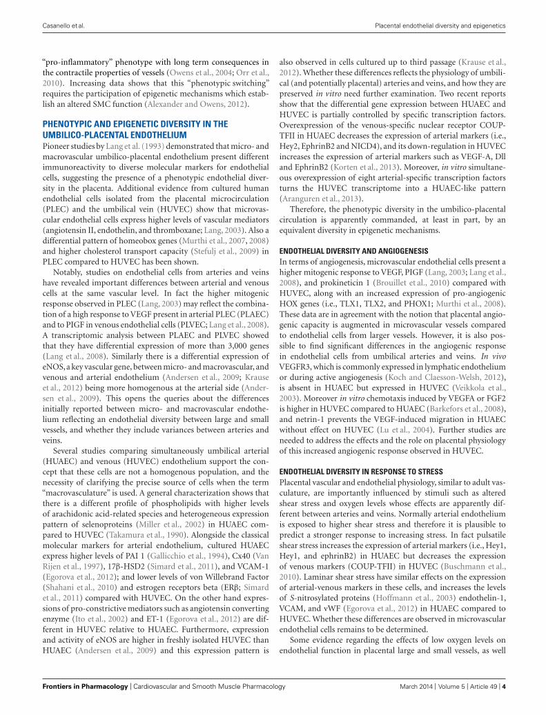
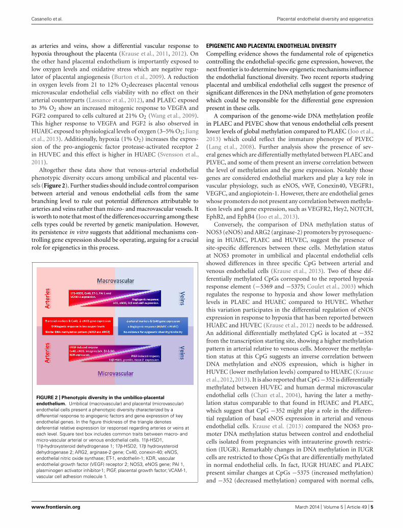
Altogether these data show that venous-arterial endothelialphenotypic diversity occurs among umbilical and placental ves-sels (Figure 2). Further studies should include control comparisonbetween arterial and venous endothelial cells from the samebranching level to rule out potential differences attributable toarteries and veins rather than micro- and macrovascular vessels. Itis worth to note that most of the differences occurring among thesecells types could be reverted by genetic manipulation. However,its persistence in vitro suggests that additional mechanisms con-trolling gene expression should be operating, arguing for a crucialrole for epigenetics in this process.
FIGURE 2 | Phenotypic diversity in the umbilico-placental
endothelium. Umbilical (macrovascular) and placental (microvascular)endothelial cells present a phenotypic diversity characterized by adifferential response to angiogenic factors and gene expression of keyendothelial genes. In the figure thickness of the triangle denotesdeferential relative expression (or response) regarding arteries or veins ateach level. Square text box includes common traits between macro- andmicro-vascular arterial or venous endothelial cells. 11β-HSD1,11β-hydroxysteroid dehydrogenase 1; 17β-HSD2, 17β hydroxysteroiddehydrogenase 2; ARG2, arginase-2 gene; Cx40, conexin-40; eNOS,endothelial nitric oxide synthase; ET-1, endothelin-1; KDR, vascularendothelial growth factor (VEGF) receptor 2; NOS3, eNOS gene; PAI 1,plasminogen activator inhibitor-1; PlGF, placental growth factor; VCAM-1,vascular cell adhesion molecule 1.
EPIGENETIC AND PLACENTAL ENDOTHELIAL DIVERSITYCompelling evidence shows the fundamental role of epigeneticscontrolling the endothelial-specific gene expression, however, thenext frontier is to determine how epigenetic mechanisms influencethe endothelial functional diversity. Two recent reports studyingplacental and umbilical endothelial cells suggest the presence ofsignificant differences in the DNA methylation of gene promoterswhich could be responsible for the differential gene expressionpresent in these cells.
A comparison of the genome-wide DNA methylation profilein PLAEC and PLVEC show that venous endothelial cells presentlower levels of global methylation compared to PLAEC (Joo et al.,2013) which could reflect the immature phenotype of PLVEC(Lang et al., 2008). Further analysis show the presence of sev-eral genes which are differentially methylated between PLAEC andPLVEC, and some of them present an inverse correlation betweenthe level of methylation and the gene expression. Notably thosegenes are considered endothelial markers and play a key role invascular physiology, such as eNOS, vWF, Conexin40, VEGFR1,VEGFC, and angiopiotein-1. However, there are endothelial geneswhose promoters do not present any correlation between methyla-tion levels and gene expression, such as VEGFR2, Hey2, NOTCH,EphB2, and EphB4 (Joo et al., 2013).
Conversely, the comparison of DNA methylation status ofNOS3 (eNOS) and ARG2 (arginase-2) promoters by pyrosequenc-ing in HUAEC, PLAEC and HUVEC, suggest the presence ofsite-specific differences between these cells. Methylation statusat NOS3 promoter in umbilical and placental endothelial cellsshowed differences in three specific CpG between arterial andvenous endothelial cells (Krause et al., 2013). Two of these dif-ferentially methylated CpGs correspond to the reported hypoxiaresponse element (−5369 and −5375; Coulet et al., 2003) whichregulates the response to hypoxia and show lower methylationlevels in PLAEC and HUAEC compared to HUVEC. Whetherthis variation participates in the differential regulation of eNOSexpression in response to hypoxia that has been reported betweenHUAEC and HUVEC (Krause et al., 2012) needs to be addressed.An additional differentially methylated CpG is located at −352from the transcription starting site, showing a higher methylationpattern in arterial relative to venous cells. Moreover the methyla-tion status at this CpG suggests an inverse correlation betweenDNA methylation and eNOS expression, which is higher inHUVEC (lower methylation levels) compared to HUAEC (Krauseet al., 2012, 2013). It is also reported that CpG −352 is differentiallymethylated between HUVEC and human dermal microvascularendothelial cells (Chan et al., 2004), having the later a methy-lation status comparable to that found in HUAEC and PLAEC,which suggest that CpG −352 might play a role in the differen-tial regulation of basal eNOS expression in arterial and venousendothelial cells. Krause et al. (2013) compared the NOS3 pro-moter DNA methylation status between control and endothelialcells isolated from pregnancies with intrauterine growth restric-tion (IUGR). Remarkably changes in DNA methylation in IUGRcells are restricted to those CpGs that are differentially methylatedin normal endothelial cells. In fact, IUGR HUAEC and PLAECpresent similar changes at CpGs −5375 (increased methylation)and −352 (decreased methylation) compared with normal cells,
www.frontiersin.org March 2014 | Volume 5 | Article 49 | 5

Casanello et al. Placental endothelial diversity and epigenetics
and these methylation levels are comparable to that find in nor-mal HUVEC. Conversely, changes in the DNA methylation statusof IUGR HUVEC where at CpGs −5369 (decreased methylation)and −352 (increased methylation), and they are comparable tothose find in normal HUAEC and PLAEC. The methylation levelsat CpG −352 in IUGR HUAEC and HUVEC are also related withthe levels of mRNA for eNOS (Krause et al., 2013), reinforcingthe potential importance of CpG −352 in the regulation of basaleNOS expression. Finally analysis of methylation status of ARG2promoter in HUAEC, PLAEC, and HUVEC show a single differ-ence between PLAEC and HUVEC, however, it is still unknown ifthere is a correlation with arginase-2 expression and activity.
DNA methylation is one of the main epigenetic mechanismsthat controls long term gene expression, showing a high repro-ducibility after every cellular replication and this characteristicis driven by the activity of DNA methyltransferase-1 (DNMT1).In IUGR HUAEC and HUVEC DNMT1 silencing shows a dif-ferential effect, reducing and increasing basal eNOS expression,respectively (Krause et al., 2013). Silencing of DNMT1 restoresto normal eNOS mRNA levels in IUGR HUAEC and HUVEC,and this effect is not observed on arginase-2 expression whereit further increases its expression in IUGR HUVEC, withoutany effect in IUGR HUAEC (Krause et al., 2013). This suggeststhat DNA methylation (Jamaluddin et al., 2007; Banerjee andBacanamwo, 2010; Kim et al., 2011) and other epigenetic mecha-nisms (Kim et al., 2001; Deroanne et al., 2002; Fish et al., 2010)control gene expression in endothelial cells in a gene-specificmanner.
Although the studies in PLAEC and PLVEC (Joo et al., 2013),and in HUAEC and HUVEC (Krause et al., 2013) used two dif-ferent approaches to analyze the DNA methylation patterns, thereare some similarities in the outcomes. First, both studies showthat methylation status of NOS3 proximal promoter inversely cor-relates with the levels of mRNA for eNOS, and this occurs in cellsexposed for several days to culture conditions. Second, PLAECand PLVEC show differential levels arginase-2 expression with-out differences in the DNA methylation in ARG2 promoter, whilstin control and IUGR HUAEC differences in DNA methylationare not associated to difference in arginase-2 expression. Finally,DNMT1 silencing in IUGR cells normalize eNOS expression butnot arginase-2 expression.
CONCLUSIONAltogether these seminal data show that epigenetic mechanismscould be responsible for the phenotypic diversity of endothe-lial cells in the umbilico-placental unit, and these mechanismswould be operating in a cell- and gene-specific manner. Thecurrent research on the area is offering novel data about poten-tial mechanisms but still further studies are required to have acomprehensive picture of the additional epigenetic mechanismscontrolling the gene expression in physiological and pathophys-iological conditions and its consequences in umbilico-placentalfunctions.
ACKNOWLEDGMENTThis work was funded by FONDECYT 1110595, 1120928,1130801.
REFERENCESAird, W. C. (2012). Endothelial cell heterogeneity. Cold Spring Harb. Perspect. Med.
2:a006429. doi: 10.1101/cshperspect.a006429Alexander, M. R., and Owens, G. K. (2012). Epigenetic control of smooth muscle cell
differentiation and phenotypic switching in vascular development and disease.Annu. Rev. Physiol. 74, 13–40. doi: 10.1146/annurev-physiol-012110-142315
Andersen, M. R., Simonsen, U., Uldbjerg, N., Aalkjaer, C., and Stender, S. (2009).Smoking cessation early in pregnancy and birth weight, length, head circumfer-ence, and endothelial nitric oxide synthase activity in umbilical and chorionicvessels: an observational study of healthy singleton pregnancies. Circulation 119,857–864. doi: 10.1161/CIRCULATIONAHA.107.755769
Aranguren, X. L., Agirre, X., Beerens, M., Coppiello, G., Uriz, M., Vandersmissen,I., et al. (2013). Unraveling a novel transcription factor code determining thehuman arterial-specific endothelial cell signature. Blood 122, 3982–3992. doi:10.1182/blood-2013-02-483255
Atkins, G. B., Jain, M. K., and Hamik, A. (2011). Endothelial differentiation: molec-ular mechanisms of specification and heterogeneity. Arterioscler. Thromb. Vasc.Biol. 31, 1476–1484. doi: 10.1161/ATVBAHA.111.228999
Banerjee, S., and Bacanamwo, M. (2010). DNA methyltransferase inhibition inducesmouse embryonic stem cell differentiation into endothelial cells. Exp. Cell Res.316, 172–180. doi: 10.1016/j.yexcr.2009.08.011
Barkefors, I., Le Jan, S., Jakobsson, L., Hejll, E., Carlson, G., Johansson, H.,et al. (2008). Endothelial cell migration in stable gradients of vascular endothe-lial growth factor A and fibroblast growth factor 2: effects on chemotaxis andchemokinesis. J. Biol. Chem. 283, 13905–13912. doi: 10.1074/jbc.M704917200
Brouillet, S., Hoffmann, P., Benharouga, M., Salomon, A., Schaal, J. P., Feige, J.J., et al. (2010). Molecular characterization of EG-VEGF-mediated angiogenesis:differential effects on microvascular and macrovascular endothelial cells. Mol.Biol. Cell 21, 2832–2843. doi: 10.1091/mbc.E10-01-0059
Burton, G. J. (2009). Oxygen, the Janus gas; its effects on human placental develop-ment and function. J. Anat. 215, 27–35. doi: 10.1111/j.1469-7580.2008.00978.x
Burton, G. J., Charnock-Jones, D. S., and Jauniaux, E. (2009). Regulation of vasculargrowth and function in the human placenta. Reproduction 138, 895–902. doi:10.1530/REP-09–0092
Buschmann, I., Pries, A., Styp-Rekowska, B., Hillmeister, P., Loufrani, L., Henrion,D., et al. (2010). Pulsatile shear and Gja5 modulate arterial identity and remodel-ing events during flow-driven arteriogenesis. Development 137, 2187–2196. doi:10.1242/dev.045351
Chaddha,V.,Viero, S., Huppertz, B., and Kingdom, J. (2004). Developmental biologyof the placenta and the origins of placental insufficiency. Semin. Fetal NeonatalMed. 9, 357–369. doi: 10.1016/j.siny.2004.03.006
Chan, Y., Fish, J. E., D’abreo, C., Lin, S., Robb, G. B., Teichert, A. M., et al.(2004). The cell-specific expression of endothelial nitric-oxide synthase: a rolefor DNA methylation. J. Biol. Chem. 279, 35087–35100. doi: 10.1074/jbc.M405063200
Chang, S., Young, B. D., Li, S., Qi, X., Richardson, J. A., and Olson, E. N.(2006). Histone deacetylase 7 maintains vascular integrity by repressing matrixmetalloproteinase 10. Cell 126, 321–334. doi: 10.1016/j.cell.2006.05.040
Chen, S., Feng, B., George, B., Chakrabarti, R., Chen, M., and Chakrabarti, S.(2010). Transcriptional coactivator p300 regulates glucose-induced gene expres-sion in endothelial cells. Am. J. Physiol. Endocrinol. Metab. 298, E127–E137. doi:10.1152/ajpendo.00432.2009
Coulet, F., Nadaud, S., Agrapart, M., and Soubrier, F. (2003). Identifica-tion of hypoxia-response element in the human endothelial nitric-oxide syn-thase gene promoter. J. Biol. Chem. 278, 46230–46240. doi: 10.1074/jbc.M305420200
Demir, R., Seval, Y., and Huppertz, B. (2007). Vasculogenesis and angio-genesis in the early human placenta. Acta Histochem. 109, 257–265. doi:10.1016/j.acthis.2007.02.008
Dennery, P. A. (2010). Oxidative stress in development: nature or nurture? FreeRadic. Biol. Med. 49, 1147–1151. doi: 10.1016/j.freeradbiomed.2010.07.011
Deroanne, C. F., Bonjean, K., Servotte, S., Devy, L., Colige, A., Clausse, N.,et al. (2002). Histone deacetylases inhibitors as anti-angiogenic agents alter-ing vascular endothelial growth factor signaling. Oncogene 21, 427–436. doi:10.1038/sj.onc.1205108
Diehl, F., Rossig, L., Zeiher, A. M., Dimmeler, S., and Urbich, C. (2007). Thehistone methyltransferase MLL is an upstream regulator of endothelial-cell sproutformation. Blood 109, 1472–1478. doi: 10.1182/blood-2006-08-039651
Frontiers in Pharmacology | Cardiovascular and Smooth Muscle Pharmacology March 2014 | Volume 5 | Article 49 | 6
Casanello et al. Placental endothelial diversity and epigenetics
Dutta, D., Ray, S., Vivian, J. L., and Paul, S. (2008). Activation of the VEGFR1chromatin domain: an angiogenic signal-ETS1/HIF-2alpha regulatory axis. J.Biol. Chem. 283, 25404–25413. doi: 10.1074/jbc.M804349200
Egorova, A. D., Deruiter, M. C., De Boer, H. C., Van De Pas, S., Gittenberger-DeGroot, A. C., Van Zonneveld, A. J., et al. (2012). Endothelial colony-forming cellsshow a mature transcriptional response to shear stress. In Vitro Cell. Dev. Biol.Anim. 48, 21–29. doi: 10.1007/s11626-011-9470-z
El-Osta, A., Brasacchio, D., Yao, D., Pocai, A., Jones, P. L., Roeder, R. G., et al.(2008). Transient high glucose causes persistent epigenetic changes and alteredgene expression during subsequent normoglycemia. J. Exp. Med. 205, 2409–2417.doi: 10.1084/jem.20081188
Fish, J. E., and Marsden, P. A. (2006). Endothelial nitric oxide synthase: insight intocell-specific gene regulation in the vascular endothelium. Cell. Mol. Life Sci. 63,144–162. doi: 10.1007/s00018-005-5421-8
Fish, J. E., Matouk, C. C., Rachlis, A., Lin, S., Tai, S. C., D’abreo, C., et al. (2005).The expression of endothelial nitric-oxide synthase is controlled by a cell-specifichistone code. J. Biol. Chem. 280, 24824–24838. doi: 10.1074/jbc.M502115200
Fish, J. E., Matouk, C. C., Yeboah, E., Bevan, S. C., Khan, M., Patil, K.,et al. (2007). Hypoxia-inducible expression of a natural cis-antisense transcriptinhibits endothelial nitric-oxide synthase. J. Biol. Chem. 282, 15652–15666. doi:10.1074/jbc.M608318200
Fish, J. E., Yan, M. S., Matouk, C. C., St Bernard, R., Ho, J. J., Gavryushova, A., et al.(2010). Hypoxic repression of endothelial nitric-oxide synthase transcription iscoupled with eviction of promoter histones. J. Biol. Chem. 285, 810–826. doi:10.1074/jbc.M109.067868
Fowden, A. L., Forhead, A. J., Coan, P. M., and Burton, G. J. (2008). The placenta andintrauterine programming. J. Neuroendocrinol. 20, 439–450. doi: 10.1111/j.1365-2826.2008.01663.x
Freyer, C., and Renfree, M. B. (2009). The mammalian yolk sac placenta. J. Exp.Zool. B Mol. Dev. Evol. 312, 545–554. doi: 10.1002/jez.b.21239
Gallicchio, M., Argyriou, S., Ianches, G., Filonzi, E. L., Zoellner, H., Hamil-ton, J. A., et al. (1994). Stimulation of PAI-1 expression in endothelial cells bycultured vascular smooth muscle cells. Arterioscler. Thromb. 14, 815–823. doi:10.1161/01.ATV.14.5.815
Gan, Y., Shen, Y. H., Wang, J., Wang, X., Utama, B., and Wang, X. L. (2005). Roleof histone deacetylation in cell-specific expression of endothelial nitric-oxidesynthase. J. Biol. Chem. 280, 16467–16475. doi: 10.1074/jbc.M412960200
Gibney, E. R., and Nolan, C. M. (2010). Epigenetics and gene expression. Heredity(Edinb.) 105, 4–13. doi: 10.1038/hdy.2010.54
Ha, C. H., Jhun, B. S., Kao, H. Y., and Jin, Z. G. (2008). VEGF stimulatesHDAC7 phosphorylation and cytoplasmic accumulation modulating matrix met-alloproteinase expression and angiogenesis. Arterioscler. Thromb. Vasc. Biol. 28,1782–1788. doi: 10.1161/ATVBAHA.108.172528
Hoffmann, J., Dimmeler, S., and Haendeler, J. (2003). Shear stress increases theamount of S-nitrosylated molecules in endothelial cells: important role for signaltransduction. FEBS Lett. 551, 153–158. doi: 10.1016/S0014-5793(03)00917-7
Illi, B., Nanni, S., Scopece, A., Farsetti, A., Biglioli, P., Capogrossi, M. C., et al.(2003). Shear stress-mediated chromatin remodeling provides molecular basisfor flow-dependent regulation of gene expression. Circ. Res. 93, 155–161. doi:10.1161/01.RES.0000080933.82105.29
Illi, B., Scopece, A., Nanni, S., Farsetti, A., Morgante, L., Biglioli, P., et al. (2005). Epi-genetic histone modification and cardiovascular lineage programming in mouseembryonic stem cells exposed to laminar shear stress. Circ. Res. 96, 501–508. doi:10.1161/01.RES.0000159181.06379.63.
Ito, M., Itakura, A., Ohno, Y., Nomura, M., Senga, T., Nagasaka, T.,et al. (2002). Possible activation of the renin-angiotensin system in the feto-placental unit in preeclampsia. J. Clin. Endocrinol. Metab. 87, 1871–1878. doi:10.1210/jcem.87.4.8422
Jamaluddin, M. D., Chen, I., Yang, F., Jiang, X., Jan, M., Liu, X., et al. (2007).Homocysteine inhibits endothelial cell growth via DNA hypomethylation of thecyclin A gene. Blood 110, 3648–3655. doi: 10.1182/blood-2007-06-096701
Jiang, Y. Z., Wang, K., Li, Y., Dai, C. F., Wang, P., Kendziorski, C., et al. (2013).Enhanced cellular responses and distinct gene profiles in human fetoplacentalartery endothelial cells under chronic low oxygen. Biol. Reprod. 89, 133. doi:10.1095/biolreprod.113.110551
Jin, G., Bausch, D., Knightly, T., Liu, Z., Li, Y., Liu, B., et al. (2011). Histonedeacetylase inhibitors enhance endothelial cell sprouting angiogenesis in vitro.Surgery 150, 429–435. doi: 10.1016/j.surg.2011.07.001
Jin, S. W., and Patterson, C. (2009). The opening act: vasculogenesis and theorigins of circulation. Arterioscler. Thromb. Vasc. Biol. 29, 623–629. doi:10.1161/ATVBAHA.107.161539
Joo, J. E., Hiden, U., Lassance, L., Gordon, L., Martino, D. J., Desoye, G., et al. (2013).Variable promoter methylation contributes to differential expression of key genesin human placenta-derived venous and arterial endothelial cells. BMC Genomics14:475. doi: 10.1186/1471-2164-14-475
Kaikkonen, M. U., Lam, M. T., and Glass, C. K. (2011). Non-coding RNAs asregulators of gene expression and epigenetics. Cardiovasc. Res. 90, 430–440. doi:10.1093/cvr/cvr097
Kanki, Y., Kohro, T., Jiang, S., Tsutsumi, S., Mimura, I., Suehiro, J., et al.(2011). Epigenetically coordinated GATA2 binding is necessary for endothelium-specific endomucin expression. EMBO J. 30, 2582–2595. doi: 10.1038/emboj.2011.173
Kim, C. S., Kim, Y. R., Naqvi, A., Kumar, S., Hoffman, T. A., Jung, S. B., et al.(2011). Homocysteine promotes human endothelial cell dysfunction via site-specific epigenetic regulation of p66shc. Cardiovasc. Res. 92, 466–475. doi:10.1093/cvr/cvr250
Kim, J., Kim, J. Y., Song, K. S., Lee, Y. H., Seo, J. S., Jelinek, J., et al. (2007). Epi-genetic changes in estrogen receptor beta gene in atherosclerotic cardiovasculartissues and in-vitro vascular senescence. Biochim. Biophys. Acta 1772, 72–80. doi:10.1016/j.bbadis.2006.10.004
Kim, M. S., Kwon, H. J., Lee, Y. M., Baek, J. H., Jang, J. E., Lee, S. W., et al.(2001). Histone deacetylases induce angiogenesis by negative regulation of tumorsuppressor genes. Nat. Med. 7, 437–443. doi: 10.1038/86507
Klose, R. J., and Bird, A. P. (2006). Genomic DNA methylation: the mark and itsmediators. Trends Biochem. Sci. 31, 89–97. doi: 10.1016/j.tibs.2005.12.008
Koch, S., and Claesson-Welsh, L. (2012). Signal transduction by vascular endothe-lial growth factor receptors. Cold Spring Harb. Perspect. Med. 2:a006502. doi:10.1101/cshperspect.a006502
Kohli, R. M., and Zhang, Y. (2013). TET enzymes, TDG and the dynamics of DNAdemethylation. Nature 502, 472–479. doi: 10.1038/nature12750
Korten, S., Brunssen, C., Poitz, D. M., Grossklaus, S., Brux, M., Schnittler, H. J.,et al. (2013). Impact of Hey2 and COUP-TFII on genes involved in arteriovenousdifferentiation in primary human arterial and venous endothelial cells. Basic Res.Cardiol. 108, 362. doi: 10.1007/s00395-013-0362-0.
Kouzarides, T. (2007). Chromatin modifications and their function. Cell 128, 693–705. doi: 10.1016/j.cell.2007.02.005
Krause, B., Sobrevia, L., and Casanello, P. (2009). Epigenetics: new concepts ofold phenomena in vascular physiology. Curr. Vasc. Pharmacol. 7, 513–520. doi:10.2174/157016109789043883
Krause, B. J., Costello, P. M., Munoz-Urrutia, E., Lillycrop, K. A., Hanson, M. A.,and Casanello, P. (2013). Role of DNA methyltransferase 1 on the altered eNOSexpression in human umbilical endothelium from intrauterine growth restrictedfetuses. Epigenetics 8, 944–952. doi: 10.4161/epi.25579
Krause, B. J., Hanson, M. A., and Casanello, P. (2011). Role of nitric oxidein placental vascular development and function. Placenta 32:797–805. doi:10.1016/j.placenta.2011.06.025
Krause, B. J., Prieto, C. P., Munoz-Urrutia, E., Martin, S. S., Sobrevia, L., andCasanello, P. (2012). Role of arginase-2 and eNOS in the differential vascularreactivity and hypoxia-induced endothelial response in umbilical arteries andveins. Placenta 33:360–366. doi: 10.1016/j.placenta.2012.02.006
Lagarkova, M. A., Shutova, M. V., Bogomazova, A. N., Vassina, E. M., Glazov,E. A., Zhang, P., et al. (2010). Induction of pluripotency in human endothe-lial cells resets epigenetic profile on genome scale. Cell Cycle 9, 937–946. doi:10.4161/cc.9.5.10869
Lagarkova, M. A., Volchkov, P. Y., Philonenko, E. S., and Kiselev, S. L. (2008).Efficient differentiation of hESCs into endothelial cells in vitro is secured byepigenetic changes. Cell Cycle 7, 2929–2935. doi: 10.4161/cc.7.18.6700
Lang, I. (2003). Heterogeneity of microvascular endothelial cells isolated fromhuman term placenta and macrovascular umbilical vein endothelial cells. Eur.J. Cell Biol. 82, 163–173. doi: 10.1078/0171-9335-00306
Lang, I., Hartmann, M., Blaschitz, A., Dohr, G., Skofitsch, G., and Desoye, G.(1993). Immunohistochemical evidence for the heterogeneity of maternal andfetal vascular endothelial cells in human full-term placenta. Cell Tissue Res. 274,211–218. doi: 10.1007/BF00318740
Lang, I., Schweizer, A., Hiden, U., Ghaffari-Tabrizi, N., Hagendorfer, G., Bilban,M., et al. (2008). Human fetal placental endothelial cells have a mature arterial
www.frontiersin.org March 2014 | Volume 5 | Article 49 | 7
Casanello et al. Placental endothelial diversity and epigenetics
and a juvenile venous phenotype with adipogenic and osteogenic differentia-tion potential. Differentiation 76, 1031–1043. doi: 10.1111/j.1432-0436.2008.00302.x
Lassance, L., Miedl, H., Konya, V., Heinemann, A., Ebner, B., Hackl, H.,et al. (2012). Differential response of arterial and venous endothelial cellsto extracellular matrix is modulated by oxygen. Histochem. Cell Biol. doi:10.1007/s00418-012-0917-4 [Epub ahead of print].
le Noble, F., Klein, C., Tintu, A., Pries, A., and Buschmann, I. (2008). Neuralguidance molecules, tip cells, and mechanical factors in vascular development.Cardiovasc. Res. 78, 232–241. doi: 10.1093/cvr/cvn058
Lu, X., Le Noble, F., Yuan, L., Jiang, Q., De Lafarge, B., Sugiyama, D., et al. (2004).The netrin receptor UNC5B mediates guidance events controlling morphogenesisof the vascular system. Nature 432, 179–186. doi: 10.1038/nature03080
Lux, C. T., Yoshimoto, M., Mcgrath, K., Conway, S. J., Palis, J., and Yoder, M.C. (2008). All primitive and definitive hematopoietic progenitor cells emergingbefore E10 in the mouse embryo are products of the yolk sac. Blood 111, 3435–3438. doi: 10.1182/blood-2007-08-107086
Matouk, C. C., and Marsden, P. A. (2008). Epigenetic regulation of vascularendothelial gene expression. Circ. Res. 102, 873–887. doi: 10.1161/CIRCRE-SAHA.107.171025
Miller, S., Walker, S. W., Arthur, J. R., Lewin, M. H., Pickard, K., Nicol,F., et al. (2002). Selenoprotein expression in endothelial cells from differenthuman vasculature and species. Biochim. Biophys. Acta 1588, 85–93. doi:10.1016/S0925-4439(02)00143-6
Murthi, P., Hiden, U., Rajaraman, G., Liu, H., Borg, A. J., Coombes, F., et al. (2008).Novel homeobox genes are differentially expressed in placental microvascularendothelial cells compared with macrovascular cells. Placenta 29, 624–630. doi:10.1016/j.placenta.2008.04.006
Murthi, P., So, M., Gude, N. M., Doherty, V. L., Brennecke, S. P., and Kalionis, B.(2007). Homeobox genes are differentially expressed in macrovascular humanumbilical vein endothelial cells and microvascular placental endothelial cells.Placenta 28, 219–223. doi: 10.1016/j.placenta.2006.02.012
Ohtani, K., Vlachojannis, G. J., Koyanagi, M., Boeckel, J. N., Urbich, C., Farcas,R., et al. (2011). Epigenetic regulation of endothelial lineage committed genesin pro-angiogenic hematopoietic and endothelial progenitor cells. Circ. Res. 109,1219–1229. doi: 10.1161/CIRCRESAHA.111.247304
Orr, A. W., Hastings, N. E., Blackman, B. R., and Wamhoff, B. R. (2010). Complexregulation and function of the inflammatory smooth muscle cell phenotype inatherosclerosis. J. Vasc. Res. 47, 168–180. doi: 10.1159/000250095
Owens, G. K., Kumar, M. S., and Wamhoff, B. R. (2004). Molecular regulation ofvascular smooth muscle cell differentiation in development and disease. Physiol.Rev. 84, 767–801. doi: 10.1152/physrev.00041.2003.
Parenti, A., Morbidelli, L., Cui, X. L., Douglas, J. G., Hood, J. D., Granger, H. J.,et al. (1998). Nitric oxide is an upstream signal of vascular endothelial growthfactor-induced extracellular signal-regulated kinase1/2 activation in postcapillaryendothelium. J. Biol. Chem. 273, 4220–4226. doi: 10.1074/jbc.273.7.4220
Peng, Y., and Jahroudi, N. (2003). The NFY transcription factor inhibitsvon Willebrand factor promoter activation in non-endothelial cells throughrecruitment of histone deacetylases. J. Biol. Chem. 278, 8385–8394. doi:10.1074/jbc.M213156200
Pirola, L., Balcerczyk, A., Tothill, R. W., Haviv, I., Kaspi, A., Lunke, S., et al.(2011). Genome-wide analysis distinguishes hyperglycemia regulated epige-netic signatures of primary vascular cells. Genome Res. 21, 1601–1615. doi:10.1101/gr.116095.110
Pojoga, L. H., Williams, J. S., Yao, T. M., Kumar, A., Raffetto, J. D., Do Nascimento,G. R., et al. (2011). Histone demethylase LSD1 deficiency during high-salt dietis associated with enhanced vascular contraction, altered NO-cGMP relaxationpathway, and hypertension. Am. J. Physiol. Heart Circ. Physiol. 301, H1862–H1871. doi: 10.1152/ajpheart.00513.2011
Rao, X., Zhong, J., Zhang, S., Zhang, Y., Yu, Q., Yang, P., et al. (2011). Loss ofmethyl-CpG-binding domain protein 2 enhances endothelial angiogenesis andprotects mice against hind-limb ischemic injury. Circulation 123, 2964–2974. doi:10.1161/CIRCULATIONAHA.110.966408
Rossig, L., Li, H., Fisslthaler, B., Urbich, C., Fleming, I., Forstermann, U.,et al. (2002). Inhibitors of histone deacetylation downregulate the expres-sion of endothelial nitric oxide synthase and compromise endothelial cellfunction in vasorelaxation and angiogenesis. Circ. Res. 91, 837–844. doi:10.1161/01.RES.0000037983.07158.B1
Rossig, L., Urbich, C., Bruhl, T., Dernbach, E., Heeschen, C., Chavakis, E., et al.(2005). Histone deacetylase activity is essential for the expression of HoxA9 andfor endothelial commitment of progenitor cells. J. Exp. Med. 201, 1825–1835. doi:10.1084/jem.20042097
Ruchko, M. V., Gorodnya, O. M., Pastukh, V. M., Swiger, B. M., Middle-ton, N. S., Wilson, G. L., et al. (2009). Hypoxia-induced oxidative basemodifications in the VEGF hypoxia-response element are associated with tran-scriptionally active nucleosomes. Free Radic. Biol. Med. 46, 352–359. doi:10.1016/j.freeradbiomed.2008.09.038
Shahani, T., Lavend’homme, R., Luttun, A., Saint-Remy, J. M., Peerlinck, K.,and Jacquemin, M. (2010). Activation of human endothelial cells from specificvascular beds induces the release of a FVIII storage pool. Blood 115, 4902–4909.doi: 10.1182/blood-2009-07-232546
Shalaby, F., Rossant, J., Yamaguchi, T. P., Gertsenstein, M., Wu, X. F., Breitman,M. L., et al. (1995). Failure of blood-island formation and vasculogenesis inFlk-1-deficient mice. Nature 376, 62–66. doi: 10.1038/376062a0
Shizukuda, Y., Tang, S., Yokota, R., and Ware, J. A. (1999). Vascular endothelialgrowth factor-induced endothelial cell migration and proliferation depend on anitric oxide-mediated decrease in protein kinase Cdelta activity. Circ. Res. 85,247–256. doi: 10.1161/01.RES.85.3.247
Simard, M., Drolet, R., Blomquist, C. H., and Tremblay, Y. (2011). Human type2 17beta-hydroxysteroid dehydrogenase in umbilical vein and artery endothelialcells: differential inactivation of sex steroids according to the vessel type. Endocrine40, 203–211. doi: 10.1007/s12020-011-9519-5
Stefulj, J., Panzenboeck, U., Becker, T., Hirschmugl, B., Schweinzer, C., Lang, I.,et al. (2009). Human endothelial cells of the placental barrier efficiently delivercholesterol to the fetal circulation via ABCA1 and ABCG1. Circ. Res. 104, 600–608.doi: 10.1161/CIRCRESAHA.108.185066
Svensson, K. J., Kucharzewska, P., Christianson, H. C., Skold, S., Lofstedt, T.,Johansson, M. C., et al. (2011). Hypoxia triggers a proangiogenic pathwayinvolving cancer cell microvesicles and PAR-2-mediated heparin-binding EGFsignaling in endothelial cells. Proc. Natl. Acad. Sci. U.S.A. 108, 13147–13152. doi:10.1073/pnas.1104261108
Takamura, H., Kasai, H., Arita, H., and Kito, M. (1990). Phospholipid molecularspecies in human umbilical artery and vein endothelial cells. J. Lipid Res. 31,709–717.
Van Rijen, H., Van Kempen, M. J., Analbers, L. J., Rook, M. B., Van Ginneken, A. C.,Gros, D., et al. (1997). Gap junctions in human umbilical cord endothelial cellscontain multiple connexins. Am. J. Physiol. 272, C117–C130.
Veikkola, T., Lohela, M., Ikenberg, K., Makinen, T., Korff, T., Saaristo, A., et al.(2003). Intrinsic versus microenvironmental regulation of lymphatic endothe-lial cell phenotype and function. FASEB J. 17, 2006–2013. doi: 10.1096/fj.03-0179com
Wang, G. G., Allis, C. D., and Chi, P. (2007a). Chromatin remodeling and cancer,Part I: covalent histone modifications. Trends Mol. Medic. 13, 363–372. doi:10.1016/j.molmed.2007.07.003
Wang, G. G., Allis, C. D., and Chi, P. (2007b). Chromatin remodeling and cancer,Part II: ATP-dependent chromatin remodeling. Trends Mol. Med. 13, 373–380.doi: 10.1016/j.molmed.2007.07.004
Wang, K., Jiang, Y. Z., Chen, D. B., and Zheng, J. (2009). Hypoxia enhances FGF2-and VEGF-stimulated human placental artery endothelial cell proliferation: rolesof MEK1/2/ERK1/2 and PI3K/AKT1 pathways. Placenta 30, 1045–1051. doi:10.1016/j.placenta.2009.10.007
Wang, W., Ha, C. H., Jhun, B. S., Wong, C., Jain, M. K., and Jin, Z. G.(2010). Fluid shear stress stimulates phosphorylation-dependent nuclear exportof HDAC5 and mediates expression of KLF2 and eNOS. Blood 115, 2971–2979.doi: 10.1182/blood-2009-05-224824
Wu, J., Iwata, F., Grass, J. A., Osborne, C. S., Elnitski, L., Fraser, P., et al. (2005).Molecular determinants of NOTCH4 transcription in vascular endothelium. Mol.Cell. Biol. 25, 1458–1474. doi: 10.1128/MCB.25.4.1458-1474.2005
Xu, X. F., Ma, X. L., Shen, Z., Wu, X. L., Cheng, F., and Du, L. Z. (2010). Epi-genetic regulation of the endothelial nitric oxide synthase gene in persistentpulmonary hypertension of the newborn rat. J. Hypertens. 28, 2227–2235. doi:10.1097/HJH.0b013e32833e08f1
Zampetaki, A., Zeng, L., Margariti, A., Xiao, Q., Li, H., Zhang, Z., et al.(2010). Histone deacetylase 3 is critical in endothelial survival and atheroscle-rosis development in response to disturbed flow. Circulation 121, 132–142. doi:10.1161/CIRCULATIONAHA.109.890491
Frontiers in Pharmacology | Cardiovascular and Smooth Muscle Pharmacology March 2014 | Volume 5 | Article 49 | 8

Casanello et al. Placental endothelial diversity and epigenetics
Zeng, L., Xiao, Q., Margariti, A., Zhang, Z., Zampetaki, A., Patel, S., et al.(2006). HDAC3 is crucial in shear- and VEGF-induced stem cell differentia-tion toward endothelial cells. J .Cell Biol. 174, 1059–1069. doi: 10.1083/jcb.200605113
Conflict of Interest Statement: The authors declare that the research was conductedin the absence of any commercial or financial relationships that could be construedas a potential conflict of interest.
Received: 15 February 2014; paper pending published: 26 February 2014; accepted: 06March 2014; published online: 27 March 2014.
Citation: Casanello P, Schneider D, Herrera EA, Uauy R and Krause BJ (2014) Endothe-lial heterogeneity in the umbilico-placental unit: DNA methylation as an innuendo ofepigenetic diversity. Front. Pharmacol. 5:49. doi:10.3389/fphar.2014.00049This article was submitted to Cardiovascular and Smooth Muscle Pharmacology, asection of the journal Frontiers in Pharmacology.Copyright © 2014 Casanello, Schneider, Herrera, Uauy and Krause. This is an open-access article distributed under the terms of the Creative Commons Attribution License(CC BY). The use, distribution or reproduction in other forums is permitted, providedthe original author(s) or licensor are credited and that the original publication in thisjournal is cited, in accordance with accepted academic practice. No use, distribution orreproduction is permitted which does not comply with these terms.
www.frontiersin.org March 2014 | Volume 5 | Article 49 | 9