Global Analysis of DNA Methylation by Methyl-Capture Sequencing Reveals Epigenetic Control of...
12
Global Analysis of DNA Methylation by Methyl-Capture Sequencing Reveals Epigenetic Control of Cisplatin Resistance in Ovarian Cancer Cell Wei Yu 1,2 , Chengmeng Jin 1 , Xiaoyan Lou 1 , Xu Han 1 , Lisha Li 1 , Yinghua He 3 , Hongyu Zhang 3 , Kelong Ma 3 , Jingde Zhu 3 , Lihua Cheng 1 *, Biaoyang Lin 1,4,5 * 1 Systems Biology Division, Zhejiang–California International Nanosystems Institute (ZCNI), Zhejiang University, Hangzhou, Zhejiang Providence, China, 2 Department of Biology, Technische Universita ¨t Darmstadt, Darmstadt Germany, 3 Shanghai Cancer Institute/Renji Hospital, Shanghai Jiaotong Univisity, Shanghai, China, 4 Swedish Neuroscience Institute, Swedish Medical Center, Seattle, Washington, United States of America, 5 Department of Urology, University of Washington, Seattle, Washington, United States of America Abstract Cisplatin resistance is one of the major reasons leading to the high death rate of ovarian cancer. Methyl-Capture sequencing (MethylCap-seq), which combines precipitation of methylated DNA by recombinant methyl-CpG binding domain of MBD2 protein with NGS, global and unbiased analysis of global DNA methylation patterns. We applied MethylCap-seq to analyze genome-wide DNA methylation profile of cisplatin sensitive ovarian cancer cell line A2780 and its isogenic derivative resistant line A2780CP. We obtained 21,763,035 raw reads for the drug resistant cell line A2780CP and 18,821,061reads for the sensitive cell line A2780. We identified 1224 hyper-methylated and 1216 hypomethylated DMRs (differentially methylated region) in A2780CP compared to A2780. Our MethylCap-seq data on this ovarian cancer cisplatin resistant model provided a good resource for the research community. We also found that A2780CP, compared to A2780, has lower observed to expected methylated CpG ratios, suggesting a lower global CpG methylation in A2780CP cells. Methylation specific PCR and bisulfite sequencing confirmed hypermethylation of PTK6, PRKCE and BCL2L1 in A2780 compared with A2780CP. Furthermore, treatment with the demethylation reagent 5-aza-dC in A2780 cells demethylated the promoters and restored the expression of PTK6, PRKCE and BCL2L1. Citation: Yu W, Jin C, Lou X, Han X, Li L, et al. (2011) Global Analysis of DNA Methylation by Methyl-Capture Sequencing Reveals Epigenetic Control of Cisplatin Resistance in Ovarian Cancer Cell. PLoS ONE 6(12): e29450. doi:10.1371/journal.pone.0029450 Editor: Matteo Pellegrini, UCLA-DOE Institute for Genomics and Proteomics, United States of America Received October 21, 2011; Accepted November 29, 2011; Published December 22, 2011 Copyright: ß 2011 Yu et al. This is an open-access article distributed under the terms of the Creative Commons Attribution License, which permits unrestricted use, distribution, and reproduction in any medium, provided the original author and source are credited. Funding: This work was supported by the funds to BL by the Ministry of Science and Technology of China [2006AA02A303, 2006AA02Z4A2, 2006DFA32950, 2007DFC30360]. This work is supported by the funds to JZ from National Science Foundation (30921140312), National Research Program for Basic Research (2009CB825606, 2009CB825607 and 2010CB912802) and International Collaboration Grant (2009DFA31010) to XD). The funders had no role in study design, data collection and analysis, decision to publish, or preparation of the manuscript. Competing Interests: The authors have declared that no competing interests exist. * E-mail: [email protected];[email protected] (BL); [email protected] (LC) Introduction Drug resistance is the major reason leading to the high death rate of ovarian cancer. The chemotherapeutic agent cisplatin (cis- diamminedi-chloroplatinum(II)) is particularly effective against ovarian carcinoma with an initial response rate of up to 70% [1]. However, ovarian cancer eventually develops resistant to cisplatin, and the 5-year survival rate for patients is only 15–20% [2]. Cisplatin mediates its actions by forming DNA adducts–primarily intra-strand crosslink adducts [3] and activates several signal transduction pathways include the ATR, p53, p73, and MAPK pathways, resulting in apoptosis [3,4]. DNA methylation is often associated with transcriptional repression of gene expression [5] and with responses to chemotherapy [6,7]. One classical example is that methylation of the MGMT (O6-methylguanine-DNA methyltransferase) pro- moter in gliomas is a useful predictor of the responsiveness of the tumors to alkylating agent carmustine [1,3-bis(2-chloroethyl)-1- nitrosourea], as well as of overall and disease-free survival in gliomas [6]. In ovarian cancers, Taniguchi et al. propose a model for ovarian tumor progression in which the initial methylation of FANCF is followed by FANCF demethylation and ultimately results in cisplatin resistance [7]. DNA methylation of several genes in ovarian cancers including HSulf-1 [8], ABCG2 [9], EZH2 [10] have been found to be associated with drug resistance. Boettcher et al. analyzed high-definition DNA methylation profiles of 800 CpG islands (CGIs) of selected genes and identified that hyper-methylation in CGIs of BRCA1, CDH1, DNAJC15 and SULF2 as well as hypo-methylation of CGIs for ABCB1, APC and HIC1 genes showed increased doxorubicin tolerance [11]. Chang et al. used global gene expression profiling to analyze cancer cells before and after treatment of DNA methyltransferase inhibitor5- aza-29-deoxycytidine, which re-activates methylation silenced gene [12]. They identified several hundred genes that were down- regulated in cisplatin resistant cancer cells and reactivated by the DNA methyltransferase inhibitor 5-aza-29-deoxycytidine [12]. Li et al. compared the methylation pattern of A2780 and their derived resistant cell line after several cycles of drug selections using global CGI methylation arrays and mRNA expression microarrays [13]. Taking advantage of the the next-generation sequencing (NGS) technologies, the methylated fraction of genome captured by PLoS ONE | www.plosone.org 1 December 2011 | Volume 6 | Issue 12 | e29450
Transcript of Global Analysis of DNA Methylation by Methyl-Capture Sequencing Reveals Epigenetic Control of...

Global Analysis of DNA Methylation by Methyl-CaptureSequencing Reveals Epigenetic Control of CisplatinResistance in Ovarian Cancer CellWei Yu1,2, Chengmeng Jin1, Xiaoyan Lou1, Xu Han1, Lisha Li1, Yinghua He3, Hongyu Zhang3, Kelong Ma3,
Jingde Zhu3, Lihua Cheng1*, Biaoyang Lin1,4,5*
1 Systems Biology Division, Zhejiang–California International Nanosystems Institute (ZCNI), Zhejiang University, Hangzhou, Zhejiang Providence, China, 2 Department of
Biology, Technische Universitat Darmstadt, Darmstadt Germany, 3 Shanghai Cancer Institute/Renji Hospital, Shanghai Jiaotong Univisity, Shanghai, China, 4 Swedish
Neuroscience Institute, Swedish Medical Center, Seattle, Washington, United States of America, 5 Department of Urology, University of Washington, Seattle, Washington,
United States of America
Abstract
Cisplatin resistance is one of the major reasons leading to the high death rate of ovarian cancer. Methyl-Capture sequencing(MethylCap-seq), which combines precipitation of methylated DNA by recombinant methyl-CpG binding domain of MBD2protein with NGS, global and unbiased analysis of global DNA methylation patterns. We applied MethylCap-seq to analyzegenome-wide DNA methylation profile of cisplatin sensitive ovarian cancer cell line A2780 and its isogenic derivativeresistant line A2780CP. We obtained 21,763,035 raw reads for the drug resistant cell line A2780CP and 18,821,061reads forthe sensitive cell line A2780. We identified 1224 hyper-methylated and 1216 hypomethylated DMRs (differentiallymethylated region) in A2780CP compared to A2780. Our MethylCap-seq data on this ovarian cancer cisplatin resistantmodel provided a good resource for the research community. We also found that A2780CP, compared to A2780, has lowerobserved to expected methylated CpG ratios, suggesting a lower global CpG methylation in A2780CP cells. Methylationspecific PCR and bisulfite sequencing confirmed hypermethylation of PTK6, PRKCE and BCL2L1 in A2780 compared withA2780CP. Furthermore, treatment with the demethylation reagent 5-aza-dC in A2780 cells demethylated the promoters andrestored the expression of PTK6, PRKCE and BCL2L1.
Citation: Yu W, Jin C, Lou X, Han X, Li L, et al. (2011) Global Analysis of DNA Methylation by Methyl-Capture Sequencing Reveals Epigenetic Control of CisplatinResistance in Ovarian Cancer Cell. PLoS ONE 6(12): e29450. doi:10.1371/journal.pone.0029450
Editor: Matteo Pellegrini, UCLA-DOE Institute for Genomics and Proteomics, United States of America
Received October 21, 2011; Accepted November 29, 2011; Published December 22, 2011
Copyright: � 2011 Yu et al. This is an open-access article distributed under the terms of the Creative Commons Attribution License, which permits unrestricteduse, distribution, and reproduction in any medium, provided the original author and source are credited.
Funding: This work was supported by the funds to BL by the Ministry of Science and Technology of China [2006AA02A303, 2006AA02Z4A2, 2006DFA32950,2007DFC30360]. This work is supported by the funds to JZ from National Science Foundation (30921140312), National Research Program for Basic Research(2009CB825606, 2009CB825607 and 2010CB912802) and International Collaboration Grant (2009DFA31010) to XD). The funders had no role in study design, datacollection and analysis, decision to publish, or preparation of the manuscript.
Competing Interests: The authors have declared that no competing interests exist.
* E-mail: [email protected];[email protected] (BL); [email protected] (LC)
Introduction
Drug resistance is the major reason leading to the high death
rate of ovarian cancer. The chemotherapeutic agent cisplatin (cis-
diamminedi-chloroplatinum(II)) is particularly effective against
ovarian carcinoma with an initial response rate of up to 70% [1].
However, ovarian cancer eventually develops resistant to cisplatin,
and the 5-year survival rate for patients is only 15–20% [2].
Cisplatin mediates its actions by forming DNA adducts–primarily
intra-strand crosslink adducts [3] and activates several signal
transduction pathways include the ATR, p53, p73, and MAPK
pathways, resulting in apoptosis [3,4].
DNA methylation is often associated with transcriptional
repression of gene expression [5] and with responses to
chemotherapy [6,7]. One classical example is that methylation
of the MGMT (O6-methylguanine-DNA methyltransferase) pro-
moter in gliomas is a useful predictor of the responsiveness of the
tumors to alkylating agent carmustine [1,3-bis(2-chloroethyl)-1-
nitrosourea], as well as of overall and disease-free survival in
gliomas [6]. In ovarian cancers, Taniguchi et al. propose a model
for ovarian tumor progression in which the initial methylation of
FANCF is followed by FANCF demethylation and ultimately
results in cisplatin resistance [7]. DNA methylation of several
genes in ovarian cancers including HSulf-1 [8], ABCG2 [9],
EZH2 [10] have been found to be associated with drug resistance.
Boettcher et al. analyzed high-definition DNA methylation profiles
of 800 CpG islands (CGIs) of selected genes and identified that
hyper-methylation in CGIs of BRCA1, CDH1, DNAJC15 and
SULF2 as well as hypo-methylation of CGIs for ABCB1, APC and
HIC1 genes showed increased doxorubicin tolerance [11]. Chang
et al. used global gene expression profiling to analyze cancer cells
before and after treatment of DNA methyltransferase inhibitor5-
aza-29-deoxycytidine, which re-activates methylation silenced gene
[12]. They identified several hundred genes that were down-
regulated in cisplatin resistant cancer cells and reactivated by the
DNA methyltransferase inhibitor 5-aza-29-deoxycytidine [12]. Li
et al. compared the methylation pattern of A2780 and their
derived resistant cell line after several cycles of drug selections
using global CGI methylation arrays and mRNA expression
microarrays [13].
Taking advantage of the the next-generation sequencing (NGS)
technologies, the methylated fraction of genome captured by
PLoS ONE | www.plosone.org 1 December 2011 | Volume 6 | Issue 12 | e29450

MeDIP-seq [14], MethylCap-seq [15] and methylcap-seq [16]
have been profiled in a greater depth than the array-based
platform, revealing many novel regions that are differentially
methylated with the biological contents. Here we report the
comparison of the global DNA methylation patterns of ciplatin
sensitive (A2780) and resistant (A2780CP) ovarian cell lines for the
key epigenetic controls responsible for cisplatin resistance. We
identified 1224 hyper-methylated and 1216 hypomethylated
DMRs (differentially methylated region) in A2780CP compared
to A2780. We also found that A2780CP, compared to A2780, has
a lower global CpG methylation. Several genes were confirmed to
have both promoter methylation and corresponding expression
changes including PTK6, PRKCE and BCL2L1, and treatment
with the demethylation reagent 5-aza-dC in A2780 cells
demethylated the promoters and restored their expression.
Results
Methylomic analysis of the cisplatin resistant A2780CPand its isogenic cisplatin sensitive A2780 cell line
Human ovarian carcinoma A2780CP cell line (cisplatin-
resistant) was derived from A2780 (cisplatin-sensitive) cell line,
but show increased resistance to cisplatin [17]. A2780CP and
A2780 are isogenic (same genomic DNA). To confirm that the
cell line A2780CP we had still maintained cisplatin resistance, we
performed MTT assay to evaluate cisplatin drug responses of
A2780CP and A2780 cells. We found that the IC50 of A2780CP
(5.0 mg/ml) is nearly 3-fold higher than that of A2780 (1.8 mg/ml)
(Fig. 1). Our data is consistent with the previous reported cisplatin
response profiles of A2780CP [17], indicating that the cell lines
we had in the laboratory retains the difference in cisplatin
resistance.
We have therefore applied MethylCap-seq to analyze the
methylomic profiles of both A2780 and A2780CP cells. We
obtained 21,763,035 raw reads for the drug resistant cell line
A2780CP and 18,821,061reads for the sensitive cell line A2780.
13,950,202 (64.1%) and 13,671,899 (72.6%) reads were
uniquely mapped to human genome for A2780CP and A2780
respectively.
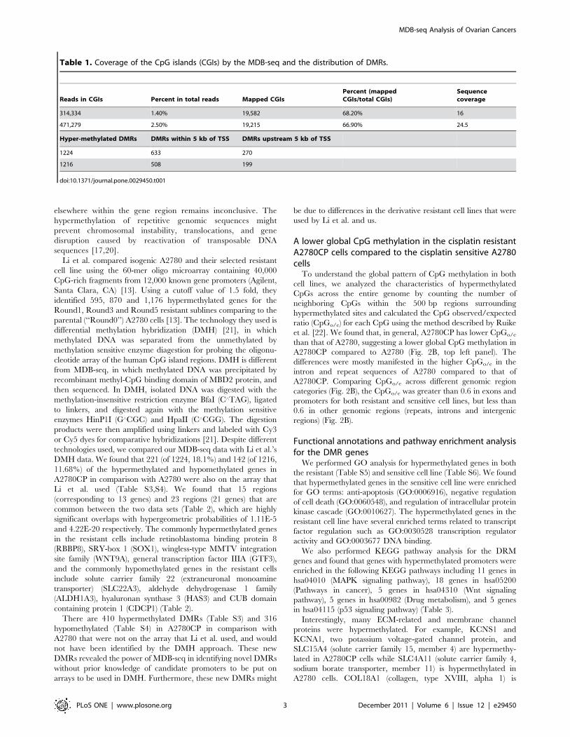
We annotated the reads with respect to the CpG islands in the
human genome (http://genome.ucsc.edu/) and found that about
1.4% and 2.5% of the reads locate inside CpG islands respectively
for A2780CP and A2780.The average sequence depth for the
CpG islands covered is about 16 and 24.5 times. About 68% and
66% of the human CpG islands were covered in our analysis for
A2780CP and A2780 respectively (Table 1). The CpG coverage
and depth we obtained in our MethylCap-seq is similar to what
previously published [18].
To detect differential methylation regions (DMRs) in the human
genome between the sensitive and resistant cells, we used
MEDIPS, a recently developed software tool specialized for
analyzing immunoprecipitation based methylation analysis (e.g.
MeDIP-seq and MethylCap-seq) [19]. We set the criteria for
significant DMRs: the length of peaks was set to 500 base pairs
and peaks with .20 rpm (reads per million), p-value less than
0.001 and ratio of rpm between two cell line .20. We obtained
1224 DMRs that is hyper-methylated in A2780CP compared to
A2780 (Table S1) and 1216 DMRs that is hyper-methylated in
A2780 compared to A2780CP (Table S2). These DMRs were
annotated with their genomic locations and associated genes
within 25 K bp to +5 K bp to the transcription start sites (Table
S3 and 4).
Nearly half of DMRs were located to within 5 k bp of
transcript start sites (TSS) of genes. 190 and 270 DMRs were
located to upstream of TSS respectively in A2780 and A2780CP
(Table 1). The rest mapped to other regions of the human
genome including introns, exons, intergenic regions and repeats
(Ensembl human genome annotations) (Fig. 2A). Hypermethy-
lated regions in promoters of genes are known to decrease gene
expression [17], but the effect of hypermethylation regions
Figure 1. Cell survival curve of A2780 and A2780CP by the MTT assay. Both A2780 and A2780CP were treated with cisplatin in differentdoses from 0 mg/ml to 16 mg/ml for 72 hours.doi:10.1371/journal.pone.0029450.g001
MDB-seq Analysis of Ovarian Cancers
PLoS ONE | www.plosone.org 2 December 2011 | Volume 6 | Issue 12 | e29450
elsewhere within the gene region remains inconclusive. The
hypermethylation of repetitive genomic sequences might
prevent chromosomal instability, translocations, and gene
disruption caused by reactivation of transposable DNA
sequences [17,20].
Li et al. compared isogenic A2780 and their selected resistant
cell line using the 60-mer oligo microarray containing 40,000
CpG-rich fragments from 12,000 known gene promoters (Agilent,
Santa Clara, CA) [13]. Using a cutoff value of 1.5 fold, they
identified 595, 870 and 1,176 hypermethylated genes for the
Round1, Round3 and Round5 resistant sublines comparing to the
parental (‘‘Round0’’) A2780 cells [13]. The technology they used is
differential methylation hybridization (DMH) [21], in which
methylated DNA was separated from the unmethylated by
methylation sensitive enzyme diagestion for probing the oligonu-
cleotide array of the human CpG island regions. DMH is different
from MDB-seq, in which methylated DNA was precipitated by
recombinant methyl-CpG binding domain of MBD2 protein, and
then sequenced. In DMH, isolated DNA was digested with the
methylation-insensitive restriction enzyme BfaI (C‘TAG), ligated
to linkers, and digested again with the methylation sensitive
enzymes HinP1I (G‘CGC) and HpaII (C‘CGG). The digestion
products were then amplified using linkers and labeled with Cy3
or Cy5 dyes for comparative hybridizations [21]. Despite different
technologies used, we compared our MDB-seq data with Li et al.’s
DMH data. We found that 221 (of 1224, 18.1%) and 142 (of 1216,
11.68%) of the hypermethylated and hypomethylated genes in
A2780CP in comparison with A2780 were also on the array that
Li et al. used (Table S3,S4). We found that 15 regions
(corresponding to 13 genes) and 23 regions (21 genes) that are
common between the two data sets (Table 2), which are highly
significant overlaps with hypergeometric probabilities of 1.11E-5
and 4.22E-20 respectively. The commonly hypermethylated genes
in the resistant cells include retinoblastoma binding protein 8
(RBBP8), SRY-box 1 (SOX1), wingless-type MMTV integration
site family (WNT9A), general transcription factor IIIA (GTF3),
and the commonly hypomethylated genes in the resistant cells
include solute carrier family 22 (extraneuronal monoamine
transporter) (SLC22A3), aldehyde dehydrogenase 1 family
(ALDH1A3), hyaluronan synthase 3 (HAS3) and CUB domain
containing protein 1 (CDCP1) (Table 2).
There are 410 hypermethylated DMRs (Table S3) and 316
hypomethylated (Table S4) in A2780CP in comparison with
A2780 that were not on the array that Li et al. used, and would
not have been identified by the DMH approach. These new
DMRs revealed the power of MDB-seq in identifying novel DMRs
without prior knowledge of candidate promoters to be put on
arrays to be used in DMH. Furthermore, these new DMRs might
be due to differences in the derivative resistant cell lines that were
used by Li et al. and us.
A lower global CpG methylation in the cisplatin resistantA2780CP cells compared to the cisplatin sensitive A2780cells
To understand the global pattern of CpG methylation in both
cell lines, we analyzed the characteristics of hypermethylated
CpGs across the entire genome by counting the number of
neighboring CpGs within the 500 bp regions surrounding
hypermethylated sites and calculated the CpG observed/expected
ratio (CpGo/e) for each CpG using the method described by Ruike
et al. [22]. We found that, in general, A2780CP has lower CpGo/e
than that of A2780, suggesting a lower global CpG methylation in
A2780CP compared to A2780 (Fig. 2B, top left panel). The
differences were mostly manifested in the higher CpGo/e in the
intron and repeat sequences of A2780 compared to that of
A2780CP. Comparing CpGo/e across different genomic region
categories (Fig. 2B), the CpGo/e was greater than 0.6 in exons and
promoters for both resistant and sensitive cell lines, but less than
0.6 in other genomic regions (repeats, introns and intergenic
regions) (Fig. 2B).
Functional annotations and pathway enrichment analysisfor the DMR genes
We performed GO analysis for hypermethylated genes in both
the resistant (Table S5) and sensitive cell line (Table S6). We found
that hypermethylated genes in the sensitive cell line were enriched
for GO terms: anti-apoptosis (GO:0006916), negative regulation
of cell death (GO:0060548), and regulation of intracellular protein
kinase cascade (GO:0010627). The hypermethylated genes in the
resistant cell line have several enriched terms related to transcript
factor regulation such as GO:0030528 transcription regulator
activity and GO:0003677 DNA binding.
We also performed KEGG pathway analysis for the DRM
genes and found that genes with hypermethylated promoters were
enriched in the following KEGG pathways including 11 genes in
hsa04010 (MAPK signaling pathway), 18 genes in hsa05200
(Pathways in cancer), 5 genes in hsa04310 (Wnt signaling
pathway), 5 genes in hsa00982 (Drug metabolism), and 5 genes
in hsa04115 (p53 signaling pathway) (Table 3).
Interestingly, many ECM-related and membrane channel
proteins were hypermethylated. For example, KCNS1 and
KCNA1, two potassium voltage-gated channel protein, and
SLC15A4 (solute carrier family 15, member 4) are hypermethy-
lated in A2780CP cells while SLC4A11 (solute carrier family 4,
sodium borate transporter, member 11) is hypermethylated in
A2780 cells. COL18A1 (collagen, type XVIII, alpha 1) is
Table 1. Coverage of the CpG islands (CGIs) by the MDB-seq and the distribution of DMRs.
Reads in CGIs Percent in total reads Mapped CGIsPercent (mappedCGIs/total CGIs)
Sequencecoverage
314,334 1.40% 19,582 68.20% 16
471,279 2.50% 19,215 66.90% 24.5
Hyper-methylated DMRs DMRs within 5 kb of TSS DMRs upstream 5 kb of TSS
1224 633 270
1216 508 199
doi:10.1371/journal.pone.0029450.t001
MDB-seq Analysis of Ovarian Cancers
PLoS ONE | www.plosone.org 3 December 2011 | Volume 6 | Issue 12 | e29450

hypermethylated in A2780 cells while KRTAP10-6 (keratin
associated protein 10–6) and LRFN5 (leucine rich repeat and
fibronectin type III domain containing 5) are hypermethylated in
A2780CP cells. We also identified hypermethylation at several
microRNA loci including hypermethylation of MI6A2, MIR129-
2, MIR124-1, MIR124-3, and MIR10A in A2780CP and
hypermethylation of MIR185, MIR548Q, MIR642A and
MIR661 in A2780 (Table 4).
Figure 2. Genomic feature and CpGo/e distribution of the hypermethylated peaks. A. Percentage of hypermethylated peaks based on theirlocalized genomic features. B. Distribution of CpGo/e for hypermethylated peaks based on their localized genomic features.doi:10.1371/journal.pone.0029450.g002
MDB-seq Analysis of Ovarian Cancers
PLoS ONE | www.plosone.org 4 December 2011 | Volume 6 | Issue 12 | e29450

Validation of genes with DMRs by MS-PCR and bisulfitesequencing
The expression of genes with hypermethylated promoter was
checked by RT-qPCR, if the expression was significant changed
then methylation specific PCR (MS-PCR) was used to validate the
methylation status of DMR regions between two cell lines. We
identified the following genes BCL2L1 (BCL2-like 1), PPKCE
(protein kinase C, epsilon), PTK6 (PTK6 protein tyrosine kinase
6), RAC2 (ras-related C3 botulinum toxin substrate 2), SECTM1
(secreted and transmembrane 1) and DDIT3 (DNA-damage-
inducible transcript 3) that changed in both promoter methylation
and expression. Figure 3A showed the expression changes of these
Table 2. Common hypermethylated or hypomethylated genes between the MBD-seq and the differential methylationhybridization.
Commonly hypermethylated in resistant cells
Names A2780CP* A2780* Ratios Data from Li et al. (GSM385747)** Descriptions
PROX1 32.86 0.00 Inf 0.98 prospero homeobox 1
PEX5L 24.94 0.00 Inf 0.80 peroxisomal biogenesis factor 5-like
INSM1 41.79 1.26 44.18 0.77 insulinoma-associated 1
JAM3 30.85 0.00 Inf 0.73 junctional adhesion molecule 3
SERF2 31.88 0.00 Inf 0.72 small EDRK-rich factor 2
COL4A1 49.59 2.68 25.51 0.71 collagen
RBBP8 23.06 0.00 Inf 0.70 retinoblastoma binding protein 8
RBBP8 92.64 0.00 Inf 0.70 retinoblastoma binding protein 8
C6orf97 24.91 0.00 Inf 0.69 chromosome 6 open reading frame 97
WNT9A 20.13 0.00 Inf 0.69 wingless-type MMTV integration site family
SEMA6A 33.86 0.50 43.01 0.68 sema domain
GTF3A 28.44 0.00 Inf 0.65 general transcription factor IIIA
SOX1 48.86 2.38 27.04 0.62 SRY (sex determining region Y)-box 1
SOX1 26.26 0.89 38.27 0.62 SRY (sex determining region Y)-box 1
ZNF329 25.72 0.00 Inf 0.62 zinc finger protein 329
Commonly hypermethylated in sensitive cells
PTMA 0.00 30.98 0.00 20.95 prothymosin
PTMA 1.29 22.14 0.04 20.95 prothymosin
SLC22A3 0.00 40.71 0.00 20.87 solute carrier family 22 (extraneuronal monoamine transporter)
COL18A1 0.00 40.43 0.00 20.84 collagen
COL18A1 0.00 28.14 0.00 20.84 collagen
MDFI 0.00 31.38 0.00 20.83 MyoD family inhibitor
NEFL 0.00 31.08 0.00 20.83 neurofilament
SECTM1 0.00 26.96 0.00 20.81 secreted and transmembrane 1
IL20RA 0.00 34.90 0.00 20.79 interleukin 20 receptor
ADRBK1 0.00 29.51 0.00 20.78 adrenergic
FBLN1 0.00 28.46 0.00 20.76 fibulin 1
HSPA12B 0.87 20.44 0.04 20.76 heat shock 70 kD protein 12B
P2RX5 0.79 23.20 0.05 20.73 purinergic receptor P2X
LGALS3 0.00 31.78 0.00 20.71 lectin
ALDH1A3 0.00 22.72 0.00 20.70 aldehyde dehydrogenase 1 family
SPINT1 0.00 32.23 0.00 20.69 serine peptidase inhibitor
HAS3 0.17 24.74 0.00 20.68 hyaluronan synthase 3
TRIP10 0.00 28.15 0.00 20.65 thyroid hormone receptor interactor 10
CDCP1 0.00 36.70 0.00 20.64 CUB domain containing protein 1
CDCP1 0.00 20.72 0.00 20.64 CUB domain containing protein 1
RHOU 0.00 20.43 0.00 20.61 ras homolog gene family
F3 0.63 22.46 0.03 20.60 coagulation factor III (thromboplastin)
CXCL14 0.64 24.06 0.02 20.59 chemokine (C-X-C motif) ligand 14
*rpm, reads per million.**log2 ratios.doi:10.1371/journal.pone.0029450.t002
MDB-seq Analysis of Ovarian Cancers
PLoS ONE | www.plosone.org 5 December 2011 | Volume 6 | Issue 12 | e29450

genes, and Figure 3B showed the promoter methylation patterns of
these genes. DDIT3 was hypomethylated in A2780 cells compared
to A2780CP, while the rest showed the opposite (Fig. 3B). To
further confirm DMRs between A2780 and A2780CP, we used
bisulfite sequencing to sequence the promoter regions of three
randomly picked gene from the 6 genes. Figure 3C showed that all
promoter regions of the picked gene PTK6, PRKCE and BCL2L1
were hypomethylated in A2780CP compared with A2780.
Restoration of gene expression of DMR affected genes bytreatmentwith 5-aza-dc demethylation reagent
In order to further confirm our data, we used 5-aza-29-
deoxycytidine (5-aza-dC) to see whether we can re-activate
methylation-silenced genes that we identified. 5-aza-29-deoxycyti-
dine (5-aza-dC) is an analogue of cytosine. When incorporated
into DNA, it irreversibly binds the methyltransferase enzymes,
resulting in passive de-methylationand reactivation of epigeneti-
cally silenced genes [23]. We used 5-aza-dC to treat both
A2780CP and A2780 cells in different doses from 0 mM to
10 mM. The expression of genes treated with the lowest (0 mM)
was compared to that treated with the highest (10 mM) dose of
demethylation reagent.
We showed that we were able to demethylate the promoters of
the hypermethylated PRKCE, BCL2L1, RAC2, PTK6, SECTM1
(Fig. 4A,B), and restored their expression after treatment with 5-
aza-dC in A2780 cells. Similarly, we were able to demethylate the
promoters of the hypermethylated gene DDIT3, and restored its
expression after treatment with 5-aza-dC in A2780CP cells
(Fig. 4A,B).
Discussion
We describe here for the first time an MDB-seq analysis of a
cisplatin response model of ovarian cancer cells. The epigenetic
changes between the isogenic pair of the cisplatin sensitive A2780
and its resistant derivative A2780CP cells suggest an epigenetic
control mechanism for cisplatin resistance. The global MDB-seq
data set generated for this pair of isogenic cells will be a useful
resource for the research community interested in cisplatin
resistance and epigenetics, and in ovarian cancer in general.
Global DNA methylation changes during carcinogenesis and is
often a predictor of therapy responses. For example, Anisowicz
et al. showed that even the normal part of the lung from a cancer
patient has already experienced a loss of global DNA methylation
Table 3. Genes mapped to the enriched pathways.
Pathways (number of genes mapped) Genes
hsa01100 Metabolic pathways (18) ACSS1 ALDH1A3 ALDH3A1 B3GNT3 BCAT1 CBS GAD1 GGT7 GLCE GLDC MAN2A2 ME1 PGLSPISD PTGES PTGS1 TPK1 UGT8
hsa05200 Pathways in cancer (18) BCL2L1 BIRC3 BMP4 CDH1 CDKN1B COL4A1 COL4A2 FGFR1 IGF1R LAMA1 LAMA5 LAMC2 MLH1RAC2 STAT3 WNT7B WNT9A ZBTB16
hsa04080 Neuroactive ligand-receptor interaction (14) ADRB2 BDKRB2 CHRNA7 CHRNB4 F2RL1 GABRB3 GLRA3 GRIN2B HRH1 NTSR1 OPRL1 P2RX5P2RY2 THRB
hsa04514 Cell adhesion molecules (CAMs) (12) CDH1 CLDN7 CNTNAP2 F11R ICAM1 ICOSLG JAM3 NLGN1 PTPRM PVRL2 SDC2 SIGLEC1
hsa04510 Focal adhesion (12) BIRC3 CAV1 CCND2 COL2A1 COL4A1 COL4A2 COL6A1 IGF1R LAMA1 LAMA5 LAMC2 RAC2
hsa04010 MAPK signaling pathway (11) CACNA1G CACNA2D3 CACNG4 DDIT3 DUSP7 FGFR1 GADD45G HSPA1A HSPA1B MAP4K4 RAC2
hsa05145 Toxoplasmosis (10) BCL2L1 BIRC3 HSPA1A HSPA1B IFNGR2 LAMA1 LAMA5 LAMC2 MYD88 STAT3
hsa04020 Calcium signaling pathway (10) ADRB2 BDKRB2 CACNA1G CHRNA7 GNA15 GNAL HRH1 NTSR1 P2RX5 6263 RYR3
hsa04512 ECM-receptor interaction (9) CD44 COL2A1 COL4A1 COL4A2 COL6A1 LAMA1 LAMA5 LAMC2 SDC2
hsa04144 Endocytosis (9) ADRB2 ADRBK1 CAV1 CHMP6 CXCR4 HSPA1A HSPA1B IGF1R SMAD7
hsa04060 Cytokine-cytokine receptor interaction (9) ACVR2A CD70 CXCL14 CXCL2 CXCR4 IFNGR2 IL20RA LTBR TNFRSF6B
hsa04974 Protein digestion and absorption (8) COL15A1 COL18A1 COL2A1 COL4A1 COL4A2 COL6A1 SLC7A8 SLC9A3
hsa05222 Small cell lung cancer (8) BCL2L1 BIRC3 CDKN1B COL4A1 COL4A2 LAMA1 LAMA5 LAMC2
hsa05146 Amoebiasis (8) COL2A1 COL4A1 COL4A2 GNA15 GNAL LAMA1 LAMA5 LAMC2
hsa04520 Adherens junction (7) CDH1 FGFR1 IGF1R PTPRM PVRL2 RAC2 SNAI2
hsa04670 Leukocyte transendothelial migration (7) CLDN7 CXCR4 F11R ICAM1 JAM3 NCF4 RAC2
hsa04062 Chemokine signaling pathway (7) ADRBK1 CXCL14 CXCL2 CXCR4 FOXO3 RAC2 STAT3
hsa05322 Systemic lupus erythematosus (7) GRIN2B H2AFJ HIST1H2BI HIST1H2BM HIST1H3A HIST1H4A HIST1H4I
hsa04630 Jak-STAT signaling pathway (6) BCL2L1 CCND2 IFNGR2 IL20RA SPRY2 STAT3
hsa04310 Wnt signaling pathway (5) CCND2 DKK2 RAC2 WNT7B WNT9A
hsa04350 TGF-beta signaling pathway (5) ACVR2A BMP4 ID4 NOG SMAD7
hsa04360 Axon guidance (5) CXCR4 EFNA1 EPHA7 RAC2 SEMA6A
hsa00982 Drug metabolism - cytochrome P450 (5) ALDH1A3 ALDH3A1 GSTA4 GSTT2 MGST2
hsa04650 Natural killer cell mediated cytotoxicity (5) ICAM1 IFNGR2 RAC2 RAET1L SYK
hsa05142 Chagas disease (American trypanosomiasis) (5) BDKRB2 GNA15 GNAL IFNGR2 MYD88
hsa00980 Metabolism of xenobiotics by cytochrome P450 (5) ALDH1A3 ALDH3A1 GSTA4 GSTT2 MGST2
hsa04115 p53 signaling pathway (5) CCND2 GADD45G PPM1D SERPINB5 SFN
doi:10.1371/journal.pone.0029450.t003
MDB-seq Analysis of Ovarian Cancers
PLoS ONE | www.plosone.org 6 December 2011 | Volume 6 | Issue 12 | e29450

compared to a normal individual [24]. Kim et al. showed that
global CpG methylation plays a role in epigenetic control of the
radiosensitivity in lung cancer cell lines [25]. In gliomas, LINE-1
methylation, a global DNA methylation marker, is proportional to
MGMT promoter methylation and higher LINE-1 methylation is
a favorable prognostic factor in primary GBMs, even compared to
MGMT promoter methylation [26]. Here we observed a lower
global CpG methylation in the cisplatin resistant A2780CP cells
compared to the cisplatin sensitive A2780 cells, suggesting that
global DNA methylation patterns might be able to predict cisplatin
resistance for ovarian cancers. Further experimentation is
necessary to evaluate this hypothesis.
We obtained 1,224 and 1,216 DMRs that is hyper-methylated
or hypo-methylated in A2780CP compared to A2780 cells
(Table S1, S2). We found that hypermethylated genes in the
sensitive cell line were enriched for GO terms for anti-apoptosis
(GO:0006916) and negative regulation of cell death (GO:00
60548), suggesting a possible general mechanism of epigenetic
silencing of anti-apoptosis genes, resulting in sensitivity to
cisplatin. We also found that the DMRs are enriched in several
signaling pathways including the hsa04010 (MAPK signaling
pathway), hsa04310 (Wnt signaling pathway), and the hsa04115
(p53 signaling pathway) pathways (Table 3). The Wnt signaling
pathway has been implicated in ovarian cancer progression and
chemoresistance [27]. Activation of JNK/p38 MAPK pathways
in response to cisplatin could lead to Fas ligand induction and
cell death in ovarian carcinoma cells [4]. P53 and its signaling
pathway were also been implicated in cisplatin resistance in
ovarian cancer cells [28,29]. Our data suggest that epigenetic
regulation of these signaling pathways is one of the mechanisms
for the involvement of these pathways in cisplatin resistance in
ovarian cancer cells.
Table 4. Interesting hypermethylated genes and MiRNA loci in A2780 or A2780CP cells.
chr. start end A2780CP* A2780* locations Names Descriptions
miRNAs
chr12 54383001 54383500 23 0 upstream MIR196A2
chr12 54383501 54384000 23 0 upstream MIR196A2
chr12 54384501 54385000 26 0 upstream MIR196A2
chr12 54387501 54388000 48 1 downstream MIR196A2
chr15 96880501 96881000 48 1 downstream MIR1469
chr22 22011001 22011500 29 0 downstream MIR130B
chr11 43598501 43599000 33 2 upstream MIR129-2
chr11 43602501 43603000 30 2 overlapStart MIR129-2
chr20 61807001 61807500 33 1 upstream MIR124-3
chr20 61807501 61808000 37 0 upstream MIR124-3
chr20 61808001 61808500 47 0 upstream MIR124-3
chr8 9763501 9764000 22 0 upstream MIR124-1
chr8 9762501 9763000 21 1 upstream MIR124-1
chr17 46656501 46657000 56 0 downstream MIR10A
chr22 20019501 20020000 1 29 upstream MIR185
chr10 12763001 12763500 0 20 downstream MIR548Q
chr19 46180501 46181000 0 27 downstream MIR642A
chr19 46181001 46181500 1 25 downstream MIR642A
chr8 145020001 145020500 0 23 upstream MIR661
ECM related genes
chr8 122654001 122654500 57 3 upstream HAS2 hyaluronan synthase 2
chr21 46016001 46016500 26 0 upstream KRTAP10-6 keratin associated protein 10-6
chr14 42074501 42075000 34 0 upstream LRFN5 leucine rich repeat and fibronectin type III domain containing 5
chr14 42075001 42075500 33 1 upstream LRFN5 leucine rich repeat and fibronectin type III domain containing 5
chr14 42075501 42076000 42 2 upstream LRFN5 leucine rich repeat and fibronectin type III domain containing 5
chr21 46824001 46824500 0 40 upstream COL18A1 collagen, type XVIII, alpha 1
chr21 46823501 46824000 0 28 upstream COL18A1 collagen, type XVIII, alpha 1
membrane channel proteins
chr20 43733001 43733500 29 1 upstream KCNS1 potassium voltage-gated channel, delayed-rectifier, subfamily S, member 1
chr12 5018501 5019000 27 2 upstream KCNA1 potassium voltage-gated channel, shaker-related subfamily, member 1(episodic ataxia with myokymia)
chr12 129309001 129309500 26 1 upstream SLC15A4 solute carrier family 15, member 4
chr20 3220501 3221000 0 50 upstream SLC4A11 solute carrier family 4, sodium borate transporter, member 11
*rpm, reads per million.doi:10.1371/journal.pone.0029450.t004
MDB-seq Analysis of Ovarian Cancers
PLoS ONE | www.plosone.org 7 December 2011 | Volume 6 | Issue 12 | e29450

We also identified hypermethylation at several microRNA loci
including hypermethylation of MI6A2, MIR129-2, MIR124-1,
MIR124-3, and MIR10A in A2780CP and hypermethylation of
MIR185, MIR548Q, MIR642A and MIR661 in A2780 (Table 4).
MicroRNAs have been implicated in cisplatin resistance in ovarian
cancer [30]. For example, Yang et al. showed that many miRNAs
are deregulated in human ovarian cancer including miR-214,
miR-199a*, miR-200a, miR-100, miR-125b, and let-7 cluster, and
that miR-214 induces cell survival and cisplatin resistance by
targeting PTEN [30]. Epigenetically silenced microRNAs have
been implicated in cancers [31,32]. However, methylation of
miRNA locus has not been reported previously for ovarian
cancers; neither are reports on the relationship between miRNA
methylation and cisplatin resistance. Our data might point to a
new direction for the study of methylation of microRNA loci and
cisplatin resistance.
We found that BCL2L1 (BCL2-like 1), PPKCE (protein kinase C,
epsilon), PTK6 (PTK6 protein tyrosine kinase 6), RAC2 (ras-related
C3 botulinum toxin substrate 2), SECTM1 (secreted and
transmembrane 1) are hypermethylated in the cisplatin sensitive
A2780 cells and DDIT3 (DNA-damage-inducible transcript 3) is
hypermethylated in the cisplatin resistant A2780CP cells. Their
expression also were also changed accordingly to the hypothesis that
hypermethylation would silence gene expression. We confirmed the
methylation pattern of these genes by MS-PCR, and also were able
to demethylate the promoters of these genes and restore their
expression after treatment with 5-aza-dC, an agent that de-
methylates and reactivates of epigenetically silenced genes [23].
We found BCL2L1 is hypermethylated in the sensitive A2780
cells. BCL2L1 (bcl-xl) encodes a protein belonging to the BCL-2
protein family, whose protein members act as anti- or pro-
apoptotic regulators [33]. Over-expression of the Bcl-xL protein is
known to confer resistance to a broad range of potentially
apoptotic stimuli in carcinogenesis processes including oncogene
activation, hypoxia and matrix detachment [34–37]. Our result
reveals that the hypomethylation of BCL2L1 in the resistant cells
(i.e. hypermethylation of BCL2L1 in the sensitive cells) would
result in increased BCL2L1 expression, thus conferring resistance
to cisplatin. Our data is consistent with the observation by
Williams et al. that expression of Bcl-xL in ovarian carcinoma is
associated with chemoresistance and recurrent disease [38].
Taking together, this suggest that modulating the expression of
BCL2L1 (Bcl-xL) by epigenetic means might be a way to
overcome cisplatin resistance in ovarian cancer cells.
We also identified PTK6 (protein tyrosine kinase 6) as
hypermethylated in the cisplatin sensitive A2780 cells compared
to A2780CP cells. PTK6 directly phosphorylates AKT and
promotes AKT activation in response to epidermal growth factor
[39]. The relationship of PTK6 with cisplatin has not been
previously studied. Further functional analysis of PTK6’s role in
modulating cisplatin resistance is warranted. PPKCE (protein
kinase C, epsilon) is another gene that is hypermethylated in the
cisplatin sensitive A2780 cells compared to A2780CP cells.
PPKCE is a member of the protein kinase C (PKC) family, whose
members phosphorylate a wide variety of protein targets and are
involved in diverse cellular signaling pathways [40]. Interestingly,
cisplatin was able to induce phosphorylation and translocation of
PPKCE from the plasma membrane to the nuclear membrane
and to the cytosolic fraction [41]. The hypermethylation of
PPKCE in the sensitive cells would reduce its expression, resulting
in less translocation to the nuclear membrane or cytosolic fraction
upon addition of cisplatin. However, whether reduced expression
of PPKCE confers sensitivity to cisplatin in ovarian cancer cells
remains to be studied.
Figure 3. Validation of genes with DMRs in their promoters byMS-PCR and bisulfite sequencing. A. Real-time PCR showing thedifferential expression of selected genes between the cisplatin sensitiveA2780 and the cisplatin resistant A2780CP cells. B. MS-PCR datashowing differential methylation states in promoter regions of theselected genes between A2780 and A2780CP cells. C. Bisulfitesequencing result for promoter regions of selected gene PTK6, PRKCEand BCL2L1 (white cycle: unmethylated CpG, Black cycle: methylatedCpG).doi:10.1371/journal.pone.0029450.g003
MDB-seq Analysis of Ovarian Cancers
PLoS ONE | www.plosone.org 8 December 2011 | Volume 6 | Issue 12 | e29450

Finally, we identified DDIT3 (DNA-damage-inducible transcript
3) as hypermethylated in the cisplatin resistant A2780CP cells
compared to A2780 cells. Also named GADD153 (Growth arrest and
DNA damage-inducible protein 153), DDIT3 encodes a member of
the CCAAT/enhancer-binding protein (C/EBP) family of transcrip-
tion factors, and is activated by endoplasmic reticulum stress, and
promotes apoptosis. DDIT3 (GADD153) expression could be
induced by cisplatin [42]. We observed that it is hypermethylation
in the resistant cells, which would result in reduced expression of
DDIT3. It is possible that cisplatin acts through DDIT3 to promote
growth arrest and apoptosis, and reduced expression of DDIT3 in the
cells would make them more resistant to cisplatin. However, the
detailed mechanism remained to be investigated.
In summary, we generated a global dataset for an ovarian
cancer cisplatin resistance model and identified several genes that
were subjected to epigenetic control to modulate cisplatin
resistance. These genes might serves as targets to overcome
chemoresistance to cisplatin in ovarian cancers.
Methods
Cell linesA2780 and A2780CP was cultured in RPMI 1640 medium
(Invitrogen) containing 10% Fetal Bovine Serum (10099-141) at
37uC and 5% CO2. Cells were kindly provided by Dr. Stephen
Collins from UC San Diego (CA, USA).
Figure 4. Restoration of gene expression of DMR affected genes by treatment with 5-aza-dc demethylation reagent and validation byMS-PCR. A. Validation of the changes of methylation status by MS-PCR for the six genes under treatment with 0 mM and 10 mM 5-aza-dC. B. Gene expressionchanges (Y-axis, relative fold changes) of the selected six genes after treatment with different concentration (X-axis) of demethylation reagent 5-aza-dC.doi:10.1371/journal.pone.0029450.g004
MDB-seq Analysis of Ovarian Cancers
PLoS ONE | www.plosone.org 9 December 2011 | Volume 6 | Issue 12 | e29450

Cytotoxicity analysisBoth resistant and sensitive cells were seeded into 96 well plates
at a concentration of 4000 cells/well in five replicates with
complete culture medium. Then cells were treated with cisplatin in
different dose from 0 mg/ml to 16 mg/ml. After 72 hours
incubation, both viable and dead cells were counted by using
MTT assay, and only the viable cells were included in data
analysis.
Bisulfite-modified DNA sequencing and Methylation-Specific PCR
We prepared genomic DNA from cultured cells using the
DNeasy Blood & tissue Kit (Qiagen). Approximately 200 ng of
DNA was bisulfite-treated with the EZ DNA Methylation-Gold kit
(Zymo Research) according to the manufacture’s protocol.
ZymoTaq Premix was used for Methylation-Specific PCR (MSP)
and according to these primers (Table S7). Methylation-specific
PCR was run in a total volume of 25 mL by using ZymoTaq
Premix (ZYMO research). MSP reactions were subjected to initial
incubation at 95uC for 10 minutes, followed by 40 cycles of 95uCfor 30 seconds, and annealing at appropriate temperature for
35 seconds and 72uC for 40 seconds. Final extension was done by
incubation at 72uC for 7 minutes. MSP products were separated
on 2% agarose gels and visualized by ethidium bromide staining.
The primers for bisulfite-modified sequencing (Table S7) and
MSP were designed by web tools MethPrimer (http://www.
urogene.org/methprimer/index1.html) [43]. The conditions for
bisulfite-modified sequencing reaction are same as MSP. The
products were purified using MinElute PCR purification Kit,
cloned into the pMD 18-T vector (Takara) and sequenced.
RNA isolation and Real time RT-PCRRNA was extracted using the protocol for Trizol reagent
(Invitrogen). 2 mg RNA was firstly reverse-transcribed in 25 mL
with an Archive Kit (Applied Biosystems) and 2 mL were amplified
by Real Time PCR (Bio-RAD CFX96 Rea-Time System). cDNA
samples were amplified with SYBRH Pre-mix Ex TaqTM. The
thermal cycling profile consisted of initial denaturation at 95uC for
30 seconds and 40 cycles at 95uC for 5 seconds, 60uC for
15 second, and 72uC for 30 seconds. Each sample was processed
in triplicate.
5-Aza-29-deoxycytidine treatmentHuman ovarian carcinoma cells A2780 and A2780CP were
grown for 5 days in the presence of various concentrations of 5-
Aza-dC (0, 0.625, 1.25, 2.5, 5, and 10 mM). Fresh drug was added
every 24 h.
Methylated DNA Binding Protein sequencing(MethylCap-seq)
3 mg of genomic DNA isolated as described above was
fragmented by sonication with Bioruptor (Diagenode, Belgium)
and end-repaired, A-tailed, and ligated to 2.5 mMol of ‘‘paired-
end’’ adapters (IDT Inc.) following the manufacturer’s recom-
mend protocol (Illumina Inc.). 1.2 mg of the DNA was fractionated
on a home-made GST-MBD resin with a buffer of a stepwise
increased salt concentration [16]. The high salt fraction was
directly amplified by 12-cycle PCR [18]. The 350–450 bp fraction
of the PCR products was gel purified and quantified using an
Agilent DNA 1000.
The 250–400 bp DNA fractions were excised and purified as
described above. The products were assessed and quantified using
an Agilent DNA 1000 series II assay and Qubitfluorometer
(Invitrogen) respectively. Each library was diluted to 8 nM for
sequencing on an Illumina Genome Analyzer II following the
manufacture’s recommended protocol. Obtained images were
analyzed and base called using Illumina provided GA pipeline
software OLB 1.6.0 with default setting. The raw reads from
MethylCap-seq were submitted to GEO database which accession
number isGSE31418.
The 250–400 bp DNA fraction of the amplified fraction was
gel- purified. Each library was diluted to 8 nM for sequencing by
Illumina Genome Analyzer II following the manufacture’s
recommended protocol. Obtained images were analyzed and
base-called using Illumina provided GA pipeline software OLB
1.6.0 with default setting. The raw reads from MethylCap-seq
were submitted to GEO database (accession number GSE31418).
Mapping of Sequence ReadsHuman genome sequence and mapping information (Feb. 2009,
GRCh37/hg19) was downloaded from ENSEMBL FTP site
(http://asia.ensembl.org/info/data/ftp/index.html). Filtered reads
were firstly cut into 36 bp to get higher quality reads then mapped
to HG19 reference allowing up 2 mismatches by software SOAP2
[44]. The matched results from SOAP2 were converted into bed
format by our own script.
Identification and Annotation of DMRsTo identify differential methylation regions (DMRs), the bed
format files were used as input for the MEDIPS program [19].
The criteria for significant DMRs were: the length of peaks was set
to 500 base pairs and peaks with .20 rpm (reads per million), p-
value less than 0.001 and ratio of rpm between two cell line .20.
The length of each DMR was set to 500 bp. Genomic feature data
was retrieved from ENSEMBL database (build 55), promoter was
defined as the 5 kb upstream of transcription start sites. Each
DMR was annotated according to its genomic location by our own
script. The DMRs that locate within the proximal promoter
(25 kb to +5 kb from their transcription start site) were annotated
with GO, KEGG pathway and Reactome Pathway by package
ChIPpeakAnno [45] in R and web tool IDConverter (http://
idconverter.bioinfo.cnio.es/) [46].
CpG Obs/Exp ratio was calculated by our own script for each
DMR and plotted by the R language. GO Miner [47] was used to
analyze GO enrichment and a P value ,0.01 was considered
significant. Pathway enrichment was analyzed through KEGG
database (http://www.genome.jp/kegg/tool/map_pathway1.html)
[48]. To compare to Li et al.’s data, we downloaded the data ftp:
//ftp.ncbi.nih.gov/pub/geo/DATA/SeriesMatrix/GSE15373/.
When there are multiple probes corresponding to the same
genes, the probe that showed the most differentially expressed
were used for comparison. For calculating hypergeometric
probabilities, the population size is set at 12,000 as there are
12, 000 genes in the array that Li et al. used.
Supporting Information
Table S1 Hypermethylated DMRs in the cisplatinresistant A2780CP cell line.
(XLS)
Table S2 Hypermethylated DMRs in the cisplatinsensitive A2780 cell line.
(XLS)
Table S3 Hypermethylated DMRs within 5 kb of tran-scription start sites in the A2780CP cell line.
(XLS)
MDB-seq Analysis of Ovarian Cancers
PLoS ONE | www.plosone.org 10 December 2011 | Volume 6 | Issue 12 | e29450

Table S4 Hypermethylated DMRs within 5 kb of tran-scription start sites in the A2780 cell line.(XLS)
Table S5 Gene Ontology enrichment result for geneswith hypermethylated promoters in the A2780CP cellline.(XLS)
Table S6 Gene Ontology enrichment result for geneswith hypermethylated promoters in the A2780 cell line.(XLS)
Table S7 Primers for MSP and bisulfite-modifiedsequencing.
(XLS)
Author Contributions
Conceived and designed the experiments: JZ LC BL. Performed the
experiments: WY CJ XL XH LL YH HZ KM. Analyzed the data: WY.
Contributed reagents/materials/analysis tools: JZ. Wrote the paper: WY
BL.
References
1. Einhorn EH (1997) Testicular cancer: an oncological success story. Clin Cancer
Res 3: 2630–2632.
2. Ozols RF (1991) Ovarian cancer: new clinical approaches. Cancer Treat Rev 18
Suppl A: 77–83.
3. Siddik ZH (2003) Cisplatin: mode of cytotoxic action and molecular basis of
resistance. Oncogene 22: 7265–7279.
4. Mansouri A, Ridgway LD, Korapati AL, Zhang Q, Tian L, et al. (2003)
Sustained activation of JNK/p38 MAPK pathways in response to cisplatin leads
to Fas ligand induction and cell death in ovarian carcinoma cells. J Biol Chem
278: 19245–19256.
5. Robertson KD (2005) DNA methylation and human disease. Nat Rev Genet 6:
597–610.
6. Esteller M, Garcia-Foncillas J, Andion E, Goodman SN, Hidalgo OF, et al.
(2000) Inactivation of the DNA-repair gene MGMT and the clinical response of
gliomas to alkylating agents. N Engl J Med 343: 1350–1354.
7. Taniguchi T, Tischkowitz M, Ameziane N, Hodgson SV, Mathew CG, et al.
(2003) Disruption of the Fanconi anemia-BRCA pathway in cisplatin-sensitive
ovarian tumors. Nat Med 9: 568–574.
8. Staub J, Chien J, Pan Y, Qian X, Narita K, et al. (2007) Epigenetic silencing of HSulf-
1 in ovarian cancer:implications in chemoresistance. Oncogene 26: 4969–4978.
9. Bram EE, Stark M, Raz S, Assaraf YG (2009) Chemotherapeutic drug-induced
ABCG2 promoter demethylation as a novel mechanism of acquired multidrug
resistance. Neoplasia 11: 1359–1370.
10. Hu S, Yu L, Li Z, Shen Y, Wang J, et al. (2010) Overexpression of EZH2
contributes to acquired cisplatin resistance in ovarian cancer cells in vitro and in
vivo. Cancer Biol Ther 10: 788–795.
11. Boettcher M, Kischkel F, Hoheisel JD (2010) High-definition DNA methylation
profiles from breast and ovarian carcinoma cell lines with differing doxorubicin
resistance. PLoS One 5: e11002.
12. Chang X, Monitto CL, Demokan S, Kim MS, Chang SS, et al. (2010)
Identification of hypermethylated genes associated with cisplatin resistance in
human cancers. Cancer Res 70: 2870–2879.
13. Li M, Balch C, Montgomery JS, Jeong M, Chung JH, et al. (2009) Integrated
analysis of DNA methylation and gene expression reveals specific signaling
pathways associated with platinum resistance in ovarian cancer. BMC Med
Genomics 2: 34.
14. Down TA, Rakyan VK, Turner DJ, Flicek P, Li H, et al. (2008) A Bayesian
deconvolution strategy for immunoprecipitation-based DNA methylome anal-
ysis. Nat Biotechnol 26: 779–785.
15. Serre D, Lee BH, Ting AH (2010) MBD-isolated Genome Sequencing provides
a high-throughput and comprehensive survey of DNA methylation in the human
genome. Nucleic Acids Res 38: 391–399.
16. Brinkman AB, Simmer F, Ma K, Kaan A, Zhu J, et al. (2010) Whole-genome
DNA methylation profiling using MethylCap-seq. Methods 53: 232–236.
17. Kido Y, Khokhar AR, Siddik ZH (1993) Differential cytotoxicity, uptake and
DNA binding of tetraplatin and analogous isomers in sensitive and resistant
cancer cell lines. Anticancer Drugs 4: 251–258.
18. Harris RA, Wang T, Coarfa C, Nagarajan RP, Hong C, et al. (2010) Comparison
of sequencing-based methods to profile DNA methylation and identification of
monoallelic epigenetic modifications. Nat Biotechnol 28: 1097–1105.
19. Chavez L, Jozefczuk J, Grimm C, Dietrich J, Timmermann B, et al. (2010)
Computational analysis of genome-wide DNA methylation during the
differentiation of human embryonic stem cells along the endodermal lineage.
Genome Res 20: 1441–1450.
20. Bestor TH (2005) Transposons reanimated in mice. Cell 122: 322–325.
21. Yan PS, Wei SH, Huang TH (2002) Differential methylation hybridization using
CpG island arrays. Methods Mol Biol 200: 87–100.
22. Ruike Y, Imanaka Y, Sato F, Shimizu K, Tsujimoto G (2010) Genome-wide
analysis of aberrant methylation in human breast cancer cells using methyl-DNA
immunoprecipitation combined with high-throughput sequencing. BMC
Genomics 11: 137.
23. Michalowsky LA, Jones PA (1987) Differential nuclear protein binding to 5-
azacytosine-containing DNA as a potential mechanism for 5-aza-29-deoxycyti-
dine resistance. Mol Cell Biol 7: 3076–3083.
24. Anisowicz A, Huang H, Braunschweiger KI, Liu Z, Giese H, et al. (2008) A
high-throughput and sensitive method to measure global DNA methylation:
application in lung cancer. BMC Cancer 8: 222.
25. Kim EH, Park AK, Dong SM, Ahn JH, Park WY (2010) Global analysis of CpG
methylation reveals epigenetic control of the radiosensitivity in lung cancer cell
lines. Oncogene 29: 4725–4731.
26. Ohka F, Natsume A, Motomura K, Kishida Y, Kondo Y, et al. (2011) The
global DNA methylation surrogate LINE-1 methylation is correlated with
MGMT promoter methylation and is a better prognostic factor for glioma. PLoS
One 6: e23332.
27. Su HY, Lai HC, Lin YW, Liu CY, Chen CK, et al. (2009) Epigenetic silencing
of SFRP5 is related to malignant phenotype and chemoresistance of ovarian
cancer through Wnt signaling pathway. Int J Cancer 127: 555–567.
28. Marx D, Meden H, Ziemek T, Lenthe T, Kuhn W, et al. (1998) Expression of
the p53 tumour suppressor gene as a prognostic marker in platinum-treated
patients with ovarian cancer. Eur J Cancer 34: 845–850.
29. Green JA, Berns EM, Coens C, van Luijk I, Thompson-Hehir J, et al. (2006)
Alterations in the p53 pathway and prognosis in advanced ovarian cancer: a
multi-factorial analysis of the EORTC Gynaecological Cancer group (study
55865). Eur J Cancer 42: 2539–2548.
30. Yang H, Kong W, He L, Zhao JJ, O’Donnell JD, et al. (2008) MicroRNA
expression profiling in human ovarian cancer: miR-214 induces cell survival and
cisplatin resistance by targeting PTEN. Cancer Res 68: 425–433.
31. Lujambio A, Ropero S, Ballestar E, Fraga MF, Cerrato C, et al. (2007) Genetic
unmasking of an epigenetically silenced microRNA in human cancer cells.
Cancer Res 67: 1424–1429.
32. Lujambio A, Calin GA, Villanueva A, Ropero S, Sanchez-Cespedes M, et al.
(2008) A microRNA DNA methylation signature for human cancer metastasis.
Proc Natl Acad Sci U S A 105: 13556–13561.
33. Jaaskelainen M, Nieminen A, Pokkyla RM, Kauppinen M, Liakka A, et al.
(2010) Regulation of cell death in human fetal and adult ovaries–role of Bok and
Bcl-X(L). Mol Cell Endocrinol 330: 17–24.
34. Dong Z, Wang J (2004) Hypoxia selection of death-resistant cells. A role for Bcl-
X(L). J Biol Chem 279: 9215–9221.
35. Vento MT, Zazzu V, Loffreda A, Cross JR, Downward J, et al. (2010) Praf2 is a
novel Bcl-xL/Bcl-2 interacting protein with the ability to modulate survival of
cancer cells. PLoS One 5: e15636.
36. Amundson SA, Myers TG, Scudiero D, Kitada S, Reed JC, et al. (2000) An
informatics approach identifying markers of chemosensitivity in human cancer
cell lines. Cancer Res 60: 6101–6110.
37. Gebauer G, Mirakhur B, Nguyen Q, Shore SK, Simpkins H, et al. (2000)
Cisplatin-resistance involves the defective processing of MEKK1 in human
ovarian adenocarcinoma 2008/C13 cells. Int J Oncol 16: 321–325.
38. Williams J, Lucas PC, Griffith KA, Choi M, Fogoros S, et al. (2005) Expression
of Bcl-xL in ovarian carcinoma is associated with chemoresistance and recurrent
disease. Gynecol Oncol 96: 287–295.
39. Zheng Y, Peng M, Wang Z, Asara JM, Tyner AL (2010) Protein tyrosine kinase
6 directly phosphorylates AKT and promotes AKT activation in response to
epidermal growth factor. Mol Cell Biol 30: 4280–4292.
40. Griner EM, Kazanietz MG (2007) Protein kinase C and other diacylglycerol
effectors in cancer. Nat Rev Cancer 7: 281–294.
41. Ohmori T, Arteaga CL (1998) Protein kinase C epsilon translocation and
phosphorylation by cis-diamminedichloroplatinum(II) (CDDP): potential role in
CDDP-mediated cytotoxicity. Cell Growth Differ 9: 345–353.
42. Gately DP, Jones JA, Christen R, Barton RM, Los G, et al. (1994) Induction of
the growth arrest and DNA damage-inducible gene GADD153 by cisplatin in
vitro and in vivo. Br J Cancer 70: 1102–1106.
43. Li LC, Dahiya R (2002) MethPrimer: designing primers for methylation PCRs.
Bioinformatics 18: 1427–1431.
44. Li R, Yu C, Li Y, Lam TW, Yiu SM, et al. (2009) SOAP2: an improved ultrafast
tool for short read alignment. Bioinformatics 25: 1966–1967.
45. Zhu LJ, Gazin C, Lawson ND, Pages H, Lin SM, et al. (2010) ChIPpeakAnno: a
Bioconductor package to annotate ChIP-seq and ChIP-chip data. BMC
Bioinformatics 11: 237.
MDB-seq Analysis of Ovarian Cancers
PLoS ONE | www.plosone.org 11 December 2011 | Volume 6 | Issue 12 | e29450

46. Alibes A, Yankilevich P, Canada A, Diaz-Uriarte R (2007) IDconverter and
IDClight: conversion and annotation of gene and protein IDs. BMC
Bioinformatics 8: 9.
47. Zeeberg BR, Feng W, Wang G, Wang MD, Fojo AT, et al. (2003) GoMiner: a
resource for biological interpretation of genomic and proteomic data. GenomeBiol 4: R28.
48. Ogata H, Goto S, Sato K, Fujibuchi W, Bono H, et al. (1999) KEGG: Kyoto
Encyclopedia of Genes and Genomes. Nucleic Acids Res 27: 29–34.
MDB-seq Analysis of Ovarian Cancers
PLoS ONE | www.plosone.org 12 December 2011 | Volume 6 | Issue 12 | e29450