Encapsulation in alginate-coated gelatin microspheres improves survival of the probiotic...
10
Encapsulation in alginate-coated gelatin microspheres improves survival of the probiotic Bifidobacterium adolescentis 15703T during exposure to simulated gastro-intestinal conditions N.T. Annan, A.D. Borza, L. Truelstrup Hansen * Food Science Program, Department of Process Engineering and Applied Science, Dalhousie University, 1360 Barrington Street, Halifax, Nova Scotia, Canada B3K 4Y3 Received 30 July 2007; accepted 4 November 2007 Abstract Alginate-coated gelatin microspheres were produced to encapsulate the probiotic Bifidobacterium adolescentis 15703T with the objec- tive of enhancing survival during exposure to the adverse conditions of the gastro-intestinal tract. Gelatin microspheres were cross-linked with the non-cytotoxic genipin and coated with alginate cross-linked by Ca 2+ from external or internal sources. The alginate coat pre- vented pepsin-induced degradation of the gelatin microspheres in simulated gastric juice (pH 2.0, 2 h), resulting in significantly (P < 0.05) higher numbers of survivors due to the buffering effect of intact microspheres. After sequential incubation in simulated gastric (1 h) and intestinal juices (pH 7.4, 4 h), number of surviving cells were 7.6 and 7.4 log cfu ml 1 for alginate coated microspheres by the internal and external Ca 2+ -source methods, respectively, while 6.7 and 6.4 log cfu ml 1 were obtained for cells in uncoated gelatin microspheres and free cells, respectively. This study presents a novel microencapsulation method, which protects probiotic bifidobacteria during exposure to adverse environmental conditions. Ó 2007 Elsevier Ltd. All rights reserved. Keywords: Gelatin; Alginate; Microencapsulation; Probiotics; Bifidobacterium adolescentis; Simulated gastro-intestinal juices; Functional foods 1. Introduction Probiotic bacteria are defined as ‘live microorganisms which, when administered in adequate amounts, confer a health benefit on the host’ (Araya et al., 2002). Due to the fastidious nature of many probiotic bacteria, survival in sufficiently high numbers during passage through the human gastro-intestinal tract (GIT) remains a major chal- lenge for effective delivery of these beneficial bacteria. Also, probiotic survival during the processing and storage of functional food products is of concern for the development of products with a guaranteed content of bioactive cells (Anal & Singh, 2007). Colonization of the intestine by an exogenous probiotic bacterium is influenced by many fac- tors including the size of the inoculum, physiological state of the bacteria, buffering capacity of the delivery food and the capacity of the microorganisms to resist acid and bile encountered in the upper segments of the GIT (Conway, Gorbach, & Goldin, 1987). Bifidobacteria, one group of commonly selected probio- tics, are indigenous to the human intestine, where they preferentially colonise the colon (Fuller, 1991). The bacte- ria must therefore survive exposure to the acid in the human stomach and bile in the intestine in order to be effective (Shah, 2000). Bifidobacterium strains, however, vary greatly in their sensitivity to the harsh acidic environ- ment of the stomach and many foods (Charteris, Kelly, Morelli, & Collins, 1998; Clark & Martin, 1994; Truelstrup Hansen, Allan-Wojtas, Jin, & Paulson, 2002). Lankapu- thra and Shah (1997) found that there were more sensitive than resistant probiotic strains and this has led to several studies on microencapsulation using food grade 0963-9969/$ - see front matter Ó 2007 Elsevier Ltd. All rights reserved. doi:10.1016/j.foodres.2007.11.001 * Corresponding author. Tel.: +1 902 494 3145; fax: +1 902 420 0219. E-mail address: [email protected] (L.T. Hansen). www.elsevier.com/locate/foodres Available online at www.sciencedirect.com Food Research International 41 (2008) 184–193
-
Upload
independent -
Category
Documents
-
view
1 -
download
0
Transcript of Encapsulation in alginate-coated gelatin microspheres improves survival of the probiotic...
Available online at www.sciencedirect.com
www.elsevier.com/locate/foodres
Food Research International 41 (2008) 184–193
Encapsulation in alginate-coated gelatin microspheres improvessurvival of the probiotic Bifidobacterium adolescentis 15703T
during exposure to simulated gastro-intestinal conditions
N.T. Annan, A.D. Borza, L. Truelstrup Hansen *
Food Science Program, Department of Process Engineering and Applied Science, Dalhousie University, 1360 Barrington Street, Halifax,
Nova Scotia, Canada B3K 4Y3
Received 30 July 2007; accepted 4 November 2007
Abstract
Alginate-coated gelatin microspheres were produced to encapsulate the probiotic Bifidobacterium adolescentis 15703T with the objec-tive of enhancing survival during exposure to the adverse conditions of the gastro-intestinal tract. Gelatin microspheres were cross-linkedwith the non-cytotoxic genipin and coated with alginate cross-linked by Ca2+ from external or internal sources. The alginate coat pre-vented pepsin-induced degradation of the gelatin microspheres in simulated gastric juice (pH 2.0, 2 h), resulting in significantly (P < 0.05)higher numbers of survivors due to the buffering effect of intact microspheres. After sequential incubation in simulated gastric (1 h) andintestinal juices (pH 7.4, 4 h), number of surviving cells were 7.6 and 7.4 log cfu ml�1 for alginate coated microspheres by the internal andexternal Ca2+-source methods, respectively, while 6.7 and 6.4 log cfu ml�1 were obtained for cells in uncoated gelatin microspheres andfree cells, respectively. This study presents a novel microencapsulation method, which protects probiotic bifidobacteria during exposureto adverse environmental conditions.� 2007 Elsevier Ltd. All rights reserved.
Keywords: Gelatin; Alginate; Microencapsulation; Probiotics; Bifidobacterium adolescentis; Simulated gastro-intestinal juices; Functional foods
1. Introduction
Probiotic bacteria are defined as ‘live microorganismswhich, when administered in adequate amounts, confer ahealth benefit on the host’ (Araya et al., 2002). Due tothe fastidious nature of many probiotic bacteria, survivalin sufficiently high numbers during passage through thehuman gastro-intestinal tract (GIT) remains a major chal-lenge for effective delivery of these beneficial bacteria. Also,probiotic survival during the processing and storage offunctional food products is of concern for the developmentof products with a guaranteed content of bioactive cells(Anal & Singh, 2007). Colonization of the intestine by anexogenous probiotic bacterium is influenced by many fac-
0963-9969/$ - see front matter � 2007 Elsevier Ltd. All rights reserved.doi:10.1016/j.foodres.2007.11.001
* Corresponding author. Tel.: +1 902 494 3145; fax: +1 902 420 0219.E-mail address: [email protected] (L.T. Hansen).
tors including the size of the inoculum, physiological stateof the bacteria, buffering capacity of the delivery food andthe capacity of the microorganisms to resist acid and bileencountered in the upper segments of the GIT (Conway,Gorbach, & Goldin, 1987).
Bifidobacteria, one group of commonly selected probio-tics, are indigenous to the human intestine, where theypreferentially colonise the colon (Fuller, 1991). The bacte-ria must therefore survive exposure to the acid in thehuman stomach and bile in the intestine in order to beeffective (Shah, 2000). Bifidobacterium strains, however,vary greatly in their sensitivity to the harsh acidic environ-ment of the stomach and many foods (Charteris, Kelly,Morelli, & Collins, 1998; Clark & Martin, 1994; TruelstrupHansen, Allan-Wojtas, Jin, & Paulson, 2002). Lankapu-thra and Shah (1997) found that there were more sensitivethan resistant probiotic strains and this has led toseveral studies on microencapsulation using food grade

N.T. Annan et al. / Food Research International 41 (2008) 184–193 185
biopolymers such as alginate and whey proteins to protectthe acid sensitive bifidobacteria with varying successes(e.g., Guerin, Vuillemard, & Subirade, 2003; Lee, Cha, &Park, 2004; Rao, Shiwnarain, & Maharaj, 1989; Ravula& Shah, 1998; Reid, Champagne, Gardner, Fustier, &Vuillemard, 2007; Sheu & Marshall, 1993; Truelstrup Han-sen et al., 2002). For example, entrapment in alginatemicrospheres with diameters of 40–80 lm resulted in insig-nificant protection of bifidobacteria during exposure tosimulated gastric juice at pH 2.0 (Truelstrup Hansenet al., 2002) while larger alginate (1–3 mm) microspheresprotected entrapped cells (Lee & Heo, 2000). However,the latter microsphere sizes are too big for incorporationinto food products without affecting the texture (Cham-pagne & Fustier, 2007) and diameters below 100 lm arepreferred for most applications. Other factors that affectthe success of probiotic encapsulation formulations in gas-tric environments include the method of microencapsula-tion, presence of single or multiple coatings, biopolymerconcentration and increase in initial cell loads (Chandra-mouli, Kailasapathy, Peiris, & Jones, 2004; Krasaekoopt,Bhandari, & Deeth, 2003).
In an earlier study, we optimized the processing param-eters for entrapment of bifidobacteria in a novel gelatinmicrosphere matrix cross-linked with the non-cytotoxicgenipin but found that micropheres cross-linked with10 mmol genipin disintegrated after 10 min when exposedto simulated gastric juice with 3 g l�1 pepsin (Annan, Bor-za, Moreau, Allan-Wojtas, & Truelstrup Hansen, 2007). Assurvival of the probiotic cell load is related to the structuralintegrity of the microspheres, this result clearly indicatedthe need to coat the pepsin-sensitive gelatin matrix with abiocompatible non-toxic and pepsin-resistant polymer,which is insoluble at acidic pH but dissolves in the alkalinepH of the intestine. Several pH-sensitive polymers, includ-ing alginates, have been investigated for use in drug deliv-ery systems (Narayani & Rao, 1995a, 1995b), however,only few studies report on the use of these polymers for tar-geted delivery of encapsulated bacteria.
Alginates are natural anionic polysaccharides made upof D-mannuronic and L-guluronic acid residues joined line-arly by (1–4)-glycosidic linkages. The variable proportionaland sequential arrangement of the D-mannuronic and L-guluronic units results in distribution of negative chargesalong the polymer backbone in aqueous solution (Thuet al., 1996), potentially allowing for polyion complexationwith positively charged gelatin polymers. As alginate gelsare stable in low pH solutions but swell in weakly basicsolutions, alginate coating of gelatin capsules and micro-spheres can be used to protect drugs from the acidity ofgastric juice while allowing subsequent release in the basicenvironment of intestinal fluids (Narayani & Rao, 1995a;Rao & Rao, 1997). Moreover, when pH is lowered belowthe pKa values of D-mannuronic and L-guluronic acid(3.6 and 3.7, respectively), alginate is converted to alginicacid with release of calcium ions and the formation of amore dense gel due to water loss, however, the entrapped
pay loads, cells and/or drugs, were found to still remainin the intact alginate only beads (Doumeche et al., 2004).
Genipin is a new cross-linker derived from Gardenia
jasminidides or Genipa americana. The fruits from theseplants are eaten raw and extracts used for medicinal pur-poses and as food colourants (Butler, Ng, & Pudney,2003). Genipin has been approved for use as an additivein pharmaceuticals and foods in countries such as Japan,Korea and Taiwan (Nickerson et al., 2006) but has yet tobe approved in Canada and USA.
The aim of the study was to develop a method for pro-ducing alginate-coated genipin cross-linked gelatin micro-spheres using food grade polymers and to investigate theeffect of this alginate-coating on the survival of probioticbifidobacteria during exposure to the adverse environmentfound in the upper segments of the gastro-intestinal tract.We report here for the first time that encapsulation in algi-nate-coated gelatin microspheres significantly (P < 0.05)improved the survival of Bifidobacterium adolescentis
15703T in simulated gastric and intestinal juices in compar-ison to the survival obtained for free cells or cells entrappedin uncoated gelatin microspheres.
2. Materials and methods
2.1. Materials
Gelatin (type A, from porcine skin, 300 Bloom), alginicacid sodium salt (medium viscosity), Tween 80, Span 85,glycerol, L-cysteine hydrochloride monohydrate, pancrea-tin (P-1750) and pepsin (P-7000), 5-aminofluorescein(HPLC grade), diphosphorpentoxide and EDC(1-[3-dimethylaminopropyl]-3-ethyl-carbodiimide hydrochlo-ride) were all supplied by Sigma Aldrich Co. (Oakville,ON, Canada). Genipin was obtained from Challenge Bio-products Co. Ltd. (CBC, Taiwan, R.O.C.), while thedeMan, Rogosa and Sharpe (MRS) broth, MRS agar, bilesalts (Oxgall) and bacteriological peptone were supplied byOxoid Inc. (Nepean, ON, Canada). Gas Pak Plus system(gas generator envelopes and anaerobic jars from BBL),1,4-dioxane and sodium chloride were supplied by FisherScientific (Nepean, ON, Canada) and Bifidobacterium
adolescentis 15703T was supplied by American Type Cul-ture Collection (Manassas, VA, USA). Canola oil wasobtained from LOBLAWS Inc. (Montreal, QC, Canada).
2.2. Preparation of cells for microencapsulation
Frozen stock cultures (2 ml, MRS with 20% v/v glycerol)of B. adolescentis 15703T were inoculated in 500 ml MRSbroth containing filter sterilized L-cysteine (0.5 g l�1) andincubated at 37 �C for 24 h to obtain a cell density of about109–1010 colony forming units per ml (cfu ml�1). Harvestingof cells was done by centrifugation at 8000 rpm (3578g) for10 min at 4 �C and after discarding the supernatant of spentculture broth, the cell pellet was resuspended in peptone sal-ine (PS, 1 g l�1 peptone, 8.5 g l�1 NaCl) and centrifuged

186 N.T. Annan et al. / Food Research International 41 (2008) 184–193
again under the same conditions. Washed cells were thensuspended in a total of 10 ml PS and stored at 4 �C untilusage. Fresh cell suspensions were prepared for each exper-iment and enumerated by pour plating in MRS agar con-taining filter sterilized L-cysteine (0.5 g l�1, MRS-cysagar). Plates were incubated anaerobically at 37 �C for threedays using the GasPak Plus system.
2.3. Preparation of microspheres with encapsulated
B. adolescentis 15703T
Uncoated genipin cross-linked gelatin microsphereswere prepared by dissolving gelatin powder in NaCl solu-tion (0.5% w/v) at 50 �C to obtain a final gelatin concentra-tion of 13% (w/v) in the microspheres. One millilitre ofwashed cell suspension was added to aqueous gelatin toobtain a cell to polymer solution ratio of 1:10 and thecell-gelatin aqueous mixture was then emulsified into tem-pered oil (45 �C, aqueous to oil phase volume ratio of 1:5)containing 0.5% Span 85 (w/w). The emulsion was pro-duced by stirring for 30 min using a Caframo Real TorqueDigital Stirrer (Caframo Ltd., Wiarton, ON, Canada) at aconstant speed (900 rpm). Genipin solution giving a finalconcentration of 1.25 mmol l�1 in the gelatin microsphereswas subsequently added and the emulsion was left stirringovernight at room temperature (22 �C) to allow for thecompletion of the genipin-gelatin cross-linking reaction.A washing mixture (8.5 g l�1 NaCl, 5 g l�1 Tween 80 in dis-tilled water) was added at a ratio of 1:1 to the aqueous gel-atin phase and the microspheres were harvested with twosubsequent washings in a separatory funnel to removethe oil phase, filtered (Whatman No. 4, filter paper, FisherScientific) and stored in PS at 4 �C until ready for use.
Procedures for alginate-coating of the gelatin micro-spheres were developed using two techniques, namely the(a) external and (b) internal Ca2+-source gelation methods.
(a) For the external Ca2+-source method, gelatin micro-spheres with encapsulated B. adolescentis were prepared asdescribed above. Ten gram of the filtered microsphereswere added to 40 g of 1% (w/v) medium viscosity alginatesolution and stirred at an initial speed of 900 rpm to dis-perse the beads. The microspheres were left to stir in algi-nate for 20 min at 600 rpm before being filtered, collectedand resuspended in 60 g of oil containing 5 g l�1 Tween80 and 62.5 mmol l�1 CaCl2 for 20 min to initiate the exter-nal Ca2+ cross-linking of the peripheral alginate layer. Tobreak the emulsion, 40 ml of 0.05 mol l�1 CaCl2 solutionprepared in PS was added followed by transfer to a separ-atory funnel. The Ca2+ cross-linked alginate coated micro-spheres were collected and stored in PS with 0.05 mol l�1
CaCl2 until further analysis.(b) An internal Ca2+-source method was developed
based on a modification of Rosenberg and Lee’s (2004)method for alginate coated whey protein based micro-spheres. Here the formation of the Ca2+-alginate coatingdepends on the diffusion of Ca2+ ions dissolved in the aque-ous gelatin to the surface of the gelatin matrix to cross-link
the peripheral alginate layer. Briefly, genipin cross-linkedgelatin microspheres were made as described above, exceptthat a final concentration of 0.1 mol l�1 CaCl2 was addedto the aqueous gelatin phase. The resulting Ca2+-contain-ing gelatin microspheres were harvested, filtered andwashed twice with deionized water. Ten grams of themicrospheres were weighed into 40 g of 1% (w/v) mediumviscosity alginate solution and dispersed at an initial speedof 900 rpm followed by stirring for 20 min at 600 rpm. Theby filtration recovered microspheres were again washedthree times with deionized water and subsequently dis-persed (900 rpm) in 60 ml PS with 0.05 mol l�1 CaCl2 andleft to stir gently (600 rpm) for 20 min. The microsphereswere filtered, rinsed and stored in PS containing0.05 mol l�1 CaCl2 until further analysis.
2.4. Enumeration of microencapsulated bacteria
Entrapped bacteria in uncoated gelatin microsphereswere released by homogenizing 1.0 g of a filtered micro-sphere slurry in 9.0 ml of tempered PS (45 �C) for 30 susing a Polytron homogeniser (Brinkman Instruments,Rexdale, ON, Canada). For alginate-coated gelatin micro-spheres, tempered (45 �C) phosphate buffer (PB,0.05 mol l�1 NaH2PO4, pH 8.0) was used because phos-phate ions chelate calcium thereby weakening the alginatecoating for effective release of cells. The homogenized sam-ples were diluted to appropriate concentrations and pourplated in MRS-cys agar. The plates were incubated anaer-obically for three days at 37 �C and the encapsulated bac-teria enumerated as cfu ml�1. The encapsulation yield(EY), which is a combined measurement of the efficacy ofentrapment and survival of viable cells during the microen-capsulation procedure, was calculated as
EY ¼ NN 0
� 100
where N is the number of viable entrapped cells releasedfrom the microspheres, and N0 is the number of free cellsadded to the biopolymer mix during the production ofthe microspheres.
2.5. Particle size measurement
The particle sizes of microspheres were determined bymeasuring diameters of 300 microspheres at �100 magnifi-cation using an optical microscope (Optihot, Nikon Can-ada Inc., Mississauga, ON, Canada) fitted with acalibrated micrometer scale. The mean diameters of micro-spheres were calculated and presented with standard devi-ations (n � 1).
2.6. Microscopy of microspheres to determine the structure
of the alginate coat
To determine the structure and thickness of alginatecoating on gelatin microspheres by fluorescence and
N.T. Annan et al. / Food Research International 41 (2008) 184–193 187
confocal laser scanning microscopy (CLSM), sodiumalginate was labelled with fluorescein according to theprocedure described by Blonk, van Eendenburg, Koning,Weisenborn, and Winkel (1995) while gelatin was notlabelled. Briefly, 5 g of sodium alginate (medium viscos-ity) were dissolved in 400 ml of distilled water and dropsof concentrated sulphuric acid (96%) added to precipitatethe alginic acid. The mixture was centrifuged at 8000 rpm(3578g) for 10 min and the alginic acid pellets collectedand washed twice with water and acetone. Washed pel-lets were dried overnight in vacuo in the presence ofdiphosphorpentoxide followed by mixing of the driedalginic acid with 80 ml distilled water, 30 ml 1,4-dioxane,50 mg 5-aminofluorescein and 1 g EDC (1-[3-dimethyl-aminopropyl]-3-ethyl-carbodiimide hydrochloride). Themixture was stirred for three hours and left overnightto slowly turn orange before filtration through Whatmanno. 1 filter paper followed by three washes with acetoneto remove the unreacted alginic acid. The washed pelletswere dried in vacuo and the resulting labelled alginatepowder applied at a concentration of 0.3% (w/v) to thesodium alginate coating-solution with a final concentra-tion of 1% (w/v). Gelatin microspheres were then coatedwith the fluorescein-labelled alginate as describedabove.
Gelatin microspheres coated with fluorescein-labelledalginate were examined by an Axioplan 2 (Carl ZeissCanada Ltd., Toronto, ON, Canada) fluorescence micro-scope (excitation: 450–490 nm, emission: 515–565 nm)and a Zeiss LSM 510 confocal laser scanning microscopewith a Plan-Apochromat (100�/1.4 oil DIC) objective.Confocal microscopy images were acquired using anargon laser emitting at 488 and 543 nm, a bypass (BP) fil-ter detecting at 560–615 nm, a long pass (LP) filter detect-ing at 505 nm and were analyzed using the LSM 510software (Carl Zeiss, Jena, Germany). For all microscopywork, the gelatin microspheres were prepared withoutgenipin in order to eliminate background fluorescenceemitted by genipin.
2.7. Survival assay of free and microencapsulated
B. adolescentis in simulated gastric and intestinal juice
Simulated gastric juice (SGJ) was prepared by dissolv-ing pepsin in saline (0.2% NaCl, w/v) to a final concentra-tion of 0.3 g l�1 and adjusting the pH to 2.0 withconcentrated HCl. The mixture was sterile filteredthrough a membrane (0.45 lm, Nalgene� Brand Products,Nalge Co., Rochester, NY, USA). The authors note, how-ever, that the simulation of gastric juices for in vitro stud-ies tends to overestimate viability losses that would occurin vivo, as it is known that the presence of food compo-nents may temporarily elevate the gastric pH (Mainville,Arcand, & Farnworth, 2005). Also, the intake of foodswith a content of carbohydrates may provide Bifidobacte-
rium strains with energy to overcome the acid stress by
usage of the F0F1 ATPase pump (Corcoran, Stanton,Fitzgerald, & Ross, 2005; Sanz, 2007).
Simulated intestinal juice (SIJ) was prepared based onthe method of Huang and Adams (2004). Pancreatin andbile salts were suspended in PB to final concentrations of1 g l�1 and 4.5 g l�1, respectively. The mixture was adjustedto a pH of 7.4 with 0.1 mol l�1 NaOH and sterile filtered.Both gastric and intestinal juices were prepared fresh foruse on the same day.
Washed cell suspensions of B. adolescentis (0.5 ml) or0.5 g of microspheres with entrapped bacteria were addedto 4.5 ml of tempered (37 �C) SGJ, mixed well by vortexingfor 10 s and incubated for 5, 30, 60 and 120 min at 37 �C withperiodical shaking and monitoring of pH in the SGJ.Entrapped cells were released from microspheres by homog-enizing using a Polytron homogeniser and surviving bacteriaafter each set time interval enumerated by pour plate countsin MRS-cys agar at 37 �C for 72 h as described above.
2.8. Survival and stability assay of free and
microencapsulated B. adolescentis after sequential
incubation in simulated gastric and intestinal juices
Washed cell suspensions of B. adolescentis (0.5 ml) or0.5 g of microspheres with entrapped bacteria were addedto 4.5 ml of tempered (37 �C) SGJ, mixed well by vortexingfor 10 s and incubated for 60 min at 37 �C. Simulated intes-tinal juice tempered at 37 �C was then added and pHadjusted to 7.4 with 0.1 mol l�1 NaOH. The final volumeof the mixture was made up to 10 ml with phosphate bufferand incubated for 60, 120 and 240 min at 37 �C with peri-odical shaking. Surviving bacteria after set times of sequen-tial incubation were enumerated by pour plate counts inMRS-cys agar at 37 �C for 72 h as described above.
2.9. Stability of alginate-coated and un-coated gelatin
microspheres during exposure to simulated gastric and
intestinal juices
The stability of microspheres with entrapped bacteriaduring sequential incubation was assessed by observingthe disintegration under an optical microscope (magnifica-tion �100) every 5 min. The time for complete disintegra-tion of microspheres was recorded.
Fluorescence and CLSM microscopy was also per-formed on alginate coated microspheres, prepared withfluorescein-labelled alginate as described above, whichhad been exposed to SGJ for 1 h and to SIJ for 1 h follow-ing exposure to SGJ for 1 h.
2.10. Statistical analysis
Four independent experiments with duplicate sampleswere conducted for each treatment. Results were statisti-cally analyzed where appropriate by Analysis of Variance(ANOVA) using the Systat software package (Systat Inc.,Evanston, IL, USA).

188 N.T. Annan et al. / Food Research International 41 (2008) 184–193
3. Results
3.1. Size and encapsulation yields of uncoated and
alginate-coated gelatin microspheres
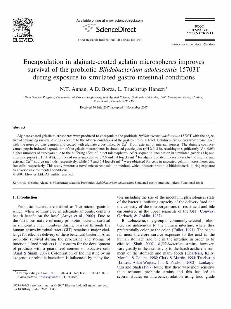
The mean diameters of uncoated and alginate-coatedgelatin microspheres containing B. adolescentis were notsignificantly (P > 0.05) different as the average diametersranged from 49.0 lm to 53.1 lm (Table 1). Alginate coat-ing of the microspheres resulted in a uniform thin exteriorlayer as shown in the confocal laser scanning micrographsin Fig. 1b. The thinness (1–2 lm) of the alginate coatexplained the minimal change in size between coated anduncoated microspheres.
Encapsulation yields (EY) for viable cells of B. adole-
scentis were similar (41–43%) for uncoated and alginate-coated gelatin microspheres produced by the internalCa2+-source method, however, EY were significantly(P < 0.05) lower (30%) for alginate-coated gelatin micro-spheres made by the external Ca2+-source method (Table1).
Table 1Size and encapsulation yields of B. adolescentis in uncoated and alginatecoated gelatin microspheres
Microsphere type Microsphere size (lm) Encapsulation yield (%)
Uncoated 49.0 ± 12.7xa 41.1 ± 3.3x
Internal Ca2+ 52.8 ± 10.8x 43.5 ± 4.4x
External Ca2+ 53.1 ± 10.3x 29.9 ± 6.2y
a Means (n = 900 ± standard deviation (n � 1)) with different letter in acolumn are significantly different (P < 0.05).
Fig. 1. Confocal laser scanning microscopy (CLSM) photographs showing thgelatin microspheres containing B. adolescentis (bold arrows). Scale bars repre
3.2. Fluorescence and confocal laser scanning microscopy
(CLSM) of uncoated and alginate-coated gelatin
microspheres
Optical CLSM images taken through the equator of themicrospheres showed bacteria immobilized in the gelatinmatrix of the microspheres with the fluorescein-labelledalginate bound as a very thin uniform membrane (1–2 lm) to the periphery of the gelatin matrix (Fig. 1b).Uncoated gelatin microspheres similarly containedentrapped bifidobacteria throughout the matrix butshowed no evidence of an outer membrane (Fig. 1a). Thealginate membrane could be distinguished from the gelatincore as the fluorescein-labelled alginate emitted an intensegreen fluorescence at the surface of the microsphere whilethe gelatin core in both the coated and uncoated micro-spheres had a dull green colour. The alginate coated micro-sphere shown in Fig. 1b was coated using the externalCa2+-source gelation method, but CLSM on microspherescoated using the internal Ca2+-source gelation methodlooked identical (results not shown).
3.3. Survival of free and encapsulated B. adolescentis insimulated gastric juice
Encapsulation in alginate-coated gelatin microspheressignificantly (P < 0.05) improved survival of bifidobacteria(Fig. 2). Cell survival after exposure to SGJ for 5 min was54%, 20%, 2% and 1% of the initial populations found inalginate-coated microspheres produced by external andinternal Ca2+-sources, uncoated gelatin microspheres andfree cells, respectively. After the initial losses, the popula-tions of bifidobacteria declined at the same rate for all treat-ments over the 2 h incubation period (Fig. 2) with final
e structure of (a) uncoated and (b) external Ca2+-source alginate-coatedsent 5 lm.

Time (minutes)0 20 40 60 80 100 120
Surv
ival
(%)
0.01
0.1
1
10
100
Fig. 2. Survival of free and gelatin encapsulated B. adolescentis duringexposure to simulated gastric juice (pH 2.0) at 37 �C. Symbols: (d) freecells, (s) uncoated, (.) alginate-coated with internal Ca2+ and (O)alginate-coated with external Ca2+. Survival (%) represents the percentageof cells surviving relative to the initial population. Means (n = 8) ± stan-dard deviation (n � 1).
N.T. Annan et al. / Food Research International 41 (2008) 184–193 189
decreases of 3.45 log (cfu ml�1) for free cells,2.55 log (cfu ml�1) for uncoated microspheres and1.21 log (cfu ml�1) and 1.75 log (cfu ml�1) for coated micro-spheres with alginate cross-linked by the external and inter-nal Ca2+-source methods, respectively. The average pH ofSGJ containing free cells after 120 min was 2.1 ± 0.1 whilepH of SGJ containing encapsulated beads was 2.2 ± 0.2.
3.4. Viability of free and encapsulated cells of B. adolescentis
during sequential incubation in simulated gastric and
intestinal juices
The sequential transfer of free cells and encapsulatedbifidobacteria after 60 min incubation in SGJ resulted inan initial reduction of viable cells during the first hour ofexposure to SIJ, however, cell numbers subsequently stabi-lised and no further reductions were observed after 120 and240 min (Table 2). Overall, the sequential exposure to SGJ(60 min) followed by SIJ (240 min) resulted in significantly(P < 0.05) higher numbers of B. adolescentis surviving inthe alginate-coated gelatin microspheres (7.35–7.57 log cfu ml�1) than were obtained for free cells and cells
Table 2Microsphere disintegration time (min) and number of surviving cells (log cfu m(37 �C) in simulated gastric (SGJ) and intestinal (SIJ) juices
Treatment Disintegration time SGJ
0 min 60
Free cells – 10.41 ± 0.17xa 7.1Uncoated �15 ± 5b 9.67 ± 0.35y 7.1Internal Ca2+ 120 ± 15 9.60 ± 0.54y 8.0External Ca2+ 240 ± 30 9.76 ± 0.09y 8.9
a Means (n = 8 ± standard deviation (n � 1)) with different letter in a columb Disintegrated after 45 ± 5 min in SGJ.
entrapped in uncoated gelatin microspheres (6.41–6.71 log cfu ml�1).
3.5. Stability of alginate-coated and un-coated gelatinmicrospheres during exposure to simulated gastric and
intestinal juices
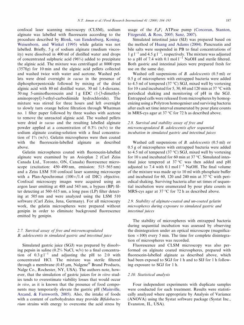
Disintegration times for alginate-coated gelatin micro-spheres in SIJ were longer for microspheres produced bythe external Ca2+-source method (240 min) than for thoseproduced by the internal Ca2+-source method (120 min),indicating a difference in the alginate surface layer obtainedby the two methods (Table 2). Uncoated gelatin micro-spheres were already disintegrated after 45 min in SGJbefore reaching the SIJ.
Fig. 3a shows a fluorescence microscope image of exter-nally coated alginate–gelatin beads after 1 h in SGJ and itcan be seen that this coating was effective in making thealginate–gelatin microspheres resistant to degradation bypepsin in the acidic gastric juice thereby maintaining theprobiotic bacteria entrapped in the gelatin matrix. Fig. 3bshows a CLSM image of the beads after sequential incuba-tion in SIJ for 1 h where the alginate coating, which main-tained the integrity of the microspheres in SGJ, isbeginning to erode under the neutral conditions found inthe small intestine with a subsequent disintegration of theunderlying gelatin matrix and release of the probiotic bac-teria (Fig. 3b).
4. Discussion
4.1. Size, encapsulation yields and structure of uncoated and
alginate-coated microspheres
Using the same concentration of gelatin, Annan et al.(2007) found the size of uncoated gelatin microspherescross-linked with 2.5 mmol l�1 genipin to be 41.2 ±13.2 lm compared to 49.0 ± 12.7 lm obtained in the pres-ent study (Table 1). A lower concentration of genipin(1.25 mmol l�1) was used in the present study which mayhave increased swelling of microspheres due to the uptakeof water. Cross-linking of gelatin microspheres controlswater uptake and swelling (Ugwoke & Kinget, 1998), hence
l�1) for free and encapsulated B. adolescentis during sequential incubation
SIJ
min 60 min 120 min 240 min
2 ± 0.09x 5.48 ± 0.15x 6.40 ± 0.31x 6.41 ± 0.20x
3 ± 0.27x 6.59 ± 0.04y 6.52 ± 0.04x 6.71 ± 0.08y
0 ± 0.17y 7.11 ± 0.45z 6.82 ± 0.34xy 7.57 ± 0.28z
2 ± 0.78y 7.38 ± 0.15z 7.31 ± 0.40y 7.35 ± 0.41z
n are significantly different (P < 0.05).

Fig. 3. Stability of alginate-coated gelatin microspheres during exposure to simulated gastro-intestinal conditions. Fluorescence microscopy photographsof (a) whole alginate coated microspheres in SGJ (1 h, 37 �C) and (b) CLSM image of alginate coated microspheres disintegrating in SIJ (1 h, 37 �C)following initial incubation in SGJ (1 h, 37 �C). Scale bars represent 20 lm.
190 N.T. Annan et al. / Food Research International 41 (2008) 184–193
a higher cross-linker concentration will minimize changesin shape and size of microspheres.
Encapsulation yields for uncoated and alginate-coated(internal Ca2+-source) microspheres were similar to yieldsobtained by Annan et al. (2007) for uncoated gelatinmicrospheres. However, EY for the alginate-coated micro-spheres prepared by the external Ca2+-source method wassignificantly lower (Table 1). These microspheres werephysically stronger as indicated by their resistance to deg-radation in simulated gastro-intestinal juices (Table 2 andFig. 3) and required longer homogenization times for com-plete disintegration. It is possible that entrapped cells werenot released or were injured during homogenization thusleading to the decrease in the apparent EY obtained forthese microspheres.
Use of fluorescein labelled alginate and CLSM enabledus to locate and estimate the thickness of the alginatelayer on the gelatin microspheres, similar to observationsby Strand, Mørch, Espevik, and Skjak-Br�k (2003) whoused the method to determine the location of alginate inalginate-L-lysine-alginate microcapsules. Leung et al.(2005) produced alginate coats on collagen beads usingan adaptation of the aqueous emulsion method developedby Calafiore and Basta (1999). Also using CLSM, theyobserved the fluorescein-labelled alginate coat (1% w/v,medium viscosity) as a uniform coherent membrane witha thickness of about 10–20 lm, which was considerablythicker than that obtained in the present study (1–2 lm)using a similar concentration of alginate. In their coatingmethod, Leung et al. (2005) added collagen beads to anemulsion of alginate, polyethylene glycol and Ficoll, anuncharged and highly branched polymer of sucrose, toachieve a uniform distribution of emulsion droplets before
cross-linking the alginate with calcium chloride. This con-ceptually different method produced a thicker coatingthan was obtained with our method, where the electro-static interaction between the oppositely charged biopoly-mers determined the thickness of the alginate layer. Thelatter method was developed in order to obtain pepsinand acid resistant alginate–gelatin microspheres of a smal-ler size and with maximum holding capacity for the cellload (i.e., the probiotic bacteria). The ability of the algi-nate coating to provide a barrier against pepsin and aciddegradation was evidenced in the fluorescence imageshown in Fig. 3a. The CLSM image in Fig. 3b showedentrapped cells being released from the microsphere dueto the erosion of the alginate coating under the neutralpH conditions of the SIJ with the subsequent degradationof the underlying gelatin matrix by pancreatic enzymes.This testifies to the success of this targeted deliverysystem.
4.2. Comparison of survival of free and encapsulated cells in
simulated gastric juice
Smith (1995) reported that food remains in the stomachfor between 2 and 4 h while liquids empty from the stom-ach in only about 20 min. With the objective of improvingsurvival of probiotic bifidobacteria during the exposure tothe low pH of the stomach, we tested the hypothesis thatthe buffering capacity of gelatin would lead to increasedsurvival of the encapsulated cells in comparison to freecells. Encapsulation of bifidobacteria in uncoated gelatinmicrospheres only marginally improved the survival overthat of free cells (Fig. 2), however, the gelatin onlymicrospheres were structurally unstable in SGJ due to

N.T. Annan et al. / Food Research International 41 (2008) 184–193 191
degradation by pepsin and completely disintegrated after45 min (Table 2). The 1–2 lm thick alginate coating pre-vented pepsin from degrading the gelatin microspheresand the entrapment in intact gelatin microspheres signifi-cantly (P < 0.05) improved survival of the entrapped bifi-dobacteria throughout the two hour trial period (Fig. 2),indicating the protective buffering effect of intact gelatinmicrospheres. Guerin et al. (2003) also reported that thebuffering effect of whey proteins contributed to higher sur-vival rates for bifidobacteria encapsulated in a mixed gel ofalginate, pectin and whey proteins in comparison to freecells when exposed to SGJ at pH 2.5. Narayani and Rao(1995a) found that gelatin capsules loaded with a cancerdrug and coated with 20% alginate remained intact forup to 8 h in SGJ while uncoated microspheres disintegratedafter 15 min, their microspheres were, however, cross-linked with the cytotoxic gluteraldehyde.
The decrease in the viable population by 3.45 log unitsfor free B. adolescentis cells was similar to findings byCharteris et al. (1998) and Truelstrup Hansen et al.(2002) who observed reductions of about 3 log cfu ml�1
for B. adolescentis exposed to SGJ (pH 2.0) for 2 to 3 h.The strength and structure of alginate hydrogels have
been shown to be influenced by increasing gelation time/rate and/or Ca2+ concentration due to the formation of agreater number of cross-links (Bodmeier & Wang, 1993;Liu & Krishnan, 1999). In the present study, both the rateof diffusion and the Ca2+ concentration are likely to havediffered between the two coating methods, resulting in theexternal Ca2+-source alginate-coated microspheres appear-ing stronger (more difficult to homogenize and longer timeto disintegration in SGJ/SIJ (Table 2)) and more protectiveto entrapped cells (Fig. 2).
4.3. Survival of free and encapsulated cells of B. adolescentis
during sequential incubation in simulated gastro-intestinal
conditions
While the ionotropic alginate gel formed by Ca2+ cross-linking of carboxylate groups is insoluble at low pH, expo-sure to neutral pH or higher solubilises the alginate (Le-Tien, Millette, Mateescu, & Lacroix, 2004; Tønnesen &Karlsen, 2002). In our study, this pH-dependent behaviourof the biopolymer was used to control the degradation ofalginate-coated gelatin microspheres and release of themicroencapsulated cell load under the neutral conditionsfound in the small intestine (Table 2 and Fig. 3), wherethe transit time for food ranges between 1 and 4 h (Smith,1995). The continued reduction of cell numbers in SIJ espe-cially during the first h of incubation in this environmentmay be attributed to the sensitivity of B. adolescentis to bileand for the entrapped cells, also to physical integrity of themicrosphere matrix. The sensitivity of many strains of Bifi-
dobacterium spp. to bile concentrations encountered in thehuman gastro-intestinal tract has been reported by severalauthors (Amor et al., 2002; Chung, Kim, Chun, & Ji, 1999;Lankaputhra & Shah, 1997; Picot & Lacroix, 2004). At bile
concentrations of 2%, Clark and Martin (1994) observed afive log decrease in viable cell counts of B. adolescentis after12 h of incubation at 37 �C. Truelstrup Hansen et al. (2002)found that B. adolescentis decreased by about2 log cfu ml�1 after 2 h of incubation in 0.5% (w/v) bileat 37 �C. Amor et al. (2002) studied the viability of B. lactisand B. adolescentis during exposure to bile salt stress andfound B. adolescentis to be more susceptible to the damag-ing effects of bile salts. They also observed that sub-lethalinjuries caused by bile salts may result in a viable butnon-culturable fraction of the population. Interestingly,the number of free cells in SIJ increased from5.48 log cfu ml�1 after 60 min to 6.40 log cfu ml�1 after120 min and remained stable up to 240 min, however, thisincrease was most likely due to cell recovery and notgrowth as there were no nutrients available in the simulatedintestinal juice. This observation agrees with previousreports of the subsequent recovery of temporarily damagedbifidobacteria cells after exposure to low pH and bile saltstresses (Picot & Lacroix, 2004).
Higher surviving numbers of cells in alginate-coated gel-atin microspheres after incubation in SGJ contributed tomore cells surviving the sequential incubation into SIJand showed that the microencapsulation matrix was effec-tive in protecting the entrapped cells with levels of survi-vors of 7.6 and 7.4 log cfu ml�1 as opposed to levels of6.4–6.7 log cfu ml�1 found for free cells and cells entrappedin gelatin-only microspheres after four h in SIJ. Guerinet al. (2003) also found that an initial immobilized B. bifi-
dum population of 1010 cells g�1 in a mixed alginate, pectinand whey protein matrix could reach the small intestine innumbers of 7.5 log cells g�1 and hence provide the hostwith a beneficial health effect.
5. Conclusions
Coating of gelatin microspheres with alginate providedsignificant protection forB. adolescentis from the harshacidic conditions of simulated gastric juice. As a result, sig-nificantly higher numbers of bacteria survived sequentialincubation from the simulated gastric juice into the simu-lated intestinal fluid where disintegration of the alginate-coated gelatin microspheres and release of entrapped cellsoccurred. The novel encapsulation matrix developed in thisstudy was designed to increase numbers of probiotic bacteriasurviving the upper segment of the GIT allowing for intro-duction into the colon where they may offer health benefitsby effecting positive changes to the host’s immune defenceand intestinal microflora. Future work will look at the devel-opment of specific food applications and methods for long-term preservation of the alginate-coated gelatinmicrospheres.
Acknowledgements
This work was supported by a research grant from theAdvanced Foods and Materials Network (AFMNet), a

192 N.T. Annan et al. / Food Research International 41 (2008) 184–193
Network Centre of Excellence, Canada, awarded to authorTruelstrup Hansen. Author Annan is a recipient of a Nat-ural Sciences and Engineering Council (NSERC, Canada)Post Doctoral Fellowship. The excellent technical assis-tance of Shahnaz Bano is acknowledged.
References
Amor, K. B., Breeuwer, P., Rombouts, P., Verbaarschot, F. M.,Akkermans, A. D. L., De Vos, W. M., et al. (2002). Multiparametricflow cytometry and cell sorting for the assessment of viable, injuredand dead bifidobacterium cells during bile salt stress. Applied and
Environmental Microbiology, 68, 5209–5216.Anal, A. K., & Singh, H. (2007). Recent advances in microencapsulation
of probiotics for industrial applications and targeted delivery. Trends
in Food Science & Technology, 18, 240–251.Annan, N. T., Borza, A., Moreau, D. L., Allan-Wojtas, P. M., &
Truelstrup Hansen, L. (2007). Effect of process variables onparticle size and viability of Bifidobacterium lactis Bb-12 ingenipin–gelatin microspheres. Journal of Microencapsulation, 24,152–162.
Araya, M., Morelli, L., Reid, G., Sanders, M.E., Stanton, C., Pineiro, M.,et al. (2002). Guidelines for the evaluation of probiotics in food. JointFAO/WHO working group report on drafting guidelines for theevaluation of probiotics in food, London, ON, Canada.
Blonk, J. C. G., van Eendenburg, J., Koning, M. M. G., Weisenborn, P.C. M., & Winkel, C. (1995). A new CSLM-based method fordetermination of the phase behaviour of aqueous mixtures ofbiopolymers. Carbohydrate Polymers, 28, 287–295.
Bodmeier, R., & Wang, J. (1993). Microencapsulation of drugs withaqueous colloidal polymer dispersions. Journal of Pharmaceutical
Sciences, 82, 191–194.Butler, M. F., Ng, Y-F., & Pudney, P. A. (2003). Mechanisms and kinetics
of the cross-linking reaction between biopolymers containing primaryamine groups and genipin. Journal of Polymer Science, Part A.
Polymer Chemistry, 441, 3941–3953.Calafiore, R., & Basta, G. (1999). Alginate/poly-L-ornithine microcapsules
for pancreatic islet cell immunoprotection. In W. M. Kuhtreiber, R. P.Lanza, & W. L. Chick, Cell encapsulation technology and therapeutics,(pp. 138–150). Boston, USA, Birkhauser.
Champagne, C. P., & Fustier, P. (2007). Microencapsulation for theimproved delivery of bioactive compounds into foods. Current Opinion
in Biotechnology., 18, 184–190.Chandramouli, V., Kailasapathy, K., Peiris, P., & Jones, M. (2004). An
improved method of microencapsulation and its evaluation to protectLactobacillus sp. in simulated gastric environments. Journal of Micro-
biological Methods, 56, 27–35.Charteris, W. P., Kelly, P. M., Morelli, L., & Collins, J. K. (1998).
Development and application of an in vitro methodology to determinethe transit tolerance of potentially probiotic Lactobacillus and Bifido-
bacterium species in the upper human gastrointestinal tract. Journal of
Applied Microbiology, 84, 759–768.Chung, H. S., Kim, Y. B., Chun, S. L., & Ji, G. E. (1999). Screening and
selection of acid and bile resistant bifidobacteria. International Journal
of Food Microbiology, 47, 25–32.Clark, P. A., & Martin, J. H. (1994). Selection of bifidobacteria for use as
dietary adjuncts in cultured dairy foods: III – Tolerance to simulatedbile concentrations of human small intestines. Cultured Dairy Products
Journal, 29, 18–21.Corcoran, B. M., Stanton, C., Fitzgerald, G. F., & Ross, R. P. (2005).
Survival of probiotic lactobacilli in acidic environments is enhanced inthe presence of metabolizable sugars. Applied and Environmental
Microbiology, 71, 3060–3067.Conway, P. L., Gorbach, S. L., & Goldin, B. R. (1987). Survival of lactic
bacteria in the human stomach and adhesion to intestinal cells. Journal
of Dairy Science, 70, 1–12.
Doumeche, B., Kuppers, M., Stapf, S., Blumich, B., Hartmeier, W., &Ansorge-Schumacher, M. B. (2004). New approaches to the visuali-zation, quantification and explanation of acid-induced water loss fromCa-alginate hydrogel beads. Journal of Microencapsulation, 21,565–573.
Fuller, R. (1991). Probiotics in human medicine. Gut, 32, 439–442.Guerin, D., Vuillemard, J.-C., & Subirade, M. (2003). Protection of
bifidobacteria encapsulated in polysaccharide–protein gel beadsagainst gastric juice and bile. Journal of Food Protection, 66,2076–2084.
Huang, Y., & Adams, M. C. (2004). In vitro assessment of the uppergastrointestinal tolerance of potential probiotic dairy propionibacteria.International Journal of Food Microbiology, 91, 253–260.
Krasaekoopt, W., Bhandari, B., & Deeth, H. (2003). Evaluation ofencapsulation techniques of probiotics for yoghurt. International Dairy
Journal, 13, 3–13.Lankaputhra, W. E. V., & Shah, N. P. (1997). Improving viability of
Lactobacillus acidophilus and bifidobacteria in yogurt using two stepfermentation and neutralized mix. Food Australia, 49, 363–366.
Leung, A., Ramaswamy, Y., Munro, P., Lawrie, G., Nielsen, L., & Trau,M. (2005). Emulsion strategies in the microencapsulation of cells:Pathways to thin coherent membranes. Biotechnology and Bioengi-
neering, 92, 45–53.Lee, K.-Y., & Heo, T.-R. (2000). Survival of Bifidobacterium longum
immobilized in calcium-alginate beads in simulated gastric juices andbile salt solution. Applied and Environmental Microbiology, 66,869–873.
Lee, J. S., Cha, D. S., & Park, H. J. (2004). Survival of freeze-driedLactobacillus bulgaricus KFRI 673 in chitosan-coated calcium alginatemicroparticles. Journal of Agricultural and Food Chemistry, 52,7300–7305.
Le-Tien, C., Millette, M., Mateescu, M.-A., & Lacroix, M. (2004).Modified alginate and chitosan for lactic acid bacteria immobilization.Biotechnology and Applied Biochemistry, 39, 347–354.
Liu, P., & Krishnan, T. R. (1999). Alginate-pectin-poly-L-lysine particu-late as a potential controlled release formulation. Journal of Pharmacy
and Pharmacology, 51, 141–149.Mainville, I., Arcand, Y., & Farnworth, E. (2005). A dynamic model that
stimulates the upper gastro-intestinal tract for the study of probiotics.International Journal of Food Microbiology, 99, 287–296.
Narayani, R., & Rao, K. P. (1995a). Polymer-coated gelatin capsules asoral delivery devices and their gastrointestinal tract behaviour inhumans. Journal of Biomaterials Science-Polymer Edition, 7, 39–48.
Narayani, R., & Rao, K. P. (1995b). pH-responsive gelatin microspheresfor oral delivery of anticancer drug methotrexate. Journal of Applied
Polymer Science, 58, 1761–1769.Nickerson, M. T., Wagar, A. T., Paulson, E., Farnworth, R., Hodge, S.
M., & Rosseau, D. (2006). Some physical properties of crosslinkedgelatin-maltodextrin hydrogels. Food Hydrocolloids, 20, 1072–1079.
Picot, A., & Lacroix, C. (2004). Encapsulation of bifidobacteria in wheyprotein-based microcapsules and survival in simulated gastrointestinalconditions and in yoghurt. International Dairy Journal, 14, 505–515.
Rao, J. K., & Rao, K. P. (1997). Controlled release of FITC-BSA frompolymer coated gelatin microspheres. Journal of Bioactive and Com-
patible Polymers, 12, 127–138.Rao, A. V., Shiwnarain, H., & Maharaj, I. (1989). Survival of microen-
capsulated Bifidobacterium pseudolongum in simulated gastric andintestinal juices. Canadian Institute of Food Science and Technology
Journal, 22, 345–349.Ravula, R. R., & Shah, N. P. (1998). Viability of probiotic bacteria in
fermented frozen dairy desserts. Food Australia, 50, 136–139.Reid, A. A., Champagne, C. P., Gardner, N., Fustier, P., & Vuillemard, J.
C. (2007). Survival in food systems of Lactobacillus rhamnosus R011microentrapped in whey protein gel particles. Journal of Food Science,
72, M31–M37.Rosenberg, M., & Lee, S.-J. (2004). Water-insoluble, whey protein-based
microspheres prepared by an all-aqueous process. Journal of Food
Science, 69, E50–E58.

N.T. Annan et al. / Food Research International 41 (2008) 184–193 193
Shah, N. P. (2000). Probiotic bacteria: selective enumeration and survivalin dairy foods. Journal of Dairy Science, 83, 894–907.
Sanz, Y. (2007). Ecological and functional applications of the acid-adaptation ability of Bifidobacterium: A way of selecting improvedprobiotic strains. International Dairy Journal, 17, 1284–1289.
Sheu, T. Y., & Marshall, R. T. (1993). Microentrapment of lactobacilli incalcium alginate gels. Journal of Food Science, 54, 557–561.
Smith, T., (1995). The digestive system. In The human body (pp. 150–173).Collingwood, UK: Ken Fin Books.
Strand, B. L., Mørch, Y. A., Espevik, T., & Skjak-Br�k, G. (2003).Visualization of alginate-poly-L-lysine-alginate microcapsules by con-focal laser scanning microscopy. Biotechnology and Bioengineering, 82,386–394.
Thu, B., Bruheim, P., Espevik, T., Smidsrød, O., Soon-Shiong, P., &Skjak-Br�k, G. (1996). Alginate polycation microcapsules. I. Interac-tion between alginate and polycation. Biomaterials, 17, 1031–1040.
Tønnesen, H. H., & Karlsen, J. (2002). Alginate in drug delivery systems.Drug Development and Industrial Pharmacy, 28, 621–630.
Truelstrup Hansen, L., Allan-Wojtas, P. M., Jin, Y. L., & Paulson, A. T.(2002). Survival of Ca-alginate microencapsulated Bifidobacterium spp.in milk and simulated gastrointestinal conditions. Food Microbiology,
19, 35–45.Ugwoke, M. I., & Kinget, R. (1998). Influence of processing variables on
the properties of gelatin microspheres prepared by the emulsificationsolvent extraction technique. Journal of Microencapsulation, 15,273–281.