Enantiomeric separation of 2-aryloxyalkyl-and 2-arylalkyl-2-aryloxyacetic acids on a Penicillin G...
8
Journal of Pharmaceutical and Biomedical Analysis 45 (2007) 211–218 Enantiomeric separation of 2-aryloxyalkyl- and 2-arylalkyl-2-aryloxyacetic acids on a Penicillin G Acylase-based chiral stationary phase: Influence of the chemical structure on retention and enantioselectivity Caterina Temporini a , Enrica Calleri a , Giuseppe Fracchiolla b , Giuseppe Carbonara b , Fulvio Loiodice b , Antonio Lavecchia c , Paolo Tortorella b , Gloria Brusotti a , Gabriella Massolini a,∗ a Dipartimento di Chimica Farmaceutica, Universit` a di Pavia, Via Taramelli 12, I-27100 Pavia, Italy b Dipartimento Farmaco-Chimico, Universit` a di Bari, Via Orabona 4, I-70126 Bari, Italy c Dipartimento di Chimica Farmaceutica e Tossicologica, Universit` a di Napoli “Federico II”, Via D. Montesano 49, I-80131 Napoli, Italy Received 27 February 2007; received in revised form 29 May 2007; accepted 1 June 2007 Available online 12 June 2007 Abstract The chiral recognition mechanism of Penicillin G Acylase (PGA) was investigated with a set of 18 new chiral acidic compounds. A series of 2-aryloxyalkyl- and 2-arylalkyl-2-aryloxyacetic acids in which the absolute configuration has been reported to exert a strong influence on pharmacological activity, were synthesized and analysed on PGA-based chiral stationary phase (CSP) and 11 racemates were completely resolved with a mobile phase composed of 50 mM phosphate buffer (pH 7.0). The influence of structural variations of analytes on retention and enantios- electivity was investigated by application of molecular modelling studies. Docking experiments were also carried out to rationalize the observed enantioselective behaviour. The computation approach revealed to be helpful in elucidating the molecular basis of the enantioselectivity observed on PGA-CSP. © 2007 Elsevier B.V. All rights reserved. Keywords: Chiral HPLC; Monolithic PGA column; Enantiomeric discrimination; Molecular modelling 1. Introduction Chiral chromatographic techniques have been used for the analytical and preparative separation of enantiomers for many years, and it can be assert that chiral HPLC is today the most widely used methodology for the separation of optical isomers both in academia and industry [1]. For this reason we assisted in the last few decades to the development of a great number of chiral stationary phases (CSP) and a lot of efforts have been spent to explain and to investigate the chi- ral recognition processes [2–4]. In this wide scenario, proteins, as chiral selectors, are of special interest because of their pecu- liar enantioselective properties and because they can separate ∗ Corresponding author. Tel.: +39 038 2987383; fax: +39 038 2422975. E-mail address: [email protected] (G. Massolini). a wide spectrum of chiral compounds [5]. A single protein may contain several sites that can contribute to the enantiosep- aration, therefore, the complex process of chiral recognition mechanism of protein-based CSP is difficult to be investigated [6]. Penicillin G Acylase (PGA) of Escherichia coli ATCC 11105 (EC 3.5.1.11) is mainly used in the pharmaceutical industry for the hydrolytic cleavage of Penicillin G into 6-aminopenicillanic acid (6-APA) and phenylacetic acid (PAA) [7]. Analysis of the crystal structure of E. coli PGA [8] identified hydrophilic and hydrophobic parts in the substrate binding site. In par- ticular, the binding site has been found to consist of three major regions: the catalytic residue SerB1, the oxyanion hole (stabilizing the negative charge present on the ligand car- boxylate group) formed by GlnB23, AlaB69, AsnB241 and a hydrophobic pocket which is able to accommodate lipophilic groups. 0731-7085/$ – see front matter © 2007 Elsevier B.V. All rights reserved. doi:10.1016/j.jpba.2007.06.005
-
Upload
independent -
Category
Documents
-
view
1 -
download
0
Transcript of Enantiomeric separation of 2-aryloxyalkyl-and 2-arylalkyl-2-aryloxyacetic acids on a Penicillin G...
A
opweeo©
K
1
aymiwgeral
0d
Journal of Pharmaceutical and Biomedical Analysis 45 (2007) 211–218
Enantiomeric separation of 2-aryloxyalkyl- and 2-arylalkyl-2-aryloxyaceticacids on a Penicillin G Acylase-based chiral stationary phase: Influence of
the chemical structure on retention and enantioselectivity
Caterina Temporini a, Enrica Calleri a, Giuseppe Fracchiolla b,Giuseppe Carbonara b, Fulvio Loiodice b, Antonio Lavecchia c,Paolo Tortorella b, Gloria Brusotti a, Gabriella Massolini a,∗
a Dipartimento di Chimica Farmaceutica, Universita di Pavia, Via Taramelli 12, I-27100 Pavia, Italyb Dipartimento Farmaco-Chimico, Universita di Bari, Via Orabona 4, I-70126 Bari, Italy
c Dipartimento di Chimica Farmaceutica e Tossicologica, Universita di Napoli “Federico II”,Via D. Montesano 49, I-80131 Napoli, Italy
Received 27 February 2007; received in revised form 29 May 2007; accepted 1 June 2007Available online 12 June 2007
bstract
The chiral recognition mechanism of Penicillin G Acylase (PGA) was investigated with a set of 18 new chiral acidic compounds. A seriesf 2-aryloxyalkyl- and 2-arylalkyl-2-aryloxyacetic acids in which the absolute configuration has been reported to exert a strong influence onharmacological activity, were synthesized and analysed on PGA-based chiral stationary phase (CSP) and 11 racemates were completely resolvedith a mobile phase composed of 50 mM phosphate buffer (pH 7.0). The influence of structural variations of analytes on retention and enantios-
lectivity was investigated by application of molecular modelling studies. Docking experiments were also carried out to rationalize the observednantioselective behaviour. The computation approach revealed to be helpful in elucidating the molecular basis of the enantioselectivity observedn PGA-CSP.
2007 Elsevier B.V. All rights reserved.
n; M
amam[
(tat
eywords: Chiral HPLC; Monolithic PGA column; Enantiomeric discriminatio
. Introduction
Chiral chromatographic techniques have been used for thenalytical and preparative separation of enantiomers for manyears, and it can be assert that chiral HPLC is today theost widely used methodology for the separation of optical
somers both in academia and industry [1]. For this reasone assisted in the last few decades to the development of areat number of chiral stationary phases (CSP) and a lot offforts have been spent to explain and to investigate the chi-
al recognition processes [2–4]. In this wide scenario, proteins,s chiral selectors, are of special interest because of their pecu-iar enantioselective properties and because they can separate∗ Corresponding author. Tel.: +39 038 2987383; fax: +39 038 2422975.E-mail address: [email protected] (G. Massolini).
atm(bhg
731-7085/$ – see front matter © 2007 Elsevier B.V. All rights reserved.oi:10.1016/j.jpba.2007.06.005
olecular modelling
wide spectrum of chiral compounds [5]. A single proteinay contain several sites that can contribute to the enantiosep-
ration, therefore, the complex process of chiral recognitionechanism of protein-based CSP is difficult to be investigated
6].Penicillin G Acylase (PGA) of Escherichia coli ATCC 11105
EC 3.5.1.11) is mainly used in the pharmaceutical industry forhe hydrolytic cleavage of Penicillin G into 6-aminopenicillaniccid (6-APA) and phenylacetic acid (PAA) [7]. Analysis ofhe crystal structure of E. coli PGA [8] identified hydrophilicnd hydrophobic parts in the substrate binding site. In par-icular, the binding site has been found to consist of three
ajor regions: the catalytic residue SerB1, the oxyanion hole
stabilizing the negative charge present on the ligand car-oxylate group) formed by GlnB23, AlaB69, AsnB241 and aydrophobic pocket which is able to accommodate lipophilicroups.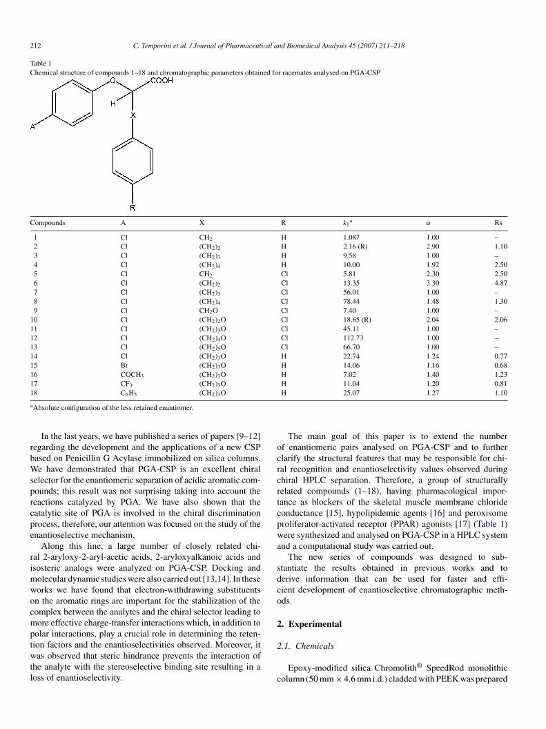
212 C. Temporini et al. / Journal of Pharmaceutical and Biomedical Analysis 45 (2007) 211–218
Table 1Chemical structure of compounds 1–18 and chromatographic parameters obtained for racemates analysed on PGA-CSP
Compounds A X R k1a α Rs
1 Cl CH2 H 1.087 1.00 –2 Cl (CH2)2 H 2.16 (R) 2.90 1.103 Cl (CH2)3 H 9.58 1.00 –4 Cl (CH2)4 H 10.00 1.92 2.505 Cl CH2 Cl 5.81 2.30 2.506 Cl (CH2)2 Cl 13.35 3.30 4.877 Cl (CH2)3 Cl 56.01 1.00 –8 Cl (CH2)4 Cl 78.44 1.48 1.309 Cl CH2O Cl 7.40 1.00 –
10 Cl (CH2)2O Cl 18.65 (R) 2.04 2.0611 Cl (CH2)3O Cl 45.11 1.00 –12 Cl (CH2)4O Cl 112.73 1.00 –13 Cl (CH2)5O Cl 66.70 1.00 –14 Cl (CH2)3O H 22.74 1.24 0.7715 Br (CH2)3O H 14.06 1.16 0.6816 COCH3 (CH2)3O H 7.02 1.40 1.2317 CF3 (CH2)3O H 11.04 1.20 0.811
a
rbWsprcpe
rimwocmptwtl
ocrcrtcpwa
sdco
2
8 C6H5 (CH2)3O
Absolute configuration of the less retained enantiomer.
In the last years, we have published a series of papers [9–12]egarding the development and the applications of a new CSPased on Penicillin G Acylase immobilized on silica columns.e have demonstrated that PGA-CSP is an excellent chiral
elector for the enantiomeric separation of acidic aromatic com-ounds; this result was not surprising taking into account theeactions catalyzed by PGA. We have also shown that theatalytic site of PGA is involved in the chiral discriminationrocess, therefore, our attention was focused on the study of thenantioselective mechanism.
Along this line, a large number of closely related chi-al 2-aryloxy-2-aryl-acetic acids, 2-aryloxyalkanoic acids andsosteric analogs were analyzed on PGA-CSP. Docking and
olecular dynamic studies were also carried out [13,14]. In theseorks we have found that electron-withdrawing substituentsn the aromatic rings are important for the stabilization of theomplex between the analytes and the chiral selector leading toore effective charge-transfer interactions which, in addition to
olar interactions, play a crucial role in determining the reten-
ion factors and the enantioselectivities observed. Moreover, itas observed that steric hindrance prevents the interaction ofhe analyte with the stereoselective binding site resulting in aoss of enantioselectivity.
2
c
H 25.07 1.27 1.10
The main goal of this paper is to extend the numberf enantiomeric pairs analysed on PGA-CSP and to furtherlarify the structural features that may be responsible for chi-al recognition and enantioselectivity values observed duringhiral HPLC separation. Therefore, a group of structurallyelated compounds (1–18), having pharmacological impor-ance as blockers of the skeletal muscle membrane chlorideonductance [15], hypolipidemic agents [16] and peroxisomeroliferator-activated receptor (PPAR) agonists [17] (Table 1)ere synthesized and analysed on PGA-CSP in a HPLC system
nd a computational study was carried out.The new series of compounds was designed to sub-
tantiate the results obtained in previous works and toerive information that can be used for faster and effi-ient development of enantioselective chromatographic meth-ds.
. Experimental
.1. Chemicals
Epoxy-modified silica Chromolith® SpeedRod monolithicolumn (50 mm × 4.6 mm i.d.) cladded with PEEK was prepared

ical a
afdp(
(a
E
2
s
Rcimwwtamsrfayepp4hsahSktc[
aec
2
Pt
(
2
lRlTApD
cw
kttattdir
2
2
fSUigaS
2
1afithWnPadetio
C. Temporini et al. / Journal of Pharmaceut
s research samples at Merck KGaA (Darmstadt, Germany)ollowing a previously reported procedure [10]. Potassiumihydrogenphosphate, dipotassium hydrogenphosphate for thereparation of the mobile phase were purchased from MerckDarmstadt, Germany).
Penicillin G Acylase crude extract from E. coli ATCC 11105EC 3.5.1.11) was kindly donated by Recordati (Milan, Italy)nd used as received.
All solvents were of analytical grade and purchased by Carlorba (Milan, Italy).
.2. Preparation of the analytes
The chemical structures of the racemic analytes used for thetudy are given in Table 1.
The synthesis of the compounds is briefly described.acemates 1–8 were prepared by condensation of diethyl 4-hlorophenoxy-malonate with the suitable aryl-alkyl-bromidesn the presence of NaH followed by alkaline hydrolysis and ther-
al decarboxylation at 160 ◦C. Stereoisomers of compound 2ere obtained by Mitsunobu condensation of 4-chlorophenolith R- or S-ethyl 2-hydroxy-4-phenylbutanoate. This reac-
ion, that is known to occur with inversion of configuration,fforded, after hydrolysis, S-2 and R-2, respectively. Race-ates 9–13 were prepared as previously reported [15]. A
imilar synthetic pathway was carried out for the prepa-ation of acids 14–18; the condensation products, derivedrom the reaction between phenoxypropylbromide and theppropriate diethyl aryloxy-malonate, gave rise, after hydrol-sis and decarboxylation to the desired compounds Thenantiomers of 10 were obtained following an alternativerocedure. The acid-catalyzed hydrolysis in CH3OH of theroduct achieved by the Mitsunobu condensation between-chlorophenol and R- or S-2-hydroxy-butyrolactone gave aydroxy-ester intermediate which underwent a second conden-ation with 4-chlorophenol under Mitsunobu conditions and
final hydrolysis to give R- and S-10. The desired acidsad the opposite absolute configuration of the starting R- or-2-hydroxy-butyrolactone as assigned on the basis of thenown stereochemical course of the Mitsunobu reaction. Allhe synthetic details as well as the characterization of theompounds used for this study have been reported elsewhere17].
Sample preparation was carried out by dissolving knownmounts of the chiral drug in 1-propanol (analytical grade) andach solution was diluted with the mobile phase buffer to aoncentration of 0.1 mM.
.3. Preparation of HPLC PGA-CSP
The in situ modification was used for the immobilization of
GA on epoxy monolithic support, and the characterization ofhe CSP was carried out as previously reported [13].The amount of immobilized units was 2178.87 U/column.When not in use, the columns were stored at 4 ◦C in a 0.01%
w/v) solution of sodium azide.
iastw
nd Biomedical Analysis 45 (2007) 211–218 213
.4. Apparatus and chromatographic conditions
Chromatographic experiments were performed with an Agi-ent 1100 liquid chromatograph (Palo Alto, CA, USA) with aheodyne sample valve (20 �l loop) equipped with an Agi-
ent 1100 variable-wavelength detector and an 1100 thermostat.he system was connected to a HPLC ChemStation, (Revision.04.01). Mobile phase used for this study was 50 mM phos-hate buffer (pH 7.0). Water was deionized by passing through airect-QTM (Millipore) system (Millipore, Bedford, MA, USA).All the chromatographic experiments were carried out at a
onstant oven temperature of 25 ◦C and the column flow-rateas set at 1.5 ml/min. The UV signal was followed at 225 nm.The retention factor (k) was calculated using the equation
= (tr/to) − 1, where tr is the retention time of the analyte ando is the retention time of an unretained compound. The voidime (to) was determined at the first disturbance of the baselinefter injection. The separation factor (α) was calculated usinghe equation, α = k2/k1 where k1 and k2 are the retention fac-ors for the first and last eluted enantiomers, respectively. Theetermination of the enantiomeric elution order was carried outnjecting solutions of (R) and (S) isomers with a concentrationatio of 2.
.5. Computational methods
.5.1. Molecular modellingMolecular modelling and graphics manipulations were per-
ormed using the SYBYL [SYBYL Molecular Modellingystem (version 7.1), TRIPOS Assoc., St. Louis, MO] andCSF CHIMERA software packages [18], running it on a Sil-
con Graphics Tezro R16000 workstation. Model building andeometry optimizations of compounds 6, 7, 10 and 13 wereccomplished with the TRIPOS force field [19] available withinYBYL.
.5.2. Docking simulationsDocking of both R- and S-enantiomers of compounds 6, 7,
0 and 13 to PGA was carried out using GOLD 3.1 version [20],genetic algorithm-based software, selecting GOLDScore as atness function. GOLDScore is made up of four components
hat account for protein-ligand binding energy: protein-ligandydrogen bond energy (external H-bond), protein-ligand van deraals energy (external vdw), ligand internal vdw energy (inter-
al vdw) and ligand torsional strain energy (internal torsion).arameters used in the fitness function (hydrogen bond energies,tom radii and polarizabilities, torsion potentials, hydrogen bondirectionalities, and so forth) are taken from the GOLD param-ter file. The fitness score is taken as the negative of the sum ofhe energy terms, so larger fitness scores indicated better bind-ngs. The fitness function has been optimized for the predictionf ligand binding positions rather than the prediction of bind-ng affinities, although some correlation with the latter can be
lso found [21]. The protein input file may be the entire proteintructure or a part of it comprising only the residues that are inhe region of the ligand binding site. In the present study, GOLDas allowed to calculate interaction energies within a sphere of
2 ical a
aP
2
sSoB[o
2
nftwSsbr
2
dsmclb
3
l[aaemi
ispafabspc
tm
tt
3
amecIg
apr5werm
mei
dwri1eobawttv
ispcam
tdoso
14 C. Temporini et al. / Journal of Pharmaceut
10 A radius centered on the HZ hydrogen atom of Phe71 inGA.
.5.3. Ligand setupThe structures of the ligands 6, 7, 10 and 13 were con-
tructed using standard bond lengths and bond angles of theYBYL fragment library. Geometry optimizations were carriedut with the SYBYL/MAXIMIN2 minimizer by applying theFGS (Broyden, Fletcher, Goldfarb, and Shannon) algorithm
22] and setting a rms gradient of the forces acting on each atomf 0.001 kcal mol−1 A−1 as the convergence criterion.
.5.4. Protein setupThe crystal structure of PGA in complex with p-
itrophenylacetic acid (entry code: 1AJN) [23], recoveredrom Brookhaven Protein Database [24], was used. The struc-ure was set up for docking as follows: polar hydrogensere added by using the BIOPOLYMERS module within theYBYL program (residues Arg, Lys, Glu and Asp were con-idered ionized, while all His were considered to be neutraly default), the p-nitrophenylacetic acid and all waters wereemoved.
.5.5. GOLD dockingFifty independent docking runs were performed for each
ocking experiment. All docking runs were carried out usingtandard default settings with a population size of 100, aaximum number of 100,000 operations, and a mutation and
rossover rate of 95. The best generated 10 solutions of eachigand were ranked according to their fitness scores calculatedy the GOLDScore function.
. Results and discussion
Over the last years we have demonstrated that immobi-ized PGA can be used as chiral selector for acidic compounds9–12]. In particular, complete chiral resolution of many 2-ryloxyalkanoic acids, isosteric analogs and 2-arylpropioniccids was achieved with PGA-CSP which shows a preferentialnantioselective binding for the S enantiomers. These experi-ental observations provided evidence that the catalytic site is
nvolved in the chiral mechanism.On the basis of our previous results, in the present work we
nvestigated the chiral properties of a PGA column towards a neweries of 2-aryloxyacetic acids (Table 1) characterized by theresence into the side chain of a phenyl ring (compounds 1–8) orfurther phenoxy group (compounds 9–18) at variable distance
rom the chiral center. Half of these compounds, moreover, hadn additional chlorine atom in the para-position of the side chainenzene ring, whereas different atoms or groups with differentize, shape and electronic properties were considered for theara-position of the phenoxy group directly linked to the chiral
enter.One goal of this study was also to enhance the knowledge ofhe chiral recognition mechanism and process on PGA-CSP. A
olecular modelling study was, therefore, undertaken, allowing
dcs
nd Biomedical Analysis 45 (2007) 211–218
o describe the relationships between the chemical structure ofhe analytes and the chromatographic results.
.1. Chromatographic studies
Compounds 1–18 were chromatographed on PGA-CSP usingmobile phase of 50 mM phosphate buffer pH 7.0. The chro-atographic results are summarized in Table 1: PGA showed
nantiorecognition for 11 racemates (α greater than 1.0) but onlyompounds 4, 5, 6 and 10 present an Rs value better than 1.5.t could be expected that further optimization of the chromato-raphic conditions can improve the chiral separations.
Some representative chromatograms are reported in Fig. 1.For compounds 1–8 an increase in retention time was
chieved increasing the length of the alkyl chain between thehenyl ring and the chiral center. The presence of a second chlo-ine atom on the side chain aromatic ring (Ar′) (compounds–8) led to a large increase in retention times. The analogsith two carbon atoms in the alkyl chains, present the better
nantioresolutions (α = 2.90 and 3.30 for compounds 2 and 6,espectively). In this sub-group of analytes only the racemicixtures of compounds 1 and 7 were not resolved.From these data seemed that the length of the chain is not the
ost significant structural parameter for the interaction with thenantioselective site, while the substituent on Ar′ plays a moremportant role.
In the subset of compounds 9–13, an oxygen atom was intro-uced between the aliphatic chain up to five methylenes and Ar′hile maintaining the chlorine substituents on both the aromatic
ings. As expected, the retention was increased by the lengthen-ng of the carbon chain up to four C atoms, while for compound3 (5 C) a surprising reduction of the k was obtained. A possiblexplanation of this behaviour might be the restricted dimensionsf the binding pocket or the necessity of an optimal distanceetween two areas of the binding site that can interact with thenalyte. Enantioselectivity was observed only for compound 10ith a carbon chain of two atoms: α = 2.04. This compound was
he only oxygenated derivative to show a better enantioresolu-ion in comparison to the corresponding methylenic isoster (10ersus 7).
Finally, we prepared a third group of derivatives (14–18)n which the phenoxypropyl side chain was maintained con-tant while varying the 4-substituent on the aromatic ring of thehenoxy group (Ar) directly linked to the chiral center. Theseompounds were selected for their pharmaceutical interest beingnalogs of 11 which is the most active blocker of the skeletaluscle membrane chloride conductance [15].The introduction of a phenyl ring in position 4 of Ar led
o a substantial increase in retention, while the acetyl groupetermined a significant reduction in retention time. The elutionrder of compounds 14, 15 and 17, with Cl, Br and CF3 as sub-tituents, is in agreement with the electron-withdrawing powerf the considered substituents.
Interestingly, all the racemic mixtures of this subset oferivatives were resolved and the enantioselectivity was almostonstant for all the compounds. Therefore, the nature of theubstituents on this aromatic ring seems to have a very little

C. Temporini et al. / Journal of Pharmaceutical and Biomedical Analysis 45 (2007) 211–218 215
lumn
ioi
ctcA(tatldadftrtat
au
Rc
3
reicdtAeef
ib6P
Fig. 1. Chromatograms of racemates 4, 5, 6, 10, 14 and 16 on PGA co
nfluence on enantioselectivity. On the other hand, the lackingf the chlorine on Ar′ seems to favour the chiral discriminationn compounds 14 and 11.
A complete overview of the data led us to derive moreomplete considerations regarding cross-correlation betweenhe effect of the chlorine atom on Ar′ and the length of thearbon chain (X). In fact, the presence of chlorine atom onr′ seems to determine (compounds 5 and 1) or to enhance
compounds 6 and 2) the enantioselectivity, but an oppositerend was observed when comparing compounds 8 with 4,nd compounds 11 with 14. One explanation of the contradic-ory behaviour observed could be the crucial role played byength of the carbon chain (X). The presence of more than 3 Coes not allow the Ar′ to fit in the hydrophobic pocket of thective site when carrying the chlorine atom, due to steric hin-rance of this substituent. This behaviour was not observedor compounds 5 in comparison to 1 and 6 in comparisono 2 where the carbon chain is composed by one or two C,espectively. We can suppose that in this case, the length ofhe chain allows the Ar′ ring to fit in the hydrophobic pocket,nd Cl substituent to positively affect the chiral discrimina-
ion.Of the whole series, only analogs 2 and 10 were prepared,lso, as optically active isomers with assigned absolute config-ration. The elution order of these enantiomers was found to be
l6ai
. Mobile phase: 50 mM phosphate buffer (pH 7.0). Flow: 1.5 ml/min.
/S and these data are consistent with the previously observedhiral interactions [9,12].
.2. Docking studies
In order to get a better comprehension of the molecularecognition process between PGA and its analytes, dockingxperiments were performed using the crystal structure of PGAn complex with the 2-(4-nitrophenyl)acetic acid (PDB entryode: 1AJN) [23]. The GOLD 3.1 program was used to carry outocking simulations, [20] since in several studies it yielded bet-er performances compared to other similar programs [25–27].n analysis of principles and methods adopted by GOLD for
nergy calculations, conformational search and clustering, andnergy ranking is briefly presented in the Section 2, whereas aully detailed description may be found elsewhere [28].
Docking investigations were focused on both R- and S-somers of 6 and 10, whose racemate were efficiently separatedy PGA. An in-depth look at the conformer population of S-and S-10 generated during the docking simulations into the
GA active site revealed that a convergent binding mode was
argely adopted with a high fitness score (64 kJ/mol for S-6 and1 kJ/mol for S-10). As depicted in Fig. 2a and c, the associ-ted binding mode is characterized by the presence of H-bondsnvolving the carboxylate group and the OH groups of both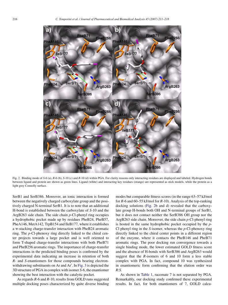
216 C. Temporini et al. / Journal of Pharmaceutical and Biomedical Analysis 45 (2007) 211–218
F clarib actingl
SbtHAaPartfaieRw3s
m
mfdlbAiCdoasasci
ig. 2. Binding mode of S-6 (a), R-6 (b), S-10 (c) and R-10 (d) within PGA. Foretween ligand and protein are shown as green lines. Ligand (white) and interight grey Connolly surface.
erB1 and SerB386. Moreover, an ionic interaction is formedetween the negatively charged carboxylate group and the posi-ively charged N-terminal SerB1. It is to note that an additional-bond is established between the carboxylate of S-10 and thergB263 side chain. The side chain p-Cl-phenyl ring occupieshydrophobic pocket made up by residues PheB24, PheB57,heA146, MetA142, TrpB154 and IleB177, where it establishes�-stacking charge-transfer interaction with PheB24 aromatic
ing. The p-Cl-phenoxy ring directly linked to the chiral cen-er projects towards a large pocket and is well oriented toorm T-shaped charge-transfer interactions with both PheB71nd PheB256 aromatic rings. The importance of charge-transfernteractions in the predicted binding poses is confirmed by thexperimental data indicating an increase in retention of both- and S-enantiomers for those compounds bearing electron-ithdrawing substituents on Ar and Ar′. In Fig. 3 is depicted the
D structure of PGA in complex with isomer S-6, the enantiomerhowing the best interaction with the catalytic pocket.As regards R-6 and R-10, results from GOLD runs suggestedultiple docking poses characterized by quite diverse binding
R
Rr
ty reasons only interacting residues are displayed and labeled. Hydrogen bondskey residues (orange) are represented as stick models, while the protein as a
odes but comparable fitness scores (in the range 63–57 kJ/molor R-6 and 60–55 kJ/mol for R-10). Analysis of the top-rankingocking solutions (Fig. 2b and d) revealed that the carboxy-ate group H-bonds both OH and N-terminal groups of SerB1,ut it does not contact neither the SerB386 OH group nor thergB263 side chain. Moreover, the side chain p-Cl-phenyl ring
s hosted in the same hydrophobic pocket occupied by the p-l phenyl ring in the S-isomer, whereas the p-Cl-phenoxy ringirectly linked to the chiral center points in a different regionf the enzyme, where it contacts the PheB146 and PheB71romatic rings. The poor docking run convergence towards aingle binding mode, the lower estimated GOLD fitness scorend the absence of H-bonds with SerB386 and ArgB263 woulduggest that the R-isomers of 6 and 10 form a less stableomplex with PGA. In fact, compound 10 was synthesizedn enantiomeric form confirming that the elution order was
:S.As shown in Table 1, racemate 7 is not separated by PGA.emarkably, our docking study confirmed these experimental
esults. In fact, for both enantiomers of 7, GOLD calcu-

C. Temporini et al. / Journal of Pharmaceutical a
Fig. 3. 3D structure of PGA in complex with isomer S-6 (white) displayed asstick model. The protein is coloured by B-factor: blue regions correspond tolow temperature factors, whereas green, yellow and red colours characterizera
lgbPetanidbt
aiOobis
oCoicto(
4
r2aog
boieise
Fia
egions of subsequently increasing B-factor. Residues lining the ligand positionre highlighted in orange, while the H-bonds are represented by dashed lines.
ated two prevailing binding modes in which the carboxylateroup and the p-Cl-phenyl ring still remained anchored to theinding site interacting with SerB1, SerB386, ArgB263 andheB24 side chains, whereas the p-Cl-phenoxy group projectedither towards the opening of the PGA active site (Fig. 4a) orowards the large hydrophobic pocket so to contact PheB71nd PheB256 (Fig. 4b). Moreover, the associated GOLD fit-ess scores were comparable for each predicted binding mode
n accordance with the chromatographic experiments, whichisplay that the stationary phase is unable to discriminateetween both enantiomers (α = 1). It could be hypothesizedhat replacement of the oxygen atom (compound 10) withdacm
ig. 4. Superimposition of the two prevailing binding orientations (a and b) of S-7 (nteracting residues are displayed and labeled. Hydrogen bonds between ligand andre represented as stick models, while the protein as a light grey Connolly surface.
nd Biomedical Analysis 45 (2007) 211–218 217
methylenic unit (compound 7) in the aliphatic chain bear-ng the Ar′ decreases the gauche effect clearly present in the–C–C–C–O chain of 10 [29] provoking the double bindingrientation of the ligand. Fig. 4 illustrates a superimpositionetween the binding mode of S-10 and the two prevailing bind-ng modes of the methylenic isoster S-7 into the PGA bindingite.
Peculiar results were obtained when docking was performedn both R- and S-isomers of 13, bearing a carbon chain of fiveatoms. No convergence towards a single binding position was
bserved. From a visual inspection of the ligand/PGA complext seems clear that the increased steric hindrance of the five Chain prevents both enantiomers from adapting themselves intohe stereoselective PGA binding, thus explaining the reductionf the observed k1 value in comparison to the homologues onesracemates 9–12).
. Conclusions
In an effort to extend the applicability of PGA as chi-al selector towards acidic compounds, a new series of-aryloxyalkyl- and 2-arylalkyl-2-aryloxyacetic acids werenalysed on PGA-CSP and the influence of structural variationsf analytes on retention and enantioselectivity was investi-ated.
PGA-based CSP proved resolving capability for a large num-er of the considered compounds. Docking experiments, basedn the known 3D structure of PGA and carried out to rational-ze the observed enantioselective behaviour of the immobilizednzyme, corroborate the importance of the charge-transfernteractions and steric effects on retention factors and chiraleparation. Moreover, the computational studies confirmed thexperimental results indicating that it would be possible to pre-
ict the enantioselectivity of PGA-CSP for the investigatednalytes. The knowledge of chiral molecule-selector interactionsan be useful to design a more efficient chiral selector based onutated PGA.spring green) about that of S-10 (white) within PGA. For clarity reasons onlyprotein are shown as green lines. Ligand and interacting key residues (orange)
2 ical a
A
22
R
[
[
[
[
[
[
[
[
[
[
[
[
[
[
[
[[[
57 (2004) 225–242.
18 C. Temporini et al. / Journal of Pharmaceut
cknowledgements
This work was supported by MURST Grant (project:004038884). Additional support from University of Pavia (FAR006).
eferences
[1] M. Lammerhofer, W. Lindner, in: K. Valco (Ed.), Recent Development inLiquid Chromatographic Enantioseparation, vol. 1, Elsevier, Amsterdam,2000, pp. 337–437.
[2] A.K. Kumerer, H.Y. Aboul-Enein, Chromatographia 63 (2006) 295–307.[3] C. Roussel, A. Del Rio, J. Pierrot-Sanders, P. Piras, N. Vanthuyne, J.
Chromatogr. A 1037 (2004) 311–328.[4] A. Del Rio, P. Piras, C. Roussel, Chirality 17 (2005) S74–S83.[5] J. Haginaka, J. Chromatogr. A 906 (2001) 253–273.[6] K.B. Lipkowitz, Acc. Chem. Res. 33 (2000) 555–562.[7] M. Arroyo, I. de la Mata, C. Acebal, M. Pilar Castillon, Appl. Microbiol.
Biotechnol. 60 (2003) 507–514.[8] H.J. Duggleby, S.P. Tolley, C.P. Hill, E.J. Dodson, G. Dodson, P.C.E.
Moody, Nature 373 (1995) 264–268.[9] E. Calleri, G. Massolini, F. Loiodice, G. Fracchiolla, C. Temporini, G.
Felix, P. Tortorella, G. Caccialanza, J. Chromatogr. A 958 (2002) 131–140.10] E. Calleri, G. Massolini, D. Lubda, C. Temporini, F. Loiodice, G. Cac-
cialanza, J. Chromatogr. A 1031 (2004) 93–100.11] E. Calleri, C. Temporini, G. Massolini, G. Caccialanza, J. Pharm. Biomed.
Anal. 35 (2004) 243–258.12] G. Massolini, G. Fracchiolla, E. Calleri, G. Carbonara, C. Temporini, A.
Lavecchia, S. Cosconati, E. Novellino, F. Loiodice, Chirality 18 (2006)633–643.
13] G. Massolini, E. Calleri, A. Lavecchia, F. Loiodice, D. Lubda, C. Temporini,G. Fracchiolla, P. Tortorella, E. Novellino, G. Caccialanza, Anal. Chem.75 (2003) 535–542.
[
[
nd Biomedical Analysis 45 (2007) 211–218
14] A. Lavecchia, S. Cosconati, E. Novellino, E. Calleri, C. Temporini, G.Massolini, G. Carbonara, G. Fracchiolla, F. Loiodice, J. Mol. Graph. Mod.25 (2007) 773–783.
15] G. Carbonara, G. Fracchiolla, F. Loiodice, P. Tortorella, D. Conte-Camerino, A.M. De Luca, A. Liantonio, Il Farmaco 56 (2001) 749–754.
16] J. Shoichi, Y. Yoshiaki, O. Takayuki, O. Shinji, Jp. Appl. 02193942, Jpn.Kokai Tokkyo Koho, (Chemical Abstract (1990), no. 113:211579).
17] G. Fracchiolla, A. Laghezza, L. Piemontese, G. Carbonara, A. Lavecchia,P. Tortorella, M. Crestani, E. Novellino, F. Loiodice, ChemMedChem 2(2007) 641–654.
18] E.F. Pettersen, T.D. Goddard, C.C. Huang, G.S. Couch, D.M. Greenblatt,E.C. Meng, T.E. Ferrin, J. Comput. Chem. 25 (2004) 1605–1612.
19] J.G. Vinter, A. Davis, M.R. Saunders, J. Comput.-Aided Mol. Des. 1 (1987)31–55.
20] G. Jones, P. Willett, R.C. Glen, A.R. Leach, R. Taylor, J. Mol. Biol. 267(1997) 727–748.
21] M.L. Verdonk, J.C. Cole, M.J. Hartshorn, C.W. Murray, R.D. Taylor, Struct.Funct. Bioinf. 52 (2003) 609–623.
22] G.B. Fitzgerald, C. Bauman, Md. Sajjat Hussoin, M. Wick, J. Head, M.C.A.Zerner, Chem. Phys. Lett. 122 (1985) 264–274.
23] S.H. Done, J.A. Brannigan, P.C. Moody, R.E. Hubbard, J. Mol. Biol. 284(1998) 463–475.
24] F.C. Bernstein, T.F. Koetzle, G.J.B. Williams, E.F.J. Meyer, M.R. Brice,J.R. Rodgers, O. Kennard, T. Shimanouchi, T. Tasumi, J. Mol. Biol. 112(1977) 535–542.
25] T. Schulz-Gasch, M. Stahl, J. Mol. Model. 9 (2003) 47–57.26] R. Wang, Y. Lu, S. Wang, J. Med. Chem. 46 (2003) 2287–2303.27] E. Kellenberger, J. Rodrigo, P. Muller, D. Rognan, Struct. Funct. Bioinf.
28] D.R. Westhead, D.E. Clark, C.W. Murray, J. Comput.-Aided Mol. Des. 11(1997) 209–228.
29] P. Bultinck, C. Van Alsenoy, A. Goeminne, J. Phys. Chem. A 105 (2001)9203–9210.


![2-[5-Methyl-2-(propan-2-yl)phenoxy]- N ′-{2-[5-methyl-2-(propan-2-yl)phenoxy]acetyl}acetohydrazide](https://static.fdokumen.com/doc/165x107/6344862303a48733920aed56/2-5-methyl-2-propan-2-ylphenoxy-n-2-5-methyl-2-propan-2-ylphenoxyacetylacetohydrazide.jpg)