![Binder 216, Terminology [Trematoda Taxon Notebooks]](https://static.fdokumen.com/doc/165x107/63338ab23108fad7760f19c8/binder-216-terminology-trematoda-taxon-notebooks.jpg)
ELEMENTS OF BARK STRUCTURE AND TERMINOLOGY ...
11
ELEMENTS OF BARK STRUCTURE AND TERMINOLOGY Robert E. Martin and John B. Crist Division of Forestry and Wildlife Sciences, Virginia Polytechnic Institute and State University, Blacksburg, Virginia 24061 ABSTRACT Bark structure varics considerably froin that of wood, although arialogics inay be drawn between specific elemcnts and overall structure and function. The terminology of bark structure is discussecl and various cellular elements are described. Several bark structures are illustrated with light and electron nlicrographs pointing out differences between species and species groups. INTRODUCTION Pd Phclloderm Bark is an important physiological and Pe Periderm l~rotcctivc component of our woody plants Po Pore or pit canal and comprises a considerable volume of Pg Phellogen the logs removed from a forest. Improved Ph Phellem utilization of this material will be enhanced R Rhytidome not only by research on its structure and Ra Ray properties, but also by better undcrstand- Rc Resin canal ing of its structure and technical terminol- S Sieve element ogy. Our intent in this paper is twofold. Sa Sieve area First, we wish to present the latest informa- Sc Sclereid tion available on bark structure and its S~ Sieve plate component cells. Second, we wish to have X Xylem in the literature a rccent, gencrally available, TERhlINOLOGY reference point on thc structure of both softwood (gymnosperm) and hard\vood If ' e might best begin our discussion of (angiosperm) barks, In order to approach bark by using the definition given by the tllesc objectives, we will discuss the termi- Society of Amcrican Foresters (1958) as nology of barks with descriptions of the "the tissues of the stem, branch, and root cells involved. We will illustrate structure outside the cambium layer." They further at a wide range of magnification in micro- define inner bark as the physiologically graphs and dra\vings, and compare bark active tissues between the cambium and the elements to wood elements to aid the wood last-formed peridem or protective layer, technologist. For more extensive descrip- and outer bark as the layer of dead tissue of tions of bark structure and formation, we a dry, corky nature outside the last-formed refcr the reader to Chang (1954a, b ) and periderm. Anatomists would refer to the Esau (1959). inner bark as phloem and the outer bark as 1.hytidome. The phloem is generally light or SYJIBOLS creamy in color, while the rhytidome is gen- The following symbols are used in identi- erally tan or brown in the interior and often fying features in tlie figures: discolored to various shades of light gray A Albuminous cell to black on its surface. C Cambium CO Companion cell Periderm Cr Crystal Yeriderm is an important layer in the F Fiber protection of stcms from desiccation and P Phloem probably also biological organisms. It Pa Parenchyma consists of three laycrs of cells. Within the 269
-
Upload
khangminh22 -
Category
Documents
-
view
0 -
download
0
Transcript of ELEMENTS OF BARK STRUCTURE AND TERMINOLOGY ...

ELEMENTS OF BARK STRUCTURE AND TERMINOLOGY
Robert E . Martin and John B. Crist Division of Forestry and Wildlife Sciences, Virginia Polytechnic Institute and
State University, Blacksburg, Virginia 24061
ABSTRACT
Bark structure varics considerably froin that of wood, although arialogics inay be drawn between specific elemcnts and overall structure and function. The terminology of bark structure is discussecl and various cellular elements are described. Several bark structures are illustrated with light and electron nlicrographs pointing out differences between species and species groups.
INTRODUCTION Pd Phclloderm
Bark is an important physiological and Pe Periderm l~rotcctivc component of our woody plants Po Pore or pit canal
and comprises a considerable volume of Pg Phellogen the logs removed from a forest. Improved Ph Phellem utilization of this material will be enhanced R Rhytidome
not only by research on its structure and Ra Ray properties, but also by better undcrstand- Rc Resin canal
ing of its structure and technical terminol- S Sieve element
ogy. Our intent in this paper is twofold. Sa Sieve area
First, we wish to present the latest informa- Sc Sclereid tion available on bark structure and its S~ Sieve plate
component cells. Second, we wish to have X Xylem in the literature a rccent, gencrally available,
TERhlINOLOGY reference point on thc structure of both softwood (gymnosperm) and hard\vood If'e might best begin our discussion of (angiosperm) barks, In order to approach bark by using the definition given by the tllesc objectives, we will discuss the termi- Society of Amcrican Foresters (1958) as nology of barks with descriptions of the "the tissues of the stem, branch, and root cells involved. We will illustrate structure outside the cambium layer." They further at a wide range of magnification in micro- define inner bark as the physiologically graphs and dra\vings, and compare bark active tissues between the cambium and the elements to wood elements to aid the wood last-formed peridem or protective layer, technologist. For more extensive descrip- and outer bark as the layer of dead tissue of tions of bark structure and formation, we a dry, corky nature outside the last-formed refcr the reader to Chang (1954a, b ) and periderm. Anatomists would refer to the Esau (1959). inner bark as phloem and the outer bark as
1.hytidome. The phloem is generally light or SYJIBOLS creamy in color, while the rhytidome is gen-
The following symbols are used in identi- erally tan or brown in the interior and often
fying features in tlie figures: discolored to various shades of light gray A Albuminous cell to black on its surface. C Cambium CO Companion cell Periderm Cr Crystal Yeriderm is an important layer in the F Fiber protection of stcms from desiccation and P Phloem probably also biological organisms. It Pa Parenchyma consists of three laycrs of cells. Within the
269
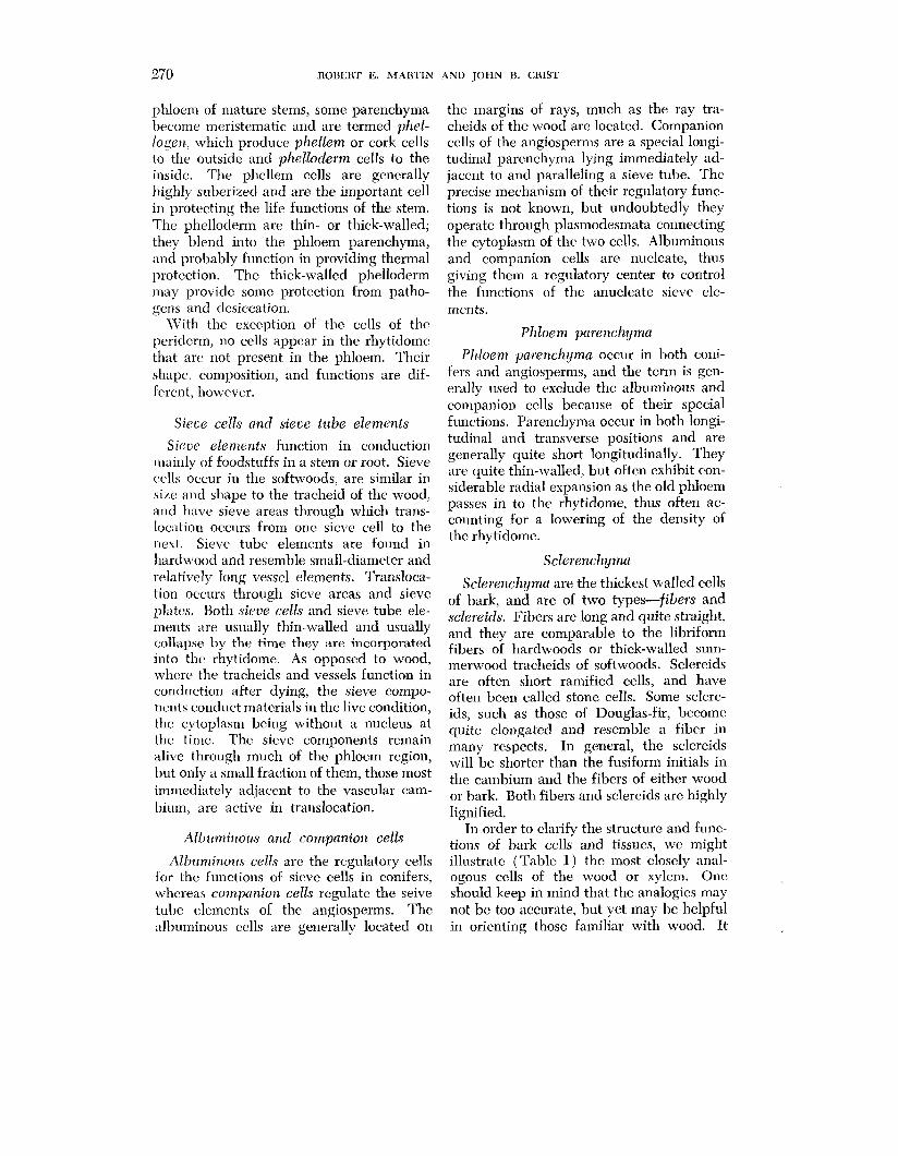
270 ROBERT E, MARTIN AND JOHN B. GRIST
phloem of mature stems, some parenchyma l~ecome ineristematic and are termed phel- logen, which produce phellem or cork cells to the outside and phelloderm cells to the insidc. The phellem cells are generally highly suberized and are the important cell in protecting the life functions of the stem. The phelloderm are thin- or thick-walled; they blend into the phloem parenchyma, and probably function in providing thermal protection. The thick-walled phelloderm may provide some protection from patho- gens and desiccation.
\Vith the exception of the cells of the periderm, no cells appear in the rhytidome that arc not present in the phloem. Their shape, composition, and functions are dif- fcrent, however.
Sieve cells and sieve tube elements
Sierje elements function in conduction mainly of foodstuffs in a stem or root. Sieve cells occur in the softwoods, are similar in sizc and slyape to the tracheid of the wood, and havc sieve areas through which trans- location occurs from one sieve cell to the next. Sieve tube elements are found in hardwood and resemble small-diameter and relatively long vessel elements. Transloca- tion occurs through sieve areas and sieve plates. Both sieve ce76 and sievt: tube ele- ments are usually thin-walled and usually collapse by the time they are incorporated into the rhytidome. As opposed to wood, where the tracheids and vessels function in conduction after dying, the sieve compo- nents conduct materials in the live condition, the cytoplasm being without a nucleus at the time. The sieve components remain alive through much of the phloem region, but only a mall fraction of them, those most immediately adjaccnt to the vascular cam- bium. are active in translocation.
Albz~minous and companion cells
All~~~minotrs cells are the regulatory cells for the functions of sieve cells in conifers, whcreas companion cells regulate the seive tube elements of the angiosperms. The :tlbuminous cells are generally located on
the margins of rays, much as the ray tra- cheids of the wood are located. Companion cells of the angiosperms are a special longi- tudinal parenchyma lying immediately ad- jacent to and paralleling a sieve tube. Thc precise mechanism of their regulatory func- tions is not known, but undoubtedly they operate through plasmodesmata connecting the cytoplasm of the two cells. Albuminous and companion cells are nucleate, thus giving them a regulatory center to control the functions of the anucleate sieve ele- ments.
Phloem parenchyma
Phloem parenchyma occur in both coni- fers and angiosperms, and the term is gen- erally used to exclude the albuminous and coinpanion cells because of their special functions. Parenchyma occur in both longi- tudinal and transverse positions and are generally quite short longitudinally. They are quite thin-walled, but often exhibit con- siderable radial expansion as the old phloem passes in to the rhytidome, thus often ac- counting for a lowering of the density of the rhytidome.
Sclerenchyma
Sclerench!gma are the thickest walled cells of bark, and are of two types-fibers and sclereids. Fibers are long and quite straight, and they are comparable to the libriform fibers of hardwoods or thick-walled sum- merwood tracheids of softwoods. Sclereids are often short ramified cells, and have often been called stone cells. Some sclere- ids, such as those of Douglas-fir, become quite elongated and resemble a fiber in many respects. In general, the sclereids will be shorter than the fusiform initials in the cambium and the fibers of either wood or bark. Both fibers and sclereids are highly lignified.
In order to clarify the structure and func- tions of bark cells and tissues, we might illustrate (Table 1) the most closely anal- ogous cells of the wood or xylem. One, should keep in mind that the analogies may not be too accurate, but yet may be helpful in orienting those familiar with wood. It
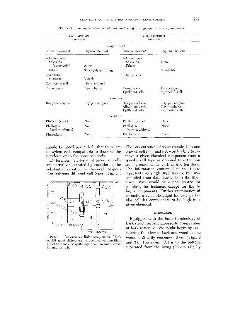
KLEhIEWrS OF BARK STKUCI'URIi AND TERhIINOLOGY 271
TAI~LE 1. Ar~alogous elerr~erlts of 1)clrk uncl tfiood in angiosperms and g!lntnosl)ernls
ANGIOSPERMS GYMNOSPERhlS Hardwoods Softmoods
I'hloem element
Sclerenchyma Sclereids
(stone cells )
liibers
Seive tube elements
C:ompanion cell5
Longitudinal
Xylem element l'ltloe~li element
iV one
Sclere~lch yma Sclereids Fibrrs
Tracheids and fibers
Seive cells Vt'ssels
( Parenchyma )
Xylem element
None
Tracheids
l'nrenchylna l 'a~.cncli~ma l'urenchyma Parenchyma Epithelial cells Epithelial cells
Transverse
Itay parenchyma Kay pnretichyma Ray parcncllyma Ray parenchyma Albl~minous cclls Ray tracheids Epithelial cells Epithelial cells
Per iderln
l'l~elleln (cork) None* Phellenl ( cork ) None
I'hellogen Nonc l'hcllogen None ( cork caml~ium ) ( cork caml~ium )
I'helloderm None Phellodenn None
should 1)c noted particularly that there are The coilcentration of some chemicals in onc no xylem cells comparable to those of the type of cell may make it worth while to re- periderm or to the short sclereids. move a given chemical component from a
Differences in internal structure of cells spt,cific cell type as opposed to extraction are partially illustrated by considering the from ground whole bark as is often done. substantial variation in chemical composi- Thc information contained in the figure tion between differcnt cell types (Fig. 1). represents no single tree species, but was
compiled from data available in the liter- - -ASH ature. Bark would be a poor source for
cellulose, for instance, except for the fi- brous components. Further examination of extractives available might indicate partic- ular cellular components to be high in a given chemical.
STRUCTURE
Equipped with the basic terminology of bark structure, let's proceed to observations of bark structure. We might begin by con-
RHYTl DOME siclerirle the view of bark and wood as one L,
Fig. 1. The various celllllar components of bark would ordinarily encounter them ( Figs. 2 exhibit great differences in chemical composition, ;I fact that 111ay bc quite significant in understand- and 3) . The xylem ( X ) is to the bottom, ing and using it. separated from the living phloem (P ) by
272 HOBEHT 13. ~ I A R T I N AND TOHN H. CHIST
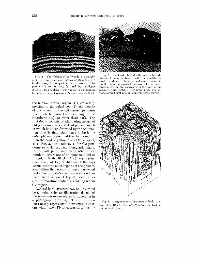
Fig. 3. Black oak illustrates the relatively wide Fig' 2' The of is phloem of lnany hardwoods with the roughly fis-
quite narrow, pond pine (Pirubs se~.otiilu Michx.) sured rhytidome, TI^^ inner phloem is darker in in this casc, in comparison to hardwoods. The the illustration, primarily because of a higher mo,is- periderm layers are quite flat, and the rhytidoine tnre content, and the contrast with the pores of the gives a soft, low-dmsity appearance in conrparison xylem is quite distinct. Peridern] layers are not to the xyleir~ ( light photograph, transverse section ) . pronounced ( light photograph, transverse section).
the narrow cambial region ( C ), essentially invisible to the naked eye. To the outside of the phloem is the last-formed periderm (Pe) , which marks the beginning of the rhytidome ( R ) , or outer dead bark. The rhytidome consists of alternating layers of old periderm layers and dead phloem, much of which has been distorted by the oblitera- tion of cells that takes place in both the outer phloem region and the rhytidome.
In the hard or yellow pines ( Piizus spp. ), as in Fig. 2, the tendency is for the pcri- tlcrms to lie flat in a nearly tangential plane. In the soft pines, and many other trees, periderm layers are often more rounded or irregular. In the Black oak (Quercu~ velu- tina Lam.) of Fig. 3, dilation of the rays occurs near the 0utc.r regions of the phloem, a condition that occurs in many hardwood barks. Some gradation in color occurs within the phloem region of Fig. 3, perhaps be- cause of moisture gradients occurring within the region.
Ccmeral bark structure can be il1ustratc:d l~cst, pcrhaps, by an illustration devoid of the inany extraneous elements appearing in a photograph (Fig. 4 ) . This illustration rnost ne:irly represents thc structure of east- c ~ n white pine (Pinus strobus L.) , but the
Fig. 4. Diagrammatic illi~stration of bark struc- t l~re. The figure no st nearly represents bark of eastern white pine.
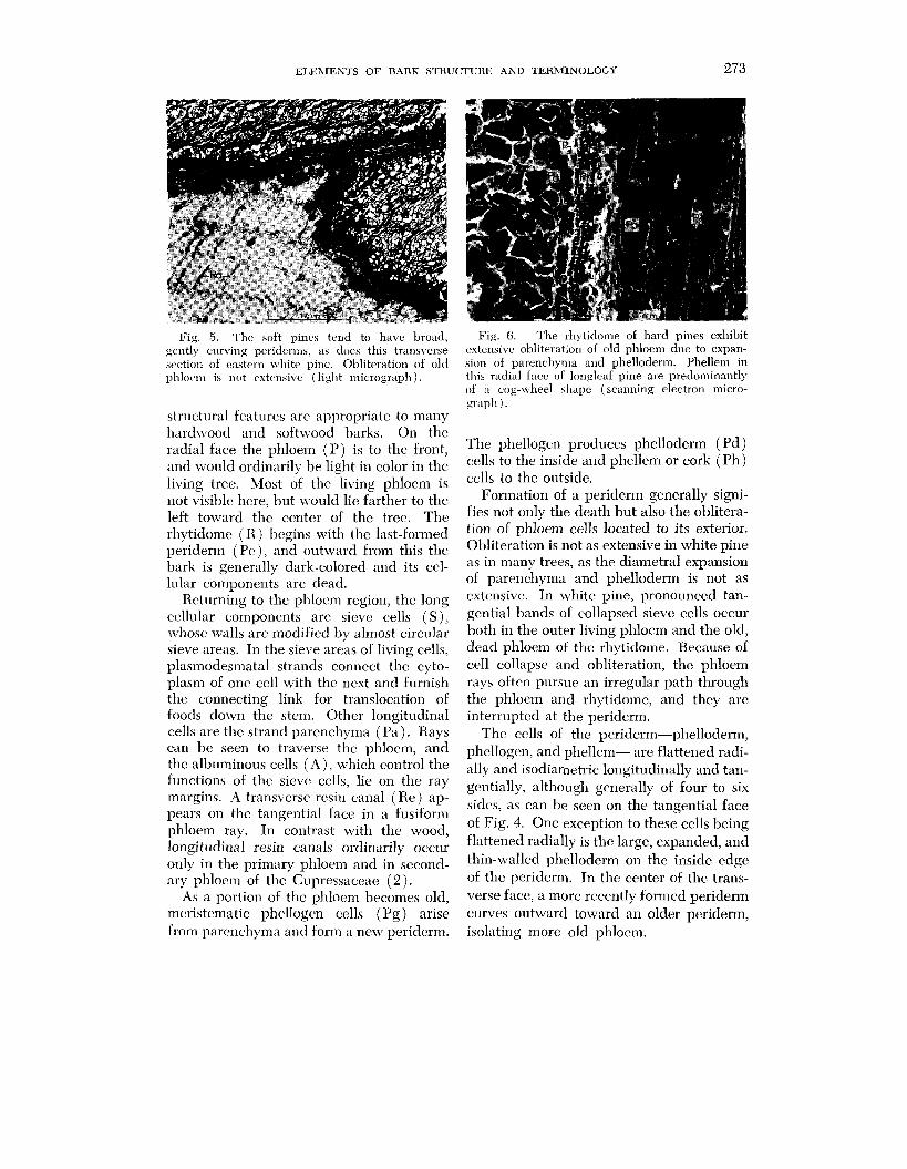
ELEAIEXTS OF BARK S T R U ~ U R E ASD TERMINOLOGY 273
Fig. 5. The soft pines tend to have broad, Fig. 6. The rhytidome of hard pines exhibit gently curving periderms, as does this transverse extensive obliteration of old phloenl due to expan- srction of eastern white pine. Obliteration of oltl sion of parenchyma and phellodern~. Phellem in phlocn~ is not extensive (light micrograph). this radial face of longleaf pine are predominantly
of a cog-wheel shape (scanning electron micro- gl.aplr ).
structural features are appropriate to many hard\vood and softwoo-& barks. On thc radial face the phloem ( P ) is to the front, and \vould ordinarily be light in color in the living tree. Most of the living phloem is not visible here, but would lie farther to the left toward the ccnter of the tree. The rhytidome ( K ) bcgins with the last-formed periderrn (PC) , and outward from this the bark is generally dark-colored and its ccl- lular components are dead.
Returning to the phloem region, the long cellular components are sicvc cells ( S ) , \vhose ~ ~ ~ a l l s arc modified by almost circular sieve areas. In the sieve areas of living cells, plasmodesmatal strands connect the cyto- plasm of one cell with the ncxt and furnish the connecting link for translocation of foods down the stem. Other longitudinal cells are the strand parenchyma (Pa ) . Rays can bc seen to traverse thc phloem, and thc albuminous cells ( A ) , which control the functions of the sieve cells, lie on the ray margins. A transverse resin canal ( Re) ap- pears on the tangential face in a fusiform phloem ray. In contrast with the wood, longitudinal resin canals ordinarily occur only in the primary phloem and in sccond- ary phlocm of the Cupressaceae ( 2 ) .
As a portion of thc phlocm becomcs old, mcristematic phellogen cells (Pg ) arise from parenchyma and form a new periderm.
The phellogen produces phelloderm ( Pd ) cells to the inside and phellem or cork (Ph) cells to the outside.
Formation of a periderm generally signi- fies not only the death but also the oblitera- tion of phloem cells located to its exterior. Obliteration is not as extensive in white pine as in many trees, as the diametral expansion of parenchyma and phelloderm is not as extensive. In white pine, pronounced tan- gential bands of collapsed sieve cells occur both in the outer living phloem and the old, dead phloem of the rhytidorne. Because of cell collapse and obliteration, the phloem rays oftcn pursue an irregular path through the phloem and rhytidome, and they arc interrupted at the periderm.
The cells of thc periderm-phelloderm, phellogen, and phellem- are flattened radi- ally and isodiametric longitudinally and tan- gentially, although generally of four to six sides, as can be seen on the tangential face of Fig. 4. One exception to these cells being flattened radially is the large, expanded, and thin-walled phelloderm on the inside edge of the periderm. In the center of the trans- verse face, a more recently formed periderm curves outward toward an older periderm, isolating more old phloem.
274 ROBERT E. ~ I A R T I N AND JOHN B. CRIST
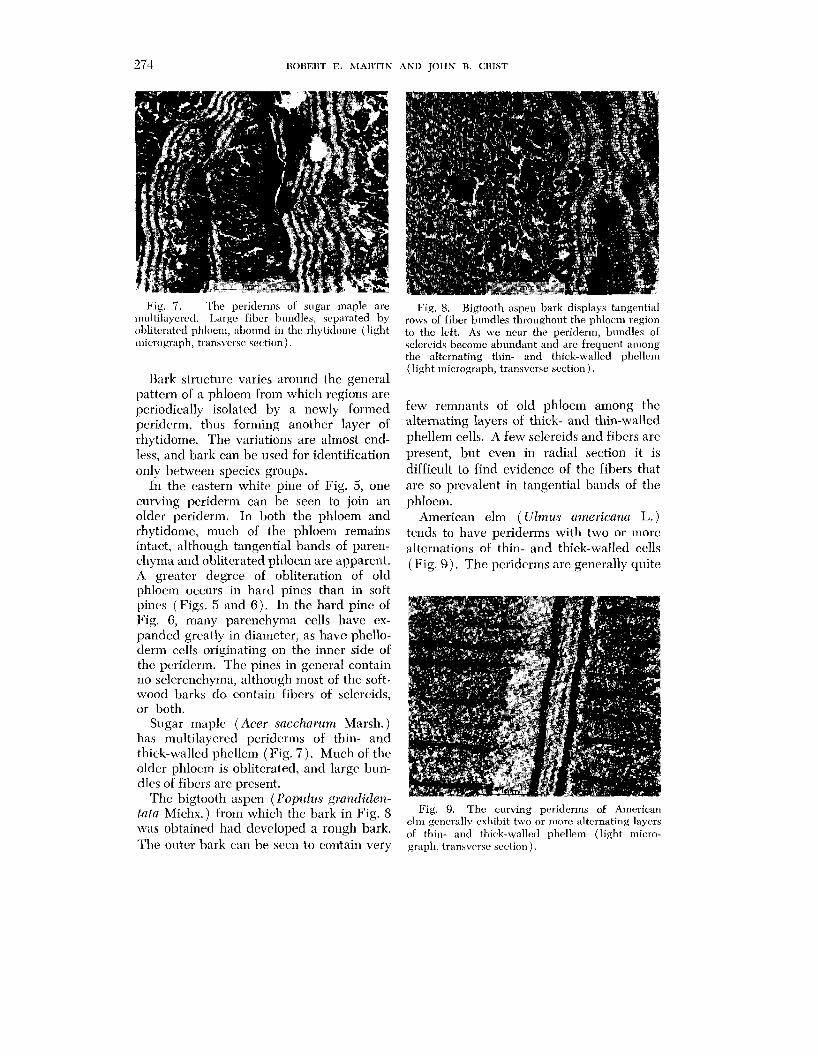
Fig. 7. The periderms of sugar maple are multilayered. Large fiber bundles, separated by obliterated phloem, abound in the rhpticlome (light micrograph, transverse section).
Bark structure varies around the general pattern of a phloem from which regions are periodically isolated by a newly formed periderm, thus forming another layer of rhytidome. The variations are almost end- less, and bark can be used for identification only between species groups.
In the eastern white pine of Fig. 5, one curving periderm can be seen to join an older periderm. In both the phloem and rhytidome, much of the phloem remains intact, although tangential bands of paren- chyma and obliterated phloem are apparent. A greatcr degree of obliteration of old phlocm occurs in hard pines than in soft pines (Figs. 5 and 6 ) . In the hard pine of Fig. 6, many parenchyma cells have ex- panded greatly in diameter, as have phello- derm cells originating on the inner side of thc pcriderm. The pines in general contain no sclerenchyma, although most of the soft- wood barks do contain fibers of sclercids, or both.
Sugar maple (Acer sacclzarum Marsh.) has multilayered periderms of thin- and thick-walled phellem (Fig. 7) . Much of the older phloem is obliterated, and large bun- dles of fibers are present.
The bigtooth aspen (Populz~s gmndiden- tutu Michx.) from which the bark in Fig. S wis obtained had developed a rough bark. The ontcr bark can be sccn to contain very
Fig. 8. Bigtooth aspen bark displays tangential rows of fiber bundles throughout the phloem region to the left. As we near the periderm, bundles of sclereids become abundant and are frequent anlong the alternating thin- and thick-walled phellem (light micrograph, transverse section ) .
few remnants of old phloem among thc alternating layers of thick- and thin-walled phellem cells. A few sclereids and fibers are present, but even in radial section it is difficult to find evidence of the fibers that are so prevalent in tangential bands of the phlocm.
American elm (Ulmus americanci L.) tends to have periderms with two or more alternations of thin- and thick-walled cells (Fig. 9). Thc peridcrms arc generally quite
Fig. 9. The curving periderms of American elin generally exhibit two or more alternating layers of thin- ant1 thick-wal1e;l pliellem (light micro- graph, transverse section).
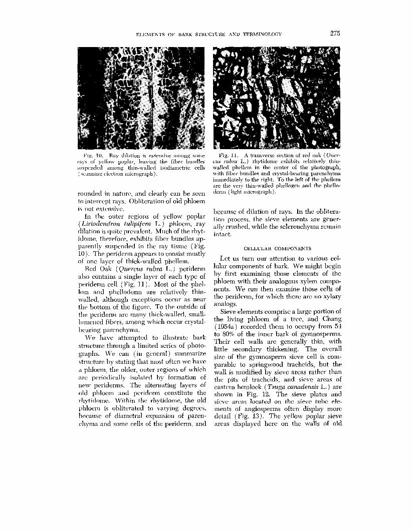
ELEMENTS OF BARK STRUCTURE AND TERMINOLOGY 275
Fig. 10. Kay dilation is extensive among some rays of yellow poplar, leaving the fiber bundles suspended anlong thin-walled isodiametric cells ( scanning rlectl.on micrograph ) .
rounded in naturc, and clearly can be seen to intercept rays. Obliteration of old phloem is not extensive.
In the outer regions of yellow poplar ( Liriodendron tulipifera L. ) phloem, ray dilation is quite prevalent. Much of the rhyt- idome, therefore, exhibits fiber bundles ap- parcntly suspended in the ray tissue (Fig. 10). The periderm appears to consist mostly of one layer of thick-walled phellem.
Ked Oak (Quercus rubra L.) periderm also contains a single layer of each type of periderm cell (Fig. 11). Most of the phel- lcm and phelloderm are relatively thin- walled, although exceptions occur as near thc bottom of the figure. To the outside of the peridcrm are many thick-walled, small- lumened fibers, among which occur crystal- bearing parenchyma.
\Ve have attempted to illustrate bark structure through a limited series of photo- graphs. We can (in gcneral) summarize structure by stating that most often we have a phloem, the older, outer regions of which are periodically isolated by formation of new periderms. The alternating layers of old phloem and periderm constitute the rh~tidomc. Within the rhytidome, the old phloem is obliterated to varying degrees, because of diametral expansion of paren- chyma and somc cells of the periderm, and
Fig. 11. A transverse section of red oak (Quer- cus rubra L.) rhytidorne exhibits relatively thin- walled phellem in the center of the photograph, with fiber bundles and crystal-bearing parenchyma immediately to the right. To the left of the phellem are the very thin-walled phellogen and the phello- derm ( light micrograph).
because of dilation of rays. In the oblitera- tion process, the sieve elements are gener- ally crushed, while the sclerenchyma remain intact.
CELLULAR COMPONENTS
Let us turn our attention to various cel- lular components of bark. We might begin by first examining those elements of the phloem with their analogous xylem compo- nents. We can then examine those cells of the periderm, for which there are no xylary analogs.
Sieve elements comprise a large portion of the living phloem of a tree, and Chang (1954a) recorded them to occupy from 54 to 80% of the inner bark of gymnosperms. Their cell walls are generally thin, with little secondary thickening. The overall size of the gymnosperm sieve cell is com- parable to springwood tracheids, but the wall is modified by sieve areas rather than the pits of tracheids, and sieve areas of eastern hemlock ( Tsuga canadensis L. ) are shown in Fig. 12. The sieve plates and sicve arcas located on the sieve tube ele- ments of angiosperms often display more detail (Fig. 13). The yellow poplar sieve areas displayed here on the walls of old

276 ROBERT E. MARTIN AND JOHN B. CRIST
Fig. 12. Sieve areas of connifers occur much ~ i ~ , 14, L~~~ narro\v spiculate crystals are as tile pitting of their tracheids, but the internal common in phloem parenchylna of many conifers, forniation of the structure is different (Eastern as illustrated by this crystal in ]oblol]y pine (Pinus hemlock, scanning electron micrograph, radial taecla L.) (scanning eIectron micrograph, radial face). face).
Fig. 13. Sievc areas on the lateral faces of a yellow poplar sieve tube element lack developnlent of a characteristic definitive callous (scanning elec- tron ~nicrograph, radial face).
sieve tube elements lack the formation of the characteristic definitive callous that generally builds up as sieve elements age. The shape of the sieve tube element is quite comparable to small diameter vessel ele- ments. Sieve areas are located laterally on the elemcnts and sieve plates on the end walls, much as the perforation plates are located on the end walls of vessel segments.
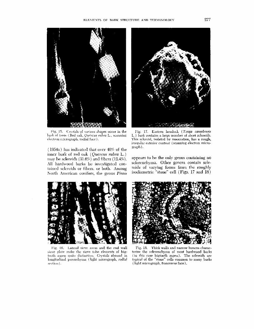
Phloem parenchyma constitutes from 12 to 3870 of the inner bark volume of North American tree spccies, according to Chang (1954a). The parenchyma may be strand or fusiform in shape and contain many of the crystals formed in bark (Figs. 14, 15, and 16). The crystals take various shapes, and may bc variable within any species.
Fibers and sclereids exhibit multilayered, birefringent walls. In most species, both types of sclerenchyma tend to be in groups surrounded by mechanically weaker tissue, a fact that has enhanced separation of fi- brous components of many barks. Chang
ELEMENTS OF BARK STRUCTURE AND TERMINOLOGY 277
Fig. 15. Crystals of various shapes occur in the I)arli of trees (Hetl oak, Qtrerczrs ~ ~ r h r a L., scanning electron micrograph, ratlial face).
(1954a) has indicatcd that over 40% of the inner bark of red oak (Quercus rubra L.) may he sclereids (31.6%) and fibers (12.4%). All hardwood barks he investigated con- tained sclerrids or fibers, or both. Among North Arncrican conifers, the genus Pinus
Fig. 17. Eastern hemlock (Tsuga canadensis L. ) bark contains a large number of short sclereids. This sclereid, isolated by maceration, has a rough, irregular exterior contour (scanning electron micro- graph).
appears to be the only genus containing no sclerenchyma. Other genera contain scle- reids of varying forms from the roughly isodiametric "stone" cell (Figs. 17 and 18)
Fig. 16. Lateral sieve areas and the end wall Fig. 18. Thick walls and narrow lumens charac- sieve plate make the sieve tube elements of big- terize the sclerenchyma of most hardwood barks tooth aspen quitc distinctive. Crystals abound in ( in this case bigtooth aspen). The sclereids are longitudinal parcncl~~ma (light micrograph, radial typical of the "stone" cells common to many barks sc,ction ) . (light micrograph, transverse face).
278 ROBERT E. MARTIN AND JOHN B. CRIST
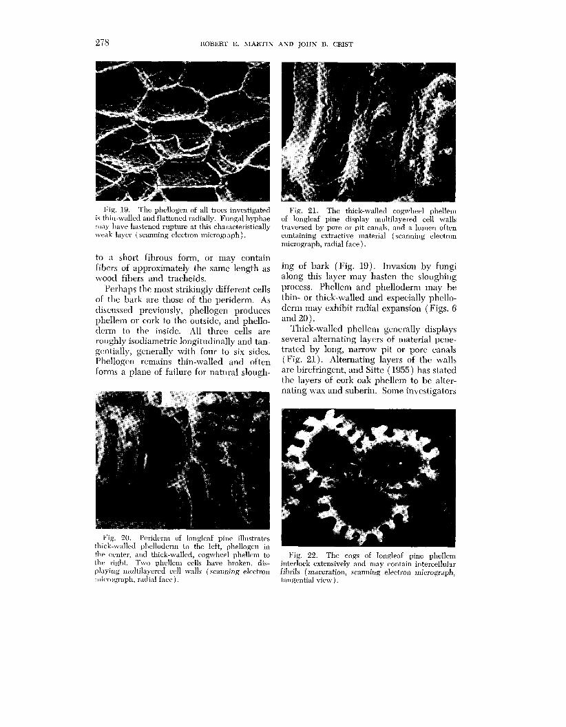
Fig. 19. The phcllogen of all trees investigated is thin-walled and flattened radially. Fungal hyphae may have hastened rupture at this characteristically weak layer (scanning electron micrograph).
to a short fibrous form, or may contain fibers of approximately the same length as wood fibers and tracheids.
Perhaps thc most strikingly different cells of the bark are those of the periderm. As discussed previously, phellogcn produces phellem or cork to the outside, and phello- dcrm to the inside. All three cells are roughly isodiametric longitudinally and tan- gentially, generally with four to six sidcs. Phellogen remains thin-walled and often forms a planc of failure for natural slough-
Fig. 20. Pcriderm of longleaf pine illustrates thick-n,alled vhelloderm to the left. ~ h e l l o ~ e n in
Fig. 21. The thick-walled cogn~heel phellern of longleaf pine display multilayered cell walls traversed by pore or pit canals, and a lumen often containing extractive material (scanning electron micrograph, radial face).
ing of bark (Fig. 19). Invasion by fungi along this layer may hasten the sloughing process. Phellcm and phelloderm may he thin- or thick-walled and especially phello- derm may exhibit radial expansion (Figs. 6 and 20).
Thick-walled phellem generally displays several alternating layers of material pene- trated by long, narrow pit or pore canals (Fig. 21). Alternating layers of the walls are birefringent, and Sitte (1955) has stated the layers of cork oak phellem to be alter- nating wax and suberin. Some investigators
, A
the ccntrr, and thick-walled, cogwheel phellem to Fig. 22. The cogs of longleaf pine phellen~ the right. Two phellem cells have broken, dis- interlock extensively and may contain intercellular playing multilayered cell walls (scanning electron fibrils (maceration, scanning electron micrograph, ~nic.l.ogl.nph, radial fact> ) . tangential view).
ELE~IEKTS OF BARK STRUCTURE AND TERMINOLOGY 279
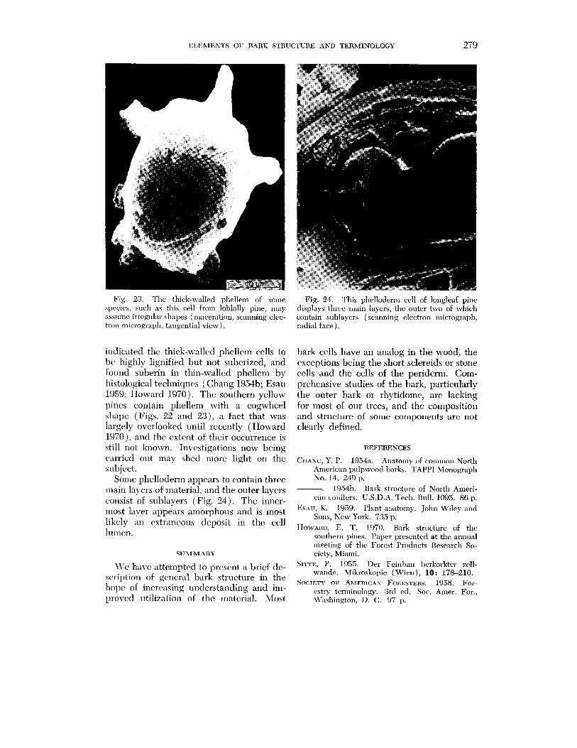
Fig. 23. The thick-walled phellem of some Fig. 24. This phelloderm cell of longleaf pine apecies, such as this cell from Ioblolly pine, may displays three main layers, the outer two of which assume irregular shapes (maceration, scanning elec- contain sublayers (scanning electron micrograph, tron micrograph, tangential view). radial face ) .
indicated thc thick-walled phellem cells to bc, highly lignified but not suberized, and found suberin in thin-walled phellem by histological techniclucs (Chang 195413; Esau 1959; Howard 1970). The southern yellow pines contain phellem with a cogwheel shape (Figs. 22 and a), a fact that was largely overlooked until recently (Howard 1970), and thc extent of their occurrence is still not known. Investigations now being curried out may shcd more light on the subject.
Some phelloderm appcars to contain three rnain layers of material, and the outer layers consist of sublayers (Fig. 24). The inner- most laver appears alnorphous and is most likely an extraneous deposit in thc cell I~lilen.
SUMMARY
bark cells have an analog in the wood, the exceptions being the short sclereids or stone cells and the cells of the periderm. Com- prehensive studies of the bark, particularly the outer bark or rhytidome, are lacking for most of our trees, and the composition and structure of some components are not clearly defined.
REFERENCES
CHANG, Y. P. 1954a. Anatomy of common North American pulpwood 1)arks. TAPPI Monograph No. 14. 249 p.
. 1954b. Bark stmcture of North Ameri- can conifers. U.S.D.A. Tech. Rnll. 1095. 86 p.
Esau, K. 1959. Plant anatomy. John Wiley ant1 Sons, New York. 735 p.
IIOWARD, E. T. 1970. Bark atructure of the southern pines. Paper presented at the annual meeting of the Fore\t Products Research So- ciety, Miami.
1 ~ 7 ~ have attempted to a brief de- S~-TF., P. 1955. Der Feinbau l~erkorkter zell- wande. Mikroskopie (Wien), 10: 178-210. scription of general bark structure in the
SO(:IETY OF AMERICAN FOHESTEHS. 1958. For- increasing understanding and im- e s t ~ ~ terminology. 3rcl eel. Sot, Arner. For,,
l ~ o \ ~ c c l litilization of the material. Most \\lashington, D. C. 07 p.