
Electronic structure, electric moments and vibrational analysis of 5-nitro-2-furaldehyde...
13
J. At. Mol. Sci. doi: 10.4208/jams.012111.022311a Vol. 2, No. 3, pp. 212-224 August 2011 Electronic structure, electric moments and vibrational analysis of 3-(2-methoxyphenoxy) propane-1,2-diol by ab initio and density functional theory Leena Sinha, Amarendra Kumar, Vijay Narayan, Rajesh K. Srivastava, and Onkar Prasad ∗ Physics Department, University of Lucknow, Lucknow 226007, India Received 21 January 2011; Accepted (in revised version) 23 February 2011 Published Online 28 March 2011 Abstract. The molecular properties and harmonic wavenumbers of 3-(2-methoxyphenoxy) propane-1,2-diol have been calculated using ab initio and density functional theory. The polarizability and first static hyperpolarizability of the title molecule have been calculated at different basis sets. In general a good agreement between experimental and calcu- lated normal modes has been observed. The frontier orbital and molecular electrostatic potential surface study has also been employed to understand the active sites of 3-(2- methoxyphenoxy) propane-1,2-diol. PACS: 31.15.A, 31.15.es, 31.15.ap Key words: Density functional theory, frontier orbital energy gap, first static hyperpolarizability 1 Introduction With the standard quantum chemical models (i.e., without the inclusion of parity violation), there is no difference whatsoever in energetics, vibrational frequencies, polarizabilities, NMR spectra, or any other non-chiral property for a given pair, i.e., (R) and (S) forms of enan- tiomers [1–4]. Differences in the properties of enantiomers arise either only within chiral en- vironments or interactions with other chiral compounds. The present investigation therefore deals with the quantum chemical study of molecular structural, energetic and vibrational data of one of the pair i.e., (R) enantiomer of 3-(2-methoxyphenoxy) propane-1,2-diol [MPPD], in gas phase, due to its biological and pharmaceutical importance. The drug MPPD, also known as guaifenesin, is an expectorant, used extensively in anti-tussive and is capable of increasing the excretion of phlegm from the respiratory tract. Bredikhin and others have carried out ∗ Corresponding author. Email address: (O. Prasad) http://www.global-sci.org/jams 212 c 2011 Global-Science Press
-
Upload
independent -
Category
Documents
-
view
4 -
download
0
Transcript of Electronic structure, electric moments and vibrational analysis of 5-nitro-2-furaldehyde...

J. At. Mol. Sci.doi: 10.4208/jams.012111.022311a
Vol. 2, No. 3, pp. 212-224August 20111
Electronic structure, electric moments and vibrational2analysis of 3-(2-methoxyphenoxy) propane-1,2-diol by3ab initio and density functional theory4Leena Sinha, Amarendra Kumar, Vijay Narayan, Rajesh K. Srivastava,5and Onkar Prasad∗6Physics Department, University of Lucknow, Lucknow 226007, India7Received 21 January 2011; Accepted (in revised version) 23 February 20118Published Online 28 March 2011910
Abstract. The molecular properties and harmonic wavenumbers of 3-(2-methoxyphenoxy)propane-1,2-diol have been calculated using ab initio and density functional theory. Thepolarizability and first static hyperpolarizability of the title molecule have been calculatedat different basis sets. In general a good agreement between experimental and calcu-lated normal modes has been observed. The frontier orbital and molecular electrostaticpotential surface study has also been employed to understand the active sites of 3-(2-methoxyphenoxy) propane-1,2-diol.
PACS: 31.15.A, 31.15.es, 31.15.ap11Key words: Density functional theory, frontier orbital energy gap, first static hyperpolarizability12131 Introduction14With the standard quantum chemical models (i.e., without the inclusion of parity violation),15there is no difference whatsoever in energetics, vibrational frequencies, polarizabilities, NMR16spectra, or any other non-chiral property for a given pair, i.e., (R) and (S) forms of enan-17tiomers [1–4]. Differences in the properties of enantiomers arise either only within chiral en-18vironments or interactions with other chiral compounds. The present investigation therefore19deals with the quantum chemical study of molecular structural, energetic and vibrational data20of one of the pair i.e., (R) enantiomer of 3-(2-methoxyphenoxy) propane-1,2-diol [MPPD], in21gas phase, due to its biological and pharmaceutical importance. The drug MPPD, also known22as guaifenesin, is an expectorant, used extensively in anti-tussive and is capable of increasing23the excretion of phlegm from the respiratory tract. Bredikhin and others have carried out24∗Corresponding author. Email address: onkarprasad1�gmail. om (O. Prasad)
http://www.global-sci.org/jams 212 c©2011 Global-Science Press

L. Sinha, A. Kumar, V. Narayan, et al. / J. At. Mol. Sci. 2 (2011) 212-224 213
extensive studies on the structure, solid state properties and issues related to the effective25resolution procedure for MPPD [5–7].26
The vibrational spectroscopic analysis is known to provide immensely invaluable molecu-27lar structure elucidation in synergy with quantum chemical calculations. In order to obtain a28complete description of molecular dynamics, vibrational wavenumber calculations along with29the normal mode analysis have been carried out at the DFT level employing the basis set306-311+G(2d ,2p). The optimized geometry of molecule under investigation and its molecu-31lar properties such as equilibrium energy, frontier orbital energy gap, molecular electrostatic32potential energy map, dipole moment, polarizability, first static hyperpolarizability have also33been used to understand the properties and active sites of the drug.342 Experimental352.1 Structure and Spectra36The model molecular structure of MPPD has been given in Fig. 1. The calculated IR spectra37has been shown in Fig. 2 and is found to match well with IR spectral data reported by NIST38Standard Reference Database 69: NIST Chemistry Web Book [8].393 Computational Details40Quantum chemical study of the MPPD, has been performed within the framework of Hartree41Fock and the density functional theory [9] with Becke’s three-parameter hybrid exchange42functional [10] with Lee-Yang-Parr correlation functionals (B3LYP) [11, 12] and employing436-311 + G(2d ,2p) basis set using the Gaussian 09 program package [13]. As the DFT hybrid44B3LYP functional tends to overestimate the fundamental normal modes of vibration, a scaling45factor of 0.9679 has been applied and a good agreement of calculated modes with experi-46mental ones has been obtained [14,15]. The vibrational wavenumber assignments have been47carried out by combining the results of the Gaussview 5 program [16], symmetry considera-48tions and the VEDA 4 program [17]. The calculated IR spectra have been plotted using the49pure Lorentzian band shape with a band width of FWHM of 10 cm−1 and are shown in Fig. 2.50
Density functional theory has also been used to calculate the dipole moment µ, meanpolarizability α and first static hyperpolarizability β based on Finite field approach. Follow-ing Buckingham’s definitions [18], the total dipole moment and the mean polarizability in aCartesian frame is defined by
µ=(µx+µy+µz)1/2, (1)
⟨α⟩=1
3(αx x+αy y+αzz). (2)
The total intrinsic hyperpolarizability βtotal and a component of the first hyperpolarizability

214 L. Sinha, A. Kumar, V. Narayan, et al. / J. At. Mol. Sci. 2 (2011) 212-224
along the direction of the dipole moment represented by βµ [19,20] are defined as
βtotal=(β2x +β
2y+β
2z )
1/2, (3)
βµ=3
5(βxµx+βyµy+βzµz)
1/2, (4)
where
βx =(βx x x+βx y y+βxzz), βy =(βy y y+βy x x+βyzz), and βz =(βzzz+βzx x+βz y y).
4 Results and discussion514.1 Molecular geometry optimization and energies52There are many possible conformations for MPPD, the conformers with lowest value of ground53state energy (without any significant difference in energetics) being the (R) and (S) enan-54
Figure 1: Optimized stru ture of R and S enantiomers of 3-(2-methoxyphenoxy) propane-1,2-diol.
Figure 2: Theoreti al IR spe tra of 3-(2-methoxyphenoxy) propane-1,2-diol.

L. Sinha, A. Kumar, V. Narayan, et al. / J. At. Mol. Sci. 2 (2011) 212-224 215
tiomers. The enantiomer couple (R) and (S) are shown in Fig. 1. The (R) enantiomer having55slightly lower energy as compared to (S) [refer to Table 1], was further optimized for the cal-56culation of its molecular properties using DFT at the B3LYP level, starting with the 6-311G(d)57basis set, the polarization and the diffused functions were incorporated in steps. The X-ray58diffraction data of the MPPD was obtained from Cambridge Crystallographic Data Center59(CCDC 693625). The initial coordinates of the title molecule thus obtained have been used60as the starting point to optimize the structure. The optimized geometry of molecule (Fig. 1)61under study is confirmed to be located at the local true minima on potential energy surface, as62the calculated vibrational spectra contains no imaginary wavenumber. The crystallographic63data of MPPD shows that the three dimensional network of molecule is linked through hy-64drogen bonds [6]. The whole molecule is nearly planer except for C1 atom and the groups65attached to it. The optimized structural parameters (bond lengths, bond angles, dihedral an-66gles) of MPPD have been compared with those in X-Ray crystal structure data [6] as shown in67Table 2. There are two dihedral angles in MPPD molecule involving C1 and C4 carbon atoms68to which hydroxyl groups are attached, where there is a significant difference between the ex-69perimentally measured [6] and calculated values (Table 2) which has been explained briefly.70The geometric differences between the optimized molecule and the molecule in solid state are71due to the fact that the molecular conformation in the gas phase is different from that in the72solid state, where inter molecular interactions play an important role in stabilizing the crystal73structure. The deviation in the dihedral angles seem to be justified in the present case as74the hydroxyl groups are involved in inter-molecular hydrogen bonding. The intra molecular75hydrogen bonding between O23-H28 atoms in calculated theoretically (2.233 Å) also agrees76well with the hydrogen bonding involved in the crystal packing data reported by Bredikhin et77al. [6]. The ester (C–O) bond lengths lie in the range (1.362–1.426 Å)/(1.360–1.424 Å) is78found to be close to the standard ester C–O bond lengths [21, 22]. The benzene endocyclic79C–C–C angles are found to lie in the range 119.30◦– 120.60◦. These calculated bond length,80bond angles are in full agreement with those standard bond lengths and bond angles.81
According to the calculations all the oxygen atoms in the title molecule carry a net neg-82ative charge. The charge on oxygen atom O(23) and O(25) being the highest and lowest83respectively.84 Table 1: Ground state optimized parameters at DFT/B3LYP level of theory for 3-(2-methoxyphenoxy)propane-1,2-diol.
Parameters 6-311+G(2d,2p)HF B3LYP
Ground state energy -686.45758 (R) -690.57982 (R)(in Hartree) -686.45710 (S) -690.57564 (S)
Frontier orbital6.38352 5.48935
energy gap (in eV)Diploe moment
2.79 2.78(in Debye)

216 L. Sinha, A. Kumar, V. Narayan, et al. / J. At. Mol. Sci. 2 (2011) 212-224Table 2: Comparison of optimized interatomi distan es (Bond lengths in Å), bond angles and dihedralangles in degrees of 3-(2-methoxyphenoxy) propane-1,2-diol (R) at B3LYP/ 6-311+G(2d,2p) level withX-Ray data.Interatomic B3LYP X-Ray Bond B3LYP X-Ray
distance Bond length data angles Bond angles dataC1–H2 1.093 0.970 C9–O26–C14 118.0 116.6C1–H3 1.091 0.971 O26–C14–C13 115.6 115.6C4–H5 1.091 0.980 O14–C13–O25 115.7 115.6C6–H8 1.093 0.969 C13–O25–C6 118.7 116.8C6–H7 1.092 0.969 O25–C6–C4 107.4 107.4C9–H10 1.093 0.960 C6–C4–C1 113.0 113.5C9–H11 1.093 0.960 C6–C4–O24 109.2 105.4C9–H12 1.086 0.960 C4–C1–O23 106.9 112.0C4–O24 1.423 1.433 C21–C13–O25 124.9 124.7C6–O25 1.424 1.428 C1–O23–H27 109.4 107.4C13–O25 1.362 1.368 C4–C1–H3 109.8 109.2C14–O26 1.363 1.370 C4–C1–H2 109.0 109.2C9–O26 1.412 1.422 O25–C6–H7 110.8 110.3
O24–H28 0.965 0.912 O25–C6–H8 110.5 110.2O23–H27 0.959 0.867
Dihedral angle B3LYP value X-ray dataO24–C4–C6–O25 172.12 177.23C4–C6–O25–C13 179.8 177.7O23–C1–C4–C6 67.5 51.2C1–C4–C6–O25 63.9 54.1
C6–O25–C13–C14 178.6 174.9O25–C13–C14–O26 0.2 0.2C13–C14–O26–C9 -179.21 -168.7
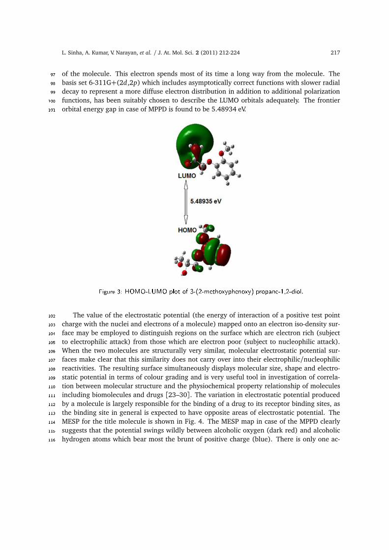
4.2 Electronic properties85The highest occupied molecular orbital (HOMO) is the orbital that primarily acts as an elec-86tron donor and the lowest unoccupied molecular orbital (LUMO) is the orbital that largely87acts as the electron acceptor. The frontier orbital energy gap helps characterize the chemical88reactivity and kinetic stability of the molecule. The 3D plots of the frontier orbitals HOMO,89LUMO and the Molecular electrostatic potential map (MESP) figures for MPPD are shown in90Figs. 3 and 4 respectively. It can be seen from Fig. 3, that the HOMO is spread heavily over the91phenyl ring region. The HOMO also shows appreciable ck bonding character. In contrast the92LUMO display Rydberg character at the alcoholic end and are located outside the molecule93and oriented perpendicular to the molecular plane. Consequently the HOMO to LUMO tran-94sition reflects the transfer of electron cloud mainly from phenyl ring to Rydberg type excited95state. The electron thus transferred to the Rydberg state is very weakly bound to the rest96
L. Sinha, A. Kumar, V. Narayan, et al. / J. At. Mol. Sci. 2 (2011) 212-224 217
of the molecule. This electron spends most of its time a long way from the molecule. The97basis set 6-311G+(2d ,2p) which includes asymptotically correct functions with slower radial98decay to represent a more diffuse electron distribution in addition to additional polarization99functions, has been suitably chosen to describe the LUMO orbitals adequately. The frontier100orbital energy gap in case of MPPD is found to be 5.48934 eV.101
Figure 3: HOMO-LUMO plot of 3-(2-methoxyphenoxy) propane-1,2-diol.The value of the electrostatic potential (the energy of interaction of a positive test point102
charge with the nuclei and electrons of a molecule) mapped onto an electron iso-density sur-103face may be employed to distinguish regions on the surface which are electron rich (subject104to electrophilic attack) from those which are electron poor (subject to nucleophilic attack).105When the two molecules are structurally very similar, molecular electrostatic potential sur-106faces make clear that this similarity does not carry over into their electrophilic/nucleophilic107reactivities. The resulting surface simultaneously displays molecular size, shape and electro-108static potential in terms of colour grading and is very useful tool in investigation of correla-109tion between molecular structure and the physiochemical property relationship of molecules110including biomolecules and drugs [23–30]. The variation in electrostatic potential produced111by a molecule is largely responsible for the binding of a drug to its receptor binding sites, as112the binding site in general is expected to have opposite areas of electrostatic potential. The113MESP for the title molecule is shown in Fig. 4. The MESP map in case of the MPPD clearly114suggests that the potential swings wildly between alcoholic oxygen (dark red) and alcoholic115hydrogen atoms which bear most the brunt of positive charge (blue). There is only one ac-116

218 L. Sinha, A. Kumar, V. Narayan, et al. / J. At. Mol. Sci. 2 (2011) 212-224
Figure 4: MESP surfa e and ontour plots of 3-(2-methoxyphenoxy) propane-1,2-diol. Contours aredrawn 1.4 Å above the mole ular plane.tive electrophilic site. The larger extent of spread of green colour which corresponds to a117potential halfway between the two extremes red and blue colour suggests that most part of118the molecule has almost the neutral potential. The sliced 2D MESP contour map of the title119molecule has also been plotted in Fig. 4. Such a representation provides more detailed infor-120mation regarding electrostatic potential distribution, by showing the values in a manifold of121spatial location around the molecule. The MESP 2D contour maps for MPPD is drawn in the122plane 1.4 Å above the molecular plane. These maps clearly shows that the maximum value of123positive and negative potential are 0.4 a.u. and -0.04 a.u. respectively.1244.3 Electric moments125The origin of intermolecular interactions involving the van der Waal type dipole-dipole forces126etc. is mainly attributed to an overall imbalance in charge from one side of a molecule to the127
L. Sinha, A. Kumar, V. Narayan, et al. / J. At. Mol. Sci. 2 (2011) 212-224 219
other side. The calculated dipole moment value is 2.78 Debye for MPPD (refer to Table 1).128The extensive and important studies on electric polarizability and hyperpolarizability,129
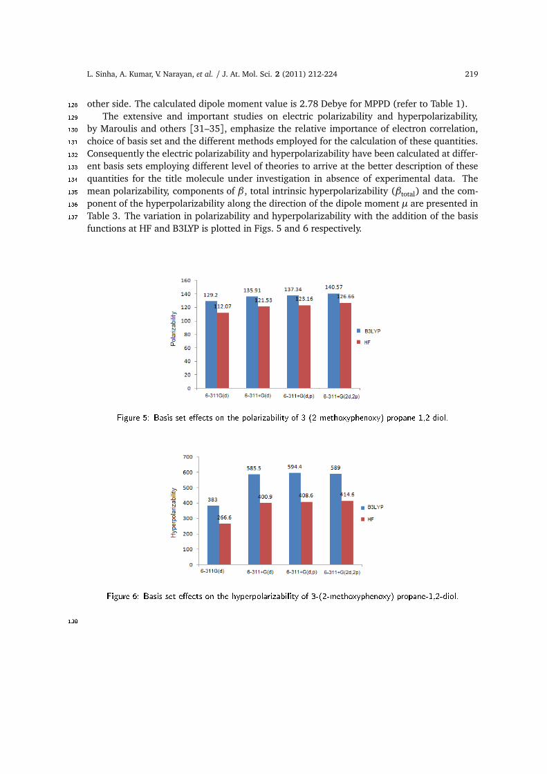
by Maroulis and others [31–35], emphasize the relative importance of electron correlation,130choice of basis set and the different methods employed for the calculation of these quantities.131Consequently the electric polarizability and hyperpolarizability have been calculated at differ-132ent basis sets employing different level of theories to arrive at the better description of these133quantities for the title molecule under investigation in absence of experimental data. The134mean polarizability, components of β , total intrinsic hyperpolarizability (βtotal) and the com-135ponent of the hyperpolarizability along the direction of the dipole moment µ are presented in136Table 3. The variation in polarizability and hyperpolarizability with the addition of the basis137functions at HF and B3LYP is plotted in Figs. 5 and 6 respectively.
Figure 5: Basis set e�e ts on the polarizability of 3-(2-methoxyphenoxy) propane-1,2-diol.
Figure 6: Basis set e�e ts on the hyperpolarizability of 3-(2-methoxyphenoxy) propane-1,2-diol.138
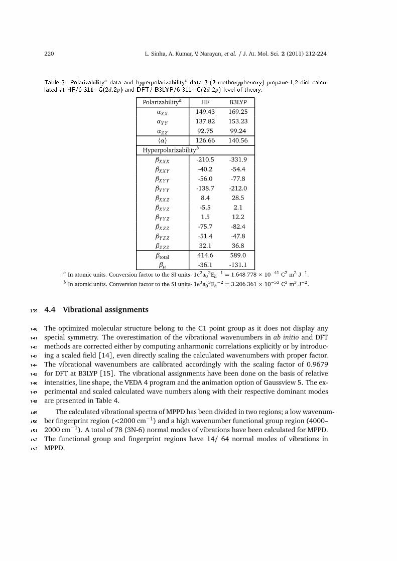
220 L. Sinha, A. Kumar, V. Narayan, et al. / J. At. Mol. Sci. 2 (2011) 212-224Table 3: Polarizabilitya data and hyperpolarizabilityb data 3-(2-methoxyphenoxy) propane-1,2-diol al u-lated at HF/6-311+G(2d,2p) and DFT/ B3LYP/6-311+G(2d,2p) level of theory.Polarizabilitya HF B3LYP
αX X 149.43 169.25
αY Y 137.82 153.23
αZ Z 92.75 99.24
⟨α⟩ 126.66 140.56
Hyperpolarizabilityb
βX X X -210.5 -331.9
βX X Y -40.2 -54.4
βX Y Y -56.0 -77.8
βY Y Y -138.7 -212.0
βX X Z 8.4 28.5
βX Y Z -5.5 2.1
βY Y Z 1.5 12.2
βX Z Z -75.7 -82.4
βY Z Z -51.4 -47.8
βZ Z Z 32.1 36.8
βtotal 414.6 589.0
βµ -36.1 -131.1a In atomic units. Conversion factor to the SI units- 1e2a0
2Eh−1 = 1.648 778 × 10−41 C2 m2 J−1.
b In atomic units. Conversion factor to the SI units- 1e3a03Eh−2 = 3.206 361 × 10−53 C3 m3 J−2.
4.4 Vibrational assignments139The optimized molecular structure belong to the C1 point group as it does not display any140special symmetry. The overestimation of the vibrational wavenumbers in ab initio and DFT141methods are corrected either by computing anharmonic correlations explicitly or by introduc-142ing a scaled field [14], even directly scaling the calculated wavenumbers with proper factor.143The vibrational wavenumbers are calibrated accordingly with the scaling factor of 0.9679144for DFT at B3LYP [15]. The vibrational assignments have been done on the basis of relative145intensities, line shape, the VEDA 4 program and the animation option of Gaussview 5. The ex-146perimental and scaled calculated wave numbers along with their respective dominant modes147are presented in Table 4.148
The calculated vibrational spectra of MPPD has been divided in two regions; a low wavenum-149ber fingerprint region (<2000 cm−1) and a high wavenumber functional group region (4000–1502000 cm−1). A total of 78 (3N-6) normal modes of vibrations have been calculated for MPPD.151The functional group and fingerprint regions have 14/ 64 normal modes of vibrations in152MPPD.153

L. Sinha, A. Kumar, V. Narayan, et al. / J. At. Mol. Sci. 2 (2011) 212-224 221Table 4: Theoreti al and experimental∗ wave numbers of 3-(2-methoxyphenoxy) propane-1,2-diol.Scaled Exp.∗ IR Assignment of dominant modes
wave no. wave no. in terms ofin cm−1 in cm−1 potential energy distribution (PED)
3741 3243 ν(O23–H27)(100)3663 3074 ν(O24–H28)(100)3107 – νs ((C–H))R(92)3101 – νas ((C–H))R(91)3085 – νas ((C–H))R(94)3069 – νas ((C–H))R(95)3030 3009 νas CH3(92)2977 – ν (C4–H5)(63)+ νas (C6H2)(36)2971 – νas (C1H2)(99)2961 νas (C6H2)( 64) + ν(C4–H5)(34)2960 2942 νas CH3(100)2920 – νs (C6H2)(63)+ νs (C1H2)(27)2918 νs (C1H2)(62) + νs(C6H2)(36)2905 2887 νs CH3(92)1580 1594 ν(C–C)R(38) + β(C–C–C) R(22)+ β(H–C–C)R(10)1576 – ν(C–C)R(71)1491 1525 β(H–C–C)R(34)+ ν(C–C)R(17)1462 1468 sc(C6H2)(58) + sc(C1H2)(15)1460 1455 sc(C1H2)(56) + sc(C6H2)(21)1458 1455 CH3 as. Deformation(78)1445 1441 CH3 as. Deformation(93)1438 – β(H–C–C)R(34)+ CH3 umbrella bending (12) + ν(C–C)R (11)1423 – CH3 umbrella bend(50) + β(H–C–C)R(14)1395 1383 ω(C4–H5)(16)+β(H28–O24–C4)(14)+β(H27–O23–C1)(12)1379 1376 ω(C6H2)(26)+ω(C4–H5)(18)+β(H28–O24–C4)(15)1368 – ω(C4–H5)(29)+ω(C6H2)(12)+ω(C1H2)(18)+ β(H28–O24–4)(10)1322 1330 ω(C4–H5)(44)+ ω(C6H2)(12)1319 1295 ν(C–C)R(43) + ω(C6H2)(16) + ω(C4–H5)(11)1272 1267 β(H–C–C)R(54)1256 – ω(C1H2)(26)+ω(C4–H5)(20)+β(H28–O24–C4)(16)1242 – ω(C1H2)(15)+ω (C6H2)(15)+ ν(O26–C14)(10)1238 1231 ω(C1H2)(24)+ω(C6H2)(24)+ ν(C–C)R(10)1212 1223 ν(O25–C13)(23) +ν(O26–C14)(20) + β (H–C–C)R(16)1195 1187 ω(C6H2)(49)+ω(C1H2)(15)+β(H28–O24–C4)(10)1162 1180 r(CH3)(65)1149 – β (H–C–C)R(65) +ν(C–C)R(11)1139 1129 β(H27–O23–C1)(44)+r(C1H2)(15)+β(H28–O24–C4)(13)+ ω(C4–H5) (12)1131 – r(CH3)(99)1110 1107 ν(C–C)R(32)+ β(H–C–C)R(24)1101 – ν(C4–C6)(26)+β(H27–O23–C1)(14)+ r(C1H2)(13)1088 1088 ν(C1–C4)(20)+ ν(O24–C4)(17)+ ν(O23–C1)(14)1042 1058 ν(C–C)R (22)+ ν(O26–C9)(13)+β (H–C–C)R(21)1023 1044 ν(O23–C1)(17)+ ν(O24–C4)(16)+ r(C6H2)(15)1022 1022 ν(O24–C4)(34)+ R Breathing(30)+ r(C6H2)(14)1017 995 ν(O25–C6)(50)+R trigonal bending(24)+ ν(O26–C9)(12)966 – ν(O24–C4)(34)+ν(C6–C4)(12)+ν(O23–C1)(37) +r(C6H2)(14)964 935 (C–H) R wag(73)919 911 r(C1H2)(26) +ν(O24–C4)(18)+ ν(O23–C1)(12)884 838 (C–H) R wag(80)825 – ν(C1–C4)(24)+ β(H–C–C) R(15) + r(C6H2) (10)819 – (C–H) R wag(53) + ρ(C–C–C–C) R(21)804 – ν(C1–C4)(34)+ β(C–C–C) R(22)756 769 β(C–C–C) R(50)+ r(CH3)(23)729 744 ρ (C–C–C–C) R(64)+ (C–H) R wag(26)723 – (C–H) R wag(83)633 641 β(O23–C1–C4)(28)+β(O24–C4–C1)(25)+ r(C6H2)(16)588 – β (C–C–C) R (44)+ r(C1H2)(11)567 – r(CH3) (29) + β(O26–C14–C15) (27) + β(O25–C13–C21)(12)557 – ρ(C–C–C–C)R(62)+ρ(O26–C13–C15–C14)(11)+ρ(O25–C14–C21–C13)(11)523 – β(C–C–C)R(31)+ β(C4–C6–O25)(12)+ β(O24–C4–C6)(10)456 – ρ(C–C–C–C)R(23)+ρ(H–C–C–C)R(20)+ρ(O26–C13–C15–C14)(12)+ρ(O25–C14–C21–C13)(12)436 – βout(H28–O24–C4)(55)+β(C6–C4–O24)(19)420 – βout(H28–O24–C4)(39)+β(C6–C4–O24)(18)
∗Experimental IR data as reported in NIST Chemistry Web Book:http://webbook.nist.gov/ hemistry/form-ser.html. Abbreviations used here have following meanings.ν : stretching; νs: symmetric stretching; νas: asymmetric stretching; β: in plane bending; βout: out of plane bending;ρ: torsion; sc: scissoring; ω: wagging; γ: twisting; r: rocking; R: phenyl ring.

222 L. Sinha, A. Kumar, V. Narayan, et al. / J. At. Mol. Sci. 2 (2011) 212-224
O–H vibrations154The O-H stretching vibration is very sensitive to hydrogen bonding. A free hydroxyl155
group or a non-hydrogen bonded hydroxyl group absorbs in the range 3700-3500 cm−1.156The intra-molecular hydrogen bonding present in the system reduces the hydroxyl stretch-157ing band to 3559-3200 cm−1 region [36]. The scaled wavenumber calculated at 3741 cm−1158and 3663 cm−1 in the case of MPPD are identified as O–H stretching with 100% contribution159to P.E.D. The large variation in the calculated O–H stretching vibrational wavenumbers with160the experimental ones may also be attributed due to the existence of four strong hydrogen161bonding sites in 3-(2-ethoxyphenoxy) propane-1,2-diol as reported by Bredikhin et al. [6].162CH2 vibrations163
MPPD has two methylene groups, at the C1 and C6 according to the numbering scheme164(Fig. 1). The asymmetric stretching of CH2 group is calculated in the range 2977–2961 cm−1165whereas symmetric stretching lies in the range 2920–2918 cm−1. The twisting and rocking166modes of methylene group are calculated to be mixed modes and start appearing around1671260 cm−1. The major methylene scissoring modes at 1462 and 1460 cm−1 are matched well168with experimental wavenumbers. The methylene wagging modes are calculated in the range1691380–1320 cm−1 for the MPPD.170CH3 group modes171
The methyl group shows several characteristic fundamental vibrations corresponding to172asymmetric and symmetric stretches, bending, rocking, and torsion modes. The CH3 asym-173metric stretching vibrations are generally observed in the region of 2980–2940 cm−1, while174the symmetric stretching vibrations usually appear between 2890 and 2850 cm−1. The wavenum-175bers for asymmetric CH3 stretching modes are calculated at 3030 cm−1 and 2960 cm−1 while176the wavenumber of symmetric stretching vibrations calculated at 2905 cm−1 and are assigned177to well to the experimental values. Methyl asymmetric deformation modes appear as dom-178inant mode at 1458, 1445 cm−1 (with more than 65% contribution to the total P.E.D.) are179assigned to 1455 cm−1 and 1441 cm−1 peak in the IR spectra. The methyl umbrella bend-180ing modes calculated for the present case are in the wavenumber range of 1440-1420 cm−1.181These bending modes are in general mixed with ring (H–C–C) bending modes (Table 6). The182dominant methyl rocking modes are calculated to be at 1162 and 1131 cm−1.183C–C and C–H vibrations184
C–C stretching wavenumbers are observed as mixed modes in the range 1100 cm−1 to185800 cm−1 and agree well with the general appearance of C–H and C–C stretching modes.186C–C stretches are calculated to be 1101, 1088, 966, 825 and 804 cm−1. The C–H stretching187vibration [ν(C4–H5)] is calculated at 2977 cm−1.188Ring vibrations189
The phenyl ring spectral region predominantly involves the C–H, C–C and C=C stretching,190and C–C–C as well as H–C–C-bending vibrations. The bands due to the ring C–H-stretching191

L. Sinha, A. Kumar, V. Narayan, et al. / J. At. Mol. Sci. 2 (2011) 212-224 223
vibrations were observed as a group of partially overlapping absorptions in the region 3110-1923069 cm−1 with more than 90% potential energy contribution. Vibrations involving C–H193in-plane bending are found in the region 1600-825 cm−1. The computed wavenumbers at1941017 cm−1 are identified as the trigonal ring bending and 1022 cm−1 as ring breathing modes195respectively. The dominant phenyl ring wagging modes are calculated 964, 884 and 723 cm−1.196A good agreement between the calculated and experimentally observed wavenumbers has197allowed establishing a detailed and precise assignment of normal mode wavenumbers in the198entire spectral region.1995 Conclusions200The present study on 3-(2-methoxyphenoxy) propane-1,2-diol, comprised of equilibrium ge-201ometries optimization and the calculation of molecular ground state properties at DFT/6-202311+G(2d ,2p) level. The electric polarizability and hyperpolarizability have also been calcu-203lated at different basis sets employing different level of theories to arrive at the better portrayal204of these quantities for the title molecule under investigation. In general, a good agreement205between experimental and calculated normal modes has been observed. The structure activity206relationship based on the study of frontier orbital gap, dipole moment data, electric polariz-207ability and first static hyperpolarizability along with the molecular electrostatic potential map208of the 3-(2-methoxyphenoxy) propane-1,2-diol, has been used to understand the active sites209of the molecule under study.210Acknowledgments. Research support is gratefully acknowledged from Authors (OP and LS)211and to UGC, India for the financial support and to Prof Jamroz for providing his VEDA 4212software. We would also like to thank Prof. Jenny Field for providing the crystal data of (R)213enantiomer of MPPD.214References215[1] G. Boon, C. V. Alsenoy, F. D. Proft, et al., J. Phys. Chem. A 110 (15) (2006) 5114.216[2] S. Janssens, C. V. Alsenoy, and P. Geerlings, J. Phys. Chem. A 111 (16) (2007) 3143.217[3] K. Le Barbu, A. Zehnacker, F. Lahmani, et al., Chirality 13 (10) (2001) 715.218[4] H. F. Skinner, Forensic Science International 48 (1990) 128.219[5] Z. A. Bredikhina, V. G. Novikova, D. V. Zakharychev, and A. A. Bredikhin, Tetrahedron: Asymme-220
try 17 (2006) 3015.221[6] A. A. Bredikhin, A. T. Gubaidullin, Z. A. Bredikhina, et al., J. Mol. Struct. 920 (2009) 377.222[7] A. A. Bredikhin, Z. A. Bredikhina, D. V. Zakharychev, and A. V. Pashagin, Tetrahedron: Asymmetry223
18 (2007) 1239.224[8] http://webbook.nist.gov/ hemistry/form-ser.html.225[9] W. Kohn and L. J. Sham, Phys. Rev. 140 (1965) A1133.226[10] A. D. Becke, J. Chem. Phys. 98 (1993) 5648.227[11] C. Lee, W. Yang, and R. G. Parr, Phys. Rev. B 37 (1988) 785.228[12] B. Miehlich, A. Savin, H. Stoll, and H. Preuss, Chem. Phys. Lett. 157 (1989) 200.229

224 L. Sinha, A. Kumar, V. Narayan, et al. / J. At. Mol. Sci. 2 (2011) 212-224
[13] M. J. Frisch, G. W. Trucks, H. B. Schlegel, et al., Gaussian 09, Rev. A. 1 (Gaussian, Inc., Walling-230ford CT, 2009).231
[14] A. P. Scott, L. Random, J. Phys. Chem. 100 (1996) 16502.232[15] P. Pulay, G. Fogarasi, G. Pongor, et al., J. Am. Chem. Soc. 105 (1983) 7037.233[16] Æ. Frisch, H. P. Hratchian, R. D. Dennington II, et al., GaussView, Version 5.0 (Gaussian, Inc.,234
Wallingford CT, 2009).235[17] M. H. Jamroz, Vibrational Energy Distribution Analysis: VEDA 4 Program (Warsaw, Poland,236
2004).237[18] A. D. Buckingham, Adv. Chem. Phys. 12 (1967) 107.238[19] D. R. Kanis, M. A. Ratner, and T. J. Marks, Chem. Rev. 94 (1994) 195.239[20] G. Maroulis, J. Chem. Phys. 113 (2000) 1813.240[21] M. Ladd, Introduction to Physical Chemistry, third ed.(Cambridge University Press, Cambridge,241
1998).242[22] F. H. Allen, O. Kennard, D. G.Watson, et al., J. Chem. Soc., Perkin Trans. II (1987) S1-S19.243[23] I. Fleming, Frontier Orbitals and Organic Chemical Reactions (John Wiley and Sons, New York,244
1976).245[24] J. S. Murray and K. Sen, Molecular Electrostatic Potentials, Concepts and Applications (Elsevier,246
Amsterdam, 1996).247[25] I. Alkorta and J. J. Perez, Int. J. Quant. Chem. 57 (1996) 123.248[26] E. Scrocco and J. Tomasi, in: Advances in Quantum Chemistry, ed. P. Lowdin (Academic Press,249
New York, 1978).250[27] F. J. Luque, M. Orozco, P. K. Bhadane, and S. R. Gadre, J. Phys. Chem. 97 (1993) 9380.251[28] J. Sponer and P. Hobza, Int. J. Quant. Chem. 57 (1996) 959.252[29] R. K. Pathak and S. R. Gadre, J. Chem. Phys. 93 (1990) 1770.253[30] S. R. Gadre and I. H. Shrivastava, J. Chem. Phys. 94 (1991) 4384.254[31] G. Maroulis, D. Begue, and C. Pouchan, J. Chem. Phys. 119 (2003) 794.255[32] G. Maroulis, P. Karamanis, and C. Pouchan, J. Chem. Phys. 126 (2007) 154316.256[33] G. Maroulis, J. Chem. Phys. 129 (2008) 044314.257[34] J. M. Stout and C. E. Dykstra, J. Am. Chem. Soc. 117 (1995) 5127.258[35] M. A. Spackman, J. Chem. Phys. 93 (1989) 7594.259[36] M. Jag, Organic Spectroscopy - Principles and Applications, 2nd ed. ( Narosa Publishing House,260
New Delhi, 2001).261