Electromagnetic controlled cortical impact device for precise ...
19
Washington University School of Medicine Digital Commons@Becker Open Access Publications 2007 Electromagnetic controlled cortical impact device for precise, graded experimental traumatic brain injury David L. Brody Washington University School of Medicine Christine Mac Donald Washington University School of Medicine Chad C. Kessens Washington University School of Medicine Carla Yuede Washington University School of Medicine Maia Parsadanian Washington University School of Medicine See next page for additional authors Follow this and additional works at: hps://digitalcommons.wustl.edu/open_access_pubs is Open Access Publication is brought to you for free and open access by Digital Commons@Becker. It has been accepted for inclusion in Open Access Publications by an authorized administrator of Digital Commons@Becker. For more information, please contact [email protected]. Recommended Citation Brody, David L.; Mac Donald, Christine; Kessens, Chad C.; Yuede, Carla; Parsadanian, Maia; Spinner, Mike; Kim, Eddie; Schwetye, Katherine E.; Holtzman, David M.; and Bayly, Philip V., ,"Electromagnetic controlled cortical impact device for precise, graded experimental traumatic brain injury." Journal of Neurotrauma.24,4. 657-673. (2007). hps://digitalcommons.wustl.edu/open_access_pubs/4741
-
Upload
khangminh22 -
Category
Documents
-
view
3 -
download
0
Transcript of Electromagnetic controlled cortical impact device for precise ...

Washington University School of MedicineDigital Commons@Becker
Open Access Publications
2007
Electromagnetic controlled cortical impact devicefor precise, graded experimental traumatic braininjuryDavid L. BrodyWashington University School of Medicine
Christine Mac DonaldWashington University School of Medicine
Chad C. KessensWashington University School of Medicine
Carla YuedeWashington University School of Medicine
Maia ParsadanianWashington University School of Medicine
See next page for additional authors
Follow this and additional works at: https://digitalcommons.wustl.edu/open_access_pubs
This Open Access Publication is brought to you for free and open access by Digital Commons@Becker. It has been accepted for inclusion in OpenAccess Publications by an authorized administrator of Digital Commons@Becker. For more information, please contact [email protected].
Recommended CitationBrody, David L.; Mac Donald, Christine; Kessens, Chad C.; Yuede, Carla; Parsadanian, Maia; Spinner, Mike; Kim, Eddie; Schwetye,Katherine E.; Holtzman, David M.; and Bayly, Philip V., ,"Electromagnetic controlled cortical impact device for precise, gradedexperimental traumatic brain injury." Journal of Neurotrauma.24,4. 657-673. (2007).https://digitalcommons.wustl.edu/open_access_pubs/4741

AuthorsDavid L. Brody, Christine Mac Donald, Chad C. Kessens, Carla Yuede, Maia Parsadanian, Mike Spinner,Eddie Kim, Katherine E. Schwetye, David M. Holtzman, and Philip V. Bayly
This open access publication is available at Digital Commons@Becker: https://digitalcommons.wustl.edu/open_access_pubs/4741

JOURNAL OF NEUROTRAUMAVolume 24, Number 4, 2007© Mary Ann Liebert, Inc.Pp. 657–673DOI: 10.1089/neu.2006.0011
Electromagnetic Controlled Cortical Impact Device for Precise, Graded Experimental Traumatic Brain Injury
DAVID L. BRODY,1 CHRISTINE MAC DONALD,2 CHAD C. KESSENS,3 CARLA YUEDE,4MAIA PARSADANIAN,1 MIKE SPINNER,1 EDDIE KIM,1 KATHERINE E. SCHWETYE,5
DAVID M. HOLTZMAN,1,5,6 and PHILIP V. BAYLY3
ABSTRACT
Genetically modified mice represent useful tools for traumatic brain injury (TBI) research and at-tractive preclinical models for the development of novel therapeutics. Experimental methods thatminimize the number of mice needed may increase the pace of discovery. With this in mind, we de-veloped and characterized a prototype electromagnetic (EM) controlled cortical impact device alongwith refined surgical and behavioral testing techniques. By varying the depth of impact between 1.0and 3.0 mm, we found that the EM device was capable of producing a broad range of injury sever-ities. Histologically, 2.0-mm impact depth injuries produced by the EM device were similar to 1.0-mm impact depth injuries produced by a commercially available pneumatic device. Behaviorally,2.0-, 2.5-, and 3.0-mm impacts impaired hidden platform and probe trial water maze performance,whereas 1.5-mm impacts did not. Rotorod and visible platform water maze deficits were also foundfollowing 2.5- and 3.0-mm impacts. No impairment of conditioned fear performance was detected.No differences were found between sexes of mice. Inter-operator reliability was very good. Behav-iorally, we found that we could statistically distinguish between injury depths differing by 0.5 mmusing 12 mice per group and between injury depths differing by 1.0 mm with 7–8 mice per group.Thus, the EM impactor and refined surgical and behavioral testing techniques may offer a reliableand convenient framework for preclinical TBI research involving mice.
Key words: behavior; controlled cortical impact; experimental traumatic brain injury; histology; mice
657
Departments of 1Neurology, 2Biomedical Engineering, 3Mechanical Engineering, 4Psychiatry, 5Anatomy and Neurobiology,6Molecular Biology & Pharmacology, Hope Center for Neurological Disorders, Washington University, St. Louis, Missouri.
INTRODUCTION
TRAUMATIC BRAIN INJURY (TBI) is a major cause ofdeath and disability, for which no effective treatment
exists other than supportive care. A total of 5.3 millionAmericans—2% of the U.S. population—currently livewith disabilities resulting from TBI (Thurman et al.,1999). Because many of the victims are young, the total
costs to society in medical care and lost productivity areextremely high. However, despite substantial research ef-fort, effective therapeutics have not been developed, andfurther preclinical and clinical research tools are needed(Narayan et al., 2002).
Animal models of TBI are critical in order to test mech-anistic hypotheses and develop preclinical therapeutics(Lighthall et al., 1989; Meaney et al., 1994; Shohami et
al., 1995; Laurer and McIntosh, 1999; Duhaime et al.,2000; Bayir et al., 2003; Pineda et al., 2004; Faden et al.,2005; Kleindienst et al., 2005; Lenzlinger et al., 2005;Maegele et al., 2005; Maxwell et al., 2005; Prins et al.,2005; Thompson et al., 2005; Truettner et al., 2005; Bukiand Povlishock, 2006; Marklund et al., 2006; Marmarouand Povlishock, 2006). There are many animal modelsof TBI, but only in transgenic mice can the effects of ge-netic factors be readily explored in an experimental set-ting. For example, the �4 allele of the apolipoprotein Egene (APOE4) may be a risk factor for poor outcome af-ter TBI in humans (Jellinger, 2004), especially in youngerpatients (Teasdale et al., 2005). Several lines of trans-genic mice expressing each of the ApoE alleles—E2, E3,and E4—have been produced (Xu et al., 1996; Sullivanet al., 1997; Sun et al., 1998), and initial studies indicatean ApoE allele-dependent effect on survival and cogni-tive outcome following experimental TBI in these ani-mals (Sabo et al., 2000). TBI has been performed in micewith many other genetic manipulations, and these exper-iments have provided important insights (Longhi et al.,2001; Hartman et al., 2002; Uryu et al., 2002; Chang etal., 2003; Hlatky et al., 2003; Conte et al., 2004; Bayiret al., 2005; Abrahamson et al., 2006; Bermpohl et al.,2006; Kochanek et al., 2006).
In mice, the controlled cortical impact (CCI) model ofexperimental TBI is widely used because it yields con-sistent histological damage and behavioral deficits. Othertechniques include weight-drop, impact acceleration, andfluid percussion models (Morales et al., 2005). In the CCItechnique, mice are fully anesthetized, the skull is ex-posed, and a craniotomy is performed. Then, an impactorindents the exposed surface of the brain by a fixed dis-tance at a fixed velocity. The mice recover well withminimal mortality, and have highly reproducible grosshistological lesions in the underlying cortex and hip-pocampus. Importantly, they have consistent, moderatelyimpaired performance on cognitive tasks such as the Mor-ris water maze (Fujimoto et al., 2004; Saatman et al.,2006). CCI is generally performed using a pneumatic im-pact device that is supported by a solid metal frame. Amilling table supporting the mouse and restraining appa-ratus can be used to precisely specify injury location rel-ative to anatomical landmarks (Fox et al., 1998). Cali-bration and adjustment of gas pressures is performed toensure reproducible impact velocities.
We have recently developed an electromagnetic im-pact device that attaches to an arm of a stereotaxic frameand delivers reproducible velocities without the need forfrequent calibration. The device was capable of produc-ing consistent, graded CCI injuries in adult mice withstereotaxic control of impact location and depth at highvelocities. An earlier version of the device has been em-
ployed to produce closed skull impacts in a post-natalday 7 rat model at moderate velocities (Bayly et al.,2006).
METHODS
Device Design, Fabrication, and Calibration
During the initial design phase, the primary design goalwas achievement of the desired velocity range while min-imizing the size of the apparatus so that it could be usedas a stereotaxic arm-mounted accessory. A wide varietyof options were generated to this end. These options in-cluded the use of solenoids, CO2 cartridges, pneumatics,spring and lever systems, and electromagnetic (EM)coils.
Of these, an EM coil-based design was chosen becauseit offered several advantages: (1) EM coil–based and so-lenoid-based designs appeared to be able to produce thehighest velocities with the most compact devices. (2) InEM coil–based systems, a stationary magnet propels awire coil, as opposed to a solenoid-based design in whicha coiled wire generates a magnetic field that propels aniron core; this is advantageous because the coil is gener-ally lighter than the iron core, making it easier to accel-erate over short distances. (3) A coil can more easily bemodified than a solenoid to allow for accessories such assensors and alternative tip geometries. (4) Most impor-tantly, the use of a true magnet makes the direction ofthe current through the coil significant; the direction ofmotion can be reversed simply by reversing the directionof the current through the coil, whereas in a solenoid-based design, a second element would be needed to re-verse the direction of motion.
A moving coil design has one significant complica-tion. When the coil moves in a magnetic field, a poten-tial is created in the coil. The resulting voltage opposesthe current through the coil, acting to decrease the ve-locity. Because this effect runs opposite the driving force,it is called a “back EMF,” or backwards electro-motiveforce (EMF). The strength of this field is directly pro-portional to the speed of the motion, as given by the equa-tion VB � kt�, where VB is the back EMF, kt is a pro-portionality constant dependent on the device, and � isthe velocity. Thus, the faster the coil moves, the strongerthe opposing voltage becomes. The end result is that, athigher speeds, a higher voltage must be applied to thecoil to induce currents.
We designed an EM coil-based impact device intendedto be mounted on the arm of a stereotaxic instrument (Fig.1A). A voice coil with stroke length 0.762 cm and coilmass 32.6 g (BEI Kimco, LA12-17-000A) was housedin a piston-like stationary cylinder (1.9 cm diameter) fab-
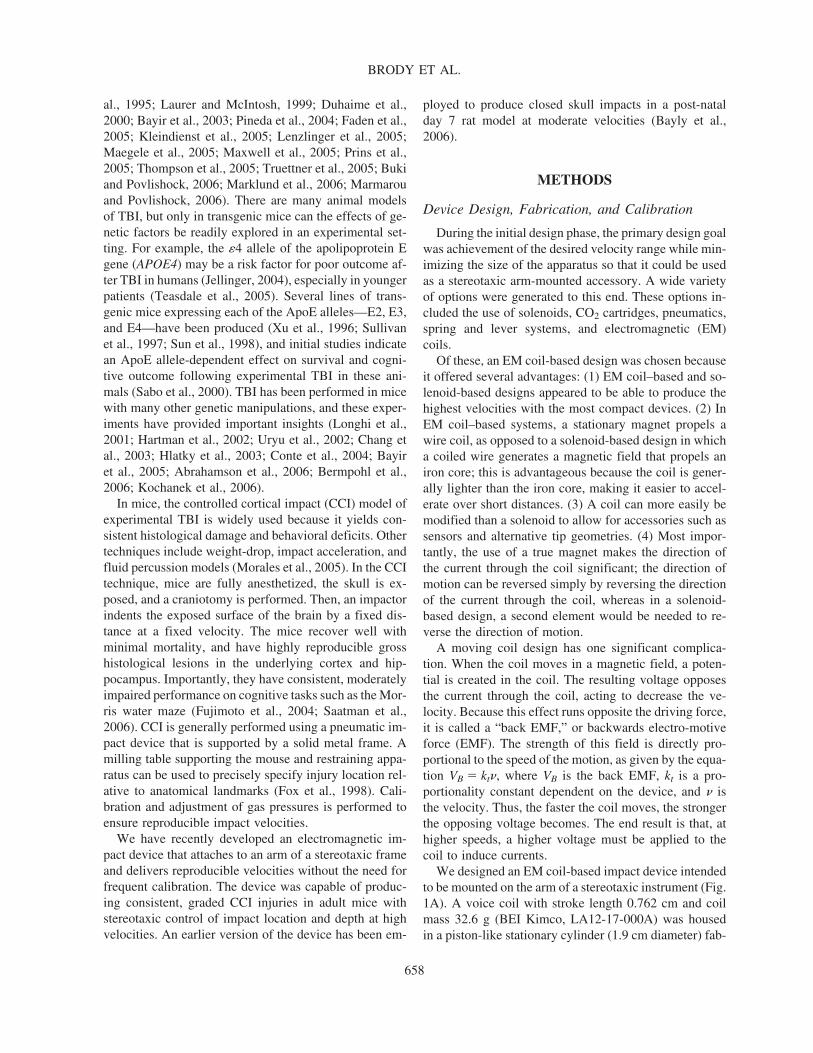
BRODY ET AL.
658

ricated to slip around the outside of the magnet and clamparound it. An inner ring of Delrin™ was added at the bot-tom of the cylinder as a stop; the semi-rigid ring distrib-utes the stopping force and lessens deformation of thepiston upon impact. A thin layer of padding was addedto the magnet to cushion the coil’s impact at the top ofits up-stroke, significantly extending the life of the de-vice. Air “vents” were milled into the sides of the piston
to prevent any pressure differential which would reduceacceleration. The outer surface of the piston and the in-ner surface of the cylinder were polished to minimizefriction between the two surfaces. The total mass of thedevice, the mass of the moving components, and the forcegenerated were designed to be relatively small (no largerthan necessary for mouse CCI), in order to avoid the ne-cessity for the large, metal crossbar commonly used withpneumatic CCI devices.
To control the motion of the EM coil, several elec-tronic devices were incorporated to provide the requiredcurrent to the actuator (Fig. 1B). A servo amplifier(B12A8; Advanced Motion Controls, Camarillo, CA)provided approximately 1.75 amps/commanded volt. A72-V power supply (PS300W; Advanced Motion Con-trols) was required in order to overcome the back EMFeffect and achieve the desired velocities. A standard Win-dows laptop running Matlab™ (version 14; The Math-works, Natick, MA) in combination with a NI-DAQ dig-ital-to-analog converter (DAQCard 6062E; NationalInstruments, Austin, TX) was used to communicate con-trol commands to the amplifier.
In the custom-written operating software, the user be-gins by entering a desired command voltage (or accept-ing a default voltage). Once the voltage is accepted, asmall current is applied, ensuring that the impactor isfully extended in its downward position. The user is thenprompted to position the impactor in the desired location.Once the “zero” position has been verified, a short, strongcurrent pulse raises the impactor to its cocked position,and a lighter current maintains the position. The user isthen prompted to lower the device to the desired impactdepth. The depth of impact is thus specified using thecontrols intrinsic to the stereotaxic instrument. The userpresses “Enter” to initiate impact. The current specifiedby the user at the start of the program is then delivered,causing an impact at the desired velocity, and the im-pactor is retracted after an adjustable dwell time, speci-fied via the Matlab-based control software. For these ex-periments, the dwell time was typically 100 msec.
To calibrate the relationship between applied com-mand voltage and tip impact velocity, the displacementof the impactor was measured with a fast, non-contact-ing laser displacement sensor (LD1605-10; Micro-Epsilon, Germany; Figs. 1C and 2). This displacementsensor acquired samples at a rate of 10,000 Hertz. Datawas acquired using Matlab Version 14 in combinationwith NI-DAQ routines. Velocity was defined over the fi-nal 10% of the stroke range. Overshoot was defined asthe maximum, transient displacement of the tip duringthe stroke beyond the user-specified set distance. Forcomparison, the relationship between applied pressure,tip impact velocity, and overshoot was also similarly de-
ELECTROMAGNETIC CCI DEVICE FOR EXPERIMENTAL TBI
659
FIG. 1. Design of an electromagnetic controlled cortical im-pact device for experimental traumatic brain injury. (A) Pho-tograph of the impactor device mounted on the left arm of astereotaxic device. Motorized drill with 5-mm trephine mountedon the right arm. (B) Schematic of the components of the im-pact system. Control signals from a Windows-based notebookcomputer running custom Matlab™ routines are fed through adigital-to-analog converter. The digital-to-analog converter out-put is sent to a servo amplifier. The servo amplifier transmitscurrent from 72-V power supply to the impactor containing anelectromagnetic voice coil. This voice coil drives the impactor.(C) Photograph of the impactor tip in the raised position. Alaser-Doppler displacement sensor was used to measure veloc-ity of the tip during the impact stroke.

termined for a commercially available pneumatic CCI de-vice (Amscien, Richmond, VA).
Experimental Traumatic Brain Injury in Adult Mice
All experiments were approved by the animal studiescommittee at Washington University. Young adultB6SJLF1 wild-type mice (age 2–3 months) of both sexeswere purchased from Jackson Labs (Bar Harbor, ME).The mice were housed in the Washington University an-imal facilities under standard conditions at 4–5 mice percage. They were given standard lab chow and water adlibitum; cages were changed twice per week. They weremaintained in a controlled temperature environment withlights on for 12 h and lights off for 12 h per day.
Mice were subjected to a single left lateral CCI withcraniotomy (Dixon et al., 1991; Smith et al., 1995, 1998;Murai et al., 1998). Mice were anesthetized with isoflu-rane, 5% for induction and 1.5–2% for maintenance. Theywere placed in a stereotaxic frame with an incisor bar at0° and cup head holders (David Kopf Instruments, Tu-junga, California). The cup head holders were used toavoid the potential for injury to the ear canals produced
by earbars in mice. Several lines of transgenic mice ap-pear to be more susceptible to ear canal injury than wild-type mice and younger mice may also be more suscepti-ble than older mice (D. Brody, unpublished data). Thehead holders were applied such that the most anterior por-tion of the cup was aligned with the posterior canthi ofthe eyes of the mouse. A consistent mild compression ofthe skull was applied by holding the head with the twocup head holders a consistent distance apart: 10.7 mm forfemale mice weighing 18–20 g and 11.7 mm for malemice weighing 24–26 g. These distances were assessedusing the rulings on the bars (Kopf) that anchor the cuphead holders to the frame. Rectal temperature was main-tained at 37°C with a warming pad and feedback con-troller (Cell Microcontrols, Norfolk, VA). Ointment toprotect vision was applied to their eyes, and their headswere shaved with an electric clipper. All tools were ster-ilized with a glass bead sterilizer. The skin was preppedwith betadine ointment, and the top of the skull was ex-posed with a 1-cm skin incision.
A craniotomy was performed over the left parietotem-poral cortex using a 5-mm trephine (Meisinger, Neuss,Germany) attached to an electric drill (Foredom, Bethel,CT) mounted on the right stereotaxic arm (Fig. 1A) of a
BRODY ET AL.
660
FIG. 2. Trajectories of the impactor tip during impact stroke: measurements of velocity and overshoot. All trajectories weremeasured using a fast laser Doppler displacement sensor aimed at the tip of the impactor. (A) EM impactor set at 3 or 7 V, yield-ing velocities of 3.6 or 5.2 m/sec. Overshoot, defined as the transient excursion of the impactor tip past the set distance specifiedby the user, was 0.31 � 0.032 mm at 7 V and essentially unchanged at 3 V. Set distance and overshoot are indicated by top andbottom dashed lines, respectively. (B) Pneumatic impactor (Amscien, AMS201) with high-pressure settings of 50 or 100 psi yield-ing velocities of 3.7 or 5.2 m/sec. (C) Overshoot as a function of velocity for the electromagnetic and pneumatic impactors. Over-shoot was strongly velocity-dependent for the pneumatic impactor as tested but there was little change in overshoot with veloc-ity for the electromagnetic impactor.

digital stereotaxic device (Benchmark Deluxe™;MyNeurolab, St. Louis, MO), though the device can beused with any compatible small animal stereotaxic sys-tem. The angle of the drill and the head of the mousewere adjusted so that the craniotomy penetrated the thinskull of the mouse without damaging the dura. There wasa consistent, mild compression of the skull produced bythe cup head holders, which caused the surface of thebrain to protrude through the craniotomy. The maximalextent of this protrusion was approximately 0.4 mm, asmeasured using a fine probe attached to one arm of thestereotaxic device. In two of 80 mice, injury during thecraniotomy resulted in swelling greater than 0.5 mmand/or bleeding from the dura. These two mice were dis-qualified and not used for any further experiments. Thedrill was then rotated out of the field, and the 5-mm discof bone was removed using a 1-mm cup rongeur and spat-ula (Roboz, Gaithersburg, MD).
Next, the impact device mounted on the left stereo-taxic arm at an angle of 15° from vertical was rotatedinto the field. The right edge of the 3-mm tip of the im-pact device was aligned with the midline suture and theposterior edge of the tip was aligned with the horizontalportion of the lambda suture using the stereotaxic arm.Because the EM cylinder was relatively bulky (37 mmdiameter), visualization of the EM tip was somewhatmore difficult than visualization of the pneumatic devicetip (9.5-mm diameter). This minor disadvantage wascompensated for by the use of a hand lens or operatingmicroscope to confirm all alignments. The tip was posi-tioned over the left fronto-parietal cortex by moving it1.5 mm anteriorly and 1.2 mm to the left using the dig-ital stereotaxic arm. This resulted in an impact centered3.0 mm anterior to lambda and 2.7 mm left of midline,within the craniotomy. The zero depth position was de-termined by aligning the tip of the impact device in thedown position with the surface of the dura. A low-volt-age DC circuit touch detector (custom built by the Wash-ington University Electronics Shop) was used to deter-mine when the tip first contacted the dura. One electrodewas clipped to the impactor tip and the mouse’s hind pawwas placed in contact with a second electrode. The sur-face of the dura was typically dry, but when fluid or bloodwas present, this was cleaned and dried carefully usingirrigation with sterile saline and sterile, dry cotton swabsbefore the zero depth was determined. Contact was ver-ified using a hand lens or operating microscope for everyimpact. The tip was raised to the cocked position, andthe depth of desired impact was set by lowering the im-pact device 1–3 mm using the stereotaxic arm. Correc-tion for overshoot was not made. CCI was triggered us-ing Matlab-based computer controller. Velocities werecontrolled by setting the voltage command to the digital-
to-analog converter. For most experiments, a 7-V com-mand was used, corresponding to a 12.25-amp currentpulse to the actuator, and resulting in a 5.23 � 0.03 m/secstroke velocity.
Bleeding of the injured cortical surface was controlledusing copious irrigation with room-temperature sterilesaline. The skull was dried, and a 6-mm-diameter plas-tic disc was glued with Vetbond to the skull to cover thecraniotomy defect and prevent infection. These discswere produced by the Washington University MachineShop from commercially available weigh-boats. The skinwas closed with five to six interrupted sterile 4-0 nylonsutures. Triple antibiotic ointment was applied to the skin.Mice were removed from the stereotaxic frame andplaced on a warming pad while they recovered from anes-thesia. Sham-injured animals went through the same pro-cedure but did not undergo CCI. The entire procedure re-quired �20 min per mouse.
In �5% of mice, brief seizure activity was observedduring emergence from anesthesia but this always ceasedwithout intervention. The mice typically began to movespontaneously within 15 min. Mice were returned to theirhome cages when fully ambulatory, typically 30–120 minafter injury. When mice appeared lethargic, they werehoused singly for up to 48 h and then returned to theirhome cages. Mice were weighed at baseline and daily for2 days. Those with weight loss greater than 30% weresacrificed. Those with weight loss of 20–30% were singlyhoused with food and water in the bottom of the cage.Wounds were inspected daily for 2 days and then weeklythereafter. Dehiscences were resutured under isofluraneanesthesia.
For CCI using a pneumatic device (Amscien, AMS201), an identical procedure was used with the followingmodifications: (1) The 3-mm-diameter impactor tip waspositioned relative to midline and lambda using a millingtable inside the frame of the pneumatic impact deviceupon which the stereotaxic frame was placed (Fox et al.,1998). (2) The depth of injury was set using the screw-mounted adjustment provided as part of the pneumaticCCI device. (3) A dwell time of 50 msec was used. (4)A velocity of 5.16 � 0.05 m/sec was obtained using set-tings of 100 pounds per square inch (psi) for the highpressure and 20 psi for the low pressure. A 54-inch cylin-der of compressed nitrogen (Airgas, Inc., St. Louis, MO)connected via a regulator (Concoa model 3124391-01-580) drove the device. We found that smaller cylindersof compressed gasses, even when yielding nominallysimilar baseline pressures, were not able to drive the de-vice with the desired velocities, even at maximal settings.This may be because of larger drops in pressure at thetime the device discharges observed with the smallertanks.
ELECTROMAGNETIC CCI DEVICE FOR EXPERIMENTAL TBI
661

Behavioral Testing
Morris water maze. Morris water maze testing was per-formed starting 13 days following experimental TBI.Each mouse was given four trials per day for 3 days witha clearly visible platform and then 4 trials a day for 5days with the platform in a different location hidden be-neath the surface of the opacified water. Each trial lasteda maximum of 60 sec. The platform was 11 cm in di-ameter; the pool was 109 cm in diameter. A single, 30-sec probe trial was performed after the last day of hid-den platform testing. In other respects, testing wasidentical to that previously described (Brody and Holtz-man, 2006).
Rotorod. Rotorod testing was performed starting 24 daysfollowing TBI. In this test of motor learning, mice wereplaced on an accelerating, rotating, horizontal cylinder(Crawley, 2000). The amount of time that the mice areable to stay on the cylinder without falling off is often usedas a measure of motor skill adaptation, as the mice mustaccelerate with the cylinder. Mice were tested with two180-sec trials per day on each of 3 days. To control forgross motor impairments and any behavioral abnormali-ties that would interfere with interpretation of the results(such as jumping off of the platform voluntarily), micewere placed on the cylinder while it rotated at a constant,slow speed for two 60-sec trials each day. Prior to testingeach day, mice were placed on the cylinder once for 60sec while it was stationary to allow them to adapt to it.
Conditioned fear. Conditioned fear testing and shocksensitivity testing were performed starting 38 days afterTBI (Crawley, 2000; Khuchua et al., 2003). In this testof associative, non-spatial memory, mice were first ex-posed to a 1.0-mA continuous brief foot shock associatedwith a cue; in our experiments, this was an 80-dB, 2800-Hz tone. The amount of time freezing was monitored asa measure of their fear response. The next day, the micewere placed in the same experimental chamber where theinitial shock occurred, and again time spent freezing wasassessed with no shock. This indicates how well they as-sociated the environmental context with the shock. Onthe third day, the amount of time spent freezing in a novelcontext when they were exposed again to the cue wasmeasured, as an indication of how well they associatedthe cue with the shock. This was performed in a differ-ent experimental chamber from the one used in the ini-tial association. Shock sensitivity and time spent freez-ing at baseline were assessed in order to control fordifferences in freezing behavior between mice that werenot related to associative memory.
Histological Analysis
A randomly selected subset of the mice were deeplyanesthetized with isoflurane and sacrificed after behav-ioral testing was complete, or for animals not tested be-haviorally, 30–45 days after TBI. Mice were perfused in-tracardially with ice-cold heparinized 0.9% saline. Brainswere carefully removed, fixed in paraformaldehyde, andequilibrated in 30% sucrose (Holtzman et al., 2000).Every sixth 50-�m frozen section was mounted on glassslides (Fisher, Superfrost Plus) and stained with bis-ben-zamide or cresyl violet. For each section containing vis-ible hippocampus (bregma �0.94 mm to �3.88 mm)(Franklin and Paxinos, 1997), four contours were traced,and their areas were measured using the Stereo Investi-gator Contour Tracing Tool (Stereo Investigator UsersGuide, version 6, MicroBrightField, Inc.). The four con-tours were the ipsilateral and contralateral hippocampus,and ipsilateral and contralateral dorsal cortex. The infe-rior border of the dorsal cortex was defined by a hori-zontal line touching the bottom margin of the dorsal thirdventricle. The sections analyzed contained both dorsaland ventral hippocampus. Volumes were estimated usingthe Cavalieri principle (Howard and Reed, 2005); the ar-eas over the 9–10 sections traced for each mouse weresummed and multiplied by the spacing between sections(300 �m). Thus, the volumes obtained represent the com-plete extent of the hippocampus, and the dorsal cortex inthe region overlying the hippocampus.
Statistical Analysis
All data was analyzed using Statistica 6.0 (StatSoft,Tulsa, OK). Factorial analysis of variance (ANOVA) wasused for all behavioral analyses; impact depth, mousegender, and experimenter performing the injury were cat-egorical predictor variables. The Tukey HSD test wasused to determine post-hoc statistical significance of pair-wise comparisons between groups. For visible and hid-den platform Morris water maze data, repeated measuresANOVAs were used with performance averaged acrosseach of the four daily trials, and day of training used asthe repeated measure variable. For probe trial data, 95%confidence intervals were calculated and compared toperformance expected by chance. For rotorod data, re-peated measures ANOVAs were used with performanceaveraged across the two trials per day; and day of train-ing used as the repeated measure variable. One-wayANOVAs were used for analysis of hippocampal and cor-tical volumes.
For statistical power calculations of sample size re-quirements, a Monte-Carlo approach was used. A cus-tom-written Visual Basic macro running inside of Statis-tica generated 150 random sub-samples of the mice
BRODY ET AL.
662
subjected to either 2.0-mm or 3.0-mm injury. The hiddenplatform data for each of these sub-samples was then an-alyzed using repeated measures ANOVA. The probabil-ity that each mouse would be picked during the randomsub-sampling was varied to produce sub-samples withvarying sample sizes. The p-values were tabulated as afunction of sample size, and the median and upper 80%confidence interval was calculated using Statistica. Thisallowed exploration of the sample size needed to achievean 80% likelihood of detecting a statistically significantdifference between groups with the characteristics ofthese two injury severities.
RESULTS
Device Mechanical Characteristics
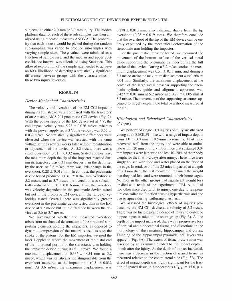
The velocity and overshoot of the EM CCI impactorduring its full stroke were compared with the trajectoryof an Amscien AMS 201 pneumatic CCI device (Fig. 2).With the power supply of the EM device set at 7 V, theend impact velocity was 5.23 � 0.026 m/sec, whereaswith the power supply set at 3 V, the velocity was 3.57 �0.032 m/sec. No statistically significant differences wereobserved when the device was retested with the samevoltage settings several weeks later without recalibrationor adjustment of the device. At 5.2 m/sec, there was asmall overshoot, 0.31 � 0.032 mm. Stated another way,the maximum depth the tip of the impactor reached dur-ing its trajectory was 0.31 mm deeper than the depth setby the user. At 3.6 m/sec, there was little change in theovershoot, 0.28 � 0.019 mm. In contrast, the pneumaticdevice tested produced a 0.61 � 0.047 mm overshoot at5.2 m/sec, and at 3.7 m/sec the overshoot was substan-tially reduced to 0.30 � 0.016 mm. Thus, the overshootwas velocity-dependent in the pneumatic device testedbut not in the prototype EM device, in the range of ve-locities tested. Overall, there was significantly greaterovershoot in the pneumatic device tested than in the EMdevice at 5.2 m/sec but little difference between the de-vices at 3.6 to 3.7 m/sec.
We investigated whether the measured overshootarises from mechanical deformation of the structural sup-porting elements holding the impactors, as opposed todynamic compression of the materials used to stop thestroke of the pistons. For the EM impactor, we used thelaser Doppler to record the movement of the distal endof the horizontal portion of the stereotaxic arm holdingthe impactor device during its full stroke. We found amaximum displacement of 0.336 � 0.054 mm at 5.2m/sec, which was statistically indistinguishable from theovershoot measured at the impactor tip (0.31 � 0.032mm). At 3.6 m/sec, the maximum displacement was
0.278 � 0.013 mm, also indistinguishable from the tipovershoot (0.28 � 0.019 mm). We therefore concludethat the overshoot of the tip of the EM device can be en-tirely explained by the mechanical deformation of thestereotaxic arm holding the impactor.
For the pneumatic impactor tested, we measured themovement of the bottom surface of the vertical metalguide supporting the pneumatic cylinder during the fullstroke of the device. During a 5.2 m/sec stroke, the max-imum displacement was 0.51 � 0.11 mm, and during a3.7 m/sec stroke the maximum displacement was 0.268 �.004 mm. Similarly, the maximum displacement at thecenter of the large metal crossbar supporting the pneu-matic cylinder, guide and alignment apparatus was0.427 � 0.01 mm at 5.2 m/sec and 0.29 � 0.005 mm at3.7 m/sec. The movement of the supporting structures ap-peared to largely explain the total overshoot measured atthe tip.
Histological and Behavioral Characteristics of Injury
We performed single CCI injuries on fully anesthetizedyoung adult B6SJLF1 mice with a range of impact depthsfrom 1.0 to 3.0 mm in 0.5-mm increments. Most micerecovered well from the injury and were able to ambu-late within 20 min of injury. Four mice that sustained 3.0-mm impacts were lethargic and lost 10–20% of their bodyweight for the first 1–2 days after injury. These mice weresingly housed with food and water placed on the floor ofthe cage. In total, two of the 25 mice impacted at a depthof 3.0 mm died; the rest recovered, regained the weightthat they had lost, and were returned to their home cages.No mice in the other groups had significant weight lossor died as a result of the experimental TBI. A total oftwo other mice died prior to injury: one due to tempera-ture controller malfunction causing hyperthermia and onedue to apnea during isoflurane anesthesia.
We assessed the histological effects of injuries pro-duced by the EM CCI device at a velocity of 5.2 m/sec.There was no histological evidence of injury to cortex orhippocampus in mice in the sham group (Fig. 3). As thedepth of the impact increased, there was a progressive lossof cortical and hippocampal tissue, and distortions in themorphology of the remaining hippocampus and cortex.Thinning of the hippocampal pyramidal cell layers wasapparent (Fig. 3A). The extent of tissue preservation wasassessed by an examiner blinded to the impact depth 1month after the injury. As the depth of impact increased,there was a decrease in the fraction of spared tissue, asmeasured relative to the contralateral side (Fig. 3B). Theeffect of impact depth was highly significant for the frac-tion of spared tissue in hippocampus (F4, 23 � 15.6, p �
ELECTROMAGNETIC CCI DEVICE FOR EXPERIMENTAL TBI
663

0.0001) and cortex (F4, 23 � 14.3, p � 0.0001). At 1.0-mm depth, there was minimal injury in either cortex orhippocampus. At 1.5-mm depth there was significant cor-tical injury and mild cell loss in the underlying hip-pocampus. At 2.0-mm and 2.5-mm depths, there was pro-gressively more injury to the hippocampus and cortex. At3.0-mm depths, the hippocampus was nearly destroyed.Mice injured at this depth also had thalamic lesions (notshown), which were not assessed in a quantitative fash-
ion. The standard deviations represent injury variabilityand not measurement error, as intra-rater reliability test-ing revealed a �3% error in volume measurements (notshown). There was no apparent injury to the contralateralhippocampus or cortex in any of the animals (not shown),although ultra-sensitive techniques, such as de Olmos sil-ver staining, were not used (Hall et al., 2005b). This in-dicated that the EM CCI device can produce a broad rangeof CCI injuries in a reliable fashion.
BRODY ET AL.
664
FIG. 3. Histological analysis of injuries produced by EM and pneumatic devices. (A) Histological images of cresyl violet–stainedcoronal sections following sham injury, and 1.0, 1.5, 2.0, 2.5, and 3.0 mm impact using the EM device. Left images in each pairshow overall dorsal cortical and hippocampal tissue loss and architecture. Right image in each pair show further details of hip-pocampal architecture. Cortical and hippocampal tissue injury and anatomical distortion increase in a graded fashion with in-creased impact depth. All images were obtained from slices through the same anatomical region (bregma �1.7 mm), selectedbased on the architecture of the contralateral hemisphere. (B) Proportion of spared ipsilateral hippocampus and dorsal cortex re-maining 1 month after TBI. Each stereologically determined hippocampal or cortical volume was normalized by the correspondingcontralateral volume for that animal. Set depth of injury on the x axis. Error bars represent standard deviations. All injuries wereperformed by the same investigator and volumes were assessed without knowledge of device or impact set depth.

ELECTROMAGNETIC CCI DEVICE FOR EXPERIMENTAL TBI
665
For comparison, we produced pneumatic CCI injuriesat a commonly used depth of 1 mm (Smith et al., 1995;Fox et al., 1998; Hall et al., 2005a) in mice of the sameage and strain. This produced a moderately severe injuryto cortex and hippocampus, as has been reported previ-ously. This 1.0-mm depth impact with the pneumatic CCIdevice was most similar to the 2.0-mm impact producedby the EM CCI device. This discrepancy cannot entirelybe explained by the difference in overshoot between thetwo devices.
Next, we performed behavioral testing on mice sub-jected to a range of injury severities. Again we found agraded effect of impact depth of behavioral impairmentin Morris water maze (Fig. 4) and rotorod (Fig. 5) test-ing. For visible platform water maze performance, therewere statistically significant effects of injury group (con-trol vs. each of three impact depths. F3, 41 � 9.95, p �0.0001) and test day (F2, 82 � 166.2, p � 0.0001; re-peated-measures ANOVA). For hidden platform watermaze performance, there were statistically significant ef-fects of injury group (F3, 41 � 18.0, p � 0.0001), test day(F4, 164 � 17.1, p � 0.0001), and the interaction of injurygroup and test day (F12, 164 � 2.99, p � 0.0008). For wa-ter maze probe performance, there were statistically sig-nificant effects of injury group (F3, 41 � 20.8, p � 0.0001for target quadrant and F3, 41 � 10.0, p � 0.0001 for ex-act target zone). For constant velocity rotorod perfor-mance, there were statistically significant effects of injurygroup (F3, 43 � 9.9, p � 0.0001) and test day (F2, 86 �32.7, p � 0.0001). For accelerating rotorod performance,there were statistically significant effects of injury group(F3, 43 � 11.7, p � 0.0001) and test day (F2, 86 � 33.3,p � 0.0001). There were no significant differences be-tween injured and sham mice in terms of conditioned fearperformance. All mice performed well in this test with a24-h interval between conditioning and testing, regard-less of injury status.
Mice with 2.0-mm impacts were impaired in watermaze performance relative to sham-injured controls buthad normal rotorod performance. In visible platform test-ing (Fig. 4A; days 13–15), 2.0-mm-impacted mice hadpoorer performance than sham mice on the first day oftesting, but both groups performed equally well by thethird day of testing. This indicates that the 2.0-mm-im-pacted mice may have had some initial procedural im-pairment, but that their vision, swimming ability and mo-tivation to escape from the pool was unimpaired overall.Hidden platform performance, however, was impaired(Fig. 4A; days 17–21) relative to sham mice (p � 0.006,repeated measures ANOVA followed by Tukey post-hoctest). Although impaired, 2.0-mm-impacted mice im-proved their performance over the 5 days of hidden plat-form training. Time and distance measures of perfor-
mance showed equivalent results. Swim speed did notdiffer appreciably between groups (data not shown). Theprobe trial performance (Fig. 4B) of 2.0-mm-impactedmice was significantly better than chance in terms of timespent in the target quadrant. However, their performancewas not above chance in terms of time spent in the ex-act region where the platform had been located. Instead,sham mice did perform better than chance in both mea-sures. This suggests that 2.0-mm-impacted mice were ca-pable of forming spatial memories, but that their spatialmaps were less precise than those formed by sham mice.Rotorod performance was not significantly different be-tween 2.0-mm-impacted mice and sham mice (Fig. 5).
Mice with 2.5-mm impacts were impaired in watermaze and rotorod performance relative to both sham-in-jured controls and 2.0-mm-impacted mice. Visible plat-form performance (Fig. 4A; days 13–15) was worse com-pared to sham mice (p � 0.039), although they were ableto find the platform in under 10 sec on average by thethird day. Hidden platform performance (Fig. 4A; days17–21) appeared more impaired in 2.5-mm-impactedmice than in 2.0-mm-impacted mice, though this did notreach statistical significance, and was profoundly im-paired relative to sham mice (p � 0.0002). There was lit-tle learning over time, and in the probe trial (Fig. 4B),performance was at or below chance levels, indicatingthat these mice had not formed detectible spatial memo-ries. Mice with 2.5-mm impacts were also impaired onthe accelerating phase of the rotorod testing (Fig. 5C) rel-ative to sham mice (p � 0.016). Their performance onthe stationary and constant speed portions of the rotorodtesting was normal. Overall, these results suggest thatthey had deficits in motor learning in addition to poorspatial learning.
Mice with 3.0-mm impacts were severely impaired inboth water maze and rotorod performance. During visi-ble platform water maze testing, two of 12 mice failed toreliably swim to the platform and were disqualified fromfurther water maze testing. The visible platform perfor-mance of those that were not disqualified (Fig. 4A) wasstill markedly worse relative to sham mice (p � 0.0002)and relative to 2.5-mm-impacted mice (p � 0.038) or 2.0-mm-impacted mice (p � 0.005). This likely reflects adeficit not only in spatial learning and memory but alsoin learning procedural aspects of the test, such as swim-ming ability, vision, and motivation to escape from thewater. Their hidden platform performance was signifi-cantly worse than 2.0-mm-impacted animals (p � 0.005)or sham mice (p � 0.0002). Floor effects may have pre-vented statistical resolution of further impairment rela-tive to 2.5-mm-impacted mice. Their accelerating rotorodperformance was very poor (Fig. 5C; p � 0.0002 vs.sham, p � 0.002 vs. 2.0- and 2.5-mm-impacted mice).

BRODY ET AL.
666
FIG. 4. Behavioral characterization of mice injured using the EM device in the Morris water maze. (A) Time to reach the plat-form as a function of day following experimental TBI. During visible platform testing, two of 12 mice subjected to 3.0-mm im-pacts failed to reliably swim to the platform and were disqualified from further water maze testing. No mice in the sham, 2.0-mm, or 2.5-mm impact groups were disqualified. Data shown represent mean and standard errors from the remaining,non-disqualified mice. During hidden platform testing, thre was a clear gradation in performance, with more severely injured an-imals performing worse. (***p � 0.0002, **p � 0.006, *p � 0.039, repeated-measures ANOVA followed by Tukey HSD post-hoc test for comparisons vs. sham). (B) Water maze probe testing; the platform was removed after the last day of hidden plat-form testing and mice were placed in the pool for a single, 30-sec trial. Sham mice spent considerably more time in the targetquadrant and exact area where the platform had been than would have been expected by chance. Mice subjected to TBI had im-paired performance, again in a graded fashion depending on the severity of injury (*95% confidence interval did not overlap withthe performance expected by chance.) (C) In a separate experiment, mice subjected to a 1.5-mm impact did not show significantwater maze performance deficits compared with concurrently tested, sham-injured mice.
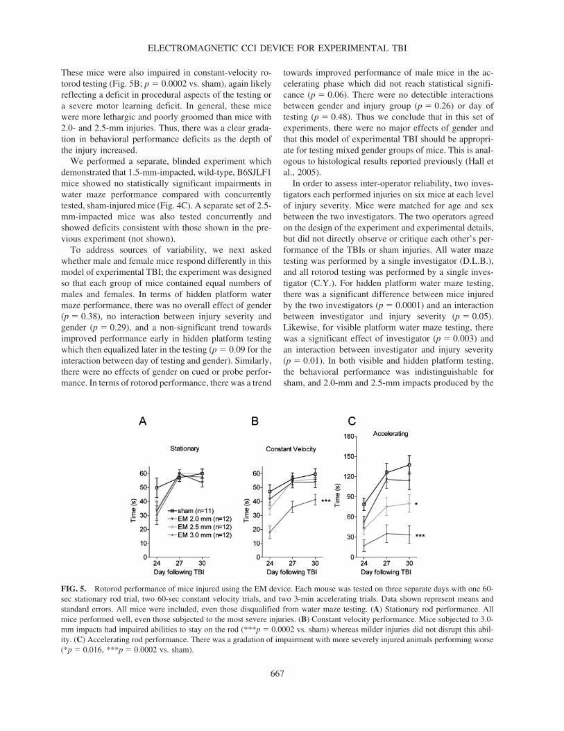
These mice were also impaired in constant-velocity ro-torod testing (Fig. 5B; p � 0.0002 vs. sham), again likelyreflecting a deficit in procedural aspects of the testing ora severe motor learning deficit. In general, these micewere more lethargic and poorly groomed than mice with2.0- and 2.5-mm injuries. Thus, there was a clear grada-tion in behavioral performance deficits as the depth ofthe injury increased.
We performed a separate, blinded experiment whichdemonstrated that 1.5-mm-impacted, wild-type, B6SJLF1mice showed no statistically significant impairments inwater maze performance compared with concurrentlytested, sham-injured mice (Fig. 4C). A separate set of 2.5-mm-impacted mice was also tested concurrently andshowed deficits consistent with those shown in the pre-vious experiment (not shown).
To address sources of variability, we next askedwhether male and female mice respond differently in thismodel of experimental TBI; the experiment was designedso that each group of mice contained equal numbers ofmales and females. In terms of hidden platform watermaze performance, there was no overall effect of gender(p � 0.38), no interaction between injury severity andgender (p � 0.29), and a non-significant trend towardsimproved performance early in hidden platform testingwhich then equalized later in the testing (p � 0.09 for theinteraction between day of testing and gender). Similarly,there were no effects of gender on cued or probe perfor-mance. In terms of rotorod performance, there was a trend
towards improved performance of male mice in the ac-celerating phase which did not reach statistical signifi-cance (p � 0.06). There were no detectible interactionsbetween gender and injury group (p � 0.26) or day oftesting (p � 0.48). Thus we conclude that in this set ofexperiments, there were no major effects of gender andthat this model of experimental TBI should be appropri-ate for testing mixed gender groups of mice. This is anal-ogous to histological results reported previously (Hall etal., 2005).
In order to assess inter-operator reliability, two inves-tigators each performed injuries on six mice at each levelof injury severity. Mice were matched for age and sexbetween the two investigators. The two operators agreedon the design of the experiment and experimental details,but did not directly observe or critique each other’s per-formance of the TBIs or sham injuries. All water mazetesting was performed by a single investigator (D.L.B.),and all rotorod testing was performed by a single inves-tigator (C.Y.). For hidden platform water maze testing,there was a significant difference between mice injuredby the two investigators (p � 0.0001) and an interactionbetween investigator and injury severity (p � 0.05).Likewise, for visible platform water maze testing, therewas a significant effect of investigator (p � 0.003) andan interaction between investigator and injury severity(p � 0.01). In both visible and hidden platform testing,the behavioral performance was indistinguishable forsham, and 2.0-mm and 2.5-mm impacts produced by the
ELECTROMAGNETIC CCI DEVICE FOR EXPERIMENTAL TBI
667
FIG. 5. Rotorod performance of mice injured using the EM device. Each mouse was tested on three separate days with one 60-sec stationary rod trial, two 60-sec constant velocity trials, and two 3-min accelerating trials. Data shown represent means andstandard errors. All mice were included, even those disqualified from water maze testing. (A) Stationary rod performance. Allmice performed well, even those subjected to the most severe injuries. (B) Constant velocity performance. Mice subjected to 3.0-mm impacts had impaired abilities to stay on the rod (***p � 0.0002 vs. sham) whereas milder injuries did not disrupt this abil-ity. (C) Accelerating rod performance. There was a gradation of impairment with more severely injured animals performing worse(*p � 0.016, ***p � 0.0002 vs. sham).
two investigators. At 3.0 mm, there was a significant dif-ference between investigators. There was no effect of in-vestigator on rotorod performance (p � 0.96), nor wasthere an interaction between investigator and injury group(p � 0.24). Thus we conclude that reliability was goodfor 2.0- and 2.5-mm injuries but not for 3.0-mm injuries.
Sample Size Calculations Using Monte-CarloRandom Resampling
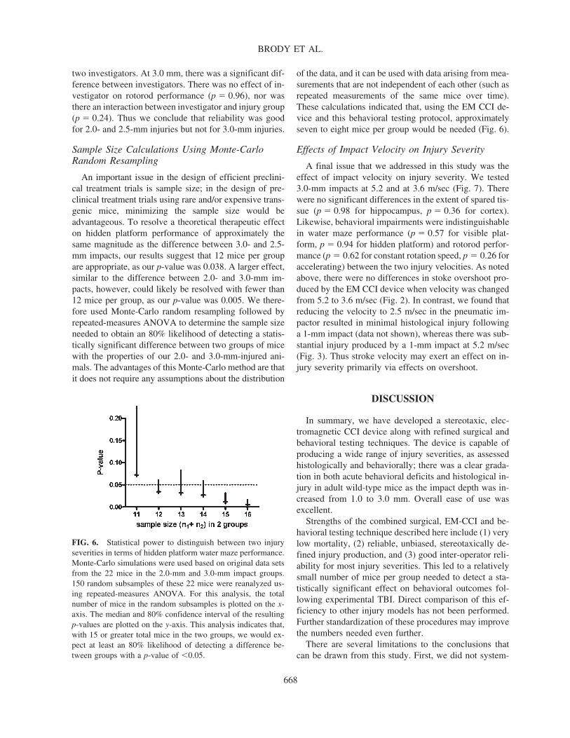
An important issue in the design of efficient preclini-cal treatment trials is sample size; in the design of pre-clinical treatment trials using rare and/or expensive trans-genic mice, minimizing the sample size would beadvantageous. To resolve a theoretical therapeutic effecton hidden platform performance of approximately thesame magnitude as the difference between 3.0- and 2.5-mm impacts, our results suggest that 12 mice per groupare appropriate, as our p-value was 0.038. A larger effect,similar to the difference between 2.0- and 3.0-mm im-pacts, however, could likely be resolved with fewer than12 mice per group, as our p-value was 0.005. We there-fore used Monte-Carlo random resampling followed byrepeated-measures ANOVA to determine the sample sizeneeded to obtain an 80% likelihood of detecting a statis-tically significant difference between two groups of micewith the properties of our 2.0- and 3.0-mm-injured ani-mals. The advantages of this Monte-Carlo method are thatit does not require any assumptions about the distribution
of the data, and it can be used with data arising from mea-surements that are not independent of each other (such asrepeated measurements of the same mice over time).These calculations indicated that, using the EM CCI de-vice and this behavioral testing protocol, approximatelyseven to eight mice per group would be needed (Fig. 6).
Effects of Impact Velocity on Injury Severity
A final issue that we addressed in this study was theeffect of impact velocity on injury severity. We tested3.0-mm impacts at 5.2 and at 3.6 m/sec (Fig. 7). Therewere no significant differences in the extent of spared tis-sue (p � 0.98 for hippocampus, p � 0.36 for cortex).Likewise, behavioral impairments were indistinguishablein water maze performance (p � 0.57 for visible plat-form, p � 0.94 for hidden platform) and rotorod perfor-mance (p � 0.62 for constant rotation speed, p � 0.26 foraccelerating) between the two injury velocities. As notedabove, there were no differences in stoke overshoot pro-duced by the EM CCI device when velocity was changedfrom 5.2 to 3.6 m/sec (Fig. 2). In contrast, we found thatreducing the velocity to 2.5 m/sec in the pneumatic im-pactor resulted in minimal histological injury followinga 1-mm impact (data not shown), whereas there was sub-stantial injury produced by a 1-mm impact at 5.2 m/sec(Fig. 3). Thus stroke velocity may exert an effect on in-jury severity primarily via effects on overshoot.
DISCUSSION
In summary, we have developed a stereotaxic, elec-tromagnetic CCI device along with refined surgical andbehavioral testing techniques. The device is capable ofproducing a wide range of injury severities, as assessedhistologically and behaviorally; there was a clear grada-tion in both acute behavioral deficits and histological in-jury in adult wild-type mice as the impact depth was in-creased from 1.0 to 3.0 mm. Overall ease of use wasexcellent.
Strengths of the combined surgical, EM-CCI and be-havioral testing technique described here include (1) verylow mortality, (2) reliable, unbiased, stereotaxically de-fined injury production, and (3) good inter-operator reli-ability for most injury severities. This led to a relativelysmall number of mice per group needed to detect a sta-tistically significant effect on behavioral outcomes fol-lowing experimental TBI. Direct comparison of this ef-ficiency to other injury models has not been performed.Further standardization of these procedures may improvethe numbers needed even further.
There are several limitations to the conclusions thatcan be drawn from this study. First, we did not system-
BRODY ET AL.
668
FIG. 6. Statistical power to distinguish between two injuryseverities in terms of hidden platform water maze performance.Monte-Carlo simulations were used based on original data setsfrom the 22 mice in the 2.0-mm and 3.0-mm impact groups.150 random subsamples of these 22 mice were reanalyzed us-ing repeated-measures ANOVA. For this analysis, the totalnumber of mice in the random subsamples is plotted on the x-axis. The median and 80% confidence interval of the resultingp-values are plotted on the y-axis. This analysis indicates that,with 15 or greater total mice in the two groups, we would ex-pect at least an 80% likelihood of detecting a difference be-tween groups with a p-value of �0.05.

atically test each of the individual variables in the pre-clinical TBI model, so we cannot definitively address thecontribution of the CCI device itself, the isoflurane anes-thesia, the touch detector, and maintenance of isothermia.However, the overall combination seems to yield a safe,consistent and convenient model for preclinical testing ofTBI in mice. Second, we did not systematically test ourinjury model on other strains of mice, although our ini-tial observations in a mouse model of Alzheimer’s dis-ease, PDAPP mice (Games et al., 1995) on a Swiss-Web-ster background suggest that histologically the injuriesare very similar to those illustrated here for wild-type
B6SJLF1 mice (data not shown). Third, issues of inter-operator reliability need to be investigated further; withinour laboratory, inter-operator reliability was very goodfor 2.0- and 2.5-mm impacts, but not for 3.0-mm impacts.This may in part be due to post-procedural care of theanimals. As noted above, 3.0-mm-impacted mice werelethargic and often lost weight following TBI. In the fu-ture, post-procedural animal care will need to be stan-dardized for more severe injuries. An analogous issue isfaced by multicenter clinical trials in human patients,where variability in outcomes and standards of careacross centers has been raised as an important covariate
ELECTROMAGNETIC CCI DEVICE FOR EXPERIMENTAL TBI
669
FIG. 7. Injury severity as a function of impact velocity. An additional group of WT mice were injured at 3.0-mm impact depthat a velocity of 3.6 m/sec and compared with 3.0-mm impats produced at a velocity of 5.2 m/sec. Data from the 5.2 m/sec groupsis the same as that shown in Figures 3-5. (A) Fraction of spared tissue in hippocampus and cortex, assessed histologically, wasnot significantly different between groups. (B) Morris water maze performance. Two of 11 mice in the 3.0-mm at 3.6 m/sec groupwere disqualified from Morris water maze testing because of poor visible platform performance. The behavioral deficits in bothvisible and hidden platform performance were equally severe in both groups. (C) Rotored performance deficits were similar inthe two groups.
(Narayan et al., 2002). Finally, mice were sacrificed 1–2months after injury, so long-term effects on behavioralperformance were not assessed.
Our results differ in several important respects fromthose of Saatman et al. (2006) in which the authors re-ported a histological and behavioral comparison of 0.5-and 1.0-mm injuries produced using the AmScien pneu-matic impact device. The previous report demonstratedbehavioral abnormalities with even the mildest injuries(0.5-mm impact depth), whereas histologically compara-ble injuries in our study (1.5-mm impact depth) did notresult in a detectible behavioral performance deficit. Thismay be due to differences in the behavioral testing pro-cedures employed; the Saatman et al. group trained micein the water maze before injury and then tested them formemory deficits 2 days following injury, whereas we per-formed all water maze, rotorod and conditioned fear test-ing starting 12 days after injury. These results are not in-compatible, as it is likely that some behavioralperformance deficits improve over time in the moremildly injured animals. Likewise, there were method-ological differences in the way that the extent of injuryin the cortex was determined in the two studies; lesionsize in mm3 was measured in Saatman et al., whereas weanalyzed the fraction of spared tissue in the current study.Again, the results are not incompatible, as the same trendtowards increasing lesion severity with increasing depthsof injury was apparent in both studies.
The current model has not been tested outside of Wash-ington University. Commercial development of the im-pactor system is in progress (MyNeurolab, St. Louis,MO) and will allow the next important step in reliabilitytesting. As previous reports have shown considerable dif-ferences between groups in results of behavioral testing(Crabbe et al., 1999), the effects of these and other ex-perimental brain injuries should be repeated in severallaboratories to ensure that behavioral effects are repro-ducible.
We found that the impact depths required to producesubstantial histologically defined lesions were higherthan has been reported for injuries produced using pneu-matic CCI devices (Smith et al., 1995; Saatman et al.,2006). This may be due to three factors: (1) the approx-imately 0.4-mm protrusion of the surface of the brainthrough the craniotomy due to mild compression of theskull by the cup head holders used instead of earbars; (2)the larger (0.6- vs. 0.3-mm) overshoot produced by thepneumatic device tested here; and (3) the use of a low-voltage electric circuit touch detector to set the zero pointfor the impact, as opposed to setting the zero point byeye. Other uncharacterized differences between tech-niques may also be important; further investigation willbe required to obtain a full understanding of the rela-
tionship between mechanical characteristics of the im-pact and the severity of injury as measured using histo-logical and behavioral techniques. This underscores theimportance of pathophysiological readouts in the char-acterization and standardization of such devices.
The first and third factors listed above may also con-tribute to the explanation of why the set impact depth ap-pears to cause a histological lesion that is less severe thanwould be expected based on the �1-mm thickness of themouse cortex. For example, it is likely that a 1.5-mm-depth impact does not appear to markedly injure the hip-pocampus because the 0.4-mm protrusion of the brainthrough the craniotomy and the early contact between thetip and brain surface afforded by the touch detector placethe tip of the impactor approximately 1.5 mm from theedge of the hippocampus at the zero position. At 2-mm-impact depth, it is likely that the tip encroaches on thehippocampus, producing the gross anatomical distortionobserved histologically. Furthermore, it should be em-phasized that the brain may deform elastically during theimpact; tissue beneath the impactor tip may move down-wards or sideways transiently, and then return to its orig-inal position. Some of this tissue may be destroyed, butsome may not. Thus, an impact at 1.5-mm depth is notnecessarily equivalent to ablating a 1.5-mm-deep cylin-drical volume of tissue.
Returning to the issue of overshoot, our measurementof the movement of the support structures during the im-pact strokes indicate that the overshoot arises largelyfrom the deformation of these support structures: thestereotaxic arm for the EM device or metal support framefor the pneumatic device tested here. To reduce the over-shoot of the EM device system, a more rigid stereotaxicarm could be employed. Similarly, a stiffer metal framemay reduce overshoot in the pneumatic device system.Although the pneumatic system we used has a more rigidsupport structure than the stereotaxic device, the pneu-matic system provides much larger forces and involvesmore moving mass, which both contribute to overshoot.Other pneumatic devices produced by other manufactur-ers will need to be tested, as these may not be generalcharacteristics of all such devices. The important point isthat overshoot should be measured accurately for eachindividual electromagnetic or pneumatic device as partof its characterization.
Interestingly, we found no important differences instroke overshoot and histological or behavioral outcomesproduced by the EM CCI device when the velocity waschanged from 5.2 to 3.6 m/sec. In light of our findingsthat overshoot was not highly dependent on velocity inthis range, the current result with the EM impactor sug-gests that in fact, the depth of impact is the importantfactor in producing brain injury, and that the previously
BRODY ET AL.
670

reported effect of velocity (Fox et al., 1998) may havebeen due to velocity-dependent changes in overshoot. Toconfirm this, similar experiments should be performedwith milder injuries and over a broader range of veloci-ties. This issue may not have come to attention previ-ously because measurements using the LVDT techniquedid not reveal as prominent an overshoot as we found us-ing laser Doppler measurements. The laser Doppler hasa much faster response (10,000-Hz specified bandwidth)than LVDTs (typically 200–300 Hz bandwidth). Thegreater bandwidth specifically enables the laser Dopplersensor to measure the sub-millisecond events typical ofimpacts. The laser Doppler sensor also measures themovement of the impactor tip directly, unlike the LVDT,which measures the movement of the back of the im-pactor shaft. In fact, the LVDT itself would be expectedto move along with the frame during the impact stroke,making LVDT measurements of overshoot caused byframe movement largely unreliable, although they appearadequate for measurements of velocity.
Several interesting results arose from the behavioraltesting performed here. First, it appeared that spatiallearning performance was the most sensitive behavioralindicator of brain injury, as 2.0-mm-impacted miceshowed deficits only in this domain. Second, with moresevere injury, there were motor learning deficits that per-sisted into the subacute period. As there was not appar-ent injury to the cerebellum or brainstem in any of thesemice (data not shown), these motor learning deficits mayreflect the effects of the extensive white matter injury thataccompanies the deeper impacts (Mac Donald et al., un-published data). Third, we did not find a significant ef-fect of gender in severity of injury in these experiments.It has been suggested that the apparent protective effectof female gender and/or female sex hormones may bemore prominent in animal models of widely diffuse TBIthan in focal injuries such as CCI with more rapid neu-rodegeneration (Hall et al., 2005a). Our results are con-sistent with this hypothesis. Fourth, we did not observedeficits in conditioned fear performance, even in the mostseverely injured mice. The ability to associate an aver-sive stimulus with environmental and sensory cues ap-pears to be a robust one. As this task has been reportedto be sensitive to both hippocampal and amygdala func-tion (Crawley, 2000), it would be interesting to explorethe effects of contusions directly targeting the amygdalaon conditioned fear performance.
CONCLUSION
We have developed and characterized an electromag-netic, stereotaxically mounted CCI device for producing
experimental TBI in mice. The device is reliable, easy touse, and capable of producing injuries that cause behav-ioral impairments and histologically defined lesions rang-ing from mild to severe. We hope that this device willaid in the development of reproducible, uniform mousemodels of TBI. The importance of this work is under-scored by the growing emphasis in TBI research on theuse of transgenic and other types of genetically modifiedmice with manipulations of relevant human genes.
ACKNOWLEDGMENTS
P.V.B., C.C.K., and Washington University may re-ceive income based on a license of related technology toMyNeuroLab. Support was provided by NIH (D.L.B.:NS049237; to D.M.H.: AG13956), Burroughs Wellcome(to D.L.B.), and myNeurolab (to P.V.B., via NIH SBIRgrants R43 NS046825 and R44 NS46825).
REFERENCES
ABRAHAMSON, E.E., IKONOMOVIC, M.D., CIALLELLA,J.R., et al. (2006). Caspase inhibition therapy abolishes braintrauma-induced increases in Abeta peptide: implications forclinical outcome. Exp. Neurol. 197, 437–450.
BAYIR, H., CLARK, R.S., and KOCHANEK, P.M. (2003).Promising strategies to minimize secondary brain injury af-ter head trauma. Crit. Care Med. 31, S112–S117.
BAYIR, H., KAGAN, V.E., BORISENKO, G.G., et al. (2005).Enhanced oxidative stress in iNOS-deficient mice after trau-matic brain injury: support for a neuroprotective role ofiNOS. J. Cereb. Blood Flow Metab. 25, 673–684.
BAYLY, P.V., DIKRANIAN, K.T., BLACK, E.E., et al.(2006). Spatiotemporal evolution of apoptotic neurodegen-eration following traumatic injury to the developing rat brain.Brain Res. 1107, 70–81.
BERMPOHL, D., YOU, Z., KORSMEYER, S.J., MOSKO-WITZ, M.A., and WHALEN, M.J. (2006). Traumatic braininjury in mice deficient in Bid: effects on histopathology andfunctional outcome. J. Cereb. Blood Flow Metab. 26, 625–633.
BRODY, D.L., and HOLTZMAN, D.M. (2006). Morris watermaze search strategy analysis in PDAPP mice before and af-ter experimental traumatic brain injury. Exp. Neurol. 197,330–340.
BUKI, A., and POVLISHOCK, J.T. (2006). All roads lead todisconnection?—traumatic axonal injury revisited. ActaNeurochir. (Wien.) 148, 181–193.
CHANG, E.F., WONG, R.J., VREMAN, H.J., et al. (2003).Heme oxygenase-2 protects against lipid peroxidation-medi-ated cell loss and impaired motor recovery after traumaticbrain injury. J. Neurosci. 23, 3689–3696.
CONTE, V., URYU, K., FUJIMOTO, S., et al. (2004). Vita-min E reduces amyloidosis and improves cognitive function
ELECTROMAGNETIC CCI DEVICE FOR EXPERIMENTAL TBI
671

in Tg2576 mice following repetitive concussive brain injury.J. Neurochem. 90, 758–764.
CRABBE, J.C., WAHLSTEN, D., and DUDEK, B.C. (1999).Genetics of mouse behavior: interactions with laboratory en-vironment. Science 284, 1670–1672.
CRAWLEY, J.N. (2000). What’s Wrong with My Mouse? Wi-ley-Liss: New York.
DIXON, C.E., CLIFTON, G.L., LIGHTHALL, J.W., YAGH-MAI, A.A., and HAYES, R.L. (1991). A controlled corticalimpact model of traumatic brain injury in the rat. J. Neurosci.Methods 39, 253–262.
DUHAIME, A.C., MARGULIES, S.S., DURHAM, S.R., et al.(2000). Maturation-dependent response of the piglet brain toscaled cortical impact. J. Neurosurg. 93, 455–462.
FADEN, A.I., MOVSESYAN, V.A., KNOBLACH, S.M.,AHMED, F., and CERNAK, I. (2005). Neuroprotective ef-fects of novel small peptides in vitro and after brain injury.Neuropharmacology 49, 410–424.
FOX, G.B., FAN, L., LEVASSEUR, R.A., and FADEN, A.I.(1998). Sustained sensory/motor and cognitive deficits withneuronal apoptosis following controlled cortical impact braininjury in the mouse. J. Neurotrauma 15, 599–614.
FRANKLIN, K.B., and PAXINOS, G. (1997). The Mouse Brainin Stereotaxic Coordinates. Academic Press: London.
FUJIMOTO, S.T., LONGHI, L., SAATMAN, K.E., CONTE,V., STOCCHETTI, N., and MCINTOSH, T.K. (2004). Mo-tor and cognitive function evaluation following experimen-tal traumatic brain injury. Neurosci. Biobehav. Rev. 28,365–378.
GAMES, D., ADAMS, D., ALESSANDRINI, R., et al. (1995).Alzheimer-type neuropathology in transgenic mice overex-pressing V717F beta-amyloid precursor protein. Nature 373,523–527.
HALL, E.D., GIBSON, T.R., and PAVEL, K.M. (2005a). Lackof a gender difference in post-traumatic neurodegenerationin the mouse controlled cortical impact injury model. J. Neu-rotrauma 22, 669–679.
HALL, E.D., SULLIVAN, P.G., GIBSON, T.R., PAVEL,K.M., THOMPSON, B.M., and SCHEFF, S.W. (2005b).Spatial and temporal characteristics of neurodegeneration af-ter controlled cortical impact in mice: more than a focal braininjury. J. Neurotrauma 22, 252–265.
HARTMAN, R.E., LAURER, H., LONGHI, L., et al. (2002).Apolipoprotein E4 influences amyloid deposition but not cellloss after traumatic brain injury in a mouse model ofAlzheimer’s disease. J. Neurosci. 22, 10083–10087.
HLATKY, R., LUI, H., CHERIAN, L., et al. (2003). The roleof endothelial nitric oxide synthase in the cerebral hemody-namics after controlled cortical impact injury in mice. J. Neu-rotrauma 20, 995–1006.
HOLTZMAN, D.M., BALES, K.R., TENKOVA, T., et al.(2000). Apolipoprotein E isoform-dependent amyloid depo-sition and neuritic degeneration in a mouse model ofAlzheimer’s disease. Proc. Natl. Acad. Sci. USA 97,2892–2897.
HOWARD, V., and REED, M.G. (2005). Unbiased Stereology:Three-Dimensional Measurement in Microscopy, 2nd ed.Oxon: Abingdon, UK.
JELLINGER, K.A. (2004). Head injury and dementia. Curr.Opin. Neurol. 17, 719–723.
KHUCHUA, Z., WOZNIAK, D.F., BARDGETT, M.E., et al.(2003). Deletion of the N-terminus of murine map2 by genetargeting disrupts hippocampal CA1 neuron architecture andalters contextual memory. Neuroscience 119, 101–111.
KLEINDIENST, A., MCGINN, M.J., HARVEY, H.B.,COLELLO, R.J., HAMM, R.J., and BULLOCK, M.R.(2005). Enhanced hippocampal neurogenesis by intraven-tricular S100B infusion is associated with improved cogni-tive recovery after traumatic brain injury. J. Neurotrauma 22,645–655.
KOCHANEK, P.M., VAGNI, V.A., JANESKO, K.L., et al.(2006). Adenosine A1 receptor knockout mice develop lethalstatus epilepticus after experimental traumatic brain injury.J. Cereb. Blood Flow Metab. 26, 565–575.
LAURER, H.L., and MCINTOSH, T.K. (1999). Experimentalmodels of brain trauma. Curr. Opin. Neurol. 12, 715–721.
LENZLINGER, P.M., SHIMIZU, S., MARKLUND, N., et al.(2005). Delayed inhibition of Nogo-A does not alter injury-induced axonal sprouting but enhances recovery of cognitivefunction following experimental traumatic brain injury inrats. Neuroscience 134, 1047–1056.
LIGHTHALL, J.W., DIXON, C.E., and ANDERSON, T.E.(1989). Experimental models of brain injury. J. Neurotrauma6, 83–97.
LONGHI, L., SAATMAN, K.E., RAGHUPATHI, R., et al.(2001). A review and rationale for the use of genetically en-gineered animals in the study of traumatic brain injury. J.Cereb. Blood Flow Metab. 21, 1241–1258.
MAEGELE, M., LIPPERT-GRUENER, M., ESTER-BODE,T., et al. (2005). Reversal of neuromotor and cognitive dys-function in an enriched environment combined with multi-modal early onset stimulation after traumatic brain injury inrats. J. Neurotrauma 22, 772–782.
MARKLUND, N., FULP, C.T., SHIMIZU, S., et al. (2006). Se-lective temporal and regional alterations of Nogo-A and smallproline-rich repeat protein 1A (SPRR1A) but not Nogo-66receptor (NgR) occur following traumatic brain injury in therat. Exp. Neurol. 197, 70–83.
MARMAROU, C.R., and POVLISHOCK, J.T. (2006). Ad-ministration of the immunophilin ligand FK506 differentiallyattenuates neurofilament compaction and impaired axonaltransport in injured axons following diffuse traumatic braininjury. Exp. Neurol. 197, 353–362.
MAXWELL, W.L., WATSON, A., QUEEN, R., et al. (2005).Slow, medium, or fast re-warming following post-traumatichypothermia therapy? An ultrastructural perspective. J. Neu-rotrauma 22, 873–884.
MEANEY, D.F., ROSS, D.T., WINKELSTEIN, B.A., et al.(1994). Modification of the cortical impact model to produceaxonal injury in the rat cerebral cortex. J. Neurotrauma 11,599–612.
MORALES, D.M., MARKLUND, N., LEBOLD, D., et al.(2005). Experimental models of traumatic brain injury: Dowe really need to build a better mousetrap? Neuroscience136, 971–989.
MURAI, H., PIERCE, J.E., RAGHUPATHI, R., et al. (1998).
BRODY ET AL.
672

Twofold overexpression of human beta-amyloid precursorproteins in transgenic mice does not affect the neuromotor,cognitive, or neurodegenerative sequelae following experi-mental brain injury. J. Comp. Neurol. 392, 428–438.
NARAYAN, R.K., MICHEL, M.E., ANSELL, B., et al. (2002).Clinical trials in head injury. J. Neurotrauma 19, 503–557.
PINEDA, J.A., WANG, K.K., and HAYES, R.L. (2004). Bio-markers of proteolytic damage following traumatic brain in-jury. Brain Pathol. 14, 202–209.
PRINS, M.L., FUJIMA, L.S., and HOVDA, D.A. (2005). Age-dependent reduction of cortical contusion volume by ketonesafter traumatic brain injury. J. Neurosci. Res. 82, 413–420.
SAATMAN, K.E., FEEKO, K.J., PAPE, R.L., and RAGHU-PATHI, R. (2006). Differential behavioral and histopatho-logical responses to graded cortical impact injury in mice. J.Neurotrauma 23, 1241–1253.
SABO, T., LOMNITSKI, L., NYSKA, A., et al. (2000). Sus-ceptibility of transgenic mice expressing human apolipopro-tein E to closed head injury: the allele E3 is neuroprotectivewhereas E4 increases fatalities. Neuroscience 101, 879–884.
SHOHAMI, E., NOVIKOV, M., and BASS, R. (1995). Long-term effect of HU-211, a novel non-competitive NMDA an-tagonist, on motor and memory functions after closed headinjury in the rat. Brain Res. 674, 55–62.
SMITH, D.H., SOARES, H.D., PIERCE, J.S., et al. (1995). Amodel of parasagittal controlled cortical impact in the mouse:cognitive and histopathologic effects. J. Neurotrauma 12,169–178.
SMITH, D.H., NAKAMURA, M., MCINTOSH, T.K., et al.(1998). Brain trauma induces massive hippocampal neurondeath linked to a surge in beta-amyloid levels in mice over-expressing mutant amyloid precursor protein. Am. J. Pathol.153, 1005–1010.
SULLIVAN, P.M., MEZDOUR, H., ARATANI, Y., et al.(1997). Targeted replacement of the mouse apolipoprotein Egene with the common human APOE3 allele enhances diet-induced hypercholesterolemia and atherosclerosis. J. Biol.Chem. 272, 17972–17980.
SUN, Y., WU, S., BU, G., et al. (1998). Glial fibrillary acidic pro-tein–apolipoprotein E (apoE) transgenic mice: astrocyte-specificexpression and differing biological effects of astrocyte-secretedapoE3 and apoE4 lipoproteins. J. Neurosci. 18, 3261–3272.
TEASDALE, G.M., MURRAY, G.D., and NICOLL, J.A.(2005). The association between APOE epsilon4, age andoutcome after head injury: a prospective cohort study. Brain128, 2556–2561.
THOMPSON, H.J., LIFSHITZ, J., MARKLUND, N., et al.(2005). Lateral fluid percussion brain injury: a 15–year re-view and evaluation. J. Neurotrauma 22, 42–75.
THURMAN, D.J., ALVERSON, C., DUNN, K.A., GUER-RERO, J., and SNIEZEK, J.E. (1999). Traumatic brain in-jury in the United States: a public health perspective. J. HeadTrauma Rehabil. 14, 602–615.
TRUETTNER, J.S., SUZUKI, T., and DIETRICH, W.D.(2005). The effect of therapeutic hypothermia on the ex-pression of inflammatory response genes following moder-ate traumatic brain injury in the rat. Brain Res. Mol. BrainRes. 138, 124–134.
URYU, K., LAURER, H., MCINTOSH, T., et al. (2002). Repet-itive mild brain trauma accelerates Abeta deposition, lipid per-oxidation, and cognitive impairment in a transgenic mousemodel of Alzheimer amyloidosis. J. Neurosci. 22, 446–454.
XU, P.T., SCHMECHEL, D., ROTHROCK-CHRISTIAN, T.,et al. (1996). Human apolipoprotein E2, E3, and E4 isoform-specific transgenic mice: human-like pattern of glial and neu-ronal immunoreactivity in central nervous system not ob-served in wild-type mice. Neurobiol. Dis. 3, 229–245.
Address reprint requests to:David L. Brody, M.D.
Box 8111660 S. Euclid AvenueSt. Louis, MO 63110
E-mail: [email protected]
ELECTROMAGNETIC CCI DEVICE FOR EXPERIMENTAL TBI
673